epidemiology and molecular characterization of ... - plos
TRANSCRIPT
RESEARCH ARTICLE
Epidemiology and molecular characterization
of Theileria annulata in cattle from central
Khyber Pakhtunkhwa, Pakistan
Raqeeb Ullah1, Sumaira Shams1, Munsif Ali KhanID1,2, Sultan Ayaz2, Noor ul Akbar3,
Qeyam ud Din4, Adil Khan1,2, Renato Leon5, Jehan ZebID1,2*
1 Department of Zoology, Abdul Wali Khan University Mardan, Mardan, Pakistan, 2 College of Veterinary
Sciences & Animal Husbandry, Abdul Wali Khan University Mardan, Mardan, Pakistan, 3 Department of
Zoology, Kohat University of Science and Technology, Kohat, Pakistan, 4 Department of Geography,
University of Peshawar, Peshawar, Pakistan, 5 Medical Entomology & Tropical Medicine Laboratory LEMMT,
Universidad San Francisco de Quito, Quito, Ecuador
Abstract
Theileria annulata is a tick-borne hemoprotozoan parasite responsible for tropical theilerio-
sis in the bovine population, which causes substantial economic losses to the livestock sec-
tor. The present study has investigated, characterized, and shaped epidemiologic and
phylogenetic profiles of T. annulata infection in the cattle population of central Khyber Pakh-
tunkhwa, Pakistan. A total of 600 blood samples were collected from cattle. Microscopy and
PCR (18S rRNA taxonomic marker) assays were performed to detect T. annulata infection
in cattle from the study area. The overall relative prevalence rates of T. annulata in the
examined cattle population were 12.8% (microscopy) and 23.7% (PCR). District-wise analy-
sis (microscopy/PCR) showed that cattle from district Mardan were found more infected
(16.0%/28.0%), as compared to cattle from district Charsadda (13.5%/25.5%) and district
Peshawar (9.0%/17.5%). Based on host demographic and ecological parameters analysis,
theileriosis was found to be higher in young, female, crossbred, freely grazing, tick-infested,
and irregular/no acaricides treated cattle. The univariate logistic analysis showed that host
age, tick infestation, acaricides use, and feeding method were significant risk factors
(P<0.05) whereas multivariate analysis indicated that host age, gender, tick infestation,
acaricidal application, and feeding method were potential risk factors (P<0.05) for tropical
theileriosis in the cattle population. Phylogenetic and sequence analysis showed that T.
annulata 18S rRNA isolates shared homology and phylogeny with other isolates from Asia
and Europe. This study has addressed the epidemiology and phylogeny of T. annulata circu-
lating in bovid in the study area where gaps were still present. These findings will serve as a
baseline and will facilitate future large-scale epidemiological investigations on tropical thei-
leriosis in the cattle population at a national level.
PLOS ONE
PLOS ONE | https://doi.org/10.1371/journal.pone.0249417 September 16, 2021 1 / 17
a1111111111
a1111111111
a1111111111
a1111111111
a1111111111
OPEN ACCESS
Citation: Ullah R, Shams S, Khan MA, Ayaz S,
Akbar Nu, Din Qu, et al. (2021) Epidemiology and
molecular characterization of Theileria annulata in
cattle from central Khyber Pakhtunkhwa, Pakistan.
PLoS ONE 16(9): e0249417. https://doi.org/
10.1371/journal.pone.0249417
Editor: Shawky M. Aboelhadid, Beni Suef
University Faculty of Veterinary Medicine, EGYPT
Received: March 4, 2021
Accepted: August 27, 2021
Published: September 16, 2021
Peer Review History: PLOS recognizes the
benefits of transparency in the peer review
process; therefore, we enable the publication of
all of the content of peer review and author
responses alongside final, published articles. The
editorial history of this article is available here:
https://doi.org/10.1371/journal.pone.0249417
Copyright: © 2021 Ullah et al. This is an open
access article distributed under the terms of the
Creative Commons Attribution License, which
permits unrestricted use, distribution, and
reproduction in any medium, provided the original
author and source are credited.
Data Availability Statement: All the sequences are
available from the NCBI Gene bank under
accession# MW046053, MW046054, and
MW046055.

Introduction
Bovine tropical theileriosis (TT) is a tick-borne disease (TBDs) caused by the hemoprotozoan
parasite Theileria annulate, which circulates in the bovine population, and results in substan-
tial economic losses to the dairy and livestock industry. It is the cause of high mortality rates
and imposes constraints upon the breeding development program, thus significantly reducing
its production output [1, 2]. Tropical theileriosis is prevalent over a broad geographic region
globally ranging from Asia, the Middle East, and southern Europe, to northern Africa [3, 4].
This pathogen is transmitted by several hard ticks (Ixodidae) species of the genus Hyalommaviz. H. anatolicum, H. lusitanicum, H. scupense, and H. dromedarii [5, 6]. Several studies have
confirmed either the occurrence of the mentioned vector species or the presence of T. annu-lata in ticks from disease-endemic regions of Pakistan [7–9].
Theilleria annulata has been found mainly infecting domestic cattle (Bos taurus indicus, Bostaurus taurus) and the Asian buffalo (Bubalus bubalis) [3, 10]. During feeding on host blood,
ticks inoculate T. annulata sporozoites into the bovid blood where the parasites undergo a
complex life cycle; the sporozoites’ invasion of the host mononuclear Leukocytes. Sporozoites
subsequently transform into macroschizonts (which replicate in leukocytes) and undergo mer-
ogony to develop into merozoites that enter the bloodstream. The merozoites infect the host
RBCs and develop into piroplasms. Finally, the infective piroplasms enter the tick during the
next blood feeding. Clinical signs associated with TT in bovine are; pyrexia (40–41.5˚C), ocular
and nasal discharge, enlargement of superficial lymph nodes, dyspnea, leucopenia, anemia,
and jaundice [2, 11].
Acute theileriosis in cattle is clinically diagnosed by microscopy which involves either piro-
plasms detection through the examination of Giemsa-stained peripheral blood smears or
macroschizonts in the lymph node biopsy smears [3, 12]. This diagnostic procedure is not
enough sensitive to allow the reliable estimation of TT in carrier animals [13]. Additional,
diagnostic tools i.e. serodiagnostic assays (IFA and ELISA) and PCR are more sensitive, and
specific molecular alternatives for the detection of T. annulata in the infected animal hosts.
The commonly used genetic markers for T. annulata identification and characterization are;
the 18S rRNA gene, the T. annulata merozoites surface antigen (Tams1) encoding gene, the β-tubulin gene, and the heat shock protein 70 encoding gene (HSP70) [14–19].
Tropical theileriosis is endemic to Pakistan, its etiologic agent is circulating in the bovine
population and is principally transmitted by H. anatolicum tick to them [2, 20]. Plenty of sci-
entific literature has reported TT from certain parts of the country [21]. Based on the micro-
scopic investigation, the mean prevalence rate of TT in the cattle population from Pakistan
is 13±5.7%, range 5–24% [9, 22–25]. However molecular diagnostic technique (PCR) has
reported a comparatively higher prevalence rate of TT in cattle (38.7±9.9%, range 19–66.1%)
[2, 21, 22, 25, 26]. The high prevalence and endemicity of bovine theileriosis to Pakistan was
further supported by the availability of its potential vectors i.e. Hyalomma species preferably in
arid and semi-arid agro-ecological zones, transmitting T. annulata in the bovine population
[2, 7, 8, 27]. Seasonal patterns have shown that T. annulata is circulating in the cattle popula-
tion throughout the year. According to documented proof, the highest prevalence of TT was
reported in bovine hosts during the summer season (with peak TT cases in June and July)
followed by winter, spring, and autumn seasons respectively [23, 28, 29]. The potential risk fac-
tors predicted to be associated with TT in Pakistan and likely facilitating T. annulata transmis-
sion dynamics are cattle age, gender, breed, acaricide application, tick infestation, and farm
management system [2, 20, 21].
Despite the availability of epidemiological and phylogenetic data from other agro-climatic
regions of Pakistan, this livestock disease has been poorly studied in the north-western part of
PLOS ONE Epidemiology and molecular characterization of Theileria annulata in cattle
PLOS ONE | https://doi.org/10.1371/journal.pone.0249417 September 16, 2021 2 / 17
Funding: This research work was funded by the
Higher Education Commission of Pakistan (HEC
Project No. 20-4722).
Competing interests: All the authors have declared
that they have no conflict of interest.
the country, where a considerable gap remains regarding T. annulata detection and its epide-
miology in the cattle population. The lack of data concerning TT prevalence from the study
area results in poor control measures. The study presented herein, detected (microscopy and
PCR based) T. annulata in the cattle population, established its molecular phylogeny based on
the study area (central Khyber Pakhtunkhwa, an important hub of cattle farming), and shaped
its epidemiologic profile in the different cattle breeds.
Materials and methods
Ethics statement
The present study followed the Animal Use Manual of the Pakistan Veterinary Association
(PVA) and obtained formal approval from the ethical committee on animal care and use from
the Department of Zoology, Abdul Wali Khan University Mardan. All blood samples were col-
lected following standard collection methods and thus avoided stress/harm procedures to the
sampled bovine animals.
Study location
Pakistan is predominantly an agricultural country and divided into five agro-ecological
zones based on remote sensing climate compound index-based climatic/aridity data analysis
(Drought/hyper-arid, Arid, Humid, Wetland, and Cold drought) [30, 31]. The sampling site
(Charsadda 34.1495˚N, 71.7428˚E; Mardan 34.2062˚N, 72.0298˚E; and Peshawar 34.0000˚N,
71.7500˚E located centrally in the Khyber Pakhtunkhwa Province of Pakistan) has a transi-
tional climatic profile i.e. a patchy pattern of semi-arid and humid agro-ecological zones with
variable climatic features in certain parts of the study area due to extensive irrigation system
[30] and hence, in turn, affect the distribution of tick and TBDs [7].
The study area experiences hot, sometimes very hot, long summer and relatively short cool
winter seasons. Summers start from mid-April and peaking in May and early June. Moreover,
the summer season is dry and covered with dust storms. There is also an onset of the seasonal
effects of monsoons and feature heavy rains on an almost daily basis with a fall in the average
daily temperature and a rise in the relative humidity. Whereas the winter seasons are usually
short, dry, and tend to be foggy with winter rainfall in January and February. The annual aver-
age temperature prevails in the study area is mini-max: 5.0±2.5˚C-40.2±5.8˚C and mean rela-
tive humidity is mini-maxi: 17.7±2.5–65±3.6 [32].
Agriculture is central to the Pakistan economy and contributes a significant part to the
country’s GDP (FY 2020, 19.3%). The livestock sector is an essential component of the agricul-
tural industry where over 8 million rural families are actively involved with livestock rearing,
production, and development. This activity represents approximately 35–40% of their income
source. The livestock sector has contributed 60.6% to the agriculture economy and 11.7% to
the country’s GDP during FY 2020 [33]. Cattle population is vital to the livestock sector in
Khyber Pakhtunkhwa where 5.97 million animals are being raised; 0.72 million coming from
the study area (district Charsadda: 0.24 million, district Mardan: 0.25 million, district Pesha-
war: 0.23 million) [34].
Study design, period, and blood sampling strategy
A cross-sectional study was designed to investigate TT in cattle from the study area. The sam-
ple size was computed for the cattle population by considering 50% assumed prevalence with a
95% confidence interval and 5% desired precision. The number of collected samples was
adjusted for a finite population and correlated with 600 blood samples of cattle. A simple
PLOS ONE Epidemiology and molecular characterization of Theileria annulata in cattle
PLOS ONE | https://doi.org/10.1371/journal.pone.0249417 September 16, 2021 3 / 17
random sampling strategy was adopted for sample collection to allow reliable estimation of TT
prevalence in the cattle population from the study area [35].
A total of 30 union councils (included villages and towns with at least one livestock farm)
were chosen with a total of 54 farms (18 cattle farms from each district). Six hundred blood
samples (2 ml of blood obtained from the jugular vein, n = 200 from each district) were col-
lected from the cattle during 2018 and 2019 from April to September of each year with signed
informed consent from the herds’ owners. Necessary information, regarding bovine host age,
gender, breed, tick infestation, acaricides practice, and feeding method, was obtained through
a predesigned questionnaire from the cattle owners. Collected blood samples were stored in
properly labeled blood vacutainers (EDTA tubes, Thermoscientific™, USA) at -20˚C until fur-
ther analysis.
Blood smear microscopic examination
All the collected blood samples were microscopically screened for intra-erythrocytic piro-
plasms of T. annulata. A thin blood smear of each blood sample was prepared, air-dried, and
fixed in 96% methanol for 5 minutes followed by staining with Giemsa stain (5%) for 30 min-
utes [36]. Each slide was observed under a 100x objective lens in a compound microscope.
More than 30 microscopic fields were examined by a single observer in each smear separately
and a slide was considered positive even with the presence of one piroplasm organism. Micros-
copy-based positive and negative blood samples from each district of the study area were
labeled and stored separately for DNA extraction and PCR-based screening. The Cohen’s
kappa statistic of reliability was computed to determine the degree of agreement between the
blood smear microscopy and PCR. The sensitivity and specificity of blood microscopy in com-
parison to PCR (reference standard) for the detection of T. annulata were determined by
using the following formulas [37, 38];
Sensitivity ¼Number of true positives
Number of true positives þ Number of false negatives� 100
Specifisity ¼Number of true negatives
Number of true negatives þ Number of false positives� 100
DNA isolation and PCR
DNA was extracted from each blood sample separately with the help of a Qiagen blood and tis-
sue DNA extraction kit following the manufacturer’s DNA extraction protocol (Qiagen, Hil-
den, Germany). The DNA concentration in the samples was quantified by OD260 and its
purity by the ratio of OD260/OD280 respectively using NanoDrop™ 1000 spectrophotometer
(NanoDrop Technologies Inc., Wilmington, USA). The DNA samples were then stored at
-80˚C for subsequent analysis.
Species-specific primers were designed to amplify the hypervariable region (V4) of T. annu-lata 18S rRNA genetic marker (Table 1). Theileria annulata 18S rRNA gene sequences (n = 100)
Table 1. Species-specific 18S rRNA gene primers set designed for T. annulata detection.
Pathogen Taxonomic marker Primer sequence 5´-3´ Amplicon Tm
T. annulata 18S rRNA F: AGCCATGCATGTCTAAGTATAAG 894 bp 57˚C
R: CGGTATTTGATATGGCTGATCTC
https://doi.org/10.1371/journal.pone.0249417.t001
PLOS ONE Epidemiology and molecular characterization of Theileria annulata in cattle
PLOS ONE | https://doi.org/10.1371/journal.pone.0249417 September 16, 2021 4 / 17
were downloaded from NCBI GenBank as a single dataset in FASTA format and subjected to
multiple sequence alignment using CLUSTALW software [39]. The primers set was picked
from aligned sequences and its thermodynamic parameters were calculated using Vector NTI
software (Oligo Analyses tools, Thermo Fisher Scientific, US). PCR reaction was performed
with a total volume of 25 μl reaction mixture; composed of 1 μl of each primer (forward and
reverse), 2 μl of template DNA (OD260: 180±20 μg/μl, OD260/280: 1.85±0.04), 14 μl of Dream-
Taq Green PCR Master Mix (2X), and 8 μl of PCR grade water. The thermo-cycling conditions
were as follows: initial denaturation at 95˚C for 5 minutes, followed by 35 cycles of denaturation
at 95˚C for 30 seconds, annealing at 57˚C for 30 seconds, extension at 72˚C for 30 seconds, and
a final extension at 72˚C for 10 minutes. Negative (PCR reaction mixture without template
DNA) and positive (previously isolated T. annulata DNA from the blood of naturally infected
cattle provided by veterinary research institute Peshawar Pakistan) controls were included in
each run for validation. PCR products were electrophoresed on a 2% agarose gel stained with
2% ethidium bromide and subjected to gel documentation for visualization.
Purification, sequencing, and analysis of PCR products
The PCR products of the 18S rRNA gene were excised from the gel and purified with a Qiagen
PCR Purification Kit (Qiagen, Hilden, Germany) following the manufacturer’s instructions.
Fifteen samples (five from each district) of purified PCR products were sequenced by Macro-
gen, Inc. (Seoul, South Korea). The nucleotide sequences were analyzed and assembled using
MEGA X [40]. Theileria annulata 18S rRNA homologous (subject sequences) were searched
by using NCBI BLASTn [41]. All the subject sequences with query coverage of 99–100% were
downloaded for downstream analysis. Query sequences were trimmed to remove unnecessary
nucleotides, followed by redundant sequences removal from the data set. Three partial
sequences of the 18S rRNA gene were submitted to NCBI GenBank under accession numbers
MW046053, MW046054, and MW046055.
Phylogenetic and sequence analysis
Partial nucleotide sequences of the 18S rRNA genetic marker of T. annulata were aligned
using MEGA X [40]. The phylogenetic tree of T. annulata was constructed using the Neigh-
bor-joining algorithm and the data set was resampled 1000 times for bootstrap values genera-
tion. Evolutionary divergence of the present study T. annulata isolates and previously
published sequences from neighboring countries was estimated based on pairwise base differ-
ence and complete deletion of positions containing gaps and missing data. Similarly, Pairwise
nucleotide percent identity was also computed for the same data set.
Risk factors investigation
Host demographic and environmental parameters were recorded during sample collection
and subjected to univariate and multivariate logistic regression analyses using R program ver-
sion 3.5.1 (R Development Core Team) to determine their possible role as potential risk factors
in TT incidence in the cattle population. P<0.05 for each statistical analysis was considered
significant with 95% CI.
Results
Demographic profile of cattle population
A total of 600 cattle were examined on 54 livestock farms in the study area. The median herd
size of the cattle farm was 10 individuals. Based on the age, more adults were included than
PLOS ONE Epidemiology and molecular characterization of Theileria annulata in cattle
PLOS ONE | https://doi.org/10.1371/journal.pone.0249417 September 16, 2021 5 / 17
young animals (median age = 4 years) whereas gender-wise, more female than male counter-
parts were present in the study. The cattle population of the study area was composed of three
cattle breeds viz. exotic breed (Bos taurus taurus), crossbreed (Bos taurus taurus × Bos taurusindicus), and indigenous breed (Bos taurus indicus) (Table 2).
Blood microscopy
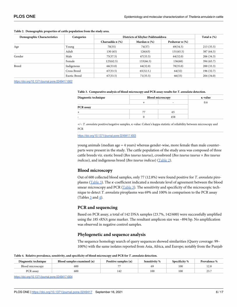
Out of 600 collected blood samples, only 77 (12.8%) were found positive for T. annulata piro-
plasms (Table 3). The κ-coefficient indicated a moderate level of agreement between the blood
smear microscopy and PCR (Table 3). The sensitivity and specificity of the microscopic tech-
nique to detect T. annulata piroplasms was 69% and 100% in comparison to the PCR assay
(Tables 3 and 4).
PCR and sequencing
Based on PCR assay, a total of 142 DNA samples (23.7%, 142/600) were successfully amplified
using the 18S rRNA gene marker. The resultant amplicon size was ~894 bp. No amplification
was observed in negative control samples.
Phylogenetic and sequence analysis
The sequence homology search of query sequences showed similarities (Query coverage: 99–
100%) with the same isolates reported from Asia, Africa, and Europe; notably from the Punjab
Table 2. Demographic properties of cattle population from the study area.
Demographic Characteristics Categories Districts of Khyber Pakhtunkhwa Total n (%)
Charsadda n (%) Mardan n (%) Peshawar n (%)
Age Young 70(35) 74(37) 69(34.5) 213 (35.5)
Adult 130 (65) 126(63) 131(65.5) 387 (64.5)
Gender Male 75(37.5) 67(33.5) 64(32.0) 206 (34.3)
Female 125(62.5) 133(66.5) 136(68) 394 (65.7)
Breed Indigenous 66(33.0) 64(32.0) 70(35.0) 200 (33.3)
Cross Breed 67(33.5) 65(32.5.) 64(32) 196 (32.7)
Exotic Breed 67(33.5) 71(35.5) 66(33) 204 (34.0)
https://doi.org/10.1371/journal.pone.0249417.t002
Table 3. Comparative analysis of blood microscopy and PCR assay results for T. annulata detection.
Diagnostic technique Blood microscopy κ-value
+ - 0.6
PCR assay
+ 77 65
- 0 458
+/-: T. annulata positive/negative samples, κ-value: Cohen’s kappa statistic of reliability between microscopy and
PCR
https://doi.org/10.1371/journal.pone.0249417.t003
Table 4. Relative prevalence, sensitivity, and specificity of blood microscopy and PCR for T. annulata detection.
Diagnostic technique Blood samples examined (n) Positive samples (n) Sensitivity % Specificity % Prevalence %
Blood microscopy 600 77 69 100 12.8
PCR assay 600 142 100 100 23.7
https://doi.org/10.1371/journal.pone.0249417.t004
PLOS ONE Epidemiology and molecular characterization of Theileria annulata in cattle
PLOS ONE | https://doi.org/10.1371/journal.pone.0249417 September 16, 2021 6 / 17
province of Pakistan (Accession No. JQ743630, MG599090), India (Accession No. MK849884,
KT367871), China (Accession No. EU083801, KF559356), Iran (Accession No. KF429800,
KF429799), Turkey (Accession No. AY524666, MG569892) and Spain (Accession No.
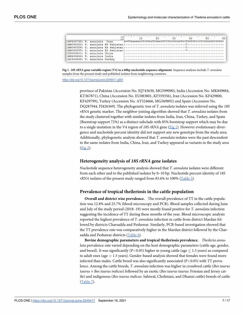
DQ287944, FJ426369). The phylogenetic tree of T. annulata isolates was inferred using the 18SrRNA genetic marker. The neighbor-joining algorithm showed that T. annulata isolates from
the study clustered together with similar isolates from India, Iran, China, Turkey, and Spain
(Bootstrap support 72%) as a distinct subclade with 85% bootstrap support which may be due
to a single mutation in the V4 region of 18S rRNA gene (Fig 1). However evolutionary diver-
gence and nucleotide percent identity did not support any new genotype from the study area.
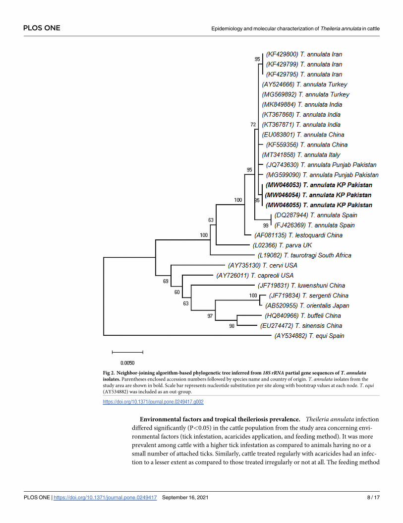
Additionally, phylogenetic analysis showed that T. annulata isolates were the past descendent
to the same isolates from India, China, Iran, and Turkey appeared as variants in the study area
(Fig 2).
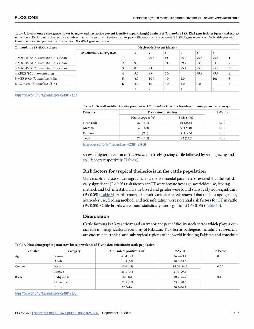
Heterogeneity analysis of 18S rRNA gene isolates
Nucleotide sequence heterogeneity analysis showed that T. annulata isolates were different
from each other and to the published isolates by 0–10 bp. Nucleotide percent identity of 18SrRNA isolates of the present study ranged from 83.6% to 100% (Table 5).
Prevalence of tropical theileriosis in the cattle population
Overall and district wise prevalence. The overall prevalence of TT in the cattle popula-
tion was 12.8% and 23.7% (blood microscopy and PCR). Blood samples collected during June
and July of the study period (2018–19) were mostly found positive for T. annulata infection
suggesting the incidence of TT during these months of the year. Blood microscopic analysis
reported the highest prevalence of T. annulata infection in cattle from district Mardan fol-
lowed by districts Charsadda and Peshawar. Similarly, PCR-based investigation showed that
the TT prevalence rate was comparatively higher in the Mardan district followed by the Char-
sadda and Peshawar districts (Table 6).
Bovine demographic parameters and tropical theileriosis prevalence. Theileria annu-lata prevalence rate varied depending on the host demographic parameters (cattle age, gender,
and breed). It was significantly (P<0.05) higher in young cattle (age� 1.5 years) as compared
to adult ones (age > 1.5 years). Gender-based analysis showed that females were found more
infected than males. Cattle breed was also significantly associated (P<0.05) with TT preva-
lence. Among the cattle breeds, T. annulata infection was higher in crossbred cattle (Bos taurustaurus × Bos taurus indicus) followed by an exotic (Bos taurus taurus: Friesian and Jersey cat-
tle) and indigenous (Bos taurus indicus: Sahiwal, Cholistani, and Dhanni cattle) breeds of cattle
(Table 7).
Fig 1. 18S rRNA gene variable region (V4) in a 60bp nucleotide sequence alignment. Sequence analyses include T. annulatasamples from the present study and published isolates from neighboring countries.
https://doi.org/10.1371/journal.pone.0249417.g001
PLOS ONE Epidemiology and molecular characterization of Theileria annulata in cattle
PLOS ONE | https://doi.org/10.1371/journal.pone.0249417 September 16, 2021 7 / 17
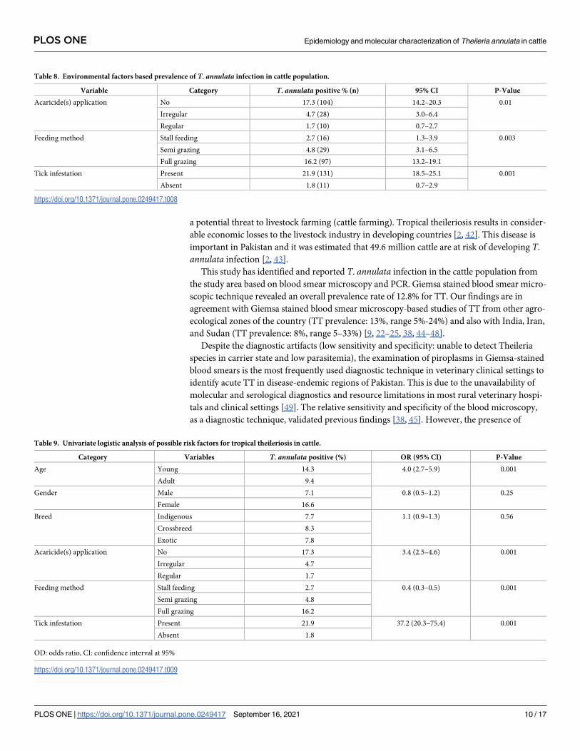
Environmental factors and tropical theileriosis prevalence. Theileria annulata infection
differed significantly (P<0.05) in the cattle population from the study area concerning envi-
ronmental factors (tick infestation, acaricides application, and feeding method). It was more
prevalent among cattle with a higher tick infestation as compared to animals having no or a
small number of attached ticks. Similarly, cattle treated regularly with acaricides had an infec-
tion to a lesser extent as compared to those treated irregularly or not at all. The feeding method
Fig 2. Neighbor-joining algorithm-based phylogenetic tree inferred from 18S rRNA partial gene sequences of T. annulataisolates. Parentheses enclosed accession numbers followed by species name and country of origin. T. annulata isolates from the
study area are shown in bold. Scale bar represents nucleotide substitution per site along with bootstrap values at each node. T. equi(AY534882) was included as an out-group.
https://doi.org/10.1371/journal.pone.0249417.g002
PLOS ONE Epidemiology and molecular characterization of Theileria annulata in cattle
PLOS ONE | https://doi.org/10.1371/journal.pone.0249417 September 16, 2021 8 / 17
showed higher infection of T. annulata in freely grazing cattle followed by semi grazing and
stall feeders respectively (Table 8).
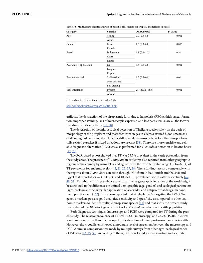
Risk factors for tropical theileriosis in the cattle population
Univariable analysis of demographic and environmental parameters revealed that the statisti-
cally significant (P<0.05) risk factors for TT were bovine host age, acaricides use, feeding
method, and tick infestation. Cattle breed and gender were found statistically non-significant
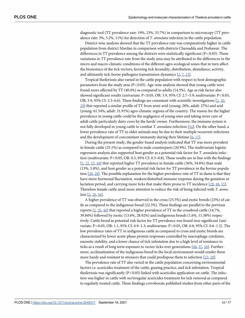
(P>0.05) (Table 9). Furthermore, the multivariable analysis showed that the host age, gender,
acaricides use, feeding method, and tick infestation were potential risk factors for TT in cattle
(P<0.05). Cattle breeds were found statistically non-significant (P>0.05) (Table 10).
Discussion
Cattle farming is a key activity and an important part of the livestock sector which plays a cru-
cial role in the agricultural economy of Pakistan. Tick-borne pathogens including T. annulataare endemic to tropical and subtropical regions of the world including Pakistan and constitute
Table 5. Evolutionary divergence (lower triangle) and nucleotide percent identity (upper triangle) analysis of T. annulata 18S rRNA gene isolates (query and subject
sequences). Evolutionary divergence analysis estimated the number of pair-wise base pairs differences per site between 18S rRNA gene sequences. Nucleotide percent
identity represented percent identity between 18S rRNA gene sequences.
T. annulata 18S rRNA isolates Nucleotide Percent Identity
Evolutionary Divergence 1 2 3 4 5 6
1)MW046053 T. annulata KP Pakistan 1 90.8 100 95.4 95.5 95.5 1
2)MW046054 T. annulata KP Pakistan 2 9.0 90.9 89.7 83.6 83.6 2
3)MW046055 T. annulata KP Pakistan 3 0.0 9.0 95.4 95.5 95.5 3
4)KF429795 T. annulata Iran 4 5.0 9.0 5.0 99.9 99.9 4
5)MK849884 T. annulata India 5 4.0 10.0 4.0 1.0 100 5
6)EU083801 T. annulata China 6 4.0 10.0 4.0 1.0 0.0 6
1 2 3 4 5 6
https://doi.org/10.1371/journal.pone.0249417.t005
Table 6. Overall and district-wise prevalence of T. annulata infection based on microscopy and PCR assays.
Districts T. annulata infection P-Value
Microscopy n (%) PCR n (%)
Charsadda 27 (13.5) 51 (25.5) 0.01
Mardan 32 (16.0) 56 (28.0) 0.01
Peshawar 18 (9.0) 35 (17.5) 0.01
Total 77 (12.8) 142 (23.7) 0.01
https://doi.org/10.1371/journal.pone.0249417.t006
Table 7. Host demographic parameters based prevalence of T. annulata infection in cattle population.
Variable Category T. annulata positive % (n) 95% CI P-Value
Age Young 40.4 (86) 36.5–43.1 0.01
Adult 14.5 (56) 10.1–18.6
Gender Male 20.9 (43) 15.04–24.2 0.27
Female 25.1 (99) 21.6–29.6
Breed Indigenous 23 (46) 20.5–26.7 0.11
Crossbreed 25.5 (50) 23.1–28.5
Exotic 22.5(46) 20.5–26.7
https://doi.org/10.1371/journal.pone.0249417.t007
PLOS ONE Epidemiology and molecular characterization of Theileria annulata in cattle
PLOS ONE | https://doi.org/10.1371/journal.pone.0249417 September 16, 2021 9 / 17
a potential threat to livestock farming (cattle farming). Tropical theileriosis results in consider-
able economic losses to the livestock industry in developing countries [2, 42]. This disease is
important in Pakistan and it was estimated that 49.6 million cattle are at risk of developing T.
annulata infection [2, 43].
This study has identified and reported T. annulata infection in the cattle population from
the study area based on blood smear microscopy and PCR. Giemsa stained blood smear micro-
scopic technique revealed an overall prevalence rate of 12.8% for TT. Our findings are in
agreement with Giemsa stained blood smear microscopy-based studies of TT from other agro-
ecological zones of the country (TT prevalence: 13%, range 5%-24%) and also with India, Iran,
and Sudan (TT prevalence: 8%, range 5–33%) [9, 22–25, 38, 44–48].
Despite the diagnostic artifacts (low sensitivity and specificity: unable to detect Theileria
species in carrier state and low parasitemia), the examination of piroplasms in Giemsa-stained
blood smears is the most frequently used diagnostic technique in veterinary clinical settings to
identify acute TT in disease-endemic regions of Pakistan. This is due to the unavailability of
molecular and serological diagnostics and resource limitations in most rural veterinary hospi-
tals and clinical settings [49]. The relative sensitivity and specificity of the blood microscopy,
as a diagnostic technique, validated previous findings [38, 45]. However, the presence of
Table 8. Environmental factors based prevalence of T. annulata infection in cattle population.
Variable Category T. annulata positive % (n) 95% CI P-Value
Acaricide(s) application No 17.3 (104) 14.2–20.3 0.01
Irregular 4.7 (28) 3.0–6.4
Regular 1.7 (10) 0.7–2.7
Feeding method Stall feeding 2.7 (16) 1.3–3.9 0.003
Semi grazing 4.8 (29) 3.1–6.5
Full grazing 16.2 (97) 13.2–19.1
Tick infestation Present 21.9 (131) 18.5–25.1 0.001
Absent 1.8 (11) 0.7–2.9
https://doi.org/10.1371/journal.pone.0249417.t008
Table 9. Univariate logistic analysis of possible risk factors for tropical theileriosis in cattle.
Category Variables T. annulata positive (%) OR (95% CI) P-Value
Age Young 14.3 4.0 (2.7–5.9) 0.001
Adult 9.4
Gender Male 7.1 0.8 (0.5–1.2) 0.25
Female 16.6
Breed Indigenous 7.7 1.1 (0.9–1.3) 0.56
Crossbreed 8.3
Exotic 7.8
Acaricide(s) application No 17.3 3.4 (2.5–4.6) 0.001
Irregular 4.7
Regular 1.7
Feeding method Stall feeding 2.7 0.4 (0.3–0.5) 0.001
Semi grazing 4.8
Full grazing 16.2
Tick infestation Present 21.9 37.2 (20.3–75.4) 0.001
Absent 1.8
OD: odds ratio, CI: confidence interval at 95%
https://doi.org/10.1371/journal.pone.0249417.t009
PLOS ONE Epidemiology and molecular characterization of Theileria annulata in cattle
PLOS ONE | https://doi.org/10.1371/journal.pone.0249417 September 16, 2021 10 / 17
artifacts, the destruction of the piroplasmic form due to hemolysis (RBCs), thick smear forma-
tion, improper staining, lack of microscopic expertise, and low parasitemia, are all the factors
that diminish its sensitivity [37, 50].
The description of the microscopical detection of Theileria species solely on the basis of
morphology of the piroplasm and macroschizont stages in Giemsa stained blood smears is a
challenging task and should include the differential diagnosis criteria for other morphologi-
cally related parasites if mixed infections are present [51]. Therefore more sensitive and reli-
able diagnostic alternative (PCR) was also performed for T. annulata detection in bovine hosts
[12, 25].
The PCR-based report showed that TT was 23.7% prevalent in the cattle population from
the study areas. The presence of T. annulata in cattle was also reported from other geographic
regions of the country by using PCR and agreed with the expected value range (19 to 66.1%) of
TT prevalence for endemic regions [2, 21, 22, 25, 26]. These findings are also comparable with
the reports about T. annulata detection through PCR from India (Punjab and Odisha) and
Egypt that reported 29.26%, 54.86%, and 10.25% TT prevalence rate in cattle respectively [47,
48, 52]. Variability in TT prevalence rate from diverse geographic localities of the world might
be attributed to the differences in animal demographic (age, gender) and ecological parameters
(agro-ecological zone, irregular application of acaricides and antiprotozoal drugs, manage-
ment practices, etc.) [52]. It has been reported that singleplex PCR targeting the 18S rRNAgenetic markers possess good analytical sensitivity and specificity as compared to other taxo-
nomic markers to identify multiple piroplasms species [11] and that’s why the present study
has preferred the 18S rRNA genetic marker for T. annulata detection in cattle population.
Both diagnostic techniques (microscopy and PCR) were compared for TT during the pres-
ent study. The relative prevalence of TT was 12.8% (microscopy) and 23.7% (PCR). PCR was
found more sensitive than microscopy for the detection of hemoprotozoan parasites in cattle.
However, the κ-coefficient showed a moderate level of agreement between the microscopy and
PCR. A similar comparison was made by multiple surveys from other agro-ecological zones
of Pakistan [23, 25, 53]. According to them, PCR was found a more sensitive and accurate
Table 10. Multivariate logistic analysis of possible risk factors for tropical theileriosis in cattle.
Category Variable OR (CI 95%) P-Value
Age Young 3.9 (2.3–6.6) 0.001
Adult
Gender Male 0.5 (0.3–0.8) 0.006
Female
Breed Indigenous 0.8 (0.6–1.2) 0.31
Cross
Exotic
Acaricide(s) application No 1.4 (0.9–2.0) 0.001
Irregular
Regular
Feeding method Stall feeding 0.7 (0.5–0.9) 0.01
Semi grazing
Full grazing
Tick Infestation Present 25.4 (12.5–56.4) 0.001
Absent
OD: odds ratio, CI: confidence interval at 95%
https://doi.org/10.1371/journal.pone.0249417.t010
PLOS ONE Epidemiology and molecular characterization of Theileria annulata in cattle
PLOS ONE | https://doi.org/10.1371/journal.pone.0249417 September 16, 2021 11 / 17
diagnostic tool (TT prevalence rate: 19%, 23%, 33.7%) in comparison to microscopy (TT prev-
alence rate: 3%, 5.2%, 11%) for detection of T. annulata infection in the cattle population.
District-wise analysis showed that the TT prevalence rate was comparatively higher in cattle
population from district Mardan in comparison with districts Charsadda and Peshawar. The
differences in TT prevalence among the districts were statistically significant (P<0.05). These
variations in TT prevalence rate from the study area may be attributed to the differences in the
micro and macro-climatic conditions of the different agro-ecological zones that in turn affect
the bionomics of the tick vectors; favoring tick fecundity, distribution, abundance, activity,
and ultimately tick-borne pathogens transmission dynamics [2, 7, 23].
Tropical theileriosis also varied in the cattle population with respect to host demographic
parameters from the study area (P<0.05). Age-wise analysis showed that young cattle were
found more affected by TT (40.4%) as compared to adults (14.5%). Age as risk factor also
showed significant results (univariate: P<0.05, OR: 3.9, 95% CI: 2.7–5.9; multivariate: P<0.05,
OR: 3.9, 95% CI: 2.3–6.6). These findings are consistent with scientific investigations [2, 20,
29] that reported a similar profile of TT from semi-arid (young: 28%, adult: 27%) and arid
(young: 61.54%, adult: 31.91%) agro-climatic regions of the country. The reason for the higher
prevalence in young cattle could be the negligence of young ones and taking more care of
adult cattle particularly dairy cows by the herds’ owner. Furthermore, the immune system is
not fully developed in young cattle to combat T. annulata infection [54]. On the other hand, a
lower prevalence rate of TT in older animals may be due to their multiple recurrent infections
and the development of concomitant immunity during their lifetime [6, 17].
During the present study, the gender-based analysis indicated that TT was more prevalent
in female cattle (25.1%) as compared to male counterparts (20.9%). The multivariate logistic
regression analysis also supported host gender as a potential risk factor for T. annulata infec-
tion (multivariate: P<0.05, OR: 0.5, 95% CI: 0.3–0.8). These results are in line with the findings
[2, 22, 25, 48] that reported higher TT prevalence in female cattle (36%, 34.8%) than male
(13%, 5.8%), and host gender as a potential risk factor for TT prevalence in the bovine popula-
tion [20, 28]. The possible explanation for the higher prevalence rate of TT in dams is that they
have more hormonal fluctuation, weaken/disturbed immune response during the gestation or
lactation period, and carrying more ticks that make them prone to TT incidence [28, 48, 55].
Therefore female cattle need more attention to reduce the risk of being infected with T. annu-lata [2, 20, 56].
A higher prevalence of TT was observed in the cross (25.5%) and exotic breeds (23%) of cat-
tle as compared to the indigenous breed (22.5%). These findings are parallel to the previous
reports [2, 20, 48] that reported a higher prevalence of TT in the crossbred cattle (14.7%,
39.84%) followed by exotic (13.6%, 28.92%) and indigenous breeds (1.6%, 11.58%) respec-
tively. Cattle breed as potential risk factor for TT prevalence was found non-significant (uni-
variate: P>0.05, OR: 1.1, 95% CI: 0.9–1.3; multivariate: P>0.05, OR: 0.8, 95% CI: 0.6–1.2). The
low prevalence rates of TT in indigenous cattle as compared to cross and exotic breeds are
characterized by lower acute-phase protein responses controlled by macrophage cytokines,
enzootic stability, and a lower chance of tick infestation due to a high level of resistance to
ticks as a result of long term exposure to vector ticks over generations [48, 57, 58]. Further-
more, acclimatization of the indigenous breed to the local environment would render them
more hardy and resistant to stressors that could predispose them to infection [23, 28].
The prevalence rate of TT also varied in the cattle population concerning environmental
factors i.e. acaricides treatment of the cattle, grazing practice, and tick infestation. Tropical
theileriosis was significantly (P<0.05) linked with acaricides application on cattle. The infec-
tion was higher in cattle with no/irregular acaricides treatment for tick removal as compared
to regularly treated cattle. These findings corroborate published studies from other parts of the
PLOS ONE Epidemiology and molecular characterization of Theileria annulata in cattle
PLOS ONE | https://doi.org/10.1371/journal.pone.0249417 September 16, 2021 12 / 17
country and elsewhere [2, 20, 47, 59], that reported lower T. annulata infection rate in herds
where cattle were regularly treated with acaricides for ticks control. Acaricides application was
also evaluated as potential risk factor that influence TT prevalence in the cattle population
(univariate: P<0.05, OR: 3.4, 95% CI: 2.5–4.6; multivariate: P<0.05, OR: 1.4, 95% CI: 0.9–2.0).
Acaricides application as a potential risk factor for TT agreed with the assessment of the pub-
lished literature from other agro-ecological zones of the country, which presented that regular
and proper acaricidal application on cattle could control tick vector and ultimately TT inci-
dence in the endemic region [2, 20, 45].
The grazing practice of cattle was significantly linked (P<0.05) with the prevalence rate of
TT in bovine. Tropical theileriosis was least prevalent in cattle with stall feeding activity (kept
in farms with forage availability) as compared to cattle with semi-grazing and full grazing prac-
tices. These findings are identical with reports about the impact of grazing practice on TT
prevalence in bovine animals [2, 20, 47, 56]. Logistic regression analyses also indicated that the
feeding method is the critical risk factor influencing TT prevalence (univariate: P<0.05, OR:
0.4 95% CI: 0.3–0.5; multivariate: P<0.05, OR: 0.7, 95% CI: 0.5–0.9). Similar results were also
reported from other parts of the country but least support is available to feeding habits as a
potential risk factor for TT in the country [2, 8]. It is also known that some farmers from the
study area allow their cattle free grazing that increases contact frequency with animals from
other herds, facilitating ticks’ exchange that results in increased T. annulata infection. How-
ever stall feeders’ cattle that are kept in fences/farms and are not allowed to roam freely, reduce
their exposure to tick sources like other tick-infested animals, plants and grasses in the grazing
field where ticks ambush and wait for attachment to a bovine host [2, 60].
During the present study, it was reported that most of the ticks-infested cattle were found
infected with T. annulata as compared to cattle with minimal or no tick infestation (P<0.05).
Tick infestation on cattle as a potent risk factor also affected T. annulata transmission dynam-
ics (univariate: P<0.05, OR: 37.2, 95% CI: 20.3–75.4; multivariate: P<0.05, OR: 25.4, 95% CI:
12.5–56.4). Our results support previously published literature from Pakistan and elsewhere
about tick infestation and its possible impact on TT prevalence pattern in cattle as a potential
risk factor [2, 20, 27, 43, 56, 61–63]. Published literature indicates that more frequent transmis-
sion of T. annulata occurs when a suitable vector and infected host exist in close proximity
[45]. In Pakistan, T. annulata is principally transmitted by H. anatolicum to bovine animals.
Other studies from Pakistan further validated the presence of T. annulata in H. anatolicum(collected from cattle) and bovine host [27, 43].
Theileria annulata evolutionary history was inferred through 18S rRNA taxonomic marker.
Homology searches of the 18S rRNA isolates shared 99–100% similarities with local and global
isolates published in NCBI GenBank. The neighbor-joining algorithm grouped the present
study 18S rRNA isolates into T. annulata clade, which include similar isolates published from
other agro-ecological zones of Pakistan, Asia, and Europe; suggesting that these are closely
related genetic variants. Similarly, 18S rRNA genetic marker has been previously used by sev-
eral studies in Pakistan and elsewhere globally to identify and establish the phylogenetic profile
of T. annulata circulating in bovine hosts [2, 26, 64–66]. Our findings support and validate
them. Target amplification of the hypervariable V4 region of the 18S rRNA gene is a preferred
option to accurately identify, classify and explore the population structures of the piroplasm
parasites [67, 68].
Conclusion
This research work investigated the molecular epidemiology of T. annulata in the cattle popu-
lation from central Khyber Pakhtunkhwa Pakistan and inferred the evolutionary history of T.
PLOS ONE Epidemiology and molecular characterization of Theileria annulata in cattle
PLOS ONE | https://doi.org/10.1371/journal.pone.0249417 September 16, 2021 13 / 17
annulata based on an 18S rRNA taxonomic marker. Theileria annulata isolates shared similari-
ties and phylogeny with similar sequences reported from China, India, Iran, Turkey, and
Spain. Additionally, the epidemiologic profile of T. annulata in cattle breeds was assessed and
potential risk factors for tropical theileriosis were also determined. This research work has pro-
vided data on T. annulata phylogeny and epidemiology in bovine hosts from central Khyber
Pakhtunkhwa Pakistan. These findings will serve as a baseline and will facilitate future large-
scale epidemiological investigations on TT at local and global levels.
Acknowledgments
We are grateful to the local veterinarians who helped during the sample collection. We would
also like to thank Dr. Michael J. Turell for his technical assistance.
Author Contributions
Data curation: Raqeeb Ullah.
Formal analysis: Jehan Zeb.
Methodology: Raqeeb Ullah.
Project administration: Raqeeb Ullah.
Software: Qeyam ud Din.
Supervision: Sumaira Shams, Sultan Ayaz.
Visualization: Munsif Ali Khan, Noor ul Akbar, Adil Khan.
Writing – original draft: Raqeeb Ullah.
Writing – review & editing: Renato Leon, Jehan Zeb.
References1. Lew-Tabor A, Valle MR. A review of reverse vaccinology approaches for the development of vaccines
against ticks and tick borne diseases. Ticks and Tick-Borne Diseases. 2016; 7(4):573–85. https://doi.
org/10.1016/j.ttbdis.2015.12.012 PMID: 26723274
2. Zeb J, Shams S, Din IU, Ayaz S, Khan A, Nasreen N, et al. Molecular epidemiology and associated risk
factors of Anaplasma marginale and Theileria annulata in cattle from North-western Pakistan. Veteri-
nary parasitology. 2020; 279:109044. https://doi.org/10.1016/j.vetpar.2020.109044 PMID: 32032840
3. Bilgic HB, Karagenc T, Shiels B, Tait A, Eren H, Weir W. Evaluation of cytochrome b as a sensitive tar-
get for PCR based detection of T. annulata carrier animals. Veterinary parasitology. 2010; 174:341–7.
https://doi.org/10.1016/j.vetpar.2010.08.025 PMID: 20880635
4. Purnell R. Theileria annulata as a hazard to cattle in countries on the northern Mediterranean littoral.
Veterinary Science Communications. 1978; 2(1):3–10.
5. Bishop RP, Odongo DO, Mann DJ, Pearson TW, Sugimoto C, Haines LR, et al. Theileria. Genome
mapping and genomics in animal-associated microbes: Springer; 2009. p. 191–231.
6. Gharbi M, Darghouth MA. A review of Hyalomma scupense (Acari, Ixodidae) in the Maghreb region:
from biology to control. Parasite. 2014; 21:12–18.
7. Ali A, Khan MA, Zahid H, Yaseen PM, Khan MQ, Nawab J, et al. Seasonal dynamics, record of ticks
infesting humans, wild and domestic animals and molecular phylogeny of Rhipicephalus microplus in
Khyber Pakhtunkhwa Pakistan. Frontiers in physiology. 2019; 10. https://doi.org/10.3389/fphys.2019.
00793 PMID: 31379587
8. Rehman A, Nijhof AM, Sauter-Louis C, Schauer B, Staubach C, Conraths FJ. Distribution of ticks infest-
ing ruminants and risk factors associated with high tick prevalence in livestock farms in the semi-arid
and arid agro-ecological zones of Pakistan. Parasites & vectors. 2017; 10(1):190. https://doi.org/10.
1186/s13071-017-2138-0 PMID: 28420420
9. Zahid I, Latif M, Baloch K. Incidence and treatment of theileriasis and babesiasis. Pakistan Veterinary
Journal. 2005; 25(3):137.
PLOS ONE Epidemiology and molecular characterization of Theileria annulata in cattle
PLOS ONE | https://doi.org/10.1371/journal.pone.0249417 September 16, 2021 14 / 17
10. Robinson P. Theileriosis annulata and its transmission—a review. Tropical animal health and produc-
tion. 1982; 14(1):3–12. https://doi.org/10.1007/BF02281092 PMID: 6805112
11. Brown C. Tropical theileriosis. In: Brown C, Torres A (Eds.), Foreign Animal Diseases, 7th ed., Publica-
tions Boca, Florida USA. 2008; 401–404.
12. Hayati M, Hassan S, Ahmed S, Salih D. Prevalence of ticks (Acari: Ixodidae) and Theileria annulata
infection of cattle in Gezira State, Sudan. Parasite Epidemiology and Control. 2020:e00148. https://doi.
org/10.1016/j.parepi.2020.e00148 PMID: 32420464
13. Pienaar R, Troskie PC, Josemans AI, Potgieter FT, Maboko BB, Latif AA, et al. Investigations into the
carrier-state of Theileria sp. (buffalo) in cattle. International journal for parasitology Parasites and wild-
life. 2020; 11:136–42. https://doi.org/10.1016/j.ijppaw.2020.01.009 PMID: 32071860
14. Cacciò S, CammàC, Onuma M, Severini C. The β-tubulin gene of Babesia and Theileria parasites is an
informative marker for species discrimination. International Journal for Parasitology. 2000; 30
(11):1181–5. https://doi.org/10.1016/s0020-7519(00)00105-3 PMID: 11027785
15. De Kok J, d’Oliveira C, Jongejan F. Detection of the protozoan parasite Theileria annulata in Hyalomma
ticks by the polymerase chain reaction. Experimental & applied acarology. 1993; 17:839–46. https://doi.
org/10.1007/BF00225857 PMID: 7628228
16. d’Oliveira C, Van Der Weide M, Habela MA, Jacquiet P, Jongejan F. Detection of Theileria annulata in
blood samples of carrier cattle by PCR. Journal of Clinical Microbiology. 1995; 33(10):2665–9. https://
doi.org/10.1128/jcm.33.10.2665-2669.1995 PMID: 8567902
17. Ilhan T, Williamson S, Kirvar E, Shiels B, Brown C. Theileria annulata: Carrier State and Immunity a.
Annals of the New York Academy of Sciences. 1998; 849(1):109–25.
18. Leemans I, Brown D, Hooshmand-Rad P, Kirvar E, Uggla A. Infectivity and cross-immunity studies of
Theileria lestoquardi and Theileria annulata in sheep and cattle: I. In vivo responses. Veterinary parasi-
tology. 1999; 82(3):179–92. https://doi.org/10.1016/s0304-4017(99)00013-8 PMID: 10348097
19. Shayan P, Biermann R, Schein E, Gerdes J, Ahmed J. Detection and Differentiation of Theileria
annulata and Theileria parva Using Macroschizont-derived DNA Probes. Annals of the New York Acad-
emy of Sciences. 1998; 849(1):88–95. https://doi.org/10.1111/j.1749-6632.1998.tb11038.x PMID:
9668454
20. Farooqi S, Ijaz M, Saleem M, Rashid M, Ahmad S, Islam S, et al. Prevalence and molecular diagnosis
of Theileria annulata in bovine from three distinct zones of Khyber Pakhtunkhwa province, Pakistan.
JAPS, Journal of Animal and Plant Sciences. 2017; 27(6):1836–41.
21. Jabbar A, Abbas T, Saddiqi HA, Qamar MF, Gasser RB. Tick-borne diseases of bovines in Pakistan:
major scope for future research and improved control. Parasites & vectors. 2015; 8(1):283. https://doi.
org/10.1186/s13071-015-0894-2 PMID: 25994588
22. Durrani A, Kamal N. Identification of ticks and detection of blood protozoa in Friesian cattle by polymer-
ase chain reaction test and estimation of blood parameters in district Kasur, Pakistan. Tropical animal
health and production. 2008; 40(6):441–7. https://doi.org/10.1007/s11250-007-9117-y PMID:
18575972
23. Durrani A, Mehmood N, Shakoori A. Comparison of three diagnostic methods for Theileria annulata in
Sahiwal and Friesian cattle in Pakistan. Pak J Zool. 2010; 42(4):467–72.
24. Khan A, Ashfaq K, ud Din I, ul Haq R, Jamil M, Ullah B, et al. Bovine theileriosis: Prevalence, estimation
of hematological profile and chemotherapy in cattle in Dera Ismail Khan, Khyber Pakhtunkhwa Prov-
ince, Pakistan. American Scientific Research Journal for Engineering, Technology, and Sciences (ASR-
JETS). 2017; 32(1):8–17.
25. Khattak R, Rabib M, Khan Z, Ishaq M, Hameed H, Taqddus A, et al. A comparison of two different tech-
niques for the detection of blood parasite, Theileria annulata, in cattle from two districts in Khyber Pukh-
toon Khwa Province (Pakistan). Parasite: journal de la Societe Francaise de Parasitologie. 2012; 19
(1):91.
26. Khan MK, He L, Hussain A, Azam S, Zhang W-J, Wang L-X, et al. Molecular epidemiology of Theileria
annulata and identification of 18S rRNA gene and ITS regions sequences variants in apparently healthy
buffaloes and cattle in Pakistan. Infection, Genetics, and Evolution. 2013; 13:124–32. https://doi.org/10.
1016/j.meegid.2012.09.007 PMID: 23059196
27. Zeb J, Szekeres S, Takacs N, Kontschan J, Shams S, Ayaz S, et al. Genetic diversity, piroplasms, and
trypanosomes in Rhipicephalus microplus and Hyalomma anatolicum collected from cattle in northern
Pakistan. Experimental and Applied Acarology. 2019; 79(2):233–43. https://doi.org/10.1007/s10493-
019-00418-9 PMID: 31578647
28. Parveen A, Ashraf S, Aktas M, Ozubek S, Iqbal F. Molecular epidemiology of Theileria annulata infec-
tion of cattle in Layyah District, Pakistan. Experimental and Applied Acarology. 2021; 83(3):461–73.
https://doi.org/10.1007/s10493-021-00595-6 PMID: 33599889
PLOS ONE Epidemiology and molecular characterization of Theileria annulata in cattle
PLOS ONE | https://doi.org/10.1371/journal.pone.0249417 September 16, 2021 15 / 17
29. Qayyum M, Farooq U, Samad HA, Chauhdry HR. Prevalence, clinicotherapeutic and prophylactic stud-
ies on theileriosis in district Sahiwal (Pakistan). Journal of Animal & Plant Sciences. 2010; 20(4):266–
270
30. Javid K, Akram MAN, Mumtaz M, Siddiqui R. Modeling and mapping of climatic classification of Paki-
stan by using remote sensing climate compound index (2000 to 2018). Applied Water Science. 2019; 9
(7):1–9.
31. Zomer RJ, Trabucco A, Bossio DA, Verchot LV. Climate change mitigation: A spatial analysis of global
land suitability for clean development mechanism afforestation and reforestation. Agriculture, ecosys-
tems & environment. 2008; 126(1–2):67–80.
32. Shamshad KM. The meteorology of Pakistan: climate and weather of Pakistan: Royal Book Company;
1988.
33. Pakistan Economic Survey 2019–20. https://www.finance.gov.pk/survey_1920.html
34. Pakistan Livestock Census, 2006. https://www.pbs.gov.pk/content/pakistan-livestock-census-2006
35. Thrusfield M. Veterinary epidemiology: John Wiley & Sons; 2018.
36. Benjamin M. Outline of Veterinary Clinical Pathology, 3rdEdn. Iowa State University Press, Ames,
USA; 1986.
37. Nayel M, El-Dakhly KM, Aboulaila M, Elsify A, Hassan H, Ibrahim E, et al. The use of different diagnostic
tools for Babesia and Theileria parasites in cattle in Menofia, Egypt. Parasitology research. 2012; 111
(3):1019–24. https://doi.org/10.1007/s00436-012-2926-6 PMID: 22543747
38. Noaman V. Comparison of molecular and microscopic technique for detection of Theileria spp. in carrier
cattle. Journal of parasitic diseases. 2014; 38(1):64–7. https://doi.org/10.1007/s12639-012-0196-y
PMID: 24505180
39. Thompson JD, Higgins DG, Gibson TJ. CLUSTAL W: improving the sensitivity of progressive multiple
sequence alignment through sequence weighting, position-specific gap penalties and weight matrix
choice. Nucleic acids research. 1994; 22(22):4673–80. https://doi.org/10.1093/nar/22.22.4673 PMID:
7984417
40. Kumar S, Stecher G, Li M, Knyaz C, Tamura K. MEGA X: molecular evolutionary genetics analysis
across computing platforms. Molecular biology and evolution. 2018; 35(6):1547–9. https://doi.org/10.
1093/molbev/msy096 PMID: 29722887
41. Altschul SF, Gish W, Miller W, Myers EW, Lipman DJ. Basic local alignment search tool. Journal of molec-
ular biology. 1990; 215(3):403–10. https://doi.org/10.1016/S0022-2836(05)80360-2 PMID: 2231712
42. Uilenberg G. Babesiosis. Encyclopedia of Arthropod-transmitted Infections of Man and Domesticated
Animals. In. CABI Publishing, Wallingford; 2001.
43. Rehman A, Conraths FJ, Sauter-Louis C, Krucken J, Nijhof AM. Epidemiology of tick-borne pathogens
in the semi-arid and the arid agro-ecological zones of Punjab province, Pakistan. Transboundary and
emerging diseases. 2019; 66(1):526–36. https://doi.org/10.1111/tbed.13059 PMID: 30383917
44. Azizi H, Shiran B, Dehkordi AF, Salehi F, Taghadosi C. Detection of Theileria annulata by PCR and its
comparison with smear method in native carrier cows. Biotechnology. 2008; 7(3):574–7.
45. Chauhan H, Patel B, Bhagat A, Patel M, Patel S, Raval S, et al. Comparison of molecular and micro-
scopic technique for detection of Theileria annulata from the field cases of cattle. Veterinary world.
46. Salih DA, Hassan S, El Hussein AM. Comparisons among two serological tests and microscopic exami-
nation for the detection of Theileria annulata in cattle in northern Sudan. Preventive veterinary medicine.
2007; 81(4):323–6. https://doi.org/10.1016/j.prevetmed.2007.05.008 PMID: 17590458
47. Selim A, Das M, Senapati S, Jena G, Mishra C, Mohanty B, et al. Molecular epidemiology, risk factors
and hematological evaluation of asymptomatic Theileria annulata infected cattle in Odisha, India. Ira-
nian Journal of Veterinary Research. 2020; 21(4):250–6. PMID: 33584836
48. Tuli A, Singla LD, Sharma A, Bal MS, Filia G, Kaur P. Molecular epidemiology, risk factors and hemato-
chemical alterations induced by Theileria annulata in bovines of Punjab (India). Acta Parasitologica.
2015 Sep 1; 60(3):378–90. https://doi.org/10.1515/ap-2015-0053 PMID: 26204174
49. Hassan MA, Liu J, Sajid MS, Rashid M, Mahmood A, Abbas Q, et al. Simultaneous detection of Thei-
leria annulata and Theileria orientalis infections using recombinase polymerase amplification. Ticks and
tick-borne diseases. 2018; 9(4):1002–5. https://doi.org/10.1016/j.ttbdis.2018.03.028 PMID: 29625920
50. Hoghooghi-Rad N, Ghaemi P, Shayan P, Eckert B, Sadr-Shirazi N. Detection of native carrier cattle
infected with Theileria annulata by semi-nested PCR and smear method in Golestan Province of Iran.
World Appl Sci J. 2011; 12(3):317–23.
51. Dumanli N, Aktas M, Cetinkaya B, Cakmak A, Koroglu E, Saki C, et al. Prevalence and distribution of
tropical theileriosis in eastern Turkey. Veterinary parasitology. 2005; 127(1):9–15. https://doi.org/10.
1016/j.vetpar.2004.08.006 PMID: 15619369
PLOS ONE Epidemiology and molecular characterization of Theileria annulata in cattle
PLOS ONE | https://doi.org/10.1371/journal.pone.0249417 September 16, 2021 16 / 17
52. Rizk MA, Salama A, El-Sayed SA, Elsify A, El-Ashkar M, Ibrahim H, et al. Animal level risk factors asso-
ciated with Babesia and Theileria infections in cattle in Egypt. Acta parasitologica. 2017 Dec 1; 62
(4):796–804. https://doi.org/10.1515/ap-2017-0096 PMID: 29035848
53. Shahnawaz S, Ali M, Aslam M, Fatima R, Chaudhry Z, Hassan M, et al. A study on the prevalence of a
tick-transmitted pathogen, Theileria annulata, and hematological profile of cattle from Southern Punjab
(Pakistan). Parasitology research. 2011; 109(4):1155–60. https://doi.org/10.1007/s00436-011-2360-1
PMID: 21451992
54. Ahmed JS, Glass EJ, Salih DA, Seitzer U. Innate immunity to tropical theileriosis. Innate Immunity.
2008; 14(1):5–12. https://doi.org/10.1177/1753425907087258 PMID: 18387915
55. Kabir MH, Mondal MM, Eliyas M, Mannan MA, Hashem MA, Debnath NC, et al. An epidemiological sur-
vey on investigation of tick infestation in cattle at Chittagong District, Bangladesh. African Journal of
Microbiology Research. 2011; 5(4):346–352.
56. Moumouni PFA, Aplogan GL, Katahira H, Gao Y, Guo H, Efstratiou A, et al. Prevalence, risk factors,
and genetic diversity of veterinary important tick-borne pathogens in cattle from Rhipicephalus micro-
plus-invaded and non-invaded areas of Benin. Ticks and tick-borne diseases. 2018; 9(3):450–64.
https://doi.org/10.1016/j.ttbdis.2017.12.015 PMID: 29307783
57. Glass EJ. The balance between protective immunity and pathogenesis in tropical theileriosis: what we
need to know to design effective vaccines for the future. Research in veterinary science. 2001; 70
(1):71–75. https://doi.org/10.1053/rvsc.2000.0428 PMID: 11170856
58. Moll G, Lohding A, Young AS. Epidemiology of theilerioses in the Trans-Mara Division, Kenya: hus-
bandry and disease background and preliminary investigations on theilerioses in calves. Preventive
Veterinary Medicine. 1984; 2(6):801–831.
59. De Meneghi D, Stachurski F, Adakal H. Experiences in tick control by acaricide in the traditional cattle
sector in Zambia and Burkina Faso: possible environmental and public health implications. Frontiers in
public health. 2016; 4:239. https://doi.org/10.3389/fpubh.2016.00239 PMID: 27882313
60. Calleja-Bueno L, Sainz A, Garcıa-Sancho M, Rodrıguez-Franco F, Gonzalez-Martın JV, Villaescusa A.
Molecular, epidemiological, haematological and biochemical evaluation in asymptomatic Theileria
annulata infected cattle from an endemic region in Spain. Ticks and tick-borne diseases. 2017; 8
(6):936–941. https://doi.org/10.1016/j.ttbdis.2017.08.006 PMID: 28887101
61. Inci A, Ica A, Yildirim A, Vatansever Z, Cakmak A, Albasan H, et al. Epidemiology of tropical theileriosis
in the Cappadocia region. Turkish Journal of Veterinary and Animal Sciences. 2008; 32(1):57–64.
62. Sajid MS, Iqbal Z, Khan MN, Muhammad G, Khan MK. Prevalence and associated risk factors for
bovine tick infestation in two districts of lower Punjab, Pakistan. Preventive veterinary medicine. 2009;
92(4):386–91. https://doi.org/10.1016/j.prevetmed.2009.09.001 PMID: 19782414
63. Ponnudurai G, Larcombe S, Velusamy R. Prevalence of tick-borne pathogens in co-grazed dairy
bovines differs by region and host-type in Tamil Nadu; India. Journal of advance dairy research. 2017;
5:177.
64. George N, Bhandari V, Reddy DP, Sharma P. Molecular and Phylogenetic analysis revealed new geno-
types of Theileria annulata parasites from India. Parasites & vectors. 2015; 8(1):1–8. https://doi.org/10.
1186/s13071-015-1075-z PMID: 26381127
65. Hassan MA, Liu J, Rashid M, Iqbal N, Guan G, Yin H, et al. Molecular survey of piroplasm species from
selected areas of China and Pakistan. Parasites & vectors. 2018; 11(1):1–7. https://doi.org/10.1186/
s13071-018-3035-x PMID: 30086785
66. Ros-Garcıa A, Nicolas A, Garcıa-Perez AL, Juste RA, Hurtado A. Development and evaluation of a
real-time PCR assay for the quantitative detection of Theileria annulata in cattle. Parasites & vectors.
2012; 5(1):1–8. https://doi.org/10.1186/1756-3305-5-171 PMID: 22889141
67. Chaudhry U, Ali Q, Rashid I, Shabbir MZ, Ijaz M, Abbas M, et al. Development of a deep amplicon
sequencing method to determine the species composition of piroplasm haemoprotozoa. Ticks and tick-
borne diseases. 2019; 10(6):101276. https://doi.org/10.1016/j.ttbdis.2019.101276 PMID: 31473098
68. Glidden CK, Koehler AV, Hall RS, Saeed MA, Coppo M, Beechler BR, et al. Elucidating cryptic dynam-
ics of Theileria communities in African buffalo using a high-throughput sequencing informatics
approach. Ecology and evolution. 2020; 10(1):70–80. https://doi.org/10.1002/ece3.5758 PMID:
31988717
PLOS ONE Epidemiology and molecular characterization of Theileria annulata in cattle
PLOS ONE | https://doi.org/10.1371/journal.pone.0249417 September 16, 2021 17 / 17