ephb4 inhibition activates er stress to promote immunogenic
TRANSCRIPT
Sagar et al. Cell Death and Disease (2019) 10:801
https://doi.org/10.1038/s41419-019-2042-y Cell Death & Disease
ART ICLE Open Ac ce s s
EPHB4 inhibition activates ER stress to promoteimmunogenic cell death of prostate cancer cellsVinay Sagar1, Rajita Vatapalli1, Barbara Lysy1, Sahithi Pamarthy2, Jonathan F. Anker1, Yara Rodriguez1, Huiying Han1,Kenji Unno1, Walter M. Stadler3, William J. Catalona4, Maha Hussain5, Parkash S. Gill6 and Sarki A. Abdulkadir1,7
AbstractThe EPHB4 receptor is implicated in the development of several epithelial tumors and is a promising therapeutictarget, including in prostate tumors in which EPHB4 is overexpressed and promotes tumorigenicity. Here, we showthat high expression of EPHB4 correlated with poor survival in prostate cancer patients and EPHB4 inhibition inducedcell death in both hormone sensitive and castration-resistant prostate cancer cells. EPHB4 inhibition reducedexpression of the glucose transporter, GLUT3, impaired glucose uptake, and reduced cellular ATP levels. This wasassociated with the activation of endoplasmic reticulum stress and tumor cell death with features of immunogenic celldeath (ICD), including phosphorylation of eIF2α, increased cell surface calreticulin levels, and release of HMGB1 andATP. The changes in tumor cell metabolism after EPHB4 inhibition were associated with MYC downregulation, likelymediated by the SRC/p38 MAPK/4EBP1 signaling cascade, known to impair cap-dependent translation. Together, ourstudy indicates a role for EPHB4 inhibition in the induction of immunogenic cell death with implication for prostatecancer therapy.
IntroductionProstate cancer (PC) is the most common cancer in
men worldwide. An estimated 174,650 new cases and31,620 new deaths will be reported in the United States in20191. While therapy targeted at the androgen receptorpathway shows initial efficacy, a majority of patients withadvanced PC go on to develop resistance2–4. Thus, there isa need for novel therapies that target additional pathwaysimportant for the growth and survival of prostate cancercells. For example, tyrosine kinase inhibitors (TKIs) arebeing investigated as an additional strategy to treatprostate cancer5–8. Receptor tyrosine kinases (RTKs)regulate of cell proliferation, migration, apoptosis, andsurvival, and plays a role in the progression and devel-opment of multiple cancers9,10. Targeting RTKs
enzymatic activity is emerging as a therapeutic strategy inmany cancer. However, this approach is facing someconstraints due to development resistance against theseinhibitors11,12. Combination therapy approaches have tobe developed to overcome this problem.EPHB4 receptor tyrosine kinase is involved in many
cellular processes such as cell growth, survival, angio-genesis, vasculogenesis, and neural development13.EPHB4 specifically binds to Ephrin B2 ligand and evokesbidirectional signaling13. Several reports have shownEPHB4 overexpression in various cancers including thoseof the prostate, breast, colon, head and neck, and lung14–18.Xi et al.19 showed that EPHB4 expression is increased in66% of prostate tumor samples. EPHB4 is induced by thekey molecular alterations implicated in prostate cancer andcastrate-resistant prostate cancer development, includingloss of tumor suppressors PTEN and TP53, and activationof the PI3K pathway19. Another report demonstrated thatEPHB4 knockdown by siRNA resulted in a decrease ininvasion and migration of prostate cancer cell lines20.However, neither the underlying mechanisms nor the
© The Author(s) 2019OpenAccessThis article is licensedunder aCreativeCommonsAttribution 4.0 International License,whichpermits use, sharing, adaptation, distribution and reproductionin any medium or format, as long as you give appropriate credit to the original author(s) and the source, provide a link to the Creative Commons license, and indicate if
changesweremade. The images or other third partymaterial in this article are included in the article’s Creative Commons license, unless indicated otherwise in a credit line to thematerial. Ifmaterial is not included in the article’s Creative Commons license and your intended use is not permitted by statutory regulation or exceeds the permitted use, you will need to obtainpermission directly from the copyright holder. To view a copy of this license, visit http://creativecommons.org/licenses/by/4.0/.
Correspondence: Sarki A. Abdulkadir ([email protected])1Department of Urology, The Robert H. Lurie Comprehensive Cancer Center,Northwestern University Feinberg School of Medicine, Chicago, IL 60611, USA2Atrin Pharmaceuticals, Pennsylvania Biotechnology Center, Doylestown, PA18902, USAFull list of author information is available at the end of the article.Edited by R. Aqeilan
Official journal of the Cell Death Differentiation Association
1234
5678
90():,;
1234
5678
90():,;
1234567890():,;
1234
5678
90():,;
functional consequences of EPHB4 inhibition in advancedprostate cancer are well understood. Here, we investigatethe mechanisms by which EPHB4 inhibition affects cellviability. EPHB4 receptor inhibition in PC cells inhibited aSRC/MAPK/MYC pathways leading to reduced glucosetransporter GLUT3 levels, decreased glucose uptake andintracellular ATP levels. Consequently, cells showed ele-vated ER stress and death with features of immunogeniccell death (ICD).
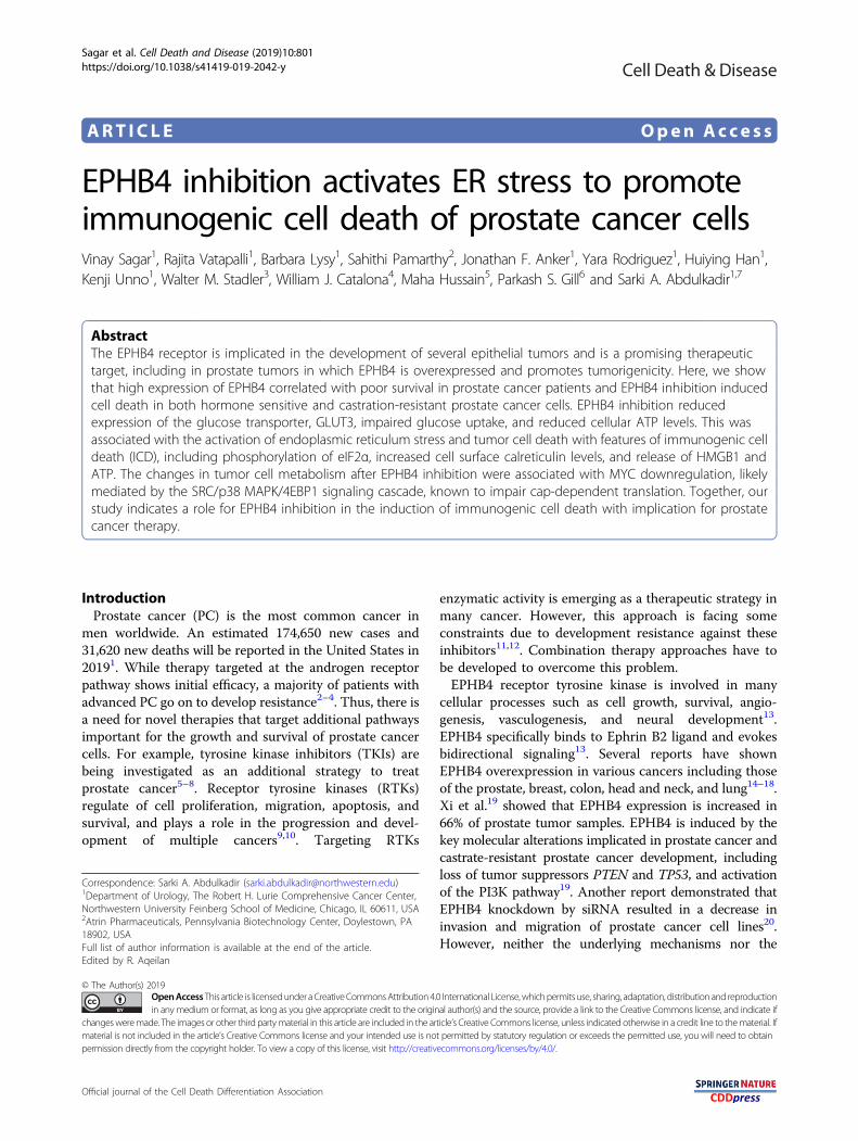
ResultsHigh EPHB4 expression correlates with advanced prostatecancer stage and poor outcomePrevious studies have shown that EPHB4 promotes
tumor cell survival and is overexpressed in prostate can-cer19,20. We examined EPHB4 mRNA expression inpublished human prostate cancer expression datasets andobserved higher EPHB4 expression in metastaticcastration-resistant prostate cancer (mCRPC) as com-pared to the corresponding normal or primary tumortissues (Fig. 1a). We also observe a positive correlationbetween EPHB4 expression levels and biochemical
relapse-free survival in both the Cancer Genome Atlas(TCGA) and Ross-Adams21 datasets (Fig. 1b). Collec-tively, these results indicate that EPHB4 is a valuableprognostic biomarker and raises the hypothesis that itcould be a therapeutic target in prostate cancer patients.
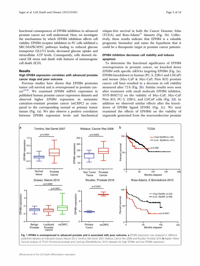
EPHB4 inhibition decreases cell viability and inducesapoptosisTo determine the functional significance of EPHB4
overexpression in prostate cancer, we knocked downEPHB4 with specific siRNAs targeting EPHB4 (Fig. 2a).EPHB4 knockdown in human (PC-3, 22Rv1 and LNCaP)and mouse (Myc-CaP & Myc-CaP; Pten KO) prostatecancer cell lines resulted in a decrease in cell viabilitymeasured after 72 h (Fig. 2b). Similar results were seenafter treatment with small molecule EPHB4 inhibitor,NVP-BHG712 on the viability of Myc-CaP, Myc-CaPPten KO, PC-3, 22Rv1, and LNCaP cells (Fig. 2d). Inaddition we observed similar effects after the knock-down of EPHB4 ligand EFNB2 (Fig. 2c). We nextexamined the effects of EPHB4i on the viability oforganoids generated from the neuroendocrine prostate
0 50 100 150 2000
50
100
Months elapsed
Per
cent
sur
viva
l
0 50 100 1500
50
100
Ross-Adams, E Biomedicine 2015
b
p=0.038
p=0.0041
High EphB4 (n=23)Low EphB4 (n=68)
0
50
100
150
200
250
EP
HB
4 ex
pres
sion
Non TumorTissue
ProstateCancer
p=0.0003
Roudier, Prostate 2016
Wallace, Cancer Res 2008 TCGAa
-1.0
-0.5
0.0
0.5
1.0
Normaltissue
ProstateCancer
p=0.0004
Tomlins, Nat Genet 2007
EP
HB
4 ex
pres
sion
-5
0
5
10
PrimaryTumor
mCRPC
p<0.0001
EP
HB
4 ex
pres
sion
Per
cent
sur
viva
l
Months elapsed
High EphB4 (n= 50)
Low EphB4 (n= 441)
-4
-2
0
2
4
Grasso, Nature 2012
BenignProstate
LocalizedProstateCancer
p=0.0080
p<0.0001
EP
HB
4 ex
pres
sion
mCRPC
Fig. 1 EPHB4 is overexpressed in advanced prostate and is associated with poor outcome. a EPHB4 expression was analyzed in differentpublished datasets as indicated: Grasso, Nature 2012; Tomlins, Nat Genet 2007; Wallace, Cancer Res 2008; and Roudier, Prostate 2016. b Kaplan–MeierSurvival analysis of TCGA Provisional prostate and CamCap (EbioMedicine, 2015) datasets for high EPHB4 and low EPHB4 expression
Sagar et al. Cell Death and Disease (2019) 10:801 Page 2 of 14
Official journal of the Cell Death Differentiation Association
siCNT
Cel
l Via
bilit
y (%
)
** **** *******
0.0
0.5
1.0
1.5
2.0
2.5
***
100
80
0
**
b
e
0
20
40
60
80
100
NCI H660 Organoid
V
Veh. EphB4i
20
40
60
Via
bilit
y (%
)
Cas
pase
3/7
act
ivity
f
Myc-CaP Pten KO
PC-3 LNCaP 22Rv1 Myc-CaP
siC
NT
siC
NT
siC
NT
siC
NT
siC
NT
siE
phB
4-1
siE
phB
4-2
siE
phB
4-3
siE
phB
4-1
siE
phB
4-2
siE
phB
4-1
siE
phB
4-2
siE
phB
4-3
siE
phB
4-4
siE
phB
4-2
siE
phB
4-3
siE
phB
4-4
EphB4
Actin
a
-42 KDa
-135 KDa
-42 KDa
-109 KDa
Per
cent
inhi
bit io
n
d 150
100
50
0
log10[EphB4i], (µM)
150
100
50
0
Per
cent
inhi
bitio
n
log10[EphB4i], (µM)
Myc-CaP (IC50: 3.3 µM)M. Pten KO (IC50: 4.4 µM)
***
siE
phB
4-3
****
-1 0 1 2
LNCaP 22Rv1
(IC50: 2.8 µM)(IC50: 3.1 µM)(IC50: 2.2 µM)
PC-3
100
80
0
20
40
60
EP
HB
4 le
vel
Cel
l Via
bilit
y (%
)
100
80
0
20
40
60 **
siCNT siEFNB2
PC-3
PC-3 Myc-CaP
siCNT siEFNB20.0
0.5
1.0
EF
NB
2 m
RN
A le
vel
PC-3
****
c
-3 -2 -1 0 1 2
siEphB4-3 siCNT siEphB4-3 siCNT siEphB4-2 siCNT siEphB4-3 siCNT siEphB4-3
siCNT siEphB4 siCNT siEphB4
PC-3 22Rv1 LNCaP Myc-CaP
eh. EphB4i
PC-3 LNCaP 22Rv1 Myc-CaP
Fig. 2 (See legend on next page.)
Sagar et al. Cell Death and Disease (2019) 10:801 Page 3 of 14
Official journal of the Cell Death Differentiation Association
cancer cell line, NCI-H660, and found a decrease inorganoid viability and size after EPHB4 (Fig. 2e). Thereduced viability caused by EPHB4 inhibition occurredthrough apoptosis, as indicated by increased caspase-3/7activation (Fig. 2f). Collectively, our results show thatinhibition of the EPHB4 receptor or its ligand EFNB2decreases cell viability and induces apoptosis in prostatecancer cells.
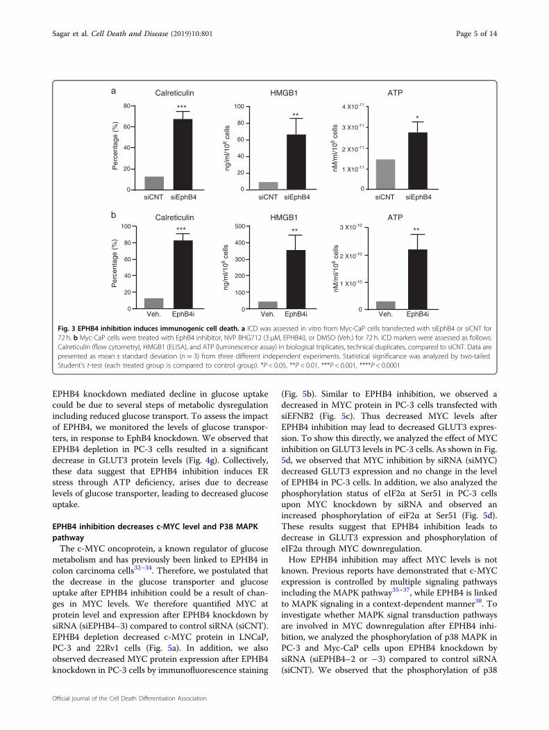
EPHB4 inhibition induces immunogenic cell deathSome therapeutic agents are known to cause cell death
by immunogenic cell death that can be exploited inimmunotherapy. We examined whether EPHB4 inhibitioninduced immunogenic cell death (ICD) by assessingchanges in the hallmarks of ICD, including cell surfacelevels of calreticulin, non-histone nuclear protein high-mobility group box 1 (HMGB1) release, and ATP releasefrom Myc-CaP cells transfected with control (siCNT) orEPHB4-targeting siRNA (siEphB4–3). EphB4 inhibitionresulted in increased cell surface localization of calreti-culin and the release of HMGB1 and ATP into theextracellular space (Fig. 3a). Similar results were obtainedin Myc-CaP cells treated with NVP-BHG712 (EphB4i)compared to Vehicle (Veh) (Fig. 3b). These results indi-cate that EPHB4 inhibition induces cell death consistentwith ICD.
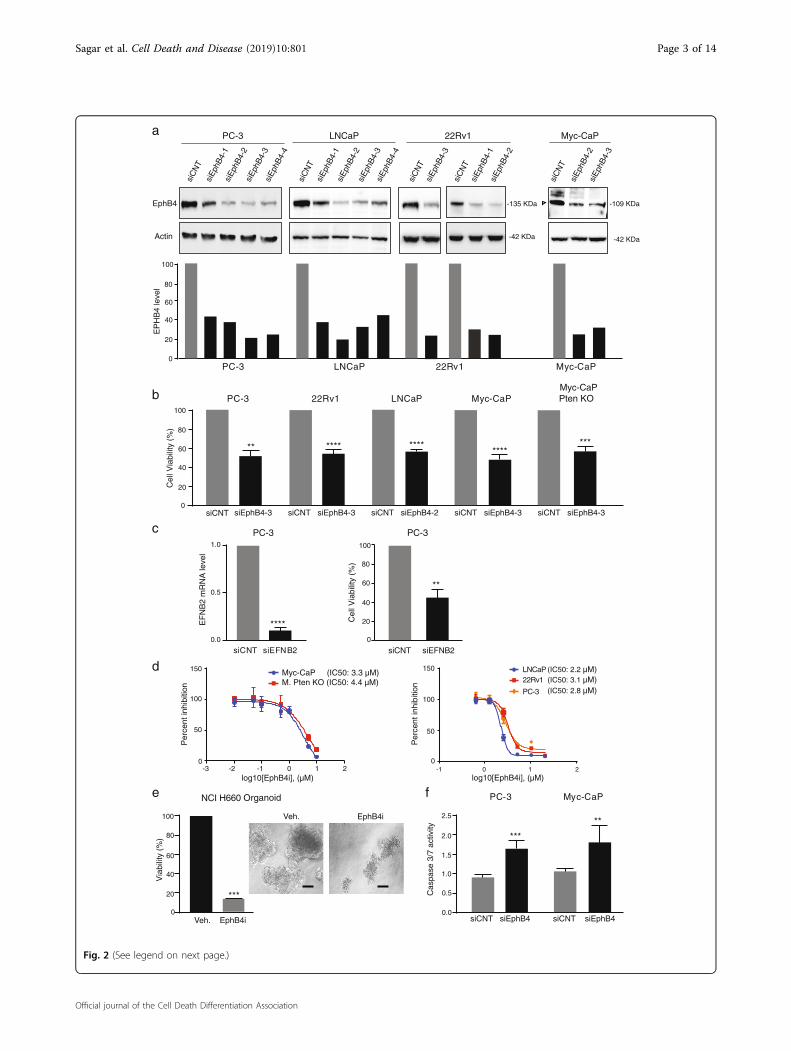
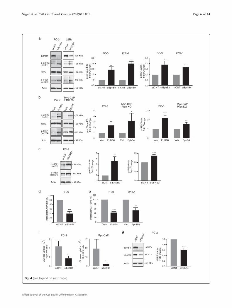
EPHB4 inhibition induces endoplasmic reticulum stressthrough metabolic changesPrevious studies have suggested that endoplasmic reti-
culum (ER) stress plays a major role in intracellular sig-naling pathways that induce ICD22,23. The ER stressresponse is initiated through phosphorylation of eukar-yotic translation initiation factor 2α (eIF2α)24, which hasbeen proposed as a characteristic marker for ICD24–26.We therefore analyzed the phosphorylation status ofeIF2α in PC-3 and 22Rv1 cells upon EPHB4 knockdownby siRNA (siEPHB4–3 or −4) compared to control siRNA(siCNT). EPHB4 inhibition increased phosphorylation of
eIF2α at serine 51 (Fig. 4a). Similar results were seen inPC-3 and Myc-CaP;Pten KO cells treated with smallmolecule EPHB4 inhibitor (EPHB4i) (Fig. 4b). In additionto eIF2α, inositol-requiring enzyme 1 (IRE1) representsanother ER protein that serves as ER stress sensor andmediates the unfolded protein response (UPR)27,28. LikeeIF2α activation, IRE1 phosphorylation increased afterEPHB4 knockdown by EPHB4-specific siRNA in PC-3and 22Rv1 cells (Fig. 4a). Similar results were observed inPC-3 and Myc-CaP;Pten KO cells treated with EphB4icompared to vehicle control (Veh.) (Fig. 4b). To investi-gate whether EPHB4 mediated effect on ER stress isligand-dependent, we analyzed the effect of EPHB4 ligand-EFNB2 knockdown on phosphorylation of eIF2α at ser-ine 51 and IRE1 at serine 724. Consistent with ourobservations above, inhibition of EFNB2 resulted in asimilar increase in phosphorylation of eIF2α at serine 51and IRE1 at serine 724 in PC-3 cells (Fig. 4c). These dataindicate that EPHB4 inhibition triggers the ER stressresponse in prostate cancer cells, which is associated withICD. Cellular processes in the ER require energy in theform of ATP and perturbation of these processes induceER stress29. Therefore, we sought to determine whetherthe ER stress induced by EPHB4 inhibition is related toATP depletion in cancer cells. We measured the intra-cellular ATP level after EPHB4 inhibition in PC-3 cellstransfected with siEPHB4–3 or siCNT. The levels ofintracellular ATP were significantly lower in siEPHB4-transfected PC-3 cells (Fig. 4d). Similar results wereobtained with EPHB4i-treated PC-3 and 22Rv1 cells (Fig. 4e).These results point to a decline in cellular ATP levelsupon EPHB4 inhibition as a possible cause of ER stress.The ER depends on extrinsic energy sources via oxi-
dative phosphorylation or glycolysis30,31. We tested whe-ther the decrease in intracellular ATP was associated withchanges in glucose metabolism. The first essential step inglucose metabolism is glucose uptake. Both PC-3 andMyc-CaP cells demonstrated significantly reduced glucoseuptake after EPHB4 knockdown by siEPHB4–3 (Fig. 4f).
(see figure on previous page)Fig. 2 EPHB4 decreases cell viability and induces apoptosis. a EPHB4 knockdown efficiency was analyzed by western analysis after 72 htransfection of EPHB4 siRNAs or non-targeted siRNA in all prostate cancer cell lines. Open triangle indicate specific bands. b Prostate cancer cell lineswere transfected with EPHB4 siRNA or scrambled siRNA for 72 h and cell viability determined using MTS assay. Experiments were performed intriplicate (n= 3). c PC-3 cells were transfected with EFNB2 siRNA or non-targeting siRNA for 72 h and cell viability determined using MTS assay andknockdown efficiency was analyzed at mRNA level by qRT-PCR. Experiments were performed in triplicate (n= 3). d Dose response of Myc-CaP, Myc-CaP Pten KO and LNCaP were assessed by CCK8 assay and, PC-3 and 22Rv1 cell viability by MTS assay after treatment with EPHB4i (NVP-BHG712 orDMSO) for 72 h. Data are presented as mean ± standard deviation (n= 3) from three different independent experiments. e NCI-H660 cell organoidswere established for 1 week, then treated with EPHB4 inhibitor, NVP-BHG712 (EphB4i) 3 µM or DMSO for 1 week. Viability of organoids weremeasured by Celltitre 3D Glo assay. Organoid images were visualized at 20X and scale bar represents 100 μm. Data are presented as mean ± standarddeviation (n= 3) from three different independent experiments. f PC-3 and Myc-CaP cells were transfected with EphB4 siRNA or non-targeted siRNAfor 72 h and cell viability and caspase activity was determined by Caspase-Glo 3/7 assay. Data are presented as mean ± standard deviation (n= 3)from three different independent experiments. Statistical significance was analyzed by two-tailed Student’s t-test (each treated group is compared tocontrol group). *P < 0.05, **P < 0.01, ***P < 0.001, ****P < 0.0001
Sagar et al. Cell Death and Disease (2019) 10:801 Page 4 of 14
Official journal of the Cell Death Differentiation Association
EPHB4 knockdown mediated decline in glucose uptakecould be due to several steps of metabolic dysregulationincluding reduced glucose transport. To assess the impactof EPHB4, we monitored the levels of glucose transpor-ters, in response to EphB4 knockdown. We observed thatEPHB4 depletion in PC-3 cells resulted in a significantdecrease in GLUT3 protein levels (Fig. 4g). Collectively,these data suggest that EPHB4 inhibition induces ERstress through ATP deficiency, arises due to decreaselevels of glucose transporter, leading to decreased glucoseuptake.
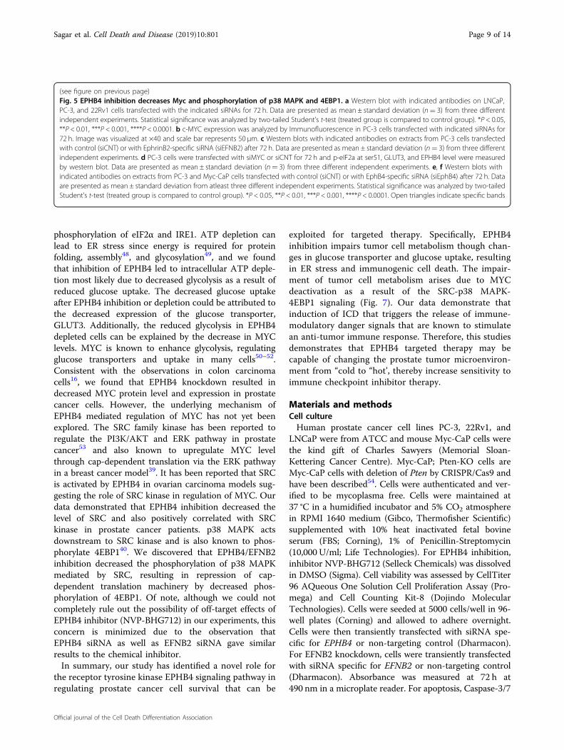
EPHB4 inhibition decreases c-MYC level and P38 MAPKpathwayThe c-MYC oncoprotein, a known regulator of glucose
metabolism and has previously been linked to EPHB4 incolon carcinoma cells32–34. Therefore, we postulated thatthe decrease in the glucose transporter and glucoseuptake after EPHB4 inhibition could be a result of chan-ges in MYC levels. We therefore quantified MYC atprotein level and expression after EPHB4 knockdown bysiRNA (siEPHB4–3) compared to control siRNA (siCNT).EPHB4 depletion decreased c-MYC protein in LNCaP,PC-3 and 22Rv1 cells (Fig. 5a). In addition, we alsoobserved decreased MYC protein expression after EPHB4knockdown in PC-3 cells by immunofluorescence staining
(Fig. 5b). Similar to EPHB4 inhibition, we observed adecreased in MYC protein in PC-3 cells transfected withsiEFNB2 (Fig. 5c). Thus decreased MYC levels afterEPHB4 inhibition may lead to decreased GLUT3 expres-sion. To show this directly, we analyzed the effect of MYCinhibition on GLUT3 levels in PC-3 cells. As shown in Fig.5d, we observed that MYC inhibition by siRNA (siMYC)decreased GLUT3 expression and no change in the levelof EPHB4 in PC-3 cells. In addition, we also analyzed thephosphorylation status of eIF2α at Ser51 in PC-3 cellsupon MYC knockdown by siRNA and observed anincreased phosphorylation of eiF2α at Ser51 (Fig. 5d).These results suggest that EPHB4 inhibition leads todecrease in GLUT3 expression and phosphorylation ofeIF2α through MYC downregulation.How EPHB4 inhibition may affect MYC levels is not
known. Previous reports have demonstrated that c-MYCexpression is controlled by multiple signaling pathwaysincluding the MAPK pathway35–37, while EPHB4 is linkedto MAPK signaling in a context-dependent manner38. Toinvestigate whether MAPK signal transduction pathwaysare involved in MYC downregulation after EPHB4 inhi-bition, we analyzed the phosphorylation of p38 MAPK inPC-3 and Myc-CaP cells upon EPHB4 knockdown bysiRNA (siEPHB4–2 or −3) compared to control siRNA(siCNT). We observed that the phosphorylation of p38
0
20
40
60
80P
erce
ntag
e (%
)***
0
ng/m
l/106
cells
ng/m
l/106
cells
nM/m
l/106
cells
nM/m
l/106
cells
a
0
100
200
300
400
500
siCNT siCNTsiEphB4
EphB4i EphB4i EphB4i
siEphB4 siCNT siEphB4
Veh. Veh. Veh.0
20
40
60
80
100 3 X10⁻¹⁰
4 X10-11
1 X10-11
2 X10-11
3 X10-11
2 X10⁻¹⁰
1 X10⁻¹⁰
0
Per
cent
age
(%)
*** ** **
b
Calreticulin HMGB1 ATP
Calreticulin HMGB1 ATP
20
40
60
**100
80
0
*
Fig. 3 EPHB4 inhibition induces immunogenic cell death. a ICD was assessed in vitro from Myc-CaP cells transfected with siEphB4 or siCNT for72 h. b Myc-CaP cells were treated with EphB4 inhibitor, NVP BHG712 (3 µM, EPHB4i), or DMSO (Veh.) for 72 h. ICD markers were assessed as follows:Calreticulin (flow cytometry), HMGB1 (ELISA), and ATP (luminescence assay) in biological triplicates, technical duplicates, compared to siCNT. Data arepresented as mean ± standard deviation (n= 3) from three different independent experiments. Statistical significance was analyzed by two-tailedStudent’s t-test (each treated group is compared to control group). *P < 0.05, **P < 0.01, ***P < 0.001, ****P < 0.0001
Sagar et al. Cell Death and Disease (2019) 10:801 Page 5 of 14
Official journal of the Cell Death Differentiation Association
EphB4
Actin
siC
NT
siE
phB
4
siC
NT
siE
phB
4
PC-3 22Rv1
**
***
0.5
1.0
1.5
2.0
2.5
0.0
0.5
1.0
1.5
2.0
2.5*
***
PC-3 22Rv1 PC-3 22Rv1
p-eIF2 α(ser51)
eIf2 α
p-IRE1(ser724)
-135 KDa
- 38 KDa
-110 KDa
- 42 KDa
- 38 KDa
-110 KDa
- 42 KDa
- 38 KDa
- 38 KDa
0
20
40
60
80
100
120
Intra
cellu
larA
TPle
vel(
%)
*******
**
0
20
40
60
80
100
120
Intra
cell u
lar A
TPle
vel(
%)
a
d ePC-3
Veh.
Eph
B4i
Veh.
Eph
B4i
PC-3 Pten KOMyc-CaP
0
1
2
3
4
5
***
PC-3 Pten KOMyc-CaP
b
0
1
2
3
p-IR
E1/
Act
inF
old
Cha
nge
p-IR
E1/
Act
inF
old
Cha
nge
PC-3 Pten KOMyc-CaP
p-eI
F2α
/eIF
2αF
old
Cha
nge
Actin
p-eIF2 α(ser51)
eIf2 α
p-IRE1(ser724)
f g
0
1
2
3
Glu
cose
upt a
ke(1
05 )
RLU
/10^
6ce
lls
0
10
20
30
*
PC-3 22Rv1
PC-3 Myc-CaP
T T siEphB4
GLUT3
EphB4
Actin0.0
0.2
0.4
0.6
0.8
1.0
GLU
T3/
Act
inF
o ld
chan
g e
***
PC-3
siC
NT
siE
phB
4
Glu
cose
upt a
ke(1
05 )
RL U
/10 ^
6c e
lls
**
-135 KDa
- 54 KDa
- 42 KDa
**
***
p-eI
F2α
/eIF
2αF
old
Cha
nge
p-eIF2 α(ser51)
p-IRE1(ser724)
Actin - 42 KDa
- 37 KDa
- 110 KDa
c
3.0
*
siC
NT
siE
FNB
2
PC-3
***
1.0
1.5
0.0
0.5p-IR
E1/
Act
inF
old
Cha
nge
**
p-eI
F2a
/Act
inF
old
Cha
nge
0
1
2
3
4
5
siCNT siEphB4 siCNT siEphB4 siCNT siEphB4 siCNT siEphB4
siCNT siEphB4 Veh. EphB4i Veh. EphB4i
Veh. EphB4i Veh. EphB4i Veh. EphB4i Veh. EphB4i
siCN siEphB4 siCN siCNT siEphB4
siCNT siEFNB2siCNT siEFNB2
Fig. 4 (See legend on next page.)
Sagar et al. Cell Death and Disease (2019) 10:801 Page 6 of 14
Official journal of the Cell Death Differentiation Association
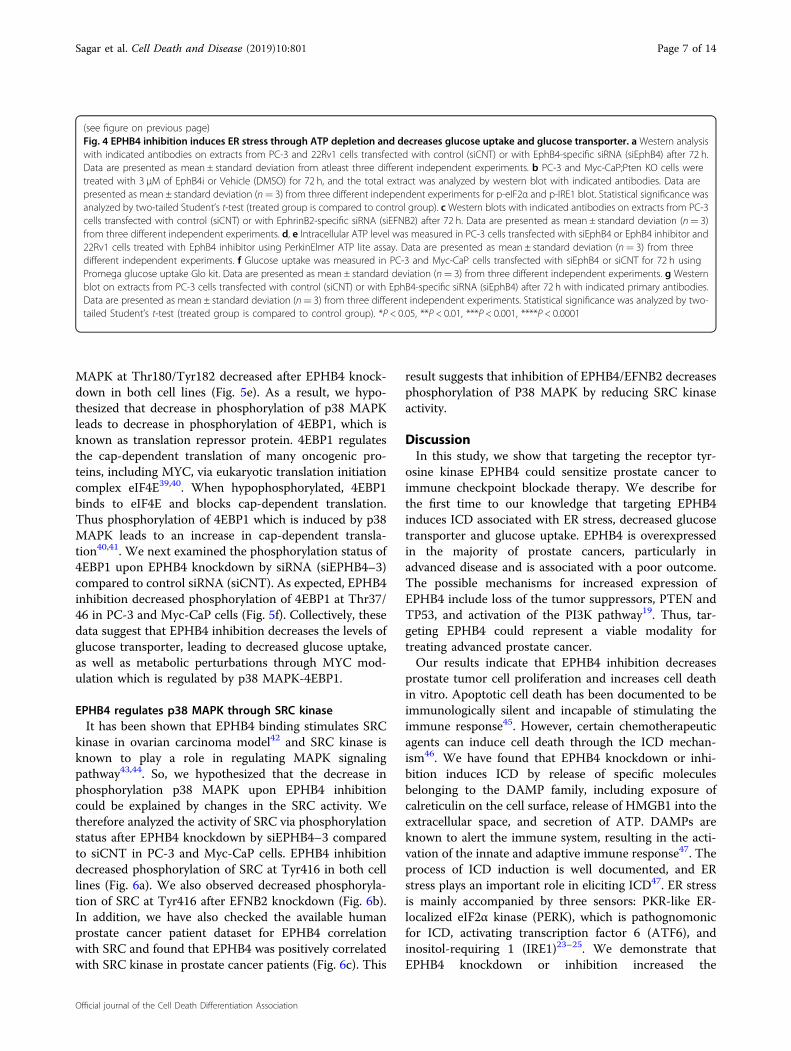
MAPK at Thr180/Tyr182 decreased after EPHB4 knock-down in both cell lines (Fig. 5e). As a result, we hypo-thesized that decrease in phosphorylation of p38 MAPKleads to decrease in phosphorylation of 4EBP1, which isknown as translation repressor protein. 4EBP1 regulatesthe cap-dependent translation of many oncogenic pro-teins, including MYC, via eukaryotic translation initiationcomplex eIF4E39,40. When hypophosphorylated, 4EBP1binds to eIF4E and blocks cap-dependent translation.Thus phosphorylation of 4EBP1 which is induced by p38MAPK leads to an increase in cap-dependent transla-tion40,41. We next examined the phosphorylation status of4EBP1 upon EPHB4 knockdown by siRNA (siEPHB4–3)compared to control siRNA (siCNT). As expected, EPHB4inhibition decreased phosphorylation of 4EBP1 at Thr37/46 in PC-3 and Myc-CaP cells (Fig. 5f). Collectively, thesedata suggest that EPHB4 inhibition decreases the levels ofglucose transporter, leading to decreased glucose uptake,as well as metabolic perturbations through MYC mod-ulation which is regulated by p38 MAPK-4EBP1.
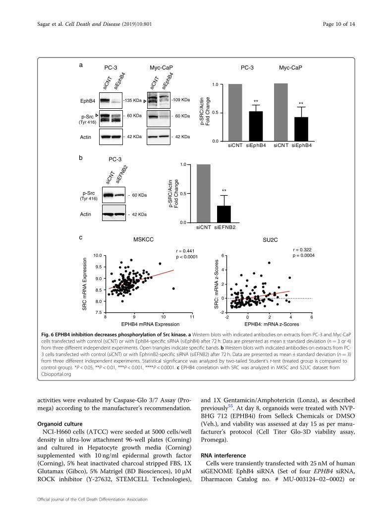
EPHB4 regulates p38 MAPK through SRC kinaseIt has been shown that EPHB4 binding stimulates SRC
kinase in ovarian carcinoma model42 and SRC kinase isknown to play a role in regulating MAPK signalingpathway43,44. So, we hypothesized that the decrease inphosphorylation p38 MAPK upon EPHB4 inhibitioncould be explained by changes in the SRC activity. Wetherefore analyzed the activity of SRC via phosphorylationstatus after EPHB4 knockdown by siEPHB4–3 comparedto siCNT in PC-3 and Myc-CaP cells. EPHB4 inhibitiondecreased phosphorylation of SRC at Tyr416 in both celllines (Fig. 6a). We also observed decreased phosphoryla-tion of SRC at Tyr416 after EFNB2 knockdown (Fig. 6b).In addition, we have also checked the available humanprostate cancer patient dataset for EPHB4 correlationwith SRC and found that EPHB4 was positively correlatedwith SRC kinase in prostate cancer patients (Fig. 6c). This
result suggests that inhibition of EPHB4/EFNB2 decreasesphosphorylation of P38 MAPK by reducing SRC kinaseactivity.
DiscussionIn this study, we show that targeting the receptor tyr-
osine kinase EPHB4 could sensitize prostate cancer toimmune checkpoint blockade therapy. We describe forthe first time to our knowledge that targeting EPHB4induces ICD associated with ER stress, decreased glucosetransporter and glucose uptake. EPHB4 is overexpressedin the majority of prostate cancers, particularly inadvanced disease and is associated with a poor outcome.The possible mechanisms for increased expression ofEPHB4 include loss of the tumor suppressors, PTEN andTP53, and activation of the PI3K pathway19. Thus, tar-geting EPHB4 could represent a viable modality fortreating advanced prostate cancer.Our results indicate that EPHB4 inhibition decreases
prostate tumor cell proliferation and increases cell deathin vitro. Apoptotic cell death has been documented to beimmunologically silent and incapable of stimulating theimmune response45. However, certain chemotherapeuticagents can induce cell death through the ICD mechan-ism46. We have found that EPHB4 knockdown or inhi-bition induces ICD by release of specific moleculesbelonging to the DAMP family, including exposure ofcalreticulin on the cell surface, release of HMGB1 into theextracellular space, and secretion of ATP. DAMPs areknown to alert the immune system, resulting in the acti-vation of the innate and adaptive immune response47. Theprocess of ICD induction is well documented, and ERstress plays an important role in eliciting ICD47. ER stressis mainly accompanied by three sensors: PKR-like ER-localized eIF2α kinase (PERK), which is pathognomonicfor ICD, activating transcription factor 6 (ATF6), andinositol-requiring 1 (IRE1)23–25. We demonstrate thatEPHB4 knockdown or inhibition increased the
(see figure on previous page)Fig. 4 EPHB4 inhibition induces ER stress through ATP depletion and decreases glucose uptake and glucose transporter. a Western analysiswith indicated antibodies on extracts from PC-3 and 22Rv1 cells transfected with control (siCNT) or with EphB4-specific siRNA (siEphB4) after 72 h.Data are presented as mean ± standard deviation from atleast three different independent experiments. b PC-3 and Myc-CaP;Pten KO cells weretreated with 3 µM of EphB4i or Vehicle (DMSO) for 72 h, and the total extract was analyzed by western blot with indicated antibodies. Data arepresented as mean ± standard deviation (n= 3) from three different independent experiments for p-eIF2α and p-IRE1 blot. Statistical significance wasanalyzed by two-tailed Student’s t-test (treated group is compared to control group). cWestern blots with indicated antibodies on extracts from PC-3cells transfected with control (siCNT) or with EphrinB2-specific siRNA (siEFNB2) after 72 h. Data are presented as mean ± standard deviation (n= 3)from three different independent experiments. d, e Intracellular ATP level was measured in PC-3 cells transfected with siEphB4 or EphB4 inhibitor and22Rv1 cells treated with EphB4 inhibitor using PerkinElmer ATP lite assay. Data are presented as mean ± standard deviation (n= 3) from threedifferent independent experiments. f Glucose uptake was measured in PC-3 and Myc-CaP cells transfected with siEphB4 or siCNT for 72 h usingPromega glucose uptake Glo kit. Data are presented as mean ± standard deviation (n= 3) from three different independent experiments. g Westernblot on extracts from PC-3 cells transfected with control (siCNT) or with EphB4-specific siRNA (siEphB4) after 72 h with indicated primary antibodies.Data are presented as mean ± standard deviation (n= 3) from three different independent experiments. Statistical significance was analyzed by two-tailed Student’s t-test (treated group is compared to control group). *P < 0.05, **P < 0.01, ***P < 0.001, ****P < 0.0001
Sagar et al. Cell Death and Disease (2019) 10:801 Page 7 of 14
Official journal of the Cell Death Differentiation Association
c-Myc
EphB4
GAPDH
siC
NT
siE
phB
4
siC
NT
siE
phB
4siCNT siEphB4
0.0
0.5
1.0
c-M
yc/G
AP
DH
Fol
d C
hnag
e ******
siCNT siEphB4
a LNCaP
bsiCNT
cyM-c
- 57 KDa
- 135 KDa
- 37 KDa
siC
NT
siM
yc
c-Myc
GLUT3
Actin
0.0
0.5
1.0
GLU
T3/
Act
inF
old
Cha
nge
- 54 KDa
- 59 KDa
- 42 KDa
PC-3
siC
NT
siE
phB
4
siC
NT
siE
phB
4
EphB4
p-P38(Th180/Ty182)
Actin
**
0.5
1.0
0.0
**
p-P
38/A
ctin
Fol
d C
hang
e
e
f
***
0.5
1.0
0.0
***
p-4E
BP
1/A
ctin
Fol
d C
hang
e
PC-3 Myc-CaP
p-4EBP1(Th37/46)
Actin
siC
NT
siE
phB
4
siC
NT
siE
phB
4
-109 KDa-135 KDa
- 40 KDa - 40 KDa
- 42 KDa - 42 KDa
- 42 KDa - 42 KDa
- 15 KDa- 15 KDa
PC-3
d
**
siCNT siEphB4
22Rv122Rv1
siC
NT
siE
phB
4
***
PC-3
EphB4 - 130 KDa
siEphB4
0.0
0.5
1.0
1.5
2.0
p-eIF2 α(ser51)
- 38 KDa
*
p-eI
F2α
/Act
inF
old
Cha
nge
c
Actin - 42 KDa
c-Myc - 57 KDa
siC
NT
siE
FNB
2
0.0
0.5
1.0
LNCaP PC-3
siCNT siMyc
PC-3 Myc-CaP
siCNT siEphB4 siCNT siEphB4
PC-3 Myc-CaP
siCNT siEphB4 siCNT siEphB4
PC-3 Myc-CaP
siCNT siMyc
siCNT siEFNB2
c-M
yc/A
ctin
Fol
d C
hnag
e
***
PC-3
Fig. 5 (See legend on next page.)
Sagar et al. Cell Death and Disease (2019) 10:801 Page 8 of 14
Official journal of the Cell Death Differentiation Association
phosphorylation of eIF2α and IRE1. ATP depletion canlead to ER stress since energy is required for proteinfolding, assembly48, and glycosylation49, and we foundthat inhibition of EPHB4 led to intracellular ATP deple-tion most likely due to decreased glycolysis as a result ofreduced glucose uptake. The decreased glucose uptakeafter EPHB4 inhibition or depletion could be attributed tothe decreased expression of the glucose transporter,GLUT3. Additionally, the reduced glycolysis in EPHB4depleted cells can be explained by the decrease in MYClevels. MYC is known to enhance glycolysis, regulatingglucose transporters and uptake in many cells50–52.Consistent with the observations in colon carcinomacells16, we found that EPHB4 knockdown resulted indecreased MYC protein level and expression in prostatecancer cells. However, the underlying mechanism ofEPHB4 mediated regulation of MYC has not yet beenexplored. The SRC family kinase has been reported toregulate the PI3K/AKT and ERK pathway in prostatecancer53 and also known to upregulate MYC levelthrough cap-dependent translation via the ERK pathwayin a breast cancer model39. It has been reported that SRCis activated by EPHB4 in ovarian carcinoma models sug-gesting the role of SRC kinase in regulation of MYC. Ourdata demonstrated that EPHB4 inhibition decreased thelevel of SRC and also positively correlated with SRCkinase in prostate cancer patients. p38 MAPK actsdownstream to SRC kinase and is also known to phos-phorylate 4EBP140. We discovered that EPHB4/EFNB2inhibition decreased the phosphorylation of p38 MAPKmediated by SRC, resulting in repression of cap-dependent translation machinery by decreased phos-phorylation of 4EBP1. Of note, although we could notcompletely rule out the possibility of off-target effects ofEPHB4 inhibitor (NVP-BHG712) in our experiments, thisconcern is minimized due to the observation thatEPHB4 siRNA as well as EFNB2 siRNA gave similarresults to the chemical inhibitor.In summary, our study has identified a novel role for
the receptor tyrosine kinase EPHB4 signaling pathway inregulating prostate cancer cell survival that can be
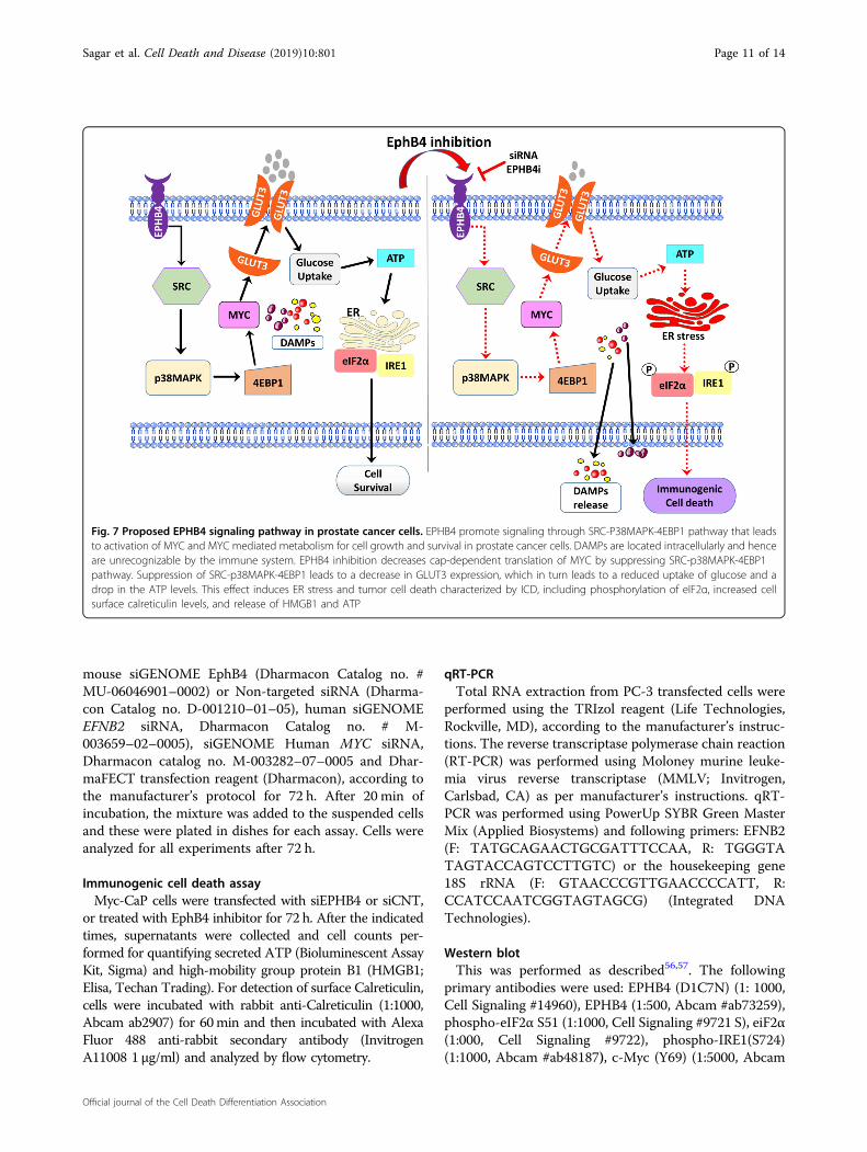
exploited for targeted therapy. Specifically, EPHB4inhibition impairs tumor cell metabolism though chan-ges in glucose transporter and glucose uptake, resultingin ER stress and immunogenic cell death. The impair-ment of tumor cell metabolism arises due to MYCdeactivation as a result of the SRC-p38 MAPK-4EBP1 signaling (Fig. 7). Our data demonstrate thatinduction of ICD that triggers the release of immune-modulatory danger signals that are known to stimulatean anti-tumor immune response. Therefore, this studiesdemonstrates that EPHB4 targeted therapy may becapable of changing the prostate tumor microenviron-ment from “cold to “hot’, thereby increase sensitivity toimmune checkpoint inhibitor therapy.
Materials and methodsCell cultureHuman prostate cancer cell lines PC-3, 22Rv1, and
LNCaP were from ATCC and mouse Myc-CaP cells werethe kind gift of Charles Sawyers (Memorial Sloan-Kettering Cancer Centre). Myc-CaP; Pten-KO cells areMyc-CaP cells with deletion of Pten by CRISPR/Cas9 andhave been described54. Cells were authenticated and ver-ified to be mycoplasma free. Cells were maintained at37 °C in a humidified incubator and 5% CO2 atmospherein RPMI 1640 medium (Gibco, Thermofisher Scientific)supplemented with 10% heat inactivated fetal bovineserum (FBS; Corning), 1% of Penicillin-Streptomycin(10,000 U/ml; Life Technologies). For EPHB4 inhibition,inhibitor NVP-BHG712 (Selleck Chemicals) was dissolvedin DMSO (Sigma). Cell viability was assessed by CellTiter96 AQueous One Solution Cell Proliferation Assay (Pro-mega) and Cell Counting Kit-8 (Dojindo MolecularTechnologies). Cells were seeded at 5000 cells/well in 96-well plates (Corning) and allowed to adhere overnight.Cells were then transiently transfected with siRNA spe-cific for EPHB4 or non-targeting control (Dharmacon).For EFNB2 knockdown, cells were transiently transfectedwith siRNA specific for EFNB2 or non-targeting control(Dharmacon). Absorbance was measured at 72 h at490 nm in a microplate reader. For apoptosis, Caspase-3/7
(see figure on previous page)Fig. 5 EPHB4 inhibition decreases Myc and phosphorylation of p38 MAPK and 4EBP1. a Western blot with indicated antibodies on LNCaP,PC-3, and 22Rv1 cells transfected with the indicated siRNAs for 72 h. Data are presented as mean ± standard deviation (n= 3) from three differentindependent experiments. Statistical significance was analyzed by two-tailed Student’s t-test (treated group is compared to control group). *P < 0.05,**P < 0.01, ***P < 0.001, ****P < 0.0001. b c-MYC expression was analyzed by Immunofluorescence in PC-3 cells transfected with indicated siRNAs for72 h. Image was visualized at ×40 and scale bar represents 50 μm. c Western blots with indicated antibodies on extracts from PC-3 cells transfectedwith control (siCNT) or with EphrinB2-specific siRNA (siEFNB2) after 72 h. Data are presented as mean ± standard deviation (n= 3) from three differentindependent experiments. d PC-3 cells were transfected with siMYC or siCNT for 72 h and p-eIF2a at ser51, GLUT3, and EPHB4 level were measuredby western blot. Data are presented as mean ± standard deviation (n= 3) from three different independent experiments. e, f Western blots withindicated antibodies on extracts from PC-3 and Myc-CaP cells transfected with control (siCNT) or with EphB4-specific siRNA (siEphB4) after 72 h. Dataare presented as mean ± standard deviation from atleast three different independent experiments. Statistical significance was analyzed by two-tailedStudent’s t-test (treated group is compared to control group). *P < 0.05, **P < 0.01, ***P < 0.001, ****P < 0.0001. Open triangles indicate specific bands
Sagar et al. Cell Death and Disease (2019) 10:801 Page 9 of 14
Official journal of the Cell Death Differentiation Association
activities were evaluated by Caspase-Glo 3/7 Assay (Pro-mega) according to the manufacturer’s recommendation.
Organoid cultureNCI-H660 cells (ATCC) were seeded at 5000 cells/well
density in ultra-low attachment 96-well plates (Corning)and cultured in Hepatocyte growth media (Corning)supplemented with 10 ng/ml epidermal growth factor(Corning), 5% heat inactivated charcoal stripped FBS, 1XGlutamax (Gibco), 5% Matrigel (BD Biosciences), 10 µMROCK inhibitor (Y-27632, STEMCELL Technologies),
and 1X Gentamicin/Amphotericin (Lonza), as describedpreviously55. At day 8, organoids were treated with NVP-BHG 712 (EPHB4i) from Selleck Chemicals or DMSO(Veh.), and viability was assessed at day 15 as per manu-facturer’s protocol (Cell Titer Glo-3D viability assay,Promega).
RNA interferenceCells were transiently transfected with 25 nM of human
siGENOME EphB4 siRNA (Set of four EPHB4 siRNA,Dharmacon Catalog no. # MU-003124–02–0002) or
**
0.5
1.0
0.0
**
p-S
RC
/Act
inF
old
Cha
nge
siC
NT
siE
phB
4
siC
NT
siE
phB
4
EphB4
p-Src(Tyr 416)
Actin
PC-3 Myc-CaP PC-3 Myc-CaP
r = 0.441p < 0.0001
SR
C m
RN
A E
xpre
ssio
n
10.0
9.5
9.0
8.5
8.0
7.5
MSKCC
EPHB4 mRNA Expression
-135 KDa -109 KDa
- 60 KDa - 60 KDa
- 42 KDa - 42 KDa
a
c
SR
C: m
RN
A z
-Sco
res
EPHB4: mRNA z-Scores
r = 0.322p = 0.00046
4
2
0
-2
SU2C
siC
NT
siE
FNB
2
p-Src(Tyr 416)
- 60 KDa
b PC-3
Actin - 42 KDa
**
0.0
0.5
1.0
siCNT siEphB4 siCNT siEphB4
8 9 10 11 -2 0 2 4 6
siCNT siEFNB2
p-S
RC
/Act
inF
old
Cha
nge
Fig. 6 EPHB4 inhibition decreases phosphorylation of Src kinase. a Western blots with indicated antibodies on extracts from PC-3 and Myc-CaPcells transfected with control (siCNT) or with EphB4-specific siRNA (siEphB4) after 72 h. Data are presented as mean ± standard deviation (n= 3 or 4)from three different independent experiments. Open triangles indicate specific bands. bWestern blots with indicated antibodies on extracts from PC-3 cells transfected with control (siCNT) or with EphrinB2-specific siRNA (siEFNB2) after 72 h. Data are presented as mean ± standard deviation (n= 3)from three different independent experiments. Statistical significance was analyzed by two-tailed Student’s t-test (treated group is compared tocontrol group). *P < 0.05, **P < 0.01, ***P < 0.001, ****P < 0.0001. c EPHB4 correlation with SRC was analyzed in MKSC and S2UC dataset fromCbioportal.org
Sagar et al. Cell Death and Disease (2019) 10:801 Page 10 of 14
Official journal of the Cell Death Differentiation Association
mouse siGENOME EphB4 (Dharmacon Catalog no. #MU-06046901–0002) or Non-targeted siRNA (Dharma-con Catalog no. D-001210–01–05), human siGENOMEEFNB2 siRNA, Dharmacon Catalog no. # M-003659–02–0005), siGENOME Human MYC siRNA,Dharmacon catalog no. M-003282–07–0005 and Dhar-maFECT transfection reagent (Dharmacon), according tothe manufacturer’s protocol for 72 h. After 20 min ofincubation, the mixture was added to the suspended cellsand these were plated in dishes for each assay. Cells wereanalyzed for all experiments after 72 h.
Immunogenic cell death assayMyc-CaP cells were transfected with siEPHB4 or siCNT,
or treated with EphB4 inhibitor for 72 h. After the indicatedtimes, supernatants were collected and cell counts per-formed for quantifying secreted ATP (Bioluminescent AssayKit, Sigma) and high-mobility group protein B1 (HMGB1;Elisa, Techan Trading). For detection of surface Calreticulin,cells were incubated with rabbit anti-Calreticulin (1:1000,Abcam ab2907) for 60min and then incubated with AlexaFluor 488 anti-rabbit secondary antibody (InvitrogenA11008 1 µg/ml) and analyzed by flow cytometry.
qRT-PCRTotal RNA extraction from PC-3 transfected cells were
performed using the TRIzol reagent (Life Technologies,Rockville, MD), according to the manufacturer’s instruc-tions. The reverse transcriptase polymerase chain reaction(RT-PCR) was performed using Moloney murine leuke-mia virus reverse transcriptase (MMLV; Invitrogen,Carlsbad, CA) as per manufacturer’s instructions. qRT-PCR was performed using PowerUp SYBR Green MasterMix (Applied Biosystems) and following primers: EFNB2(F: TATGCAGAACTGCGATTTCCAA, R: TGGGTATAGTACCAGTCCTTGTC) or the housekeeping gene18S rRNA (F: GTAACCCGTTGAACCCCATT, R:CCATCCAATCGGTAGTAGCG) (Integrated DNATechnologies).
Western blotThis was performed as described56,57. The following
primary antibodies were used: EPHB4 (D1C7N) (1: 1000,Cell Signaling #14960), EPHB4 (1:500, Abcam #ab73259),phospho-eIF2α S51 (1:1000, Cell Signaling #9721 S), eiF2α(1:000, Cell Signaling #9722), phospho-IRE1(S724)(1:1000, Abcam #ab48187), c-Myc (Y69) (1:5000, Abcam
Fig. 7 Proposed EPHB4 signaling pathway in prostate cancer cells. EPHB4 promote signaling through SRC-P38MAPK-4EBP1 pathway that leadsto activation of MYC and MYC mediated metabolism for cell growth and survival in prostate cancer cells. DAMPs are located intracellularly and henceare unrecognizable by the immune system. EPHB4 inhibition decreases cap-dependent translation of MYC by suppressing SRC-p38MAPK-4EBP1pathway. Suppression of SRC-p38MAPK-4EBP1 leads to a decrease in GLUT3 expression, which in turn leads to a reduced uptake of glucose and adrop in the ATP levels. This effect induces ER stress and tumor cell death characterized by ICD, including phosphorylation of eIF2α, increased cellsurface calreticulin levels, and release of HMGB1 and ATP
Sagar et al. Cell Death and Disease (2019) 10:801 Page 11 of 14
Official journal of the Cell Death Differentiation Association
#ab32072), GLUT3 (1:5000, Abcam #ab191071), Calreti-culin (1:400, Abcam #ab2907), HMGB1 (Abcam#ab18256), Actin (1:10000, Cell Signaling #5125),GAPDH (1:5000, Cell Signaling #3683), Phospho-SrcFamily Tyr416 (1:1000, Cell Signaling #2101), Phospho-p38 MAPK Thr180/Tyr182 (1:1000, Cell Signaling#9211), and Phospho-4EBP1 Thr37/46 (236B4) (1:1000,Cell Signaling #2855). Following incubation with horse-radish peroxidase-conjugated goat anti-rabbit/mouse(1:5000, Biorad) or goat anti-rat (1:5000, Santa Cruz) for1 h, the binding of secondary antibodies were detectedwith the Supersignal West Femto Maximum SensitivitySubstrate detection system (Pierce) followed by visuali-zation. Quantification analyses were performed by BioradChemiDoc Imager and Bio-Rad Image Lab software.
ImmunofluorescenceCells were transfected with siEPHB4 or siCNT, after
transfection cells were plated in 4-well chamber slides for72 h, slides were incubated with primary antibody c-Mycand followed by secondary antibody labeled with AlexaFluor 488 anti-rabbit (Thermo Scientific # A11008). Slideswere mounted with ProLong Gold Antifade reagent(Invitrogen/Molecular Probes #P36961). Immuno-fluorescence images were visualized using fluorescentmicroscope or Leica A1R spectral confocal microscope.
Glucose uptake assayCells were transfected with siEphB4 or siCNT in 96-well
plates. Glucose uptake was measured 72 h post-transfection using Glucose Uptake-Glo™ Assay (Pro-mega) according to the manufacturer’s instructions.
Intracellular ATP assayCells were transfected with siRNA or treated with
EphB4 inhibitor in 96-well plates. After 72 h, ATP levelwas measured using the ATPlite Luminescence AssaySystem from Perkin Elmer as described58.
Human Prostate cancer dataGene expression data were downloaded from the NCBI
Geo for the following datasets: Tomlins Prostate cancerdataset [GSE6099; Normal tissues (Adjacent NormalEpithelium, BPH; n= 22), Prostate cancers (PIN, PrimaryProstate cancer, Metastatic cancer samples; n= 60)],Wallace Prostate cancer dataset [GSE6956; surroundingnon-tumor tissues (n= 18), prostate cancers (n= 69)],Grasso Prostate cancer dataset [GSE35988; benign pros-tate tissues (n= 28), localized primary cancers (n= 59),metastatic CRPC (n= 35)], and Roudier prostate cancerdataset [GSE74367; Primary prostate cancer (n= 11),CRPC metastases (n= 45)]59–62. P-values were determinedby Welsh’s t-test. For survival analysis, data were
downloaded from NCBI Geo (GEO accession no.GSE70769)21 and cBioPortal (TCGA provisional). Patientgroups were classified as high EPHB4 and low EPHB4expression by the 75th percentile for the Ross-Adamsdataset and the 90th percentile for the TCGA studypopulation. Patient survivals were assessed by theKaplan–Meier method. P-values were determined by log-rank test.For Correlation analysis, the gene expression of EPHB4
and SRC were analyzed using Spearman’s rank correla-tion. Data used for this analysis was from the MSKCCdataset (n= 150) and SU2C dataset (n= 118) downloadedfrom cbioportal.org.
Statistical analysisAll statistical analyses were performed in GraphPad
Prism 7 software. The number of technical replicates,biological replicates, and independent experiments per-formed are indicated in the figure legends. Statisticalanalyses were by a two-tailed Student’s t-test. Data arepresented as mean ± standard error of the mean (S.E.M.),for analyses, results were considered statistically sig-nificant with P < 0.05, *P < 0.05, **P < 0.01, ***P < 0.001,and ****P < 0.0001.
AcknowledgementsThis work was supported by grant P50 CA180995 from the NCI.
Author details1Department of Urology, The Robert H. Lurie Comprehensive Cancer Center,Northwestern University Feinberg School of Medicine, Chicago, IL 60611, USA.2Atrin Pharmaceuticals, Pennsylvania Biotechnology Center, Doylestown, PA18902, USA. 3Department of Medicine, Section of Hematology/Oncology,University of Chicago, Chicago, IL 60637, USA. 4Department of Urology andMedical Social Sciences (DEV), Northwestern University Feinberg School ofMedicine, Chicago, IL 60611, USA. 5Division of Hematology/Oncology,Department of Medicine, Northwestern University Feinberg School ofMedicine, Chicago, IL 60611, USA. 6Division of Hematology, Department ofMedicine, USC Norris Comprehensive Cancer Center, Keck School of Medicine,University of Southern California, Los Angeles, CA 90033, USA. 7Department ofPathology, Northwestern University Feinberg School of Medicine, Chicago, IL60611, USA
Author contributionsV.S., R.V. and S.P. performed the experiments. V.S., R.V., S.P., B.L. and J.F.A.contributed to data acquisition. V.S., R.V., S.P., J.F.A. and S.A.A. analyzed the data.V.S., P.S.G. and S.A.A. designed the research and wrote the manuscript. Y.R., K.U.,H.H., P.S.G., W.J.C., W.M.S., M.H. and S.A.A. reviewed and edited the manuscript.All authors read and approved the final manuscript.
Conflict of interestThe authors declare that they have no conflict of interest.
Publisher’s noteSpringer Nature remains neutral with regard to jurisdictional claims inpublished maps and institutional affiliations.
Received: 14 June 2019 Revised: 9 September 2019 Accepted: 3 October2019
Sagar et al. Cell Death and Disease (2019) 10:801 Page 12 of 14
Official journal of the Cell Death Differentiation Association
References1. Siegel, R. L., Miller, K. D. & Jemal, A. Cancer statistics, 2019. CA Cancer J. Clin. 69,
7–34 (2019).2. Wang, G., Zhao, D., Spring, D. J. & DePinho, R. A. Genetics and biology of
prostate cancer. Genes Dev. 32, 1105–1140 (2018).3. Teoh, J. Y. et al. Prognostic significance of time to prostate-specific antigen
(PSA) nadir and its relationship to survival beyond time to PSA nadir forprostate cancer patients with bone metastases after primary androgendeprivation therapy. Ann. Surg. Oncol. 22, 1385–1391 (2015).
4. Roviello, G., Petrioli, R., Laera, L. & Francini, E. The third line of treatment formetastatic prostate cancer patients: Option or strategy? Crit. Rev. Oncol.Hematol. 95, 265–271 (2015).
5. Limvorasak, S. & Posadas, E. M. Kinase inhibitors in prostate cancer. AnticancerAgents Med. Chem. 9, 1089–1104 (2009).
6. Ojemuyiwa, M. A., Madan, R. A. & Dahut, W. L. Tyrosine kinase inhibitors in thetreatment of prostate cancer: taking the next step in clinical development.Expert Opin. Emerg. Drugs 19, 459–470 (2014).
7. Schneider, M. et al. The tyrosine kinase inhibitor nilotinib has antineoplasticactivity in prostate cancer cells but up-regulates the ERK survival signal-Implications for targeted therapies. Urol. Oncol. 33, 72 e71–72 e77 (2015).
8. Christenson, E. S. & Antonarakis, E. S. PARP inhibitors for homologousrecombination-deficient prostate cancer. Expert Opin. Emerg. Drugs 23,123–133 (2018).
9. Mirshafiey, A., Ghalamfarsa, G., Asghari, B. & Azizi, G. Receptor tyrosine kinaseand tyrosine kinase inhibitors: new hope for success in multiple sclerosistherapy. Innov. Clin. Neurosci. 11, 23–36 (2014).
10. Regad, T. & Targeting, R. T. K. Signaling pathways in cancer. Cancers 7,1758–1784 (2015).
11. Vouri, M. & Hafizi, S. TAM receptor tyrosine kinases in cancer drug resistance.Cancer Res. 77, 2775–2778 (2017).
12. Corcoran, C. & O'Driscoll, L. Receptor tyrosine kinases and drug resistance:development and characterization of in vitro models of resistance to RTKinhibitors. Methods Mol. Biol. 1233, 169–180 (2015).
13. Chen, Y., Zhang, H. & Zhang, Y. Targeting receptor tyrosine kinase EphB4 in cancertherapy. Semin. Cancer Biol. https://doi.org/10.1016/j.semcancer.2017.10.002 (2017).
14. Pasquale, E. B. Eph-ephrin bidirectional signaling in physiology and disease.Cell 133, 38–52 (2008).
15. Kumar, S. R. et al. Receptor tyrosine kinase EphB4 is a survival factor in breastcancer. Am. J. Pathol. 169, 279–293 (2006).
16. McCall, J. L. et al. KSR1 and EPHB4 regulate Myc and PGC1beta to promotesurvival of human colon tumors. Mol. Cell Biol. 36, 2246–2261 (2016).
17. Yavrouian, E. J. et al. The significance of EphB4 and EphrinB2 expression andsurvival in head and neck squamous cell carcinoma. Arch. Otolaryngol. HeadNeck Surg. 134, 985–991 (2008).
18. Ferguson, B. D. et al. The EphB4 receptor tyrosine kinase promotes lung cancergrowth: a potential novel therapeutic target. PLoS ONE 8, e67668 (2013).
19. Xia, G. et al. EphB4 expression and biological significance in prostate cancer.Cancer Res. 65, 4623–4632 (2005).
20. Mertens-Walker, I. et al. The tumour-promoting receptor tyrosine kinase,EphB4, regulates expression of integrin-beta8 in prostate cancer cells. BMCCancer 15, 164 (2015).
21. Ross-Adams, H. et al. Integration of copy number and transcriptomics providesrisk stratification in prostate cancer: a discovery and validation cohort study.EBioMedicine 2, 1133–1144 (2015).
22. Rufo, N., Garg, A. D. & Agostinis, P. The unfolded protein response in immu-nogenic cell death and cancer immunotherapy. Trends Cancer 3, 643–658 (2017).
23. Kepp, O. et al. Crosstalk between ER stress and immunogenic cell death.Cytokine Growth Factor Rev. 24, 311–318 (2013).
24. Bezu, L. et al. eIF2alpha phosphorylation: a hallmark of immunogenic celldeath. Oncoimmunology 7, e1431089 (2018).
25. Bezu, L. et al. eIF2alpha phosphorylation is pathognomonic for immunogeniccell death. Cell Death Differ. 25, 1375–1393 (2018).
26. Kepp, O. et al. eIF2alpha phosphorylation as a biomarker of immunogenic celldeath. Semin Cancer Biol. 33, 86–92 (2015).
27. Badiola, N. et al. Induction of ER stress in response to oxygen-glucosedeprivation of cortical cultures involves the activation of the PERK and IRE-1pathways and of caspase-12. Cell Death Dis. 2, e149 (2011).
28. So, J. S. Roles of endoplasmic reticulum stress in immune responses. Mol. Cells41, 705–716 (2018).
29. Vishnu, N. et al. ATP increases within the lumen of the endoplasmic reticulumupon intracellular Ca2+ release. Mol. Biol. Cell 25, 368–379 (2014).
30. Hirschberg, C. B., Robbins, P. W. & Abeijon, C. Transporters of nucleotide sugars,ATP, and nucleotide sulfate in the endoplasmic reticulum and Golgi appa-ratus. Annu. Rev. Biochem. 67, 49–69 (1998).
31. Lee, M. & Yoon, J. H. Metabolic interplay between glycolysis and mitochondrialoxidation: the reverse Warburg effect and its therapeutic implication. World J.Biol. Chem. 6, 148–161 (2015).
32. Dang, C. V. MYC metabolism, cell growth, and tumorigenesis. Cold Spring Harb.Perspect. Med. 3, https://doi.org/10.1101/cshperspect.a014217 (2013).
33. Morrish, F. & Hockenbery, D. MYC and mitochondrial biogenesis. Cold SpringHarb. Perspect. Med. 4, https://doi.org/10.1101/cshperspect.a014225 (2014).
34. Tateishi, K. et al. Myc-Driven glycolysis is a therapeutic target in glioblastoma.Clin. Cancer Res. 22, 4452–4465 (2016).
35. Zhu, J., Blenis, J. & Yuan, J. Activation of PI3K/Akt and MAPK pathways reg-ulates Myc-mediated transcription by phosphorylating and promoting thedegradation of Mad1. Proc. Natl Acad. Sci. USA 105, 6584–6589 (2008).
36. Tsai, W. B. et al. Activation of Ras/PI3K/ERK pathway induces c-Myc stabilizationto upregulate argininosuccinate synthetase, leading to arginine deiminaseresistance in melanoma cells. Cancer Res. 72, 2622–2633 (2012).
37. Zhao, Q. et al. Inhibition of c-MYC with involvement of ERK/JNK/MAPK andAKT pathways as a novel mechanism for shikonin and its derivatives in killingleukemia cells. Oncotarget 6, 38934–38951 (2015).
38. Xiao, Z. et al. EphB4 promotes or suppresses Ras/MEK/ERK pathway in acontext-dependent manner: Implications for EphB4 as a cancer target. CancerBiol. Ther. 13, 630–637 (2012).
39. Jain, S. et al. Src inhibition blocks c-Myc translation and glucose metabolism toprevent the development of breast cancer. Cancer Res. 75, 4863–4875 (2015).
40. Liu, G., Zhang, Y., Bode, A. M., Ma, W. Y. & Dong, Z. Phosphorylation of 4E-BP1 ismediated by the p38/MSK1 pathway in response to UVB irradiation. J. Biol.Chem. 277, 8810–8816 (2002).
41. Showkat, M., Beigh, M. A. & Andrabi, K. I. mTOR signaling in protein translationregulation: implications in cancer genesis and therapeutic interventions. Mol.Biol. Int. 2014, 686984 (2014).
42. Pradeep, S. et al. Erythropoietin stimulates tumor growth via EphB4. Cancer Cell28, 610–622 (2015).
43. Jang, E. J. et al. Src tyrosine kinase activation by 4-hydroxynonenal upregulatesp38, ERK/AP-1 signaling and COX-2 expression in YPEN-1 cells. PLoS ONE 10,e0129244 (2015).
44. Thobe, B. M. et al. Src family kinases regulate p38 MAPK-mediated IL-6 pro-duction in Kupffer cells following hypoxia. Am. J. Physiol. Cell Physiol. 291,C476–C482 (2006).
45. Kepp, O. et al. Consensus guidelines for the detection of immunogenic celldeath. Oncoimmunology 3, e955691 (2014).
46. Hodge, J. W. et al. Chemotherapy-induced immunogenic modulation oftumor cells enhances killing by cytotoxic T lymphocytes and is distinct fromimmunogenic cell death. Int J. Cancer 133, 624–636 (2013).
47. Kroemer, G., Galluzzi, L., Kepp, O. & Zitvogel, L. Immunogenic cell death incancer therapy. Annu Rev. Immunol. 31, 51–72 (2013).
48. Naidoo, N. ER and aging-Protein folding and the ER stress response. AgeingRes. Rev. 8, 150–159 (2009).
49. Banerjee, D. K. N-glycans in cell survival and death: cross-talk between gly-cosyltransferases. Biochim. Biophys. Acta 1820, 1338–1346 (2012).
50. Dang, C. V. Therapeutic targeting of Myc-reprogrammed cancer cell meta-bolism. Cold Spring Harb. Symp. Quant. Biol. 76, 369–374 (2011).
51. Li, F. et al. Myc stimulates nuclearly encoded mitochondrial genes andmitochondrial biogenesis. Mol. Cell Biol. 25, 6225–6234 (2005).
52. Wang, H. et al. Structurally diverse c-Myc inhibitors share a commonmechanismof action involving ATP depletion. Oncotarget 6, 15857–15870 (2015).
53. Chang, Y. M. et al. Src family kinase oncogenic potential and pathways inprostate cancer as revealed by AZD0530. Oncogene 27, 6365–6375 (2008).
54. Anker, J. F. et al. Multi-faceted immunomodulatory and tissue-tropic clinicalbacterial isolate potentiates prostate cancer immunotherapy. Nat. Commun. 9,1591 (2018).
55. Unno, K. et al. Modeling African American prostate adenocarcinoma by inducingdefined genetic alterations in organoids. Oncotarget 8, 51264–51276 (2017).
56. Carneiro, B. A. et al. Anaplastic lymphoma kinase mutation (ALK F1174C) insmall cell carcinoma of the prostate and molecular response to alectinib. Clin.Cancer Res. 24, 2732–2739 (2018).
57. Sagar, V. et al. PIM1 destabilization activates a p53-dependent response toribosomal stress in cancer cells. Oncotarget 7, 23837–23849 (2016).
58. Njoroge, R. N. et al. Organoids model distinct Vitamin E effects at differentstages of prostate cancer evolution. Sci. Rep. 7, 16285 (2017).
Sagar et al. Cell Death and Disease (2019) 10:801 Page 13 of 14
Official journal of the Cell Death Differentiation Association
59. Tomlins, S. A. et al. Integrative molecular concept modeling of prostate cancerprogression. Nat. Genet. 39, 41–51 (2007).
60. Wallace, T. A. et al. Tumor immunobiological differences in prostate cancerbetween African-American and European-American men. Cancer Res. 68,927–936 (2008).
61. Grasso, C. S. et al. The mutational landscape of lethal castration-resistantprostate cancer. Nature 487, 239–243 (2012).
62. Roudier, M. P. et al. Characterizing the molecular features of ERG-positivetumors in primary and castration resistant prostate cancer. Prostate 76,810–822 (2016).
Sagar et al. Cell Death and Disease (2019) 10:801 Page 14 of 14
Official journal of the Cell Death Differentiation Association