enzymatic remodelling of the exopolysaccharide stewartan
TRANSCRIPT

Institut für Biochemie und Biologie,
Arbeitsgruppe für Physikalische Biochemie
Enzymatic Remodelling of the Exopolysaccharide
Stewartan Network: Implications for the Diffusion of
Nano-sized Objects
Dissertation
zur Erlangung des akademischen Grades
"doctor rerum naturalium" (Dr. rer. nat.)
in der Wissenschaftsdisziplin "Physikalische Biochemie"
eingereicht an der
Mathematisch-Naturwissenschaftlichen Fakultät
der
Universität Potsdam
von
Tobias Irmscher
Potsdam, Dezember 2019

Supervisor: Dr. habil. Stefanie Barbirz Date of final exam: 9 June 2020 Published online in the Institutional Repository of the University of Potsdam: https://doi.org/10.25932/publishup-47248 https://nbn-resolving.org/urn:nbn:de:kobv:517-opus4-472486

i
I. Abstract
In nature, bacteria are found to reside in multicellular communities encased in self-produced
extracellular matrices. Indeed, biofilms are the default lifestyle of the bacteria which cause persistent
infections in humans. The biofilm assembly protects bacterial cells from desiccation and limits the
effectiveness of antimicrobial treatments. A myriad of biomolecules in the extracellular matrix,
including proteins, exopolysaccharides, lipids, extracellular DNA and other, form a dense and
viscoelastic three dimensional network. Many studies emphasized that a destabilization of the
mechanical integrity of biofilm architectures potentially eliminates the protective shield and renders
bacteria more susceptible to the immune system and antibiotics. Pantoea stewartii is a plant
pathogen which infects monocotyledons such as maize and sweet corn. These bacteria produce dense
biofilms in the xylem of infected plants which cause wilting of plants and crops. Stewartan is an
exopolysaccharide which is produced by Pantoea stewartii and secreted as the major component to
the extracellular matrix. It consists of heptasaccharide repeating units with a high degree of
polymerization (2-4 MDa). In this work, the physicochemical properties of stewartan were
investigated to understand the contributions of this exopolysaccharide to the mechanical integrity and
cohesiveness of Pantoea stewartii biofilms. Therefore, a coarse-grained model of stewartan was
developed with computational techniques to obtain a model for its three dimensional structural
features. Here, coarse-grained molecular dynamic simulations revealed that the exopolysaccharide
forms a hydrogel in which the exopolysaccharide chains arrange into a three dimensional mesh-like
network. Simulations at different concentrations were used to investigate the influence of the water
content on the network formation. Stewartan was further purified from 72 h grown Pantoea stewartii
biofilms and the diffusion of bacteriophage and differently-sized nanoparticles (which ranged from
1.1 to 193 nm diameter) was analyzed in reconstituted stewartan solutions. Fluorescence correlation
spectroscopy and single-particle tracking revealed that the stewartan network impeded the mobility
of a set of differently-sized fluorescent particles in a size-dependent manner. Diffusion of these
particles became more anomalous, as characterized by fitting the diffusion data to an anomalous
diffusion model, with increasing stewartan concentrations. Further bulk and microrheological
experiments were used to analyze the transitions in stewartan fluid behavior and stewartan chain
entanglements were described. Moreover, it was noticed, that a small fraction of bacteriophage
particles was trapped in small-sized pores deviating from classical random walks which highlighted the
structural heterogeneity of the stewartan network. Additionally, the mobility of fluorescent particles

ii
also depended on the charge of the stewartan exopolysaccharide and a model of a molecular sieve for
the stewartan network was proposed. The here reported structural features of the stewartan
polymers were used to provide a detailed description of the mechanical properties of typically glycan-
based biofilms such as the one from Pantoea stewartii.
In addition, the mechanical properties of the biofilm architecture are permanently sensed by the
embedded bacteria and enzymatic modifications of the extracellular matrix take place to address
environmental cues. Hence, in this work the influence of enzymatic degradation of the stewartan
exopolysaccharides on the overall exopolysaccharide network structure was analyzed to describe
relevant physiological processes in Pantoea stewartii biofilms. Here, the stewartan hydrolysis kinetics
of the tailspike protein from the ΦEa1h bacteriophage, which is naturally found to infect Pantoea
stewartii cells, was compared to WceF. The latter protein is expressed from the Pantoea stewartii
stewartan biosynthesis gene cluster wce I-III. The degradation of stewartan by the ΦEa1h tailspike
protein was shown to be much faster than the hydrolysis kinetics of WceF, although both enzymes
cleaved the β-D-GalIII(1→3)-α-D-GalI glycosidic linkage from the stewartan backbone. Oligosaccharide
fragments which were produced during the stewartan cleavage, were analyzed in size-exclusion
chromatography and capillary electrophoresis. Bioinformatic studies and the analysis of a WceF crystal
structure revealed a remarkably high structural similarity of both proteins thus unveiling WceF as a
bacterial tailspike-like protein. As a consequence, WceF might play a role in stewartan chain length
control in Pantoea stewartii biofilms.

iii
II. Zusammenfassung
In der Natur lagern sich Bakterien zu großen und komplexen Gemeinschaften zusammen, die als
Biofilme bezeichnet werden. Diese multizellulären Biofilme sind der Ursprung vieler langlebiger und
gefährlicher Infektionskrankheiten. Die bakteriellen Zellen produzieren und umgeben sich mit einen
biofilm-spezifischen Schleim, der aus einer Unzahl von Biomolekülen, wie z.B. Exopolysaccharide,
Lipide und extrazelluläre DNA, besteht. Diese Biofilmarchitektur schützt Bakterien vor Austrocknung
und begrenzen die Wirksamkeit von antimikrobiellen Wirkstoffen (z.B. Antibiotika). Viele Studien
haben gezeigt, dass die Destabilisierung der mechanischen Festigkeit des Biofilmapparates eine neue
Behandlungsstrategie darstellt, in der das bakterielle Schutzschild eliminiert wird, sodass die Zellen
wieder anfälliger gegenüber dem menschlichen Immunsystem oder Antibiotika werden.
Pantoea stewartii ist ein Pflanzenpathogen, welches Mais und Süßmais befällt. Diese Bakterien
produzieren Biofilme im Inneren der Pflanze, sodass der freie Wassertransport gestört wird. Daraufhin
verwelken die Blätter und Früchte. In dieser Arbeit wurde das Exopolysaccharid Stewartan untersucht,
welches lange Ketten ausbildet und als häufigste Komponente in den Biofilmen von Pantoea stewartii
vorkommt. Dabei wurden die mechanischen Eigenschaften von Stewartan untersucht, um zu
verstehen, wie diese den Biofilm beeinflussen. Dafür wurde eine Lösung aus mehreren Stewartan
Molekülen computergestützt simuliert. Hierbei konnte beobachtet werden, dass die Stewartan Ketten
ein dreidimensionales Netzwerk ausbilden, welches Poren aufweist. Außerdem wurde Stewartan aus
Pantoea stewartii Biofilmen isoliert und die Diffusion von verschieden großen Nanopartikeln in dem
Exopolysaccharidnetzwerk untersucht. Je höher die Stewartankonzentration war, desto mehr wurde
die Diffusion der Nanopartikeln abgebremst. Außerdem wurden große Partikel stärker von dem
Netzwerk zurückgehalten. Diese Untersuchungen wurden auf die Diffusion von Bakteriophagen, das
sind Viren, die spezifisch Bakterien infizieren, ausgeweitet. Infolgedessen wurde gezeigt, dass
Bakteriophagen in kleine Stewartanporen feststecken können. Die Diffusion all dieser Partikeln war
aber auch abhängig von der Oberflächenladung des Partikels. Folglich bildet Stewartan ein Netzwerk
aus, welches ganz spezifisch den Transport von Molekülen mit bestimmten Eigenschaften unterbindet.
Außerdem ist bekannt, dass die Bakterien in der Lage sind, die mechanischen Eigenschaften des
Biofilms zu modulieren, um sie an Veränderungen in der Umgebung anzupassen. Dies geschieht über
bakterielle Enzyme. Daher wurde in dieser Arbeit der enzymatische Abbau von Stewartan untersucht,
der eine dramatische Änderung der Eigenschaften des Biofilms zufolge haben kann. Dabei wurde die
Stewartan Spaltung durch das Enzym WceF untersucht, welches von Pantoea stewartii produziert

iv
wird. Dieses Enzym spaltete die Stewartanketten nur sehr langsamen, sodass das Stewartannetzwerk
erhalten blieb. Die Ergebnisse wurden mit dem tailspike Protein verglichen, welches von dem ΦEa1h
Bakteriophagen produziert wird, dem natürlichen Feind des Bakteriums. Im Gegensatz zu WceF, baute
das tailspike Protein Stewartan deutlich schneller ab und die gesamte mechanische Festigkeit des
Netzwerkes wurde beseitigt. Beide Enzyme, trotz der unterschiedlichen Aktivität, besitzen eine sehr
ähnliche Struktur, was vermuten lässt, dass sie von einem gleichen Vorgängerprotein abstammen. In
dieser Arbeit wird vorgeschlagen, dass WceF möglicherweise in der Kettenlängekontrolle von
Stewartan involviert ist.

v
III. Declaration of Authorship
I hereby declare, that I am the sole author and composer of my thesis and that no other sources or
help than those listed, have been used. Wherever contributions of others are involved, this
contribution is indicated, clearly acknowledged and due reference is given to the author and source.
Furthermore, I declare that I have not submitted this thesis, either in its entirety or excerpts thereof,
at any other institution.
(Place, Date) (Signature)

vi
IV. Contributions to This Thesis
This work was done under the auspices of the International Max Planck Research School "Multiscale
Bio-Systems". Experiments were carried out at the laboratory of the Physical Biochemistry (University
of Potsdam, Head: Prof. Robert Seckler) under the supervision of PD Dr. habil. Stefanie Barbirz.
Computational simulations were done at the Department of Theory and Bio-Systems (Max-Planck-
Institute for Colloids and Interfaces, Head: Prof. Reinhard Lipowsky) under the supervision of Dr.
Andrea Grafmüller.
The following contributions to this thesis work were obtained from experiments performed by other
collaborators as listed below
Valentin Dunsing and Prof. Salvatore Chiantia, Physical Cellular Biochemistry, University of Potsdam:
analysis of the diffusion studies of particles of different size using fluorescence correlation
spectroscopy and single-particle tracking including the optimization of the microscope set-up
and data processing
bulk rheology viscosimetric analysis of differently concentrated stewartan solutions
light-scattering experiments to determine size and zeta potential of nanoparticles
Igor Gayk, Physical Biochemistry, University of Potsdam:
cloning of WceF and ΦEa1h TSP DNA constructs (plasmids: pET-23a(+)_ΦEa1hTSP_TEV_His6N
and pET-23a(+)_WceF_tat_TEV_His6N)
initial purification protocols for WceF, ΦEa1h TSP and stewartan which were further modified
during this thesis work
Dr. Yvette Roske and Prof. Udo Heinemann, Crystallography group, Max-Delbrück Center for
Molecular Medicine, Berlin:
crystallization and structure refinement of WceF

vii
V. Publications and Manuscripts
The contribution of Tobias Irmscher to each publication or manuscript is listed below:
1. Dunsing, V., Irmscher, T., Barbirz, S., Chiantia, S.
Purely Polysaccharide-Based Biofilm Matrix Provides Size-Selective Diffusion Barriers for
Nanoparticles and Bacteriophages, Biomacromolecules, 2019, 20(10):3842-3854
Pantoea stewartii biofilm growth
purification and resuspension of stewartan
recombinant expression, purification and fluorescently labeling of the ΦEa1h TSP
MBTH reducing end test for the determination of the activity of native and labeled ΦEa1h TSP
labeling, separation from free dye and concentrating of P22 bacteriophage particles
determination of the overall stewartan concentration in the Pantoea stewartii biofilm by the
phenol-sulfuric acid method
overall conceptualization of the diffusion experiments with Valentin Dunsing1
proof-reading of the manuscript
2. Irmscher, T., Roske, Y., Gayk, I., Heinemann, U., Barbirz, S.
Pantoea stewartii WceF is a glycan biofilm modifying enzyme with a bacteriophage tailspike-
like parallel beta-helix fold. Manuscript.
Pantoea stewartii biofilm growth
purification and resuspension of stewartan
recombinant expression and purification of WceF and ΦEa1h TSP
analysis of the three dimensional crystal structure with Yvette Roske and Stefanie Barbirz
MBTH reducing end test for the determination of the activity of WceF and ΦEa1h TSP under
different pH and NaCl conditions
SDS-PAGE experiments to analyse SDS-resistance of WceF, ΦEa1h TSP and P22 TSP
purification and analysis of oligosaccharide fragments from the stewartan hydrolysis reactions
with capillary electrophoresis
preparation of the manuscript with Yvette Roske and Stefanie Barbirz

viii
3. Irmscher, T., Singhal, A., Barbirz,S., Grafmüller, A.
Coarse-grain modelling of the heteropolysaccharide biofilm matrix component stewartan for
the description of its microviscosity properties. In preparation.
all-atomistic and coarse-grain simulations of stewartan
developing of a coarse-grain model of stewartan
analysis of the intermolecular contacts, pore size distributions and diffusion dynamics of the
stewartan network

ix
VI. List of Abbreviations
CG - coarse grain
CV - column volume
DNA - deoxyribonucleic acid
E. coli - Escherichia coli
ExoPS - exopolysaccharide
FCS - fluorescence correlation spectroscopy
Gal - galactose
Glc - glucose
GlcA - glucoronic acid
MBTH - 3-Methyl-2-benzothiazolinon-hydrazon Hydrochlorid
MD - molecular dynamics
PS - polystyrene
P. stewartii - Pantoea stewartii
RDF - radial distribution function
RMSD - root mean square deviation
RU - repeating unit
SDS-PAGE - dodecyl sulfate–polyacrylamide gel electrophoresis
SPT - single-particle tracking
taMSD - time-averaged mean square displacement
TSP - tailspike protein

x
VII. List of Figures
Figure 1: The Biofilm lifecycle ............................................................................................................................................................. 3
Figure 2: The extracellular space in biofilms ...................................................................................................................................... 4
Figure 3: Reconstruction of the Salmonella P22 bacteriophage (emdb: 1222) ................................................................................ 10
Figure 4: Structural overview of the ΦAB6 TSP ................................................................................................................................ 12
Figure 5: P. stewartii infects maize and sweet corn ......................................................................................................................... 13
Figure 6: Structure of a single RU of the ExoPS stewartan from P. stewartii ................................................................................... 14
Figure 7: The biosynthesis of stewartan is encoded in the P. stewartii wce I-III gene cluster .......................................................... 15
Figure 8: Heat maps of the glycosidic torsion angles psi ψ and phi φ for the different linkage types of 3 RU stewartan ............... 39
Figure 9: Coarse-Grain mapping scheme of stewartan .................................................................................................................... 40
Figure 10: Representative examples of nonbonded potentials used in the coarse-grained simulation of stewartan ..................... 41
Figure 11: Representative examples of bonded distribution functions of atomistic and coarse-grained stewartan simulations ... 42
Figure 12: Representative examples of angular and dihedral distribution functions of atomistic and coarse-grained stewartan
simulations ....................................................................................................................................................................................... 44
Figure 13: Examples of improvements of the coarse-grained stewartan model .............................................................................. 45
Figure 14: Representative examples of radial distribution functions of the simulation of atomistic and coarse-grained 3 RU
stewartan ......................................................................................................................................................................................... 46
Figure 15: Radial distribution function of the D-D interaction for the atomistic and differently-mapped coarse-grained 3 RU
stewartan simulations. ..................................................................................................................................................................... 47
Figure 16: End-to-end distance distributions of atomistic and coarse-grained 3 RU stewartan ...................................................... 49
Figure 17: Representative examples of radial distribution functions of the simulation of coarse-grained 20 RU stewartan .......... 51
Figure 18: Snapshots of different concentrated coarse-grained 20 RU stewartan systems............................................................. 53
Figure 19: Conformation of the coarse-grained 20 RU stewartan chains ........................................................................................ 54
Figure 20: Pore diameter distribution of the coarse-grained 20 RU stewartan systems.................................................................. 55
Figure 21: Network dynamics of the coarse-grained 20 RU stewartan system ................................................................................ 56
Figure 22: Pantoea stewartii colonies .............................................................................................................................................. 57
Figure 23: Purification of stewartan exopolysaccharide .................................................................................................................. 58
Figure 24: Diffusion of fluorescent particles at different concentrations of stewartan ................................................................... 61
Figure 25: Power-law scaling of the specific viscosity/ specific hindrance in dependence to the stewartan concentration ........... 62
Figure 26: Single-particle tracking of 193 nm polystyrene microspheres at different stewartan concentrations ........................... 63
Figure 27: Diffusion of P22 bacteriophage particles at different stewartan concentrations and 40 % (w/v) sucrose ..................... 64
Figure 28: Single-particle tracking of P22 bacteriophages at different stewartan concentrations and 40 % (w/v) sucrose ............ 65
Figure 29: Probability distribution of the angle theta in single-particle tracks of P22 bacteriophages ............................................ 66
Figure 30: Overview of representative examples of WceF homologs .............................................................................................. 67
Figure 31: Overall structure of trimeric WceF .................................................................................................................................. 68
Figure 32: Structure of monomeric WceF ........................................................................................................................................ 69
Figure 33: WceF superimposition with P22 TSP (pdb: 2XC1) ........................................................................................................... 70
Figure 34: SDS-Resistance test of TSPs and WceF ............................................................................................................................ 71

xi
Figure 35: Phyre2 structure prediction of the ΦEa1h TSP β-helix .................................................................................................... 73
Figure 36: Superimposition of the Phyre2 predicted structure of ΦEa1h TSP with WceF ................................................................ 73
Figure 37: Stewartan digestion by WceF and ΦEa1h TSP ................................................................................................................. 74
Figure 38: Stewartan digestion by WceF at different temperatures ................................................................................................ 76
Figure 39: Kinetic analysis of the stewartan digestion by the ΦEa1h TSP ........................................................................................ 77
Figure 40: Size-Exclusion Chromatography analysis of stewartan degradation by WceF and ΦEa1h TSP ....................................... 78
Figure 41: Capillary electrophoresis analysis of the stewartan digestion by WceF or ΦEa1h TSP ................................................... 79
Figure 42: Stewartan degradation under different conditions ......................................................................................................... 80
Figure 43: Stewartan degradation at different stewartan concentrations ....................................................................................... 80
Figure 44: Characterization of the diffusion of WceF molecules in the stewartan network ............................................................ 81
Figure 45: Elimination of the stewartan matrix confinement by the ΦEa1h TSP ............................................................................. 83
Figure 46: Diffusion of fluorescent particles at different stewartan concentrations after the addition of ΦEa1h TSP.................... 84
VIII. List of Tables
Table 1: Overview of exopolysaccharides used in commercial applications ...................................................................................... 8
Table 2: Composition of the SDS-PAGE gels used in this work ......................................................................................................... 26
Table 3: Average number of contacts between 20 RU coarse-grained stewartan chains ................................................................ 55
Table 4: Stewartan concentration in Pantoea Stewartii biofilms ..................................................................................................... 59
Table 5: Overview of fluorescent tracer molecules used in this study and characterization by light-scattering experiments ........ 59
Table 6: Hindrance factors determined from the diffusion of different-sized fluorescent tracer particles ..................................... 60
Table 7: Biophysical characterization of the interfaces of WceF and TSPs ....................................................................................... 72

xii
IX. Table of Contents
I. Abstract ........................................................................................................................................................i
II. Zusammenfassung ..................................................................................................................................... iii
III. Declaration of Authorship ......................................................................................................................... v
IV. Contributions to This Thesis ..................................................................................................................... vi
V. Publications and Manuscripts .................................................................................................................. vii
VI. List of Abbreviations ................................................................................................................................. ix
VII. List of Figures ........................................................................................................................................... x
VIII. List of Tables ........................................................................................................................................... xi
IX. Table of Contents .................................................................................................................................... xii
1. Introduction ............................................................................................................................................... 1
1.1 Biofilms – A Natural Bacterial Lifestyle ............................................................................................... 1
1.2 Properties of Bacterial Exopolysaccharides ........................................................................................ 5
1.3 Bacteriophages as an Alternative Treatment in Biofilm Infections ..................................................... 9
1.4 Conserved Folds in Bacteriophage Tailspike Proteins ....................................................................... 11
1.5 Pantoea stewartii – A Biofilm Producing Plant Pathogen ................................................................. 12
1.6 Aims of this Study .............................................................................................................................. 16
2. Materials .................................................................................................................................................. 17
2.1 Chemicals........................................................................................................................................... 17
2.2 Buffers and Solutions ........................................................................................................................ 19
2.3 Enzymes and Proteins ....................................................................................................................... 20
2.4 Kits and Standards ............................................................................................................................. 20
2.5 Further Materials............................................................................................................................... 20
2.6 Plasmids ............................................................................................................................................. 21
2.7 Bacteria .............................................................................................................................................. 21
2.8 Software ............................................................................................................................................ 22

xiii
3. Methods .................................................................................................................................................. 23
3.1 Microbiological and Molecular Biological Methods .......................................................................... 23
3.2 Protein Biochemical Methods ........................................................................................................... 25
3.3 Carbohydrate Methods ..................................................................................................................... 29
3.4 Biophysical Methods ......................................................................................................................... 31
3.5 Computational Methods ................................................................................................................... 35
4. Results ..................................................................................................................................................... 38
4.1 Short Chain-Length Stewartan Forms a Hydrogel with Transient Contacts ...................................... 38
4.2 Reconstituted Stewartan Forms a Diffusion-Limited Network ......................................................... 57
4.3 Biophysical Characterization of Biofilm Remodeling Enzymes ......................................................... 66
5. Discussion ................................................................................................................................................ 85
5.1 Physicochemical Characterization of the Stewartan Network .......................................................... 85
5.2 The Stewartan Network in Pantoea stewartii Biofilms ..................................................................... 86
5.3 Limitations and improvements of the coarse-grained stewartan model ......................................... 87
5.4 Stewartan Chain Dynamics and Consequences for the Diffusion of Biomolecules .......................... 88
5.5 Phage Particle Diffusion in Biofilm .................................................................................................... 90
5.6 Interactions of WceF and ΦEa1h TSP with the Stewartan Matrix .................................................... 91
5.7 Putative Function of WceF in Pantoea stewartii biofilms ................................................................. 94
5.8 Structural Analysis of WceF and ΦEa1h TSP ..................................................................................... 96
6. Summary and Outlook............................................................................................................................. 98
7. References ............................................................................................................................................. 101
8. Supplementary ...................................................................................................................................... 147
VI. Acknowledgement ............................................................................................................................... 169

xiv

1. Introduction
1
1. Introduction
1.1 Biofilms – A Natural Bacterial Lifestyle
Bacteria are the oldest organisms on earth and first appeared over three billion years ago
(Errington, 2013). Over millions of years, bacteria and archea shaped the global environment and
formed suitable physicochemical conditions for globe spanning ecosystems on land, water and air
(Jones et al., 1994; Thomsen et al., 2010; Flemming et al., 2016). Nowadays, bacteria are highly
appreciated for their atmospheric nitrogen fixation (Tao et al., 2019), as decomposers of organic
material (Sekhohola-Dlamini and Tekere, 2019) and lastly as indispensable biotechnological tools
(McKay and Baldwin, 1990; Zhao et al., 2019).
Nevertheless, sharing the world with bacteria comes at a price. Every multicellular organism is
populated by a greatly diverse microbiota but disturbances in the balance of these associations can
cause pathogenicity (Pfeilmeier et al., 2016; Wang et al., 2018). Apparently, bacteria account for
economically damaging diseases in plant crops (Narayanasamy, 2011; Mansfield et al., 2012) and
provoke serious diseases in animals livestocks and humans (Guilbaud et al., 2015; Ivana et al., 2015).
Pathogenesis is driven by an ensemble of various associations which determine the outcome of the
invasion of the host, colonization, and evasion of the host defense (Kamoshida et al., 2016; Koeppen
et al., 2016; Racicot et al., 2016). Consequently, molecular interactions of bacteria with their host cells
and bacterial strategies to proliferate and to survive in infected organisms are an immense research
field.
Bacteria naturally form cellular communities encased in a self-produced slimy extracellular matrix
(Donlan and Costerton, 2002; Hall-Stoodley et al., 2004; Flemming et al., 2016). These cellular
assemblies were first described by van Leeuwenhoek who examined the plaque from his teeth:
“Indeed all the people living in our United Netherlands are not as many as the living animals I carry in
my own mouth this very day” (Gest, 2004). These biofilms gained a high attention over the past
decades because they adhere to surfaces and account for persistent infections (Laganà et al., 2015;
Wessman et al., 2015; Oliveira et al., 2016; Høiby et al., 2017; Lynch et al., 2019). Consequently,
60-80 % of all microbial diseases are caused by opportunistic bacteria which form biofilms (Di Lorenzo
et al., 2005; Estrela et al., 2009). Biofilm based infections are commonly associated with lung disease
(Høiby et al., 2017), otitis (Wessman et al., 2015), periodontitis (Oliveira et al., 2016) and endocarditis

1. Introduction
2
(Laganà et al., 2015; Lynch et al., 2019). Likewise, many plant-associated biofilms infect crop plants
and spoil fruits which cause high agricultural losses (Mansfield et al., 2012; Bogino et al., 2013).
Biofilm formation is the way to adopt to environmental conditions and to survive under harsh
conditions (Mosier et al., 2015; Charles et al., 2017; Panitz et al., 2019). It helps bacteria by a
facilitated binding of ions and nutrients (Zhang et al., 2015; Yu et al., 2019), prevention of desiccation
(Hansen and Vogel, 2011; Piercey et al., 2017) and faster exchange of genetic material
(Arias-Andres et al., 2018; Olsen et al., 2018a). The extracellular matrix therefore acts as a physical
barrier against toxins, host defense substances and environmental stress factors (Birjiniuk et al., 2014;
Rybtke et al., 2015; Sandai et al., 2016; Singh et al., 2016). Indeed, Bacteria embedded in a biofilm
show a 1000-fold increase in tolerance to antibiotics when compared with their free living
counterparts (Høiby et al., 2011). Other factors also contribute to the resistance against antibiotics
such as metabolically dormant persister cells and genetically acquired antibiotic resistances (Hall and
Mah, 2017; Hughes and Webber, 2017). Data from the European Antimicrobial Resistance Surveillance
Network was analyzed and estimated 671,689 infections by antibiotic resistant bacteria in 2015 in the
European Economic Area (Cassini et al., 2019). From these incidents a number of 33,110 led to death.
Consequently antibiotic resistances are forecasted to be one of the most serious threats to the human
society as conventional therapies are rendered ineffective.
As a consequence, alternative treatments of bacterial infections aim to develop strategies which
effectively destabilize the integrity of these biofilm aggregates (Cavaliere et al., 2014;
Gordon et al., 2017; Fleming and Rumbaugh, 2018). Hence, there is an immense interest in identifying
the forces which hold a biofilm together and attaches it to a surface.
1.1.1 Formation of Biofilms
Biofilms were characterized as sticky cellular assemblies as they attach to a diverse set of surfaces. In
addition to the colonization of human biotic surfaces, biofilms contaminate medical devices
(catheters, pacemakers, dentures) (Percival et al., 2015), technical equipment (Kim et al., 2013) and
food products (Shi and Zhu, 2009). Biofilms were also found to grow on ship hulls resulting in an
increased fuel expenditure (Hunsucker et al., 2018).
Surface-associated biofilm formation is usually characterized by a cycle of several stages (Figure 1)
(Shi and Zhu, 2009; Toyofuku et al., 2016; Santos et al., 2018). The initial step is the attachment
(1. Attachment) of a highly motile bacterium which will consequently produce large colonies by
consecutive cell divisions (2. Microcolony Formation). The growth and physiology of biofilms strongly

1. Introduction
3
depend on environmental cues and the ability of cell-cell communications via quorum sensing
(Koutsoudis et al., 2006; Toyofuku et al., 2016; Tseng et al., 2016). This is of huge importance, as
bacteria need to reach a critical cell density before a throughout infection can occur
(Li and Tian, 2012). Here, bacteria regulate physiological processes by the production and response to
small signal molecules including peptides or acyl-homoserine lactones. Subsequently, the production
of the extracellular matrix is triggered in order to form the mature biofilm structures (3. Maturation).
The whole process is underlined by a stage-specific microbiome and protein expression distinguishing
the biofilm lifestyle from the planktonic state (Karatan and Watnick, 2009; Rodesney et al., 2017).
Nevertheless, biofilm formation is not necessarily irreversible and bacteria can escape from their
sessile cellular community which leads to the dispersal of the biofilm and colonization of new surfaces
(4. Dispersion) (Kim and Lee, 2016).
Figure 1: The Biofilm lifecycle, from Santos et al., 2018
Biofilm formation is characterized by a circle of distinct steps: planktonic cells attach to a surface and initiate the assembly
of microcolonies. The maturation of the biofilm includes the production of the extracellular matrix. Biofilm embedded
bacteria can switch to the planktonic lifestyle again and are able to disperse from the biofilm in order to colonize new
surfaces.
The ability to form biofilms is not only restricted to bacteria and has been also found in other
organism such as archea and funghi (Sheppard and Howell, 2016; van Wolferen et al., 2018). Indeed,
in nature often mixed-species biofilms are found organized in physically enclosed microconsortia
(Kay et al., 2011; Chew et al., 2014).
1.1.2 Mechanical Stability of the Three Dimensional Biofilm Architecture
Disruption of the biofilm renders bacteria more susceptible to antibiotics (Houry et al., 2012;
Cavaliere et al., 2014; Fleming and Rumbaugh, 2018). Apparently, the increased tolerance against the
host immune system and antimicrobials in biofilms is intimately linked to the structure and
composition of the extracellular matrix. The highly hydrated extracellular space consist of a myriad of

1. Introduction
4
various kinds of biomolecules such as exopolysaccharides (ExoPSs), proteins, extracellular
deoxyribonucleic acid (DNA) and phospholipids (Figure 2, A) (Karatan and Watnick, 2009; Flemming
and Wingender, 2010). The amount and ratio of the extracellular materials differs greatly between
bacterial species.
Figure 2: The extracellular space in biofilms, from Kamjunke et al., 2015 and Serra et al., 2013b
(A) Fluorescent stain of a river biofilm developed on a pebble stone to emphasize the high density of various extracellular
biomolecules. Here, nucleic acids and polysaccharides are stained with SybrGreen (green) and AAL-Alexa568 (red),
respectively (Kamjunke et al., 2015). Algae and cyanobacteria are colored in blue and purple/ white due to their
autofluorescence. (B) Electron microscopy image of E. coli cells which are embedded in a tightly packed extracellular matrix
(Serra et al., 2013b).
Elucidation of the physiochemical properties of the biofilm is highly difficult due to the heterogeneity
of the extracellular matrix. In addition, biofilms may be investigated under unphysiological conditions
with deviations in matrix compositions or genetic profile (Gordon et al., 2017). Fluorescence based
microscopy has been widely used to visualize the three dimensional biofilm assemblies
(Bridier et al., 2010; Guilbaud et al., 2015; Oniciuc et al., 2016). Nevertheless, despite the complex
mixture in the extracellular matrix it could been shown that biofilms form large supracellular
architectures with highly defined spatial organizations (Figure 2, B) (Wilking et al., 2013;
Birjiniuk et al., 2014; Stewart et al., 2015). The resulting biofilm morphology was shown to be flat,
rough, filamentous or mushroom-like (Karatan and Watnick, 2009). Biofilms are permeated by
fluid-like channels which guarantee the introduction of water and small molecules like ions
(Wilking et al., 2013; Birjiniuk et al., 2014). In contrast, the extracellular matrix acts as a molecular
filter that selectively determines which molecule are permitted to deeply penetrate the biofilm
(Flemming and Wingender, 2010). Thus, diffusion strongly depends on the size and charge of the
investigated diffusing agent. Especially ionic interactions or complexations with the matrix contributes

1. Introduction
5
to limiting the therapeutic effects of antimicrobials (Birjiniuk et al., 2014; Singh et al., 2016). In
addition, biofilms might be shielded by a hydrophobic outer layer of proteins which also influence the
diffusivity (Epstein et al., 2011; Zeng et al., 2015).
Particle tracking and cryo electron microscopy have revealed microscopic details of the extracellular
biomolecules which form a tight mesh covering the bacteria cells (Figure 2, B) (Serra et al., 2013a;
Hart et al., 2019). The density of the extracellular material increases with the depth of the biofilm
(Birjiniuk et al., 2014; Abedon, 2016). In addition, the biopolymers of the extracellular matrix account
for the barrier function of the biofilms. Additionally, atomic force microscopy and rheological studies
have shown that the overall biofilm material deforms in a viscoelastic gel-like manner
(Stewart et al., 2013; Gordon et al., 2017; Vidakovic et al., 2018). It is assumed that weak
physiochemical interactions such as hydrogen bonds, van der Waals forces and other electrostatic
interactions in the biofilm can be easily broken and reformed to ensure elastic deformation (Flemming
and Wingender, 2010). This is supported by studies showing that the addition of sodium chloride and
bivalent cations support the formation of the biofilm (Guvensen et al., 2013; Tischler et al., 2018;
Dubois et al., 2019). Especially Ca2+-Ions chemically crosslink the ExoPS chains and therefore increase
biofilm cohesiveness and stability (Seviour et al., 2012; Chalykh et al., 2017; Nakauma et al., 2017).
Inherent to these viscoelastic mechanical processes is the sensing of mechanical inputs by the bacteria
which are therefore able to respond to environmental stresses (Belas and Suvanasuthi, 2005; Hickman
et al., 2005; Rodesney et al., 2017). Likewise, during the biofilm lifetime the extracellular matrix is
constantly modified by the bacteria (Rochex et al., 2008; Flemming, 2011; Peterson et al., 2015). This
includes alterations in the compositions of the extracellular matrix as well as enzymatic remodeling
ultimately adjusting the physicochemical parameters of the biofilm architecture (Vuong et al., 2004;
Tielen et al., 2010; Houry et al., 2012). For example, bacteria respond to the experienced shear stress
under fluid flow with an increased upregulation of biofilm-specific genes to form biofilms with higher
mechanical cohesiveness (Rodesney et al., 2017). Additionally, biofilm dispersion is initiated by the use
of matrix-degrading enzymes (Houry et al., 2012; Yu et al., 2015; Jang et al., 2016; Torelli et al., 2017).
Hence, the mechanical strength of the extracellular matrix and its flexible reconfiguration allow
biofilms to survive and adapt to harsh environmental conditions.

1. Introduction
6
1.2 Properties of Bacterial Exopolysaccharides
ExoPSs are the major constituents of biofilms and accounts for up to 90 % of the dry weight
(Flemming and Wingender, 2010). ExoPSs exhibit a high degree of polymerization with high molecular
weights (up to 105-109 Da) (Nwodo et al., 2012). These glycans are secreted and accumulated in the
extracellular space. Their biosynthesis pathway depends on the polysaccharide compositions
(Nwodo et al., 2012; Schmid et al., 2015). Homopolymeric polysaccharides are often polymerized and
secreted by a single synthase as part of a multimeric complex which spans the distance from the inner
to the outer membrane. In contrast, heteropolymeric ExoPSs which show a large variety of
combinations of different monosaccharides, are assembled into repeating units at the cytoplasmic
face of the inner membrane. Translocation into the periplasm takes place in associations with a Wzx
protein and they are then polymerized by a Wzy protein preceding the final secretion (Whitfield, 2006;
Schmid et al., 2015).
ExoPSs can be branched or unbranched and modified with acetyl, glycerol and phosphate groups
(Ruas-Madiedo et al., 2002; Nwodo et al., 2012). They usually contain a mix of neutral and charged
sugar residues, such as glucoronic acid. In concert with pyruvate and sulphate modifications these
polysaccharides give rise to the polyanionic nature of the biofilm which maintains a proper hydration
due to the binding capacity for water (Sutherland, 2001b).
ExoPSs form the mechanical scaffolds which confer a high stability and integrity to the biofilms
(Chew et al., 2014). Likewise, they are implicated in surface adhesion and in mediating the three-
dimensional biofilm structure cohesiveness (Danese et al., 2000; Borgersen et al., 2018; Liu and
Catchmark, 2018). Bacteria produce different types of ExoPSs which account for variations in the
mechanical properties of the respective biofilms, such as cohesiveness and stiffness
(Gordon et al., 2017). They are also involved in cell-cell interactions (Ma et al., 2009). Bacterial
mutants in which ExoPS biosynthesis is disrupted are not able to form mature biofilms (Stewart et al.,
2015; Rodesney et al., 2017; Vidakovic et al., 2018). Similarly, ExoPSs play an important part in biofilm
associated diseases and were found to be constantly produced by bacteria during chronic infections in
human (Götz, 2002; Wang et al., 2016a; Jean-Gilles Beaubrun et al., 2017).
Similar or equivalent polysaccharides can be also found in plants and in humans (Freitas et al., 2011;
Nwodo et al., 2012). Therefore, ExoPSs are of high biotechnological relevance for medical and
pharmaceutical applications (Table 1). Additionally, they show anti-inflammatory and anti-metastatic
properties. They have been used in cosmetics and as moisturizers due to their hygroscopic properties

1. Introduction
7
(Freitas et al., 2015; Petri, 2015; Ma and Suh, 2019). Furthermore, ExoPSs have been included as
thickening and stabilizing agents in food products to alter the rheological properties (Ullah et al., 2016;
Baruah et al., 2017; Qin et al., 2018). Due to their environmental compatibility they are also
considered for green thermoset coatings (Zheng et al., 2015; Gandini et al., 2016). The number of
studies grow which concentrate on their usability in wastewater treatment (Mittal et al., 2016) and oil
recovery (Jang et al., 2015). There is a demand for finding new polysaccharide-derived materials with
complementing functionalities. Therefore, the composition and structure of the polysaccharides are
engineered to achieve precisely tailored materials with superior properties (Schmid et al., 2015;
Schmid and Sieber, 2015).

1. In
tro
du
ctio
n 8
Tab
le 1
: Ove
rvie
w o
f ex
op
oly
sacc
har
ides
use
d in
co
mm
erci
al a
pp
licat
ion
s
Exo
PSs
C
om
po
nen
ts
Bac
teri
al h
ost
Ex
amp
les
for
app
licat
ion
s R
efer
ence
s
Alg
inat
e G
luo
ron
ic a
cid
M
ann
uro
nic
aci
d
Ace
tate
Pse
udo
mo
nas
ae
rugi
no
sa
Azo
tob
acte
r vi
nel
and
ii
Foo
d t
hic
ken
ing
Co
smet
ics:
Su
spen
sio
n s
tab
ilize
r W
ou
nd
dre
ssin
gs
Co
ntr
olle
d d
rug
rele
ase
An
tiac
id s
tom
ach
pro
tect
or
Trea
tmen
t o
f th
rom
boem
bolic
dis
ord
ers
Peñ
a e
t a
l.,
2008
; K
amo
un
e
t a
l.,
2015
; C
hen
et
al.
, 20
18;
Ko
zlo
wsk
a e
t
al.
, 20
19;
Leo
n e
t a
l.,
2019
; W
ilkin
son
et
al.
, 201
9
Cel
lulo
se
Glu
cose
A
ceto
bac
ter
spec
ies
Gel
ling
Age
nt
Dru
g D
eliv
ery/
co
atin
g W
ou
nd
hea
ling/
dre
ssin
g
Hak
kara
inen
et
al.
, 20
16;
Ulla
h e
t a
l.,
2016
Dex
tran
G
luco
se
Leu
con
ost
oc
mes
ente
roid
es
Foo
d S
tab
ilize
r C
on
tro
ls w
ou
nd
sh
ock
C
hro
mat
ogr
aph
ic m
edia
Ko
thar
i e
t a
l.,
2015
; Yi
e
t a
l.,
2015
; A
libo
lan
di
et
al.
, 20
17;
Bar
uah
et
al.
, 20
17; L
iu e
t a
l., 2
017
Hya
luro
nan
G
luco
ron
ic a
cid
Ace
tylg
luco
sam
ine
Stre
pto
cocc
us
equ
isim
ilis/
zo
oep
idem
icu
s B
acill
us
sub
tilu
s
Co
smet
ics:
Su
spen
sio
n s
tab
ilize
r C
osm
etic
s: M
ois
turi
zer
Pro
mo
te a
ngi
oge
nes
is a
nd
inh
ibit
s tu
mo
r p
rogr
essi
on
Ti
ssu
e En
gin
eeri
ng
Vit
reo
us
sub
stit
ute
in e
ye s
urg
ery
Ch
anm
ee
et
al.
, 2
016;
Wan
g e
t a
l.,
2016
b;
Zhan
g e
t a
l.,
2016
; Ja
nu
sch
ow
ski
et
al.
, 20
19;
Ma
and
Su
h, 2
019
Succ
ino
glyc
an
Glu
cose
G
alac
tose
A
ctet
ate
Pyr
uvat
e Su
ccin
ate
3-h
ydro
xyb
uty
rate
Alc
alig
enes
fae
calis
su
bsp
ecie
s m
yxo
gen
es
Foo
d t
hic
ken
ing
Co
smet
ics:
Su
spen
sio
n s
tab
ilize
r O
il d
rilli
ng/
rec
ove
ry
Hal
der
e
t a
l.,
2017
; K
avit
ake
et
al.
, 20
19;
Ped
roso
et
al,
201
9; Y
ang
et
al.
, 20
19
Xan
than
G
luco
se
Man
no
se
Glu
coro
nic
aci
d A
ceta
te
Pyr
uvat
e
Xan
tho
mo
nas
sp
ecie
s Fo
od
th
icke
nin
g C
osm
etic
s: S
usp
ensi
on
sta
bili
zer
Oil
dri
llin
g/ f
ract
uri
ng
and
pip
elin
e cl
ean
ing
Ther
mo
set
coat
ings
W
ater
-bas
ed p
ain
ts
Jan
g e
t a
l.,
2015
; Kr
ston
ošić
e
t a
l.,
2015
; Rw
ei e
t a
l., 2
015;
Gan
din
i et
al.
, 20
16;
Mit
tal
et
al.
, 20
16;
Has
nai
n a
nd
N
ayak
, 201
9; S
ingh
vi e
t a
l., 2
019

1. Introduction
9
1.3 Bacteriophages as an Alternative Treatment in Biofilm Infections
Bacteriophages have been used in the treatment of bacterial infections long before Alexander Fleming
discovered antibiotics. In 1917 the Canadian microbiologist Felix d’Herelle isolated bacteriophages and
termed them to be a bacteria-eating entity (Dublanchet and Bourne, 2007). Soon, first patients
suffering from various diseases and injuries were treated with great success (Fruciano and Bourne,
2007; Moelling et al., 2018). Nevertheless, as soon as antibiotics were shown to be fast and reliable in
killing bacteria, the interest in phage therapy rapidly decreased (Bush, 2010). Nevertheless, scientists
especially in the former Soviet Union carried on with the phage treatment either alone or in
combination with antibiotics (Kutateladze and Adamia, 2008). Nowadays, due to the rise of the
antibiotic crisis marked by multidrug-resistant bacteria, bacteriophage therapy has come back in
focus.
Bacteriophages are viruses specifically infecting bacterial hosts which they use to replicate. They are
described as the most abundant entity on earth with their weight in oceans estimated to be equal to
the weight of human beings (Comeau et al., 2008). Caudovirales are double-stranded DNA viruses
which package their genome into an icosahedral capsid (Ackermann, 2003; Fokine and Rossmann,
2014). Here, the size of the genome ranges from 15 – 500 kilo base pairs. They are classified according
to their tail morphology. Myoviridae are characterized by long contractile tails, Siphoviridae posses
long non-contractile tails and Podoviridae have short non-contractile tails. The tail is connected to the
head at one vertex of the capsid. It is responsible for host recognition and mediates the initiation of
the infection process (Fernandes and São-José, 2018; Nobrega et al., 2018). Tail fibers, spikes and tips
serve as receptor-binding proteins and mediate the attachment of the phage to the bacterium which
results in an essentially irreversible adsorption of the bacteriophage onto the bacterium's surface. The
tail structure penetrates the host membrane and subsequently the phage's genetic material is injected
into the host cytoplasm. A wide variety of bacterial surface polysaccharides have been studied as host
cell receptors, including lipopolysaccharides with (smooth) or without (rough) O-antigens, teichoic
acids and capsules (Chaturongakul and Ounjai, 2014; Nobrega et al., 2018). A very well studied
examples of a glycan-specific podoviridae is the P22 phage which recognizes the Salmonella O-antigen
(Figure 3) (Steinbacher et al., 1994; Andres et al., 2010a; Andres et al., 2013). In addition,
proteinaceous structures have also been known to be recognized by bacteriophages
(Nobrega et al., 2018). Infection of bacteria embedded in biofilms poses another challenge for
bacteriophages as they need to overcome this additional protective layer (Hughes and Webber, 2017;

1. Introduction
10
Fernandes and São-José, 2018). The penetration and diffusion of bacteriophages into the biofilm
assembly is reduced due to both steric constraints and tight binding events. Bacteriophages revealed a
higher activity in less mature biofilms which displayed a lower density of extracellular material
(Abedon, 2016; Vidakovic et al., 2018). The genome of phages contains enzymes suitable for the
degradation of matrix components. Bacteriophage polysaccharide depolymerases, endopeptidases
and DNases have been shown to be involved in matrix digestion to render the biofilm more porous
(Pires et al., 2016; Fernandes and São-José, 2018).
Figure 3: Reconstruction of the Salmonella P22
bacteriophage (emdb: 1222)
Bacteriophages consist of an icosahedral capsid structure
and are classified according their tail morphology.
Podoviridae, like the Salmonella P22 phage, exhibit short
tails including tail fibers and spikes.
In vitro experiments and studies in infected patients have emphasized that phages can be used to
eradicate biofilms, which were initially tolerant to antibiotics (Harper et al., 2014). A set of
commercially available phage cocktails are regularly used in agriculture to protect plants from
bacterial biofilm infection and spread (Svircev et al., 2018). Phages have also been approved for the
use in food products against Listeria monocytogenes (Bren, 2007). Persister cells are infected by
bacteriophages as soon as the cells switch to normal growth (Harms et al., 2017; Di Luca et al., 2018;
Tkhilaishvili et al., 2018). Due to their high host specificity, phage therapy reduces potential side
effect, as for example the reduction of commensal bacteria. At the moment, a regulatory framework
has to be devised for the use of bacteriophages as drugs against biofilm infections. This includes the
systematic investigation of phage formulation administering, phage safety and other possible side
effects such as a facilitated bacterial horizontal gene transfer (Hughes and Webber, 2017).

1. Introduction
11
1.4 Conserved Folds in Bacteriophage Tailspike Proteins
Tailspike proteins (TSPs) are commonly used by the bacteriophage to recognize and enzymatically
cleave exposed glycan structures on the bacterial surface to fulfill the infection process
(Andres et al., 2012; Lee et al., 2017; Broeker et al., 2018). Additionally, they are indispensable tools
to overcome the biofilm barrier to permit access to the bacterial cell surface (Gutiérrez et al., 2015;
Majkowska-Skrobek et al., 2016; Lee et al., 2017). For example, TSPs administered to chicken were
shown to reduce Salmonella typhimurium colonization of the cecum (Waseh et al., 2010).
Until today, many crystal structures of TSPs have been published, among them, TSPs cleaving the
O-antigen (Salmonella P22 TSP, Steinbacher et al., 1994) or the biofilm ExoPS (Acinetobacter phage
ΦAB6 TSP, Lee et al., 2017). The studies visualized the cocrystallization of TSPs with digested
polysaccharide fragments of one to three repeating units (RU) length. TSPs usually do not share any
consensus sequences, but assemble into structurally similar elongated and tightly packed trimeric
β-solenoids (Figure 4) (Barbirz et al., 2008; Müller et al., 2008; Lee et al., 2017; Greenfield et al., 2019).
In addition, the monomeric subunits interdigitate to form highly thermostable and protease resistant
units (Barbirz et al., 2009; Broeker et al., 2017). The hallmark of the three dimensional TSP structures
is a central oligomeric parallel β-helix. The latter motif is often found in proteins from all kingdoms of
life associated with polysaccharide modifications (Jenkins and Pickersgill, 2001; Cowen et al., 2002;
Kajava and Steven, 2006). Each monomeric β-helix is build from by coils composed of the three β-
sheets B1, B2 and B3 which are connected by the turns T1, T2 and T3. The latter ones vary in their
length and sometimes loops or helical elements are integrated. The coils of the β-helix have L- or
kidney-shaped cross sections. The β-helix accommodates a hydrophobic interior which is sealed by an
α-helix lying on top of the β-helix. Elongated and shallow grooves on the surface of the β-helix
represent polysaccharide binding sites with high specificity (Kang et al., 2016; Lee et al., 2017;
Greenfield et al., 2019).
Preceding the β-helix, TSPs usually comprise an N-terminal particle-binding domain which connects
the protein to the phage particle. However, attempts to crystallize the full-length TSPs were often
hindered due to a flexible linker between the N-terminal domain and the protein. Therefore, often
only N-terminal shortened constructs have been crystallized (Barbirz et al., 2008; Müller et al., 2008;
Lee et al., 2017). The C-termini of TSPs also differ between the bacteriophage species but usually
display β-sheet motifs.

1. Introduction
12
Figure 4: Structural overview of the ΦAB6 TSP
The three-dimensional structure of the ΦAB6 TSP (pdb: 5js4) is shown as an example of the β-solenoid TSP fold. (A) The
trimer of the ΦAB6 TSP is given with each monomer to be depicted in cartoon representation in green, red and blue. (B) A
single monomeric subunit of the ΦAB6 TSP is displayed with the N-terminus in orange, the α-helix in blue, the β-helix in
green and the C-terminus in purple. (C) Cross-section of the β-helical structure revealed kidney-shaped coils.
1.5 Pantoea stewartii – A Biofilm Producing Plant Pathogen
Pantoea stewartii (P. stewarti) is a rod-shaped plant pathogen which was first reported in the 1890s
(Stewart, 1897). It is the causative agent of the stewartas wilt disease in maize and sweet corn.
Infected plants show characteristics of wilted leaves up to necrosis of crops which are deadly to
seedlings and young plants (Braun, 1982; Pataky, 2003; Roper, 2011). Other hosts are dent, flint and
flour (OEPP/EPPO). P. stewartii cells are transmitted to susceptible plants by the corn flee beetle
(Chaetocnema pulicaria) via feeding wounds (Block et al., 1998; Cook et al., 2005). The bacterium
immigrates into the intercellular spaces provoking water soaked lesions in leaves with green to yellow
streaks and wavy margins (Figure 5, C) (Roper, 2011). It further colonizes the xylem of infected plants
which allows the systemic spread of the disease (Koutsoudis et al., 2006) (Figure 5, A,B). P. stewartii
produces dense biofilms which account for blockage of the free water flow in the xylem which cause
the wilt in infected plants (Bellemann et al., 1994; Roper, 2011).

1. Introduction
13
Figure 5: P. stewartii infects maize and sweet corn, from Koutsoudis et al., 2006, Williams S.D., M.J., Hand, F.P., 2019 and
Jeger et al., 2018
(A) During the infection P. stewartii produces dense biofilms in the xylem of infected plants (Koutsoudis et al., 2006).
(B) When such a stem is cut, oozing of P. stewartii biofilm can be seen (Williams S.D., M.J., Hand, F.P., 2019). (C) Blockage of
the free water flow leads to the characteristic wilting of leaves and crops (Jeger et al., 2018).
Infection of susceptible plants led to substantial economic losses of corn in the 1930s and P. stewartii
was therefore put under quarantine restrictions (OEPP/EPPO, 1978; Freeman and Pataky, 2001).
Nowadays, resistant plant cultivars have diminished the threat of P. stewartii infections but this
pathogen is still active in Europe, North and South America where susceptible plants grow
(Jeger et al., 2018). Furthermore import regulations on maize seeds has been set up to prevent further
spread of the pathogen (Bragard et al., 2019).
Pantoea species are highly diverse and have been isolated from diverse ecological sources
(Walterson and Stavrinides, 2015). They carry out infections in rice, maize, eucalyptus, millet and
pineapple all over the world. The closely related Erwinia amylovora spoils populations of rosaceous
plants like apple and pear (Hinze et al., 2016). The latter was put on place 7 on the list of the top ten
most important plant pathogenic bacteria (Mansfield et al., 2012). In addition, Pantoea species have
appeared to be associated with inflammations in humans such as arthritis (Baere et al., 2004;
Cruz et al., 2007). However, their implication in human infections is still under debate as Pantoea is
not an opportunistic human colonizer (Dutkiewicz et al., 2016). Still, studies have pointed towards a
correlation of nosocomial infections in hospitals with the presence of Pantoea bacteria
(Bicudo et al., 2007). In addition, Pantoea species were found to be highly environmental versatile
(Walterson and Stavrinides, 2015; Weller-Stuart et al., 2017). They have been used for bioremediation
and as biological control agents against insect pests and other plant pathogens.

1. Introduction
14
During plant infection by P. stewartii high amounts of the ExoPS stewartan are secreted into the
biofilm (Bellemann et al., 1994; Nimtz et al., 1996; Schollmeyer et al., 2012). Stewartan is the most
abundant component of the extracellular matrix and has consequently been shown to be the major
P. stewartii virulence factor (Nimtz et al., 1996; Herrera et al., 2008). Stewartan also protects the
bacterium from the host defense mechanisms (Koutsoudis et al., 2006; Piqué et al., 2015). Stewartan
is a branched anionic heteropolymer consisting of repeating heptasaccharide units (Figure 6) (Nimtz et
al., 1996). The ExoPS backbone structure [→3)-α-D-GalI(1→6)-β-D-GlcII(1→3)-β-D-GalIII(1→]n is
branched at GalI with [(4→1)-β-D-GlcAIV(4→1)-α-D-GalV(6→1)-β-D-GlcVI]. Additionally, 90% of GalI is
modified with (6→1)-β-D-GlcVII. Stewartan chains are highly polymerized with sizes of 2-4 MDa
(ca. 2000-4000 RUs) (Schollmeyer et al., 2012).
Figure 6: Structure of a single RU of the ExoPS
stewartan from P. stewartii
The monosaccharides of stewartan are shown as blue
(glucose) and yellow circles (galactose). The gluocoronic
acid is depicted as a blue/ white diamond.
The genes encoding the biosynthesis of stewartan are organized on the three different loci
wce I-III (Figure 7) (Bernhard, 1996; Carlier et al., 2009). The wce gene cluster has been found to be
cross-complementary to the Erwinia pyrifoliae and Erwinia amylovora ExoPSs loci. Therefore, the
function of the proteins from the wce gene cluster were assigned by sequence homology
(Bernhard, 1996; Langlotz et al., 2011). In addition, mutational analysis in P. stewartii further revealed
the roles of the wce I-III proteins (Carlier et al., 2009; Wang et al., 2012). This includes the
characterization of the stewartan biosynthesis pathway as Wzx/Wzy dependent (Vanneste, 2000;
Schmid et al., 2015). Stewartan RU synthesis starts with the transfer of a galactose onto an
undecaprenylphopshate-linker by WceG1 and 2 (Carlier et al., 2009). The glycosyltransferases
WceB, K, M, N and O add further monosaccharide units to complete the RU structure
(Carlier et al., 2009; Langlotz et al., 2011). Subsequently, stewartan subunits are flipped by Wzx1 and 2
across the inner membrane and are polymerized in a Wzy-dependent manner using WceL

1. Introduction
15
(Vanneste, 2000; Wang et al., 2012). Finally, Wza,b and c take part in the export process of
polymerized stewartan. WceJ was found to be a non-functional pyruvate-transferase which is not
required for P. stewartii virulence (Wang et al., 2012).
Figure 7: The biosynthesis of stewartan is encoded in the P. stewartii wce I-III gene cluster
The synthesis of stewartan RUs is accomplished by the glycosyltransferases WceB, G1, G2, K, M, N and O (orange).
Afterwards RUs are flipped by Wzx1 and 2 (blue) to the periplasm and polymerized by WceL (green). Finally stewartan chains
are secreted (Wza, b and c, purple). The gene wceJ (white) codes for a non-functional pyruvate-transferase. No function has
yet been assigned to WceF (black).

1. Introduction
16
1.6 Aims of this Study
The extracellular matrix is the key in understanding the assembly and integrity of biofilms. It controls
the transport of drugs and antimicrobials. Consequently, bacterial biofilm assemblies are of high
clinical relevance. Previous studies have rather concentrated on the macroscopic properties of
biofilms but these bulk experiments are insufficient to obtain a detailed understanding of structural
details at high spatiotemporal resolution. Therefore, reductionist approaches are needed to
understand the contribution of single components of the extracellular matrix in constantly varying
biofilm assemblies. In this study stewartan from P. stewartii was chosen as a model system for several
reasons:
a. the extracellular matrix of the P. stewartii biofilm displays a comparatively well-defined composition
with only one ExoPS species and
b. stewartan is the most abundant component in P. stewartii biofilms and consequently the leading
factor that dominates P. stewartii biofilm function and integrity.
Just a few methods are capable of resolving polysaccharide nanostructures. In this work, the three
dimensional structure of stewartan polymers was elucidated using Coarse-Grained Molecular
Dynamics Simulations. For experimental investigations stewartan was purified from P. stewartii
biofilms to further analyze the influence of the three dimensional stewartan network arrangement on
the diffusion of fluorescently labeled model particles of varying size, bacteriophages and proteins. To
derive a mathematical stochastic description of these processes fluorescence correlation spectroscopy
and single particle tracking were used. It is of great interest to understand how bacteria and other
biofilm interacting species modulate the mechanical properties of the biofilm. Likewise, it was the aim
to find suitable enzyme candidates which are involved in the remodeling of the polysaccharide
structure. Hence, the structure of WceF, from the P. stewartii wce I locus, and ΦEa1h TSP was
biophysically characterized and their interaction with stewartan was investigated to give a description
of the relevant underlying physiological processes.

2. Materials
17
2. Materials
2.1 Chemicals
Chemical Company
Acetic acid (CH3COOH) Carl Roth, Karlsruhe, Germany Acetonitrile VWR, Darmstadt, Germany Agar-agar Carl Roth, Karlsruhe, Germany 8-Aminopyrene-1,3,6-trisulfonic acid, trisodium salt
(APTS) Biomol GmbH, Hamburg, Germany
Ammonium iron(III) sulfate dodecahydrate (NH4Fe(SO4)2∙12H2O) Carl Roth, Karlsruhe, Germany Ammoniumperoxodisulfat (APS) Carl Roth, Karlsruhe, Germany Ampicillin Carl Roth, Karlsruhe, Germany ATTO 488 NHS-Ester ATTO-TEC GmbH, Siegen, Germany Bromophenol blue Sigma-Aldrich, St. Louis, USA
DifcoTM Casamino Acids Thermo Fisher Scientific, Waltham,
USA Calcium chloride dihydrate (CaCl2∙2H2O) Merck, Darmstadt, Germany 3-[(3-Cholamidopropyl)dimethylammonio]-1- propanesulfonate
(CHAPS) Serva Feinbiochemica GmbH,
Heidelberg, Germany
Coomassie Brilliant Blue R250 GE Healthcare, Chicago, USA
N-Cyclohexyl-2-aminoethanesulfonic acid (CHES) Serva Feinbiochemica GmbH,
Heidelberg, Germany Dihydroxybenzoic acid (DHB) Sigma-Aldrich, St. Louis, USA Dimethyl sulfoxide (DMSO) Sigma-Aldrich, St. Louis, USA Dithioerythritol (DTE) Carl Roth, Karlsruhe, Germany
Dithiothreitol (DTT) AppliChem GmbH, Darmstadt,
Germany di-Sodium hydrogen phosphate dihydrate (Na2HPO4∙2H2O) Carl Roth, Karlsruhe, Germany Ethanol VWR, Darmstadt, Germany Ethylenediaminetetraacetic acid (EDTA) Carl Roth, Karlsruhe, Germany D-(+)-Glucose Sigma-Aldrich, St. Louis, USA Glycerol Carl Roth, Karlsruhe, Germany Glycine Carl Roth, Karlsruhe, Germany
Guanidine hydrochloride (Gdm-HCl) Thermo Fisher Scientific, Waltham,
USA Hydrochlorid acid (HCl) Carl Roth, Karlsruhe, Germany 4-(2-Hydroxyethyl)-1-piperazineethanesulfonic acid
(HEPES) Carl Roth, Karlsruhe, Germany
Imidazole Carl Roth, Karlsruhe, Germany Isopropanole VWR, Darmstadt, Germany Isopropyl β-D-1-thiogalactopyranoside (IPTG) Carl Roth, Karlsruhe, Germany Lithium acetate (CH3COOLi) Carl Roth, Karlsruhe, Germany Magnesium chloride hexahydrate (MgCl2∙6H2O) Carl Roth, Karlsruhe, Germany

2. Materials
18
Magnesium sulfate heptahydrate (MgSO4∙7H2O) Carl Roth, Karlsruhe, Germany Manganese(II)-chloride tetrahydrate (MnCl2∙4H2O) Carl Roth, Karlsruhe, Germany Meat extract Carl Roth, Karlsruhe, Germany 3-Methyl-2-benzothiazolinon-hydrazon Hydrochlorid
(MBTH) Sigma-Aldrich, St. Louis, USA
2-(N-morpholino)ethanesulfonic acid (MES) Carl Roth, Karlsruhe, Germany Nickel(II)-chloride hexahydrate (NiCl2∙6H2O) Carl Roth, Karlsruhe, Germany Phenol Carl Roth, Karlsruhe, Germany Polyethylene glycol 300 (PEG 300) Carl Roth, Karlsruhe, Germany Potassium chloride (KCl) Merck, Darmstadt, Germany Potassium dihydrogen phosphate (KH2PO4) Carl Roth, Karlsruhe, Germany Rotiphorese® Gel 30 Carl Roth, Karlsruhe, Germany Sodium acetate trihydrate (CH3COONa∙3H2O) Merck, Darmstadt, Germany Sodium chloride (NaCl) Carl Roth, Karlsruhe, Germany Sodium cyanoborohydride (NaBH3CN) Carl Roth, Karlsruhe, Germany
Sodium dodecyl sulfate (SDS) AppliChem GmbH, Darmstadt,
Germany Sodium hydroxide (NaOH) Carl Roth, Karlsruhe, Germany Sodium hydrogen carbonate (NaHCO3) Carl Roth, Karlsruhe, Germany
Sucrose AppliChem GmbH, Darmstadt,
Germany Sulfamic acid (H3NSO3) Carl Roth, Karlsruhe, Germany Sulfuric Acid (H2SO4) Carl Roth, Karlsruhe, Germany Tetrahydrofuran (THF) N,N,N′,N′-Tetramethylethylendiamin (TEMED) Carl Roth, Karlsruhe, Germany Tris(hydroxymethyl)-aminomethan (Tris) Carl Roth, Karlsruhe, Germany Trifluoroaetic acid (TFA) Merck, Darmstadt, Germany tri-Sodium citrate Merck, Darmstadt, Germany tri-Sodium phosphate dodecahydrate (Na3PO4∙12H2O) Carl Roth, Karlsruhe, Germany Tryptone/ peptone from casein Carl Roth, Karlsruhe, Germany Yeast extract Carl Roth, Karlsruhe, Germany
YO-PRO-1 iodide (491/509) Thermo Fisher Scientific, Waltham,
USA

2. Materials
19
2.2 Buffers and Solutions
For the preparation of buffers and solutions pure water with a conductivity lower than 0.055 µS/cm
(Purelab flex, ELGA LabWater, Celle, Germany) was used.
Buffer Compounds
Benzonase buffer 50 mM Tris-HCl, 1 mM MgCl2, pH 8 Capillary electrophoresis running buffer
(CE) 25 mM CH3COOLi, 0.4 % (w/v) PEG 300
Casamino acid-peptone-glucose medium
(CPG) 1 g ∙ L-1 DifcoTM Casamino Acids, 10 g ∙ L-1 Tryptone, 10 g ∙ L-1 Glucose, pH 6.7, if needed 17 g ∙ L-1 Agar
Competent cells preparation buffer
(CCP) 9.8 % (v/v) Glycerol, 10 mM MES-NaOH, 38 mM CaCl2, 45.5 mM MnCl2
Denaturation buffer 50 mM Sodium phosphate, pH 7, 7 M Gdm-HCl Immobilized metal affinity chromatography buffer A
(IMAC A) 50 mM HEPES-NaOH, 300 mM NaCl, 20 mM Imidazole, pH 8
Immobilized metal affinity chromatography buffer B
(IMAC B) 50 mM HEPES-NaOH, 300 mM NaCl, 500 mM Imidazole, pH 8
IMAC equilibration buffer 50 mM CH3COONa, 300 mM NaCl, pH 4 IMAC regeneration buffer 20 mM Na3PO4, 1 M NaCl, 200 mM EDTA, pH 7.4
Liquid medium 1 0.5 % (w/v) Tryptone/ peptone, 0.3 % (w/v) Meat extract, pH 7
Lysogeny Broth (LB) 10 g ∙ L-1 Tryptone/ peptone, 5 g ∙ L-1 Yeast extract, 10 g ∙ L-1 NaCl, pH 7, if needed 20 g ∙ L-1 Agar
MBTH Oxidizing solution 0.5 % NH4Fe(SO4)2, 0.5 % H3NSO3, 0.5 % HCl P. stewartii storage solution 65 % (v/v) Glycerin, 0.1 M MgSO4, 25 mM Tris-HCl, pH 7 Phage buffer 50 mM Tris-HCl, 4 mM MgCl2, pH 7.6 Phosphate-buffered saline (PBS) 137 mM NaCl, 2.7 mM KCl, 10 mM Na2HPO4, 1.8 mM KH2PO4 Size-exclusion chromatography buffer
(SEC) 50 mM HEPES-NaOH, 200 mM NaCl, pH 8
SDS-PAGE gel stain 25 % (v/v) Isopropanol, 10 % (v/v) Acetic acid, 0.5 % (w/v) Coomassie Brilliant Blue R250
SDS-PAGE sample buffer 65 mM Tris-HCl, 10 % (v/v) Glycerol, 0.1 mg ∙ mL-1 Bromophenol blue, 2 % (w/v) SDS, 2.5 mM DTE, pH 6.8
SDS-PAGE stacking gel buffer 0.125 M Tris-HCl, 0.2 % (w/v) SDS, pH 6.8 SDS-PAGE resolving gel buffer 0.75 M Tris-HCl, 0.4 % (w/v) SDS, pH 8.8 SDS-PAGE runing buffer 25 mM Tris-HCl, 192 mM Glycin, 0.1 % (w/v) SDS, pH 8.8 Storage buffer 50 mM MES-HCl, 50 mM NaCl, pH 5 Super optimal catabolite repression medium
(SOC) 20 g ∙ L-1 Tryptone, 5 g ∙ L-1 Yeast extract, 0.5 g ∙ L-1 NaCl, 1 mM KCl, 10 mM MgCl2, 20 mM Glucose
TEV protease buffer 50 mM HEPES-NaOH, 100 mM NaCl, 1 mM EDTA, 1 mM DTT, pH 8
Tris buffer 50 mM Tris-HCl, 50 mM NaCl, pH 8

2. Materials
20
2.3 Enzymes and Proteins
Company
Benzonase® Nuclease Merck, Darmstadt, Germany Proteinase K Sigma-Aldrich, St. Louis, USA
AEBSF (hydrochloride) protease inhibitor Carl Roth, Karlsruhe, Germany
2.4 Kits and Standards
Company
60 nm polystyrene microspheres Bangs Laboratories, Inc., Fishers, USA 200 nm polystyrene microspheres Bangs Laboratories, Inc., Fishers, USA
ACTH Fragment 18-39 Sigma-Aldrich, St. Louis, USA Alexa Flour® 488 NHS-Ester Thermo Fisher Scientific, Waltham, USA
Color Prestained Protein Standard, Broad Range New England Biolabs, Ipswich, USA
Gel Filtration Calibration Kit Serva Feinbiochemica GmbH, Heidelberg,
Germany Hexaacetyl-chitohexaose Megazyme, Bray, Ireland
JBScreen JCSG crystallization screening matrix Jena Bioscience, Jena, Germany Mannohexaose Megazyme, Bray, Ireland
Roti®-Prep Plasmid MINI Carl Roth, Karlsruhe, Germany
2.5 Further Materials
Company
N-CHO coated capillary, 50 μm ∙ 65 cm AB Sciex Germany GmbH, Darmstadt, Germany Nunc-Immuno™ MicroWell™ PolySorp® 96 well
plates Sigma-Aldrich, St. Louis, USA
Amicon® Ultra-15, MW: 10 kDa Merck, Darmstadt, Germany IMAC ProfinityTM IMAC Resin GE Healthcare, Chicago, USA
MF-Millipore mixed cellulose ester, 0.45 μm pore size
Merck, Darmstadt, Germany
PD-10 Desalting Columns containing Sephadex G-25 GE Healthcare, Chicaco, USA SuperdexTM peptide 10/300 GE Healthcare, Chicaco, USA
SuperdexTM 200 10/300 GE Healthcare, Chicaco, USA SuperdexTM 30 26/60 GE Healthcare, Chicaco, USA

2. Materials
21
2.6 Plasmids
Name Description
pET-23a(+) Merck, Karlsruhe, Germany: Provides a multiple cloning site
under the control of a T7 promoter and a bla coding sequence
pET-23a(+)_ΦEa1hTSP_TEV_His6N The plasmid contains the coding sequence of the ΦEa1h TSP as a construct with an N-terminal His6 tag which precedes a TEV-
protease cleavage site (Gayk, 2012)
pET-23a(+)_WceF_tat_TEV_His6N The plasmid contains the coding sequence of WceF as a
construct with an N-terminal His6 tag which precedes a TEV-protease cleavage site (Gayk, 2012)
2.7 Bacteria
Name Company
E. coli BL21 (DE3) Merck, Darmstadt, Germany: F- ompT hsdSB (rB-mB-) gal dcm(DE3) (Dubendorf and Studier, 1991)
E. coli XL1 blue
Stratagene, La Jolla, USA: recA1 endA1 gyrA96 thi-1 hsdR17supE44 relA1 lac[F’ proAB lacIq ZΔM15 Tn10 (Tetr)]c (Bullock et al., 1987) Bullock, W.O., Fernandez, J.M. and Short J.M. (1987) XL1-Blue—a high-efficiency plasmid transforming recA E. coli strain with β-galactosidase selection, Biotechniques 5(3), 376-379.
P. stewartii DSM 30176 German Collection of Microorganisms and Cell Cultures (DSMZ), Braunschweig, Germany

2. Materials
22
2.8 Software
Software Version
32 Karat 10.1 Blast https://blast.ncbi.nlm.nih.gov/Blast.cgi Coot 0.8.9.2 GROMACS 4.6.4 and 5.1.6 LabScan 4.0 LC solutions 1.03 mMass 5.5.0 Molprobity molprobity.biochem.duke.edu Phaser 2.8.3 Phenix 1.17.1 Phyre2 http://www.sbg.bio.ic.ac.uk/phyre2 PISA https://www.ebi.ac.uk/pdbe/pisa/ PyMOL 2.2.3 and 2.3.2 Refmac 5.8.0238 SigmaPlot 11.0 TrackMate 3.8.0 Unicorn 6.0 VMD 1.9.3 VOTCA 1.4.1 XDSapp 2.99 ZEN 2.1

3. Methods
23
3. Methods
3.1 Microbiological and Molecular Biological Methods
3.1.1 Production of Bacterial Cell Media
P. stewartii bacteria were grown in Liquid medium 1, while Escherichia coli (E. coli) cultivation required
LB medium which was supplemented with 100 μg ∙ mL-1 ampicillin. All media were autoclaved at 120°C
and the ampicillin was added to LBmedia after cooling. In case of agar plates, the agar was added right
after the medium was autoclaved and poured into cell culture dishes under sterile conditions.
3.1.2 Production of Sub Cultures
Different volumes of LB medium (with 100 μg ∙ mL-1 ampicillin) or 15 mL of Liquid medium 1 were
inoculated with E. coli or P. stewartii, respectively, by either using the glycerol stock or by picking a
colony from the respective agar plates. The sub cultures were afterwards shaken over night at
30°C (P. stewartii) or 37°C (E. coli).
3.1.3 Cultivation and Storage of Bacterial Cells
Bacterial cells were cultivated in 50 mL sub cultures. In case of P. stewartii cells the OD600 of the
subculture was adjusted to 0.5 by diluting the culture with autoclaved P. stewartii storage solution.
E. coli cells were mixed with 87 % (v/v) glycerol to a final concentration of 20 % (v/v) glycerol.
Afterwards the cultures were aliquoted to 1 mL and incubated for 2 h on ice. Finally, the aliqouts were
snap-freezed in liquid nitrogen and stored at -80°C.
3.1.4 Preparation of Chemically Competent E. coli Cells
An E. coli 100 mL sub culture (in LB medium with 20 mM MgSO4) was cooled in ice water for 2 min and
incubated for further 28 min on ice. After centrifugation of the cells at 3,500 xg for 12 min at 4°C, the
pellet was resuspended in 30 mL CCP buffer and again centrifuged. The final pellet was resuspended
again in CCP buffer and 100 μL aliqouts were snap-freezed in liquid nitrogen and stored at -80°C.
3.1.5 Transformation of Chemically Competent E. coli Cells
100 μL of competent cells were thawed on ice for 3 min. After the addition of ca. 20 ng plasmid DNA
the cells were further kept on ice for 30 min. The bacteria were heat shocked for 45 s at 42°C and 1 mL
of prewarmed (37°C) SOC medium was added. Afterwards, the cells were incubated for 30 min at 37°C

3. Methods
24
and centrifuged for 5 min at 6200 xg. The pellet was resupended in 200 μL LB-medium and the cells
were plated on LB agar, supplemented with 100 μg ∙ mL-1 ampicillin.
3.1.6 Plasmid Preparation
Plasmids have been amplified in 4 mL E. coli XL1 blue subcultures and extracted using the plasmid
isolation kit Roti®-Prep Plasmid MINI (Carl Roth, Karlsruhe, Germany). The final DNA concentration
and purity was determined from the absorbance at 260 nm or absorbance ratio at 260 and 280 nm,
respectively. The sequence from the 5' end of WceF and ΦEa1h TSP were validated using the LPP1
primer from GATC Biotech AG (Konstanz, Germany). Plasmid-DNA was stored at -20°C.
3.1.7 Recombinant Protein Expression in E. coli BL21 (DE3)
50 mL LB medium (with 100 μg ∙ mL-1 ampicillin) were inoculated with glycerol cultures of E. coli BL21
(transformed with either pET-23a(+)_WceF_tat_TEV_His6N or pET-23a(+)_ΦEa1hTSP_ His6N) and
shaken over night at 37°C. Four main cultures, each 1 L LB medium, were supplemented with
100 μg ∙ mL-1 ampicillin and inoculated with the pre culture to a final OD600 of 0.055. The E. coli cells
were then grown under shaking at 37°C to an OD600 of 0.9 to 1 and the protein expression was induced
by the addition of 1 mM IPTG. Afterwards the flasks were shaken at 20°C over night and the cells
harvested at 4248 xg for 15 min at 4°C. Finally, the biomass was weighted and stored at -20°C.
3.1.8 Biofilm Growth
CPG medium was poured into around ten cell culture dishes and allowed to solidify.
150 μL P. stewartii were plated on membrane filters (MF-Millipore mixed cellulose ester, 0.45 μm pore
size, Merck, Darmstadt, Germany) which were beforehand placed on top of each dish. Biofilms were
grown for 3 days at 28°C. After one day a mucoid yellowish biofilm was visible (Figure 22). After three
days the biofilm embedded cells were scratched off from the membrane and suspended in
0.85 % (w/v) NaCl. Soluble components were removed from bacterial cells by centrifugation
(13,000 xg for 1 h). Further separation of stewartan was accomplished by ultracentrifugation at
160,000 xg for 3 h. Afterwards, the supernatant was precipitated with 80 % absolute ethanol. The
crude stewartan preparation was resuspended in Benzonase buffer and nucleic acid contaminations
were removed with the addition of 50 U ∙ mL-1 Benzonase® Nuclease (Merck, Darmstadt, Germany)
over night. The stewartan solution was then adjusted to 800 mM guanidine hydrochloride and 50 mM
EDTA before the addition of 15 μg ∙ mL-1 proteinase K to clean off proteinaceous entities. After dialyses
in 100 mM, 50 mM acetic acid and water, the stewartan preparation was lyophilized and the amount

3. Methods
25
of pure stewartan determined from its mass. Usually, the yield was around 10-2 g stewartan per
gramm crude biofilm.
3.2 Protein Biochemical Methods
3.2.1 Protein Purification
3.2.1.1 Cell Disruption
WceF and the ΦEa1h TSP were expressed as constructs with an N-terminal His6 tag which precedes a
TEV-protease cleavage site. One aliquot (around 1.5 g) of WceF or ΦEa1h TSP protein overexpressed
E. coli BL21 (DE3) biomass was dissolved in 15 mL IMAC A buffer and 0.25 mg ∙ mL-1 AEBSF
hydrochloride protease inhibitor and subsequently the cells were disrupted in five French press
passages (SLM-aminco, Polytec, Waldbronn) at 70 bar at 4 °C. Finally, the lysate was centrifuged at
38,000 xg for 1 h.
3.2.1.2 Column Equilibration and Regeneration
The IMAC ProfinityTM IMAC Resin (GE Healthcare, Chicago, USA) were initially equilibrated with three
column volumes (CV) of IMAC equilibration buffer (2 mL ∙ min-1) prior to loading with three CV of
0.1 M NiCl2 (1 mL ∙ min-1). Excess Ni-ions were washed with 3 CV IMAC equilibration buffer and 5 CV
water, each with 2 mL ∙ min-1. Before samples were loaded, the resin was equilibrated with IMAC A
buffer for 3 CV with 2 mL ∙ min-1. After the protein purification, Ni-ions were stripped with IMAC
regeneration buffer and the material was stored at 4°C.
3.2.1.3 Immobilized Metal Affinity Chromatography Based Protein Purification
After the centrifugation, the supernatant of the cell lysate was loaded on 35 mL IMAC ProfinityTM
IMAC Resin (GE Healthcare, Chicago, USA) with 1.5 ml ∙ min-1 IMAC A buffer using an Äkta FPLC system
(GE Healthcare, Chicago, USA). Purification of proteins was achieved by using standard immobilized
metal affinity chromatography (IMAC) protocols and recording of the 280 nm absorbance. The resin
was washed with IMAC A buffer until the 280 nm absorbance reached baseline level. The proteins
eluted at around 75 % of IMAC buffer B in a 1 h gradient of 0 % to 100 % IMAC B buffer. Afterwards
the His6 tag was cleaved off by adding 0.51 mg/mL TEV Protease (which itself contained an N-terminal
His6 tag) and additional dialysis over night in TEV protease buffer at 4 °C. Proteins were separated from
His6 tag and TEV protease by an additional second IMAC. Here, the tag-free protein eluted in the flow-
through of the column and was rebuffered in storage buffer. Throughout the protein purification
process, the purity of the protein preparations was judged from sodium dodecyl sulfate–

3. Methods
26
polyacrylamide gel electrophoresis (SDS-PAGE). The final protein preparations exhibited
homogeneities of more than 95 % with a yield of typically 20 - 35 mg (5.1 to 8.9 mg protein/ g cell wet
weight). The C-terminal fragment of P22 TSP, which lacks the N-terminal head binding domain, has
been cloned and purified as described earlier (Miller et al., 1998a). TEV protease was purified using
standard protocols (Doyle, 2009).
3.2.2 Oligomeric State Analysis
The degree of oligomerization of WceF and ΦEa1h TSP was analyzed on a SuperdexTM 200 10/30
(GE Healthcare, Chicago, USA) by loading 2 mL of a 1.5 mg ∙ mL-1 protein solution with SEC buffer on an
Äkta FPLC system (GE Healthcare, Chicago, USA). The molecular weight was determined from a
calibration of globular proteins of different sizes (Figure S19) (Gel Filtration Calibration Kit, Serva
Feinbiochemica GmbH, Heidelberg, Germany): Ferritin (Horse, 480 kDa), Aldolase (Rabbit, 147 kDa),
Albumin (Bovine, 67 kDa), Albumin (chicken, 45 kDa) and Cytochrom C (Horse, 12.4 kDa).
3.2.3 Sodium Dodecyl Sulfate–Polyacrylamide Gel Electrophoresis
Discontinuous SDS-PAGE systems, according to (Laemmli, 1970), have been used to separate protein
samples. Here, the gel is divided into a resolving and stacking gel. The composition of each gel type is
shown in Table 2.
Table 2: Composition of the SDS-PAGE gels used in this work
10 % (v/v) acrylamide stacking gel
3 % (v/v) acrylamide resolving gel
Rotiphorese® Gel 30 3.35 mL 0.5 mL SDS PAGE Stacking gel buffer - 1.5 mL
SDS PAGE Resolving gel buffer 2.5 mL - H2O 4.15 mL 1.0 mL
10 % (w/v) APS 100 μL 60 μL TEMED 6 μL 3.0 μL
The samples for the SDS-PAGE were mixed with 1x SDS-PAGE sample buffer and heated at 100°C for
5 min. Typically, 5 to 30 μL samples were loaded per lane and the protein molecular weight was
estimated by comparing with the Color Prestained Protein Standard, Broad Range (New England
Biolabs, Ipswich, USA). The electrophoretic separation was then conducted with SDS-PAGE running
buffer at 200 V until the migration front reached the end of the gel. Afterwards, the gel was stained
(SDS-PAGE gel stain) over night and migrated proteins were detected after destaining with 10 % (v/v)
acetic acid.

3. Methods
27
3.2.4 Enzymatic Stewartan Hydrolysis
Stewartan solutions with the indicated concentrations were incubated with 1.2 μM WceF or
0.141 μM ΦEa1h TSP. The storage buffer was used as standard buffer throughout all kinetic
experiments. Other buffer systems (50 mM of each buffer) with varying NaCl concentration
(50-500 μM) were used in the pH and NaCl dependent activity tests: sodium phosphate-HCl buffer
(pH 2), citrate-NaOH buffer (pH 3.5), acetate-NaOH buffer (pH 5), MES-NaOH (pH 6.5), Tris-HCl (pH 8),
CHES-NaOH (pH 9.5), CHAPS-NaOH (pH 11).
3.2.5 Reducing End Quantification with the 3-Methyl-2-benzothiazolinon-hydrazon
Hydrochlorid-Test
70 μL samples were taken from stewartan hydrolysis experiments at different time points and
inactivated with 70 µL 0.5 M NaOH. The amount of reducing ends was measured using the 3-Methyl-2-
benzothiazolinon-hydrazon Hydrochlorid (MBTH)-test as described by Anthon and Barrett, 2002.
Briefly, 70 μL of 3 mg ∙ mL MBTH were added to inactivated samples and heated for 15 min at 80°C.
Afterwards, the samples were incubated with 140 μL of MBTH oxidizing solution and allowed to cool.
100 μL of each reaction was pipetted into 96-well plates and the absorbance at 655 nm was
determined using a microplate reader (Epoch, BioTek, Winooski, USA). The concentration of reducing
ends was calculated from a D-(+)-glucose calibration curve (Figure S4).
3.2.6 Kinetic Analysis of the Stewartan Degradation Process
The maximal enzymatic velocity Vmax,app of the stewartan hydrolysis was determined from the slope of
the initial phase of the hydrolysis reaction with (Rigouin et al., 2009; Zakariassen et al., 2010):
Vmax,app = cREt
(1)
where cRE is the concentration of reducing ends and t the reaction time. The apparent turnover
number kcat,app of the reaction is then calculated with:
kcat,app = Vmax, app
nE (2)
with nE the number of molecules of enzyme. To note, the turnover, measured as the production of
reducing ends, is given in units (μmol ∙ min-1).
In addition, the random degradation of polymers by acids and glycosyl-hydrolases has been described
with zeroth order kinetics (Hjerde et al., 1996; Tayal et al., 1999; Tømmeraas and Melander, 2008):

3. Methods
28
-dpdt
= k0 (3)
p = p0 - k0 ∙ t (4)
with k0 to be the zeroth order rate constant of the stewartan degradation. The ExoPS is considered
here as a polymer of N0 repeating units. When no hydrolysis has occurred yet, the polymer contains
N0-1 bonds. Consequently, the total number of bonds is expressed in terms of the fraction of bonds p0
at the time point zero (Gaylord and Gibbs, 1962). At the beginning of the reaction, the number of
substrates, represented by the number of hydrolysable stewartan bonds, exceeds the number of
molecules of the catalyzing agent. Hence, the reaction velocity is independent of the substrate
concentration, which served as the explanation of the choice of a zeroth order reaction law (Tayal et
al., 1999; Li et al., 2004). In addition, it is here assumed, that all bonds in stewartan are equally
susceptible to hydrolysis and the ExoPS is cleaved at random position (random scission) (Gaylord and
Gibbs, 1962; Tayal et al., 1999; Staggs, 2002). As the degradation reaction proceeds, the fraction of
intact bonds in equation 4 is expressed with p. The portion of hydrolyzed bonds q can then be
calculated with:
q = p0 - p (5)
Substituting equation 5 with 3 yields:
q = k0 ∙ t (6)
The concentration of reducing ends cRE formed during the reaction is proportional to q (Hjerde et al.,
1996). Hence, the apparent rate constants k0,app can be determined from the slope of:
cRE = k0,app ∙ t (7)
3.2.7 Preparation of Fluorophore-Labeled Proteins
Proteins were dialyzed in PBS and labeled with ATTO 488 NHS-Ester according to manufacturer
protocols (ATTO-Tec, Siegen, Germany). Briefly, 1820 µL protein was mixed with 131 μL NaHCO3 to
obtain a final pH of 8.4. Incubation of the protein with 2 μg ∙ μL ATTO 488 NHS-Ester (dissolved in
DMSO) was carried out for 1h at room temperature in the dark. The labeling reaction was stopped by
subjecting the sample to size-exclusion chromatography with 1 mL ∙ min-1 flow (eluent: PBS) using the

3. Methods
29
SuperdexTM S200 10/300 (GE Healthcare, Chicago, USA) on a HPLC system equipped with SPD-10A
absorption detector (Shimadzu, Kyoto, Japan) (Figure S2, A). The concentration of purified
fluorescently labeled proteins was determined by comparing the absorption at 280 and 550 nm and
an average labeling ratio of 1.59 and 1.67 dye molecules per protein for WceF and ΦEa1h TSP,
respectively, was calculated (Figure S2, B).
3.2.8 Fluorescent Labeling of P22 Salmonella Phages
P22 phages were purified as described in (Andres et al., 2010b). 2.15 ∙ 1012 P22 phage particles ∙ mL-1
were incubated with 1 µM YO-PRO-1 iodide (491/509) (Thermo Fisher Scientific, Waltham, USA) in
phage buffer for 2 h at room temperature. Labeled phages were purified using Sephadex G-25 in
prepacked PD-10 Desalting Columns (GE Healthcare, Chicaco, USA). The material was equilibrated
with 20 mL phage buffer and the solution of labeled phages was diluted to 100 μL with phage buffer
prior to loading on the Sephadex G-25. 1 mL phage buffer was added in 200 μL steps on top of the
column. The absorbance at 260 nm was used to determine elution fractions which contained labeled
phage particles (Figure S1). The latter were afterwards concentrated to 1.63 ∙ 1012 particles ∙ mL-1
(Amicon® Ultra-15, MW: 10 kDa, Merck, Darmstadt, Germany).
3.3 Carbohydrate Methods
3.3.1 Monosaccharide Quantification by the Phenol-Sulfuric Acid Method
A P. stewartii biofilm, resuspended in 2 mL H2O, was centrifuged for 1 h at 17,000 xg and the
supernatant was analyzed by the phenol-sulfuric acid method described by Masuko et al., 2005 to
determine the overall stewartan concentration. Here, different dilutions of 50 µL sample were mixed
with 150 µL concentrated sulfuric acid and 30 µL of 5 % (v/v) phenol. The reaction was incubated at
90°C for 5 min. 100 μL of the mixtures were pipetted in 96 well plate and the absorbance at 490 nm
read with a microplate reader (Epoch, BioTek, Winooski, USA). Stewartan concentrations were
obtained using a stewartan calibration curve (0 - 1 mg ∙ mL-1) (Figure S3). Additionally, the linearity of
the phenol-sulfuric acid method was confirmed by glucose and sucrose calibrations (Figure S3).
3.3.2 Capillary Electrophoresis
The analysis of oligosaccharide fragments with capillary electrophoresis was done in the group of Prof.
apl. Dr. habil Jörg Fettke (Biopolymer Analytics, University of Potsdam) with the support of Shadha
Abduljaleel Al-Rawi. At different time points during a stewartan hydrolysis reaction 50 μL samples
were taken and stopped with the addition of 200 μL absolute ethanol. This reaction mixture was then

3. Methods
30
centrifuged (14,000 xg, 10 min), the supernatant dried in a rotation vacuum concentrator (RVC 2-18,
Martin Christ Gefriertrocknungsanlagen GmbH, Osterode am Harz, Germany) for 1 h at 50°C and
resuspended in 1.5 µL of a 200 mM APTS solution in 15 % (v/v) acetic acid. Finally, 1.5 µL NaBH3CN in
1 M THF was added and the whole mixture incubated over night at 37 °C. Afterwards, the samples
were diluted to 100 µL with H2O and subjected to capillary electrophoresis (N-CHO coated capillary)
with CE buffer as eluent on a PA-800 (Beckman Coulter, Brea, USA) equipped with a laser-induced
fluorescence detector as described in (Malinova et al., 2014).
3.3.3 Size-Exclusion Chromatography of Glycan Samples
Size-exclusion chromatography has been used to evaluate the purity of stewartan preparations or to
resolve stewartan oligosaccharides which were produced in a stewartan hydrolysis reaction. In each
case the glycan species was loaded on a SuperdexTM peptide 10/300 with 100 mM acetic acid and the
changes in refractive indices were recorded (HPLC system with RID-10A refractive index and SPD-10A
absorption detector, Shimadzu, Tokyo, Japan). Oligosaccharide fragments from a 4 mL stewartan
hydrolysis reaction (0.14 μM ΦEa1h TSP and 12.5 mg ∙ mL-1 stewartan in 50 mM acetic acid) were
loaded on a SuperdexTM 30 26/60. One RU and two RU fragments were pooled, lyophilized and used as
standards in capillary electrophoresis.
3.3.4 MALDI-TOF Analysis of Glycan Samples
The mass spectrometric analysis of oligosaccharide fragments was done in the group and with the
support of Prof. apl. Dr. habil Jörg Fettke (Biopolymer Analytics, University of Potsdam). 2 µL of
oligosaccharide samples, supplemented with calibration substances (0.4 mg ∙ mL-1 mannohexaose,
0.46 mg ∙ mL-1 hexaacetyl-chitohexaose and 23 µM ACTH Fragment 18-39), where mixed with 0.2 µL
50 mg ∙ mL-1 dihydroxybenzoic acid in 1:1:0.1 (v/v/v) H2O/acetonitrile/TFA (Ropartz et al., 2011) on a
matrix-assisted laser desorption/ ionization (MALDI) target plate and allowed to crystallize. MALDI
time-of-flight (TOF) mass spectra were acquired in the positive ion mode on a MicroflexTM (Bruker,
Billerica, USA) and analyzed with the mMass software (Strohalm et al., 2010).
3.3.5 Stewartan Bulk Viscometric Analysis
The bulk viscosity measurements were done by Valentin Dunsing (Physical Cellular Biochemistry,
University of Potsdam) in the group of Dr. Ryan Guterman (Ionic Liquids as Reagents and Polymers
Laboratory, Max-Planck-Institute for Colloids and Interfaces) with the help of Antje Völkel. Bulk
viscosimetric analysis of differently concentrated stewartan was performed on a falling ball

3. Methods
31
viscosimeter (AMVn Automated Micro Viscometer, Anton Paar, Graz, Austria) with a 70° angle. The
specific viscosity ηspec was determined from six replicates with:
ηspec = η − ηbufferηbuffer (8)
where η is the bulk viscosity determined for the stewartan solution and ηbuffer the viscosity of the
buffer.
3.4 Biophysical Methods
3.4.1 Crystallization of WceF
The crystallization of WceF was performed by Dr. Yvette Roske (Crystallography group, Max-Delbrück
Center for Molecular Medicine, Berlin). Conditions for the crystallization of WceF were found using
the JCSG crystallization screening matrix (Jena Bioscience, Jena, Germany). 200 nL of
selenomethionine (SeMet)-substituted WceF or native WceF were mixed in a 1:1 ratio with the
reservoir solution containing 18% PEG 2000 MME, 100 mM ammonium sulfate, 100 mM Na-acetate
buffer (pH 4.6) or 20 % PEG3350, 200 mM di-ammonium-hydrogen citrate (pH 5.0), respectively, using
a Gryphon pipetting robot (Art Robbins Instruments, Sunnyvale, USA). Protein crystals were grown in
sitting drops at 20 °C and images were taken with the Rock Imager storage system (Formulatrix®,
Bedford, USA). Crystals appeared within one week and were flash-cooled in liquid nitrogen, prior the
transfer of the protein crystals in cryo-solution which contained the reservoir solution supplemented
with 20 % ethylene glycol.
3.4.2 Diffraction Data Collection and Structure Refinement
Data collection and structure refinement of WceF was done by Dr. Yvette Roske (Crystallography
group, Max-Delbrück Center for Molecular Medicine, Berlin). The refraction datasets were collected at
BL14.1 and 2 at BESSY II, Berlin, Germany at a wavelength of 0.9798 Å and 0.9184 Å for the SeMet-
substituted and native WceF, respectively. Both data sets were recorded at a temperature of 100 K
from a single crystal, processed and scaled using the XDS program suite (Sparta et al., 2016). Three
molecules in the asymmetric unit were identified with 18 out of 24 Se sites with a 54 % solvent
content, as detected by Autosol/PHENIX (Terwilliger et al., 2009). The peptide backbone was identified
from the electron density providing a partially continuous trace. Positions of the selenomethionine
side chains were seen from clear anomalous signals. The manual model building was performed in
COOT (Emsley and Cowtan, 2004) and iteratively refined with Refmac including non-crystallographic

3. Methods
32
symmetries (Murshudov et al., 2011). Phases of the improved model were obtained by molecular
replacement with Phaser (McCoy et al., 2007) from the native 2.55 Å dataset with a final Rwork and Rfree
of 22.42 % and 26.93 %, respectively. Several ethylene glycol molecules are built into remaining
difference density at the end of the refinement. After refinement, the structure quality was assessed
using MolProbity (Chen et al., 2010). 1,916 residues out of 2,041 refined residues (94%) are in the
most favoured regions of the Ramachandran plot and 3 residues were in the disallowed regions
(0.15%).
3.4.3 Denaturation Studies with Guanidine Hydrochloride
WceF was dialyzed in 50 mM sodium phosphate buffer, pH 7, over night at 4°C. The denaturation
experiments were conducted by mixing WceF with different amounts of denaturation buffer. For the
renaturation experiments 500 µg · mL-1 WceF was fully denatured at 6 M Gdm-HCl and afterwards
diluted in 50 mM sodium phosphate buffer, pH 7, to different Gdm-HCl concentrations. Finally, two
sets of solutions with WceF mixed with increasing Gdm-HCl concentration were obtained with a final
WceF concentration of 10 μg ∙ mL-1. The samples were always excited after 24 h of preparation at
280 nm and fluorescence recorded at 328 nm using the Fluorolog®-3 spectrofluorimeter (HORIBA
Jobin Yvon, Bensheim, Germany).
3.4.4 Confocal Laser Scanning Microscopy
All fluorescent microscopy studies were performed by Valentin Dunsing (Physical Cellular
Biochemistry, University of Potsdam) on a Zeiss LSM780 system (Carl Zeiss, Oberkochen, Germany)
using a 40x, 1.2NA water immersion objective. Samples were excited with a 488 nm Argon laser which
was reflected by a 488 nm dichroic mirror. Fluorescence was detected at a sample depth of 10 μm
between 500 and 600 nm and filtered by the pinhole (set on one Airy Unit). The GaAsP detector was
set in photon counting mode. The laser power was kept at a count rate below 2 MHz which avoided
photobleaching of the sample and detector saturation. The confocal volume was determined by
calibration with Alexa Fluor® 488 (Petrásek and Schwille, 2008). In addition the collar ring was
adjusted before every measurement to maximize the signal of Alexa Fluor® 488.

3. Methods
33
3.4.4.1 Fluorescence Correlations Spectroscopy
Fluorescence Correlation Spectroscopy (FCS) studies were conducted with using AlexaFlour® 488,
polystyrene (PS) microspheres and fluorescently labeled WceF and ΦEa1h TSP to determine the
particle diffusion dynamics. The acquisition of the dynamic confocal fluctuations and data processing
is described by Dunsing et al., 2019. Briefly, the autocorrelation function was calculated based on the
fluctuations δF(t) of the fluorescence signal F(t) around the mean ⟨F(t)⟩: G(τ) = ⟨δF(t)δF(t+τ)⟩t⟨F(t)⟩t
2 (9)
with τ the lag time. A model describing anomalous diffusion and a Gaussian focal volume geometry
was then fitted to the autocorrelation curves (Weiss et al., 2004):
G(τ) = 1N
(1 + T1-T
∙ e-
ττb) ∙ (1 + ( τ
τd)α)-1
∙ (1 + 1S2 ∙ ( τ
τd)α)-1/2
(10)
Here, the number of particles N, the structure parameter S and the exponential term, which accounts
for photophysical transitions, is given. The anomalous exponent α describes the power law scaling of
the diffusion process (Saxton and Jacobson, 1997; Metzler et al., 2014). In case for α<1 the process is
referred as subdiffusion whereas diffusion in normal fluids is characterized by α=1. The diffusion time
τD was used to obtain the diffusion coefficient:
D = ω0
2
4 ∙ τd (11)
where ω0 is the lateral extension of the confocal volume. Equation 3 is strictly valid only for normal
Brownian diffusion. In cases where α<1 the apparent diffusion coefficient Dapp is calculated as an
approximation of D.
Furthermore, to characterize the experienced confinement to the diffusion of particles, the hindrance
factor τhind is introduced with:
τhind = τd
τd, buffer (12)
where τd, buffer denotes the diffusion time in buffer.
In analogy to equation 1, the specific hindrance τd,spec is expressed by:
τd,spec = τd - τd,buffer
τd, buffer (13)

3. Methods
34
3.4.4.2 Single-Particle Tracking
Single-Particle Tracking (SPT) was done as described in (Dunsing et al., 2019). Briefly, typically 1000-
2000 images of an area of 180 μm2 were recorded. The SPT data was analyzed using the TrackMate
v3.8.0 plugin (Tinevez et al., 2017) for Fiji (Schindelin et al., 2012). Every individual time-averaged
mean square displacements (taMSD) was calculated with:
taMSD = ⟨r2(τ)⟩t = 1N-k
∙ ∑ {r((i + k) ∙ Δt) - r ∙ (i∙Δt)}2N-ki=1 (14)
where N is the number of particle positions in the track, t the frame time and τ = k ∙ Δt the lag time
between the positions in frames i and i + k. The two-dimensional vector r(t) describes the position of
a particle at time point t, r(t) = (x(t),y(t)). Ensemble averaged taMSDs were obtained by averaging the
individual trajectories and Dapp was determined from the first 10 points by fitting (Metzler et al., 2014):
taMSD(τ) = 4 ∙ Dapp ∙ τ + c (15)
with c as a free fit parameter which accounted for static and dynamic errors. The anomaly exponent α
was determined from
taMSD(τ) = 4 ∙ D ∙ τα + d (16)
in which d is a free fit parameter to compensate for localization errors. The anomalous transport
coefficient is denoted with Dα.
Additionally the angle θ was defined as:
θ = cos-1( [r ∙ (t + δt) - r(t)]*[r ∙ (t + δt + τ) - r ∙ (t + τ)]
|r(t + δt) - r(t)|∙|r(t + δt + τ) - r(t + τ)| ) (17)
which is the angle between successive steps of the particle in an interval δt and separated by a lag
time τ. To note, * denotes the dot product of the vectors.
3.4.5 Dynamic Light Scattering and Zeta Potential Analysis
The diameter and Zeta potential of polystyrene microspheres was analyzed by dynamic light scattering
and laser Doppler micro-electrophoresis using a Zetasizer Nano ZS (Malvern Pananalytical Ltd,
Malvern, UK).

3. Methods
35
3.5 Computational Methods
3.5.1 Atomistic Molecular Dynamics Simulations
Four stewartan systems with all-atom resolution were simulated:
1. 27 chains of stewartan with a length of 3 RU for developing the coarse-grain model (reference
system),
2. 40 chains of 20 RU lengths to investigate the structural properties of the coarse-grained ExoPS
network,
3. One chain of 3 RU stewartan and
4. One chain of 20 RU stewartan
System 1 was used as a reference system to develop the coarse-grain (CG) model which was
transferred to system 2. The purpose of system 3 and 4 was to compare the end-to-end distances with
system 1 and 2, respectively. Initial stewartan structures were constructed with the Carbohydrate
Builder from the GLYCAM-Web (http://glycam.org) and the topology and coordinate files converted
using the glycam2gmx script (Sorin and Pande, 2005; Wehle et al., 2012). Solvation of the structures
was carried out with 25 TIP5P water molecules per monosaccharide. One sodium ion per stewartan
RU was added to neutralize the negative charge of the β-D-GlcAIV residue. The atomistic Molecular
Dynamics (MD) Simulations were performed using the GROMACS 5.1.6 package
(van der Spoel et al., 2005) and periodic boundary conditions. The GlycamOSMOr14TIP5P force field (Kirschner
et al., 2008; Sauter and Grafmüller, 2016) was used throughout the simulations and interactions
parameters for the sodium ions were taken from (Cordomí et al., 2009). A cut-off of 1.4 nm was used
for Lennard-Jones and electrostatic interactions and long-range electrostatics were evaluated with the
Particle Mesh Ewald method (Darden et al., 1993). Constraints have been introduced for bonds
containing hydrogen atoms with the LINCS algorithm (Hess et al., 1997), and water molecules were
kept rigid with Settle (Miyamoto and Kollman, 1992). The Leap-Frog integrator was used throughout
all atomistic simulations (Birdsall and Langdon, 2005). The configurations were stored every 100 ps.
System 1 and 2 were equilibrated with an initial energy-minimization with steepest decent (Hess et al.,
2008), a short 1 ns simulation run in the NVT ensemble (Noseé-Hoover thermostat
(Nosé and Klein, 1983; Hoover, 1985), reference temperature 300 K) and a 50 ns run in the NPT
ensemble (Parinello-Rahmann barostat (Parrinello and Rahman, 1981; Nosé and Klein, 1983),
reference pressure 0.1 MPa). Additionally, the system with short stewartan chains (system 1) was
further equilibrated for 400 ns in the NVT ensemble using the optimal box size obtained from the
previous NPT run followed by a final 100 ns productive MD run (Figure S5). The trajectory of system 2

3. Methods
36
was used to obtain the optimal box size for the large polymer chains and to derive initial
conformations for 20 RU stewartan CG model. System 3 and 4 were simulated to compare the end-to-
end distances with system 1 and 2, respectively.
3.5.2 Coarse-Grain Mapping and Assignment of Bonded Potentials
The CG stewartan monosaccharides were mapped onto the three CG beads A, B and C (Figure 9). In
case of the charged β-D-GlcAIV the CG C bead, comprising the negative charge, was replaced with an
additional CG bead type D. The interaction potentials between the CG sites were generated following
the procedure as described by Sauter and Grafmüller, 2017, using VOTCA 1.4.1 (Rühle et al., 2009;
Mashayak et al., 2015). Briefly, non-bonded interactions between the CG beads were obtained from
the Force Matching method using a cutoff of 1.8 nm. Specific bonded interactions were derived using
Boltzmann inversion. Here, a CG monosaccharide was defined by three bonds. An additionally bond
described the connections between two CG monosaccharides. In case of 1-3 and 1-4 linkages the
angles Ci-Bi-Ai+1 and Bi-Ai+1-Ci+1 and dihedrals Ai-Ci-Bi-Ai+1, Bi-Ai+1-Ci+1-Bi+1 and Ci-Bi-Ai+1-Ci+1 were used.
For 1-6 linkages the angles Bi-Ci-Ai+1 and Ci-Ai+1-Bi+1 and dihedrals Ai-Bi-Ci-Ai+1, Ci-Ai+1-Bi+1-Ci+1 and
Bi-Ci-Ai+1-Bi+1 were chosen. The angular A5-B3-A1 and dihedral potential A5-B3-A1-B1 (Figure 9) were
additionally added to prevent collapsing of CG A beads and to correct the dihedral distribution
function which otherwise would not resemble the atomistic behavior (Figure 13). A final set of explicit
short range intramolecular 1-3 and 1-4 interactions (virtual bonds) was implemented which improved
the overall reproduction of the atomistic configuration. These were: A1-A2, A2-A3, A3-A4, A3-A5, A5-
A6, A6-A7, A3-A1, C2-C3 and D-C6 (Figure 9). CG systems were simulated with the solute-solute
interactions obtained from the MD trajectory which were beforehand separated from solute-solvent
and solvent-solvent potentials with the Reaction Field method using the rerun option
(Tironi et al., 1995). Water molecules are not explicitly represented in the CG simulations.
3.5.3 Solvent-Free Coarse-Grained Molecular Dynamics Simulations
Gromacs 4.6.4 were used in the following coarse-grained (CG) MD simulations. The cut-off for
calculating non-bonded interactions was set to 1.8 nm. All CG MD Simulations were done in NVT
ensemble using the Leap-Frog integrator. The Noseé-Hoover thermostat was set to T = 300 and bead
types were separately coupled. A time step of 1 fs was used and calculations stored every 500 ps.
Crashed systems were restarted at the previous time frame. In this way, system 2 was modeled at
different RU concentrations of 86, 50, 20 and 10 mM (box edge lengths of 25, 30, 40 and 50 nm,
respectively) for 100 ns. In addition, a single CG simulation of system 2 was performed using the Leap-

3. Methods
37
Frog Stochastic Dynamics Integrator (van Gunsteren and Berendsen, 1988) to correct for the
occurrences of stewartan chains with low kinetic temperature (see section 4.1.5). Systems 3 and 4
were simulated for 1 μs.
3.5.4 Coarse-grain model validation and Stewartan Network Analysis
For model validation the radial distribution functions (RDF, calculated with the VMD programme) and
distribution functions (obtained from VOTCA) were compared to the atomistic and CG simulations. For
the comparison of RDFs of stewartan systems with 3 RU and 20 RU length the RDFs were area
normalized.
Pore size distributions were calculated as described by Bhattacharya and Gubbins, 2006. Here, the
pore size distribution is defined from the largest sphere which can be fitted at random positions in a
pore cavity.
Diffusion coefficients were determined from linear least squares fitting of the MSD over a simulation
time of the first 40 ns using:
MSD(t) = 4 ∙ D ∙ t + c (18)

4. Results
38
4. Results
4.1 Short Chain-Length Stewartan Forms a Hydrogel with Transient Contacts
Exopolysaccharides (ExoPSs) serve as structural scaffolds forming tight supramolecular structures thus
being highly implicated in the stability of the biofilm architecture. In addition, ExoPSs account for
viscoelastic gel-like solutions (Maalej et al., 2016; Rederstorff et al., 2017). To analyze the structure of
a pure polymeric stewartan network at the micrometer to nanometer scale, Molecular Dynamics (MD)
simulations of short stewartan chains and different water content were carried out. Equilibration of
long polysaccharide chains takes place on time and length scales that are highly challenging for all-
atom MD simulations. Therefore a coarse-grained (CG) model of stewartan was derived, which
enables the simulation of large stewartan polysaccharide networks with reasonable computational
effort.
4.1.1 All-Atomistic Simulation of Stewartan
The CG stewartan model needs to be able to reproduce the atomistic properties of the ExoPS. Here,
system 1 was used for coarse-graining the ExoPS. It comprises stewartan with 3 RU length which is a
highly feasible chain length for developing a CG model of stewartan. In addition, this system served as
the reference structure from which a CG model can be derived and transferred to different stewartan
lengths and concentrations (see section 4.1.2 and 4.1.4). The stewartan chains were solvated with
25 TIP5P water molecules per monosaccharide and simulated in atomistic resolution. System 1 was
equilibrated in isothermal–isobaric (NPT) and canonical (NVT) ensembles to acquire a constant
pressure and minimized potential energy (Figure S5).
Bacterial polysaccharides display a high conformational diversity (Pol-Fachin et al., 2010;
Kang et al., 2014; Tsereteli and Grafmüller, 2017). Indeed, in case of stewartan, this is emphasized by
multiple combinations of the stereochemical configurations and linkage patterns of the glycosidic
bonds. Consequently, in the atomistic stewartan simulations, the conformational preferences of the
dihedrals psi and phi are dependent on the glycosidic linkage types (Figure 8). Over a simulation time
of 100 ns, one main maximum was sampled for β 1-3 linkages in the ψ and φ heatmap. In addition,
stewartan exhibits 1-4 linkages with either α or β configuration. Here, the location of the maximum in
the ψ and φ distribution, was depend on the stereochemistry, with opposite values for the ψ torsion
angle. The ψ and φ map of 1-6 linkages displayed a different topology with one main maximum and a
less populated state of ψ and φ. In addition, the dihedral omega ω also contributed significantly to the

4. Results
39
flexibility of 1-6 glycosidic linkages (Figure S6). Hence, the flexibility of stewartan has to be considered
in the derivation of the CG stewartan model.
Figure 8: Heat maps of the glycosidic torsion angles psi ψ and phi φ for the different linkage types of 3 RU stewartan
Conformations of the stewartan glycosidic bonds were taken from the 100 ns MD run of the atomistic stewartan reference
system.

4. Results
40
4.1.2 Development of a Coarse-Grained Stewartan Model
The stewartan chains were mapped on the four different CG bead types A, B, C and D (Figure 9), as
described in section 3.5.2.
Figure 9: Coarse-Grain mapping scheme of stewartan
The atomistic stewartan was coarse-grained using the four bead types A, B, C and D. (A) The coarse-grain mapping scheme is
shown for one RU with labels describing the monosaccharide unit and linkage type. The beginning of the next RU is
suggested by a shaded structure. (B) The structure of 3 RU stewartan is shown in atomistic and coarse-grain representation.
The atomistic carbons, oxygens and hydrogens are displayed in bond representation in cyan, red and white, respectively.
coarse-grained beads A, B, C and D are shown in CPK depiction in light red, grey, cyan and purple, respectively. The glycosidic
bonds which connect the RUs are marked with an asterisk.
The interaction potentials between the CG beads were obtained from the atomistic reference system
(System 1). The non-covalent interactions were calculated using the force matching method. The
resulting potentials are therefore not predefined analytical functions. Nonetheless, the nonbonded
interactions between the CG beads resembled Lennard-Jones potentials and are characterized by
strong short range repulsion (Figure 10 and Figure S7). The CG interactions exhibited one (A-A) or two
attractive wells (B-D) at distances around 0.5 nm. The interaction between CG D atoms appeared
purely repulsive as a consequence of their net charges.

4. Results
41
Figure 10: Representative examples of nonbonded potentials used in the coarse-grained simulation of stewartan
The nonbonded potentials between the coarse-grained beads of the stewartan model were obtained from the atomistic
reference structure using the force matching method. The coarse-grained mapping of the stewartan structure is shown in
Figure 9.
Specific covalent interactions (including potentials for bonds, angles and dihedrals) were derived from
the atomistic simulation using Boltzmann Inversion. The bonds within all monosaccharides were
described by the same bonded potentials A-B*, A-C* and B-C*. In case of the GlcAIV monomer the A-D*
and B-D*potential was used instead of A-C* and B-C, respectively. However, as the position of the CG
beads are highly dependent on the stereochemistry of the glycosidic linkage type, different potentials
were used to describe each glycosidic link in the 3 RU stewartan molecules. Afterwards, the
performance of the CG model with its ability to reproduce the atomistic stewartan behaviour was
analyzed. For this purpose, first the distribution functions of the bonded potentials were compared
(Figure 11 and Figure S8-S9). For example the atomistic bond distributions of A-C*, B-D* and B3-A8,
which connects GalIII and the GalI subunit from the next RU, are very well captured in the CG

4. Results
42
simulation. For some inter-monomer bonds, as for example the C2-A3 bond between GluII and GalIII,
the distance distribution, sampled in the all-atomistic system, displayed two maxima. However,
throughout the bonded distributions from the CG simulations a slight overestimation of the main
maximum was observed in the CG simulations whereas the second peak is usually underrepresented.
This behavior has been similarly observed in other studies and is typically associated with Inverse
Boltzmann (see section 5) (Hynninen et al., 2011; Sauter and Grafmüller, 2017).
Figure 11: Representative examples of bonded distribution functions of the atomistic and coarse-grained stewartan
simulations
The reproduction of the atomistic configurations by the coarse-grained model was analyzed by comparing the bond
distribution functions of the atomistic (black) and coarse-grained simulations (red). Bonded potentials were derived from the
atomistic reference structure using Boltzmann inversion. Here, the bonds within all monosaccharides are described by the
same bonded potentials A-B*, A-C*, B-C* and B-D* (the latter potential in case of the GlcAIV monomer). Different potentials,
as shown in Figure 9, were used to describe the linkages between the monosaccharide subunits.

4. Results
43
Similarly, angular and dihedral potentials are well captured by the CG model (Figure 12 and
Figure S10-S13). Typically, the number of peaks as well as their locations is very well reproduced.
Often, dihedrals which account for rotations around bonds within a monosaccharide, e.g. the dihedral
B3-A8-C8-B8, showed maxima in the range of 150 to 180° or -150 to -180°. Hence, the glycosidic bonds
(in the latter case: B3-A8) lie approximately in the plane of the monomer. In contrast, rotations
around bonds which connect the monomers, are much more flexible and posse several local maxima.
For example, the B6-C6-A7-B7 dihedral has a main maximum of around 100° and several local maxima
at -150°, -80° and 25°. Hence, folding of the stewartan polysaccharide depends on these
intermonosaccharide bond rotations and thus typically reflects the behaviour of polysaccharide chains
at the atomistic scale. However, similar to the bonded distributions, the main maxima of the angular
and dihedral distribution functions are sometimes slightly overrepresented, while other parts of the
distribution are somewhat or underrepresented.

4. Results
44
Figure 12: Representative examples of angular and dihedral distribution functions of the atomistic and coarse-grained
stewartan simulations
The reproduction of the atomistic configurations by the coarse-grained stewartan model was analyzed by comparing the
angular and dihedral distribution functions of the atomistic (black) and coarse-grained simulations (red). Bonded potentials
were derived from the atomistic reference structure using Boltzmann inversion. The coarse-grained mapping of the
stewartan structure is shown in Figure 9.
The set of bonded potentials used in this work was previously defined from CG cellulose
(see section 3.5.2) (Sauter and Grafmüller, 2017). In contrast, stewartan exhibit a more branched
structure. This set of specific bonded interactions did not sufficiently reproduced the atomistic
configurations in the branched region around the CG GalIII residue which result in unphysical
conformations. Therefore, to adapt the CG model to the branched geometry, an additional angular
potential (A5-B3-A1) was implemented to prevent collapsing of CG A beads onto each other
(Figure 13, A). Moreover, an extra dihedral potential (A5-B3-A1-B1) in the same region was needed to
properly describe the rotation of the stewartan branches (Figure 13, B). In the latter case, the CG

4. Results
45
beads A5, B3, A1 and B1 form a dihedral with a minimum centered at around 80° in the atomistic
simulation.
Figure 13: Examples of improvements of the coarse-grained stewartan model
A model for the coarse-graining of atomistic (black) 3 RU stewartan was developed. The initial coarse-grained model (without
the addition of the A5-B3-A1 angular and A5-B3-A8-B8 dihedral potential, red, dashed) was further modified (with addition of
these two potentials, red, line) to improve (A) the coarse-grained A-A radial distribution function and (B) the rotation of the
A5-B3-A8-B8 dihedral, respectively. The coarse-grained mapping of the stewartan structure is shown in Figure 9.
Finally, potentials accounting for explicit short range intra-molecular 1-3 and 1-4 interactions (virtual
bonds) were added to improve the reproduction of the atomistic structure.
The local structure and aggregation behaviour of the final CG model of stewartan revealed good
agreement with the atomistic system as judged by comparing the radial distribution functions (RDFs)
of the respective CG and atomistic systems (Figure 14 and Figure S14). The short range structure seen
in the RDFS often display many peaks which originate from the different monosaccharide types in
stewartan. Here, for example the short range structural features in the A-A and A-B RDF are
excellently captured by the CG simulation. Throughout all RDFs shown the behaviour in the range of
0.5-1.5 nm is very well reproduced. Positions of the peaks in the CG B-B, C-C and C-D RDFs agree with
their atomistic counterpart but showed in some cases a slight over- (e.g. in C-D at 0.3 nm) or
underrepresentation (e.g. in B-B and C-C from 0.5-1.2 nm). One general exception for which the
location of short range peaks are not captured as well are the interactions between the CG D atoms.
Here, in the CG simulation the D-D RDF shifted considerable by around 0.5 nm in comparison to the
analogues atomistic RDF.

4. Results
46
Figure 14: Representative examples of radial distribution functions of the simulation of atomistic and coarse-grained 3 RU
stewartan
The reproduction of the atomistic configurations by the coarse-grained model was analyzed by comparing the radial
distribution functions of the atomistic simulation (black) and coarse-grained simulations (red). The coarse-grained mapping
of the stewartan structure is shown in Figure 9.

4. Results
47
However, in preliminary experiments, in which the CG stewartan structure was mapped on eight
different bead types (Figure S15), the short-range interaction of the D-D interactions improved and
the D-D RDF exhibited no shift in comparison to the atomistic simulation (Figure 15). Hence, the latter
mapping scheme provided a better description of the D-D interactions.
Figure 15: Radial distribution function of the D-D
interaction for the atomistic and differently-mapped
coarse-grained 3 RU stewartan simulations.
The D-D radial distribution function is shown for the
atomistic stewartan simulation (black) and for
coarse-grained stewartan systems with a four (red) or
eight (blue) bead type mapping scheme. Each simulation
was carried out for 100 ns.
To note, the CG simulation of stewartan showed a high tendency to crash (around every nanosecond).
Here, CG beads typically moved to close to each other and consequently gained a high potential
energy. As a result in the next step the bonded interaction between the both CG atoms increased in a
unphysical manner above 1.8 nm where no tabulated potentials were available. Nevertheless,
simulations were continued from previous frames and complete 100 ns simulations were obtained. It
is here assumed, that the stewartan simulation crashed due to an imbalance in the CG force field.
Hence, a more careful evaluation of the choice of explicit non- and bonded potentials has to be
undertaken to check if certain potentials are in conflict with each other.
4.1.3 Conformation of 3 RU Coarse-Grained Stewartan Chains
A CG model of 3RU stewartan was developed in the previous section in which each monosaccharide
was mapped on three different beady types. Here, an overrepresentation of the bonded potentials
was seen. This overdetermination is likely to impose a higher rigidity to the stewartan chains,
especially in case of those bonded potential which determine the conformations of the glycosidic
links, and the overall structure of 3 RU stewartan can be affected. Hence, to evaluate how strongly the
overall polymer conformations are influenced, a single CG stewartan was simulated for 1 μs and
revealed an end-to-end distances distribution with a maximum at 4 nm (Figure 16, A and C). The CG

4. Results
48
end-to-end distance distributions highly overlap with the atomistic one with a maximum at 3 nm.
However, the lower end of the end-to-end distance distribution is not captured by the CG simulation
which reflects more restricted angular conformations in the CG stewartan model. The conformational
space of the stewartan chains in both, the atomistic and CG simulations, are highly confined when an
ensemble of chains (27 chains of the same length) are simulated for 100 ns (Figure 16, B and D). Here,
the end-to-end distance distributions are narrowed to widths of 0.7 nm. Again, the CG distribution
shows slightly more extended chains with a maximum around 3.6 nm whereas in comparison the
maximal end-to-end distance from the atomistic counterpart were found to be 3.2 nm. Overall, the CG
stewartan polymers highly resemble the atomistic chain conformations in despite the slight
overrepresentation of bonded potentials.

4. Results
49
Figure 16: End-to-end distance distributions of atomistic and coarse-grained 3 RU stewartan
The 3 RU stewartan system was used for the development of a coarse-grained model. (A), (B) To verify that the coarse-
grained stewartan chains exhibit similar conformations, the end-to-end distances of the atomistic reference (black) were
compared to the coarse-grained simulations (red). Here, the distributions are shown for (A) a single stewartan chain and for
(B) an ensemble of 27 chains. (C), (D) Simulation snapshots are shown for (C) a single coarse-grained stewartan chain with an
end-to-end distance of 3 nm and for (D) the simulation of 27 coarse-grained stewartan chains. The ExoPS polymers are
shown in surface depiction with the backbone coarse-grained atoms in grey and the coarse-grained representations of the
[(4→1)-β-D-GlcAIV(4→1)-α-D-GalV(6→1)-β-D-GlcVI and (6→1)-β-D-GlcVII branch residues in red and blue, respectively.

4. Results
50
4.1.4 Coarse-Grained Simulation of 20 RU Stewartan
CG simulations of 3 RU stewartan revealed that the CG model highly agrees with the atomistic
properties as verified by comparing the RDFs and bonded distribution functions. However, stewartan
chains with a higher degree of polymerization are required to elucidate a potential stewartan network
assembly. In previous studies it was shown, that the potentials from a reference system are
transferable to longer chains and other concentrations (Hynninen et al., 2011; Sauter and Grafmüller,
2017; Schneible et al., 2019). Therefore, the 3 RU stewartan CG model, with a concentration of
313 mM, was transferred to a stewartan system with a length of 20 RUs. In the following, the RDFs for
the stewartan system with a RU concentration of 86 mM is exemplary analyzed (Figure 17 and
Figure S16). Overall, the neighbours in the RDFs highly agree with those for the reference CG and
atomistic simulation. In almost all cases, short range structures of 0.2 to 1.2 nm were captured with an
excellent agreement. However, the magnitude of the peaks for the longer polymers is higher, as the
absolute amount of nearest neighbours is higher in the 20 RU stewartan system. To note, because of
the high number of atoms, the simulation of an atomistic stewartan system with the same
concentration and polymer length of the CG 20 RU stewartan was not simulated. Therefore, it was not
possible to compare the RDFs of CG 20 RU stewartan system to an atomistic reference system.
The RDF of the D-D interaction revealed a peak around 2.0 nm which is in agreement with the RDF
from the reference system. Although, close range interactions between 0.8 to 1.5 nm are further
reduced in the 20 RU stewartan system. Furthermore, the D-D RDF of the 20 RU stewartan simulation
is noisy and consequently reflects the poor sampling of D-D interactions as the number of D beads is
lower compared to other CG bead types. Prolonged simulations are proposed to increase the sampling
of D-D interactions.

4. Results
51
Figure 17: Representative examples of radial distribution functions of the simulation of coarse-grained 20 RU stewartan
The radial distribution functions of coarse-grained 20 RU stewartan system (green) were compared to the 3 RU stewartan
systems (atomistic - black, coarse-grained - red). The coarse-grained mapping of the stewartan structure is shown in Figure 9.

4. Results
52
4.1.5 The Structure of the Coarse-Grained 20 RU Stewartan Network
Four different concentrations of 20 RU stewartan (with RU concentrations of 86, 50, 20 and 10 mM)
were simulated by increasing the box size and keeping the number of molecules constant. Snapshots
of the simulation box already reveal a dense arrangement with evenly distributed 20 RU stewartan
chains at the highest RU concentrations (86 and 50 mM) (Figure 18). In contrast, gaps between the
stewartan chains increase with the dilution of the system (RU concentration of 20 and 10 mM).

4. Results
53
Figure 18: Snapshots of different concentrated coarse-grained 20 RU stewartan systems
Coarse-grained stewartan system with RU concentrations of (A) 86, (B) 50, (C) 20 and (D) 10 mM were obtained by increasing
the box size to 15,625, 27,000, 64,000 and 125,000 nm3, respectively, and keeping the number of stewartan molecules
constant. The edge length of the box is shown below each depiction. Snapshots were taken at the end of the 100 ns
simulations. The ExoPS polymers are shown in surface depiction with the backbone coarse-grained atoms in grey and the
coarse-grained representations of the [(4→1)-β-D-GlcAIV(4→1)-α-D-GalV(6→1)-β-D-GlcVI and (6→1)-β-D-GlcVII branch residues
in red and blue, respectively.

4. Results
54
The end-to-end distances of the 20 RU stewartan chains reached a constant value of approximately
19.5 nm in the first 20 ns of the simulation independent of the RU concentrations (Figure 19, A). In a
typical conformation, the backbone of the stewartan polymer had a snake-like form with the branches
pointing away with nearly 90° angles as seen in Figure 19, B.
Figure 19: Conformation of the coarse-grained 20 RU stewartan chains
(A) The end-to-end distances over the 100 ns of the coarse-grained simulation of 20 RU stewartan is shown. Stewartan
systems with a RU concentration of 86, 50, 20 and 10 mM are depicted in green, blue, cyan and purple. (B) Additionally a
snapshot of a single stewartan chain from the 86 mM RU system is provided. The ExoPS polymer is shown in CPK depiction
with the backbone coarse-grained atoms in grey and the coarse-grained representations of the [(4→1)-β-D-GlcAIV(4→1)-α-D-
GalV(6→1)-β-D-GlcVI and (6→1)-β-D-GlcVII branch residues in red and blue, respectively. The other stewartan chains are found
in the background of the snapshot and are indicated in grey.
To quantitatively analyze the morphology of the stewartan network at different concentrations the
number of interchain contacts and pore size distribution in the network were calculated (Table 3). At
high RU concentrations (86 and 50 mM) the number of contacts per stewartan molecule within 1 nm
remained in the same range. Four to ten-fold less contacts were determined at RU concentrations of
20 and 10 mM. For example 389 ± 86 average contacts per residue were formed during the CG
simulation of 86 mM RU whereas the number of contacts decreased to 38 ± 25 in the most diluted
system with a RU concentration of 10 mM.

4. Results
55
Table 3: Average number of contacts between 20 RU coarse-grained stewartan chains
The number of contacts per stewartan chain was averaged over the 100 ns of the 20 RU stewartan simulation.
RU concentration / mM Average number of contacts per chain Error*
86 389.1 ± 85.7
50 411.7 ± 80.3
20 114.6 ± 42.1
10 38.2 ± 24.5 * the standard deviation is shown as errors
A similar picture emerges from the analysis of the distribution of pore sizes within the stewartan
networks (Figure 20). The pore diameters are of the same size for the two higher concentrations of
86 and 50 mM RU (around 3 nm) and increase at lower RU concentration to 7 and 11 nm for 20 and
10 mM RUs, respectively. Hence, the swelling of the polymer solution with increasing water content is
reflected.
Figure 20: Pore diameter distribution of the coarse-
grained 20 RU stewartan systems
20 RU stewartan was simulated at different RU
concentrations: 86 (green), 50 (blue), 20 (cyan) and 10
mM (purple) for 100 ns and the pore diameter
distribution of the stewartan networks were calculated
as described by Bhattacharya and Gubbins, 2006. The
errors shown represent the standard deviation of the
pore diameter calculation at 90, 95 and 100 ns.
Next, it was examined whether the stewartan solutions formed rigid polysaccharide networks in which
the interchain contacts influence the diffusion dynamics of the stewartan molecules. On one hand,
independent of the dilution of the 20 RU stewartan systems, the number of contacts between the
stewartan molecules varied throughout the simulation time (Figure 21, A). Hence, interchain contacts
were only transiently formed. On the other hand, although more contacts are formed in high
concentrated stewartan systems (RU concentrations of 86 and 50 mM), their mean square
displacements (MSDs) overlay with that from the more diluted systems (Figure 21, B). This is
emphasized by the ExoPS diffusion coefficients determined from the MSD plots which are almost
similar for all investigated systems (31, 28, 29 and 33 ∙ 10-6 cm2 ∙ s-1 for RU concentration of 86, 50, 20

4. Results
56
and 10 mM, respectively). Hence, the contacts between stewartan chains did not account for a strong
long-term association of ExoPSs molecules.
Figure 21: Network dynamics of the coarse-grained 20 RU stewartan system
Coarse-grained 20 RU stewartan was simulated at different RU concentrations: 86 (green), 50 (blue), 20 (cyan) and 10 mM
(purple) for 100 ns. (A) The number of interchain contacts of an example stewartan molecule within 1 nm and (B) the average
mean square displacement of the stewartan chains were analyzed over the first 60 ns trajectory.
Lastly, inspection of the CG simulation of 20 RU stewartan revealed a portion of stewartan chains
which stopped fluctuating and effectively freezed during the 100 ns simulation time (Figure S17). This
phenomenon occurred with around 15-18 % (6-7 molecules) of the chains at different time points
during the simulations for all analyzed concentrations. This was confirmed by the root mean square
deviation (RMSD) of an example chain which showed a fluctuation around its mean position until
60 ns at which the kinetic temperature suddenly dropped (to around 0.8 K). Freezing of individual
stewartan chains was not detected in the reference stewartan simulation. Stewartan chains did not
freeze when the leap-frog stochastic dynamics integrator was used. At the moment the latter
integrator has only been applied for the CG 20 RU stewartan system with a RU concentration of
86 mM and the average properties of the stewartan solution were found to be similar regarding the
analysis of the MSDs, interchain contacts and pore size distributions. It is therefore proposed that the
freezing of chains result as an artefact of the thermostat parameters used in the CG simulations of
stewartan. Still, the first chains were found to freeze after a simulation time around 30 ns. In this
regime, the stewartan network was already formed and hence the results here shown are still
representative for the physicochemical properties of the stewartan solution.

4. Results
57
4.2 Reconstituted Stewartan Forms a Diffusion-Limited Network
The biofilm is a thick multilayered bacterial assembly in which biochemical properties and its
composition vary in time and space. These properties govern a tight control of the diffusion of
biomolecules and prevent access of toxins and antibiotics. As shown in section 4.1.5, stewartan chains
with a length of 20 RU formed hydrogels with transient interchain contacts. Next, the influence of the
stewartan matrix on the mobility of fluorescent particles was investigated. Therefore stewartan
ExoPSs were isolated from Pantoea stewartii (P. stewartii) biofilms.
4.2.1 Purification of the Exopolysaccharide Stewartan
P. stewartii colonies were grown at 30°C where the highest proliferation rate can be expected. Biofilm
formation was initiated by the addition of glucose to the growth medium. After 24 h slimy, yellowish
colonies were visible which enlarged after additional 48 h incubation (Figure 22). P. stewartii biofilms
showed a mucoid and ropy character as found for other biofilm-forming bacteria
(Ruas-Madiedo and de los Reyes-Gavilán, 2005). Likewise colonies exhibited a high viscosity when
pipetted up and down.
Figure 22: Pantoea stewartii colonies
Pantoea stewartii cells were plated on membrane filters
on top of the casamino acid-peptone-glucose agar
medium. After 72 h incubation at 30°C an image of the
colonies was taken.
Afterwards the biofilm was centrifuged to remove bacterial cells/ debris and the ExoPS was
precipitated with 80 % (v/v) ethanol. This crude stewartan preparation showed contaminations in the
absorbance around 260 nm (Figure 23, A). Biofilms usually contain a high amount of extracellular DNA
which originates from dead bacterial cells (Okshevsky et al., 2015; Okshevsky and Meyer, 2015).
Therefore, DNA contaminations from the stewartan preparation were removed by treatment with

4. Results
58
Benzonase® Nuclease and subsequent dialysis against water. Afterwards the stewartan preparation
showed reduced nucleic acid contaminations. Additionally, homogeneity was also judged from size-
exclusion chromatography (SuperdexTM peptide 10/300) in which other contaminations eluted around
a retention volume of 15 mL (Figure 23, B). These contaminations did not arise from nucleic acids as
they remained in the stewartan preparation even after Benzonase® Nuclease digestion. Nevertheless,
contaminations were removed by dialyzing stewartan twice against 100 mM and 50 mM acetic acid,
respectively. Presumably, positively charged contaminations bound to the negative charges of the
stewartan polymer but were competitively removed by the high concentration of H+ ions at low pH.
Consequently both purification steps were combined (Benzonase® Nuclease treatment with
subsequent dialysis in acetic acid) to purify homogenous stewartan ExoPSs from P. stewartii biofilms.
Figure 23: Purification of stewartan exopolysaccharide
Stewartan was purified from Pantoea stewartii colonies. The homogeneity of the stewartan preparation was judged from
(A) absorbance spectra and from (B) size-exclusion chromatography runs (SuperdexTM peptide 10/300). The crude stewartan
preparation (black) was either treated with Benzonase® Nuclease (red) or dialyzed against acetic acid (blue).
To estimate the concentration of stewartan in a P. stewartii biofilm, the ExoPS concentration was
determined using the phenol sulfuric acid method (Table 4). The amount of secreted stewartan
increased with the age of biofilm from 13.5 mg ∙ mL-1 to 15.3 mg ∙ mL-1 for 24 h and 72 h old biofilms,
respectively. Nevertheless, as the biofilm grows more bacterial cells proliferate and the amount of
ExoPS produced by each cell remained the same.

4. Results
59
Table 4: Stewartan concentration in Pantoea stewartii biofilms
Pantoea stewartii biofilms were grown on CPG medium for 20.5 or 65 h. The overall stewartan concentration was
determined with the phenol sulfuric acid method.
Biofilm Age / h 20.5 65
Stewartan Concentration / g · L-1 13.5 15.3
OD600 0.93 1.04
Stewartan Secreted Per Cell* / g · L-1 14.5 14.6
* determined as the ratio of the concentration of stewartan and the OD600
4.2.2 Diffusion Studies of Nanoparticles in the Stewartan Network
The size of the analyzed fluorescent particles ranged from a Stokes diameter of 1.1 nm to 190 nm to
cover a broad size distribution. Polystyrene microspheres (PS) with 42 and 190 nm diameter were
bought from Polysciences, Inc. (Hirschberg an der Bergstrasse, Germany). They were chosen as they
didn't contain reactive chemical groups on their surface which presumably reflects chemical inertness.
However, their size was checked in light scattering studies (Table 5). It was found that 42 nm
microsphere actually differed in their size and showed a larger diameter of around 64 nm. Similarly,
190 nm beads were found to be slightly larger (193 nm diameter). Consequently, throughout this work
they will be referred to as 64 and 193 nm PS microspheres. In addition, PS microspheres were found
to have a negative zeta potential in the analyzed buffers (Table 5).
Table 5: Overview of fluorescent tracer molecules used in this study and their characterization by light-scattering experiments
Fluorescent tracer molecule
Buffer Zeta
potential Error
Diameter / nm
Error
Alexa Fluor® 488 water n.d.* / 1.1** / 42 nm PS Microsphere MES, pH 5 -19,9 ± 0.5 64.1 ± 0.5
Tris, pH 8 -33.4 ± 1.1 64.1 ± 0.1 190 nm PS Microsphere MES, pH 5 -12.1 ± 2.2 192.7 ± 2.6
Tris, pH 8 -25.3 ± 2.7 193.1 ±1.3 P22 bacteriophages MES, pH 5 -7.5 ± 0.3 70.9 ±1.0
Tris, pH 8 -13.6 ± 1.8 72.9 ± 0.7
*n.d.: not determined
**the diameter of Alexa Fluor® 488 was determined from fluorescence correlation spectroscopy measurements
(Petrášek and Schwille, 2008)
The diffusion of fluorescent particles was analyzed using fluorescence correlation spectroscopy. In
polymer solutions, the diffusivity of nanoparticles with a size smaller or similar than that of the
polymer is often insufficiently described by the Stokes-Einstein law (Cai et al., 2011; Nath et al., 2018).

4. Results
60
Hence, to characterize the particle's mobility, the diffusion time in stewartan τd is normalized with the
diffusion time of the same particle in buffer τbuffer. This hindrance factor τhind = τdτd,buffer enables the
determination of the relative diffusion times and magnitude of confinement instead using the
viscosity term from the Stokes-Einstein relation.
Indeed, the fluorescent particles are hindered in their mobility as they diffuse through a 10 mg ∙ mL-1
stewartan solution (Table 6). The hindrance for the smallest particle analyzed, Alexa Fluor®488, was
characterized by a hindrance factor of 1.1, but the confinement increased for 64 and 193 nm PS
microspheres (hindrance factor of 11.9 and 23.3, respectively). Apparently, the nano- and
microviscosity experienced by these particles differed with particle size and consequently rose from
the diffusion of Alexa Fluor®488 to 193 nm PS beads. As a result Alexa Fluor®488 was almost not
confined by the stewartan matrix whereas 64 and 193 nm PS microspheres experienced the most
hindrance.
In comparison, diffusion in 40 % (w/v) sucrose revealed similar values for the diffusion hindrance of
the same set of tracer molecules (hindrance factors ranged from 5.0 to 6.1s). Consequently, in
40 % (w/v) sucrose the diffusion of all particles was almost evenly confined. Sucrose was chosen as a
non-polymer model system, which still produces viscous solutions. In conclusion, a size dependent
hindrance on the diffusion of molecules is an inherent feature of the polymeric stewartan matrix.
Table 6: Hindrance factors determined from the diffusion of different-sized fluorescent tracer particles
The hindrance factor was used as a description of the experienced confinement to the diffusion of fluorescent tracer
molecules in 10 mg ∙ mL-1 stewartan. The diffusion time τ in stewartan or buffer was determined from fluorescence
correlation spectroscopy experiments.
Fluorescent tracer molecule Hindrance factor τhind
10 mg ∙ mL-1 stewartan 40 % (w/v) sucrose Alexa Fluor® 488 1.1 6.1
64 nm bead 11.9 5.0 193 nm bead 23.3 5.2
The confinement of the diffusion of fluorescent particle became more apparent with increasing
stewartan concentrations (Figure 24, A). Hence, 193 nm PS beads were not confined in a 1 mg ∙ mL-1
stewartan solution (hindrance factor of 1) whereas the mobility was greatly slowed down in
10 mg ∙ mL-1 stewartan by a factor of 20. Similarly, 64 nm microspheres decrease in their mobility up
to a hindrance factor of 40 in 1 and 20 mg ∙ mL-1 stewartan. This diffusion behavior is reflected by the
anomaly exponents that characterizes the anomalous diffusion behavior (Figure 24, C). Here, the
PS microspheres showed free and unhindered Brownian diffusion in buffer (0 mg ∙ mL-1 stewartan,

4. Results
61
α = 1), but their mobility decreased with increasing stewartan concentrations (up to an anomaly factor
of 0.6 in 20 mg ∙ mL-1 stewartan). In contrast, Alexa Fluor®488 was only slightly confined by highly
concentrated stewartan solutions (a hindrance factor of around 1.5 in 20 mg ∙ mL-1 stewartan) with
only a minor increase of α (Figure 24, B and D).
To investigate, if size is the only variable which affects the mobility, diffusion studies were extended to
a pH of 8. Indeed, the diffusion of 64 nm PS beads at a pH of 8 was less subdiffusive as shown by lower
hindrance factors and higher anomaly exponents as compared to the diffusion at pH 5
(Figure 24, A and C). Minor differences were seen for the mobility of 193 nm PS particles at these two
pH values over all investigated stewartan concentrations. The diffusion of Alexa Fluor®488 was
unaffected by the change in pH (Figure 24, B and D).
Figure 24: Diffusion of fluorescent particles at different concentrations of stewartan
The diffusion of Alexa Fluor®488 (grey) and PS beads (64 nm, green, and 193 nm, blue) was conducted at pH 5 (dark color) or
pH 8 (light color). Diffusion of these particles in different stewartan concentrations was analyzed with (A), (B) the hindrance
factor and (C), (D) the anomaly exponent α.

4. Results
62
Analysis of the specific viscosities has been used to elucidate the polymeric properties of
polysaccharides (Krause et al., 2001; Ganesan et al., 2016). In analogy, the specific hindrance factor,
determined from the diffusion times, as a measure for the confinement experienced by the
fluorescent PS beads in a stewartan solution was plotted against the stewartan concentration
(Figure 25). For every PS microsphere type, this analysis revealed two distinct diffusion regimes in
which the data points scale with different exponents. Consequently, a transition point ct was
determined as the intersection of two power-law fits (6.9 and 6.9 mg ∙ ml-1 for 64 and 193 nm beads).
A similar behavior was seen for the specific viscosity of stewartan which were obtained from the
falling ball viscosimeter measurements (ct = 7.1 mg ∙ ml-1). The first regimes are characterized by an
almost linear dependence with exponents of 1.0, 0.9 and 1.0 for the dynamic viscosity, 64 nm and
193 nm PS beads, respectively. Accordingly, the high concentration regimes scaled with c3.6, c2.0 and
c3.7.
Figure 25: Power-law scaling of the specific viscosity/
specific hindrance in dependence to the stewartan
concentration
The specific viscosities of stewartan were determined from the diffusion of 64 (green), 193 nm polystyrene microspheres (blue) and from the falling ball viscosimetric measurements. The different regimes were separately fitted by a power-law function and the transition point ct was obtained at the intersection.
To gain a deeper understanding into the slow diffusion dynamics of 193 nm PS microspheres above
10 mg ∙ mL-1 stewartan, the time-averaged mean square displacements (taMSD) of 193 nm PS particles
were analyzed (Figure 26, A). Here, the values for the taMSD decreased with increasing stewartan
concentrations. The anomaly exponent α ranged from 0.8 to 0.6 for 5 to 20 mg ∙ mL-1 stewartan,
respectively (Figure 26, B). Apparently, the diffusion of 193 nm PS beads is marked by a subdiffusive
behavior which increased with the stewartan concentration. The diffusion of 193 nm PS beads in
40 % (w/v) sucrose showed no subdiffusion with α around 1. In comparison the diffusion in simple
fluids is also described with α=1 but showed higher taMSD values than compared to the diffusion in

4. Results
63
40 % (w/v) sucrose. To note, the taMSD for the diffusion in sucrose deviates from a linear slope (above
0.5 s) which presumably originates from poor statistics at large lag times.
Figure 26: Single-particle tracking of 193 nm polystyrene microspheres at different stewartan concentrations
The diffusion of 193 nm beads in 40 % (w/v) sucrose (green), 5 (orange), 10 (black), 15 (dark yellow) and
20 mg ∙ mL-1 stewartan (red) was analyzed in (A) time-averaged mean square displacement plots and (B) by determination of
the anomaly exponent α of fits from the individual time-averaged mean square displacement curves. As comparison a
simulation of a diffusion in a simple fluid (0 mg ∙ mL-1 stewartan, Dapp=2.5 μm2 ∙ s-1, α=1) is shown in cyan.
4.2.3 Characterization of the Bacteriophage Particle Diffusivity in the Stewartan Matrix
The investigations of the diffusion dynamics of small-sized particles in the stewartan network were
extended on bacteriophages as they are of high clinical relevance used as antibiotic alternatives
(Harper et al., 2014). P22 bacteriophages were chosen as a model system. These phages are not
infective for P. stewartii and lack an enzymatic activity towards stewartan (Andres et al., 2012;
Andres et al., 2013). P22 bacteriophages showed a negative zeta potential with a diameter of 71 to
73 nm and are therefore of comparable size to the 64 nm PS microspheres (Table 5). Similar to the
64 nm PS beads, P22 bacteriophages are confined by the stewartan matrix in dependence of the
concentration of the ExoPS (Figure 27, A). Diffusion in 10 and 20 mg ∙ mL-1 stewartan solutions was
characterized by hindrance factors of around 3 and 20, respectively. The anomaly exponent indicated
subdiffusion with 0.9 and 0.8 (Figure 27, B). Consequently, the mobility of P22 phages was higher and
less confined compared to the diffusion of 64 nm PS beads. In contrast, diffusion of bacteriophages in
40 % (w/v) sucrose showed no subdiffusion (α ~ 1) and a confinement around 4.

4. Results
64
Figure 27: Diffusion of P22 bacteriophage particles at different stewartan concentrations and 40 % (w/v) sucrose
Diffusion of P22 bacteriophages (yellow) was compared to the mobility of 64 nm beads (green). Diffusion was characterized
by (A) the hindrance experienced and (B) the anomaly exponent α.
Single-particle tracking (SPT) experiments with bacteriophages revealed two populations of P22
particles which differed in their mobility (Figure 28, A and B). One population randomly diffuses
through a 20 mg ∙ mL-1 stewartan matrix with diffusion coefficients around 1 μm2 ∙ s-1 whereas the
mobility of a second population was found to be confined to a small area (Dapp = 0.001 to
0.01 μm2 ∙ s-1). The latter was not present in 10 mg ∙ mL-1 stewartan or in 40 % (v/v) sucrose. Again the
diffusion of P22 bacteriophages was moderate subdiffusive with α values of 0.8 in 10 and 20 mg ∙ mL-1
stewartan whereas the confined population showed highly subdiffusive diffusion dynamics with an
anomaly exponent of 0.3 (Figure 28, C and D). Free Brownian motion was seen for the mobility of the
phage particles in 40 % (w/v) sucrose.

4. Results
65
Figure 28: Single-particle tracking of P22 bacteriophages at different stewartan concentrations and 40 % (w/v) sucrose
(A) Snapshot of P22 bacteriophage trajectories in 20 mg ∙ mL-1 stewartan. The mobility of a population of P22 particles was
confined to a small area and examples are indicated with arrows. (B), (C) and (D) The diffusion of P22 phages was analyzed in
40 % (w/v) sucrose (green), 10 (black) and 20 mg ∙ mL-1 stewartan (mobile population, red, confined population, purple). (B)
The distribution of the apparent diffusion coefficients are shown. (C) Mean square displacement plots for the mobility of P22
bacteriophages were plotted and (D) the anomaly exponent α derived. (C), (D) As comparison a simulated diffusion in a
simple fluid (0 mg ∙ mL-1 stewartan, Dapp=1.1 μm2 ∙ s-1, α=1) is shown in cyan.
The analysis of the distribution of the angle θ between successive steps calculated from the P22 phage
SPT trajectories was used to further characterize the diffusion behavior of P22 particles (Figure 29).
Here the motion of the phages in 40 % (v/v) sucrose and 10 mg ∙ mL-1 stewartan can be described by a
stochastically random distribution of θ from 0 to 180°. However, in 20 mg ∙ mL-1 stewartan the
distribution is shifted towards large angles with a maximum at 180°. Apparently, the diffusion of the
bacteriophages in the latter stewartan solution is more prone to be described by a complete

4. Results
66
turnaround of the direction between two successive steps. This maximum is even more pronounced
for the confined population of P22 particles.
Figure 29: Probability distribution of the angle theta in
single-particle tracks of P22 bacteriophages
The probability distribution for the diffusion of P22
particles was analyzed in 40 % (v/v) sucrose (green),
10 (black) and 20 mg ∙ mL-1 stewartan (mobile
population, red, confined population, purple).
4.3 Biophysical Characterization of Biofilm Remodeling Enzymes
Stewartan polymers formed a diffusion-limited network. The structural features of the ExoPS three
dimensional structure are intrinsically coded in the monosaccharide sequence of stewartan
(Bernhard, 1996; Carlier et al., 2009). Mutations in the P. stewartii wce I-III gene cluster resulted in
alterations of the final stewartan monosaccharide composition (Langlotz et al., 2011;
Wang et al., 2012). Likewise, any modifications of the stewartan chains will influence the three-
dimensional arrangement and the associated physicochemical properties. In the following, protein
candidates were found which enzymatically modify the stewartan chains and the structural impact on
the ExoPS matrix was analyzed.
4.3.1 Structural Analysis of WceF – A Bacterial Tailspike-Like Protein
The structure and function of the gene product of wceF from the wceI gene cluster in P. stewartii has
yet not been elucidated. P. stewartii mutants, in which wceF was deleted, produced stewartan chains
of higher size compared to the wildtype (Schollmeyer et al., 2012). Consequently, the gene product of
wceF might be involved in the stewartan biosynthesis. WceF is a 80.4 kDa protein (UniProt: H3REJ8).
The first 27 amino acids comprise a twin-arginine translocation signal peptide which is commonly
found in bacteria in proteins which are translocated across the inner membrane (DeLisa et al., 2003;
Palmer and Berks, 2012). To elucidate the function of WceF the latter was first analyzed in a Blast

4. Results
67
search and homologs were found in more than 20 P. stewartii related species such as Pantoea
ananatis, Pantoea agglomerand or Erwinia amylovora (Figure 30). Here, the sequence identity ranged
from 96 to 61 %. Additionally, further homologs, which are mainly polygalacturonases and thus
implicated in polysaccharide degradation, were found in other species such as the Yersinia
enterocolitica exopolygalacturonase (38 %), Thermotoga maritima exopolygalacturonase (38 %),
Asperigillus flavus putative rhamnogalacturonase (33 %) and Prunus persica polygalacturonase (31 %).
The sequences which were covered by the homology analysis rather lie between the amino acids 200-
400 of WceF. Other sequence similarities of WceF have been found with the tailspike proteins (TSPs)
from the Salmonella Phage P22 (26 %) and Shighella Phage Sf6 (25 % sequence identity). In the latter
homologs, the sequence identities were found at the N-terminus of WceF (between amino acids
1-300). Hence, these results therefore point to a putative polysaccharide degradation function of
WceF in P. stewartii.
Figure 30: Overview of representative examples of WceF homologs
Homologs and the respective sequence identities to full-length WceF (UniProt: H3REJ8) were found with BlastP in a) a non-
redundant sequence database, b) UniProtKN/Swiss-Prot and c) Protein Data Bank. Proteins are depicted in bold and hosts in
italic. The sequence identities of the homologs to WceF are given.
To further elucidate the structural and functional context of the protein, WceF was purified and
crystallized. For this purpose, a WceF variant was used in which the N-terminal twin-arginine signal

4. Results
68
peptide was deleted to ensure a feasible recombinant expression in E. coli. A crystal structure of WceF
was then refined of 2.9 Å resolution. As confirmed by size exclusion chromatography WceF crystallized
as a trimer in the asymmetric unit (Figure 31 and Figure S19).
The overall structure of the trimer revealed an elongated molecule with an overall length of 165 Å
(Figure 31, A). The subunits arranged themselves around a threefold axis with intertwined monomers.
Especially at the C-terminus the molecule was more tightly packed resulting in a funnel-like structure
which narrowed from top (funnel diameter ca. 74 Å) to bottom (ca. 38 Å) (Figure 31, B and C). The
interior space of the funnel was mainly dominated by hydrophilic interactions solvated with 42 crystal
water molecules. Two adjacent monomeric subunits buried an 5086 Å2 interface including 75
hydrogen bonds and 22 salt bridges.
Figure 31: Overall structure of trimeric WceF
WceF crystallized as a trimer and the monomeric subunits (depicted in red, blue and green in cartoon representation) are
arranged around a threefold axis with view (A) from the side (B) from top and (C) from bottom.
The monomeric structure of WceF comprised separate distinguishable motifs which are partitioned by
loop regions (Figure 32, A and B). The N-terminal domain of WceF (amino acids: 38-147) consisted of
two β-strands connected by three α-helices and a three stranded antiparallel β-sheet. This domain is
succeeded by a four stranded Greek key motif formed by the amino acids 148-234. The most central
part of the monomer folded into a right handed parallel β-helix (amino acids: 269-527). It rises up to

4. Results
69
49 Å with nine coils in total. Every coil was composed of the β-strands B1, B2 and B3 connected by the
loops L1, L2 and L3. The cross-sections showed L-shaped coils (Figure 32, C). An α-helix lied orthogonal
on top of the β-helical motif and capped the hydrophobic interior. The latter is dominated by stacking
interactions of cysteins, isoleucins, leucins, phenylalanins, valins and mixed amino acids residues
(Figure 32, D and Table S1). Apparently, water molecules are excluded from that area.
Finally, the proceeding polypeptide chain adopted a concave shape with a four stranded antiparallel
jelly roll (amino acids: 528-674) and ended with two additional antiparallel β-sheets (amino acids:
675-699 and 700-736). The latter domain, being the most intertwined segment in the trimeric
structure and thus were tightly packed, resulted in the final trimeric orientation in which each
monomer is tilted away from the common axis in a funnel-like manner.
Figure 32: Structure of monomeric WceF
(A) A WceF monomer consists of an N-terminal domain
(red, amino acids: 38-147), a Greek key motif (orange,
amino acids: 148-234), a capping α-helix (blue, amino
acids 235-268), a β-helix (green, amino acids 269-527), a
jelly roll (purple, amino acids 528-674) and additional
β-sheets (wheat, amino acids 675-736) at the end of the
polypeptide chain. (B) The WceF monomeric structure is
shown in cartoon representation with (C) a cross-section
of the central β-helix and (D) the hydrophobic interior of
the β-helix which is dominated by hydrophobic stacking
amino acids, here displayed with a selection of residues.
The β-solenoid architecture of WceF is highly characteristic for many bacterial proteins implicated in
degradation and modification of polysaccharides (Larsson et al., 2003; Jenkins et al., 2004;
Czerwinski et al., 2005). Especially triple-stranded β-helices has been shown for the bacteriophage P22
and Sf6 TSPs (Steinbacher et al., 1994; Müller et al., 2008; Seul et al., 2014) with the latter proteins

4. Results
70
already shown to share a homologous N-terminal domain with WceF. The N-terminal domains of P22
TSP and WceF superimposed with a RMSD of 5.5 Å (Figure 33, B). In P22 TSP this domain is known as
the particle-binding domain connecting the TSP to the bacteriophage forming a dome-like structure in
the trimer (Steinbacher et al., 1994). In contrast, the β-helices of both proteins were more similar in
their structure which superimposed with a RMSD of 1.3 Å in despite of the poor sequence identity in
that region (Figure 33, A and Figure 30, respectively). Apparently, the overall fold is highly conserved
throughout both protein species. As a distinguishing feature, WceF displayed a shorter β-helix and the
β-helical loops lacked integrated α-helical elements.
Figure 33: WceF superimposition with P22 TSP (pdb: 2XC1)
The sequences of (A) the β-helices of WceF (green), amino acids 269-527, and P22 TSP (blue), amino acids 141-543, and (B) of
the N-terminal domains (WceF amino acids: 38-147, P22 TSP amino acids: 7-124) were superimposed with an root mean
square deviation of 1.3 and 5.5 respectively. Protein structures are shown in cartoon representation.
4.3.2 WceF lacks the high Protein Stability
The three dimensional structure of WceF and bacteriophage TSPs showed a high similarity. The latter
were found to be thermostable enzymes which ensure the infectivity of the bacteriophage even under
extreme extracellular conditions. But TSPs slowly denature in the presence of detergents above
temperatures of 70°C (Barbirz et al., 2009). However, in spite of the trimeric β-solenoid structure,
WceF denatured when incubated with 2 % (w/v) SDS at room temperature and migrated as a

4. Results
71
monomer in an SDS-PAGE (Figure 34). In comparison, P22 TSP is SDS resistant under these conditions
and the trimer was detected (apparent molecular mass of 190 kDa), in agreement with previous
results (Manning and Colón, 2004; Mishra et al., 2007). Eventually, when heated to 100°C in 2 % SDS
prior to electrophoresis the P22 TSP denatured to monomers.
Figure 34: SDS-Resistance test of TSPs and WceF
Prior to the SDS-PAGE protein samples were incubated
with SDS-PAGE sample buffer, including 2 % (w/v) SDS, at
room temperature (-) or heated for 5 min at 100°C
(+).The amount loaded for each protein species, P22 TSP,
WceF and ΦEa1h TSP, was 2.3 μg.
Next, the stability of the trimeric interfaces of WceF was analyzed and compared to TSPs, in order to
reveal potential structural differences which might explain the sensitivity of WceF towards SDS.
Previous studies showed, that the C-terminal fragments of TSPs, constructs which lack the N-terminal
phage-binding domain, account for the thermostability, protease and SDS resistance
(Miller et al., 1998a, 1998b; Kreisberg et al., 2002; Freiberg et al., 2003). Hence, the interfaces of the
C-terminal parts of WceF and TSPs were compared (Table 7). WceF exhibits an interface area of
4998 Å which is higher than the interface area of P22 and ΦAB6 TSP. Similarly, WceF comprises more
residues in the interface. Here, the C-terminal trimer is stabilized by 86 hydrogen bonds and 25 salt
bridges. In contrast, the P22 TSP and ΦAB6 TSP interfaces exhibit around 65 hydrogen bonds and 11
salt interactions. Finally, the trimer association of WceF is more stable with a G of -42.1 kcal ∙ mol-1
than compared to the P22 TSP and ΦAB6 TSP interfaces with -36.2 kcal ∙ mol-1 and -40.1 kcal ∙ mol-1,
respectively. Hence, the stabilization of the trimeric oligomerization of WceF is similar to those of
other TSP and thus do not account for differences in the resistance against SDS.
In addition, the comparison of the full-length proteins showed that the N-terminal domain of WceF
and ΦAB6 TSP did not contribute to the stabilization of the trimeric associations (Table S2).

4. Results
72
Table 7: Biophysical characterization of the interfaces of WceF and TSPs
The interface between the amino acid sequences of the C-terminal fragment (polypeptide chain from the β-helix to the
C-terminus) for P22 (pdb: 2XC1) and φAB6 (pdb: 5JS4) were compared to WceF. Here the interface is defined as the area
between two adjacent subunits.
Interface of C-terminal fragments
WceF P22 TSP φAB6 TSP amino acids 232-735 126-666 98-565
Chains forming the interface E-F A-B A-C Interface area / Å 4998 4170 4241
Number of residues in interface 140 115 118 Number of hydrogen bonds 86 65 64
Number of salt bridges 25 11 11
Solvation free energy G -42.1 -36.2 -40.1
4.3.3 Structure Prediction of the ΦEa1h Bacteriophage Tailspike Protein
The ΦEa1h bacteriophage is used in biocontrol agents against Erwinia and Pantoea species
(Boulé et al., 2011; Müller et al., 2011; Schwarczinger et al., 2017). It expresses a TSP
(UniProt: Q9G072) which cleaves stewartan polysaccharide chains and consequently has been used in
the elucidation of the stewartan monosaccharide structure (Nimtz et al., 1996). No crystals could be
obtained in crystallization attempts of the ΦEa1h TSP. Hence, the native three dimensional structure
was predicted using the Phyre2 server (Figure 35). With the full sequence of the ΦEa1h TSP, twenty
structures were modeled based on sequence alignments with proteins from the Protein Data Bank.
The results covered the first 339 amino acids of ΦEa1h TSP. The first three models (templates:
rhamnogalacturonase from aspergillus aculeatus (Petersen et al., 1997), exopolygalacturonase from
thermotoga maritima (Pijning et al., 2009) and bacteriophage Φ29 gp 12 (Xiang and Rossmann, 2011)
revealed a central parallel three stranded β-helix which differed in the number of coils and
consequently in the length of the β-helix. Similar to the previous described structures of WceF and P22
TSP an α-helix lies on top of the β-helical structures to seal the helical interior.

4. Results
73
Figure 35: Phyre2 structure prediction of the ΦEa1h TSP β-helix
Three models predicted a central β-helix for the amino acids 82-323 (grey), 75-316 (cyan) and 83-318 (purple) with
confidences of 98.9 %. Predictions are shown in cartoon representation.
At the N-Terminus a four stranded Greek key motif was predicted (confidence 99.3 %) based on the
template structure of TSP 2 from bacteriophage cba120 (Plattner et al., 2018). The order of β-strands
was found to be highly identical to the Greek key motif of WceF and both structural motifs highly
superimposed with a RMSD of 0.62 Å (Figure 36, A and B). Additionally, the β-helical structures of both
proteins overlap with a RMSD of 1.80 Å (Figure 36, C). Nevertheless no particle-binding domain was
predicted as seen in WceF and other TSP (Seul et al., 2014; Lee et al., 2017).
Figure 36: Superimposition of the Phyre2 predicted
structure of ΦEa1h TSP with WceF
The protein structures are shown in cartoon
representation of (A) the Greek key motif of WceF
(green, amino acids 148-234) and a model of the
N-terminus of ΦEa1h TSP (grey, amino acids 14-106,
Phyre2 confidence: 99.3) sharing (B) the same Greek key
topology. (C) Superimposition of the β-helices of WceF
(green, amino acids: 269-527) and the model of ΦEa1h
TSP (grey, amino acids: 82-323, Phyre2 confidence: 98.9
%) with root mean square deviations of (A) 0.62 Å and
(C) 1.80 Å respectively.

4. Results
74
Hence, the fold of the ΦEa1h TSP is highly similar to the three dimensional structure of WceF and
other TSPs. Additionally, it oligomerized into trimers as confirmed by size exclusion chromatography
(Figure S19). Nevertheless, in agreement with WceF the ΦEa1h TSP showed no SDS resistance at room
temperature (Figure 34).
4.3.4 WceF and the ΦEa1h TSP are Stewartan-Specific Glycosidases
TSPs have been shown to cleave bacterial surface exposed glycan structures which results in small
oligosaccharide fragments as products of the digestion (Barbirz et al., 2008; Schulz et al., 2010;
Lee et al., 2017). In case of a biofilm forming bacterial culture this enzymatic activity can be beneficial
for the bacteriophage in order to pass the biofilm barrier in fulfillment of the infection process. WceF
shares the domain architecture as found for TSPs (see section 4.3.1). Consequently, in analogy to TSPs,
WceF was incubated with isolated stewartan, which is the most abundant component in P. stewartii
biofilms (Roper, 2011), at 10°C (Figure 37). Indeed, WceF accepted the ExoPS as a substrate and
furthermore cleavage of stewartan was seen in an increase in reducing ends over time. Here, an initial
moderate rise in cleavage of stewartan took place, but after around 225 h the reaction accelerated
and a higher number of reducing ends up to 0.4 mM were produced. Reactions without stewartan or
WceF showed no increase in reducing ends which therefore ruled out any unspecific cleavage of the
stewartan ExoPSs. As supposed, WceF binds and cleaves stewartan over a time period of 300 h
(12.5 days). These results were compared to a stewartan digestion by the Erwinia Phage ΦEa1h TSP.
Here, the ΦEa1h TSP cleaved stewartan at significantly higher velocity than WceF. The number of
reducing ends immediately increased with the addition of TSP and rose up to 0.3 mM in 0.5 h, at
which the reaction slowly saturated.
Figure 37: Stewartan digestion by WceF and ΦEa1h TSP
1 mg ∙ mL-1 stewartan was incubated with 1.2 μM WceF (black) or 141 nM ΦEa1h TSP (red) at 10°C. Reducing
ends were analyzed over the reaction time using the
MBTH-test. Reactions without WceF (green) or without
stewartan (blue) served as controls.

4. Results
75
4.3.5 Enzymatic Stewartan Degradation at Different Temperatures
The hydrolysis of stewartan by WceF at 10°C was shown to be a slow process (Figure 37). The activity
of WceF towards stewartan was increased by rising the temperature to 30°C (Figure 38). Here,
reducing ends were formed roughly after 75 h. It was further noticed, that the reducing end kinetic
followed an exponential increase. No saturation in the formation of reducing ends was seen on this
time scale and most likely, an even longer time period need to be investigated to see the completion
of the reaction. It became apparent, that at low temperatures the stewartan degradation is
sufficiently slowed down to reveal separate kinetic regimes. To derive an estimation about the
reaction velocity at different temperatures the slope of these isolated kinetic regimes were analyzed.
Consequently, the reducing end formation in the time interval of 225 h to 300 h is characterized with a
kcat,app of 0.044 U ∙ μmol-1. As expected for the stewartan degradation at higher temperatures, the
turnover number at 30°C increases to kcat,app = 0.085 U ∙ μmol-1.
In addition, the degradation of polymers has also been described by zeroth order kinetics
(see section 3.2.6) (Hjerde et al., 1996; Tayal et al., 1999; Tømmeraas and Melander, 2008). A physical
explanation has been given as at the beginning of the reaction, sufficient substrate is available to form
an enzyme-substrate complex (Tayal et al., 1999; Li et al., 2004). Increasing the number of substrate
will not result in an increase of substrate-bound enzyme complexes and thus an increase in the
reaction velocity. Supportingly, it is here assumed that stewartan has a weight-average molecular
weight of 3 MDa (Schollmeyer et al., 2012). This would yield a polymer of roughly 3000 RUs or
hydrolysable bonds. Indeed, a stewartan concentration of 1 mg ∙ mL-1 would then equal approximately
3 mM substrate bonds, which is in contrast to 1200 mM WceF. Hence, the initial stewartan hydrolysis
kinetic is presumably independent of the substrate concentration and was consequently fitted with a
zeroth order kinetic rate law. With this, the apparent rate constants k0,app of 6.15 mM ∙ h-1 and
3.18 mM ∙ h-1 for the stewartan hydrolysis at 30°C and 10°C, respectively, were obtained. The latter,
when normalized to the enzyme concentration and expressed in Units, are similar to the turnover
numbers kcat,app obtained by the classical Michaelis-Menten kinetic analysis.

4. Results
76
Figure 38: Stewartan digestion by WceF at different
temperatures
1 mg ∙ mL-1 stewartan was digested by WceF at 10°C
(black) or 30°C (purple). Reducing ends were analyzed
over the reaction time using the MBTH-test. The
apparent turnover number or rate constants were
determined from the slopes of separate kinetic regimes.
In contrast to the hydrolysis kinetics of WceF, the ΦEa1h TSP is a very efficient stewartan
depolymerase at 10°C (Figure 39, A). The apparent turnover number kcat,app, as determined from the
slope of the plot of reducing end concentration against the time, was 149 U ∙ μmol-1. The same
analysis was applied to the stewartan degradation by the ΦEa1h TSP at 30°C (Figure 39, B) and kcat,app
with a value of 307 U ∙ μmol-1 was determined. Hence, the stewartan hydrolysis of the ΦEa1h TSP is
approximately 3300 to 3600-fold faster at 10°C and 30°C, respectively, than compared to WceF.
In addition, the stewartan hydrolysis by the ΦEa1h TSP at 30°C saturated approximately after 0.5 h
(Figure 39, B). However, after the reaction resided for about 50 h in saturation, an additional increase
in the enzymatic activity was seen. This additional increase is characterized with an apparent turnover
of 0.27 U ∙ μmol-1. Consequently, the cleavage of stewartan by the ΦEa1h TSP at 30° is marked by a
first fast hydrolysis of the ExoPS which precedes a second slower stewartan degradation.

4. Results
77
Figure 39: Kinetic analysis of the stewartan digestion by the ΦEa1h TSP 1 mg ∙ mL-1 stewartan was digested by ΦEa1h TSP at (A) 10°C and (B) 30°C. Reducing ends were analyzed over the reaction
time using the MBTH-test. The apparent turnover number or rate constants were determined from the slopes of the increase
in reducing ends.
4.3.6 WceF and ΦEa1h TSP Produce Similar Oligosaccharide Fragments
Cleavage of glycan structures by TSPs produced oligosaccharide fragments which were analyzed by
means of SDS-PAGE, size-exclusion chromatography or capillary electrophoresis (Andres et al., 2013;
Broeker et al., 2018; Kunstmann et al., 2018b). In agreement, the reaction products of the stewartan
degradation at 30°C were analyzed using size-exclusion chromatography (Figure 40). In case of the
degradation reaction of WceF an increase in reducing ends was already visible from 30 h (up to 80 h).
At specific time points, samples of the reaction were stopped by 0.5 M NaOH and applied to size-
exclusion chromatography. The large excess of NaOH caused large changes of the refractive index at
the migration front (around 18-20 mL) (Figure S21). In rare cases, the NaOH also accounted for signals
around an elution time of 17 mL. However, the signals which originated from the stewartan
oligofragments were detected at retention volumina of 10 - 16 mL and were not disturbed by the
NaOH signal (Figure 40). Throughout the reaction single RUs (as verified by mass spectrometry, Figure
S23), which eluted at 15 mL in size-exclusion chromatography, as well as oligosaccharide fragments of
larger size (around 10 mL) were produced. The actual amount of each species increased during the
reaction time. Reactions without WceF showed no products formed during the incubation
(Figure S21). Stewartan digestion by the ΦEa1h TSP at 30°C over a time of 150 h was found to be
biphasic (Figure 39, B). In the first 30 min the production of reducing ends saturated and in

4. Results
78
agreement, as confirmed with mass spectrometry (Figure S20), one and two RUs with a retention
volume of 15 and 14 mL, respectively, were produced in this time period (Figure 40, B). Larger
fragments were also seen at retention volumina of 10-12 mL. The respective signals of the latter
consisted of different oligosaccharide species which were not completely baseline separated in these
experiments. These signals disappeared before the hydrolysis reaction completely saturated and thus
more one and two RUs were produced. As soon as after 50 h the additional increase in reducing ends
took place, more one and two RU species were formed (Figure 40, B). Hence in this time period, long
stewartan chains, which were not resolved by size-exclusion chromatography, were digested to the
latter two species.
Figure 40: Size-Exclusion Chromatography analysis of stewartan degradation by WceF and ΦEa1h TSP
1 mg ∙ mL-1 stewartan was digested at 30°C by (A) WceF or (B) the ΦEa1h TSP. The curves for the reducing end formation
over time are shown in Figure 38 and Figure 39. Samples from the hydrolysis reaction were stopped at different time points
with NaOH. Afterwards, the inactivated samples were loaded on a SuperdexTM peptide 10/300 and the stewartan
oligosaccharide products formed during the reaction analyzed. Time points in the stewartan degradation by WceF were 24 h,
44 h and 49 h, 68 h, 73 h, 94 h, 118, 144, 168, 193 h, 215 h (Figure 38). In case of the stewartan cleavage by the ΦEa1h TSP the time points 0.04 h, 0.08 h, 0.1 h, 0.3 h, 0.7 h, 3.1 h and 21 h, 27 h, 48 h and 70 h, 92 h, 117 h, 145 h were chosen
(Figure 39). Each reaction is marked by different phases in the reducing end (cRE) formation which are indicated.
During the stewartan digestion, WceF as well as ΦEa1h TSP produced different lengths of RU
oligosaccharides. These fragments were further analyzed by capillary electrophoresis to provide a
better resolution (Figure 41). Due to the different cleaving velocities of WceF and ΦEa1h TSP the
oligosaccharide products were compared after different reaction times, 197 h and 6 min for WceF and
ΦEa1h TSP, respectively. At 30°C, WceF produced oligosaccharides species at 5.4, 6.0, 6.3, 6.6 and
6.9 mL (Figure 41, A). The first two species were assigned to one and two RU, respectively, using
purified standards of each oligosaccharide (Figure S20). The separation of different sized

4. Results
79
oligosaccharides with identical mass-to-charge ratio in capillary electrophoresis follows a linear
dependence of the retention time to the logarithm of the molecular mass (Werner et al., 1993;
Guttman et al., 1994). Assuming the other signals originate from three, four and five RU fragments,
this dependency is given (Figure 41, B and Figure S22). The same products were found for stewartan
digestion by the ΦEa1h TSP already after 6 min, again reflecting the high activity of the TSP against
stewartan. Consequently, the capillary electrophoresis profile of stewartan digestion by WceF to the
electropherogramm of ΦEa1h TSP-stewartan-cleavage and (one and two) RUs standards were highly
similar. Therefore, WceF is likely to by an endogalactosidase and cleaves the β-D-GalIII(1→3)-α-D-GalI
linkage between the stewartan repeating units.
Figure 41: Capillary electrophoresis analysis of the stewartan digestion by WceF or ΦEa1h TSP
(A) 1 mg ∙ mL-1 stewartan was digested with WceF for 197 h (black) or ΦEa1h TSP for 0.5 h (red) and oligosaccharide
fragments were fluorescently labeled to enable capillary electrophoresis. Purified one RU (cyan) and two RU (purple) were
loaded as standards. (B) The retention volumes of each oligosaccharide species was determined and plotted against the
logarithm of its molecular weight (one and two RU, circles) or the molecular weight was assumed to be three, four and five
RU (squares).
4.3.7 Activity Profiles of WceF and ΦEa1h TSP at Different Conditions
Finally, beside the difference in the hydrolysis velocity (Figure 37), the activity of both enzymes under
different conditions was investigated. The stewartan digestion by WceF is highly dependent on the
NaCl concentration (Figure 42, B). The highest activity is reached at a moderate concentration of
50 µM NaCl and consequently dropped with increasing NaCl concentrations. Furthermore, WceF
showed an activity only at a pH of 5 and did not cleave stewartan at other pH values (Figure 42, A). In

4. Results
80
contrast, the ΦEa1h TSP showed enzymatic activity throughout a broad range of NaCl concentrations
(50-500 µM) and pH values (5 – 9.5) (Figure 42).
Figure 42: Stewartan degradation under different conditions
1 mg ∙ mL-1 stewartan was digested at 30°C by WceF for 120 h (grey) or ΦEa1h TSP for 72 h (red) at different (A) pH and
(B) NaCl concentrations. Reducing ends were analyzed over the reaction time using the MBTH-test. Measurements were
done in triplicates and errors are shown as standard deviation.
Next, the influence of the confinement of the stewartan matrix on the diffusion of WceF and ΦEa1h
TSP was investigated. Again, the stewartan degradation activity of both proteins was analyzed after
different incubation times (WceF: 120 h, ΦEa1h TSP: 24 h). WceF produced reducing ends up to 0.3
and 0.4 mM at 1 and 5 mg ∙ mL-1 of the ExoPS, respectively, but showed a reduced activity at
10 mg ∙ mL-1 stewartan (Figure 43). In contrast, the ΦEa1h TSP already cleaved stewartan to RU
concentration of around 0.4 mM when incubated with 1 mg ∙ mL-1 stewartan or 10 mg ∙ mL-1
stewartan. Still, the ΦEa1h TSP has the highest activity in 5 mg ∙ mL-1 stewartan.
Figure 43: Stewartan degradation at different stewartan
concentrations
Stewartan with different concentrations was digested at
30°C by WceF for 120 h (grey) or with the ΦEa1h TSP for
72 h (red). Reducing ends were analyzed over the
reaction time using the MBTH-test

4. Results
81
4.3.8 Characterization of Enzymatic Remodeling of the Stewartan Matrix
FCS experiments with fluorescently labeled WceF were conducted to clarify if the reduced activity of
WceF in highly concentrated stewartan solutions arose from a reduced diffusion. Indeed, in
10 mg ∙ mL-1 stewartan, WceF immediately associated into spherical protein aggregates (Figure 44, B).
The fluorescently labeled WceF formed clusters or even clumps as seen by intense fluorescent spots.
The sizes of these cluster ranged from around 5 μm (low dense clumps) to 1.5 μm (tightly packed
aggregates). A similar picture emerges from the analysis of the aggregation behaviour of WceF in
1 mg ∙ mL-1 stewartan (Figure 44, A). Here, the number of spots of fluorescent crosslinked WceF
populations is lower than compared to the WceF clusters in 10 mg ∙ mL-1 stewartan but in contrast
they exhibit larger sizes. The latter ranged from approximately 3 μm to 10 μm. Presumably, less
stewartan interaction sites for WceF are provided in stewartan solutions with a low ExoPS density
which results in a decreased number of clusters. However, to a much lower extent, aggregation of
WceF was also seen in pure buffer solutions. Hence, WceF is slightly prone to undergo aggregation
under these conditions which is enhanced by the presence of the ExoPS stewartan. It is to note here,
that the images of fluorescently labeled WceF were taken with different magnifications.
Figure 44: Characterization of the diffusion of WceF molecules in the stewartan network
WceF was fluorescently labeled and incubated with (A) 1 mg ∙ mL-1 or (B) 10 mg ∙ mL-1 stewartan. Examples of WceF clusters
are denoted by yellow (low density clumps) or white (high density aggregates) arrows.

4. Results
82
When ΦEa1h TSP was added to stewartan solutions of high concentration (10 mg ∙ mL-1), the
stewartan ExoPSs were cleaved in an exponential manner and the reaction saturated after 30 min at
around 400 mM RUs (Figure 45, B). The standard deviations, compared to stewartan digestions at
lower ExoPS concentrations, were considerably higher. The latter might result from errors due to
pipetting with highly viscous stewartan solutions. The apparent rate constant k0,app was determined
with 102 U ∙ h-1 (Figure 45, B). Hence, the degradation of 10 mg ∙ mL-1 stewartan was slightly slower
than the hydrolysis of the ExoPS in 1 mg ∙ mL-1 stewartan solutions which was caused due to an
increased diffusional restriction of the reaction. In contrast to WceF, no aggregates of ΦEa1h TSP
proteins were seen (Figure 45, A). Instead, the confinement to the diffusion experienced by the ΦEa1h
TSP proteins and 64 nm PS microspheres was already released after 0.1 h (6 min) and 0.8 h (48 min),
respectively (Figure 45, C). The anomaly exponent for the diffusion of 64 nm beads increased from
sub- to normal Brownian diffusion (α from 0.75 to around 1) (Figure 45, D). Hence, ΦEa1h TSP
enzymatically cleaved the stewartan polymers and eliminated the associated ExoPS matrix
confinements. To note, the fluorescent label did not influence the enzymatic activity of the ΦEa1h TSP
as confirmed by a comparison of a stewartan hydrolysis with the native enzyme (Figure S24).

4. Results
83
Figure 45: Elimination of the stewartan matrix confinement by the ΦEa1h TSP 10 mg ∙ mL-1 stewartan were incubated with the ΦEa1h TSP. (A) Snapshot of the diffusion of fluorescently labeled
ΦEa1h TSPs in the stewartan lattice. (B) The production of reducing ends was followed over time using the MBTH-test (red)
and a reaction without ΦEa1h TSP (blue) served as control. (C) The hindrance factor and (D) anomaly exponent α were analyzed for the diffusion of ΦEa1h TSP (red) or 64 nm PS microspheres (green).
The loss of the intrinsic confinements of the stewartan matrix due to the ExoPS digestion of ΦEa1h
TSP was investigated for 64 and 193 nm PS microspheres over a broad range of stewartan
concentrations (Figure 46). As expected, after 1 h incubation with the ΦEa1h TSP the hindrance factor
was reduced to almost no hindrance (τhind ~ 1) (Figure 46, A). In the same way, the anomaly exponent
increased to unity (Figure 46, B). Consequently, the ΦEa1h TSP effectively reduces diffusion
hindrances independent of the density of the stewartan hydrogel network.

4. Results
84
Figure 46: Diffusion of fluorescent particles at different stewartan concentrations after the addition of ΦEa1h TSP
The diffusion of PS beads was analyzed with (A) the hindrance factor and (B) the anomaly exponent α before (PS beads: 64 nm, green, and 193 nm, blue) or after 1 h (PS beads: 64 nm, light red, and 193 nm, red) incubation with the
ΦEa1h TSP.

5. Discussion
85
5. Discussion
5.1 Physicochemical Characterization of the Stewartan Network
Bacterial ExoPSs are highly soluble polymeric entities (Sutherland, 2001a; Miao et al., 2015;
Insulkar et al., 2018; Yang et al., 2018), which can form large networks (Ganesan et al., 2013;
Chalykh et al., 2017; Schneible et al., 2019) or even crystalline supracellular structures
(Fang and Catchmark, 2015; Giese et al., 2015). In this work, stewartan was purified from P. stewartii
biofilms and the ExoPS formed networks which impeded the diffusion of fluorescent particles up to a
factor of 60 (compared to free diffusion in buffer). This impaired diffusion was shown to be dependent
on the size of the tracer molecule and pH of the buffer solution. A detailed discussion on the diffusion
behaviour can be read below (see section 5.4). In this work, the flow behaviour of purified long-
chained stewartan was investigated. Two separate regimes were found in a plot of the specific
viscosity/ specific hindrance against the stewartan concentrations. These crossovers in the flow
behaviour of polysaccharides were used as a mechanical description of the association of the
polymers (Arvidson et al., 2006; Poinot et al., 2014; Ganesan et al., 2016). A transition point ct was
determined at a concentration of 7 mg ∙ mL stewartan. As found for other ExoPSs the commonly
observed transition from c1 to c2-3.7 scaling regimes is indicative for a crossover from semidilute
nonentangled to semidilute entangled solutions (ct = ce with ce = critical entanglement concentration)
(Heo and Larson, 2005; Mitsumata et al., 2013; Ganesan et al., 2016). ExoPSs, which exhibit charged or
hydrophobic modifications, undergo a transition of flow behaviour at similar ct's due to increased
interchain associations (Akiyama et al., 2007; Nyström et al., 2009; Ganesan et al., 2013). Additionally,
rheological investigations of ExoPSs described a critical coil overlap concentrations c* (the point of
interpenetration of polymers) which is usually found roughly at the point where the viscosity is half till
twice the solvent viscosity (Krause et al., 2001; Desbrieres, 2002; Wyatt and Liberatore, 2009).
However, due to a small resolution of data points in the range of 0 to 0.5 mg ∙ mL-1 stewartan, the
c* for polymeric stewartan solutions was not determined in this work.
The here reported magnitude of the scaling regimes and position of the transition points imply rather
disordered and flexible stewartan solutions (Arvidson et al., 2006) which is supported by multiple
combinations of stereochemical configurations and linkage patterns in stewartan. In contrast the
solution regimes of highly stabilized supramolecular structures or dense hydrogels of polysaccharides
showed early overlap transitions and semidilute regimes with steep slopes (up to exponents of c9)
(Desbrieres, 2002; Arvidson et al., 2006). Typically, these types of polysaccharides lack a highly
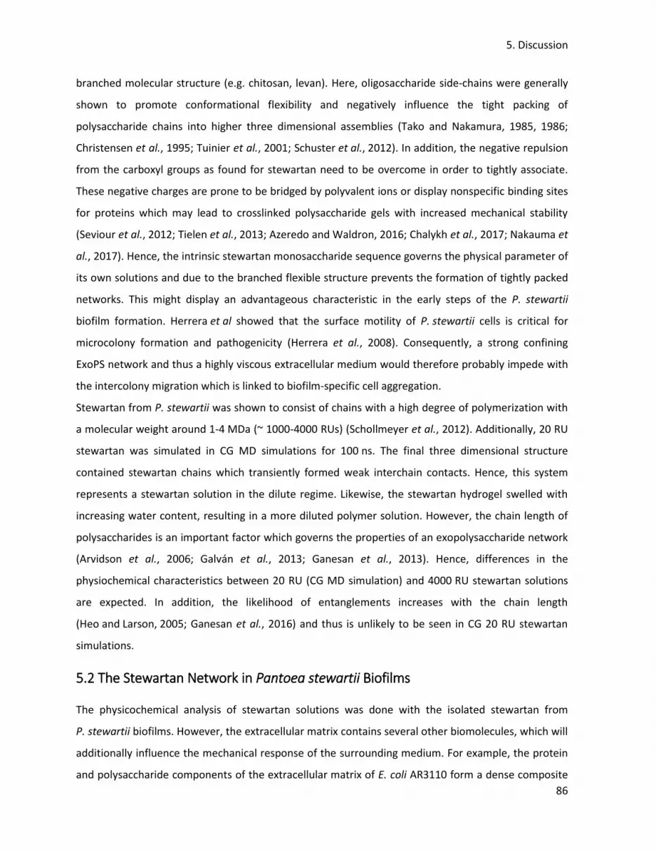
5. Discussion
86
branched molecular structure (e.g. chitosan, levan). Here, oligosaccharide side-chains were generally
shown to promote conformational flexibility and negatively influence the tight packing of
polysaccharide chains into higher three dimensional assemblies (Tako and Nakamura, 1985, 1986;
Christensen et al., 1995; Tuinier et al., 2001; Schuster et al., 2012). In addition, the negative repulsion
from the carboxyl groups as found for stewartan need to be overcome in order to tightly associate.
These negative charges are prone to be bridged by polyvalent ions or display nonspecific binding sites
for proteins which may lead to crosslinked polysaccharide gels with increased mechanical stability
(Seviour et al., 2012; Tielen et al., 2013; Azeredo and Waldron, 2016; Chalykh et al., 2017; Nakauma et
al., 2017). Hence, the intrinsic stewartan monosaccharide sequence governs the physical parameter of
its own solutions and due to the branched flexible structure prevents the formation of tightly packed
networks. This might display an advantageous characteristic in the early steps of the P. stewartii
biofilm formation. Herrera et al showed that the surface motility of P. stewartii cells is critical for
microcolony formation and pathogenicity (Herrera et al., 2008). Consequently, a strong confining
ExoPS network and thus a highly viscous extracellular medium would therefore probably impede with
the intercolony migration which is linked to biofilm-specific cell aggregation.
Stewartan from P. stewartii was shown to consist of chains with a high degree of polymerization with
a molecular weight around 1-4 MDa (~ 1000-4000 RUs) (Schollmeyer et al., 2012). Additionally, 20 RU
stewartan was simulated in CG MD simulations for 100 ns. The final three dimensional structure
contained stewartan chains which transiently formed weak interchain contacts. Hence, this system
represents a stewartan solution in the dilute regime. Likewise, the stewartan hydrogel swelled with
increasing water content, resulting in a more diluted polymer solution. However, the chain length of
polysaccharides is an important factor which governs the properties of an exopolysaccharide network
(Arvidson et al., 2006; Galván et al., 2013; Ganesan et al., 2013). Hence, differences in the
physiochemical characteristics between 20 RU (CG MD simulation) and 4000 RU stewartan solutions
are expected. In addition, the likelihood of entanglements increases with the chain length
(Heo and Larson, 2005; Ganesan et al., 2016) and thus is unlikely to be seen in CG 20 RU stewartan
simulations.
5.2 The Stewartan Network in Pantoea stewartii Biofilms
The physicochemical analysis of stewartan solutions was done with the isolated stewartan from
P. stewartii biofilms. However, the extracellular matrix contains several other biomolecules, which will
additionally influence the mechanical response of the surrounding medium. For example, the protein
and polysaccharide components of the extracellular matrix of E. coli AR3110 form a dense composite

5. Discussion
87
material (Serra et al., 2013a; Serra et al., 2015). Here, cellulose provided filamentous connections
between the cells, whereas curli protein fibers formed cell surrounding "baskets" (Serra et al., 2013a).
Consequently, brittle biofilm cryosection were obtained from bacterial mutants, which were deficient
in the cellulose production. Hence, the here presented results have to be considered as an estimate of
the whole biofilm properties, but as a very close approximation, as stewartan is the most abundant
component and major virulence factor in P. stewartii biofilms (Nimtz et al., 1996; Herrera et al., 2008;
Roper, 2011). Additionally, it is to note here, that the diffusion experiments in this work were not
conducted at conditions similar to those provided by the lumen of the xylem, which is a rich pool of
different salts (van Ieperen, 2007; Nardini et al., 2011). For example, the ionic strength was found to
be important in modulating the physicochemical properties (e.g. viscosity) of the ExoPS solution
(Orgad et al., 2011; Morrow et al., 2015; Torres et al., 2015; Yu et al., 2017). Consequently, more work
has to be done in characterizing the influence of ions on the viscoelastic properties of stewartan.
The average stewartan concentration of around 15 mg · mL-1 was calculated in this work for 65 h old
biofilms, a concentration which is above the critical entanglement concentration. Nevertheless, in
these experiments, the ExoPS concentration is averaged over the entire biofilm and did not consider
biofilms as rather heterogeneous structures with respect to the density of the extracellular matrix
(Lawrence et al., 2007; Xiao and Koo, 2010; Chew et al., 2014; Mosquera-Fernández et al., 2016). The
lowest concentration of the extracellular material is usually found at the periphery of the biofilm
assembly (Abedon, 2016; Vidakovic et al., 2018). Therefore, P. stewartii biofilms may exhibit regions of
high stewartan concentrations (close to the bacterial cells) which are characterized by a non-
Newtonian fluid with entangled polysaccharide polymers whereas other areas might display a rather
dilute flow behaviour of stewartan below the transition concentration of 7 mg · mL-1.
5.3 Limitations and improvements of the coarse-grained stewartan model
In this work, a stewartan CG model was derived from an all-atomistic MD simulation in a hybrid
approach by obtaining bonded and nonbonded potentials using the force matching method and
Boltzmann inversion, respectively. This strategy was shown to successfully reproduce the atomistic
configurations in cellulose and chitosan systems (Sauter and Grafmüller, 2017; Schneible et al., 2019).
In agreement, the RDFs of the CG stewartan system highly overlay with those of the atomistic
reference system. However, the simulations of CG stewartan often crashed which might be a result of
an overdetermination of the interactions in the CG stewartan model. Hence, too many bonded
potentials were used to coarse-grain stewartan which most likely overconstrained the flexible nature
of the ExoPS. For that reason, it is proposed here, to adopt the choice of bonded potentials to the

5. Discussion
88
stereochemistry of each specific glycosidic linkage types. The overdetermination of bonded potentials
was also seen, when the distribution functions of the CG model were compared to the atomistic
analogue. Here, the calculation of bonded potentials by Boltzmann inversion sometimes
overestimated the main maximum in the angular or dihedral distributions whereas secondary maxima
were poorly represented. This behaviour was also recognized in other studies when using Boltzmann
inversion (Hynninen et al., 2011; Sauter and Grafmüller, 2017). However, it is important to mention,
that the distribution functions of the atomistic and CG stewartan systems in this work are generally in
excellent agreement. Particular distributions could be improved by an iterative refinement of the
Boltzmann inversion derived potentials (iterative Boltzmann inversion) (Moore et al., 2014;
Choudhury et al., 2016; Li et al., 2016) where the limitations of Boltzmann inversion, such as the
sensitivity to environmental parameters (density, temperature or neighbour CG sites), are overcome
(Markutsya et al., 2013; Moore et al., 2014; Fan and Maranas, 2015).
Furthermore, the charged interactions between the CG glucoronic acid residues were less well
sampled in CG stewartan in agreement with the all-atomistic model. The extension of the CG model to
an eight bead mapping scheme further improved the charged interactions. Although, other
suggestions might additionally refine the force matching of the charged potentials. First, the mapping
of the CG sites on the center of charge (instead of center of mass) of the underlying group of atoms
resulted in a better description of the coulomb interactions (Cao and Voth, 2015; Dannenhoffer-
Lafage et al., 2019; Jin et al., 2019). Secondly, the potential of mean force provides a description of the
atomistic model and in CG simulations intra- and intermolecular potentials are added pairwise to give
an approximation of the potential of mean force (Das and Andersen, 2009; Dunn et al., 2016). The
calculation of separate CG potentials derived from the atomistic Lennard-Jones or Coulomb
interaction, which can be further modified, might also contribute to an additional improvement of the
charged interactions (Dannenhoffer-Lafage et al., 2019). Thirdly, electrostatic interactions are
screened by the surrounding solvent and ions (Wu et al., 2010; Ingólfsson et al., 2014; Lafond and
Izvekov, 2018). Therefore, many-body CG interactions have been introduced which recover the
potential of mean force with increased accuracy (Das and Andersen, 2012; John and Csányi, 2017).
5.4 Stewartan Chain Dynamics and Consequences for the Diffusion of Biomolecules
Many studies focused on the description of the diffusion dynamics of particles in polymer solvents
(Holyst et al., 2009; Guo et al., 2012; Grabowski and Mukhopadhyay, 2014; Kort et al., 2015;
Banks et al., 2016). The mobility of particles depend on separate time and length scales and within
these certain regimes the Stokes-Einstein relation does not apply (Cai et al., 2011; Nath et al., 2018).

5. Discussion
89
In this work, one important parameter which determined the particles diffusivity was the probe's
diameter. As shown, the hindrance experienced by the fluorescent tracer molecules increased with
particle size. Depending on the stewartan concentration, the diffusion of large PS microspheres
(193 nm) scaled in the same order than the macroviscosity determined with the falling ball
viscosimeter. It appears that these large PS beads experience the full macroviscosity of the stewartan
solution beyond the continuum limit which makes the description with the Stokes-Einstein relation
valid. Higher diffusion dynamics were obtained as the size of the PS microspheres decreased, thus the
local viscosity was probed (Holyst et al., 2009; Kohli and Mukhopadhyay, 2012). The latter varies with
the particle diameter in relation to the correlation length (average mesh size ξ) and radius of gyration
of the polysaccharide polymers (Cai et al., 2011; Nath et al., 2018). Lastly, a third regime was
described in which the particle diameter is much smaller than ξ and the probe will exhibit diffusion
coefficients very close to the ones in pure solvent (Cai et al., 2011; Nath et al., 2018). This is in
agreement with the diffusion dynamics of Alexa Fluor® 488 which yielded diffusion times similar to
those in buffer alone (hindrance factor close to unity). Hence, the pore diameter of the stewartan
network must be small enough (below 193 nm) to prevent the penetration of the interstitial space by
the largest PS beads (Cai et al., 2015), but in the range of 64 nm to partially hinder the diffusion of
64 nm PS microspheres. In agreement, pore formation between the stewartan chains were seen in CG
MD Simulations. The pore diameter varied with the stewartan concentration from 3 to 11 nm for the
RU concentrations of 86 and 10 mM, respectively. However, simulations with increased stewartan
chain lengths might be able to reproduce the pore dimensions as seen in the diffusion experiments.
In addition, differences in the hindrance experienced by the florescent tracer molecules in stewartan
were also observed in dependence to the pH. This suggests that the mobility is additionally affected by
electrostatic interactions of the probes with the stewartan polymer. As one obvious explanation, the
stewartan carboxylic groups (pKa ~ 3, Wang et al., 1991) are more negatively charged at pH 8 which
will influence the interaction with PS beads, which exhibit negative zeta potentials, by electrostatic
repulsion. Indeed, interactions of biomolecules or nano-sized objects with the extracellular matrix of
the biofilm have been reported (Dengler et al., 2015; Ikuma et al., 2015; Harper et al., 2019).
Furthermore, the biofilm permeability of antibiotics depends on the overall charge and may render
them ineffective in cases where they adsorb to biofilm components (Stewart et al., 2009; Tseng et al.,
2013; Singh et al., 2016). In addition, the negative charges of the glucuoronic acids subunits account
for a strong water association thus increasing the particle's local viscous drag (Nockemann et al., 2008;
Yang et al., 2009; Besbes et al., 2011). Further evidence from this work, that interactions with the
stewartan matrix resulted in a reduced mobility, comes from diffusion experiments with P22

5. Discussion
90
bacteriophage particles. Here, the mobility of the phages was found to be three to four times faster
than the diffusion of 64 nm PS beads. As the phage particle diameter is larger (71-73 nm) than that of
these microspheres, the individual experienced confinement cannot sufficiently be explained with the
differences in size of the particles. Indeed, the P22 phages exhibited lower negative Zeta potentials
than the PS microspheres and likely are therefore less repelled from the stewartan polymer material.
Other types of interactions, such as hydrophobic interactions, are also expected to influence the
diffusion behaviour of PS beads and P22 phages in stewartan solutions due to differences in their
surface properties (Asensio et al., 2013; Janczarek et al., 2015; Kuttel et al., 2015; Chin et al., 2017).
These results suggest that the stewartan polymers are prone to form a three dimensional network
which is sufficient to display characteristics from intact biofilms, such as the selective transport of
particles. Here, biofilms were described as molecular sieves (Flemming and Wingender, 2010).
5.5 Phage Particle Diffusion in Biofilm
During the stewartan diffusion experiments one population of phages was highly confined by the
ExoPS matrix with diffusion coefficients 100 to 1000 fold lower than freely diffusing P22
bacteriophages (at a stewartan concentration of 10 mg ∙ mL-1). This might prove an effective
entanglement of stewartan polysaccharides in which the meshes of the entangled stewartan network
are transiently formed in dependence to the entanglement length (Cai et al., 2011; Nath et al., 2018).
These length scales account for heterogeneities in the stewartan mesh-like network and eventually
phages became trapped in pores. In support of this finding, a population of 64 nm PS microspheres
were also found to be trapped in pores in preliminary experiments, resulting in low diffusion
coefficients. Indeed, in contrast to the freely diffusing agents, the diffusion behaviour of trapped
bacteriophage particles deviate from a classical random walk and showed a biased towards successive
steps in opposite direction (θ = 180°). Here, it is proposed that bacteriophages reside within the
interstitial space where they often collide with the stewartan material. In agreement, trapping of
phages in biofilms was seen even for bacteriophages which exhibited a respective enzymatic activity
toward the ExoPS (Hu et al., 2012; Vidakovic et al., 2018). As a result phages accumulated at the low-
dense periphery of the biofilm and the infection rate decreased with the deepness of biofilm
penetration. Additionally, bacteria associate into mixed-biofilm species which further impair with the
bacteriophages infectivity (Kay et al., 2011; Chew et al., 2014). Consequently, there is a big demand
for engineered phages with flexible host specificity and additional hydrolytic functions to overcome
dense biofilm architectures with different compositions of the extracellular matrix
(Lu and Collins, 2007; Alcaine et al., 2016; Cao et al., 2016; Lin et al., 2017).

5. Discussion
91
5.6 Interactions of WceF and ΦEa1h TSP with the Stewartan Matrix
The TSP from the ΦEa1h bacteriophage is a very fast and efficient stewartan hydrolase compared to
WceF. Incubations of the TSP with 10 mg ∙ mL-1 stewartan results in an almost complete loss of the
diffusion-limiting properties of the ExoPS network within 45 min at room temperature. The mobility of
64 nm PS microspheres underwent a transition from anomalous subdiffusion to normal Brownian
diffusion (anomaly exponent α ~ 1). This findings are in agreement with TSPs being highly efficient
polysaccharide depolymerases (Schwarzer et al., 2009; Cornelissen et al., 2012; Gutiérrez et al., 2015;
Kunstmann et al., 2018a). Additionally, TSPs comprise highly stable three dimensional folds which
ensure enzymatic activities at different environmental conditions (Barbirz et al., 2009). Consequently,
the ΦEa1h TSP is active over a broad range of pH values and NaCl concentrations.
Here, it was shown that the ΦEa1h TSP cleaves stewartan to oligosaccharides of different sizes. During
the stewartan hydrolysis a distribution of stewartan oligosaccharides was produced which presumably
spans the whole length from one RU to uncleaved stewartan polysaccharide. Analysis of the hydrolysis
products by capillary electrophoresis resolved oligosaccharide fragments up to 5 RU lengths. The
depolymerisation of stewartan by the ΦEa1h TSP at 30°C was shown to be biphasic. Reducing ends
were immediately produced in the beginning of the reaction. Here, large oligosaccharide fragments
were detected in size-exclusion chromatography which were further digested to one and two RUs.
After a saturation of the hydrolysis activity of 30 h and additional second increase in reducing ends
was detected and more 1 and 2 RUs were produced. Several studies revealed, that ExoPS solutions
contained a mixture of ordered and unordered polysaccharide regions (Christensen et al., 1993;
Christensen et al., 1996). Indeed, the depolymerisation of these ExoPS solutions by acid hydrolysis was
dependent on the conformational state of the polysaccharide (Hjerde et al., 1996). Comparable
enzymatic degradation studies showed that the cleavage of polysaccharides is characterized by
distinct kinetic regimes (Tayal et al., 1999; Li et al., 2004), in agreement with the biphasic stewartan
hydrolysis of the ΦEa1h TSP. Hence, the fast stewartan hydrolysis kinetic at the beginning of the
reaction could be explained by the degradation of more accessible stewartan populations whereas
less accessible polymers are cleaved in a slower second degradation step. In addition, in this second
kinetic regime long stewartan precursor molecules, which were not resolved by size-exclusion
chromatography, are further digested to small-sized RUs oligomers instead of the hydrolysis of two
RUs to one RUs. This emphasize that the ΦEa1h TSP might favour polysaccharide chains with a high
degree of polymerization. In support of this hypothesis, enzymes, which degrade high molecular
weight polysaccharides, usually bind the large polymer chains in a set of binding subsites

5. Discussion
92
(Highsmith et al., 1975; Michel et al., 2003; Nielsen et al., 2009; Kang et al., 2016). Here, a critical
number of binding sites must be occupied to position the polysaccharide chain in a more favourable
conformation for the succeeding hydrolysis step (Highsmith et al., 1975; Cramer et al., 1994; Nielsen
et al., 2009). Consequently, lower KM value are obtained for the binding of long polysaccharide chains
and thus the enzymatic efficiency increases (Cramer et al., 1994). Hence, the biphasic stewartan
degradation by the ΦEa1h TSP might also be a result of the early cleavage of long stewartan chains
and a delayed hydrolysis of shorter ExoPS. However, these two kinetic regimes were separated by a
50 h long saturation of long chained stewartan degradation. It is here proposed, that the cleaved
fragments still reside in the active site by nonproductive binding (Varrot et al., 2003;
Yamasaki et al., 2005). As soon, as the oligosaccharide products are displaced from the active site,
new substrate can be bound.
Nevertheless, studies which concentrated on the interactions of TSPs with high-molecular weight
polysaccharide fragments (above several RU) are rare but are highly needed to understand the
influence of chain length and polysaccharide network formation on the enzymes hydrolysis activity
(Cramer et al., 1994; Fennouri et al., 2012; Fennouri et al., 2013; Kang et al., 2016). The active site of
TSPs are composed of elongated shallow binding grooves on the surface of the β-helix
(Barbirz et al., 2008; Andres et al., 2013; Schwarzer et al., 2015; Lee et al., 2017). The interaction with
their ligands is mainly mediated by electrostatic interactions, such as hydrogen bonds, with a strong
negative enthalpic contribution (Baxa et al., 2001; Andres et al., 2013; Kang et al., 2016;
Kunstmann et al., 2018a). Binding of P22 TSP to a Shigella flexneri O-antigen dodecasaccharide
requires a flexible polysaccharide chain and partially unconstrained protein backbone to avoid steric
clashes (Kang et al., 2016). In agreement with the stewartan digestion by the ΦEa1h TSP, enzymatic
degradation of high molecular weight hyaluronan resulted in an oligosaccharide size distribution
which shifted towards smaller fragments depending on to the incubation time (Fennouri et al., 2012;
Fennouri et al., 2013).
The fast stewartan hydrolysis kinetic of the ΦEa1h TSP is in contrast to the ExoPS degradation by
WceF which is much more slower process. Here, the increase in reducing ends is characterized by a
pronounced lag phase. To best of knowledge, this is the first description of a bacterial tailspike-like
protein. It was noticed that WceF formed aggregates with stewartan. The size of these highly dense
protein clusters correspond to roughly 500 to 1000 WceF molecules in 10 and 1 mg ∙ mL-1 stewartan,
respectively, as deduced from the analysis of the hydrodynamic radius from WceF size-exclusion
chromatography experiments. In the previous chapter (section 5.4) the model of a molecular sieve

5. Discussion
93
was derived, describing the influence of non covalent interactions on the diffusion of difference sized
probes in the polymeric stewartan network. As a consequence, it is here assumed that WceF clusters
are formed in association with the ExoPS which provide interfaces for electrostatic and hydrophobic
interactions (Tielen et al., 2013). A similar behaviour has been found for lectins which are multivalent
carbohydrate binding proteins (Sharon, 2007; Kumar et al., 2012). They provide several glycan
epitopes which give rise to the cross-linking activities of lectins (Dam and Brewer, 2010). For example,
the lectin Con A formed aggregates with mannose-modified polymers of 300 to 500 nm
(Kim et al., 2005). Hence, it is here proposed that the trimeric WceF provide three independent
stewartan binding sites which results in a lectin-like cluster formation. These protein-ExoPS
interactions might then account for a hindered diffusion of WceF and consequently result in the
observed lag phase in the production of reducing ends.
Furthermore the long-lasting attachment of WceF with its substrate might be indicative for a
processive mode of stewartan hydrolysis. Here, the processivity of enzymes is defined as the strong
attachment of the protein to the substrate where it performs a series of catalytic events before it
dissociates (Breyer and Matthews, 2001; Horn et al., 2006). Indeed, mainly one RU fragments were
produced in incubations of WceF with stewartan. In agreement, the processivity of the chitinase B
hydrolysis of chitosan was roughly measured by studying the ratio between the production of
oligosaccharide dimers and monomers (Sørbotten et al., 2005; Horn et al., 2006). Hence, WceF might
remain bound to the stewartan polymer and moves along the chain while enzymatically releasing
single RUs. As a result, the long chains of stewartan would be degraded in a stepwise fashion. In
agreement with what was observed in this work, as soon as the stewartan substrate polymers are
small enough, they would be detected a later time points in the hydrolysis kinetic in the size-exclusion
chromatography experiments. Still, although to a lesser extent, fragments with other sizes were
produced, which proves that WceF is an endo-acting stewartan-specific depolymerase. Furthermore, a
high processivity usually comes with a trade-off between a high affinity of the enzyme towards the
substrate and a low hydrolysis velocity (Cramer et al., 1994; Breyer and Matthews, 2001;
Nakamura et al., 2014) which is in agreement with the low hydrolytic activity seen for WceF. In
contrast, the ΦEa1h TSP hydrolysis of stewartan is marked by less processivity and a more even
distribution of produced oligosaccharide lengths. Indeed, large oligosaccharides were immediately
seen in the size-exclusion chromatography experiments. Hence, the ΦEa1h bacteriophage cleaves
stewartan and more deeply penetrates the biofilm in order to reach the bacterial cell due to a fast
dissociation of the TSP from the initial ExoPS substrates. In contrast, other TSPs were shown to cleave

5. Discussion
94
the capsular polysaccharide in a highly processive manner which helps in guiding the bacteriophage
along the covalently attached polysaccharide towards the bacterium (Schwarzer et al., 2009).
5.7 Putative Function of WceF in Pantoea stewartii biofilms
The wildtype WceF exhibits an N-terminal twin-arginine translocation signal peptide thus in
P. stewartii WceF is translocated across the inner membrane (DeLisa et al., 2003;
Palmer and Berks, 2012). In this work, it was not investigated, if WceF is secreted to the periplasm or
the extracellular medium but this information would be highly relevant in the discussion of the
putative function of WceF in P. stewartii biofilms. For this reason, antibodies against WceF were raised
in the group of Prof. Dr. Katja Hanack (Immunotechnology, University Potsdam) which are currently
tested for their sensitivity and cross-reactivity. These antibodies would allow the detection of WceF in
subcellular fractionations of P. stewartii by enzyme-linked immunosorbent assay or western blots
(Mansell et al., 2010; Blair et al., 2018). The latter could be complemented with real-time polymerase
chain reaction to detect the WceF mRNA which might be linked to stage-specific biofilm phenotypes.
Hence, in the following, the putative function of WceF is discussed for both cases in which the enzyme
is secreted to either the extracellular medium or the periplasm.
In the diffusion experiment it was seen that WceF formed aggregates with the stewartan matrix. Such
clustering of colloidal particles with components of the extracellular matrix is a common feature which
has been recognized in many other bacterial biofilms (Duran-Pinedo et al., 2011; Stewart et al., 2013;
Vidakovic et al., 2018). Likewise, clumping of bacterial cells co-localizes with dense regions in the
extracellular matrix of S. epidermidis (Stewart et al., 2013). Furthermore, when fluorescent beads are
incubated with purified protein curli fibers, one component of the extracellular matrix of E. coli, they
spontaneously formed clusters (Vidakovic et al., 2018). All the here mentioned clusters of cells or
beads had a size (1 to 5 μm) similar to the WceF clusters seen in this work. Although, the
physicochemical properties of stewartan, S. epidermidis extracellular matrix and curli fibers differ, this
points to an important function of the extracellular matrix to specifically accumulate proteins with
beneficial functions (Tielen et al., 2013). Indeed, as a proof of principle, Tielen et al showed that
proteins can interact with the extracellular matrix components thereby preventing their degradation
by extracellular proteases (Tielen et al., 2013). Flemming et al introduced the concept of the
extracellular matrix as an external digestion system (Flemming et al., 2016). As a consequence
proteins can reside in the extracellular matrix and provide the enzymatic degradation activity of the
extracellular material as nutrient source (Flemming, 2016). For these reasons a slow digestion of
stewartan, as seen by WceF, might be useful in times of starvation to provide a long-lasting energy

5. Discussion
95
source. In agreement, other endoglycanases have been described in the extracellular medium which
trim the chain length of already secreted ExoPSs (York and Walker, 1997; Jones et al., 2007).
As WceF is part of the wce gene cluster WceF might also be involved in the biosynthesis of stewartan
in the periplasm. Here, stewartan is synthesized by the Wzx/Wzy-dependent pathway. This route of
polysaccharide biosynthesis is highly conserved throughout bacterial species and ExoPSs
(Whitfield, 2006; Islam and Lam, 2014; Schmid et al., 2015). Likewise, the Wzx/Wzy-dependent
pathway comprises glycosyltransferase and enzymes for the modification of oligosaccharides.
Biosynthesis is commonly initiated with the transfer of the first monosaccharide onto a
phosphorylated lipid linker which serves as an anchor for stepwise ExoPS synthesis. Polysaccharides
are secreted by wzx and outer membrane polysaccharide export (OPX) proteins. Polymerization of the
single building blocks is accomplished by the wzy protein. Additionally, the respective operons contain
polysaccharide co-polymerases (POP). POP and OPX proteins form an contiguous scaffold which spans
the cell envelope (Collins et al., 2007). These complexes are believed to be involved in periplasmic
chain length control of the final polymer (Marolda et al., 2006; Galván et al., 2013; Bianco et al., 2014).
In the ExoPS synthesis pathway of Rhizobium leguminosarum bv. trifolii disruption of the PCP gene
results in the production of ExoPS chains with a higher molecular weight than compared to the
wildtype (Marczak et al., 2014). In contrast, another study showed that loss of the function of PCP
proteins reduced the chain length distributions of the ExoPSs (Galván et al., 2013). This raised the
concept of PCP proteins which adjust the balance between chain extension and termination
(Cuthbertson et al., 2009). In contrast, no PCP proteins are expressed from the wce gene cluster in
P. stewartii. However, Schollmeyer at al observed a connection between the wceF deletion on the
P. stewartii genome and an increase of the ExoPS size distribution profile by a factor of around four
(Schollmeyer et al., 2012). Presumably, this shift in the size distribution of stewartan will dramatically
influence the viscous properties of the biofilm. Hence, WceF therefore might still be involved in
stewartan chain length determination in the periplasm and compensates for the non-existing PCP
homolog. Indeed, this hypothesis is supported by a case where the glycoside hydrolase Sph3 has been
reported to be encoded in the glucosamingalactan biosynthesis cluster (Bamford et al., 2015). Similar
to WceF, Sph3 showed no homology to known glycoside hydrolase families. Targeting of
polysaccharide chains in the periplasm is a common feature to regulate the ExoPS chain length
(Galván et al., 2013; Marczak et al., 2014) or to degrade ExoPS which failed to become exported
(Bakkevig et al., 2005). Within these types of enzymes is the periplasmic lyase VexL from Salmonella
enterica serovar typhi which showed a narrow maximum of K-antigen cleavage at pH 5.5
(Liston et al., 2018). It was proposed that the protein has evolved structural and catalytic advantages

5. Discussion
96
which facilitated the cleavage of the Vi-antigen at a low periplasmic pH. Indeed, a similar sharp activity
profile was seen for the stewartan hydrolysis of WceF at different pH and NaCl concentrations.
5.8 Structural Analysis of WceF and ΦEa1h TSP
The three dimensional structure of WceF displayed a β-solenoidal fold which resembled the domain
architecture of bacteriophage TSPs. This is emphasized by a central compact β-helix and an N-terminal
domain which superimposed with the phage particle-binding domain of P22 TSP. The C-terminus
showed a rich composition of β-sheet elements in agreement with other TSPs (Barbirz et al., 2008;
Müller et al., 2008; Lee et al., 2017). The central β-helix was also predicted for the ΦEa1h TSP. In
contrast, the N-terminus of φEa1h TSP lacked a comparable particle-binding domain. In these cases,
adapter proteins are encoded on the genome of the phage which attach the TSP to the bacteriophage
particle (Stummeyer et al., 2006; Leiman et al., 2007; Tu et al., 2017). Additionally, a Greek key motif
was predicted for the ΦEa1h TSP N-terminus. The latter was also found on WceF and shares the same
topology of β-sheets. Due to the high structural similarity of the Greek key motif between both
proteins, it is tempting to speculate, that both proteins originate from a same ancestor template.
Indeed, a similar conclusion was made, when the structure of three bacterial pectate lyases were
compared to the P22 TSP (Jenkins et al., 1998). In support of this hypothesis, the integration of phage
genes into the bacterial genome by horizontal gene transfer is a frequently observed process which
enhances the bacterial fitness (Obeng et al., 2016; Touchon et al., 2017).
It has been observed in this work, that neither WceF nor the ΦEa1h TSP showed a SDS-resistance at
ambient temperatures as typically seen for TSPs (Barbirz et al., 2009; Lee et al., 2017). No difference in
the stability of the trimeric interface of WceF was observed when compared to other TSPs. However,
in thermal unfolding experiments of TSPs it was shown, that the slow trimeric dissociation is rate-
limiting and is followed by a more rapid unfolding of the less stable subunits (Chen and King, 1991;
Miller et al., 1998b). Barbirz et al proposed that hydrogen bonds of the β-helix extend into connecting
arches and turns which thus account for the high stability of the TSP structures
(Manning and Colón, 2004; Barbirz et al., 2009). To overcome the high activation enthalpy, these
interactions need to be broken in a cooperative process (Truhlar et al., 2004; Barbirz et al., 2009). The
latter results in a kinetic stabilization of the TSPs which is emphasized by long equilibration times and
hysteresis between the denaturation and renaturation states (Fuchs et al., 1991; Lai et al., 1997;
Barbirz et al., 2009). In preliminary guanidine hydrochloride fluorescence titration experiments of
WceF no significant hysteresis was seen between the unfolding and refolding of WceF thus ruling out

5. Discussion
97
a high kinetic barrier. It is therefore proposed, that although the β-helix overall architecture does not
differ from that of the TSPs, it does not result in a similar kinetically stabilized hydrogen bond
network. This might be due to the overall funnel structure of WceF, in which the C-terminus of the
trimer is the most intertwined region and the preceding trimeric fold rather opens up instead of
forming a tightly packed assembly. In agreement with further studies on TSPs, the design and
biophysical characterization of truncated WceF constructs would further contribute to the
understanding of WceF trimer folding and stabilization processes (Schuler et al., 2000;
Freiberg et al., 2003; Reich et al., 2009).

6. Summary and Outlook
98
6. Summary and Outlook
Biofilms are dense and complex arrangements of bacterial cells and extracellular polymeric
substances. In this work a bottom up approach was chosen to effectively describe the structural
properties of the extracellular stewartan matrix which is ultimately linked to the infectivity of glycan-
based biofilms such as the one from P. stewartii. Here, the physicochemical characteristics of
stewartan solutions were analyzed in an interdisciplinary work which relates the structural aspects of
stewartan polymers to the functional implications in biofilms.
This work provided a detailed description of the physicochemical properties of the ExoPS species
stewartan from P. stewartii. It was found that purified stewartan polymers alone are sufficient to form
three dimensional networks which resemble characteristics of whole biofilms, such as the
viscoelasticity and diffusion-limiting properties. Analysis of the diffusion dynamics of fluorescent
tracer molecules in reconstituted stewartan solutions showed that the mobility of these particles
depend on the size of the nanoparticle and charge of the stewartan polymer. Furthermore, the
diffusion of fluorescent molecules became more anomalous as a result of an increased density of
stewartan polymers and the appearance of stewartan chain entanglements. The pores between the
stewartan chains were heterogeneous regarding their size and a fraction of bacteriophage particles
was trapped within small-sized network meshes.
In agreement with other biological hydrogels, stewartan acts as a molecular sieve and selectively
controls the transport of molecules within the stewartan network (Witten and Ribbeck, 2017). From
these findings, this work gave a description of the material properties of glycan-based biofilms such as
P. stewartii. Furthermore, the structure of the exopolysaccharide, including branches, monosaccharide
composition and modifications, ultimately determines the arrangement of polysaccharide chains in
the respective network. This work therefore proposes, that this approach might be generalized for
other ExoPSs from biofilms of different bacterial species. The increased knowledge about the fine-
tuning of the strength of polysaccharide network assemblies is highly important towards the design of
polysaccharide-based drug delivery agents (Di Meo et al., 2007; Hu et al., 2013; García-González et al.,
2015; Nayak et al., 2016; Schneible et al., 2019) or for bioremediation approaches in which heavy
metal contaminations are removed from the environment (Prado Acosta et al., 2005; Mota et al.,
2016; Sajayan et al., 2017).
Further diffusion and bulk rheology studies should extend the reduced systems from this work by
adding additional components from the extracellular matrix until a whole biofilm-like setup has been

6. Summary and Outlook
99
generated. The thorough analysis of these systems would then lead to a comprehensive
understanding of the contribution of each extracellular species to the overall biofilm architecture.
These whole systems can thus be exploited to design drugs with superior physicochemical properties
which permit the facilitated penetration of the biofilm and effective killing of bacterial cells.
In this work a coarse-grained computational stewartan model was developed with stewartan polymers
which exhibited a degree of polymerization of 20 RU. This system constitutes a first initial CG
description of stewartan and further modifications on the CG model development have been
suggested to enable MD Simulations with microsecond time scales. Additionally, increasing the size of
the CG stewartan polymers could elucidate the influence of chain length and entanglements on
stewartan network properties accessible from experiments like its microviscosity. Furthermore, in
equilibrated ExoPS networks, simulations could mimic the diffusion of probes with different properties
(size, charge, hydrophobic interactions) and thus allow a more detailed study of the interaction of
nano-sized objects with the network (Schneible et al., 2019).
Currently, there is just a scarce body of literature available that might describe how mechanical
properties of biofilms relate to biochemical control and dynamic changes within a bacterial
community. Especially, enzymatic remodeling processes take place which might be the driving forces
that control pathogenicity. In this work, the hydrolysis of stewartan polymers by the enzymes WceF
and ΦE1h TSP was analyzed. Surprisingly, WceF, which is expressed from the P. stewartii stewartan
biosynthesis gene cluster, exhibited a highly similar three dimensional structure to TSPs. Hence, a
common ancestor of WceF and the ΦEa1h TSP was proposed. Still, despite the structural similarity,
WceF lacked the high stability seen in many other TSP. Additionally, the hydrolysis of stewartan
polymers by the ΦEa1h TSP is a highly fast process which reflects the ability of the bacteriophage to
efficiently pass the biofilm barrier in order to infect the bacterial cells. Cleavage of stewartan by the
ΦE1h TSP resulted in a complete loss of the intrinsic diffusion-limiting properties of the stewartan
network and hence the mobility of bacteriophages would therefore not be confined anymore. Finally,
WceF formed tight interactions with the stewartan network and micrometer clusters were formed
which are might be the reason for the slow stewartan hydrolysis kinetic. Furthermore, stewartan is
degraded by WceF in a processive manner, in contrast to the ΦEa1h TSP.
Differences in the stewartan degradation activities of ΦEa1h TSP and WceF might be encoded in the
structural properties of the active sites. Binding of oligosaccharide fragments to the TSP have been
analyzed by soaking experiments or MD simulations (Barbirz et al., 2008; Müller et al., 2008;
Schulz et al., 2010; Kang et al., 2016; Lee et al., 2017). The association of oligosaccharides was further

6. Summary and Outlook
100
characterized in surface plasmon resonance, isothermal titration calorimetry or fluorescence titration
(Singh et al., 2010; Andres et al., 2013; Broeker et al., 2013; Kunstmann et al., 2018a). Consequently
all the latter methods might be used to further investigate the difference in stewartan binding sites
between ΦEa1h TSP and WceF. It is of crucial interest to understand the driving forces of protein
interactions with the components of the extracellular matrix to pave the way for treatments of biofilm
infections which involve the hydrolase-mediated biofilm degradation (Fleming et al., 2017;
Olsen et al., 2018b; Patel et al., 2019).

7. References
101
7. References
Abedon ST 2016. Bacteriophage exploitation of bacterial biofilms. Phage preference for less mature
targets? FEMS microbiology letters 363. doi:10.1093/femsle/fnv246.
Ackermann H-W 2003. Bacteriophage observations and evolution. Research in Microbiology 154, 245–
251. doi:10.1016/S0923-2508(03)00067-6.
Akiyama E, Morimoto N, Kujawa P, Ozawa Y, Winnik FM and Akiyoshi K 2007. Self-assembled nanogels
of cholesteryl-modified polysaccharides: effect of the polysaccharide structure on their association
characteristics in the dilute and semidilute regimes. Biomacromolecules 8, 2366–2373.
doi:10.1021/bm070136q.
Alcaine SD, Law K, Ho S, Kinchla AJ, Sela DA and Nugen SR 2016. Bioengineering bacteriophages to
enhance the sensitivity of phage amplification-based paper fluidic detection of bacteria. Biosensors
& bioelectronics 82, 14–19. doi:10.1016/j.bios.2016.03.047.
Alibolandi M, Mohammadi M, Taghdisi SM, Abnous K and Ramezani M 2017. Synthesis and
preparation of biodegradable hybrid dextran hydrogel incorporated with biodegradable curcumin
nanomicelles for full thickness wound healing. International journal of pharmaceutics 532, 466–
477. doi:10.1016/j.ijpharm.2017.09.042.
Andres D, Baxa U, Hanke C, Seckler R and Barbirz S 2010a. Carbohydrate binding of Salmonella phage
P22 tailspike protein and its role during host cell infection. Biochemical Society transactions 38,
1386–1389. doi:10.1042/BST0381386.
Andres D, Gohlke U, Broeker NK, Schulze S, Rabsch W, Heinemann U, Barbirz S and Seckler R 2013. An
essential serotype recognition pocket on phage P22 tailspike protein forces Salmonella enterica
serovar Paratyphi A O-antigen fragments to bind as nonsolution conformers. Glycobiology 23, 486–
494. doi:10.1093/glycob/cws224.
Andres D, Hanke C, Baxa U, Seul A, Barbirz S and Seckler R 2010b. Tailspike interactions with
lipopolysaccharide effect DNA ejection from phage P22 particles in vitro. The Journal of biological
chemistry 285, 36768–36775. doi:10.1074/jbc.M110.169003.

7. References
102
Andres D, Roske Y, Doering C, Heinemann U, Seckler R and Barbirz S 2012. Tail morphology controls
DNA release in two Salmonella phages with one lipopolysaccharide receptor recognition system.
Molecular microbiology 83, 1244–1253. doi:10.1111/j.1365-2958.2012.08006.x.
Anthon GE and Barrett DM 2002. Determination of reducing sugars with 3-methyl-2-
benzothiazolinonehydrazone. Analytical Biochemistry 305, 287–289. doi:10.1006/abio.2002.5644.
Arias-Andres M, Klümper U, Rojas-Jimenez K and Grossart H-P 2018. Microplastic pollution increases
gene exchange in aquatic ecosystems. Environmental pollution (Barking, Essex : 1987) 237, 253–
261. doi:10.1016/j.envpol.2018.02.058.
Arvidson SA, Rinehart BT and Gadala-Maria F 2006. Concentration regimes of solutions of levan
polysaccharide from Bacillus sp. Carbohydrate Polymers 65, 144–149.
doi:10.1016/j.carbpol.2005.12.039.
Asensio JL, Ardá A, Cañada FJ and Jiménez-Barbero J 2013. Carbohydrate-aromatic interactions.
Accounts of chemical research 46, 946–954. doi:10.1021/ar300024d.
Azeredo HMC and Waldron KW 2016. Crosslinking in polysaccharide and protein films and coatings for
food contact – A review. Trends in Food Science & Technology 52, 109–122.
doi:10.1016/j.tifs.2016.04.008.
Baere T de, Verhelst R, Labit C, Verschraegen G, Wauters G, Claeys G and Vaneechoutte M 2004.
Bacteremic infection with Pantoea ananatis. Journal of clinical microbiology 42, 4393–4395.
doi:10.1128/JCM.42.9.4393–4395.2004.
Bakkevig K, Sletta H, Gimmestad M, Aune R, Ertesvåg H, Degnes K, Christensen BE, Ellingsen TE and
Valla S 2005. Role of the Pseudomonas fluorescens alginate lyase (AlgL) in clearing the periplasm of
alginates not exported to the extracellular environment. Journal of Bacteriology 187, 8375–8384.
doi:10.1128/JB.187.24.8375–8384.2005.
Bamford NC, Snarr BD, Gravelat FN, Little DJ, Lee MJ, Zacharias CA, Chabot JC, Geller AM, Baptista SD,
Baker P, Robinson H, Howell PL and Sheppard DC 2015. Sph3 Is a Glycoside Hydrolase Required for
the Biosynthesis of Galactosaminogalactan in Aspergillus fumigatus. The Journal of biological
chemistry 290, 27438–27450. doi:10.1074/jbc.M115.679050.

7. References
103
Banks DS, Tressler C, Peters RD, Höfling F and Fradin C 2016. Characterizing anomalous diffusion in
crowded polymer solutions and gels over five decades in time with variable-lengthscale
fluorescence correlation spectroscopy. Soft Matter 12, 4190–4203. doi:10.1039/c5sm01213a.
Barbirz S, Becker M, Freiberg A and Seckler R 2009. Phage tailspike proteins with beta-solenoid fold as
thermostable carbohydrate binding materials. Macromolecular bioscience 9, 169–173.
doi:10.1002/mabi.200800278.
Barbirz S, Müller JJ, Uetrecht C, Clark AJ, Heinemann U and Seckler R 2008. Crystal structure of
Escherichia coli phage HK620 tailspike. Podoviral tailspike endoglycosidase modules are
evolutionarily related. Molecular microbiology 69, 303–316. doi:10.1111/j.1365-
2958.2008.06311.x.
Baruah R, Maina NH, Katina K, Juvonen R and Goyal A 2017. Functional food applications of dextran
from Weissella cibaria RBA12 from pummelo (Citrus maxima). International journal of food
microbiology 242, 124–131. doi:10.1016/j.ijfoodmicro.2016.11.012.
Baxa U, Cooper A, Weintraub A, Pfeil W and Seckler R 2001. Enthalpic Barriers to the Hydrophobic
Binding of Oligosaccharides to Phage P22 Tailspike Protein †. Biochemistry 40, 5144–5150.
doi:10.1021/bi0020426.
Belas R and Suvanasuthi R 2005. The ability of Proteus mirabilis to sense surfaces and regulate
virulence gene expression involves FliL, a flagellar basal body protein. Journal of Bacteriology 187,
6789–6803. doi:10.1128/JB.187.19.6789-6803.2005.
Bellemann P, Bereswill S, Berger S and Geider K 1994. Visualization of capsule formation by Erwinia
amylovora and assays to determine amylovoran synthesis. International Journal of Biological
Macromolecules 16, 290–296.
Bernhard F 1996. Genetic transfer of amylovoran and stewartan between Erwinia amylovora and
Erwinia stewartii. Microbiology 142, 1087–1096.
Besbes I, Alila S and Boufi S 2011. Nanofibrillated cellulose from TEMPO-oxidized eucalyptus fibres:
Effect of the carboxyl content. Carbohydrate Polymers 84, 975–983.
doi:10.1016/j.carbpol.2010.12.052.

7. References
104
Bhattacharya S and Gubbins KE 2006. Fast method for computing pore size distributions of model
materials. Langmuir : the ACS journal of surfaces and colloids 22, 7726–7731.
doi:10.1021/la052651k.
Bianco MI, Jacobs M, Salinas SR, Salvay AG, Ielmini MV and Ielpi L 2014. Biophysical characterization of
the outer membrane polysaccharide export protein and the polysaccharide co-polymerase protein
from Xanthomonas campestris. Protein expression and purification 101, 42–53.
doi:10.1016/j.pep.2014.06.002.
Bicudo EL, Macedo VO, Carrara MA, Castro FFS and Rage RI 2007. Nosocomial outbreak of Pantoea
agglomerans in a pediatric urgent care center. The Brazilian journal of infectious diseases : an
official publication of the Brazilian Society of Infectious Diseases 11, 281–284. doi:10.1590/s1413-
86702007000200023.
Birdsall CK and Langdon AB 2005. Plasma physics via computer simulation, New York Taylor & Francis.
Birjiniuk A, Billings N, Nance E, Hanes J, Ribbeck K and Doyle PS 2014. Single particle tracking reveals
spatial and dynamic organization of the E. coli biofilm matrix. New journal of physics 16, 85014.
doi:10.1088/1367-2630/16/8/085014.
Blair KM, Mears KS, Taylor JA, Fero J, Jones LA, Gafken PR, Whitney JC and Salama NR 2018. The
Helicobacter pylori cell shape promoting protein Csd5 interacts with the cell wall, MurF, and the
bacterial cytoskeleton. Molecular microbiology 110, 114–127. doi:10.1111/mmi.14087.
Block CC, Hill JH and McGee DC 1998. Seed Transmission of Pantoea stewartii in Field and Sweet Corn.
Plant Disease 82, 775–780. doi:10.1094/PDIS.1998.82.7.775.
Bogino PC, Oliva MdlM, Sorroche FG and Giordano W 2013. The role of bacterial biofilms and surface
components in plant-bacterial associations. International journal of molecular sciences 14, 15838–
15859. doi:10.3390/ijms140815838.
Borgersen Q, Bolick DT, Kolling GL, Aijuka M, Ruiz-Perez F, Guerrant RL, Nataro JP and Santiago AE
2018. Abundant production of exopolysaccharide by EAEC strains enhances the formation of
bacterial biofilms in contaminated sprouts. Gut microbes 9, 264–278.
doi:10.1080/19490976.2018.1429877.

7. References
105
Boulé J, Sholberg PL, Lehman SM, O'gorman DT and Svircev AM 2011. Isolation and characterization of
eight bacteriophages infecting Erwinia amylovora and their potential as biological control agents in
British Columbia, Canada. Canadian Journal of Plant Pathology 33, 308–317.
doi:10.1080/07060661.2011.588250.
Bragard C, Dehnen‐Schmutz K, Di Serio F, Gonthier P, Jacques M‐A, Jaques Miret JA, Justesen AF,
MacLeod A, Magnusson CS, Milonas P, Navas‐Cortes JA, Parnell S, Potting R, Reignault PL, Thulke
H‐H, Civera AV, Yuen J, Zappalà L, Battilani P, Pautasso M and van der Werf W 2019. Risk
assessment of the entry of Pantoea stewartii subsp. stewartii on maize seed imported by the EU
from the USA. EFSA Journal 17, 39. doi:10.2903/j.efsa.2019.5851.
Braun EJ 1982. Ultraestructural investigation of resistant and susceptible maize inbred infected with
Erwinia stewartii. Phytopathology 72, 159–166.
Bren L 2007. Bacteria-eating virus approved as food additive. FDA consumer 41, 20–22.
Breyer WA and Matthews BW 2001. A structural basis for processivity. Protein science : a publication
of the Protein Society 10, 1699–1711.
Bridier A, Dubois-Brissonnet F, Boubetra A, Thomas V and Briandet R 2010. The biofilm architecture of
sixty opportunistic pathogens deciphered using a high throughput CLSM method. Journal of
microbiological methods 82, 64–70. doi:10.1016/j.mimet.2010.04.006.
Broeker NK, Andres D, Kang Y, Gohlke U, Schmidt A, Kunstmann S, Santer M and Barbirz S 2017.
Complex carbohydrate recognition by proteins: Fundamental insights from bacteriophage cell
adhesion systems. Perspectives in Science 11, 45–52. doi:10.1016/j.pisc.2016.10.001.
Broeker NK, Gohlke U, Müller JJ, Uetrecht C, Heinemann U, Seckler R and Barbirz S 2013. Single amino
acid exchange in bacteriophage HK620 tailspike protein results in thousand-fold increase of its
oligosaccharide affinity. Glycobiology 23, 59–68. doi:10.1093/glycob/cws126.
Broeker NK, Kiele F, Casjens SR, Gilcrease EB, Thalhammer A, Koetz J and Barbirz S 2018. In Vitro
Studies of Lipopolysaccharide-Mediated DNA Release of Podovirus HK620. Viruses 10.
doi:10.3390/v10060289.
Bush K 2010. The coming of age of antibiotics: discovery and therapeutic value. Annals of the New
York Academy of Sciences 1213, 1–4. doi:10.1111/j.1749-6632.2010.05872.x.

7. References
106
Cai L-H, Panyukov S and Rubinstein M 2011. Mobility of Nonsticky Nanoparticles in Polymer Liquids.
Macromolecules 44, 7853–7863. doi:10.1021/ma201583q.
Cai L-H, Panyukov S and Rubinstein M 2015. Hopping Diffusion of Nanoparticles in Polymer Matrices.
Macromolecules 48, 847–862. doi:10.1021/ma501608x.
Cao B, Yang M and Mao C 2016. Phage as a Genetically Modifiable Supramacromolecule in Chemistry,
Materials and Medicine. Accounts of chemical research 49, 1111–1120.
doi:10.1021/acs.accounts.5b00557.
Cao Z and Voth GA 2015. The multiscale coarse-graining method. XI. Accurate interactions based on
the centers of charge of coarse-grained sites. The Journal of Chemical Physics 143, 243116.
doi:10.1063/1.4933249.
Carlier A, Burbank L and Bodman SB von 2009. Identification and characterization of three novel
EsaI/EsaR quorum-sensing controlled stewartan exopolysaccharide biosynthetic genes in Pantoea
stewartii ssp. stewartii. Molecular microbiology 74, 903–913. doi:10.1111/j.1365-
2958.2009.06906.x.
Cassini A, Högberg LD, Plachouras D, Quattrocchi A, Hoxha A, Simonsen GS, Colomb-Cotinat M,
Kretzschmar ME, Devleesschauwer B, Cecchini M, Ouakrim DA, Oliveira TC, Struelens MJ, Suetens
C, Monnet DL, Strauss R, Mertens K, Struyf T, Catry B, Latour K, Ivanov IN, Dobreva EG, Tambic
Andraševic A, Soprek S, Budimir A, Paphitou N, Žemlicková H, Schytte Olsen S, Wolff Sönksen U,
Märtin P, Ivanova M, Lyytikäinen O, Jalava J, Coignard B, Eckmanns T, Abu Sin M, Haller S, Daikos
GL, Gikas A, Tsiodras S, Kontopidou F, Tóth Á, Hajdu Á, Guólaugsson Ó, Kristinsson KG, Murchan S,
Burns K, Pezzotti P, Gagliotti C, Dumpis U, Liuimiene A, Perrin M, Borg MA, Greeff SC de, Monen
JCM, Koek MBG, Elstrøm P, Zabicka D, Deptula A, Hryniewicz W, Caniça M, Nogueira PJ, Fernandes
PA, Manageiro V, Popescu GA, Serban RI, Schréterová E, Litvová S, Štefkovicová M, Kolman J, Klavs
I, Korošec A, Aracil B, Asensio A, Pérez-Vázquez M, Billström H, Larsson S, Reilly JS, Johnson A and
Hopkins S 2019. Attributable deaths and disability-adjusted life-years caused by infections with
antibiotic-resistant bacteria in the EU and the European Economic Area in 2015: a population-level
modelling analysis. The Lancet Infectious Diseases 19, 56–66. doi:10.1016/S1473-3099(18)30605-4.
Cavaliere R, Ball JL, Turnbull L and Whitchurch CB 2014. The biofilm matrix destabilizers, EDTA and
DNaseI, enhance the susceptibility of nontypeable Hemophilus influenzae biofilms to treatment
with ampicillin and ciprofloxacin. MicrobiologyOpen 3, 557–567. doi:10.1002/mbo3.187.

7. References
107
Chalykh AE, Matveev VV, Muravlev DA, Mityuk DY and Philippova OE 2017. Nanostructure of xanthan
networks. Nanotechnologies in Russia 12, 1–8. doi:10.1134/S1995078017010037.
Chanmee T, Ontong P and Itano N 2016. Hyaluronan: A modulator of the tumor microenvironment.
Cancer letters 375, 20–30. doi:10.1016/j.canlet.2016.02.031.
Charles C, Rout S, Laws A, Jackson B, Boxall S and Humphreys P 2017. The Impact of Biofilms upon
Surfaces Relevant to an Intermediate Level Radioactive Waste Geological Disposal Facility under
Simulated Near-Field Conditions. Geosciences 7, 57. doi:10.3390/geosciences7030057.
Chaturongakul S and Ounjai P 2014. Phage-host interplay: examples from tailed phages and Gram-
negative bacterial pathogens. Frontiers in microbiology 5, 442. doi:10.3389/fmicb.2014.00442.
Chen B and King J 1991. Thermal unfolding pathway for the thermostable P22 tailspike
endorhamnosidase. Biochemistry 30, 6260–6269. doi:10.1021/bi00239a026.
Chen F, Zhang Z, Deng Z, Zhang R, Fan G, Ma D and McClements DJ 2018. Controlled-release of
antacids from biopolymer microgels under simulated gastric conditions: Impact of bead
dimensions, pore size, and alginate/pectin ratio. Food research international (Ottawa, Ont.) 106,
745–751. doi:10.1016/j.foodres.2018.01.038.
Chen VB, Arendall WB, Headd JJ, Keedy DA, Immormino RM, Kapral GJ, Murray LW, Richardson JS and
Richardson DC 2010. MolProbity: all-atom structure validation for macromolecular crystallography.
Acta crystallographica. Section D, Biological crystallography 66, 12–21.
doi:10.1107/S0907444909042073.
Chew SC, Kundukad B, Seviour T, van der Maarel JRC, Yang L, Rice SA, Doyle P and Kjelleberg S 2014.
Dynamic remodeling of microbial biofilms by functionally distinct exopolysaccharides. mBio 5,
e01536-14. doi:10.1128/mBio.01536-14.
Chin J, Sinha S, Nalaparaju A, Yam J, Qin Z, Ma L, Liang Z-X, Lu L, Bhattacharjya S and Yang L 2017.
Pseudomonas aeruginosa Psl Exopolysaccharide Interacts with the Antimicrobial Peptide LG21.
Water 9, 681. doi:10.3390/w9090681.
Choudhury CK, Carbone P and Roy S 2016. Scalability of Coarse-Grained Potentials Generated from
Iterative Boltzmann Inversion for Polymers: Case Study on Polycarbonates. Macromolecular Theory
and Simulations 25, 274–286. doi:10.1002/mats.201500079.

7. References
108
Christensen BE, Smidsrød O and Stokke BT 1996. Metastable, Partially Depolymerized Xanthans and
Rearrangements toward Perfectly Matched Duplex Structures. Macromolecules 29, 2939–2944.
doi:10.1021/ma951445o.
Christensen BE, Smidsroed O, Elgsaeter A and Stokke BT 1993. Depolymerization of double-stranded
xanthan by acid hydrolysis: characterization of partially degraded double strands and single-
stranded oligomers released from the ordered structures. Macromolecules 26, 6111–6120.
doi:10.1021/ma00074a037.
Christensen BE, Stokke BT and Smidsrød O 1995. Xanthan—The natural water soluble cellulose
derivative. In Cellulose and cellulose derivatives. Physico-chemical aspects and industrial
applications (eds JF Kennedy, GO Phillips, PA Williams and L Piculell), pp. 265–278. Woodhead,
Cambridge.
Collins RF, Beis K, Dong C, Botting CH, McDonnell C, Ford RC, Clarke BR, Whitfield C and Naismith JH
2007. The 3D structure of a periplasm-spanning platform required for assembly of group 1 capsular
polysaccharides in Escherichia coli. Proceedings of the National Academy of Sciences of the United
States of America 104, 2390–2395. doi:10.1073/pnas.0607763104.
Comeau AM, Hatfull GF, Krisch HM, Lindell D, Mann NH and Prangishvili D 2008. Exploring the
prokaryotic virosphere. Research in Microbiology 159, 306–313. doi:10.1016/j.resmic.2008.05.001.
Cook KA, Weinzierl RA, Pataky JK, Esker PD and Nutter FW 2005. Population Densities of Corn Flea
Beetle (Coleoptera: Chrysomelidae) and Incidence of Stewart’s Wilt in Sweet Corn. Journal of
Economic Entomology 98, 673–682. doi:10.1603/0022-0493-98.3.673.
Cordomí A, Edholm O and Perez JJ 2009. Effect of Force Field Parameters on Sodium and Potassium
Ion Binding to Dipalmitoyl Phosphatidylcholine Bilayers. Journal of chemical theory and
computation 5, 2125–2134. doi:10.1021/ct9000763.
Cornelissen A, Ceyssens P-J, Krylov VN, Noben J-P, Volckaert G and Lavigne R 2012. Identification of
EPS-degrading activity within the tail spikes of the novel Pseudomonas putida phage AF. Virology
434, 251–256. doi:10.1016/j.virol.2012.09.030.
Cowen L, Bradley P, Menke M, King J and Berger B 2002. Predicting the beta-helix fold from protein
sequence data. Journal of computational biology : a journal of computational molecular cell biology
9, 261–276. doi:10.1089/10665270252935458.

7. References
109
Cramer JA, Bailey LC, Bailey CA and Miller RT 1994. Kinetic and mechanistic studies with bovine
testicular hyaluronidase. Biochimica et biophysica acta 1200, 315–321. doi:10.1016/0304-
4165(94)90173-2.
Cruz AT, Cazacu AC and Allen CH 2007. Pantoea agglomerans, a plant pathogen causing human
disease. Journal of clinical microbiology 45, 1989–1992. doi:10.1128/JCM.00632-07.
Cuthbertson L, Mainprize IL, Naismith JH and Whitfield C 2009. Pivotal roles of the outer membrane
polysaccharide export and polysaccharide copolymerase protein families in export of extracellular
polysaccharides in gram-negative bacteria. Microbiology and molecular biology reviews : MMBR
73, 155–177. doi:10.1128/MMBR.00024-08.
Czerwinski EW, Midoro-Horiuti T, White MA, Brooks EG and Goldblum RM 2005. Crystal structure of
Jun a 1, the major cedar pollen allergen from Juniperus ashei, reveals a parallel beta-helical core.
Journal of Biological Chemistry 280, 3740–3746. doi:10.1074/jbc.M409655200.
Dam TK and Brewer CF 2010. Lectins as pattern recognition molecules: the effects of epitope density
in innate immunity. Glycobiology 20, 270–279. doi:10.1093/glycob/cwp186.
Danese PN, Pratt LA and Kolter R 2000. Exopolysaccharide production is required for development of
Escherichia coli K-12 biofilm architecture. Journal of Bacteriology 182, 3593–3596.
doi:10.1128/jb.182.12.3593-3596.2000.
Dannenhoffer-Lafage T, Wagner JW, Durumeric AEP and Voth GA 2019. Compatible observable
decompositions for coarse-grained representations of real molecular systems. The Journal of
Chemical Physics 151, 134115. doi:10.1063/1.5116027.
Darden T, York D and Pedersen L 1993. Particle mesh Ewald: An N ⋅log(N ) method for Ewald sums in
large systems. The Journal of Chemical Physics 98, 10089–10092. doi:10.1063/1.464397.
Das A and Andersen HC 2009. The multiscale coarse-graining method. III. A test of pairwise additivity
of the coarse-grained potential and of new basis functions for the variational calculation. The
Journal of Chemical Physics 131, 34102. doi:10.1063/1.3173812.
Das A and Andersen HC 2012. The multiscale coarse-graining method. IX. A general method for
construction of three body coarse-grained force fields. The Journal of Chemical Physics 136,
194114. doi:10.1063/1.4705417.

7. References
110
DeLisa MP, Tullman D and Georgiou G 2003. Folding quality control in the export of proteins by the
bacterial twin-arginine translocation pathway. Proceedings of the National Academy of Sciences of
the United States of America 100, 6115–6120. doi:10.1073/pnas.0937838100.
Dengler V, Foulston L, DeFrancesco AS and Losick R 2015. An Electrostatic Net Model for the Role of
Extracellular DNA in Biofilm Formation by Staphylococcus aureus. Journal of Bacteriology 197,
3779–3787. doi:10.1128/JB.00726-15.
Desbrieres J 2002. Viscosity of semiflexible chitosan solutions: influence of concentration,
temperature, and role of intermolecular interactions. Biomacromolecules 3, 342–349.
doi:10.1021/bm010151+.
Di Lorenzo A, Varcamonti M, Parascandola P, Vignola R, Bernardi A, Sacceddu P, Sisto R and Alteriis E
de 2005. Characterization and performance of a toluene-degrading biofilm developed on pumice
stones. Microbial cell factories 4, 4. doi:10.1186/1475-2859-4-4.
Di Luca, M., Tkhilaishvili, T., Trampuz, A. 2018. SPECIFIC ANTIBIOFILM PROPERTIES OF
BACTERIOPHAGE SB-1 MAKE IT SUITABLE FOR THE THERAPY OF PROSTHETIC JOINT INFECTIONS
DUE TO STAPHYLOCOCCUS AUREUS: BIOFILM MATRIX DEGRADATION AND PERSISTER CELLS
KILLING. Orthopaedic Proceedings Vol, 100. No SUPP_17.
Di Meo C, Panza L, Capitani D, Mannina L, Banzato A, Rondina M, Renier D, Rosato A and Crescenzi V
2007. Hyaluronan as carrier of carboranes for tumor targeting in boron neutron capture therapy.
Biomacromolecules 8, 552–559. doi:10.1021/bm0607426.
Donlan RM and Costerton JW 2002. Biofilms: survival mechanisms of clinically relevant
microorganisms. Clinical microbiology reviews 15, 167–193. doi:10.1128/cmr.15.2.167-193.2002.
Doyle SA 2009. High Throughput Protein Expression and Purification. Methods and Protocols. Humana
Press, Totowa, NJ.
Dublanchet A and Bourne S 2007. The epic of phage therapy. The Canadian journal of infectious
diseases & medical microbiology = Journal canadien des maladies infectieuses et de la
microbiologie medicale 18, 15–18. doi:10.1155/2007/365761.
Dubois T, Tremblay YDN, Hamiot A, Martin-Verstraete I, Deschamps J, Monot M, Briandet R and
Dupuy B 2019. A microbiota-generated bile salt induces biofilm formation in Clostridium difficile.
NPJ biofilms and microbiomes 5, 14. doi:10.1038/s41522-019-0087-4.

7. References
111
Dunn NJH, Foley TT and Noid WG 2016. Van der Waals Perspective on Coarse-Graining: Progress
toward Solving Representability and Transferability Problems. Accounts of chemical research 49,
2832–2840. doi:10.1021/acs.accounts.6b00498.
Dunsing V, Irmscher T, Barbirz S and Chiantia S 2019. Purely Polysaccharide-Based Biofilm Matrix
Provides Size-Selective Diffusion Barriers for Nanoparticles and Bacteriophages.
Biomacromolecules 20, 3842–3854. doi:10.1021/acs.biomac.9b00938.
Duran-Pinedo AE, Paster B, Teles R and Frias-Lopez J 2011. Correlation network analysis applied to
complex biofilm communities. PloS one 6, e28438. doi:10.1371/journal.pone.0028438.
Dutkiewicz J, Mackiewicz B, Kinga Lemieszek M, Golec M and Milanowski J 2016. Pantoea
agglomerans: a mysterious bacterium of evil and good. Part III. Deleterious effects: infections of
humans, animals and plants. Annals of agricultural and environmental medicine : AAEM 23, 197–
205. doi:10.5604/12321966.1203878.
Emsley P and Cowtan K 2004. Coot: model-building tools for molecular graphics. Acta
crystallographica. Section D, Biological crystallography 60, 2126–2132.
doi:10.1107/S0907444904019158.
Epstein AK, Pokroy B, Seminara A and Aizenberg J 2011. Bacterial biofilm shows persistent resistance
to liquid wetting and gas penetration. Proceedings of the National Academy of Sciences of the
United States of America 108, 995–1000. doi:10.1073/pnas.1011033108.
Errington J 2013. L-form bacteria, cell walls and the origins of life. Open biology 3, 120143.
doi:10.1098/rsob.120143.
Estrela A, Heck M and Abraham W-R 2009. Novel Approaches to Control Biofilm Infections. Current
Medicinal Chemistry 16, 1512–1530. doi:10.2174/092986709787909640.
Fan B and Maranas JK 2015. Coarse-grained simulation of cellulose Iβ with application to long fibrils.
Cellulose 22, 31–44. doi:10.1007/s10570-014-0481-2.
Fang L and Catchmark JM 2015. Characterization of cellulose and other exopolysaccharides produced
from Gluconacetobacter strains. Carbohydrate Polymers 115, 663–669.
doi:10.1016/j.carbpol.2014.09.028.

7. References
112
Fennouri A, Daniel R, Pastoriza-Gallego M, Auvray L, Pelta J and Bacri L 2013. Kinetics of enzymatic
degradation of high molecular weight polysaccharides through a nanopore: experiments and data-
modeling. Analytical chemistry 85, 8488–8492. doi:10.1021/ac4020929.
Fennouri A, Przybylski C, Pastoriza-Gallego M, Bacri L, Auvray L and Daniel R 2012. Single molecule
detection of glycosaminoglycan hyaluronic acid oligosaccharides and depolymerization enzyme
activity using a protein nanopore. ACS nano 6, 9672–9678. doi:10.1021/nn3031047.
Fernandes S and São-José C 2018. Enzymes and Mechanisms Employed by Tailed Bacteriophages to
Breach the Bacterial Cell Barriers. Viruses 10. doi:10.3390/v10080396.
Fleming D, Chahin L and Rumbaugh K 2017. Glycoside Hydrolases Degrade Polymicrobial Bacterial
Biofilms in Wounds. Antimicrobial Agents and Chemotherapy 61. doi:10.1128/AAC.01998-16.
Fleming D and Rumbaugh K 2018. The Consequences of Biofilm Dispersal on the Host. Scientific
reports 8, 10738. doi:10.1038/s41598-018-29121-2.
Flemming H-C 2011. The perfect slime. Colloids and surfaces. B, Biointerfaces 86, 251–259.
doi:10.1016/j.colsurfb.2011.04.025.
Flemming H-C 2016. EPS-Then and Now. Microorganisms 4. doi:10.3390/microorganisms4040041.
Flemming H-C and Wingender J 2010. The biofilm matrix. Nature reviews. Microbiology 8, 623–633.
doi:10.1038/nrmicro2415.
Flemming H-C, Wingender J, Szewzyk U, Steinberg P, Rice SA and Kjelleberg S 2016. Biofilms: an
emergent form of bacterial life. Nature reviews. Microbiology 14, 563–575.
doi:10.1038/nrmicro.2016.94.
Fokine A and Rossmann MG 2014. Molecular architecture of tailed double-stranded DNA phages.
Bacteriophage 4, e28281. doi:10.4161/bact.28281.
Freeman ND and Pataky JK 2001. Levels of Stewart's Wilt Resistance Necessary to Prevent Reductions
in Yield of Sweet Corn Hybrids. Plant Disease 85, 1278–1284. doi:10.1094/PDIS.2001.85.12.1278.
Freiberg A, Morona R, van den Bosch L, Jung C, Behlke J, Carlin N, Seckler R and Baxa U 2003. The
tailspike protein of Shigella phage Sf6. A structural homolog of Salmonella phage P22 tailspike
protein without sequence similarity in the beta-helix domain. The Journal of biological chemistry
278, 1542–1548. doi:10.1074/jbc.M205294200.

7. References
113
Freitas F, Alves VD and Reis MAM 2011. Advances in bacterial exopolysaccharides. From production to
biotechnological applications. Trends in biotechnology 29, 388–398.
doi:10.1016/j.tibtech.2011.03.008.
Freitas F, Alves VD and Reis MAM 2015. Bacterial Polysaccharides: Production and Applications in
Cosmetic Industry. In Polysaccharides (eds KG Ramawat and J-M Mérillon), pp. 2017–2043.
Springer International Publishing, Cham.
Fruciano DE and Bourne S 2007. Phage as an antimicrobial agent: d'Herelle's heretical theories and
their role in the decline of phage prophylaxis in the West. The Canadian journal of infectious
diseases & medical microbiology = Journal canadien des maladies infectieuses et de la
microbiologie medicale 18, 19–26. doi:10.1155/2007/976850.
Fuchs A, Seiderer C and Seckler R 1991. In vitro folding pathway of phage P22 tailspike protein.
Biochemistry 30, 6598–6604. doi:10.1021/bi00240a032.
Galván EM, Ielmini MV, Patel YN, Bianco MI, Franceschini EA, Schneider JC and Ielpi L 2013. Xanthan
chain length is modulated by increasing the availability of the polysaccharide copolymerase protein
GumC and the outer membrane polysaccharide export protein GumB. Glycobiology 23, 259–272.
doi:10.1093/glycob/cws146.
Gandini A, Lacerda TM, Carvalho AJF and Trovatti E 2016. Progress of Polymers from Renewable
Resources: Furans, Vegetable Oils, and Polysaccharides. Chemical Reviews 116, 1637–1669.
doi:10.1021/acs.chemrev.5b00264.
Ganesan M, Knier S, Younger JG and Solomon MJ 2016. Associative and Entanglement Contributions
to the Solution Rheology of a Bacterial Polysaccharide. Macromolecules 49, 8313–8321.
doi:10.1021/acs.macromol.6b01598.
Ganesan M, Stewart EJ, Szafranski J, Satorius AE, Younger JG and Solomon MJ 2013. Molar mass,
entanglement, and associations of the biofilm polysaccharide of Staphylococcus epidermidis.
Biomacromolecules 14, 1474–1481. doi:10.1021/bm400149a.
García-González CA, Jin M, Gerth J, Alvarez-Lorenzo C and Smirnova I 2015. Polysaccharide-based
aerogel microspheres for oral drug delivery. Carbohydrate Polymers 117, 797–806.
doi:10.1016/j.carbpol.2014.10.045.

7. References
114
Gayk I 2012. Biochemische Charakterisierung des Proteins WceF aus dem
Exopolysaccharidbiosynthese-Gencluster von Pantoea stewartii. Diploma Thesis.
Gaylord NG and Gibbs JH 1962. Physical chemistry of macromolecules. C. TANFORD. Wiley, New York,
1961. vii+7lOpp. $18.00. Journal of Polymer Science 62, S22-S23.
doi:10.1002/pol.1962.1206217338.
Gest H 2004. The discovery of microorganisms by Robert Hooke and Antoni Van Leeuwenhoek, fellows
of the Royal Society. Notes and records of the Royal Society of London 58, 187–201.
doi:10.1098/rsnr.2004.0055.
Giese M, Blusch LK, Khan MK and MacLachlan MJ 2015. Functional materials from cellulose-derived
liquid-crystal templates. Angewandte Chemie (International ed. in English) 54, 2888–2910.
doi:10.1002/anie.201407141.
Gordon VD, Davis-Fields M, Kovach K and Rodesney CA 2017. Biofilms and mechanics. A review of
experimental techniques and findings. Journal of Physics D: Applied Physics 50, 223002.
doi:10.1088/1361-6463/aa6b83.
Götz F 2002. Staphylococcus and biofilms. Molecular microbiology 43, 1367–1378. doi:10.1046/j.1365-
2958.2002.02827.x.
Grabowski CA and Mukhopadhyay A 2014. Size Effect of Nanoparticle Diffusion in a Polymer Melt.
Macromolecules 47, 7238–7242. doi:10.1021/ma501670u.
Greenfield J, Shang X, Luo H, Zhou Y, Heselpoth RD, Nelson DC and Herzberg O 2019. Structure and
tailspike glycosidase machinery of ORF212 from E. coli O157:H7 phage CBA120 (TSP3). Scientific
reports 9, 7349. doi:10.1038/s41598-019-43748-9.
Guilbaud M, Piveteau P, Desvaux M, Brisse S and Briandet R 2015. Exploring the diversity of Listeria
monocytogenes biofilm architecture by high-throughput confocal laser scanning microscopy and
the predominance of the honeycomb-like morphotype. Applied and environmental microbiology
81, 1813–1819. doi:10.1128/AEM.03173-14.
Guo H, Bourret G, Lennox RB, Sutton M, Harden JL and Leheny RL 2012. Entanglement-controlled
subdiffusion of nanoparticles within concentrated polymer solutions. Physical review letters 109,
55901. doi:10.1103/PhysRevLett.109.055901.

7. References
115
Gutiérrez D, Briers Y, Rodríguez-Rubio L, Martínez B, Rodríguez A, Lavigne R and García P 2015. Role of
the Pre-neck Appendage Protein (Dpo7) from Phage vB_SepiS-phiIPLA7 as an Anti-biofilm Agent in
Staphylococcal Species. Frontiers in microbiology 6, 1315. doi:10.3389/fmicb.2015.01315.
Guttman A, Cooke N and Starr CM 1994. Capillary electrophoresis separation of oligosaccharides: I.
Effect of operational variables. Electrophoresis 15, 1518–1522. doi:10.1002/elps.11501501217.
Guvensen NC, Demir S and Ozdemir G 2013. Effects of magnesium and calcium cations on biofilm
formation by Sphingomonas paucimobilis from an industrial environment. Current Opinion in
Biotechnology 24, S68. doi:10.1016/j.copbio.2013.05.185.
Hakkarainen T, Koivuniemi R, Kosonen M, Escobedo-Lucea C, Sanz-Garcia A, Vuola J, Valtonen J,
Tammela P, Mäkitie A, Luukko K, Yliperttula M and Kavola H 2016. Nanofibrillar cellulose wound
dressing in skin graft donor site treatment. Journal of controlled release : official journal of the
Controlled Release Society 244, 292–301. doi:10.1016/j.jconrel.2016.07.053.
Halder U, Banerjee A and Bandopadhyay R 2017. Structural and Functional Properties, Biosynthesis,
and Patenting Trends of Bacterial Succinoglycan: A Review. Indian journal of microbiology 57, 278–
284. doi:10.1007/s12088-017-0655-3.
Hall CW and Mah T-F 2017. Molecular mechanisms of biofilm-based antibiotic resistance and
tolerance in pathogenic bacteria. FEMS microbiology reviews 41, 276–301.
doi:10.1093/femsre/fux010.
Hall-Stoodley L, Costerton JW and Stoodley P 2004. Bacterial biofilms. From the natural environment
to infectious diseases. Nature reviews. Microbiology 2, 95–108. doi:10.1038/nrmicro821.
Hansen LT and Vogel BF 2011. Desiccation of adhering and biofilm Listeria monocytogenes on stainless
steel: Survival and transfer to salmon products. International journal of food microbiology 146, 88–
93. doi:10.1016/j.ijfoodmicro.2011.01.032.
Harms A, Fino C, Sørensen MA, Semsey S and Gerdes K 2017. Prophages and Growth Dynamics
Confound Experimental Results with Antibiotic-Tolerant Persister Cells. mBio 8.
doi:10.1128/mBio.01964-17.
Harper D, Parracho H, Walker J, Sharp R, Hughes G, Werthén M, Lehman S and Morales S 2014.
Bacteriophages and Biofilms. Antibiotics 3, 270–284. doi:10.3390/antibiotics3030270.

7. References
116
Harper RA, Carpenter GH, Proctor GB, Harvey RD, Gambogi RJ, Geonnotti AR, Hider R and Jones SA
2019. Diminishing biofilm resistance to antimicrobial nanomaterials through electrolyte screening
of electrostatic interactions. Colloids and surfaces. B, Biointerfaces 173, 392–399.
doi:10.1016/j.colsurfb.2018.09.018.
Hart JW, Waigh TA, Lu JR and Roberts IS 2019. Microrheology and Spatial Heterogeneity of
Staphylococcus aureus Biofilms Modulated by Hydrodynamic Shear and Biofilm-Degrading
Enzymes. Langmuir : the ACS journal of surfaces and colloids 35, 3553–3561.
doi:10.1021/acs.langmuir.8b04252.
Hasnain MS and Nayak AK (Eds) 2019. Natural polysaccharides in drug delivery and biomedical
applications. Academic Press, an imprint of Elsevier, London.
Heo Y and Larson RG 2005. The scaling of zero-shear viscosities of semidilute polymer solutions with
concentration. Journal of Rheology 49, 1117–1128. doi:10.1122/1.1993595.
Herrera CM, Koutsoudis MD, Wang X and Bodman SB von 2008. Pantoea stewartii subsp. stewartii
exhibits surface motility, which is a critical aspect of Stewart's wilt disease development on maize.
Molecular plant-microbe interactions : MPMI 21, 1359–1370. doi:10.1094/MPMI-21-10-1359.
Hess B, Bekker H, Berendsen HJC and Fraaije JGEM 1997. LINCS: A linear constraint solver for
molecular simulations. Journal of Computational Chemistry 18, 1463–1472.
doi:10.1002/(SICI)1096-987X(199709)18:12<1463::AID-JCC4>3.0.CO;2-H.
Hess B, Kutzner C, van der Spoel D and Lindahl E 2008. GROMACS 4: Algorithms for Highly Efficient,
Load-Balanced, and Scalable Molecular Simulation. Journal of chemical theory and computation 4,
435–447. doi:10.1021/ct700301q.
Hickman JW, Tifrea DF and Harwood CS 2005. A chemosensory system that regulates biofilm
formation through modulation of cyclic diguanylate levels. Proceedings of the National Academy of
Sciences of the United States of America 102, 14422–14427. doi:10.1073/pnas.0507170102.
Highsmith S, Garvin JH and Chipman DM 1975. Mechanism of action of bovine testicular
hyaluronidase. Mapping of the active site. The Journal of biological chemistry 250, 7473–7480.
Hinze M, Köhl L, Kunz S, Weißhaupt S, Ernst M, Schmid A and Voegele RT 2016. Real-time PCR
detection of Erwinia amylovora on blossoms correlates with subsequent fire blight incidence. Plant
Pathology 65, 462–469. doi:10.1111/ppa.12429.

7. References
117
Hjerde T, Smidsrød O and Christensen BE 1996. The influence of the conformational state of κ- and τ-
carrageenan on the rate of acid hydrolysis. Carbohydrate Research 288, 175–187.
doi:10.1016/S0008-6215(96)90795-8.
Høiby N, Bjarnsholt T, Moser C, Jensen PØ, Kolpen M, Qvist T, Aanaes K, Pressler T, Skov M and Ciofu
O 2017. Diagnosis of biofilm infections in cystic fibrosis patients. APMIS : acta pathologica,
microbiologica, et immunologica Scandinavica 125, 339–343. doi:10.1111/apm.12689.
Høiby N, Ciofu O, Johansen HK, Song Z-j, Moser C, Jensen PØ, Molin S, Givskov M, Tolker-Nielsen T and
Bjarnsholt T 2011. The clinical impact of bacterial biofilms. International journal of oral science 3,
55–65. doi:10.4248/IJOS11026.
Holyst R, Bielejewska A, Szymański J, Wilk A, Patkowski A, Gapiński J, Zywociński A, Kalwarczyk T,
Kalwarczyk E, Tabaka M, Ziebacz N and Wieczorek SA 2009. Scaling form of viscosity at all length-
scales in poly(ethylene glycol) solutions studied by fluorescence correlation spectroscopy and
capillary electrophoresis. Physical chemistry chemical physics : PCCP 11, 9025–9032.
doi:10.1039/b908386c.
Hoover WG 1985. Canonical dynamics: Equilibrium phase-space distributions. Physical Review A 31,
1695–1697. doi:10.1103/PhysRevA.31.1695.
Horn SJ, Sikorski P, Cederkvist JB, Vaaje-Kolstad G, Sørlie M, Synstad B, Vriend G, Vårum KM and Eijsink
VGH 2006. Costs and benefits of processivity in enzymatic degradation of recalcitrant
polysaccharides. Proceedings of the National Academy of Sciences of the United States of America
103, 18089–18094. doi:10.1073/pnas.0608909103.
Houry A, Gohar M, Deschamps J, Tischenko E, Aymerich S, Gruss A and Briandet R 2012. Bacterial
swimmers that infiltrate and take over the biofilm matrix. Proceedings of the National Academy of
Sciences of the United States of America 109, 13088–13093. doi:10.1073/pnas.1200791109.
http://glycam.org. Woods Group. (2005-2019). Complex Carbohydrate Research Center, University of
Georgia, Athens, GA.
Hu J, Miyanaga K and Tanji Y 2012. Diffusion of bacteriophages through artificial biofilm models.
Biotechnology progress 28, 319–326. doi:10.1002/btpr.742.
Hu L, Sun Y and Wu Y 2013. Advances in chitosan-based drug delivery vehicles. Nanoscale 5, 3103–
3111. doi:10.1039/c3nr00338h.

7. References
118
Hughes G and Webber MA 2017. Novel approaches to the treatment of bacterial biofilm infections.
British journal of pharmacology 174, 2237–2246. doi:10.1111/bph.13706.
Hunsucker KZ, Vora GJ, Hunsucker JT, Gardner H, Leary DH, Kim S, Lin B and Swain G 2018. Biofilm
community structure and the associated drag penalties of a groomed fouling release ship hull
coating. Biofouling 34, 162–172. doi:10.1080/08927014.2017.1417395.
Hynninen A-P, Matthews JF, Beckham GT, Crowley MF and Nimlos MR 2011. Coarse-Grain Model for
Glucose, Cellobiose, and Cellotetraose in Water. Journal of chemical theory and computation 7,
2137–2150. doi:10.1021/ct200092t.
Ikuma K, Decho AW and Lau BLT 2015. When nanoparticles meet biofilms-interactions guiding the
environmental fate and accumulation of nanoparticles. Frontiers in microbiology 6, 591.
doi:10.3389/fmicb.2015.00591.
Ingólfsson HI, Lopez CA, Uusitalo JJ, Jong DH de, Gopal SM, Periole X and Marrink SJ 2014. The power
of coarse graining in biomolecular simulations. Wiley interdisciplinary reviews. Computational
molecular science 4, 225–248. doi:10.1002/wcms.1169.
Insulkar P, Kerkar S and Lele SS 2018. Purification and structural-functional characterization of an
exopolysaccharide from Bacillus licheniformis PASS26 with in-vitro antitumor and wound healing
activities. International Journal of Biological Macromolecules 120, 1441–1450.
doi:10.1016/j.ijbiomac.2018.09.147.
Islam ST and Lam JS 2014. Synthesis of bacterial polysaccharides via the Wzx/Wzy-dependent
pathway. Canadian journal of microbiology 60, 697–716. doi:10.1139/cjm-2014-0595.
Ivana Č, Marija Š, Jovanka L, Bojana K, Nevena B, Dubravka M and Ljiljana S 2015. Biofilm Forming
Ability Of Salmonella Enteritidis In Vitro. Acta Veterinaria 65, 1493. doi:10.1515/acve-2015-0031.
Janczarek M, Rachwał K, Cieśla J, Ginalska G and Bieganowski A 2015. Production of exopolysaccharide
by Rhizobium leguminosarum bv. trifolii and its role in bacterial attachment and surface properties.
Plant and Soil 388, 211–227. doi:10.1007/s11104-014-2320-5.
Jang CH, Piao YL, Huang X, Yoon EJ, Park SH, Lee K, Zhan C-G and Cho H 2016. Modeling and Re-
Engineering of Azotobacter vinelandii Alginate Lyase to Enhance Its Catalytic Efficiency for
Accelerating Biofilm Degradation. PloS one 11, e0156197. doi:10.1371/journal.pone.0156197.

7. References
119
Jang HY, Zhang K, Chon BH and Choi HJ 2015. Enhanced oil recovery performance and viscosity
characteristics of polysaccharide xanthan gum solution. Journal of Industrial and Engineering
Chemistry 21, 741–745. doi:10.1016/j.jiec.2014.04.005.
Januschowski K, Schnichels S, Hurst J, Hohenadl C, Reither C, Rickmann A, Pohl L, Bartz-Schmidt K-U
and Spitzer MS 2019. Ex vivo biophysical characterization of a hydrogel-based artificial vitreous
substitute. PloS one 14, e0209217. doi:10.1371/journal.pone.0209217.
Jean-Gilles Beaubrun J, Tall BD, Flamer M-L, Patel I, Gopinath G, Auguste W, Jean C, George M, Tartera
C, Ewing L and Hanes DE 2017. Increased secretion of exopolysaccharide and virulence potential of
a mucoid variant of Salmonella enterica serovar Montevideo under environmental stress. Microbial
pathogenesis 103, 107–113. doi:10.1016/j.micpath.2016.12.005.
Jeger M, Bragard C, Candresse T, Chatzivassiliou E, Dehnen‐Schmutz K, Gilioli G, Grégoire J‐C, Jaques
Miret JA, MacLeod A, Navajas Navarro M, Niere B, Parnell S, Potting R, Rafoss T, Rossi V, Urek G,
van Bruggen A, van der Werf W, West J, Winter S, Manceau C, Pautasso M and Caffier D 2018. Pest
categorisation of Pantoea stewartii subsp. stewartii. EFSA Journal 16, 5575.
doi:10.2903/j.efsa.2018.5356.
Jenkins J, Mayans O and Pickersgill R 1998. Structure and evolution of parallel beta-helix proteins.
Journal of structural biology 122, 236–246. doi:10.1006/jsbi.1998.3985.
Jenkins J and Pickersgill R 2001. The architecture of parallel β-helices and related folds. Progress in
Biophysics and Molecular Biology 77, 111–175. doi:10.1016/S0079-6107(01)00013-X.
Jenkins J, Shevchik VE, Hugouvieux-Cotte-Pattat N and Pickersgill RW 2004. The crystal structure of
pectate lyase Pel9A from Erwinia chrysanthemi. Journal of Biological Chemistry 279, 9139–9145.
doi:10.1074/jbc.M311390200.
Jin J, Han Y and Voth GA 2019. Coarse-graining involving virtual sites: Centers of symmetry coarse-
graining. The Journal of Chemical Physics 150, 154103. doi:10.1063/1.5067274.
John ST and Csányi G 2017. Many-Body Coarse-Grained Interactions Using Gaussian Approximation
Potentials. The journal of physical chemistry. B 121, 10934–10949. doi:10.1021/acs.jpcb.7b09636.
Jones CG, Lawton JH and Shachak M 1994. Organisms as Ecosystem Engineers. Oikos 69, 373.
doi:10.2307/3545850.

7. References
120
Jones KM, Kobayashi H, Davies BW, Taga ME and Walker GC 2007. How rhizobial symbionts invade
plants: the Sinorhizobium-Medicago model. Nature reviews. Microbiology 5, 619–633.
doi:10.1038/nrmicro1705.
Kajava AV and Steven AC 2006. β‐Rolls, β‐Helices, and Other β‐Solenoid Proteins. In Fibrous proteins.
Amyloids, prions and beta proteins (eds A Kajava, J Squire and D Parry), pp. 55–96. Acad. Elsevier
Press, Amsterdam.
Kamjunke N, Herzsprung P and Neu TR 2015. Quality of dissolved organic matter affects planktonic but
not biofilm bacterial production in streams. The Science of the total environment 506-507, 353–
360. doi:10.1016/j.scitotenv.2014.11.043.
Kamoshida G, Tansho-Nagakawa S, Kikuchi-Ueda T, Nakano R, Hikosaka K, Nishida S, Ubagai T, Higashi
S and Ono Y 2016. A novel bacterial transport mechanism of Acinetobacter baumannii via activated
human neutrophils through interleukin-8. Journal of leukocyte biology 100, 1405–1412.
doi:10.1189/jlb.4AB0116-023RR.
Kamoun EA, Kenawy E-RS, Tamer TM, El-Meligy MA and Mohy Eldin MS 2015. Poly (vinyl alcohol)-
alginate physically crosslinked hydrogel membranes for wound dressing applications:
Characterization and bio-evaluation. Arabian Journal of Chemistry 8, 38–47.
doi:10.1016/j.arabjc.2013.12.003.
Kang Y, Barbirz S, Lipowsky R and Santer M 2014. Conformational diversity of O-antigen
polysaccharides of the Gram-negative bacterium Shigella flexneri serotype Y. The journal of
physical chemistry. B 118, 2523–2534. doi:10.1021/jp4111713.
Kang Y, Gohlke U, Engström O, Hamark C, Scheidt T, Kunstmann S, Heinemann U, Widmalm G, Santer
M and Barbirz S 2016. Bacteriophage Tailspikes and Bacterial O-Antigens as a Model System to
Study Weak-Affinity Protein-Polysaccharide Interactions. Journal of the American Chemical Society
138, 9109–9118. doi:10.1021/jacs.6b00240.
Karatan E and Watnick P 2009. Signals, regulatory networks, and materials that build and break
bacterial biofilms. Microbiology and molecular biology reviews : MMBR 73, 310–347.
doi:10.1128/MMBR.00041-08.

7. References
121
Kavitake D, Delattre C, Devi PB, Pierre G, Michaud P, Shetty PH and Andhare P 2019. Physical and
functional characterization of succinoglycan exopolysaccharide produced by Rhizobium radiobacter
CAS from curd sample. International Journal of Biological Macromolecules 134, 1013–1021.
doi:10.1016/j.ijbiomac.2019.05.050.
Kay MK, Erwin TC, McLean RJC and Aron GM 2011. Bacteriophage ecology in Escherichia coli and
Pseudomonas aeruginosa mixed-biofilm communities. Applied and environmental microbiology 77,
821–829. doi:10.1128/AEM.01797-10.
Kim I-B, Wilson JN and Bunz UHF 2005. Mannose-substituted PPEs detect lectins: a model for Ricin
sensing. Chemical communications (Cambridge, England), 1273–1275. doi:10.1039/b416587j.
Kim S-K and Lee J-H 2016. Biofilm dispersion in Pseudomonas aeruginosa. Journal of microbiology
(Seoul, Korea) 54, 71–85. doi:10.1007/s12275-016-5528-7.
Kim W, Tengra FK, Young Z, Shong J, Marchand N, Chan HK, Pangule RC, Parra M, Dordick JS, Plawsky
JL and Collins CH 2013. Spaceflight promotes biofilm formation by Pseudomonas aeruginosa. PloS
one 8, e62437. doi:10.1371/journal.pone.0062437.
Kirschner KN, Yongye AB, Tschampel SM, González-Outeiriño J, Daniels CR, Foley BL and Woods RJ
2008. GLYCAM06: a generalizable biomolecular force field. Carbohydrates. Journal of
computational chemistry 29, 622–655. doi:10.1002/jcc.20820.
Koeppen K, Hampton TH, Jarek M, Scharfe M, Gerber SA, Mielcarz DW, Demers EG, Dolben EL,
Hammond JH, Hogan DA and Stanton BA 2016. A Novel Mechanism of Host-Pathogen Interaction
through sRNA in Bacterial Outer Membrane Vesicles. PLoS pathogens 12, e1005672.
doi:10.1371/journal.ppat.1005672.
Kohli I and Mukhopadhyay A 2012. Diffusion of Nanoparticles in Semidilute Polymer Solutions: Effect
of Different Length Scales. Macromolecules 45, 6143–6149. doi:10.1021/ma301237r.
Kort DW de, Rombouts WH, Hoeben FJM, Janssen HM, van As H and van Duynhoven JPM 2015. Scaling
Behavior of Dendritic Nanoparticle Mobility in Semidilute Polymer Solutions. Macromolecules 48,
7585–7591. doi:10.1021/acs.macromol.5b01530.
Kothari D, Das D, Patel S and Goyal A 2015. Dextran and Food Application. In Polysaccharides (eds KG
Ramawat and J-M Mérillon), pp. 735–752. Springer International Publishing, Cham.

7. References
122
Koutsoudis MD, Tsaltas D, Minogue TD and Bodman SB von 2006. Quorum-sensing regulation governs
bacterial adhesion, biofilm development, and host colonization in Pantoea stewartii subspecies
stewartii. Proceedings of the National Academy of Sciences of the United States of America 103,
5983–5988. doi:10.1073/pnas.0509860103.
Kozlowska J, Prus W and Stachowiak N 2019. Microparticles based on natural and synthetic polymers
for cosmetic applications. International Journal of Biological Macromolecules 129, 952–956.
doi:10.1016/j.ijbiomac.2019.02.091.
Krause WE, Bellomo EG and Colby RH 2001. Rheology of sodium hyaluronate under physiological
conditions. Biomacromolecules 2, 65–69. doi:10.1021/bm0055798.
Kreisberg JF, Betts SD, Haase-Pettingell C and King J 2002. The interdigitated beta-helix domain of the
P22 tailspike protein acts as a molecular clamp in trimer stabilization. Protein science : a
publication of the Protein Society 11, 820–830. doi:10.1110/ps.3440102.
Krstonošić V, Dokić L, Nikolić I and Milanović M 2015. Influence of xanthan gum on oil-in-water
emulsion characteristics stabilized by OSA starch. Food Hydrocolloids 45, 9–17.
doi:10.1016/j.foodhyd.2014.10.024.
Kumar K, Reddy G, Reddy BVR, Shekar P, Sumanthi J and Chandra KP 2012. Biological role of lectins: A
review. Journal of Orofacial Sciences 4, 20. doi:10.4103/0975-8844.99883.
Kunstmann S, Gohlke U, Broeker NK, Roske Y, Heinemann U, Santer M and Barbirz S 2018a. Solvent
Networks Tune Thermodynamics of Oligosaccharide Complex Formation in an Extended Protein
Binding Site. Journal of the American Chemical Society 140, 10447–10455.
doi:10.1021/jacs.8b03719.
Kunstmann S, Scheidt T, Buchwald S, Helm A, Mulard LA, Fruth A and Barbirz S 2018b. Bacteriophage
Sf6 Tailspike Protein for Detection of Shigella flexneri Pathogens. Viruses 10.
doi:10.3390/v10080431.
Kutateladze M and Adamia R 2008. Phage therapy experience at the Eliava Institute. Medecine et
maladies infectieuses 38, 426–430. doi:10.1016/j.medmal.2008.06.023.
Kuttel MM, Jackson GE, Mafata M and Ravenscroft N 2015. Capsular polysaccharide conformations in
pneumococcal serotypes 19F and 19A. Carbohydrate Research 406, 27–33.
doi:10.1016/j.carres.2014.12.013.

7. References
123
Laemmli UK 1970. Cleavage of structural proteins during the assembly of the head of bacteriophage
T4. Nature 227, 680–685.
Lafond PG and Izvekov S 2018. Multiscale Coarse-Graining with Effective Polarizabilities: A Fully
Bottom-Up Approach. Journal of chemical theory and computation 14, 1873–1886.
doi:10.1021/acs.jctc.7b00917.
Laganà P, Melcarne L and Delia S 2015. Acinetobacter baumannii and endocarditis, rare complication
but important clinical relevance. International journal of cardiology 187, 678–679.
doi:10.1016/j.ijcard.2015.04.019.
Lai Z, McCulloch J, Lashuel HA and Kelly JW 1997. Guanidine hydrochloride-induced denaturation and
refolding of transthyretin exhibits a marked hysteresis: equilibria with high kinetic barriers.
Biochemistry 36, 10230–10239. doi:10.1021/bi963195p.
Langlotz C, Schollmeyer M, Coplin DL, Nimtz M and Geider K 2011. Biosynthesis of the repeating units
of the exopolysaccharides amylovoran from Erwinia amylovora and stewartan from Pantoea
stewartii. Physiological and Molecular Plant Pathology 75, 163–169.
doi:10.1016/j.pmpp.2011.04.001.
Larsson AM, Andersson R, Ståhlberg J, Kenne L and Jones TA 2003. Dextranase from Penicillium
minioluteum. Structure 11, 1111–1121. doi:10.1016/S0969-2126(03)00147-3.
Lawrence JR, Swerhone GDW, Kuhlicke U and Neu TR 2007. In situ evidence for microdomains in the
polymer matrix of bacterial microcolonies. Canadian journal of microbiology 53, 450–458.
doi:10.1139/W06-146.
Lee I-M, Tu I-F, Yang F-L, Ko T-P, Liao J-H, Lin N-T, Wu C-Y, Ren C-T, Wang AH-J, Chang C-M, Huang K-F
and Wu S-H 2017. Structural basis for fragmenting the exopolysaccharide of Acinetobacter
baumannii by bacteriophage ΦAB6 tailspike protein. Scientific reports 7, 42711.
doi:10.1038/srep42711.
Leiman PG, Battisti AJ, Bowman VD, Stummeyer K, Mühlenhoff M, Gerardy-Schahn R, Scholl D and
Molineux IJ 2007. The structures of bacteriophages K1E and K1-5 explain processive degradation of
polysaccharide capsules and evolution of new host specificities. Journal of molecular biology 371,
836–849. doi:10.1016/j.jmb.2007.05.083.

7. References
124
Leon AM, Aguilera JM and Park DJ 2019. Mechanical, rheological and structural properties of fiber-
containing microgels based on whey protein and alginate. Carbohydrate Polymers 207, 571–579.
doi:10.1016/j.carbpol.2018.11.094.
Li G, Qi L, Li A, Ding R and Zong M 2004. Study on the Kinetics for Enzymatic Degradation of a Natural
Polysaccharide,Konjac Glucomannan. Macromolecular Symposia 216, 165–178.
doi:10.1002/masy.200451216.
Li Y-H and Tian X 2012. Quorum sensing and bacterial social interactions in biofilms. Sensors (Basel,
Switzerland) 12, 2519–2538. doi:10.3390/s120302519.
Li Z, Bian X, Yang X and Karniadakis GE 2016. A comparative study of coarse-graining methods for
polymeric fluids: Mori-Zwanzig vs. iterative Boltzmann inversion vs. stochastic parametric
optimization. The Journal of Chemical Physics 145, 44102. doi:10.1063/1.4959121.
Lin DM, Koskella B and Lin HC 2017. Phage therapy: An alternative to antibiotics in the age of multi-
drug resistance. World journal of gastrointestinal pharmacology and therapeutics 8, 162–173.
doi:10.4292/wjgpt.v8.i3.162.
Liston SD, McMahon SA, Le Bas A, Suits MDL, Naismith JH and Whitfield C 2018. Periplasmic
depolymerase provides insight into ABC transporter-dependent secretion of bacterial capsular
polysaccharides. Proceedings of the National Academy of Sciences of the United States of America
115, E4870-E4879. doi:10.1073/pnas.1801336115.
Liu K and Catchmark JM 2018. Effects of exopolysaccharides from Escherichia coli ATCC 35860 on the
mechanical properties of bacterial cellulose nanocomposites. Cellulose 25, 2273–2287.
doi:10.1007/s10570-018-1709-3.
Liu T, Angelo JM, Lin D-Q, Lenhoff AM and Yao S-J 2017. Characterization of dextran-grafted
hydrophobic charge-induction resins: Structural properties, protein adsorption and transport.
Journal of chromatography. A 1517, 44–53. doi:10.1016/j.chroma.2017.07.090.
Lu TK and Collins JJ 2007. Dispersing biofilms with engineered enzymatic bacteriophage. Proceedings
of the National Academy of Sciences of the United States of America 104, 11197–11202.
doi:10.1073/pnas.0704624104.

7. References
125
Lynch C, O'Connor JA, O'Brien D, Vaughan C, Bolton D, Coffey A and Lucey B 2019. First reported
detection of biofilm formation by Campylobacter fetus during investigation of a case of prosthetic
valve endocarditis. Journal of clinical pathology. doi:10.1136/jclinpath-2018-205677.
Ma L, Conover M, Lu H, Parsek MR, Bayles K and Wozniak DJ 2009. Assembly and development of the
Pseudomonas aeruginosa biofilm matrix. PLoS pathogens 5, e1000354.
doi:10.1371/journal.ppat.1000354.
Ma W and Suh WH 2019. Cost-Effective Cosmetic-Grade Hyaluronan Hydrogels for ReNcell VM Human
Neural Stem Cell Culture. Biomolecules 9. doi:10.3390/biom9100515.
Maalej H, Hmidet N, Boisset C, Bayma E, Heyraud A and Nasri M 2016. Rheological and emulsifying
properties of a gel-like exopolysaccharide produced by Pseudomonas stutzeri AS22. Food
Hydrocolloids 52, 634–647. doi:10.1016/j.foodhyd.2015.07.010.
Majkowska-Skrobek G, Łątka A, Berisio R, Maciejewska B, Squeglia F, Romano M, Lavigne R, Struve C
and Drulis-Kawa Z 2016. Capsule-Targeting Depolymerase, Derived from Klebsiella KP36 Phage, as a
Tool for the Development of Anti-Virulent Strategy. Viruses 8. doi:10.3390/v8120324.
Malinova I, Mahlow S, Alseekh S, Orawetz T, Fernie AR, Baumann O, Steup M and Fettke J 2014.
Double knockout mutants of Arabidopsis grown under normal conditions reveal that the plastidial
phosphorylase isozyme participates in transitory starch metabolism. Plant physiology 164, 907–
921. doi:10.1104/pp.113.227843.
Manning M and Colón W 2004. Structural basis of protein kinetic stability: resistance to sodium
dodecyl sulfate suggests a central role for rigidity and a bias toward beta-sheet structure.
Biochemistry 43, 11248–11254. doi:10.1021/bi0491898.
Mansell TJ, Linderman SW, Fisher AC and DeLisa MP 2010. A rapid protein folding assay for the
bacterial periplasm. Protein science : a publication of the Protein Society 19, 1079–1090.
doi:10.1002/pro.388.
Mansfield J, Genin S, Magori S, Citovsky V, Sriariyanum M, Ronald P, Dow M, Verdier V, Beer SV,
Machado MA, Toth I, Salmond G and Foster GD 2012. Top 10 plant pathogenic bacteria in
molecular plant pathology. Molecular plant pathology 13, 614–629. doi:10.1111/J.1364-
3703.2012.00804.X.

7. References
126
Marczak M, Matysiak P, Kutkowska J and Skorupska A 2014. PssP2 is a polysaccharide co-polymerase
involved in exopolysaccharide chain-length determination in Rhizobium leguminosarum. PloS one
9, e109106. doi:10.1371/journal.pone.0109106.
Markutsya S, Devarajan A, Baluyut JY, Windus TL, Gordon MS and Lamm MH 2013. Evaluation of
coarse-grained mapping schemes for polysaccharide chains in cellulose. The Journal of Chemical
Physics 138, 214108. doi:10.1063/1.4808025.
Marolda CL, Tatar LD, Alaimo C, Aebi M and Valvano MA 2006. Interplay of the Wzx translocase and
the corresponding polymerase and chain length regulator proteins in the translocation and
periplasmic assembly of lipopolysaccharide o antigen. Journal of Bacteriology 188, 5124–5135.
doi:10.1128/JB.00461-06.
Mashayak SY, Jochum MN, Koschke K, Aluru NR, Rühle V and Junghans C 2015. Relative Entropy and
Optimization-Driven Coarse-Graining Methods in VOTCA. PloS one 10, e0131754.
doi:10.1371/journal.pone.0131754.
Masuko T, Minami A, Iwasaki N, Majima T, Nishimura S-I and Lee YC 2005. Carbohydrate analysis by a
phenol-sulfuric acid method in microplate format. Analytical Biochemistry 339, 69–72.
doi:10.1016/j.ab.2004.12.001.
McCoy AJ, Grosse-Kunstleve RW, Adams PD, Winn MD, Storoni LC and Read RJ 2007. Phaser
crystallographic software. Journal of applied crystallography 40, 658–674.
doi:10.1107/S0021889807021206.
McKay LL and Baldwin KA 1990. Applications for biotechnology: present and future improvements in
lactic acid bacteria. FEMS microbiology reviews 7, 3–14. doi:10.1111/j.1574-6968.1990.tb04876.x.
Metzler R, Jeon J-H, Cherstvy AG and Barkai E 2014. Anomalous diffusion models and their properties:
non-stationarity, non-ergodicity, and ageing at the centenary of single particle tracking. Physical
chemistry chemical physics : PCCP 16, 24128–24164. doi:10.1039/c4cp03465a.
Miao M, Ma Y, Huang C, Jiang B, Cui SW and Zhang T 2015. Physicochemical properties of a water
soluble extracellular homopolysaccharide from Lactobacillus reuteri SK24.003. Carbohydrate
Polymers 131, 377–383. doi:10.1016/j.carbpol.2015.05.066.

7. References
127
Michel G, Helbert W, Kahn R, Dideberg O and Kloareg B 2003. The structural bases of the processive
degradation of iota-carrageenan, a main cell wall polysaccharide of red algae. Journal of molecular
biology 334, 421–433. doi:10.1016/j.jmb.2003.09.056.
Miller S, Schuler B and Seckler R 1998a. A reversibly unfolding fragment of P22 tailspike protein with
native structure: the isolated beta-helix domain. Biochemistry 37, 9160–9168.
doi:10.1021/bi980190e.
Miller S, Schuler B and Seckler R 1998b. Phage P22 tailspike protein: removal of head-binding domain
unmasks effects of folding mutations on native-state thermal stability. Protein science : a
publication of the Protein Society 7, 2223–2232. doi:10.1002/pro.5560071021.
Mishra R, Bhat R and Seckler R 2007. Chemical chaperone-mediated protein folding: stabilization of
P22 tailspike folding intermediates by glycerol. Biological chemistry 388, 797–804.
doi:10.1515/BC.2007.096.
Mitsumata T, Miura T, Takahashi N, Kawai M, Okajima MK and Kaneko T 2013. Ionic state and chain
conformation for aqueous solutions of supergiant cyanobacterial polysaccharide. Physical review.
E, Statistical, nonlinear, and soft matter physics 87, 42607. doi:10.1103/PhysRevE.87.042607.
Mittal H, Ray SS and Okamoto M 2016. Recent Progress on the Design and Applications of
Polysaccharide-Based Graft Copolymer Hydrogels as Adsorbents for Wastewater Purification.
Macromolecular Materials and Engineering 301, 496–522. doi:10.1002/mame.201500399.
Miyamoto S and Kollman PA 1992. Settle: An analytical version of the SHAKE and RATTLE algorithm for
rigid water models. Journal of Computational Chemistry 13, 952–962. doi:10.1002/jcc.540130805.
Moelling K, Broecker F and Willy C 2018. A Wake-Up Call: We Need Phage Therapy Now. Viruses 10.
doi:10.3390/v10120688.
Moore TC, Iacovella CR and McCabe C 2014. Derivation of coarse-grained potentials via multistate
iterative Boltzmann inversion. The Journal of Chemical Physics 140, 224104.
doi:10.1063/1.4880555.
Morrow BH, Payne GF and Shen J 2015. pH-Responsive Self-Assembly of Polysaccharide through a
Rugged Energy Landscape. Journal of the American Chemical Society 137, 13024–13030.
doi:10.1021/jacs.5b07761.

7. References
128
Mosier AC, Li Z, Thomas BC, Hettich RL, Pan C and Banfield JF 2015. Elevated temperature alters
proteomic responses of individual organisms within a biofilm community. The ISME journal 9, 180–
194. doi:10.1038/ismej.2014.113;
Mosquera-Fernández M, Sanchez-Vizuete P, Briandet R, Cabo ML and Balsa-Canto E 2016.
Quantitative image analysis to characterize the dynamics of Listeria monocytogenes biofilms.
International journal of food microbiology 236, 130–137. doi:10.1016/j.ijfoodmicro.2016.07.015.
Mota R, Rossi F, Andrenelli L, Pereira SB, Philippis R de and Tamagnini P 2016. Released
polysaccharides (RPS) from Cyanothece sp. CCY 0110 as biosorbent for heavy metals
bioremediation: interactions between metals and RPS binding sites. Applied microbiology and
biotechnology 100, 7765–7775. doi:10.1007/s00253-016-7602-9.
Müller I, Lurz R, Kube M, Quedenau C, Jelkmann W and Geider K 2011. Molecular and physiological
properties of bacteriophages from North America and Germany affecting the fire blight pathogen
Erwinia amylovora. Microbial biotechnology 4, 735–745. doi:10.1111/j.1751-7915.2011.00272.x.
Müller JJ, Barbirz S, Heinle K, Freiberg A, Seckler R and Heinemann U 2008. An intersubunit active site
between supercoiled parallel beta helices in the trimeric tailspike endorhamnosidase of Shigella
flexneri Phage Sf6. Structure (London, England : 1993) 16, 766–775. doi:10.1016/j.str.2008.01.019.
Murshudov GN, Skubák P, Lebedev AA, Pannu NS, Steiner RA, Nicholls RA, Winn MD, Long F and Vagin
AA 2011. REFMAC5 for the refinement of macromolecular crystal structures. Acta
crystallographica. Section D, Biological crystallography 67, 355–367.
doi:10.1107/S0907444911001314.
Nakamura A, Watanabe H, Ishida T, Uchihashi T, Wada M, Ando T, Igarashi K and Samejima M 2014.
Trade-off between processivity and hydrolytic velocity of cellobiohydrolases at the surface of
crystalline cellulose. Journal of the American Chemical Society 136, 4584–4592.
doi:10.1021/ja4119994.
Nakauma M, Funami T, Fang Y, Nishinari K, Draget KI and Phillips GO 2017. Calcium binding and
calcium-induced gelation of normal low-methoxyl pectin modified by low molecular-weight
polyuronate fraction. Food Hydrocolloids 69, 318–328. doi:10.1016/j.foodhyd.2016.12.035.
Narayanasamy P 2011. Microbial Plant Pathogens-Detection and Disease Diagnosis. Bacterial and
Phytoplasmal Pathogens, Vol.2. Springer Science+Business Media B.V, Dordrecht.

7. References
129
Nardini A, Salleo S and Jansen S 2011. More than just a vulnerable pipeline: xylem physiology in the
light of ion-mediated regulation of plant water transport. Journal of Experimental Botany 62, 4701–
4718. doi:10.1093/jxb/err208.
Nath P, Mangal R, Kohle F, Choudhury S, Narayanan S, Wiesner U and Archer LA 2018. Dynamics of
Nanoparticles in Entangled Polymer Solutions. Langmuir : the ACS journal of surfaces and colloids
34, 241–249. doi:10.1021/acs.langmuir.7b03418.
Nayak AK, Pal D and Santra K 2016. Swelling and drug release behavior of metformin HCl-loaded
tamarind seed polysaccharide-alginate beads. International Journal of Biological Macromolecules
82, 1023–1027. doi:10.1016/j.ijbiomac.2015.10.027.
Nielsen MM, Bozonnet S, Seo E-S, Mótyán JA, Andersen JM, Dilokpimol A, Abou Hachem M, Gyémánt
G, Naested H, Kandra L, Sigurskjold BW and Svensson B 2009. Two secondary carbohydrate binding
sites on the surface of barley alpha-amylase 1 have distinct functions and display synergy in
hydrolysis of starch granules. Biochemistry 48, 7686–7697. doi:10.1021/bi900795a.
Nimtz M, Mort A, Wray V, Domke T, Zhang Y, Coplin DL and Geider K 1996. Structure of stewartan, the
capsular exopolysaccharide from the corn pathogen Erwinia stewartii. Carbohydrate Research 288,
189–201.
Nobrega FL, Vlot M, Jonge PA de, Dreesens LL, Beaumont HJE, Lavigne R, Dutilh BE and Brouns SJJ
2018. Targeting mechanisms of tailed bacteriophages. Nature reviews. Microbiology 16, 760–773.
doi:10.1038/s41579-018-0070-8.
Nockemann P, Thijs B, Parac-Vogt TN, van Hecke K, van Meervelt L, Tinant B, Hartenbach I, Schleid T,
Ngan VT, Nguyen MT and Binnemans K 2008. Carboxyl-functionalized task-specific ionic liquids for
solubilizing metal oxides. Inorganic chemistry 47, 9987–9999. doi:10.1021/ic801213z.
Nosé S and Klein ML 1983. Constant pressure molecular dynamics for molecular systems. Molecular
Physics 50, 1055–1076. doi:10.1080/00268978300102851.
Nwodo UU, Green E and Okoh AI 2012. Bacterial exopolysaccharides: functionality and prospects.
International journal of molecular sciences 13, 14002–14015. doi:10.3390/ijms131114002.
Nyström B, Kjøniksen A-L, Beheshti N, Zhu K and Knudsen KD 2009. Rheological and structural aspects
on association of hydrophobically modified polysaccharides. Soft Matter 5, 1328.
doi:10.1039/b817349d.

7. References
130
Obeng N, Pratama AA and van Elsas JD 2016. The Significance of Mutualistic Phages for Bacterial
Ecology and Evolution. Trends in Microbiology 24, 440–449. doi:10.1016/j.tim.2015.12.009.
OEPP/EPPO. Pantoea stewartii subsp. stewartii. EPPO quarantine pest.
OEPP/EPPO 1978. Erwinia stewartii. OEPP/EPPO Bulletin 8.
Okshevsky M and Meyer RL 2015. The role of extracellular DNA in the establishment, maintenance
and perpetuation of bacterial biofilms. Critical reviews in microbiology 41, 341–352.
doi:10.3109/1040841X.2013.841639.
Okshevsky M, Regina VR and Meyer RL 2015. Extracellular DNA as a target for biofilm control. Current
Opinion in Biotechnology 33, 73–80. doi:10.1016/j.copbio.2014.12.002.
Oliveira RRDS, Fermiano D, Feres M, Figueiredo LC, Teles FRF, Soares GMS and Faveri M 2016. Levels
of Candidate Periodontal Pathogens in Subgingival Biofilm. Journal of dental research 95, 711–718.
doi:10.1177/0022034516634619.
Olsen I, Chen T and Tribble GD 2018a. Genetic exchange and reassignment in Porphyromonas
gingivalis. Journal of oral microbiology 10, 1457373. doi:10.1080/20002297.2018.1457373.
Olsen NMC, Thiran E, Hasler T, Vanzieleghem T, Belibasakis GN, Mahillon J, Loessner MJ and
Schmelcher M 2018b. Synergistic Removal of Static and Dynamic Staphylococcus aureus Biofilms by
Combined Treatment with a Bacteriophage Endolysin and a Polysaccharide Depolymerase. Viruses
10. doi:10.3390/v10080438.
Oniciuc E-A, Cerca N and Nicolau AI 2016. Compositional Analysis of Biofilms Formed by
Staphylococcus aureus Isolated from Food Sources. Frontiers in microbiology 7, 390.
doi:10.3389/fmicb.2016.00390.
Orgad O, Oren Y, Walker SL and Herzberg M 2011. The role of alginate in Pseudomonas aeruginosa
EPS adherence, viscoelastic properties and cell attachment. Biofouling 27, 787–798.
doi:10.1080/08927014.2011.603145.
P. Ruas-Madiedo and C.G. de los Reyes-Gavilán. Invited Review: Methods for the Screening, Isolation,
and Characterization of Exopolysaccharides Produced by Lactic Acid Bacteria.
Palmer T and Berks BC 2012. The twin-arginine translocation (Tat) protein export pathway. Nature
reviews. Microbiology 10, 483–496. doi:10.1038/nrmicro2814.

7. References
131
Panitz C, Frösler J, Wingender J, Flemming H-C and Rettberg P 2019. Tolerances of Deinococcus
geothermalis Biofilms and Planktonic Cells Exposed to Space and Simulated Martian Conditions in
Low Earth Orbit for Almost Two Years. Astrobiology 19, 979–994. doi:10.1089/ast.2018.1913.
Parrinello M and Rahman A 1981. Polymorphic transitions in single crystals: A new molecular
dynamics method. Journal of Applied Physics 52, 7182–7190. doi:10.1063/1.328693.
Pataky JK 2003. Stewart's Wilt of Corn. APSnet Feature Articles. doi:10.1094/APSnetFeature-2003-
0703.
Patel KK, Tripathi M, Pandey N, Agrawal AK, Gade S, Anjum MM, Tilak R and Singh S 2019. Alginate
lyase immobilized chitosan nanoparticles of ciprofloxacin for the improved antimicrobial activity
against the biofilm associated mucoid P. aeruginosa infection in cystic fibrosis. International journal
of pharmaceutics 563, 30–42. doi:10.1016/j.ijpharm.2019.03.051.
Pedroso et al 2019. An innovative approach for the biotechnological production of succinoglycan from
rice husks. Industrial Crops and Products 137, 615–627. doi:10.1016/j.indcrop.2019.05.052.
Peña C, Millán M and Galindo E 2008. Production of alginate by Azotobacter vinelandii in a stirred
fermentor simulating the evolution of power input observed in shake flasks. Process Biochemistry
43, 775–778. doi:10.1016/j.procbio.2008.02.013.
Percival SL, Suleman L, Vuotto C and Donelli G 2015. Healthcare-associated infections, medical devices
and biofilms: risk, tolerance and control. Journal of medical microbiology 64, 323–334.
doi:10.1099/jmm.0.000032.
Petersen TN, Kauppinen S and Larsen S 1997. The crystal structure of rhamnogalacturonase A from
Aspergillus aculeatus: a right-handed parallel beta helix. Structure (London, England : 1993) 5, 533–
544.
Peterson BW, He Y, Ren Y, Zerdoum A, Libera MR, Sharma PK, van Winkelhoff A-J, Neut D, Stoodley P,
van der Mei HC and Busscher HJ 2015. Viscoelasticity of biofilms and their recalcitrance to
mechanical and chemical challenges. FEMS microbiology reviews 39, 234–245.
doi:10.1093/femsre/fuu008.
Petrásek Z and Schwille P 2008. Precise measurement of diffusion coefficients using scanning
fluorescence correlation spectroscopy. Biophysical journal 94, 1437–1448.
doi:10.1529/biophysj.107.108811.

7. References
132
Petri DFS 2015. Xanthan gum: A versatile biopolymer for biomedical and technological applications.
Journal of Applied Polymer Science 132, n/a-n/a. doi:10.1002/app.42035.
Pfeilmeier S, Caly DL and Malone JG 2016. Bacterial pathogenesis of plants: future challenges from a
microbial perspective: Challenges in Bacterial Molecular Plant Pathology. Molecular plant
pathology 17, 1298–1313. doi:10.1111/mpp.12427.
Piercey MJ, Ells TC, Macintosh AJ and Truelstrup Hansen L 2017. Variations in biofilm formation,
desiccation resistance and Benzalkonium chloride susceptibility among Listeria monocytogenes
strains isolated in Canada. International journal of food microbiology 257, 254–261.
doi:10.1016/j.ijfoodmicro.2017.06.025.
Pijning T, van Pouderoyen G, Kluskens L, van der Oost J and Dijkstra BW 2009. The crystal structure of
a hyperthermoactive exopolygalacturonase from Thermotoga maritima reveals a unique tetramer.
FEBS letters 583, 3665–3670. doi:10.1016/j.febslet.2009.10.047.
Piqué N, Miñana-Galbis D, Merino S and Tomás JM 2015. Virulence Factors of Erwinia amylovora: A
Review. International journal of molecular sciences 16, 12836–12854. doi:10.3390/ijms160612836.
Pires DP, Oliveira H, Melo LDR, Sillankorva S and Azeredo J 2016. Bacteriophage-encoded
depolymerases: their diversity and biotechnological applications. Applied microbiology and
biotechnology 100, 2141–2151. doi:10.1007/s00253-015-7247-0.
Plattner M, Sheider MM and Leiman PG 2018. Crystal structure of Bacteriophage CBA120 tailspike
protein 2 enzymatically active domain (TSP2dN, orf211).
Poinot T, Govin A and Grosseau P 2014. Importance of coil-overlapping for the effectiveness of
hydroxypropylguars as water retention agent in cement-based mortars. Cement and Concrete
Research 56, 61–68. doi:10.1016/j.cemconres.2013.11.005.
Pol-Fachin L, Serrato RV and Verli H 2010. Solution conformation and dynamics of exopolysaccharides
from Burkholderia species. Carbohydrate Research 345, 1922–1931.
doi:10.1016/j.carres.2010.06.010.
Prado Acosta M, Valdman E, Leite SGF, Battaglini F and Ruzal SM 2005. Biosorption of Copper by
Paenibacillus polymyxa Cells and their Exopolysaccharide. World Journal of Microbiology and
Biotechnology 21, 1157–1163. doi:10.1007/s11274-005-0381-6.

7. References
133
Qin Y, Jiang J, Zhao L, Zhang J and Wang F 2018. Applications of Alginate as a Functional Food
Ingredient. In Biopolymers for Food Design, pp. 409–429. Elsevier.
Racicot K, Kwon JY, Aldo P, Abrahams V, El-Guindy A, Romero R and Mor G 2016. Type I Interferon
Regulates the Placental Inflammatory Response to Bacteria and is Targeted by Virus: Mechanism of
Polymicrobial Infection-Induced Preterm Birth. American journal of reproductive immunology (New
York, N.Y. : 1989) 75, 451–460. doi:10.1111/aji.12501.
Rederstorff E, Rethore G, Weiss P, Sourice S, Beck-Cormier S, Mathieu E, Maillasson M, Jacques Y,
Colliec-Jouault S, Fellah BH, Guicheux J and Vinatier C 2017. Enriching a cellulose hydrogel with a
biologically active marine exopolysaccharide for cell-based cartilage engineering. Journal of tissue
engineering and regenerative medicine 11, 1152–1164. doi:10.1002/term.2018.
Reich L, Becker M, Seckler R and Weikl TR 2009. Invivo folding efficiencies for mutants of the P22
tailspike beta-helix protein correlate with predicted stability changes. Biophysical chemistry 141,
186–192. doi:10.1016/j.bpc.2009.01.015.
Rigouin C, Ladrat CD, Sinquin C, Colliec-Jouault S and Dion M 2009. Assessment of biochemical
methods to detect enzymatic depolymerization of polysaccharides. Carbohydrate Polymers 76,
279–284. doi:10.1016/j.carbpol.2008.10.022.
Rochex A, Godon J-J, Bernet N and Escudié R 2008. Role of shear stress on composition, diversity and
dynamics of biofilm bacterial communities. Water research 42, 4915–4922.
doi:10.1016/j.watres.2008.09.015.
Rodesney CA, Roman B, Dhamani N, Cooley BJ, Katira P, Touhami A and Gordon VD 2017.
Mechanosensing of shear by Pseudomonas aeruginosa leads to increased levels of the cyclic-di-
GMP signal initiating biofilm development. Proceedings of the National Academy of Sciences of the
United States of America 114, 5906–5911. doi:10.1073/pnas.1703255114.
Ropartz D, Bodet PE, Przybylski C, Gonnet F, Daniel R, Fer M, Helbert W, Bertrand D, Rogniaux H 2011
Performance evalutation on a wide set of matrix-assisted laser desorption ionization matrices for
the detection of oligosaccharides in a high-throughput mass spectrometric screening of
carbohydrate depolymerizing enzymes. Rapid Communications in Mass Spectrometry 14, 2059-70.
doi:10.1002/rcm.5060

7. References
134
Roper MC 2011. Pantoea stewartii subsp. stewartii. Lessons learned from a xylem-dwelling pathogen
of sweet corn. Molecular plant pathology 12, 628–637. doi:10.1111/j.1364-3703.2010.00698.x.
Ruas-Madiedo P, Hugenholtz J and Zoon P 2002. An overview of the functionality of
exopolysaccharides produced by lactic acid bacteria. International Dairy Journal 12, 163–171.
doi:10.1016/S0958-6946(01)00160-1.
Ruas-Madiedo P, de los Reyes-Gavilán CG 2005. Invited Review: Methods for the Screening, Isolation,
and Characterization of Exopolysaccharides Produced by Lactic Acid Bacteria. Journal of Dairy
Science 88, 843-856. doi:10.3168/jds.S0022-0302(05)72750-8
Rühle V, Junghans C, Lukyanov A, Kremer K and Andrienko D 2009. Versatile Object-Oriented Toolkit
for Coarse-Graining Applications. Journal of chemical theory and computation 5, 3211–3223.
doi:10.1021/ct900369w.
Rwei S-P, Nguyen T-A, Chien-Jung Lee J and Chiang W-Y 2015. Liquid crystal formation and rheological
study in aqueous blends of xanthan/acacia gum. Food Hydrocolloids 46, 52–58.
doi:10.1016/j.foodhyd.2014.12.013.
Rybtke M, Hultqvist LD, Givskov M and Tolker-Nielsen T 2015. Pseudomonas aeruginosa Biofilm
Infections: Community Structure, Antimicrobial Tolerance and Immune Response. Journal of
molecular biology 427, 3628–3645. doi:10.1016/j.jmb.2015.08.016.
Sajayan A, Seghal Kiran G, Priyadharshini S, Poulose N and Selvin J 2017. Revealing the ability of a
novel polysaccharide bioflocculant in bioremediation of heavy metals sensed in a Vibrio
bioluminescence reporter assay. Environmental pollution (Barking, Essex : 1987) 228, 118–127.
doi:10.1016/j.envpol.2017.05.020.
Sandai D, Tabana YM, Ouweini AE and Ayodeji IO 2016. Resistance of Candida albicans Biofilms to
Drugs and the Host Immune System. Jundishapur journal of microbiology 9, e37385.
doi:10.5812/jjm.37385.
Santos ALSD, Galdino ACM, Mello TP de, Ramos LdS, Branquinha MH, Bolognese AM, Columbano Neto
J and Roudbary M 2018. What are the advantages of living in a community? A microbial biofilm
perspective! Memorias do Instituto Oswaldo Cruz 113, e180212. doi:10.1590/0074-02760180212.

7. References
135
Sauter J and Grafmüller A 2016. Predicting the Chemical Potential and Osmotic Pressure of
Polysaccharide Solutions by Molecular Simulations. Journal of chemical theory and computation
12, 4375–4384. doi:10.1021/acs.jctc.6b00295.
Sauter J and Grafmüller A 2017. Procedure for Transferable Coarse-Grained Models of Aqueous
Polysaccharides. Journal of chemical theory and computation 13, 223–236.
doi:10.1021/acs.jctc.6b00613.
Saxton MJ and Jacobson K 1997. Single-particle tracking: applications to membrane dynamics. Annual
review of biophysics and biomolecular structure 26, 373–399.
doi:10.1146/annurev.biophys.26.1.373.
Schindelin J, Arganda-Carreras I, Frise E, Kaynig V, Longair M, Pietzsch T, Preibisch S, Rueden C,
Saalfeld S, Schmid B, Tinevez J-Y, White DJ, Hartenstein V, Eliceiri K, Tomancak P and Cardona A
2012. Fiji: an open-source platform for biological-image analysis. Nature methods 9, 676–682.
doi:10.1038/nmeth.2019.
Schmid J and Sieber V 2015. Enzymatic transformations involved in the biosynthesis of microbial exo-
polysaccharides based on the assembly of repeat units. Chembiochem : a European journal of
chemical biology 16, 1141–1147. doi:10.1002/cbic.201500035.
Schmid J, Sieber V and Rehm B 2015. Bacterial exopolysaccharides. Biosynthesis pathways and
engineering strategies. Frontiers in microbiology 6, 496. doi:10.3389/fmicb.2015.00496.
Schneible JD, Singhal A, Lilova RL, Hall CK, Grafmüller A and Menegatti S 2019. Tailoring the Chemical
Modification of Chitosan Hydrogels to Fine-Tune the Release of a Synergistic Combination of
Chemotherapeutics. Biomacromolecules 20, 3126–3141. doi:10.1021/acs.biomac.9b00707.
Schollmeyer M, Langlotz C, Huber A, Coplin DL and Geider K 2012. Variations in the molecular masses
of the capsular exopolysaccharides amylovoran, pyrifolan and stewartan. International Journal of
Biological Macromolecules 50, 518–522. doi:10.1016/j.ijbiomac.2012.01.003.
Schuler B, Fürst F, Osterroth F, Steinbacher S, Huber R and Seckler R 2000. Plasticity and steric strain in
a parallel beta-helix: rational mutations in the P22 tailspike protein. Proteins 39, 89–101.

7. References
136
Schulz EC, Schwarzer D, Frank M, Stummeyer K, Mühlenhoff M, Dickmanns A, Gerardy-Schahn R and
Ficner R 2010. Structural basis for the recognition and cleavage of polysialic acid by the
bacteriophage K1F tailspike protein EndoNF. Journal of molecular biology 397, 341–351.
doi:10.1016/j.jmb.2010.01.028.
Schuster E, Lundin L and Williams MAK 2012. Investigating the Relationship between Network
Mechanics and Single-Chain Extension Using Biomimetic Polysaccharide Gels. Macromolecules 45,
4863–4869. doi:10.1021/ma300724n.
Schwarczinger I, Kolozsváriné Nagy J, Künstler A, Szabó L, Geider K, Király L and Pogány M 2017.
Characterization of Myoviridae and Podoviridae family bacteriophages of Erwinia amylovora from
Hungary - potential of application in biological control of fire blight. European Journal of Plant
Pathology 149, 639–652. doi:10.1007/s10658-017-1214-9.
Schwarzer D, Browning C, Stummeyer K, Oberbeck A, Mühlenhoff M, Gerardy-Schahn R and Leiman
PG 2015. Structure and biochemical characterization of bacteriophage phi92 endosialidase.
Virology 477, 133–143. doi:10.1016/j.virol.2014.11.002.
Schwarzer D, Stummeyer K, Haselhorst T, Freiberger F, Rode B, Grove M, Scheper T, Itzstein M von,
Mühlenhoff M and Gerardy-Schahn R 2009. Proteolytic release of the intramolecular chaperone
domain confers processivity to endosialidase F. The Journal of biological chemistry 284, 9465–
9474. doi:10.1074/jbc.M808475200.
Sekhohola-Dlamini L and Tekere M 2019. Microbiology of municipal solid waste landfills: a review of
microbial dynamics and ecological influences in waste bioprocessing. Biodegradation.
doi:10.1007/s10532-019-09890-x.
Serra DO, Klauck G and Hengge R 2015. Vertical stratification of matrix production is essential for
physical integrity and architecture of macrocolony biofilms of Escherichia coli. Environmental
microbiology 17, 5073–5088. doi:10.1111/1462-2920.12991.
Serra DO, Richter AM and Hengge R 2013a. Cellulose as an architectural element in spatially
structured Escherichia coli biofilms. Journal of Bacteriology 195, 5540–5554. doi:10.1128/JB.00946-
13.

7. References
137
Serra DO, Richter AM, Klauck G, Mika F and Hengge R 2013b. Microanatomy at cellular resolution and
spatial order of physiological differentiation in a bacterial biofilm. mBio 4, e00103-13.
doi:10.1128/mBio.00103-13.
Seul A, Müller JJ, Andres D, Stettner E, Heinemann U and Seckler R 2014. Bacteriophage P22 tailspike.
Structure of the complete protein and function of the interdomain linker. Acta crystallographica.
Section D, Biological crystallography 70, 1336–1345. doi:10.1107/S1399004714002685.
Seviour T, Malde AK, Kjelleberg S, Yuan Z and Mark AE 2012. Molecular dynamics unlocks atomic level
self-assembly of the exopolysaccharide matrix of water-treatment granular biofilms.
Biomacromolecules 13, 1965–1972. doi:10.1021/bm3005808.
Sharon N 2007. Lectins: carbohydrate-specific reagents and biological recognition molecules. Journal
of Biological Chemistry 282, 2753–2764. doi:10.1074/JBC.X600004200.
Sheppard DC and Howell PL 2016. Biofilm Exopolysaccharides of Pathogenic Fungi. Lessons from
Bacteria. The Journal of biological chemistry 291, 12529–12537. doi:10.1074/jbc.R116.720995.
Shi X and Zhu X 2009. Biofilm formation and food safety in food industries. Trends in Food Science &
Technology 20, 407–413. doi:10.1016/j.tifs.2009.01.054.
Singh A, Arya SK, Glass N, Hanifi-Moghaddam P, Naidoo R, Szymanski CM, Tanha J and Evoy S 2010.
Bacteriophage tailspike proteins as molecular probes for sensitive and selective bacterial detection.
Biosensors & bioelectronics 26, 131–138. doi:10.1016/j.bios.2010.05.024.
Singh R, Sahore S, Kaur P, Rani A and Ray P 2016. Penetration barrier contributes to bacterial biofilm-
associated resistance against only select antibiotics, and exhibits genus-, strain- and antibiotic-
specific differences. Pathogens and disease 74. doi:10.1093/femspd/ftw056.
Singhvi G, Hans N, Shiva N and Kumar Dubey S 2019. Chapter 5 - Xanthan gum in drug delivery
applications. In Natural polysaccharides in drug delivery and biomedical applications (eds MS
Hasnain and AK Nayak), pp. 121–144. Academic Press, an imprint of Elsevier, London.
Sørbotten A, Horn SJ, Eijsink VGH and Vårum KM 2005. Degradation of chitosans with chitinase B from
Serratia marcescens. Production of chito-oligosaccharides and insight into enzyme processivity. The
FEBS journal 272, 538–549. doi:10.1111/j.1742-4658.2004.04495.x.
Sorin EJ and Pande VS 2005. Exploring the helix-coil transition via all-atom equilibrium ensemble
simulations. Biophysical journal 88, 2472–2493. doi:10.1529/biophysj.104.051938.

7. References
138
Sparta KM, Krug M, Heinemann U, Mueller U and Weiss MS 2016. XDSAPP2.0. Journal of Applied
Crystallography 49, 1085–1092. doi:10.1107/S1600576716004416.
Staggs JEJ 2002. Modelling random scission of linear polymers. Polymer Degradation and Stability 76,
37–44. doi:10.1016/S0141-3910(01)00263-4.
Steinbacher S, Seckler R, Miller S, Steipe B, Huber R and Reinemer P 1994. Crystal structure of P22
tailspike protein: interdigitated subunits in a thermostable trimer. Science (New York, N.Y.) 265,
383–386. doi:10.1126/science.8023158.
Steinbacher Sea. Phage P22 Tailspike Protein: Crystal Structure of the Head-binding Domain at 2.3 A ,
Fully Refined Structure of the Endorhamnosidase at 1.56 A Resolution, and the Molecular Basis of
O-Antigen Recognition and Cleavage.
Stewart EJ, Ganesan M, Younger JG and Solomon MJ 2015. Artificial biofilms establish the role of
matrix interactions in staphylococcal biofilm assembly and disassembly. Scientific reports 5, 13081.
doi:10.1038/srep13081.
Stewart EJ, Satorius AE, Younger JG and Solomon MJ 2013. Role of environmental and antibiotic stress
on Staphylococcus epidermidis biofilm microstructure. Langmuir : the ACS journal of surfaces and
colloids 29, 7017–7024. doi:10.1021/la401322k.
Stewart FC 1897. A bacterial disease of sweet corn. N. Y. Agric. Exp. Sta. Bull. 130, 422–439.
Stewart PS, Davison WM and Steenbergen JN 2009. Daptomycin rapidly penetrates a Staphylococcus
epidermidis biofilm. Antimicrobial Agents and Chemotherapy 53, 3505–3507.
doi:10.1128/AAC.01728-08.
Strohalm M, Kavan D, Novák P, Volný M and Havlícek V 2010. mMass 3: a cross-platform software
environment for precise analysis of mass spectrometric data. Analytical chemistry 82, 4648–4651.
doi:10.1021/ac100818g.
Stummeyer K, Schwarzer D, Claus H, Vogel U, Gerardy-Schahn R and Mühlenhoff M 2006. Evolution of
bacteriophages infecting encapsulated bacteria: lessons from Escherichia coli K1-specific phages.
Molecular microbiology 60, 1123–1135. doi:10.1111/j.1365-2958.2006.05173.x.
Sutherland I 2001a. Biofilm exopolysaccharides: a strong and sticky framework. Microbiology
(Reading, England) 147, 3–9. doi:10.1099/00221287-147-1-3.

7. References
139
Sutherland I 2001b. The biofilm matrix – an immobilized but dynamic microbial environment. Trends
in Microbiology 9, 222–227. doi:10.1016/S0966-842X(01)02012-1.
Svircev A, Roach D and Castle A 2018. Framing the Future with Bacteriophages in Agriculture. Viruses
10. doi:10.3390/v10050218.
Tako M and Nakamura S 1985. Synergistic interaction between xanthan and guar gum. Carbohydrate
Research 138, 207–213. doi:10.1016/0008-6215(85)85104-1.
Tako M and Nakamura S 1986. D-Mannose-specific interaction between xanthan and D-galacto-D-
mannan. FEBS letters 204, 33–36. doi:10.1016/0014-5793(86)81382-5.
Tao K, Kelly S and Radutoiu S 2019. Microbial associations enabling nitrogen acquisition in plants.
Current opinion in microbiology 49, 83–89. doi:10.1016/j.mib.2019.10.005.
Tayal A, Kelly RM and Khan SA 1999. Rheology and Molecular Weight Changes during Enzymatic
Degradation of a Water-Soluble Polymer. Macromolecules 32, 294–300. doi:10.1021/ma980773w.
Terwilliger TC, Adams PD, Read RJ, McCoy AJ, Moriarty NW, Grosse-Kunstleve RW, Afonine PV, Zwart
PH and Hung LW 2009. Decision-making in structure solution using Bayesian estimates of map
quality: the PHENIX AutoSol wizard. Acta crystallographica. Section D, Biological crystallography 65,
582–601. doi:10.1107/S0907444909012098.
Thomsen MS, Wernberg T, Altieri A, Tuya F, Gulbransen D, McGlathery KJ, Holmer M and Silliman BR
2010. Habitat cascades: the conceptual context and global relevance of facilitation cascades via
habitat formation and modification. Integrative and comparative biology 50, 158–175.
doi:10.1093/icb/icq042.
Tielen P, Kuhn H, Rosenau F, Jaeger K-E, Flemming H-C and Wingender J 2013. Interaction between
extracellular lipase LipA and the polysaccharide alginate of Pseudomonas aeruginosa. BMC
microbiology 13, 159. doi:10.1186/1471-2180-13-159.
Tielen P, Rosenau F, Wilhelm S, Jaeger K-E, Flemming H-C and Wingender J 2010. Extracellular
enzymes affect biofilm formation of mucoid Pseudomonas aeruginosa. Microbiology (Reading,
England) 156, 2239–2252. doi:10.1099/mic.0.037036-0.
Tinevez J-Y, Perry N, Schindelin J, Hoopes GM, Reynolds GD, Laplantine E, Bednarek SY, Shorte SL and
Eliceiri KW 2017. TrackMate: An open and extensible platform for single-particle tracking. Methods
(San Diego, Calif.) 115, 80–90. doi:10.1016/j.ymeth.2016.09.016.

7. References
140
Tironi IG, Sperb R, Smith PE and van Gunsteren WF 1995. A generalized reaction field method for
molecular dynamics simulations. The Journal of Chemical Physics 102, 5451–5459.
doi:10.1063/1.469273.
Tischler AH, Lie L, Thompson CM and Visick KL 2018. Discovery of Calcium as a Biofilm-Promoting
Signal for Vibrio fischeri Reveals New Phenotypes and Underlying Regulatory Complexity. Journal of
Bacteriology 200. doi:10.1128/JB.00016-18.
Tkhilaishvili T, Lombardi L, Klatt A-B, Trampuz A and Di Luca M 2018. Bacteriophage Sb-1 enhances
antibiotic activity against biofilm, degrades exopolysaccharide matrix and targets persisters of
Staphylococcus aureus. International journal of antimicrobial agents 52, 842–853.
doi:10.1016/j.ijantimicag.2018.09.006.
Tømmeraas K and Melander C 2008. Kinetics of hyaluronan hydrolysis in acidic solution at various pH
values. Biomacromolecules 9, 1535–1540. doi:10.1021/bm701341y.
Torelli R, Cacaci M, Papi M, Paroni Sterbini F, Martini C, Posteraro B, Palmieri V, Spirito M de,
Sanguinetti M and Bugli F 2017. Different effects of matrix degrading enzymes towards biofilms
formed by E. faecalis and E. faecium clinical isolates. Colloids and surfaces. B, Biointerfaces 158,
349–355. doi:10.1016/j.colsurfb.2017.07.010.
Torres CAV, Ferreira ARV, Freitas F, Reis MAM, Coelhoso I, Sousa I and Alves VD 2015. Rheological
studies of the fucose-rich exopolysaccharide FucoPol. International Journal of Biological
Macromolecules 79, 611–617. doi:10.1016/j.ijbiomac.2015.05.029.
Touchon M, Moura de Sousa JA and Rocha EP 2017. Embracing the enemy: the diversification of
microbial gene repertoires by phage-mediated horizontal gene transfer. Current opinion in
microbiology 38, 66–73. doi:10.1016/j.mib.2017.04.010.
Toyofuku M, Inaba T, Kiyokawa T, Obana N, Yawata Y and Nomura N 2016. Environmental factors that
shape biofilm formation. Bioscience, biotechnology, and biochemistry 80, 7–12.
doi:10.1080/09168451.2015.1058701.
Truhlar SME, Cunningham EL and Agard DA 2004. The folding landscape of Streptomyces griseus
protease B reveals the energetic costs and benefits associated with evolving kinetic stability.
Protein science : a publication of the Protein Society 13, 381–390. doi:10.1110/ps.03336804.

7. References
141
Tseng BS, Majerczyk CD, Passos da Silva D, Chandler JR, Greenberg EP and Parsek MR 2016. Quorum
Sensing Influences Burkholderia thailandensis Biofilm Development and Matrix Production. Journal
of Bacteriology 198, 2643–2650. doi:10.1128/JB.00047-16.
Tseng BS, Zhang W, Harrison JJ, Quach TP, Song JL, Penterman J, Singh PK, Chopp DL, Packman AI and
Parsek MR 2013. The extracellular matrix protects Pseudomonas aeruginosa biofilms by limiting
the penetration of tobramycin. Environmental microbiology 15, 2865–2878. doi:10.1111/1462-
2920.12155.
Tsereteli L and Grafmüller A 2017. An accurate coarse-grained model for chitosan polysaccharides in
aqueous solution. PloS one 12, e0180938. doi:10.1371/journal.pone.0180938.
Tu J, Park T, Morado DR, Hughes KT, Molineux IJ and Liu J 2017. Dual host specificity of phage SP6 is
facilitated by tailspike rotation. Virology 507, 206–215. doi:10.1016/j.virol.2017.04.017.
Tuinier R, van Casteren WHM, Looijesteijn PJ, Schols HA, Voragen AGJ and Zoon P 2001. Effects of
structural modifications on some physical characteristics of exopolysaccharides from Lactococcus
lactis. Biopolymers 59, 160–166. doi:10.1002/1097-0282(200109)59:3<160::AID-BIP1015>3.0.CO;2-
V.
Ullah H, Santos HA and Khan T 2016. Applications of bacterial cellulose in food, cosmetics and drug
delivery. Cellulose 23, 2291–2314. doi:10.1007/s10570-016-0986-y.
van der Spoel D, Lindahl E, Hess B, Groenhof G, Mark AE and Berendsen HJC 2005. GROMACS: fast,
flexible, and free. Journal of Computational Chemistry 26, 1701–1718. doi:10.1002/jcc.20291.
van Gunsteren WF and Berendsen HJC 1988. A Leap-frog Algorithm for Stochastic Dynamics.
Molecular Simulation 1, 173–185. doi:10.1080/08927028808080941.
van Ieperen W 2007. Ion-mediated changes of xylem hydraulic resistance in planta: fact or fiction?
Trends in plant science 12, 137–142. doi:10.1016/j.tplants.2007.03.001.
van Wolferen M, Orell A and Albers S-V 2018. Archaeal biofilm formation. Nature reviews.
Microbiology 16, 699–713. doi:10.1038/s41579-018-0058-4.
Vanneste JL 2000. Fire blight: the disease and its causative agent, Erwinia amylovora. CABI Publishing,
Wallingford.

7. References
142
Varrot A, Frandsen TP, Ossowski I von, Boyer V, Cottaz S, Driguez H, Schülein M and Davies GJ 2003.
Structural Basis for Ligand Binding and Processivity in Cellobiohydrolase Cel6A from Humicola
insolens. Structure 11, 855–864. doi:10.1016/S0969-2126(03)00124-2.
Vidakovic L, Singh PK, Hartmann R, Nadell CD and Drescher K 2018. Dynamic biofilm architecture
confers individual and collective mechanisms of viral protection. Nature microbiology 3, 26–31.
doi:10.1038/s41564-017-0050-1.
Vuong C, Kocianova S, Voyich JM, Yao Y, Fischer ER, DeLeo FR and Otto M 2004. A crucial role for
exopolysaccharide modification in bacterial biofilm formation, immune evasion, and virulence.
Journal of Biological Chemistry 279, 54881–54886. doi:10.1074/jbc.M411374200.
Walterson AM and Stavrinides J 2015. Pantoea. Insights into a highly versatile and diverse genus
within the Enterobacteriaceae. FEMS microbiology reviews 39, 968–984.
doi:10.1093/femsre/fuv027.
Wang C, Saito M, Ogawa M and Yoshida S-I 2016a. Colony types and virulence traits of Legionella
feeleii determined by exopolysaccharide materials. FEMS microbiology letters 363.
doi:10.1093/femsle/fnw098.
Wang H, Wei C-X, Min L and Zhu L-Y 2018. Good or bad: gut bacteria in human health and diseases.
Biotechnology & Biotechnological Equipment 32, 1075–1080.
doi:10.1080/13102818.2018.1481350.
Wang HM, Loganathan D and Linhardt RJ 1991. Determination of the pKa of glucuronic acid and the
carboxy groups of heparin by 13C-nuclear-magnetic-resonance spectroscopy. The Biochemical
journal 278 (Pt 3), 689–695. doi:10.1042/bj2780689.
Wang X, Yang F and Bodman SB von 2012. The genetic and structural basis of two distinct terminal
side branch residues in stewartan and amylovoran exopolysaccharides and their potential role in
host adaptation. Molecular microbiology 83, 195–207. doi:10.1111/j.1365-2958.2011.07926.x.
Wang Y, Han G, Guo B and Huang J 2016b. Hyaluronan oligosaccharides promote diabetic wound
healing by increasing angiogenesis. Pharmacological reports : PR 68, 1126–1132.
doi:10.1016/j.pharep.2016.07.001.
Waseh S, Hanifi-Moghaddam P, Coleman R, Masotti M, Ryan S, Foss M, MacKenzie R, Henry M,
Szymanski CM and Tanha J 2010. Orally administered P22 phage tailspike protein reduces

7. References
143
salmonella colonization in chickens. Prospects of a novel therapy against bacterial infections. PloS
one 5, e13904. doi:10.1371/journal.pone.0013904.
Wehle M, Vilotijevic I, Lipowsky R, Seeberger PH, Silva DV and Santer M 2012. Mechanical
compressibility of the glycosylphosphatidylinositol (GPI) anchor backbone governed by
independent glycosidic linkages. Journal of the American Chemical Society 134, 18964–18972.
doi:10.1021/ja302803r.
Weiss M, Elsner M, Kartberg F and Nilsson T 2004. Anomalous subdiffusion is a measure for
cytoplasmic crowding in living cells. Biophysical journal 87, 3518–3524.
doi:10.1529/biophysj.104.044263.
Weller-Stuart T, Maayer P de and Coutinho T 2017. Pantoea ananatis. Genomic insights into a versatile
pathogen. Molecular plant pathology 18, 1191–1198. doi:10.1111/mpp.12517.
Werner WE, Demorest DM, Stevens J and Wiktorowicz JE 1993. Size-dependent separation of proteins
denatured in SDS by capillary electrophoresis using a replaceable sieving matrix. Analytical
Biochemistry 212, 253–258. doi:10.1006/abio.1993.1319.
Wessman M, Bjarnsholt T, Eickhardt-Sørensen SR, Johansen HK and Homøe P 2015. Mucosal biofilm
detection in chronic otitis media: a study of middle ear biopsies from Greenlandic patients.
European archives of oto-rhino-laryngology : official journal of the European Federation of Oto-
Rhino-Laryngological Societies (EUFOS) : affiliated with the German Society for Oto-Rhino-
Laryngology - Head and Neck Surgery 272, 1079–1085. doi:10.1007/s00405-014-2886-9.
Whitfield C 2006. Biosynthesis and assembly of capsular polysaccharides in Escherichia coli. Annual
review of biochemistry 75, 39–68. doi:10.1146/annurev.biochem.75.103004.142545.
Wilking JN, Zaburdaev V, Volder M de, Losick R, Brenner MP and Weitz DA 2013. Liquid transport
facilitated by channels in Bacillus subtilis biofilms. Proceedings of the National Academy of Sciences
of the United States of America 110, 848–852. doi:10.1073/pnas.1216376110.
Wilkinson J, Abd-Elaziz K, den Daas I, Wemer J, van Haastert M, Hodgkinson V, Foster M and Coyle C
2019. Two placebo-controlled crossover studies in healthy subjects to evaluate gastric acid
neutralization by an alginate-antacid formulation (Gaviscon Double Action). Drug development and
industrial pharmacy 45, 430–438. doi:10.1080/03639045.2018.1546314.

7. References
144
Williams S.D., M.J., Hand, F.P. 2019. https://ohioline.osu.edu/factsheet/plpath-gen-6. Accessed:
24.09.2019.
Witten J and Ribbeck K 2017. The particle in the spider's web. Transport through biological hydrogels.
Nanoscale 9, 8080–8095. doi:10.1039/c6nr09736g.
Wu Z, Cui Q and Yethiraj A 2010. A new coarse-grained model for water: the importance of
electrostatic interactions. The journal of physical chemistry. B 114, 10524–10529.
doi:10.1021/jp1019763.
Wyatt NB and Liberatore MW 2009. Rheology and viscosity scaling of the polyelectrolyte xanthan
gum. Journal of Applied Polymer Science 114, 4076–4084. doi:10.1002/app.31093.
Xiang Y and Rossmann MG 2011. Crystal structure of the pre-mature bacteriophage phi29 gene
product 12.
Xiao J and Koo H 2010. Structural organization and dynamics of exopolysaccharide matrix and
microcolonies formation by Streptococcus mutans in biofilms. Journal of applied microbiology 108,
2103–2113. doi:10.1111/j.1365-2672.2009.04616.x.
Yamasaki M, Ogura K, Hashimoto W, Mikami B and Murata K 2005. A structural basis for
depolymerization of alginate by polysaccharide lyase family-7. Journal of molecular biology 352,
11–21. doi:10.1016/j.jmb.2005.06.075.
Yang J, Chen S and Fang Y 2009. Viscosity study of interactions between sodium alginate and CTAB in
dilute solutions at different pH values. Carbohydrate Polymers 75, 333–337.
doi:10.1016/j.carbpol.2008.07.037.
Yang Y, Feng F, Zhou Q, Zhao F, Du R, Zhou Z and Han Y 2018. Isolation, purification and
characterization of exopolysaccharide produced by Leuconostoc pseudomesenteroides YF32 from
soybean paste. International Journal of Biological Macromolecules 114, 529–535.
doi:10.1016/j.ijbiomac.2018.03.162.
Yang Y, Sun Q, Xu X, Yang X, Gao Y, Sun X, Zhao Y, Ding Z, Ge W, Cheng R and Zhang J 2019. Oral
Administration of Succinoglycan Riclin Improves Diet-Induced Hypercholesterolemia in Mice.
Journal of agricultural and food chemistry 67, 13307–13317. doi:10.1021/acs.jafc.9b06034.
Yi L, Ouyang Y, Sun X, Xu N, Linhardt RJ and Zhang Z 2015. Qualitative and quantitative analysis of
branches in dextran using high-performance anion exchange chromatography coupled to

7. References
145
quadrupole time-of-flight mass spectrometry. Journal of chromatography. A 1423, 79–85.
doi:10.1016/j.chroma.2015.10.064.
York GM and Walker GC 1997. The Rhizobium meliloti exoK gene and prsD/prsE/exsH genes are
components of independent degradative pathways which contribute to production of low-
molecular-weight succinoglycan. Molecular microbiology 25, 117–134. doi:10.1046/j.1365-
2958.1997.4481804.x.
Yu L, Xu X, Zhou J, Lv G and Chen J 2017. Chain conformation and rheological behavior of
exopolysaccharide from Bacillus mucilaginosus SM-01. Food Hydrocolloids 65, 165–174.
doi:10.1016/j.foodhyd.2016.11.013.
Yu S, Su T, Wu H, Liu S, Di Wang, Zhao T, Jin Z, Du W, Zhu M-J, Chua SL, Yang L, Zhu D, Gu L and Ma LZ
2015. PslG, a self-produced glycosyl hydrolase, triggers biofilm disassembly by disrupting
exopolysaccharide matrix. Cell research 25, 1352–1367. doi:10.1038/cr.2015.129;
Yu Y, Singh H, Tsitrin T, Kwon K, Bekele S, Eguez RV, Lin Y-H, Sikorski P, Moncera KJ, Torralba MG,
Morrow L, Wolcott R, Nelson KE and Pieper R 2019. Nutritional Immunity and Antibiotic Drug
Treatments Influence Microbial Composition but Fail to Eliminate Urethral Catheter Biofilms in
Recurrently Catheterized Patients.
Zakariassen H, Eijsink VGH and Sørlie M 2010. Signatures of activation parameters reveal substrate-
dependent rate determining steps in polysaccharide turnover by a family 18 chitinase.
Carbohydrate Polymers 81, 14–20. doi:10.1016/j.carbpol.2010.01.048.
Zeng G, Vad BS, Dueholm MS, Christiansen G, Nilsson M, Tolker-Nielsen T, Nielsen PH, Meyer RL and
Otzen DE 2015. Functional bacterial amyloid increases Pseudomonas biofilm hydrophobicity and
stiffness. Frontiers in microbiology 6, 1099. doi:10.3389/fmicb.2015.01099.
Zhang P, Guo J-S, Shen Y, Yan P, Chen Y-P, Wang H, Yang J-X, Fang F and Li C 2015. Microbial
communities, extracellular proteomics and polysaccharides: A comparative investigation on biofilm
and suspended sludge. Bioresource technology 190, 21–28. doi:10.1016/j.biortech.2015.04.058.
Zhang Y, Rossi F, Papa S, Violatto MB, Bigini P, Sorbona M, Redaelli F, Veglianese P, Hilborn J and
Ossipov DA 2016. Non-invasive in vitro and in vivo monitoring of degradation of fluorescently
labeled hyaluronan hydrogels for tissue engineering applications. Acta biomaterialia 30, 188–198.
doi:10.1016/j.actbio.2015.11.053.

7. References
146
Zhao T, Tashiro Y and Sonomoto K 2019. Smart fermentation engineering for butanol production:
designed biomass and consolidated bioprocessing systems. Applied microbiology and
biotechnology 103, 9359–9371. doi:10.1007/s00253-019-10198-2.
Zheng Y, Monty J and Linhardt RJ 2015. Polysaccharide-based nanocomposites and their applications.
Carbohydrate Research 405, 23–32. doi:10.1016/j.carres.2014.07.016.

8. Supplementary
147
8. Supplementary
Figure S1: Purification of labeled P22 bacteriophages ................................................................................................................... 148
Figure S2: Purification of fluorescently labeled proteins................................................................................................................ 148
Figure S3: Phenol sulfuric acid calibration curves .......................................................................................................................... 149
Figure S4: MBTH-Test calibration curve ......................................................................................................................................... 149
Figure S5: MD run of the atomistic simulation of 3 RU stewartan ................................................................................................. 150
Figure S6: Heat maps of the torsion angle omega ω in dependence to psi ψ and phi φ of 1-6 linkages in 3 RU stewartan .......... 151
Figure S7: All nonbonded interactions used in the coarse-grained stewartan systems ................................................................. 152
Figure S8: Bonded distribution functions of the atomistic and coarse-grained stewartan simulations ......................................... 153
Figure S9: Distribution functions of virtual bonds functions of the atomistic and coarse-grained stewartan simulations (I)........ 154
Figure S10: Angular distribution functions of the atomistic and coarse-grained stewartan simulations (I) .................................. 155
Figure S11: Angular distribution functions of the atomistic and coarse-grained stewartan simulations (II) ................................. 156
Figure S12: Dihedral distribution functions of the atomistic and coarse-grained stewartan simulations (I) ................................. 157
Figure S13: Dihedral distribution functions of the atomistic and coarse-grained stewartan simulations (II) ................................ 158
Figure S14: Radial distribution functions of the simulation of atomistic and coarse-grained 3 RU stewartan .............................. 159
Figure S15: Eight bead coarse-grain mapping scheme for stewartan ............................................................................................ 160
Figure S16: Radial distribution functions of the simulation of coarse-grained 20 RU stewartan ................................................... 161
Figure S17: Coarse-grained 20 RU stewartan chains effectively freeze during the coarse-grained simulations ............................ 162
Figure S18: Examples of normalized autocorrelation functions from fluorescence correlation spectroscopy measurements ..... 162
Figure S19: Oligomeric state analysis of WceF and ΦEa1h TSP ...................................................................................................... 163
Figure S20: Characterization of the oligosaccharide fragments which were used as standards in capillary electrophoresis ........ 165
Figure S21: Size-exclusion chromatography analysis of a stewartan hydrolysis by WceF .............................................................. 166
Figure S22: Capillary electrophoresis analysis of a stewartan digestion by Ea1h TSP .................................................................... 166
Figure S23: Mass spectrometric analysis of the oligosaccharide fragment produced during the stewartan digestion by WceF ... 167
Figure S24: Comparison of stewartan glycosidase activity of labeled and unlabeled ΦEa1h TSP .................................................. 167
Figure S25: Aggregation behavior of WceF in pure buffer ............................................................................................................. 168
Table S1: Types of inward oriented amino acid stacks in the WceF β-helix (amino acids: 250-522) ............................................. 163
Table S2: Biophysical characterization of the interfaces of WceF and TSPs ................................................................................... 164

8. Supplementary
148
Figure S1: Purification of labeled P22 bacteriophages
The absorbance spectra of P22 phages are shown after
incubation with Yo-Pro (black) or after purification over a PD-
10 Desalting Column (red).
Figure S2: Purification of fluorescently labeled proteins
(A) WceF (black) or ΦEa1h TSP (red) were fluorescently labeled with ATTO 488 and separated from free dye by size-exclusion
chromatography (SuperdexTM S200 10/300). (B) The protein concentration was determined from each absorbance spectra by
comparing the absorption at 280 and 550 nm.

8. Supplementary
149
Figure S3: Phenol sulfuric acid calibration curves
The linearity of the phenol sulfuric acid method was tested at
different glucose (black), sucrose (red) and stewartan
concentrations (green).
Figure S4: MBTH-Test calibration curve
The linearity of the MBTH-Test was tested at different glucose
concentrations.

8. Supplementary
150
Figure S5: MD run of the atomistic simulation of 3 RU
stewartan
An atomistic system of stewartan was equilibrated in NPT and
NVT ensembles. Here, the potential energy (black) and the
pressure (red) of the final 100 ns MD run is shown.

8. Supplementary
151
Figure S6: Heat maps of the torsion angle omega ω in dependence to psi ψ and phi φ of 1-6 linkages in 3 RU stewartan
Conformations of the stewartan torsion angles were taken from the 100 ns MD run of the atomistic stewartan reference
system.

8. Supplementary
152
Figure S7: All nonbonded interactions used in the coarse-grained stewartan systems
The nonbonded potentials between the coarse-grained beads of the stewartan model were obtained from the atomistic
reference structure using the force matching method. The coarse-grained mapping of the stewartan structure is shown in
Figure 9.

8. Supplementary
153
Figure S8: Bonded distribution functions of the atomistic and coarse-grained stewartan simulations
The reproduction of the atomistic configurations by the coarse-grained model was analyzed by comparing the bond
distribution functions of the atomistic (black) and coarse-grained simulations (red). Bonded potentials were derived from the
atomistic reference structure using Boltzmann inversion. The coarse-grained mapping of the stewartan structure is shown in
Figure 9.

8. Supplementary
154
Figure S9: Distribution functions of virtual bonds functions of the atomistic and coarse-grained stewartan simulations (I)
The reproduction of the atomistic configurations by the coarse-grained model was analyzed by comparing the bond
distribution functions of the atomistic (black) and coarse-grained simulations (red). Bonded potentials were derived from the
atomistic reference structure using Boltzmann inversion. Virtual bonds, either 1-3 or 1-4, are indicated. The coarse-grained
mapping of the stewartan structure is shown in Figure 9.

8. Supplementary
155
Figure S10: Angular distribution functions of the atomistic and coarse-grained stewartan simulations (I)
The reproduction of the atomistic configurations by the coarse-grained model was analyzed by comparing the bond
distribution functions of the atomistic (black) and coarse-grained simulations (red). Angular potentials were derived from the
atomistic reference structure using Boltzmann inversion. The coarse-grained mapping of the stewartan structure is shown in
Figure 9.

8. Supplementary
156
Figure S11: Angular distribution functions of the atomistic and coarse-grained stewartan simulations (II)
The reproduction of the atomistic configurations by the coarse-grained model was analyzed by comparing the bond
distribution functions of the atomistic (black) and coarse-grained simulations (red). Angular potentials were derived from the
atomistic reference structure using Boltzmann inversion. The coarse-grained mapping of the stewartan structure is shown in
Figure 9.

8. Supplementary
157
Figure S12: Dihedral distribution functions of the atomistic and coarse-grained stewartan simulations (I)
The reproduction of the atomistic configurations by the coarse-grained model was analyzed by comparing the bond
distribution functions of the atomistic (black) and coarse-grained simulations (red). Dihedral potentials were derived from
the atomistic reference structure using Boltzmann inversion. The coarse-grained mapping of the stewartan structure is
shown in Figure 9.

8. Supplementary
158
Figure S13: Dihedral distribution functions of the atomistic and coarse-grained stewartan simulations (II)
The reproduction of the atomistic configurations by the coarse-grained model was analyzed by comparing the bond
distribution functions of the atomistic (black) and coarse-grained simulations (red). Dihedral potentials were derived from
the atomistic reference structure using Boltzmann inversion. The coarse-grained mapping of the stewartan structure is
shown in Figure 9.

8. Supplementary
159
Figure S14: Radial distribution functions of the simulation of atomistic and coarse-grained 3 RU stewartan
The reproduction of the atomistic configurations by the coarse-grained model was analyzed by comparing the radial
distribution functions of the atomistic simulation (black) and coarse-grained simulations (red). The coarse-grained mapping
of the stewartan structure is shown in Figure 9.

8. Supplementary
160
Figure S15: Eight bead coarse-grain mapping scheme for stewartan
The atomistic stewartan was coarse-grained using the eight bead types A, B, C, D, E, F and G. Here, the coarse-grain mapping
scheme is shown for one RU with labels describing the monosaccharide unit and linkage type. The beginning of the next RU is
suggested by a shaded structure.

8. Supplementary
161
Figure S16: Radial distribution functions of the simulation of coarse-grained 20 RU stewartan
The radial distribution functions of coarse-grained 20 RU stewartan system (green) were compared to the 3 RU stewartan
systems (atomistic -black, coarse-grained -red). The coarse-grained mapping of the stewartan structure is shown in Figure 9.

8. Supplementary
162
Figure S17: Coarse-grained 20 RU stewartan chains effectively freeze during the coarse-grained simulations
A population of stewartan molecules (15 %) stopped vibrating during the 100 ns simulation of coarse-grained 20 RU
stewartan. This effect is here exemplified with a stewartan system with a RU concentration of 86 mM. The root mean square
deviation of an (A) example chain which froze and (B) of one chain of the other 85 % is shown.
Figure S18: Examples of normalized autocorrelation
functions from fluorescence correlation spectroscopy
measurements
The diffusion time τD and anomaly exponent α for the diffusion of Alexa Fluor® 488 (gray), 46 nm microspheres
(green) and 194 nm microspheres (blue) were determined
from fitting the normalized autocorrelation functions obtained
from fluorescence correlation spectroscopy measurements.
Diffusion studies were done in buffer (light colors) or in
10 mg ∙ mL-1 stewartan (dark colors).

8. Supplementary
163
Figure S19: Oligomeric state analysis of WceF and ΦEa1h TSP
(A) The oligomeric states of WceF (black) and ΦEa1h TSP (red) were obtained by size-exclusion chromatography
(SuperdexTM 200 10/30). (B) The apparent molecular mass was determined from a linear fit of the calibration data points.
Here, the used standards were: Ferritin (Horse, 480 kDa), Aldolase (Rabbit, 147 kDa), Albumin (Bovine, 67 kDa), Albumin
(chicken, 45 kDa) and Cytochrom C (Horse, 12.4 kDa).
Table S1: Types of inward oriented amino acid stacks in the WceF β-helix (amino acids: 250-522)
stacking type amino acid number
Cystein C365, C398, C422, C443
Isoleucin I269, I303
Phenylalanine F308, F339
Valin V369, V403
Mixed F267, V301, F332, C355, I388
F294, L321
L322, L347, I370, L404
L337, W360, F393
I357, L390, I414, I435
L406, I428

8. Supplementary
164
Table S2: Biophysical characterization of the interfaces of WceF and TSPs
The interface between the amino acid sequences of the C-terminal fragment (polypeptide chain from the β-helix to the
C-terminus) or full-length protein for P22 (pdb: 2XC1) and φAB6 (pdb:5JS4) were compared to WceF. Here the interface is defined as the area between two adjacent subunits.
Interface of C-terminal fragments
Full-length
WceF P22 TSP φAB6 TSP WceF P22 TSP φAB6 TSP amino acids 250-735 126-666 98-565 38-527 7-546 19-405
Chains forming the interface E-F A-B A-C E-F A-B A-C Interface area / Å 4998 4170 4241 5147 5747 5429
Number of residues in interface
140 115 118 148 155 164
Number of hydrogen bonds 86 65 64 88 84 85 Number of salt bridges 25 11 11 25 18 13
Solvation free energy G -42.1 -36.2 -40.1 -40.5 -52.1 -49.1

8. Supplementary
165
Figure S20: Characterization of the oligosaccharide fragments which were used as standards in capillary electrophoresis
12.5 mg ∙ mL-1 stewartan was digested with ΦEa1h TSP for 24 h at 37°C. (A) Two oligosaccharide fragments were purified in
size-exclusion chromatography which eluted at (B) 210 mL and (C) 180 mL retention volume. (B), (C) Mass spectrometric
analysis of the oligosaccharide fragments.

8. Supplementary
166
Figure S21: Size-exclusion chromatography analysis of a stewartan hydrolysis by WceF
1 mg ∙ ml-1 stewartan was digested with WceF at 30°C (black) or incubated without WceF (cyan). (A) NaOH stopped reactions
at the indicated time points were subjected to size-exclusion chromatography (SuperdexTM peptide 10/300). (B) Stacked
chromatograms in the retention volume regime of 6-18 mL are shown.
Figure S22: Capillary electrophoresis analysis of a stewartan
digestion by Ea1h TSP
1 mg ∙ mL-1 stewartan was digested with ΦEa1h TSP for 0.5 h
and oligosaccharide fragments were fluorescently labeled to
enable the analysis of the oligosaccharide hydrolysis products
using capillary electrophoresis. The retention volumes of each
oligosaccharide species in the capillary electrophoresis was
determined and plotted against the logarithm of its molecular
weight (one and two RU, circles) or the molecular weight was
assumed to be three, four and five RU (squares). Finally, all
oligosaccharide species were fitted linearly.

8. Supplementary
167
Figure S23: Mass spectrometric analysis of the
oligosaccharide fragment produced during the stewartan
digestion by WceF
1 mg ∙ mL-1 stewartan was digested with WceF for 197 h at
30°C and an oligosaccharide fragment eluting around 15 mL
retention time was purified in size-exclusion chromatography
and analyzed in mass spectrometry.
Figure S24: Comparison of stewartan glycosidase activity of
labeled and unlabeled ΦEa1h TSP
10 mg ∙ mL-1 stewartan was digested with the fluorescent
labeled ΦEa1h TSP (ΦEa1h TSP-Atto 488) or with the
unlabeled ΦEa1h TSP. The concentration of reducing ends was
determined after 30 min using the MBTH-test.

VI. Acknowledgement
168
Figure S25: Aggregation behavior of WceF in pure buffer
An image of fluorescently labeled WceF in the buffer solution
is shown (50 mM MES, pH 5, 50 mM NaCl). Examples of WceF
aggregates are denoted with white arrows.

VI. Acknowledgement
169
VI. Acknowledgement
Eine ganze Menge Freunde und Kollegen haben mich in den letzen fast dreieinhalb Jahren unterstützt,
ohne die diese Arbeit nicht möglich gewesen wäre. Ihnen gebühren diese Zeilen!
Zuallererst möchte ich mich zutiefst bei Stefanie Barbirz bedanken, die mich bei diesem Projekt von
dem ersten Bewerbungsschreiben bis hin zu dieser Arbeit begleitet hat. Ihre Fragen und Anregungen
haben mich zu dem Biochemiker gemacht der ich heute bin. Sie hat mir die große, weite Forscherwelt
gezeigt und mir es ermöglicht mich mit den großen Wissenschaftlern aus der Szene auszutauschen.
Danke auch für die unglaubliche Geduld und Hilfsbereitschaft bei der Ausarbeitung von diversen
Vorträgen und Berichten.
Interimstrainerin Andrea Grafmüller danke ich für die Möglichkeit die MD Simulation durchzuführen.
Die Beantwortung von unzähligen Fragen und die Analysen meiner Stewartan Simulationen haben das
coarse grain Model von Stewartan immer wieder vorangetrieben.
Robert Seckler gebührt natürlich auch ein riesiges Dankeschön dafür, dass er als Erster die
Hauptbetreuerverantwortung übernahm und somit den Grundstein eines angenehmen und
erfolgreichen Verlaufs meiner Doktorarbeit legte.
Ich bedanke mich bei Thomas Weikl und Lothar Elling, dass sie nicht zögerten als Gutachter für diese
Arbeit zu fungieren.
Valentin Dunsing, ohne deine Ideen und Experimente wäre diese Arbeit nie auf dieses Niveau
angehoben worden. Dein Anteil an dieser Arbeit steht außer Frage.
To Ankush Singhal, Nina Bröker, Tobias Rindfleisch, Mandy Schietke, Jana Kramer, Simone Brockmann,
Sonja Kunstmann, Mareike Stephan, Martin Wolff, Melanie Anding, Andreas Schmidt, Patrick
Knox-Brown, Anja Thalhammer, Angelo Valleriani, Pallavi Banerjee, Ankansha Moga, Isabell Tunn and
many, many more... you helped me with all kinds of protocols, script writing, strange air bubbles in
the simulations, melted plastic bins, annoying instruments, finding of highly hidden chemicals, intense
football trainings session in Italy and reading of this work. Thank you for just having all this fun during
this work.
Ankush and Swapnil... I am going to really miss our lunch talks! I wished we could have finished your
side project, Anki!

VI. Acknowledgement
170
Dankeschön auch an Yvette Roske, die meine ständigen Fragen zu der WceF Struktur und dem PISA
Server beantworten musste.
Susan Weber, ich habe am Anfang gar nicht erahnt, wie viel Papierkram und Organisationsarbeit
während der Doktorarbeit und dem Elterndasein auf einem zukommt. Du hast aber alles mit
unglaublichem Enthusiasmus aufgefangen und bearbeitet...
Vielen Dank auch an Jörg Fettke und Antje Völkel, die mir die massenspektrometrischen,
kapillarelektrophoretischen und makroviskosimetrischen Analysen möglich gemacht haben.
Zu guter Letzt: ein Dankeschön an alle Freunde und Verwandte, die mich schon viel länger begleitet
haben und immer Verständnis hatten, wenn ich die SMS und Whatsapp Nachrichten nicht sofort
beantwortet habe. Ich würde gerne sagen, nach dieser Arbeit wird es besser... hoffen wir es mal.
Peter, ohne dich wäre ich niemals in Potsdam gelandet!
Liebe Anni, du musstest viel aushalten in den letzten Jahren und besonders in den letzten Monaten.
Ich weiß, dass ich mich immer auf dich verlassen kann!
Und du kleiner Mann, der mit seinen kleinen Grabbelfingern ständig an die Tür geklopft und mich vom
Arbeiten abgehalten hat. Vielleicht sogar absichtlich hast du mich immer wieder, selbst in den größten
Stressphasen, geerdet und meine Gedanken auf das wirklich Wichtige im Leben gelenkt.