energetic materials containing the trinitromethyl
TRANSCRIPT

DISSERTATION ZUR ERLANGUNG DES DOKTORGRADES DER FAKULTÄT FÜR CHEMIE UND PHARMAZIE
DER LUDWIG-MAXIMILIANS-UNIVERSITÄT MÜNCHEN
ENERGETIC MATERIALS CONTAINING THE TRINITROMETHYL PSEUDOHALIDE
FUNCTIONALITY
MICHAEL GÖBEL AUS
LANDAU / PFALZ 2009

Erklärung Diese Dissertation wurde im Sinne von § 1 Abs. 3 bzw. 4 der Promotionsordnung vom 29. Januar 1998 von Herrn Prof. Dr. Thomas M. Klapötke betreut.
Ehrenwörtliche Versicherung Diese Dissertation wurde selbstständig, ohne unerlaubte Hilfe erarbeitet. München, den 18.11.2009 __________________________ Michael Göbel Dissertation eingereicht am 20.11.2009 1. Gutachter: Prof. Dr. Thomas M. Klapötke 2. Gutachter: Prof. Dr. Wolfgang Beck Mündliche Prüfung am 18.12.2009


Contents
Scope…………………………………………………………………………………… 11
1 Introduction…………………………………………………………………….….. 12
1.1 General Characteristics of Energetic Materials………..………. 20
1.1.1 Types of Energetic Materials………………………………………………… 20 1.1.2 Classification of Energetic Materials……………………………………… 20 1.1.3 Properties Energetic Materials…………………………………………….... 24 1.2 General Characteristics of the Trinitromethyl Group………. 35 1.2.1 Tetranitromethane as Source to the Trinitromethanide Ion…… 36 1.2.2 Trinitromethanide Ion as a Nucleophile………………………………… 38 Carbonyl Condensation Reactions ………………………….…… 38 Addition Reactions…………………………………………………..….. 40 Alkylation Reactions…………………………………………………… 41 1.2.3 Stepwise Construction of the Trinitromethyl Group……..………. 42 1.2.4 Reactions of Trinitromethyl Compounds……………………………….. 44 1.3 Experimental Methods…………………….…………………………………. 48
Safety Analysis…….……………………………………………………………….. 49 2 Results and Discussion……………………………………………….………. 59 2.1 Energetic Materials: Salts…………………….………………………….…. 59 2.1.1 Salts of Trinitromethane……………………………………………….………. 59
Indroduction……………………………………………………………………..….. 59 Synthesis and Characterization……………………………………………… 60 Crystal Structure Analysis……………………………………………………. 64
Potassium Nitroformate……..…………………………………..…… 65
Ammonium Nitroformate……………………………………………. 66 Hydrazinium Nitroformate……………..………….………..………. 68 Melaminium Nitroformate……………………….…............………. 69 Guanidinium Nitroformate Hydrate.......................................... 74
Aminoguanidinium Nitroformate……..………………….………. 75 Diaminoguanidinium Nitroformate ………………….........……. 77 Triaminoguanidinium Nitroformate………………………….…. 78
Thermal Stability, Sensitivity and Performance Data….…………. 80 Experimental………………………………………………………………………… 84 2.1.2 Salts of Nitric Acid…………………………………………….……...….………. 94
Melaminium Dinitrate ……………………………………………….…………. 94 Introduction…………………………………………………………………………. 94 Sensitivity and Performance………………………………………………….. 95 Crystal Structure Analysis……………………………………………………. 96 Experimental……………………………………………………………………..…. 99 Guanidinium Nitrate : Azidoformamidinium Nitrate (1:1)…….... 100 Crystal Structure Analysis……………………………………….……….…… 100 Experimental……………………………………………………………………..…. 104
2.1.3 Salts of Diaminopicric Acid…………………………………….…….………. 105
Introduction…………………………………………………………………………. 105 Sensitivity and Performance…………………………….……………………. 105 Crystal Structure Analysis………………………………….…………….…… 107
Guanidinium Diaminopicrate………………….……………………. 109 Aminoguanidinium Diaminopicrate………………………………. 111 Diaminoguanidinium Diaminopicrate………………..………….. 113 Triaminoguanidinium Diaminopicrate………………….………. 116
Experimental…………………………………………………………………..……. 118
2.1.4 Salts of Hydrochloric Acid……………………………………..……….…….. 119 3,6-Diamino-1,2,4,5-tetrahydro-tetrazinium dichloride………..… 119 Crystal Structure Analysis………………………………………………….… 120 Experimental…………………………………………………………………..……. 124

2.1.5 Salts of Perchloric Acid…………………….……………..……………………. 125 1-Methyl-5-(N-methyl)-aminotetrazolium Perchlorate………….. 125 Crystal Structure Analysis…………………………………………………….. 126 Experimental…………………………………………………………………..……. 127
2.2 Energetic Materials: Molecules………………………………….………. 128 Molecules Containing the Trinitroethyl Functionality……....….. 128
Introduction…………………………………………………………………………. 128 Synthesis and Thermal Stability……………………………………………. 130 Heat of Formation…………………………………………………..….………… 141 Sensitivity and Performance………………………………….………………. 144 Crystal Structure Analysis…………………………………….……..……….. 149
2,2,2-Trinitroethanol…………………………………….…………..… 150 Bis-(2,2,2-trinitroethyl)-amine…………..…………………..…..…. 154
N1-(2,2,2-Trinitroethyl)-1H-tetrazole-1,5-diamine.............. 157
N1,N5-Bis-(2,2,2-trinitroethyl)-1H-tetrazole-1,5-diamine 159
(E)-1-Methyl-1-(1H-tetrazol-5-yl)- 2-(2,2,2-trinitroethylidene)-hydrazine ..................................... 161
1-Methyl-5-(1-methyl-2-(2,2,2-trinitroethyl)hydrazinyl)- 1H-tetrazole......................................................................................... 165 2-(5-(1-Methyl-2-(2,2,2-trinitroethyl)hydrazinyl)-
1H-tetrazol-1-yl)ethanol................................................................. 168 1-(4N-2,2,2-Trinitroethyl)-2,5-hydroxymethyltriazine….. 170 N3,N6-Bis-(2,2,2-trinitroethyl)-1,2,4,5-tetrazine-3,6-diamine 172
S-Ethyl-2,2,2-trinitroethyl-thioformate................................... 174 Bis-(2,2,2-trinitroethyl)carbonate............................................... 177
2,2,2-Trinitroethyl-azidoformate.................................................. 181 Bis-(2,2,2-trinitroethyl)-hydrazodicarboxylate..................... 183 Structural Relationships and the Concept of Higher Densities.. 186
Conclusion…………..……………………………………………………………….. 193 Experimental………………………………………………………….…………….. 194

2.3 Precursor Molecules for the Synthesis of Energetic Materials 213 2.3.1 Guanidine……………….............................................................................……. 213 Introduction………………...….....................................................................…. 213 Synthesis………………...............................................................................……. 214 Crystal Structure Analysis………………...….........................................…. 215 Experimental.................................................................................................... 219 2.3.2 Silylated 1,5-diamino-1H-tetrazole derivatives………………...……. 220
Introduction………………………………………………………………………….. 220 Synthesis............................................................................................................ 222 Crystal Structure Analysis......................................................................... 226
N5-Trimethylsilyl-1,5-diamino-1H-tetrazole........................ 228 N1,N5-Bis(trimethylsilyl)-1,5-diamino-1H-tetrazole.......... 229
N1-(Propan-2-ylidene)-1,5-diamino-1H-tetrazole................ 232
N1,N1-Bis(trimethylsilyl)-1,5-diamino-1H-tetrazole ......... 235
N1,N1,N5-Tris(trimethylsilyl)- 1,5-diamino-1H-tetrazole 237 Conclusion………..………………………………………………………………….. 239
Experimental.................................................................................................... 239 2.3.3 Pyridazines…………………………………………………………………..………. 248 Introduction...................................................................................................... 248 Biological Activity.......................................................................................... 250 Crystal Structure Analysis......................................................................... 251
3,4,6-Trichloropyridazine.............................................................. 251 3,4,5,6-Tetrachloropyridazine...................................................... 253 4-Azido-3,6-dichloropyridazine................................................... 255 4,6-Dichloro-3(2)H-pyridazinone............................................... 256
Experimental.................................................................................................... 259
2.4 Molecules Containing Pseudohalide Functionalities.……..…. 262 2.4.1 Mercury Fulminate……………………………………………………….………. 262 Introduction………………………………………………………………………….. 262 X-ray Powder Diffraction…………….……………..………………………… 265 Single Crystal X-ray Diffracton…….………………………………………. 267 Crystal Structure Analysis…………………………………………………….. 268 Experimental……………………………………………………………….……….. 273 2.4.2 Hexaazidocyclotriphosphazene……….….…………………………………. 274
Introduction………………………………………………………………………….. 274 Synthesis……………………………………………………………………..……….. 275 Characterization………………………………………………..………………….. 275 Crystal Structure Analysis……………………………………………………. 278 Thermal Stability and Sensitivity………………………………………….. 282 Experimental……………………………………………………..…………………. 282
2.4.3 Trinitromethanes…………………..…….………………………………….….… 285
Trinitromethane……………………………………...……………………………. 285
Trinitromethanol …………………………………..…………………………….. 287
Chlorotrinitromethane…………………………………………………….……. 288
Introduction………………………………………………………………………….. 288
Crystal Structure Analysis…………………………………………………….. 290
The Carbon-Chlorine Bond Length ………………………………………. 294
Molecular Electrostatic Potential Analysis…………………….………. 298
Chloromethanes – Concluding Remarks …………………..…….…….… 303
Experimental……………………………………………………..…………………. 308

3 Summary……………………………………….……………………………….……. 309
4 Appendix…………………….…………………………………………………….…. 314
4.1 Abbreviations…………………………………………………………………..…… 314 4.2 General Safety Regulations……………………………………….………….. 317 4.3 Steel Sleeve Test Procedure……………………………………..…………… 319 4.4 Summary of Standard Operation Procedures……….….…..…………. 322 4.5 Explosives Hazard Classification Procedures……..….………………. 346 4.6 Frequency Analyses………………………………………..…………….………. 351 4.7 Single Crystal X-ray Structures……………………………………………. 355 4.8 Bibliography…………………………………………..………………………….…. 379 4.9 Curriculum Vitae………………………………………………………………….. 387 4.10 Acknowledgements………………………………………………………………. 388 4.11 References…………………………………………………………………………….. 389
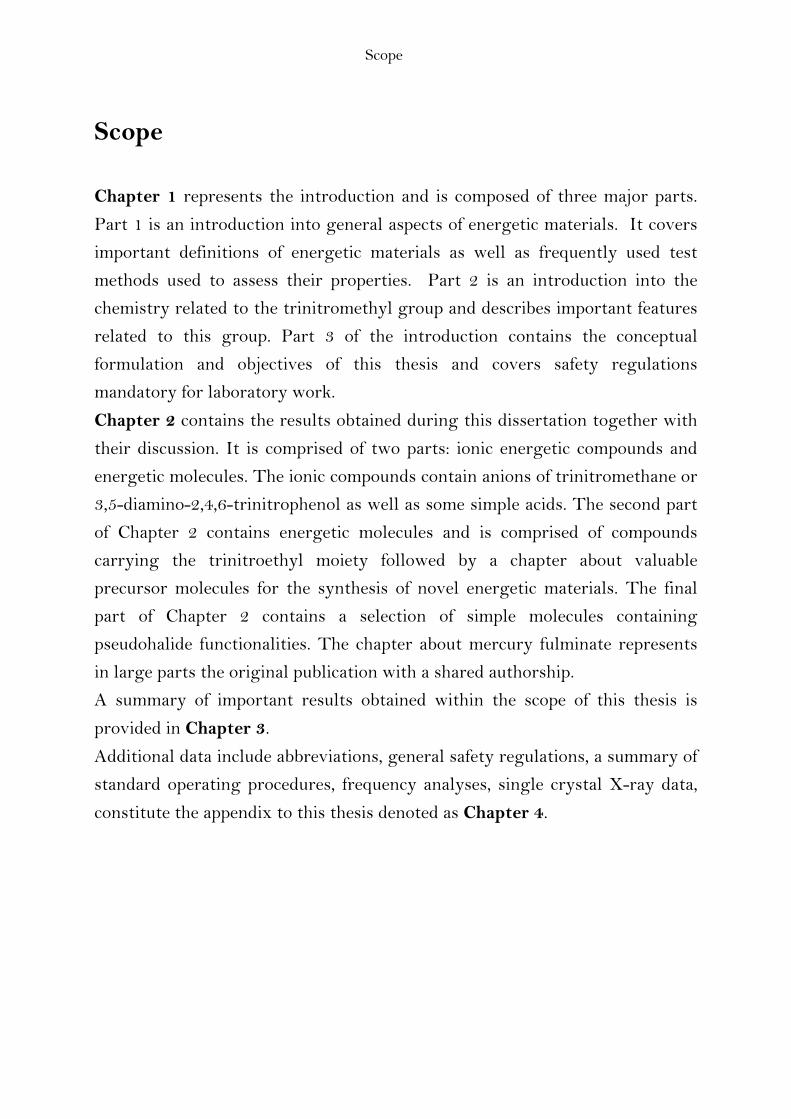
Scope
Scope
Chapter 1 represents the introduction and is composed of three major parts.
Part 1 is an introduction into general aspects of energetic materials. It covers
important definitions of energetic materials as well as frequently used test
methods used to assess their properties. Part 2 is an introduction into the
chemistry related to the trinitromethyl group and describes important features
related to this group. Part 3 of the introduction contains the conceptual
formulation and objectives of this thesis and covers safety regulations
mandatory for laboratory work.
Chapter 2 contains the results obtained during this dissertation together with
their discussion. It is comprised of two parts: ionic energetic compounds and
energetic molecules. The ionic compounds contain anions of trinitromethane or
3,5-diamino-2,4,6-trinitrophenol as well as some simple acids. The second part
of Chapter 2 contains energetic molecules and is comprised of compounds
carrying the trinitroethyl moiety followed by a chapter about valuable
precursor molecules for the synthesis of novel energetic materials. The final
part of Chapter 2 contains a selection of simple molecules containing
pseudohalide functionalities. The chapter about mercury fulminate represents
in large parts the original publication with a shared authorship.
A summary of important results obtained within the scope of this thesis is
provided in Chapter 3.
Additional data include abbreviations, general safety regulations, a summary of
standard operating procedures, frequency analyses, single crystal X-ray data,
constitute the appendix to this thesis denoted as Chapter 4.

Chapter 1 – Introduction
- 12 -
Chapter 1
Introduction
The development and testing of energetic materials is an exciting and
challenging area of chemistry both as far as fundamental and applied aspects
are concerned. Though the development of this kind of materials, which
include high explosives, propellants, and pyrotechnics has a long standing
tradition in the chemical sciences, research and efforts are undertaken world-
wide as never before, foremost driven by the prospect of outstanding materials
properties in general, and in order to discover new representatives having
significant advantages over compounds currently used. Environmental
considerations and safety requirements are important driving factors next to
higher performance and tailored properties for special applications. Due to
their unique properties, these materials are useful for manifold and highly
diverse applications ranging from military to civilian areas in many industries
including but not limited to construction, mining, oil exploration as well as
space exploration. Solid high explosives produce a velocity of detonation of up
to three times the velocity of sound in the explosive (9000 m s-1), a high
liberated energy density of about 6 megajoules per kilogram (MJ kg-1) and an
initial material density of about 2000 kilograms per cubic meter (kg m-3). The
product of these quantities corresponds to a power density of 1 x 10¹⁰ W cm-2.
By comparison, a detonating explosive having a surface of 100 cm2 operates at
a power level of 1.000.000 MW which is equal to the total average electric
generating capacity of the United States in the year 2007. (1) This very rapid
rate of energy liberation is what makes explosives unique. (2) Obtaining such
materials is complex owing to the fact that several different and mutually
exclusive appearing material properties have to be met to find the molecule
fulfilling all the qualification criteria in order to become widely accepted. The
development of energetic materials is a whole world of trade-offs between
energy content of a molecule and other desirable properties like higher
performance, insensitivity against accidental initiation, thermal stability as well
as a non-toxic and non-polluting behaviour when exposed to the environment

Chapter 1 – Introduction
- 13 -
next to other additional properties. The traditional procedure for formulating
new materials has been largely guided by intuition, experience and testing,
relying foremost on trial and error. In turn, a better understanding of the basic
principles is highly desirable to yield a more rational design process. However,
exploiting these possibilities requires an understanding of the properties of the
individual molecules, their interaction amongst each other as well as to
surrounding matter next to an understanding of kinetic energy release and
dynamics of initiation and decomposition processes. This bottom-up approach to
energetic materials would allow for a more fundamental understanding of the
evolution of properties with the size of the system as well as an understanding
of the effects of the interaction of matter at different molecular-length scale
with external stimuli and finally a detailed understanding of the functionalities
of matter at molecular-length scale. The information obtained could provide
breakthroughs not only in the area of energetic materials but additionally also
in all areas of material science and chemistry in general both as far as
fundamental and applied aspects are concerned. Energetic materials, due to
their very nature, can offer a variety of unique insights into structure and
matter. For example, detonations of high explosives produce thousands of
Kelvins and a few hundred thousand atmospheres thus providing a unique
means of elucidating the exotic chemical reactivity of matter under extreme
conditions - similar to the conditions in the interiors of giant planets. In this
context it has recently been reported that water formed during the detonation
of the high explosive pentaerythritol tetranitrate (PETN) displays catalytic
behaviour challenging the traditional view of water in high-explosives
chemistry where water was considered to be one of the stable detonation
products next to carbon dioxide and dinitrogen. These novel findings suggest
that water may catalyse reactions in other explosives and in planetary
interiours. (3) At the same time, the extreme conditions inside a detonating
explosive have made it extremely difficult to perform measurements and
consequently the detailed chemical reactions that cause a detonation are largely
not understood. (4) Empirical observations are important to gain a better
understanding of the final chemical composition after detonation and the
corresponding reaction mechanisms are still not known for many explosives
rendering this science to be very young and advances to be likely with the

Chapter 1 – Introduction
- 14 -
advent of novel techniques allowing to acquire experimental information
previously not available. (5)
As mentioned above, it is important to discover new representatives having
significant advantages over compounds currently used not only for military but
also for civilian purpose. As far as the military is concerned, U.S. Defense
Secretary Robert M. Gates recently announced to spend less money on
traditional weapon systems for conventional warfare against large nations like
China and Russia and shift more money to counterterrorism in Iraq and
Afghanistan representing the first broad rethinking of American military
strategy under the Obama administration. (6) Under this program a
combination of evolutionary and novel technologies are under development to
facilitate intelligence and surveillance using unmanned vehicles like the
Predator or Reaper drones currently used in Pakistan, Afghanistan and Iraq.
Today these systems rely on munitions made up of yesterday’s explosives and
propellants. In contrast, novel energetic materials with increased performance,
tailored energy release and insensitivity to unintended initiation would
significantly improve existing systems and additionally enable new technology.
As well as performance properties, the desired criteria for a new material in
order to become widely accepted are insensitivity towards destructive stimuli
such as electrostatic discharge, heat, friction, and impact to ensure safe
handling procedures and enhance controllability of kinetic energy release and
further low water solubility and hydrolytic stability for environmental reasons,
as well as longevity- and compatibility questions and other criteria addressing
high-priority ecological toxicity requirements.
Chapter 1 – Introduction
- 15 -
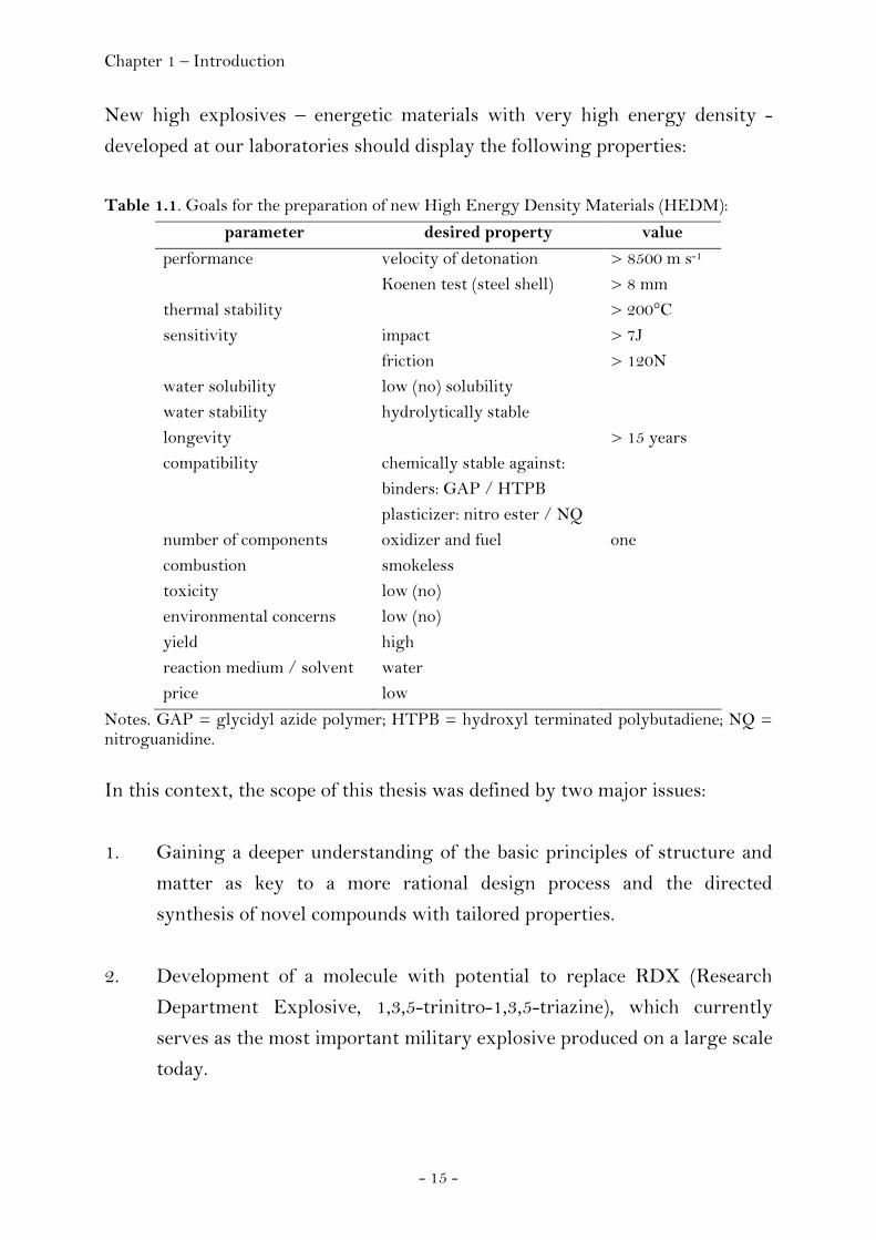
New high explosives – energetic materials with very high energy density -
developed at our laboratories should display the following properties:
Table 1.1. Goals for the preparation of new High Energy Density Materials (HEDM):
parameter desired property value
performance velocity of detonation > 8500 m s-1 Koenen test (steel shell) > 8 mm thermal stability > 200°C sensitivity impact > 7J friction > 120N water solubility low (no) solubility water stability hydrolytically stable longevity > 15 years compatibility chemically stable against: binders: GAP / HTPB plasticizer: nitro ester / NQ number of components oxidizer and fuel one combustion smokeless toxicity low (no) environmental concerns low (no) yield high reaction medium / solvent water price low
Notes. GAP = glycidyl azide polymer; HTPB = hydroxyl terminated polybutadiene; NQ = nitroguanidine. In this context, the scope of this thesis was defined by two major issues:
1. Gaining a deeper understanding of the basic principles of structure and
matter as key to a more rational design process and the directed
synthesis of novel compounds with tailored properties.
2. Development of a molecule with potential to replace RDX (Research
Department Explosive, 1,3,5-trinitro-1,3,5-triazine), which currently
serves as the most important military explosive produced on a large scale
today.

Chapter 1 – Introduction
- 16 -
The primary objective of this thesis was to develop a new energetic compound
with potential to replace the most important high explosive currently used in
the United States and world-wide. RDX can chemically be classified as a
nitramine. Nitramines generally are highly energetic compounds having found
wide acceptance as explosives or rocket propellants. The most common
nitramines in use today are RDX and HMX (Fig. 1.1).
Figure 1.1. Molecular structures of RDX (1,3,5-trinitro-1,3,5-triaza-cyclohexane) and HMX (1,3,5,7-tetranitro-1,3,5,7-tetraaza-cyclooctane). The acceptance of RDX and HMX is generally attributed to the high energetic
performance and the high energy density possessed by these compounds. In
essence, RDX and HMX are the standards of energetic performance and
energy density by which other energetic compounds are measured. RDX was
first prepared by Henning in 1899 intended for medicinal use, its explosive
properties have been realized in 1920 by Herz. (7) As with most explosives,
RDX can be used alone or as a component in explosive compositions like C-4
(when mixed with plasticizers) or Semtex (a combination of RDX and PETN,
pentaerythritol tetranitrate), or as a base charge in detonators and high
explosives. A drawback to RDX and HMX is that these nitramine compounds
are relatively sensitive to shock, friction, and impact. The high sensitivities
associated with RDX and HMX make these nitramine compounds less
desirable for some applications, especially in an environment where external
stimuli on RDX or HMX can lead to catastrophic damage with destruction of
surrounding objects and loss of human life. Processing of neat RDX or HMX
can be difficult due to their high melting points rendering melt casting to be
dangerous.

Chapter 1 – Introduction
- 17 -
A breakthrough in energetic materials research was the development of CL-20
(2,4,6,8,10,12-hexanitrohexaazaisowurtzitane, HNIW), a caged polynitramine
compound that is 20 percent more powerful than HMX. It was first
synthesised by A. Nielson in 1987 using a novel chemical reaction to construct
the CL-20 cage in a single step thereby establishing a new type of amine
glyoxal chemistry. While there have been several other new ingredients over
the years, none of them have been successfully scaled up to mass production
levels.
Figure 1.2. Molecular Structure of CL-20.
In contrast, CL-20 has been called the most significant energetic ingredient in
energetic materials research since the discovery of RDX and HMX because it
has made the jump from laboratory scale synthesis to scale up and finally to
mass production levels. Its major limitations are due to the rather high costs of
its production involving expensive reagents like nitronium tetrafluoroborate or
a palladium catalyst. In the best case the potential of CL-20 stimulates
increased demand leading to improved production processes. Availability will
go up and cost will go down. Next to above mentioned criteria that may
prevent the widespread use or even the development of an energetic material,
both explosive and environmental safety issues are of major impact. Regarding
the safety of the material, recent and growing interest in less sensitive
energetic materials can be seen to be a consequence of national and
international insensitive munitions policies (8) as well as additions to UN
transport regulations. (9) According to BAM (Bundesanstalt für
Materialprüfung), a compound can be classified as being insensitive, less
sensitive, sensitive, very sensitive or extremely sensitive. According to their
friction and impact sensitivity data, RDX and HMX are sensitive whereas CL-
20 has been found to be very sensitive.
Chapter 1 – Introduction
- 18 -
At our laboratories we aim to develop energetic materials that will enable the
design of novel insensitive high-energy propellants and explosives with
tailored energy release. Our approach to these materials is based on compounds
that combine both high oxygen and nitrogen contents allowing not only to
obtain superior performance characteristics but additionally compounds that
are more environmentally benign and less toxic - during storage or use and
also during their preparation. High nitrogen compounds are highly desirable
due to their high heats of formation and the formation of environmentally
friendly and toxicologically harmless dinitrogen. Nitrogen is unique amongst
all other elements of the periodic table in so far that the bond energy per two-
electron bond increases from a single over a double to a triple bond resulting in
dinitrogen being more stable than any other polynitrogen species. (10)
Compounds that preferentially contain only nitrogen can be very useful in
propellants, but their use as high explosives is limited. The heat of formation of
dinitrogen equals zero and consequently the products formed on decomposition
of high nitrogen compounds display less negative values of their heats of
formation resulting in lower heats of detonation (see page 30) and less
thermochemical energy produced during detonation and usable for the work to
be done by the explosive. To overcome this drawback but at the same time
benefit from the advantages of high nitrogen compounds we are trying to
develop new energetic materials preferentially containing both high nitrogen
and oxygen content. These compounds release energy not only due to the high
heats of formation of high nitrogen compounds but additionally through the
release of energy produced from the oxidation-products formed during
detonation. Within the scope of this thesis we were interested in developing
oxygen rich energetic derivatives of high nitrogen compounds.
Traditional representatives of high oxygen explosives (HOX) have been
reported in public literature as research reports initiated by the Office of Naval
Research (ONR) became declassified in the early seventies of the twentieth
century; however, relevant data were published mainly in the patent literature,
often without giving information about synthetic procedures or specifying the
physicochemical characteristics of the compounds obtained. (11) Some of the
most promising materials initially considered were polynitroaliphatic
compounds containing the dinitromethyl, fluorodinitromethyl and

Chapter 1 – Introduction
- 19 -
trinitromethyl groups. (12) Among them, the trinitromethyl compounds were
found to have the most favourable heats of detonation and oxygen balance
values. However, thermal stability was reported to be generally limited to
150°C when solid and 100°C when molten reversing further investigation into
trinitromethyl substituted compounds. (13) We have now investigated both
compounds mentioned in the literature and we have developed and tested novel
compounds carrying the trinitromethyl functionality in order to explore its
potential for the design of next generation energetic materials trying to
enhance the thermal stability of this class of compounds and finding the
molecule offering the best trade-off between energy capability and thermal
stability.
Chapter 1.1 – General Characteristics of Energetic Materials
- 20 -
1.1 General Characteristics of Energetic Materials
1.1.1 Types of Energetic Materials
Energetic materials are chemical compounds, or mixtures of chemical
compounds, that are divided into three classes according to use:
• Explosives
• Propellants
• Pyrotechnics
Explosives and propellants that have been properly initiated evolve large
volumes of hot gas in a short time. The difference between explosives and
propellants is the rate at which the reaction proceeds. In explosives, a fast
reaction produces a very high pressure shock in the surrounding medium. This
shock is capable of shattering objects. In propellants, a slower reaction
produces lower pressure over a longer period of time. This lower, sustained
pressure is used to propel objects. Pyrotechnics evolve large amounts of heat
but much less gas than propellants or explosives. The exothermic chemical
reactions occurring in pyrotechnics are generally speaking non-explosive,
relatively slow, self-sustaining, and self-contained.
1.1.2 Classification of Energetic Materials
There is considerable variation among the properties of the compounds that
constitute each of the three major classifications of energetic materials,
explosives, propellants and pyrotechnics. Generally, they can be divided into
composites and monomolecular energetic materials. Composites like black
powder are obtained on physically mixing solid oxidizers and fuels whereas in
monomolecular energetic materials like TNT (2,4,6-trinitrotoluene), each
molecule contains an oxidizing component and a fuel component.
Chapter 1.1 – General Characteristics of Energetic Materials
- 21 -
Explosives. An explosive is defined as a material that can be initiated to
undergo very rapid, self-propagating decomposition that results in the
formation of more stable material, the liberation of heat, or the development of
a sudden pressure effect through the action of heat on produced or adjacent
gases. A chemical explosive is a compound or a mixture of compounds which,
when subjected to heat, impact, friction, or shock, undergoes very rapid, self-
propagating, heat- producing decomposition. This decomposition produces
gases that exert tremendous pressures as they expand at the high temperature
of the reaction. The work done by an explosive depends primarily on the
amount of heat given off during the explosion. The term detonation indicates
that the reaction is moving through the explosive faster than the speed of
sound in the unreacted explosive; whereas, deflagration indicates a slower
reaction (rapid burning). Denser explosives usually give higher detonation
velocities and pressures.
A high explosive will detonate; a low explosive will deflagrate. Low-order
explosives (LE) create a subsonic explosion and lack the over-pressurization
wave generated by high explosives. Low-order explosives with lower density
like ANFO (Ammonium Nitrate Fuel Oil) will suffice in easily fragmented or
closely jointed rocks and are preferred for quarrying coarse material for mining
and construction purpose. A High Explosive (HE) is a compound or mixture
which, when initiated, is capable of sustaining a detonation shockwave to
produce a powerful blast effect. A detonation is the powerful explosive effect
caused by the propagation of a high-speed shockwave through a high explosive
compound or mixture. During the process of detonation, the high explosive is
largely decomposed into hot, rapidly expanding gas. These high density
explosives may be desirable for difficult blasting conditions or where fine
fragmentation is required. Ingredients of high explosives are classified as
explosive bases, combustibles, oxygen carriers, antacids, and absorbents. Some
ingredients perform more than one function. An explosive base is a solid or
liquid which, upon the application of sufficient heat or shock, decomposes to
gases with an accompanying release of considerable heat. A combustible
combines with excess oxygen to reduce the formation of nitrogen oxides. An
oxygen carrier assures complete oxidation of the carbon to reduce the
formation of carbon monoxide. The formation of nitrogen oxides or carbon

Chapter 1.1 – General Characteristics of Energetic Materials
- 22 -
monoxide, in addition to being undesirable from the standpoint of fumes,
results in lower heat of explosion and efficiency than when carbon dioxide and
nitrogen are formed. Antacids increase stability in storage, and absorbents
absorb liquid explosive bases.
Explosives are classified as primary or secondary based on their susceptibility
to initiation. Primary explosives, which include lead azide and lead styphnate,
are highly susceptible to initiation. Primary explosives often are referred to as
initiating explosives because they can be used to ignite secondary explosives.
Secondary explosives include nitroaromatics and nitramines and are
formulated to detonate only under specific circumstances. Secondary explosives
often are used as main charge. Secondary explosives can be loosely categorized
into melt-castable explosives, which are based on nitroaromatics such as TNT,
and plastic-bonded explosives which are based on a binder and crystalline
explosive such as RDX.
Chapter 1.1 – General Characteristics of Energetic Materials
- 23 -
Propellants can be divided into four classes:
(I) composites (II) single-base (III) double-base (IV) triple-base
Propellants include both rocket and gun propellants. The choice of a propellant
for a specific use is determined by ballistic and physical requirements rather
than on the basis of composition. A given propellant composition may be
suitable for use in several applications.
(I) Most rocket propellants are composites. They generally consist of a physical
mixture of a fuel such as metallic aluminum, a binder which is normally an
organic polymer (generally a synthetic rubber which is also a fuel), and an
inorganic oxidizing agent such as ammonium perchlorate. These are
heterogeneous physical structures.
(II) One group of gun propellants are called single-base and principally consist
of nitrocellulose.
(III) Double-base propellants usually consist of nitrocellulose and
nitroglycerine. In general, double-base propellants contain nitrocellulose and a
liquid organic nitrate which will gelatinize nitrocellulose.
(IV) The term triple-base applies to propellants containing three explosive
ingredients, usually with nitroguanidine as the major ingredient. The other
two explosive ingredients frequently used are nitroglycerine and nitro-
cellulose. As in the double-base propellant, other gelatinizers may be
substituted for the nitroglycerine. The nitroguanidine in the formulation
produces a lower flame temperature and a greater amount of gaseous
combustion products. The lower flame temperature considerably reduces
erosion of gun barrels and the greater amounts of gas produce a greater force
on the projectile.
Chapter 1.1 – General Characteristics of Energetic Materials
- 24 -
Pyrotechnics include illuminating flares, signalling flares, colored and white
smoke generators, tracers, incendiary delays, fuses, and photo-flash
compounds. Pyrotechnics usually are composed of an inorganic oxidizer and
metal powder in a binder. Illuminating flares contain sodium nitrate,
magnesium, and a binder. Signaling flares contain barium, strontium, or other
metal nitrates. Flares burn to produce intense light that is used for
illumination. Signals produce colored flames that can exemplarily be used for
recognition purpose during emergencies. Colored smoke is used for signalling
while white smoke is used for screening. Tracers and fumers are small, smoke
producing charges that are placed in projectiles. During the flight of the
projectile, the charge burns. In a tracer, the smoke is used to track the flight of
the projectile. A fumer produces smoke at the proper rate to fill the partial
vacuum that movement through the air creates behind the projectile. This cuts
drag and increases range. Incendiaries produce large amounts of heat that
cause fires. A delay is an element that consists of an initiator, a delay column,
and an output charge or relay in a specially designed inert housing. The delay
column burns for a predetermined amount of time. Delays are used to provide
an interval between initiation and functioning of a device. A fuse is a cord of
combustible material commonly used in demolition. Photoflash powders are
loose mixtures of oxidizers with metallic fuels. When ignited, these mixtures
burn with explosive violence in a very short time. Igniters and initiators are
used to ignite propellant charges and initiate detonation in explosive charges.
1.1.3 Properties of Energetic Materials
Chemical properties of individual energetic materials are discussed in the
appropriate chapters of this thesis. The basic definitions and properties
discussed for these materials include the following:
Brisance. Brisance is the shattering capability of an explosive. Several tests are
commonly used to determine brisance. In the sand test 0.400 grams of the
explosive are placed in 200 grams of sand and detonated. The amount of sand
crushed by the explosive is a measure of brisance. The plate dent test, in which
a sample of the explosive is detonated in close proximity to a metal plate, is
Chapter 1.1 – General Characteristics of Energetic Materials
- 25 -
also used to measure brisance. The size of the dent is proportional to the
brisance. Another method of measurement involves detonating a sample of
explosive on top of a cylinder made of copper and measuring the contraction in
length of the cylinder. The number, size distribution, and velocity of fragments
produced by an explosive in a projectile is also related to the brisance of the
explosive. With a limited number of exceptions, increased detonation velocity
increases brisance.
Burning. The process of exothermic redox reactions taking place in energetic
materials without introduction of atmospheric oxygen is preferably denoted as
burning. The reaction is self-sustaining after an initial activating energy has
been applied. Many explosives are capable of burning without detonation, if
unconfined.
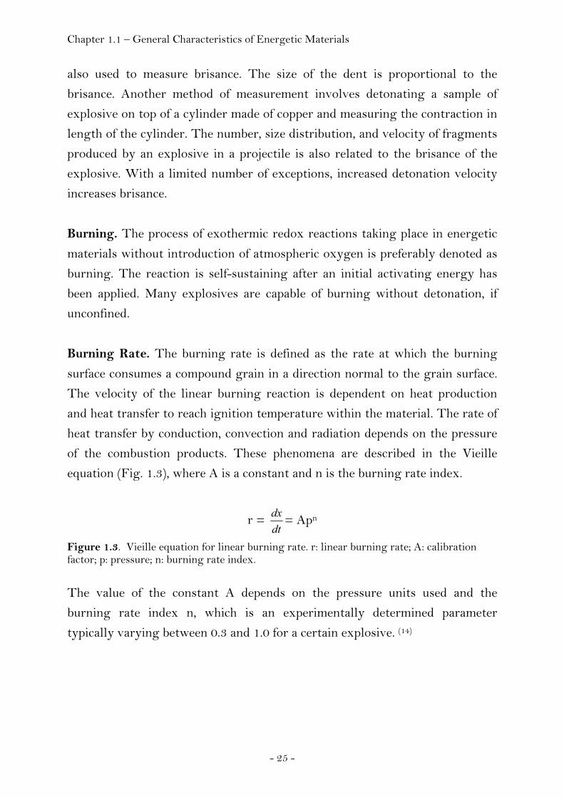
Burning Rate. The burning rate is defined as the rate at which the burning
surface consumes a compound grain in a direction normal to the grain surface.
The velocity of the linear burning reaction is dependent on heat production
and heat transfer to reach ignition temperature within the material. The rate of
heat transfer by conduction, convection and radiation depends on the pressure
of the combustion products. These phenomena are described in the Vieille
equation (Fig. 1.3), where A is a constant and n is the burning rate index.
r = dtdx = Apn
Figure 1.3. Vieille equation for linear burning rate. r: linear burning rate; A: calibration factor; p: pressure; n: burning rate index. The value of the constant A depends on the pressure units used and the
burning rate index n, which is an experimentally determined parameter
typically varying between 0.3 and 1.0 for a certain explosive. (14)

Chapter 1.1 – General Characteristics of Energetic Materials
- 26 -
The equation shown in Figure 1.4 allows the mass of explosive consumed per
unit time to be calculated provided that the burning surface area and the
density are known. The surface area of fine powders becomes very large and
due to the enhanced convective heat transfer mechanism, the risk of a change
from a burning reaction turning into a deflagration or detonation reaction is
enhanced. (15)
dtdm = S(t) ρ r
Figure 1.4. Mass consumption of a burning reaction. m: mass; t: time; S: surface; ρ: density; r: linear burning rate.
Chapman-Jouget Theory. The Chapman-Jouget (CJ) theory is a fluid
dynamical model of detonations and is used to calculate performance
characteristics of an explosive expressed by the four parameters velocity of
propagation (D), pressure (p), density (ρ) and particle velocity (u) behind the
wave front. The performance of a chemical explosive or its usable energy is
determined by the expansion of product gases arising from the chemical
reactions taking place during detonation. The variation of pressure depends on
the particle velocity of the product gases during adiabatic or free expansion and
in order to describe this behaviour and calculate performance, the state (that is
the pressure and particle velocity) of the materials at the end of the reaction
zone and their equation of state (describing the pressure variation with the
particle velocity during adiabatic or free expansion) have to be known. The CJ
theory relates the detonation wave velocity to the properties of the gases
behind the detonation wave front. It is assumed that all chemical energy is
released at the detonation front and the reaction zone has no thickness. This
approximation allows the detonation wave to be considered as a self-sustained
supersonic wave travelling through the explosive at constant velocity.
Generally, shock waves in inert materials can be described on the basis of
three equations of conservation of energy, momentum and mass across the
shock front. These equations are also called jump conditions. They allow the
determination of the shock velocity (U) in terms of the pressure (p), density (ρ)
and particle velocity (u) variables. Though these jump conditions do apply in
the CJ theory as well, an additional condition is needed to determine the

Chapter 1.1 – General Characteristics of Energetic Materials
- 27 -
detonation wave velocity (D) because energy is released at the front of the
detonation wave making it self-propagating.
Figure 1.5. Initiation and Propagation of a Detonation Wave according to the model of Zel’dovic, Neuman and Döring (ZND). (a) A schematic 1-D (planar) experiment is shown at different times. In the experiment, the impact of a plate thrown on one face of a cube of explosive (t = t0) produces a planar shock wave (t = t1) that gradually accelerates (t = t2) to a steady-state detonation (t = t3) as the shock wave sweeps through the explosive and causes chemical energy to be released to the flow at a finite rate. (b) The corresponding pressure-vs-distance snapshots show the evolution of an essentially inert shock wave at t = t1
growing into a classical 1-D ZND detonation structure at t = t3, namely, a shock, or pressure, discontinuity at the ZND point followed by decreasing pressure through the reaction zone, ending at the CJ point, the pressure predicted by the CJ model (see text). (c) Pressure-vs-time plots for material particles originally at the shock front locations in (b) show the particle pressure (or velocity) histories. Only at the location of the right-most particle has a ZND detonation fully formed. (2) (Credit: Courtesy of Dr. J.B. Bdzil, LANL)

Chapter 1.1 – General Characteristics of Energetic Materials
- 28 -
Combustion. Combustion of energetic materials designates any exothermic
oxidation reaction, including, but not limited to those produced by introduction
of atmospheric oxygen (see burning).
Deflagration. Deflagration (lat. de + flagrare = to burn) of propellants
proceeds the same as normal burning and designates the process of very rapid
burning without introduction of atmospheric oxygen but propagating at a
velocity less than the speed of sound in that material. Due to the rate
determining factors in the reaction being the rate of heat transfer into the
propellant grain from the burning surface and the rate of decomposition of the
propellant formulation, deflagration can result from having a fuel and oxidant
in very close contact.
Deflagration to Detonation Transition. Deflagration can be considered to be
an intermediate process between burning and detonation. As mentioned above
(see Burning Rate), a large surface area of an energetic material can result into
deflagration and the same is true for confined materials due to the burning
reaction and the deflagration reaction being based on heat transfer. The
velocity of the reaction is rising until it reaches the magnitude of the material’s
sound speed generating first shockwaves passing the material. A further
acceleration to supersonic velocities yielding a detonation reaction depends on
the type of material, its surface area and its confinement. (15) Hence, a burning
reaction can be accelerated to a deflagration and by corresponding interference
of shock waves further to a detonation reaction. The latter process is called
deflagration-to-detonation transition (DDT). The detonation reaction is
triggered by a shock wave. It occurs if enough of the explosive compound is
compressed such that a chemical reaction can occur before it physically
fragments and a shock wave is formed inside the sample. A shock wave
moving at supersonic speed proceeds through the explosive causing further
decomposition of the explosive material. Detonation designates the supersonic
propagation of chemical reactions through an explosive. It can be rationalized
as a shock wave moving through an explosive accompanied by chemical
reactions. The shock compresses the material thus increasing its temperature
to the point of ignition. The energy release of the chemical reactions taking
Chapter 1.1 – General Characteristics of Energetic Materials
- 29 -
place in the ignited material behind the shock front support the shock
propagation. This self-sustained detonation wave is different from a
deflagration, which propagates at a subsonic speed and without a shock or any
significant pressure change. If there is no shock wave, the reaction is called a
deflagration. The major difference in the two reactions is the velocity of
propagation of the reaction front. The shock wave has a velocity of the order of
km s-1 instead of cm s-1 in the case of a deflagration. In the case of a detonation,
the reaction rate is determined by the velocity of the shock wave, not by the
rate of heat transfer.
Explosion. An Explosion (lat. explodere = to shatter) is a sudden expansion of
matter accompanied by an increase of its volume. (16) Explosions may both be
caused by explosive chemical or nuclear reactions and physical processes.
Accordingly, the term explosion includes effects that follow rapid burning,
detonation, as well as physical processes like the bursting of a vessel filled with
compressed gas. The resulting effect of an explosion is a sudden expansion of
gases or vapor, whether they were present before, or originated during the
explosion process. A chemical explosion is accompanied by the formation of
large amounts of gaseous decomposition products and liberation of heat during
a very short time. The gaseous products (usually several hundred liters during
microseconds) are heated to several thousands of degrees centigrade and can
not expand instantaneously resulting in a sudden pressure rise ranging to
several hundred of kbar during detonation reactions. Subsequently they expand
and exert strong impact effects and shock waves to the surroundings.
Gap Test. In contrast to sensitivity towards impact, sensitivity of an explosive
to shock is a very reproducible quantity. Shocks generated by a donor
explosive can cause detonation in another explosive material. The strength of
the shock wave required is a relative measure of the sensitivity of the material
under test. In practice, a strong shock is produced and attenuated in an inert
medium. The width of the medium that will allow detonation in 50 percent of
the trials is reported as the test result. These tests are called gap tests. Gap test
results are much more reliable data than impact test results, although there is
Chapter 1.1 – General Characteristics of Energetic Materials
- 30 -
some dependence on the geometry of the test apparatus. Gap test have not been
performed within the scope of this thesis.
Heat of Combustion. The heat of combustion is the amount of heat produced
when a material is burned. This differs from the heat of detonation because the
products formed are different. Generally, the products formed in combustion
are at a lower energy level than the products formed during detonation. For
example, carbon monoxide and carbon dioxide may be products of both
detonation and combustion for a particular explosive. However, the detonation
process might produce more carbon monoxide, while combustion might
produce more carbon dioxide. Heat of combustion is usually measured for a
new explosive for the determination of the heat of formation.
Heat of Detonation. The heat of detonation is considered to be the
thermochemical energy produced during detonation and usable for the work to
be done by the explosive. According to first principles – energy conservation –
considerations, this quantity would seem to be a primary parameter of an
explosive, upon which performance depends. Indeed it has been suggested (17)
that some other detonation condition or parameter is as important as CJ
pressure for the performance of explosives. Two explosives may serve as an
example: TATB and NQ. In spite of their reasonably high calculated CJ
pressure and measurements of detonation velocity and pressure they produce
rather low performance. Kamlet (18) wondered whether, for some reason,
insensitive explosives were not reaching the infinite-medium steady-state condition. He
also suggested that the trouble with NQ (poor performance) was the low Q (heat of
detonation), and that no formulation very rich in NQ or other low-Q explosives would
have high performance. Two quantities are usually given for the heat of
detonation, one with liquid water in the reaction products and one with
gaseous water in the reaction products. The test used to determine these
quantities uses a standard calorimeter. When the water is allowed to condense
to liquid, the total heat produced by the detonation reaction is measured. The
heat of detonation with gaseous water more accurately reflects the process of
detonation in a non-laboratory setting. However, the results are less
reproducible.
Chapter 1.1 – General Characteristics of Energetic Materials
- 31 -
Heat of Fusion. The heat of fusion is the amount of heat necessary to
transform (melt or fuse) a unit of solid into a liquid at the same temperature
and standard pressure. This quantity is usually expressed in terms of calories
per gram.
Heat of Sublimation. The heat of sublimation is the amount of heat necessary
to convert a weight of solid directly into vapor in a constant temperature
process. This quantity is usually expressed in calories per gram.
Heat of Vaporization. The heat of vaporization is the amount of heat
necessary to convert a unit of liquid to vapor at the same temperature. This
quantity is usually expressed in terms of calories per gram.
Oxygen Balance. The oxygen balance (OB) of the explosive is closely related
to the power. The oxygen balance is the ratio of oxygen contained in the
explosive material to the amount of oxygen required for complete oxidation of
the explosive material. Explosive compositions with better oxygen balances are
more powerful.
OB = (O – 2C – 0.5H) · M
1600
Figure 1.6. OB: Oxygen balance [%] for a compound composed of the elements C/H/N/O to be oxidized completely to H2O and CO2. M: molecular weight of the compound. Negative values of oxygen balance indicate that the explosive does not contain
enough oxygen to convert each atom of carbon and hydrogen to CO2
and H2O
during detonation, where no atmospheric oxygen is consumed. Most energetic
materials are oxygen deficient resulting in lower heats of detonation compared
to the condition of complete oxidation. In contrast, zero-balanced compounds
contain exactly the amount of oxygen necessary for carbon and hydrogen to be
oxidized to carbon dioxide and water. Examples are ethylene glycol dinitrate
(EGDN, C2H4N2O6), azidoformamidinium dinitramide (C1H4N8O4),
aminotetrazolium dinitramide (C1H4N8O4) (19) or bis-(2,2,2-trinitroethyl)-urea
(C5H6N8O13). Compounds with positive values of oxygen balance contain
excess oxygen and can serve as oxidizer components in energetic compositions.
Chapter 1.1 – General Characteristics of Energetic Materials
- 32 -
Examples are ammonium perchlorate (AP, NH4ClO4 + 33.8), ammonium
dinitramide (ADN, N4H4O4 +25.8), ammonium nitrate (AN, N2H4O3, +20) or
hydrazinium nitroformate (HNF, C1H5N5O6, +13.1)
Performance. One of the driving forces for the development of any new
energetic material is performance. Usually the discussion of new compounds
includes a comparison of their ‘performance’ to the current highest energy
density materials. The performance indicators used as a guide to the most
promising materials are detonation velocity or detonation pressure in case of
high explosives or values of the specific impulse in the case of propellants.
However useful and apparent these indicators are, evaluation of performance is
more complex and can lead to different results based on the intended use of the
compound. There is no single indicator that allows judging the performance of
either high explosive or propellants. For example, the effect of shaped charges
is dependent on the composition of the product gases and the detonation
energy, not solely on the detonation velocity. (20) As far as propellants are
concerned, important criteria include the burning rate, burning rate exponent,
temperature sensitivity and other parameters. Moreover, increased
‘performance’ in a compound will not necessarily yield increased available
performance in a usable composition. Compositions containing any new
material usually contain a binder to tune the mechanical properties and achieve
a tolerable safety and comparisons of single performance indicators do not
allow for the amount of binder required in different applications to obtain
acceptable mechanical, processing or safety properties. (21) Promoting new
compounds based on single performance parameters like detonation velocity or
detonation pressure would miss those materials that might give similar
performance when used in compositions to those compositions in use today
with increased safety. Less performance in terms of the above mentioned
indicators do not necessarily have to be a drawback in case the physical and
safety properties allow for higher useable proportions in compositions. One
example mentioned by Sanderson would be ‘super-TNT’, a compound that can
be melt casted and used like TNT in neat form or melt cast with other high
explosives. (21) Though its performance would intrinsically be less compared to
HMX, it could lead to compositions of higher performance.
Chapter 1.1 – General Characteristics of Energetic Materials
- 33 -
Power. The power of an explosive is the total energy available to do work and
depends both on its energy density and its detonation wave speed. This is a
different quantity than brisance. Two explosives may serve as example:
ammonium nitrate and RDX. If a charge of each is placed beneath a rock, the
ammonium nitrate might hurl the rock many meters but the RDX might
pulverize the boulder into many fragments. The former quality is power
whereas the latter quality is brisance. Power is measured by the Trauzl lead
block test in which a sample of the explosive is detonated in a cavity in a lead
block. The expansion of the cavity is a measure of the power of the explosive.
The ballistic pendulum and ballistic mortar tests are also used to measure
power. A heavy weight is accelerated by the detonation of an explosive. The
swing of the pendulum or movement of the mortar's weight is a measure of the
power of the explosive.
Prediction of Detonation Properties. The prediction of detonation
properties (detonation pressure, velocity of detonation) from a given molecular
structure and the known or estimated crystal density is of fundamental
importance especially for the synthesis of new high-explosive compound and
aided by the calculated properties a decision can be made whether it is worth
the effort to attempt a new and complex synthesis. It has been found that
estimates of detonation pressure and velocity are possible for C/H/N/O
explosives by means of relatively simple empirical equations. These equations
imply that the mechanical properties of the detonation depend only on the
number of moles of detonation gases per unit weight of explosive, the average
molecular weight of these gases, the chemical energy of the detonation reaction
(Q = -ΔH0), and the loading density. (22)
(a) Detonation Pressure
P = K ρ02 Φ Figure 1.7. P [kbar]; K: 15.58; ρ0 [g cm-3]; Φ = N M0.5 Q0.5; N [moles of gas per gram of explosive]; M [grams of gas per mole of gas]; Q [cal g-1].
Chapter 1.1 – General Characteristics of Energetic Materials
- 34 -
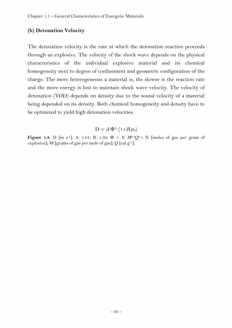
(b) Detonation Velocity
The detonation velocity is the rate at which the detonation reaction proceeds
through an explosive. The velocity of the shock wave depends on the physical
characteristics of the individual explosive material and its chemical
homogeneity next to degree of confinement and geometric configuration of the
charge. The more heterogeneous a material is, the slower is the reaction rate
and the more energy is lost to maintain shock wave velocity. The velocity of
detonation (VOD) depends on density due to the sound velocity of a material
being depended on its density. Both chemical homogeneity and density have to
be optimized to yield high detonation velocities.
D = A Φ½ (1+B ρ0) Figure 1.8. D [m s-1]; A: 1.01; B: 1.30; Φ = N M0.5Q0.5; N [moles of gas per gram of explosive]; M [grams of gas per mole of gas]; Q [cal g-1].
Chapter 1.2 – General Characteristics of the Trinitromethyl Group
- 35 -
1.2 General Characteristics of the Trinitromethyl Group
The large oxygen content (Ω(CO2) = +37%) and the reactive hydrogen atom in
trinitromethane (nitroform) render this molecule to be very interesting for the
preparation of high oxygen explosives (HOX). Trinitromethane has been
known as early as 1857. (23) In contrast, reports of the chemistry of
trinitromethyl compounds appeared rather late in the open literature.
According to a review published by Noble, Borgardt and Reed, a program
initiated by the Office of Naval Research (ONR) was initiated in 1947 to
investigate the nitroaliphatics for potential use as explosives and propellants. (11)
Portions of this work have appeared in patents, reviews and monographs when
reports became gradually declassified in the early 1970s but often few or no
information about synthetic procedures or physicochemical characteristics were
given. Today, excellent reviews describing the chemistry of the trinitromethyl
group are available summarizing the data on synthesis and properties of these
polynitro compounds. (11, 24-25) The purpose of this introduction into the
chemistry and characteristics of the trinitromethyl group is to summarize the
most important general findings thus providing necessary and useful basic
information intended to serve as a first navigation into the wide and complex
area of polynitro chemistry.
Depending on the hybridization of the carbon atom carrying the three nitro
groups two major groups of reactions can be differentiated. The first group
refers to the chemistry of the trinitromethanide (nitroformate) ion whereas the
second group involves the chemistry of the tetrahedral hybridized
trinitromethyl group.
Chapter 1.2 – General Characteristics of the Trinitromethyl Group
- 36 -
1.2.1 Tetranitromethane as Source to the Trinitromethanide Ion
Synthesis
An efficient and inexpensive method for the production of tetranitromethane or
trinitromethane is a condition for its practical utility. Today, tetranitromethane
is no longer commercially available though it has been produced in Germany
during World War II on industrial scale utilizing the nitration of acetic
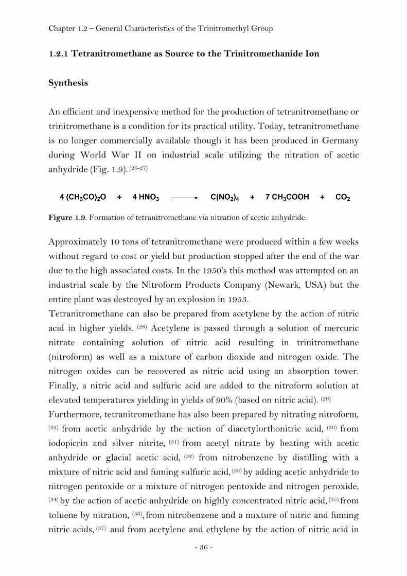
anhydride (Fig. 1.9). (26-27)
Figure 1.9. Formation of tetranitromethane via nitration of acetic anhydride.
Approximately 10 tons of tetranitromethane were produced within a few weeks
without regard to cost or yield but production stopped after the end of the war
due to the high associated costs. In the 1950's this method was attempted on an
industrial scale by the Nitroform Products Company (Newark, USA) but the
entire plant was destroyed by an explosion in 1953.
Tetranitromethane can also be prepared from acetylene by the action of nitric
acid in higher yields. (28) Acetylene is passed through a solution of mercuric
nitrate containing solution of nitric acid resulting in trinitromethane
(nitroform) as well as a mixture of carbon dioxide and nitrogen oxide. The
nitrogen oxides can be recovered as nitric acid using an absorption tower.
Finally, a nitric acid and sulfuric acid are added to the nitroform solution at
elevated temperatures yielding in yields of 90% (based on nitric acid). (29)
Furthermore, tetranitromethane has also been prepared by nitrating nitroform, (23) from acetic anhydride by the action of diacetylorthonitric acid, (30) from
iodopicrin and silver nitrite, (31) from acetyl nitrate by heating with acetic
anhydride or glacial acetic acid, (32) from nitrobenzene by distilling with a
mixture of nitric acid and fuming sulfuric acid, (33) by adding acetic anhydride to
nitrogen pentoxide or a mixture of nitrogen pentoxide and nitrogen peroxide,
(34) by the action of acetic anhydride on highly concentrated nitric acid, (35) from
toluene by nitration, (36), from nitrobenzene and a mixture of nitric and fuming
nitric acids, (37) and from acetylene and ethylene by the action of nitric acid in
Chapter 1.2 – General Characteristics of the Trinitromethyl Group
- 37 -
the presence of a catalyst. (38) The nitration of 4,6-dihydroxypyrimidine in
sulfuric acid has recently been found to yield trinitromethane as the sole
product. (39)
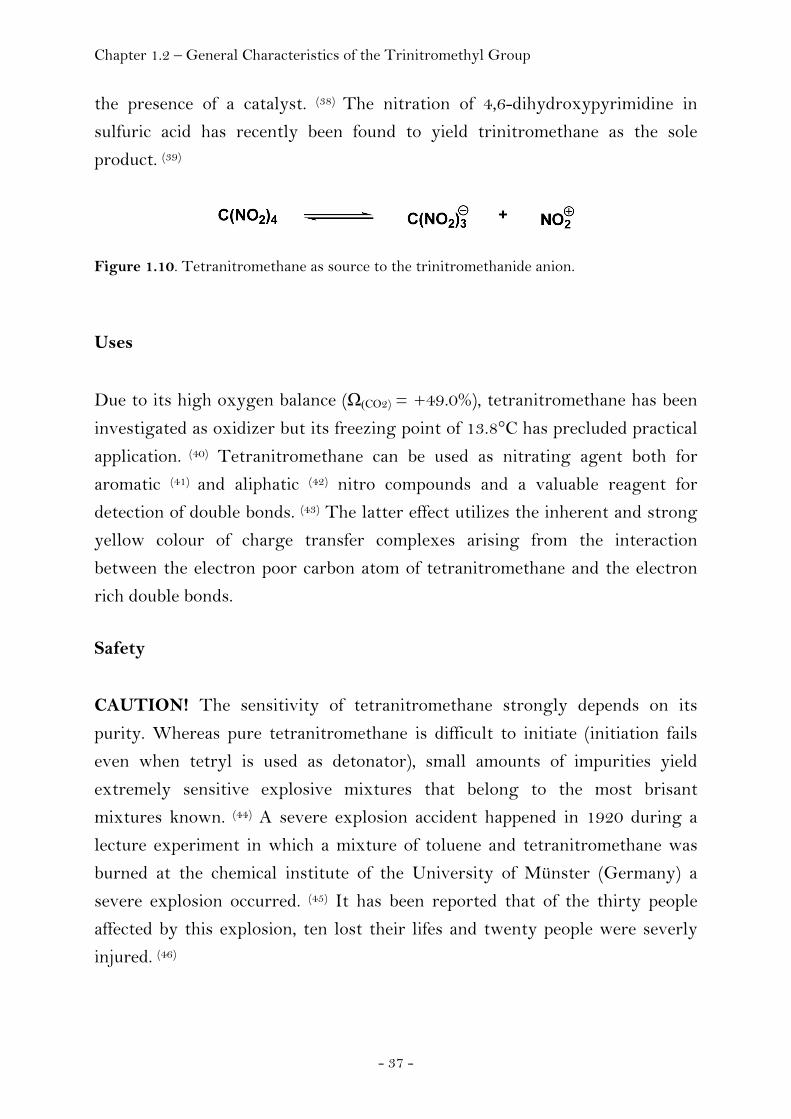
Figure 1.10. Tetranitromethane as source to the trinitromethanide anion.
Uses
Due to its high oxygen balance (Ω(CO2) = +49.0%), tetranitromethane has been
investigated as oxidizer but its freezing point of 13.8°C has precluded practical
application. (40) Tetranitromethane can be used as nitrating agent both for
aromatic (41) and aliphatic (42) nitro compounds and a valuable reagent for
detection of double bonds. (43) The latter effect utilizes the inherent and strong
yellow colour of charge transfer complexes arising from the interaction
between the electron poor carbon atom of tetranitromethane and the electron
rich double bonds.
Safety
CAUTION! The sensitivity of tetranitromethane strongly depends on its
purity. Whereas pure tetranitromethane is difficult to initiate (initiation fails
even when tetryl is used as detonator), small amounts of impurities yield
extremely sensitive explosive mixtures that belong to the most brisant
mixtures known. (44) A severe explosion accident happened in 1920 during a
lecture experiment in which a mixture of toluene and tetranitromethane was
burned at the chemical institute of the University of Münster (Germany) a
severe explosion occurred. (45) It has been reported that of the thirty people
affected by this explosion, ten lost their lifes and twenty people were severly
injured. (46)

Chapter 1.2 – General Characteristics of the Trinitromethyl Group
- 38 -
1.2.2 Trinitromethanide Ion as a Nucleophile
In principal, the reactions that can be utilized to synthesise compounds
containing the trinitromethyl group include carbonyl condensation reactions,
addition reactions, as well as alkylation reactions.
Carbonyl Condensation Reactions
The condensation reaction of an aldehyde and a polynitroalkane having an
acidified α proton yielding a β-nitro substituted alcohol is referred to as Henry
reaction. The Henry reaction between trinitromethane and formaldehyde
affords good yields of 2,2,2-trinitroethanol. However, attempts to add
trinitromethane to a variety of other aldehydes or ketones have been reported
to yield no isolable products. (47) Though not isolable, formation of 1-alkyl-
2,2,2-trinitroethanols was shown to occur in solution (see Table 1.2) and the
extent of dissociation of these alcohols was found to increase in the order
Y = –CH2– < –CH(CH3)– < (CH2)3C < –C(CH2)2–
Figure 1.11. Dissociation of trinitromethylalcohols according to Hall et al. (see Table 1.2). the extent of dissociation of these alcohols was found to increase in the order Y = –CH2– < –CH(CH3)– < –C(CH2)2– Table 1.2. Dissociation Constants determined by Hall in aqueous acid. (48)
Compound K, M-1
(NO2)3C–CH2–OH 7.80 · 10-7 (NO2)3C–CH(CH3)–OH 2.80 · 10-4 (NO2)3C–C(CH2)2–OH a)
Notes. a) No detectable amount of alcohol was produced.
It was concluded that steric interactions between the substituent on the alpha
carbon atom and the trinitromethyl group governs the position of the
equilibrium. The reaction is acid catalyzed and reversed in base affording the
salt of the nitro compound and formaldehyde.
Chapter 1.2 – General Characteristics of the Trinitromethyl Group
- 39 -
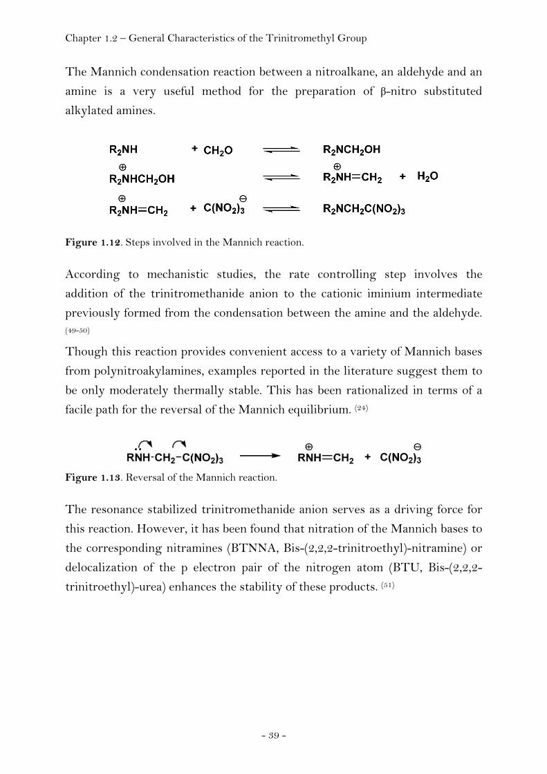
The Mannich condensation reaction between a nitroalkane, an aldehyde and an
amine is a very useful method for the preparation of β-nitro substituted
alkylated amines.
Figure 1.12. Steps involved in the Mannich reaction.
According to mechanistic studies, the rate controlling step involves the
addition of the trinitromethanide anion to the cationic iminium intermediate
previously formed from the condensation between the amine and the aldehyde. (49-50)
Though this reaction provides convenient access to a variety of Mannich bases
from polynitroakylamines, examples reported in the literature suggest them to
be only moderately thermally stable. This has been rationalized in terms of a
facile path for the reversal of the Mannich equilibrium. (24)
Figure 1.13. Reversal of the Mannich reaction. The resonance stabilized trinitromethanide anion serves as a driving force for
this reaction. However, it has been found that nitration of the Mannich bases to
the corresponding nitramines (BTNNA, Bis-(2,2,2-trinitroethyl)-nitramine) or
delocalization of the p electron pair of the nitrogen atom (BTU, Bis-(2,2,2-
trinitroethyl)-urea) enhances the stability of these products. (51)
Chapter 1.2 – General Characteristics of the Trinitromethyl Group
- 40 -
Addition Reactions
A wide variety of Michael systems have been utilized in Michael addition
reactions with the trinitromethanide ion. Noble et al. has reviewed the various
adducts that have been prepared. (11)
In principal, addition to a variety of α, β – unsaturated Michael systems of the
general formula CH2=CHY (Y = EWG) yields the corresponding 3-Y-1,1,1-
trinitropropyl derivatives.
1,2-Addition reactions to unconjugated olefin systems have been reported
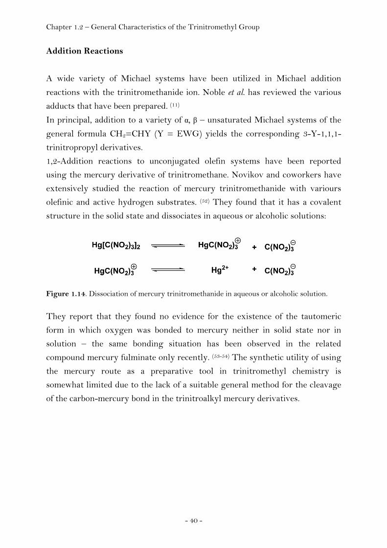
using the mercury derivative of trinitromethane. Novikov and coworkers have
extensively studied the reaction of mercury trinitromethanide with variours
olefinic and active hydrogen substrates. (52) They found that it has a covalent
structure in the solid state and dissociates in aqueous or alcoholic solutions:
Figure 1.14. Dissociation of mercury trinitromethanide in aqueous or alcoholic solution. They report that they found no evidence for the existence of the tautomeric
form in which oxygen was bonded to mercury neither in solid state nor in
solution – the same bonding situation has been observed in the related
compound mercury fulminate only recently. (53-54) The synthetic utility of using
the mercury route as a preparative tool in trinitromethyl chemistry is
somewhat limited due to the lack of a suitable general method for the cleavage
of the carbon-mercury bond in the trinitroalkyl mercury derivatives.

Chapter 1.2 – General Characteristics of the Trinitromethyl Group
- 41 -
Alkylation Reactions
The nucleophilic displacement of halogen atoms from a saturated carbon by
trinitromethanide is not a general synthetic strategy to obtain the
corresponding carbon alkylated trinitromethyl compound. It has been reported
that only simple primary alkyl iodides produced the desired derivatives
whereas several other halide substrates including α-halogen acids, α-halogen
esters, α-halogen ketones, α-halogen acetals and acetylenic halides yielded no
carbon alkylation products though quantitative yields of the by product silver
halide was observed together with the formation of a complex product mixture
containing considerable amounts of unstable, red oils, which were assumed to
be O-alkylation products. (55) The trinitromethanide ion is an ambident
nucleophile and accordingly alkylation can either occur at the carbon or the
oxygen atom. Assuming the alkylation taking place at the more electronegative
oxygen atom, the formation of the carbon alkylated products would require
subsequent rearrangement.
Figure 1.15. Formation of O-alkylated intermediate and subsequent rearrangement to the C-alkylated product. Sterically demanding substituents certainly disfavour this rearrangement and
products usually do not form. In contrast, the formation of carbon alkylation
products in the case of simple primary alkyl halides or the formation of
1,1,1,6,6,6-hexanitro-3-hexyne from the reaction between 1,4-dibromo-2-
butyne and silver trinitromethanide can be rationalized in terms of sterically
less demanding substituents. (56)
Chapter 1.2 – General Characteristics of the Trinitromethyl Group
- 42 -
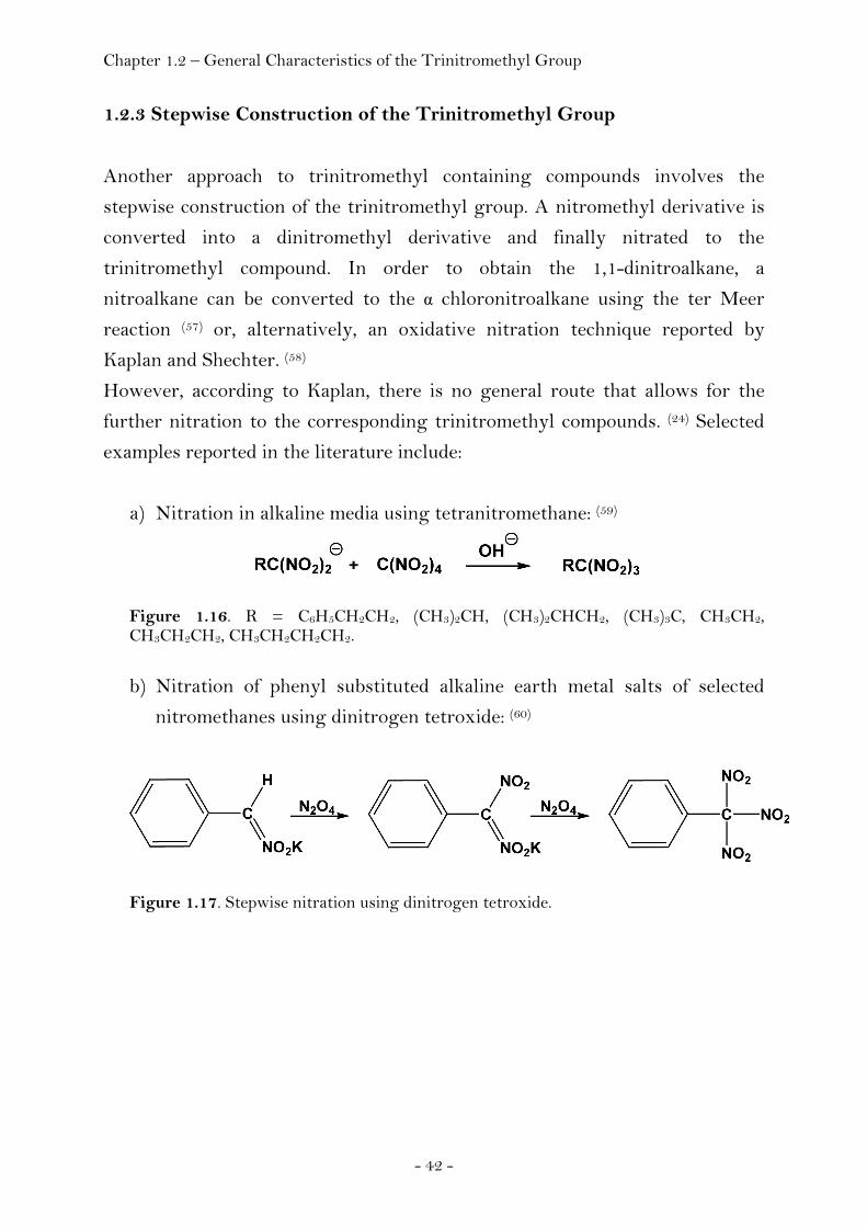
1.2.3 Stepwise Construction of the Trinitromethyl Group
Another approach to trinitromethyl containing compounds involves the
stepwise construction of the trinitromethyl group. A nitromethyl derivative is
converted into a dinitromethyl derivative and finally nitrated to the
trinitromethyl compound. In order to obtain the 1,1-dinitroalkane, a
nitroalkane can be converted to the α chloronitroalkane using the ter Meer
reaction (57) or, alternatively, an oxidative nitration technique reported by
Kaplan and Shechter. (58)
However, according to Kaplan, there is no general route that allows for the
further nitration to the corresponding trinitromethyl compounds. (24) Selected
examples reported in the literature include:
a) Nitration in alkaline media using tetranitromethane: (59)
Figure 1.16. R = C6H5CH2CH2, (CH3)2CH, (CH3)2CHCH2, (CH3)3C, CH3CH2, CH3CH2CH2, CH3CH2CH2CH2. b) Nitration of phenyl substituted alkaline earth metal salts of selected
nitromethanes using dinitrogen tetroxide: (60)
Figure 1.17. Stepwise nitration using dinitrogen tetroxide.

Chapter 1.2 – General Characteristics of the Trinitromethyl Group
- 43 -
c) Destructive nitration of the carboxyl group to the trinitromethyl entity
using a 4:3 mixture of sulfuric acid (ρ = 1.84 g cm-3) and nitric acid (ρ = 1.5
g cm-3): (61)
Figure 1.18. Synthesis of 2,4,6-Tris(trinitromethyl)-1,3,5-triazine. According to the reported properties the compound is unstable when exposed to air and displays a melting point of 90-91°C.
Further examples of nitration of dinitroalkanes or other intermediates include
the formation of hexanitroethane from 1,1,2,2-tetranitroethane (62) or
trinitroacetonitrile from cyanoacetic acid (63) as well as the formation of
tetranitromethane from acetylene (28) or ketene (64).

Chapter 1.2 – General Characteristics of the Trinitromethyl Group
- 44 -
1.2.4 Reactions of Trinitromethyl Compounds
As already pointed out by Kaplan, a diversity of chemical reactions can be
employed not affecting the trinitromethyl group yielding a variety of valuable
products. (24) For example, 4,4,4-trinitrobutyric acid can be converted to the
acid chloride and subsequently to the corresponding isocyanate which in turn
can undergo typical reactions including amine, urea and urethane formation
(Fig. 1.19). (65)
Figure 1.19. Stepwise formation of 4,4,4-trinitroisocyanate from 4,4,4-trinitrobutyric acid. In addition, the acid chloride itself allows for a variety of other product, for
example the formation of bis-(4,4,4-trinitrobutyryl)-peroxide (Fig. 1.20). (66)
Figure 1.20. Formation of bis-(4,4,4-trinitrobutyryl)peroxide.
Instead of using the 4,4,4-trinitrobutyryl moiety (C4H6N3O6, Ω(CO2) = -41.6%)
for the development of oxygen rich energetic materials, we chose to introduce
the trinitromethyl group using 2,2,2-trinitroethanol due to the positive oxygen
balance value of the 2,2,2-trinitroethyl moiety (C2H2N3O6, Ω(CO2) = +9.8%).
As pointed out earlier (see Table 1.2, page 38) and in contrast to the products
arising from the reactions between a variety of aldehydes and ketones and
trinitromethane, 2,2,2-trinitroethanol is an example of a stable condensation
product. At the same time it displays a positive oxygen balance. It can readily
be obtained in high yields and high purity from the Henry reaction between
trinitromethane and formaldehyde, can be safely handled, stored and it can
Chapter 1.2 – General Characteristics of the Trinitromethyl Group
- 45 -
conveniently and stoichiometrically be applied under ambient conditions. Other
advantages of this group will be discussed in the appropriate sections of this
thesis.
Unfortunately, much of the chemistry reported for the trinitrobutyryl or
trinitropropyl groups is not applicable in case of the trinitroethyl group
because of the specific electronic properties of the trinitromethyl group that are
most pronounced in the case of the trinitroethyl group (σ* = 1.62) and
significantly limit the chemistry to introduce this group compared to the
chemistry available in the case of the trinitropropyl group or trinitrobutyryl
group. The chemistry of 2,2,2-trinitroethanol is different to that of other
alcohols owing to the electron withdrawing inductive effect of the
trinitromethyl group (σ* = 4.54) decreasing the oxygen basicity of the
hydroxyl group. The alcohol becomes acidic (pKa = 6.1) and at pH values
greater than 6, the equilibrium lies in the direction of the trinitromethanide
anion and formaldehyde. This dissociation under weakly acidic or basic
conditions precludes the possibility of 2,2,2-trinitroethoxy derivatives through
the use of nucleophilic displacement reactions utilizing the 2,2,2,-
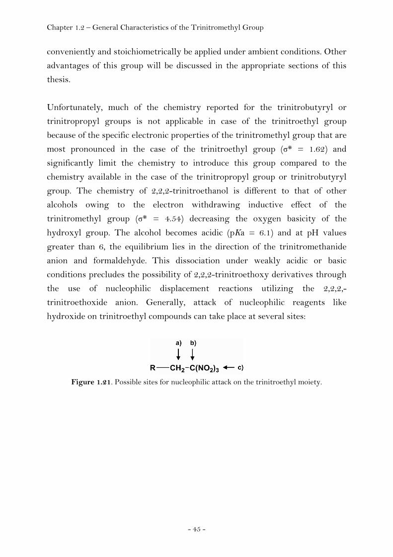
trinitroethoxide anion. Generally, attack of nucleophilic reagents like
hydroxide on trinitroethyl compounds can take place at several sites:
Figure 1.21. Possible sites for nucleophilic attack on the trinitroethyl moiety.
Chapter 1.2 – General Characteristics of the Trinitromethyl Group
- 46 -
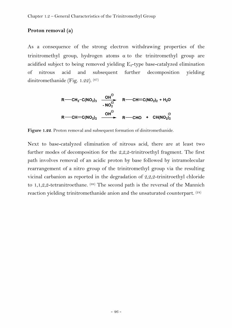
Proton removal (a)
As a consequence of the strong electron withdrawing properties of the
trinitromethyl group, hydrogen atoms α to the trinitromethyl group are
acidified subject to being removed yielding E2-type base-catalyzed elimination
of nitrous acid and subsequent further decomposition yielding
dinitromethanide (Fig. 1.22). (67)
Figure 1.22. Proton removal and subsequent formation of dinitromethanide. Next to base-catalyzed elimination of nitrous acid, there are at least two
further modes of decomposition for the 2,2,2-trinitroethyl fragment. The first
path involves removal of an acidic proton by base followed by intramolecular
rearrangement of a nitro group of the trinitromethyl group via the resulting
vicinal carbanion as reported in the degradation of 2,2,2-trinitroethyl chloride
to 1,1,2,2-tetranitroethane. (68) The second path is the reversal of the Mannich
reaction yielding trinitromethanide anion and the unsaturated counterpart. (24)
Chapter 1.2 – General Characteristics of the Trinitromethyl Group
- 47 -
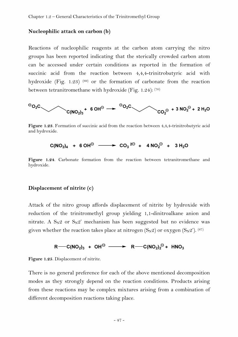
Nucleophilic attack on carbon (b)
Reactions of nucleophilic reagents at the carbon atom carrying the nitro
groups has been reported indicating that the sterically crowded carbon atom
can be accessed under certain conditions as reported in the formation of
succinic acid from the reaction between 4,4,4-trinitrobutyric acid with
hydroxide (Fig. 1.23) (69) or the formation of carbonate from the reaction
between tetranitromethane with hydroxide (Fig. 1.24): (70)
Figure 1.23. Formation of succinic acid from the reaction between 4,4,4-trinitrobutyric acid and hydroxide.
Figure 1.24. Carbonate formation from the reaction between tetranitromethane and hydroxide.
Displacement of nitrite (c)
Attack of the nitro group affords displacement of nitrite by hydroxide with
reduction of the trinitromethyl group yielding 1,1-dinitroalkane anion and
nitrate. A SN2 or SN2’ mechanism has been suggested but no evidence was
given whether the reaction takes place at nitrogen (SN2) or oxygen (SN2’). (67)
Figure 1.25. Displacement of nitrite.
There is no general preference for each of the above mentioned decomposition
modes as they strongly depend on the reaction conditions. Products arising
from these reactions may be complex mixtures arising from a combination of
different decomposition reactions taking place.
Chapter 2.1 – Salts of Trinitromethane 1.3 Methods
Standard analytical methods that have been applied to characterize chemical
compounds include elemental analysis next to spectroscopic methods like
NMR spectroscopy, optical spectroscopy (UV-Vis) or vibrational spectroscopy
(Infrared and Raman spectroscopy) as well as mass spectrometry, bomb
calorimetry or X-ray diffraction and are mentioned in the appropriate chapters
of this thesis. A few additional notes regarding single crystal X-ray diffraction
are mentioned here due to the importance of this technique within the scope of
this thesis. X-ray diffraction techniques have been developed that enable to
extract information that goes beyond routine structure determination -
provided the reliability of the data is high enough. In contrast to neutron
diffraction where the nuclei are responsible for the diffraction, X-ray diffraction
involves the internal and valence electrons of a molecule. In the case of the
hydrogen atom the centre of gravity of the electron density does not coincide
with the nucleus yielding systematically shorter bond distances. When judging
the quality of a structure, it is useful to keep the following two criteria in mind:
The first one has to do with the agreement between the observed structure
factors (Fo) and the calculated ones (Fc). They should be of the same order of
magnitude as the experimental accuracy to which the Fo’s are found. The
second criterion involves the reliability index (R). Usually more reliable results
are obtained with lower R values. However, it can be important to keep in mind
that the information derived using the R value is based on the extent to which
the model chosen for the calculation of the structure fits to the experimental
results. Calculated structure factors that match the observed ones are a
necessary condition for a good crystallographic analysis but they are not a
solely sufficient condition to avoid erroneous results with excellent R values.
In contrast to the standard analytical methods mentioned above, those methods
with relevance to energetic materials research are briefly mentioned in this
section to serve as a guide for the reader not familiar in the area of energetic
materials research.

Chapter 1.3 – Methods
- 49 -
Safety Analysis
The great amount of energy delivered from energetic materials places a
premium on all aspects of their safety including manufacture, handling,
transportation and storage. Likewise, much of the high-explosives work
involves determining the sensitivity of new materials to impact, friction,
electrostatic discharge or heat prior to further handling to obtain their
sensitivity characteristics.
Sensitivity. Various external stimuli can cause release of the energy contained
in energetic materials. Knowing the response of individual energetic materials
to specific stimuli like impact, shock, heat, friction or electric spark is important
in terms of safety and in determining the suitability of a material for a specific
application. However, sensitivity is anything but an absolute quantity and
dependent on a variety of factors. Sensitivity reflects the ease of triggering a
detonation which in turn depends on a complex interplay of a number of factors
including, but not limited to molecular and crystal properties, physical
conditions, the nature of the stimulus etc. (71-74) There are different types of
machines and apparatus to measure the respond of an explosive to external
stimuli and determine the corresponding value. This value, however, is always
obtained for a sample in the particular environment of the test that was used.
The criteria chosen for determining whether a test resulted in an explosion
(‘go’) or failure (‘no go’) may vary between different laboratories and further
depend on the operator to signify a ‘go’ result, often based on visual
observation of smoke, flash or flame, crackling or sound of explosion. One
consequence of this evaluation together with the complex interplay of factors
affecting sensitivity is the difficulty or reproducibly quantifying sensitivity.
Nevertheless, examination of the respond of a material to these external stimuli
like electrostatic discharge (Electrostatic Sensitivity), friction (Friction
Sensitivity) or impact (Impact Sensitivity) is very important to be able to make
at least some estimates of the level of sensitivity and thus important
information in terms of safety and handling of these substances as well as a
qualitative comparison between them. Electrostatic sensitivity is an equally
important quantity in terms of handling a compound under study compared to
Chapter 1.3 – Methods
- 50 -
friction or impact sensitivity. Some explosives can be detonated from the shock
of a person charged with static electricity. The test procedures used to
determine sensitivity are covered in Chapter 4. As far as safety is concerned, it
is important to notice that sensitivity of most materials is not a fundamental
and unalterable property. Sensitive materials can sometimes be made to behave
acceptably insensitive (desensitization).
Impact Sensitivity. The initiation response of an explosive to the blow of a
falling weight is called impact sensitivity. Impact sensitivity is generally
reported as the corresponding impact energy in Joule, given by the product
mgh, where m is the mass of the falling weight, g is the acceleration due to
gravity and h is the height of the falling weight. As mentioned above, it is very
difficult to obtain reproducible impact sensitivity values. They should therefore
be regarded as, at best, qualitative indicators of relative impact sensitivities. (75)
there are a variety of different machines for determining this value. They are
different in detail, but they are technically essentially similar though test
procedures and test data analyses may differ. Traditionally, explosives have
been subjected to falling weights to measure impact sensitivity. Within the
scope of this work a standardized BAM drophammer (Bundesanstalt für
Materialprüfung) has been employed to measure the impact sensitivity. In this
test, the substance is placed between two steel cylinders that are subject to
being impacted by a falling weight. The sound produced serves as information
whether an explosion or failure has occurred together with a visual inspection
of the sample after each experiment. The value taken as impact sensitivity (in
Joule) is defined as the product of the mass of the falling weight and the
distance necessary to yield not more than one explosion out of six experiments.

Chapter 1.3 – Methods
- 51 -
There have been numerous attempts trying to relate impact sensitivity (usually
within a given class of compounds) to various other parameters like the
strengths of certain bonds, enthalpy of formation (Table 1.3) or decomposition
temperature (Table 1.4): Table 1.3. Impact sensitivity compared to enthalpy of formation. (76)
Compound ΔfH0 FI
1,2,4-Trinitrobenzene -56 103 1,3,5-Trinitrobenzene -38 109 2,4,6-Trinitroaniline +22 111 2,3,4,6-Tetranitroaniline -43 86 Pentanitroaniline -339 36 2,3,4-Trinitrotoluene -124 92 2,3,5-Trinitrotoluene -86 101 2,4,6-Trinitrotoluene (TNT) -74 114 3,4,5-Trinitrotoluene -104 95 3,4,6-Trinitrotoluene -93 102
Notes. Test method: Rotter Impact Test (77); FI: Figure of Insensitiveness; FI (picric acid) = 100 by definition; ΔfH0 / kJ mol-1. Table 1.3 is a summary of the impact sensitivity data of isomers of
trinitrotoluene, polynitroanilines and trinitrobenzene. The stabilities of the
aromatic compounds are generally higher as long as the nitro groups are not in
adjacent positions on the ring. It appears to show a relation between the
enthalpy of formation (enthalpy of destabilization) as one method of
characterizing impact sensitivity.
Table 1.4. Impact sensitivity versus thermal stability. (Fehler! Textmarke nicht definiert.)
Compound IS / cm T / °C
Hexanitrobenzene (HNB) 15 165 2,4,6-Trinitrotoluene (TNT) 148 250 Picric Acid 191 260 Diaminotrinitrobenzene (DATB) 320 300 Triaminotrinitrobenzene (TATB) > 320 340
Notes. Test method: Type 12 machine. The value given represents the result of 25 drops to give the height at which the propability of explosion is 50%. Setup: 40 mg of sample are placed on sandpaper between an anvil and a steel cylinder and a 2.5 kg weight is dropped from different heights. The temperature is given as the onset of exotherm.

Chapter 1.3 – Methods
- 52 -
The results of Table 1.4 suggest a dependence of impact sensitivity and
thermal stability. The higher the thermal stability of an explosive, the less
impact sensitive it appears to be. In addition, it has been observed, that impact
sensitivity is temperature dependent (Table 1.5). (78)
Table 1.5. The impact sensitivity of TNT increases with increasing temperature.
Temperature / °C Height / inches Height / cm
-40 17 43.18 25 14 35.56 80 7 17.78 90 3 7.62 105 2* 5.08
Notes. Test method: Picatinny Arsenal apparatus (79), 2 kg weight; * five explosions in 20 trials. Another contribution to the possible factors affecting impact sensitivities of
energetic compounds has recently been reported. It has been suggested that
there may be a link in terms of a crystal volume factor between the impact
sensitivities of energetic compounds and the space available to their molecules
in their crystal lattice. The authors suggested ‘that the availabiltiy of more space
enhances the molecule’s ability to absorb and localize, vibrationally or translationally,
the external energy coming from the impact.’ (75)
Additional approaches for the prediction of some aspects of safety that appear
in the literature are listed in Table 1.6. Assistance for the targeted design of
safe compounds based on predictive methods available today is however by far
not well established as there is yet no complete approach to sensitivity at
molecular level.
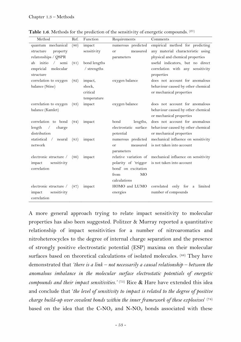
Chapter 1.3 – Methods
- 53 -
Table 1.6. Methods for the prediction of the sensitivity of energetic compounds. (21) Method Ref. Function Requirements Comments
quantum mechanical structure property relatonships / QSPR
(80) impact sensitivity
numerous predicted or measured parameters
empirical method for predicting any material characteristic using physical and chemical properties
ab initio / semi empricial molecular structure
(81) bond lengths / strengths
useful indicators, but no direct correlation with any sensitivity properties
correlation to oxygen balance (Stine)
(82) impact, shock, critical temperature
oxygen balance does not account for anomalous behaviour caused by other chemical or mechanical properties
correlation to oxygen balance (Kamlet)
(83) impact oxygen balance does not account for anomalous behaviour caused by other chemical or mechanical properties
correlation to bond length / charge distribution
(84) impact bond lengths, electrostatic surface potential
does not account for anomalous behaviour caused by other chemical or mechanical properties
statistical / neural network
(85) impact numerous predicted or measured parameters
mechanical influence on sensitivity is not taken into account
electronic structure / impact sensitivity correlation
(86) impact relative variation of polarity of ‘trigger bond’ on excitation from MO calculations
mechanical influence on sensitivity is not taken into account
electronic structure / impact sensitivity correlation
(87) impact HOMO and LUMO energies
correlated only for a limited number of compounds
A more general approach trying to relate impact sensitivity to molecular
properties has also been suggested. Politzer & Murray reported a quantitative
relationship of impact sensitivities for a number of nitroaromatics and
nitroheterocycles to the degree of internal charge separation and the presence
of strongly positive electrostatic potential (ESP) maxima on their molecular
surfaces based on theoretical calculations of isolated molecules. (88) They have
demonstrated that ‘there is a link – not necessarily a causal relationship – between the
anomalous imbalance in the molecular surface electrostatic potentials of energetic
compounds and their impact sensitivities.’ (75) Rice & Hare have extended this idea
and conclude that ‘the level of sensitivity to impact is related to the degree of positive
charge build-up over covalent bonds within the inner framework of these explosives’ (74) based on the idea that the C-NO2 and N-NO2 bonds associated with these

Chapter 1.3 – Methods
- 54 -
positive regions concomitantly become weaker. The unifying concept is based
on the ideal that the breaking of a certain type of bond, a trigger linkage, is a key
step in the decomposition process. Politzer & Murray have recently
summarzied this as follows: ‘Thus, to the extent that C-NO2 and N-NO2 bonds are
trigger linkages in some energetic compounds, the link between the imbalance in their
molecular surface potentials and impact sensitivity may be that the former reflects the
electronic charge depletion that facilitates the breaking of these bonds.’ (75) In doing so,
they also recognized the limitations imposed by the number of factors affecting
impact sensitivity and the uncertainty associated with its measurement and
stated that their goal, therefore, was not a precise structure/activity
correlation but rather a better understanding of the factors and properties
actually influencing sensitivity as opposed to relationships that are
symptomatic or even coincidental. (75) Meanwhile it has been recommended to
include the experimentally derived charge density distribution and derived
ESP of high resolution X-ray diffraction experiments in future theoretical
studies. Impact sensitivity is a bulk property and the ESP of isolated molecules
in the gas phase neglect the effects of intermolecular interactions. In contrast,
the ESP obtained from a diffraction experiment represents molecules as part of
the crystal and therefore includes both these effects. (89)
A novel approach towards sensitivity of energetic compounds has been
suggested very recently and outlining the relevance of kinetics of
decomposition reactions on sensitivity. It has been reported that water displays
catalytic behaviour in PETN (Pentaerythritol tetranitrate) reactions
challenging the traditional view of water in high explosives chemistry which
had previously only been considered to be one of the three major
decomposition products next to carbon dioxide and molecular nitrogen. In
contrast this recent study has demonstrated that water plays an active role in
detonation chemistry and ‘the kinetics of water formation may also contribute to
high explosives sensitivity’. (3)

Chapter 1.3 – Methods
- 55 -
Stability. Stability is the ability of energetic materials to retain, unaltered, such
properties as detonation velocity and sensitivity after long periods of storage
under adverse conditions. All energetic materials are unstable to some extent.
The degree of instability varies greatly. Compatibility with other materials and
long-term stability is expected for safety in handling and use as well as storage
of the products. Whatever its performance, an energetic material will not be
widely used unless it can be used in formulations with a reasonable life
expectancy. The safe service life of an explosive is made up of the safe storage
life, also referred to as the chemical shelf life and the so-called functional life.
Shelf life covers the period of time during which the explosive can safely be
stored and is limited by chemical ageing reactions. The functional life is the
period of time where a safe use of the explosive and its functional requirements
remain fulfilled. The functional life is limited both by (i) chemical ageing
reactions of the explosive that may yield lower energy content or reactions
with the binder that may change the mechanical properties and (ii) physical
processes within the explosive like diffusion or phase changes. (90)
Picric acid may serve as an example. Though more powerful than TNT it was
initially extensively used until it was found that it is incompatible with acid
sensitive materials, particularly in the formation of sensitive metal salts. (91)
There are two types of reactions that have to be distinguished when
considering the stability of energetic materials. (92) The first type of thermal
decomposition can be described as chemical ageing and occurs at low
temperatures or at room temperature. The second type refers to thermal
decomposition with ignition of the material. Generally, thermal decomposition
occurs when bonding forces within the corresponding molecules or ions are
weaker compared to those between atoms of neighbouring units. An increase in
temperature may result in bond redistribution and the formation of
decomposition products. It has been estimated that activation energies of
decomposition higher than 170 kJ mol-1 correspond to a chemical shelf life in
the order of thousands of years at room temperature whereas values below 155
kJ mol-1 indicate limited stability and require further examination of the
stability. (93) An estimation of the shelf life time of an energetic material based
on the measurements of the activation energy has been reported by Klapötke et
al. (94)

Chapter 1.3 – Methods
- 56 -
Thermal Stability. When heated, an explosive will begin to decompose
exothermically. If a large enough sample is heated above its critical
temperature, it can explode. The factors responsible to reach this critical
temperature include the shape and size of the explosive sample, its thermal
conductivity and its kinetics of decomposition. (95)
(a) Differential Scanning Calorimetry. A small-scale test for the thermal
stability of energetic materials is differential scanning calorimetry (DSC).
Typically two milligrams of sample are placed inside an aluminium container
and heated inside a furnace purged with dry nitrogen against a reference. A
temperature programme is run and the instrument records the difference in
heat flow of the two compartments as a function of temperature. A positive
deflection corresponds to an exothermic process and a negative deflection
corresponds to an endothermic one. The use of DSC is not limited to the
characterization of the thermal stability of energetic materials but additionally
yields useful information:
Solids that do not exhibit the same high degree of ordering found in crystalline
samples can be detected through the presence of a broad temperature range on
melting. This is known as glass transition. Melting of a crystalline solid
appears as a quite sharp peak in contrast. Information about the exothermic
thermal decomposition includes the decomposition temperature, the width of
the thermal reaction, the onset and offset temperatures of the peak and the
enthalpy of the thermal decomposition corresponding to the peak area. A
kinetic evaluation of data obtained at several heating rates (non-isothermal)
allows for the estimation of the activation energy of decomposition.
(b) Isothermal Long Term Testing. This test is used to obtain information
on thermal ageing properties at elevated temperatures. Within the scope of this
work, a RADEX oven has been employed. (96) Typically, 100 mg of sample were
placed inside a steel container and isothermally heated for 48 hours inside the
RADEX oven at elevated temperatures up to 40°C below the thermal
decomposition point of the sample obtained from DSC measurements. Any heat
flow occurring during the measurement is recorded. Howerver, it is important
to note that a RADEX measurement is not that sensitive as microcalorimetry
and thermal ageing might have occurred although no heat flow has been

Chapter 1.3 – Methods
- 57 -
recorded in case the heat flow was too small and thermal decomposition
occurred slowly. For this reason it is important to visually inspect the sample
after a measurement and carry out additional analytical tests to be able to
guarantee that no ageing has occurred. Those tests include DSC measurements
to judge wether a change in decomposition of the material has occurred
compared to the decomposition temperature of a sample that has not been
heated using the RADEX oven as well as other standard analytical methods
like NMR spectroscopy.
Chemical Compatibility. Next to thermal stability of the neat sample it is
additionally possible to test chemical compatibility of the sample using
differential scanning calorimetry (DSC). For this purpose selected compounds
like nitroguanidine or methylammonium nitrate are added to the sample and
the mixture heated. A subsequent comparison to the results obtained from the
measurement of the neat sample allows drawing conclusions of any
unfavourable interactions of the sample to the compounds added.
Classification. In order to obtain an interim hazard classification for the
transport of dangerous goods standardized tests have to be carried out for
substances prior to shipment according to the instructions as specificated in the
technical bulletin Department of Defense Ammunition and Explosives Hazard
Classification Procedures. (97) The test procedures (Table 1.7) include the results
of impact sensitivity (UN 3a), friction sensitivity (UN 3b), thermal stability
(UN 3c), small scale burning (UN 3d) and may additionally include a test of
‘thermal stability for articles and packaged articles’ (UN 4a) in case the test
(UN 3c) fails as well as a 12 m free fall test (UN 4b) in case one of the tests
(UN 3a, UN 3b, UN 3c) fails. Details of the procedures to these tests are
documented in the appendix to this thesis (Chapter 4).

Chapter 1.3 – Methods
- 58 -
Table 1.7. UN hazard classification procedures for articles and substances.
test conditions + (failure) - (pass)
UN 3a impact
sensitivity
5 tests,
50%
≤ 3.5 J > 3.5 J
UN 3b friction
sensitivity
5 tests,
1 / 5
≤ 184 N,
visible sparks,
visible flame,
audible expl.,
loud crackling noise
> 184
UN 3c thermal
stability
75°C, 48 h visual:
- color change,
- explosion,
- ignition,
- weight loss (any more than H2O)
RADEX:
self-heating > 3°C
UN 3d small scale
burning
Kerosene-
soaked
sawdust,
unconfined
explosion
or
detonation
UN 4a thermal stability for articles and packaged articles
75°C, 48 h visual: - color change, - explosion, - ignition, - weight loss (any more than H2O) RADEX: self-heating > 3°C
UN 4b free fall 12 m free fall fire, explosion or detonation
Chapter 2.1 – Salts of Trinitromethane
Chapter 2
2.1 Energetic Materials: Salts
2.1.1 Salts of Trinitromethane
Introduction
Energetic salts containing the nitroformate anion have been known for over a
century (98), however, only one compound hydrazinium nitroformate (HNF) has
gained practical application as high performing, halogen free oxidizer for
advanced propellant formulations. It has been called a promising candidate for
the replacement of ammonium perchlorate (AP) oxidizer and indeed HNF-
based propellants show improved performance in terms of specific impulse over
AP-based propellants due to the high oxygen content and only slightly
negative heat of formation (ΔfH 0(s) = -94 cal g-1). (99) Since the discovery of
HNF in 1951, more than eighty publications (100) have appeared, showing that
HNF is of continued interest. Recently, two derivatives of hydrazinium
nitroformate (HNF), namely monomethylhydrazinium nitroformate
(MMHNF) and dimethylhydrazinium nitroformate (DMHNF) were
synthesised and suggested as being promising new high performance energetic
materials. (101) In the course of our investigations into high energy density
materials (HEDM), we recently focussed our attention on various nitroformate
salts, in order to explore their potential as possible new ingredients for high
performance, halogen-free propellant formulations, which would avoid the
problematic formation of hydrogen chloride from the use of ammonium
perchlorate as oxidizer in rocket propellant formulations (see page 125). The
cations which were combined with the nitroformate anion in this work were
potassium, ammonium, hydrazinium, melaminium, guanidinium,
aminoguanidinium, diaminoguanidinium and triaminoguanidinium. Potassium
nitroformate, (102) ammonium nitroformate, (98) hydrazinium nitroformate, (98)
guanidinium nitroformate, (103) and melaminium nitroformate (104) have already
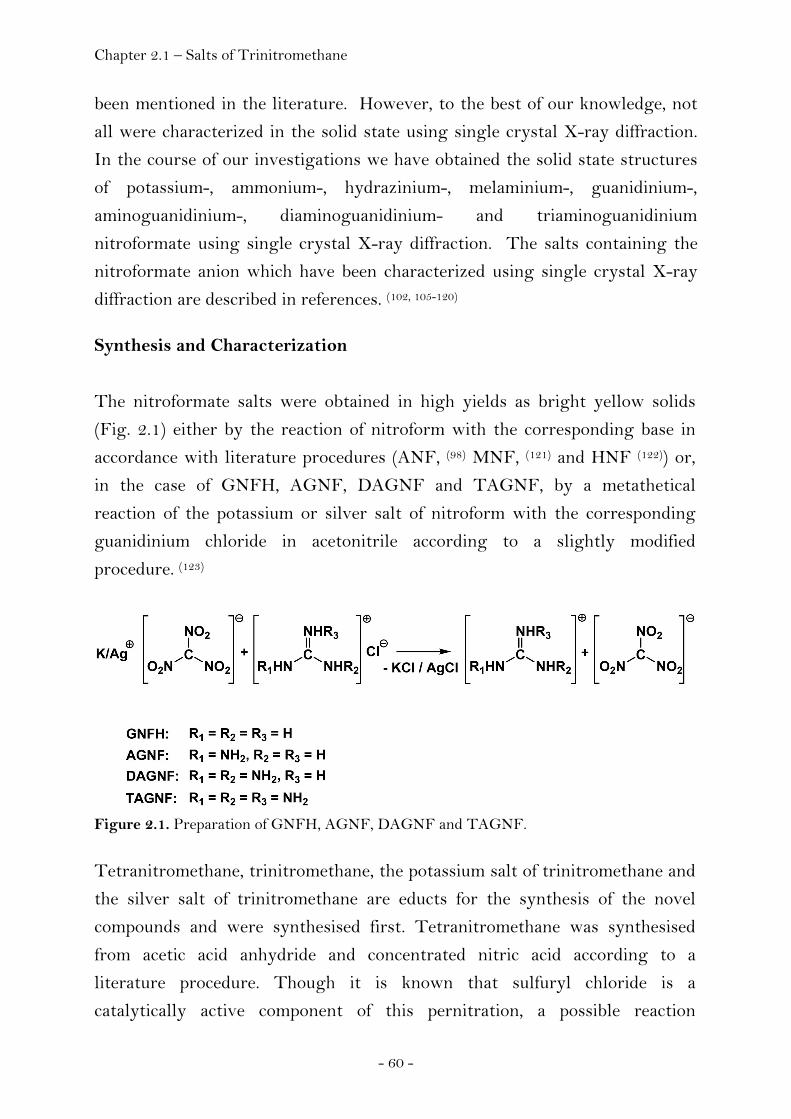
Chapter 2.1 – Salts of Trinitromethane
- 60 -
been mentioned in the literature. However, to the best of our knowledge, not
all were characterized in the solid state using single crystal X-ray diffraction.
In the course of our investigations we have obtained the solid state structures
of potassium-, ammonium-, hydrazinium-, melaminium-, guanidinium-,
aminoguanidinium-, diaminoguanidinium- and triaminoguanidinium
nitroformate using single crystal X-ray diffraction. The salts containing the
nitroformate anion which have been characterized using single crystal X-ray
diffraction are described in references. (102, 105-120) Synthesis and Characterization
The nitroformate salts were obtained in high yields as bright yellow solids
(Fig. 2.1) either by the reaction of nitroform with the corresponding base in
accordance with literature procedures (ANF, (98) MNF, (121) and HNF (122)) or,
in the case of GNFH, AGNF, DAGNF and TAGNF, by a metathetical
reaction of the potassium or silver salt of nitroform with the corresponding
guanidinium chloride in acetonitrile according to a slightly modified
procedure. (123)
Figure 2.1. Preparation of GNFH, AGNF, DAGNF and TAGNF. Tetranitromethane, trinitromethane, the potassium salt of trinitromethane and
the silver salt of trinitromethane are educts for the synthesis of the novel
compounds and were synthesised first. Tetranitromethane was synthesised
from acetic acid anhydride and concentrated nitric acid according to a
literature procedure. Though it is known that sulfuryl chloride is a
catalytically active component of this pernitration, a possible reaction
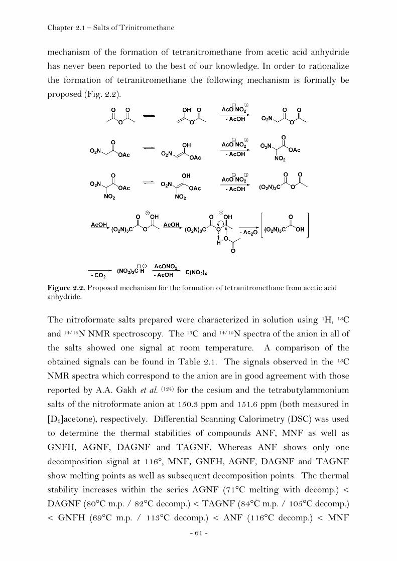
Chapter 2.1 – Salts of Trinitromethane
- 61 -
mechanism of the formation of tetranitromethane from acetic acid anhydride
has never been reported to the best of our knowledge. In order to rationalize
the formation of tetranitromethane the following mechanism is formally be
proposed (Fig. 2.2).
Figure 2.2. Proposed mechanism for the formation of tetranitromethane from acetic acid anhydride. The nitroformate salts prepared were characterized in solution using 1H, 13C
and 14/15N NMR spectroscopy. The 13C and 14/15N spectra of the anion in all of
the salts showed one signal at room temperature. A comparison of the
obtained signals can be found in Table 2.1. The signals observed in the 13C
NMR spectra which correspond to the anion are in good agreement with those
reported by A.A. Gakh et al. (124) for the cesium and the tetrabutylammonium
salts of the nitroformate anion at 150.3 ppm and 151.6 ppm (both measured in
[D6]acetone), respectively. Differential Scanning Calorimetry (DSC) was used
to determine the thermal stabilities of compounds ANF, MNF as well as
GNFH, AGNF, DAGNF and TAGNF. Whereas ANF shows only one
decomposition signal at 116°, MNF, GNFH, AGNF, DAGNF and TAGNF
show melting points as well as subsequent decomposition points. The thermal
stability increases within the series AGNF (71°C melting with decomp.) <
DAGNF (80°C m.p. / 82°C decomp.) < TAGNF (84°C m.p. / 105°C decomp.)
< GNFH (69°C m.p. / 113°C decomp.) < ANF (116°C decomp.) < MNF
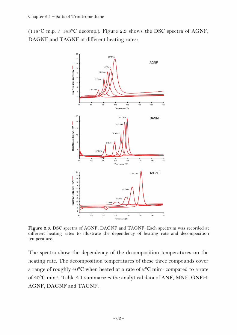
Chapter 2.1 – Salts of Trinitromethane
- 62 -
(118°C m.p. / 143°C decomp.). Figure 2.3 shows the DSC spectra of AGNF,
DAGNF and TAGNF at different heating rates:
Figure 2.3. DSC spectra of AGNF, DAGNF and TAGNF. Each spectrum was recorded at different heating rates to illustrate the dependency of heating rate and decomposition temperature. The spectra show the dependency of the decomposition temperatures on the
heating rate. The decomposition temperatures of these three compounds cover
a range of roughly 40°C when heated at a rate of 2°C min-1 compared to a rate
of 20°C min-1. Table 2.1 summarizes the analytical data of ANF, MNF, GNFH,
AGNF, DAGNF and TAGNF.
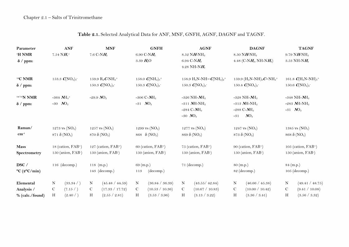
Chapter 2.1 – Salts of Trinitromethane
Table 2.1. Selected Analytical Data for ANF, MNF, GNFH, AGNF, DAGNF and TAGNF.
Parameter ANF MNF GNFH AGNF DAGNF TAGNF 1H NMR
δ / ppm:
7.54 NH4+ 7.6 C-NH2
6.90 C-NH2 3.39 H2O
8.52 NH-NH2 6.94 C-NH2 4.28 NH-NH2
8.50 NH-NH2 4.48 (C-NH2, NH-NH2)
9.79 NH-NH2 3.53 NH-NH2
13C NMR
δ / ppm:
153.3 C(NO2)3- 159.9 R2C-NH2+
150.3 C(NO2)3- 158.0 C(NH2)3+ 150.3 C(NO2)3-
158.9 H2N-NH=C(NH2)2+ 150.3 C(NO2)3-
159.9 (H2N-NH)2C=NH2+ 150.4 C(NO2)3-
161.8 C(H2N-NH)3+ 150.6 C(NO2)3-
14/15N NMR
δ / ppm:
-364 NH4+
-30 NO2
-29.9 NO2
-306 C-NH2 -31 NO2
-326 NH-NH2 -311 NH-NH2 -284 C-NH2 -30 NO2
-328 NH-NH2 -313 NH-NH2 -288 C-NH2 -31 NO2
-348 NH-NH2 -283 NH-NH2 -31 NO2
Raman/ cm-1
1273 νs (NO2)
871 δ (NO2)
1257 νs (NO2)
870 δ (NO2)
1299 νs (NO2)
868 δ (NO2)
1277 νs (NO2)
869 δ (NO2)
1247 νs (NO2)
873 δ (NO2)
1385 νs (NO2)
868 δ (NO2)
Mass Spectrometry
18 (cation, FAB+) 150 (anion, FAB-)
127 (cation, FAB+) 150 (anion, FAB-)
60 (cation, FAB+) 150 (anion, FAB-)
75 (cation, FAB+) 150 (anion, FAB-)
90 (cation, FAB+) 150 (anion, FAB-)
105 (cation, FAB+) 150 (anion, FAB-)
DSC / °C (2°C/min)
116 (decomp.) 118 (m.p.) 143 (decomp.)
69 (m.p.) 113 (decomp.)
71 (decomp.)
80 (m.p.) 82 (decomp.)
84 (m.p.) 105 (decomp.)
Elemental Analysis / % (calc./found)
N (33.34 / ) C (7.15 / ) H (2.40 / )
N (45.48 / 44.59) C (17.33 / 17.72) H (2.55 / 2.81)
N (36.84 / 36.39) C (10.53 / 10.36) H (3.53 / 3.96)
N (43.55/ 42.84) C (10.67 / 10.83) H (3.13 / 3.22)
N (46.66 / 45.38) C (10.00 / 10.42) H (3.36 / 3.41)
N (49.41 / 48.75) C (9.41 / 10.08) H (3.56 / 3.32)
Chapter 2.1 – Salts of Trinitromethane Crystal Structure Analysis
The influence of the many factors that affect the geometry of the nitroformate
anion is complex and has been the subject to several publications. (123, 125-126)
Previously reported data include the results of ab initio calculations (125-126) and
single crystal X-ray diffraction studies. (102, 106-108, 112, 115, 119, 127-136) Competition
between slow crystal growth and decomposition of the nitroformate moiety is a
problem when trying to obtain single crystals of salts containing the
nitroformate anion which are suitable for X-ray diffraction. Using Infrared and
Raman spectroscopy, the decomposition of concentrated solutions of GNF,
AGNF and DAGNF on standing at room temperature yielding the
corresponding guanidinium nitrates could be observed. It is known that silver
nitroformate solutions on standing decompose to form silver nitrate (Figure
2.4). (119)
Figure 2.4. Decomposition of the trinitromethanide anion. In much the same way, GNF, AGNF and DAGNF are expected to decompose
with the formation of the corresponding guanidinium nitrates. However, a
systematic study of the crystal structure was of interest to us in order to obtain
reliable values for the density, cell volume and the number of formula units per
unit cell, in order to be able to predict performance data for these energetic
materials. Crystals suitable for X-ray diffraction were therefore measured as
soon as they were obtained. Detailed structural parameters of all compounds
determined using single crystal X-ray diffraction are provided in the appendix
(Chapter 4).
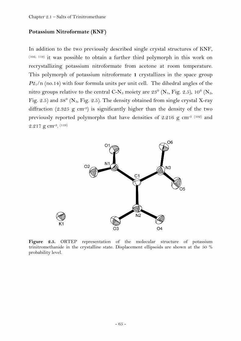
Chapter 2.1 – Salts of Trinitromethane
- 65 -
Potassium Nitroformate (KNF)
In addition to the two previously described single crystal structures of KNF, (102, 119) it was possible to obtain a further third polymorph in this work on
recrystallizing potassium nitroformate from acetone at room temperature.
This polymorph of potassium nitroformate 1 crystallizes in the space group
P21/n (no.14) with four formula units per unit cell. The dihedral angles of the
nitro groups relative to the central C-N3 moiety are 23° (N1, Fig. 2.5), 10° (N2,
Fig. 2.5) and 38° (N3, Fig. 2.5). The density obtained from single crystal X-ray
diffraction (2.325 g cm-3) is significantly higher than the density of the two
previously reported polymorphs that have densities of 2.216 g cm-3 (102) and
2.217 g cm-3. (119)
Figure 2.5. ORTEP representation of the molecular structure of potassium trinitromethanide in the crystalline state. Displacement ellipsoids are shown at the 50 % probability level.
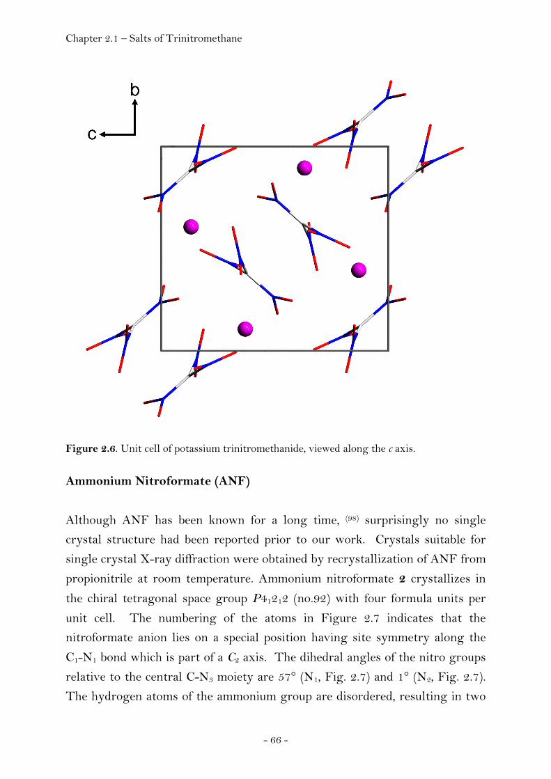
Chapter 2.1 – Salts of Trinitromethane
- 66 -
Figure 2.6. Unit cell of potassium trinitromethanide, viewed along the c axis. Ammonium Nitroformate (ANF)
Although ANF has been known for a long time, (98) surprisingly no single
crystal structure had been reported prior to our work. Crystals suitable for
single crystal X-ray diffraction were obtained by recrystallization of ANF from
propionitrile at room temperature. Ammonium nitroformate 2 crystallizes in
the chiral tetragonal space group P41212 (no.92) with four formula units per
unit cell. The numbering of the atoms in Figure 2.7 indicates that the
nitroformate anion lies on a special position having site symmetry along the
C1-N1 bond which is part of a C2 axis. The dihedral angles of the nitro groups
relative to the central C-N3 moiety are 57° (N1, Fig. 2.7) and 1° (N2, Fig. 2.7).
The hydrogen atoms of the ammonium group are disordered, resulting in two
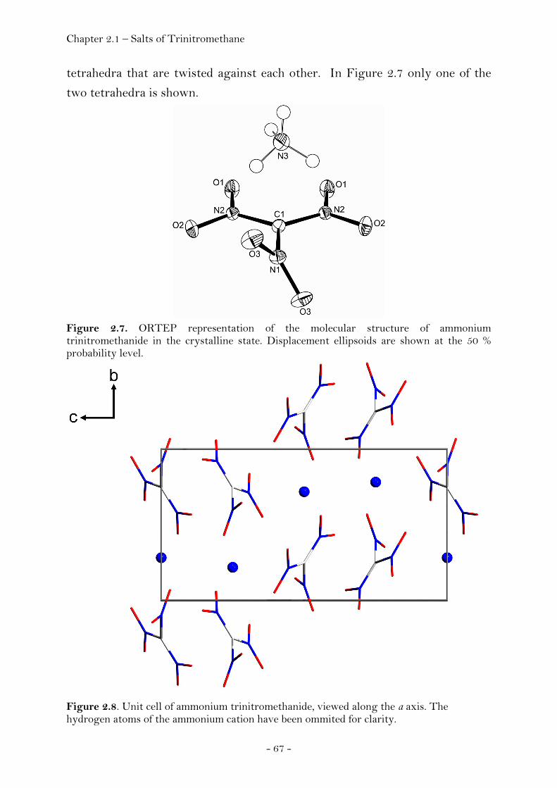
Chapter 2.1 – Salts of Trinitromethane
- 67 -
tetrahedra that are twisted against each other. In Figure 2.7 only one of the
two tetrahedra is shown.
Figure 2.7. ORTEP representation of the molecular structure of ammonium trinitromethanide in the crystalline state. Displacement ellipsoids are shown at the 50 % probability level.
Figure 2.8. Unit cell of ammonium trinitromethanide, viewed along the a axis. The hydrogen atoms of the ammonium cation have been ommited for clarity.
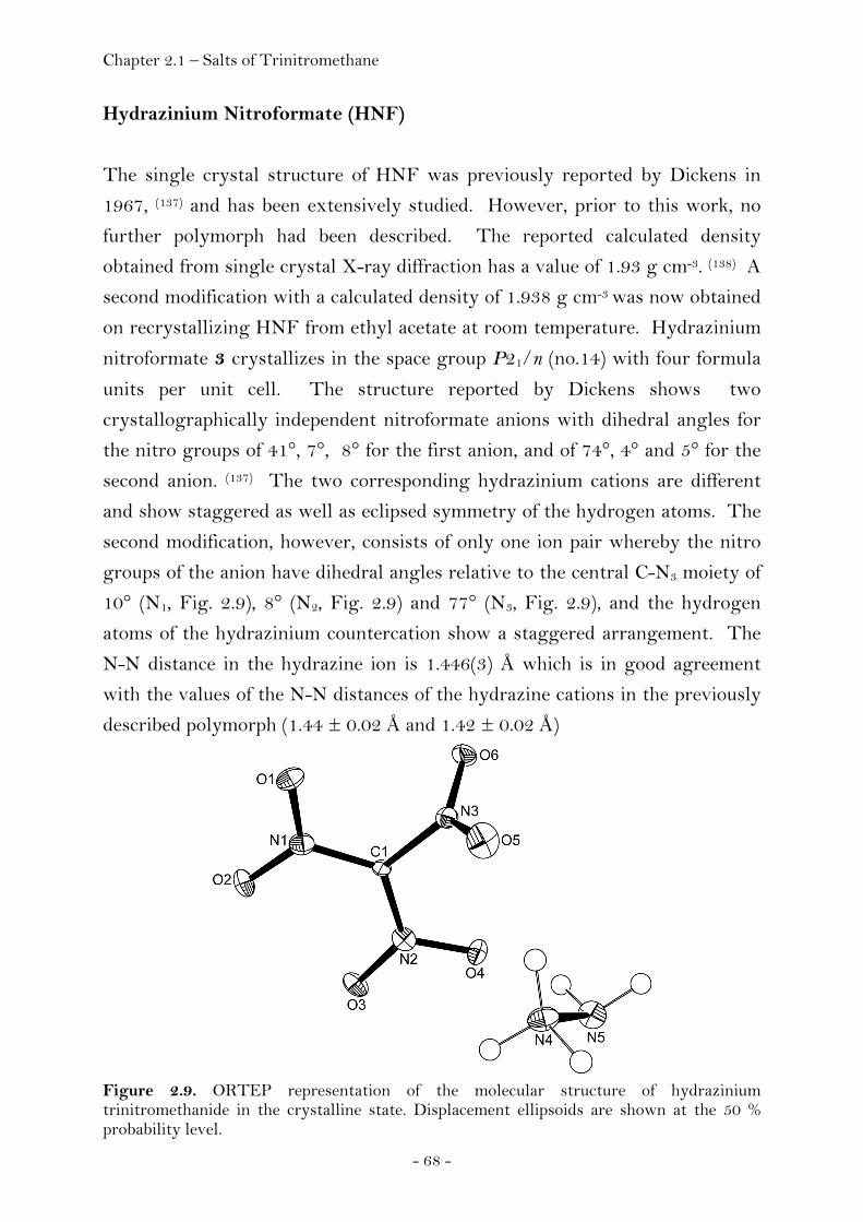
Chapter 2.1 – Salts of Trinitromethane
- 68 -
Hydrazinium Nitroformate (HNF)
The single crystal structure of HNF was previously reported by Dickens in
1967, (137) and has been extensively studied. However, prior to this work, no
further polymorph had been described. The reported calculated density
obtained from single crystal X-ray diffraction has a value of 1.93 g cm-3. (138) A
second modification with a calculated density of 1.938 g cm-3 was now obtained
on recrystallizing HNF from ethyl acetate at room temperature. Hydrazinium
nitroformate 3 crystallizes in the space group P21/n (no.14) with four formula
units per unit cell. The structure reported by Dickens shows two
crystallographically independent nitroformate anions with dihedral angles for
the nitro groups of 41°, 7°, 8° for the first anion, and of 74°, 4° and 5° for the
second anion. (137) The two corresponding hydrazinium cations are different
and show staggered as well as eclipsed symmetry of the hydrogen atoms. The
second modification, however, consists of only one ion pair whereby the nitro
groups of the anion have dihedral angles relative to the central C-N3 moiety of
10° (N1, Fig. 2.9), 8° (N2, Fig. 2.9) and 77° (N3, Fig. 2.9), and the hydrogen
atoms of the hydrazinium countercation show a staggered arrangement. The
N-N distance in the hydrazine ion is 1.446(3) Å which is in good agreement
with the values of the N-N distances of the hydrazine cations in the previously
described polymorph (1.44 ± 0.02 Å and 1.42 ± 0.02 Å)
Figure 2.9. ORTEP representation of the molecular structure of hydrazinium trinitromethanide in the crystalline state. Displacement ellipsoids are shown at the 50 % probability level.

Chapter 2.1 – Salts of Trinitromethane
- 69 -
Figure 2.10. Unit cell of hydrazinium trinitromethanide, viewed along the b axis. Yellow dashed lines indicate hydrogen bonding.
Melaminium Nitroformate (MNF)
Although two and three-fold protonated melaminium salts are known, (139) the
reaction between melamine and nitroform only afforded a 1:1 adduct. In the
course of studying this reaction, however, several single crystal X-ray
structures could be obtained. Two different modifications of MNF have been
determined; a high density modification (MNFHD) with a density of 1.914 g cm-
3, and a low density modification (MNFLD) with a density of 1.771 g cm-3 (Figs.
2.11 and 2.13). The high density modification of MNF crystallizes in the space
group P21/n (no.14) with four formula units per unit cell, whereas the low
density modification crystallizes in the chiral space group P21 (no.4) with two
independent formula units.

Chapter 2.1 – Salts of Trinitromethane
- 70 -
Figure 2.11. ORTEP representation of the molecular structure of melaminium trinitromethanide (high density modification) in the crystalline state. Displacement ellipsoids are shown at the 50 % probability level.
Figure 2.12. Unit cell of melaminium trinitromethanide (high density modification), viewed along the a axis. The asymmetric unit of the high density modification consists of one formula
unit whereas the asymmetric unit of the low density modification consists of
two independent formula units. The nitroformate anion in the high density
modification shows dihedral angles of the three nitro groups of 16° (N7, Fig.
2.11), 41° (N8, Fig. 2.11) and 8° (N9, Fig. 2.11), whereas the dihedral angles of
the nitro groups of the two crystallographically independent nitroformate
anions of the low density modification are 51° (N13, Fig. 2.13), 12° (N14, Fig.
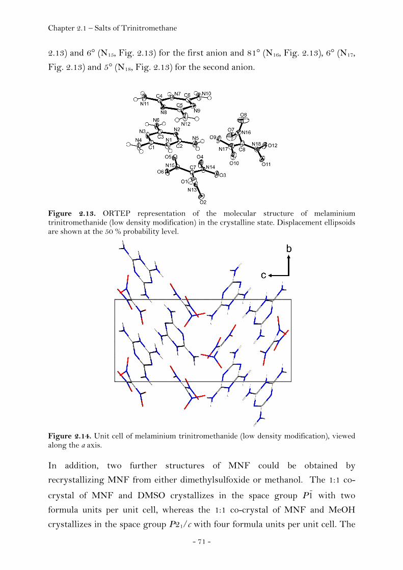
Chapter 2.1 – Salts of Trinitromethane
- 71 -
2.13) and 6° (N15, Fig. 2.13) for the first anion and 81° (N16, Fig. 2.13), 6° (N17,
Fig. 2.13) and 5° (N18, Fig. 2.13) for the second anion.
Figure 2.13. ORTEP representation of the molecular structure of melaminium trinitromethanide (low density modification) in the crystalline state. Displacement ellipsoids are shown at the 50 % probability level.
Figure 2.14. Unit cell of melaminium trinitromethanide (low density modification), viewed along the a axis. In addition, two further structures of MNF could be obtained by
recrystallizing MNF from either dimethylsulfoxide or methanol. The 1:1 co-
crystal of MNF and DMSO crystallizes in the space group P1 with two
formula units per unit cell, whereas the 1:1 co-crystal of MNF and MeOH
crystallizes in the space group P21/c with four formula units per unit cell. The

Chapter 2.1 – Salts of Trinitromethane
- 72 -
asymmetric unit of MNF · MeOH (Fig. 2.15) consists of one formula unit
whereas the asymmetric unit of MNF · DMSO (Fig. 2.17) consists of two
independent formula units.
Figure 2.15. ORTEP representation of the molecular structure of melaminium trinitromethanide · methanol (1:1) in the crystalline state. Displacement ellipsoids are shown at the 50 % probability level.
Figure 2.16. Unit cell of melaminium trinitromethanide · methanol (1:1), viewed along the b axis.
The nitroformate anion in MNF · MeOH shows dihedral angles for the three
nitro groups of 4° (N1, Fig. 2.15), 88° (N2, Fig. 2.15) and 2° (N3, Fig. 2.15),
whereas the dihedral angles of the nitro groups of the two independent
nitroformate anions in MNF · DMSO are 82° (N13, Fig. 2.17), 9° (N14, Fig.
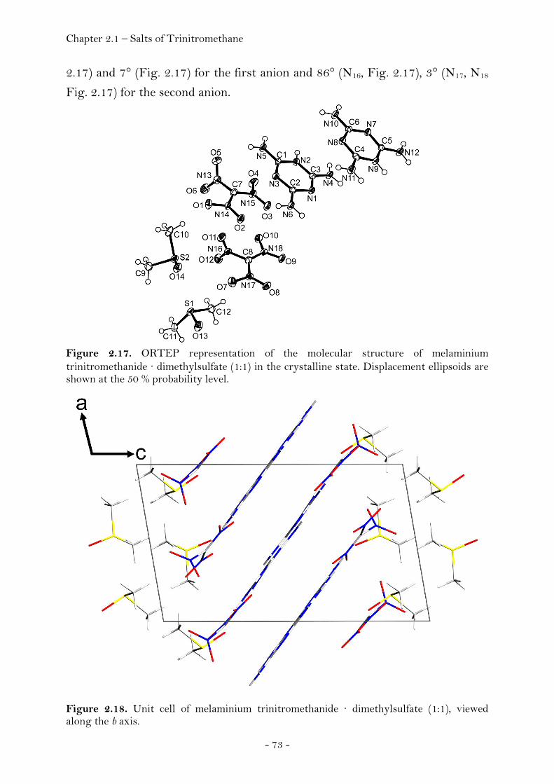
Chapter 2.1 – Salts of Trinitromethane
- 73 -
2.17) and 7° (Fig. 2.17) for the first anion and 86° (N16, Fig. 2.17), 3° (N17, N18
Fig. 2.17) for the second anion.
Figure 2.17. ORTEP representation of the molecular structure of melaminium trinitromethanide · dimethylsulfate (1:1) in the crystalline state. Displacement ellipsoids are shown at the 50 % probability level.
Figure 2.18. Unit cell of melaminium trinitromethanide · dimethylsulfate (1:1), viewed along the b axis.

Chapter 2.1 – Salts of Trinitromethane
- 74 -
Guanidinium Nitroformate Hydrate (GNFH)
The X-ray structure of guanidinium nitroformate hydrate (GNFH) consists of
a two dimensional layer of guanidinium cations, nitroformate anions and water.
The anion shows both the characteristic planar C-N3 moiety and the typical
conformation of the nitro groups, which has been previously described in the
literature. (140) Two of the nitro groups (Fig. 2.19) are almost co-planar with
ONCN torsion angles of 5°, whereas the third nitro group is twisted with a
ONCN torsion angle of 81°. The C-N3 moiety in the guanidinium cation is
planar as expected, and the hydrogen atoms of the amino groups are slightly
twisted out of plane with HNCN torsion angles of 1°, 3° and 9°.
Figure 2.19. ORTEP representation of the molecular structure of guanidinium trinitromethanide · water (1:1) in the crystalline state. Displacement ellipsoids are shown at the 50 % probability level.
Two anions form hydrogen bonds with one in-plane nitro group towards two
amino groups of the cation. The remaining hydrogen atoms of the guanidinium
cation show hydrogen bonding to the neighboring water molecule. The third
nitro group, which is almost perpendicular to the C-N3 moiety, is not involved
in the formation of hydrogen-bonds. Both planar nitro groups of the anion
form a pair of chains with the guanidinium cation, which are interrupted by a
chain of water (Fig. 2.20).

Chapter 2.1 – Salts of Trinitromethane
- 75 -
Figure 2.20. Unit cell of guanidinium trinitromethanide · water (1:1), viewed along the a axis. Aminoguanidinium Nitroformate (AGNF)
Single crystals of aminoguanidinium nitroformate (AGNF) were obtained by
recrystallizing AGNF from propionitrile at room temperature. Since the above
mentioned competition between formation of single crystals and decomposition
is a major issue in this case, crystals formed in the saturated solution were
measured immediately. In contrast to the C2v like orientation of the nitro
groups found in GNFH, the nitro groups aminoguanidinium nitroformate are
twisted in a propeller-like manner (Fig. 2.21) with dihedral angles of 25°, 17°
and 36°.
Figure 2.21. ORTEP representation of the molecular structure of aminoguanidinium trinitromethanide in the crystalline state. Displacement ellipsoids are shown at the 50 % probability level.

Chapter 2.1 – Salts of Trinitromethane
- 76 -
The b-c plane of the single crystal reveals an alternating, chessboard pattern of
the anions and the cations (Fig. 2.22), whereas along the a axis the structure of
AGNF is composed of chains of alternating cations and anions.
Figure 2.22. Unit cell of aminoguanidinium trinitromethanide, viewed along the b axis.
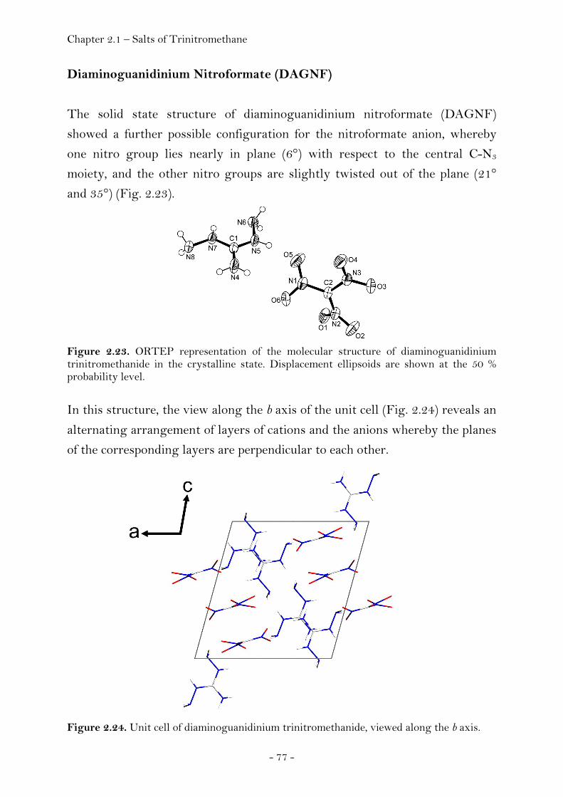
Chapter 2.1 – Salts of Trinitromethane
- 77 -
Diaminoguanidinium Nitroformate (DAGNF)
The solid state structure of diaminoguanidinium nitroformate (DAGNF)
showed a further possible configuration for the nitroformate anion, whereby
one nitro group lies nearly in plane (6°) with respect to the central C-N3
moiety, and the other nitro groups are slightly twisted out of the plane (21°
and 35°) (Fig. 2.23).
Figure 2.23. ORTEP representation of the molecular structure of diaminoguanidinium trinitromethanide in the crystalline state. Displacement ellipsoids are shown at the 50 % probability level. In this structure, the view along the b axis of the unit cell (Fig. 2.24) reveals an
alternating arrangement of layers of cations and the anions whereby the planes
of the corresponding layers are perpendicular to each other.
Figure 2.24. Unit cell of diaminoguanidinium trinitromethanide, viewed along the b axis.

Chapter 2.1 – Salts of Trinitromethane
- 78 -
Triaminoguanidinium Nitroformate (TAGNF)
In GNFH as well as AGNF both the anions and the cations lie in a common
plane concerning the C-N3 moieties, whereas triaminoguanidinium
nitroformate (TAGNF) can be compared with DAGNF in that the C-N3
moieties of the cation and the anion are perpendicular to each other (Fig. 2.25).
Figure 2.25. ORTEP representation of the molecular structure of triaminoguanidinium trinitromethanide in the crystalline state. Displacement ellipsoids are shown at the 50 % probability level.
The structure reveals the characteristic C2v like conformation of the nitro
groups of the nitroformate anion. Two of the nitro groups are almost co-planar
with ONCN torsion angles of 4° and 1° respectively, whereas the third nitro
group is twisted with a ONCN torsion angle of 86° (Fig. 2.25). The central C-
N3 moiety is planar, as was also observed for GNFH, AGNF and DAGNF.
According to our calculations using Gaussian 03, (141) the calculated structure
of the triaminoguanidinium cation is planar in the gas phase at the MP2 level
of theory using a ccPVDZ basis set, whereas the amino groups of the X-ray
structure are slightly twisted out of the plane with NCNN torsion angles of
10°, 9° and 7°.

Chapter 2.1 – Salts of Trinitromethane
- 79 -
Figure 2.26. Unit cell of triaminoguanidinium trinitromethanide, viewed along the c axis.

Chapter 2.1 – Salts of Trinitromethane
- 80 -
Thermal Stability, Sensitivity and Performance Data
The sensitivity data of ANF, MNF, GNFH, AGNF, DAGNF and TAGNF
were measured in order to establish safe handling procedures for these
compounds. Two different instruments were used to determine the friction
and impact sensitivity: the Bundesanstalt für Materialforschung und Prüfung
(BAM) drop hammer (BAM fh (Fallhammer)) and friction tester (BAM ft). (142)
GNFH was found to meet the United Nations (UN) recommendations for the
transport of dangerous goods, with a friction sensitivity of greater than 360 N
and an impact sensitivity of greater than 29.6 J. In contrast, great care should
be taken when handling the other salts (Table 2.3) which are considerably
more sensitive. The sensitivity data and the thermal behaviour of the series of
guanidinium nitroformate salts appear to follow a general trend. The
sensitivity towards friction and impact increases within the series AGNF <
DAGNF < TAGNF, as does the thermal stability. GNFH is the least sensitive
compound and also shows the highest decomposition point in the series of
guanidinium nitroformate salts investigated. Hydrogen bonding between
GNF and water stabilizes this compound. ANF, MNF, GNFH, AGNF,
DAGNF and TAGNF showed no sensitivity towards electrostatic discharge.
Calorimetric measurements of the energies of combustion of nitro compounds
present considerable difficulties because the compounds often explode on
exposure to heat or mechanical shock. (143) The heats of combustion for nitro
compounds are usually lower than for hydrocarbons, and the handling of larger
quantities of such substances is dangerous because of the possible change from
combustion to detonation. In addition, it is difficult to obtain accurate values
for the enthalpy of formation since the accurate determination of the enthalpy
of combustion effects the accuracy of the derived enthalpy of formation. The
energies and heats of formation for ANF, HNF, MNF, GNF, AGNF, DAGNF
and TAGNF were therefore calculated at the MP2 level of theory using an
aug-cc-pVDZ basis set (Table 2.2). The values for the heat of formation
obtained from our calculations can be compared with the previously reported
values for ANF (-47.3 ± 0.2 kcal mol-1), (144) HNF (-18.4 ± 0.3 kcal mol-1), (144)
and GNF (-50.9 ± 0.3 kcal mol-1) (144) which were determined using both
combustion calorimetry and reaction calorimetry. Although the previously
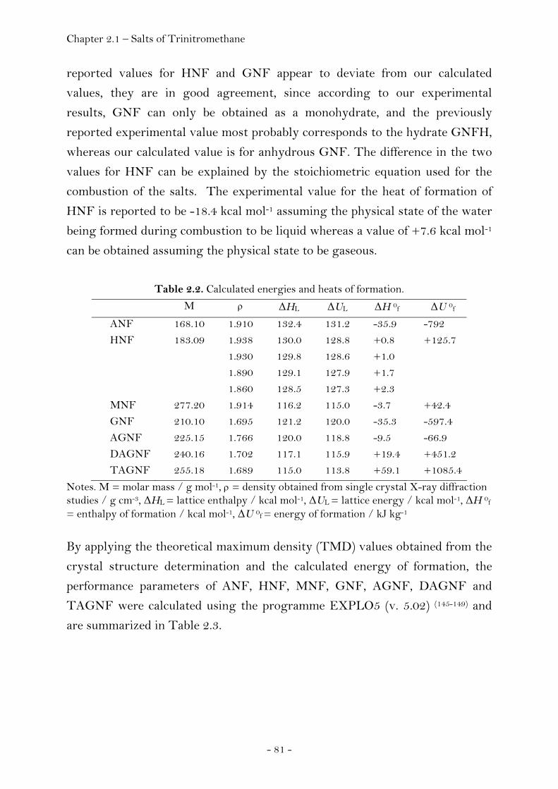
Chapter 2.1 – Salts of Trinitromethane
- 81 -
reported values for HNF and GNF appear to deviate from our calculated
values, they are in good agreement, since according to our experimental
results, GNF can only be obtained as a monohydrate, and the previously
reported experimental value most probably corresponds to the hydrate GNFH,
whereas our calculated value is for anhydrous GNF. The difference in the two
values for HNF can be explained by the stoichiometric equation used for the
combustion of the salts. The experimental value for the heat of formation of
HNF is reported to be -18.4 kcal mol-1 assuming the physical state of the water
being formed during combustion to be liquid whereas a value of +7.6 kcal mol-1
can be obtained assuming the physical state to be gaseous.
Table 2.2. Calculated energies and heats of formation.
M ρ ΔHL ΔUL ΔH 0f ΔU 0f
ANF 168.10 1.910 132.4 131.2 -35.9 -792 HNF 183.09 1.938
1.930 1.890 1.860
130.0 129.8 129.1 128.5
128.8 128.6 127.9 127.3
+0.8 +1.0 +1.7 +2.3
+125.7
MNF 277.20 1.914 116.2 115.0 -3.7 +42.4 GNF 210.10 1.695 121.2 120.0 -35.3 -597.4 AGNF 225.15 1.766 120.0 118.8 -9.5 -66.9 DAGNF 240.16 1.702 117.1 115.9 +19.4 +451.2 TAGNF 255.18 1.689 115.0 113.8 +59.1 +1085.4
Notes. M = molar mass / g mol-1, ρ = density obtained from single crystal X-ray diffraction studies / g cm-3, ΔHL = lattice enthalpy / kcal mol-1, ΔUL = lattice energy / kcal mol-1, ΔH 0f
= enthalpy of formation / kcal mol-1, ΔU 0f = energy of formation / kJ kg-1 By applying the theoretical maximum density (TMD) values obtained from the
crystal structure determination and the calculated energy of formation, the
performance parameters of ANF, HNF, MNF, GNF, AGNF, DAGNF and
TAGNF were calculated using the programme EXPLO5 (v. 5.02) (145-149) and
are summarized in Table 2.3.

Chapter 2.1 – Salts of Trinitromethane
Table 2.3. Calculated performance data and experimental sensitivity data of ANF, HNF, MNF, GNF, AGNF, DAGNF, TAGNF and comparison
with TNT and RDX.
ρ Ω
Qv
V0
Tex
P
D
impact sensitivity
friction sensitivity
ANF 1.910 +19.0 -4328 800 3575 312 8532 3 96 HNF 1.938
1.930 1.890 1.860
+13.1 +13.1 +13.1 +13.1
-5451 -5452 -5447 -5443
826 826 826 826
4085 4057 4086 4107
380 368 354 344
9286 9146 9028 8948
15 (150) 25 (150)
MNF 1.914 -31.8 -4729 764 3579 329 8693 - - GNF 1.695 -7.6 -5250 853 4012 290 8558 30 > 360 AGNF 1.766 -10.7 -5617 854 4091 328 8877 10 144 DAGNF 1.702 -13.3 -5853 873 4209 317 8846 5 32 TAGNF 1.689 -15.7 -6274 885 4358 330 8982 2 20 TNT 1.64 (151) -74 -5089 622 3741 202
210a) (152) 7150 6900a) (151)
15 (151) > 353 (151)
RDX 1.80 (152)
-21.6 -6034 796 4334 340 347a) (153)
8882 8750a) (153)
7.4 (153) 120 (153)
Notes. a) experimental value, D = ρ / g cm-3 , Ω = oxygen balance / %, Qv = heat of explosion / kJ kg, V0 = volume of gaseous detonation products / L kg -1, Tex = explosion temperature / K, P = detonation pressure / kbar, D = speed of detonation / m s-1, impact sensitivity / J, friction sensitivity / N.
Chapter 2.1 – Salts of Trinitromethane A comparison of the calculated values of TNT and RDX with their
corresponding experimental values is included in Table 2.3 and shows that the
calculated values obtained using this method are in good agreement with the
experimental values. The theoretically predicted values for the velocity of
detonation and the detonation pressure for ANF, HNF, MNF, GNF, AGNF,
DAGNF and TAGNF all lie within the range expected for high explosives
such as RDX and are even superior compared to TNT, while at the same time
releasing larger amounts of gaseous decomposition products and having a
more favourable oxygen balance. Of all the compounds studied in this work,
HNF has the highest predicted values for the detonation pressure (380 kbar)
and the velocity of detonation (9286 m s-1), whereas ANF shows the most
positive oxygen balance with a value of +19%. Of the new compounds studied,
TAGNF is not only thermally more stable in contrast to AGNF and DAGNF
which decompose on standing at room temperature after several hours, it also
shows the highest predicted values for the detonation pressure (330 kbar) the
speed of detonation (8982 m s-1). Furthermore, TAGNF is predicted to release
the largest amount of gaseous detonation products (885 L kg-1) and display the
highest positive heat of formation (+59.1 kcal mol-1).
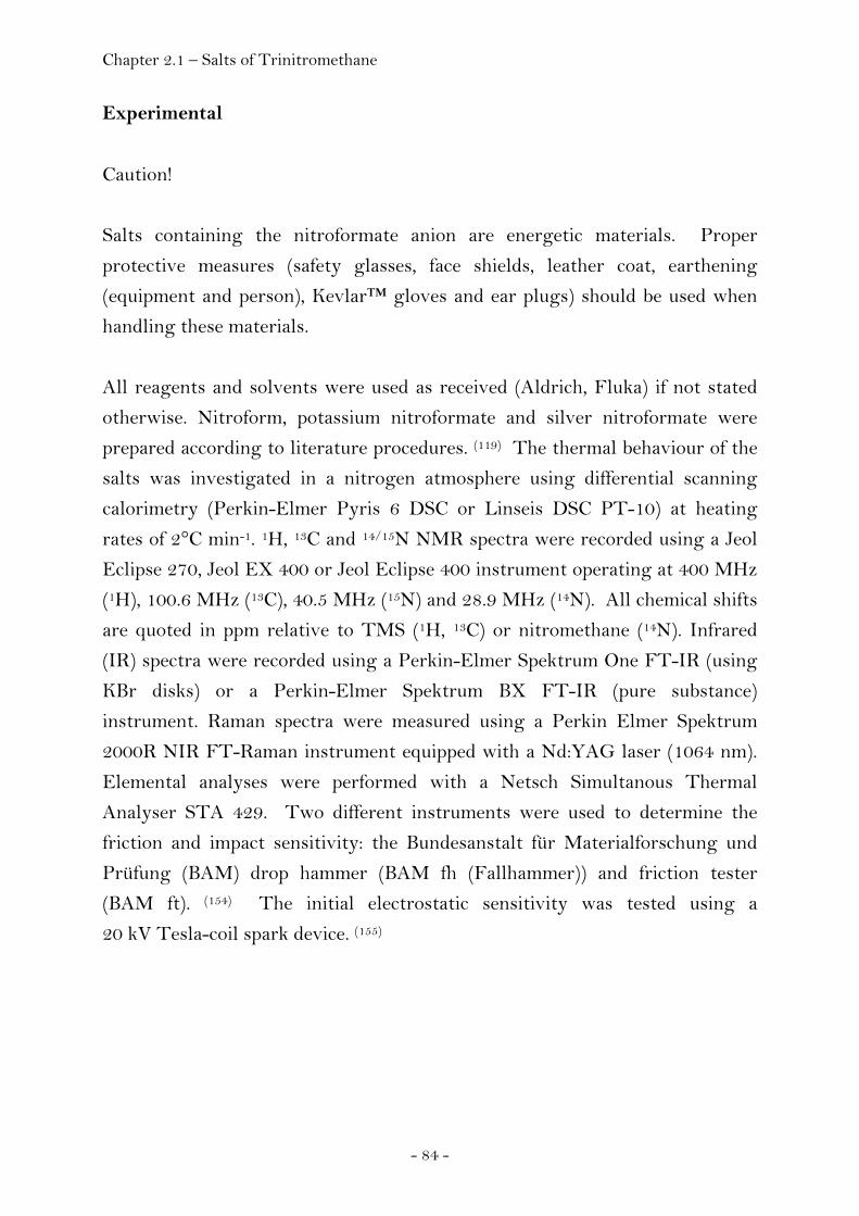
Chapter 2.1 – Salts of Trinitromethane
- 84 -
Experimental
Caution!
Salts containing the nitroformate anion are energetic materials. Proper
protective measures (safety glasses, face shields, leather coat, earthening
(equipment and person), Kevlar™ gloves and ear plugs) should be used when
handling these materials.
All reagents and solvents were used as received (Aldrich, Fluka) if not stated
otherwise. Nitroform, potassium nitroformate and silver nitroformate were
prepared according to literature procedures. (119) The thermal behaviour of the
salts was investigated in a nitrogen atmosphere using differential scanning
calorimetry (Perkin-Elmer Pyris 6 DSC or Linseis DSC PT-10) at heating
rates of 2°C min-1. 1H, 13C and 14/15N NMR spectra were recorded using a Jeol
Eclipse 270, Jeol EX 400 or Jeol Eclipse 400 instrument operating at 400 MHz
(1H), 100.6 MHz (13C), 40.5 MHz (15N) and 28.9 MHz (14N). All chemical shifts
are quoted in ppm relative to TMS (1H, 13C) or nitromethane (14N). Infrared
(IR) spectra were recorded using a Perkin-Elmer Spektrum One FT-IR (using
KBr disks) or a Perkin-Elmer Spektrum BX FT-IR (pure substance)
instrument. Raman spectra were measured using a Perkin Elmer Spektrum
2000R NIR FT-Raman instrument equipped with a Nd:YAG laser (1064 nm).
Elemental analyses were performed with a Netsch Simultanous Thermal
Analyser STA 429. Two different instruments were used to determine the
friction and impact sensitivity: the Bundesanstalt für Materialforschung und
Prüfung (BAM) drop hammer (BAM fh (Fallhammer)) and friction tester
(BAM ft). (154) The initial electrostatic sensitivity was tested using a
20 kV Tesla-coil spark device. (155)
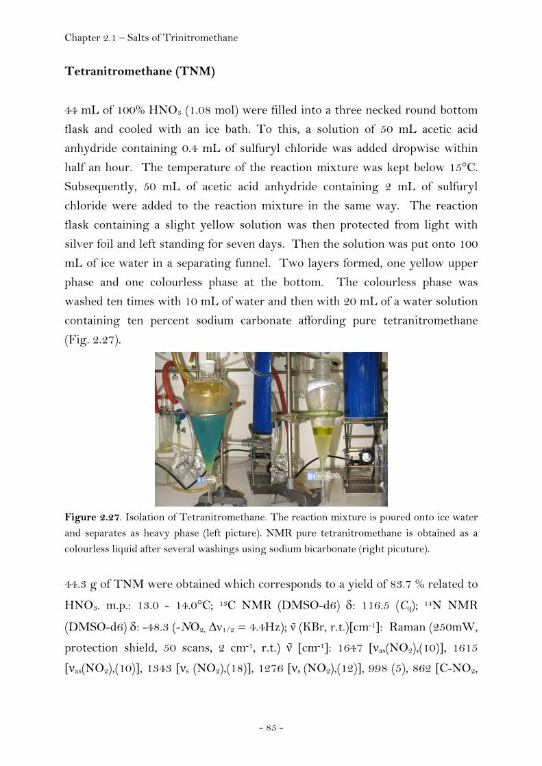
Chapter 2.1 – Salts of Trinitromethane
- 85 -
Tetranitromethane (TNM)
44 mL of 100% HNO3 (1.08 mol) were filled into a three necked round bottom
flask and cooled with an ice bath. To this, a solution of 50 mL acetic acid
anhydride containing 0.4 mL of sulfuryl chloride was added dropwise within
half an hour. The temperature of the reaction mixture was kept below 15°C.
Subsequently, 50 mL of acetic acid anhydride containing 2 mL of sulfuryl
chloride were added to the reaction mixture in the same way. The reaction
flask containing a slight yellow solution was then protected from light with
silver foil and left standing for seven days. Then the solution was put onto 100
mL of ice water in a separating funnel. Two layers formed, one yellow upper
phase and one colourless phase at the bottom. The colourless phase was
washed ten times with 10 mL of water and then with 20 mL of a water solution
containing ten percent sodium carbonate affording pure tetranitromethane
(Fig. 2.27).
Figure 2.27. Isolation of Tetranitromethane. The reaction mixture is poured onto ice water and separates as heavy phase (left picture). NMR pure tetranitromethane is obtained as a colourless liquid after several washings using sodium bicarbonate (right picuture). 44.3 g of TNM were obtained which corresponds to a yield of 83.7 % related to
HNO3. m.p.: 13.0 - 14.0°C; 13C NMR (DMSO-d6) δ: 116.5 (Cq); 14N NMR
(DMSO-d6) δ: -48.3 (-NO2, Δν1/2 = 4.4Hz); ν (KBr, r.t.)[cm-1]: Raman (250mW,
protection shield, 50 scans, 2 cm-1, r.t.) ν [cm-1]: 1647 [νas(NO2),(10)], 1615
[νas(NO2),(10)], 1343 [νs (NO2),(18)], 1276 [νs (NO2),(12)], 998 (5), 862 [C-NO2,

Chapter 2.1 – Salts of Trinitromethane
- 86 -
(100)], 801 [C-NO2, (4)], 605 [δs (NO2),(5)], 414 [δC-N, (18)], 358 [δC-N, (82)], 227
[δC-N, (11)], 197 [δC-N , (12)].
Potassium Nitroformate (KNF)
Potassium nitroformate was prepared according to the literature. (119)
Potassium hydroxide (111mg, 2 mmol) and glycerol (183 mg, 2 mmol) was
dissolved in 5 mL of water and stirred at ice bath temperature. To this
solution, were added portion-wise over a period of ten minutes, 130 mg of
tetranitromethane (0.66 mmol). The ice-bath was removed and the reaction
mixture was allowed to warm up to room temperature and stirred for one hour.
The precipitate was filtered off and washed with 2 mL of diethylether. The
mother liquor was used again instead of the 5 mL of water used initially and
the procedure was repeated. Crystals suitable for single X-ray crystallography
were obtained by recrystallizing potassium nitroformate from nitromethane at
room temperature and were measured immediately.
The yield was 180 mg (72%) of bright yellow KNF. m.p.: 83°C (decomp. with
evolution of gas); 13C NMR (DMSO-d6) δ: 150.8 (C(NO2)3); 14N NMR (DMSO-
d6) δ: -30.3 (-NO2, Δν1/2 = 22.3 Hz); Raman (100 mW, protection shield, 60
scans, 4cm-1) ν [cm-1]: 1508(5), 1466 (3), 1422 (6), 1394 (38), 1291 (34), 1272
(100), 1246 (21), 1168 (7), 873 (35), 794 (11), 719 (7), 465 (8), 442 (3), 283 (6),
257 (14), 155 (12), 113 (8); UV-VIS (Acetone): 349 nm (E=0.525); m/z (FAB-
/NBA): 150 [C(NO2)3-, vs], 62 [NO3-, w], 46 [NO2-, m]; Details of the single
crystal X-ray diffraction experiment are listed in the appendix (Chapter 4).
Nitroform (NFM)
313 mg of KNF (1.66 mmol) were suspended in 2.5 mL of pentane and cooled
with an ice bath. To this suspension 1 mL of concentrated sulfuric acid (98%)
was given dropwise over a period of ten minutes. The sulfuric acid turned
yellow at once. After two hours the color had disappeared and the sulfuric acid
was extracted ten times with 2 mL of pentane. Alternatively, the potassium salt
of trinitromethane can be acidifiend using gaseous hydrochloric acid (Fig.
Chapter 2.1 – Salts of Trinitromethane
- 87 -
2.28). In order to isolate the nitroform as a white solid, the collected pentane
phases were treated with a current of nitrogen. Because of the heat of
evaporation of pentane it was ensured that the precipitating white nitroform
crystals would not liquefy and clean product was obtained.
Figure 2.28. Synthesis of trinitromethane from potassium nitroformate. Dry potassium nitroformate is suspended in pentane and acidified using gaseous hydrochloric acid (left picture) affording a colourless solution of trinitromethane in pentane after a few minutes. On passing a stream of nitrogen over the pentane solution, trinitromethane precipitates from the solution in high yield and purity (right picture).
The yield was 157.5 mg (63%); m.p.: 25.4°C ; 1H NMR (CDCl3) δ: 7.46 (s, 1H,
H-C(NO2)3); 13C NMR (CDCl3) δ: 114.4 (sept., 1C, J 8.7 Hz, H-C(NO2)3); 14N
NMR (DMSO-d6) δ: -38.3 (-NO2, Δν1/2 = 7.2 Hz); ν (gas, -196°C defrost)[cm-
1]: 3063 [ν(CH)], 1618 [s, νas(NO2)], 1608 [s, νas(NO2)], 1303 [s, νs(NO2)
antiphase], 947 [w, ν(CN)], 845 [δ(NO2) synphase], 779 [s, δ(NO2) antiphase], 630 [w, w(NO2)], 569 [w, w(NO2)]; Raman (100 mW, protection shield, 40
scans, 0°C, 2 cm-1 ) ν [cm-1]: 3029 (25), 2758 (5), 1626 (49), 1611 (24), 1375 (50),
1327 (22), 1309 (36), 1243 (26), 950 (69), 943 (57), 840 (51), 774 (22), 628 (36),
574 (21), 405 (100), 375 (95), 237 (12), 204 (18), 121 (14); m/z (CI+, NH3) 152
[(M+1)(0,6)], 105 [(M-NO2)(0,07)], 46 [(NO2)(100)], 30,0 [(NO)(32,6)].

Chapter 2.1 – Salts of Trinitromethane
- 88 -
Ammonium Nitroformate (ANF)
Ammonium nitroformate was prepared according to the literature. (98) Crystals
suitable for X-ray diffraction were obtained by recrystallizing ANF from
propionitrile at room temperature.
m.p.: 116 °C (onset, decomp., Linseis DSC); ν~ (pure substance, solid) [cm-1]: 3222 (m), 1681 (w), 1533 (m), 1460 (m), 1415 (s), 1375 (s), 1247 (s), 1147 (s),
1006 (m), 926 (m), 869 (m), 823 (w), 788 (s), 735 (s), 689 (m); Raman (200 mW,
100 scans, 2 cm-1, protection shield) ν~ [cm-1]: 3172 (4), 2986 (7), 1610 (7), 1535
(6), 1515 (6), 1475 (10), 1380 (43), 1349 (19), 1310 (28), 1273 (72), 1225 (39),
1156 (36), 1138 (25), 871 (100), 859 (35), 791 (12), 727 (10), 466 (24), 443 (18),
415 (20), 396 (16), 375 (21), 275 (23); m/z (FAB+, NBA): 18 [NH4+, w]; (FAB-,
NBA): 150 [(C(NO2)3-), vs]; 1H NMR ([D6]acetone) δ: 7.54 (NH4+, t); 13C NMR
([D6]acetone) δ: 153.3 (C(NO2)3-); 14N NMR ([D6]acetone) δ: -30 (NO2), -364
(NH4+); C1H4N4O6: calc.: N: 33.3 %, C: 7.2 %, H: 2.4 %; found: N: 33.5 %, C: 7.6
%, H: 2.7 %; Details of the single crystal X-ray diffraction experiment are listed
in the appendix (Chapter 4).
Silver Nitroformate Hydrate (AgNFH)
258 mg of freshly prepared and moist silver oxide (1.11 mmol) were suspended
in 5 mL of water and the suspension was filtered.
146 mg of nitroform (0.97 mmol) were dissolved in 5 mL of freshly distilled
diethylether and cooled with an ice-bath. Freshly filtrated and moist silver
oxide was then added to this solution while stirring vigorously. After fifteen
minutes, the reaction mixture was filtered and the solvent removed affording
bright yellow crystals. Single crystals suitable for X-ray structure
determination were obtained by recrystallizing these crystals from water and
were immediately measured. 185 mg of yellow silver nitroformate
monohydrate were obtained (74% yield).
m.p.: 70°C (decomp. with evolution of gas); 13C NMR (DMSO-D6) δ: 150.4
(C(NO2)3); 14N NMR (DMSO-D6) δ: -29.9 (-NO2, Δν1/2 = 13.5 Hz); Raman (100
mW, protection shield, 100 scans, 4cm-1) ν [cm-1]: 1463 (14), 1379 (78), 1257

Chapter 2.1 – Salts of Trinitromethane
- 89 -
(87), 1153 (35), 878 (100), 791 (13), 722 (8), 473 (16), 455 (15), 420 (8), 280
(16), 166 (24); UV-VIS (Acetone): 349 nm (E=0.382); m/z (FAB-, NBA): 150
[(C(NO2)3-), vs], 104 [(C(NO2)2-, w], 46 [(NO2), w]; Details of the single crystal
X-ray diffraction experiment are listed in the appendix (Chapter 4).
Hydrazinium Nitroformate (HNF)
Hydrazinium nitroformate was prepared according to the literature. (156)
Crystals suitable for X-ray diffraction were obtained by recrystallizing HNF
from ethyl acetate at room temperature. Details of the single crystal X-ray
diffraction experiment are listed in the appendix (Chapter 4).
Melaminium Nitroformate (MNF)
Melaminium nitroformate was prepared according to the literature. (157)
Crystals suitable for X-ray crystallography were obtained by recrystallizing
MNF from acetone (high density modification and low density modification)
dimethylsulfoxide (DMSO adduct) or methanol (methanol adduct) at room
temperature.
m.p.: 118 °C (onset, Linseis DSC), 143 °C(onset, Linseis DSC); ν~ (pure solid
substance) [cm-1]: 3446 (w), 3367 (m), 3317 (m), 3149 (m), 2960 (m), 1712 (m),
1645 (s), 1613 (s), 1509 (s), 1467 (s), 1409 (s), 1367 (m), 1338 (m), 1263 (s),
1213 (s), 1124 (s), 995 (s), 976 (s), 869 (m), 792 (s), 773 (s), 744 (s), 732 (s), 688
(m); Raman (200 mW, 200 scans, 1 cm-1) ν~ [cm-1]: 3330 (6), 2989 (6), 1696 (8),
1650 (7), 1596 (8), 1557 (9), 1520 (18), 1504 (13), 1479 (13), 1410 (16), 1375
(48), 1257 (100), 1223 (68), 1156 (32), 1127 (45), 980 (13), 870 (96), 797 (22),
739 (8), 723 (12), 687 (61), 578 (16), 562 (26), 478 (18), 444 (13), 384 (15), 279
(23), 195 (14), 161 (13), 137 (14); 1H NMR ([D6]acetone) δ: 7.6 (s, NH2); 13C
NMR ([D6]acetone) δ: 159.9 (C3, H7N6+), 150.3 (C(NO2)3-); 14N NMR
([D6]acetone) δ: -29.9 (NO2); C4H7N9O6: calc.: N: 45.48 %, C: 17.33 %, H: 2.55
%; found: N: 44.59 %, C: 17.72 %, H: 2.81 %; Details of the single crystal X-ray
diffraction experiment are listed in the appendix (Chapter 4).
Chapter 2.1 – Salts of Trinitromethane
- 90 -
Guanidinium nitroformate (GNF)
To an ice-cooled suspension of 108.3 mg of guanidinium chloride (1.1 mmol) in
6 mL freshly distilled acetonitrile, a solution of 222 mg silver nitroformate (0.8
mmol) dissolved in 7.6 mL of freshly distilled acetonitrile was added drop-wise
over a period of 10 minutes. Silver chloride precipitated instantly. The
reaction mixture was stirred for a further 2.5 hours. The reaction mixture was
allowed to warm up to room temperature and was then stirred for further 2
hours. The precipitated silver chloride was then filtered off and washed with 4
mL of acetonitrile. In order to isolate GNFH, the solvent was first removed
using a rotary evaporator affording a yellow, highly viscous liquid. The
remaining solvent was removed using high vacuum. GNFH was obtained as a
bright yellow solid.
m.p.: 69°C (PE DSC), 113°C (decomp., PE DSC); ν~ (KBr pellets) [cm-1]: 3397
(m), 3280 (w), 3197 (w), 2917 (w), 2851 (w), 1662 (m), 1640 (m), 1513 (m), 1495
(m), 1421 (m), 1358 (w), 1272 (s), 1177 (m), 1144 (w), 976 (w), 866 (w), 794 (m),
733 (m), 515 (w); Raman (200 mW, 100 scans, 4 cm-1, protection shield) ν~ [cm-
1]: 3287 (3), 1528 (11), 1470 (10), 1388 (63), 1299 (37), 1244 (35), 1155 (46),
1055 (12), 1013 (63), 868 (100), 789 (15), 728 (13), 533 (24), 472 (25), 442 (15),
252 (17); m/z (FAB+, NBA): 213 [(C(NH2)3+ + NBA), s], 119 [(C(NH2)3+ +
HN=C(NH2)2), m], 60 [(C(NH2)3+), vs]; (FAB-, NBA): 360 [(2 C(NO2)3- +
C(NH2)3+), s], 303 [(C(NO2)3- + NBA), m], 150 [(C(NO2)3-), vs], 104 [(C(NO2)2-),
w]; 1H NMR ([D6]DMSO) δ: 6.90 (NH2), 3.39 (H2O); 13C NMR ([D6]DMSO) δ:
158.0 (C(NH2)3+), 150.3 (C(NO2)3-); 15N NMR ([D6]DMSO) δ: -31 (NO2), -306
(NH2); C2H8N6O7: calc.: N: 36.8 %, C: 10.5 %, H: 3.5 %; found: N: 36.4 %, C:
10.4 %, H: 3.9 %; Details of the single crystal X-ray diffraction experiment are
listed in the appendix (Chapter 4).
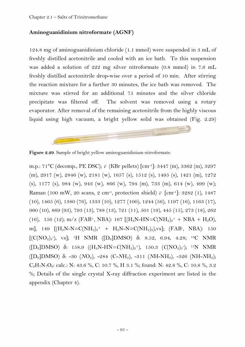
Chapter 2.1 – Salts of Trinitromethane
- 91 -
Aminoguanidinium nitroformate (AGNF)
124.8 mg of aminoguanidinium chloride (1.1 mmol) were suspended in 3 mL of
freshly distilled acetonitrile and cooled with an ice bath. To this suspension
was added a solution of 222 mg silver nitroformate (0.8 mmol) in 7.6 mL
freshly distilled acetonitrile drop-wise over a period of 10 min. After stirring
the reaction mixture for a further 30 minutes, the ice bath was removed. The
mixture was stirred for an additional 75 minutes and the silver chloride
precipitate was filtered off. The solvent was removed using a rotary
evaporator. After removal of the remaining acetonitrile from the highly viscous
liquid using high vacuum, a bright yellow solid was obtained (Fig. 2.29)
Figure 2.29. Sample of bright yellow aminoguanidinium nitroformate.
m.p.: 71°C (decomp., PE DSC); ν~ (KBr pellets) [cm-1]: 3447 (m), 3362 (m), 3297
(m), 2917 (w), 2846 (w), 2181 (w), 1657 (s), 1512 (s), 1495 (s), 1421 (m), 1272
(s), 1177 (s), 984 (w), 943 (w), 866 (w), 794 (m), 733 (m), 614 (w), 499 (w);
Raman (100 mW, 20 scans, 2 cm-1, protection shield) ν~ [cm-1]: 3282 (1), 1487
(10), 1465 (6), 1380 (76), 1333 (10), 1277 (100), 1244 (56), 1197 (16), 1163 (17),
960 (10), 869 (93), 793 (13), 788 (13), 721 (11), 501 (19), 445 (15), 273 (18), 262
(16), 156 (12); m/z (FAB+, NBA): 167 [(H2N-HN=C(NH2)2+ + NBA + H2O),
m], 149 [(H2N-N=C(NH2)2+ + H2N-N=C(NH2)2),vs]; (FAB-, NBA): 150
[(C(NO2)3-), vs]; 1H NMR ([D6]DMSO) δ: 8.52, 6.94, 4.28; 13C NMR
([D6]DMSO) δ: 158.9 ((H2N-HN=C(NH2)2+), 150.3 (C(NO2)3-); 15N NMR
([D6]DMSO) δ: -30 (NO2), -284 (C-NH2), -311 (NH-NH2), -326 (NH-NH2);
C2H7N7O6: calc.: N: 43.6 %, C: 10.7 %, H 3.1 %; found: N: 42.8 %, C: 10.8 %, 3.2
%; Details of the single crystal X-ray diffraction experiment are listed in the
appendix (Chapter 4).

Chapter 2.1 – Salts of Trinitromethane
- 92 -
Diaminoguanidinium nitroformate (DAGNF)
A solution of 182 mg silver nitroformate (0.66 mmol) in 5 mL of acetonitrile
was added dropwise over a period of 10 minutes to a suspension of 85.5 mg
diaminoguanidinium chloride (0.69 mmol) in 2 mL of acetonitrile at 0°C. The
reaction mixture was stirred for a further 40 minutes. The reaction mixture
was then allowed to warm up to room temperature and was stirred for a
further 2 hours. The silver chloride precipitate was filtered off and the solvent
was removed using a rotary evaporator. The highly viscous yellow liquid was
dried using high vacuum affording a bright yellow solid.
m.p.: 80°C (PE DSC), 82°C (decomp., PE DSC); ν~ (KBr pellets) [cm-1]: 3432
(m), 3307 (s), 3247 (w), 2978 (w), 2554 (w), 2175 (w), 1682 (s), 1621 (m), 1512
(s), 1495 (s), 1421 (s), 1352 (w), 1270 (s), 1177 (s), 992 (m), 957 (m), 869 (w),
794 (s), 733 (s), 653 (w), 549 (w); Raman (200 mW, 100 scans, 4 cm-1,
protection shield) ν~ [cm-1]: 3299 (3), 1379 (66), 1247 (100), 1184 (33), 1158
(25), 873 (81), 793 (12), 725 (9), 277 (19), 152 (14); m/z (FAB+, NBA): 243
[(H2N=C(NH-NH2)2+ + NBA), w], 90.1 [(H2N=C(NH-NH2)2+, vs], 89.1
[(HN=C(NH-NH2)2, m]; m/z (FAB-, NBA): 303 [(C(NO2)3- + NBA), w], 150
[(C(NO2)3-), vs]; 1H NMR ([D6]DMSO) δ: 8.50 (NH-NH2), 4.48 (C-NH2, NH-
NH2); 13C NMR ([D6]DMSO) δ: 159.9 ((H2N-C(NH-NH2)2)+), 150.4 (C(NO2)3-); 15N NMR ([D6]DMSO) δ: -31 (NO2), -288 (C-NH2), -313 (NH-NH2), -328 (NH-
NH2); C2H8N8O6: calc.: N: 46.7, C: 10.0 %, H: 3.4 %; found: N: 45.4 %, C: 10.4%,
H: 3.4 %; Details of the single crystal X-ray diffraction experiment are listed in
the appendix (Chapter 4).

Chapter 2.1 – Salts of Trinitromethane
- 93 -
Triaminoguanidinium nitroformate (TAGNF)
To a suspension of 185 mg triaminoguanidinium chloride (1.33 mmol) in 2 mL
of freshly distilled acetonitrile, a solution consisting of 363 mg (1.32 mmol)
silver nitroformate and 10 mL of acetonitrile was added dropwise within a
period of 10 minutes at 0°C. The reaction mixture was then stirred for a
further 40 minutes. The solution was allowed to warm up to room
temperature and stirred for a further 70 minutes.
After removing the precipitated silver chloride from the reaction mixture, the
solution was concentrated using the rotary evaporator. A highly viscous
yellow liquid was obtained. The remaining solvent was removed using high
vacuum, whereby a bright yellow solid could be obtained.
m.p.: 84°C (PE DSC), 105°C (decomp., PE DSC); ν~ (KBr pellets) [cm-1]: 3317
(m), 3210 (s), 1683 (m), 1614 (w), 1513 (s) 1421 (m), 1333 (w), 1276 (s), 1177
(s), 1127 (m), 951 (m), 871 (w), 793 (s), 733 (s), 638 (w), 603 (m), 483 (w);
Raman (200 mW, 200 scans, 4 cm-1, protection shield)ν~ [cm-1]: 3287 (6), 1648
(25), 1461 (16), 1437 (15), 1385 (71), 1331 (38), 1260 (27), 1153 (67), 868 (100),
789 (19), 735 (6), 470 (34), 430 (24), 152 (20);
m/z (FAB+, NBA): 227 [(H2N-N=C(NH-NH2)2 + (C(NH-NH2)3+ + H2O), w], 209 [(H2N-N=C(NH-NH2)2 + (C(NH-NH2)3+), w], 105 [(C(NH-NH2)3+, vs]; 1H
NMR ([D6]acetone) δ: 9.79 (C(NH-NH2)3+), 3.53 (C(NH-NH2)3+); 13C NMR
([D6]acetone) δ: 161.8 (C(NH-NH2)3+), 150.6 (C(NO2)3-); 15N NMR
([D6]acetone) δ: -31 (C(NO2)3-), -283 (C(NH-NH2)3+), -348 (C(NH-NH2)3+);
C2H9N9O6: calc.: N: 49.4 %, C: 9.4 %, H: 3.6 %; found: N: 47.8 %, C: 10.4 %, H:
3.3 %; Details of the single crystal X-ray diffraction experiment are listed in
the appendix (Chapter 4).
Chapter 2.1 – Salts of Diaminopicric acid
- 94 -
2.1.2 Salts of Nitric Acid
Melaminium Dinitrate (MDN)
Introduction
Melamine (2,4,6-triamino-1,3,5-triazine) is a nitrogen-rich, planar compound
which can form salts containing mono- or diprotonated melaminium cations.
Melaminium nitrate was synthesised by Tanbug et al. and classified as a novel
energetic material. (158) However, no sensitivity or performance data were
reported. In the course of investigating oxygen-rich energetic materials, the
earlier unknown nitrogen and oxygen-rich energetic salt melaminium dinitrate
was readily obtained from the reaction between melamine and concentrated
nitric acid. (159) The salt was characterized using vibrational spectroscopy (IR,
Raman), multinuclear NMR spectroscopy and elemental analysis. The thermal
behaviour of MDN was monitored using differential scanning as well as
isothermal long term calorimetry. In addition, the impact, friction and
electrostatic sensitivity data were measured. The crystal structure of MDN
was determined using single crystal X-ray diffraction.
Chapter 2.1 – Salts of Diaminopicric acid
- 95 -
Sensitivity and Performance
The friction and impact sensitivities were measured with a BAM drop-hammer
and a BAM friction tester. The friction sensitivity of this compound was
determined as being greater than 350 N, and the impact sensitivity more than
30 J. The thermal long term stability was studied using a RADEX V5 Oven.
Isothermal tempering of the substance showed that the substance is thermally
stable for at least 48 hours at a temperature of 140°C
Table 2.4. Performance Data of MDN and comparison to TNT.
MDN TNT sum formula C3H8N8O6 C7H5N3O6 m.w. / g mol-1 252.2 227.1 ρ / g cm-3 1.852(2) 1.64 (160) ΔfH0(s) / kcal mol-1 -149.8 -13.27 ΔfU0(s) / kJ kg-1 -2377.3 -175 Ω / % -25.4 -74.0 Q / kJ kg-1 2977 5064 Tex / K 2562 3749 V0 / L kg-1 803 625 PCJ / kbar 236 203 D / m s-1 7723 7170
Although MDN has a favourable oxygen balance (Ω = -25.4) and a combined
oxygen and nitrogen content of greater than 80%, it shows a remarkable
thermodynamic stability (Tdecomp. = 330 °C). It was found to meet the United
Nations (UN) recommendations for the transport of dangerous goods, with a
friction sensitivity of greater than 360 N and an impact sensitivity of greater
than 30 J. Excessive hydrogen bonding in the solid state not only stabilizes this
compound but also results in a dense packing of the crystalline material leading
to a remarkably high density (1.852 g cm-3) and performance values exceeding
the detonation pressure, the detonation velocity of TNT while at the same time
producing a favourably larger amount of decomposition gases and lower
explosion temperature. These properties render MDN to be an interesting
material displaying potential for applications requiring insensitivity and
thermal stability, for example for perforation and fracturing purposes for
oilfield exploration (Table 2.4).

Chapter 2.1 – Salts of Diaminopicric acid
- 96 -
Crystal Structure Analysis
MDN at 200 K has monoclinic symmetry; space group P21/c (no.14) The
asymmetric unit contains one doubly protonated melaminium cation and two
nitrate anions (Fig. 2.30).
Figure 2.30. ORTEP representation of the molecular structure of melaminium dinitrate in the crystalline state. Displacement ellipsoids are shown at the 50 % probability level. Selected bond lengths [Å] and angles [°]: C1-N1 1.337(3), N1-C2 1.369(3), C2-N2 1.307(3), N2-C3 1.309(3), C3-N3 1.361(3), N3-C1 1.335(3), C1-N4 1.278(3), C2-N5 1.311(3), C3-N6 1.300(3), N7-O1 1.240(3), N7-O2 1.236(3), N7-O3 1.249(3), N8-O4 1.232(3), N8-O5 1.243(3), N8-O6 1.230(3), C1-N1-C2 120.5(2), N1-C2-N2 122.2(2), C2-N2-C3 117.4(2), N2-C3-N3 122.1(2), C3-N3-C1 121.0(2), N3-C1-N1 116.7(2), O1-N7-O2 120.3(2), O1-N7-O3 121.1(2), O2-N7-O3 118.6(2), O4-N8-O5 120.4(2), O4-N8-O6 119.3(2), O5-N8-O6 120.3(2).
The melaminium cation is essentially planar. Considering the plane defined by
the three ring carbon atoms, the deviations from planarity of the ring nitrogen
atoms are 0.025Å (N1), 0.009Å (N2) and -0.038Å (N3) and the deviation of the
exocyclic nitrogen atoms are 0.014Å (N4), -0.053 Å (N5) and 0.003Å (N6).
Compared to the corresponding deviations observed in melamine itself (161)
where a slight boat configuration has been observed, the deviations from
planarity in the case of the doubly protonated melaminium cation are likely to
be due to packing effects in the solid state including hydrogen bonding.
Protonation of the aromatic ring in melamine results in a redistribution of the
π-electrons. The exocyclic C-NH2 bonds (1.278(3)–1.311(3) Å) are considerably
shorter compared to the parent neutral base (1.337(1)–1.362(4) Å) (161) and even
shorter when compared to the monoprotonated melaminium cation (1.311(3)–
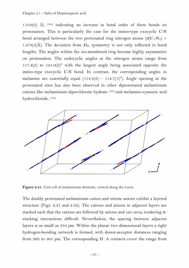
Chapter 2.1 – Salts of Diaminopicric acid
- 97 -
1.319(3) Å) (162) indicating an increase in bond order of these bonds on
protonation. This is particularly the case for the imino-type exocyclic C-N
bond arranged between the two protonated ring nitrogen atoms (d(C1-N4) =
1.278(3)Å). The deviation from D3h symmetry is not only reflected in bond
lengths. The angles within the six-membered ring become highly asymmetric
on protonation. The endocyclic angles at the nitrogen atoms range from
117.4(2) to 121.0(2)° with the largest angle being associated opposite the
imino-type exocyclic C-N bond. In contrast, the corresponding angles in
melamine are essentially equal (114.3(2) – 114.7(1)°). Angle opening at the
protonated sites has also been observed in other diprotonated melaminium
cations like melaminium diperchlorate hydrate (163) and melamine-cyanuric acid
hydrochloride. (164)
Figure 2.31. Unit cell of melaminium dinitrate, viewed along the b axis.
The doubly protonated melaminium cation and nitrate anions exhibit a layered
structure (Figs. 2.31 and 2.32). The cations and anions in adjacent layers are
stacked such that the cations are followed by anions and vice versa, rendering π-
stacking interactions difficult. Nevertheless, the spacing between adjacent
layers is as small as 334 pm. Within the planar two dimensional layers a tight
hydrogen-bonding network is formed, with donor-acceptor distances ranging
from 269 to 305 pm. The corresponding H…A contacts cover the range from
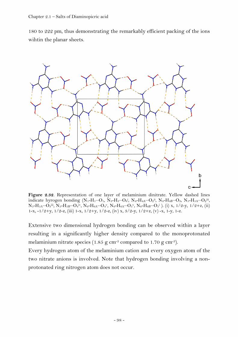
Chapter 2.1 – Salts of Diaminopicric acid
- 98 -
180 to 222 pm, thus demonstrating the remarkably efficient packing of the ions
wihtin the planar sheets.
Figure 2.32. Representation of one layer of melaminium dinitrate. Yellow dashed lines indicate hyrogen bonding (N1-H1···O1, N3-H3···O6i, N4-H4A···O2ii, N4-H4B···O3, N5-H5A···O2iii, N5-H5A···O3iii, N5-H5B···O4iv, N6-H6A···O4v, N6-H6A···O5v, N6-H6B···O5i ). (i) x, 1/2-y, 1/2+z, (ii) 1-x, -1/2+y, 1/2-z, (iii) 1-x, 1/2+y, 1/2-z, (iv) x, 3/2-y, 1/2+z, (v) -x, 1-y, 1-z.
Extensive two dimensional hydrogen bonding can be observed within a layer
resulting in a significantly higher density compared to the monoprotonated
melaminium nitrate species (1.85 g cm-3 compared to 1.70 g cm-3).
Every hydrogen atom of the melaminium cation and every oxygen atom of the
two nitrate anions is involved. Note that hydrogen bonding involving a non-
protonated ring nitrogen atom does not occur.

Chapter 2.1 – Salts of Diaminopicric acid
- 99 -
Experimental
Nitric acid (7 mL, 0.1 mol, 65 %) was added drop wise to a hot aqueous solution
(95 °C) of melamine (0.226 mg, 1 mmol in 5 mL H2O). Subsequent cooling of
the reaction mixture to room temperature afforded precipitation of colorless
crystalline product in pure form containing single crystals suitable for X-ray
diffraction analysis.
DSC (Linseis, 2 K min-1): 330°C (decomp.); Radex (isothermal long term
stability, 48h, 140°C): unchanged; 1H NMR ([D4]methanol) δ: 9.8 (s, 2H), 7.7
(s, 6H); 13C NMR ([D4]methanol) δ: 159.7; 14N NMR ([D4]methanol) δ: -11.6;
IR (KBr) v~ /cm-1: 3425(s), 3294(m), 3130(s), 1683(s), 1644(m), 1618(m),
1574(w),1520(m), 1384(s), 1190(m), 1149(w), 1116(w), 1007(w), 981(w),
816(w), 769(w), 724(w), 696(w), 660(w), 584(w), 575(w); Raman (4 cm-1) v~ /cm-
1: 1052(100), 690(69), 556(21), 391(9), 372(9), 141(20); Calc. for C3H8N8O6: N
(44.14%), C (14.29%), H (3.20%), found: N (44.16%), C (14.64%), H (3.24%);
impact sensitivity: > 30 J, friction sensitivity: > 160 N; Details of the single
crystal X-ray diffraction experiment are listed in the appendix (Chapter 4).
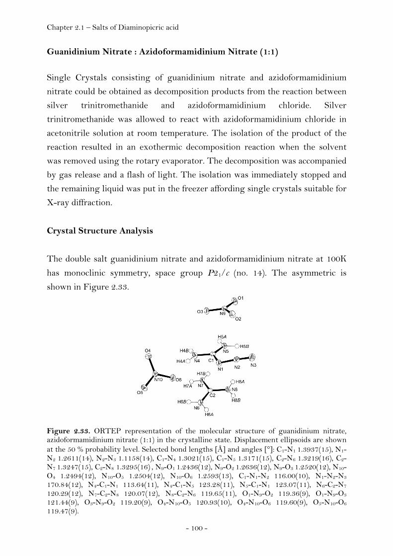
Chapter 2.1 – Salts of Diaminopicric acid
- 100 -
Guanidinium Nitrate : Azidoformamidinium Nitrate (1:1)
Single Crystals consisting of guanidinium nitrate and azidoformamidinium
nitrate could be obtained as decomposition products from the reaction between
silver trinitromethanide and azidoformamidinium chloride. Silver
trinitromethanide was allowed to react with azidoformamidinium chloride in
acetonitrile solution at room temperature. The isolation of the product of the
reaction resulted in an exothermic decomposition reaction when the solvent
was removed using the rotary evaporator. The decomposition was accompanied
by gas release and a flash of light. The isolation was immediately stopped and
the remaining liquid was put in the freezer affording single crystals suitable for
X-ray diffraction.
Crystal Structure Analysis
The double salt guanidinium nitrate and azidoformamidinium nitrate at 100K
has monoclinic symmetry, space group P21/c (no. 14). The asymmetric is
shown in Figure 2.33.
Figure 2.33. ORTEP representation of the molecular structure of guanidinium nitrate, azidoformamidinium nitrate (1:1) in the crystalline state. Displacement ellipsoids are shown at the 50 % probability level. Selected bond lengths [Å] and angles [°]: C1-N1 1.3937(15), N1-N2 1.2611(14), N2-N3 1.1158(14), C1-N4 1.3021(15), C1-N5 1.3171(15), C2-N6 1.3219(16), C2-N7 1.3247(15), C2-N8 1.3295(16) , N9-O1 1.2436(12), N9-O2 1.2636(12), N9-O3 1.2520(12), N10-O4 1.2494(12), N10-O5 1.2504(12), N10-O6 1.2593(13), C1-N1-N2 116.00(10), N1-N2-N3
170.84(12), N4-C1-N1 113.64(11), N4-C1-N5 123.28(11), N5-C1-N1 123.07(11), N6-C2-N7 120.29(12), N7-C2-N8 120.07(12), N8-C2-N6 119.65(11), O1-N9-O2 119.36(9), O1-N9-O3
121.44(9), O3-N9-O2 119.20(9), O4-N10-O5 120.93(10), O4-N10-O6 119.60(9), O5-N10-O6
119.47(9).

Chapter 2.1 – Salts of Diaminopicric acid
- 101 -
Each of the guanidinium-type cations of the double salt guanidinium nitrate -
azidoformamidinium nitrate (1:1) contains one carbon atom carrying three
nitrogen containing substituents (guanidinium cation (II) /
azidoformamidinium cation (III), Fig. 2.34). Further single crystal X-ray
structural data within this are only available for the triazidocarbonium cation
(IV, 2.34) (165) and the free base guanidin (I, 2.34) whose structure could be
determined for the first time within the scope of this thesis (see page 213). A
summary of bond lengths of each of the four types of is provided in Table 2.5.
Figure 2.34. Types of guanidine-based motivs where single crystal structural data are available.
Table 2.5. Comparison of selected bond lengths of guanidine (I), guanidinium (II), azidoformamidinium (III) and triazidocarbenium (IV). d(C-NH) /Å d(C-NH2) /Å d(C-N3) /Å d(Nα-Nβ) /Å d(Nβ-Nα) /Å
(I) a) 1.295(2) (C1-N1) 1.366(2) (C1-N2) - - -
- 1.355(2) (C1-N3) - - -
(I) b) 1.304(2) (C1-N1) 1.350(3) (C1-N2) - - -
- 1.359(3) (C1-N3) - - -
1.300(3) (C7-N8) 1.361(3) (C7-N9) - - -
1.361(3) (C7-N10) - - -
(II) c) - 1.3219(16) (C2-N6) - - -
- 1.3247(15) (C2-N7) - - -
- 1.3295(16) (C2-N8) - - -
(III) d) - 1.3021(15) (C1-N4) 1.3937(15) (C1-N1) 1.2611(14) (N1-N2) 1.1158(14) (N2-N3)
- 1.3171(15) (C1-N5) - - -
(III) e) - 1.302(4) (C1-N1) 1.393(4) (C1-N3) 1.265(4) (N3-N4) 1.110(4) (N4-N5)
- 1.314(4) (C1-N2) - - -
(IV) f) - - 1.312(18) (C1-N1) 1.411(19) (N1-N2) 1.022(17) (N2-N3)
- - 1.358(18) (C1-N4) 1.361(19) (N4-N5) 1.069(16) (N5-N6)
- - 1.348(18) (C1-N7) 1.398(18) (N7-N8) 1.062(17) (N8-N9)
Notes. a) co-crystal of guanidine and 2-amino-4,6-dimethyl-1,3,5-triazine (1:1), b) co-crystal of guanidine and 2-amino-4,6-dimethyl-1,3,5-triazine (2:1), reference (166); c) guanidinium nitrate, this work, double salt; d) azidoformamidinium nitrate, this work, double salt; e) azidoformamidinium chloride, reference (167) f) triazidocarbonium hexachloroantimonate, reference (165).

Chapter 2.1 – Salts of Diaminopicric acid
- 102 -
The C-N bond lengths of the free base guanidine (I) are different and indicate
substantial double bond character of the imino-type C-N bond (range:
1.295(2)Å – 1.304(2)Å) and single bond character of the two remaining amino-
type C-N bonds (range: 1.350(3)Å – 1.366(2)Å). In contrast, the C-N bond
length of the guanidinium cation (II) in the double salt are all of comparable
size (range: 1.3219(16)Å – 1.3295(16)Å) indicating Y-aromaticity (168) with
delocalisation of π electron density affording a formal bond order of 1.33 of
each C-N bond and agree well to typical bond lengths of guanidinium cations.
It has been mentioned that the azide groups of triazidocarbonium (IV) display
unusual bond lengths compared to other covalently bonded azides. Müller and
Bärnighausen (165) have reported the structure of triazidocarbonium-
hexachloroantimonate and assigned single bond character to the Nα-Nβ
nitrogen bonds (range: 1.361(19)Å – 1.411(19)Å) and triple bond character to
the Nβ-Nγ bonds (range: 1.022(17)Å – 1.069(16)Å), in marked contrast to the
values observed in other covalently bonded azides like methyl azide (d(Nα-Nβ) = 1.24Å corresponding to double bond character) (169). They also concluded
that delocalisation of π electron density towards the electrophilic carbon atom
was evenly distributed across the three C-N bonds in the guanidinium cation
and the triazidocarbonium cation due to the fact that the corresponding bonds
in each molecule were of comparable size and significantly shorter compared to
a formal C-N single bond (1.47Å). Though the CN3 moiety of the
azidoformamidinium cation has the same planar geometry as the CN3 moieties
of the guanidinium and the triazidocarbonium cations, the C-N bond lenghts
differ. The bond between the carbon atom and the Nα atom of the azide group
(1.3937(15)Å) is significantly longer compared to the bond lengths of the
carbon atom to the nitrogen atoms of the NH2 groups (1.3021(15) Å,
1.3171(15)Å) indicating only weak interaction between the π systems of the
guanyl-moiety, (H2N)2-C, and the π systems of the azide group. Compared to
the unusual bond length of the azide group observed in the triazidocarbonium
cation, the bond lengths of the azide group of the azidoformamidinium cation
have typical values compared to other covalent azides like methyl azide. These
geometrical data of the azidoformamidinium cation present in the double salt
agree well to those reported for azidoformamidinium chloride. (170) The

Chapter 2.1 – Salts of Diaminopicric acid
- 103 -
extended structure of the double salt of guanidinium nitrate and
azidoformamidinium nitrate is shown in Figure 2.35.
Figure 2.35. Unit cell of guanidinium nitrate, azidoformamidinium nitrate (1:1), viewed along the b axis.
The structure is mainly governed by a complex hydrogen bond network. Every
hydrogen atom of both the guanidinium and the azidoformamidinium cations is
involved in intermolecular N−H⋅⋅⋅O hydrogen bonding between those cations
and the nitrate anions (Table 2.6). Of interest, the azide group is not involved
in N−H⋅⋅⋅N hydrogen bonding.

Chapter 2.1 – Salts of Diaminopicric acid
- 104 -
Table 2.6. Intermolecular hydrogen bonding of the double salt guanidinium nitrate and azidoformamidinium nitrate.
D−H⋅⋅⋅A D−H / Å H⋅⋅⋅ A / Å D⋅⋅⋅A / Å D−H⋅⋅⋅A / °
N4−H4A⋅⋅⋅O6 0.852(15) 1.960(15) 2.8081(15) 173.8(13)
N4−H4B⋅⋅⋅O1(i) 0.834(14) 2.072(14) 2.9057(14) 179.8(19)
N5−H5A⋅⋅⋅O3(i) 0.854(13) 2.097(13) 2.9504(14) 176.8(11)
N5−H5B⋅⋅⋅O2 0.927(15) 1.976(15) 2.8822(14) 165.4(13)
N6−H6A⋅⋅⋅O6(ii) 0.869(15) 2.039(15) 2.8728(14) 160.3(13)
N6−H6B⋅⋅⋅O4(iii) 0.868(16) 2.055(15) 2.8995(14) 164.2(13)
N7−H7A⋅⋅⋅O4(iv) 0.862(16) 2.412(15) 3.1614(14) 145.7(13)
N7−H7A⋅⋅⋅O5(iii) 0.862(16) 2.505(15) 3.1378(14) 131.0(12)
N7−H7B⋅⋅⋅O3(v) 0.824(14) 2.242(14) 3.0518(15) 167.6(13)
N8−H8A⋅⋅⋅O2(v) 0.808(14) 2.220(14) 3.0033(15) 163.3(13)
N8−H8B⋅⋅⋅O5(ii) 0.865(16) 2.154(15) 3.0113(14) 171.3(13)
Notes. Symmetry code: (i) x,1/2-y,-1/2+z, (ii) 1-x,-1/2+y,1/2-z, (iii) 1-x,1-y,-z, (iv) x,-1+y,z, (v) -x,-1/2+y,1/2-z.
Experimental
Single Crystals consisting of guanidinium nitrate and azidoformamidinium
nitrate could be obtained as decomposition products from the reaction between
silver trinitromethanide and azidoformamidinium chloride. Silver
trinitromethanide was allowed to react with azidoformamidinium chloride in
acetonitrile solution at room temperature. The isolation of the product of the
reaction resulted in an exothermic decomposition reaction when the solvent
was removed using the rotary evaporator. The decomposition was accompanied
by gas release and a flash of light. The isolation was immediately stopped and
the remaining liquid was put in the freezer affording single crystals suitable for
X-ray diffraction.
Details of the single crystal X-ray diffraction experiment are listed in the
appendix (Chapter 4).

Chapter 2.1 – Salts of Diaminopicric acid
- 105 -
2.1.3 Salts of Diaminopicric Acid
Introduction
Bellamy et al. have recently investigated a variety of salts of 3,5-diaminopicric
acid. (171) Amongst the compounds the series of guanidinium salts showed
particularly promising thermal stability and sensitivity data. The compounds
were characterized using multinuclear NMR spectroscopy, IR spectroscopy,
differential scanning calorimetry, thermogravimetry, scanning electron
microscopy and CHN elemental analysis. Their heats of combustion were
measured using bomb calorimetry in order to calculate their heats of formation.
We have now determined the structures of guanidinium 3,5-diaminopicrate
(GDAP), aminoguanidinium 3,5-diaminopicrate (AGDAP),
diaminoguanidinium 3,5-diaminopicrate (DAGDAP) and triaminoguanidinium
3,5-diaminopicrate (TAGDAP) in the solid state using single crystal X-ray
diffraction as a preliminary step in an investigation of the relationships
between crystal density and explosive performance parameters. Sensitivity and Performance
The detonation rate and detonation pressure values of GDAP, AGDAP,
DAGDAP and TAGDAP have been reported according to the Rothstein and
Petersen method. Based on the calculated densities from our crystal structure
analysis and using the programme EXPLO5 (v. 5.02), we carried out
performance calculations in order to predict the performance properties of of
GDAP, AGDAP, DAGDAP and TAGDAP. A comparison of the results
obtained using the two different methods is summarized in Table 2.7. A
comparison of the calculated values of TNT with the corresponding
experimental values shows that the calculated values obtained using this
method are in good agreement with the experimental analogues.
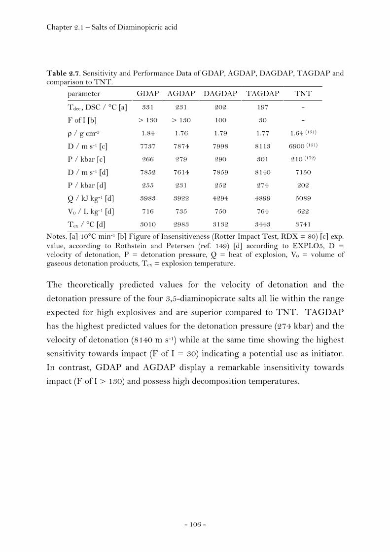
Chapter 2.1 – Salts of Diaminopicric acid
- 106 -
Table 2.7. Sensitivity and Performance Data of GDAP, AGDAP, DAGDAP, TAGDAP and comparison to TNT.
parameter GDAP AGDAP DAGDAP TAGDAP TNT
Tdec., DSC / °C [a] 331 231 202 197 -
F of I [b] > 130 > 130 100 30 -
ρ / g cm-3 1.84 1.76 1.79 1.77 1.64 (151)
D / m s-1 [c] 7737 7874 7998 8113 6900 (151)
P / kbar [c] 266 279 290 301 210 (172)
D / m s-1 [d] 7852 7614 7859 8140 7150
P / kbar [d] 255 231 252 274 202
Q / kJ kg-1 [d] 3983 3922 4294 4899 5089
V0 / L kg-1 [d] 716 735 750 764 622
Tex / °C [d] 3010 2983 3132 3443 3741
Notes. [a] 10°C min-1 [b] Figure of Insensitiveness (Rotter Impact Test, RDX = 80) [c] exp. value, according to Rothstein and Petersen (ref. 149) [d] according to EXPLO5, D = velocity of detonation, P = detonation pressure, Q = heat of explosion, V0 = volume of gaseous detonation products, Tex = explosion temperature.
The theoretically predicted values for the velocity of detonation and the
detonation pressure of the four 3,5-diaminopicrate salts all lie within the range
expected for high explosives and are superior compared to TNT. TAGDAP
has the highest predicted values for the detonation pressure (274 kbar) and the
velocity of detonation (8140 m s-1) while at the same time showing the highest
sensitivity towards impact (F of I = 30) indicating a potential use as initiator.
In contrast, GDAP and AGDAP display a remarkable insensitivity towards
impact (F of I > 130) and possess high decomposition temperatures.
Chapter 2.1 – Salts of Diaminopicric acid
- 107 -
Crystal Structure Analysis
Though the structure of picric acid and many structural reports containing
salts thereof have been well documented, there are no structural reports
containing the 3,5-diaminopicrate anion. In fact, only the structure of the
parent compound, 3,5-diamino-2,4,6-trinitrophenol has been reported (173)
rendering the four structures of guanidinium 3,5-diaminopicrate (GDAP),
aminoguanidinium 3,5-diaminopicrate (AGDAP), diaminoguanidinium 3,5-
diaminopicrate (DAGDAP) as well as triaminoguanidinium 3,5-diaminopicrate
(TAGDAP) to be first examples of a crystallographic characterization of the
3,5-diaminopicrate anion.
Before the structural features of the 3,5-diaminopicrate anion will be discussed,
a brief summary of general features that have been observed and explored by
various authors is given. (174) Compared to benzene, the benzene ring of various
nitroanilines exhibits characteristic distortions due to substitution of a
hydrogen atom by both nitro- and amino-groups. The angle having C-NH2 as
vertex is smaller than 120°, whereas the corresponding angles with C-NO2 are
greater than 120°. There seems to be a correlation between the C-N bond
distances and the values of the adjacent C-C bond lengths of the phenyl ring in
aromatic amines and various interpretations of the ring deformation have been
made (175-177) based on (1) intramolecular nonbonded interactions; (178-180) (2)
coulombic interaction between formally charged atoms; (175) (3) hybridization
effects at the carbon atom to which the substituent is bonded; (181-182) and (4)
valence-shell electron-pair repulsion. (183-188) Accordingly, the overall
deformation of the phenyl ring due to the presence of an electron-attracting
(nitro) substitutent consists of a shortening of the adjacent C-C bonds, an
increase in the endocyclic bond angle opposite to the functional group and a
minor decrease in the two adjacent endocyclic angles. The reverse is true for an
electron-donating (amino) substituent. The presence of ortho and meta
derivatives seems not to be rationalizable due to a multiplication of the various
effects.
A comparison of selected structural parameters of the 3,5-diaminopicrate anion
in the structures of the series of guanidinium 3,5-diaminopicrates can be found
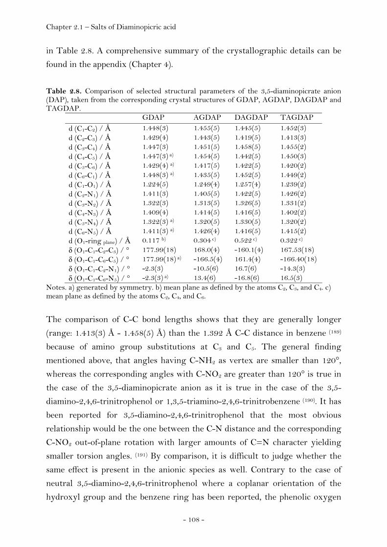
Chapter 2.1 – Salts of Diaminopicric acid
- 108 -
in Table 2.8. A comprehensive summary of the crystallographic details can be
found in the appendix (Chapter 4).
Table 2.8. Comparison of selected structural parameters of the 3,5-diaminopicrate anion (DAP), taken from the corresponding crystal structures of GDAP, AGDAP, DAGDAP and TAGDAP.
GDAP AGDAP DAGDAP TAGDAP d (C1-C2) / Å 1.448(3) 1.455(5) 1.445(5) 1.452(3) d (C2-C3) / Å 1.429(4) 1.443(5) 1.419(5) 1.413(3) d (C3-C4) / Å 1.447(3) 1.451(5) 1.458(5) 1.455(2) d (C4-C5) / Å 1.447(3) a) 1.454(5) 1.442(5) 1.450(3) d (C5-C6) / Å 1.429(4) a) 1.417(5) 1.422(5) 1.420(2) d (C6-C1) / Å 1.448(3) a) 1.435(5) 1.452(5) 1.449(2) d (C1-O1) / Å 1.224(5) 1.249(4) 1.257(4) 1.239(2) d (C2-N1) / Å 1.411(3) 1.405(5) 1.422(5) 1.426(2) d (C3-N2) / Å 1.322(3) 1.313(5) 1.326(5) 1.331(2) d (C4-N3) / Å 1.409(4) 1.414(5) 1.416(5) 1.402(2) d (C5-N4) / Å 1.322(3) a) 1.320(5) 1.330(5) 1.320(2) d (C6-N5) / Å 1.411(3) a) 1.426(4) 1.416(5) 1.415(2) d (O1-ring plane) / Å 0.117 b) 0.304 c) 0.522 c) 0.322 c) δ (O1-C1-C2-C3) / ° 177.99(18) 168.0(4) -160.1(4) 167.53(18) δ (O1-C1-C6-C5) / ° 177.99(18) a) -166.5(4) 161.4(4) -166.40(18) δ (O1-C1-C2-N1) / ° -2.3(3) -10.5(6) 16.7(6) -14.3(3) δ (O1-C1-C6-N5) / ° -2.3(3) a) 13.4(6) -16.8(6) 16.5(3)
Notes. a) generated by symmetry. b) mean plane as defined by the atoms C2, C3, and C4. c) mean plane as defined by the atoms C2, C4, and C6.
The comparison of C-C bond lengths shows that they are generally longer
(range: 1.413(3) Å - 1.458(5) Å) than the 1.392 Å C-C distance in benzene (189)
because of amino group substitutions at C3 and C5. The general finding
mentioned above, that angles having C-NH2 as vertex are smaller than 120°,
whereas the corresponding angles with C-NO2 are greater than 120° is true in
the case of the 3,5-diaminopicrate anion as it is true in the case of the 3,5-
diamino-2,4,6-trinitrophenol or 1,3,5-triamino-2,4,6-trinitrobenzene (190). It has
been reported for 3,5-diamino-2,4,6-trinitrophenol that the most obvious
relationship would be the one between the C-N distance and the corresponding
C-NO2 out-of-plane rotation with larger amounts of C=N character yielding
smaller torsion angles. (191) By comparison, it is difficult to judge whether the
same effect is present in the anionic species as well. Contrary to the case of
neutral 3,5-diamino-2,4,6-trinitrophenol where a coplanar orientation of the
hydroxyl group and the benzene ring has been reported, the phenolic oxygen
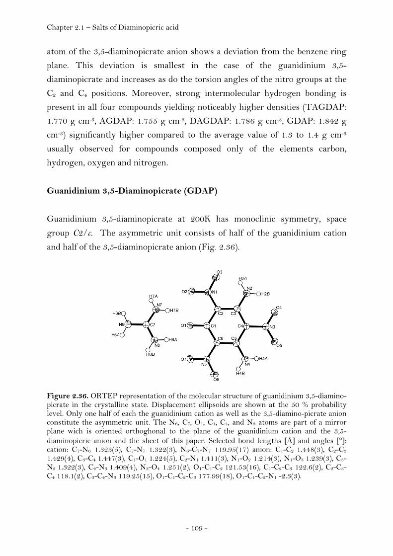
Chapter 2.1 – Salts of Diaminopicric acid
- 109 -
atom of the 3,5-diaminopicrate anion shows a deviation from the benzene ring
plane. This deviation is smallest in the case of the guanidinium 3,5-
diaminopicrate and increases as do the torsion angles of the nitro groups at the
C2 and C4 positions. Moreover, strong intermolecular hydrogen bonding is
present in all four compounds yielding noticeably higher densities (TAGDAP:
1.770 g cm-3, AGDAP: 1.755 g cm-3, DAGDAP: 1.786 g cm-3, GDAP: 1.842 g
cm-3) significantly higher compared to the average value of 1.3 to 1.4 g cm-3
usually observed for compounds composed only of the elements carbon,
hydrogen, oxygen and nitrogen.
Guanidinium 3,5-Diaminopicrate (GDAP)
Guanidinium 3,5-diaminopicrate at 200K has monoclinic symmetry, space
group C2/c. The asymmetric unit consists of half of the guanidinium cation
and half of the 3,5-diaminopicrate anion (Fig. 2.36).
Figure 2.36. ORTEP representation of the molecular structure of guanidinium 3,5-diamino-picrate in the crystalline state. Displacement ellipsoids are shown at the 50 % probability level. Only one half of each the guanidinium cation as well as the 3,5-diamino-picrate anion constitute the asymmetric unit. The N6, C7, O1, C1, C4, and N3 atoms are part of a mirror plane wich is oriented orthoghonal to the plane of the guanidinium cation and the 3,5-diaminopicric anion and the sheet of this paper. Selected bond lengths [Å] and angles [°]: cation: C7-N6 1.323(5), C7-N7 1.322(3), N6-C7-N7 119.95(17) anion: C1-C2 1.448(3), C2-C3 1.429(4), C3-C4 1.447(3), C1-O1 1.224(5), C2-N1 1.411(3), N1-O2 1.214(3), N1-O3 1.239(3), C3-N2 1.322(3), C4-N3 1.409(4), N3-O4 1.251(2), O1-C1-C2 121.53(16), C1-C2-C3 122.6(2), C2-C3-C4 118.1(2), C3-C4-N3 119.25(15), O1-C1-C2-C3 177.99(18), O1-C1-C2-N1 -2.3(3).
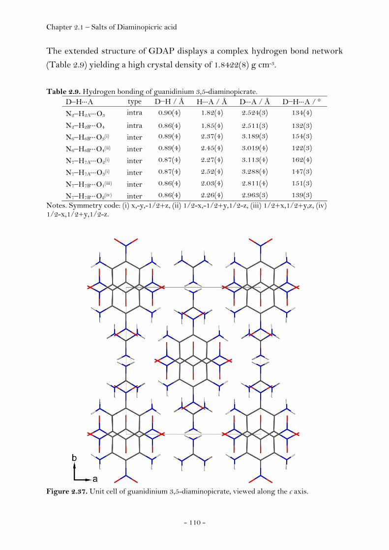
Chapter 2.1 – Salts of Diaminopicric acid
- 110 -
The extended structure of GDAP displays a complex hydrogen bond network
(Table 2.9) yielding a high crystal density of 1.8422(8) g cm-3.
Table 2.9. Hydrogen bonding of guanidinium 3,5-diaminopicrate.
D−H⋅⋅⋅A type D−H / Å H⋅⋅⋅A / Å D⋅⋅⋅A / Å D−H⋅⋅⋅A / °
N2−H2A⋅⋅⋅O3 intra 0.90(4) 1.82(4) 2.524(3) 134(4)
N2−H2B⋅⋅⋅O4 intra 0.86(4) 1.85(4) 2.511(3) 132(3)
N6−H6B⋅⋅⋅O3(i) inter 0.89(4) 2.37(4) 3.189(3) 154(3)
N6−H6B⋅⋅⋅O4(ii) inter 0.89(4) 2.45(4) 3.019(4) 122(3)
N7−H7A⋅⋅⋅O2(i) inter 0.87(4) 2.27(4) 3.113(4) 162(4)
N7−H7A⋅⋅⋅O3(i) inter 0.87(4) 2.52(4) 3.288(4) 147(3)
N7−H7B⋅⋅⋅O1(iii) inter 0.86(4) 2.03(4) 2.811(4) 151(3)
N7−H7B⋅⋅⋅O2(iv) inter 0.86(4) 2.26(4) 2.963(3) 139(3) Notes. Symmetry code: (i) x,-y,-1/2+z, (ii) 1/2-x,-1/2+y,1/2-z, (iii) 1/2+x,1/2+y,z, (iv) 1/2-x,1/2+y,1/2-z.
Figure 2.37. Unit cell of guanidinium 3,5-diaminopicrate, viewed along the c axis.
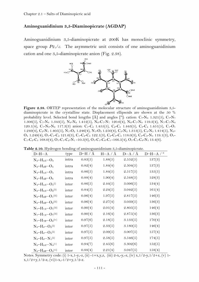
Chapter 2.1 – Salts of Diaminopicric acid
- 111 -
Aminoguanidinium 3,5-Diaminopicrate (AGDAP)
Aminoguanidinium 3,5-diaminopicrate at 200K has monoclinic symmetry,
space group P21/c. The asymmetric unit consists of one aminoguanidinium
cation and one 3,5-diaminopicrate anion (Fig. 2.38).
Figure 2.38. ORTEP representation of the molecular structure of aminoguanidinium 3,5-diaminopicrate in the crystalline state. Displacement ellipsoids are shown at the 50 % probability level. Selected bond lengths [Å] and angles [°]: cation: C7-N6 1.321(5), C7-N7 1.306(5), C7-N8 1.343(5), N8-N9 1.412(5), N6-C7-N7 120.6(4), N6-C7-N8 119.3(4), N7-C7-N8 120.1(4), C7-N8-N9 117.3(3) anion: C1-C2 1.455(5), C2-C3 1.443(5), C3-C4 1.451(5), C1-O1
1.249(4), C2-N1 1.405(5), N1-O2 1.246(4), N1-O3 1.250(4), C3-N2 1.313(5), C4-N3 1.414(5), N3-O4 1.249(4), O1-C1-C2 121.6(3), C1-C2-C3 122.1(3), C2-C3-C4 118.0(3), C3-C4-N3 119.1(3), O1-C1-C2-C3 168.0(4), O1-C1-C2-N1 -10.5(6), O1-C1-C6-C5 -166.5(4), O1-C1-C6-N5 13.4(6). Table 2.10. Hydrogen bonding of aminoguanidinium 3,5-diaminopicrate.
D−H⋅⋅⋅A type D−H / Å H⋅⋅⋅A / Å D⋅⋅⋅A / Å D−H⋅⋅⋅A / °
N2−H2A⋅⋅⋅O3 intra 0.83(5) 1.88(5) 2.552(5) 137(3)
N2−H2B⋅⋅⋅O4 intra 0.82(4) 1.84(4) 2.504(5) 137(3)
N4−H4A⋅⋅⋅O5 intra 0.86(5) 1.84(5) 2.517(5) 135(5)
N4−H4B⋅⋅⋅O6 intra 0.88(4) 1.90(4) 2.548(5) 128(3)
N4−H4A⋅⋅⋅O5(i) inter 0.86(5) 2.44(5) 3.096(5) 134(4)
N6−H6A⋅⋅⋅O2(ii) inter 0.84(5) 2.24(5) 3.042(5) 161(4)
N6−H6B⋅⋅⋅O1(iii) inter 0.96(4) 1.97(5) 2.817(5) 146(3)
N6−H6B⋅⋅⋅O2(iii) inter 0.96(4) 2.27(4) 3.039(5) 136(3)
N7−H7A⋅⋅⋅O1(iii) inter 0.90(4) 2.01(4) 2.805(5) 146(4)
N7−H7A⋅⋅⋅O7(iii) inter 0.90(4) 2.16(4) 2.875(4) 136(3)
N7−H7B⋅⋅⋅O6(iv) inter 0.97(6) 2.18(5) 3.133(5) 170(4)
N8−H8⋅⋅⋅O2(ii) inter 0.97(5) 2.33(5) 3.180(5) 146(4)
N8−H8⋅⋅⋅O3(ii) inter 0.97(5) 2.09(5) 3.007(5) 157(4)
N8−H8⋅⋅⋅N1(ii) inter 0.97(5) 2.58(5) 3.546(5) 174(5)
N9−H9A⋅⋅⋅N8(v) inter 0.94(7) 2.45(6) 3.302(6) 152(5)
N9−H9B⋅⋅⋅O4(vi) inter 0.88(4) 2.21(4) 3.047(5) 158(4) Notes. Symmetry code: (i) 1-x,1-y,-z, (ii) -1+x,y,z, (iii) 2-x,-y,-z, (iv) x,1/2-y,1/2+z, (v) 1-x,1/2+y,1/2-z, (vi)1-x,-1/2+y,1/2-z.
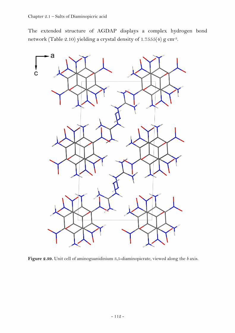
Chapter 2.1 – Salts of Diaminopicric acid
- 112 -
The extended structure of AGDAP displays a complex hydrogen bond
network (Table 2.10) yielding a crystal density of 1.7555(4) g cm-3.
Figure 2.39. Unit cell of aminoguanidinium 3,5-diaminopicrate, viewed along the b axis.
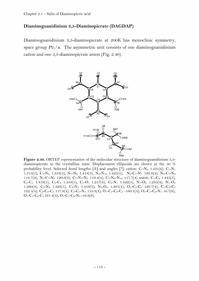
Chapter 2.1 – Salts of Diaminopicric acid
- 113 -
Diaminoguanidinium 3,5-Diaminopicrate (DAGDAP)
Diaminoguanidinium 3,5-diaminopicrate at 200K has monoclinic symmetry,
space group P21/n. The asymmetric unit consists of one diaminoguanidinium
cation and one 3,5-diaminopicrate anion (Fig. 2.40).
Figure 2.40. ORTEP representation of the molecular structure of diaminoguanidinium 3,5-diaminopicrate in the crystalline state. Displacement ellipsoids are shown at the 50 % probability level. Selected bond lengths [Å] and angles [°]: cation: C7-N6 1.331(6), C7-N7 1.313(5), C7-N9 1.333(5), N7-N8 1.418(5), N9-N10 1.425(5), N6-C7-N7 120.8(4), N6-C7-N9 118.7(4), N7-C7-N9 120.6(4), C7-N7-N8 119.4(4), C7-N9-N10 117.7(4) anion: C1-C2 1.445(5), C2-C3 1.419(5), C3-C4 1.458(5), C1-O1 1.257(4), C2-N1 1.422(5), N1-O2 1.245(4), N1-O3
1.260(4), C3-N2 1.326(5), C4-N3 1.416(5), N3-O4 1.261(4), O1-C1-C2 120.7(4), C1-C2-C3 122.5(4), C2-C3-C4 117.6(4), C3-C4-N3 119.3(4), O1-C1-C2-C3 -160.1(4), O1-C1-C2-N1 16.7(6), O1-C1-C6-C5 161.4(4), O1-C1-C6-N5 -16.8(6).
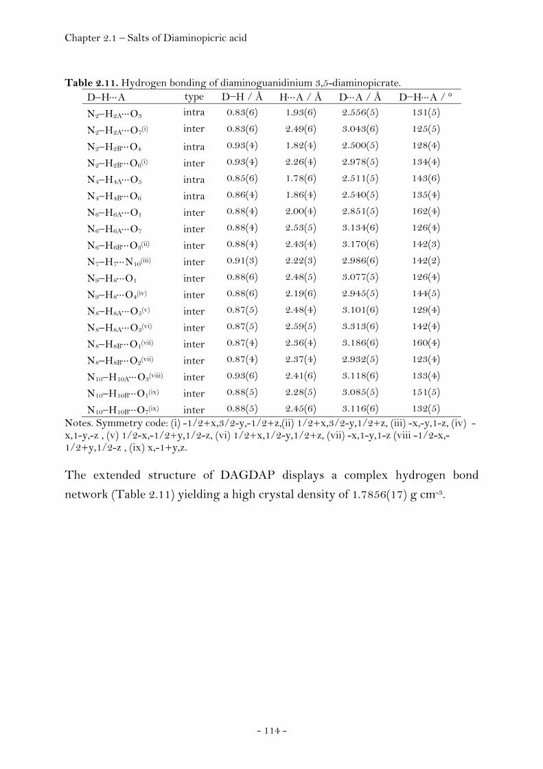
Chapter 2.1 – Salts of Diaminopicric acid
- 114 -
Table 2.11. Hydrogen bonding of diaminoguanidinium 3,5-diaminopicrate.
D−H⋅⋅⋅A type D−H / Å H⋅⋅⋅A / Å D⋅⋅⋅A / Å D−H⋅⋅⋅A / °
N2−H2A⋅⋅⋅O3 intra 0.83(6) 1.93(6) 2.556(5) 131(5)
N2−H2A⋅⋅⋅O7(i) inter 0.83(6) 2.49(6) 3.043(6) 125(5)
N2−H2B⋅⋅⋅O4 intra 0.93(4) 1.82(4) 2.500(5) 128(4)
N2−H2B⋅⋅⋅O6(i) inter 0.93(4) 2.26(4) 2.978(5) 134(4)
N4−H4A⋅⋅⋅O5 intra 0.85(6) 1.78(6) 2.511(5) 143(6)
N4−H4B⋅⋅⋅O6 intra 0.86(4) 1.86(4) 2.540(5) 135(4)
N6−H6A⋅⋅⋅O1 inter 0.88(4) 2.00(4) 2.851(5) 162(4)
N6−H6A⋅⋅⋅O7 inter 0.88(4) 2.53(5) 3.134(6) 126(4)
N6−H6B⋅⋅⋅O3(ii) inter 0.88(4) 2.43(4) 3.170(6) 142(3)
N7−H7⋅⋅⋅N10(iii) inter 0.91(3) 2.22(3) 2.986(6) 142(2)
N9−H8⋅⋅⋅O1 inter 0.88(6) 2.48(5) 3.077(5) 126(4)
N9−H8⋅⋅⋅O4(iv) inter 0.88(6) 2.19(6) 2.945(5) 144(5)
N8−H8A⋅⋅⋅O5(v) inter 0.87(5) 2.48(4) 3.101(6) 129(4)
N8−H8A⋅⋅⋅O3(vi) inter 0.87(5) 2.59(5) 3.313(6) 142(4)
N8−H8B⋅⋅⋅O1(vii) inter 0.87(4) 2.36(4) 3.186(6) 160(4)
N8−H8B⋅⋅⋅O2(vii) inter 0.87(4) 2.37(4) 2.932(5) 123(4)
N10−H10A⋅⋅⋅O3(viii) inter 0.93(6) 2.41(6) 3.118(6) 133(4)
N10−H10B⋅⋅⋅O1(ix) inter 0.88(5) 2.28(5) 3.085(5) 151(5)
N10−H10B⋅⋅⋅O7(ix) inter 0.88(5) 2.45(6) 3.116(6) 132(5) Notes. Symmetry code: (i) -1/2+x,3/2-y,-1/2+z,(ii) 1/2+x,3/2-y,1/2+z, (iii) -x,-y,1-z, (iv) -x,1-y,-z , (v) 1/2-x,-1/2+y,1/2-z, (vi) 1/2+x,1/2-y,1/2+z, (vii) -x,1-y,1-z (viii -1/2-x,-1/2+y,1/2-z , (ix) x,-1+y,z.
The extended structure of DAGDAP displays a complex hydrogen bond
network (Table 2.11) yielding a high crystal density of 1.7856(17) g cm-3.
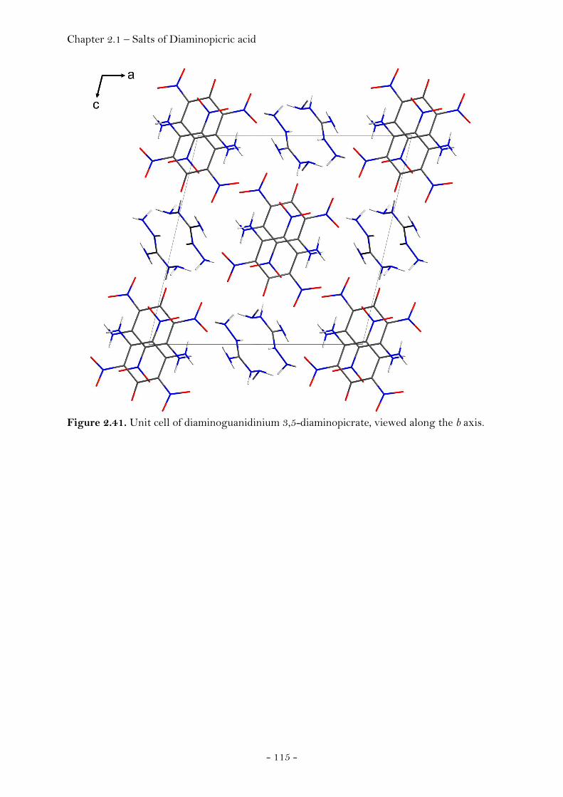
Chapter 2.1 – Salts of Diaminopicric acid
- 115 -
Figure 2.41. Unit cell of diaminoguanidinium 3,5-diaminopicrate, viewed along the b axis.

Chapter 2.1 – Salts of Diaminopicric acid
- 116 -
Triaminoguanidinium 3,5-Diaminopicrate hydrate (TAGDAP · H2O)
Triaminoguanidinium 3,5-diaminopicrate monohydrate at 100K has monoclinic
symmetry, space group P21/n. The asymmetric unit consists of one
diaminoguanidinium cation and one 3,5-diaminopicrate anion (Fig. 2.42).
Figure 2.42. ORTEP representation of the molecular structure of triaminoguanidinium 3,5-diaminopicrate hydrate in the crystalline state. Displacement ellipsoids are shown at the 50 % probability level. Selected bond lengths [Å] and angles [°]: cation: C7-N6 1.331(6), C7-N8 1.313(5), C7-N10 1.333(5), N6-N7 1.418(5), N8-N9 1.425(5), N10-N11, N6-C7-N8 120.8(4), N6-C7-N10 118.7(4), N8-C7-N10 120.6(4), C7-N6-N7 119.4(4), C7-N8-N9 117.7(4), C7-N10-N11 anion: C1-C2 1.452(3), C2-C3 1.413(3), C3-C4 1.455(2), C1-O1 1.239(2), C2-N1 1.426(2), N1-O2 1.2386(19), N1-O3 1.248(2), C3-N2 1.331(2), C4-N3 1.402(2), N3-O4 1.252(2), O1-C1-C2
122.55(16), C1-C2-C3 124.24(16), C2-C3-C4 117.68(16), C3-C4-N3 119.50(16), O1-C1-C2-C3 167.53(18), O1-C1-C2-N1 -14.3(3), O1-C1-C6-C5 -166.40(18), O1-C1-C6-N5 16.5(3).

Chapter 2.1 – Salts of Diaminopicric acid
- 117 -
Table 2.12. Hydrogen bonding of triaminoguanidinium 3,5-diaminopicrate, hydrate. D−H⋅⋅⋅A type D−H / Å H⋅⋅⋅A / Å D⋅⋅⋅A / Å D−H⋅⋅⋅A / °
N2−H2A⋅⋅⋅O3 intra 0.88(2) 1.92(2) 2.609(2) 133.7(19)
N2−H2A⋅⋅⋅O7(i) inter 0.88(2) 2.40(2) 2.984(2) 124.3(18)
N2−H2B⋅⋅⋅O4 intra 0.84(3) 1.84(3) 2.520(2) 137(2)
N2−H2B⋅⋅⋅O6(i) inter 0.84(3) 2.41(2) 2.958(2) 124(2)
N4−H4A⋅⋅⋅O5 intra 0.86(2) 1.82(2) 2.518(2) 138.0(19)
N4−H4A⋅⋅⋅O8(ii) inter 0.86(2) 2.50(2) 3.040(3) 121.5(16)
N4−H4B⋅⋅⋅O6 Intra 0.86(2) 1.89(3) 2.585(2) 137(2)
N4−H4B⋅⋅⋅N11(iii) Inter 0.86(2) 2.53(2) 3.187(3) 134(2)
N6−H6⋅⋅⋅O8(iv) Inter 0.90(2) 2.04(2) 2.858(2) 151(2)
N7−H7B⋅⋅⋅O2(v) Inter 0.89(2) 2.38(2) 2.943(2) 121.2(16)
N7−H7B⋅⋅⋅O3(v) Inter 0.89(2) 2.52(2) 3.305(2) 147.5(18)
N8−H8⋅⋅⋅O5(vi) Inter 0.86(3) 2.21(3) 3.031(2) 160(2)
O8−H8A⋅⋅⋅O1 Intra 0.87(3) 1.95(3) 2.795(2) 163(3)
O8−H8A⋅⋅⋅O7 Intra 0.87(3) 2.41(3) 2.974(2) 123(2)
O8−H8B⋅⋅⋅N7(vii) Inter 0.85(3) 2.57(3) 3.078(2) 119(2)
N9−H9A⋅⋅⋅O1(viii) Inter 0.90(2) 2.33(2) 3.072(2) 140.6(18)
N9−H9B⋅⋅⋅O8 Intra 0.91(3) 2.49(2) 3.098(2) 124.2(19)
N9−H9B⋅⋅⋅O2(viii) Inter 0.91(3) 2.36(3) 3.047(2) 132.1(18)
N10−H10⋅⋅⋅O1 Intra 0.80(2) 2.50(2) 3.045(2) 127(2)
N10−H10⋅⋅⋅O2 Intra 0.80(2) 2.30(2) 2.945(2) 139(2)
N11−H11A⋅⋅⋅O4(ix) Inter 0.90(2) 2.18(2) 3.020(2) 155(2)
N11−H11A⋅⋅⋅O4 Intra 0.90(2) 2.28(2) 3.122(2) 155(2)
Notes. Symmetry code: (i) -1/2+x,3/2-y,-1/2+z, (ii) 1/2-x,1/2+y,1/2-z, (iii) 1/2-x,-1/2+y,1/2-z, (iv) x,1+y,z, (v) 1-x,2-y,-z, (vi) 1+x,y,z, (vii) 3/2-x,-1/2+y,1/2-z, (viii) 1-x,1-y,-z, (ix) -x,2-y,-z.
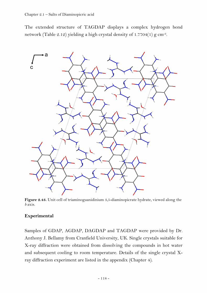
Chapter 2.1 – Salts of Diaminopicric acid
- 118 -
The extended structure of TAGDAP displays a complex hydrogen bond
network (Table 2.12) yielding a high crystal density of 1.7704(1) g cm-3.
Figure 2.43. Unit cell of triaminoguanidinium 3,5-diaminopicrate hydrate, viewed along the b axis.
Experimental
Samples of GDAP, AGDAP, DAGDAP and TAGDAP were provided by Dr.
Anthony J. Bellamy from Cranfield University, UK. Single crystals suitable for
X-ray diffraction were obtained from dissolving the compounds in hot water
and subsequent cooling to room temperature. Details of the single crystal X-
ray diffraction experiment are listed in the appendix (Chapter 4).

Chapter 2.1 – Salts of hydrochloric acid
- 119 -
2.1.4 Salts of Hydrochloric Acid
3,6-Diamino-1,2,4,5-tetrahydro-tetrazinium dichloride
3,6-diamino-1,2,4,5-tetrazine is a starting material for the synthesis of bis-
(2,2,2-trinitroethyl)-3,6-diamino-1,2,4,5-tetrazine (BTAT) and was synthesised
from the condensation of diaminoguanidinium hydrochloride with 2,4-
pentanedione, followed by oxidation of the resulting dihydrotetrazine with
sodium perborate (see section 2.2). (192) Two pathways have been found for the
preparation and isolation of BTAT. According to the first pathway, BTAT can
be obtained on reaction of a solution of 3,6-diaminotetrazine in concentrated
hydrochloric acid (37%) at a temperature of 70°C with stoichiometric amounts
of trinitroethanol. The product precipitates from the solution and can be
obtained in high purity on simple filtration. However, we observed that the
yields of this approach are quite low.
The reason for the low yields can now be rationalized in terms of formation of
the previously unkown compound 3,6-diamino-1,2,4,5-tetrahydro-1,2,4,5-
tetrazinium dichloride (Fig. 2.44).
Fig. 2.44. Synthesis of 3,6-diamino-1,2,4,5-tetrahydro-tetrazinium dichloride from the reaction between 3,6-diamino-1,2,4,5-tetrazine and hydrochloric acid.
The compound becomes reoxidized to the corresponding 3,6-diamino-1,2,4,5-
tetrazine educt on contact with air already at ambient conditions within a few
minutes. However, it was possible to determine the structure of the the
tetrahydrotetrazinium dichloride using single crystal X-ray diffraction.
Attempts to facilitate the formation of BTAT taking advantage of the higher
reactivity of the amino groups present in this salt compared to the less reactive
amino groups present in 3,6-diamino-1,2,4,5-tetrazine were unsuccessful so far
due to the fact that oxidation of the tetrahydrotetrazinium salt on contact with
trinitroethanol is preferred over the formation of BTAT.
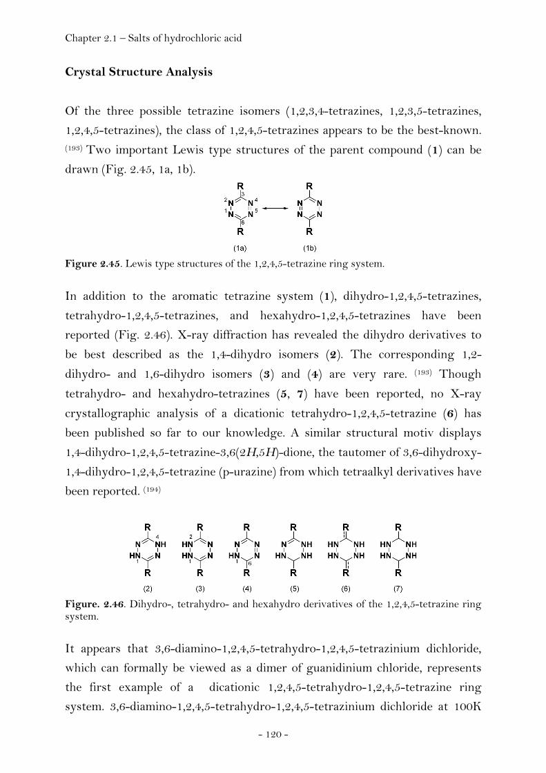
Chapter 2.1 – Salts of hydrochloric acid
- 120 -
Crystal Structure Analysis
Of the three possible tetrazine isomers (1,2,3,4-tetrazines, 1,2,3,5-tetrazines,
1,2,4,5-tetrazines), the class of 1,2,4,5-tetrazines appears to be the best-known. (193) Two important Lewis type structures of the parent compound (1) can be
drawn (Fig. 2.45, 1a, 1b).
Figure 2.45. Lewis type structures of the 1,2,4,5-tetrazine ring system.
In addition to the aromatic tetrazine system (1), dihydro-1,2,4,5-tetrazines,
tetrahydro-1,2,4,5-tetrazines, and hexahydro-1,2,4,5-tetrazines have been
reported (Fig. 2.46). X-ray diffraction has revealed the dihydro derivatives to
be best described as the 1,4-dihydro isomers (2). The corresponding 1,2-
dihydro- and 1,6-dihydro isomers (3) and (4) are very rare. (193) Though
tetrahydro- and hexahydro-tetrazines (5, 7) have been reported, no X-ray
crystallographic analysis of a dicationic tetrahydro-1,2,4,5-tetrazine (6) has
been published so far to our knowledge. A similar structural motiv displays
1,4-dihydro-1,2,4,5-tetrazine-3,6(2H,5H)-dione, the tautomer of 3,6-dihydroxy-
1,4-dihydro-1,2,4,5-tetrazine (p-urazine) from which tetraalkyl derivatives have
been reported. (194)
Figure. 2.46. Dihydro-, tetrahydro- and hexahydro derivatives of the 1,2,4,5-tetrazine ring system.
It appears that 3,6-diamino-1,2,4,5-tetrahydro-1,2,4,5-tetrazinium dichloride,
which can formally be viewed as a dimer of guanidinium chloride, represents
the first example of a dicationic 1,2,4,5-tetrahydro-1,2,4,5-tetrazine ring
system. 3,6-diamino-1,2,4,5-tetrahydro-1,2,4,5-tetrazinium dichloride at 100K

Chapter 2.1 – Salts of hydrochloric acid
- 121 -
has triclinic symmetry, space group P-1 (no.2). The asymmetric unit is shown
in Figure 2.47.
Figure 2.47. ORTEP representation of the molecular structure of 3,6-diamino-1,2,4,5-tetrahydro-1,2,4,5-tetrazinium dichloride in the crystalline state. Displacement ellipsoids are shown at the 50 % probability level. Selected bond lengths [Å] and angles [°]: N1-N2 1.402(2), N2-C2 1.326(2), C2-N3 1.348(2), N3-N4 1.405(2), N4-C1 1.326(2), C1-N1 1.346(2), C1-N5 1.311(3), C2-N6 1.307(3), N4-C1-N5 123.13(18), N5-C1-N1 119.98(17), N4-C1-N1 116.78(17), C1-N1-N2 115.92(15), N1-N2-C2 119.58(15), N2-C2-N6 122.27(17), N6-C2-N3 121.15(17), N2-C2-N3 116.57(17), C2-N3-N4 116.16(15), N3-N4-C1 119.78(16), N5-C1-N4-N3 162.05(19), N5-C1-N1-N2 163.51(18), N6-C2-N3-N4 168.52(18), N6-C2-N2-N1 156.85(18), C1-N4-C3-N2 34.88(24), C1-N1-N2-C2 37.32(23).
Boeyens mentioned that there was no generally accepted nomenclature or
symbolic formalism to distinguish between the many conformations of six-
membered rings and introduced a new formalism. (195) The established
terminology for the classical forms (chair, boat, half-chair, twist-boat) are
retained and rendered more precisely. According to this ring conformational
analysis, the tetrazine ring of 3,6-diamino-1,2,4,5-tetrahydro-1,2,4,5-
tetrazinium dichloride displays a twist-boat conformation (Fig. 2.48) of 2T4
type (puckering amplitude (Q) = 0.3771(15) Å, θ = 88.5(2)°, φ = 218.6(2)°).
Figure 2.48. Representation of the molecular structure of 3,6-diamino-1,2,4,5-tetrahydro-1,2,4,5-tetrazinium dichloride in the crystalline state. According to the ring conformational analysis formalism suggested by Boeyens, the ring system displays a 2T4 type twist form. (195)
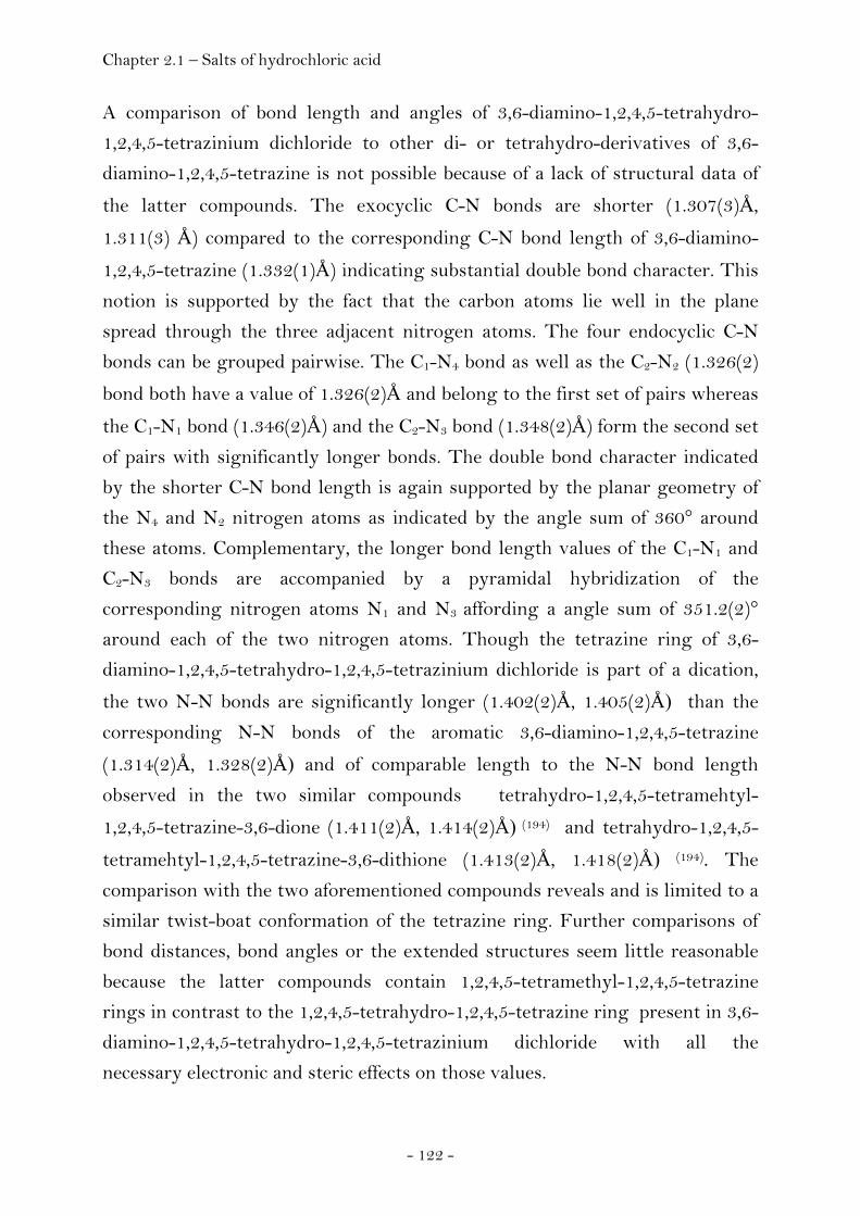
Chapter 2.1 – Salts of hydrochloric acid
- 122 -
A comparison of bond length and angles of 3,6-diamino-1,2,4,5-tetrahydro-
1,2,4,5-tetrazinium dichloride to other di- or tetrahydro-derivatives of 3,6-
diamino-1,2,4,5-tetrazine is not possible because of a lack of structural data of
the latter compounds. The exocyclic C-N bonds are shorter (1.307(3)Å,
1.311(3) Å) compared to the corresponding C-N bond length of 3,6-diamino-
1,2,4,5-tetrazine (1.332(1)Å) indicating substantial double bond character. This
notion is supported by the fact that the carbon atoms lie well in the plane
spread through the three adjacent nitrogen atoms. The four endocyclic C-N
bonds can be grouped pairwise. The C1-N4 bond as well as the C2-N2 (1.326(2)
bond both have a value of 1.326(2)Å and belong to the first set of pairs whereas
the C1-N1 bond (1.346(2)Å) and the C2-N3 bond (1.348(2)Å) form the second set
of pairs with significantly longer bonds. The double bond character indicated
by the shorter C-N bond length is again supported by the planar geometry of
the N4 and N2 nitrogen atoms as indicated by the angle sum of 360° around
these atoms. Complementary, the longer bond length values of the C1-N1 and
C2-N3 bonds are accompanied by a pyramidal hybridization of the
corresponding nitrogen atoms N1 and N3 affording a angle sum of 351.2(2)°
around each of the two nitrogen atoms. Though the tetrazine ring of 3,6-
diamino-1,2,4,5-tetrahydro-1,2,4,5-tetrazinium dichloride is part of a dication,
the two N-N bonds are significantly longer (1.402(2)Å, 1.405(2)Å) than the
corresponding N-N bonds of the aromatic 3,6-diamino-1,2,4,5-tetrazine
(1.314(2)Å, 1.328(2)Å) and of comparable length to the N-N bond length
observed in the two similar compounds tetrahydro-1,2,4,5-tetramehtyl-
1,2,4,5-tetrazine-3,6-dione (1.411(2)Å, 1.414(2)Å) (194) and tetrahydro-1,2,4,5-
tetramehtyl-1,2,4,5-tetrazine-3,6-dithione (1.413(2)Å, 1.418(2)Å) (194). The
comparison with the two aforementioned compounds reveals and is limited to a
similar twist-boat conformation of the tetrazine ring. Further comparisons of
bond distances, bond angles or the extended structures seem little reasonable
because the latter compounds contain 1,2,4,5-tetramethyl-1,2,4,5-tetrazine
rings in contrast to the 1,2,4,5-tetrahydro-1,2,4,5-tetrazine ring present in 3,6-
diamino-1,2,4,5-tetrahydro-1,2,4,5-tetrazinium dichloride with all the
necessary electronic and steric effects on those values.
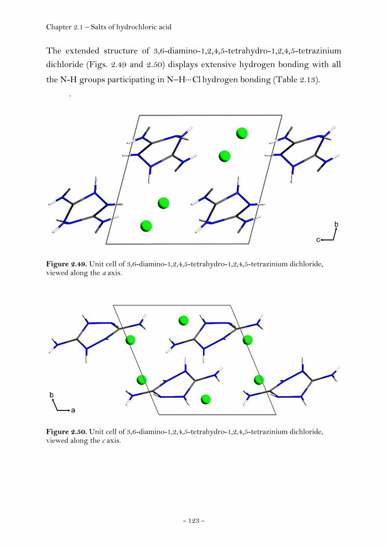
Chapter 2.1 – Salts of hydrochloric acid
- 123 -
The extended structure of 3,6-diamino-1,2,4,5-tetrahydro-1,2,4,5-tetrazinium
dichloride (Figs. 2.49 and 2.50) displays extensive hydrogen bonding with all
the N-H groups participating in N−H⋅⋅⋅Cl hydrogen bonding (Table 2.13).
.
Figure 2.49. Unit cell of 3,6-diamino-1,2,4,5-tetrahydro-1,2,4,5-tetrazinium dichloride, viewed along the a axis.
Figure 2.50. Unit cell of 3,6-diamino-1,2,4,5-tetrahydro-1,2,4,5-tetrazinium dichloride, viewed along the c axis.

Chapter 2.1 – Salts of hydrochloric acid
- 124 -
Table 2.13. Intermolecular hydrogen bonding of 3,6-diamino-1,2,4,5-tetrahydro-tetrazinium dichloride.
D−H⋅⋅⋅A D−H / Å H⋅⋅⋅A / Å D⋅⋅⋅A / Å D−H⋅⋅⋅A / ° N1−H1⋅⋅⋅Cl2 (i) 0.83(2) 2.25(2) 3.0838(14) 176(2) N2−H2⋅⋅⋅Cl1 (ii) 0.78(2) 2.38(2) 3.1135(14) 157(2) N3−H3⋅⋅⋅Cl1 (iii) 0.84(2) 2.31(2) 3.1359(14) 167(2) N4−H4⋅⋅⋅Cl2 0.776(18) 2.68(2) 3.2438(14) 131.5(17) N5−H5A⋅⋅⋅Cl1 (i) 0.80(2) 2.65(2) 3.2331(15) 132(2) N5−H5B⋅⋅⋅Cl1 (iv) 0.82(2) 2.41(2) 3.2055(15) 164(2) N6−H6A⋅⋅⋅Cl2 (v) 0.87(2) 2.63(2) 3.2691(15) 131.9(19) N6−H6A⋅⋅⋅Cl1 (ii) 0.87(2) 2.77(2) 3.4439(15) 135.3(19) N6−H6B⋅⋅⋅Cl1 (vi) 0.77(2) 2.46(2) 3.2201(15) 170(2)
Notes. Symmetry code: (i) 1-x,1-y,1-z; (ii) -x,1-y,-z; (iii) -x,1-y,1-z; (iv) 1+x,y,z; (v) -1+x,y,-1+z; (vi) -x,-y,1-z.
Experimental
3,6-diamino-1,2,4,5-tetrazine (0.5 g, 4.5 mmol) was dissolved in concentrated
hydrochloric acid (500 mL). 2,2,2-Trinitroethanol (2.42 g, 13.4 mmol) was
added and the solution heated to 100°C over a period of three hours using
reflux conditions. During the reaction the initially red colour of the solution
disappeared. Removal of the solvent afforded crystalline 3,6-diamino-1,2,4,5-
tetrahydro-tetrazinium dichloride as colourless solid. Details of the single
crystal X-ray diffraction experiment are listed in the appendix (Chapter 4).
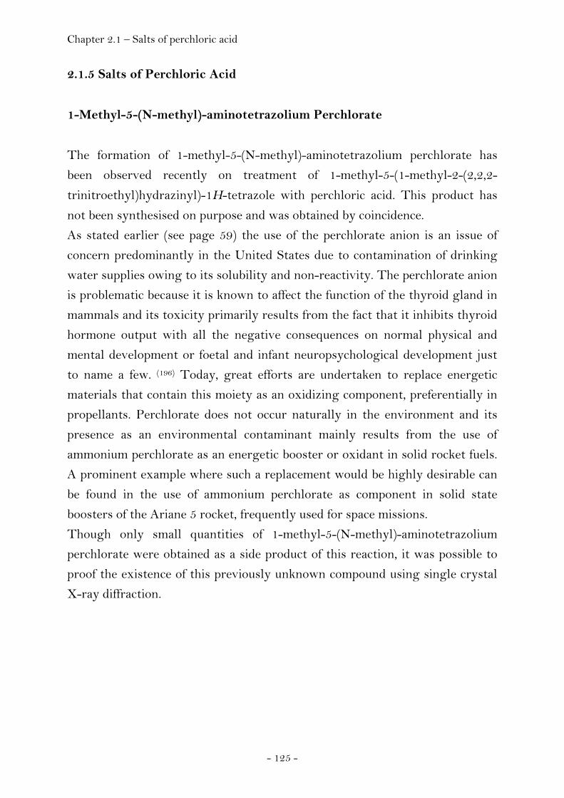
Chapter 2.1 – Salts of perchloric acid
- 125 -
2.1.5 Salts of Perchloric Acid
1-Methyl-5-(N-methyl)-aminotetrazolium Perchlorate
The formation of 1-methyl-5-(N-methyl)-aminotetrazolium perchlorate has
been observed recently on treatment of 1-methyl-5-(1-methyl-2-(2,2,2-
trinitroethyl)hydrazinyl)-1H-tetrazole with perchloric acid. This product has
not been synthesised on purpose and was obtained by coincidence.
As stated earlier (see page 59) the use of the perchlorate anion is an issue of
concern predominantly in the United States due to contamination of drinking
water supplies owing to its solubility and non-reactivity. The perchlorate anion
is problematic because it is known to affect the function of the thyroid gland in
mammals and its toxicity primarily results from the fact that it inhibits thyroid
hormone output with all the negative consequences on normal physical and
mental development or foetal and infant neuropsychological development just
to name a few. (196) Today, great efforts are undertaken to replace energetic
materials that contain this moiety as an oxidizing component, preferentially in
propellants. Perchlorate does not occur naturally in the environment and its
presence as an environmental contaminant mainly results from the use of
ammonium perchlorate as an energetic booster or oxidant in solid rocket fuels.
A prominent example where such a replacement would be highly desirable can
be found in the use of ammonium perchlorate as component in solid state
boosters of the Ariane 5 rocket, frequently used for space missions.
Though only small quantities of 1-methyl-5-(N-methyl)-aminotetrazolium
perchlorate were obtained as a side product of this reaction, it was possible to
proof the existence of this previously unknown compound using single crystal
X-ray diffraction.
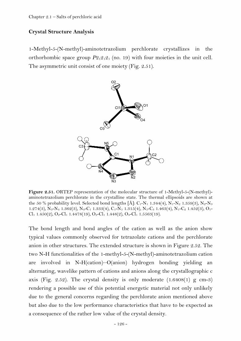
Chapter 2.1 – Salts of perchloric acid
- 126 -
Crystal Structure Analysis
1-Methyl-5-(N-methyl)-aminotetrazolium perchlorate crystallizes in the
orthorhombic space group P212121 (no. 19) with four moieties in the unit cell.
The asymmetric unit consist of one moiety (Fig. 2.51).
Figure 2.51. ORTEP representation of the molecular structure of 1-Methyl-5-(N-methyl)-aminotetrazolium perchlorate in the crystalline state. The thermal ellipsoids are shown at the 50 % probability level. Selected bond lengths [Å]: C1-N1 1.344(4), N1-N2 1.359(3), N2-N3 1.274(3), N3-N4 1.362(3), N4-C1 1.333(4), C1-N5 1.315(4), N5-C3 1.463(4), N1-C2 1.452(3), O1-Cl1 1.450(2), O2-Cl1 1.4478(19), O3-Cl1 1.448(2), O4-Cl1 1.5563(19).
The bond length and bond angles of the cation as well as the anion show
typical values commonly observed for tetrazolate cations and the perchlorate
anion in other structures. The extended structure is shown in Figure 2.52. The
two N-H functionalities of the 1-methyl-5-(N-methyl)-aminotetrazolium cation
are involved in N-H(cation)···O(anion) hydrogen bonding yielding an
alternating, wavelike pattern of cations and anions along the crystallographic c
axis (Fig. 2.52). The crystal density is only moderate (1.6408(1) g cm-3)
rendering a possible use of this potential energetic material not only unlikely
due to the general concerns regarding the perchlorate anion mentioned above
but also due to the low performance characteristics that have to be expected as
a consequence of the rather low value of the crystal density.
Chapter 2.1 – Salts of perchloric acid
- 127 -
Figure 2.52. Unit cell of 1-Methyl-5-(N-methyl)-aminotetrazolium perchlorate, viewed along the a axis (left) and c axis (right). Yellow dashed lines indicate hyrogen bonding (N4-H4···O2i, N5-H5···O3ii). (i) 1/2-x,1-y,-1/2+z, (ii) 1+x,y,z.
Experimental
The formation of 1-methyl-5-(N-methyl)-aminotetrazolium perchlorate has
been observed as decomposition product from treatment of 1-methyl-5-(1-
methyl-2-(2,2,2-trinitroethyl)hydrazinyl)-1H-tetrazole with perchloric acid at
ambient conditions and letting stand the reaction mixture for several weeks at
ambient conditions. Though only small quantities of 1-methyl-5-(N-methyl)-
aminotetrazolium perchlorate were obtained as a side product of this reaction,
it was possible to proof the existence of this previously unknown compound
using single crystal X-ray diffraction. Details of the single crystal X-ray
diffraction experiment are listed in the appendix (Chapter 4).

Chapter 2.2 – Molecules containing the trinitroethyl functionality
- 128 -
2.2 Energetic Materials: Molecules
Molecules Containing the Trinitroethyl Functionality
Introduction
The development and testing of energetic materials is an exciting and
challenging area of chemistry both as far as applied and fundamental aspects
are concerned. Considering the many applications of non nuclear energetic
materials as explosives or propellants, it is important to discover new
representatives having significant advantages over compounds currently used
not only for military but also for civilian purpose because of environmental
considerations and safety requirements while at the same time securing high
performance. A new generation energetic material has to meet several
standards in order to become widely accepted.
Next to performance properties, the desired criteria are insensitivity towards
destructive stimuli like electrostatic discharge, heat, friction and impact to
ensure safe handling procedures and enhance controllability of kinetic energy
release as well as low water solubility and hydrolytic stability to protect the
ground, longevity- and compatibility questions and furthermore criteria
addressing highest priority ecological toxicity requirements. The Strategic
Environmental Research and Development Program (SERDP) and the
Environmental Security Technology Certification Program (ESTCP) are the United
States of America Department of Defence’s (DoD) and Department of Energy
(DOE) environmental technology programs supporting such progress ever
since having been established in the late eighties of the twentieth century.
Traditional representatives of high oxygen explosives (HOX) have been
reported in public literature as research reports initiated by the Office of Naval
Research (ONR) became declassified in the early seventies of the twentieth
century; however, relevant data were published mainly in the patent literature,
often without giving information about synthetic procedures or specifying the
physicochemical characteristics of the compounds obtained. (11) Some of the
most promising materials initially considered were polynitroaliphatic

Chapter 2.2 – Molecules containing the trinitroethyl functionality
- 129 -
compounds containing the dinitromethyl, fluorodinitromethyl and
trinitromethyl groups. (197) Among them, the trinitroethyl compounds were
found to have the most favourable heats of detonation and oxygen balance
values. However, thermal stability was reported to be generally limited to
150°C when solid and 100°C when molten reversing further investigation into
trinitroethyl substituted compounds. (13)
We have now investigated compounds mentioned in the literature and we have
developed and tested novel compounds with the trinitroethyl functionality in
order to explore its potential for the design of next generation energetic
materials trying to enhance the thermal stability of this class of compounds and
finding the molecule offering the best trade-off between energy capability and
thermal stability. In the course of the global emerging interest in high-
energetic, dense materials (HEDM) (198) we are currently developing new
energetic materials preferentially containing both high oxygen and nitrogen
content with the trinitroethyl group contributing to a positive oxygen balance
value. Oxygen balance (OB) is defined as the ratio of the oxygen content of a
compound to the total oxygen required for the complete oxidation of all
carbon, hydrogen and other elements that can be oxidised to form CO2, H2O,
etc and is used to classify energetic materials as either oxygen deficient or
oxygen rich.
Most energetic materials are oxygen deficient. In contrast, this new class of
compounds contains sufficient amounts of oxygen and releases energy not only
from oxidation of the carbon back bone but also from the liberation of
dinitrogen. Nitrogen is unique amongst all other elements of the periodic table
in so far that the bond energy per two-electron bond increases from a single
over a double to a triple bond resulting in dinitrogen being more stable than
any other nitrogen species. (10) Here we report the synthesis, characterization
and the energetic properties of compounds belonging to this new class of
energetic materials and we are investigating structure property relationships of
the trinitroethyl group affecting the density.
Higher performance of a high explosive material crucially depends on density
next to other critical parameters responsible for the effectiveness like the
energy of the decomposition reactions, the number of moles and the molecular
weight of the gaseous products. The velocity of detonation increases linearly

Chapter 2.2 – Molecules containing the trinitroethyl functionality
- 130 -
with density and the detonation pressure increases to the density squared. (22) In
view of the high densities reported for trinitroethyl substituted compounds it
seemed desirable to study the effect of this group on crystal density. In turn, all
the compounds have been structurally characterized using single crystal x-ray
analysis as a preliminary step in an investigation of the relationships between
structure and crystal density.
Synthesis and thermal stability
The chemistry of 2,2,2-trinitroethanol is different to that of other alcohols
owing to the electron withdrawing inductive effect of the trinitromethyl group
(σ* = 4.54) (199) decreasing the oxygen basicity of the hydroxyl group. The
alcohol becomes acidic (pKa = 6.1) and at pH values greater than 6, the
equilibrium lies in the direction of the trinitromethanide anion and
formaldehyde. (200) Due to the equilibrium position of the reaction forming
trinitroethanol being dependent on the pH value, two pathways are possible for
the Mannich reaction to occur with either the trinitromethane or
trinitroethanol as the active hydrogen component. To study the behaviour of
various heterocyclic amines in the Mannich reaction, we prepared a variety of
amines of the tetrazole series and screen their properties in order to be able to
judge which compounds would show promising properties in terms of fulfilling
the desired criteria a new energetic material has to meet (see Table 1.1, page
15). We were interested in determining the possibility of condensing these
amines with formaldehyde and trinitromethane or directly with 2,2,2-
trinitroethanol and establishing the conditions of this reaction (Fig. 2.53).

Chapter 2.2 – Molecules containing the trinitroethyl functionality
- 131 -
Figure 2.53. Condensation of the amine can either occur directly with the alcohol or via the stepwise reaction with formaldehyde and trinitromethane.
Figure 2.54. The two step reaction involves the condensation between the amine and formaldehyde yielding an intermediate iminium-type cation and subsequent product formation after reaction with trinitromethanide.
The screening revealed that triazol or tetrazol derivatives carrying a hydrazine
group were most readily converted into the corresponding condensation
products already at room temperature and within a few minutes. In detail, 1-
methyl-1-(1H-tetrazol-5-yl)hydrazine, 1-methyl-1-(1-methyl-1H-tetrazol-5-
yl)hydrazine, 2-(5-(1-methyl-hydrazinyl)-1H-tetrazol-1-yl)ethanol and 1-
amino-2,5-hydroxymethyltriazine underwent this reaction and gave the
corresponding products 5-(1-methyl-2-(2,2,2-trinitroethyl)hydrazinyl)-1H-
tetrazol (MTHT), 1-methyl-5-(1-methyl-2-(2,2,2-trinitroethyl)hydrazinyl)-1H-
tetrazol (MMTHT), 2-(5-(1-methyl-2-(2,2,2-trinitroethyl)hydrazinyl)-1H-
tetrazol-1-yl)ethanol (MTHTE) and 2-(5-(1-methyl-2-(2,2,2-
trinitroethyl)hydrazinyl)-1H-tetrazol-1-yl)ethanol (MTHTE). However, the
reversal of the Mannich reaction was most developed in these hydrazine-type
compounds as well.
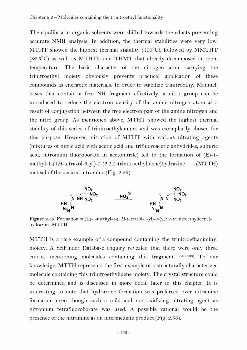
Chapter 2.2 – Molecules containing the trinitroethyl functionality
- 132 -
The equilibria in organic solvents were shifted towards the educts preventing
accurate NMR analysis. In addition, the thermal stabilities were very low.
MTHT showed the highest thermal stability (100°C), followed by MMTHT
(82.5°C) as well as MTHTE and THMT that already decomposed at room
temperature. The basic character of the nitrogen atom carrying the
trinitroethyl moiety obviously prevents practical application of these
compounds as energetic materials. In order to stabilize trinitroethyl Mannich
bases that contain a free NH fragment effectively, a nitro group can be
introduced to reduce the electron density of the amine nitrogen atom as a
result of conjugation between the free electron pair of the amine nitrogen and
the nitro group. As mentioned above, MTHT showed the highest thermal
stability of this series of trinitroethylamines and was exemplarily chosen for
this purpose. However, nitration of MTHT with various nitrating agents
(mixtures of nitric acid with acetic acid and trifluoroacetic anhydrides, sulfuric
acid, nitronium fluoroborate in acetonitrile) led to the formation of (E)-1-
methyl-1-(1H-tetrazol-5-yl)-2-(2,2,2-trinitroethylidene)hydrazine (MTTH)
instead of the desired nitramine (Fig. 2.55).
Figure 2.55. Formation of (E)-1-methyl-1-(1H-tetrazol-5-yl)-2-(2,2,2-trinitroethylidene)-hydrazine, MTTH.
MTTH is a rare example of a compound containing the trinitroethaniminyl
moiety. A SciFinder Database enquiry revealed that there were only three
entries mentioning molecules containing this fragment. (201-203) To our
knowledge, MTTH represents the first example of a structurally characterized
molecule containing this trinitroethylidene moiety. The crystal structure could
be determined and is discussed in more detail later in this chapter. It is
interesting to note that hydrazone formation was preferred over nitramine
formation even though such a mild and non-oxidizing nitrating agent as
nitronium tetrafluoroborate was used. A possible rational would be the
presence of the nitramine as an intermediate product (Fig. 2.56).
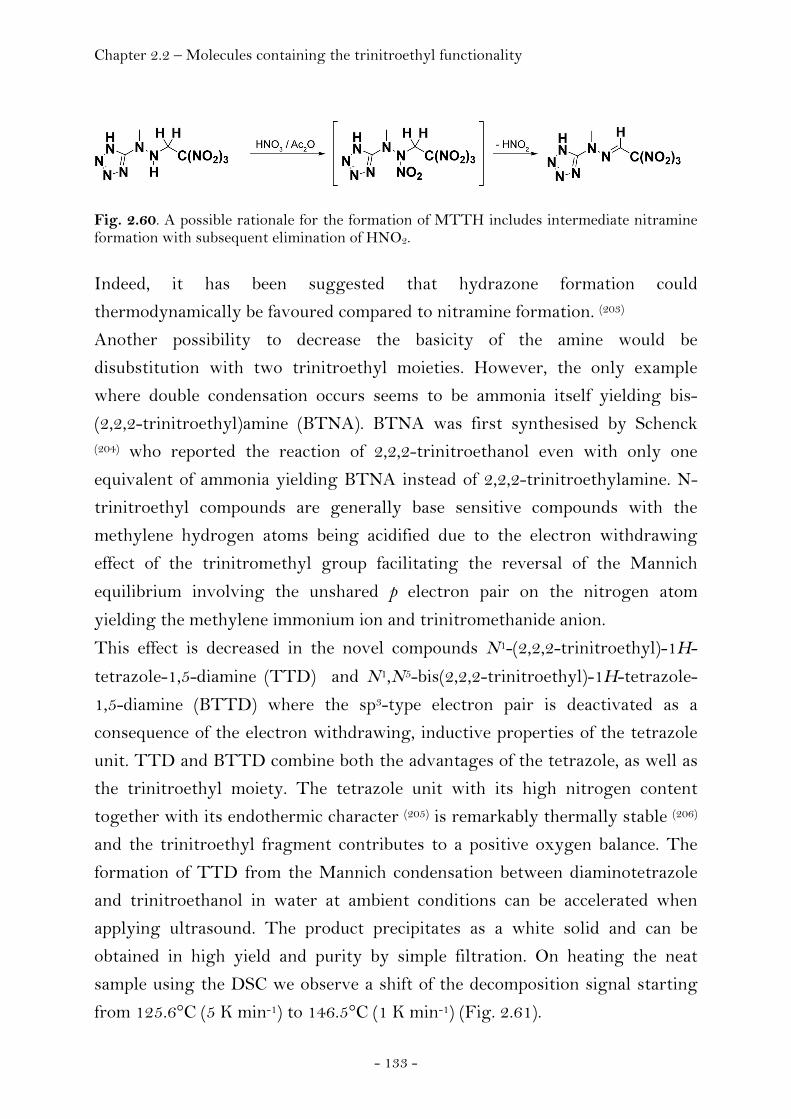
Chapter 2.2 – Molecules containing the trinitroethyl functionality
- 133 -
Fig. 2.60. A possible rationale for the formation of MTTH includes intermediate nitramine formation with subsequent elimination of HNO2. Indeed, it has been suggested that hydrazone formation could
thermodynamically be favoured compared to nitramine formation. (203)
Another possibility to decrease the basicity of the amine would be
disubstitution with two trinitroethyl moieties. However, the only example
where double condensation occurs seems to be ammonia itself yielding bis-
(2,2,2-trinitroethyl)amine (BTNA). BTNA was first synthesised by Schenck (204) who reported the reaction of 2,2,2-trinitroethanol even with only one
equivalent of ammonia yielding BTNA instead of 2,2,2-trinitroethylamine. N-
trinitroethyl compounds are generally base sensitive compounds with the
methylene hydrogen atoms being acidified due to the electron withdrawing
effect of the trinitromethyl group facilitating the reversal of the Mannich
equilibrium involving the unshared p electron pair on the nitrogen atom
yielding the methylene immonium ion and trinitromethanide anion.
This effect is decreased in the novel compounds N1-(2,2,2-trinitroethyl)-1H-
tetrazole-1,5-diamine (TTD) and N1,N5-bis(2,2,2-trinitroethyl)-1H-tetrazole-
1,5-diamine (BTTD) where the sp3-type electron pair is deactivated as a
consequence of the electron withdrawing, inductive properties of the tetrazole
unit. TTD and BTTD combine both the advantages of the tetrazole, as well as
the trinitroethyl moiety. The tetrazole unit with its high nitrogen content
together with its endothermic character (205) is remarkably thermally stable (206)
and the trinitroethyl fragment contributes to a positive oxygen balance. The
formation of TTD from the Mannich condensation between diaminotetrazole
and trinitroethanol in water at ambient conditions can be accelerated when
applying ultrasound. The product precipitates as a white solid and can be
obtained in high yield and purity by simple filtration. On heating the neat
sample using the DSC we observe a shift of the decomposition signal starting
from 125.6°C (5 K min-1) to 146.5°C (1 K min-1) (Fig. 2.61).

Chapter 2.2 – Molecules containing the trinitroethyl functionality
- 134 -
Figure 2.61. DSC plot of TTD at heat rates β = 5 K min -1, β = 2 K min -1 and β = 1 K min -1 (up = exo).
Thermal rearrangement of substituted 5-aminotetrazoles is known to be a
classic example of the Dimroth rearrangement and Moderhack et al. reported
this reaction also to occur in case of substituted 1,5-diaminotetrazoles (Fig.
2.62). (207)
Figure 2.62. Thermal rearrangement of 5-amino-1H-tetrazole-1-carboxamide to 1-(1H-tetrazol-5-yl)urea.
In analogy we suppose the signal appearing in the DSC measurement at higher
temperature to belong to 1-(1H-tetrazol-5-yl)-2-(2,2,2-trinitroethyl)-hydrazine
or its open-ring tautomer azido-(2-(2,2,2-trinitroethyl)-hydrazinyl)-
methanamine (Fig. 2.63).
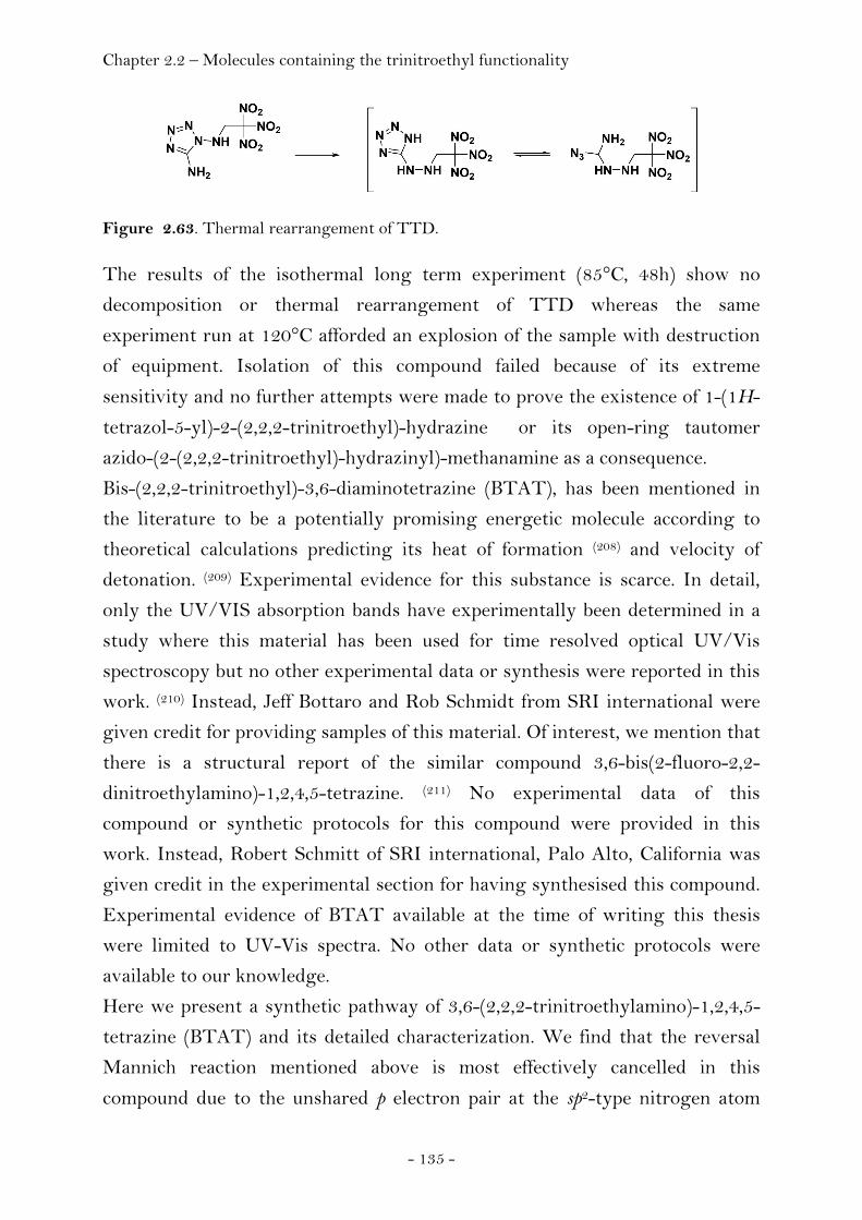
Chapter 2.2 – Molecules containing the trinitroethyl functionality
- 135 -
Figure 2.63. Thermal rearrangement of TTD.
The results of the isothermal long term experiment (85°C, 48h) show no
decomposition or thermal rearrangement of TTD whereas the same
experiment run at 120°C afforded an explosion of the sample with destruction
of equipment. Isolation of this compound failed because of its extreme
sensitivity and no further attempts were made to prove the existence of 1-(1H-
tetrazol-5-yl)-2-(2,2,2-trinitroethyl)-hydrazine or its open-ring tautomer
azido-(2-(2,2,2-trinitroethyl)-hydrazinyl)-methanamine as a consequence.
Bis-(2,2,2-trinitroethyl)-3,6-diaminotetrazine (BTAT), has been mentioned in
the literature to be a potentially promising energetic molecule according to
theoretical calculations predicting its heat of formation (208) and velocity of
detonation. (209) Experimental evidence for this substance is scarce. In detail,
only the UV/VIS absorption bands have experimentally been determined in a
study where this material has been used for time resolved optical UV/Vis
spectroscopy but no other experimental data or synthesis were reported in this
work. (210) Instead, Jeff Bottaro and Rob Schmidt from SRI international were
given credit for providing samples of this material. Of interest, we mention that
there is a structural report of the similar compound 3,6-bis(2-fluoro-2,2-
dinitroethylamino)-1,2,4,5-tetrazine. (211) No experimental data of this
compound or synthetic protocols for this compound were provided in this
work. Instead, Robert Schmitt of SRI international, Palo Alto, California was
given credit in the experimental section for having synthesised this compound.
Experimental evidence of BTAT available at the time of writing this thesis
were limited to UV-Vis spectra. No other data or synthetic protocols were
available to our knowledge.
Here we present a synthetic pathway of 3,6-(2,2,2-trinitroethylamino)-1,2,4,5-
tetrazine (BTAT) and its detailed characterization. We find that the reversal
Mannich reaction mentioned above is most effectively cancelled in this
compound due to the unshared p electron pair at the sp2-type nitrogen atom
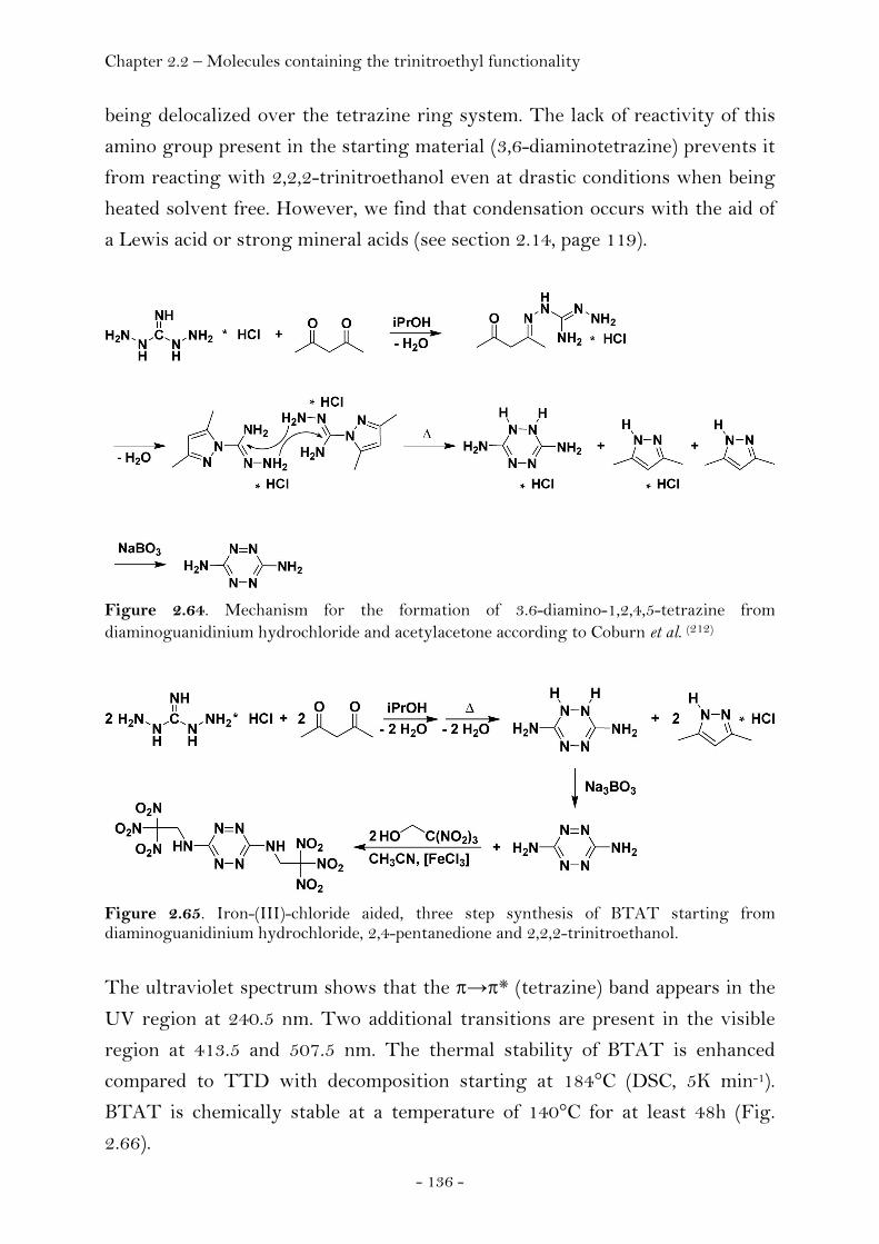
Chapter 2.2 – Molecules containing the trinitroethyl functionality
- 136 -
being delocalized over the tetrazine ring system. The lack of reactivity of this
amino group present in the starting material (3,6-diaminotetrazine) prevents it
from reacting with 2,2,2-trinitroethanol even at drastic conditions when being
heated solvent free. However, we find that condensation occurs with the aid of
a Lewis acid or strong mineral acids (see section 2.14, page 119).
Figure 2.64. Mechanism for the formation of 3.6-diamino-1,2,4,5-tetrazine from diaminoguanidinium hydrochloride and acetylacetone according to Coburn et al. (212)
Figure 2.65. Iron-(III)-chloride aided, three step synthesis of BTAT starting from diaminoguanidinium hydrochloride, 2,4-pentanedione and 2,2,2-trinitroethanol.
The ultraviolet spectrum shows that the π→π* (tetrazine) band appears in the
UV region at 240.5 nm. Two additional transitions are present in the visible
region at 413.5 and 507.5 nm. The thermal stability of BTAT is enhanced
compared to TTD with decomposition starting at 184°C (DSC, 5K min-1).
BTAT is chemically stable at a temperature of 140°C for at least 48h (Fig.
2.66).
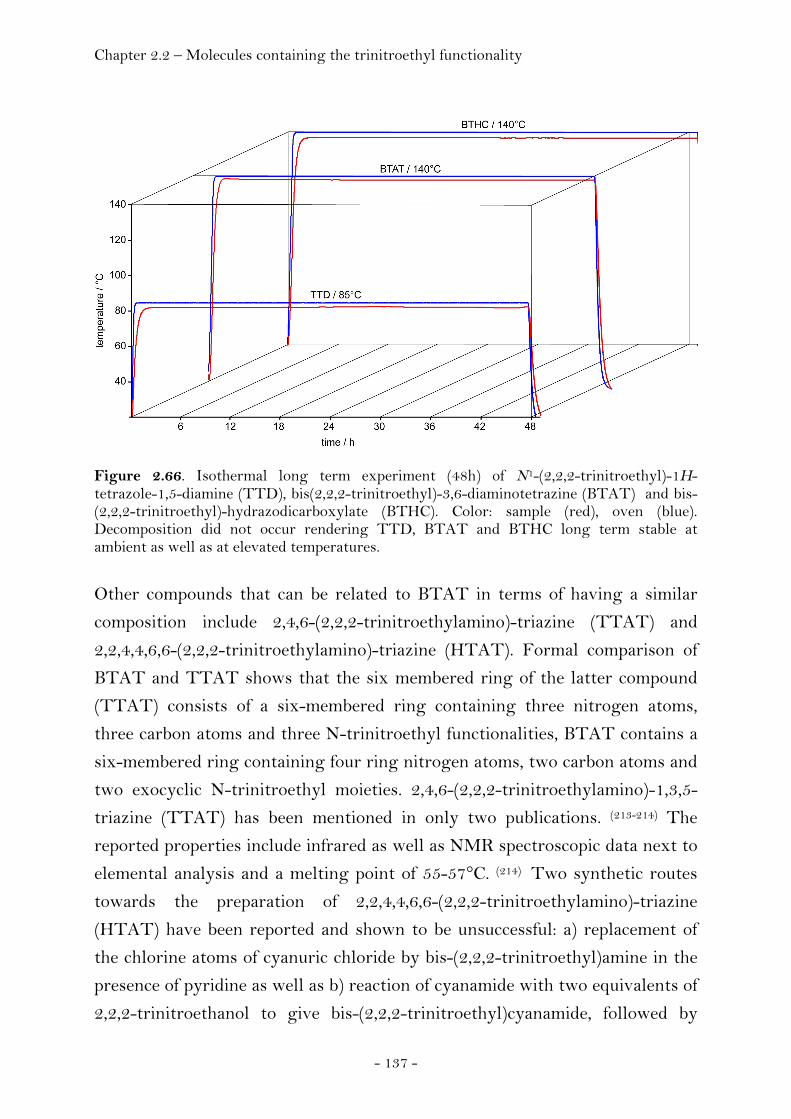
Chapter 2.2 – Molecules containing the trinitroethyl functionality
- 137 -
Figure 2.66. Isothermal long term experiment (48h) of N1-(2,2,2-trinitroethyl)-1H-tetrazole-1,5-diamine (TTD), bis(2,2,2-trinitroethyl)-3,6-diaminotetrazine (BTAT) and bis-(2,2,2-trinitroethyl)-hydrazodicarboxylate (BTHC). Color: sample (red), oven (blue). Decomposition did not occur rendering TTD, BTAT and BTHC long term stable at ambient as well as at elevated temperatures.
Other compounds that can be related to BTAT in terms of having a similar
composition include 2,4,6-(2,2,2-trinitroethylamino)-triazine (TTAT) and
2,2,4,4,6,6-(2,2,2-trinitroethylamino)-triazine (HTAT). Formal comparison of
BTAT and TTAT shows that the six membered ring of the latter compound
(TTAT) consists of a six-membered ring containing three nitrogen atoms,
three carbon atoms and three N-trinitroethyl functionalities, BTAT contains a
six-membered ring containing four ring nitrogen atoms, two carbon atoms and
two exocyclic N-trinitroethyl moieties. 2,4,6-(2,2,2-trinitroethylamino)-1,3,5-
triazine (TTAT) has been mentioned in only two publications. (213-214) The
reported properties include infrared as well as NMR spectroscopic data next to
elemental analysis and a melting point of 55-57°C. (214) Two synthetic routes
towards the preparation of 2,2,4,4,6,6-(2,2,2-trinitroethylamino)-triazine
(HTAT) have been reported and shown to be unsuccessful: a) replacement of
the chlorine atoms of cyanuric chloride by bis-(2,2,2-trinitroethyl)amine in the
presence of pyridine as well as b) reaction of cyanamide with two equivalents of
2,2,2-trinitroethanol to give bis-(2,2,2-trinitroethyl)cyanamide, followed by
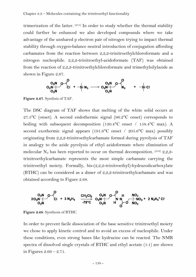
Chapter 2.2 – Molecules containing the trinitroethyl functionality
- 138 -
trimerization of the latter. (215) In order to study whether the thermal stability
could further be enhanced we also developed compounds where we take
advantage of the unshared p electron pair of nitrogen trying to impact thermal
stability through oxygen-balance neutral introduction of conjugation affording
carbamates from the reaction between 2,2,2-trinitroethylchloroformate and a
nitrogen nucleophile. 2,2,2-trinitroethyl-azidoformate (TAF) was obtained
from the reaction of 2,2,2-trinitroethylchloroformate and trimethylsilylazide as
shown in Figure 2.67.
Figure 2.67. Synthsis of TAF.
The DSC diagram of TAF shows that melting of the white solid occurs at
27.5°C (onset). A second endothermic signal (96.2°C onset) corresponds to
boiling with subsequent decomposition (130.4°C onset / 158.4°C max). A
second exothermic signal appears (191.6°C onset / 205.6°C max) possibly
originating from 2,2,2-trinitroethylcarbamate formed during pyrolysis of TAF
in analogy to the azide pyrolysis of ethyl azidoformate where elimination of
molecular N2 has been reported to occur on thermal decomposition. (216) 2,2,2-
trinitroethylcarbamate represents the most simple carbamate carrying the
trinitroethyl moiety. Formally, bis-(2,2,2-trinitroethyl)-hydrazodicarboxylate
(BTHC) can be considered as a dimer of 2,2,2-trinitroethylcarbamate and was
obtained according to Figure 2.68.
Figure 2.68. Synthesis of BTHC.
In order to prevent facile dissociation of the base sensitive trinitroethyl moiety
we chose to apply kinetic control and to avoid an excess of nucleophile. Under
these conditions, even strong bases like hydrazine can be reacted. The NMR
spectra of dissolved single crystals of BTHC and ethyl acetate (1:1) are shown
in Figures 2.69 – 2.71.
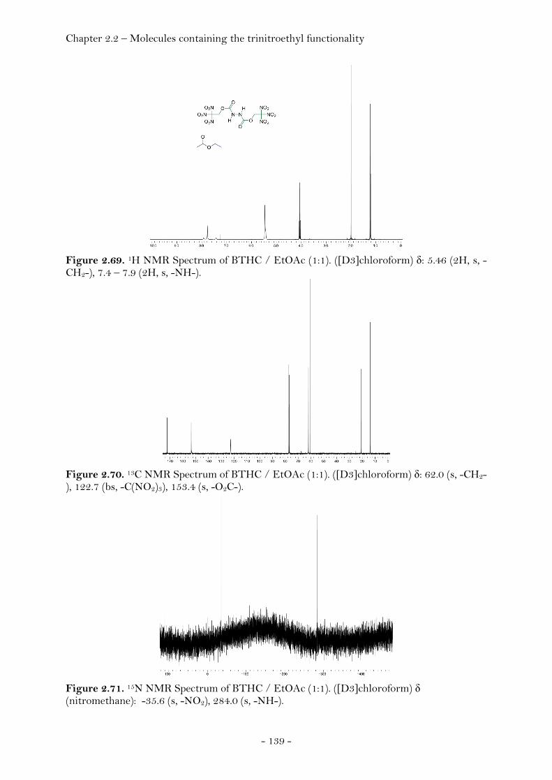
Chapter 2.2 – Molecules containing the trinitroethyl functionality
- 139 -
Figure 2.69. 1H NMR Spectrum of BTHC / EtOAc (1:1). ([D3]chloroform) δ: 5.46 (2H, s, -CH2-), 7.4 – 7.9 (2H, s, -NH-).
Figure 2.70. 13C NMR Spectrum of BTHC / EtOAc (1:1). ([D3]chloroform) δ: 62.0 (s, -CH2-), 122.7 (bs, -C(NO2)3), 153.4 (s, -O2C-).
Figure 2.71. 15N NMR Spectrum of BTHC / EtOAc (1:1). ([D3]chloroform) δ (nitromethane): -35.6 (s, -NO2), 284.0 (s, -NH-).
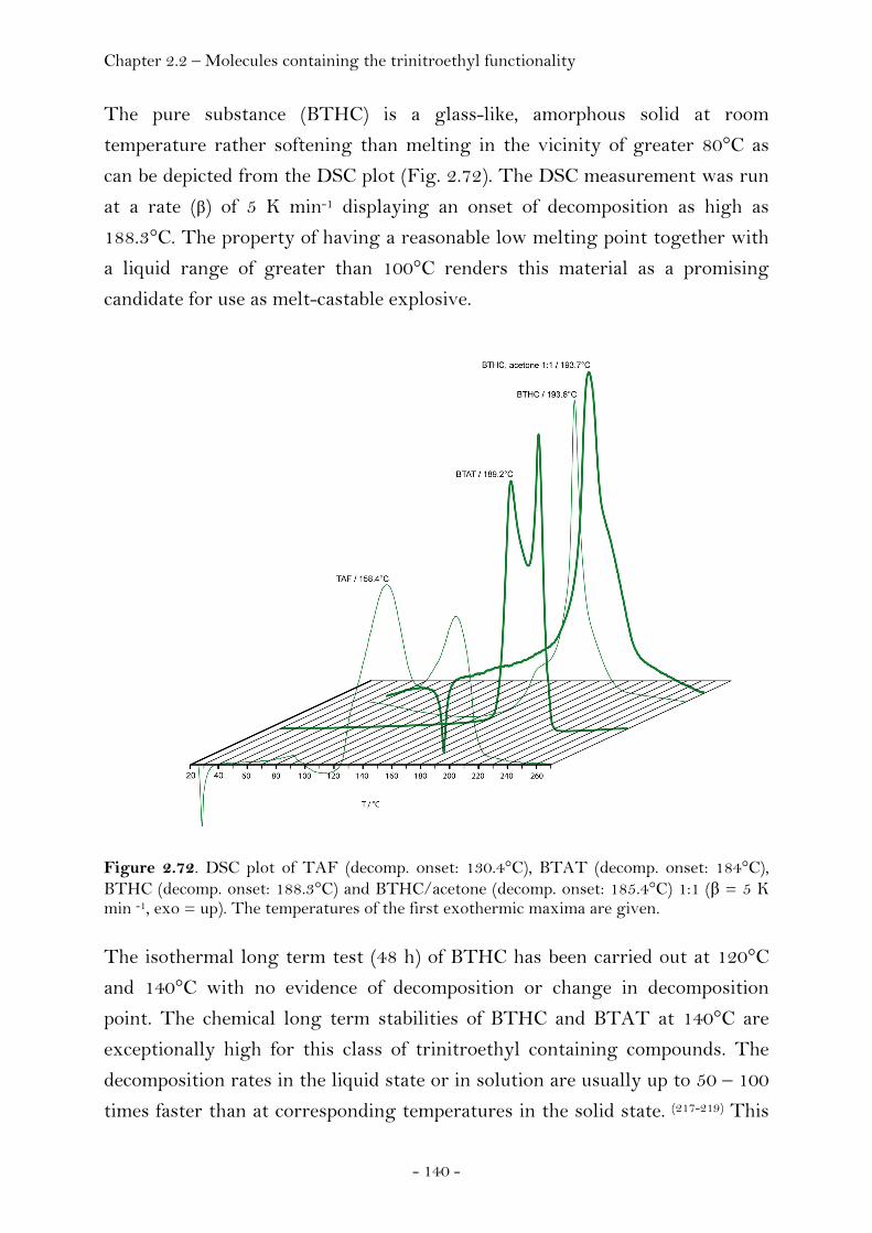
Chapter 2.2 – Molecules containing the trinitroethyl functionality
- 140 -
The pure substance (BTHC) is a glass-like, amorphous solid at room
temperature rather softening than melting in the vicinity of greater 80°C as
can be depicted from the DSC plot (Fig. 2.72). The DSC measurement was run
at a rate (β) of 5 K min-1 displaying an onset of decomposition as high as
188.3°C. The property of having a reasonable low melting point together with
a liquid range of greater than 100°C renders this material as a promising
candidate for use as melt-castable explosive.
Figure 2.72. DSC plot of TAF (decomp. onset: 130.4°C), BTAT (decomp. onset: 184°C), BTHC (decomp. onset: 188.3°C) and BTHC/acetone (decomp. onset: 185.4°C) 1:1 (β = 5 K min -1, exo = up). The temperatures of the first exothermic maxima are given. The isothermal long term test (48 h) of BTHC has been carried out at 120°C
and 140°C with no evidence of decomposition or change in decomposition
point. The chemical long term stabilities of BTHC and BTAT at 140°C are
exceptionally high for this class of trinitroethyl containing compounds. The
decomposition rates in the liquid state or in solution are usually up to 50 – 100
times faster than at corresponding temperatures in the solid state. (217-219) This
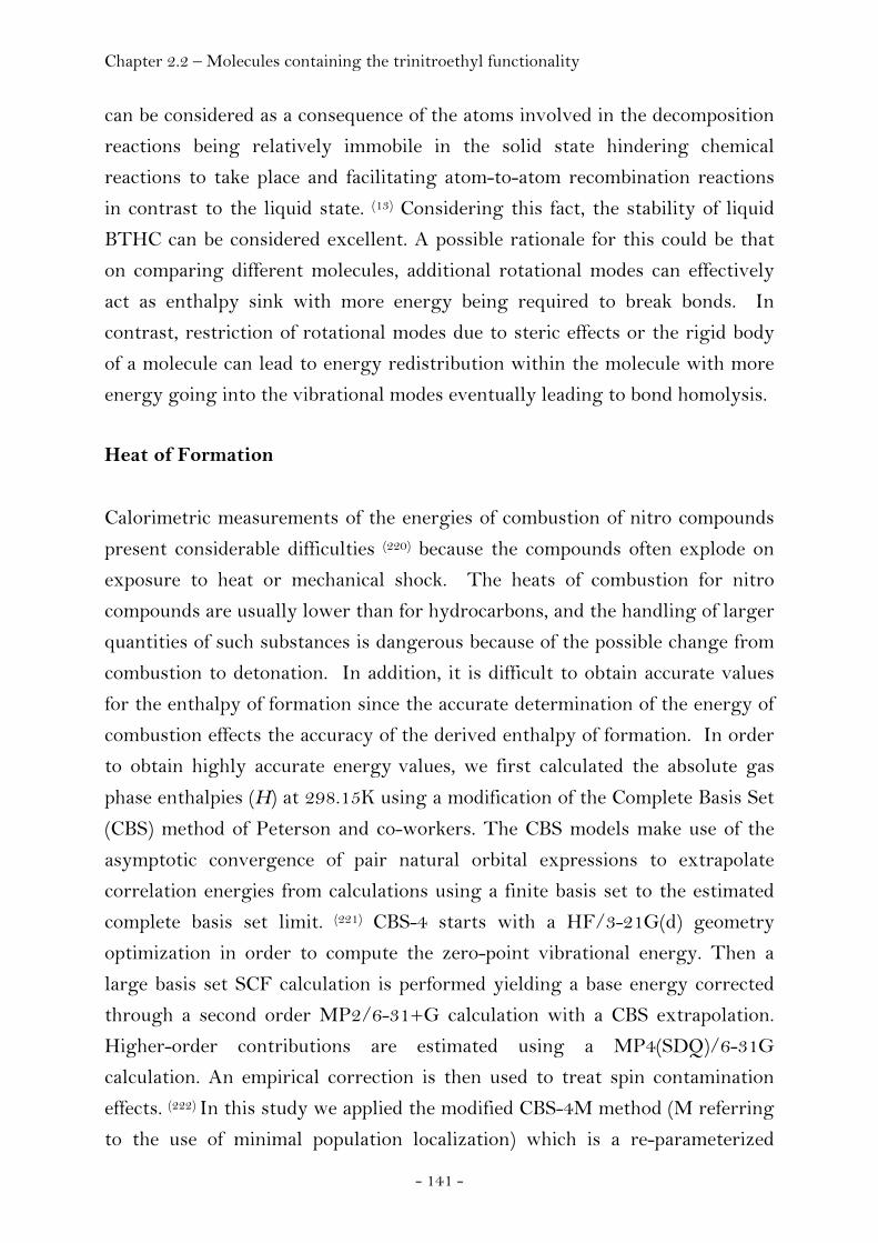
Chapter 2.2 – Molecules containing the trinitroethyl functionality
- 141 -
can be considered as a consequence of the atoms involved in the decomposition
reactions being relatively immobile in the solid state hindering chemical
reactions to take place and facilitating atom-to-atom recombination reactions
in contrast to the liquid state. (13) Considering this fact, the stability of liquid
BTHC can be considered excellent. A possible rationale for this could be that
on comparing different molecules, additional rotational modes can effectively
act as enthalpy sink with more energy being required to break bonds. In
contrast, restriction of rotational modes due to steric effects or the rigid body
of a molecule can lead to energy redistribution within the molecule with more
energy going into the vibrational modes eventually leading to bond homolysis.
Heat of Formation
Calorimetric measurements of the energies of combustion of nitro compounds
present considerable difficulties (220) because the compounds often explode on
exposure to heat or mechanical shock. The heats of combustion for nitro
compounds are usually lower than for hydrocarbons, and the handling of larger
quantities of such substances is dangerous because of the possible change from
combustion to detonation. In addition, it is difficult to obtain accurate values
for the enthalpy of formation since the accurate determination of the energy of
combustion effects the accuracy of the derived enthalpy of formation. In order
to obtain highly accurate energy values, we first calculated the absolute gas
phase enthalpies (H) at 298.15K using a modification of the Complete Basis Set
(CBS) method of Peterson and co-workers. The CBS models make use of the
asymptotic convergence of pair natural orbital expressions to extrapolate
correlation energies from calculations using a finite basis set to the estimated
complete basis set limit. (221) CBS-4 starts with a HF/3-21G(d) geometry
optimization in order to compute the zero-point vibrational energy. Then a
large basis set SCF calculation is performed yielding a base energy corrected
through a second order MP2/6-31+G calculation with a CBS extrapolation.
Higher-order contributions are estimated using a MP4(SDQ)/6-31G
calculation. An empirical correction is then used to treat spin contamination
effects. (222) In this study we applied the modified CBS-4M method (M referring
to the use of minimal population localization) which is a re-parameterized

Chapter 2.2 – Molecules containing the trinitroethyl functionality
- 142 -
version of the original CBS-4 method. (223) The determination of the gas phase
heats of formation was accomplished by using the method of atomization
energies (Fig. 2.73). (224)
∑∑ Δ+−=Δatomsatoms
HHHH ),A(),A(),M(),M( (g)f(g))g()g(f 298298298298 oooo
Fig. 2.73. Notes. M refers to molecule, A refers to atoms, and 298.15 K is denoted as 298 for clarity.
Finally, sublimation enthalpies were estimated according to Trouton’s rule
(Fig. 2.74) and subtracted from the gas phase enthalpies of formation in order
to obtain the solid state heat of formation values. (225)
ΔsubH°(M(s), 298) [J mol-1] = 188 · Tm [K]
Figure 2.74. Estimation of the heat of sublimation.
The values obtained for the heat of formation obtained from our calculations
can be compared with the previously reported values for ethanol (EtOH), 2,2,2-
trinitroethanol (TNE), 2,4,6-trinitrotoluene (TNT) and
cyclotrimethylenetrinitramine (RDX) as well as
cyclotetramethylenetetranitramine (HMX) and hexanitrohexaazaisowurtzitane
(HNIW / CL-20) which were determined using both precision combustion
calorimetry and reaction calorimetry (Table 2.14). Table 2.14. Comparison of experimentally determined and calculated heat of formation.
ΔfH0 / kcal mol-1 CBS-4M experimental a)
EtOH -58.0 (g) -56.1 ± 0.1 (g) (226)
TNE -47.0 (g) / -62.5 (s) -62.3 ± 0.7 (s) (227)
TNT 2.21 (g) 5.8 ± 0.8 (g) (228)
RDX 41.6 (g) 45.9 (g) (229)
HMX 30.1 (s) 24.6 ± 0.7 (230) CL-20 96.7 (s) 102.9 (s) ± 3.1 (231)
The differences between the experimentally determined and the calculated
ΔfH° values do not exceed a range of 5 kcal mol-1 rendering this method valid
for the estimation of the heats of formation (Table 2.15). Hence, the calculated
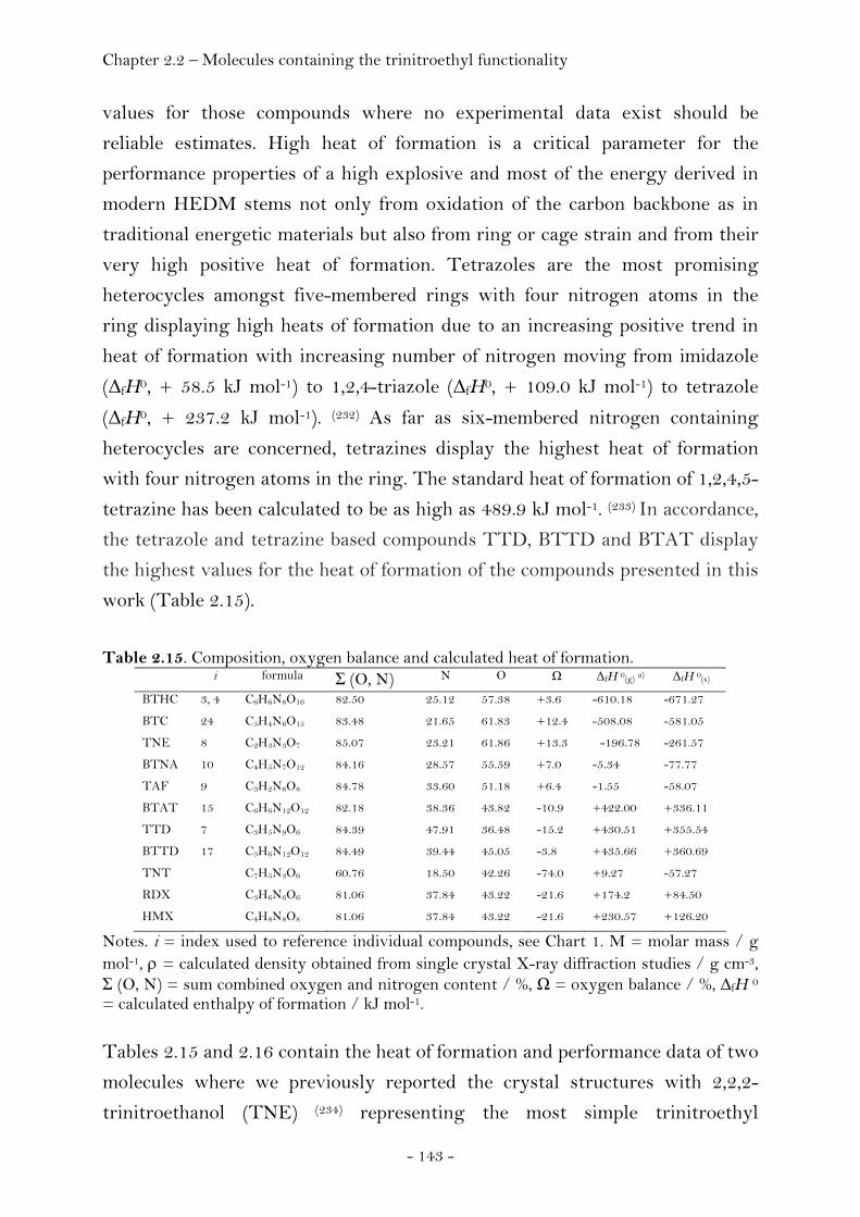
Chapter 2.2 – Molecules containing the trinitroethyl functionality
- 143 -
values for those compounds where no experimental data exist should be
reliable estimates. High heat of formation is a critical parameter for the
performance properties of a high explosive and most of the energy derived in
modern HEDM stems not only from oxidation of the carbon backbone as in
traditional energetic materials but also from ring or cage strain and from their
very high positive heat of formation. Tetrazoles are the most promising
heterocycles amongst five-membered rings with four nitrogen atoms in the
ring displaying high heats of formation due to an increasing positive trend in
heat of formation with increasing number of nitrogen moving from imidazole
(ΔfH0, + 58.5 kJ mol-1) to 1,2,4-triazole (ΔfH0, + 109.0 kJ mol-1) to tetrazole
(ΔfH0, + 237.2 kJ mol-1). (232) As far as six-membered nitrogen containing
heterocycles are concerned, tetrazines display the highest heat of formation
with four nitrogen atoms in the ring. The standard heat of formation of 1,2,4,5-
tetrazine has been calculated to be as high as 489.9 kJ mol-1. (233) In accordance,
the tetrazole and tetrazine based compounds TTD, BTTD and BTAT display
the highest values for the heat of formation of the compounds presented in this
work (Table 2.15).
Table 2.15. Composition, oxygen balance and calculated heat of formation.
i formula Σ (O, N) N O Ω ΔfH 0(g) a) ΔfH 0(s)
BTHC 3, 4 C6H6N8O16 82.50 25.12 57.38 +3.6 -610.18 -671.27
BTC 24 C5H4N6O15 83.48 21.65 61.83 +12.4 -508.08 -581.05
TNE 8 C2H3N3O7 85.07 23.21 61.86 +13.3 -196.78 -261.57
BTNA 10 C4H5N7O12 84.16 28.57 55.59 +7.0 -5.34 -77.77
TAF 9 C3H2N6O8 84.78 33.60 51.18 +6.4 -1.55 -58.07
BTAT 15 C6H6N12O12 82.18 38.36 43.82 -10.9 +422.00 +336.11
TTD 7 C3H5N9O6 84.39 47.91 36.48 -15.2 +430.51 +355.54
BTTD 17 C5H6N12O12 84.49 39.44 45.05 -3.8 +435.66 +360.69
TNT C7H5N3O6 60.76 18.50 42.26 -74.0 +9.27 -57.27
RDX C3H6N6O6 81.06 37.84 43.22 -21.6 +174.2 +84.50
HMX C4H8N8O8 81.06 37.84 43.22 -21.6 +230.57 +126.20
Notes. i = index used to reference individual compounds, see Chart 1. M = molar mass / g mol-1, ρ = calculated density obtained from single crystal X-ray diffraction studies / g cm-3, Σ (O, N) = sum combined oxygen and nitrogen content / %, Ω = oxygen balance / %, ∆fH 0
= calculated enthalpy of formation / kJ mol-1. Tables 2.15 and 2.16 contain the heat of formation and performance data of two
molecules where we previously reported the crystal structures with 2,2,2-
trinitroethanol (TNE) (234) representing the most simple trinitroethyl
Chapter 2.2 – Molecules containing the trinitroethyl functionality
- 144 -
substituted compound as well as bis(2,2,2-trinitroethyl)carbonate (BTC) (235)
displaying the highest crystal density amongst all trinitroethyl substituted
compounds as well as the heat of formation and performance data of standard
explosives such as 2,4,6-trinitrotoluene (TNT), 1,3,4-trinitro-1,3,5-
triazacyclohexane (RDX) and 1,3,5,7-tetranitro-1,3,5,7-tetraazacyclooctane
(HMX) for comparison.
Sensitivity and Performance
In order to establish safe handling procedures the sensitivity data of the new
compounds towards mechanical destructive stimuli like impact or friction and
electrostatic discharge have been measured. The sensitivity can be used to
classify them as primary or secondary explosives. Whereas primary explosives
explode readily from light to modest mechanical stimuli or application of heat,
some secondary explosives or simply high explosives need a high energy
impulse like the shock of a detonation in order to initiate detonation and will
simply burn in small enough quantities. According to BAM (Bundesanstalt für
Materialprüfung), a compound can be classified as being insensitive (impact
energy > 40 J / friction force > 360 N), less sensitive (impact energy ≥ 35 J /
friction force = 360 N), sensitive (impact energy ≥ 4 J / friction force < 360 N
to > 80 N), very sensitive (impact energy ≤ 3 J / friction force ≤ 80 N) and
extremely sensitive (impact energy ≤ 3 J / friction force ≤ 10 N). Of all the
compounds tested, the following order of sensitivity including the standard
explosives TNT, RDX, HMX and CL-20 arises: CL-20 (very sensitive, 4 J /
48N) (236) > TTD (sensitive, < 30 J / 40 N) > HMX (sensitive, 7.4 J / < 120 N); (236) RDX (sensitive, 7.4 J / 120 N); (236) BTAT (sensitive, 7J / > 160 N) > TNT
(sensitive, 15 J / 353 N) (236) > BTNA (sensitive, 15 J / > 360 N) > BTHC
(insensitive, > 100 J / > 360 N). By applying the calculated density values
obtained from the crystal structure determination and the calculated energy of
formation, the performance parameters of TTD, BTTD, BTAT, TAF and
BTHC as well as 2,2,2-trinitroethanol (TNE), bis-(2,2,2-trinitroethyl)-amine
(BTNA) and bis-(2,2,2-trinitroethyl)-carbonate (BTC) were calculated using
the software EXPLO5 (v. 5.02) (237-241) and are summarized in Table 2.16. The
software is based on the chemical equilibrium, steady-state model of detonation

Chapter 2.2 – Molecules containing the trinitroethyl functionality
- 145 -
and uses the Becker-Kistiakowsky-Wilson’s equation of state (BKWN EOS) for
gaseous detonation products.
Table 2.16. Selection of calculated performance properties and comparison to TNT, RDX and HMX.
ρ ∆fU0 QCJ V0 Tex PCJ D
BTHC calc. 1.8 -1421 5333 682 4376 284 8188
TNE calc. 1.839 -1355 4875 712 4041 285 8180
TAF calc. 1.853 -153 5353 675 4549 303 8359
BTC calc. 1.975 -1417 4792 665 4068 317 8396
BTNA calc. 1.881 -138 6010 705 4733 343 8815
TTD calc. 1.831 +1443 6018 788 4650 370 9194
BTTD calc. 1.897 +932 6479 739 5009 388 9323
BTAT calc. 1.886 +852 6135 743 4867 389 9261
TNT calc. 1.64 -175 5064 625 3749 203 7170
TNT exp. 1.64 (242) - - - - 210 a) (242) 6950 a) (242)
RDX calc. 1.80 +280 5875 797 4290 338 8894
RDX exp. 1.80 (242) - - - - 347 a) (242) 8750 a) (242)
HMX calc. 1.89 +526 6169 791 4385 384 9288
HMX exp. 1.89 (242) - - - - 390 a) (242) 9110 a) (242)
Notes. a) experimental value, ρ = calculated density obtained from single crystal X-ray diffraction studies / g cm-3, ∆fU0 = energy of formation / kJ kg-1, Ω = oxygen balance / %, QCJ = heat of explosion / kJ kg-1, V0 = volume of gaseous detonation products / L kg -1, Tex = explosion temperature / K, PCJ = detonation pressure / kbar, D = velocity of detonation / m s-1.
Chapter 2.2 – Molecules containing the trinitroethyl functionality
- 146 -
Figure 2.75. Small scale burning test (UN 3d) of BTAT as part of the interim hazard classification (IHC) procedure. An unconfined plastic transportcontainer filled with BTAT (2g) was placed in kerosene-soaked sawdust which was subsequently ignited. The picture in the middle shows deflagration of the substance indicating a pass of this test due to failure of explosion or detonation.
A comparison of the calculated values of 2,4,6-trinitrotoluene (TNT), 1,3,4-
trinitro-1,3,5-triazacyclohexane (RDX) and 1,3,5,7-tetranitro-1,3,5,7-
tetraazacyclooctane (HMX) based on the calculated (CBS-4M) solid state heat
of formation to their corresponding experimental values is included in Table
2.16 and shows that the calculated values obtained using this method are in
good agreement with the corresponding experimental values. TNT can be
considered as first generation explosive, whereas the nitramine-type explosives
RDX and HMX belong to the second generation and are standard explosives
for most military applications today. The theoretically predicted performance
values for TNE, BTC, BTNA, TTD, BTTD, BTAT, TAF and BTHC all lie
within the range expected for high explosives while at the same time
displaying a much more favourable oxygen balance compared to TNT, RDX
and HMX. Oxygen balance values near or greater zero are highly desirable in
order to reduce toxic fume gases like carbon monoxide. (236) The combined
oxygen and nitrogen content of TNE, BTNA, BTC, TTD, BTTD, BTAT,
TAF and BTHC all lie within 82.2% to 85.1% with oxygen balance values
ranging from -15.2% to +13.3%. Of all the compounds studied in this work,
TTD, BTTD and BTAT show the highest predicted values for the velocity of
detonation (BTTD: 9323 m s-1 / BTAT: 9261 m s-1/ TTD: 9194 m s-1),
detonation pressure (BTAT: 389 kbar / BTTD: 388 kbar / TTD: 370 kbar)
and heat of explosion (BTTD: 6479 kJ kg-1 / BTAT: 6135 kJ kg-1 / TTD: 6018
kJ kg-1 ) displaying performance values superior to RDX and comparable to
HMX.

Chapter 2.2 – Molecules containing the trinitroethyl functionality
- 147 -
Figure 2.76. High Speed Video Images (4000 frames per second) of the electrostatic ignition of BTAT and subsequent explosive decomposition. The four pictures show the unreacted sample, ignition of the sample by electrostatic discharge accompanied by formation of dark coloured gases, detonation accompanied by a flash of light and burning of the hot decomposition gases (from left to right).
Figure 2.77 displays the pictures of a Steel Sleeve test (Koenen test) on TTD.
This is indicative for the respond to thermal shock and performance of a
compound under confinement according to German law of explosives as well as
the United Nation Recommendations on the Transport of Dangerous Goods. (243) A
steel core [d] (Fig. 2.77) filled with 25 mL of compound gets locked using a
perforated disk [b] with variable whole width using screw threads [a] + [c]. The sample is then placed into the test setup where four Bunsen burners
simultaneously heat it up to five minutes. Subsequently, large amounts of
liberated gaseous decomposition products arise. The whole diameter of the
perforated disk where no destruction of the steel assembly occurs is called the
limiting diameter. This diameter has been found to be 6 mm for TNT, and 8
mm for RDX and HMX. In case the steel sleeve is ruptured into three or more
pieces the test is evaluated as explosion. A whole width of 10 mm was used in
this test (Fig. 2.77) and the steel tube was fragmented into powder-like pieces.
Furthermore, the screw threads [a] + [c] were also broken into pieces
rendering TTD as sensitive compound with a critical diameter of greater
10mm (Type H). The explosion was recorded using a high speed camera at
3500 frames per second and found to take place within less than 0.5
milliseconds according to two frames with the first frame showing ignition and
the second frame (picture B, Fig. 2.77) revealing that no visible metal pieces are
present any longer.

Chapter 2.2 – Molecules containing the trinitroethyl functionality
- 148 -
Figure 2.77. Steel Sleeve test (Koenen) of TTD (10mm). A: Setup, B: High Speed Image (3500 fs-1) of the detonation, C: Steel sleeve assembly before the test: [a] + [c] screw thread, [b] perforated disk (10mm whole width), [d] steel core, filled with TTD, D: Steel sleeve residues.
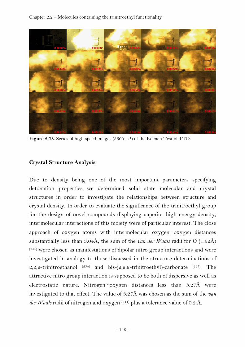
Chapter 2.2 – Molecules containing the trinitroethyl functionality
- 149 -
Figure 2.78. Series of high speed images (3500 fs-1) of the Koenen Test of TTD.
Crystal Structure Analysis
Due to density being one of the most important parameters specifying
detonation properties we determined solid state molecular and crystal
structures in order to investigate the relationships between structure and
crystal density. In order to evaluate the significance of the trinitroethyl group
for the design of novel compounds displaying superior high energy density,
intermolecular interactions of this moiety were of particular interest. The close
approach of oxygen atoms with intermolecular oxygen···oxygen distances
substantially less than 3.04Å, the sum of the van der Waals radii for O (1.52Å) (244) were chosen as manifestations of dipolar nitro group interactions and were
investigated in analogy to those discussed in the structure determinations of
2,2,2-trinitroethanol (234) and bis-(2,2,2-trinitroethyl)-carbonate (235). The
attractive nitro group interaction is supposed to be both of dispersive as well as
electrostatic nature. Nitrogen···oxygen distances less than 3.27Å were
investigated to that effect. The value of 3.27Å was chosen as the sum of the van
der Waals radii of nitrogen and oxygen (244) plus a tolerance value of 0.2 Å.
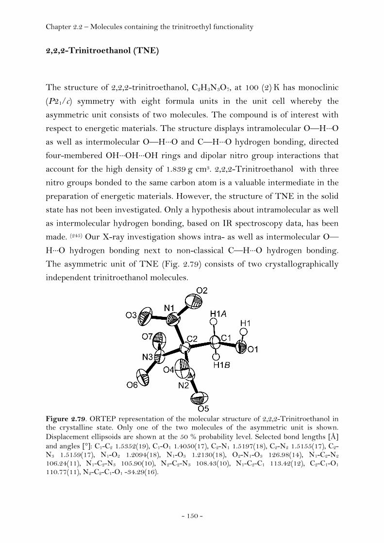
Chapter 2.2 – Molecules containing the trinitroethyl functionality
- 150 -
2,2,2-Trinitroethanol (TNE)
The structure of 2,2,2-trinitroethanol, C2H3N3O7, at 100 (2) K has monoclinic
(P21/c) symmetry with eight formula units in the unit cell whereby the
asymmetric unit consists of two molecules. The compound is of interest with
respect to energetic materials. The structure displays intramolecular O—H···O
as well as intermolecular O—H···O and C—H···O hydrogen bonding, directed
four-membered OH···OH···OH rings and dipolar nitro group interactions that
account for the high density of 1.839 g cm3. 2,2,2-Trinitroethanol with three
nitro groups bonded to the same carbon atom is a valuable intermediate in the
preparation of energetic materials. However, the structure of TNE in the solid
state has not been investigated. Only a hypothesis about intramolecular as well
as intermolecular hydrogen bonding, based on IR spectroscopy data, has been
made. (245) Our X-ray investigation shows intra- as well as intermolecular O—
H···O hydrogen bonding next to non-classical C—H···O hydrogen bonding.
The asymmetric unit of TNE (Fig. 2.79) consists of two crystallographically
independent trinitroethanol molecules.
Figure 2.79. ORTEP representation of the molecular structure of 2,2,2-Trinitroethanol in the crystalline state. Only one of the two molecules of the asymmetric unit is shown. Displacement ellipsoids are shown at the 50 % probability level. Selected bond lengths [Å] and angles [°]: C1-C2 1.5352(19), C1-O1 1.4050(17), C2-N1 1.5197(18), C2-N2 1.5155(17), C2-N3 1.5159(17), N1-O2 1.2094(18), N1-O3 1.2130(18), O2-N1-O3 126.98(14), N1-C2-N2 106.24(11), N1-C2-N3 105.90(10), N2-C2-N3 108.43(10), N1-C2-C1 113.42(12), C2-C1-O1 110.77(11), N2-C2-C1-O1 -34.29(16).

Chapter 2.2 – Molecules containing the trinitroethyl functionality
- 151 -
These two moieties display a very similar molecular geometry with a propeller-
type orientation of the nitro groups (D3) bonded to the β carbon atom. In both
molecules the conformation of the substituents of the α- as well as the β carbon
atom is found to be staggered and intramolecular O—H···O hydrogen bonding
does occur (O1—H1···O3, O8—H8···O13). The length of the C-N bonds joining
the three nitro groups to the β carbon atom (range 1.5150 ± 0.0018 Å - 1.5197
± 0.0018 Å) are significantly longer than the normal C-N bond distance of 1.47
Å (246) as was already observed in the determination of the crystal structure of
N,N'-bis-(2,2,2-trinitroethyl)-urea (247). A comparison of the geometrical trends
for the bonding of the three nitro groups to one carbon atom in TNE with
those in N,N'-bis-(2,2,2-trinitroethyl)-urea reported by Lind again shows good
agreement taking into account that the measurement of Lind was undertaken
at 296 K whereas our experiment was run at 100 K. The independent N-C-N
bond angles are less (range 105.90 (0.10)° - 108.43 (0.10)°) than the tetrahedral
value whereas the corresponding N-C-C bond angles are greater (range 110.18
(0.11)° - 113.42 (0.12)°) than the tetrahedral value. The three independent nitro
groups of each TNE molecule are identical in structure within the limits of
error and display common geometry parameters like N-O distances (range
1.2079 (0.0018)Å - 1.2181 (0.0018) Å), O-N-O bond angles (range 126.96
(0.14)° - 127.91 (0.15)°) and O-N-C bond angles (range 113.63 (0.12)° - 118.64
(0.12)°). In turn, the arrangement of the C-N and N-O bonds is coplanar with
the sums of the three bond angles around one nitrogen atom being 360° within
the limits of error.
The extended structure of TNE involves secondary interactions in terms of
intermolecular O—H···O hydrogen bonding, intermolecular C—H···O
hydrogen bonding as well as dipolar nitro group interactions. The circular O—
H···O hydrogen bonding between the hydroxyl groups of four trinitroethanol
molecules results in four-membered homodromic rings (O1—H1···O8—
H8···O1—H1···O8—H8). The structure that can be observed along the
crystallographic a axis shows a stacking of these rings. Every ring is
surrounded by four neighbouring rings whereby two of the four molecules of
trinitroethanol that form such a ring interconnect the central ring to the
surrounding rings via C3—H3B···O14 hydrogen bonding (Fig. 2.80).
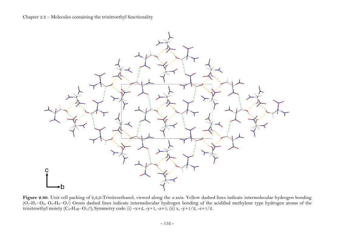
Chapter 2.2 – Molecules containing the trinitroethyl functionality
- 152 -
Figure 2.80. Unit cell packing of 2,2,2-Trinitroethanol, viewed along the a axis. Yellow dashed lines indicate intermolecular hydrogen bonding (O1-H1···O8, O8-H8···O1i) Green dashed lines indicate intermolecular hydrogen bonding of the acidified methylene type hydrogen atoms of the trinitroethyl moiety (C3-H3B···O14ii); Symmetry code: (i) –x+2, -y+1, -z+1; (ii) x, -y+1/2, -z+1/2.

Chapter 2.2 – Molecules containing the trinitroethyl functionality
- 153 -
Additional contacts: Intramolecular hydrogen bonds: O1-H1···O3 and O8-
H8···O13.The close approach of oxygen atoms found in the extended structure of
TNE suggest the possibility of dipolar nitro group interactions in analogy to a
variety of non-covalent interactions such as halogen···O(nitro) (248), halogen···O=C (249) and carbonyl interactions (250). Short intermolecular oxygen···oxygen
distances with values substantially less than 3.04Å, the sum of the van der
Waals radii for O (1.52Å) (244), were investigated to that effect. Dipolar nitro
group interactions were accepted for nitrogen···oxygen contacts less than
3.17Å. The value of 3.17Å was chosen as the sum of the van der Waals radii of
nitrogen and oxygen plus a tolerance value of 0.1 Å. (244) Given these values,
two dipolar nitro group contacts were identified. Those two interactions were
found for the N2O4O5 nitro group interacting with the N5O11O12 nitro group in
one case and with itself in the other leading to oxygen···oxygen distances with
values of 2.8519 (18) Å (O5···O12) and 2.8251 (15) Å (O4···O4). The correspondig
values for the nitrogen···oxygen contacts are 3.1184 (17) Å (O5···N5) and 3.1234
(16) Å (O4···N2). Figure 2.81 displays the symmetric interaction of the two
N2O4O5 nitro groups.
Figure 2.81. Example of dipolar nitro group interaction in trinitroethanol. The interaction between the N2 and the O4 atoms [d(O···Ni) = 3.1234 (16) Å, symmetry code i = -x+1, -y+1, -z+1] obviously brings the O atoms into close proximity. (234)
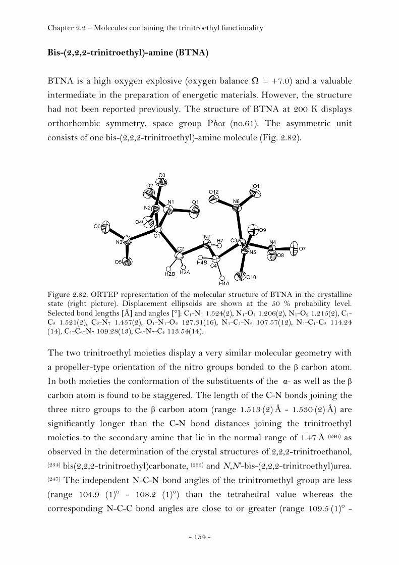
Chapter 2.2 – Molecules containing the trinitroethyl functionality
- 154 -
Bis-(2,2,2-trinitroethyl)-amine (BTNA)
BTNA is a high oxygen explosive (oxygen balance Ω = +7.0) and a valuable
intermediate in the preparation of energetic materials. However, the structure
had not been reported previously. The structure of BTNA at 200 K displays
orthorhombic symmetry, space group Pbca (no.61). The asymmetric unit
consists of one bis-(2,2,2-trinitroethyl)-amine molecule (Fig. 2.82).
Figure 2.82. ORTEP representation of the molecular structure of BTNA in the crystalline state (right picture). Displacement ellipsoids are shown at the 50 % probability level. Selected bond lengths [Å] and angles [°]: C1-N1 1.524(2), N1-O1 1.206(2), N1-O2 1.215(2), C1-C2 1.521(2), C2-N7 1.457(2), O1-N1-O2 127.31(16), N1-C1-N2 107.57(12), N1-C1-C2 114.24 (14), C1-C2-N7 109.28(13), C2-N7-C4 113.54(14).
The two trinitroethyl moieties display a very similar molecular geometry with
a propeller-type orientation of the nitro groups bonded to the β carbon atom.
In both moieties the conformation of the substituents of the α- as well as the β
carbon atom is found to be staggered. The length of the C-N bonds joining the
three nitro groups to the β carbon atom (range 1.513 (2) Å - 1.530 (2) Å) are
significantly longer than the C-N bond distances joining the trinitroethyl
moieties to the secondary amine that lie in the normal range of 1.47 Å (246) as
observed in the determination of the crystal structures of 2,2,2-trinitroethanol, (234) bis(2,2,2-trinitroethyl)carbonate, (235) and N,N'-bis-(2,2,2-trinitroethyl)urea. (247) The independent N-C-N bond angles of the trinitromethyl group are less
(range 104.9 (1)° - 108.2 (1)°) than the tetrahedral value whereas the
corresponding N-C-C bond angles are close to or greater (range 109.5 (1)° -
Chapter 2.2 – Molecules containing the trinitroethyl functionality
- 155 -
114.2 (1)°) than the tetrahedral value. The six independent nitro groups of each
molecule are identical in structure within the limits of error and display
common geometry parameters. In turn, the arrangement of the C-N and N-O
bonds is coplanar with the sums of the three bond angles around one nitrogen
atom being 360° within the limits of error. The extended structure of BTNA
involves secondary interactions in terms of intermolecular C—H···O hydrogen
bonding as well as dipolar nitro group interactions. The amine proton is
involved in one intermolecular N—H···O hydrogen bond (N7-H7···O10,
symmetry code: x-1/2, -y+1/2, -z+1) and two intramolecular hydrogen bonds
(N7-H7···O1 and N7-H7···O8). Figure 2.83 displays the unit cell packing of BTNA
together with trinitroethyl mediated intermolecular interactions. A color code
was chosen in order to more clearly reveal the packing mode of the crystal
structure of BTNA when comparing the two different views of the unit cell in
Figure 2.83 with grey denoting all the molecules the unit cell is comprised of
whereas the colors blue, orange, red and light blue were used in order to
highlight the underlying packing motiv. The wave like pattern present in the
view along the crystallographic b axis turns out to be comprised of molecules of
two different layers when looking at the structure along the crystallographic a
axis. The view along the crystallographic b axis reveals two dipolar nitro group
interactions (green dashed lines) connecting the molecules along the
crystallographic a and c axis (left picture). Intermolecular C—H···O hydrogen
bonding (yellow dashed lines) between two BTNA molecules results in two
membered, infinite chains oriented along the crystallographic b axis (right
picture).
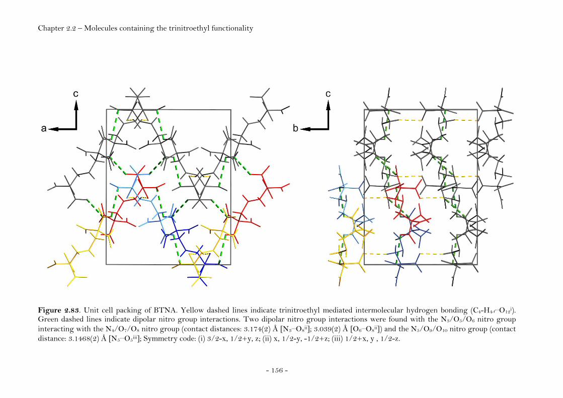
Chapter 2.2 – Molecules containing the trinitroethyl functionality
- 156 -
Figure 2.83. Unit cell packing of BTNA. Yellow dashed lines indicate trinitroethyl mediated intermolecular hydrogen bonding (C4-H4A…O12i).
Green dashed lines indicate dipolar nitro group interactions. Two dipolar nitro group interactions were found with the N3/O5/O6 nitro group interacting with the N4/O7/O8 nitro group (contact distances: 3.174(2) Å [N3…O8ii]; 3.039(2) Å [O6…O8ii]) and the N5/O9/O10 nitro group (contact distance: 3.1468(2) Å [N5…O5iii]; Symmetry code: (i) 3/2-x, 1/2+y, z; (ii) x, 1/2-y, -1/2+z; (iii) 1/2+x, y , 1/2-z.
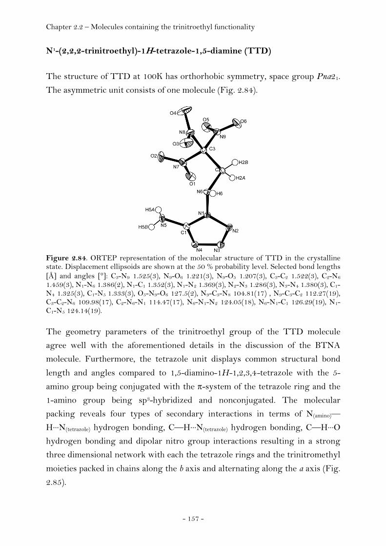
Chapter 2.2 – Molecules containing the trinitroethyl functionality
- 157 -
N1-(2,2,2-trinitroethyl)-1H-tetrazole-1,5-diamine (TTD)
The structure of TTD at 100K has orthorhobic symmetry, space group Pna21.
The asymmetric unit consists of one molecule (Fig. 2.84).
Figure 2.84. ORTEP representation of the molecular structure of TTD in the crystalline state. Displacement ellipsoids are shown at the 50 % probability level. Selected bond lengths [Å] and angles [°]: C3-N9 1.525(3), N9-O6 1.221(3), N9-O5 1.207(3), C3-C2 1.522(3), C2-N6 1.459(3), N1-N6 1.386(2), N1-C1 1.352(3), N1-N2 1.369(3), N2-N3 1.286(3), N3-N4 1.380(3), C1-N4 1.325(3), C1-N5 1.333(3), O5-N9-O6 127.5(2), N9-C3-N8 104.81(17) , N9-C3-C2 112.27(19), C3-C2-N6 109.98(17), C2-N6-N1 114.47(17), N6-N1-N2 124.05(18), N6-N1-C1 126.29(19), N1-C1-N5 124.14(19).
The geometry parameters of the trinitroethyl group of the TTD molecule
agree well with the aforementioned details in the discussion of the BTNA
molecule. Furthermore, the tetrazole unit displays common structural bond
length and angles compared to 1,5-diamino-1H-1,2,3,4-tetrazole with the 5-
amino group being conjugated with the π-system of the tetrazole ring and the
1-amino group being sp3-hybridized and nonconjugated. The molecular
packing reveals four types of secondary interactions in terms of N(amino)—
H···N(tetrazole) hydrogen bonding, C—H···N(tetrazole) hydrogen bonding, C—H···O
hydrogen bonding and dipolar nitro group interactions resulting in a strong
three dimensional network with each the tetrazole rings and the trinitromethyl
moieties packed in chains along the b axis and alternating along the a axis (Fig.
2.85).
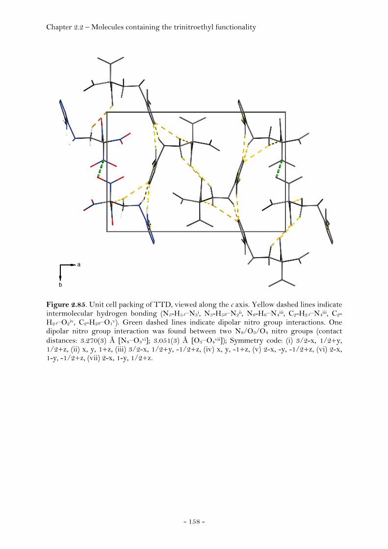
Chapter 2.2 – Molecules containing the trinitroethyl functionality
- 158 -
Figure 2.85. Unit cell packing of TTD, viewed along the c axis. Yellow dashed lines indicate intermolecular hydrogen bonding (N5-H5A
…N3i, N5-H5B…N2ii, N6-H6…N4iii, C2-H2A
…N4iii, C2-H2A
…O2iv, C2-H2B…O1v). Green dashed lines indicate dipolar nitro group interactions. One
dipolar nitro group interaction was found between two N8/O3/O4 nitro groups (contact distances: 3.270(3) Å [N8…O3vi]; 3.051(3) Å [O3…O4vii]); Symmetry code: (i) 3/2-x, 1/2+y, 1/2+z, (ii) x, y, 1+z, (iii) 3/2-x, 1/2+y, -1/2+z, (iv) x, y, -1+z, (v) 2-x, -y, -1/2+z, (vi) 2-x, 1-y, -1/2+z, (vii) 2-x, 1-y, 1/2+z.

Chapter 2.2 – Molecules containing the trinitroethyl functionality
- 159 -
N1,N5-Bis-(2,2,2-trinitroethyl)-1H-tetrazole-1,5-diamine (BTTD)
The structure of BTTD at 100K has monoclinic symmetry, space group P21/c.
The asymmetric unit consists of one molecule (Fig. 2.86).
Figure 2.86. ORTEP representation of the molecular structure of BTTD in the crystalline state. Displacement ellipsoids are shown at the 50 % probability level. Selected bond lengths [Å] and angles [°]:C3-N9 1.523(5), N9-O6 1.214(5), N9-O5 1.217(4), C3-C2 1.516(5), C2-N6 1.469(5), N1-N6 1.392(4), N1-C1 1.333(5), N1-N2 1.369(4), N2-N3 1.291(4), N3-N4 1.372(4), C1-N4 1.322(4), C1-N5 1.359(4), N5-C4 1.441(5), C4-C5 1.522(6), C5-N11 1.537(5), N11-O9 1.207(4), N11-O10 1.211(5), O5-N9-O6 127.7(4), N9-C3-N8 106.7(3), N9-C3-C2 111.7(3), C3-C2-N6 109.1(3), C2-N6-N1 111.5(3), N6-N1-N2 124.6(3), N6-N1-C1 126.6(3), N1-C1-N5 123.8(3), C1-N5-C4 119.3(3), N5-C4-C5 111.3(4), C4-C5-N11 113.0(3) , N11-C5-N12 106.3(3), O9-N11-O10. The presence of the second trinitroethyl group in the BTTD molecule does not
noticeably change either the bond length and angles of the trinitroethyl group
or the tetrazole ring compared to TTD and 1,5-diamino-1H-1,2,3,4-tetrazole.
The 5-amino group remains sp2 hybridized as indicated by the angles around
the N5 atom being close to 120°. The orientation of the N5-C4 bond and the C1-
N1 bond are trans relative to the C1-N5 bond directing the N5 trinitroethyl
group into the less sterically demanding position. The 1-amino group remains
sp3 hybridized with the H6 atom and the C2 atom being located on different
sides of the tetrazole plane and the angles around the N6 atom being close to
the tetrahedral angle. Two BTTD molecules form dimers interconnected with
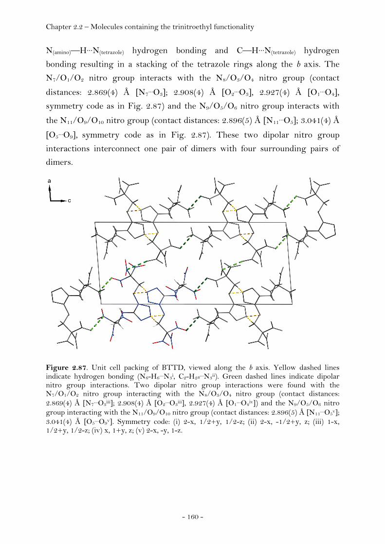
Chapter 2.2 – Molecules containing the trinitroethyl functionality
- 160 -
N(amino)—H···N(tetrazole) hydrogen bonding and C—H···N(tetrazole) hydrogen
bonding resulting in a stacking of the tetrazole rings along the b axis. The
N7/O1/O2 nitro group interacts with the N8/O3/O4 nitro group (contact
distances: 2.869(4) Å [N7…O3]; 2.908(4) Å [O2
…O3], 2.927(4) Å [O1…O4],
symmetry code as in Fig. 2.87) and the N9/O5/O6 nitro group interacts with
the N11/O9/O10 nitro group (contact distances: 2.896(5) Å [N11…O5]; 3.041(4) Å
[O5…O9], symmetry code as in Fig. 2.87). These two dipolar nitro group
interactions interconnect one pair of dimers with four surrounding pairs of
dimers.
Figure 2.87. Unit cell packing of BTTD, viewed along the b axis. Yellow dashed lines indicate hydrogen bonding (N6-H6…N3i, C2-H2B
…N3ii). Green dashed lines indicate dipolar nitro group interactions. Two dipolar nitro group interactions were found with the N7/O1/O2 nitro group interacting with the N8/O3/O4 nitro group (contact distances: 2.869(4) Å [N7…O3iii]; 2.908(4) Å [O2…O3iii], 2.927(4) Å [O1…O4iv]) and the N9/O5/O6 nitro group interacting with the N11/O9/O10 nitro group (contact distances: 2.896(5) Å [N11…O5v]; 3.041(4) Å [O5…O9v]. Symmetry code: (i) 2-x, 1/2+y, 1/2-z; (ii) 2-x, -1/2+y, z; (iii) 1-x, 1/2+y, 1/2-z; (iv) x, 1+y, z; (v) 2-x, -y, 1-z.
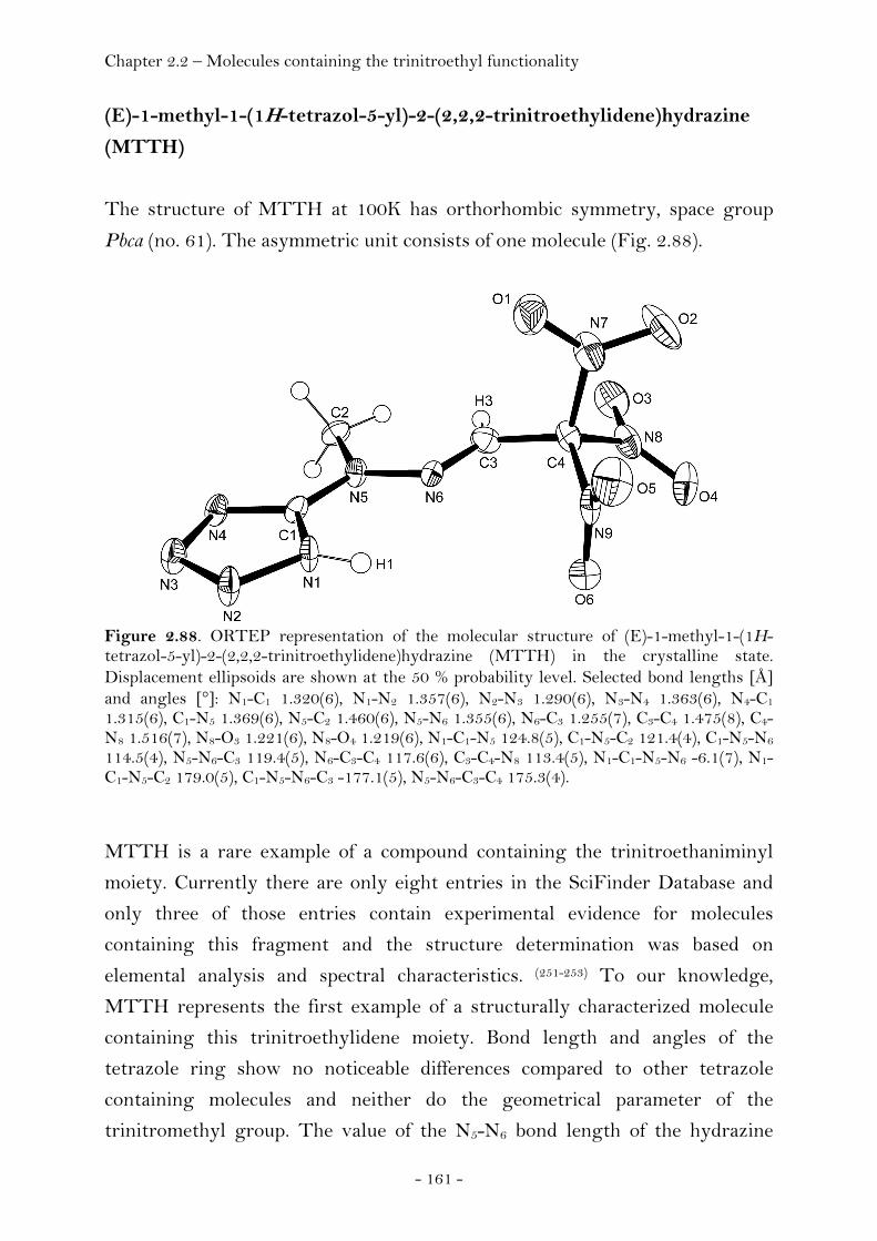
Chapter 2.2 – Molecules containing the trinitroethyl functionality
- 161 -
(E)-1-methyl-1-(1H-tetrazol-5-yl)-2-(2,2,2-trinitroethylidene)hydrazine
(MTTH)
The structure of MTTH at 100K has orthorhombic symmetry, space group
Pbca (no. 61). The asymmetric unit consists of one molecule (Fig. 2.88).
Figure 2.88. ORTEP representation of the molecular structure of (E)-1-methyl-1-(1H-tetrazol-5-yl)-2-(2,2,2-trinitroethylidene)hydrazine (MTTH) in the crystalline state. Displacement ellipsoids are shown at the 50 % probability level. Selected bond lengths [Å] and angles [°]: N1-C1 1.320(6), N1-N2 1.357(6), N2-N3 1.290(6), N3-N4 1.363(6), N4-C1 1.315(6), C1-N5 1.369(6), N5-C2 1.460(6), N5-N6 1.355(6), N6-C3 1.255(7), C3-C4 1.475(8), C4-N8 1.516(7), N8-O3 1.221(6), N8-O4 1.219(6), N1-C1-N5 124.8(5), C1-N5-C2 121.4(4), C1-N5-N6 114.5(4), N5-N6-C3 119.4(5), N6-C3-C4 117.6(6), C3-C4-N8 113.4(5), N1-C1-N5-N6 -6.1(7), N1-C1-N5-C2 179.0(5), C1-N5-N6-C3 -177.1(5), N5-N6-C3-C4 175.3(4).
MTTH is a rare example of a compound containing the trinitroethaniminyl
moiety. Currently there are only eight entries in the SciFinder Database and
only three of those entries contain experimental evidence for molecules
containing this fragment and the structure determination was based on
elemental analysis and spectral characteristics. (251-253) To our knowledge,
MTTH represents the first example of a structurally characterized molecule
containing this trinitroethylidene moiety. Bond length and angles of the
tetrazole ring show no noticeable differences compared to other tetrazole
containing molecules and neither do the geometrical parameter of the
trinitromethyl group. The value of the N5-N6 bond length of the hydrazine
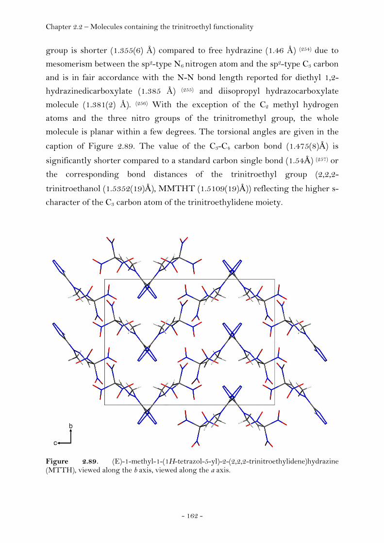
Chapter 2.2 – Molecules containing the trinitroethyl functionality
- 162 -
group is shorter (1.355(6) Å) compared to free hydrazine (1.46 Å) (254) due to
mesomerism between the sp2-type N6 nitrogen atom and the sp2-type C3 carbon
and is in fair accordance with the N-N bond length reported for diethyl 1,2-
hydrazinedicarboxylate (1.385 Å) (255) and diisopropyl hydrazocarboxylate
molecule (1.381(2) Å). (256) With the exception of the C2 methyl hydrogen
atoms and the three nitro groups of the trinitromethyl group, the whole
molecule is planar within a few degrees. The torsional angles are given in the
caption of Figure 2.89. The value of the C3-C4 carbon bond (1.475(8)Å) is
significantly shorter compared to a standard carbon single bond (1.54Å) (257) or
the corresponding bond distances of the trinitroethyl group (2,2,2-
trinitroethanol (1.5352(19)Å), MMTHT (1.5109(19)Å)) reflecting the higher s-
character of the C3 carbon atom of the trinitroethylidene moiety.
Figure 2.89. (E)-1-methyl-1-(1H-tetrazol-5-yl)-2-(2,2,2-trinitroethylidene)hydrazine (MTTH), viewed along the b axis, viewed along the a axis.
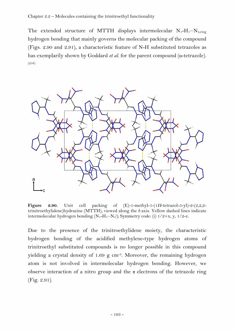
Chapter 2.2 – Molecules containing the trinitroethyl functionality
- 163 -
The extended structure of MTTH displays intermolecular N1-H1···N4,ring
hydrogen bonding that mainly governs the molecular packing of the compound
(Figs. 2.90 and 2.91), a characteristic feature of N-H substituted tetrazoles as
has exemplarily shown by Goddard et al. for the parent compound (α-tetrazole). (258)
Figure 2.90. Unit cell packing of (E)-1-methyl-1-(1H-tetrazol-5-yl)-2-(2,2,2-trinitroethylidene)hydrazine (MTTH), viewed along the b axis. Yellow dashed lines indicate intermolecular hydrogen bonding (N1-H1···N4i); Symmetry code: (i) 1/2+x, y, 1/2-z. Due to the presence of the trinitroethylidene moiety, the characteristic
hydrogen bonding of the acidified methylene-type hydrogen atoms of
trinitroethyl substituted compounds is no longer possible in this compound
yielding a crystal density of 1.69 g cm-3. Moreover, the remaining hydrogen
atom is not involved in intermolecular hydrogen bonding. However, we
observe interaction of a nitro group and the π electrons of the tetrazole ring
(Fig. 2.91).
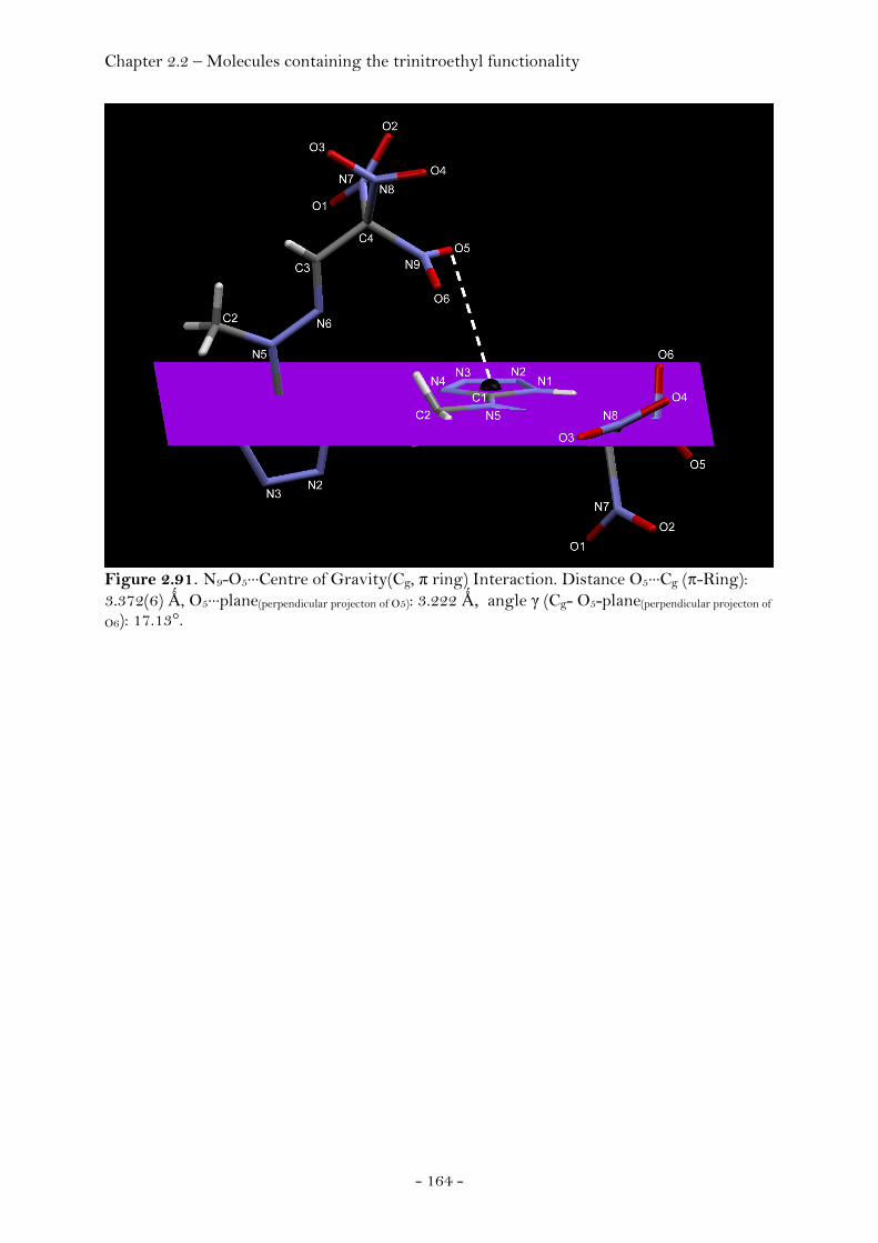
Chapter 2.2 – Molecules containing the trinitroethyl functionality
- 164 -
Figure 2.91. N9-O5···Centre of Gravity(Cg, π ring) Interaction. Distance O5···Cg (π-Ring): 3.372(6) Ǻ, O5···plane(perpendicular projecton of O5): 3.222 Ǻ, angle γ (Cg- O5-plane(perpendicular projecton of
O6): 17.13°.
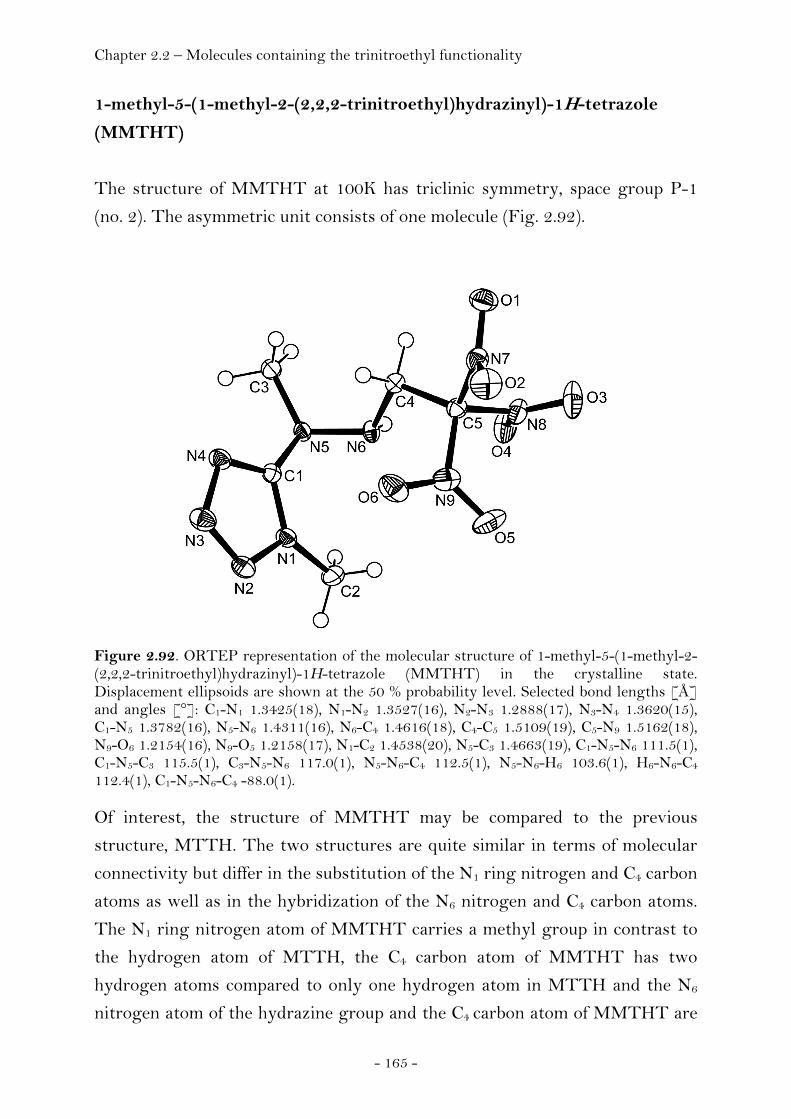
Chapter 2.2 – Molecules containing the trinitroethyl functionality
- 165 -
1-methyl-5-(1-methyl-2-(2,2,2-trinitroethyl)hydrazinyl)-1H-tetrazole
(MMTHT)
The structure of MMTHT at 100K has triclinic symmetry, space group P-1
(no. 2). The asymmetric unit consists of one molecule (Fig. 2.92).
Figure 2.92. ORTEP representation of the molecular structure of 1-methyl-5-(1-methyl-2-(2,2,2-trinitroethyl)hydrazinyl)-1H-tetrazole (MMTHT) in the crystalline state. Displacement ellipsoids are shown at the 50 % probability level. Selected bond lengths [Å] and angles [°]: C1-N1 1.3425(18), N1-N2 1.3527(16), N2-N3 1.2888(17), N3-N4 1.3620(15), C1-N5 1.3782(16), N5-N6 1.4311(16), N6-C4 1.4616(18), C4-C5 1.5109(19), C5-N9 1.5162(18), N9-O6 1.2154(16), N9-O5 1.2158(17), N1-C2 1.4538(20), N5-C3 1.4663(19), C1-N5-N6 111.5(1), C1-N5-C3 115.5(1), C3-N5-N6 117.0(1), N5-N6-C4 112.5(1), N5-N6-H6 103.6(1), H6-N6-C4 112.4(1), C1-N5-N6-C4 -88.0(1). Of interest, the structure of MMTHT may be compared to the previous
structure, MTTH. The two structures are quite similar in terms of molecular
connectivity but differ in the substitution of the N1 ring nitrogen and C4 carbon
atoms as well as in the hybridization of the N6 nitrogen and C4 carbon atoms.
The N1 ring nitrogen atom of MMTHT carries a methyl group in contrast to
the hydrogen atom of MTTH, the C4 carbon atom of MMTHT has two
hydrogen atoms compared to only one hydrogen atom in MTTH and the N6
nitrogen atom of the hydrazine group and the C4 carbon atom of MMTHT are
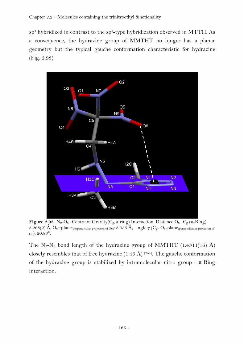
Chapter 2.2 – Molecules containing the trinitroethyl functionality
- 166 -
sp3 hybridized in contrast to the sp2-type hybridization observed in MTTH. As
a consequence, the hydrazine group of MMTHT no longer has a planar
geometry but the typical gauche conformation characteristic for hydrazine
(Fig. 2.93).
Figure 2.93. N9-O6···Centre of Gravity(Cg, π ring) Interaction. Distance O6···Cg (π-Ring): 3.268(2) Ǻ, O6···plane(perpendicular projecton of O6): 3.055 Ǻ, angle γ (Cg- O6-plane(perpendicular projecton of
O6): 20.83°. The N5-N6 bond length of the hydrazine group of MMTHT (1.4311(16) Å)
closely resembles that of free hydrazine (1.46 Å) (254). The gauche conformation
of the hydrazine group is stabilized by intramolecular nitro group - π-Ring
interaction.

Chapter 2.2 – Molecules containing the trinitroethyl functionality
- 167 -
Figure 2.94. Unit cell packing of 1-methyl-5-(1-methyl-2-(2,2,2-trinitroethyl)hydrazinyl)-1H-tetrazole (MMTHT), viewed along the a axis. Yellow dashed lines indicate intermolecular hydrogen bonding (N6-H6···O2i, N6-H6···N2ii). Green dashed lines indicate intermolecular hydrogen bonding of the acidified methylene type hydrogen atoms of the trinitroethyl moiety (C4-H4A···N4iii, C4-H4B···N3ii); Symmetry code: (i) x, -1+y, z; (ii) 1+x, 1+y, z; (iii) 1-x, 1-y, -z.
The extended structure of MMTHT is shown in Figure 2.94. The acidified
methylene-type hydrogen atoms are involved in intermolecular hydrogen
bonding yielding a pair of dimers running along the crystallographic a axis
(C4-H4A···N4, C4-H4B···N3, Fig. 2.94). Due to the presence of the methyl groups,
the characteristic intermolecular N-H···Nring hydrogen bonding frequently
observed in tetrazole compounds cannot occur. However, the hydrazinic-type
hydrogen atom H6 is involved in a bifurcated hydrogen bond (N6-H6···O2, N6-
H6···N2, Fig. 2.94) towards both the tetrazole ring and one nitro group
interconnecting the pairs of dimers formed due to C-H···N hydrogen bonding
and mentioned above. Compared to MTTH (1.69 g cm-3), the crystal density of
MMTHT (1.63 g cm-3) is slightly lower although – a tribute to the two bulky
methyl groups present in MMTHT.

Chapter 2.2 – Molecules containing the trinitroethyl functionality
- 168 -
2-(5-(1-methyl-2-(2,2,2-trinitroethyl)hydrazinyl)-1H-tetrazol-1-yl)ethanol
(MTHTE)
The structure of MTHTE at 100K has monoclinic symmetry, space group
P21/c (no. 14). The asymmetric unit consists of one molecule (Fig. 2.95).
Figure 2.95. ORTEP representation of the molecular structure of 2-(5-(1-methyl-2-(2,2,2-trinitroethyl)hydrazinyl)-1H-tetrazol-1-yl)ethanol (MTHTE) in the crystalline state. Displacement ellipsoids are shown at the 50 % probability level. Selected bond lengths [Å] and angles [°]: C1-N1 1.3476(15), N1-N2 1.3567(14), N2-N3 1.2838(15), N3-N4 1.3685(15), N4-C1 1.3240(15), N1-C5 1.4723(16), C5-C6 1.5118(19), C6-O7 1.4195(15), C1-N5 1.3720(15), N5-C2 1.4662(15), N5-N6 1.4232(14), N6-C3 1.4713(16), C3-C4 1.5113(17), C4-N8 1.5223(15), N8-O3 1.2157(13), N8-O4 1.2178(13), N1-C1-N5 126.47(11), C1-N5-C2 116.18(10), C1-N5-N6 114.68(9), N5-N6-C3 110.79(9), N6-C3-C4 110.08(10), C3-C4-N8 112.41(10), C1-N5-N6-C3 92.93(12), C2-N5-N6-C3 -49.10(14), N5-C1-N1-N2 174.27(11), N5-C1-N1-C5 4.9(2), C6-C5-N1-C1 69.05(16), C6-C5-N1-N2 -99.28(13), N4-C1-N5-N6 -141.91(12), N1-C1-N5-N6 43.89(16), N4-C1-N5-C2 0.68(17), N1-C1-N5-C2 -173.52(12). The presence of the hydroxyethyl group in MTHTE compared to the methyl
group present in MMTHT does not noticeably change the values of bond
lengths or bond angles. The gauche conformation of the hydrazine group is
retained and the hydrazinic-type H6 hydrogen atom is involved in a bifurcated
hydrogen bond yielding one intramolecular hydrogen bond (N6-H6···O4) and
one intermolecular hydrogen bond (N6-H6···O2, Fig. 2.96).
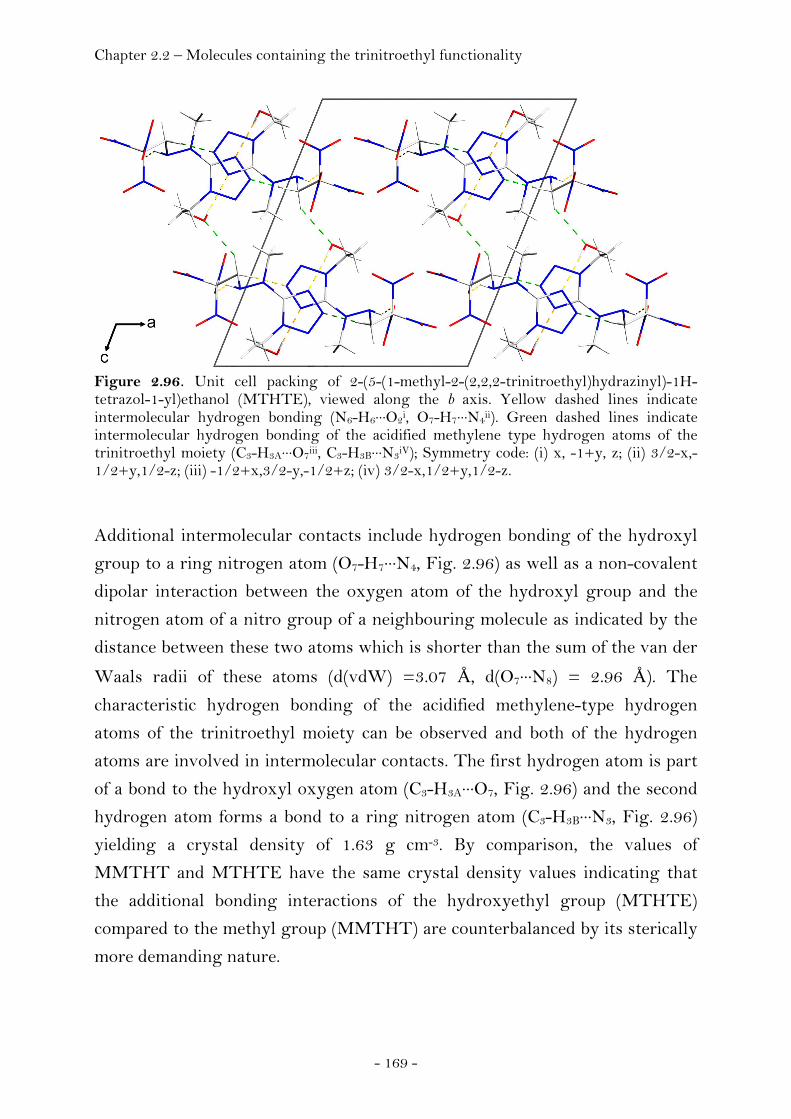
Chapter 2.2 – Molecules containing the trinitroethyl functionality
- 169 -
Figure 2.96. Unit cell packing of 2-(5-(1-methyl-2-(2,2,2-trinitroethyl)hydrazinyl)-1H-tetrazol-1-yl)ethanol (MTHTE), viewed along the b axis. Yellow dashed lines indicate intermolecular hydrogen bonding (N6-H6···O2i, O7-H7···N4ii). Green dashed lines indicate intermolecular hydrogen bonding of the acidified methylene type hydrogen atoms of the trinitroethyl moiety (C3-H3A···O7iii, C3-H3B···N3iV); Symmetry code: (i) x, -1+y, z; (ii) 3/2-x,-1/2+y,1/2-z; (iii) -1/2+x,3/2-y,-1/2+z; (iv) 3/2-x,1/2+y,1/2-z.
Additional intermolecular contacts include hydrogen bonding of the hydroxyl
group to a ring nitrogen atom (O7-H7···N4, Fig. 2.96) as well as a non-covalent
dipolar interaction between the oxygen atom of the hydroxyl group and the
nitrogen atom of a nitro group of a neighbouring molecule as indicated by the
distance between these two atoms which is shorter than the sum of the van der
Waals radii of these atoms (d(vdW) =3.07 Å, d(O7···N8) = 2.96 Å). The
characteristic hydrogen bonding of the acidified methylene-type hydrogen
atoms of the trinitroethyl moiety can be observed and both of the hydrogen
atoms are involved in intermolecular contacts. The first hydrogen atom is part
of a bond to the hydroxyl oxygen atom (C3-H3A···O7, Fig. 2.96) and the second
hydrogen atom forms a bond to a ring nitrogen atom (C3-H3B···N3, Fig. 2.96)
yielding a crystal density of 1.63 g cm-3. By comparison, the values of
MMTHT and MTHTE have the same crystal density values indicating that
the additional bonding interactions of the hydroxyethyl group (MTHTE)
compared to the methyl group (MMTHT) are counterbalanced by its sterically
more demanding nature.
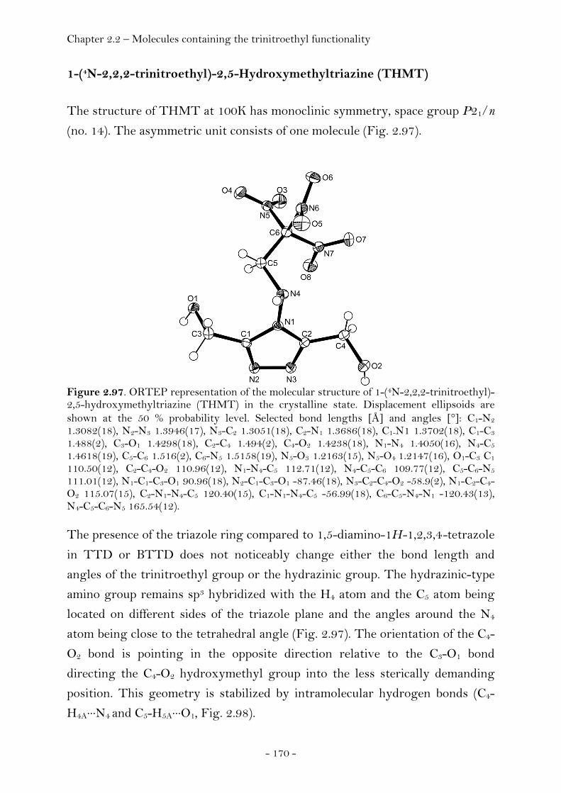
Chapter 2.2 – Molecules containing the trinitroethyl functionality
- 170 -
1-(4N-2,2,2-trinitroethyl)-2,5-Hydroxymethyltriazine (THMT)
The structure of THMT at 100K has monoclinic symmetry, space group P21/n
(no. 14). The asymmetric unit consists of one molecule (Fig. 2.97).
Figure 2.97. ORTEP representation of the molecular structure of 1-(4N-2,2,2-trinitroethyl)-2,5-hydroxymethyltriazine (THMT) in the crystalline state. Displacement ellipsoids are shown at the 50 % probability level. Selected bond lengths [Å] and angles [°]: C1-N2 1.3082(18), N2-N3 1.3946(17), N3-C2 1.3051(18), C2-N1 1.3686(18), C1-N1 1.3702(18), C1-C3 1.488(2), C3-O1 1.4298(18), C2-C4 1.494(2), C4-O2 1.4238(18), N1-N4 1.4050(16), N4-C5 1.4618(19), C5-C6 1.516(2), C6-N5 1.5158(19), N5-O3 1.2163(15), N5-O4 1.2147(16), O1-C3 C1 110.50(12), C2-C4-O2 110.96(12), N1-N4-C5 112.71(12), N4-C5-C6 109.77(12), C5-C6-N5 111.01(12), N1-C1-C3-O1 90.96(18), N2-C1-C3-O1 -87.46(18), N3-C2-C4-O2 -58.9(2), N1-C2-C4-O2 115.07(15), C2-N1-N4-C5 120.40(15), C1-N1-N4-C5 -56.99(18), C6-C5-N4-N1 -120.43(13), N4-C5-C6-N5 165.54(12).
The presence of the triazole ring compared to 1,5-diamino-1H-1,2,3,4-tetrazole
in TTD or BTTD does not noticeably change either the bond length and
angles of the trinitroethyl group or the hydrazinic group. The hydrazinic-type
amino group remains sp3 hybridized with the H4 atom and the C5 atom being
located on different sides of the triazole plane and the angles around the N4
atom being close to the tetrahedral angle (Fig. 2.97). The orientation of the C4-
O2 bond is pointing in the opposite direction relative to the C3-O1 bond
directing the C4-O2 hydroxymethyl group into the less sterically demanding
position. This geometry is stabilized by intramolecular hydrogen bonds (C4-
H4A···N4 and C5-H5A···O1, Fig. 2.98).
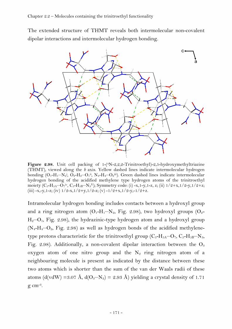
Chapter 2.2 – Molecules containing the trinitroethyl functionality
- 171 -
The extended structure of THMT reveals both intermolecular non-covalent
dipolar interactions and intermolecular hydrogen bonding.
Figure 2.98. Unit cell packing of 1-(4N-2,2,2-Trinitroethyl)-2,5-hydroxymethyltriazine (THMT), viewed along the b axis. Yellow dashed lines indicate intermolecular hydrogen bonding (O1-H1···N2i, O2-H2···O1ii, N4-H4···O2iii). Green dashed lines indicate intermolecular hydrogen bonding of the acidified methylene type hydrogen atoms of the trinitroethyl moiety (C5-H5A···O7iv, C5-H5B···N3V); Symmetry code: (i) -x,1-y,1-z, z; (ii) 1/2+x,1/2-y,1/2+z; (iii) -x,-y,1-z; (iv) 1/2-x,1/2+y,1/2-z; (v) -1/2+x,1/2-y,-1/2+z.
Intramolecular hydrogen bonding includes contacts between a hydroxyl group
and a ring nitrogen atom (O1-H1···N2, Fig. 2.98), two hydroxyl groups (O2-
H2···O1, Fig. 2.98), the hydrazinic-type hydrogen atom and a hydroxyl group
(N4-H4···O2, Fig. 2.98) as well as hydrogen bonds of the acidified methylene-
type protons characteristic for the trinitroethyl group (C5-H5A···O7, C5-H5B···N3,
Fig. 2.98). Additionally, a non-covalent dipolar interaction between the O3
oxygen atom of one nitro group and the N2 ring nitrogen atom of a
neighbouring molecule is present as indicated by the distance between these
two atoms which is shorter than the sum of the van der Waals radii of these
atoms (d(vdW) =3.07 Å, d(O3···N2) = 2.93 Å) yielding a crystal density of 1.71
g cm-3.

Chapter 2.2 – Molecules containing the trinitroethyl functionality
- 172 -
N3,N6-bis-(2,2,2-trinitroethyl)-1,2,4,5-tetrazine-3,6-diamine (BTAT)
The structure of BTAT at 100K has orthorhombic symmetry, space group
Pna21. The asymmetric unit consists of one molecule (Fig. 2.99).
Figure 2.99. Photographic picture of single crystals of BTAT (left picture). ORTEP representation of the molecular structure of BTAT in the crystalline state (right picture). Displacement ellipsoids are shown at the 50 % probability level. Selected bond lengths [Å] and angles [°]: C1-N1 1.337(2), N1-N2 1.320(2), N2-C2 1.349(2), C2-N3 1.344(2), N3-N4
1.3179(19), N4-C1 1.355(2), C1-N5 1.370(2), N5-C5 1.433(2), C5-C6 1.537(2), C6-N10 1.529(2), N10-O7 1.211(2), N10-O8 1.215(2), C2-N6 1.365(2), N6-C3 1.441(2), C3-C4 1.530(2), C4-N7
1.531(2), N7-O1 1.213(2), N7-O2 1.220(2), C1-N5-C5 120.15(15), N5-C5-C6 112.69(14), C5-C6-N10 112.67(14), N10-C6-N12 105.53(13), O7-N10-O8 127.98(16), N3-C2-N6 118.14(15), C2-N6-C3
120.35(14), N6-C3-C4 112.33(14), C3-C4-N7 112.67(14), N7-C4-N8 105.87(13), O1-N7-O2
127.64(17), N1-C1-N4 124.78(15), C1-N4-N3 116.87(14), N4-N3-C2 118.32(14), N3-C2-N2
124.64(15), C2-N2-N1 117.06(14) , N2-N1-C1 118.32(14).
The planar geometry reported for 3,6-diamino-1,2,3,4-tetrazine (259) is retained
in the structure of BTAT. The two trinitroethyl groups bonded to the sp2-type
amino nitrogen atoms are oriented trans to each other and are located on
different sides of the tetrazine plane. Bond length and angles of the
trinitroethyl group display no distinctive features compared to the structures
of BTNA, TTD or BTTD and neither do the geometric parameters of the
tetrazine moiety compared to the values of 3,6-diamino-1,2,3,4-tetrazine. The
unit cell of solid BTAT consists of pairs of dimers of BTAT molecules
connected through N(amino)—H···N(tetrazine) hydrogen bonding. In turn, these
pairs of dimers are interconnected with each other through dipolar nitro group
interactions yielding a two dimensional chain of BTAT molecules running
along the axis. The chains are connected through C—H···O hydrogen bonding
along the c axis yielding a high crystal density of 1.89 g cm-3 (Fig. 2.100).
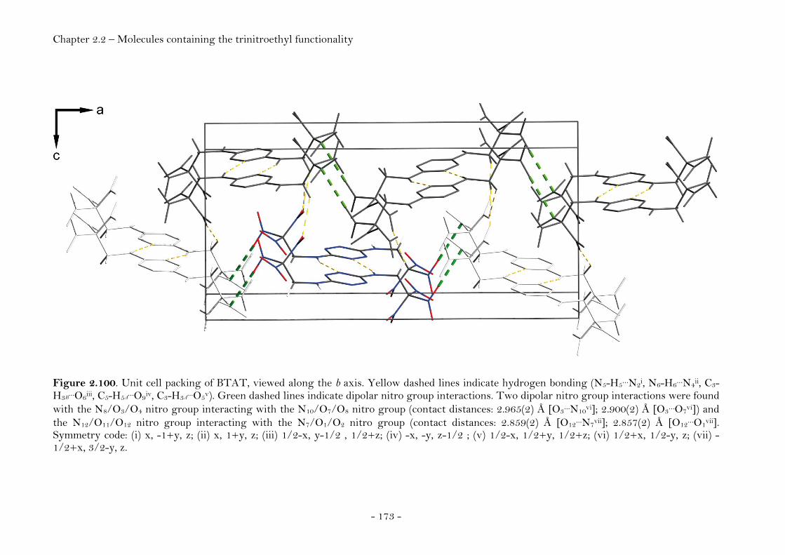
Chapter 2.2 – Molecules containing the trinitroethyl functionality
- 173 -
Figure 2.100. Unit cell packing of BTAT, viewed along the b axis. Yellow dashed lines indicate hydrogen bonding (N5-H5…N2i, N6-H6…N4ii, C3-H3B
…O6iii, C5-H5A…O9iv, C3-H3A
…O5v). Green dashed lines indicate dipolar nitro group interactions. Two dipolar nitro group interactions were found with the N8/O3/O4 nitro group interacting with the N10/O7/O8 nitro group (contact distances: 2.965(2) Å [O3…N10vi]; 2.900(2) Å [O3…O7vi]) and the N12/O11/O12 nitro group interacting with the N7/O1/O2 nitro group (contact distances: 2.859(2) Å [O12…N7vii]; 2.857(2) Å [O12…O1vii]. Symmetry code: (i) x, -1+y, z; (ii) x, 1+y, z; (iii) 1/2-x, y-1/2 , 1/2+z; (iv) -x, -y, z-1/2 ; (v) 1/2-x, 1/2+y, 1/2+z; (vi) 1/2+x, 1/2-y, z; (vii) -1/2+x, 3/2-y, z.
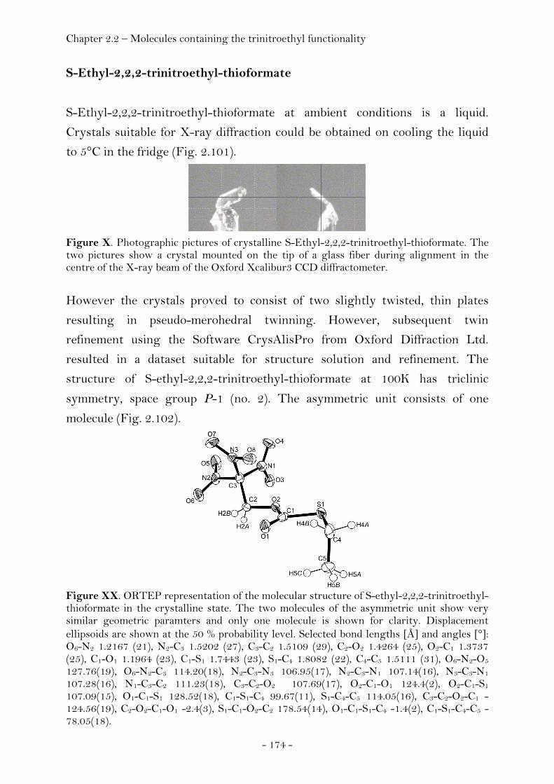
Chapter 2.2 – Molecules containing the trinitroethyl functionality
- 174 -
S-Ethyl-2,2,2-trinitroethyl-thioformate
S-Ethyl-2,2,2-trinitroethyl-thioformate at ambient conditions is a liquid.
Crystals suitable for X-ray diffraction could be obtained on cooling the liquid
to 5°C in the fridge (Fig. 2.101).
Figure X. Photographic pictures of crystalline S-Ethyl-2,2,2-trinitroethyl-thioformate. The two pictures show a crystal mounted on the tip of a glass fiber during alignment in the centre of the X-ray beam of the Oxford Xcalibur3 CCD diffractometer.
However the crystals proved to consist of two slightly twisted, thin plates
resulting in pseudo-merohedral twinning. However, subsequent twin
refinement using the Software CrysAlisPro from Oxford Diffraction Ltd.
resulted in a dataset suitable for structure solution and refinement. The
structure of S-ethyl-2,2,2-trinitroethyl-thioformate at 100K has triclinic
symmetry, space group P-1 (no. 2). The asymmetric unit consists of one
molecule (Fig. 2.102).
Figure XX. ORTEP representation of the molecular structure of S-ethyl-2,2,2-trinitroethyl-thioformate in the crystalline state. The two molecules of the asymmetric unit show very similar geometric paramters and only one molecule is shown for clarity. Displacement ellipsoids are shown at the 50 % probability level. Selected bond lengths [Å] and angles [°]: O6-N2 1.2167 (21), N2-C3 1.5202 (27), C3-C2 1.5109 (29), C2-O2 1.4264 (25), O2-C1 1.3737 (25), C1-O1 1.1964 (23), C1-S1 1.7443 (23), S1-C4 1.8082 (22), C4-C5 1.5111 (31), O6-N2-O5 127.76(19), O6-N2-C3 114.20(18), N2-C3-N3 106.95(17), N2-C3-N1 107.14(16), N3-C3-N1 107.28(16), N1-C3-C2 111.23(18), C3-C2-O2 107.69(17), O2-C1-O1 124.4(2), O2-C1-S1 107.09(15), O1-C1-S1 128.52(18), C1-S1-C4 99.67(11), S1-C4-C5 114.05(16), C3-C2-O2-C1 -124.56(19), C2-O2-C1-O1 -2.4(3), S1-C1-O2-C2 178.54(14), O1-C1-S1-C4 -1.4(2), C1-S1-C4-C5 -78.05(18).
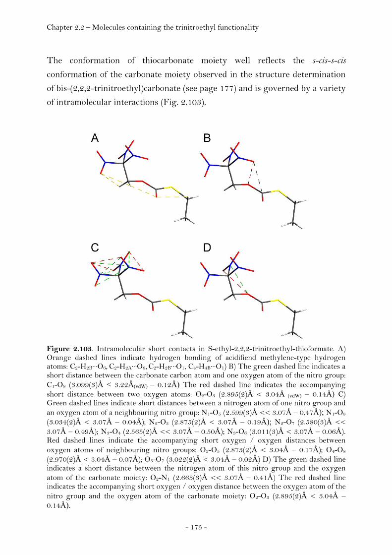
Chapter 2.2 – Molecules containing the trinitroethyl functionality
- 175 -
The conformation of thiocarbonate moiety well reflects the s-cis-s-cis
conformation of the carbonate moiety observed in the structure determination
of bis-(2,2,2-trinitroethyl)carbonate (see page 177) and is governed by a variety
of intramolecular interactions (Fig. 2.103).
Figure 2.103. Intramolecular short contacts in S-ethyl-2,2,2-trinitroethyl-thioformate. A) Orange dashed lines indicate hydrogen bonding of acidifiend methylene-type hydrogen atoms: C2-H2B···O6, C2-H2A···O6, C2-H2B···O1, C4-H4B···O1) B) The green dashed line indicates a short distance between the carbonate carbon atom and one oxygen atom of the nitro group: C1-O8 (3.099(3)Å < 3.22Å(vdW) – 0.12Å) The red dashed line indicates the accompanying short distance between two oxygen atoms: O2-O3 (2.895(2)Å < 3.04Å (vdW) – 0.14Å) C) Green dashed lines indicate short distances between a nitrogen atom of one nitro group and an oxygen atom of a neighbouring nitro group: N1-O5 (2.599(3)Å << 3.07Å – 0.47Å); N1-O8
(3.034(2)Å < 3.07Å – 0.04Å); N2-O3 (2.875(2)Å < 3.07Å – 0.19Å); N2-O7 (2.580(3)Å << 3.07Å – 0.49Å); N3-O4 (2.565(2)Å << 3.07Å – 0.50Å); N3-O6 (3.011(3)Å < 3.07Å – 0.06Å). Red dashed lines indicate the accompanying short oxygen / oxygen distances between oxygen atoms of neighbouring nitro groups: O3-O5 (2.873(2)Å < 3.04Å – 0.17Å); O4-O8 (2.970(2)Å < 3.04Å – 0.07Å); O5-O7 (3.022(2)Å < 3.04Å – 0.02Å) D) The green dashed line indicates a short distance between the nitrogen atom of this nitro group and the oxygen atom of the carbonate moiety: O2-N1 (2.663(3)Å << 3.07Å – 0.41Å) The red dashed line indicates the accompanying short oxygen / oxygen distance between the oxygen atom of the nitro group and the oxygen atom of the carbonate moiety: O2-O3 (2.895(2)Å < 3.04Å – 0.14Å).
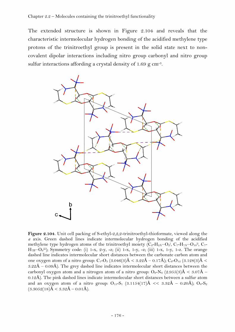
Chapter 2.2 – Molecules containing the trinitroethyl functionality
- 176 -
The extended structure is shown in Figure 2.104 and reveals that the
characteristic intermolecular hydrogen bonding of the acidified methylene type
protons of the trinitroethyl group is present in the solid state next to non-
covalent dipolar interactions including nitro group carbonyl and nitro group
sulfur interactions affording a crystal density of 1.69 g cm-3.
Figure 2.104. Unit cell packing of S-ethyl-2,2,2-trinitroethyl-thioformate, viewed along the a axis. Green dashed lines indicate intermolecular hydrogen bonding of the acidified methylene type hydrogen atoms of the trinitroethyl moiety (C2-H2A···O2i, C7-H7A···O10ii, C7-H7B···O9iii); Symmetry code: (i) 1-x, 2-y, -z; (ii) 1-x, 1-y, -z; (iii) 1-x, 1-y, 1-z. The orange dashed line indicates intermolecular short distances between the carbonate carbon atom and one oxygen atom of a nitro group: C1-O3 (3.046(3)Å < 3.22Å – 0.17Å); C6-O12 (3.128(3)Å < 3.22Å – 0.09Å). The grey dashed line indicates intermolecular short distances between the carbonyl oxygen atom and a nitrogen atom of a nitro group: O9-N6 (2.955(3)Å < 3.07Å – 0.12Å). The pink dashed lines indicate intermolecular short distances between a sulfur atom and an oxygen atom of a nitro group: O15-S1 (3.1154(17)Å << 3.32Å – 0.20Å); O8-S2 (3.3052(18)Å < 3.32Å – 0.01Å).

Chapter 2.2 – Molecules containing the trinitroethyl functionality
- 177 -
Bis-(2,2,2-trinitroethyl)carbonate (BTC)
The structure of bis-(2,2,2-trinitroethyl)carbonate (BTC), C5H4N6O15, at 100K
has orthorhombic (Pbca) symmetry. The asymmetric unit consists of one
molecule (Fig. 2.105).
Figure 2.105. ORTEP representation of the molecular structure of bis-(2,2,2-trinitroethyl)carbonate in the crystalline state. Displacement ellipsoids are shown at the 50 % probability level. Selected bond lengths [Å] and angles [°]: C1-O1 1.1915(12), C1-O2 1.3451(12), C1-O3 1.3399(12), C2-O3 1.4255(12), C3-N1 1.5238(13), C3-N2 1.5214(13), C3-N3 1.5201(13), C4-O2 1.4294(13), C5-N4 1.5262(14), C5-N5 1.5131(13), C5-N6 1.5271(13), O1-C1-O3 127.57(10), O1-C1-O2 126.98(9), O3-C1-O2 105.46(8), O3-C2-C3 107.83(8), C2-C3-N3 110.72(8), C2-C3-N2 112.73(8), N3-C3-N2 107.22(8), C2-C3-N1 113.01(8), N3-C3-N1 106.17(8), N2-C3-N1 106.59(8), N5-C5-C4 112.25(8), N5-C5-N4 107.55(8), C4-C5-N4 113.26(8), N5-C5-N6 107.57(8), C4-C5-N6 109.88(8), N4-C5-N6 105.99(8), C1-O2-C4 116.48(8), C1-O3-C2 116.39(8), O1-C1-O2-C4 17.35(15), O3-C1-O2-C4 -162.59(8), C5-C4-O2-C1 118.99(9), O1-C1-O3-C2 9.22(15), O2-C1-O3-C2 -170.84(8), C3-C2-O3-C1 117.22(9).
The structure displays s-cis-s-cis conformation of the carbonate group, intra as
well as intermolecular C—H···O hydrogen bonding and dipolar nitro group
interactions that account for its exceptionally high density of 1.975 g cm-3,
which is significantly higher than the reported value of 1.88 g cm-3. (260). As a
consequence of the relationship between structure and crystal density this
polymorph contains available oxygen in amounts even superior to liquid
oxygen. The geometry in both trinitroethyl moieties is very similar with a
propeller-type orientation of the nitro groups (C3) bonded to the β-C atoms and
the conformation of the substituents of the α- and β-C atoms being staggered.
Bond lengths of the trinitroethyl units show unusual values in that the C-N
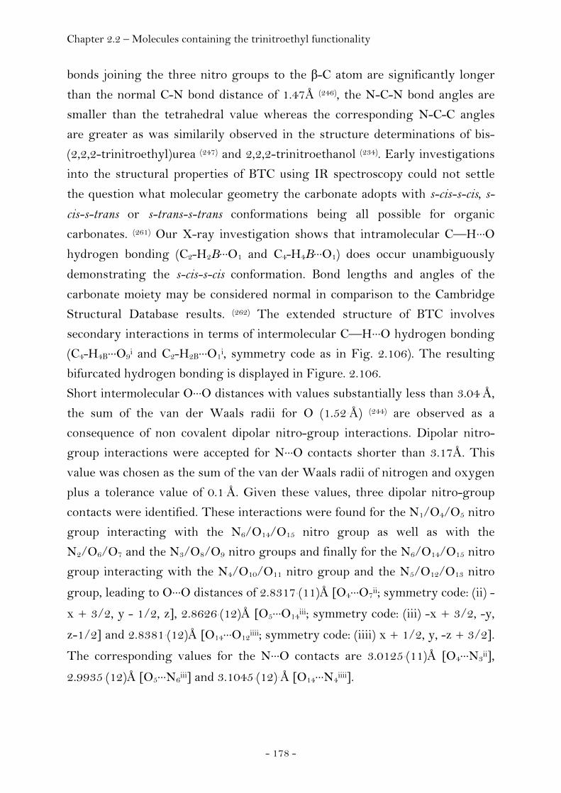
Chapter 2.2 – Molecules containing the trinitroethyl functionality
- 178 -
bonds joining the three nitro groups to the β-C atom are significantly longer
than the normal C-N bond distance of 1.47Å (246), the N-C-N bond angles are
smaller than the tetrahedral value whereas the corresponding N-C-C angles
are greater as was similarily observed in the structure determinations of bis-
(2,2,2-trinitroethyl)urea (247) and 2,2,2-trinitroethanol (234). Early investigations
into the structural properties of BTC using IR spectroscopy could not settle
the question what molecular geometry the carbonate adopts with s-cis-s-cis, s-
cis-s-trans or s-trans-s-trans conformations being all possible for organic
carbonates. (261) Our X-ray investigation shows that intramolecular C—H···O
hydrogen bonding (C2-H2B···O1 and C4-H4B···O1) does occur unambiguously
demonstrating the s-cis-s-cis conformation. Bond lengths and angles of the
carbonate moiety may be considered normal in comparison to the Cambridge
Structural Database results. (262) The extended structure of BTC involves
secondary interactions in terms of intermolecular C—H···O hydrogen bonding
(C4-H4B···O9i and C2-H2B···O1i, symmetry code as in Fig. 2.106). The resulting
bifurcated hydrogen bonding is displayed in Figure. 2.106.
Short intermolecular O···O distances with values substantially less than 3.04 Å,
the sum of the van der Waals radii for O (1.52 Å) (244) are observed as a
consequence of non covalent dipolar nitro-group interactions. Dipolar nitro-
group interactions were accepted for N···O contacts shorter than 3.17Å. This
value was chosen as the sum of the van der Waals radii of nitrogen and oxygen
plus a tolerance value of 0.1 Å. Given these values, three dipolar nitro-group
contacts were identified. These interactions were found for the N1/O4/O5 nitro
group interacting with the N6/O14/O15 nitro group as well as with the
N2/O6/O7 and the N3/O8/O9 nitro groups and finally for the N6/O14/O15 nitro
group interacting with the N4/O10/O11 nitro group and the N5/O12/O13 nitro
group, leading to O···O distances of 2.8317 (11)Å [O4···O7ii; symmetry code: (ii) -
x + 3/2, y - 1/2, z], 2.8626 (12)Å [O5···O14iii; symmetry code: (iii) -x + 3/2, -y,
z-1/2] and 2.8381 (12)Å [O14···O12iiii; symmetry code: (iiii) x + 1/2, y, -z + 3/2]. The corresponding values for the N···O contacts are 3.0125 (11)Å [O4···N3ii], 2.9935 (12)Å [O5···N6iii] and 3.1045 (12) Å [O14···N4iiii].
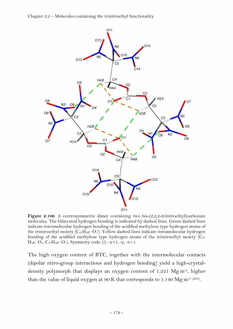
Chapter 2.2 – Molecules containing the trinitroethyl functionality
- 179 -
Figure 2.106. A centrosymmetric dimer containing two bis-(2,2,2-trinitroethyl)carbonate molecules. The bifurcated hydrogen bonding is indicated by dashed lines. Green dashed lines indicate intermolecular hydrogen bonding of the acidified methylene type hydrogen atoms of the trinitroethyl moiety (C2-H2B···O1i). Yellow dashed lines indicate intramolecular hydrogen bonding of the acidified methylene type hydrogen atoms of the trinitroethyl moiety (C2-H2B···O1, C4-H4B···O1). Symmetry code: (i) –x+1, -y, -z+1.
The high oxygen content of BTC, together with the intermolecular contacts
(dipolar nitro-group interactions and hydrogen bonding) yield a high-crystal-
density polymorph that displays an oxygen content of 1.221 Mg m-3, higher
than the value of liquid oxygen at 90 K that corresponds to 1.140 Mg m-3 (263).
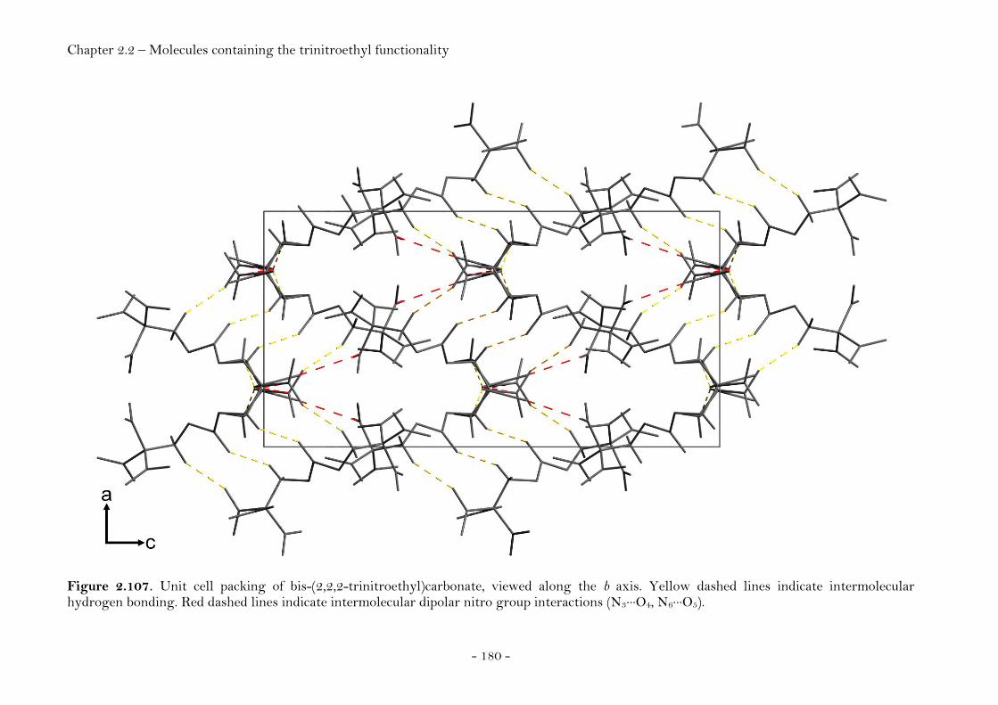
Chapter 2.2 – Molecules containing the trinitroethyl functionality
- 180 -
Figure 2.107. Unit cell packing of bis-(2,2,2-trinitroethyl)carbonate, viewed along the b axis. Yellow dashed lines indicate intermolecular hydrogen bonding. Red dashed lines indicate intermolecular dipolar nitro group interactions (N3···O4, N6···O5).
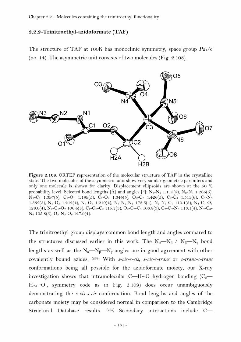
Chapter 2.2 – Molecules containing the trinitroethyl functionality
- 181 -
2,2,2-Trinitroethyl-azidoformate (TAF)
The structure of TAF at 100K has monoclinic symmetry, space group P21/c
(no. 14). The asymmetric unit consists of two molecules (Fig. 2.108).
Figure 2.108. ORTEP representation of the molecular structure of TAF in the crystalline state. The two molecules of the asymmetric unit show very similar geometric paramters and only one molecule is shown for clarity. Displacement ellipsoids are shown at the 50 % probability level. Selected bond lengths [Å] and angles [°]: N3-N2 1.115(5), N2-N1 1.266(5), N1-C1 1.397(5), C1-O1 1.199(5), C1-O2 1.345(5), O2-C2 1.426(5), C2-C3 1.513(6), C3-N5 1.532(5), N5-O5 1.212(4), N5-O6 1.219(4), N3-N2-N1 173.5(4), N2-N1-C1 110.1(3), N1-C1-O1 128.0(4), N1-C1-O2 106.4(3), C1-O2-C2 115.7(3), O2-C2-C3 106.8(3), C2-C3-N5 113.1(4), N5-C3-N6 105.8(3), O5-N5-O6 127.9(4).
The trinitroethyl group displays common bond length and angles compared to
the structures discussed earlier in this work. The Nα—Nβ / Nβ—Nγ bond
lengths as well as the Nα—Nβ—Nγ angles are in good agreement with other
covalently bound azides. (264) With s-cis-s-cis, s-cis-s-trans or s-trans-s-trans
conformations being all possible for the azidoformate moiety, our X-ray
investigation shows that intramolecular C—H···O hydrogen bonding (C2—
H2A···O1, symmetry code as in Fig. 2.109) does occur unambiguously
demonstrating the s-cis-s-cis conformation. Bond lengths and angles of the
carbonate moiety may be considered normal in comparison to the Cambridge
Structural Database results. (265) Secondary interactions include C—
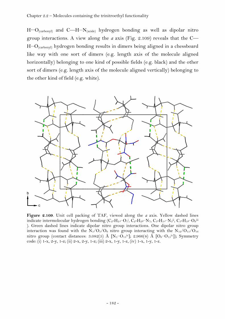
Chapter 2.2 – Molecules containing the trinitroethyl functionality
- 182 -
H···O(carbonyl) and C—H···N(azide) hydrogen bonding as well as dipolar nitro
group interactions. A view along the a axis (Fig. 2.109) reveals that the C—
H···O(carbonyl) hydrogen bonding results in dimers being aligned in a chessboard
like way with one sort of dimers (e.g. length axis of the molecule aligned
horizontally) belonging to one kind of possible fields (e.g. black) and the other
sort of dimers (e.g. length axis of the molecule aligned vertically) belonging to
the other kind of field (e.g. white).
Figure 2.109. Unit cell packing of TAF, viewed along the a axis. Yellow dashed lines indicate intermolecular hydrogen bonding (C2-H2A
…O1i, C2-H2B…N7, C5-H5A
…N3ii, C5-H5B…O9iii
). Green dashed lines indicate dipolar nitro group interactions. One dipolar nitro group interaction was found with the N5/O5/O6 nitro group interacting with the N12/O15/O16 nitro group (contact distances: 3.082(5) Å [N5…O15iv]; 2.966(4) Å [O6…O15iv]); Symmetry code: (i) 1-x, 2-y, 1-z; (ii) 2-x, 2-y, 1-z; (iii) 2-x, 1-y, 1-z, (iv) 1-x, 1-y, 1-z.

Chapter 2.2 – Molecules containing the trinitroethyl functionality
- 183 -
Bis-(2,2,2-trinitroethyl)-hydrazodicarboxylate (BTHC)
Pure BTHC solidifies as a glass-like amorphous solid. However, two solvents
were found to combine with BTHC to form stoichiometric crystal inclusion
complexes. In both co-crystals the host : guest ratio equals 1:1 for the acetone
as well as the ethyl acetate complex (BTHCa / BTHCe).
Figure 2.110. Photographic picture of a single crystal of the 1:1 co-crystal of BTHC and ethyl acetate (left picture). ORTEP representation of the molecular structure of BTHC in the 1:1 co-crystal of BTHC and ethyl acetate (right picture). Displacement ellipsoids are shown at the 50 % probability level. Selected bond lengths [Å] and angles [°]: O7-N5 1.2124(17), N5-O8 1.2079(18), N5-C3 1.5291(19), C3-C2 1.507(2), C2-O2 1.427(2), O2-C1 1.3603(19), C1-O1 1.2040(18), C1-N2 1.345(2), N2-N1 1.3779(19), N1-C4 1.360(2), C4-O9 1.2001(18), C4-O10 1.3641(18), O10-C5 1.4265(18), C5-C6 1.519(2), C6-N7 1.523(2), N7-O13 1.2117(17), N7-O14 1.2131(16), O7-N5-O8 126.53(13), N5-C3-N3 108.00(11), N5-C3-C2 113.90(13), C3-C2-O2 104.55(12), C2-O2-C1 114.11(12), O2-C1-O1 124.12(14), O2-C1-N2 108.70(13), O1-C1-N2 127.18(14), C1-N2-N1-C4 70.93(18), C1-N2-N1 119.47(13), N2-N1-C4 117.32(13), N1-C4-O9 126.31(14), N1-C4-O10 108.66(12), O9-C4-O10 125.03(14), C4-O10-C5 115.87(11), O10-C5-C6 108.45(12), C5-C6-N7 111.43(12), N7-C6-N8 106.10(12), O13-N7-O14 127.60(13).
Each of these mixed crystals crystallise in the space group P-1. Compared to
the 2,2,2-trinitroethylformyl moiety present in the structure of 2,2,2-
trinitroethylazidoformate, the geometric parameters of this group present in
the BTHC molecule of the two co-crystals show no significant differences.
Figure 2.110 exemplarily displays one BTHC molecule of the co-crystal of
BTHC with ethyl acetate. The values of the N1-N2 bond length of the
hydrazine group are shorter (1.3779(19) Å / BTHCe and 1.3790(14) Å /
BTHCa) compared to free hydrazine (1.46 Å) (254) due to mesomerism between
the nitrogen, carbon and carbonyl oxygen atoms and are in fair accordance
with the N-N bond length reported for diethyl 1,2-hydrazinedicarboxylate

Chapter 2.2 – Molecules containing the trinitroethyl functionality
- 184 -
(1.385 Å) (255) and diisopropyl hydrazocarboxylate molecule (1.381(2) Å). (266)
Bond lengths and angles of the carbonate moiety agree well to the results of a
comparison with Cambridge Structural Database entries. (267) Intramolecular
C—H···O(carbonyl) hydrogen bonding is present in BTHCe and BTHCa favouring
the the s-cis-s-cis conformation of the carbamate group.
The C1-N2-N1-C4 torsion angle of BTHCe (70.93(18)°) slightly differs from the
corresponding angle in BTHCa (67.80(14)°) due to secondary N—
H···O(carbonyl) hydrogen bonding of the hydrazine hydrogen atoms and the
carbonyl oxygen atom of the ethyl acetate or acetone molecule present in the
co-crystals. Intermolecular C—H···O(nitro) as well as C—H···O(carbonyl) hydrogen
bonding and dipolar nitro group interactions are present between two BTHC
molecules in the structures of BTHCe and BTHCa resulting in the formation of
BTHC dimers in both cases. These dimers are interconnected through N—
H···O(carbonyl) hydrogen bonding of the hydrazine hydrogen atom of a BTHC
molecule and the carbonyl oxygen atom of acetone in the case of BTHCa or
ethyl acetate in the case of BTHCe yielding alternating pairs of BTHC host
and acetone or ethyl acetate guest molecules stacked along the b axis (BTHCa)
and a axis (BTHCe) (Fig. 2.111). A comparison of the extended structures of
BTHCe and BTHCa reveals that the same types of interactions are present in
both mixed crystals between the BTHC molecules and the acetyl groups of
acetone and ethyl acetate. A remarkable fact mirrored in the very similar cell
axes and angles of the single crystals. Details of the structure determinations
are listed in the appendix (Chapter 4).
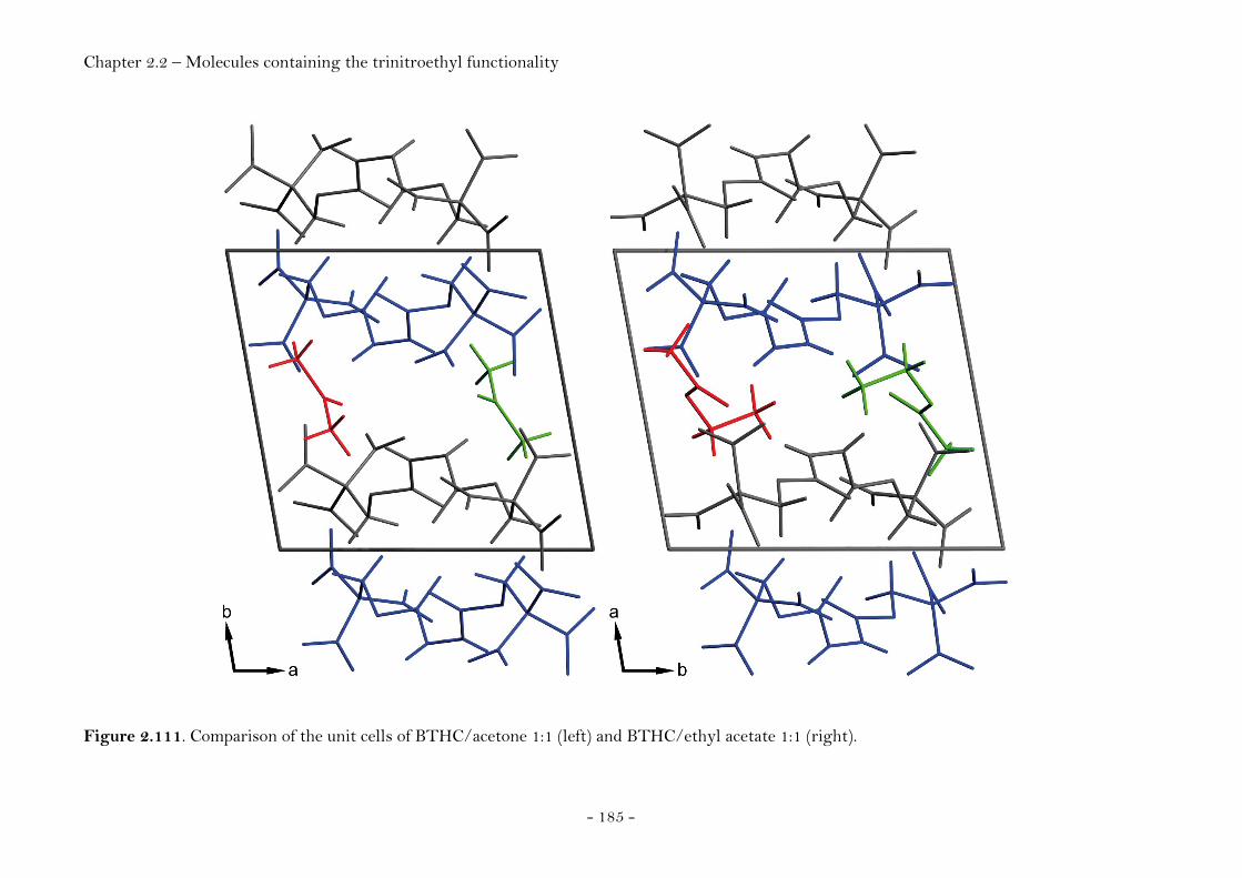
Chapter 2.2 – Molecules containing the trinitroethyl functionality
- 185 -
Figure 2.111. Comparison of the unit cells of BTHC/acetone 1:1 (left) and BTHC/ethyl acetate 1:1 (right).
Chapter 2.2 – Molecules containing the trinitroethyl functionality
- 186 -
Structural Relationships and the Concept of Higher Densities
The number of crystal structures carrying the trinitroethyl group and
published by us (234-235, 268) is nine with the number of different molecules being
eight. A search of the Cambridge Structural Database (CSD version 5.29,
November 2007) resulted in fifteen entries considering compounds carrying
the trinitroethyl moiety. The number of different molecules amongst these
entries is thirteen with three entries (refcodes NOETNA, NOETNA01,
NOETNA02) describing polymorphs of the same molecule (bis-(2,2,2-
trinitroethyl)-nitramine / BTNNA). One structure (N-nitro-N-(2,2,2-
trinitroethyl)-guanidine) has been reported without having been deposited at
the CCDC resulting in a total of only twenty two different molecules where
crystal structure data have been reported. A summary of all structures is
displayed in Figure 2.112. The corresponding names of the molecules are given
and assigned a crystal index number, used to reference individual molecules in
the figures and tables.
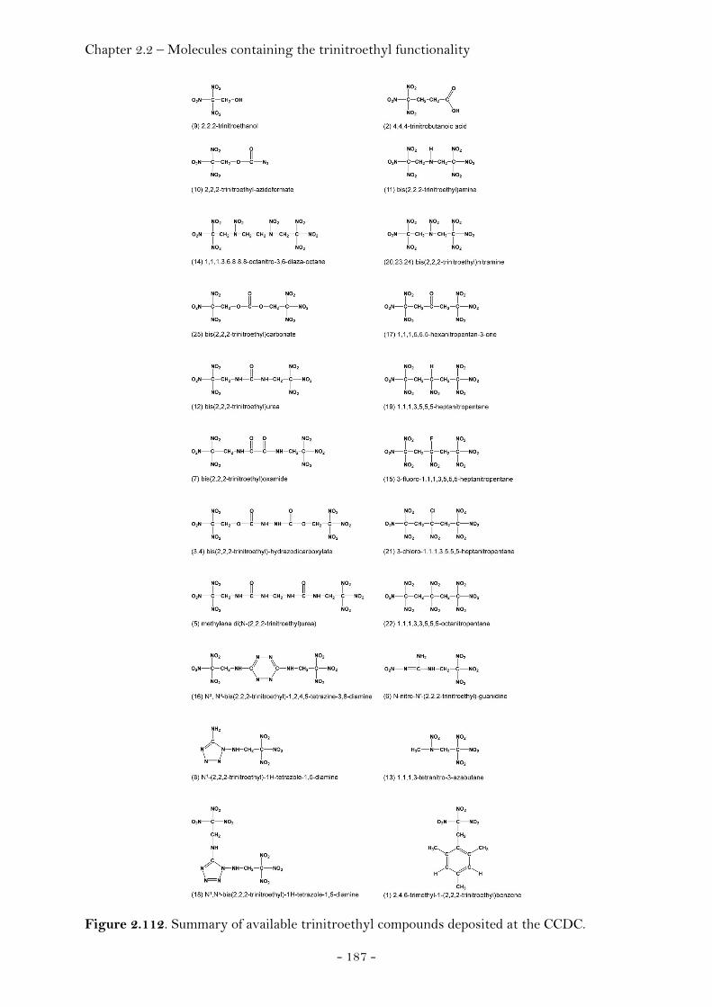
Chapter 2.2 – Molecules containing the trinitroethyl functionality
- 187 -
Figure 2.112. Summary of available trinitroethyl compounds deposited at the CCDC.
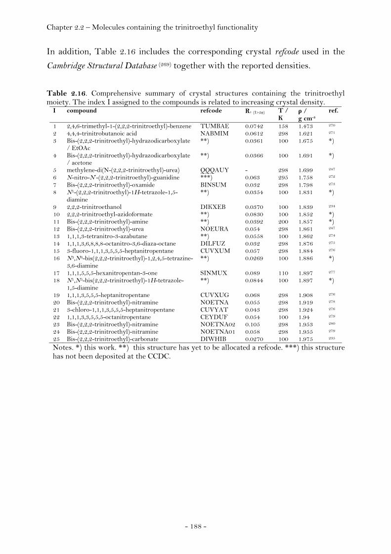
Chapter 2.2 – Molecules containing the trinitroethyl functionality
- 188 -
In addition, Table 2.16 includes the corresponding crystal refcode used in the
Cambridge Structural Database (269) together with the reported densities.
Table 2.16. Comprehensive summary of crystal structures containing the trinitroethyl moiety. The index I assigned to the compounds is related to increasing crystal density.
I compound refcode R1 (I>2σ) T / K
ρ / g cm-3
ref.
1 2,4,6-trimethyl-1-(2,2,2-trinitroethyl)-benzene TUMBAE 0.0742 158 1.473 270
2 4,4,4-trinitrobutanoic acid NABMIM 0.0612 298 1.621 271
3 Bis-(2,2,2-trinitroethyl)-hydrazodicarboxylate / EtOAc
**) 0.0361 100 1.675 *)
4 Bis-(2,2,2-trinitroethyl)-hydrazodicarboxylate / acetone
**) 0.0366 100 1.691 *)
5 methylene-di(N-(2,2,2-trinitroethyl)-urea) QQQAUY - 298 1.699 247
6 N-nitro-N’-(2,2,2-trinitroethyl)-guanidine ***) 0.063 295 1.758 272
7 Bis-(2,2,2-trinitroethyl)-oxamide BINSUM 0.032 298 1.798 273
8 N1-(2,2,2-trinitroethyl)-1H-tetrazole-1,5-diamine
**) 0.0354 100 1.831 *)
9 2,2,2-trinitroethanol DIKXEB 0.0370 100 1.839 234
10 2,2,2-trinitroethyl-azidoformate **) 0.0830 100 1.852 *) 11 Bis-(2,2,2-trinitroethyl)-amine **) 0.0392 200 1.857 *) 12 Bis-(2,2,2-trinitroethyl)-urea NOEURA 0.054 298 1.861 247
13 1,1,1,3-tetranitro-3-azabutane **) 0.0558 100 1.862 274
14 1,1,1,3,6,8,8,8-octanitro-3,6-diaza-octane DILFUZ 0.032 298 1.876 275
15 3-fluoro-1,1,1,3,5,5,5-heptanitropentane CUVXUM 0.057 298 1.884 276
16 N3,N6-bis(2,2,2-trinitroethyl)-1,2,4,5-tetrazine-3,6-diamine
**) 0.0269 100 1.886 *)
17 1,1,1,5,5,5-hexanitropentan-3-one SINMUX 0.089 110 1.897 277
18 N1,N5-bis(2,2,2-trinitroethyl)-1H-tetrazole-1,5-diamine
**) 0.0844 100 1.897 *)
19 1,1,1,3,5,5,5-heptanitropentane CUVXUG 0.068 298 1.908 276
20 Bis-(2,2,2-trinitroethyl)-nitramine NOETNA 0.055 298 1.919 278
21 3-chloro-1,1,1,3,5,5,5-heptanitropentane CUVYAT 0.043 298 1.924 276
22 1,1,1,3,3,5,5,5-octanitropentane CEYDUF 0.054 100 1.94 279
23 Bis-(2,2,2-trinitroethyl)-nitramine NOETNA02 0.105 298 1.953 280
24 Bis-(2,2,2-trinitroethyl)-nitramine NOETNA01 0.058 298 1.955 279
25 Bis-(2,2,2-trinitroethyl)-carbonate DIWHIB 0.0270 100 1.975 235
Notes. *) this work. **) this structure has yet to be allocated a refcode. ***) this structure has not been deposited at the CCDC.
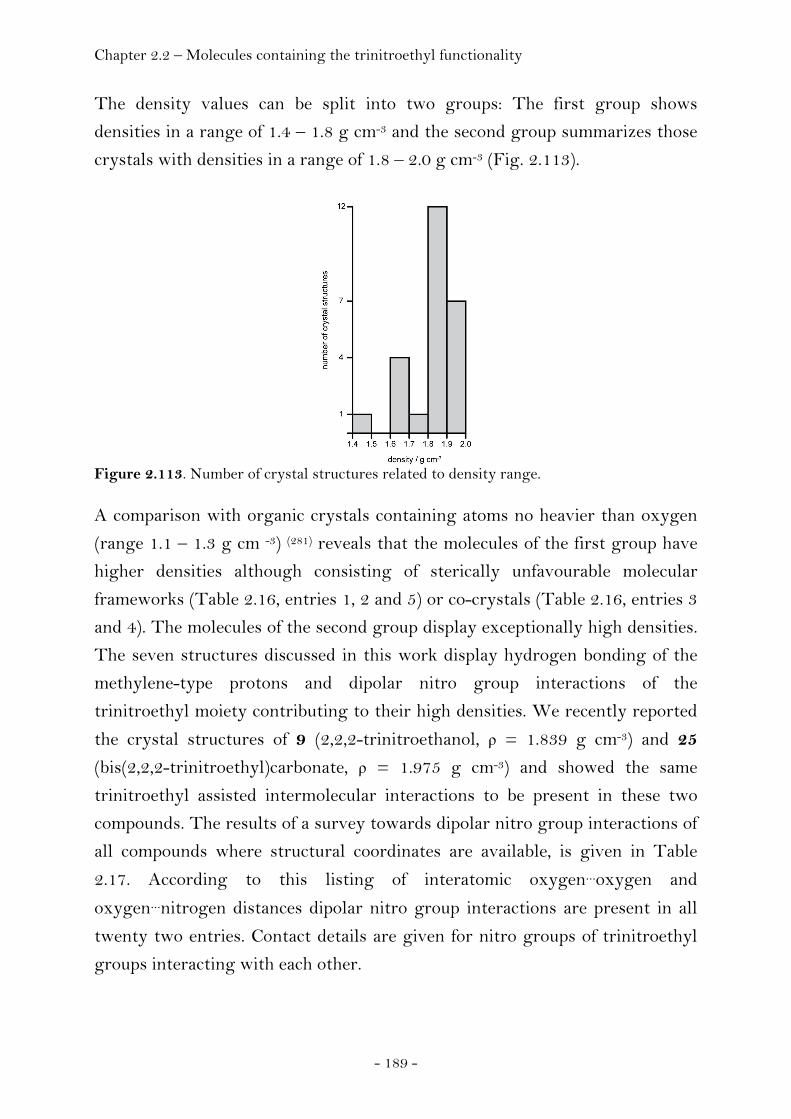
Chapter 2.2 – Molecules containing the trinitroethyl functionality
- 189 -
The density values can be split into two groups: The first group shows
densities in a range of 1.4 – 1.8 g cm-3 and the second group summarizes those
crystals with densities in a range of 1.8 – 2.0 g cm-3 (Fig. 2.113).
Figure 2.113. Number of crystal structures related to density range.
A comparison with organic crystals containing atoms no heavier than oxygen
(range 1.1 – 1.3 g cm -3) (281) reveals that the molecules of the first group have
higher densities although consisting of sterically unfavourable molecular
frameworks (Table 2.16, entries 1, 2 and 5) or co-crystals (Table 2.16, entries 3
and 4). The molecules of the second group display exceptionally high densities.
The seven structures discussed in this work display hydrogen bonding of the
methylene-type protons and dipolar nitro group interactions of the
trinitroethyl moiety contributing to their high densities. We recently reported
the crystal structures of 9 (2,2,2-trinitroethanol, ρ = 1.839 g cm-3) and 25
(bis(2,2,2-trinitroethyl)carbonate, ρ = 1.975 g cm-3) and showed the same
trinitroethyl assisted intermolecular interactions to be present in these two
compounds. The results of a survey towards dipolar nitro group interactions of
all compounds where structural coordinates are available, is given in Table
2.17. According to this listing of interatomic oxygen…oxygen and
oxygen…nitrogen distances dipolar nitro group interactions are present in all
twenty two entries. Contact details are given for nitro groups of trinitroethyl
groups interacting with each other.

Chapter 2.2 – Molecules containing the trinitroethyl functionality
- 190 -
Table 2.17. Summary of short intermolecular nitrogen/oxygen and corresponding oxygen distances of compounds 1 – 25. Only nitro groups being part of the trinitroethyl substituent have been considered. The intermolecular interactions are limited to those less than the sum of the van der Waals radii (∑(vdWN,O) = 3.07 Å, ∑(vdWO,O) = 3.04 Å) plus a tolerance value of 0.2 Å.
I nitro interaction between N/O
contact
d(N…O)
/ Å
Δ /
vdW
O/O
contact
d(O…O)
/ Å
Δ /
vdW
1 N4/O7/O8 and N2/O3/O4 N4…O3 3.025(6) -0.04 O3
…O8 3.047(6) 0.01
2 N3/O7/O8 and N2/O5/O6 N3…O6 2.966(6) -0.10 O6
…O7 3.011(7) -0.03
3 N3/O3/O4 and N7/O13/O14 N3…O13 3.196(2) 0.13 O3
…O14 2.985(2) -0.06
O3…O14 3.170(2) 0.13
O3…O13 3.118(2) 0.08
O4…O13 3.021(2) -0.02
N4/O5/O6 and N5/O7/O8 N4…O7 3.259(2) 0.19 O6
…O7 2.991(2) -0.05
O7…O7 2.971(2) -0.07
N7/O13/O14 and N3/O3/O4 N7…O3 3.251(2) 0.18 O14
…O3 2.985(2) -0.06
4 N6/O11/O12 and N6/O11/O12 N6…O11 3.225(2) 0.16 O11
…O12 3.087(2) 0.05
6 N3/O5/O6 and N3/O5/O6 *) - - O6…O6 2.823 -0.22
7 N3/O4/O5 and N4/O6/O7 N3…O6 2.9506 -0.12 O6
…O5 3.0210 -0.02
8 N8/O3/O4 and N8/O3/O4 N8…O3 3.270(3) 0.20 O3
…O4 3.051(3) 0.01
9 N2/O4/O5 and N2/O4/O5 N2…O4 3.123(2) 0.05 O4
…O4 2.825(2) -0.21
N2/O4/O5 and N5/O11/O12 N5…O5 3.118(2) 0.05 O5
…O12 2.852(2) -0.19
10 N5/O5/O6 and N12/O15/O16 N5…O15 3.082(5) 0.01 O6
…O15 2.966(4) -0.07
11 N3/O5/O6 and N4/O7/O8 N3…O8 3.174(2) 0.10 O6
…O8 3.039(2)
N3/O5/O6 and N5/O9/O10 N5…O5 3.147(2) 0.08 - - -
12 N2/O2/O3 and N3/O4/O5 N2…O5 3.1647 0.09 O2
…O4 3.1632 0.12
O3…O5 3.2100 0.17
N3/O4/O5 and N4/O6/O7 N3…O6 3.2421 0.17 O4
…O6 3.0480 0.01
O4…O7 2.9010 -0.14
O5…O6 3.1179 0.08
N4/O6/O7 and N2/O2/O3 N4…O3 3.2385 0.17 O6
…O2 2.9963 -0.04
13 N3/O3/O4 and N5/O7/O8 N3…O8 3.254(5) 0.18 O3
…O8 2.837(5) -0.20
N3/O3/O4 and N3/O3/O4 N3…O3 3.247(5) 0.18
14 *) - - - - - -
15 N5/O9/O10 and N1/O1/O2 N5…O2 3.250(6) 0.18 O2
…O10 3.215(8) 0.17
16 N7/O1/O2 and N12/O11/O12 N7…O12 2.859(2) -0.21 O12
…O1 2.857(2) -0.18
N8/O3/O4 and N10/O7/O8 N10…O3 2.965(2) -0.11 O3
…O7 2.900(2) -0.14
18 N7/O1/O2 and N8/O3/O4 N7…O3 2.869(4) -0.20 O2
…O3 2.908(4) -0.13
O1…O4 2.927(4) -0.11
N9/O5/O6 and N11/O9/O10 N11…O5 2.896(5) -0.17 O5
…O9 3.041(4) 0.00
19 N5/O9/O10 and N6/O11/O12 N5…O12 3.154(3) 0.08 O9
…O11 3.150(3) 0.11
O10…O11 3.208(3) 0.17
O10…O12 3.136(3) 0.10
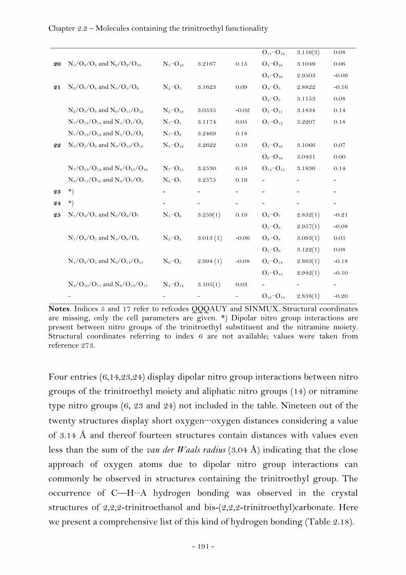
Chapter 2.2 – Molecules containing the trinitroethyl functionality
- 191 -
O11…O12 3.116(3) 0.08
20 N5/O3/O4 and N6/O9/O10 N5…O10 3.2167 0.15 O3
…O10 3.1049 0.06
O4…O10 2.9503 -0.09
21 N2/O3/O4 and N3/O5/O6 N2…O5 3.1623 0.09 O4
…O5 2.8822 -0.16
O4…O5 3.1153 0.08
N2/O3/O4 and N6/O11/O12 N2…O12 3.0535 -0.02 O3
…O11 3.1834 0.14
N7/O13/O14 and N1/O1/O2 N7…O1 3.1174 0.05 O1
…O13 3.2207 0.18
N7/O13/O14 and N1/O1/O2 N7…O2 3.2469 0.18
22 N3/O5/O6 and N8/O15/O16 N3…O16 3.2622 0.19 O5
…O16 3.1066 0.07
O6…O16 3.0431 0.00
N7/O13/O14 and N8/O15/O16 N7…O15 3.2530 0.18 O14
…O15 3.1836 0.14
N8/O15/O16 and N4/O7/O8 N8…O7 3.2575 0.19 - - -
23 *) - - - - - -
24 *) - - - - - -
25 N1/O4/O5 and N2/O6/O7 N1…O6 3.259(1) 0.19 O4
…O7 2.832(1) -0.21
O5…O6 2.957(1) -0.08
N1/O4/O5 and N3/O8/O9 N3…O4 3.013 (1) -0.06 O4
…O8 3.093(1) 0.05
O5…O9 3.122(1) 0.08
N1/O4/O5 and N6/O14/O15 N6…O5 2.994 (1) -0.08 O5
…O14 2.863(1) -0.18
O5…O15 2.942(1) -0.10
N4/O10/O11 and N6/O14/O15 N4…O14 3.105(1) 0.03 - - -
- - - - O12…O14 2.838(1) -0.20
Notes. Indices 5 and 17 refer to refcodes QQQAUY and SINMUX. Structural coordinates are missing, only the cell parameters are given. *) Dipolar nitro group interactions are present between nitro groups of the trinitroethyl substituent and the nitramine moiety. Structural coordinates referring to index 6 are not available; values were taken from reference 273.
Four entries (6,14,23,24) display dipolar nitro group interactions between nitro
groups of the trinitroethyl moiety and aliphatic nitro groups (14) or nitramine
type nitro groups (6, 23 and 24) not included in the table. Nineteen out of the
twenty structures display short oxygen···oxygen distances considering a value
of 3.14 Å and thereof fourteen structures contain distances with values even
less than the sum of the van der Waals radius (3.04 Å) indicating that the close
approach of oxygen atoms due to dipolar nitro group interactions can
commonly be observed in structures containing the trinitroethyl group. The
occurrence of C—H···A hydrogen bonding was observed in the crystal
structures of 2,2,2-trinitroethanol and bis-(2,2,2-trinitroethyl)carbonate. Here
we present a comprehensive list of this kind of hydrogen bonding (Table 2.18).
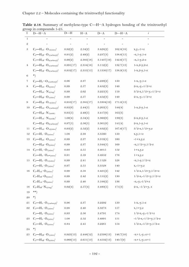
Chapter 2.2 – Molecules containing the trinitroethyl functionality
- 192 -
Table 2.18. Summary of methylene-type C—H···A hydrogen bonding of the trinitroethyl group in compounds 1-25.
I D—H···A D—H H···A D···A D—H···A i
1 - - - - - -
2 - - - - - -
3 C2—H2A…O12(nitro)i 0.92(2) 2.54(2) 3.428(2) 162.8(18) x,y,-1+z
C2—H2B…O1(carbonyl)i 0.91(2) 2.49(2) 3.237(3) 138.6(15) -x,1-y,1-z
C5—H5B…O9(carbonyl)i 0.90(2) 2.383(18) 3.1437(19) 142.6(17) -x,1-y,2-z
4 C2—H2B…O1(carbonyl)i 0.991(17) 2.354(18) 3.112(2) 132.7(13) 1-x,2-y,2-z
C5—H5B…O9(carbonyl)i 0.952(17) 2.355(15) 3.1350(17) 138.9(13) 1-x,2-y,1-z
6 *)
7 C2—H3···O1(carbonyl)i 0.96 2.37 3.229(2) 150 1-x,-y,1-z
8 C2—H2A…O2(nitro)i 0.99 2.57 3.452(3) 149 2-x,-y,-1/2+z
C2—H2A…N4(ring)i 0.99 2.62 3.225(3) 119 3/2-x,1/2+y,-1/2+z
C2—H2B…O1(nitro)i 0.99 2.57 3.452(3) 149 2-x,-y,-1/2+z
9 C3—H3B…O14(nitro)i 0.953(17) 2.385(17) 3.3304(19) 171.0(13)
10 C2—H2A…O1(carbonyl)i 0.95(6) 2.44(5) 3.263(5) 144(4) 1-x,2-y,1-z
C2—H2B…N7(azide) 0.95(5) 2.49(5) 3.417(6) 165(3)
C5—H5A…N3(azide)i 1.00(4) 2.54(4) 3.360(6) 139(3) 2-x,2-y,1-z
C5—H5B…O9(carbonyl)i 0.97(5) 2.56(5) 3.361(6) 141(4) 2-x,1-y,1-z
11 C4—H4A…O12(nitro)i 0.93(2) 2.52(2) 3.432(2) 167.6(17) 3/2-x,1/2+y,z
12 C2—H1…O3(nitro)i 1.08 2.38 3.3560 150 x,y,1+z
13 C2—H2A…O1(nitro)i 0.99 2.37 3.319(5) 160 -1+x,y,z
C2—H2B…O2(nitro)i 0.99 2.37 3.344(5) 169 -x,1/2+y,1/2-z
14 C2—H1…O7(nitro)i 0.93 2.55 3.4015 152 1+x,y,z
C3—H3…O8B*(nitro)i 0.91 2.59 3.4952 176 1+x,y,z
15 C2—H3…O13(nitro)i 0.99 2.41 3.1129 128 -x,1-y,1/2+z
C2—H4…O7(nitro)i 0.97 2.55 3.3528 140 x,-1+y,z
16 C3-H3A…O5(nitro)i 0.99 2.58 3.421(2) 142 1/2-x,1/2+y,1/2+z
C3-H3B…O6(nitro)i 0.99 2.42 3.155(2) 130 1/2-x,-1/2+y,1/2+z
C5-H5A…O9(nitro)i 0.99 2.40 3.188(2) 136 -x,-y,-1/2+z
18 C2-H2B…N3(ring)i 0.92(4) 2.57(4) 3.480(5) 171(3) 2-x, -1/2+y, z
19 **)
20 *)
21 C2—H3…O1(carbonyl)i 0.96 2.37 3.2292 150 1-x,-y,1-z
22 C4—H3…O1(nitro)i 0.98 2.40 3.3273 157 x,1+y,z
C4—H4…O8(nitro)i 0.93 2.56 3.4791 174 1/2-x,-y,-1/2+z
23 C1—H2…O8(nitro)i 1.08 2.52 3.4991 151 -1/2-x,-1/2+y,1/2-z
C3—H4…O2(nitro)i 0.84 2.45 3.2261 154 1/2-x,-1/2+y,1/2-z
24 *)
25 C2—H2B…O1(nitro)i 0.923(13) 2.448(12) 3.2598(13) 146.7(10) -x+1,-y,-z+1
C4—H4B…O9(nitro)i 0.966(13) 2.651(13) 3.4532(13) 140.7(9) -x+1,-y,-z+1
Chapter 2.2 – Molecules containing the trinitroethyl functionality
- 193 -
Notes. Indices 5 and 17 refer to refcodes QQQAUY and SINMUX. Structural coordinates are missing, only the cell parameters are given. *) no hydrogen atom coordinates available. **) 19: C3—H5…O1(nitro) hydrogen bond between a non trinitroethyl hydrogen atom and an oxygen atom of a trinitroethyl nitro group.
Of all the structures listed in Table 2.16 were hydrogen atom positions were
available, C—H···A hydrogen bonding of the methylene-type hydrogen atoms
of the trinitroethyl group was observed in seventeen out of twenty structures
rendering C—H···A hydrogen bonding as another important intermolecular
feature of the trinitroethyl functionality supporting higher densities.
Conclusion
Based on the crystal structures reported in this work and available in the
literature, we find that trinitroethyl mediated intermolecular interactions like
dipolar nitro group interactions and hydrogen bonding of the acidified
methylene-type protons govern mainly the molecular packing of these
compounds yielding high-crystal-density polymorphs with promising explosive
performance parameters. Five new highly explosive compounds are introduced
belonging to a new class of energetic materials preferentially containing both
high-nitrogen and high-oxygen content. TTD, BTTD and BTAT display
superior performance properties to RDX with BTAT at the same time being
less sensitive and displaying a better oxygen balance value yielding smokeless
combustion and less toxic fumes on decomposition. The compounds are water
insoluble in contrast to energetic salts, a prerequisite to protect the ground and
one important environmental advantage amongst the qualification criteria for
new HEDM. It is shown that temperature stability can be higher than 140°C
for solid and liquid compounds carrying the trinitroethyl moiety not only as far
as decomposition temperatures but also as far as chemical long term stabilities
are concerned. BTAT and BTHC are shown to be the molecules offering the
best trade-off between energy capability and thermal stability. Next to its
excellent thermal stability as well as its positive oxygen balance value, BTHC
displays the rare and desirable property of being a solid with a reasonable low
melting point and a liquid range of greater than 100°C while at the same time
being insensitive according to BAM standards rendering a possible use as safe
Chapter 2.2 – Molecules containing the trinitroethyl functionality
- 194 -
melt-castable explosive. The tendency of conglomerate crystallization of
BTHC is a further important point allowing for the specific design of its
performance as well as sensitivity properties.
Experimental
CAUTION: The compounds described in this work are potential explosives,
which may be subject to accidental initiation by such environmental stimuli as
impact, friction, heat, or electrostatic discharge. Appropriate precautions and
proper protective measures (safety glasses, face shields, leather coat, grounding
(equipment and person), Kevlar™ gloves and ear plugs) should be taken and
used when handling these materials. To avoid difficulties with the sensitivity of
these compounds only millimolar amounts should be handled with care.
Calculations. All calculations were carried out using the Gaussian G03W
(revision B.03) program package. (282)
Materials. All reagents and solvents were used as received (Acros, Aldrich,
Fluka) if not stated otherwise.
Instrumentation and Measurement. Electronic absorption spectra were
acquired by using a Varian CARY 50 Conc UV-visible spectrophotometer in
quartz cuvettes. 1H, 13C and 14/15N NMR spectra were recorded using a Jeol
Eclipse 270, Jeol EX 400 or Jeol Eclipse 400 instrument operating at 400 MHz
(1H), 100.6 MHz (13C), 40.5 MHz (15N) and 28.9 MHz (14N). All chemical shifts
are quoted in ppm relative to TMS (1H, 13C) or nitromethane (14N / 15N).
Infrared (IR) spectra were recorded using a Perkin-Elmer Spektrum One FT-
IR (using KBr disks) or a Perkin-Elmer Spektrum BX FT-IR (pure substance)
instrument. Raman spectra were measured using a Perkin Elmer Spektrum
2000R NIR FT-Raman instrument equipped with a Nd:YAG laser (1064 nm).
Elemental analyses were performed with a Netsch Simultanous Thermal
Analyser STA 429. The thermal behaviour of the salts was investigated in a
nitrogen atmosphere using differential scanning calorimetry (Linseis DSC PT-
10) at heating rates (β) of 5 K min-1 and a nitrogen flow of 5 L h-1. Typically, 2
mg of substance were measured inside a pressed aluminium container equipped
with a hole (0.1 mm) for the gas release. Isothermal long term experiments
were performed using a Systag TSC (isothermal safety calorimeter) station.
Chapter 2.2 – Molecules containing the trinitroethyl functionality
- 195 -
Ultrasound was applied using a Bandelin Sonorex RK 510 ultrasonic bath. The
single crystal X-ray diffraction data were collected using an Oxford Xcalibur3
diffractometer equipped with a Kappa CCD detector. The MoKα radiation (λ =
0.71073 Å) was generated from a Spellman generator (50 kV, 40 mA) and
focussed using a graphite collimator. The data collection was undertaken using
the CrysAlis CCD software (283) and data reduction was performed using the
CrysAlis RED software. (284) The structures were solved using SIR-92 (285) and
refined using SHELXL-97 (286) implemented in the program package WinGX (287) and finally checked using PLATON. (288) The impact sensitivity tests were
carried out according to STANAG 4489 (289) modified according to instruction (290) using a BAM (Bundesanstalt für Materialprüfung) drophammer. The
friction sensitivity tests were carried out according to STANAG 4487 (291)
modified according to instruction (292) using the BAM friction tester. The
particle size was determined using a DIN 4188 testing sieve (Retsch). The
respond to thermal shock and performance was tested using a Koenen steel
sleeve apparatus. (293) The electrostatic sensitivity tests were carried out using
an electric spark tester ESD 2010EN (OZM Research) operating with the
‘Winspark 1.15 software package’. High speed images were recorded using a
Visario G2 high-speed camera (Speed Cam Visario G2, Weinberger
Deutschland GmbH, Erlangen, Germany).
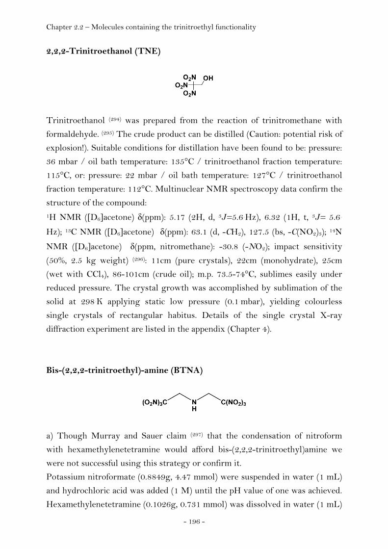
Chapter 2.2 – Molecules containing the trinitroethyl functionality
- 196 -
2,2,2-Trinitroethanol (TNE)
Trinitroethanol (294) was prepared from the reaction of trinitromethane with
formaldehyde. (295) The crude product can be distilled (Caution: potential risk of
explosion!). Suitable conditions for distillation have been found to be: pressure:
36 mbar / oil bath temperature: 135°C / trinitroethanol fraction temperature:
115°C, or: pressure: 22 mbar / oil bath temperature: 127°C / trinitroethanol
fraction temperature: 112°C. Multinuclear NMR spectroscopy data confirm the
structure of the compound: 1H NMR ([D6]acetone) δ(ppm): 5.17 (2H, d, 3J=5.6 Hz), 6.32 (1H, t, 3J= 5.6
Hz); 13C NMR ([D6]acetone) δ(ppm): 63.1 (d, -CH2), 127.5 (bs, -C(NO2)3); 14N
NMR ([D6]acetone) δ(ppm, nitromethane): -30.8 (-NO2); impact sensitivity
(50%, 2.5 kg weight) (296): 11cm (pure crystals), 22cm (monohydrate), 25cm
(wet with CCl4), 86-101cm (crude oil); m.p. 73.5-74°C, sublimes easily under
reduced pressure. The crystal growth was accomplished by sublimation of the
solid at 298 K applying static low pressure (0.1 mbar), yielding colourless
single crystals of rectangular habitus. Details of the single crystal X-ray
diffraction experiment are listed in the appendix (Chapter 4).
Bis-(2,2,2-trinitroethyl)-amine (BTNA)
a) Though Murray and Sauer claim (297) that the condensation of nitroform
with hexamethylenetetramine would afford bis-(2,2,2-trinitroethyl)amine we
were not successful using this strategy or confirm it.
Potassium nitroformate (0.8849g, 4.47 mmol) were suspended in water (1 mL)
and hydrochloric acid was added (1 M) until the pH value of one was achieved.
Hexamethylenetetramine (0.1026g, 0.731 mmol) was dissolved in water (1 mL)
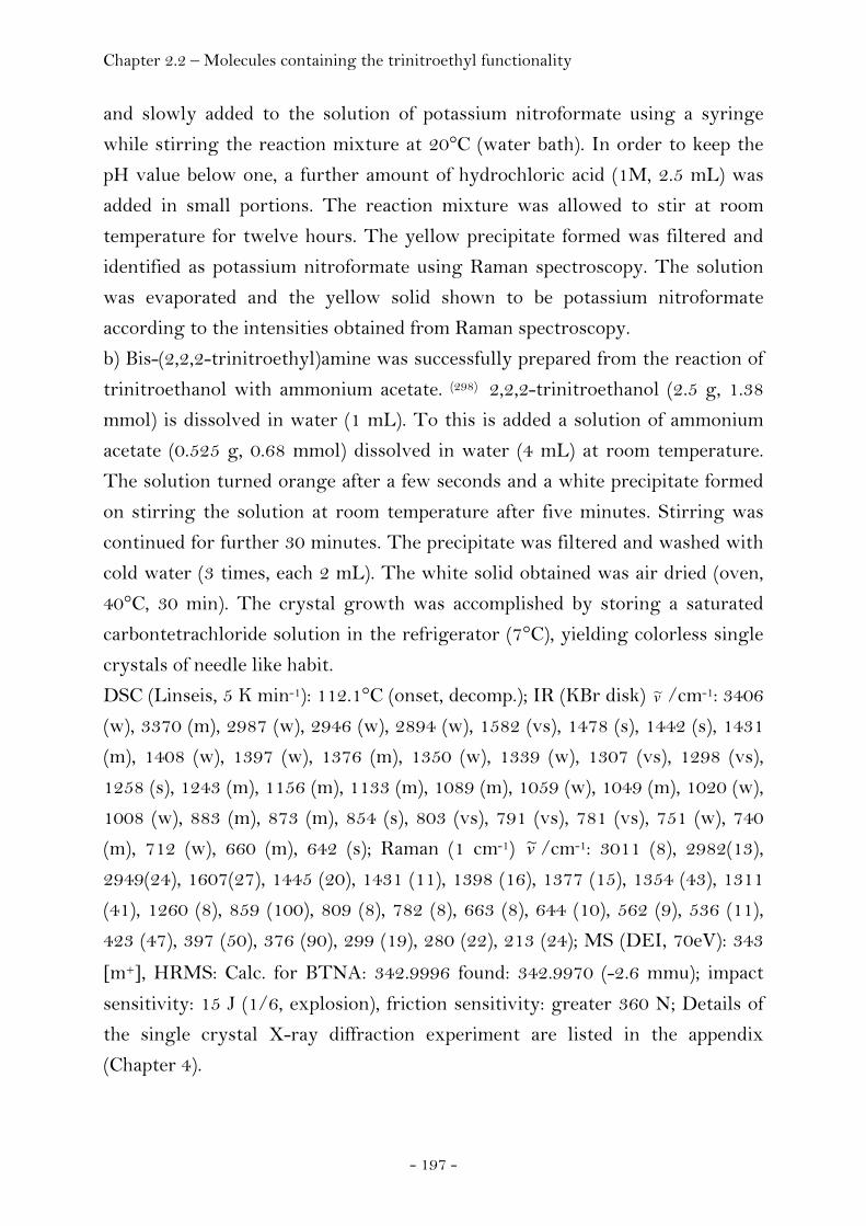
Chapter 2.2 – Molecules containing the trinitroethyl functionality
- 197 -
and slowly added to the solution of potassium nitroformate using a syringe
while stirring the reaction mixture at 20°C (water bath). In order to keep the
pH value below one, a further amount of hydrochloric acid (1M, 2.5 mL) was
added in small portions. The reaction mixture was allowed to stir at room
temperature for twelve hours. The yellow precipitate formed was filtered and
identified as potassium nitroformate using Raman spectroscopy. The solution
was evaporated and the yellow solid shown to be potassium nitroformate
according to the intensities obtained from Raman spectroscopy.
b) Bis-(2,2,2-trinitroethyl)amine was successfully prepared from the reaction of
trinitroethanol with ammonium acetate. (298) 2,2,2-trinitroethanol (2.5 g, 1.38
mmol) is dissolved in water (1 mL). To this is added a solution of ammonium
acetate (0.525 g, 0.68 mmol) dissolved in water (4 mL) at room temperature.
The solution turned orange after a few seconds and a white precipitate formed
on stirring the solution at room temperature after five minutes. Stirring was
continued for further 30 minutes. The precipitate was filtered and washed with
cold water (3 times, each 2 mL). The white solid obtained was air dried (oven,
40°C, 30 min). The crystal growth was accomplished by storing a saturated
carbontetrachloride solution in the refrigerator (7°C), yielding colorless single
crystals of needle like habit.
DSC (Linseis, 5 K min-1): 112.1°C (onset, decomp.); IR (KBr disk) v~ /cm-1: 3406
(w), 3370 (m), 2987 (w), 2946 (w), 2894 (w), 1582 (vs), 1478 (s), 1442 (s), 1431
(m), 1408 (w), 1397 (w), 1376 (m), 1350 (w), 1339 (w), 1307 (vs), 1298 (vs),
1258 (s), 1243 (m), 1156 (m), 1133 (m), 1089 (m), 1059 (w), 1049 (m), 1020 (w),
1008 (w), 883 (m), 873 (m), 854 (s), 803 (vs), 791 (vs), 781 (vs), 751 (w), 740
(m), 712 (w), 660 (m), 642 (s); Raman (1 cm-1) v~ /cm-1: 3011 (8), 2982(13),
2949(24), 1607(27), 1445 (20), 1431 (11), 1398 (16), 1377 (15), 1354 (43), 1311
(41), 1260 (8), 859 (100), 809 (8), 782 (8), 663 (8), 644 (10), 562 (9), 536 (11),
423 (47), 397 (50), 376 (90), 299 (19), 280 (22), 213 (24); MS (DEI, 70eV): 343
[m+], HRMS: Calc. for BTNA: 342.9996 found: 342.9970 (-2.6 mmu); impact
sensitivity: 15 J (1/6, explosion), friction sensitivity: greater 360 N; Details of
the single crystal X-ray diffraction experiment are listed in the appendix
(Chapter 4).
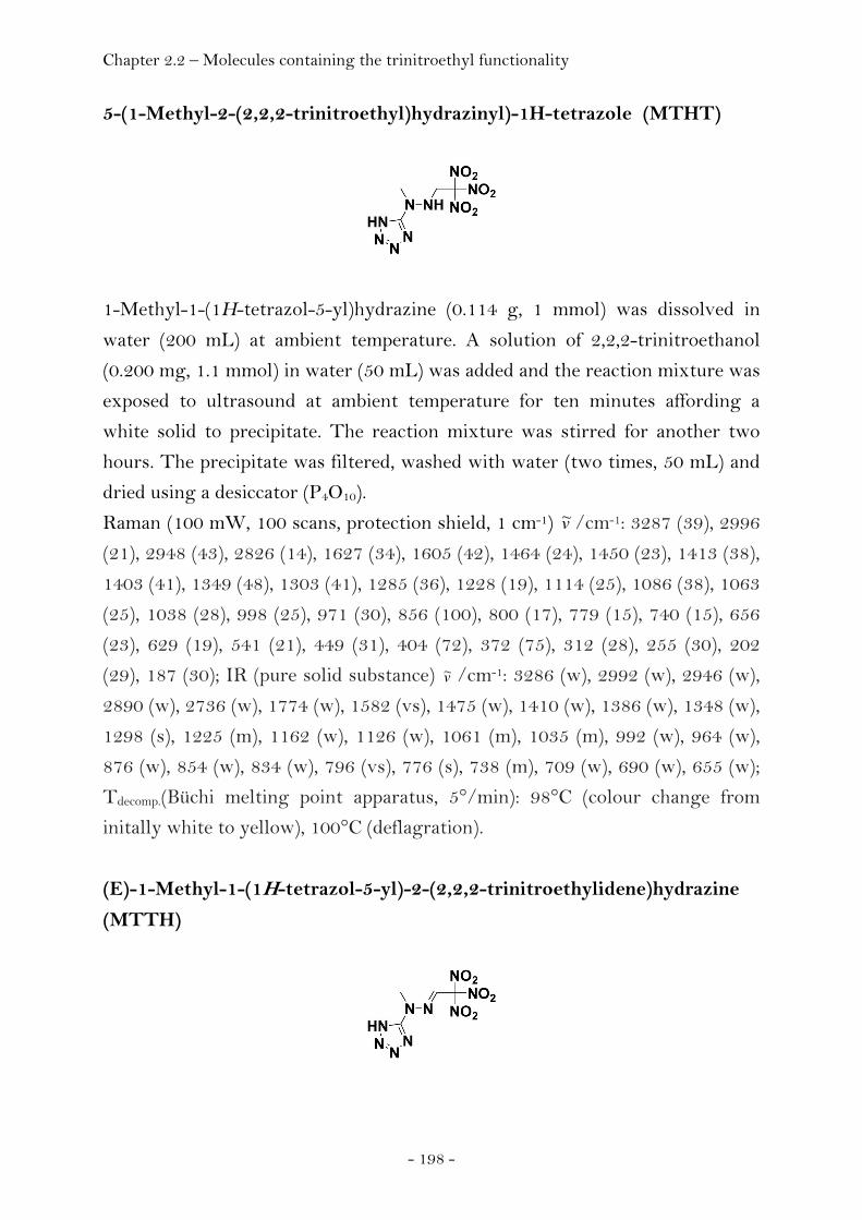
Chapter 2.2 – Molecules containing the trinitroethyl functionality
- 198 -
5-(1-Methyl-2-(2,2,2-trinitroethyl)hydrazinyl)-1H-tetrazole (MTHT)
1-Methyl-1-(1H-tetrazol-5-yl)hydrazine (0.114 g, 1 mmol) was dissolved in
water (200 mL) at ambient temperature. A solution of 2,2,2-trinitroethanol
(0.200 mg, 1.1 mmol) in water (50 mL) was added and the reaction mixture was
exposed to ultrasound at ambient temperature for ten minutes affording a
white solid to precipitate. The reaction mixture was stirred for another two
hours. The precipitate was filtered, washed with water (two times, 50 mL) and
dried using a desiccator (P4O10).
Raman (100 mW, 100 scans, protection shield, 1 cm-1) v~ /cm-1: 3287 (39), 2996
(21), 2948 (43), 2826 (14), 1627 (34), 1605 (42), 1464 (24), 1450 (23), 1413 (38),
1403 (41), 1349 (48), 1303 (41), 1285 (36), 1228 (19), 1114 (25), 1086 (38), 1063
(25), 1038 (28), 998 (25), 971 (30), 856 (100), 800 (17), 779 (15), 740 (15), 656
(23), 629 (19), 541 (21), 449 (31), 404 (72), 372 (75), 312 (28), 255 (30), 202
(29), 187 (30); IR (pure solid substance) v~ /cm-1: 3286 (w), 2992 (w), 2946 (w),
2890 (w), 2736 (w), 1774 (w), 1582 (vs), 1475 (w), 1410 (w), 1386 (w), 1348 (w),
1298 (s), 1225 (m), 1162 (w), 1126 (w), 1061 (m), 1035 (m), 992 (w), 964 (w),
876 (w), 854 (w), 834 (w), 796 (vs), 776 (s), 738 (m), 709 (w), 690 (w), 655 (w);
Tdecomp.(Büchi melting point apparatus, 5°/min): 98°C (colour change from
initally white to yellow), 100°C (deflagration).
(E)-1-Methyl-1-(1H-tetrazol-5-yl)-2-(2,2,2-trinitroethylidene)hydrazine
(MTTH)
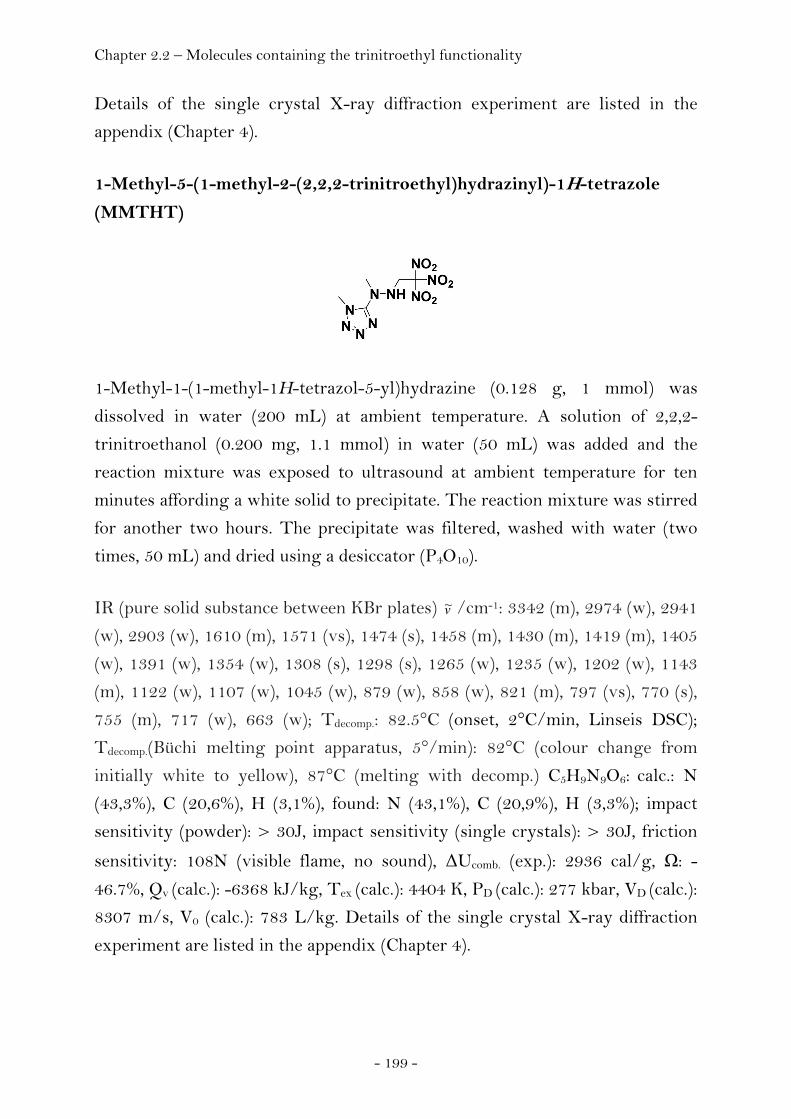
Chapter 2.2 – Molecules containing the trinitroethyl functionality
- 199 -
Details of the single crystal X-ray diffraction experiment are listed in the
appendix (Chapter 4).
1-Methyl-5-(1-methyl-2-(2,2,2-trinitroethyl)hydrazinyl)-1H-tetrazole
(MMTHT)
1-Methyl-1-(1-methyl-1H-tetrazol-5-yl)hydrazine (0.128 g, 1 mmol) was
dissolved in water (200 mL) at ambient temperature. A solution of 2,2,2-
trinitroethanol (0.200 mg, 1.1 mmol) in water (50 mL) was added and the
reaction mixture was exposed to ultrasound at ambient temperature for ten
minutes affording a white solid to precipitate. The reaction mixture was stirred
for another two hours. The precipitate was filtered, washed with water (two
times, 50 mL) and dried using a desiccator (P4O10).
IR (pure solid substance between KBr plates) v~ /cm-1: 3342 (m), 2974 (w), 2941
(w), 2903 (w), 1610 (m), 1571 (vs), 1474 (s), 1458 (m), 1430 (m), 1419 (m), 1405
(w), 1391 (w), 1354 (w), 1308 (s), 1298 (s), 1265 (w), 1235 (w), 1202 (w), 1143
(m), 1122 (w), 1107 (w), 1045 (w), 879 (w), 858 (w), 821 (m), 797 (vs), 770 (s),
755 (m), 717 (w), 663 (w); Tdecomp.: 82.5°C (onset, 2°C/min, Linseis DSC);
Tdecomp.(Büchi melting point apparatus, 5°/min): 82°C (colour change from
initially white to yellow), 87°C (melting with decomp.) C5H9N9O6: calc.: N
(43,3%), C (20,6%), H (3,1%), found: N (43,1%), C (20,9%), H (3,3%); impact
sensitivity (powder): > 30J, impact sensitivity (single crystals): > 30J, friction
sensitivity: 108N (visible flame, no sound), ΔUcomb. (exp.): 2936 cal/g, Ω: -
46.7%, Qv (calc.): -6368 kJ/kg, Tex (calc.): 4404 K, PD (calc.): 277 kbar, VD (calc.):
8307 m/s, V0 (calc.): 783 L/kg. Details of the single crystal X-ray diffraction
experiment are listed in the appendix (Chapter 4).

Chapter 2.2 – Molecules containing the trinitroethyl functionality
- 200 -
2-(5-(1-Methyl-2-(2,2,2-trinitroethyl)hydrazinyl)-1H-tetrazol-1-
yl)ethanol (MTHTE)
2-(5-(1-Methyl-hydrazinyl)-1H-tetrazol-1-yl)ethanol (0.158 g, 1 mmol) was
dissolved in water (200 mL) at ambient temperature. A solution of 2,2,2-
trinitroethanol (0.200 mg, 1.1 mmol) in water (50 mL) was added and the
reaction mixture was exposed to ultrasound at ambient temperature for ten
minutes affording a white solid to precipitate. The reaction mixture was stirred
for another two hours. The precipitate was filtered, washed with water (two
times, 50 mL) and dried using a desiccator (P4O10).
Details of the single crystal X-ray diffraction experiment are listed in the
appendix (Chapter 4).
1-(4N-2,2,2-Trinitroethyl)-2,5-hydroxymethyltriazine (THMT)
1-Amino-2,5-hydroxymethyltriazine (0.144 g, 1 mmol) was dissolved in water
(200 mL) at ambient temperature. A solution of 2,2,2-trinitroethanol (0.200
mg, 1.1 mmol) in water (50 mL) was added and the reaction mixture was
exposed to ultrasound at ambient temperature for ten minutes affording a
white solid to precipitate. The reaction mixture was stirred for another two
hours. The precipitate was filtered, washed with water (two times, 50 mL) and
dried using a desiccator (P4O10).
Details of the single crystal X-ray diffraction experiment are listed in the
appendix (Chapter 4).
Chapter 2.2 – Molecules containing the trinitroethyl functionality
- 201 -
N1-(2,2,2-Trinitroethyl)-1H-tetrazole-1,5-diamine (TTD)
2,2,2-Trinitroethanol is a starting material for the synthesis of TTD and was
synthesised from the condensation of formaldehyde and trinitromethane. (295)
1,5-Diaminotetrazole (0.100 g, 1 mmol) was dissolved in water (200 mL) at
ambient temperature. A solution of 2,2,2-trinitroethanol (0.200 mg, 1.1 mmol)
in water (50 mL) was added and the reaction mixture was exposed to
ultrasound at ambient temperature for ten minutes affording a white solid to
precipitate. The reaction mixture was stirred for another two hours. The
precipitate was filtered, washed with water (two times, 50 mL) and dried using
a desiccator (P4O10).
DSC (Linseis, 5 K min-1): exo (125.6°C, onset / 129.1°C, max / 131.9°C, offset);
Tdec(Büchi melting point apparatus, 10°/min): 156°C-157°C (explosion);
isothermal long term stability (48 h, 85°C): unchanged; isothermal long term
stability (isoperibol steps, temperature was held constant every 20°C for one
hour): explosion at 100°C; 1H NMR ([D6]acetone) δ: 5.33 (2H, -CH2-, d, 3J =
6.1 Hz) 6.09 (2H, s, -NH2), 7.55 (1H, -NH-, t, 3J = 6.1 Hz); 13C NMR
([D6]acetone) δ: 52.4 (s, -CH2-), 126.6 (bs, -C(NO2)3), 153.5 (s, Cring); IR (KBr)
v~ /cm-1: 3414 (vs), 3311 (s), 3173 (vs), 3001 (m), 2955 (m), 1651 (vs), 1610 (vs),
1585 (vs), 1494 (s), 1459 (w), 1414 (s), 1381 (w), 1347 (w), 1321 (vs), 1302 (vs),
1136 (m), 1120 (m), 1081 (m), 1040 (w), 989 (w), 907 (w), 855 (w), 835 (w), 806
(vs), 774 (m), 739 (w), 715 (w), 673 (w), 648 (m), 582 (w), 546 (w), 459 (w);
Raman (4 cm-1) v~ /cm-1: 3172 (19), 3001 (23), 2987 (59), 2958 (41), 1652 (37),
1612 (42), 1585 (25), 1495 (33), 1463 (31), 1416 (42), 1381 (34), 1349 (50), 1323
(81), 1305 (43), 1278 (29), 1141 (24), 1116 (29), 1080 (23), 1041 (30), 1003 (18),
991 (21), 909 (18), 857 (100), 808 (38), 780 (95), 736 (14), 675 (20), 643 (20),
543 (19), 519 (23), 460 (26), 416 (57), 400 (58), 375 (100), 335 (61), 305 (32),
264 (42), 225 (32), 207 (42), 172 (46); MS (DCI+, isobutane): 264 [m+H]; Calc
for C3H5N9O6: N (47.9%), C (13.7%), H (1.9%), found: N (47.5%), C (13.9%), H

Chapter 2.2 – Molecules containing the trinitroethyl functionality
- 202 -
(2.0%); impact sensitivity: < 30 J (decomposition), friction sensitivity: 40 N
(explosion), electrostatic spark sensitivity (particle size 0.08 – 0.16 mm): < 0.1 J
(explosion); Koenen Test: > 10 mm; Details of the single crystal X-ray
diffraction experiment are listed in the appendix (Chapter 4).
N1,N5-Bis-(2,2,2-trinitroethyl)-1H-tetrazole-1,5-diamine (BTTD)
TTD (0.158 g, 0.6 mmol) was dissolved in concentrated hydrochloric acid (50
mL). To the stirred solution was added 2,2,2-trinitroethanol (0.254 g, 1.4
mmol) at room temperature. The colorless solution was heated to 50°C for one
hour and extracted using dichloromethane. The collected organic fractions
were dried using magnesium sulfate, filtered and concentrated using a rotary
evaporator.
DSC (Linseis, 5 K min-1): exo (126.6°C, onset / 132.0°C, max / 137.4°C, offset);
MS (DCI+, isobutane): 427 [m+H]; Calc. for C5H6N12O12: N (39.4%), C
(14.1%), H (1.4%), found: N (39.0%), C (14.2%), H (1.5%); Details of the single
crystal X-ray diffraction experiment are listed in the appendix (Chapter 4).
Chapter 2.2 – Molecules containing the trinitroethyl functionality
- 203 -
N3,N6-Bis-(2,2,2-trinitroethyl)-1,2,4,5-tetrazine-3,6-diamine (BTAT)
3,6-Diamino-1,2,4,5-tetrazine is a starting material for the synthesis of BTAT
and was synthesised from the condensation of diaminoguanidinium
hydrochloride with 2,4-pentanedione, followed by oxidation of the resulting
dihydrotetrazine with sodium perborate. (299) Two pathways have been found
for the preparation and isolation of BTAT. Method a) According to the first
pathway, BTAT can be obtained on treating a solution of 3,6-diaminotetrazine
in concentrated hydrochloric acid (37%) at a temperature of 70°C. The product
precipitates from the solution and can be obtained in high purity on simple
filtration. We observed that the yields of this approach are quite low (see
section 2.1.4, page 119). Method b) 3,6-diamino-1,2,4,5-tetrazine (0.067 g, 0.6
mmol) was suspended in acetonitrile (100 mL) at room temperature. A solution
of iron-(III)-chloride (0.100 g, 0.6 mmol) dissolved in nitromethane (5 mL) was
added. 2,2,2-trinitroethanol (0.253 g, 1.4 mmol) was subsequently added and
the reaction stirred for four hours at room temperature. The reaction mixture
was then treated with hydrochloric acid (2M, 100 mL) and extracted three
times using dichloromethane (100 mL). The collected organic phases were
dried using magnesium sulfate, filtered and concentrated using a rotary
evaporator affording a red solid containing BTAT of rather low purity.
Method c) 3,6-diaminotetrazine (2,2g) was suspended in acetonitrile (200 mL).
Trinitroethanol (9g) and subsequently a solution of iron-(III)-chloride (12g)
dissolved in nitromethane (75mL) were added yielding a clear solution of all of
the components. The reaction was allowed to stir at ambient conditions for
10h. The reaction mixture was concentrated using a rotary evaporator (60°C,
90mbar) affording a sticky, nearly solid dark residue. Subsequently, the
following steps were involved in the work up procedure:

Chapter 2.2 – Molecules containing the trinitroethyl functionality
- 204 -
1) The residue was dissolved in acetonitrile (20 mL) and filtered to remove
unreacted starting material (3,6-diamino-1,2,4,5-tetrazine, 200mg).
2) Hydrochloric acid (2M, 2L) was added to the filtrate to precipitate the
BTAT from solution and keep the iron complex in solution. It takes several
hours for the product to precipitate (bright orange colour). The picture on the
right was taken after one day.
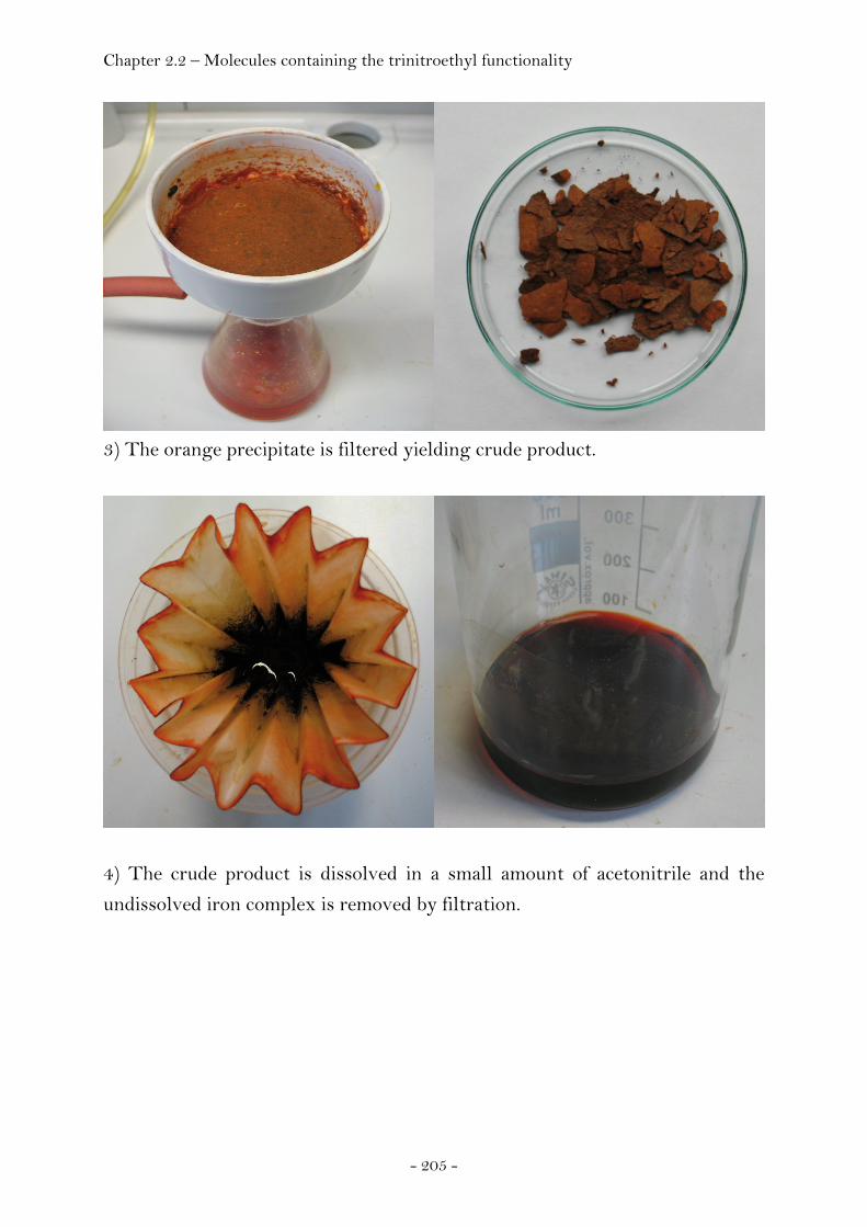
Chapter 2.2 – Molecules containing the trinitroethyl functionality
- 205 -
3) The orange precipitate is filtered yielding crude product.
4) The crude product is dissolved in a small amount of acetonitrile and the
undissolved iron complex is removed by filtration.
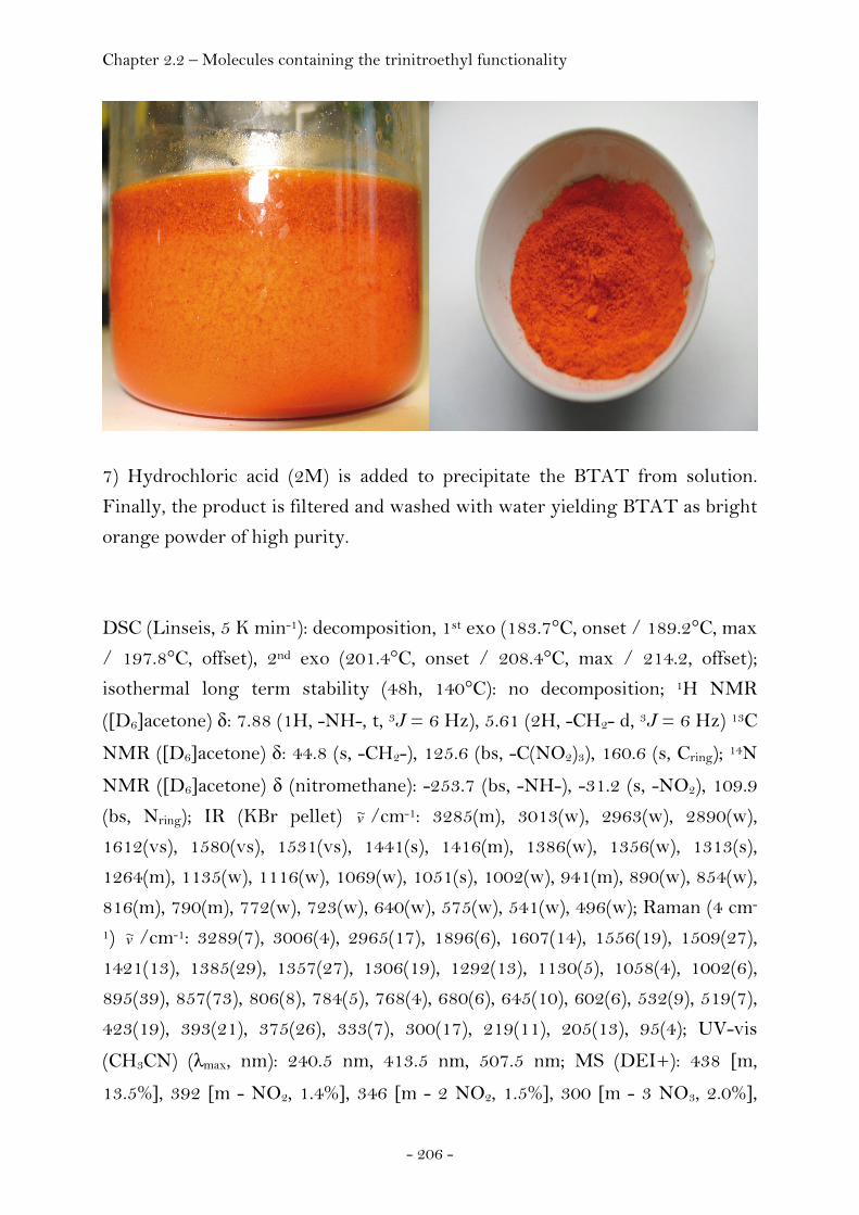
Chapter 2.2 – Molecules containing the trinitroethyl functionality
- 206 -
7) Hydrochloric acid (2M) is added to precipitate the BTAT from solution.
Finally, the product is filtered and washed with water yielding BTAT as bright
orange powder of high purity.
DSC (Linseis, 5 K min-1): decomposition, 1st exo (183.7°C, onset / 189.2°C, max
/ 197.8°C, offset), 2nd exo (201.4°C, onset / 208.4°C, max / 214.2, offset);
isothermal long term stability (48h, 140°C): no decomposition; 1H NMR
([D6]acetone) δ: 7.88 (1H, -NH-, t, 3J = 6 Hz), 5.61 (2H, -CH2- d, 3J = 6 Hz) 13C
NMR ([D6]acetone) δ: 44.8 (s, -CH2-), 125.6 (bs, -C(NO2)3), 160.6 (s, Cring); 14N
NMR ([D6]acetone) δ (nitromethane): -253.7 (bs, -NH-), -31.2 (s, -NO2), 109.9
(bs, Nring); IR (KBr pellet) v~ /cm-1: 3285(m), 3013(w), 2963(w), 2890(w),
1612(vs), 1580(vs), 1531(vs), 1441(s), 1416(m), 1386(w), 1356(w), 1313(s),
1264(m), 1135(w), 1116(w), 1069(w), 1051(s), 1002(w), 941(m), 890(w), 854(w),
816(m), 790(m), 772(w), 723(w), 640(w), 575(w), 541(w), 496(w); Raman (4 cm-
1) v~ /cm-1: 3289(7), 3006(4), 2965(17), 1896(6), 1607(14), 1556(19), 1509(27),
1421(13), 1385(29), 1357(27), 1306(19), 1292(13), 1130(5), 1058(4), 1002(6),
895(39), 857(73), 806(8), 784(5), 768(4), 680(6), 645(10), 602(6), 532(9), 519(7),
423(19), 393(21), 375(26), 333(7), 300(17), 219(11), 205(13), 95(4); UV-vis
(CH3CN) (λmax, nm): 240.5 nm, 413.5 nm, 507.5 nm; MS (DEI+): 438 [m,
13.5%], 392 [m - NO2, 1.4%], 346 [m - 2 NO2, 1.5%], 300 [m - 3 NO3, 2.0%],
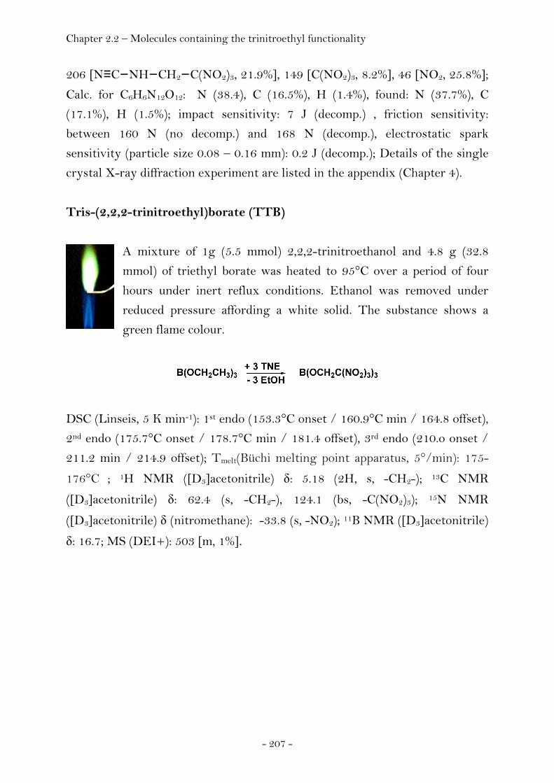
Chapter 2.2 – Molecules containing the trinitroethyl functionality
- 207 -
206 [N≡C−NH−CH2−C(NO2)3, 21.9%], 149 [C(NO2)3, 8.2%], 46 [NO2, 25.8%]; Calc. for C6H6N12O12: N (38.4), C (16.5%), H (1.4%), found: N (37.7%), C
(17.1%), H (1.5%); impact sensitivity: 7 J (decomp.) , friction sensitivity:
between 160 N (no decomp.) and 168 N (decomp.), electrostatic spark
sensitivity (particle size 0.08 – 0.16 mm): 0.2 J (decomp.); Details of the single
crystal X-ray diffraction experiment are listed in the appendix (Chapter 4).
Tris-(2,2,2-trinitroethyl)borate (TTB)
A mixture of 1g (5.5 mmol) 2,2,2-trinitroethanol and 4.8 g (32.8
mmol) of triethyl borate was heated to 95°C over a period of four
hours under inert reflux conditions. Ethanol was removed under
reduced pressure affording a white solid. The substance shows a
green flame colour.
DSC (Linseis, 5 K min-1): 1st endo (153.3°C onset / 160.9°C min / 164.8 offset),
2nd endo (175.7°C onset / 178.7°C min / 181.4 offset), 3rd endo (210.o onset /
211.2 min / 214.9 offset); Tmelt(Büchi melting point apparatus, 5°/min): 175-
176°C ; 1H NMR ([D3]acetonitrile) δ: 5.18 (2H, s, -CH2-); 13C NMR
([D3]acetonitrile) δ: 62.4 (s, -CH2-), 124.1 (bs, -C(NO2)3); 15N NMR
([D3]acetonitrile) δ (nitromethane): -33.8 (s, -NO2); 11B NMR ([D3]acetonitrile)
δ: 16.7; MS (DEI+): 503 [m, 1%].

Chapter 2.2 – Molecules containing the trinitroethyl functionality
- 208 -
S-Ethyl-2,2,2-trinitroethyl-thioformate
Ethyl chlorothioformate (Acros, ρ (20°C) = 1.195 g mL-1, 5g, 40 mmol) is
added to a stirred solution of 2,2,2-trinitroethanol (7.24g, 40 mmol) in
dichloromethane (15 mL), followed by the addition of a solution of
iron(III)chloride in nitromethane (1.4g FeCl3, 4mL CH3NO2). A vigorous
reaction with evolution of hydrogen chloride begins immediately and is
essentially complete within a few minutes. To insure complete reaction,
stirring is continued for 30 minutes at ambient temperature. The reaction
mixture is taken up in additional dichloromethane (50mL) and washed
consecutively with dilute hydrochloric acid (2M, 100mL) and water
(3x100mL). The organic phase is dried using magnesium sulfate, filtered and
the solvent removed in vacuo yielding essentially pure product.
Figure 2.114. Trinitroethanol (left picture), colourless solution of trinitroethanol and ethyl chlorothioformate (second left picture). A vigorous reaction starts immediately after the addition of iron(III)chloride (picuture in the middle). The formation of hydrogen chloride can be monitored using indicator paper (right picture). IR (KBr disk) v~ /cm-1: 2964 (s), 2932 (m), 2878 (m), 2641 (w), 2592 (w), 1723
(vs), 1599 (vs), 1440 (s), 1379 (s), 1298 (vs), 1118 (vs), 1088 (vs), 1059 (s), 1032
(m), 970 (m), 881 (w), 852 (m), 817 (m), 795 (vs), 776 (s), 758 (w), 739 (w), 720
(w), 658 (m), 545 (m); Raman (200 mW, 100 scans, protection shield, 4 cm-1)
v~ /cm-1: 2980 (60), 2939 (92), 2878 (41), 1727 (37), 1613 (40), 1451 (40), 1419
(37), 1381 (43), 1350 (49), 1302 (43), 1055 (34), 1032 (33), 890 (29), 857 (75),
795 (25), 706 (36), 684 (33), 668 (42), 658 (42), 636 (31), 550 (30), 509 (38), 401
(49), 373 (70), 271 (53). Details of the single crystal X-ray diffraction
experiment are listed in the appendix (Chapter 4).
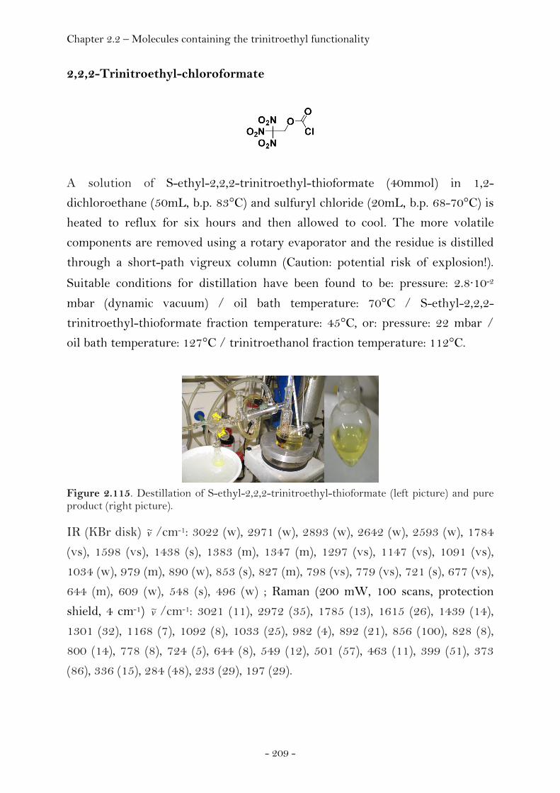
Chapter 2.2 – Molecules containing the trinitroethyl functionality
- 209 -
2,2,2-Trinitroethyl-chloroformate
A solution of S-ethyl-2,2,2-trinitroethyl-thioformate (40mmol) in 1,2-
dichloroethane (50mL, b.p. 83°C) and sulfuryl chloride (20mL, b.p. 68-70°C) is
heated to reflux for six hours and then allowed to cool. The more volatile
components are removed using a rotary evaporator and the residue is distilled
through a short-path vigreux column (Caution: potential risk of explosion!).
Suitable conditions for distillation have been found to be: pressure: 2.8·10-2
mbar (dynamic vacuum) / oil bath temperature: 70°C / S-ethyl-2,2,2-
trinitroethyl-thioformate fraction temperature: 45°C, or: pressure: 22 mbar /
oil bath temperature: 127°C / trinitroethanol fraction temperature: 112°C.
Figure 2.115. Destillation of S-ethyl-2,2,2-trinitroethyl-thioformate (left picture) and pure product (right picture). IR (KBr disk) v~ /cm-1: 3022 (w), 2971 (w), 2893 (w), 2642 (w), 2593 (w), 1784
(vs), 1598 (vs), 1438 (s), 1383 (m), 1347 (m), 1297 (vs), 1147 (vs), 1091 (vs),
1034 (w), 979 (m), 890 (w), 853 (s), 827 (m), 798 (vs), 779 (vs), 721 (s), 677 (vs),
644 (m), 609 (w), 548 (s), 496 (w) ; Raman (200 mW, 100 scans, protection
shield, 4 cm-1) v~ /cm-1: 3021 (11), 2972 (35), 1785 (13), 1615 (26), 1439 (14),
1301 (32), 1168 (7), 1092 (8), 1033 (25), 982 (4), 892 (21), 856 (100), 828 (8),
800 (14), 778 (8), 724 (5), 644 (8), 549 (12), 501 (57), 463 (11), 399 (51), 373
(86), 336 (15), 284 (48), 233 (29), 197 (29).
Chapter 2.2 – Molecules containing the trinitroethyl functionality
- 210 -
Bis-(2,2,2-trinitroethyl)-carbonate (BTC)
Bis-(2,2,2-trinitroethyl)-carbonate (Hill, 1956) was prepared from the reaction
of trinitroethanol with phosgene (Hall, 1968). The crystal growth was
accomplished by concentration of a saturated CHCl3 solution at ambient
temperature, yielding colourless single crystals.
The following experimental data are available in the literature: (300-301) m.p.
115°C, impact sensitivity (50%, NOL app., 2.5 kg, 16cm): 4 J; Details of the
single crystal X-ray diffraction experiment are listed in the appendix (Chapter
4).
2,2,2-Trinitroethyl-azidoformate (TAF)
2,2,2-Trinitroethyl-chloroformate is a starting material for the synthesis of
TAF and was synthesised according to literature procedure. (302)
Trimethylsilyazide (0.524 g, 4.55 mmol) was dissolved in acetonitrile (10 mL).
2,2,2-Trinitroethyl-chloroformate (0.499 g, 2.05 mmol) dissolved in
acetonitrile (1 mL) was added to this solution drop wise at room temperature
and kept under a constant stream of nitrogen. The color of the solution
changed from colorless to orange and got turbid after a few minutes with the
formation of a precipitate. The reaction was allowed to stir for further twelve
hours. The solvent was then removed using a high vacuum line affording
orange oil together with precipitate. Pure TAF was obtained as a white solid
on sublimation of the reaction residue using a cold finger (-78°C).
DSC (Linseis, 5 K min-1): 1st endo (27.5°C, onset / 29.9°C, min / 32.3°C, offset),
2nd endo (96.2°C, onset / 118.4°C, min / 129.2°C, offset), 1st exo (130.4°C,
onset / 158.4°C, max / 174.5°C, offset), 2nd exo (191.6°C, onset / 205.6°C, max

Chapter 2.2 – Molecules containing the trinitroethyl functionality
- 211 -
/ 218.6, offset); 1H NMR ([D6]acetone) δ: 5.96 (2H, s, -CH2-); 13C NMR
([D6]acetone) δ: 63.7 (s, -CH2-), 123.9 (bs, -C(NO2)3), 156.1 (s, O2CN3); 15N
NMR ([D6]acetone) δ (nitromethane): -34.7 (s, -NO2), -141.1 (s, Nbeta), -148.2 (s,
Ngamma), -266.9 (s, Nalpha); IR (KBr disk) v~ /cm-1: 3018 (vs), 2971 (vs), 2893 (s),
2651 (w), 2601 (w), 2399 (w), 2190 (vs), 1751 (s), 1598 (s), 1442 (s), 1386 (s),
1354 (s), 1297 (s), 1236 (s), 1186 (s), 1099 (m), 1049 (m), 1007 (m), 941 (m), 879
(m), 855 (m), 806 (m), 783 (m), 742 (m), 708 (w); Raman (4 cm-1) v~ /cm-1: 2974
(36), 2170 (11), 1615 (38), 1384 (29), 1357 (43), 1304 (44), 1099 (15), 1009 (22),
942 (27), 894 (19), 857 (83), 734 (21), 531 (20), 408 (61), 375 (73), 292 (53), 256
(41); MS (DEI+): 250 [m, 1.7%], 204 [m - NO2, 2%], 164 [m - O2C-N3, 100%], 158 [m - 2 NO2, 19.8%], 118 [m - (O2C-N3, 2 NO2), 100%], 70 [N3CO, 10.5%], 44 [CO2, 3.8%]; Calc. for C3H2N6O8: C (14.4 %), H (0.8 %), N (33.6 %), found: C
(14.7 %), H (1.0 %), N (32.5 %) ; Details of the single crystal X-ray diffraction
experiment are listed in the appendix (Chapter 4).
Bis-(2,2,2-trinitroethyl)-hydrazodicarboxylate (BTHC)
2,2,2-Trinitroethylchloroformate (4.00 g, 18.7 mmol) was dissolved in
dichloromethane (20 mL). The solution was cooled to -78°C. To this was added
23.92 g of a solution of hydrazine in THF (1 molar, density 0.89 g/mL). A
white precipitate formed. The reaction mixture was allowed to warm to 0°C
over a period of one hour. Cold water (0°C) was added (20 mL) and the solution
extracted with diethyl ether (5x, each 50 mL). The collected organic phases
were dried using magnesium sulfate, filtered and concentrated using a rotary
evaporator. The density of the pure substance was estimated to be 1.8 g cm-3
according to weighing several samples of different volume. Solidified BTHC
was dissolved in acetone or ethyl acetate affording single crystals on standing
at room temperature.
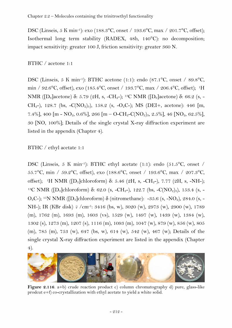
Chapter 2.2 – Molecules containing the trinitroethyl functionality
- 212 -
DSC (Linseis, 5 K min-1): exo (188.3°C, onset / 193.6°C, max / 201.7°C, offset);
Isothermal long term stability (RADEX, 48h, 140°C): no decomposition;
impact sensitivity: greater 100 J, friction sensitivity: greater 360 N.
BTHC / acetone 1:1
DSC (Linseis, 5 K min-1): BTHC acetone (1:1): endo (87.1°C, onset / 89.8°C,
min / 92.6°C, offset), exo (185.4°C, onset / 193.7°C, max / 206.4°C, offset); 1H
NMR ([D6]acetone) δ: 5.79 (2H, s, -CH2-); 13C NMR ([D6]acetone) δ: 66.2 (s, -
CH2-), 128.7 (bs, -C(NO2)3), 158.2 (s, -O2C-); MS (DEI+, acetone): 446 [m,
7.4%], 400 [m - NO2, 0.6%], 266 [m – O-CH2-C(NO2)3, 2.5%], 46 [NO2, 62.5%], 30 [NO, 100%]; Details of the single crystal X-ray diffraction experiment are
listed in the appendix (Chapter 4).
BTHC / ethyl acetate 1:1
DSC (Linseis, 5 K min-1): BTHC ethyl acetate (1:1): endo (51.5°C, onset /
55.7°C, min / 59.2°C, offset), exo (188.6°C, onset / 193.6°C, max / 207.3°C,
offset); 1H NMR ([D3]chloroform) δ: 5.46 (2H, s, -CH2-), 7.77 (2H, s, -NH-); 13C NMR ([D3]chloroform) δ: 62.0 (s, -CH2-), 122.7 (bs, -C(NO2)3), 153.4 (s, -
O2C-); 15N NMR ([D3]chloroform) δ (nitromethane): -35.6 (s, -NO2), 284.0 (s, -
NH-); IR (KBr disk) v~ /cm-1: 3416 (bs, w), 3020 (w), 2973 (w), 2900 (w), 1789
(m), 1762 (m), 1693 (m), 1603 (vs), 1529 (w), 1497 (w), 1439 (w), 1384 (w),
1302 (s), 1273 (m), 1207 (s), 1116 (m), 1093 (m), 1047 (w), 879 (w), 856 (w), 805
(m), 785 (m), 753 (w), 647 (bs, w), 614 (w), 542 (w), 467 (w); Details of the
single crystal X-ray diffraction experiment are listed in the appendix (Chapter
4).
Figure 2.116. a+b) crude reaction product c) column chromatography d) pure, glass-like prodcut e+f) co-crystallization with ethyl acetate to yield a white solid.
a) b) c) d) e) f)
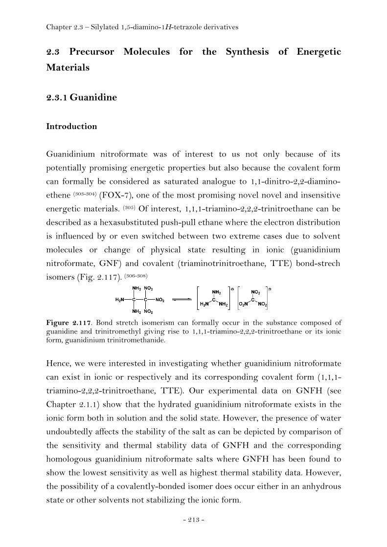
Chapter 2.3 – Silylated 1,5-diamino-1H-tetrazole derivatives
- 213 -
2.3 Precursor Molecules for the Synthesis of Energetic
Materials
2.3.1 Guanidine
Introduction
Guanidinium nitroformate was of interest to us not only because of its
potentially promising energetic properties but also because the covalent form
can formally be considered as saturated analogue to 1,1-dinitro-2,2-diamino-
ethene (303-304) (FOX-7), one of the most promising novel novel and insensitive
energetic materials. (305) Of interest, 1,1,1-triamino-2,2,2-trinitroethane can be
described as a hexasubstituted push-pull ethane where the electron distribution
is influenced by or even switched between two extreme cases due to solvent
molecules or change of physical state resulting in ionic (guanidinium
nitroformate, GNF) and covalent (triaminotrinitroethane, TTE) bond-strech
isomers (Fig. 2.117). (306-308)
Figure 2.117. Bond stretch isomerism can formally occur in the substance composed of guanidine and trinitromethyl giving rise to 1,1,1-triamino-2,2,2-trinitroethane or its ionic form, guanidinium trinitromethanide.
Hence, we were interested in investigating whether guanidinium nitroformate
can exist in ionic or respectively and its corresponding covalent form (1,1,1-
triamino-2,2,2-trinitroethane, TTE). Our experimental data on GNFH (see
Chapter 2.1.1) show that the hydrated guanidinium nitroformate exists in the
ionic form both in solution and the solid state. However, the presence of water
undoubtedly affects the stability of the salt as can be depicted by comparison of
the sensitivity and thermal stability data of GNFH and the corresponding
homologous guanidinium nitroformate salts where GNFH has been found to
show the lowest sensitivity as well as highest thermal stability data. However,
the possibility of a covalently-bonded isomer does occur either in an anhydrous
state or other solvents not stabilizing the ionic form.
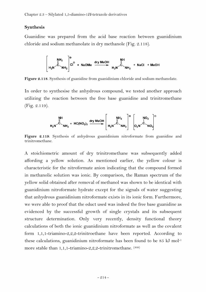
Chapter 2.3 – Silylated 1,5-diamino-1H-tetrazole derivatives
- 214 -
Synthesis
Guanidine was prepared from the acid base reaction between guanidinium
chloride and sodium methanolate in dry methanole (Fig. 2.118).
Figure 2.118. Synthesis of guanidine from guanidinium chloride and sodium methanolate.
In order to synthesise the anhydrous compound, we tested another approach
utilizing the reaction between the free base guanidine and trinitromethane
(Fig. 2.119).
Figure 2.119. Synthesis of anhydrous guanidinium nitroformate from guanidine and trinitromethane.
A stoichiometric amount of dry trinitromethane was subsequently added
affording a yellow solution. As mentioned earlier, the yellow colour is
characteristic for the nitroformate anion indicating that the compound formed
in methanolic solution was ionic. By comparison, the Raman spectrum of the
yellow solid obtained after removal of methanol was shown to be identical with
guanidinium nitroformate hydrate except for the signals of water suggesting
that anhydrous guanidinium nitroformate exists in its ionic form. Furthermore,
we were able to proof that the educt used was indeed the free base guanidine as
evidenced by the successful growth of single crystals and its subsequent
structure determination. Only very recently, density functional theory
calculations of both the ionic guanidinium nitroformate as well as the covalent
form 1,1,1-triamino-2,2,2-trinitroethane have been reported. According to
these calculations, guanidinium nitroformate has been found to be 85 kJ mol-1
more stable than 1,1,1-triamino-2,2,2-trinitromethane. (309)

Chapter 2.3 – Silylated 1,5-diamino-1H-tetrazole derivatives
- 215 -
Crystal Structure Analysis
Due to the high basicity, guanidine (aminoformamidine) was considered to be
the strongest neutral organic base (pKa = 13.6) (310) until proton sponges (311-312)
were synthesised. The question of its molecular structure and the reason for its
exceptional properties inspired several controversial theoretical works (313-322)
since an experimentally determined structure for guanidine was not available.
To our knowledge, only a few neutral derivatives of guanidines containing
sterically demanding organic substituents have been structurally characterized
using single-crystal X-ray diffraction. Rare examples describe 1,1,3,3-
tetrasubstituted (323-324) as well as 1,2,3-trisubstituted (325) guanidine derivatives
acting as ligands in transition metal complexes. Here we report crystal
structures containing the unsubstituted free base guanidine and 2-amino-4,6-
dimethyl-1,3,5-triazine. Single crystals consisting of guanidine and 2-amino-
4,6-dimethyl-1,3,5-triazine were obtained when acetonitrile was allowed to
slowly diffuse into a methanolic solution of guanidine. The presence of 2-
amino-4,6-dimethyl-1,3,5-triazine in the crystal structures of the 1:1 co-crystal
as well as the 2:1 co-crystal corresponds to and confirms the formation of
substituted 2-amino-triazines from aliphatic nitriles and guanidine in the
presence of alkoholates. (326)
Figure 2.120. Ortep representation of the structure of 2-amino-4,6-dimethyl-1,3,5-triazine · guanidine. The thermal ellipsoids are shown at the 50 % probability level.
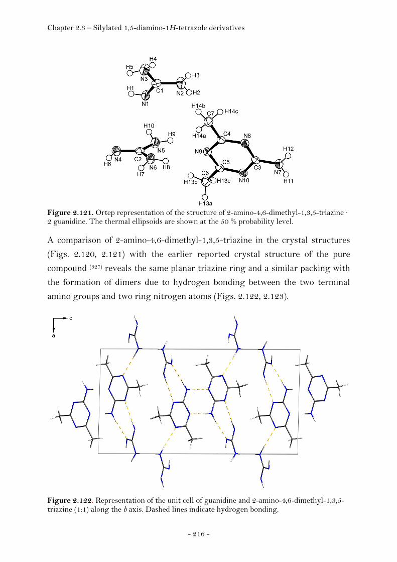
Chapter 2.3 – Silylated 1,5-diamino-1H-tetrazole derivatives
- 216 -
Figure 2.121. Ortep representation of the structure of 2-amino-4,6-dimethyl-1,3,5-triazine · 2 guanidine. The thermal ellipsoids are shown at the 50 % probability level. A comparison of 2-amino-4,6-dimethyl-1,3,5-triazine in the crystal structures
(Figs. 2.120, 2.121) with the earlier reported crystal structure of the pure
compound (327) reveals the same planar triazine ring and a similar packing with
the formation of dimers due to hydrogen bonding between the two terminal
amino groups and two ring nitrogen atoms (Figs. 2.122, 2.123).
Figure 2.122. Representation of the unit cell of guanidine and 2-amino-4,6-dimethyl-1,3,5-triazine (1:1) along the b axis. Dashed lines indicate hydrogen bonding.
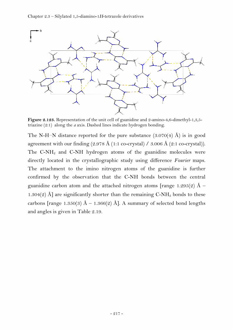
Chapter 2.3 – Silylated 1,5-diamino-1H-tetrazole derivatives
- 217 -
Figure 2.123. Representation of the unit cell of guanidine and 2-amino-4,6-dimethyl-1,3,5-triazine (2:1) along the a axis. Dashed lines indicate hydrogen bonding.
The N-H…N distance reported for the pure substance (3.070(4) Å) is in good
agreement with our finding (2.978 Å (1:1 co-crystal) / 3.006 Å (2:1 co-crystal)).
The C-NH2 and C-NH hydrogen atoms of the guanidine molecules were
directly located in the crystallographic study using difference Fourier maps.
The attachment to the imino nitrogen atoms of the guanidine is further
confirmed by the observation that the C-NH bonds between the central
guanidine carbon atom and the attached nitrogen atoms [range 1.295(2) Å –
1.304(2) Å] are significantly shorter than the remaining C-NH2 bonds to these
carbons [range 1.350(3) Å – 1.366(2) Å]. A summary of selected bond lengths
and angles is given in Table 2.19.

Chapter 2.3 – Silylated 1,5-diamino-1H-tetrazole derivatives
- 218 -
Table 2.19. Selected bond lengths and angles of the guanidine molecule.
structure 1:1 (G : ADMT) 2:1 (G : ADMT)
guanidine #1 #2 #3
d / Å C1-N1 1.295(2) C1-N1 1.304 (2) C7-N8 1.300 (3)
C1-N2 1.366 (2) C1-N2 1.350 (3) C7-N10 1.361 (3)
C1-N3 1.355 (2) C1-N3 1.359 (3) C7-N9 1.359 (3)
angle / ° N1-C1-N2 124 (0) N1-C1-N3 125 (0) N8-C7-N9 125 (0)
N1-C1-N3 120 (0) N1-C1-N2 120 (0) N8-C7-N10 120 (0)
N2-C1-N3 116 (0) N2-C1-N3 115 (0) N10-C7-N9 115 (0)
C1-N1-H1 110 (1) C1-N1-H1 109 (1) C7-N8-H8 110 (1)
† / ° N2 356 (4) N2 359 (5) N10 348 (5)
N3 351 (3) N3 350 (6) N9 353 (5) † Degree of planarity given as the angle sum around the nitrogen atom of the corresponding amino group ΣCNH,CNH,HNH.
The central carbon atom of the guanidine molecule in structures of the 1:1 and
2:1 co-crystals of guanidine and 2-amino-4,6-dimethyl-1,3,5-triazine forms one
plane with the three surrounding nitrogen atoms (ΣNCN = 360 °). In contrast,
the two amino groups of guanidine show a pyramidal geometry confirming a
theoretical study published by Frenking et al. (328) where a nonplanar geometry
of the guanidine molecule with strongly pyramidal NH2 groups was predicted
at the MP2 level of theory using a 6-31G(d) basis set. In addition, our
experimentally determined bond lengths agree well with the calculated ones of
the theoretical study where values of 1.284 Å, 1.396 Å and 1.400 Å (MP2/6-
31G(d)) were proposed. The deviations of the experimentally obtained values
for the structure of guanidine in the solid state compared to the calculated
equilibrium gas phase data are surprisingly small. Although the environment of
the guanidine molecules in the two crystal structures is different and hydrogen
bonding between guanidine itself as well as guanidine and 2-amino-4,6-
dimethyl-1,3,5-triazine does occur (Table 2.20), a comparison of the three
observed guanidine molecules shows that the interatomic distances and angles
(Table 2.19) are in excellent agreement and are not noticeably influenced by
packing effects of the crystal. The obtained geometrical data of the free base
guanidine thus provide reliable experimental evidence for the nature of the
molecular structure of guanidine for the first time.

Chapter 2.3 – Silylated 1,5-diamino-1H-tetrazole derivatives
- 219 -
Table 2.20. Hydrogen bonding in the crystal structures of guanidine (G) and 2-amino-4,6-dimethyl-1,3,5-triazine (ADMT) (1:1 and 2:1).
structure D-H..A d (D-H) / Å d (H..A) / Å d (D..A) / Å < DHA
1:1 (G:ADMT) N3-H3B..N5 0.908(15) 2.202(16) 3.1100(16) 178.7(13)
N2-H2A..N6i 0.927(18) 2.177(19) 3.1021(17) 175.7(15)
N7-H7A..N4ii 0.856(15) 2.122(16) 2.9777(17) 177.6(12)
N7-H7B..N1 0.915(15) 2.018(16) 2.9273(16) 172.50(13)
2:1 (G:ADMT) N2-H2A..N4iii 0.89(3) 2.17(3) 3.052(3) 170(2)
N3-H3A..N8iv 0.88(3) 2.17(3) 3.041(3) 171(2)
N9-H9A..N8v 0.93(2) 2.11(3) 3.030(3) 170.2(18)
N7-H7A..N1iii 0.88(2) 2.05(2) 2.934(3) 178(2)
N7-H7B..N6vi 0.93(2) 2.08(2) 3.006(2) 176.7(19) Notes. Symmetry codes: (i) x+1, y, z, (ii) –x+1, -y, -z, (iii) –x+2, -y, -z, (iv) x+1, y, z, (v) x, -y+1/2, z+1/2, (vi) –x+3, -y, -z+1.
Experimental
Guanidine was prepared by adding a THF solution of guanidinium chloride
(16.7 mmol, 24 mL) to a THF solution of sodium methanolate (16.8 mmol, 24
mL) using standard Schlenk techniques. Acetonitrile was allowed to slowly
diffuse into the solution until crystals suitable for X-ray diffraction formed at
4°C (guanidine and 2-amino-4,6-dimethyl-1,3,5-triazine, 1:1) or 25°C
(guanidine and 2-amino-4,6-dimethyl-1,3,5-triazine, 2:1). Details of the single
crystal X-ray diffraction experiment are listed in the appendix (Chapter 4).
Anhydrous GNF was obtained for the first time by reacting the free
guanidinium base with nitroform in dry methanol. A yellow solution was
obtained indicating the presence of the trinitromethanide anion. After
evaporation of the methanol a yellow solid was obtained. The Raman spectrum
of this substance closely resembles that of the monohydrate of GNF. Hence,
the anhydrous GNF obtained exist as ionic species rather than the covalent
isomer.
Raman (r.t.): ν = 2987(13), 1528(15), 1469(14), 1388(76), 1298(46), 1244(44),
1156(54), 1014(56), 867(100), 790(18), 729(14), 533(25), 473(30), 442(20),
253(24), 164(25).

Chapter 2.3 – Silylated 1,5-diamino-1H-tetrazole derivatives
- 220 -
Silylated 1,5-Diamino-1H-tetrazole derivatives
Introduction
Tetrazoles have manifold and highly diverse applications in chemistry. As far
as energetic materials are concerned, they represent important building blocks
for the synthesis of novel energetic materials. Of the many excellent reviews
about tetrazoles, the work of Ostrovskii et al. (329) as well as the work of Benson
et al. (330) is particularly noteworthy. Recent results and developments obtained
in our laboratories at LMU Munich are documented in the dissertations of
Hammerl (331), Weigand (332) and Stierstorfer (333) and references cited therein.
As mentioned earlier, high heat of formation is a critical parameter for the
performance properties of a high explosive and most of the energy derived in
modern HEDM stems not only from oxidation of the carbon backbone as in
traditional energetic materials but also from ring or cage strain and from their
very high positive heat of formation. Tetrazoles are the most promising
heterocycles amongst five-membered rings with four nitrogen atoms in the
ring displaying high heats of formation due to an increasing positive trend in
heat of formation with increasing number of nitrogen. Among the advantages
of tetrazole based explosive and propellants we mention the large amount of
gaseous decomposition products preferentially containing environmentally- as
well as (eco)-toxicologically benign dinitrogen, a significant advantage in order
to meet one of the major goals of contemporary research on next generation
energetic materials. We were interested in synthesizing derivatives of 1,5-
diamino-1H-tetrazole (DAT) that would give access to novel synthetic routes
for the preparation of functionalized derivatives of this high nitrogen
containing (84.0 %) compound. Two examples of such derivatives have already
been mentioned in Chapter 2.2.1, N1-(2,2,2-trinitroethyl)-1H-tetrazole-1,5-
diamine (TTD) as well as N1,N5-bis-(2,2,2-trinitroethyl)-1H-tetrazole-1,5-
diamine (BTTD) and were shown to have superior performance properties
approaching or even exceeding those of RDX. However, they were lacking
sufficient thermal stability and thus, an increase in this property would be
highly desirable. We have shown that it is possible to keep the advantages of
the trinitroethyl group and improve the thermal stability of the class of N-
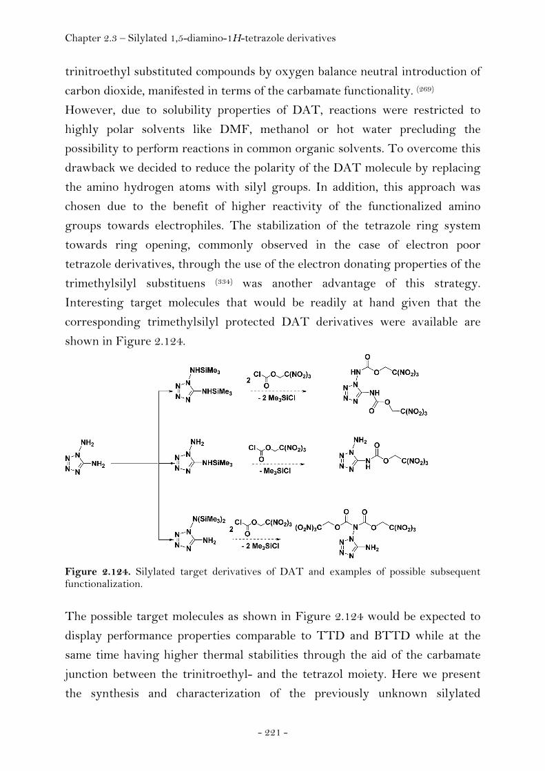
Chapter 2.3 – Silylated 1,5-diamino-1H-tetrazole derivatives
- 221 -
trinitroethyl substituted compounds by oxygen balance neutral introduction of
carbon dioxide, manifested in terms of the carbamate functionality. (269)
However, due to solubility properties of DAT, reactions were restricted to
highly polar solvents like DMF, methanol or hot water precluding the
possibility to perform reactions in common organic solvents. To overcome this
drawback we decided to reduce the polarity of the DAT molecule by replacing
the amino hydrogen atoms with silyl groups. In addition, this approach was
chosen due to the benefit of higher reactivity of the functionalized amino
groups towards electrophiles. The stabilization of the tetrazole ring system
towards ring opening, commonly observed in the case of electron poor
tetrazole derivatives, through the use of the electron donating properties of the
trimethylsilyl substituens (334) was another advantage of this strategy.
Interesting target molecules that would be readily at hand given that the
corresponding trimethylsilyl protected DAT derivatives were available are
shown in Figure 2.124.
Figure 2.124. Silylated target derivatives of DAT and examples of possible subsequent functionalization.
The possible target molecules as shown in Figure 2.124 would be expected to
display performance properties comparable to TTD and BTTD while at the
same time having higher thermal stabilities through the aid of the carbamate
junction between the trinitroethyl- and the tetrazol moiety. Here we present
the synthesis and characterization of the previously unknown silylated

Chapter 2.3 – Silylated 1,5-diamino-1H-tetrazole derivatives
- 222 -
derivatives of 1,5-diamino-1H-tetrazole including NMR- and vibrational
spectroscopy, mass spectrometry and single crystal X-ray diffractometry.
Structural optimizations of the isolated molecules in the gas phase and
frequency analyses were also performed as a preliminary step for the
assignment of vibrational frequencies.
Synthesis
Different methods have been reported for the silylation of amines in the
literature. (335-341) Due to the fact, that DAT bears two amino groups, each
exhibiting different chemical behaviour, selective monosilylation of each group
is not a straightforward reaction. According to the literature, the following
three silylation strategies are frequently employed:
1. Direct silylation using trimethylsilyl chloride (TMSCl) in presence of
a base. (342-343)
2. Deprotonation of the amino group using n-buthyllithium and
subsequent reaction with TMSCl. (344)
3. Silylation using hexamethyldisilazane (HMDS) in presence of Lewis
acids. (345-346)
Among the reaction conditions applied, direct reaction of DAT with TMSCl
using THF as solvent and applying reflux conditions proofed to be succesfull
and yielded the doubly silylated product N1,N5-bis-(trimethylsilyl)-1,5-
diamino-1H-tetrazole (1,5-BTMSD, Fig. 2.125).
Figure 2.125. Synthesis of N1,N5-bis-(trimethylsilyl)-1,5-diamino-1H-tetrazole (1,5-BTMSD) from the reaction between DAT and TMSCL.
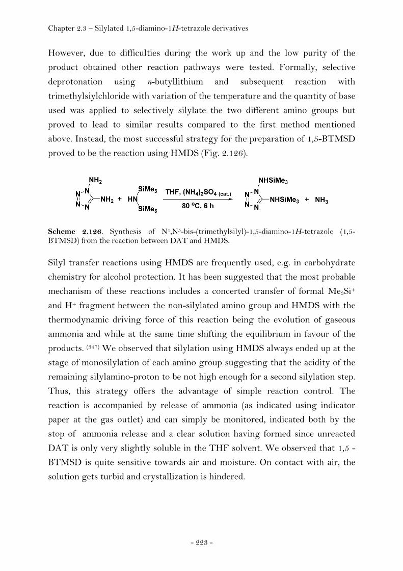
Chapter 2.3 – Silylated 1,5-diamino-1H-tetrazole derivatives
- 223 -
However, due to difficulties during the work up and the low purity of the
product obtained other reaction pathways were tested. Formally, selective
deprotonation using n-butyllithium and subsequent reaction with
trimethylsiylchloride with variation of the temperature and the quantity of base
used was applied to selectively silylate the two different amino groups but
proved to lead to similar results compared to the first method mentioned
above. Instead, the most successful strategy for the preparation of 1,5-BTMSD
proved to be the reaction using HMDS (Fig. 2.126).
Scheme 2.126. Synthesis of N1,N5-bis-(trimethylsilyl)-1,5-diamino-1H-tetrazole (1,5-BTMSD) from the reaction between DAT and HMDS. Silyl transfer reactions using HMDS are frequently used, e.g. in carbohydrate
chemistry for alcohol protection. It has been suggested that the most probable
mechanism of these reactions includes a concerted transfer of formal Me3Si+
and H+ fragment between the non-silylated amino group and HMDS with the
thermodynamic driving force of this reaction being the evolution of gaseous
ammonia and while at the same time shifting the equilibrium in favour of the
products. (347) We observed that silylation using HMDS always ended up at the
stage of monosilylation of each amino group suggesting that the acidity of the
remaining silylamino-proton to be not high enough for a second silylation step.
Thus, this strategy offers the advantage of simple reaction control. The
reaction is accompanied by release of ammonia (as indicated using indicator
paper at the gas outlet) and can simply be monitored, indicated both by the
stop of ammonia release and a clear solution having formed since unreacted
DAT is only very slightly soluble in the THF solvent. We observed that 1,5 -
BTMSD is quite sensitive towards air and moisture. On contact with air, the
solution gets turbid and crystallization is hindered.
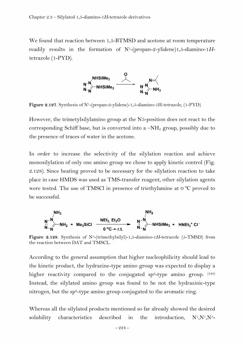
Chapter 2.3 – Silylated 1,5-diamino-1H-tetrazole derivatives
- 224 -
We found that reaction between 1,5-BTMSD and acetone at room temperature
readily results in the formation of N1-(propan-2-ylidene)1,5-diamino-1H-
tetrazole (1-PYD).
Figure 2.127. Synthesis of N1-(propan-2-ylidene)-1,5-diamino-1H-tetrazole, (1-PYD)
However, the trimetylsilylamino group at the N5-position does not react to the
corresponding Schiff base, but is converted into a –NH2 group, possibly due to
the presence of traces of water in the acetone.
In order to increase the selectivity of the silylation reaction and achieve
monosilylation of only one amino group we chose to apply kinetic control (Fig.
2.128). Since heating proved to be necessary for the silylation reaction to take
place in case HMDS was used as TMS-transfer reagent, other silylation agents
were tested. The use of TMSCl in presence of triethylamine at 0 ºC proved to
be successful.
Figure 2.128. Synthesis of N5-(trimethylsilyl)-1,5-diamino-1H-tetrazole (5-TMSD) from the reaction between DAT and TMSCL.
According to the general assumption that higher nucleophilicity should lead to
the kinetic product, the hydrazine-type amino group was expected to display a
higher reactivity compared to the conjugated sp2-type amino group. (348)
Instead, the silylated amino group was found to be not the hydrazinic-type
nitrogen, but the sp2-type amino group conjugated to the aromatic ring.
Whereas all the silylated products mentioned so far already showed the desired
solubility characteristics described in the introduction, N1,N1,N5-
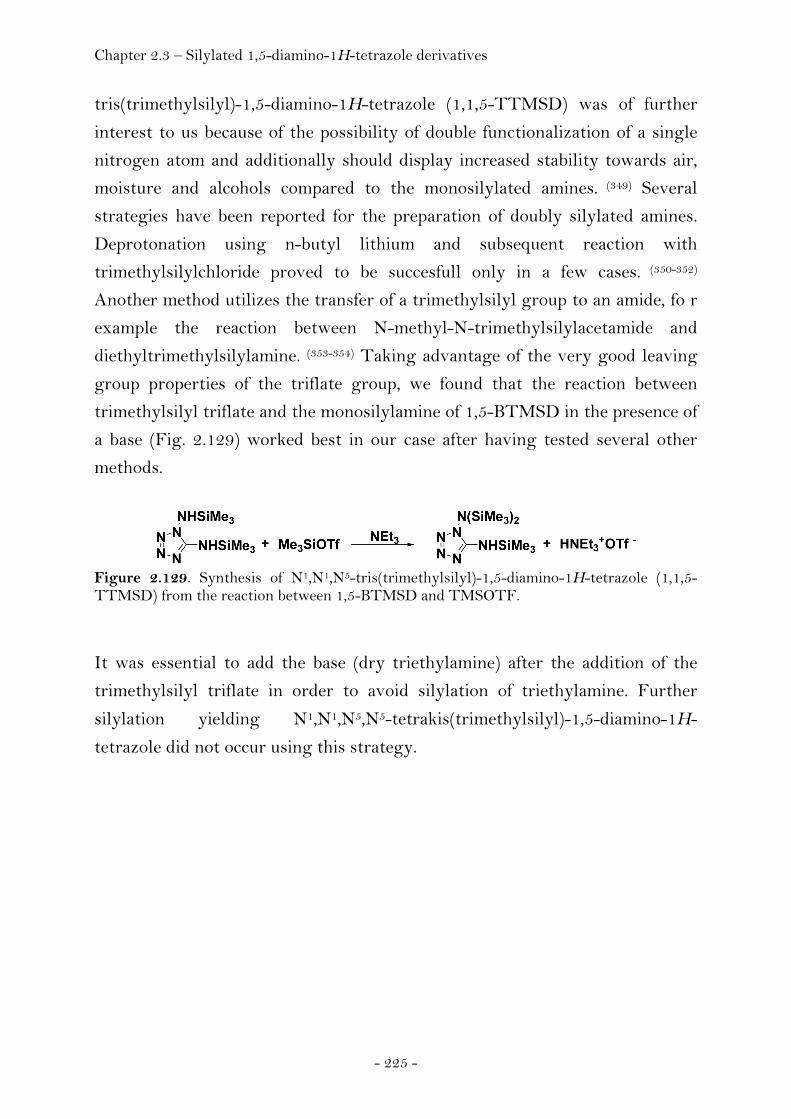
Chapter 2.3 – Silylated 1,5-diamino-1H-tetrazole derivatives
- 225 -
tris(trimethylsilyl)-1,5-diamino-1H-tetrazole (1,1,5-TTMSD) was of further
interest to us because of the possibility of double functionalization of a single
nitrogen atom and additionally should display increased stability towards air,
moisture and alcohols compared to the monosilylated amines. (349) Several
strategies have been reported for the preparation of doubly silylated amines.
Deprotonation using n-butyl lithium and subsequent reaction with
trimethylsilylchloride proved to be succesfull only in a few cases. (350-352)
Another method utilizes the transfer of a trimethylsilyl group to an amide, fo r
example the reaction between N-methyl-N-trimethylsilylacetamide and
diethyltrimethylsilylamine. (353-354) Taking advantage of the very good leaving
group properties of the triflate group, we found that the reaction between
trimethylsilyl triflate and the monosilylamine of 1,5-BTMSD in the presence of
a base (Fig. 2.129) worked best in our case after having tested several other
methods.
Figure 2.129. Synthesis of N1,N1,N5-tris(trimethylsilyl)-1,5-diamino-1H-tetrazole (1,1,5-TTMSD) from the reaction between 1,5-BTMSD and TMSOTF.
It was essential to add the base (dry triethylamine) after the addition of the
trimethylsilyl triflate in order to avoid silylation of triethylamine. Further
silylation yielding N1,N1,N5,N5-tetrakis(trimethylsilyl)-1,5-diamino-1H-
tetrazole did not occur using this strategy.
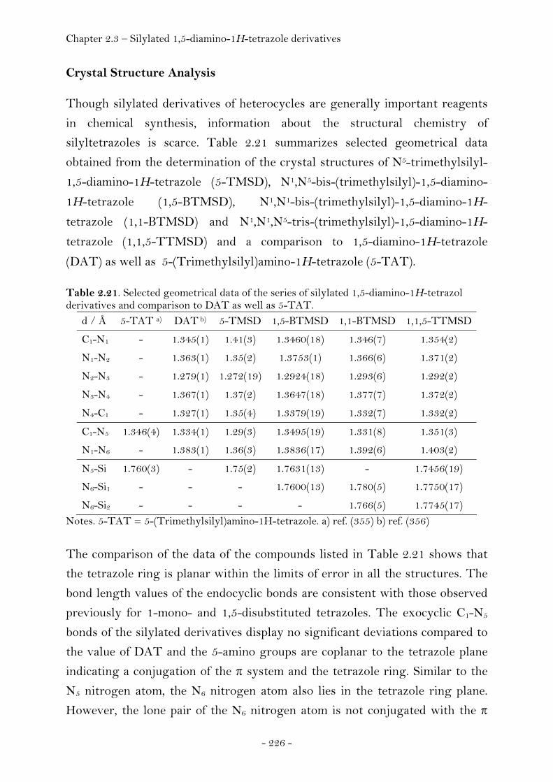
Chapter 2.3 – Silylated 1,5-diamino-1H-tetrazole derivatives
- 226 -
Crystal Structure Analysis
Though silylated derivatives of heterocycles are generally important reagents
in chemical synthesis, information about the structural chemistry of
silyltetrazoles is scarce. Table 2.21 summarizes selected geometrical data
obtained from the determination of the crystal structures of N5-trimethylsilyl-
1,5-diamino-1H-tetrazole (5-TMSD), N1,N5-bis-(trimethylsilyl)-1,5-diamino-
1H-tetrazole (1,5-BTMSD), N1,N1-bis-(trimethylsilyl)-1,5-diamino-1H-
tetrazole (1,1-BTMSD) and N1,N1,N5-tris-(trimethylsilyl)-1,5-diamino-1H-
tetrazole (1,1,5-TTMSD) and a comparison to 1,5-diamino-1H-tetrazole
(DAT) as well as 5-(Trimethylsilyl)amino-1H-tetrazole (5-TAT). Table 2.21. Selected geometrical data of the series of silylated 1,5-diamino-1H-tetrazol derivatives and comparison to DAT as well as 5-TAT.
d / Å 5-TAT a) DAT b) 5-TMSD 1,5-BTMSD 1,1-BTMSD 1,1,5-TTMSD
C1-N1 - 1.345(1) 1.41(3) 1.3460(18) 1.346(7) 1.354(2)
N1-N2 - 1.363(1) 1.35(2) 1.3753(1) 1.366(6) 1.371(2)
N2-N3 - 1.279(1) 1.272(19) 1.2924(18) 1.293(6) 1.292(2)
N3-N4 - 1.367(1) 1.37(2) 1.3647(18) 1.377(7) 1.372(2)
N4-C1 - 1.327(1) 1.35(4) 1.3379(19) 1.332(7) 1.332(2)
C1-N5 1.346(4) 1.334(1) 1.29(3) 1.3495(19) 1.331(8) 1.351(3)
N1-N6 - 1.383(1) 1.36(3) 1.3836(17) 1.392(6) 1.403(2)
N5-Si 1.760(3) - 1.75(2) 1.7631(13) - 1.7456(19)
N6-Si1 - - - 1.7600(13) 1.780(5) 1.7750(17)
N6-Si2 - - - - 1.766(5) 1.7745(17) Notes. 5-TAT = 5-(Trimethylsilyl)amino-1H-tetrazole. a) ref. (355) b) ref. (356)
The comparison of the data of the compounds listed in Table 2.21 shows that
the tetrazole ring is planar within the limits of error in all the structures. The
bond length values of the endocyclic bonds are consistent with those observed
previously for 1-mono- and 1,5-disubstituted tetrazoles. The exocyclic C1-N5
bonds of the silylated derivatives display no significant deviations compared to
the value of DAT and the 5-amino groups are coplanar to the tetrazole plane
indicating a conjugation of the π system and the tetrazole ring. Similar to the
N5 nitrogen atom, the N6 nitrogen atom also lies in the tetrazole ring plane.
However, the lone pair of the N6 nitrogen atom is not conjugated with the π
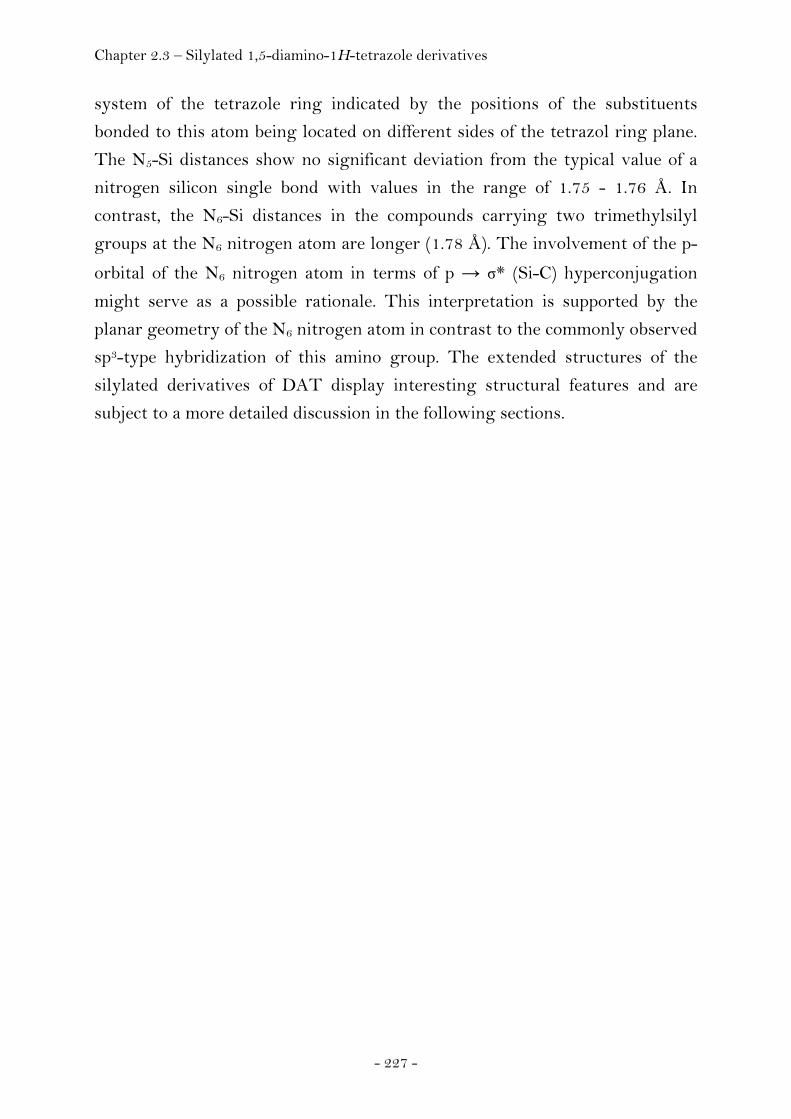
Chapter 2.3 – Silylated 1,5-diamino-1H-tetrazole derivatives
- 227 -
system of the tetrazole ring indicated by the positions of the substituents
bonded to this atom being located on different sides of the tetrazol ring plane.
The N5-Si distances show no significant deviation from the typical value of a
nitrogen silicon single bond with values in the range of 1.75 - 1.76 Å. In
contrast, the N6-Si distances in the compounds carrying two trimethylsilyl
groups at the N6 nitrogen atom are longer (1.78 Å). The involvement of the p-
orbital of the N6 nitrogen atom in terms of p → σ* (Si-C) hyperconjugation
might serve as a possible rationale. This interpretation is supported by the
planar geometry of the N6 nitrogen atom in contrast to the commonly observed
sp3-type hybridization of this amino group. The extended structures of the
silylated derivatives of DAT display interesting structural features and are
subject to a more detailed discussion in the following sections.
Chapter 2.3 – Silylated 1,5-diamino-1H-tetrazole derivatives
- 228 -
N5-Trimethylsilyl-1,5-diamino-1H-tetrazole (5-TMSD)
N5-trimethylsilyl-1,5-diamino-1H-tetrazole (5-TMSD) crystallizes in the
monoclinic space group P21/c (no. 14) with four molecules in the unit cell. The
asymmetric unit is comprised of one molecule (Fig. 2.130).
Figure 2.130. ORTEP representation of the molecular structure of N5-trimethylsilyl-1,5-diamino-1H-tetrazole (5-TMSD) in the crystalline state. Displacement ellipsoids are shown at the 50 % probability level. Selected bond lengths [Å] and angles [°]: C1-N1 1.41(3), N1-N2 1.35(2), N2-N3 1.272(19), N3-N4 1.37(2), N4-C1 1.35(4), N1-N6 1.36(3), C1-N5 1.29(3), N5-Si1 1.75(2), Si1-C2 1.838(15), Si1-C3 1.868(15), Si1-C4 1.819(16), C1-N5-Si1 122(2), N1-C1-N5-Si1 175.4(13), N4-C1-N5-Si1 5(4).
The single crystals were of rather small size giving rise to only a weak
diffraction pattern precluding the possibility of an independent refinement of
the hydrogen atoms and a detailed discussion of the intermolecular contacts. Of
interest, we mention that due to the overall bond precision of this structure
being lower by one digit compared to the other silylated structures mentioned
in this chapter, a comparision of the bond length and angles is only possible
within the error margins.
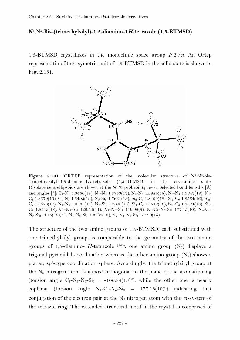
Chapter 2.3 – Silylated 1,5-diamino-1H-tetrazole derivatives
- 229 -
N1,N5-Bis-(trimethylsilyl)-1,5-diamino-1H-tetrazole (1,5-BTMSD)
1,5-BTMSD crystallizes in the monoclinic space group P 21/n. An Ortep
representatin of the asymetric unit of 1,5-BTMSD in the solid state is shown in
Fig. 2.131.
Figure 2.131. ORTEP representation of the molecular structure of N1,N5-bis-(trimethylsilyl)-1,5-diamino-1H-tetrazole (1,5-BTMSD) in the crystalline state. Displacement ellipsoids are shown at the 50 % probability level. Selected bond lengths [Å] and angles [°]: C1-N1 1.3460(18), N1-N2 1.3753(17), N2-N3 1.2924(18), N3-N4 1.3647(18), N4-C1 1.3379(19), C1-N5 1.3495(19), N5-Si2 1.7631(13), Si2-C5 1.8499(18), Si2-C6 1.8564(16), Si2-C7 1.8576(17), N1-N6 1.3836(17), N6-Si1 1.7600(13), Si1-C2 1.8512(18), Si1-C3 1.8624(18), Si1-C4 1.8513(18), C1-N5-Si2 122.54(11), N1-N6-Si1 119.92(9), N1-C1-N5-Si2 177.15(10), N4-C1-N5-Si2 -4.15(19), C1-N1-N6-Si1 106.84(13), N2-N1-N6-Si1 -77.20(15).
The structure of the two amino groups of 1,5-BTMSD, each substituted with
one trimethylsilyl group, is comparable to the geometry of the two amino
groups of 1,5-diamino-1H-tetrazole (360): one amino group (N6) displays a
trigonal pyramidal coordination whereas the other amino group (N5) shows a
planar, sp2-type coordination sphere. Accordingly, the trimethylsilyl group at
the N6 nitrogen atom is almost orthogonal to the plane of the aromatic ring
(torsion angle C1-N1-N6-Si1 = -106.84(13)°), while the other one is nearly
coplanar (torsion angle N1-C1-N5-Si2 = 177.15(10)°) indicating that
conjugation of the electron pair at the N5 nitrogen atom with the π-system of
the tetrazol ring. The extended structural motif in the crystal is comprised of
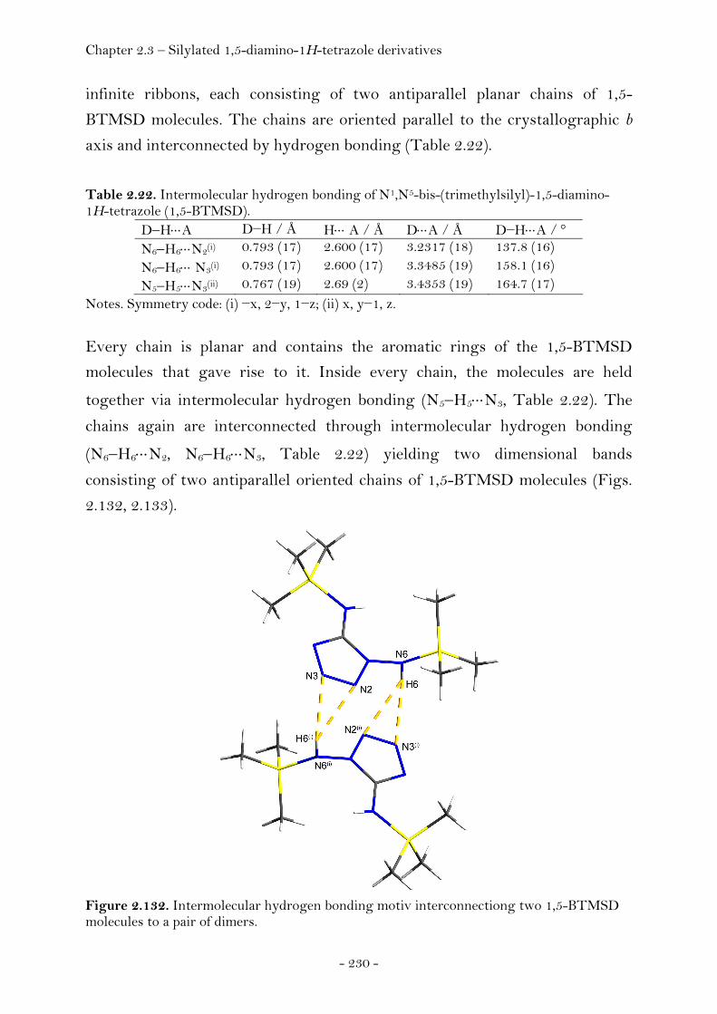
Chapter 2.3 – Silylated 1,5-diamino-1H-tetrazole derivatives
- 230 -
infinite ribbons, each consisting of two antiparallel planar chains of 1,5-
BTMSD molecules. The chains are oriented parallel to the crystallographic b
axis and interconnected by hydrogen bonding (Table 2.22).
Table 2.22. Intermolecular hydrogen bonding of N1,N5-bis-(trimethylsilyl)-1,5-diamino-1H-tetrazole (1,5-BTMSD).
D−H⋅⋅⋅A D−H / Å H⋅⋅⋅ A / Å D⋅⋅⋅A / Å D−H⋅⋅⋅A / ° N6−H6⋅⋅⋅N2(i) 0.793 (17) 2.600 (17) 3.2317 (18) 137.8 (16) N6−H6⋅⋅⋅ N3(i) 0.793 (17) 2.600 (17) 3.3485 (19) 158.1 (16) N5−H5⋅⋅⋅N3(ii) 0.767 (19) 2.69 (2) 3.4353 (19) 164.7 (17)
Notes. Symmetry code: (i) −x, 2−y, 1−z; (ii) x, y−1, z.
Every chain is planar and contains the aromatic rings of the 1,5-BTMSD
molecules that gave rise to it. Inside every chain, the molecules are held
together via intermolecular hydrogen bonding (N5−H5⋅⋅⋅N3, Table 2.22). The
chains again are interconnected through intermolecular hydrogen bonding
(N6−H6⋅⋅⋅N2, N6−H6⋅⋅⋅N3, Table 2.22) yielding two dimensional bands
consisting of two antiparallel oriented chains of 1,5-BTMSD molecules (Figs.
2.132, 2.133).
Figure 2.132. Intermolecular hydrogen bonding motiv interconnectiong two 1,5-BTMSD molecules to a pair of dimers.

Chapter 2.3 – Silylated 1,5-diamino-1H-tetrazole derivatives
- 231 -
Figure 2.133. View along the b axis of the crystal structure of 1,5-BTMSD. Hydrogen bonding is indicated by yellow dashed lines and is shown for only one pair of 1,5-BTMSD molecules.
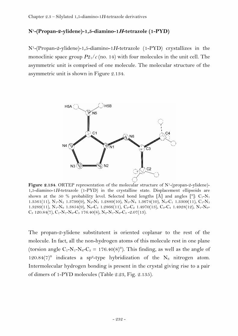
Chapter 2.3 – Silylated 1,5-diamino-1H-tetrazole derivatives
- 232 -
N1-(Propan-2-ylidene)-1,5-diamino-1H-tetrazole (1-PYD)
N1-(Propan-2-ylidene)-1,5-diamino-1H-tetrazole (1-PYD) crystallizes in the
monoclinic space group P21/c (no. 14) with four molecules in the unit cell. The
asymmetric unit is comprised of one molecule. The molecular structure of the
asymmetric unit is shown in Figure 2.134.
Figure 2.134. ORTEP representation of the molecular structure of N1-(propan-2-ylidene)-1,5-diamino-1H-tetrazole (1-PYD) in the crystalline state. Displacement ellipsoids are shown at the 50 % probability level. Selected bond lengths [Å] and angles [°]: C1-N1 1.3561(11), N1-N2 1.3799(9), N2-N3 1.2889(10), N3-N4 1.3674(10), N4-C1 1.3309(11), C1-N5 1.3289(11), N1-N6 1.3854(9), N6-C3 1.2866(11), C3-C2 1.4970(13), C3-C4 1.4928(12), N1-N6-C3 120.84(7), C1-N1-N6-C3 176.40(8), N2-N1-N6-C3 -2.07(13).
The propan-2-ylidene substitutent is oriented coplanar to the rest of the
molecule. In fact, all the non-hydrogen atoms of this molecule rest in one plane
(torsion angle C1-N1-N6-C3 = 176.40(8)°). This finding, as well as the angle of
120.84(7)° indicates a sp2-type hybridization of the N6 nitrogen atom.
Intermolecular hydrogen bonding is present in the crystal giving rise to a pair
of dimers of 1-PYD molecules (Table 2.23, Fig. 2.135).
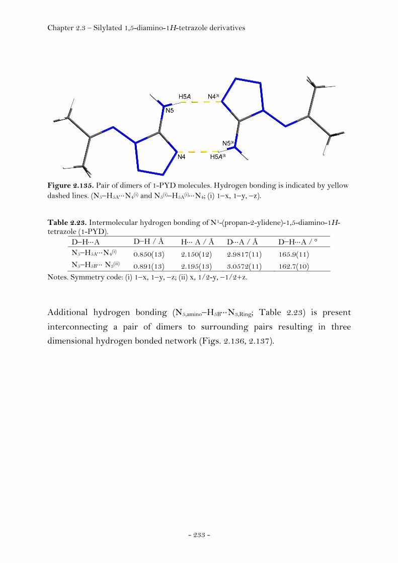
Chapter 2.3 – Silylated 1,5-diamino-1H-tetrazole derivatives
- 233 -
Figure 2.135. Pair of dimers of 1-PYD molecules. Hydrogen bonding is indicated by yellow dashed lines. (N5−H5A⋅⋅⋅N4(i) and N5(i)−H5A(i)⋅⋅⋅N4; (i) 1−x, 1−y, −z). Table 2.23. Intermolecular hydrogen bonding of N1-(propan-2-ylidene)-1,5-diamino-1H-tetrazole (1-PYD).
D−H⋅⋅⋅A D−H / Å H⋅⋅⋅ A / Å D⋅⋅⋅A / Å D−H⋅⋅⋅A / ° N5−H5A⋅⋅⋅N4(i) 0.850(13) 2.150(12) 2.9817(11) 165.9(11) N5−H5B⋅⋅⋅ N3(ii) 0.891(13) 2.195(13) 3.0572(11) 162.7(10)
Notes. Symmetry code: (i) 1−x, 1−y, −z; (ii) x, 1/2-y, −1/2+z.
Additional hydrogen bonding (N5,amino−H5B⋅⋅⋅N3,Ring; Table 2.23) is present
interconnecting a pair of dimers to surrounding pairs resulting in three
dimensional hydrogen bonded network (Figs. 2.136, 2.137).
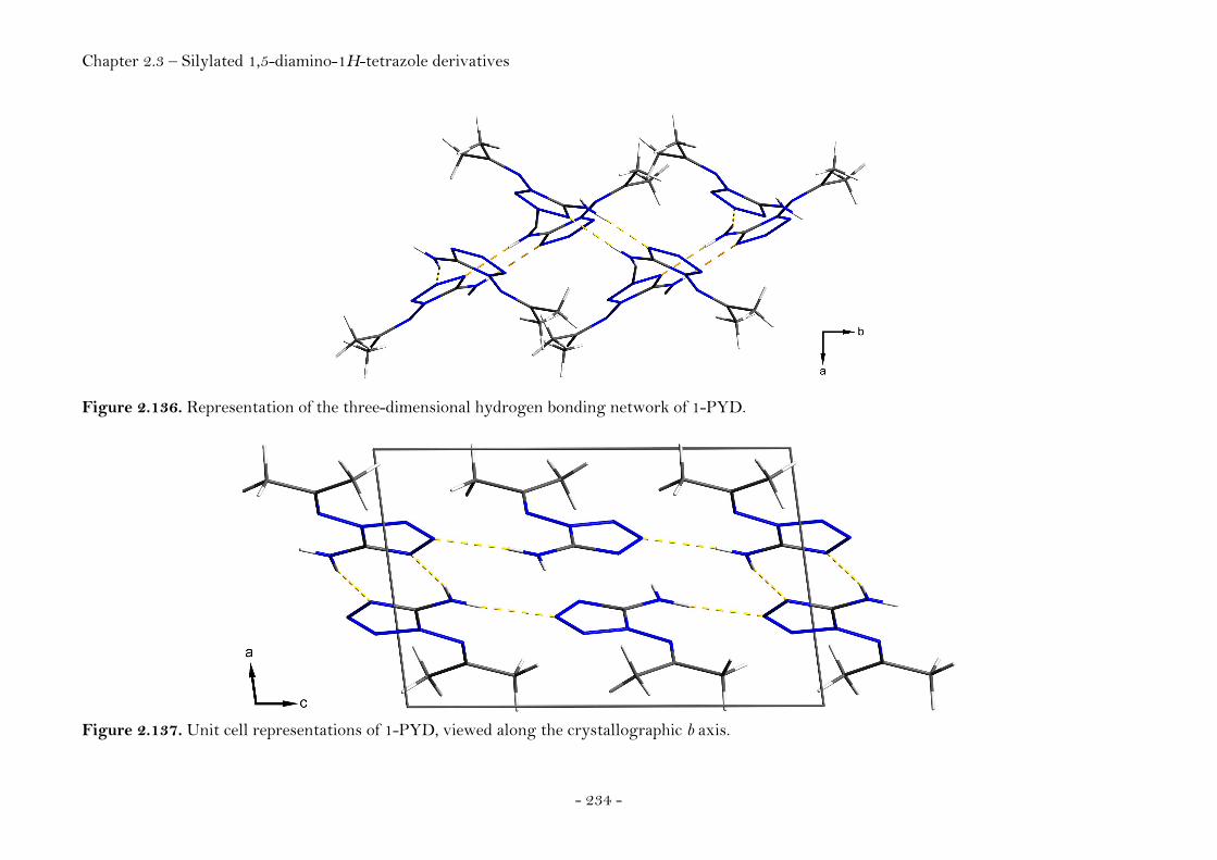
Chapter 2.3 – Silylated 1,5-diamino-1H-tetrazole derivatives
- 234 -
Figure 2.136. Representation of the three-dimensional hydrogen bonding network of 1-PYD.
Figure 2.137. Unit cell representations of 1-PYD, viewed along the crystallographic b axis.

Chapter 2.3 – Silylated 1,5-diamino-1H-tetrazole derivatives
- 235 -
N1,N1-Bis(trimethylsilyl)-1,5-diamino-1H-tetrazole (1,1-BTMSD)
1,1-BTMSD crystallizes in the monoclinic space group P21/c (no. 14) with four
molecules in the unit cell. The asymmetric unit is comprised of one molecule.
The molecular structure of the asymmetric unit is shown in Figure 2.138.
Figure 2.138. ORTEP representation of the molecular structure of N1,N1-bis(trimethylsilyl)-1,5-diamino-1H-tetrazole (1,1-BTMSD) in the crystalline state. Displacement ellipsoids are shown at the 50 % probability level. Selected bond lengths [Å] and angles [°]: C1-N1 1.346(7), N1-N2 1.366(6), N2-N3 1.293(6), N3-N4 1.377(7), N4-C1 1.332(7), C1-N5 1.331(8), N1-N6 1.392(6), N6-Si1 1.780(5), Si1-C2 1.854(6), Si1-C3 1.851(7), Si1-C4 1.847(6), N6-Si2 1.766(5), Si2-C5 1.849(7), Si2-C6 1.853(7), Si2-C7 1.861(6), N1-N6-Si1 114.3(4), N1-N6-Si2 113.4(4), C1-N1-N6-Si1 -92.4(6), N2-N1-N6-Si1 75.9(6), C1-N1-N6-Si2 106.3(6), N2-N1-N6-Si2 -85.5(6).
The compound exhibits an interesting pattern of intermolecular hydrogen
bonding (Table 2.24, Figs. 2.139, 2.140).
Table 2.24. Intermolecular hydrogen bonding of N1,N1-bis(trimethylsilyl)-1,5-diamino-1H-tetrazole (1,1-BTMSD).
D−H⋅⋅⋅A D−H / Å H⋅⋅⋅ A / Å D⋅⋅⋅A / Å D−H⋅⋅⋅A / °
N5−H5A⋅⋅⋅N4(i) 0.91(7) 2.11(7) 3.004(9) 170(7)
N5−H5B⋅⋅⋅N3(ii) 0.80(6) 2.39(6) 3.107(9) 151(5) Notes. Symmetry code: (i) 1−x, 1−y, −z; 1+x, y z.
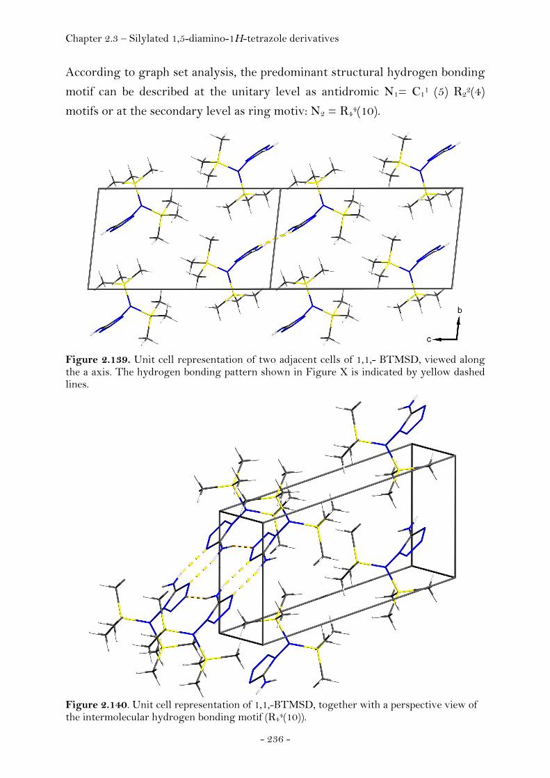
Chapter 2.3 – Silylated 1,5-diamino-1H-tetrazole derivatives
- 236 -
According to graph set analysis, the predominant structural hydrogen bonding
motif can be described at the unitary level as antidromic N1= C11 (5) R22(4)
motifs or at the secondary level as ring motiv: N2 = R44(10).
Figure 2.139. Unit cell representation of two adjacent cells of 1,1,- BTMSD, viewed along the a axis. The hydrogen bonding pattern shown in Figure X is indicated by yellow dashed lines.
Figure 2.140. Unit cell representation of 1,1,-BTMSD, together with a perspective view of the intermolecular hydrogen bonding motif (R44(10)).
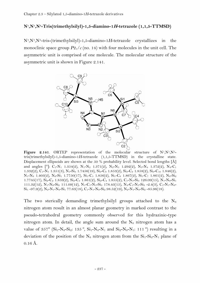
Chapter 2.3 – Silylated 1,5-diamino-1H-tetrazole derivatives
- 237 -
N1,N1,N5-Tris(trimethylsilyl)-1,5-diamino-1H-tetrazole (1,1,5-TTMSD)
N1,N1,N5-tris-(trimethylsilyl)-1,5-diamino-1H-tetrazole crystallizes in the
monoclinic space group P21/c (no. 14) with four molecules in the unit cell. The
asymmetric unit is comprised of one molecule. The molecular structure of the
asymmetric unit is shown in Figure 2.141.
Figure 2.141. ORTEP representation of the molecular structure of N1,N1,N5-tris(trimethylsilyl)-1,5-diamino-1H-tetrazole (1,1,5-TTMSD) in the crystalline state. Displacement ellipsoids are shown at the 50 % probability level. Selected bond lengths [Å] and angles [°]: C1-N1 1.354(2), N1-N2 1.371(2), N2-N3 1.292(2), N3-N4 1.372(2), N4-C1 1.332(2), C1-N5 1.351(3), N5-Si3 1.7456(19), Si3-C8 1.853(2), Si3-C9 1.858(2), Si3-C10 1.846(2), N1-N6 1.403(2), N6-Si1 1.7750(17), Si1-C5 1.856(2), Si1-C6 1.867(2), Si1-C7 1.861(2), N6-Si2 1.7745(17), Si2-C2 1.859(2), Si2-C3 1.861(2), Si2-C4 1.855(2), C1-N5-Si3 126.09(15), N1-N6-Si1 111.32(12), N1-N6-Si2 111.09(12), N1-C1-N5-Si3 178.43(15), N4-C1-N5-Si3 -2.4(3), C1-N1-N6-Si1 -97.9(2), N2-N1-N6-Si1 77.63(18), C1-N1-N6-Si2 98.52(19), N2-N1-N6-Si2 -85.96(18).
The two sterically demanding trimethylsilyl groups attached to the N6
nitrogen atom result in an almost planar geometry in marked contrast to the
pseudo-tetrahedral geometry commonly observed for this hydrazinic-type
nitrogen atom. In detail, the angle sum around the N6 nitrogen atom has a
value of 357° (Si1-N6-Si2: 135 º, Si1-N6-N1 and Si2-N6-N1: 111 º) resulting in a
deviation of the position of the N6 nitrogen atom from the Si1-Si2-N1 plane of
0.16 Å.

Chapter 2.3 – Silylated 1,5-diamino-1H-tetrazole derivatives
- 238 -
Compared to the structure of 1,5-BTMSD, this compound has only limited
possibilities for the formation of hydrogen bonds yielding a less dense packing
and a lower density (1,1,5-TTMSD: 1.0861(1) g cm-3, 1,5-BTMSD: 1.1702(1) g
cm-3. The hydrogen bonding pattern is presented in Table 2.25 and Figure
2.142. In fact, there is only a single hydrogen bond between the proton of the
amino group and the N3 ring nitrogen atom of a neighboring molecule.
Table 2.25. Intermolecular hydrogen bonding of N1,N1,N5-tris-(trimethylsilyl)-1,5-diamino-1H-tetrazole (1,1,5-TTMSD).
D−H⋅⋅⋅A D−H / Å H⋅⋅⋅ A / Å D⋅⋅⋅A / Å D−H⋅⋅⋅A / °
N5−H5⋅⋅⋅N3(i) 0.74(2) 2.26(2) 2.969(2) 162(2) Notes. (i) x, 1/2−y, −1/2+z.
Figure 2.142. Unit cell representation of the structure of 1,1,5-TTMSD, viewed along the a axis. Intermolecular hydrogen bonding is indicated by yellow dashed lines.
Chapter 2.3 – Silylated 1,5-diamino-1H-tetrazole derivatives
- 239 -
Conclusion
The attempts to synthesise the different singly and multiply silylated
diaminotetrazoles lead to five new molecules, which have been characterized.
Next to the acetone adduct 1-PYD, 5-TMSD represents a single silylated
species whereas 1,1-BTMSD and 1,5-BTMSD represent doubly silylated.
Furthermore, 1,1,5-TTMSD could be synthesised representing a triple
silylated species.
The synthetic conditions, especially towards 1,5-BTMSD were optimized with
respect of the purity and yield of product.
The initially promising strategy involving deprotonation using n-BuLi, did not
lead to good synthetic results due to problems with the workup of the
products. The preparation of the silylated compounds opens new possibilities
for the functionalization of diaminotetrazole. In this way the DAT unit could
be used in terms of a synthon and be reacted with a large spectrum of
electrophiles as already mentioned in the introduction. Other silylated
compounds of interest would include 1-TMSD as well as the persilylated
compound N1,N1,N5,N5-tetrakis(trimethylsilyl)-1,5-diamino-1H-tetrazole. In
terms of retrosynthesis, double silylation of a nitrogen atom converts it into a
(d,d) (double donor) centre giving rise to further interesting reactions like ring
closures, possibly leading to new fused heterocycles.
Experimental
All reagents were of p.a. purity and were used as received from Aldrich, Fluka
or Merck (if not stated otherwise). THF and diethyl ether were dried and
freshly distilled prior to use. 1H, 13C, 15N and 29Si NMR spectra were measured
on a Jeol Eclipse 270, Jeol EX 400 or Jeol Eclipse 400 instrument. Infrared
spectra were measured in KBr pellets using a Perkin-Elmer Spectrum One FT-
IR instrument equipped with CsI windows. Raman spectra were measured on a
Perkin Elmer Spectrum 2000R NIR FT-Raman instrument (Nd:YAG laser,
1064 nm, laser power 200 – 400 mW). Mass spectra were recorded on a JEOL
Chapter 2.3 – Silylated 1,5-diamino-1H-tetrazole derivatives
- 240 -
MStation JMS 700 instrument. Differential scanning calorimetry
measurements were performed using a Perkin ElmerPyris 6 DSC instrument.
The reactions were generally performed using standard Schlenk techniques (if
not stated otherwise). The reaction flasks, stirring bars and condensers were
heated three times using of a heat gun in vacuo (2.5·10-2 mbar) and vented with
nitrogen before being loaded with reagents. Solids were added over the neck of
the flask in a counterflow of dry nitrogen, liquids were added using a syringe.
Single crystal X-ray diffraction data for all compounds were collected using an
Xcalibur3 CCD diffractometer from Oxford Diffraction equipped with a
graphite monochromator affording Mo-Kα radiation (λ = 0.71073 Ǻ). The
structures were solved with direct methods using the program SHELXS 97
and refined using the program SHELXL 97.
1,5-Diamino-1H-tetrazole (DAT)
1,5-Diamino-1H-tetrazole (DAT) was synthesised according to a modified
literature procedure. (357) DMF (350mL) was filled into a 500 mL two-necked,
round-bottomed flask equipped with a reflux condenser, a large magnetic
stirring bar and a glass stopper. Lead(II) oxide (89.2 g, 0.400 mol, 2.0 eq) and
ammonium chloride (21.3 g, 0.400 mol; 2.0 eq) were added. After the mixture
had been stirred at room temperature for 30 min., thiosemicarbazide (18.0 g,
0.2 mol; 1.0 eq) was added and the reaction mixture heated to 90 ºC.
Subsequently, sodium azide (21.3 g, 0.4 mol, 2.0 eq) was added in small
portions over a period of 10 minutes. The reaction mixture was stirred at
105 ºC for 7 h.
The hot mixture was filtered off using a Büchner funnel with two layers of
filter paper. The reddish filtrate was evaporated until dryness (Caution!) and
the solid residue dissolved in hot water (80 mL). The hot solution was filtered
through Celite and allowed to cool down to room temperature over night. The
crystalline material obtained was filtered off and washed using ice water.
Chapter 2.3 – Silylated 1,5-diamino-1H-tetrazole derivatives
- 241 -
Concentration of the mother liquor to half of its volume yielded additional
product. The crude product was recrystallized in water. After filtration, the
purified product was washed three times using both cold ethyl acetate and
subsequently cold diethyl ether. Colorless crystals (9.91 g; 197.5 mmol; yield
50.1%) were obtained and later shown to be DAT.
Caution: Explosive lead(II) azide (Pb(N3)2) is formed as a side product during
the reaction. The black precipitate obtained after the filtration of the reaction
mixture should be kept wet and finally be destroyed by putting it into a water
solution of sodium nitrite (5%) subsequent acidification with hydrochloric acid
(1M).
1H NMR (400 MHz, [D6]-DMSO, 25 °C): δ = 6.35 (2H; NH2), 6.38 ppm (2H;
NH2); 13C NMR (100 MHz, [D6]-DMSO, 25°C): δ = 155.00 ppm; 15N NMR
(40.55 MHz, [D6]-DMSO, 25°C): δ = −338.8 (N6), −315.2 (N5), −167.8 (N1),
−97.5 (N4), −20.8 (N2), 5.5 (N3); IR (KBr disk) v~ /cm-1: 3324 (vs), 3237 (s),
3154 (s), 1656 (vs), 1578 (m), 1487 (w), 1329 (vs), 1109 (s), 1077 (m), 1002 (m),
933 (m), 746 (w), 700 (w), 608 (m); Raman (200 mW) v~ /cm-1: 3243 (6), 3244
(11), 1671 (9), 1623 (6) 1547 (20), 1497 (6), 1329 (15), 1307 (19), 106 (16), 1079
(12), 792 (100), 698 (15), 323 (26), 231 (13).
Chapter 2.3 – Silylated 1,5-diamino-1H-tetrazole derivatives
- 242 -
N1,N5-Bis(trimethylsilyl)-1,5-diamino-1H-tetrazole (1,5-BTMSD)
a) Hexamethyldisilazane route
All procedures were carried out using Schlenk techniques. DAT (0.500 g, 5.00
mmol, 1.0 eq) and ammonium sulfate (0.10 g, 0.75 mmol, 0.15 eq) were placed
inside a dry Schlenk flask (100 mL), equipped with a reflux condenser and
bubble counter. Dry THF (15 mL) was added by means of a syringe and the
suspension was stirred at room temperature. HMDS (2,42 g, 15.0 mmol, 3.0 eq)
was added slowly to this mixture using a syringe and the temperature was
raised to 80 ºC. The reaction mixture was stirred at this temperature for 6 h
until no ammonia evolved any longer at the outlet of the bubble counter
(indicator paper) and the solids in the reaction mixture were almost completely
dissolved. The hot reaction mixture was filtered into another Schlenk flask
using a Schlenk frit (pore size 3) and the solvent removed in vacuo. The
colorless solid residue was then dissolved in hot and dry THF (30 mL), filtered.
The volume of the solvent was slowly reduced (in vacuo) affording prismatic
colorless single crystals that were later shown to be 1,5-BTMSD (0.971 g, 80%
yield). The product can alternatively be purified by sublimation at 100 ºC in
static vacuum (2.5·10-2 mbar) and using a cold finger cooled with liquid
nitrogen.
1H NMR (400 MHz, CDCl3, 25°C): δ = 0.08 (s, 9H, Si(CH3)3), 0.22 (s, 9H,
Si(CH3)3), 4.25 (s, 1H, NH), 4.82 ppm (s, 1H, NH); 13C NMR (100 MHz, CDCl3,
25°C): δ = − 1.23 (Si(CH3)3), − 0.83 (Si(CH3)3), 154.53 ppm (tetrazole C); 15N
NMR (40.55 MHz, CDCl3, 25°C): δ = −332.6 (d, J(29Si-15N) = 82.2 Hz, N6),
−313.4 (d, J(29Si-15N) = 81.3 Hz, N5), −164.1 (s, N1), − -97.5 (s, N4), −20.2 (s,
N2), −6.8 ppm (s, N3); 29Si NMR (79.5 MHz, CDCl3, 25°C, INEPT, 9H,
J = 6.5 Hz): δ = 10.57, 15.82 ppm; IR (for an assignment of the vibrations, see
Appendix, KBr disk) v~ /cm-1: 3334 (vs), 2903 (vs), 2557 (m), 2411 (m), 1656
(vs), 1585 (vs), 1483 (s), 1428 (vs), 1378 (vs), 1352 (vs), 1289 (vs), 1250 (vs),
1209 (vs), 1102 (vs), 987 (vs), 877 (vs), 848 (vs), 760 (vs), 715 (s), 696 (s), 650
Chapter 2.3 – Silylated 1,5-diamino-1H-tetrazole derivatives
- 243 -
(s), 616 (s), 585 (m), 494 (w); Raman (for an assignment of the vibrations, see
Appendix (Chapter 4); 200 mW) v~ /cm-1: 3328 (16), 2964 (41), 2904 (50), 1587
(20), 1485 (6), 1419 (14), 1289 (21), 1264 (6), 1209 (14), 1103 (17), 988 (5), 896
(6), 856 (12), 840 (12), 698 (14), 625 (100), 522 (13), 375 (10), 341 (11), 315
(20), 263 (22), 194 (20); m.p. (DSC, Tonset, 2 ºC/min) 90.1 ºC, 231.3 ºC decomp.;
MS (DEI+, CHCl3): m/z = 244.3 [M+], 171.2 [M+ − Me3Si·], 98.08
[M+ − 2 Me3Si·], 146.2, 115.2, 73.2 [SiMe3+], 45.2, 43.1; Details of the single
crystal X-ray diffraction experiment are listed in the appendix (Chapter 4).
b) Chlorotrimethylsilane route
DAT (100 mg, 0.100 mmol, 1.00 eq) and diethyl ether (1 mL) were placed
inside a dry Schlenk flask (50 mL) and sealed with a septum. TMSCl (119 mg)
was added dropwise through the septum using a syringe. The mixture was
stirred for 30 minutes at room temperature. The septum was replaced with a
reflux condenser and triethylamine (111 mg, dried over 3 Å molecular sieve)
was added over a period of five minutes and the temperature of the reaction
mixture was raised to 80 ºC. Diethylether (1 mL) was added after three hours.
The heating was stopped after a total of 6 hours. The reaction mixture was
filtered using a Schlenk frit, the flask washed with pentane (two times 2 mL)
and the filtrate was evaporated to dryness. The solid residue was recrystallized
from pentane and cooled to -20 ºC. The resulting white amorphous precipitate
was identified as 1,5-BTMSD using raman spectroscopy and mass
spectrometry. The product obtained was of lower purity compared to the
product obtained from the HMDS procedure.
N5-Trimethylsilyl-1,5-diamino-1H-tetrazole (5-TMSD)
DAT (100 mg, 0.100 mmol, 1.00 eq) and diethyl ether (1 mL) were placed
inside a dry Schlenk flask (50 mL) and sealed with a septum. Using a syringe,
TMSCl (119 mg) was added dropwise through the septum. The mixture was
stirred for 30 minutes at 0 ºC. Then 111 mg of triethylamine (dried over 3 Å
molecular sieve) were added over 5 min and the reaction mixture was heated up
Chapter 2.3 – Silylated 1,5-diamino-1H-tetrazole derivatives
- 244 -
to 80 ºC. After 3 hours, further diethyl ether (1 mL) was added. The heating
was stopped after 6 hours. The reaction mixture was filtered of using a Schlenk
frit, the flask was washed with two times with 2 mL pentane and the filtrate
was evaporated in vacuo. The solid residue was dissolved in excess of pentane
(30 mL) and the solvent was slowly removed in vacuo. Thereby colorless
needles of the product were formed (101 mg; 0.563 mmol; 58%). The structure
was determined by single crystal x-ray diffraction.
IR (KBr disk) v~ /cm-1: 3436 (vs), 2960 (w), 1646 (s), 1586 (vs), 1383 (m), 1289
(m), 1254 (s), 1209 (m), 1102 (m), 1102 (m), 995 (m), 988 (w), 875 (s), 849 (s),
760 (w), 651 (w), 579 (w); Raman (400 mW, 25 °C, 100 scans) v~ /cm-1: 3328
(16), 2963 (34), 2903 (57), 1586 (23), 1413 (22), 1290 (34), 1210 (25), 1104 (30),
838 (24), 792 (28), 698 (29 ), 653 (28), 625 (89), 520 (26), 494 (32), 316 (31), 264
(37). Details of the single crystal X-ray diffraction experiment are listed in the
appendix (Chapter 4).
1,1-Bis(trimethylsilyl)-1,5-diamino-1H-tetrazole (1,1-BTMSD)
Inside a dry 100 mL Schlenk flask, equipped with a reflux condenser and a
bubble counter, were placed 0.500 g (5.00 mmol; 1.00 eq) of DAT and 0.100 g
(0.75 mmol; 0.15 eq) ammonium sulfate. Dry THF (15 mL) was added and the
suspension was stirred at room temperature. HMDS (2.421 g, 15.00 mmol,
3.00 eq) was added slowly to this mixture and the temperature was increased to
120 ºC. The reaction mixture was stirred at this temperature for 12 h until the
evolution of ammonia could no longer be detected as indicated by indicator
paper at the outlet of the bubble counter and the solids in the reaction mixture
had almost completely dissolved. Slow evaporation of the solvent at ambient
conditions yielded a white precipitate containing single crystals (colorless
needles) suitable for X-ray diffraction
IR (KBr disk) v~ /cm-1: 3408 (s), 3307 (m), 3241 (m), 3179 (m), 2959 (m), 1645
(vs), 1579 (m), 1416 (w), 1309 (m), 1097 (m), 979 (w), 933 (vs), 886 (vs), 847
(vs), 760 (w), 690 (w), 625 (w), 524 (w); Raman (400 mW, 25 °C, 100 scans)
v~ /cm-1: 3148 (8), 2904 (75), 1646 (21), 1452 (19), 1413 (15), 1310 (16), 1102

Chapter 2.3 – Silylated 1,5-diamino-1H-tetrazole derivatives
- 245 -
(12), 1051 (10), 982 (10), 833 (12), 769 (20), 745 (22), 964 (17), 646 (100), 426
(29), 361 (20), 290 (15), 251 (23); MS (DEI+, CHCl3): m/z = 244.2 [M+], 174.2
[M+ − Me3Si·], 174.2, 146.2, 130.1, 115.2, 100.1 [M − Me3Si·+2H]+, 73.2
[SiMe3+], 45.1; Details of the single crystal X-ray diffraction experiment are
listed in the appendix (Chapter 4).
N1-(Propan-2-ylidene)-1,5-diamino-1H-tetrazole (1-PYD)
1,5-BTMSD (200 mg, 0.818 mmol) were placed inside a dry Schlenk flask
(50 mL), equipped with a septum and magnetic stirring bar. By using a syringe,
dry acetone ([D6]acetone, 2 mL, 1.43 g, 24.5 mmol, 30 eq) was added and the
reaction mixture was stirred at room temperature for 6 h. The slightly turbid
solution was filtered and the filtrate slowly evaporated affording colorless
prismatic crystals (95.1 mg; 0.679 mmol; 83%).
(C4H2D6N6): 1H NMR (400 MHz, [D6]acetone) δ:, 25°C): δ = 0.08 (s, 9H,
Si(CH3)3), 0.22 (s, 9H, Si(CH3)3), 4.25 (s, 1H, NH), 4.82 ppm (s, 1H, NH); 13C
NMR (100 MHz, [D6]acetone): δ = 29.6 (septet, J(13C-D)=19.3 Hz, C2, C4),
132.6 (C1), 205.8 ppm (C3); MS (DEI+, CHCl3): m/z = 146.3 [M+], 62.3, 48.2,
28.2 [N2+]; Details of the single crystal X-ray diffraction experiment are listed
in the appendix (Chapter 4).
N1,N1,N5-Tris(trimethylsilyl)- 1,5-diamino-1H-tetrazole (1,1,5-TTMSD)
1,5-BTMSD (312 mg, 1.28 mmol, 1.00 eq) and triethylamine (freshly distilled,
260 mg, 2.57 mmol, 2.00 eq) were added to a dry Schlenk flask (25 mL),
equipped with a reflux condenser, bubble counter and a stirring bar by means
of a syringe over the stopcock of the flask. The suspension was stirred at room
temperature and subsequently trimethylsilyl triflate (569 mg, 2.57 mmol,
2.00 eq) was added dropwise. A pale reddish coloured solution was obtained
and the reaction mixture was stirred for further 8 h at 80 ºC. Colorless crystals
precipitated from the solution already after approximately three hours.
The reaction was cooled to room temperature, and freshly distilled diethyl
ether (10 mL) were added affording a sepparation into two phases: a lower, red
Chapter 2.3 – Silylated 1,5-diamino-1H-tetrazole derivatives
- 246 -
coloured oily phase, containing mainly triethylammonium triflate, and an upper
colorless phase, containing the product. The phases were separated (by means
of a Pasteur pipette) and the lower phase was washed three times with 5 mL of
ether. The ether solutions were combined and collected inside a dry Schlenk
flask put under a nitrogen atmosphere. The volume of the solvent was reduced
by fifty percent and the solution was cooled to 0 ºC. The product crystallized
over night in form of colorless prismatic crystals. The yield was 320 mg
(0.823 mmol; 64%). 1H NMR (400 MHz, [D6]benzene, 25°C): δ = 0.29 (s, 18H, (Si(CH3)3)2),
0.12 ppm (s, 9H, Si(CH3)3); 13C NMR (100 MHz, [D6]benzene, 25°C): δ = 1.15,
1.79; 29Si NMR (79.5 MHz, [D6]benzene, 25°C, INEPT, 9H, J = 6.5 Hz):
δ = 7.59, − 21.35 ppm; IR (for an assignment of the vibrations, see Appendix,
KBr disk) v~ /cm-1: 3410 (s), 3237 (m), 3181 (s), 2958 (m), 2903 (w), 1645 (s),
1580 (s), 1477 (w), 1454 (w), 1440 (w), 1347 (w), 1309 (w), 1298 (m), 1256 (vs),
1204 (m), 1165 (m), 1097 (m), 1031 (m), 982 (w), 936 (s), 884 (s), 840 (vs), 759
(m), 690 (w). 638 (m), 576 (w), 525 (w); Raman (for an assignment of the
vibrations, see Appendix, 400 mW, 25°C, 100 scans) v~ /cm-1: 2962 (50), 2902
(100), 1645 (5), 1580 (9), 1452 (10), 1413 (15), 1299 (11), 1205 (8), 1105 (11),
1034 (6), 983 (7), 899 (5), 759 (11), 692 (19), 644 (49), 621 (32), 531 (14), 425
(21), 358 (19), 290 (14), 210 (25); MS (DCI+, isobutane): m/z = 317.2 [M+H]+,
245.3 [M − Me3Si· + 2 H]+, 173.2 [M − 2Me3Si· + 3H]+, 102.2, 57.2, 34.2;
Details of the single crystal X-ray diffraction experiment are listed in the
appendix (Chapter 4).
Chapter 2.3 – Silylated 1,5-diamino-1H-tetrazole derivatives
- 247 -
Reaction of 1,5-BTMSD with n-Buthyllithium and TMSCl
A dry Schlenk flask (100 mL) was charged with 1,5-BTMSD (255 mg,
1.04 mmol, 1.00 eq) using a dry box. The substance was dissolved in dry THF
(15 mL) and the solution was cooled to −70 ºC. By means of a syringe, a 0.707
M solution of a 0.707 M n-BuLi in hexanes (1.48 mL, 1.04 mmol, 1.00 eq) was
added over a period of 10 minutes whith stirring of the reaction mixture. The
reaction mixture was taken out of the cooling bath, warmed up to room
temperature and stirred for additional 2 h. The solution was cooled again
(−70 ºC) and TMSCl (113 mg) was added over a period of five minutes with a
syringe during which the reaction mixture turned yellow. The reaction
mixture was stirred overnight under an atmosphere of nitrogen at room
temperature. Then THF (20 mL) was added, the mixture was heated to 40 ºC
and filtered using a Schlenk frit, and the solvent was removed. Mass
spectrometry of the solid residue revealed both the presence of 1,1,5,5-TTMSD
[C13H36N6Si4] and 1,1,5,-TTMSD [C10H28N6Si3] according to the signals
having a m/z of 388.2 and 316.17. No attempt was made to purify the product
and optimize the procedure.
Raman (400 mW, 25 °C, 100 scans) v~ /cm-1: 2951 (65), 2893 (100), 1368 (45),
1283 (55), 1223 (60), 1105 (45), 997 (25), 961 (35), 675 (25), 615 (75), 321 (40),
210 (35). MS (DEI+): selected m/z: 388.2, 316.17.

Chapter 2.3 – Pyridazines
- 248 -
2.3.3 Pyridazines
Introduction
Halogenated pyridazines have been synthesised as possible precursor molecules
for the synthesis of novel energetic matierials like the elusive 3,4,5,6-
tetraazido-1,2-pyridazine molecule. Next to our interest into the energetic
properties of this compound pyridazines decorated with azide groups are
interesting model compounds to study possible pyridazine azide cyclisation
reactions.
Azide groups α-substituted to annular N-atoms in aromatic compounds are
subject to tautomeric conversion to a tetrazole ring. (358) The potential energy
surface on which this type of reaction takes place in solution has recently been
investigated experimentally and theoretically. (359-360) It has been established for
a series of 2,4-diazidopyrimidines and 4,6-diazidopyrimidines that the azido-
tetrazole tautomerism is strongly solvent and temperature dependent and that
the azide-azide and azide-tetrazole forms are energetically more favourable
than the tetrazole-tetrazole form. (361) In the crystalline state, 3,6-
diazidopyridazine has been found to exist in the azide-tetrazole form (Fig.
2.142). (362-363)
Figure 2.143. Formation of 6-azidotetrazolo-[1,5-b]pyridazine upon treatment of 3,6-dichloropyridazine wit sodium azide in a sealed tube with an ethanol-water solvent (90°C, 3h). (364)
In 2004, Allan et al. reported a study (365) investigating the reaction between 4-
methyl-3,5,6-tribromopydridazine and sodium azide. They reported the
formation of 4-methyl-3,5,6-triazidopyridazine as intermediate and subsequent
cyclisation to one of the two possible bicyclic tetrazolo[1,5-b]pyridazine
products. They stated that this product was thermally unstable and nitrogen

Chapter 2.3 – Pyridazines
- 249 -
was liberated on heating this compound giving rise to 5-amino-3-diazido-4-
methyl-tetrazolo[1,5-b]pydridazine. Finally they succeeded in determining the
crystal structure of 3,5-diazido-4-methyl[1,5-b]tetrazolopyridazine confirming
theoretical predictions that compound exists in the bicyclic (tetrazol-
pyridazine) rather than the tricyclic (tetrazol-pyridazine-tetrazol) form.
However, their attempts to grow single crystals resulted in ‘twinned or
otherwise unsuitable crystals that were prone to decomposition’ and
synchrotron radiation had to be used to measure the very small crystals. The
data obtained were sufficient as a structural proof but they mentioned that ‘due
to the highly mosaic nature of the crystal the data is not of sufficient quality to
give precise structural parameter’. We have synthesised both 3,4,6-
trichloropyridazine and 3,4,5,6-trichloropyridazine from 3,6-dichloropyridazine
in order to subsequently substitute the chlorine atoms with azide and study
these products. In accordance with the well established behaviour that nitrogen
para to the site of nucleophilic attack is activating in perhalogenated
heteroaromatic systems we were only able to substitute the chlorine atom at
the 4-position yielding 3,6-dichloro-4-azidopyridazine. A more comprehensive
study of the regioselectivity of the reactions between tetrachloropyridazine and
a variety of aliphatic nitrogen nucleophiles has only recently been reported
confirming our finding of the regioselectivity. (366) The reported ‘violently
explosive’ nature of 6-azidotetrazolo-[1,5-b]pyridazine together with its low
thermal stability (Tdec. = 128-129°C) (367) rendered it unlikely to obtain a
material of sufficient stability to fulfil the qualification criteria for a novel
energetic material (Table 1.1, page 15) and no further efforts were undertaken
to overcome the difficulties experienced in obtaining a higher degree of azide
substitution at this time.
However, due to the importance of 3,4,6-trichloropyridazine and 3,4,5,6-
tetrachloropyridazine as educts in organic chemistry, their exceptional
biological activity we investigated the solid state structures using single
crystal X-ray diffraction because structural data of these two very simple
compounds in the solid state were unkown.

Chapter 2.3 – Pyridazines
- 250 -
Biological Activity
Especially the chloro derivatives we have synthesised are synthetically valuable
precursor molecules in organic synthesis and display remarkable biological
activities. The trihalopyridazines are excellent herbicides and insecticides, but
are less bacterically against S-aureus than the tetrahalopyridazines. The
tetrahalopyridazines are excellent pre- and post-emergent herbicides and they
also possess exceptional fungicidal and bactericidal properties. From the
reported data (368) it is apparent that 3,4,6-trichloropyridazine and 3,4,5,6-
tetrachloropyridazine are unique in their extremely broad spectrum of
biological activity in that they are as effective or more effective than the
reference standards, even though in some instances the concentration was
much lower than the reference standard against which the pyridazines were
tested. Of interest, we mention that 3,4,6-trichloropyridazine has been reported
to be not only toxic and an irritant, but also a strong contact allergen to
humans and special care has to be taken when working with this substances. (369)
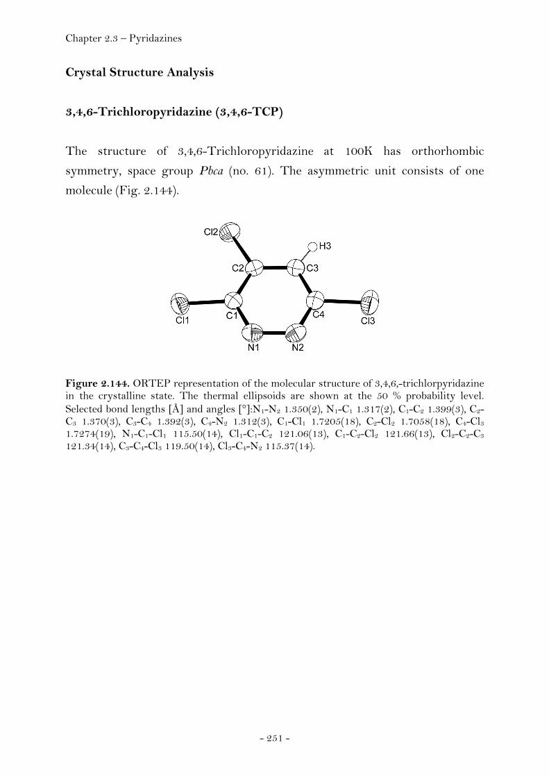
Chapter 2.3 – Pyridazines
- 251 -
Crystal Structure Analysis
3,4,6-Trichloropyridazine (3,4,6-TCP)
The structure of 3,4,6-Trichloropyridazine at 100K has orthorhombic
symmetry, space group Pbca (no. 61). The asymmetric unit consists of one
molecule (Fig. 2.144).
Figure 2.144. ORTEP representation of the molecular structure of 3,4,6,-trichlorpyridazine in the crystalline state. The thermal ellipsoids are shown at the 50 % probability level. Selected bond lengths [Å] and angles [°]:N1-N2 1.350(2), N1-C1 1.317(2), C1-C2 1.399(3), C2-C3 1.370(3), C3-C4 1.392(3), C4-N2 1.312(3), C1-Cl1 1.7205(18), C2-Cl2 1.7058(18), C4-Cl3 1.7274(19), N1-C1-Cl1 115.50(14), Cl1-C1-C2 121.06(13), C1-C2-Cl2 121.66(13), Cl2-C2-C3 121.34(14), C3-C4-Cl3 119.50(14), Cl3-C4-N2 115.37(14).

Chapter 2.3 – Pyridazines
- 252 -
Figure 2.145. Unit cell of 3,4,6-trichlorpyridazine, viewed along the a axis (left) and c axis (right). Yellow dashed lines indicate hyrogen bonding (C3-H3···N1i , C3-H3···N2i). (i) 1/2+x,1/2-y,-z.
Figure 2.146. Detail of the unit cell of 3,4,6-trichlorpyridazine, viewed along the a axis (left) and c axis (right). Yellow dashed lines indicate hyrogen bonding (C3-H3···N1i , C3-H3···N2i). (i) 1/2+x,1/2-y,-z.
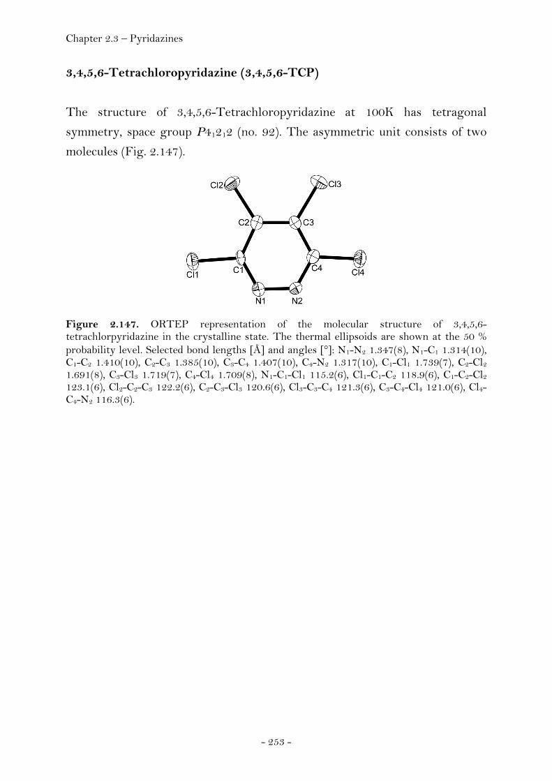
Chapter 2.3 – Pyridazines
- 253 -
3,4,5,6-Tetrachloropyridazine (3,4,5,6-TCP)
The structure of 3,4,5,6-Tetrachloropyridazine at 100K has tetragonal
symmetry, space group P41212 (no. 92). The asymmetric unit consists of two
molecules (Fig. 2.147).
Figure 2.147. ORTEP representation of the molecular structure of 3,4,5,6-tetrachlorpyridazine in the crystalline state. The thermal ellipsoids are shown at the 50 % probability level. Selected bond lengths [Å] and angles [°]: N1-N2 1.347(8), N1-C1 1.314(10), C1-C2 1.410(10), C2-C3 1.385(10), C3-C4 1.407(10), C4-N2 1.317(10), C1-Cl1 1.739(7), C2-Cl2 1.691(8), C3-Cl3 1.719(7), C4-Cl4 1.709(8), N1-C1-Cl1 115.2(6), Cl1-C1-C2 118.9(6), C1-C2-Cl2 123.1(6), Cl2-C2-C3 122.2(6), C2-C3-Cl3 120.6(6), Cl3-C3-C4 121.3(6), C3-C4-Cl4 121.0(6), Cl4-C4-N2 116.3(6).
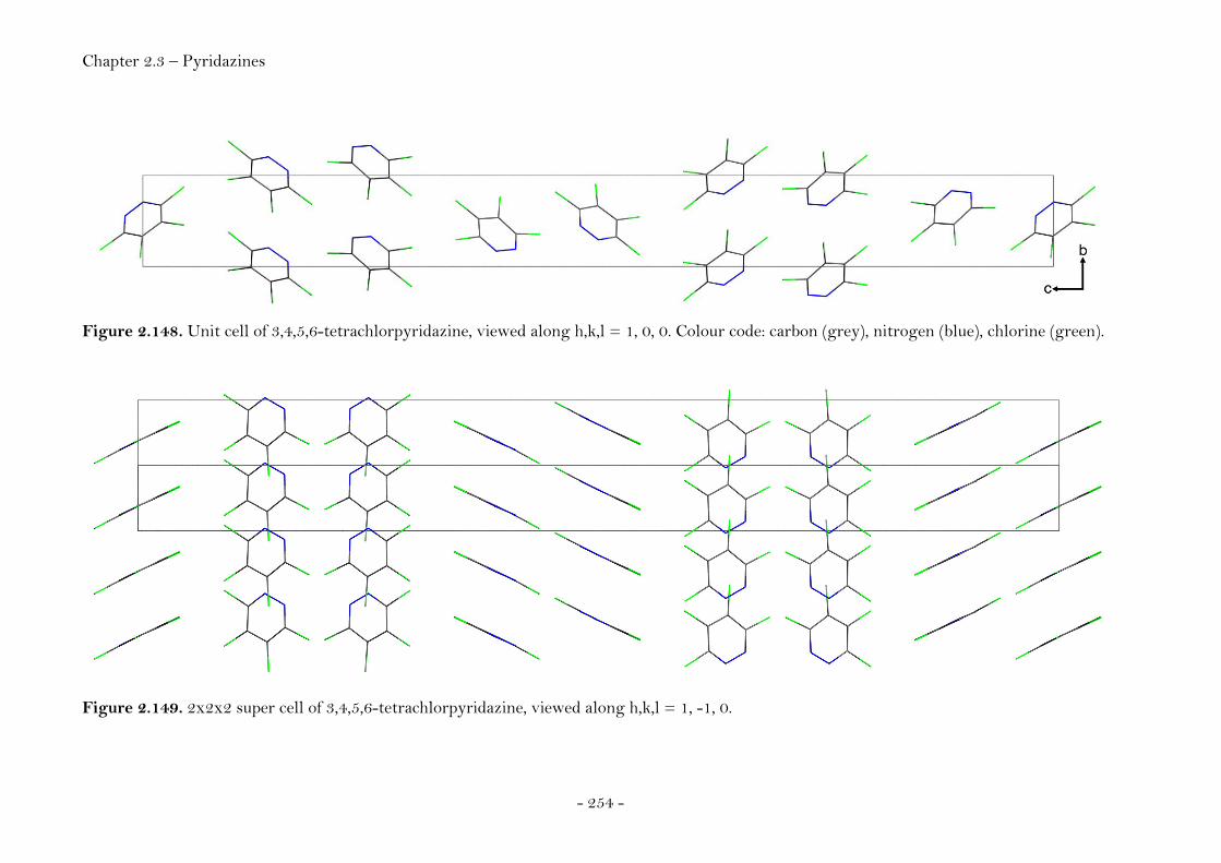
Chapter 2.3 – Pyridazines
- 254 -
Figure 2.148. Unit cell of 3,4,5,6-tetrachlorpyridazine, viewed along h,k,l = 1, 0, 0. Colour code: carbon (grey), nitrogen (blue), chlorine (green).
Figure 2.149. 2x2x2 super cell of 3,4,5,6-tetrachlorpyridazine, viewed along h,k,l = 1, -1, 0.

Chapter 2.3 – Pyridazines
- 255 -
4-Azido-3,6-dichloropyridazine (4A-3,6-DCP)
The structure of 4-Azido-3,6-dichloropyridazine at 100K has orthorhombic
symmetry, space group P212121 (no. 19). The asymmetric unit consists of one
molecule (Fig. 2.150).
Figure 2.150. ORTEP representation of the molecular structure of 4-azido-3,6-dichloropyridazine in the crystalline state. The thermal ellipsoids are shown at the 50 % probability level. Selected bond lengths [Å] and angles [°]:N1-N2 1.361(3), N1-C1 1.310(4), C1-C2 1.415(4), C2-C3 1.372(4), C3-C4 1.389(4), C4-N2 1.318(4), C1-Cl1 1.728(3), C2-N3 1.403(4), N3-N4 1.272(4), N4-N5 1.107(4), C4-Cl2 1.727(3), N1-C1-Cl1 116.0(2), Cl1-C1-C2 119.4(2), C1-C2-N3 117.0(3), N3-C2-C3 127.2(3), C2-N3-N4 113.3(2), N3-N4-N5 170.7(3), C3-C4-Cl2 118.8(2), Cl2-C4-N2 115.4(2).
Figure 2.151. Unit cell of 4-azido-3,6-dichloropyridazine, viewed along the a axis. Yellow dashed lines indicate hyrogen bonding (C3-H3···N2i). (i) 2-x,-1/2+y,1/2-z.
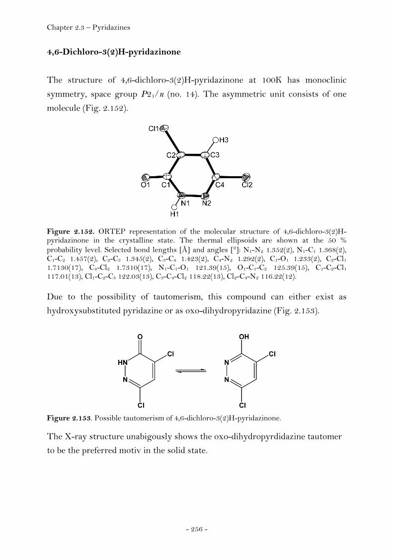
Chapter 2.3 – Pyridazines
- 256 -
4,6-Dichloro-3(2)H-pyridazinone
The structure of 4,6-dichloro-3(2)H-pyridazinone at 100K has monoclinic
symmetry, space group P21/n (no. 14). The asymmetric unit consists of one
molecule (Fig. 2.152).
Figure 2.152. ORTEP representation of the molecular structure of 4,6-dichloro-3(2)H-pyridazinone in the crystalline state. The thermal ellipsoids are shown at the 50 % probability level. Selected bond lengths [Å] and angles [°]: N1-N2 1.352(2), N1-C1 1.368(2), C1-C2 1.457(2), C2-C3 1.345(2), C3-C4 1.423(2), C4-N2 1.292(2), C1-O1 1.233(2), C2-Cl1 1.7130(17), C4-Cl2 1.7310(17), N1-C1-O1 121.39(15), O1-C1-C2 125.39(15), C1-C2-Cl1 117.01(13), Cl1-C2-C3 122.03(13), C3-C4-Cl2 118.22(13), Cl2-C4-N2 116.22(12). Due to the possibility of tautomerism, this compound can either exist as
hydroxysubstituted pyridazine or as oxo-dihydropyridazine (Fig. 2.153).
Figure 2.153. Possible tautomerism of 4,6-dichloro-3(2)H-pyridazinone. The X-ray structure unabigously shows the oxo-dihydropyrdidazine tautomer
to be the preferred motiv in the solid state.
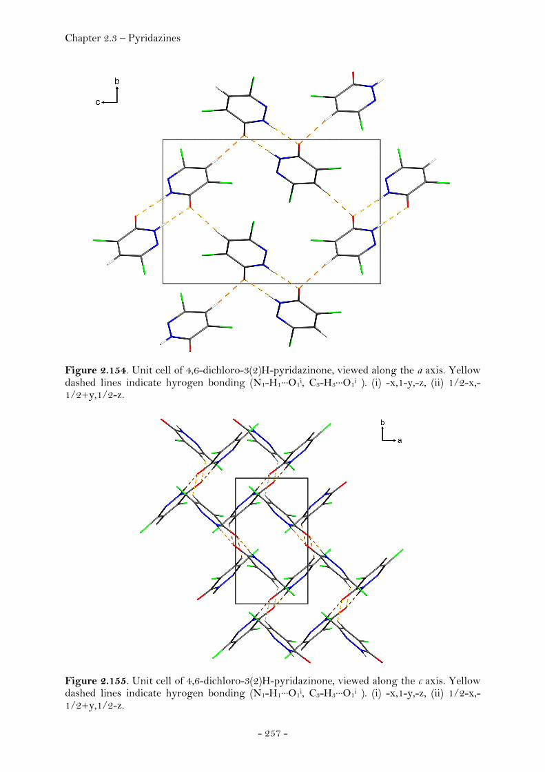
Chapter 2.3 – Pyridazines
- 257 -
Figure 2.154. Unit cell of 4,6-dichloro-3(2)H-pyridazinone, viewed along the a axis. Yellow dashed lines indicate hyrogen bonding (N1-H1···O1i, C3-H3···O1i ). (i) -x,1-y,-z, (ii) 1/2-x,-1/2+y,1/2-z.
Figure 2.155. Unit cell of 4,6-dichloro-3(2)H-pyridazinone, viewed along the c axis. Yellow dashed lines indicate hyrogen bonding (N1-H1···O1i, C3-H3···O1i ). (i) -x,1-y,-z, (ii) 1/2-x,-1/2+y,1/2-z.

Chapter 2.3 – Pyridazines
- 258 -
Figure 2.156. Unit cell of 4,6-dichloro-3(2)H-pyridazinone, viewed along h,k,l = -2.66, 7.63, 1.22. Yellow dashed lines indicate hyrogen bonding (N1-H1···O1i, C3-H3···O1i ). (i) -x,1-y,-z, (ii) 1/2-x,-1/2+y,1/2-z.
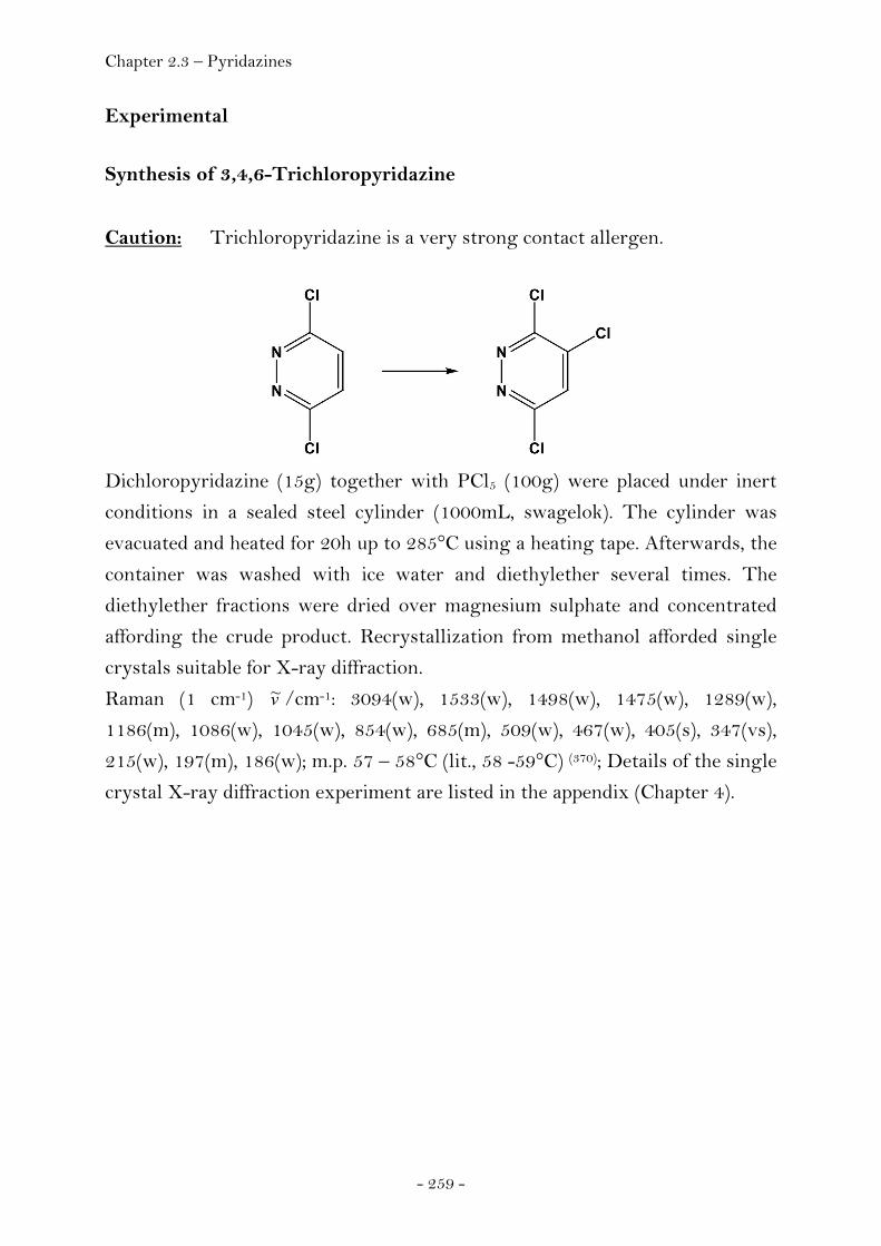
Chapter 2.3 – Pyridazines
- 259 -
Experimental
Synthesis of 3,4,6-Trichloropyridazine
Caution: Trichloropyridazine is a very strong contact allergen.
Dichloropyridazine (15g) together with PCl5 (100g) were placed under inert
conditions in a sealed steel cylinder (1000mL, swagelok). The cylinder was
evacuated and heated for 20h up to 285°C using a heating tape. Afterwards, the
container was washed with ice water and diethylether several times. The
diethylether fractions were dried over magnesium sulphate and concentrated
affording the crude product. Recrystallization from methanol afforded single
crystals suitable for X-ray diffraction.
Raman (1 cm-1) v~ /cm-1: 3094(w), 1533(w), 1498(w), 1475(w), 1289(w),
1186(m), 1086(w), 1045(w), 854(w), 685(m), 509(w), 467(w), 405(s), 347(vs),
215(w), 197(m), 186(w); m.p. 57 – 58°C (lit., 58 -59°C) (370); Details of the single
crystal X-ray diffraction experiment are listed in the appendix (Chapter 4).

Chapter 2.3 – Pyridazines
- 260 -
Synthesis of 3,4,5,6-Tetrachloropyridazine
Though tetrachloropyridazine had previously been prepared from
dichloromaleic anhydride, (371) we chose to prepare it using a slight modification
from the reaction between commercially available 3,6-dichloropyridazine and
phosphorus pentachloride at elevated temperature and pressure. (372)
Dichloropyridazine (15g) together with PCl5 (100g) were placed under inert
conditions in a sealed steel cylinder (1000mL, swagelok). The cylinder was
evacuated and heated for 48h up to 360°C using a heating tape. Afterwards, the
container was washed with ice water and diethylether several times. The
diethylether fractions were dried over magnesium sulphate and concentrated
affording the crude product. Recrystallization from methanol afforded light
brown coloured single crystals suitable for X-ray diffraction. 13C NMR ([D6]acetone) δ: 155.2 (C1,C4), 138.2 (C2,C3); 15N NMR ([D6]acetone)
δ (nitromethane): 4.5 (N1,N2); IR (KBr disk) v~ /cm-1: 1505, 1480, 1360, 1290,
1200, 1120, 1090, 890, 800, 630; Raman (1 cm-1) v~ /cm-1: 1495(w), 1475(w),
1186(vs), 1085(w), 874(w), 617(w), 516(w), 500(w), 405(w), 348(s), 215(w); m.p.
86 – 88°C (lit., 85 - 86°C) (373) ; Details of the single crystal X-ray diffraction
experiment are listed in the appendix (Chapter 4).
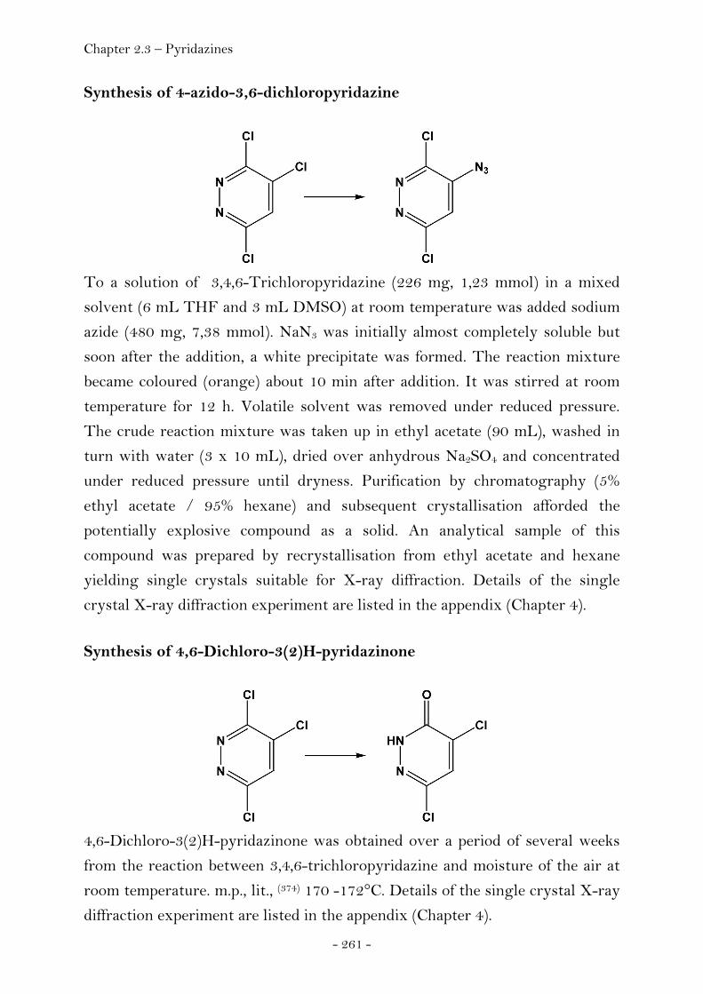
Chapter 2.3 – Pyridazines
- 261 -
Synthesis of 4-azido-3,6-dichloropyridazine
To a solution of 3,4,6-Trichloropyridazine (226 mg, 1,23 mmol) in a mixed
solvent (6 mL THF and 3 mL DMSO) at room temperature was added sodium
azide (480 mg, 7,38 mmol). NaN3 was initially almost completely soluble but
soon after the addition, a white precipitate was formed. The reaction mixture
became coloured (orange) about 10 min after addition. It was stirred at room
temperature for 12 h. Volatile solvent was removed under reduced pressure.
The crude reaction mixture was taken up in ethyl acetate (90 mL), washed in
turn with water (3 x 10 mL), dried over anhydrous Na2SO4 and concentrated
under reduced pressure until dryness. Purification by chromatography (5%
ethyl acetate / 95% hexane) and subsequent crystallisation afforded the
potentially explosive compound as a solid. An analytical sample of this
compound was prepared by recrystallisation from ethyl acetate and hexane
yielding single crystals suitable for X-ray diffraction. Details of the single
crystal X-ray diffraction experiment are listed in the appendix (Chapter 4).
Synthesis of 4,6-Dichloro-3(2)H-pyridazinone
4,6-Dichloro-3(2)H-pyridazinone was obtained over a period of several weeks
from the reaction between 3,4,6-trichloropyridazine and moisture of the air at
room temperature. m.p., lit., (374) 170 -172°C. Details of the single crystal X-ray
diffraction experiment are listed in the appendix (Chapter 4).
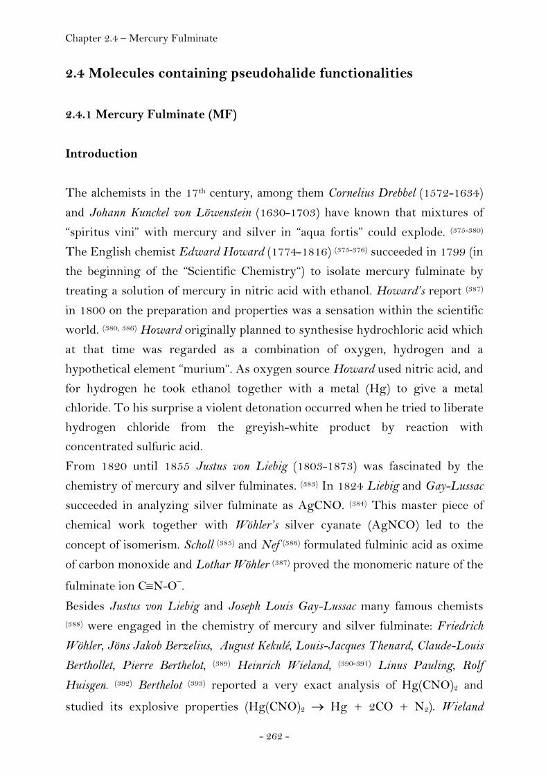
Chapter 2.4 – Mercury Fulminate
- 262 -
2.4 Molecules containing pseudohalide functionalities
2.4.1 Mercury Fulminate (MF)
Introduction
The alchemists in the 17th century, among them Cornelius Drebbel (1572-1634)
and Johann Kunckel von Löwenstein (1630-1703) have known that mixtures of
“spiritus vini” with mercury and silver in “aqua fortis” could explode. (375-380)
The English chemist Edward Howard (1774-1816) (375-376) succeeded in 1799 (in
the beginning of the “Scientific Chemistry“) to isolate mercury fulminate by
treating a solution of mercury in nitric acid with ethanol. Howard’s report (387)
in 1800 on the preparation and properties was a sensation within the scientific
world. (380, 386) Howard originally planned to synthesise hydrochloric acid which
at that time was regarded as a combination of oxygen, hydrogen and a
hypothetical element “murium“. As oxygen source Howard used nitric acid, and
for hydrogen he took ethanol together with a metal (Hg) to give a metal
chloride. To his surprise a violent detonation occurred when he tried to liberate
hydrogen chloride from the greyish-white product by reaction with
concentrated sulfuric acid.
From 1820 until 1855 Justus von Liebig (1803-1873) was fascinated by the
chemistry of mercury and silver fulminates. (383) In 1824 Liebig and Gay-Lussac
succeeded in analyzing silver fulminate as AgCNO. (384) This master piece of
chemical work together with Wöhler’s silver cyanate (AgNCO) led to the
concept of isomerism. Scholl (385) and Nef (386) formulated fulminic acid as oxime
of carbon monoxide and Lothar Wöhler (387) proved the monomeric nature of the
fulminate ion C≡N-O−.
Besides Justus von Liebig and Joseph Louis Gay-Lussac many famous chemists (388) were engaged in the chemistry of mercury and silver fulminate: Friedrich
Wöhler, Jöns Jakob Berzelius, August Kekulé, Louis-Jacques Thenard, Claude-Louis
Berthollet, Pierre Berthelot, (389) Heinrich Wieland, (390-391) Linus Pauling, Rolf
Huisgen. (392) Berthelot (393) reported a very exact analysis of Hg(CNO)2 and
studied its explosive properties (Hg(CNO)2 → Hg + 2CO + N2). Wieland
Chapter 2.4 – Mercury Fulminate
- 263 -
offered - after his own important contributions to the chemistry of fulminic
acid - a widely accepted interpretation for Howard’s formation of mercury
fulminate from mercury, nitric acid and ethanol. (394)
Mercury fulminate was widely used as primary explosive for nearly a hundred
years. In the beginning of the 20th century the annual production of mercury
fulminate only in Germany was about 100 000 kg per year. (380, 395) A. Nobel
used this energetic compound as a component in his recent developed metal
blasting cap detonator to initiate dynamite. (380, 396) The wide application of
dynamite was only possible when the use of Hg(CNO)2 as primary explosive
guaranteed a safe ignition. (400) For this purpose it is now replaced by lead azide
which is more stable on storage. (397)
To our knowledge a detailed X-ray crystal structure determination of mercury
fulminate has not been carried out. Miles reported a good method for the
crystallization of mercury fulminate and performed also first investigations on
the crystal structure of mercury fulminate with single crystals already in 1931. (398) He correctly derived the holohedric crystal class and also the lattice
parameters (a = 5.48, b = 7.71, c = 10.43 Å, V = 441 Å3), But atomic positions
of the constituent atoms were not given.
About twenty years later, Suzuki performed a single crystal investigation on
mercury fulminate. (399) He could set Hg atoms correctly in the positions of a
face centred lattice with a total of only 49 reflections, indexed using the cell
parameters given by Miles. (400) However, due to the wrong space group, the
positions of the C, N and O atoms could not be located and a bent CNO-Hg-
ONC structure was proposed as it was generally assumed at that time.
Within the last decades, two results of X-ray powder investigations have also
been published. The first was presented by the International Centre for
Diffraction Data (ICCD) as powder diffraction file 00-002-0287 for mercury
fulminate, HgC2N2O2, determined with CuKα radiation (λ = 1.540598 Å). It
contained 22 d-values for non-indexed reflections. (401) These data were
obtained from Canadian Industries Limited as private communication. The
lattice parameters obtained by this d-values were calculated by least squares fit
and correspond to a = 5.398(5), c = 10.214(4) and c = 7.630(10) Å. The result
of the second X-ray powder investigation on mercury fulminate was published
1981 by Brown and Swallowe. (402) In this case, NiKα radiation was used (λ =
Chapter 2.4 – Mercury Fulminate
- 264 -
1.6592 Å) with longer wave length in comparison to that with Cu radiation.
The precision of the obtained values is lower in this case since mercury
fulminate strongly absorbs X-ray radiation. As a consequence, the obtained
lattice parameters calculated by least squares fit show higher standard
deviations (a = 5.44(3), b = 10.38(4), c = 7.75(5) Å).
Here we report the results of the X-ray investigations of single crystals as well
as powders of Hg(CNO)2. The single crystal investigation reveals - as expected
- almost linear O-N-C-Hg-C-N-O bonds, similar to those in mercury cyanide
NC-Hg-CN. (403) Liebig already recognized the close analogy between metal
fulminates and metal cyanides. (404-405) In analogy, the correct structure for
fulminic acid is H-C≡N-O (383, 406-407) and not C≡N-OH. Furthermore, the
fulminate ligand forms transition metal complexes that are very similar to
those of cyanide. (408-409) The X-ray structure determination of two polymorphic
forms of silver fulminate revealed very interesting structures containing CNO
bridges and three centred Ag-C-Ag bonds in hexameric units or infinite chains. (410) The structures of the metal complexes [(Ph3P)2Pt(CNO)2], (411)
[Ph3PAuCNO], (412) [Au(CNO)2]-, (413) [M(CNO)4]2- (M = Ni, Pt, Zn), (414)
[Hg(CNO)4]2- (415) as well as [Co(CNO)6]3- (416) with almost linear M-C≡N-O
bonds were determined using X-ray diffraction. Density functional theory
(DFT) calculations for these fulminato complexes are in good agreement with
the observed structural parameters. (417) In contrast, a recent DFT calculation
predicts bent CNO-Hg-ONC units, (418) a structure which is still present in the
literature to our surprise.
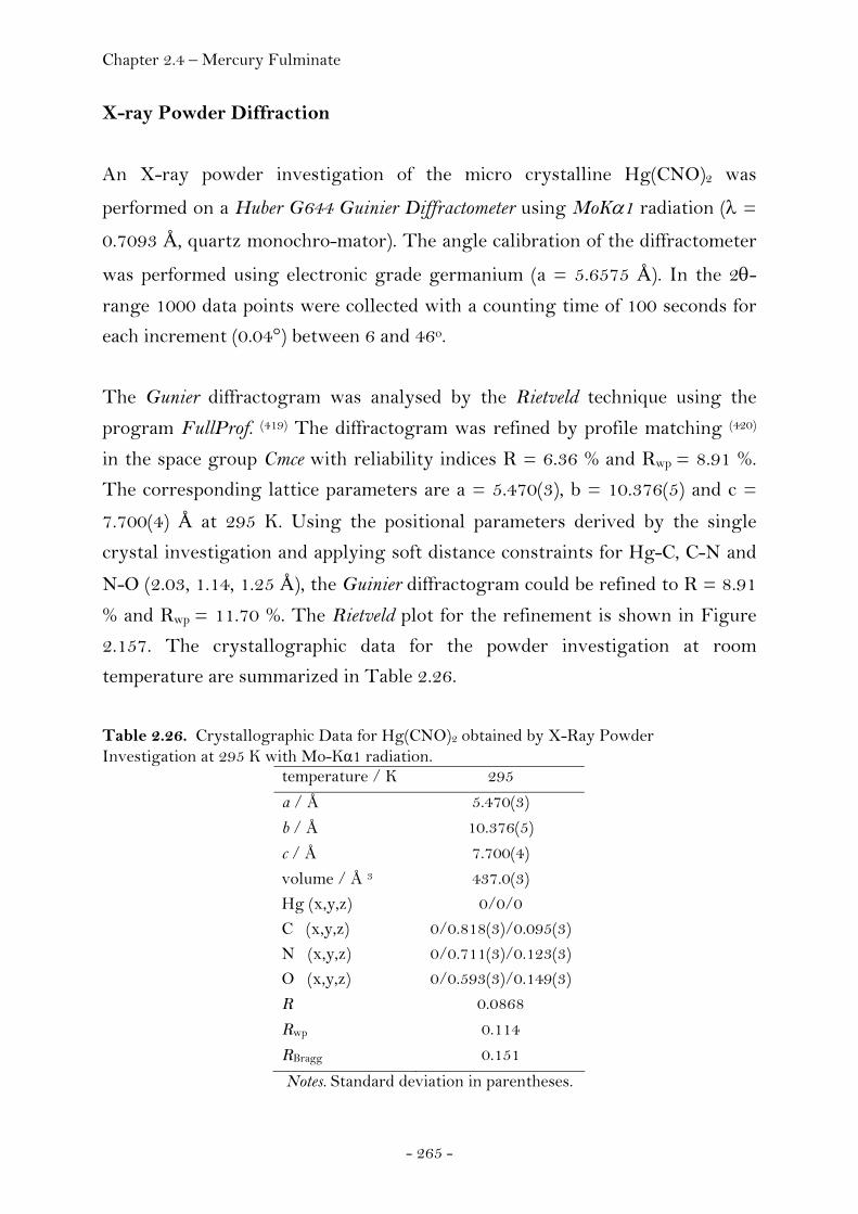
Chapter 2.4 – Mercury Fulminate
- 265 -
X-ray Powder Diffraction
An X-ray powder investigation of the micro crystalline Hg(CNO)2 was
performed on a Huber G644 Guinier Diffractometer using MoKα1 radiation (λ =
0.7093 Å, quartz monochro-mator). The angle calibration of the diffractometer
was performed using electronic grade germanium (a = 5.6575 Å). In the 2θ-
range 1000 data points were collected with a counting time of 100 seconds for
each increment (0.04°) between 6 and 46o.
The Gunier diffractogram was analysed by the Rietveld technique using the
program FullProf. (419) The diffractogram was refined by profile matching (420)
in the space group Cmce with reliability indices R = 6.36 % and Rwp = 8.91 %.
The corresponding lattice parameters are a = 5.470(3), b = 10.376(5) and c =
7.700(4) Å at 295 K. Using the positional parameters derived by the single
crystal investigation and applying soft distance constraints for Hg-C, C-N and
N-O (2.03, 1.14, 1.25 Å), the Guinier diffractogram could be refined to R = 8.91
% and Rwp = 11.70 %. The Rietveld plot for the refinement is shown in Figure
2.157. The crystallographic data for the powder investigation at room
temperature are summarized in Table 2.26.
Table 2.26. Crystallographic Data for Hg(CNO)2 obtained by X-Ray Powder Investigation at 295 K with Mo-Kα1 radiation.
temperature / K 295
a / Å 5.470(3)
b / Å 10.376(5)
c / Å 7.700(4)
volume / Å 3 437.0(3) Hg (x,y,z) 0/0/0 C (x,y,z) 0/0.818(3)/0.095(3) N (x,y,z) 0/0.711(3)/0.123(3) O (x,y,z) 0/0.593(3)/0.149(3) R 0.0868
Rwp 0.114
RBragg 0.151
Notes. Standard deviation in parentheses.
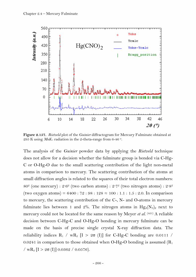
Chapter 2.4 – Mercury Fulminate
- 266 -
Figure 2.157. Rietveld plot of the Guinier diffractogram for Mercury Fulminate obtained at 295 K using MoKa1 radiation in the 2-theta-range from 6-46 o.
The analysis of the Guinier powder data by applying the Rietveld technique
does not allow for a decision whether the fulminate group is bonded via C-Hg-
C or O-Hg-O due to the small scattering contribution of the light non-metal
atoms in comparison to mercury. The scattering contribution of the atoms at
small diffraction angles is related to the squares of their total electron numbers:
802 (one mercury) : 2·62 (two carbon atoms) : 2·72 (two nitrogen atoms) : 2·82
(two oxygen atoms) = 6400 : 72 : 98 : 128 ≈ 100 : 1.1 : 1.5 : 2.0. In comparison
to mercury, the scattering contribution of the C-, N- and O-atoms in mercury
fulminate lies between 1 and 2%. The nitrogen atoms in Hg2(N3)2 next to
mercury could not be located for the same reason by Meyer et al. (421) A reliable
decision between C-Hg-C and O-Hg-O bonding in mercury fulminate can be
made on the basis of precise single crystal X-ray diffraction data. The
reliability indices R1 / wR2 [I > 2σ (I)] for C-Hg-C bonding are 0.0111 /
0.0241 in comparison to those obtained when O-Hg-O bonding is assumed (R1
/ wR2 [I > 2σ (I)] 0.0362 / 0.0576).

Chapter 2.4 – Mercury Fulminate
- 267 -
Single Crystal X-ray Diffraction
A single crystal of mercury fulminate (0.05 X 0.05 X 0.01 mm) was measured
using an Oxford Xcalibur3 CCD single crystal diffractometer from Oxford
Diffraction (MoKα radiation, graphite monochromator, λ = 0.71073 Å). The
temperature (100 K) of the single crystal was maintained using a Cryojet
Controller from Oxford Diffraction. The conditions limiting possible reflections
were hkl: h + k = 2n, h0l: l = 2n, hk0: h = 2n and yielding the space group
Cmce (No.64, former space group Cmca). The derived centre of symmetry of this
space group is in agreement with morphological studies of Miles. (422) A total of
4428 data were collected in the 2-Theta range up to 55.0o according to a
reflection range from -6 < h < 6, -13 < k < 13, -9 < l < 9. The data were
corrected for absorption. After merging, 257 unique reflections with a
redundancy of seventeen remained resulting in an reliability index Rint =
4.18%. The structure was solved using SHELXS-97 (423) and refined using
SHELXL-97. (290) A summary of the crystallographic data for mercury
fulminate obtained by the single crystal investigation is shown in the appendix
(Chapter 4). The positional parameters and the thermal displacement
parameters are listed in Table 2.27.
Table 2.27. Atomic Coordinates and Thermal Displacement Parameters (Å)2 for Hg(CNO)2 at100 K obtained from a Single-Crystal Investigation.
atom Hg C N O
x/a 0 0 0 0 y/b 0 0.8186 (6) 0.7109 (5) 0.5932 (4) z/c 0 0.0951 (8) 0.1210 (6) 0.1481 (6) U11 0.0148 (1) 0.0126 (3) 0.0122 (2) 0.0245 (2) U22 0.0141 (1) 0.0213 (3) 0.0263 (3) 0.0193 (2) U33 0.0209 (2) 0.0220 (3) 0.0144 (2) 0.0299 (2) U23 -0.0032 (3) 0.0013 (2) 0.0040 (2) 0.0104 (2) U13 0 0 0 0 U12 0 0 0 0 Ueq 0.0166 (1) 0.0187 (1) 0.0176 (1) 0.0246 (9)
Notes. Standard deviation in parentheses.
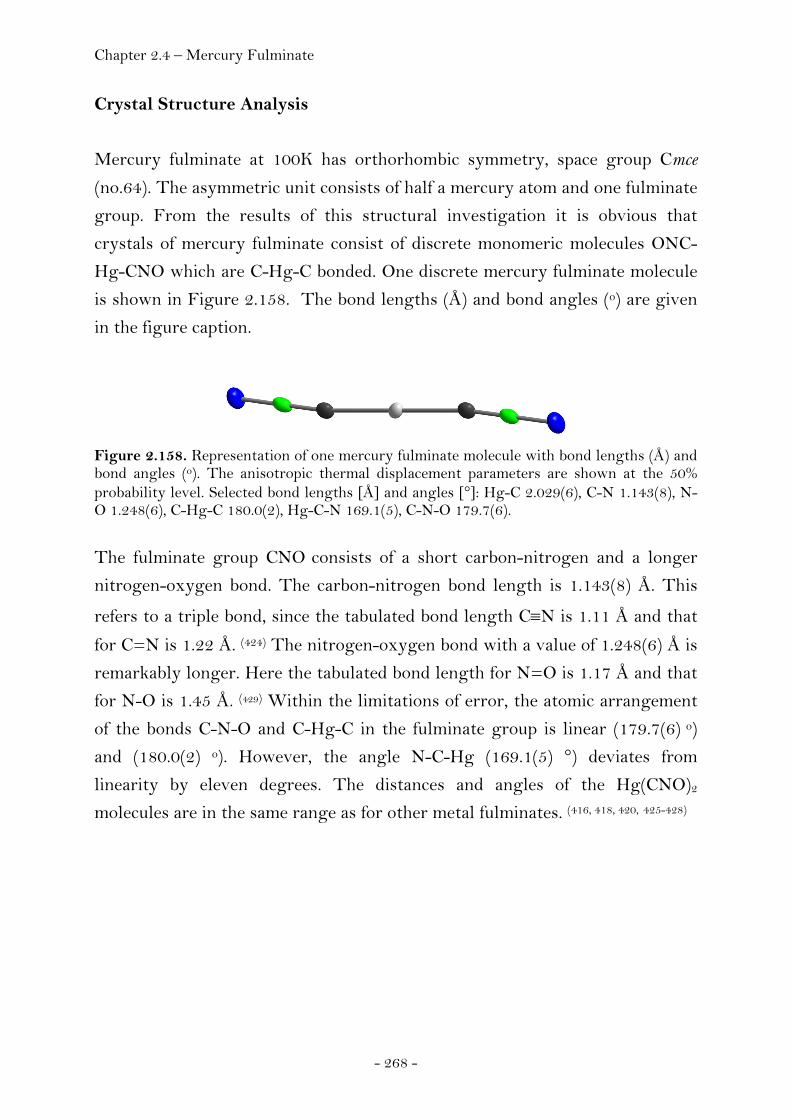
Chapter 2.4 – Mercury Fulminate
- 268 -
Crystal Structure Analysis
Mercury fulminate at 100K has orthorhombic symmetry, space group Cmce
(no.64). The asymmetric unit consists of half a mercury atom and one fulminate
group. From the results of this structural investigation it is obvious that
crystals of mercury fulminate consist of discrete monomeric molecules ONC-
Hg-CNO which are C-Hg-C bonded. One discrete mercury fulminate molecule
is shown in Figure 2.158. The bond lengths (Å) and bond angles (o) are given
in the figure caption.
Figure 2.158. Representation of one mercury fulminate molecule with bond lengths (Å) and bond angles (o). The anisotropic thermal displacement parameters are shown at the 50% probability level. Selected bond lengths [Å] and angles [°]: Hg-C 2.029(6), C-N 1.143(8), N-O 1.248(6), C-Hg-C 180.0(2), Hg-C-N 169.1(5), C-N-O 179.7(6).
The fulminate group CNO consists of a short carbon-nitrogen and a longer
nitrogen-oxygen bond. The carbon-nitrogen bond length is 1.143(8) Å. This
refers to a triple bond, since the tabulated bond length C≡N is 1.11 Å and that
for C=N is 1.22 Å. (424) The nitrogen-oxygen bond with a value of 1.248(6) Å is
remarkably longer. Here the tabulated bond length for N=O is 1.17 Å and that
for N-O is 1.45 Å. (429) Within the limitations of error, the atomic arrangement
of the bonds C-N-O and C-Hg-C in the fulminate group is linear (179.7(6) o)
and (180.0(2) o). However, the angle N-C-Hg (169.1(5) °) deviates from
linearity by eleven degrees. The distances and angles of the Hg(CNO)2
molecules are in the same range as for other metal fulminates. (416, 418, 420, 425-428)
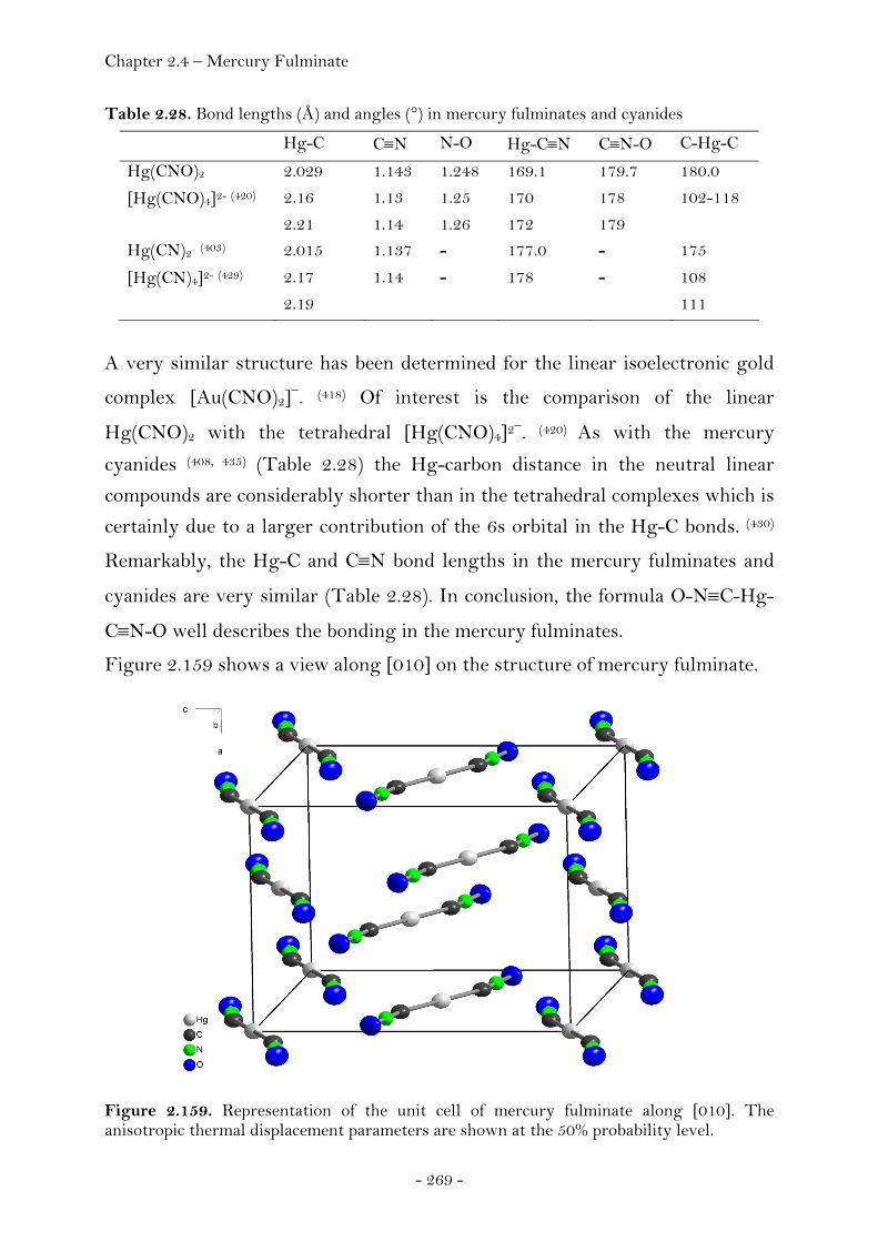
Chapter 2.4 – Mercury Fulminate
- 269 -
Table 2.28. Bond lengths (Å) and angles (°) in mercury fulminates and cyanides
Hg-C C≡N N-O Hg-C≡N C≡N-O C-Hg-C
Hg(CNO)2 2.029 1.143 1.248 169.1 179.7 180.0
[Hg(CNO)4]2- (420) 2.16 1.13 1.25 170 178 102-118
2.21 1.14 1.26 172 179 Hg(CN)2 (403) 2.015 1.137 - 177.0 - 175
[Hg(CN)4]2- (429) 2.17 1.14 - 178 - 108
2.19 111
A very similar structure has been determined for the linear isoelectronic gold
complex [Au(CNO)2]−. (418) Of interest is the comparison of the linear
Hg(CNO)2 with the tetrahedral [Hg(CNO)4]2−. (420) As with the mercury
cyanides (408, 435) (Table 2.28) the Hg-carbon distance in the neutral linear
compounds are considerably shorter than in the tetrahedral complexes which is
certainly due to a larger contribution of the 6s orbital in the Hg-C bonds. (430)
Remarkably, the Hg-C and C≡N bond lengths in the mercury fulminates and
cyanides are very similar (Table 2.28). In conclusion, the formula O-N≡C-Hg-
C≡N-O well describes the bonding in the mercury fulminates.
Figure 2.159 shows a view along [010] on the structure of mercury fulminate.
Figure 2.159. Representation of the unit cell of mercury fulminate along [010]. The anisotropic thermal displacement parameters are shown at the 50% probability level.
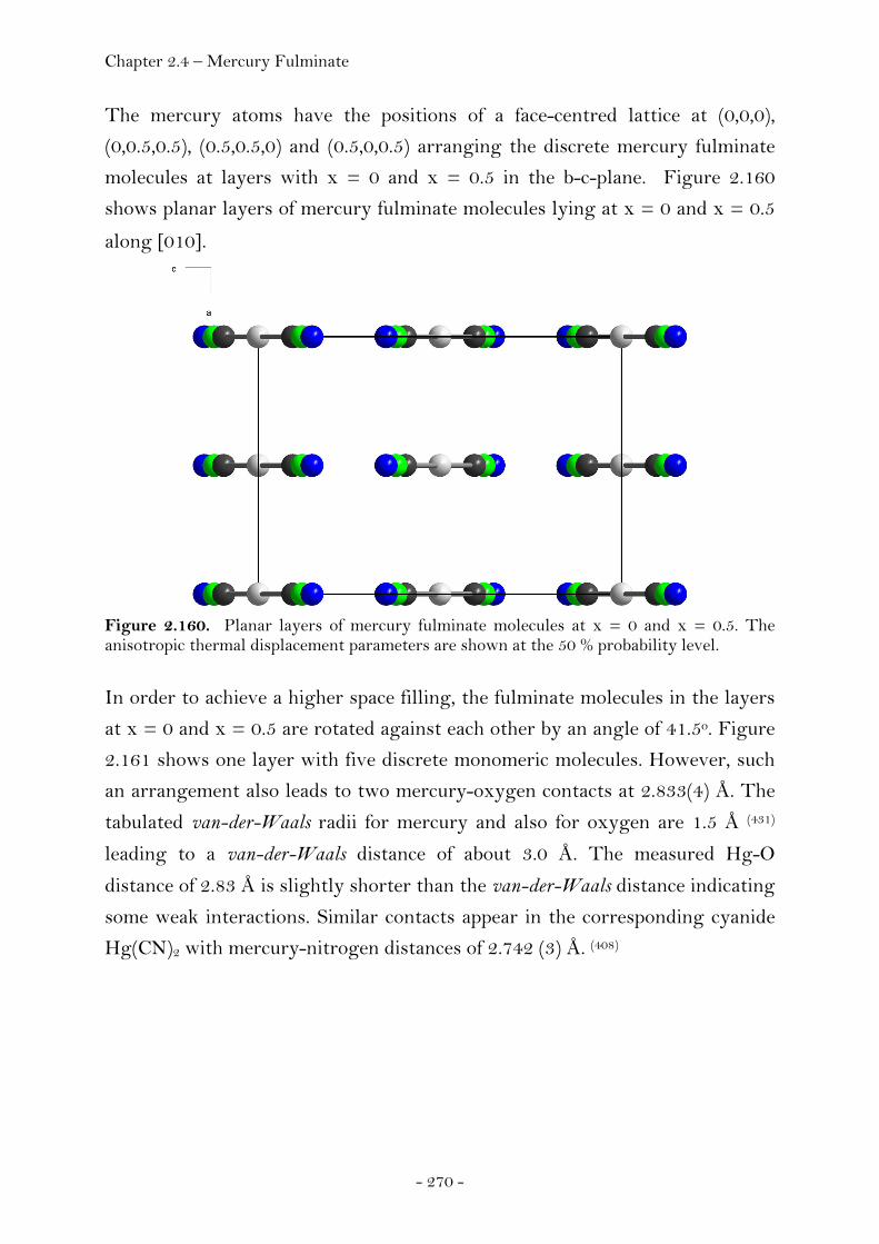
Chapter 2.4 – Mercury Fulminate
- 270 -
The mercury atoms have the positions of a face-centred lattice at (0,0,0),
(0,0.5,0.5), (0.5,0.5,0) and (0.5,0,0.5) arranging the discrete mercury fulminate
molecules at layers with x = 0 and x = 0.5 in the b-c-plane. Figure 2.160
shows planar layers of mercury fulminate molecules lying at x = 0 and x = 0.5
along [010].
Figure 2.160. Planar layers of mercury fulminate molecules at x = 0 and x = 0.5. The anisotropic thermal displacement parameters are shown at the 50 % probability level.
In order to achieve a higher space filling, the fulminate molecules in the layers
at x = 0 and x = 0.5 are rotated against each other by an angle of 41.5o. Figure
2.161 shows one layer with five discrete monomeric molecules. However, such
an arrangement also leads to two mercury-oxygen contacts at 2.833(4) Å. The
tabulated van-der-Waals radii for mercury and also for oxygen are 1.5 Å (431)
leading to a van-der-Waals distance of about 3.0 Å. The measured Hg-O
distance of 2.83 Å is slightly shorter than the van-der-Waals distance indicating
some weak interactions. Similar contacts appear in the corresponding cyanide
Hg(CN)2 with mercury-nitrogen distances of 2.742 (3) Å. (408)
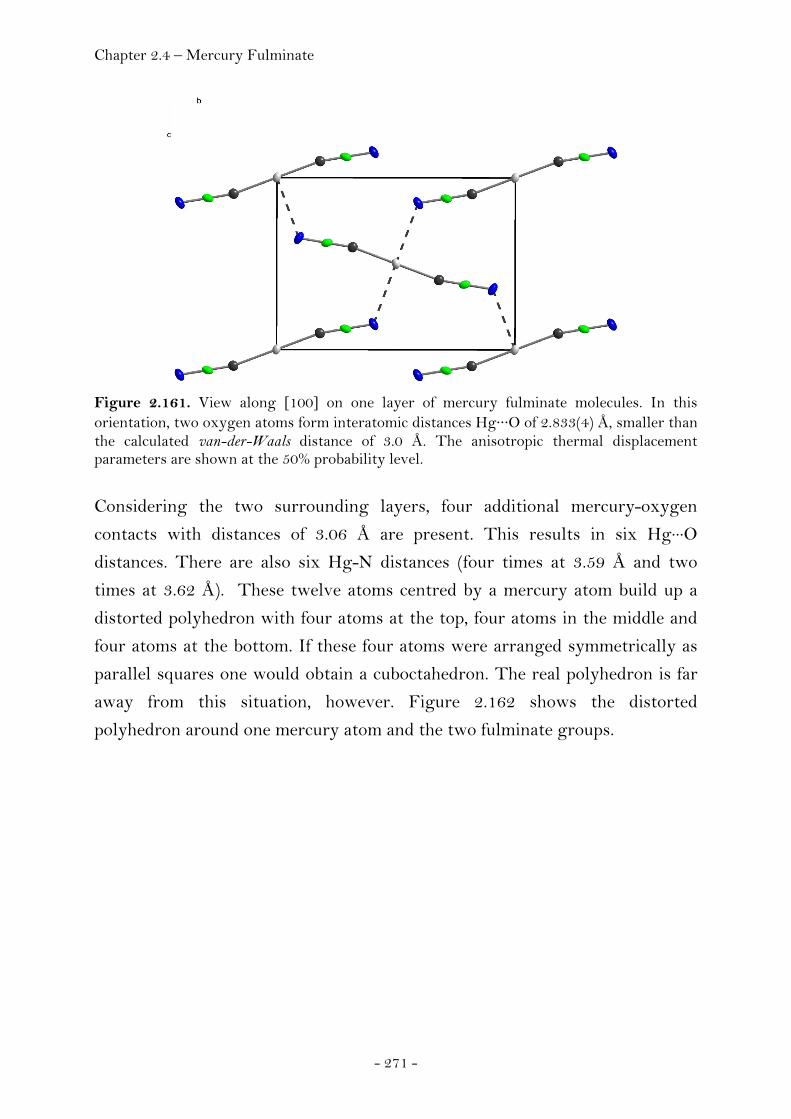
Chapter 2.4 – Mercury Fulminate
- 271 -
Figure 2.161. View along [100] on one layer of mercury fulminate molecules. In this orientation, two oxygen atoms form interatomic distances Hg···O of 2.833(4) Å, smaller than the calculated van-der-Waals distance of 3.0 Å. The anisotropic thermal displacement parameters are shown at the 50% probability level.
Considering the two surrounding layers, four additional mercury-oxygen
contacts with distances of 3.06 Å are present. This results in six Hg···O
distances. There are also six Hg-N distances (four times at 3.59 Å and two
times at 3.62 Å). These twelve atoms centred by a mercury atom build up a
distorted polyhedron with four atoms at the top, four atoms in the middle and
four atoms at the bottom. If these four atoms were arranged symmetrically as
parallel squares one would obtain a cuboctahedron. The real polyhedron is far
away from this situation, however. Figure 2.162 shows the distorted
polyhedron around one mercury atom and the two fulminate groups.
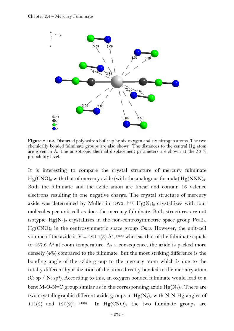
Chapter 2.4 – Mercury Fulminate
- 272 -
Figure 2.162. Distorted polyhedron built up by six oxygen and six nitrogen atoms. The two chemically bonded fulminate groups are also shown. The distances to the central Hg atom are given in Å. The anisotropic thermal displacement parameters are shown at the 50 % probability level. It is interesting to compare the crystal structure of mercury fulminate
Hg(CNO)2 with that of mercury azide (with the analogous formula) Hg(NNN)2.
Both the fulminate and the azide anion are linear and contain 16 valence
electrons resulting in one negative charge. The crystal structure of mercury
azide was determined by Müller in 1973. (432) Hg(N3)2 crystallizes with four
molecules per unit-cell as does the mercury fulminate. Both structures are not
isotypic. Hg(N3)2 crystallizes in the non-centrosymmetric space group Pca21,
Hg(CNO)2 in the centrosymmetric space group Cmce. However, the unit-cell
volume of the azide is V = 421.1(3) Å3, (438) whereas that of the fulminate equals
to 437.6 Å3 at room temperature. As a consequence, the azide is packed more
densely (4%) compared to the fulminate. But the most striking difference is the
bonding angle of the azide group to the mercury atom which is due to the
totally different hybridization of the atom directly bonded to the mercury atom
(C: sp / N: sp3). According to this, an oxygen bonded fulminate would lead to a
bent M-O-N≡C group similar as in the corresponding azide Hg(N3)2. There are
two crystallographic different azide groups in Hg(N3)2 with N-N-Hg angles of
111(2) and 120(2)o. (438) In Hg(CNO)2 the two fulminate groups are
Chapter 2.4 – Mercury Fulminate
- 273 -
crystallographic identical and have bond angles N-C-Hg of 169.1(5)o. In
addition, the atomic arrangement of N-N-N group in the the azide anions
deviate with bond angles of 171(3) and 176(2) from linearity. This is also true
for the atomic arrangement N-Hg-N with a bond angle of 175(1)o. Quite a
puzzle are the Hg-N bond distances of the two crystallographically different
azide groups. One Hg-N distance is 2.04(2) whereas the other is 2.14(2) Å. (438)
In summary, the molecular and crystal structure of the historically important
mercury fulminate has been solved, more than 200 years after its discovery.
Experimental
Synthesis. Hg(CNO)2 was synthesised by dissolving mercury (1g) in nitric acid
(12g, ρ = 1.4 g cm-3) and adding ethanol (11g) to this solution in two portions. (433) It is important to add the first half of ethanol before the red brown gases
have disappeared. Caution: Mercury fulminate is sensitive to impact and friction
and is easily detonated by sparks and flames. Before use it should be stored
under water and with exclusion of light.
Powder Preparation. For X-ray powder experiments freshly prepared
microcrystalline mercury fulminate was used. After drying on filter paper, a
Lindemann capillary (d = 0.5 mm) was carefully filled in order to prevent
grinding. Single Crystal Preparation. Single crystals of sufficient quality for
the structure determination were obtained from aqueous ammonia / ethanol
solutions (1:1:1) (434) and dried on filter paper with the exclusion of light. Small
crystals of rhombic habitus with well-developed faces were selected under a
polarization microscope and then cooled to 100 K using a Cryojet Controller of
an Oxford Xcalibur3 CCD single crystal diffractometer from Oxford Diffraction.
Details of the single crystal X-ray diffraction experiment are listed in the
appendix (Chapter 4).
Chapter 2.4 – P3N21
- 274 -
2.4.2 Hexaazidocyclotriphosphazene (P3N21)
Introduction
Compounds which are composed of only the elements phosphorus and nitrogen
can exist either as molecular species or as three dimensional polymeric solids.
Examples of known solid state compounds include the structurally well
characterised phases of the binary compound P3N5, which were reported by
Schnick et al. (435) In contrast, none of the binary PN molecules described in the
literature, namely P4N4, (436) P(N3)3, (437) P(N3)5, (438) [PN(N3)2]3 (439-441) or the
ionic compound (N5)P(N3)6 (442) have been structurally characterised. The
difficulties in the isolation and handling of these compounds have been shown
by Christe et al. for (N5)P(N3)6 to arise from their highly endothermic
character, as well as their extremely low barriers towards an often
uncontrollable explosive decomposition. (443–445, 448) Here we report the single
crystal X-ray structure of the P3N21 molecule and thereby the first example of a
structurally characterised binary P-N molecule.
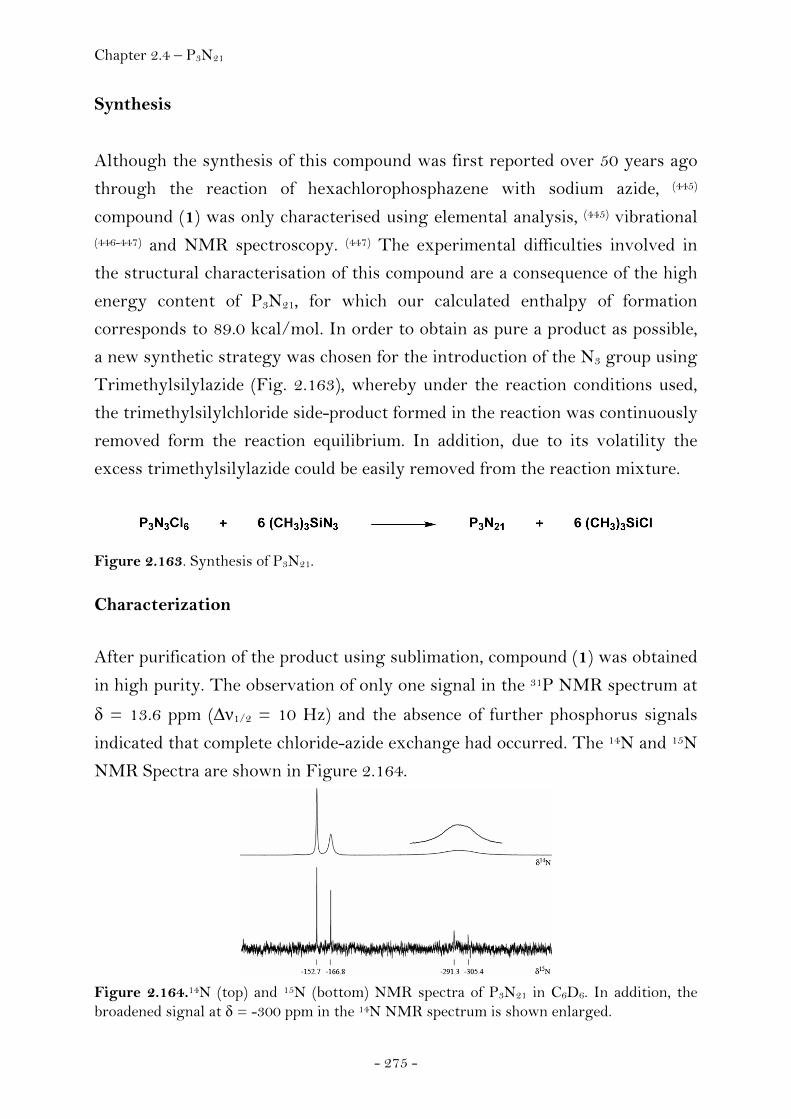
Chapter 2.4 – P3N21
- 275 -
Synthesis
Although the synthesis of this compound was first reported over 50 years ago
through the reaction of hexachlorophosphazene with sodium azide, (445)
compound (1) was only characterised using elemental analysis, (445) vibrational
(446-447) and NMR spectroscopy. (447) The experimental difficulties involved in
the structural characterisation of this compound are a consequence of the high
energy content of P3N21, for which our calculated enthalpy of formation
corresponds to 89.0 kcal/mol. In order to obtain as pure a product as possible,
a new synthetic strategy was chosen for the introduction of the N3 group using
Trimethylsilylazide (Fig. 2.163), whereby under the reaction conditions used,
the trimethylsilylchloride side-product formed in the reaction was continuously
removed form the reaction equilibrium. In addition, due to its volatility the
excess trimethylsilylazide could be easily removed from the reaction mixture.
Figure 2.163. Synthesis of P3N21.
Characterization
After purification of the product using sublimation, compound (1) was obtained
in high purity. The observation of only one signal in the 31P NMR spectrum at
δ = 13.6 ppm (Δν1/2 = 10 Hz) and the absence of further phosphorus signals
indicated that complete chloride-azide exchange had occurred. The 14N and 15N
NMR Spectra are shown in Figure 2.164.
Figure 2.164.14N (top) and 15N (bottom) NMR spectra of P3N21 in C6D6. In addition, the broadened signal at δ = -300 ppm in the 14N NMR spectrum is shown enlarged.
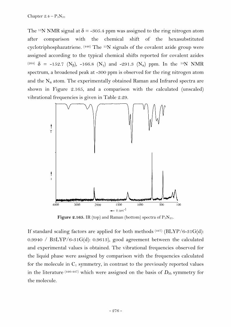
Chapter 2.4 – P3N21
- 276 -
The 15N NMR signal at δ = -305.4 ppm was assigned to the ring nitrogen atom
after comparison with the chemical shift of the hexasubstituted
cyclotriphosphazatriene. (446) The 15N signals of the covalent azide group were
assigned according to the typical chemical shifts reported for covalent azides (264) δ = -152.7 (Nβ), -166.8 (Nγ) and -291.3 (Nα) ppm. In the 14N NMR
spectrum, a broadened peak at -300 ppm is observed for the ring nitrogen atom
and the Nα atom. The experimentally obtained Raman and Infrared spectra are
shown in Figure 2.165, and a comparison with the calculated (unscaled)
vibrational frequencies is given in Table 2.29.
Figure 2.165. IR (top) and Raman (bottom) spectra of P3N21.
If standard scaling factors are applied for both methods (447) (BLYP/6-31G(d):
0.9940 / B3LYP/6-31G(d): 0.9613), good agreement between the calculated
and experimental values is obtained. The vibrational frequencies observed for
the liquid phase were assigned by comparison with the frequencies calculated
for the molecule in C1 symmetry, in contrast to the previously reported values
in the literature (446-447) which were assigned on the basis of D3h symmetry for
the molecule.
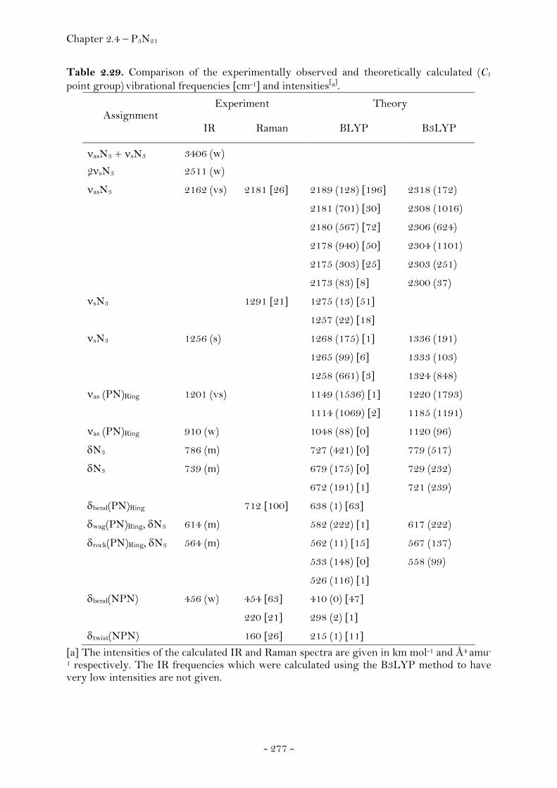
Chapter 2.4 – P3N21
- 277 -
Table 2.29. Comparison of the experimentally observed and theoretically calculated (C1 point group) vibrational frequencies [cm-1] and intensities[a].
Experiment Theory Assignment
IR Raman BLYP B3LYP
νasN3 + νsN3 3406 (w)
2νsN3 2511 (w)
νasN3 2162 (vs) 2181 [26] 2189 (128) [196] 2318 (172)
2181 (701) [30] 2308 (1016)
2180 (567) [72] 2306 (624)
2178 (940) [50] 2304 (1101)
2175 (303) [25] 2303 (251)
2173 (83) [8] 2300 (37)
νsN3 1291 [21] 1275 (13) [51]
1257 (22) [18]
νsN3 1256 (s) 1268 (175) [1] 1336 (191)
1265 (99) [6] 1333 (103)
1258 (661) [3] 1324 (848)
νas (PN)Ring 1201 (vs) 1149 (1536) [1] 1220 (1793)
1114 (1069) [2] 1185 (1191)
νas (PN)Ring 910 (w) 1048 (88) [0] 1120 (96)
δN3 786 (m) 727 (421) [0] 779 (517)
δN3 739 (m) 679 (175) [0] 729 (232)
672 (191) [1] 721 (239)
δbend(PN)Ring 712 [100] 638 (1) [63]
δwag(PN)Ring, δN3 614 (m) 582 (222) [1] 617 (222)
δrock(PN)Ring, δN3 564 (m) 562 (11) [15] 567 (137)
533 (148) [0] 558 (99)
526 (116) [1]
δbend(NPN) 456 (w) 454 [63] 410 (0) [47]
220 [21] 298 (2) [1]
δtwist(NPN) 160 [26] 215 (1) [11] [a] The intensities of the calculated IR and Raman spectra are given in km mol-1 and Å4 amu-
1 respectively. The IR frequencies which were calculated using the B3LYP method to have very low intensities are not given.
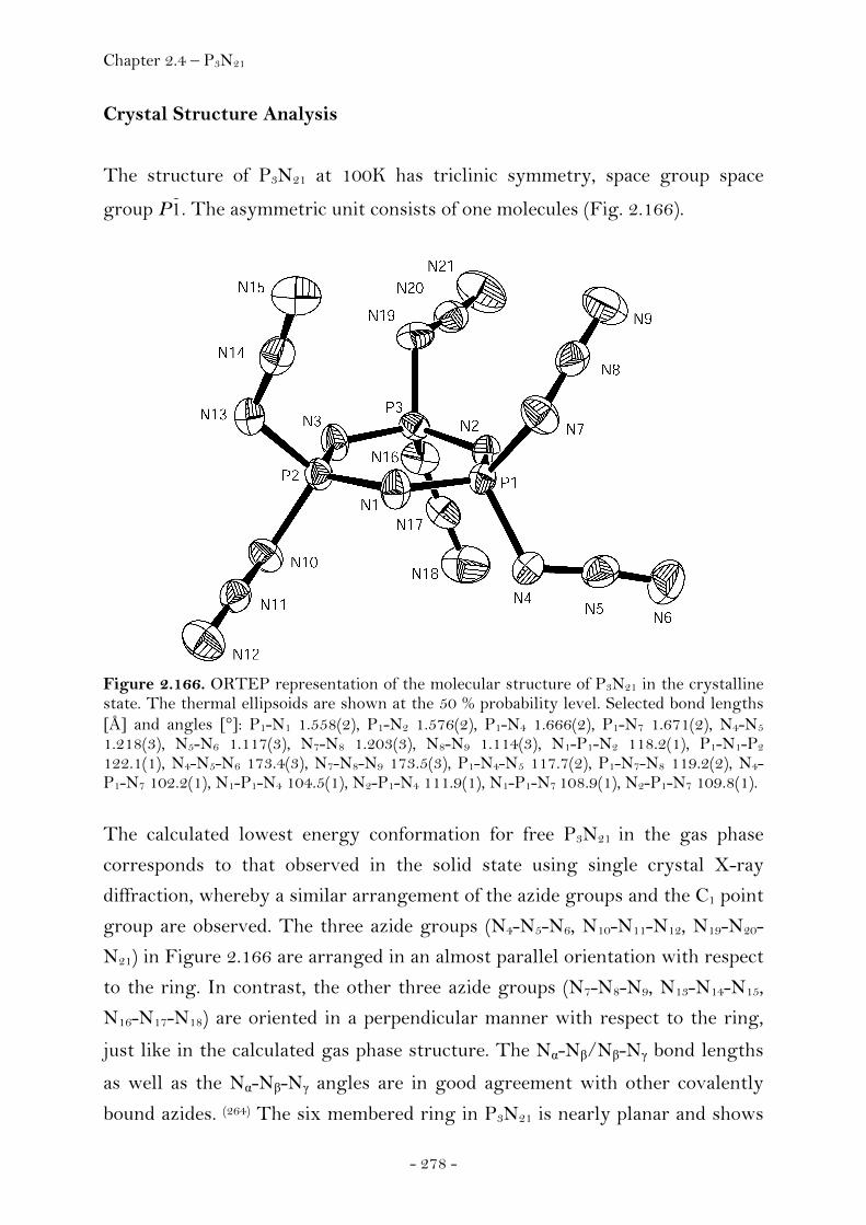
Chapter 2.4 – P3N21
- 278 -
Crystal Structure Analysis
The structure of P3N21 at 100K has triclinic symmetry, space group space
group P1. The asymmetric unit consists of one molecules (Fig. 2.166).
Figure 2.166. ORTEP representation of the molecular structure of P3N21 in the crystalline state. The thermal ellipsoids are shown at the 50 % probability level. Selected bond lengths [Å] and angles [°]: P1-N1 1.558(2), P1-N2 1.576(2), P1-N4 1.666(2), P1-N7 1.671(2), N4-N5 1.218(3), N5-N6 1.117(3), N7-N8 1.203(3), N8-N9 1.114(3), N1-P1-N2 118.2(1), P1-N1-P2 122.1(1), N4-N5-N6 173.4(3), N7-N8-N9 173.5(3), P1-N4-N5 117.7(2), P1-N7-N8 119.2(2), N4-P1-N7 102.2(1), N1-P1-N4 104.5(1), N2-P1-N4 111.9(1), N1-P1-N7 108.9(1), N2-P1-N7 109.8(1).
The calculated lowest energy conformation for free P3N21 in the gas phase
corresponds to that observed in the solid state using single crystal X-ray
diffraction, whereby a similar arrangement of the azide groups and the C1 point
group are observed. The three azide groups (N4-N5-N6, N10-N11-N12, N19-N20-
N21) in Figure 2.166 are arranged in an almost parallel orientation with respect
to the ring. In contrast, the other three azide groups (N7-N8-N9, N13-N14-N15,
N16-N17-N18) are oriented in a perpendicular manner with respect to the ring,
just like in the calculated gas phase structure. The Nα-Nβ/Nβ-Nγ bond lengths
as well as the Nα-Nβ-Nγ angles are in good agreement with other covalently
bound azides. (264) The six membered ring in P3N21 is nearly planar and shows
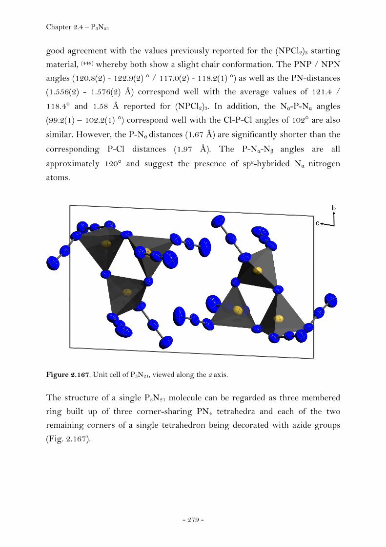
Chapter 2.4 – P3N21
- 279 -
good agreement with the values previously reported for the (NPCl2)3 starting
material, (448) whereby both show a slight chair conformation. The PNP / NPN
angles (120.8(2) - 122.9(2) ° / 117.0(2) - 118.2(1) °) as well as the PN-distances
(1.556(2) - 1.576(2) Å) correspond well with the average values of 121.4 /
118.4° and 1.58 Å reported for (NPCl2)3. In addition, the Nα-P-Nα angles
(99.2(1) – 102.2(1) °) correspond well with the Cl-P-Cl angles of 102° are also
similar. However, the P-Nα distances (1.67 Å) are significantly shorter than the
corresponding P-Cl distances (1.97 Å). The P-Nα-Nβ angles are all
approximately 120° and suggest the presence of sp2-hybrided Nα nitrogen
atoms.
Figure 2.167. Unit cell of P3N21, viewed along the a axis.
The structure of a single P3N21 molecule can be regarded as three membered
ring built up of three corner-sharing PN4 tetrahedra and each of the two
remaining corners of a single tetrahedron being decorated with azide groups
(Fig. 2.167).
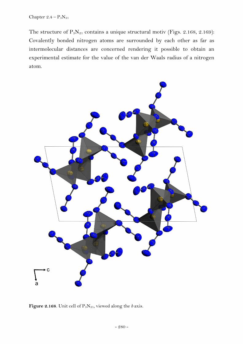
Chapter 2.4 – P3N21
- 280 -
The structure of P3N21 contains a unique structural motiv (Figs. 2.168, 2.169):
Covalently bonded nitrogen atoms are surrounded by each other as far as
intermolecular distances are concerned rendering it possible to obtain an
experimental estimate for the value of the van der Waals radius of a nitrogen
atom.
Figure 2.168. Unit cell of P3N21, viewed along the b axis.

Chapter 2.4 – P3N21
- 281 -
Figure 2.169. Unit cell of P3N21, viewed along the c axis.
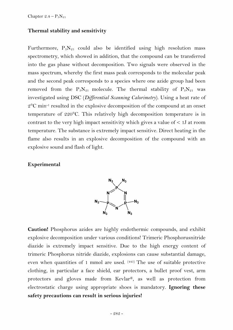
Chapter 2.4 – P3N21
- 282 -
Thermal stability and sensitivity
Furthermore, P3N21 could also be identified using high resolution mass
spectrometry, which showed in addition, that the compound can be transferred
into the gas phase without decomposition. Two signals were observed in the
mass spectrum, whereby the first mass peak corresponds to the molecular peak
and the second peak corresponds to a species where one azide group had been
removed from the P3N21 molecule. The thermal stability of P3N21 was
investigated using DSC (Differential Scanning Calorimetry). Using a heat rate of
2°C min-1 resulted in the explosive decomposition of the compound at an onset
temperature of 220°C. This relatively high decomposition temperature is in
contrast to the very high impact sensitivity which gives a value of < 1J at room
temperature. The substance is extremely impact sensitive. Direct heating in the
flame also results in an explosive decomposition of the compound with an
explosive sound and flash of light.
Experimental
Caution! Phosphorus azides are highly endothermic compounds, and exhibit
explosive decomposition under various conditions! Trimeric Phosphorusnitride
diazide is extremely impact sensitive. Due to the high energy content of
trimeric Phosphorus nitride diazide, explosions can cause substantial damage,
even when quantities of 1 mmol are used. (445) The use of suitable protective
clothing, in particular a face shield, ear protectors, a bullet proof vest, arm
protectors and gloves made from Kevlar®, as well as protection from
electrostatic charge using appropriate shoes is mandatory. Ignoring these
safety precautions can result in serious injuries!
Chapter 2.4 – P3N21
- 283 -
P3N3Cl6 and TMS-N3 were purchased from Aldrich. Propionitrile was dried
over P4O10 and distilled prior to use. Raman spectra were measured using a
Perkin Elmer Spectrum 2000R NIR FT-Raman instrument (Nd:YAG Laser
(1064 nm)). The Infrared spectra (IR) were recorded using a Perkin-Elmer
Spektrum One FT-IR instrument. The 31P, 15N and 14N NMR Spectra were
obtained using a Jeol EX 400 NMR spectrometer operating at 28.9 MHz (14N),
40.6 MHz (15N) and 162.0 MHz (31P), and the chemical shifts are given in ppm
relative to nitromethane (14/15N) and 85% phosphoric acid (31P). The mass
spectra were measured using a Jeol MStation JMS-700 mass spectrometer. The
decomposition temperature was determined using a Pyris 6 DSC instrument.
The values for the impact sensitivity of the substance at room temperature
were determined using a BAM-drop hammer (Bundesanstalt für
Materialforschung und Prüfung). Computational methods: BLYP and B3LYP
density functional theory (DFT) calculations were used. Calculation of the
geometry, IR and Raman spectra employing the 6-31G(d) Basis sets were
achieved using the Gaussian programme. (81)
Preparation of P3N21: 224 mg (0.644 mmol) trimeric phosphorus nitride
dichloride were added to a flamed out Schlenk flask under Argon, and dissolved
in 20 mL of anhydrous Propionitrile at room temperature. Trimethylsilylazide
(893 mg (7.750 mmol)) was added drop-wise to the stirred solution under a
nitrogen purge. A blubberer was connected to the flask and a slow stream of
nitrogen gas was passed continuously through the apparatus. The colourless
solution was warmed to 60°C, and stirred for three hours at this temperature.
Finally, the pale yellow solution was stirred for 19 hours at room temperature.
The reaction mixture was subsequently concentrated using the rotary
evaporator (30°C, 50 mbar) and the remaining solvent removed under vacuum
using a Schlenk line (1·10-3 mbar, room temperature, several minutes). The pale
yellow liquid that was obtained was purified using sublimation (1·10-3 mbar,
130°C oil bath temperature, -86°C cold finger temperature) and the product
was obtained as a colourless liquid. Single crystals of P3N21 were obtained via
the controlled warming and cooling of the solid/liquid substance around its
melting point. Repeated cooling of the substance to -78.5°C with dry ice and
warming to -17°C in a cold room was monitored under a microscope, until
Chapter 2.4 – P3N21
- 284 -
crystals formed. As soon as a liquid phase formed, the substance was re-cooled
using dry ice. Handling of the single crystals requires great care !
Raman (liq., r.t.): s. Tab. 2.29. IR (Nujol, KBr, background subtracted): Tab.
2.29. 31P NMR (C6D6, 25°C): δ = 13.6 ppm (Δν1/2 = 10 Hz); 15N NMR (C6D6,
25°C): δ = -152.7 (Nβ), -166.8 (Nγ), -291.3 (Nα), -305.4 (NRing). 14N NMR (C6D6,
25°C): δ = -152.7 (Nβ, Δν1/2 = 34 Hz), -166.8 (Nγ, Δν1/2 = 115 Hz), -300 (Nα/
NRing, Δν1/2 = 950 Hz). MS (DEI, 70eV): 387 [m+, 30%], 345 [m-N3+, 100%]; MS (HR): calculated for P3N21: 386.9858, found: 386.9851 (-0.7 mmu). DSC
(2°/min): 220°C (decomposition). Impact sensitivity: < 1J; Details of the single
crystal X-ray diffraction experiment are listed in the appendix (Chapter 4).

Chapter 2.4 – Trinitromethanes
- 285 -
2.4.3 Trinitromethanes
Trinitromethane (Nitroform, NFM)
The potential of the trinitromethyl group, C(NO2)3, as a component of
energetic compounds has long been recognized. (13, 449-451) The parent molecule
can be viewed as being trinitromethane, HC(NO2)3 (also called nitroform),
which can be prepared by the reaction of acetylene with nitric acid. (457) It has a
high crystal density (1.806 g cm-3) and a low melting point (25 ºC). (133) Though
the presence of three nitro groups attached at one carbon atom might render
this compound a priori to be an effective explosive, nitroform has found no use
as an explosive. It has been reported to be very difficult to initiate, display poor
performance characteristics and a low brisance. (452) Furthermore it’s high
acidity is connected with many disadvantages like its solubility in water
contributing to make it hygroscopic and rendering it unsuitable for practical
application as explosive. Finally, it displays unsatisfactory thermal stability
yielding considerable (5-10%) decomposition even at such low temperatures as
30°C. However, trinitromethane can be used to prepare various compounds of
energetic interest, like derivatives of 2,2,2-trinitroethanol, which can be
obtained from the reaction between trinitromethane and formaldehyde and it
displays some interesting academic aspects. (453) The three strongly electron-
attracting NO2 groups make the hydrogen very acidic (although not as much as
might be anticipated); the experimental pKa is 0.1 (457). This should promote aci
tautomerization, which involves the transfer of the proton to one of the nitro
oxygens (Figs. 2.170, 2.171).
Figure 2.170. Formation of aci-trinitromethane.
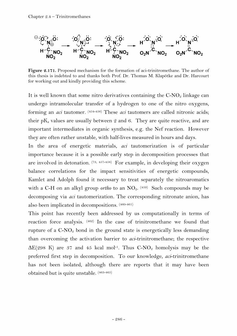
Chapter 2.4 – Trinitromethanes
- 286 -
Figure 2.171. Proposed mechanism for the formation of aci-trinitromethane. The author of this thesis is indebted to and thanks both Prof. Dr. Thomas M. Klapötke and Dr. Harcourt for working out and kindly providing this scheme.
It is well known that some nitro derivatives containing the C-NO2 linkage can
undergo intramolecular transfer of a hydrogen to one of the nitro oxygens,
forming an aci tautomer. (454-456) These aci tautomers are called nitronic acids;
their pKa values are usually between 2 and 6. They are quite reactive, and are
important intermediates in organic synthesis, e.g. the Nef reaction. However
they are often rather unstable, with half-lives measured in hours and days.
In the area of energetic materials, aci tautomerization is of particular
importance because it is a possible early step in decomposition processes that
are involved in detonation. (73, 457-458) For example, in developing their oxygen
balance correlations for the impact sensitivities of energetic compounds,
Kamlet and Adolph found it necessary to treat separately the nitroaromatics
with a C-H on an alkyl group ortho to an NO2. (459) Such compounds may be
decomposing via aci tautomerization. The corresponding nitronate anion, has
also been implicated in decompositions. (460-461)
This point has recently been addressed by us computationally in terms of
reaction force analysis. (462) In the case of trinitromethane we found that
rupture of a C-NO2 bond in the ground state is energetically less demanding
than overcoming the activation barrier to aci-trinitromethane; the respective
ΔE(298 K) are 37 and 45 kcal mol-1. Thus C-NO2 homolysis may be the
preferred first step in decomposition. To our knowledge, aci-trinitromethane
has not been isolated, although there are reports that it may have been
obtained but is quite unstable. (463-465)

Chapter 2.4 – Trinitromethanes
- 287 -
Trinitromethanol
Whereas trinitroethanol is widely used in synthesis (269) and is well-
characterized crystallographically (234), trinitromethanol is not known, as far as
we are aware. Using reaction force analysis, we have recently analyzed
trinitromethanol computationally. For trinitromethanol, we found the NO2
groups in ground-state not to be in the propeller-like arrangement in contrast
to trinitromethane. In trinitromethanol, one of them, e.g. NOaOb, was found to
be nearly coplanar with the C-O-H portion of the molecule, while the other two
were found to be approximately perpendicular to each other. This permits O-
H···Oa hydrogen bonding as indicated by a H···Oa distance of 1.922 Å as
opposed to the sum of the van der Waals radii (2.72 Å). (244) It is to Oa that the
hydrogen will migrate. However, according to our gas phase calculations the
result will not be an aci tautomer. Instead, they are indicative for
trinitromethanol breaking up into a weakly-bound complex of (O2N)2C=O and
HOaNOb (Fig. 2.172), which is lower in energy by only 0.4 kcal mol-1 relative
to the separate molecules.
Figure 2.172. Decompositon of the elusive trinitromethanol into dinitromethanone and nitrous acid.
This fragmentation has a very low activation barrier (10 kcal mol-1). This
barrier is less than the dissociation energy of the C-NOaOb bond alone (30 kcal
mol-1) since a new bond, Oa-H is being formed and the C-O bond is being
converted into a double bond rendering it plausible why trinitromethanol is
not known as far as we know.

Chapter 2.4 – Trinitromethanes
- 288 -
Chlorotrinitromethane
Introduction
Describing interactions of atoms and atomic groups in molecules is a
fundamental chemical challenge. Any compound showing deviations from
normal geometric parameters is useful for critically assessing the strengths and
failings of chemical bond models used to explain experimental results and thus
helps aiding their further development. (466) In addition, current computer
simulations of molecular systems are limited by and require reliable
experimental data for the accurate description of the energies of interacting
molecules and forces between them. (467) A better understanding of molecular
interactions has recently been shown to be important for the manipulation of
molecular recognition processes involving halogen bonding (468-469) and the
design of specific material properties. (269) One class of compounds displaying
remarkable inter- as well as intramolecular effects encompasses molecules
having the trinitromethyl group, with three nitro groups bonded to one carbon
atom. Consisting of elements of Groups IV, V and VI, the properties of the
trinitromethyl group are comparable to those of a Group VII element; early
studies describing the chemical properties of α-halogen derivatives of
trinitromethane have demonstrated the potential of the uncharged
trinitromethyl moiety to behave as a pseudohalogen. (470) Today, experimental
evidence supporting this notion include the description of pseudointerhalogen
compounds like azidotrinitromethane (471-472) and cyanotrinitromethane (473)
along with the series of α-halogen derivatives of trinitromethane (474) and its
dimer hexanitroethane. (475) Structural data for the halogen derivatives of
trinitromethane have been limited to theoretical calculations (476-479) and
spectroscopic analyses. (480-481) However, the determination of the equilibrium
structure of a polyatomic molecule using only spectroscopic information
becomes increasingly difficult with the number of atoms. (482-483) The difficulties
in the case of chlorotrinitromethane were discussed by Sadova et al. (487)
Reliable structural data for α-halogen-substituted trinitromethane molecules in
the solid state did not exist at the beginning of this work. An attempted X-ray
structure determination has been reported for iodotrinitromethane, IC(NO2)3.
Chapter 2.4 – Trinitromethanes
- 289 -
(106) While this was the only representative amongst the halogen derivatives of
trinitromethane retaining the crystalline state under standard conditions, the
structure determination of iodotrinitromethane suffered from decomposition of
several different single crystals exposed to the X-ray beam, considerably
affecting the quality of the experimental data and precluding the possibility of a
refinement of the individual parameters of the light atoms, resulting in a
considerable scatter of the bond lengths. In regard to the trinitromethyl group
as a pseudohalogen, chlorotrinitromethane holds a unique position amongst the
halogen derivatives of trinitromethane. The question arises as to which
halogen atom is most closely related to the C(NO2)3 group. Whereas the
charges of the bromine and iodine atoms in the respective analogues are
reported to be positive, (476, 484) there are contradictory estimates of the charge
of the chlorine in chlorotrinitromethane, as being positive (476, 485) or negative. (106, 486) Experimental efforts to clarify this question were not successful, with
neither positive nor negative mass spectra giving rise to parent ions even at
low electron energies. (487) Another interesting point is that the structure of
gaseous 1 derived from electron diffraction revealed the carbon-chlorine bond
length (1.712 Å) to be the shortest yet found for a tetrahedral carbon. (486) In
view of these findings, it seemed desirable to investigate single crystals of
chlorotrinitromethane in order to obtain reliable geometrical data for the solid
state.
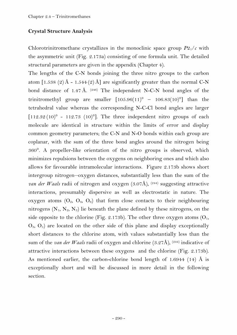
Chapter 2.4 – Trinitromethanes
- 290 -
Crystal Structure Analysis
Chlorotrinitromethane crystallizes in the monoclinic space group P21/c with
the asymmetric unit (Fig. 2.173a) consisting of one formula unit. The detailed
structural parameters are given in the appendix (Chapter 4).
The lengths of the C-N bonds joining the three nitro groups to the carbon
atom [1.538 (2) Å - 1.544 (2) Å] are significantly greater than the normal C-N
bond distance of 1.47 Å. (246) The independent N-C-N bond angles of the
trinitromethyl group are smaller [105.96(11)° – 106.83(10)°] than the
tetrahedral value whereas the corresponding N-C-Cl bond angles are larger
[112.32 (10)° - 112.73 (10)°]. The three independent nitro groups of each
molecule are identical in structure within the limits of error and display
common geometry parameters; the C-N and N-O bonds within each group are
coplanar, with the sum of the three bond angles around the nitrogen being
360°. A propeller-like orientation of the nitro groups is observed, which
minimizes repulsions between the oxygens on neighboring ones and which also
allows for favourable intramolecular interactions. Figure 2.173b shows short
intergroup nitrogen···oxygen distances, substantially less than the sum of the
van der Waals radii of nitrogen and oxygen (3.07Å), (244) suggesting attractive
interactions, presumably dispersive as well as electrostatic in nature. The
oxygen atoms (O2, O4, O6) that form close contacts to their neighbouring
nitrogens (N1, N2, N3) lie beneath the plane defined by these nitrogens, on the
side opposite to the chlorine (Fig. 2.173b). The other three oxygen atoms (O1,
O3, O5) are located on the other side of this plane and display exceptionally
short distances to the chlorine atom, with values substantially less than the
sum of the van der Waals radii of oxygen and chlorine (3.27Å), (244) indicative of
attractive interactions between these oxygens and the chlorine (Fig. 2.173b).
As mentioned earlier, the carbon-chlorine bond length of 1.6944 (14) Å is
exceptionally short and will be discussed in more detail in the following
section.
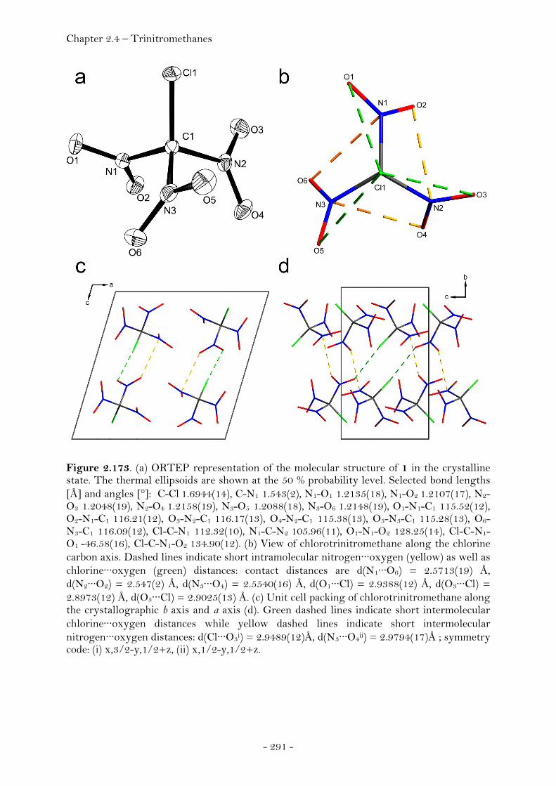
Chapter 2.4 – Trinitromethanes
- 291 -
Figure 2.173. (a) ORTEP representation of the molecular structure of 1 in the crystalline state. The thermal ellipsoids are shown at the 50 % probability level. Selected bond lengths [Å] and angles [°]: C-Cl 1.6944(14), C-N1 1.543(2), N1-O1 1.2135(18), N1-O2 1.2107(17), N2-O3 1.2048(19), N2-O4 1.2158(19), N3-O5 1.2088(18), N3-O6 1.2148(19), O1-N1-C1 115.52(12), O2-N1-C1 116.21(12), O3-N2-C1 116.17(13), O4-N2-C1 115.38(13), O5-N3-C1 115.28(13), O6-N3-C1 116.09(12), Cl-C-N1 112.32(10), N1-C-N2 105.96(11), O1-N1-O2 128.25(14), Cl-C-N1-O1 -46.58(16), Cl-C-N1-O2 134.90(12). (b) View of chlorotrinitromethane along the chlorine carbon axis. Dashed lines indicate short intramolecular nitrogen···oxygen (yellow) as well as chlorine···oxygen (green) distances: contact distances are d(N1···O6) = 2.5713(19) Å, d(N2···O2) = 2.547(2) Å, d(N3···O4) = 2.5540(16) Å, d(O1···Cl) = 2.9388(12) Å, d(O3···Cl) = 2.8973(12) Å, d(O5···Cl) = 2.9025(13) Å. (c) Unit cell packing of chlorotrinitromethane along the crystallographic b axis and a axis (d). Green dashed lines indicate short intermolecular chlorine···oxygen distances while yellow dashed lines indicate short intermolecular nitrogen···oxygen distances: d(Cl···O3i) = 2.9489(12)Å, d(N3···O4ii) = 2.9794(17)Å ; symmetry code: (i) x,3/2-y,1/2+z, (ii) x,1/2-y,1/2+z.
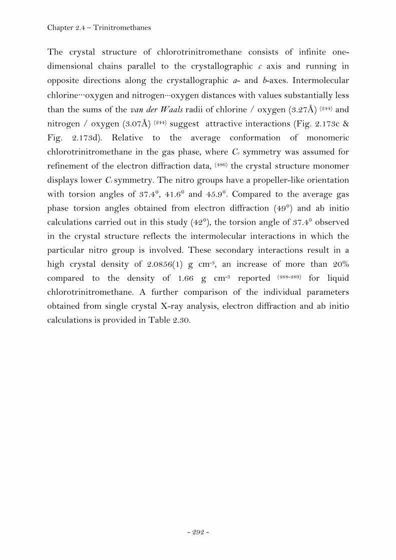
Chapter 2.4 – Trinitromethanes
- 292 -
The crystal structure of chlorotrinitromethane consists of infinite one-
dimensional chains parallel to the crystallographic c axis and running in
opposite directions along the crystallographic a- and b-axes. Intermolecular
chlorine···oxygen and nitrogen···oxygen distances with values substantially less
than the sums of the van der Waals radii of chlorine / oxygen (3.27Å) (244) and
nitrogen / oxygen (3.07Å) (244) suggest attractive interactions (Fig. 2.173c &
Fig. 2.173d). Relative to the average conformation of monomeric
chlorotrinitromethane in the gas phase, where C3 symmetry was assumed for
refinement of the electron diffraction data, (486) the crystal structure monomer
displays lower C1 symmetry. The nitro groups have a propeller-like orientation
with torsion angles of 37.4°, 41.6° and 45.9°. Compared to the average gas
phase torsion angles obtained from electron diffraction (49°) and ab initio
calculations carried out in this study (42°), the torsion angle of 37.4° observed
in the crystal structure reflects the intermolecular interactions in which the
particular nitro group is involved. These secondary interactions result in a
high crystal density of 2.0856(1) g cm-3, an increase of more than 20%
compared to the density of 1.66 g cm-3 reported (488-489) for liquid
chlorotrinitromethane. A further comparison of the individual parameters
obtained from single crystal X-ray analysis, electron diffraction and ab initio
calculations is provided in Table 2.30.
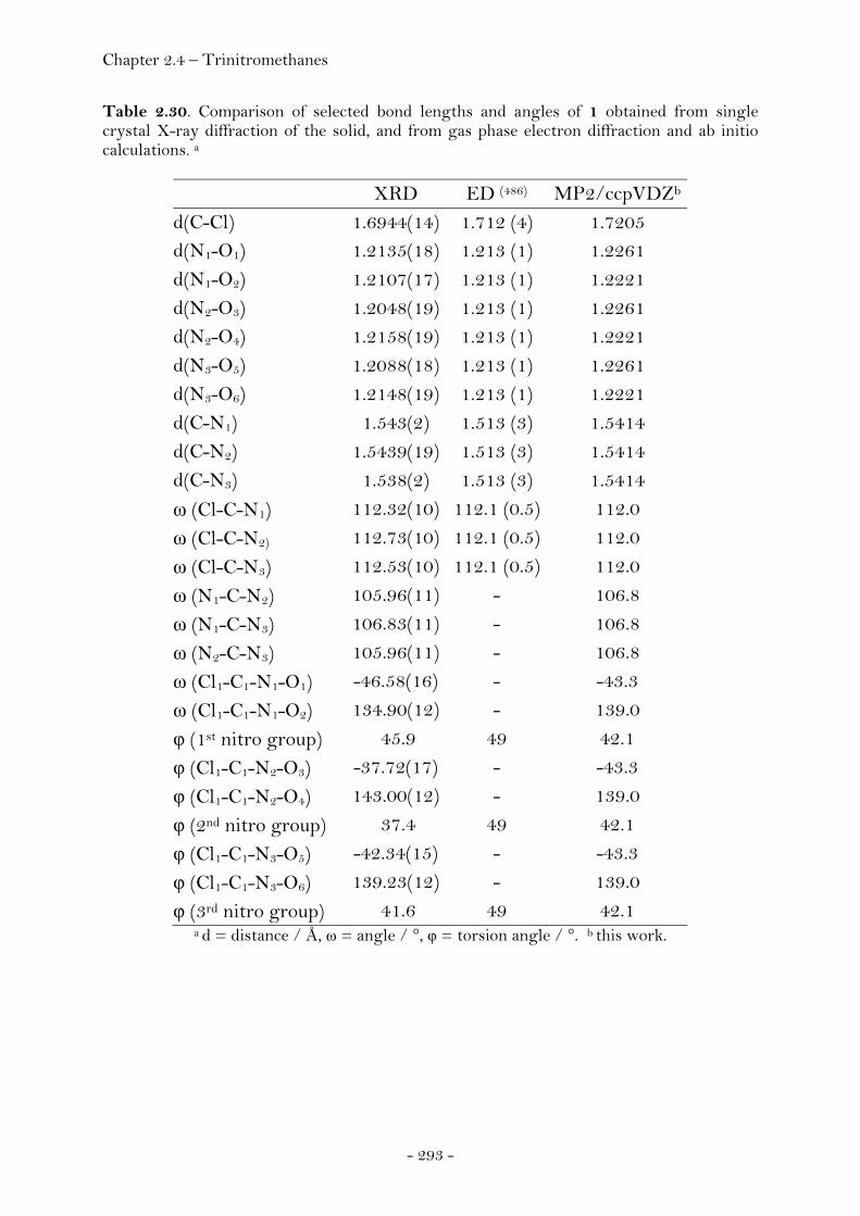
Chapter 2.4 – Trinitromethanes
- 293 -
Table 2.30. Comparison of selected bond lengths and angles of 1 obtained from single crystal X-ray diffraction of the solid, and from gas phase electron diffraction and ab initio calculations. a
XRD ED (486) MP2/ccpVDZb
d(C-Cl) 1.6944(14) 1.712 (4) 1.7205 d(N1-O1) 1.2135(18) 1.213 (1) 1.2261 d(N1-O2) 1.2107(17) 1.213 (1) 1.2221 d(N2-O3) 1.2048(19) 1.213 (1) 1.2261 d(N2-O4) 1.2158(19) 1.213 (1) 1.2221 d(N3-O5) 1.2088(18) 1.213 (1) 1.2261 d(N3-O6) 1.2148(19) 1.213 (1) 1.2221 d(C-N1) 1.543(2) 1.513 (3) 1.5414 d(C-N2) 1.5439(19) 1.513 (3) 1.5414 d(C-N3) 1.538(2) 1.513 (3) 1.5414 ω (Cl-C-N1) 112.32(10) 112.1 (0.5) 112.0 ω (Cl-C-N2) 112.73(10) 112.1 (0.5) 112.0 ω (Cl-C-N3) 112.53(10) 112.1 (0.5) 112.0 ω (N1-C-N2) 105.96(11) - 106.8 ω (N1-C-N3) 106.83(11) - 106.8 ω (N2-C-N3) 105.96(11) - 106.8 ω (Cl1-C1-N1-O1) -46.58(16) - -43.3 ω (Cl1-C1-N1-O2) 134.90(12) - 139.0 φ (1st nitro group) 45.9 49 42.1 φ (Cl1-C1-N2-O3) -37.72(17) - -43.3 φ (Cl1-C1-N2-O4) 143.00(12) - 139.0 φ (2nd nitro group) 37.4 49 42.1 φ (Cl1-C1-N3-O5) -42.34(15) - -43.3 φ (Cl1-C1-N3-O6) 139.23(12) - 139.0 φ (3rd nitro group) 41.6 49 42.1
a d = distance / Å, ω = angle / °, φ = torsion angle / °. b this work.
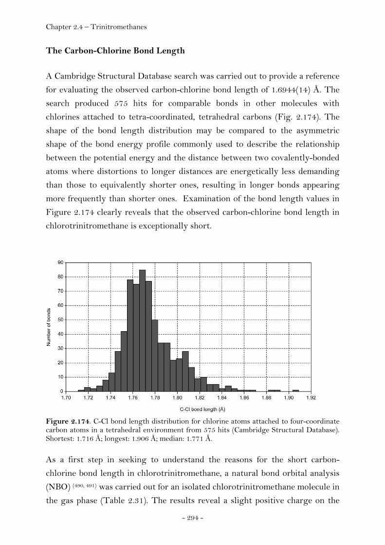
Chapter 2.4 – Trinitromethanes
- 294 -
The Carbon-Chlorine Bond Length
A Cambridge Structural Database search was carried out to provide a reference
for evaluating the observed carbon-chlorine bond length of 1.6944(14) Å. The
search produced 575 hits for comparable bonds in other molecules with
chlorines attached to tetra-coordinated, tetrahedral carbons (Fig. 2.174). The
shape of the bond length distribution may be compared to the asymmetric
shape of the bond energy profile commonly used to describe the relationship
between the potential energy and the distance between two covalently-bonded
atoms where distortions to longer distances are energetically less demanding
than those to equivalently shorter ones, resulting in longer bonds appearing
more frequently than shorter ones. Examination of the bond length values in
Figure 2.174 clearly reveals that the observed carbon-chlorine bond length in
chlorotrinitromethane is exceptionally short.
Figure 2.174. C-Cl bond length distribution for chlorine atoms attached to four-coordinate carbon atoms in a tetrahedral environment from 575 hits (Cambridge Structural Database). Shortest: 1.716 Å; longest: 1.906 Å; median: 1.771 Å.
As a first step in seeking to understand the reasons for the short carbon-
chlorine bond length in chlorotrinitromethane, a natural bond orbital analysis
(NBO) (490, 491) was carried out for an isolated chlorotrinitromethane molecule in
the gas phase (Table 2.31). The results reveal a slight positive charge on the
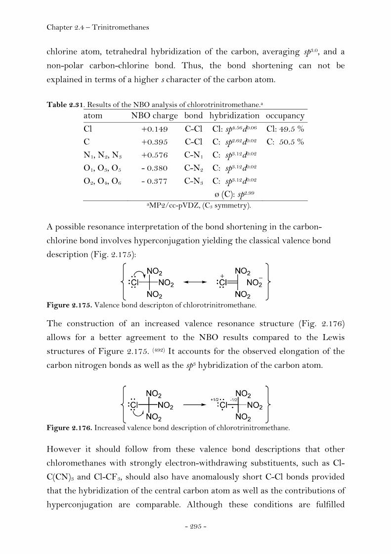
Chapter 2.4 – Trinitromethanes
- 295 -
chlorine atom, tetrahedral hybridization of the carbon, averaging sp3.0, and a
non-polar carbon-chlorine bond. Thus, the bond shortening can not be
explained in terms of a higher s character of the carbon atom.
Table 2.31. Results of the NBO analysis of chlorotrinitromethane.a atom NBO charge bond hybridization occupancy Cl +0.149 C-Cl Cl: sp4.56d0.06 Cl: 49.5 % C +0.395 C-Cl C: sp2.62d0.02 C: 50.5 % N1, N2, N3 +0.576 C-N1 C: sp3.12d0.02 O1, O3, O5 - 0.380 C-N2 C: sp3.12d0.02 O2, O4, O6 - 0.377 C-N3 C: sp3.12d0.02 ø (C): sp2.99
aMP2/cc-pVDZ, (C3 symmetry).
A possible resonance interpretation of the bond shortening in the carbon-
chlorine bond involves hyperconjugation yielding the classical valence bond
description (Fig. 2.175):
Figure 2.175. Valence bond descripton of chlorotrinitromethane.
The construction of an increased valence resonance structure (Fig. 2.176)
allows for a better agreement to the NBO results compared to the Lewis
structures of Figure 2.175. (492) It accounts for the observed elongation of the
carbon nitrogen bonds as well as the sp3 hybridization of the carbon atom.
Figure 2.176. Increased valence bond description of chlorotrinitromethane.
However it should follow from these valence bond descriptions that other
chloromethanes with strongly electron-withdrawing substituents, such as Cl-
C(CN)3 and Cl-CF3, should also have anomalously short C-Cl bonds provided
that the hybridization of the central carbon atom as well as the contributions of
hyperconjugation are comparable. Although these conditions are fulfilled

Chapter 2.4 – Trinitromethanes
- 296 -
according to the results of the NBO analyses for these molecules (Table 2.32), a
comparison of the carbon chlorine bond length values in Table 2.33 shows that
a similar bond shortening is not observed in Cl-C(CN)3 or Cl-CF3.
Table 2.32. Comparison of the NBO analysis results of chlorotrinitromethane, chloro-tricyanomethane and chlorotrifluoromethane.
molecule atom NBO charge bond hybridization occupancy energy
Cl-C(NO2)3 Cl +0.149 C-Cl Cl: sp4.56d0.06 Cl: 49.5 % 52.9 a)
C +0.395 C-Cl C: sp2.62d0.02 C: 50.5 %
N1, N2, N3 +0.576 C-N1 C: sp3.12d0.02
O1, O3, O5 - 0.380 C-N2 C: sp3.12d0.02
O2, O4, O6 - 0.377 C-N3 C: sp3.12d0.02
ø (C): sp2.99
Cl-C(CN)3 Cl +0.067 C1-Cl Cl: sp6d0.06 Cl: 49.8 % 18.3 b)
C1 - 0.245 C1-Cl C1: sp4.33d0.02 C: 50.2%
C2, C3, C4 +0.314 C1-C2 C1: sp2.68d0.01
N1, N2, N3 - 0.255 C1-C3 C1: sp2.68d0.01
C1-C4 C1: sp2.68d0.01
ø (C1): sp3.09
Cl-CF3 Cl - 0.046 C-Cl Cl: sp5.17d0.07 Cl: 55.6 % 39.8 c)
C +1.276 C-Cl C: sp2.73d0.03 C: 44.4 %
F1, F2, F3 - 0.410 C-F1 C: sp3.05d0.03
C-F2 C: sp3.05d0.03
C-F3 C: sp3.05d0.03
ø (C): sp2.97 Sum of intramolecular donor-acceptor interactions: a) LP (Cl) → σ*(C1-N1,2,3) / kcal mol-1 b) LP (Cl) → σ*(C1-C2,3,4) / kcal mol-1 c) LP (Cl) → σ*(C1-F1,2,3) / kcal mol-1. MP2/cc-pVDZ.
Table 2.33. Comparison of carbon chlorine bond lengths in related molecules.a
molecule method temp. / K d(C-Cl) / Å state reference Cl-C(NO2)3 X-ray
ED Calc. Calc. Calc. Calc.
100 318
1.6944(14) 1.712 (4) 1.721 1.708 1.720 1.713
solid gas gas gas gas gas
this work (486) (482), B3LYP/6-311+G(d,p) (482), MP2/6-31G(d,p) this work, MP2/ccPVDZ this work, B3PW91/6-31G(d,p)
Cl-C(CN)3 X-ray Calc.
r.t. 1.78(1) 1.809
solid gas
(493) this work, MP2/ccPVDZ
Calc. 1.818 gas this work, B3PW91/6-31G(d,p) Cl-CF3 Calc. 1.765 gas this work, MP2/ccPVDZ Calc 1.772 gas this work, B3PW91/6-31G(d,p)
a X-ray = X-ray diffraction, ED = electron diffraction, Calc. = calculated value.
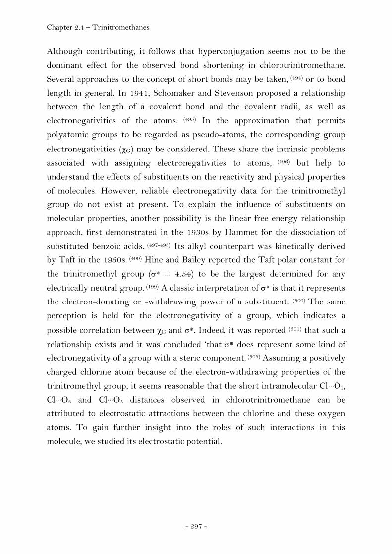
Chapter 2.4 – Trinitromethanes
- 297 -
Although contributing, it follows that hyperconjugation seems not to be the
dominant effect for the observed bond shortening in chlorotrinitromethane.
Several approaches to the concept of short bonds may be taken, (494) or to bond
length in general. In 1941, Schomaker and Stevenson proposed a relationship
between the length of a covalent bond and the covalent radii, as well as
electronegativities of the atoms. (495) In the approximation that permits
polyatomic groups to be regarded as pseudo-atoms, the corresponding group
electronegativities (χG) may be considered. These share the intrinsic problems
associated with assigning electronegativities to atoms, (496) but help to
understand the effects of substituents on the reactivity and physical properties
of molecules. However, reliable electronegativity data for the trinitromethyl
group do not exist at present. To explain the influence of substituents on
molecular properties, another possibility is the linear free energy relationship
approach, first demonstrated in the 1930s by Hammet for the dissociation of
substituted benzoic acids. (497-498) Its alkyl counterpart was kinetically derived
by Taft in the 1950s. (499) Hine and Bailey reported the Taft polar constant for
the trinitromethyl group (σ* = 4.54) to be the largest determined for any
electrically neutral group. (199) A classic interpretation of σ* is that it represents
the electron-donating or -withdrawing power of a substituent. (500) The same
perception is held for the electronegativity of a group, which indicates a
possible correlation between χG and σ*. Indeed, it was reported (501) that such a
relationship exists and it was concluded ‘that σ* does represent some kind of
electronegativity of a group with a steric component. (506) Assuming a positively
charged chlorine atom because of the electron-withdrawing properties of the
trinitromethyl group, it seems reasonable that the short intramolecular Cl···O1,
Cl···O3 and Cl···O5 distances observed in chlorotrinitromethane can be
attributed to electrostatic attractions between the chlorine and these oxygen
atoms. To gain further insight into the roles of such interactions in this
molecule, we studied its electrostatic potential.
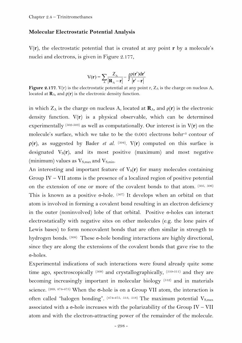
Chapter 2.4 – Trinitromethanes
- 298 -
Molecular Electrostatic Potential Analysis
V(r), the electrostatic potential that is created at any point r by a molecule’s
nuclei and electrons, is given in Figure 2.177,
A
A A
Z ρ( )dV( ) = ′ ′
−′− −∑ ∫r rr
R r r r
Figure 2.177. V(r) is the electrostatic potential at any point r, ZA is the charge on nucleus A, located at RA, and ρ(r) is the electronic density function.
in which ZA is the charge on nucleus A, located at RA, and ρ(r) is the electronic
density function. V(r) is a physical observable, which can be determined
experimentally (502-503) as well as computationally. Our interest is in V(r) on the
molecule’s surface, which we take to be the 0.001 electrons bohr-3 contour of
ρ(r), as suggested by Bader et al. (504). V(r) computed on this surface is
designated VS(r), and its most positive (maximum) and most negative
(minimum) values as VS,max and VS,min.
An interesting and important feature of VS(r) for many molecules containing
Group IV – VII atoms is the presence of a localized region of positive potential
on the extension of one or more of the covalent bonds to that atom. (505, 506)
This is known as a positive σ-hole. (507) It develops when an orbital on that
atom is involved in forming a covalent bond resulting in an electron deficiency
in the outer (noninvolved) lobe of that orbital. Positive σ-holes can interact
electrostatically with negative sites on other molecules (e.g. the lone pairs of
Lewis bases) to form noncovalent bonds that are often similar in strength to
hydrogen bonds. (508) These σ-hole bonding interactions are highly directional,
since they are along the extensions of the covalent bonds that gave rise to the
σ-holes.
Experimental indications of such interactions were found already quite some
time ago, spectroscopically (509) and crystallographically, (510-511) and they are
becoming increasingly important in molecular biology (512) and in materials
science. (269, 474-475) When the σ-hole is on a Group VII atom, the interaction is
often called “halogen bonding”. (474-475, 513, 518) The maximum potential VS,max
associated with a σ-hole increases with the polarizability of the Group IV – VII
atom and with the electron-attracting power of the remainder of the molecule.

Chapter 2.4 – Trinitromethanes
- 299 -
Accordingly, positive σ-holes are less likely to be found on the lightest (least
polarizable) atoms in Groups IV – VII (i.e. C, N, O and F), although they do
develop if the remainder of the molecule is sufficiently electron-withdrawing. (412) Chlorotrinitromethane illustrates well the importance of both inter- and
intramolecular nonbonded interactions. Figure 2.173 shows that there are
several of each type, the origins of which can be understood by examining the
electrostatic potential on the molecule’s surface. Four views of this are in
Figure 2.178, and its most positive and most negative values (the VS,max and
VS,min) are listed in Table 2.34 along with the key inter- and intramolecular
close contacts.
Figure 2.178. The molecular electrostatic potential of Cl−C(NO2)3 (in kcal mol-1) computed on the 0.001 electrons bohr-3 isodensity surface, with chlorine facing the reader (a), facing away from the reader (b) and pointing to the top right (c, d). The colors form a continuum from most positive, blue (greater than or equal to 30 kcal mol-1), to most negative, red (equal to or more negative than -9.4 kcal mol-1), with blue, light blue and green increasingly less positive, and yellow and red as negative. Note the strongly positive σ-hole on the chlorine surface (blue), on the extension of the C−Cl bond, and the weak one (green) on the carbon (middle of top right view) on the extension of the Cl−C bond. Color bars giving the continuum scales in atomic units are given to aid the reader (1 atomic unit = 627.5 kcal mol-
1).
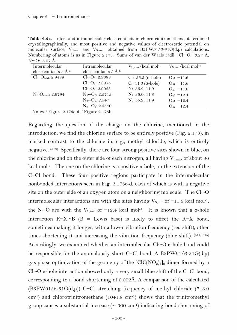
Chapter 2.4 – Trinitromethanes
- 300 -
Table 2.34. Inter- and intramolecular close contacts in chlorotrinitromethane, determined crystallographically, and most positive and negative values of electrostatic potential on molecular surface, VS,max and VS,min, obtained from B3PW91/6-31G(d,p) calculations. Numbering of atoms is as in Figure 2.173. Sums of van der Waals radii: Cl···O: 3.27 Å, N···O: 3.07 Å.
Intermolecular close contacts / Å a
Intramolecular close contacts / Å b
VS,max/kcal mol-1 VS,min/kcal mol-1
Cl···Oodd: 2.9489 Cl···O1: 2.9388 Cl: 35.5 (σ-hole) O1: −11.6 Cl···O3: 2.8973 C: 11.3 (σ-hole) O3: −11.6 Cl···O5: 2.9025 N: 36.2, 11.9 O5: −11.6 N···Oeven: 2.9794 N1···O6: 2.5713 N: 36.0, 11.8 O6: −12.4 N2···O2: 2.547 N: 35.9, 11.9 O2: −12.4 N3···O4: 2.5540 O4: −12.4 Notes. a Figure 2.173c-d. b Figure 2.173b.
Regarding the question of the charge on the chlorine, mentioned in the
introduction, we find the chlorine surface to be entirely positive (Fig. 2.178), in
marked contrast to the chlorine in, e.g., methyl chloride, which is entirely
negative. (513) Specifically, there are four strong positive sites shown in blue, on
the chlorine and on the outer side of each nitrogen, all having VS,max of about 36
kcal mol-1. The one on the chlorine is a positive σ-hole, on the extension of the
C−Cl bond. These four positive regions participate in the intermolecular
nonbonded interactions seen in Fig. 2.173c-d, each of which is with a negative
site on the outer side of an oxygen atom on a neighboring molecule. The Cl···O
intermolecular interactions are with the sites having VS,min of −11.6 kcal mol-1,
the N···O are with the VS,min of −12.4 kcal mol-1. It is known that a σ-hole
interaction R−X···B (B = Lewis base) is likely to affect the R−X bond,
sometimes making it longer, with a lower vibration frequency (red shift), other
times shortening it and increasing the vibration frequency (blue shift). (514, 515)
Accordingly, we examined whether an intermolecular Cl···O σ-hole bond could
be responsible for the anomalously short C−Cl bond. A B3PW91/6-31G(d,p)
gas phase optimization of the geometry of the [ClC(NO2)3]2 dimer formed by a
Cl···O σ-hole interaction showed only a very small blue shift of the C−Cl bond,
corresponding to a bond shortening of 0.002Å. A comparison of the calculated
(B3PW91/6-31G(d,p)) C−Cl stretching frequency of methyl chloride (743.9
cm-1) and chlorotrinitromethane (1041.8 cm-1) shows that the trinitromethyl
group causes a substantial increase (~ 300 cm-1) indicating bond shortening of
Chapter 2.4 – Trinitromethanes
- 301 -
the isolated molecule in the gas phase. This result together with the small
difference between the experimentally determined gas phase and solid state
values of the C−Cl bond (Table 2.33) indicates that the primary factor causing
the observed shortening is most likely intramolecular. It was already pointed
out that while the chlorine VS,max is on its outermost side, the entire surface of
the chlorine is positive (Fig. 2.178). The propeller-like orientation of the nitro
groups in 1 puts three of the oxygens (O1, O3 and O5) close to the chlorine (Fig.
2.173); the Cl···O separations are about 2.90 Å and the Cl−C−N−O dihedral
angles average 42° (Table 2.30). Thus the electrostatic attractions between the
negative potentials on the inner sides of these oxygens (Fig. 2.178) and the
positive chlorine are presumably the cause of the unusual shortening of the
C−Cl bond.These Cl···O attractions would of course be maximized if the nitro
groups were exactly coplanar with the C−Cl bond. We carried out a
B3PW91/6-31G(d,p) calculation in which we forced this to be the case. The
Cl···O separations decrease by 0.15 Å compared to the optimized geometry and
the C−Cl bond is indeed shortened 0.013 Å. However this constrained
structure is less stable by 6 kcal mol-1 than the optimized one, because it
eliminates the intramolecular close contacts between the nitrogens and O2, O4
and O6. These reflect electrostatic interactions between the respectively
positive and negative inner sides of the nitrogens and these oxygens (Fig.
2.173b). The relevant nitrogen VS,max are only 12 kcal mol-1, compared to their
other VS,max of 36 kcal mol-1, because they have been partially neutralized due
to the proximity of the oxygens; for the analogous reason, inner-side VS,min of
these oxygens cannot even be identified. With the observed structure being in
equilibrium where the forces are balanced and the potential energy is
minimized, our findings are in support of an intramolecular electrostatic
attraction between the positively charged chlorine atom and the negatively
charged oxygen atoms (O1, O3, O5). Together with their axial symmetry
around the C−Cl bond, these interactions are reflected in the anomalously short
character of this bond.
Of interest, the data of Levchenkov et al. show a comparable shortening of the
C−F and C−Br bonds in F−C(NO2)3 and Br−C(NO2)3. (482) Our calculated
B3PW91/6-31G(d,p) electrostatic potentials on the molecular surfaces of

Chapter 2.4 – Trinitromethanes
- 302 -
fluoro- and bromotrinitromethane show the surfaces of the halogen atoms to be
entirely positive (Figs. 2.179, 2.180), just like the chlorine in Cl−C(NO2)3.
Figure 2.179. Computed electrostatic potential on the molecular surface of F−C(NO2)3. The fluorine atom is facing the viewer. The color ranges of the electrostatic potential, in kcal mol-1, form a continuum from royal blue (more positive than 14) to red (more negative than -10). The surface of the fluorine is completely positive, with the most positive value on the extension of the C−F bond (VS,max = 15 kcal mol-1).
Figure 2.180. Computed electrostatic potential on the molecular surface of Br−C(NO2)3. The bromine atom is facing the viewer. The color ranges of the electrostatic potential, in kcal mol-1, form a continuum from royal blue (more positive than 14) to red (more negative than -10). The surface of the bromine is completely positive, with the most positive value on the extension of the C−Br bond (VS,max = 43 kcal mol-1).
Chapter 2.4 – Trinitromethanes
- 303 -
A quantitative measure of the very strong electron-withdrawing power of the
three NO2 groups can be obtained by comparing the corresponding Taft polar
constants (σ*), which are 4.54 (516-517) for the C(NO2)3 group vs 3.19 (523) for
fluorine, 2.94 (523) for chlorine and 2.80 (523) for bromine. It seems likely,
therefore, that the predicted (482) short F−C and Br−C bond lengths in
F−C(NO2)3 and Br−C(NO2)3 can also be explained in terms of attractive
electrostatic interactions, F···O and Br···O.
Chloromethanes – Concluding Remarks
The preceding discussion of the chlorotrinitromethane molecule has
emphasized the striking effects that intramolecular interactions involving NO2
groups can have upon molecular structures and properties. More recently,
Macaveiu, Murray and Politzer examined this view in more detail
computationally. (518) Chloromethane, Cl-CH3, was selected as a reference
molecule and the consequences of progressively introducing nitro groups were
examined. For comparison, the corresponding cyano and fluoro derivatives
were also included yielding a total of ten molecules that were included in this
study: Cl-CH3, Cl-CHn(NO2)3-n, Cl-CHn(CN)3-n and Cl-CHnF3-n, (n = 0, 1 and
2). The focus of this study has been not only upon the molecular geometries
but also the accompanying changes in the strengths of the carbon-chlorine
bonds. The detailed procedure and findings of the latter can be found in the
original article. (524) Here we present a summary of significant findings in
regard to the potential of the nitro group in stabilizing intramolecular contacts.
As pointed out earlier, a significant feature of trinitromethane is the orientation
of the NO2 groups. Electron diffraction (486), rotational spectral analysis (519)
and X-ray diffraction (133) have successively shown that they are in a propeller-
like arrangement (Figure 2.181); the angle by which the nitro groups are
rotated out of the respective H-C-N planes was found to be 44º
crystallographically (133) and 42º computationally (459).

Chapter 2.4 – Trinitromethanes
- 304 -
Figure 2.181. Structure of trinitromethane, optimized using the density functional B3PW91 procedure at the 6-311G(3d,2p) level, displaying a propeller-type arrangement of the nitro groups.
Figure 2.181 shows that the oxygens and the nitrogens are in three distinct
planes, with the one containing the nitrogens being between the other two.
This propeller-like structure is found for the trinitromethyl group in other
molecules containing this group as well, and has been attributed to
intramolecular electrostatic interactions between neighbouring nitrogen- and
oxygen atoms involving the oxygens in the lowest plane (Fig. 2.181). (234, 520) In
the case of the chlorotrinitromethane molecule it was concluded that the
anomalous shortening of the carbon chlorine bond was due to intramolecular
interactions between the chlorine atom and the oxygens in the upper (closer)
plane after a detailed analysis of its molecular electrostatic potential. (526) In the
present series of molecules, molecular electrostatic surface potential
considerations revealed the chlorine in chloromethane to be entirely negative
(VS,min = −16.6 kcal mol-1) and progressively becoming less negative as
fluorines were introduced with a positive VS,max appearing on the surface of the
chlorine along the extension of the C-Cl bond already in chlorofluoromethane
(+3.8 kcal mol-1), becoming stronger in chlorodifluoromethane (+8.9 kcal mol-
1) and chlorotrifluoromethane (+16.3 kcal mol-1). In the fluorine series,
therefore, the chlorine was shown to have both positive and negative regions
on its surface. The same was found also to be the case for chlorocyanomethane,
but in the other cyano systems and in all of the nitro derivatives, the chlorine
was shown to have no VS,min and its surface to be essentially totally positive.

Chapter 2.4 – Trinitromethanes
- 305 -
Its VS,max was found to increase as cyano and especially nitro groups were
added, to a high of +35.5 kcal mol-1 in chlorotrinitromethane. It was suggested
that the electrostatic attraction between this very positive chlorine and the
three negative upper oxygens contributed to the short carbon chlorine bond in
chlorotrinitromethane. The most notable interactions were found to be the
1,4-contacts Cl···O and the N···O, which are well below the sums of the
respective van der Waals radii. By comparison between the nitro derivatives
and the other molecules, a key structural feature is the torsional possibilities
available only to the nitro groups. In chloronitromethane, the nitro group was
found to be in the Cl-C-N plane (Fig. 2.182). Chlorodinitromethane was found
to have one nitro group coplanar with the Cl-C-N plane and the other rotated
out of the corresponding plane by 70º. The propeller-type arrangement of the
nitro groups in chlorotrinitromethane mentioned earlier and found in the
crystal was confirmed with all three nitro groups rotated out of their respective
Cl-C-N planes by 42º (Fig. 2.182). These conformations reflect and are
stabilized by favourable intramolecular electrostatic interactions.
Figure 2.182. Structure of chloronitromethanes, optimized using the density functional B3PW91 procedure at the 6-311G(3d,2p) level. Chlorodinitromethane has one nitro group coplanar with the Cl-C-N plane (left), chlorodinitromethane has one nitro group is coplanar with the Cl-C-N plane and the other rotated out of the corresponding plane by 70° (middle) and chlorotrinitromethane displays a propeller-type arrangement of the nitro groups, rotated out of their respective Cl-C-N planes by 42° (right). The conformation of the molecules reflects N···O, Cl···O and H···O attractive interactions, as well as O···O repulsions.
A closer inspection of these interactions within the series of
chloronitromethanes now yields additional support for the interpretation of
carbon chlorine bond shortening due to electrostatic attraction between Cl···O,
since an increase of the carbon chlorine bond length in chlorodinitromethane

Chapter 2.4 – Trinitromethanes
- 306 -
and chloronitromethane was observed as the chlorine become less positive and
was interacting with only one oxygen in each case. Further evidence was
provided by rotating the nitro group in chloronitromethane out of its
equilibrium position coplanar with the carbon chlorine bond. A change of the
Cl-C-N-O dihedral angle from 0º to 90º was shown to successively cause an
increase of the Cl···O separation and a subsequent lengthening of the carbon
chlorine bond length from 1.744 Å (0º) to 1.758 Å (90º). Forcing the Cl-C-N-O
dihedral angles in chlorotrinitromethane to be 0° was shoen to also decrease
the Cl···O distances and further shortened the C-Cl bond. However the
molecular energy increased, because of the unfavourable effect upon the
interactions between the nitro groups. Obviously, the propeller-type
arrangement of the nitro groups in chlorotrinitromethane has two favourable
consequences:
(1) It promotes the attractive interactions between the nitrogens and the
neighboring lower oxygens.
(2) It increases the O···O separations and hence diminishes repulsions
between the lower oxygens on different nitro groups.
In the case of the cyano series, comparable interactions between the chlorines
and the cyano nitrogens cannot take place because the latter are constrained by
the C-C≡N linearity to be relatively far from the chlorines. The Cl···N
distances were found to be 3.63 Å in Cl-C(CN)3, considerably more than the
sum of their van der Waals radii of 3.25 Å. In the fluoro series, the
approximately 2.5 Å Cl···F separations were shown to be governed primarily by
the Cl-C-F bond angles. Though the distances were found to be less than the
sum of their van der Waals radii, they were not taken into account because in
this case 1,3 contacts have to be considered and they are intrinsically governed
by distances less than the sum of the van der Waals radii and cannot be called
indicative for attractive interactions. Furthermore, the surface potentials
supported the notion that no significant Cl···F attraction was present. In view
of these findings, we suggest that the nitro group can play a unique role in
intramolecular interactions.

Chapter 2.4 – Trinitromethanes
- 307 -
This uniqueness comes from not one but a combination of factors:
(1) It includes three centers of charge, one positive (the nitrogen) and two
negative (the oxygens).
(2) It is one of the most strongly electron-withdrawing groups, meaning
that it can create an additional positive center or centers.
(3) It has rotational options that allow it to maximize the possibilities for
favourable electrostatic interactions and minimize unfavourable ones.
The effects of these factors multiply when a molecule contains more than
one nitro group in close proximity.
As a consequence, only the chloronitromethanes have the unusual feature that
intramolecular nonbonded Cl···O interactions are actually reinforcing covalent
C-Cl bonds. This leads to the C-Cl bond energies being higher than expected,
although they do decrease as additional nitro groups are introduced, as
anticipated. Since the C-Cl bond lengths decrease in the same direction, we see
that the chloronitromethanes (and Cl-CH3) do not show the common inverse
relationship between bond energy and bond length.
Chapter 2.4 – Trinitromethanes
- 308 -
Experimental
All calculations were carried out using the Gaussian G03W (revision B.03,
NBO version 5.G) program package. The molecular electrostatic potentials
were visualized using the program MOLISO. (521) Cambridge Structural
Database (CSD version 5.29 (November 2007), search criteria: angle around
the carbon atom 109.5° ± 3° and substitution pattern of the carbon atom with
at least two substituents not being hydrogen / deuterium. Methylene chloride,
chloroform and tetracarbonchloride containing structures were excluded in
those cases where the corresponding molecules are part of the structure as
solvent molecules in order to make sure that no structures are included within
the histogram containing disordered solvent molecules frequently obtained in
these mixed crystals. Not included are furthermore structures containing
disorder, R value greater ten percent, errors, polymeric compounds, ions and
those structures where three dimensional coordinates are not available as well
as powder structures.Instrumentation and Measurement. 13C and 15N NMR
spectra were recorded using a Jeol EX 400 instrument operating at 100.6 MHz
(13C) and 40.5 MHz (15N). All chemical shifts are quoted in ppm relative to
TMS (13C) or nitromethane (15N). The single crystal X-ray diffraction data
were collected using an Oxford Xcalibur3 diffractometer equipped with a
Kappa CCD detector. The MoKα1 radiation (λ = 0.71073 Å) was generated
from a Spellman generator (50 kV, 40 mA) and focussed using a graphite
collimator. The data collection was undertaken using the CrysAlis CCD
software and data reduction was performed using the CrysAlis RED software.
The structures were solved using SIR-92 and refined using SHELXL-97
implemented in the program package WinGX and finally checked using
PLATON. Chlorotrinitromethane was prepared from the reaction between
trinitromethane and concentrated hydrochloric acid. (522) 13C NMR (C6D6,
25°C) δ: 126.5 (bs, -C(NO2)3); 15N NMR (C6D6, 25°C) δ (nitromethane): -36.2 (s,
-NO2). Single crystals of 1 were obtained on cooling the liquid to -30°C. A cold
room tempered at -17°C was used to select suitable crystals for X-ray analysis
in order to prevent thermal decomposition. Details of the single crystal X-ray
diffraction experiment are listed in the appendix (Chapter 4).
Chapter 3 – Summary
- 309 -
Chapter 3
Summary
Today, the traditional procedure for formulating new energetic materials is
largely guided by intuition, experience and testing, relying foremost on trial
and error. In turn, a better understanding of the basic principles and
relationships which are necessary to predict the properties of an energetic
material are highly desirable in order to be able to more rationally design novel
compounds with tailored properties and facilitate the development of next
generation energetic materials. However, exploiting these possibilities requires
an understanding of the properties of the individual molecules, their interaction
amongst each other and to surrounding matter as well as an understanding of
kinetic energy release and dynamics of initiation and decomposition processes.
This bottom-up approach to energetic materials would allow for a more
fundamental understanding of the evolution of properties with the size of the
system as well as an understanding of the effects of the interaction of matter at
different molecular-length scales with external stimuli, and finally a detailed
understanding of the functionalities of matter at molecular-length scales. In
this context, the scope of this thesis was defined by two major issues:
1. Gaining a deeper understanding of the basic principles of structure and
matter as key to a more rational design process and the directed synthesis of
novel compounds with tailored properties.
2. Development of a molecule with potential to replace RDX (hexahydro-1,3,5-
trinitro-1,3,5-triazine), which is the most important highest energy explosive
produced on a large scale.
In order to shed light on the question how performance arises from molecular
parameters, an in-depth investigation of the trinitroethyl functionality (-CH2-
C(NO2)3) - chosen as model system - was conducted. The scope of this approach

Chapter 3 – Summary
- 310 -
covers both qualitative and a quantitative aspects. Various novel compounds
carrying this moiety were synthesised, fully structurally characterized and
screened for potential use as energetic materials. A closer analysis of these
results and related molecules containing this fragment and available in the
open literature revealed a key result of this thesis:
Based on the crystal structures reported in this work and available in the
literature, we find that trinitroethyl mediated intermolecular interactions like
dipolar nitro group interactions and hydrogen bonding of the acidified
methylene-type protons govern mainly the molecular packing of these
compounds yielding high-crystal-density polymorphs with promising explosive
performance parameters. Whereas compounds based on the elements carbon,
hydrogen, nitrogen and oxygen usually display density values in the range of
1.3 g cm-3 to 1.4 g cm-3, compounds based on the same elements and containing
the trinitroethyl functionality display density values of 1.8 g cm-3 on average
and approaching 2 g cm-3. This result is useful because two very important
performance properties of an energetic material are directly related to density:
the velocity of detonation is proportional to the density and the detonation
pressure is proportional to the density squared.
• Taking advantage of the above mentioned concept of obtaining higher
densities through the use of the trinitroethyl functionality, it was possible to
synthesise BTAT (Bis-(2,2,2-Trinitroethyl)-3,6-diAmino-1,2,4,5-Tetrazine,
sum formula C6H6N12O12), which represents a structural isomer of CL-20 (sum
formula C6H6N12O12). While BTAT is less difficult to synthesise compared to
CL-20, its performance characteristic is superiour to RDX by 15% and it
displays a better oxygen balance value (BTAT: -10.9%, RDX: -21.6%) yielding
smokeless combustion and less toxic fumes on decompositon. The compound is
water insoluble in contrast to energetic salts, a prerequisite to protect the
ground and one important environmental advantage amongst the qualification
criteria for new HEDM. Increased performance and the results of initial safety
characteristics render BTAT to be a suitable candidate in the process of
replacing RDX subject to further investigations currently being carried out
together with our collaboration partners.

Chapter 3 – Summary
- 311 -
• As far as thermal stability of trinitroethyl-derivatives is concerned, it was
generally assumed that the decomposition temperatures of molecules carrying
this functionality was generally limited to 150°C for solid compounds and
100°C for liquid compounds. In contrast, we were able to show that not only
decomposition temperatures can be significantly higher but also thermal long
term stabilities (140°C / 48h) showed no evidence of decomposition both in the
case of solid and liquid compounds. BTAT and BTHC are shown to be the
molecules offering the best trade-off between energy capability and thermal
stability. Next to its excellent thermal stability as well as its positive oxygen
balance value of +3.6%, BTHC displays the rare and desirable property of
being a solid with a reasonable low melting point and a liquid range of greater
than 100°C while at the same time being insensitive according to BAM
standards rendering a possible use as safe melt-castable explosive. The
tendency of conglomerate crystallization of BTHC is a further important point
allowing for the specific design of its performance as well as sensitivity
properties.
• The synthetic approaches towards novel engergetic materials preferentially
containing the tetrazole moiety were often restricted to polar solvents like
water or methanol due to the solubility behaviour of this nitrogen rich
heterocycle. Within this thesis we have developed a series of tetrazole
derivatives which are readily soluble in standard organic solvents and may
serve as valuable educts for the manyfold chemical reactions possible in these
solvents to overcome this former drawback.
• Of all the ionic energetic materials containing the trinitromethanide anion,
only one compound hydrazinium nitroformate (HNF) has gained practical
application as ingredient of propellant formulations so far. We have found a
novel polymorph of HNF displaying a higher density compared to the
previously reported one and we have investigated a series of novel compounds
containing the trinitromethanide anion. Amongst these compounds,
triaminoguanidinium nitroformate (TAGNF) was predicted to display superior
performance characteristics in terms of detonation pressure, velocity of
detonation and the highest positive heat of formation. Furthermore TAGNF
Chapter 3 – Summary
- 312 -
has been predicted to release the largest amount of gaseous decomposition
products within the series of all known trinitromethanide salts rendering a
possible use as high performance, halogen-free ingredient of propellant
formulations that would avoid the problematic formation of hydrogen chloride
from the use of ammonium perchlorate frequently used today. Furthermore, its
water solubility would allow using concentrated solutions of it while at the
same time reducing its sensitivity characteristics.
Our investigations in developing a more fundamental understanding of the
evolution of properties with the size of the system - from molecular to
macroscopic scale – was often based on structure determination in the solid
state using single crystal X-ray diffraction. Aside from the results and
conceptual advances mentioned above, it was possible to shed ‘light’ on a
variety of other questions and determine the structures of several interesting
compounds including:
• The structure of Mercury fulminate (Hg(CNO)2), a historically important
compound that allowed Alfred Nobel to establish the safe use of Dynamite
could finally be revealed more than 300 years after it’s first synthesis and it was
established that the fulminate group is bonded to the mercury atom via the
carbon atom.
• The structure of the free base guanidine (C1N3H5) could be determined.
Guanidine was considered to be the strongest organic and neutral base until
the so-called proton sponges were developed. It could be demonstrated that its
structure, which was subject to a long standing and controversial discussion is
in fact not completely planar in contrast to its protonated form, the
guanidinium cation, rendering Y-aromaticity to be the cause for its basic
behaviour.
• The structure of hexaazidocylotriphosphazen (P3N21) could be determined
representing the first structural characterisation of a binary molecule
composed only of the elements phosphorus and nitrogen. The presence of
covalently bonded nitrogen atoms being surrounded by covalently bonded
Chapter 3 – Summary
- 313 -
nitrogen atoms in this structure is a rare example and permits a very accurate
experimental estimate for the size of the van der Waals radius of a nitrogen
atom.
• The structure determination of the pseudo-interhalogencompound
chlorotrinitromethane revealed an exceptionally short carbon-chlorine bond
and led to the investigation of its possible cause using a concerted approach
based on single crystal X-ray diffraction techniques and theoretical
investigations of the molecular electrostatic potential. We observed that
electrostatic interactions between neighbouring atoms to atoms of that bond
are useful to clarify the cause of the tight bond. This in turn led to further
studies that have shown how intra- and intermolecular interactions can
significantly affect not only bond length but also properties including
structure, acidity or tautomerisation behaviour.
• The structure of 3,6-diamino-1,2,4,5-tetrazinium chloride could be
determined representing a novel energetic dication and the first example of a
1,2,4,5-tetrahydro-1,2,4,5-tetrazinium heterocyclic ring system.
• The structure of (E)-1-methyl-1-(1H-tetrazol-5-yl)-2-(2,2,2-
trinitroethylidene)-hydrazine could be determined. The 2,2,2-trinitroethylidene
moiety has only very rarely been mentioned in the literature and this structure
determination represents the first structural proof for its existence. The use of
this moiety in energetic materials research would enable compounds with
better oxygen balance values compared to the corresponding 2,2,2-trinitroethyl
derivatives while possibly retaining the favourable intermolecular interactions
of the 2,2,2-trinitroethyl moiety contributing to higher densities.
Chapter 4 – Appendix
- 314 -
Chapter 4
Appendix 4.1 Abbreviations
1,1-BTMSD N1,N1-bis(trimethylsilyl)-1,5-diamino-1H-tetrazole
1,5-BTMSD N1,N5-bis(trimethylsilyl)-1,5-diamino-1H-tetrazole
1-PYD N1-(propan-2-ylidene)-1,5-diamino-1H-tetrazole
5-TMSD N5-trimethylsilyl-1,5-diamino-1H-tetrazole
1,1,5-TTMSD N1,N1,N5-tris(trimethylsilyl)- 1,5-diamino-1H-tetrazole
ADN Ammonium Dinitramide
ANFO Ammonium Nitrate Fuel Oil
AN Ammonium Nitrate
AP Ammonium Perchlorate
DAT 1,5-diamino-1H-tetrazole
DATB Diaminotrinitrobenzene
DMF N,N-Dimethylformamide
DMSO Dimethylsulfoxide
BAM Bundesanstalt für Materialprüfung
BTAT N3, N6-Bis(2,2,2-trinitroethyl)-1,2,4,5-tetrazine-3,6-diamine
BTC Bis(2,2,2-trinitroethyl)carbonate
BTHC Bis(2,2,2-trinitroethyl)-hydrazodicarboxylate
BTNA Bis(2,2,2-trinitroethyl)amine
BTTD N1, N5-Bis(2,2,2-trinitroethyl)-1H-tetrazole-1,5-diamine
CCDC Cambridge Crystallographic Data Center
CJ Chapman-Jouget
CL-20 2,4,6,8,10,12-hexanitrohexaazaisowurtzitane
CSD Cambridge Structural Database
DDT Deflagration to Detonation Reaction
DOD Department of Defence

Chapter 4 – Appendix
- 315 -
DOE Department of Energy
DSC Differential Scanning Calorimetry
ED Electron Diffration
EGDN Ethylene Glycol Dinitrate
ESD Electrostatic Spark Discharge
ESP Electrostatic Surface Potential
ESTCP Environmental Security Technology Certification Program
FI Figure of Insesitiveness
FIZ Fachinformationszentrum Karlsruhe
FS Friction Sensitivity
GAP Glycidyl Azide Polymer
HE High Explosive
HEDO High Energy Dense Oxizider
HEDM High Energy Dense Material
HMDS Hexamethyldisilazane HMX Hexahydro-1,3,5,7-tetranitro-1,3,5,7-tetrazocene /
Cyclotetramethylene-1,3,5,7-tetranitramine, 1,3,5,7-
tetranitro-1,3,5,7-tetraazacyclooctane / Octogen
HNB Hexanitrobenzene
HNF Hydrazinium Nitroformate
HOX High Oxygen Explosive
HTPB Hydroxyl Terminated Polybutadiene
IHC Insensitive Hazard Classification
IR Infrared Spectroscopy
IS Impact Sensitivity
LE Low order Explosive
MF Mercury Fulminate
MMTHT 1-Methyl-5-(1-methyl-2-(2,2,2-trinitroethyl)hydrazinyl)-
1H-tetrazole
MP Melting Point
MTHT 5-(1-Methyl-2-(2,2,2-trinitroethyl)hydrazinyl)-1H-tetrazole
MTHTE 2-(5-(1-Methyl-2-(2,2,2-trinitroethyl)hydrazinyl)-1H-
tetrazol-1-yl)ethanol
MTTH (E)-1-methyl-1-(1H-tetrazol-5-yl)-

Chapter 4 – Appendix
- 316 -
2-(2,2,2-trinitroethylidene)-hydrazine
NF Nitroformate (Trinitromethanide)
NFM Nitroform (Trinitromethane)
NMR Nuclear Magnetic Resonance Spectroscopy
NQ Nitroguanidine
OB Oxygen Balance
ONR Office of Naval Research
PETN Pentaerythritol tetranitrate
PPM Parts Per Million
RDX Research Department Explosive, Hexahydro-1,3,5-trinitro-
1,3,5-triazine, Cyclotrimethylenetrinitramine, Cyclonite,
Hexahydro-1,3,5-trinitro-1,3,5-triazine
SERDP Strategic Environmental Research and Development
Program
STANAG Standardisation Agreement of NATO/PfP
TAF 2,2,2-Trinitroethyl-azidoformate
TATB Triaminotrinitrobenze
TCF 2,2,2-Trinitroethyl-chloroformate
THF Tetrahydrofurane
THMT 1-(4N-2,2,2-Trinitroethyl)-2,5-hydroxymethyltriazine
TMSCl Trimethylsilylchloride / Chlorotimethylsilane
TNE 2,2,2-Trinitroethanol
TNM Tetranitromethane
TNT 2,4,6-Trinitrotoluene
TTAT 2,4,6-(2,2,2-trinitroethylamino)-1,3,5-triazine
TTB Tris-(2,2,2-trinitroethyl)-borate
TTD N1-(2,2,2-Trinitroethyl)-1H-tetrazole-1,5-diamine
TTE 1,1,1-Triamino-2,2,2-trinitroethane
VOD Velocity of Detonation
XRD X-ray Diffraction

Chapter 4 – Appendix
- 317 -
4.2 General Safety Regulations

Chapter 4 – Appendix
- 318 -
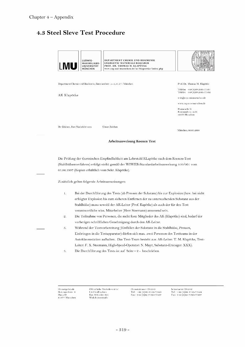
Chapter 4 – Appendix
- 319 -
4.3 Steel Sleve Test Procedure
Chapter 4 – Appendix
- 320 -
Chapter 4 – Appendix
- 321 -
Chapter 4 – Appendix
- 322 -
4.4 Summary of Standard Operation Procedures a)
1. Könen-Test (Steel Sleeve)
a. Vorschrift: „Prüfung der thermischen Empfindlichkeit
(Stahlhülsenverfahren)“; Standardarbeitsanweisung 410/001 des
Wehrwissenschaftlichen Instituts für Werk-, Explosiv- und
Betriebsstoffe (WIWEB) i.d.F.v. 28. Mai 1997 ;geläufiger: EG A.141
b. Ziel: Test der Explosionsfähigkeit einer Verbindung i.S.d.
Sprengstoffgesetztes (SprengG)
c. Probenvorbereitung: Die nichtwiederverwendbare Stahlhülse wird
im Falle von Feststoffen unter Anpressen mit einer Kraft von 80 N
bis zu einer Füllhöhe von 15 mm unterhalb des Hülsenrandes
befüllt. Im Falle einer zu testenden Flüssigkeit – bzw. eines Gels –
wird die Testsubstanz in die Hülse eingegossen, bis eine Lunker-
freie Füllhöhe von 60 mm erreicht ist.
Substanzen mit einer Reibeempfindlichkeit von F < 80 N sollen
nicht angepresst werden!
Die Hülse wird mit der (u.U.) wiederverwendbaren Verschraubung
verschlossen ohne Substanz zwischen Bund und Platte oder im
Gewinde einzuschließen.
d. Durchführung: Die Stahlhülse wird von vier Bunsenbrennern
gleichmäßig erhitzt, die Heizgeschwindigkeit liegt im
Temperaturbereich von 135 bis 285 °C zwischen 185 K/min und
215 K/min. Es wird erhitzt, bis Explosion bzw. Zersetzung
1 EG A.14: Prüfverfahren nach Anhang I Teil A.14 der Richtlinie 92/69/EWG der Kommission vom 31. Juli 1992 zur Siebzehnten Anpassung der Richtlinie 67/548/EWG zur Angleichung der Rechts- und Verwaltungsvorschriften für die Einstufung, Verpackung und Kennzeichnung gefährlicher Stoffe an den technischen Fortschritt (ABl. EG Nr. L 383 S. 113 und Nr. L 383 A S. 1 (S. 87)) Und: Recommendations on the Transport of Dangerous Goods: Tests and criteria, 1990, United Nations, New York. a) The author of this thesis is indebted to and thanks Dr. F.X. Steemann for providing the summary of standard operation procedures mentionend in section 4.4.
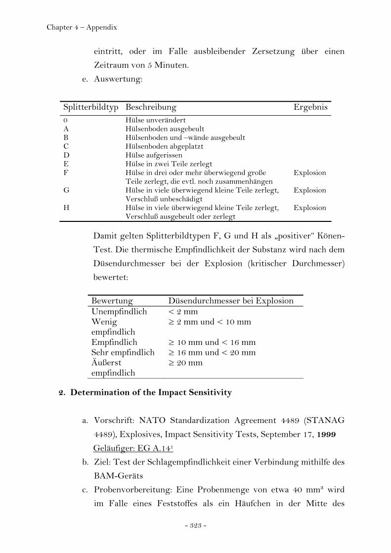
Chapter 4 – Appendix
- 323 -
eintritt, oder im Falle ausbleibender Zersetzung über einen
Zeitraum von 5 Minuten.
e. Auswertung:
Splitterbildtyp Beschreibung Ergebnis 0 Hülse unverändert A Hülsenboden ausgebeult B Hülsenboden und –wände ausgebeult C Hülsenboden abgeplatzt D Hülse aufgerissen E Hülse in zwei Teile zerlegt F Hülse in drei oder mehr überwiegend große
Teile zerlegt, die evtl. noch zusammenhängen Explosion
G Hülse in viele überwiegend kleine Teile zerlegt, Verschluß unbeschädigt
Explosion
H Hülse in viele überwiegend kleine Teile zerlegt, Verschluß ausgebeult oder zerlegt
Explosion
Damit gelten Splitterbildtypen F, G und H als „positiver“ Könen-
Test. Die thermische Empfindlichkeit der Substanz wird nach dem
Düsendurchmesser bei der Explosion (kritischer Durchmesser)
bewertet: Bewertung Düsendurchmesser bei Explosion Unempfindlich < 2 mm Wenig empfindlich
≥ 2 mm und < 10 mm
Empfindlich ≥ 10 mm und < 16 mm Sehr empfindlich ≥ 16 mm und < 20 mm Äußerst empfindlich
≥ 20 mm
2. Determination of the Impact Sensitivity
a. Vorschrift: NATO Standardization Agreement 4489 (STANAG
4489), Explosives, Impact Sensitivity Tests, September 17, 1999
Geläufiger: EG A.141
b. Ziel: Test der Schlagempfindlichkeit einer Verbindung mithilfe des
BAM-Geräts
c. Probenvorbereitung: Eine Probenmenge von etwa 40 mm³ wird
im Falle eines Feststoffes als ein Häufchen in der Mitte des
Chapter 4 – Appendix
- 324 -
Metallstempels aufgetragen und der obere Stempel leicht
angedrückt, bis er den Haufen berührt. Bei insb. leichtflüssigen
Flüssigkeiten kann u.U. das Problem auftreten, dass bei dem
Aufprall des Fallgewichts die Probensubstanz zwischen Stempel
und Fassung gepresst wird. Für diese Fälle enthält die hier zitierte
STANAG-Vorschrift kein besonderes Vorgehen, nach EG A.141
ist ein Abstand von 1 mm zwischen den Stempeln zu belassen.
Wird z.B. Schleifpapier als zusätzliche Unterlage zwischen
Stempel und Probe eingefügt, sollte dies gesondert angegeben
werden und auf mögliche Auswirkungen auf das Testergebnis
hingewiesen werden.
d. Durchführung: Die Fallhöhe des Gewichtes (1kg, 5 kg, 10 kg)
wird in 10 cm Inkrementen verringert, pro Versuch ist eine neue
Probe einzusetzen. Messungen unterhalb einer Fallhöhe von 10
cm sind nach STANAG nicht vorgesehen.
Nach EG A.141 werden pro Fallhöhe sechs Versuche durchgeführt:
Es werden sechs Einzelversuche unter Verwendung des
Fallgewichts von 10 kg und Anwendung einer Fallhöhe von 0,40
m (40 J) ausgeführt. Wenn es während der sechs Versuche bei 40 J
zu einer Explosion kommt, sind weitere sechs Einzelversuche mit
einem Fallgewicht von 5 kg und einer Fallhöhe von 0,15 m (7,5 J)
auszuführen. In geeigneten Inkrementen wird die Fallhöhe der
Einzelversuche verringert, bis sechs Versuche negativ verlaufen.
Dieser Wert gilt als Schlagempfindlichkeit nach EG A.141. Nach
STANAG ist der 50%-Wert ausgehend von dem wie eben
beschrieben ermittelten Wert zu bestimmen, indem mit jeweils 30
Einzelversuchen die Fallhöhe gefunden wird, bei welcher 50% der
Versuche positiv ausfallen (Bruceton-Methode, „aufwärts“ und
„abwärts“ gemessen). Zeigen dabei zehn Einzelversuche konsistent
eine höhere oder niedrigere Fallhöhe an, ist ein neuer Startwert zu
messen.
e. Auswertung: Als positiver Verlauf wird eine Zersetzung in Gestalt
von Explosion, Entflammung oder Knall gedeutet.
Chapter 4 – Appendix
- 325 -
Unterhalb einer Fallenergie von 40 J gilt eine Substanz als
schlagempfindlich gem. Richtlinie EG A.141; ist keiner von sechs
Versuchen positiv, ist der Stoff bei der aktuellen Fallenergie nicht
empfindlich nach EG A.141.
3. Determination of the Friction Sensitivity
a. Vorschrift: NATO Standardization Agreement 4487 (STANAG
4487), Explosives, Friction Sensitivity Tests, August 22, 2002
Geläufiger: EG A.141
b. Ziel: Test der Reibempfindlichkeit einer Verbindung mithilfe des
BAM-Geräts
c. Probenvorbereitung: Porzellanplatte wird mit Riffelung quer zur
Bewegungsrichtung des Porzellanstempels eingespannt. Eine
Probenmenge von etwa 10 mm³ wird vor und unter den
Porzellanstempel auf die Porzellanplatte aufgebracht, so dass der
Stempel über die Probe gezogen wird.
d. Durchführung: Jeder Einzelversuch hat zwischen unbenutzten
Oberflächen zu erfolgen, d.h. neben einer bestehenden Reibspur
oder auf einer neuen Platte und mit einer unbenutzten Seite des
Stempels. Es werden sechs Einzelversuche unter Verwendung
einer Belastung von 360 N ausgeführt. Wenn es während der
sechs Versuche zu einer positiven Reaktion kommt, sind weitere
sechs Einzelversuche mit einer Belastung von 120 N auszuführen.
Der höchste Belastungswert, bei welchem in sechs Versuchen kein
positiver Testverlauf zu beobachten ist, gilt als
Reibeempfindlichkeit.
Nach STANAG wird der 50%-Wert ermittelt, indem ausgehend
von dem Ergebnis der „1 aus 6“-Methode statistisch nach
Bruceton in 25 bis 30 Versuchen gemessen wird, bis 50% der
Versuche positiv verlaufen.
e. Auswertung: Als positiver Verlauf wird eine Zersetzung in Gestalt
von Explosion, Entflammung oder Knall (nach STANAG auch
Knistern) gedeutet.
Chapter 4 – Appendix
- 326 -
Unterhalb einer Reibekraft von 120 N gilt eine Substanz als
reibeempfindlich gem. Richtlinie EG A.141.
4. Determination of the Electrostatic Discharge Sensitivity
a. Vorschrift: NATO Standardization Agreement 4239 (STANAG
4239), Electrostatic Discharge, Munitions Test Procedures,
October 13, 1997
b. Ziel: Test der Empfindlichkeit einer Verbindung gegenüber
elektrostatischer Entladung mithilfe des ESD-Geräts2
c. Probenvorbereitung: Gem. Benutzerhandbuch zum ESD-Gerät.
d. Durchführung: Der Kondensator wird bis auf den Stellwert
aufgeladen und dann über die Probe entladen. Es werden
mindestens 20 Einzelversuche für Personen-verursachte
Entladung (25 kV, 15 kV, 10 kV, 5 kV mit jeweils 5000 Ω und 500
Ω bei jeweils 500 pF, Abweichungen von ±5% zulässig) und
mindestens 10 Einzelversuche für Helikopter-verursachte
Entladung (250 kV, 200 kV, 150 kV, 100 kV, 50 kV, 25 kV mit
jeweils Gesamtstromkreiswiderstand bei Entladung mit ±5%
Genauigkeit und 1000 pF ±10% gemessen) durchgeführt. Dabei
genügt es, die Entladung bei jedem Einzelversuch an einer
anderen Stelle der Probe herbeizuführen (Probe muss nicht immer
wieder ausgetauscht werden). Die Auf- und Entladungszeit soll im
Falle der Personen-verursachten Entladung anhand der 10%- und
90%-Werte ermittelt und notiert werden, ebenso die
Testergebnisse.
e. Auswertung: STANAG 4239 gibt keine genaue Auskunft darüber,
ob ein 50%-Wert zu ermitteln ist, oder ob ein positiver
Testverlauf Empfindlichkeit gegenüber der jew. Entladungsstärke
bedeutet. Letzteres erscheint jedoch sinnvoll.
2 Die STANAG 4239 bezieht sich hauptsächlich auf ein Prüfverfahren für Munitionssorten mit elektro-explosiven Bauteilen (EED) und ähnlichen elektrischen/elektronischen Subsystemen.
Chapter 4 – Appendix
- 327 -
ANHANG:
EG A.14:
Prüfverfahren nach Anhang I Teil A.14 der Richtlinie 92/69/EWG der
Kommission vom 31. Juli 1992 zur Siebzehnten Anpassung der Richtlinie
67/548/EWG zur Angleichung der Rechts- und Verwaltungsvorschriften
für die Einstufung, Verpackung und Kennzeichnung gefährlicher Stoffe an
den technischen Fortschritt (ABl. EG Nr. L 383 S. 113 und Nr. L 383 A S. 1
(S. 87))
A.14.1. Methode
A.14.1.1. Einleitung
Die Methode stellt ein Prüfschema dar zur Feststellung, ob feste oder
pastenförmige Stoffe bei Flammenzündung (thermische Empfindlichkeit)
oder bei Einwirkung von Schlag oder Reibung (mechanische
Empfindlichkeit) und ob Flüssigkeiten bei Flammenzündung oder bei
Einwirkung von Schlag eine Explosionsgefahr darstellen.
Die Methode besteht aus drei Teilen:
a. Prüfung der thermischen Empfindlichkeit (1);
b. Prüfung der mechanischen Empfindlichkeit bei Schlagbeanspruchung
(1);
c. Prüfung der mechanischen Empfindlichkeit bei Reibbeanspruchung (1).
Die Methode liefert Ergebnisse, mit denen die Möglichkeit der Auslösung
einer Explosion bei Einwirkung bestimmter, nicht außergewöhnlicher
Beanspruchungen festgestellt werden kann. Sie dient nicht zur Feststellung,
ob ein Stoff unter beliebigen Bedingungen explosionsfähig ist.
Die Methode eignet sich zur Feststellung, ob ein Stoff unter den
besonderen, in der Richtlinie festgelegten Bedingungen eine
Explosionsgefahr darstellt (thermische und mechanische Empfindlichkeit).
Sie beruht auf der Verwendung mehrerer Arten von Apparaturen, die
international weit verbreitet sind (1) und die im allgemeinen
aussagekräftige Ergebnisse ergeben. Dabei wird eingeräumt, daß die
Chapter 4 – Appendix
- 328 -
Methode keine endgültige Lösung darstellt. Es können andere als die
genannten Apparaturen verwendet werden, wenn diese international
anerkannt sind und die Ergebnisse in angemessener Form mit denen aus
den genannten Apparaturen korreliert werden können.
Die Prüfungen brauchen nicht vorgenommen zu werden, wenn verfügbare
thermodynamische Daten (z.B. Bildungs-, Zersetzungsenthalpie) und/oder
das Fehlen bestimmter reaktiver Gruppen (2) in der Strukturformel
zweifelsfrei erkennen lassen, daß sich der Stoff nicht unter Bildung von
Gasen oder Freisetzung von Wärme schnell zersetzen kann (d.h. die
Substanz keine Explosionsgefahr darstellt). Eine Prüfung der mechanischen
Empfindlichkeit bei Reibbeanspruchung ist für Flüssigkeiten nicht
erforderlich.
A.14.1.2. Definitionen und Einheiten
Explosionsgefährlich:
Stoffe, die durch Flammenzündung zur Explosion gebracht werden können
oder die gegen Schlag oder Reibung in den genannten Apparaturen
empfindlich sind (oder die in alternativen Apparaturen eine höhere
mechanische Empfindlichkeit zeigen als 1,3-Dinitrobenzol).
A.14.1.3. Referenzsubstanzen
1,3-Dinitrobenzol, kristallin, gesiebt auf Korngröße 0,5 mm, technisches
Produkt für die Prüfung der Schlag- und Reibempfindlichkeit.
Perhydro-1,3,5-trinitro-1,3,5-triazin (RDX, Hexogen, Cyclonit - CAS 121-
82-4), umkristallisiert aus wäßrigem Cyclohexanon, naßgesiebt durch ein
Sieb 250 im und als Rückstand auf einem Sieb 150 μm gewonnen,
anschließend bei 103 ± 2 °C (über 4 Stunden) getrocknet für die zweite
Reihe der Prüfung auf Schlag- und Reibempfindlichkeit.

Chapter 4 – Appendix
- 329 -
A.14.1.4. Prinzip der Methode
Um sichere Bedingungen für die Ausführung der drei
Empfindlichkeitsprüfungen zu finden, ist die Durchführung von
Vorversuchen erforderlich.
A.14.1.4.1. Prüfung auf die Sicherheit des Umgangs mit der Substanz
Aus sicherheitstechnischen Gründen werden vor Durchführung der
Hauptprüfungen sehr kleine Proben (etwa 10 mg) der Prüfsubstanz ohne
Einschluß mit einer Gasbrennerflamme erhitzt, in einem geeigneten Gerät
einem Schlag ausgesetzt und unter Verwendung eines Reibstiftes und eines
Widerlagers oder in einer beliebigen Reibmaschine gerieben. Das Ziel
dieser Vorversuche ist festzustellen, ob der Stoff so empfindlich und so
explosiv ist, daß zur Vermeidung von Verletzungen des Prüfenden bei der
Durchführung der vorgeschriebenen Empfindlichkeitsprüfungen,
insbesondere der Prüfung der thermischen Empfindlichkeit, besondere
Schutzmaßnahmen vorzusehen sind.
A.14.1.4.2. Thermische Empfindlichkeit
Für die Prüfung wird die Prüfsubstanz in einer Stahlhülse erhitzt, die durch
Düsenplatten mit Öffnungen verschiedenen Durchmessers verschlossen ist.
Auf diese Weise wird bestimmt, ob der Stoff unter intensiver thermischer
Beanspruchung bei definiertem Einschluß explodieren kann.
A.14.1.4.3. Mechanische Empfindlichkeit (Schlag)
Die Prüfung besteht darin, die Prüfsubstanz dem Schlag eines festgelegten
Fallgewichtes aus einer festgelegten Höhe auszusetzen.

Chapter 4 – Appendix
- 330 -
A.14.1.4.4. Mechanische Empfindlichkeit (Reibung)
Bei dieser Prüfung werden feste oder pastenförmige Substanzen der
Reibung zwischen standardisierten Oberflächen unter festgelegten
Bedingungen der Belastung und der relativen Bewegung ausgesetzt.
A.14.1.5. Qualitätskriterien
Nicht festgelegt.
A.14.1.6. Beschreibung der Methode
A.14.1.6.1. Thermische Empfindlichkeit (Flammenzündung)
A.14.1.6.1.1. Apparatur
Die Apparatur besteht aus einer nicht wiederverwendbaren Stahlhülse mit
deren wiederverwendbarer Verschraubung (Abbildung 1), die in eine Heiz-
und Schutzvorrichtung eingesetzt wird. Jede Hülse wird aus Blech im
Tiefziehverfahren hergestellt (siehe Anlage) und hat einen inneren
Durchmesser von 24 mm, eine Länge von 75 mm und eine Wanddicke von
0,5 mm. Am offenen Ende sind die Hülsen mit einem Bund versehen, an
dem sie mit der Düsenplatte verschlossen werden können. Der Verschluß
besteht aus einer druckfesten Düsenplatte mit einer zentrischen Bohrung,
die mit der aus Gewindering und Mutter bestehenden Verschraubung fest
mit einer Hülse verbunden wird. Gewindering und Mutter bestehen aus
Chrom-Mangan-Stahl (siehe Anlage), der bis 800 °C zunderfest ist. Die
Düsenplatten sind 6 mm dick, bestehen aus warmfestem Stahl (siehe
Anlage) und stehen mit verschiedenen Öffnungsdurchmessern zur
Verfügung.
A.14.1.6.1.2. Versuchsbedingungen
Normalerweise wird die Substanz im Auslieferungszustand geprüft, obwohl
in einigen Fällen, z.B. bei gepreßten, gegossenen oder anderweitig
Chapter 4 – Appendix
- 331 -
verdichteten Stoffen, vor der Prüfung ein Zerkleinern erforderlich werden
kann.
Bei Feststoffen wird die Menge des pro Prüfung zu verwendenden
Materials durch ein zweistufiges Probeverfahren für die Befüllung
bestimmt. Dabei wird eine gewogene Hülse mit 9 cm3 Prüfsubstanz gefüllt
und die Prüfsubstanz unter Anwendung einer Kraft von 80 N, bezogen auf
den Gesamtquerschnitt der Hülse, angedrückt. Aus sicherheitstechnischen
Gründen oder in solchen Fällen, wo der Aggregatzustand der Probe durch
Druck verändert werden kann, können andere Füllverfahren angewendet
werden; wenn z.B. die Substanz sehr reibempfindlich ist, empfiehlt sich das
Andrücken nicht. Wenn der Stoff sich als kompressibel erweist, wird
weitere Substanz hinzugefügt und angedrückt, bis die Hülse bis zu einer
Höhe von 55 mm vom Rand gefüllt ist. Danach wird die Gesamtmenge
bestimmt, die für die Füllung bis zum Niveau von 55 mm unter dem Rand
benötigt wurde, und es werden zwei weitere gleichgroße Portionen
zugegeben, wobei auch diese unter Anwendung einer Kraft von je 80 N
angedrückt werden. Schließlich wird Substanz entweder zugefügt (unter
Andrücken) oder ggf. entnommen, bis die Hülse bis zu einer Höhe von 15
mm unter dem Rand gefüllt ist. Dann wird eine zweite Probebefüllung
durchgeführt, die mit einer angedrückten Menge von einem Drittel der
Gesamtmenge der ersten Probebefüllung beginnt. Danach werden zwei
weitere solche Portionen unter Anwendung von 80 N hinzugefügt und die
Höhe der Substanz in der Hülse durch Hinzufügen oder Entnehmen bis auf
15 mm under dem Rand gebracht. Die bei der zweiten Probebefüllung
ermittelte Feststoffmenge wird für jeden der eigentlichen Versuche
verwendet, wobei das Füllen mit drei gleichgroßen Mengen vorgenommen
wird, deren jede durch Anwendung der erforderlichen Kraft auf 9 cm3
komprimiert wird. (Dies kann durch Verwendung von Abstandsringen
erleichtert werden.)
Flüssigkeiten und gelatinöse Substanzen werden in die Hülse bis zu einer
Höhe von 60 mm eingefüllt, wobei im letzteren Fall besondere Sorge dafür
zu tragen ist, daß keine Lunker gebildet werden. Der Gewindering wird von
unten auf die Hülse aufgeschoben, die geeignete Düsenplatte eingesetzt und
die Mutter nach Aufbringen eines Schmiermittels auf Molybdändisulfid-
Chapter 4 – Appendix
- 332 -
Basis angezogen. Es muß darauf geachtet werden, daß keine Substanz
zwischen dem Bund und der Platte oder im Gewinde eingeschlossen ist.
Zum Aufheizen wird Propangas verwendet, das aus einer handelsüblichen
Stahlflasche mit Druckminderer (60 bis 70 mbar) entnommen und über
einen Durchflußmesser und einen Verteiler gleichmäßig vier Brennern
zugeführt wird (was durch Beobachtung der Flammen der einzelnen
Brenner festgestellt werden kann). Die Brenner sind entsprechend
Abbildung 1 an dem Schutzkasten angeordnet. Die vier Brenner haben
zusammen einen Verbrauch von etwa 3,2 l Propan pro Minute. Die
Verwendung alternativer Heizgase und Brenner ist möglich, doch muß die
Heizgeschwindigkeit der in Abbildung 3 genannten entsprechen. Für alle
Apparaturen ist die Heizgeschwindigkeit regelmäßig unter Verwendung
von Hülsen mit Dibutylphthalatfüllung zu kontrollieren (vgl. Abbildung 3).
A.14.1.6.1.3. Versuchsausführung
Jeder Versuch wird fortgeführt, bis die Stahlhülse entweder zerlegt oder
fünf Minuten erhitzt worden ist. Ein Versuch, der zu einer Zerlegung der
Hülse in drei oder mehr Teile führt (diese können in einigen Fällen noch
durch schmale Metallstreifen miteinander verbunden sein - vgl. Abbildung
2), wird als Explosion eingestuft. Ein Versuch mit weniger Teilen oder
überhaupt keiner Zerlegung wird nicht als Explosion eingestuft.
Zunächst wird eine erste Reihe mit drei Versuchen unter Verwendung einer
Düsenplatte mit einem Öffnungsdurchmesser von 6,0 mm durchgeführt;
wenn es hier zu keiner Explosion kommt, folgt eine zweite Reihe, ebenfalls
mit drei Versuchen, mit einer Düsenplatte von 2,0 mm
Öffnungsdurchmesser. Tritt während einer dieser Versuchsreihen eine
Explosion ein, kann auf die Durchführung weiterer Versuche verzichtet
werden.
A.14.1.6.1.4. Auswertung
Das Versuchsergebnis wird als positiv eingestuft, wenn es in einer der
genannten Versuchsreihen zu einer Explosion kommt.
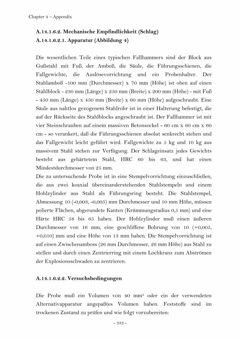
Chapter 4 – Appendix
- 333 -
A.14.1.6.2. Mechanische Empfindlichkeit (Schlag)
A.14.1.6.2.1. Apparatur (Abbildung 4)
Die wesentlichen Teile eines typischen Fallhammers sind der Block aus
Gußstahl mit Fuß, der Amboß, die Säule, die Führungsschienen, die
Fallgewichte, die Auslösevorrichtung und ein Probenhalter. Der
Stahlamboß -100 mm (Durchmesser) x 70 mm (Höhe) ist oben auf einen
Stahlblock - 230 mm (Länge) x 250 mm (Breite) x 200 mm (Höhe) - mit Fuß
- 450 mm (Länge) x 450 mm (Breite) x 60 mm (Höhe) aufgeschraubt. Eine
Säule aus nahtlos gezogenem Stahlrohr ist in einer Halterung befestigt, die
auf der Rückseite des Stahlblocks angeschraubt ist. Der Fallhammer ist mit
vier Steinschrauben auf einem massiven Betonsockel - 60 cm x 60 cm x 60
cm - so verankert, daß die Führungsschienen absolut senkrecht stehen und
das Fallgewicht leicht geführt wird. Fallgewichte zu 5 kg und 10 kg aus
massivem Stahl stehen zur Verfügung. Der Schlageinsatz jedes Gewichts
besteht aus gehärtetem Stahl, HRC 60 bis 63, und hat einen
Mindestdurchmesser von 25 mm.
Die zu untersuchende Probe ist in eine Stempelvorrichtung einzuschließen,
die aus zwei koaxial übereinanderstehenden Stahlstempeln und einem
Hohlzylinder aus Stahl als Führungsring besteht. Die Stahlstempel,
Abmessung 10 (-0,003, -0,005) mm Durchmesser und 10 mm Höhe, müssen
polierte Flächen, abgerundete Kanten (Krümmungsradius 0,5 mm) und eine
Härte HRC 58 bis 65 haben. Der Hohlzylinder muß einen äußeren
Durchmesser von 16 mm, eine geschliffene Bohrung von 10 (+0,005,
+0,010) mm und eine Höhe von 13 mm haben. Die Stempelvorrichtung ist
auf einen Zwischenamboss (26 mm Durchmesser, 26 mm Höhe) aus Stahl zu
stellen und durch einen Zentrierring mit einem Lochkranz zum Abströmen
der Explosionsschwaden zu zentrieren.
A.14.1.6.2.2. Versuchsbedingungen
Die Probe muß ein Volumen von 40 mm3 oder ein der verwendeten
Alternativapparatur angepaßtes Volumen haben. Feststoffe sind im
trockenen Zustand zu prüfen und wie folgt vorzubereiten:
Chapter 4 – Appendix
- 334 -
a. Pulverförmige Substanzen sind zu sieben (Maschenweite 0,5 mm); der
gesamte Siebdurchgang ist zur Prüfung zu verwenden;
b. Gepreßte, gegossene oder anderweitig verdichtete Substanzen sind zu
zerkleinern und zu sieben; zur Prüfung ist die Siebfraktion 0,5 bis 1 mm
Durchmesser zu verwenden; sie muß für die Originalsubstanz
repräsentativ sein.
Substanzen, die in der Regel pastenförmig geliefert werden, sollten, wenn
möglich, im trockenen Zustand geprüft werden, auf jeden Fall aber nach
Entfernen der größtmöglichen Menge an Verdünnungsmittel. Bei der
Prüfung flüssiger Substanzen ist zwischen dem oberen und dem unteren
Stahlstempel ein Abstand von 1 mm zu halten.
A.14.1.6.2.3. Versuchsausführung
Es werden sechs Einzelversuche unter Verwendung des Fallgewichts von
10 kg und Anwendung einer Fallhöhe von 0,40 m (40 J) ausgeführt. Wenn
es während der sechs Versuche bei 40 J zu einer Explosion kommt, sind
weitere sechs Einzelversuche mit einem Fallgewicht von 5 kg und einer
Fallhöhe von 0,15 m (7,5 J) auszuführen. Bei Verwendung einer anderen
Apparatur wird die Probe mit der gewählten Referenzsubstanz unter
Benutzung einer anerkannten Auswertungsmethode (z.B. up-and-down
technique usw.) verglichen.
A.14.1.6.2.4. Auswertung
Das Prüfergebnis wird als positiv eingestuft, wenn es mit der beschriebenen
Apparatur zumindest in einem der genannten Versuche zu einer Explosion
(eine Entflammung und/oder ein Knall steht einer Explosion gleich) kommt
oder wenn bei Verwendung einer alternativen Apparatur die Probe
empfindlicher ist als 1,3-Dinitrobenzol oder Hexogen (RDX).
A.14.1.6.3. Mechanische Empfindlichkeit (Reibung)
A.14.1.6.3.1. Apparatur (Abbildung 5)
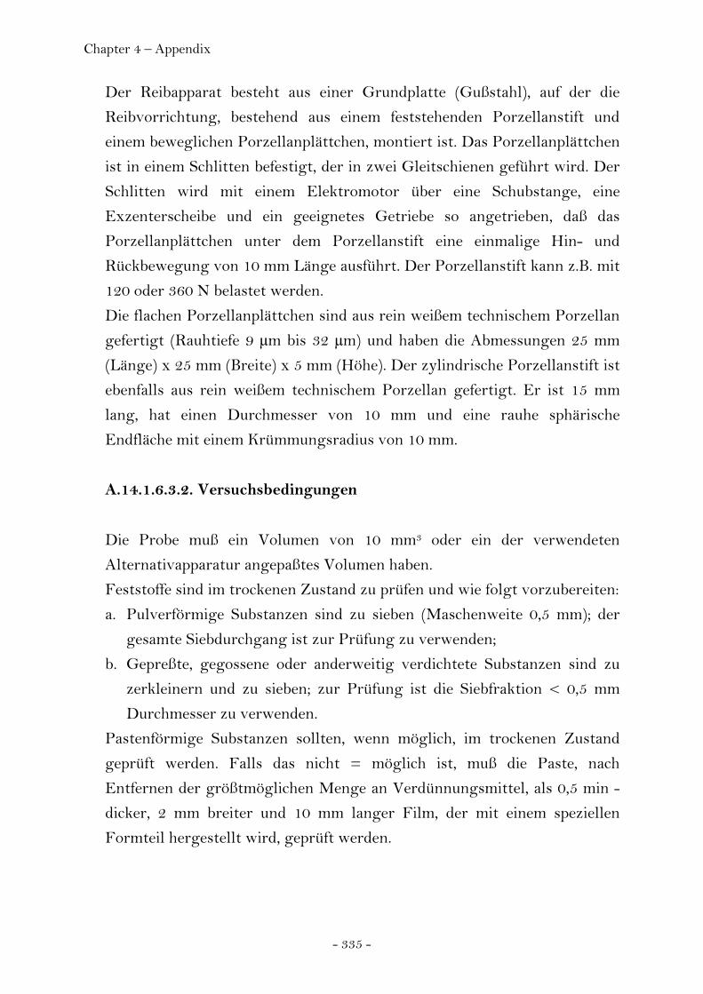
Chapter 4 – Appendix
- 335 -
Der Reibapparat besteht aus einer Grundplatte (Gußstahl), auf der die
Reibvorrichtung, bestehend aus einem feststehenden Porzellanstift und
einem beweglichen Porzellanplättchen, montiert ist. Das Porzellanplättchen
ist in einem Schlitten befestigt, der in zwei Gleitschienen geführt wird. Der
Schlitten wird mit einem Elektromotor über eine Schubstange, eine
Exzenterscheibe und ein geeignetes Getriebe so angetrieben, daß das
Porzellanplättchen unter dem Porzellanstift eine einmalige Hin- und
Rückbewegung von 10 mm Länge ausführt. Der Porzellanstift kann z.B. mit
120 oder 360 N belastet werden.
Die flachen Porzellanplättchen sind aus rein weißem technischem Porzellan
gefertigt (Rauhtiefe 9 μm bis 32 μm) und haben die Abmessungen 25 mm
(Länge) x 25 mm (Breite) x 5 mm (Höhe). Der zylindrische Porzellanstift ist
ebenfalls aus rein weißem technischem Porzellan gefertigt. Er ist 15 mm
lang, hat einen Durchmesser von 10 mm und eine rauhe sphärische
Endfläche mit einem Krümmungsradius von 10 mm.
A.14.1.6.3.2. Versuchsbedingungen
Die Probe muß ein Volumen von 10 mm3 oder ein der verwendeten
Alternativapparatur angepaßtes Volumen haben.
Feststoffe sind im trockenen Zustand zu prüfen und wie folgt vorzubereiten:
a. Pulverförmige Substanzen sind zu sieben (Maschenweite 0,5 mm); der
gesamte Siebdurchgang ist zur Prüfung zu verwenden;
b. Gepreßte, gegossene oder anderweitig verdichtete Substanzen sind zu
zerkleinern und zu sieben; zur Prüfung ist die Siebfraktion < 0,5 mm
Durchmesser zu verwenden.
Pastenförmige Substanzen sollten, wenn möglich, im trockenen Zustand
geprüft werden. Falls das nicht = möglich ist, muß die Paste, nach
Entfernen der größtmöglichen Menge an Verdünnungsmittel, als 0,5 min -
dicker, 2 mm breiter und 10 mm langer Film, der mit einem speziellen
Formteil hergestellt wird, geprüft werden.
Chapter 4 – Appendix
- 336 -
A.14.1.6.3.3. Versuchsausführung
Der Porzellanstift wird auf die zu untersuchende Probe gesetzt und
belastet. Bei Durchführung des Versuchs muß der Schwammstrich des
Porzellanplättchens quer zu dessen Bewegungsrichtung liegen. Es ist
darauf zu achten, daß der Stift auf der Probe steht und daß soviel
Prüfsubstanz vor dem Stift liegt, daß bei der Plättchenbewegung genügend
Prüfsubstanz unter den Stift gelangt. Pastenförmige Substanzen werden
mittels einer Lehre (Dicke: 0,5 mm) mit einer Öffnung von 2 mm x 10 mm
auf das Plättchen aufgetragen. Das Porzellanplättchen wird unter dem
Porzellanstift in einer Zeit von 0,44 s je 10 mm hin- und herbewegt. jeder
Oberflächenbezirk des Plättchens und des Stiftes darf nur einmal verwendet
werden; die beiden Enden eines jeden Stiftes können für zwei Versuche, und
die beiden Oberflächen eines jeden Plättchens können für je drei Versuche
benutzt werden.
Es werden sechs Einzelversuche unter Verwendung einer Belastung von
360 N ausgeführt. Wenn es während der sechs Versuche zu einer positiven
Reaktion kommt, sind weitere sechs Einzelversuche mit einer Belastung
von 120 N auszuführen. Bei Verwendung einer anderen Apparatur wird die
Probe mit der gewählten Referenzsubstanz unter Benutzung einer
anerkannten Auswertungsmethode (z.B. up-and-down technique usw.)
verglichen.
A.14.1.6.3.4. Auswertung
Das Prüfergebnis wird als positiv eingestuft, wenn es mit dem
beschriebenen Reibapparat zumindest in einem der genannten Versuche zu
einer Explosion (ein Knistern und/oder ein Knall oder eine Entflammung
stehen einer Explosion gleich) kommt oder wenn bei Verwendung einer
alternativen Reibprüfung die äquivalenten Kriterien erfüllt werden.
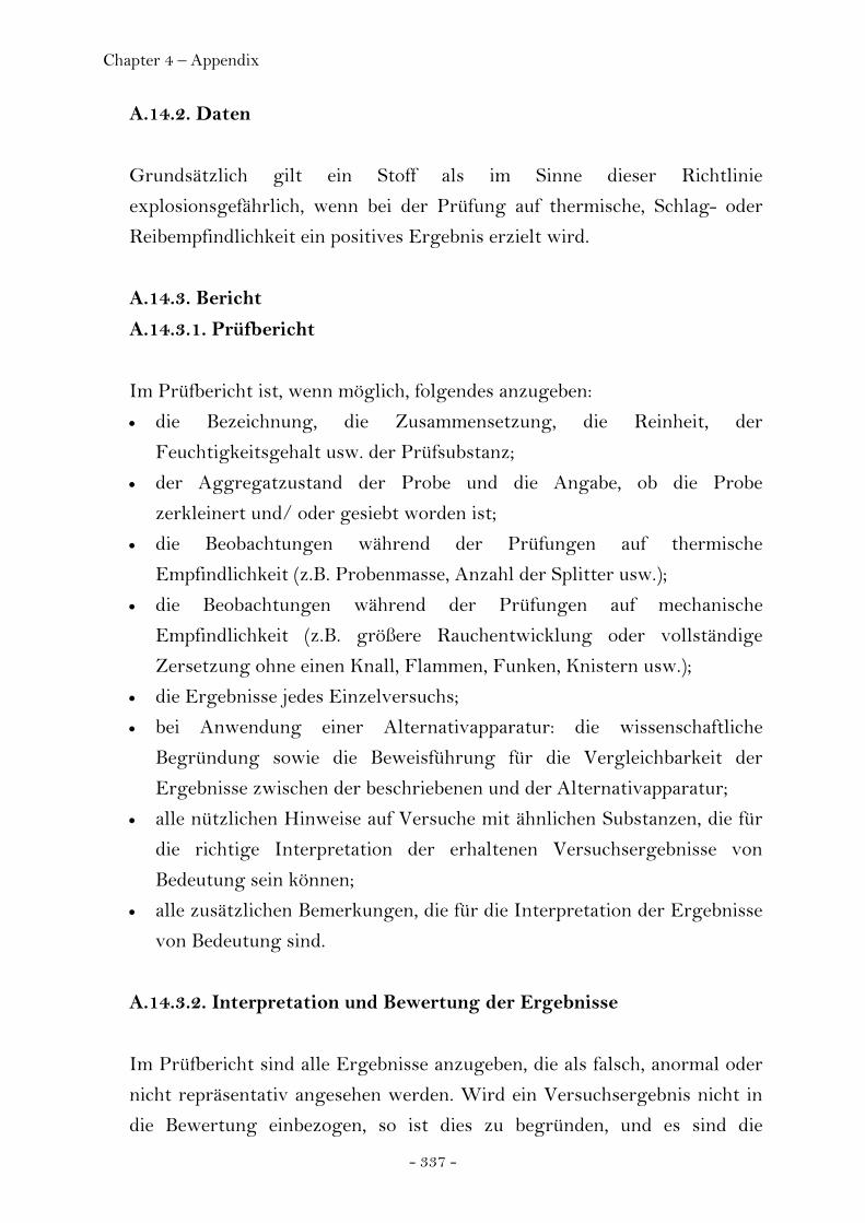
Chapter 4 – Appendix
- 337 -
A.14.2. Daten
Grundsätzlich gilt ein Stoff als im Sinne dieser Richtlinie
explosionsgefährlich, wenn bei der Prüfung auf thermische, Schlag- oder
Reibempfindlichkeit ein positives Ergebnis erzielt wird.
A.14.3. Bericht
A.14.3.1. Prüfbericht
Im Prüfbericht ist, wenn möglich, folgendes anzugeben:
• die Bezeichnung, die Zusammensetzung, die Reinheit, der
Feuchtigkeitsgehalt usw. der Prüfsubstanz;
• der Aggregatzustand der Probe und die Angabe, ob die Probe
zerkleinert und/ oder gesiebt worden ist;
• die Beobachtungen während der Prüfungen auf thermische
Empfindlichkeit (z.B. Probenmasse, Anzahl der Splitter usw.);
• die Beobachtungen während der Prüfungen auf mechanische
Empfindlichkeit (z.B. größere Rauchentwicklung oder vollständige
Zersetzung ohne einen Knall, Flammen, Funken, Knistern usw.);
• die Ergebnisse jedes Einzelversuchs;
• bei Anwendung einer Alternativapparatur: die wissenschaftliche
Begründung sowie die Beweisführung für die Vergleichbarkeit der
Ergebnisse zwischen der beschriebenen und der Alternativapparatur;
• alle nützlichen Hinweise auf Versuche mit ähnlichen Substanzen, die für
die richtige Interpretation der erhaltenen Versuchsergebnisse von
Bedeutung sein können;
• alle zusätzlichen Bemerkungen, die für die Interpretation der Ergebnisse
von Bedeutung sind.
A.14.3.2. Interpretation und Bewertung der Ergebnisse
Im Prüfbericht sind alle Ergebnisse anzugeben, die als falsch, anormal oder
nicht repräsentativ angesehen werden. Wird ein Versuchsergebnis nicht in
die Bewertung einbezogen, so ist dies zu begründen, und es sind die

Chapter 4 – Appendix
- 338 -
Ergebnisse anderer oder zusätzlicher Versuche aufzuführen. Kann die
Abnormität eines Ergebnisses nicht erklärt werden, muß das Ergebnis als
solches akzeptiert und der Stoff entsprechend eingestuft werden.
A.14.4. Literatur
(1) Recommendations on the Transport of Dangerous Goods: Tests and
criteria, 1990, United Nations, New York.
(2) Bretherick, L., Handbook of Reactive Chemical Hazards, 4. Auflage,
Butterworths, London, ISBN 0-750-60103-5, 1990.
(3) Koenen, H., Ide, K.H. und Swart, K.H., Explosivstoffe, 1961, Bd. 3, 6-13
und 30-42.
(4) NF T 20-033 (Sept. 35). Chemical products for industrial use -
Determination of explosion risk.
Anlage zu RL 67/548/EWG Anhang V A.14
Beispiel für Werkstoffspezifikation zur Prüfung auf thermische
Empfindlichkeit (vgl. DIN 1623)
(1) Hülse: Werkstoffspezifikation Nr. 1.0336.505 g
(2) Düsenplatte: Werkstoffspezifikation Nr. 1.4373
(3) Gewindering und Mutter: Werkstoffspezifikation Nr. 1.3817

Chapter 4 – Appendix
- 339 -
Abbildung 1: Apparatur für die Prüfung auf thermische Empfindlichkeit
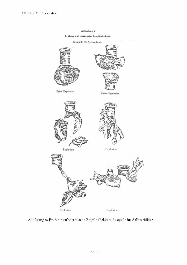
Chapter 4 – Appendix
- 340 -
Abbildung 2: Prüfung auf thermische Empfindlichkeit; Beispiele für Splitterbilder
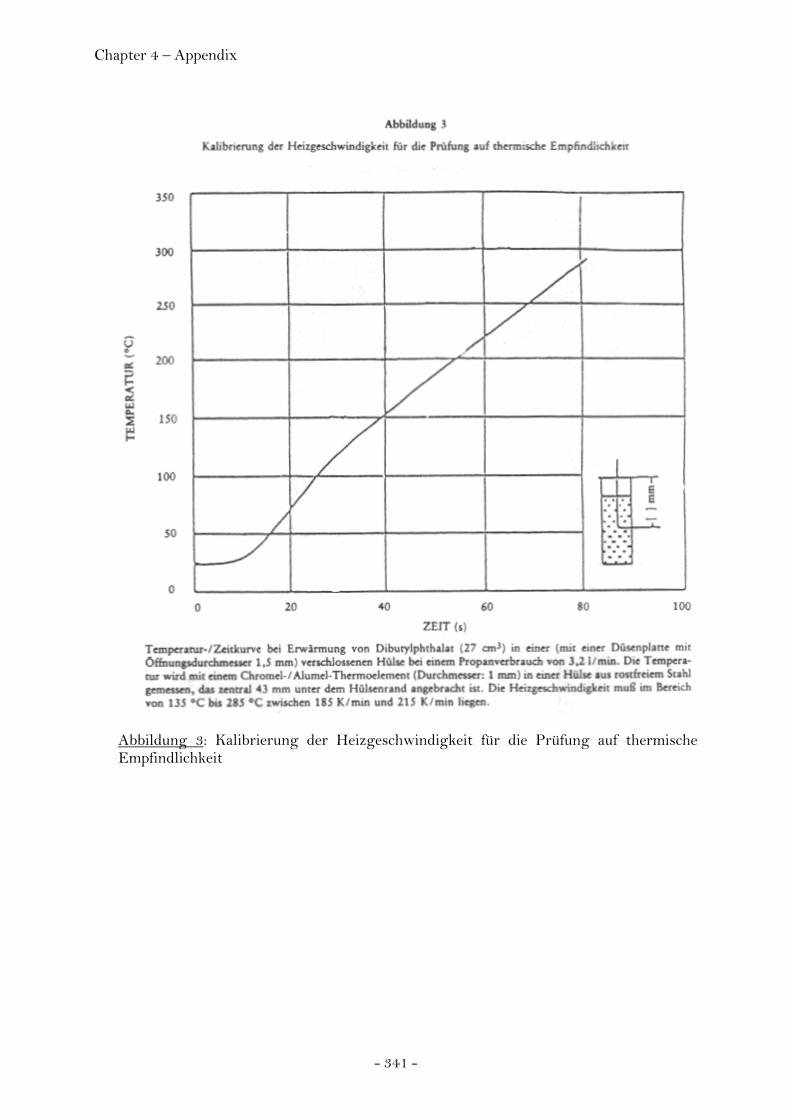
Chapter 4 – Appendix
- 341 -
Abbildung 3: Kalibrierung der Heizgeschwindigkeit für die Prüfung auf thermische Empfindlichkeit
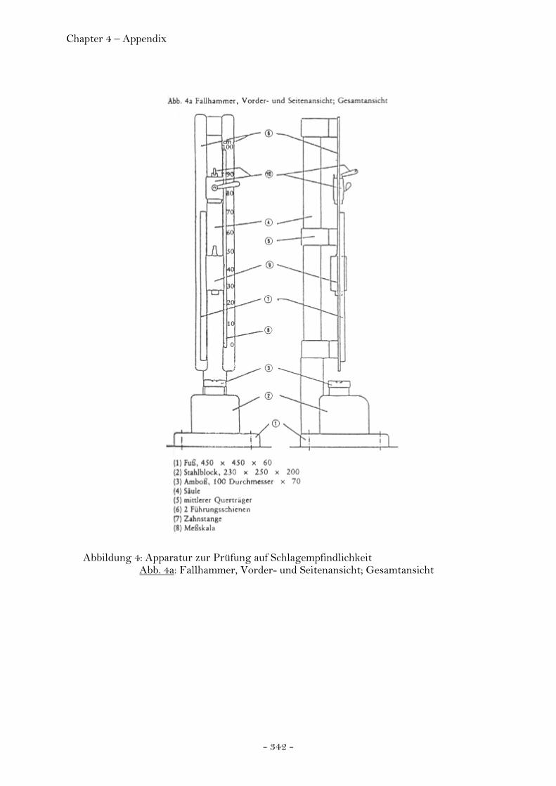
Chapter 4 – Appendix
- 342 -
Abbildung 4: Apparatur zur Prüfung auf Schlagempfindlichkeit
Abb. 4a: Fallhammer, Vorder- und Seitenansicht; Gesamtansicht
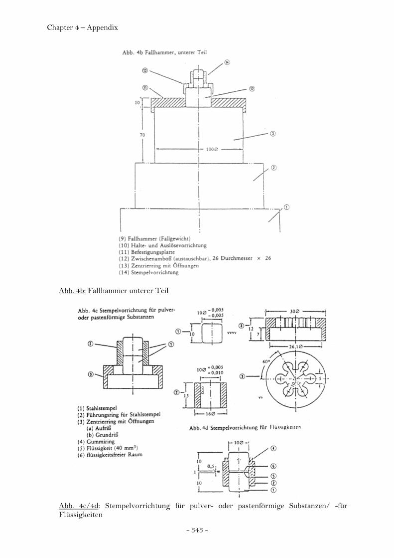
Chapter 4 – Appendix
- 343 -
Abb. 4b: Fallhammer unterer Teil
Abb. 4c/4d: Stempelvorrichtung für pulver- oder pastenförmige Substanzen/ -für Flüssigkeiten
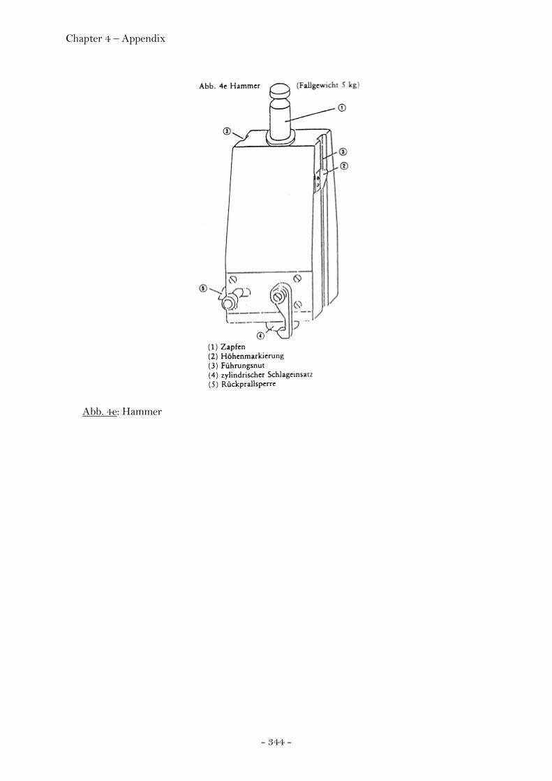
Chapter 4 – Appendix
- 344 -
Abb. 4e: Hammer

Chapter 4 – Appendix
- 345 -
Abbildung 5: Apparatur zur Prüfung auf Reibempfindlichkeit Abb. 5a Reibapparat; Aufriß und Grundriß Abb. 5b Ausgangsstellung des Stiftes auf der Probe

Chapter 4 – Appendix
- 346 -
4.5 Explosives Hazard Classification Procedures
Figure 5-1. UN hazard classification procedures for articles and substances.

Chapter 4 – Appendix
- 347 -
Figure 5-13. Thermal stability test configuration (UN 3c).
Figure 5-14. Small scale burning test configuration (UN 3d).

Chapter 4 – Appendix
- 348 -
Figure 5-15. Thermal stability test for articles (UN 4a).
Figure 5-17. Twelve meter drop test configuration (UN 4b).

Chapter 4 – Appendix
- 349 -
Test results according to:
12 m free fallUN 4b (ii)
Thermal stability for articles and packaged articles
UN 4a (i)
Small scale burningUN 3d
Thermal stabilityUN 3c
Friction sensitivityUN 3b
Impact sensitivityUN 3a
Comment+ / - TestTest #
Munich, …………………………
Name: …………………………………………….
Signature: …………………………………………….
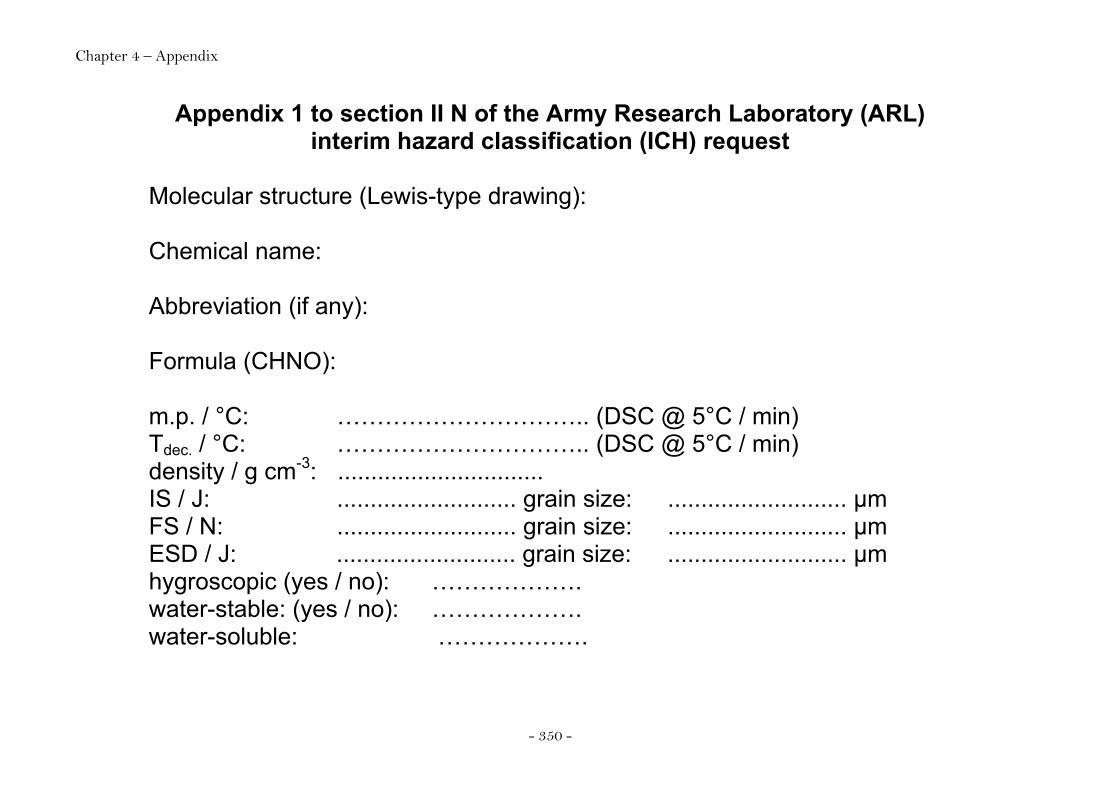
Chapter 4 – Appendix
- 350 -
Appendix 1 to section II N of the Army Research Laboratory (ARL) interim hazard classification (ICH) request
Molecular structure (Lewis-type drawing): Chemical name: Abbreviation (if any): Formula (CHNO): m.p. / °C: ………………………….. (DSC @ 5°C / min) Tdec. / °C: ………………………….. (DSC @ 5°C / min) density / g cm-3: ............................... IS / J: ........................... grain size: ........................... µm FS / N: ........................... grain size: ........................... µm ESD / J: ........................... grain size: ........................... µm hygroscopic (yes / no): ………………. water-stable: (yes / no): ………………. water-soluble: ……………….
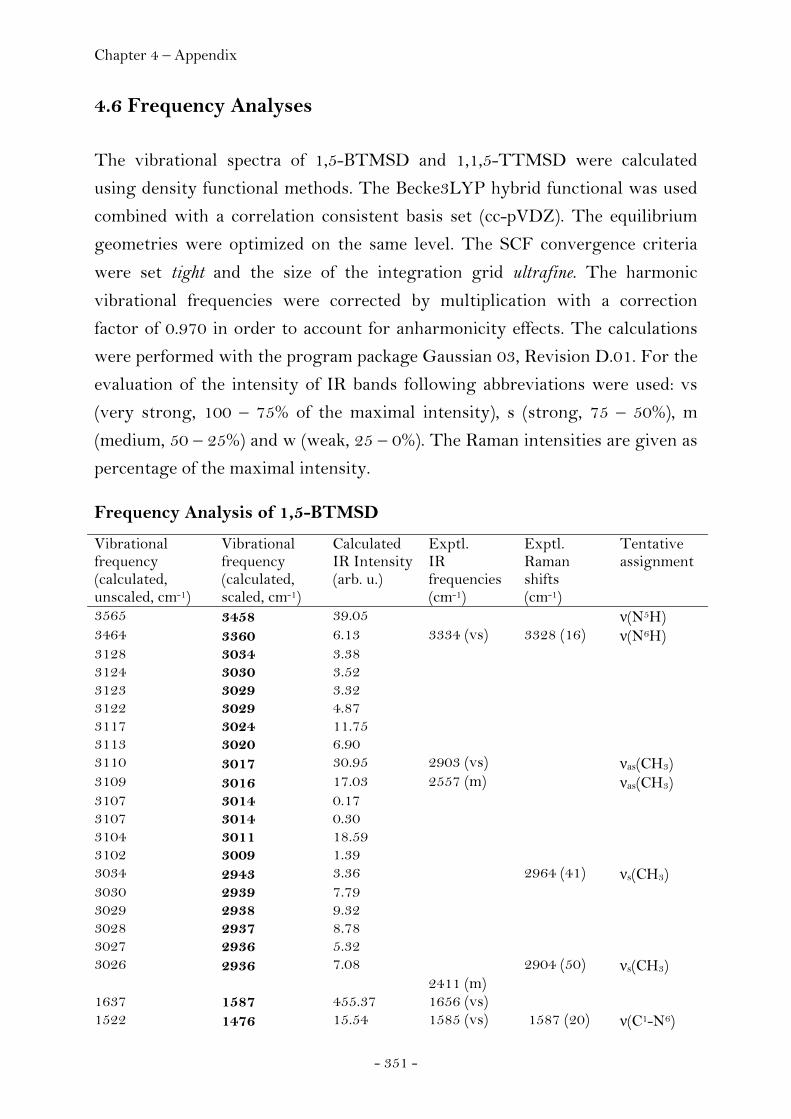
Chapter 4 – Appendix
- 351 -
4.6 Frequency Analyses
The vibrational spectra of 1,5-BTMSD and 1,1,5-TTMSD were calculated
using density functional methods. The Becke3LYP hybrid functional was used
combined with a correlation consistent basis set (cc-pVDZ). The equilibrium
geometries were optimized on the same level. The SCF convergence criteria
were set tight and the size of the integration grid ultrafine. The harmonic
vibrational frequencies were corrected by multiplication with a correction
factor of 0.970 in order to account for anharmonicity effects. The calculations
were performed with the program package Gaussian 03, Revision D.01. For the
evaluation of the intensity of IR bands following abbreviations were used: vs
(very strong, 100 – 75% of the maximal intensity), s (strong, 75 – 50%), m
(medium, 50 – 25%) and w (weak, 25 – 0%). The Raman intensities are given as
percentage of the maximal intensity.
Frequency Analysis of 1,5-BTMSD
Vibrational frequency (calculated, unscaled, cm-1)
Vibrational frequency (calculated, scaled, cm-1)
Calculated IR Intensity (arb. u.)
Exptl. IR frequencies (cm-1)
Exptl. Raman shifts (cm-1)
Tentative assignment
3565 3458 39.05 ν(N5H) 3464 3360 6.13 3334 (vs) 3328 (16) ν(N6H) 3128 3034 3.38 3124 3030 3.52 3123 3029 3.32 3122 3029 4.87 3117 3024 11.75 3113 3020 6.90 3110 3017 30.95 2903 (vs) νas(CH3) 3109 3016 17.03 2557 (m) νas(CH3) 3107 3014 0.17 3107 3014 0.30 3104 3011 18.59 3102 3009 1.39 3034 2943 3.36 2964 (41) νs(CH3) 3030 2939 7.79 3029 2938 9.32 3028 2937 8.78 3027 2936 5.32 3026 2936 7.08 2904 (50) νs(CH3) 2411 (m) 1637 1587 455.37 1656 (vs) 1522 1476 15.54 1585 (vs) 1587 (20) ν(C1-N6)
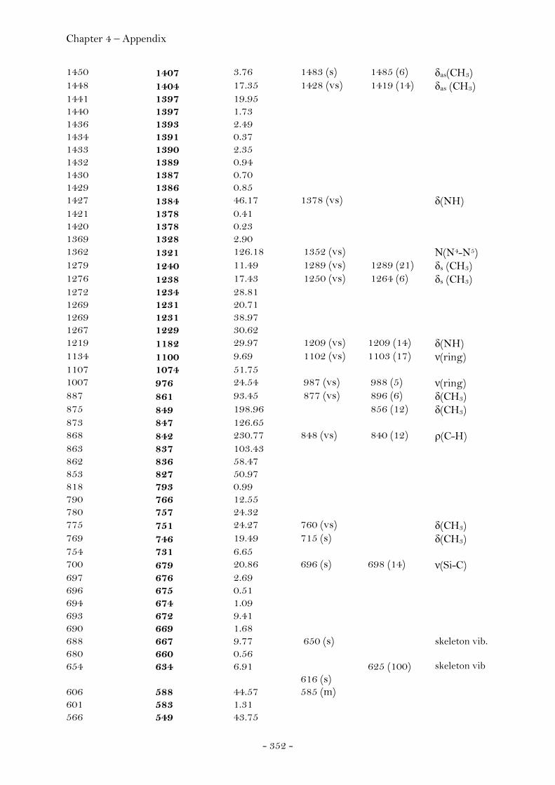
Chapter 4 – Appendix
- 352 -
1450 1407 3.76 1483 (s) 1485 (6) δas(CH3) 1448 1404 17.35 1428 (vs) 1419 (14) δas (CH3) 1441 1397 19.95 1440 1397 1.73 1436 1393 2.49 1434 1391 0.37 1433 1390 2.35 1432 1389 0.94 1430 1387 0.70 1429 1386 0.85 1427 1384 46.17 1378 (vs) δ(NH) 1421 1378 0.41 1420 1378 0.23 1369 1328 2.90 1362 1321 126.18 1352 (vs) Ν(N4-N5) 1279 1240 11.49 1289 (vs) 1289 (21) δs (CH3) 1276 1238 17.43 1250 (vs) 1264 (6) δs (CH3) 1272 1234 28.81 1269 1231 20.71 1269 1231 38.97 1267 1229 30.62 1219 1182 29.97 1209 (vs) 1209 (14) δ(NH) 1134 1100 9.69 1102 (vs) 1103 (17) ν(ring) 1107 1074 51.75 1007 976 24.54 987 (vs) 988 (5) ν(ring) 887 861 93.45 877 (vs) 896 (6) δ(CH3) 875 849 198.96 856 (12) δ(CH3) 873 847 126.65 868 842 230.77 848 (vs) 840 (12) ρ(C-H) 863 837 103.43 862 836 58.47 853 827 50.97 818 793 0.99 790 766 12.55 780 757 24.32 775 751 24.27 760 (vs) δ(CH3) 769 746 19.49 715 (s) δ(CH3) 754 731 6.65 700 679 20.86 696 (s) 698 (14) ν(Si-C) 697 676 2.69 696 675 0.51 694 674 1.09 693 672 9.41 690 669 1.68 688 667 9.77 650 (s) skeleton vib. 680 660 0.56 654 634 6.91 625 (100) skeleton vib 616 (s) 606 588 44.57 585 (m) 601 583 1.31 566 549 43.75
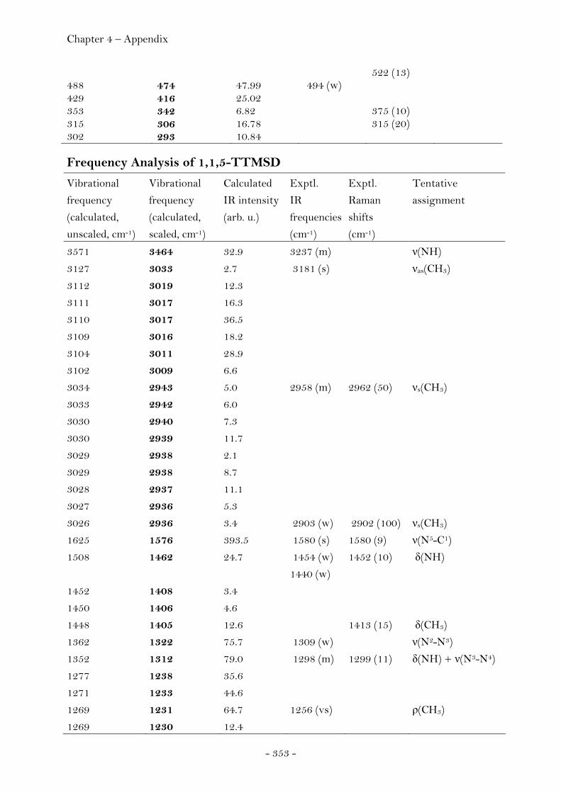
Chapter 4 – Appendix
- 353 -
522 (13) 488 474 47.99 494 (w) 429 416 25.02 353 342 6.82 375 (10) 315 306 16.78 315 (20) 302 293 10.84
Frequency Analysis of 1,1,5-TTMSD
Vibrational
frequency
(calculated,
unscaled, cm-1)
Vibrational
frequency
(calculated,
scaled, cm-1)
Calculated
IR intensity
(arb. u.)
Exptl.
IR
frequencies
(cm-1)
Exptl.
Raman
shifts
(cm-1)
Tentative
assignment
3571 3464 32.9 3237 (m) ν(NH)
3127 3033 2.7 3181 (s) νas(CH3)
3112 3019 12.3
3111 3017 16.3
3110 3017 36.5
3109 3016 18.2
3104 3011 28.9
3102 3009 6.6
3034 2943 5.0 2958 (m) 2962 (50) νs(CH3)
3033 2942 6.0
3030 2940 7.3
3030 2939 11.7
3029 2938 2.1
3029 2938 8.7
3028 2937 11.1
3027 2936 5.3
3026 2936 3.4 2903 (w) 2902 (100) νs(CH3)
1625 1576 393.5 1580 (s) 1580 (9) ν(N5-C1)
1508 1462 24.7 1454 (w) 1452 (10) δ(NH)
1440 (w)
1452 1408 3.4
1450 1406 4.6
1448 1405 12.6 1413 (15) δ(CH3)
1362 1322 75.7 1309 (w) ν(N2-N3)
1352 1312 79.0 1298 (m) 1299 (11) δ(NH) + ν(N3-N4)
1277 1238 35.6
1271 1233 44.6
1269 1231 64.7 1256 (vs) ρ(CH3)
1269 1230 12.4
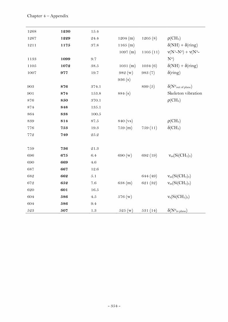
Chapter 4 – Appendix
- 354 -
1268 1230 15.4
1267 1229 24.4 1204 (m) 1205 (8) ρ(CH3)
1211 1175 37.8 1165 (m) δ(NH) + δ(ring)
1133 1099 9.7
1097 (m) 1105 (11) ν(N1-N2) + ν(N3-
N4)
1105 1072 38.5 1031 (m) 1034 (6) δ(NH) + δ(ring)
1007 977 19.7 982 (w) 983 (7) δ(ring)
936 (s)
903 876 374.1 899 (5) δ(N6out of plane)
901 874 153.8 884 (s) Skeleton vibration
876 850 370.1 ρ(CH3)
874 848 135.1
864 838 100.5
839 814 87.5 840 (vs) ρ(CH3)
776 753 19.3 759 (m) 759 (11) δ(CH3)
772 749 25.2
759 736 21.3
696 675 6.4 690 (w) 692 (19) νas(Si(CH3)3)
690 669 4.6
687 667 12.6
682 662 5.1 644 (49) νas(Si(CH3)3)
672 652 7.6 638 (m) 621 (32) νas(Si(CH3)3)
620 601 16.5
604 586 4.5 576 (w) νs(Si(CH3)3)
604 586 9.4
523 507 1.3 525 (w) 531 (14) δ(N6in plane)
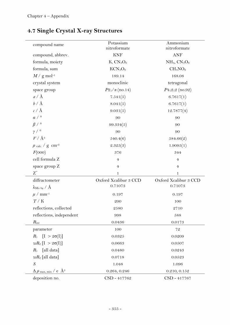
Chapter 4 – Appendix
- 355 -
4.7 Single Crystal X-ray Structures
compound name Potassium nitroformate
Ammonium nitroformate
compound, abbrev. KNF ANF
formula, moiety K, CN3O6 NH4, CN3O6
formula, sum KCN3O6 CH4NO6
M / g mol-1 189.14 168.08
crystal system monoclinic tetragonal
space group P21/n (no.14) P41212 (no.92)
a / Å 7.541(5) 6.7617(1)
b / Å 8.041(5) 6.7617(1)
c / Å 9.031(5) 12.7877(4)
α / ° 90 90
β / ° 99.334(5) 90
γ / ° 90 90
V / Å3 540.4(6) 584.66(2)
ρ calc. / g cm-3 2.325(3) 1.9095(1)
F(000) 376 344
cell formula Z 4 4
space group Z 4 4
Z΄ 1 1
diffractometer Oxford Xcalibur 3 CCD Oxford Xcalibur 3 CCD
λMo Kα / Å 0.71073 0.71073
μ / mm-1 0.197 0.197
T / K 200 100
reflections, collected 2580 2710
reflections, independent 998 588
Rint 0.0436 0.0173
parameter 100 72
R1 [I > 2σ(I)] 0.0325 0.0209
wR2 [I > 2σ(I)] 0.0663 0.0507
R1 [all data] 0.0480 0.0243
wR2 [all data] 0.0718 0.0523
S 1.048 1.096
Δ ρ max, min / e Å3 0.264, 0.246 0.210, 0.152
deposition no. CSD - 417762 CSD - 417767
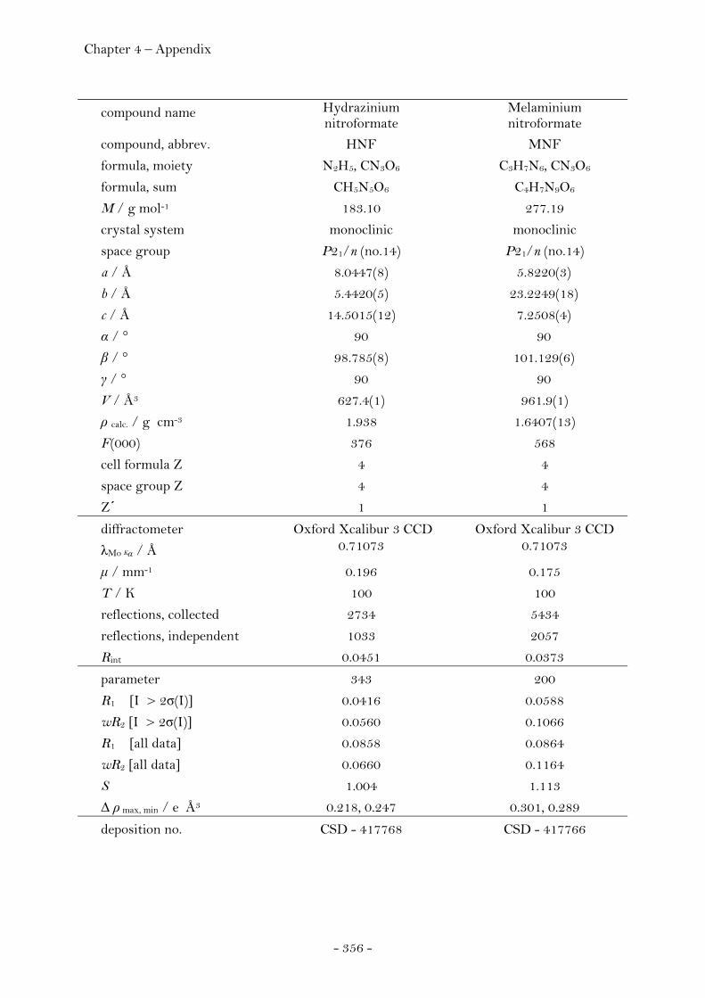
Chapter 4 – Appendix
- 356 -
compound name Hydrazinium nitroformate
Melaminium nitroformate
compound, abbrev. HNF MNF
formula, moiety N2H5, CN3O6 C3H7N6, CN3O6
formula, sum CH5N5O6 C4H7N9O6
M / g mol-1 183.10 277.19
crystal system monoclinic monoclinic
space group P21/n (no.14) P21/n (no.14)
a / Å 8.0447(8) 5.8220(3)
b / Å 5.4420(5) 23.2249(18)
c / Å 14.5015(12) 7.2508(4)
α / ° 90 90
β / ° 98.785(8) 101.129(6)
γ / ° 90 90
V / Å3 627.4(1) 961.9(1)
ρ calc. / g cm-3 1.938 1.6407(13)
F(000) 376 568
cell formula Z 4 4
space group Z 4 4
Z΄ 1 1
diffractometer Oxford Xcalibur 3 CCD Oxford Xcalibur 3 CCD
λMo Kα / Å 0.71073 0.71073
μ / mm-1 0.196 0.175
T / K 100 100
reflections, collected 2734 5434
reflections, independent 1033 2057
Rint 0.0451 0.0373
parameter 343 200
R1 [I > 2σ(I)] 0.0416 0.0588
wR2 [I > 2σ(I)] 0.0560 0.1066
R1 [all data] 0.0858 0.0864
wR2 [all data] 0.0660 0.1164
S 1.004 1.113
Δ ρ max, min / e Å3 0.218, 0.247 0.301, 0.289
deposition no. CSD - 417768 CSD - 417766

Chapter 4 – Appendix
- 357 -
compound name Melaminium nitroformate
Melaminium nitroformate,
Dimethylsulfate, 1:1 compound, abbrev. MNF MNF · DMSO
formula, moiety C3H7N6, CN3O6 C3H7N6, CN3O6 · (CH3)2SO
formula, sum C4H7N9O6 C6H13N9O7S
M / g mol-1 277.19 355.32
crystal system monoclinic triclinic
space group P21 (no.4) P-1 (no.2)
a / Å 9.2736(6) 9.966(5)
b / Å 7.2249(4) 10.920(5)
c / Å 15.660(1) 15.751(5)
α / ° 90 108.257(5)
β / ° 97.884(6) 90.342(5)
γ / ° 90 116.274(5)
V / Å3 1039.3(1) 1438.5(1)
ρ calc. / g cm-3 1.771 1.6407(13)
F(000) 568 736
cell formula Z 4 4
space group Z 2 2
Z΄ 2 2
diffractometer Oxford Xcalibur 3 CCD Oxford Xcalibur 3 CCD
λMo Kα / Å 0.71073 0.71073
μ / mm-1 0.162 0.282
T / K 100 100
reflections, collected 4182 11548
reflections, independent 1475 5583
Rint 0.0215 0.0434
parameter 343 420
R1 [I > 2σ(I)] 0.0344 0.0662
wR2 [I > 2σ(I)] 0.0783 0.1542
R1 [all data] 0.0497 0.0956
wR2 [all data] 0.0831 0.1683
S 0.962 0.957
Δ ρ max, min / e Å3 0.382, 0.244 0.019, 0.317
deposition no. CSD - 417765 CSD - 417764
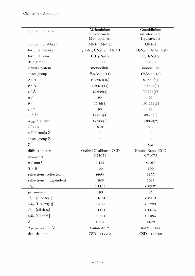
Chapter 4 – Appendix
- 358 -
compound name Melaminium nitroformate, Methanol, 1:1
Guanidinium nitroformate, Hydrate, 1:1
compound, abbrev. MNF · MeOH GNFH
formula, moiety C3H7N6, CN3O6 · CH3OH CH6N3, CN3O6 · H2O
formula, sum C5H11N9O7 C2H8N6O7
M / g mol-1 309.23 228.14
crystal system monoclinic monoclinic
space group P21/c (no.14) C2/c (no.15)
a / Å 10.8294(19) 8.1859(4)
b / Å 5.8091(11) 14.3541(7)
c / Å 19.646(3) 7.7556(5)
α / ° 90 90
β / ° 95.82(1) 101.132(2)
γ / ° 90 90
V / Å3 1229.5(4) 894.1(1)
ρ calc. / g cm-3 1.6706(5) 1.6948(2)
F(000) 640 472
cell formula Z 4 8
space group Z 4 4
Z΄ 1 0.5
diffractometer Oxford Xcalibur 3 CCD Nonius Kappa CCD
λMo Kα / Å 0.71073 0.71073
μ / mm-1 0.152 0.167
T / K 100 200
reflections, collected 9950 5277
reflections, independent 1928 1021
Rint 0.1163 0.0937
parameters 193 87
R1 [I > 2σ(I)] 0.1034 0.0513
wR2 [I > 2σ(I)] 0.2025 0.1293
R1 [all data] 0.1424 0.0645
wR2 [all data] 0.2294 0.1395
S 1.223 1.072
Δ ρ max, min / e Å3 0.305, 0.308 0.293, 0.433
deposition no. CSD - 417763 CSD - 417769
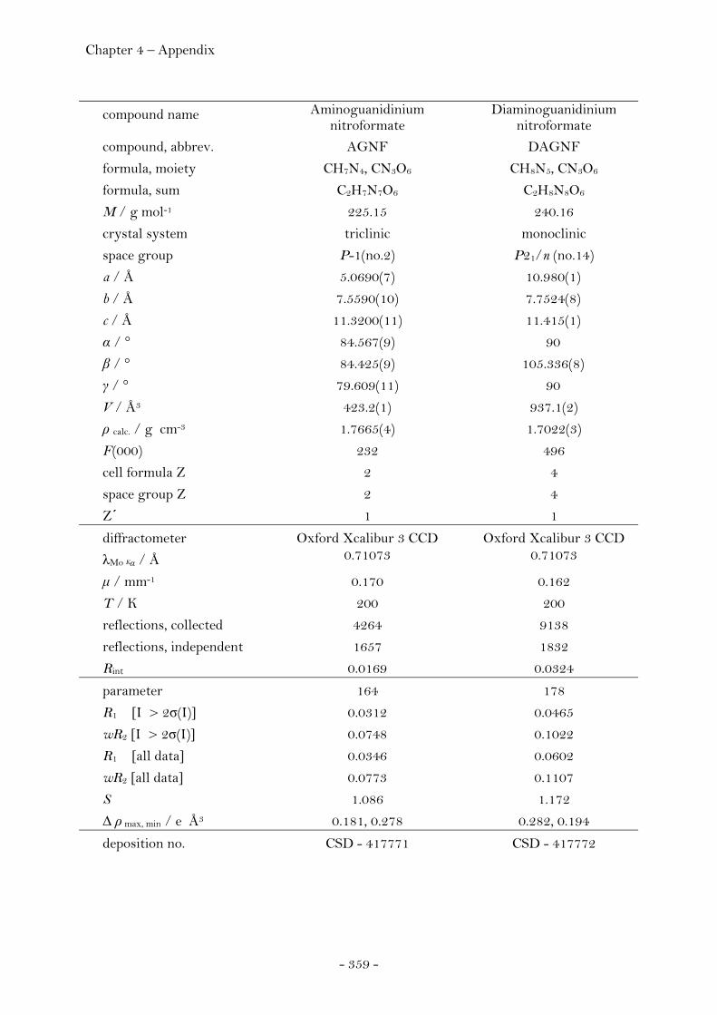
Chapter 4 – Appendix
- 359 -
compound name Aminoguanidinium nitroformate
Diaminoguanidinium nitroformate
compound, abbrev. AGNF DAGNF
formula, moiety CH7N4, CN3O6 CH8N5, CN3O6
formula, sum C2H7N7O6 C2H8N8O6
M / g mol-1 225.15 240.16
crystal system triclinic monoclinic
space group P-1(no.2) P21/n (no.14)
a / Å 5.0690(7) 10.980(1)
b / Å 7.5590(10) 7.7524(8)
c / Å 11.3200(11) 11.415(1)
α / ° 84.567(9) 90
β / ° 84.425(9) 105.336(8)
γ / ° 79.609(11) 90
V / Å3 423.2(1) 937.1(2)
ρ calc. / g cm-3 1.7665(4) 1.7022(3)
F(000) 232 496
cell formula Z 2 4
space group Z 2 4
Z΄ 1 1
diffractometer Oxford Xcalibur 3 CCD Oxford Xcalibur 3 CCD
λMo Kα / Å 0.71073 0.71073
μ / mm-1 0.170 0.162
T / K 200 200
reflections, collected 4264 9138
reflections, independent 1657 1832
Rint 0.0169 0.0324
parameter 164 178
R1 [I > 2σ(I)] 0.0312 0.0465
wR2 [I > 2σ(I)] 0.0748 0.1022
R1 [all data] 0.0346 0.0602
wR2 [all data] 0.0773 0.1107
S 1.086 1.172
Δ ρ max, min / e Å3 0.181, 0.278 0.282, 0.194
deposition no. CSD - 417771 CSD - 417772

Chapter 4 – Appendix
- 360 -
compound name Triaminoguanidinium nitroformate
Melaminium dinitrate
compound, abbrev. TAGNF MDN
formula, moiety CH9N6, CN3O6 C3H8N6, 2(NO3)
formula, sum C2H9N9O6 C3H8N8O6
M / g mol-1 255.18 252.17
crystal system triclinic monoclinic
space group P-1(no.2) P21/c (no.14)
a / Å 8.020(2) 7.758(5)
b / Å 8.347(2) 9.804(5)
c / Å 8.515(2) 12.094(5)
α / ° 105.49(2) 90
β / ° 95.03(2) 100.526(5)
γ / ° 111.10(2) 90
V / Å3 501.7(2) 904.4(8)
ρ calc. / g cm-3 1.6892(7) 1.8521(16)
F(000) 264 520
cell formula Z 2 4
space group Z 2 4
Z΄ 1 1
diffractometer Oxford Xcalibur 3 CCD Oxford Xcalibur 3 CCD
λMo Kα / Å 0.71073 0.71073
μ / mm-1 0.159 0.173
T / K 200 200
reflections, collected 2634 10088
reflections, independent 1962 2084
Rint 0.019 0.0694
parameter 190 186
R1 [I > 2σ(I)] 0.0379 0.0647
wR2 [I > 2σ(I)] 0.0970 0.1402
R1 [all data] 0.0476 0.0896
wR2 [all data] 0.1041 0.1575
S 1.070 1.185
Δ ρ max, min / e Å3 0.215, 0.301 0.273, 0.318
deposition no. CSD - 417770 -
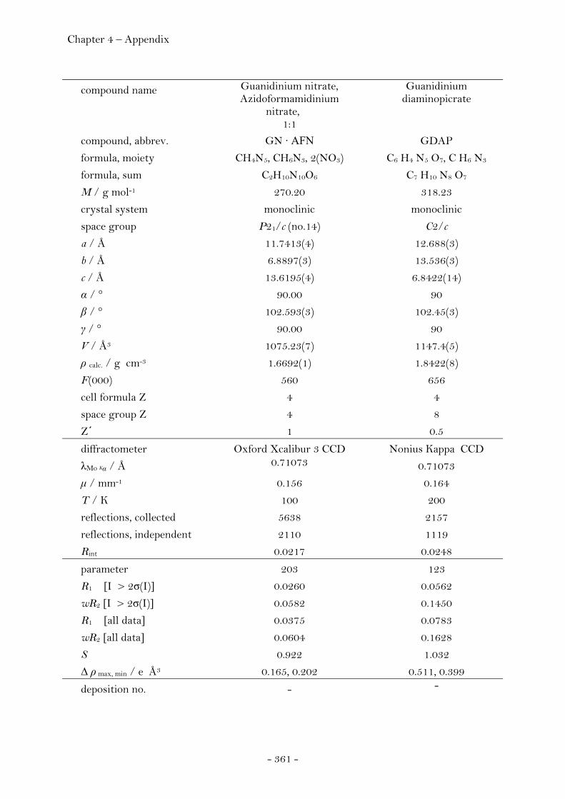
Chapter 4 – Appendix
- 361 -
compound name Guanidinium nitrate, Azidoformamidinium
nitrate, 1:1
Guanidinium diaminopicrate
compound, abbrev. GN · AFN GDAP
formula, moiety CH4N5, CH6N3, 2(NO3) C6 H4 N5 O7, C H6 N3
formula, sum C2H10N10O6 C7 H10 N8 O7
M / g mol-1 270.20 318.23
crystal system monoclinic monoclinic
space group P21/c (no.14) C2/c
a / Å 11.7413(4) 12.688(3)
b / Å 6.8897(3) 13.536(3)
c / Å 13.6195(4) 6.8422(14)
α / ° 90.00 90
β / ° 102.593(3) 102.45(3)
γ / ° 90.00 90
V / Å3 1075.23(7) 1147.4(5)
ρ calc. / g cm-3 1.6692(1) 1.8422(8)
F(000) 560 656
cell formula Z 4 4
space group Z 4 8
Z΄ 1 0.5
diffractometer Oxford Xcalibur 3 CCD Nonius Kappa CCD
λMo Kα / Å 0.71073 0.71073
μ / mm-1 0.156 0.164
T / K 100 200
reflections, collected 5638 2157
reflections, independent 2110 1119
Rint 0.0217 0.0248
parameter 203 123
R1 [I > 2σ(I)] 0.0260 0.0562
wR2 [I > 2σ(I)] 0.0582 0.1450
R1 [all data] 0.0375 0.0783
wR2 [all data] 0.0604 0.1628
S 0.922 1.032
Δ ρ max, min / e Å3 0.165, 0.202 0.511, 0.399
deposition no. - -
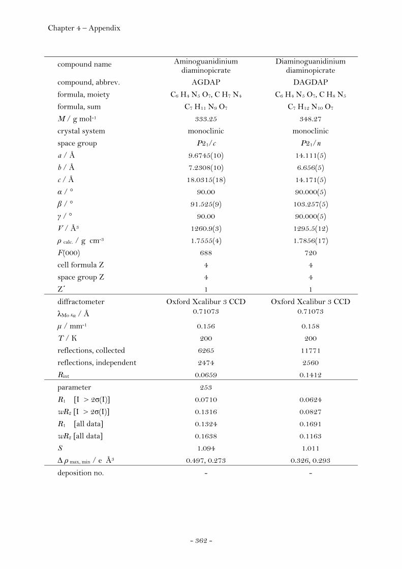
Chapter 4 – Appendix
- 362 -
compound name Aminoguanidinium diaminopicrate
Diaminoguanidinium diaminopicrate
compound, abbrev. AGDAP DAGDAP
formula, moiety C6 H4 N5 O7, C H7 N4 C6 H4 N5 O7, C H8 N5
formula, sum C7 H11 N9 O7 C7 H12 N10 O7
M / g mol-1 333.25 348.27
crystal system monoclinic monoclinic
space group P21/c P21/n
a / Å 9.6745(10) 14.111(5)
b / Å 7.2308(10) 6.656(5)
c / Å 18.0315(18) 14.171(5)
α / ° 90.00 90.000(5)
β / ° 91.525(9) 103.257(5)
γ / ° 90.00 90.000(5)
V / Å3 1260.9(3) 1295.5(12)
ρ calc. / g cm-3 1.7555(4) 1.7856(17)
F(000) 688 720
cell formula Z 4 4
space group Z 4 4
Z΄ 1 1
diffractometer Oxford Xcalibur 3 CCD Oxford Xcalibur 3 CCD
λMo Kα / Å 0.71073 0.71073
μ / mm-1 0.156 0.158
T / K 200 200
reflections, collected 6265 11771
reflections, independent 2474 2560
Rint 0.0659 0.1412
parameter 253
R1 [I > 2σ(I)] 0.0710 0.0624
wR2 [I > 2σ(I)] 0.1316 0.0827
R1 [all data] 0.1324 0.1691
wR2 [all data] 0.1638 0.1163
S 1.094 1.011
Δ ρ max, min / e Å3 0.497, 0.273 0.326, 0.293
deposition no. - -
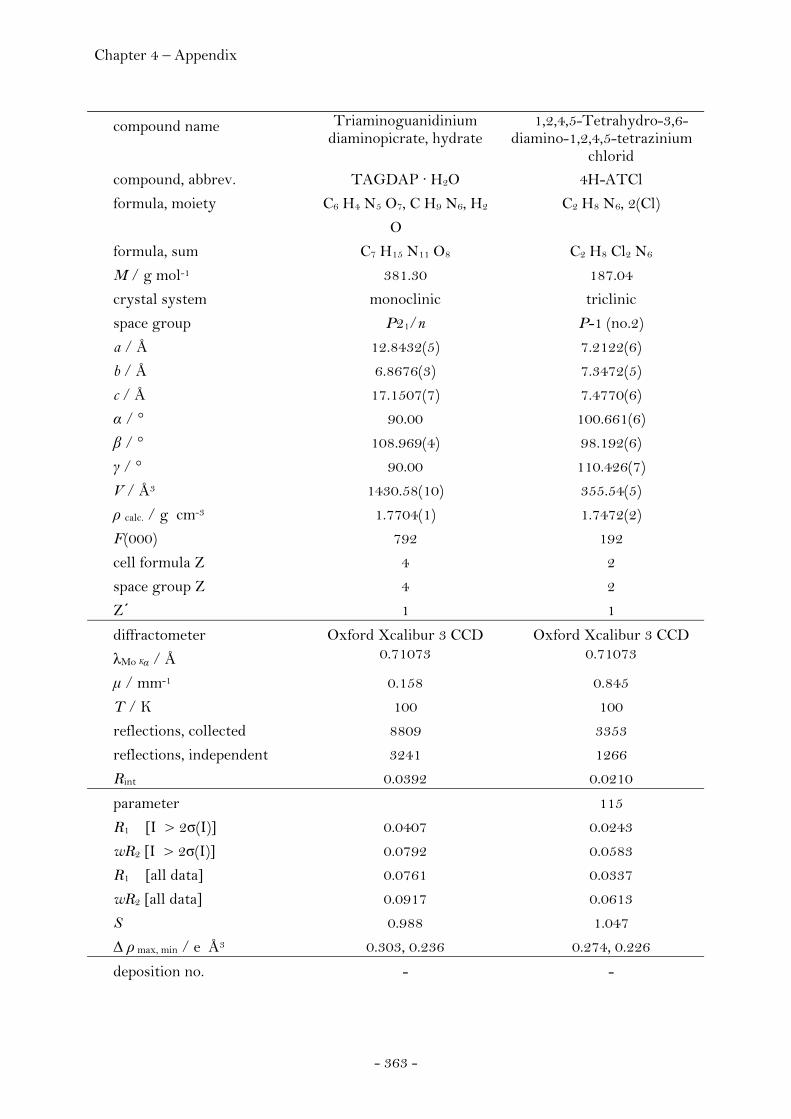
Chapter 4 – Appendix
- 363 -
compound name Triaminoguanidinium diaminopicrate, hydrate
1,2,4,5-Tetrahydro-3,6-diamino-1,2,4,5-tetrazinium
chlorid compound, abbrev. TAGDAP · H2O 4H-ATCl
formula, moiety C6 H4 N5 O7, C H9 N6, H2
O
C2 H8 N6, 2(Cl)
formula, sum C7 H15 N11 O8 C2 H8 Cl2 N6
M / g mol-1 381.30 187.04
crystal system monoclinic triclinic
space group P21/n P-1 (no.2)
a / Å 12.8432(5) 7.2122(6)
b / Å 6.8676(3) 7.3472(5)
c / Å 17.1507(7) 7.4770(6)
α / ° 90.00 100.661(6)
β / ° 108.969(4) 98.192(6)
γ / ° 90.00 110.426(7)
V / Å3 1430.58(10) 355.54(5)
ρ calc. / g cm-3 1.7704(1) 1.7472(2)
F(000) 792 192
cell formula Z 4 2
space group Z 4 2
Z΄ 1 1
diffractometer Oxford Xcalibur 3 CCD Oxford Xcalibur 3 CCD
λMo Kα / Å 0.71073 0.71073
μ / mm-1 0.158 0.845
T / K 100 100
reflections, collected 8809 3353
reflections, independent 3241 1266
Rint 0.0392 0.0210
parameter 115
R1 [I > 2σ(I)] 0.0407 0.0243
wR2 [I > 2σ(I)] 0.0792 0.0583
R1 [all data] 0.0761 0.0337
wR2 [all data] 0.0917 0.0613
S 0.988 1.047
Δ ρ max, min / e Å3 0.303, 0.236 0.274, 0.226
deposition no. - -
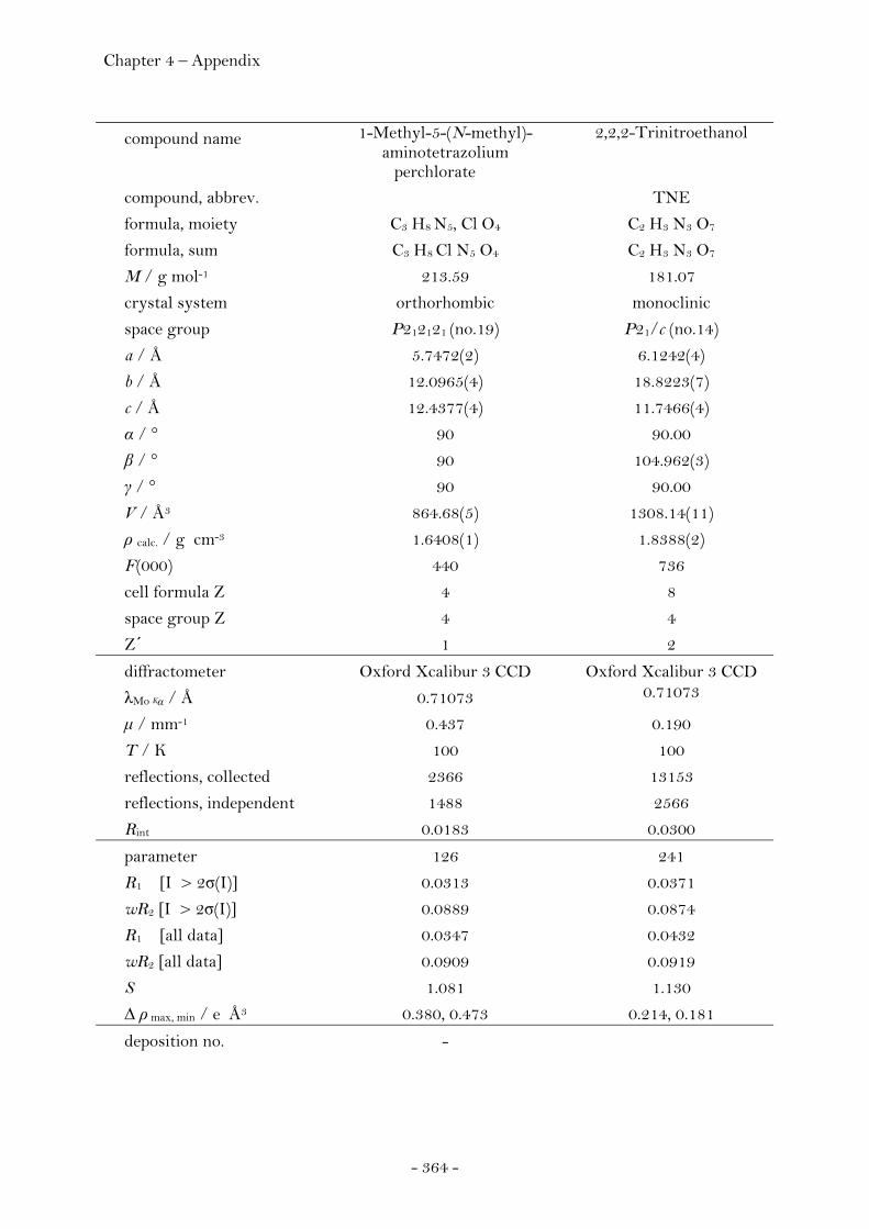
Chapter 4 – Appendix
- 364 -
compound name 1-Methyl-5-(N-methyl)- aminotetrazolium
perchlorate
2,2,2-Trinitroethanol
compound, abbrev. TNE
formula, moiety C3 H8 N5, Cl O4 C2 H3 N3 O7
formula, sum C3 H8 Cl N5 O4 C2 H3 N3 O7
M / g mol-1 213.59 181.07
crystal system orthorhombic monoclinic
space group P212121 (no.19) P21/c (no.14)
a / Å 5.7472(2) 6.1242(4)
b / Å 12.0965(4) 18.8223(7)
c / Å 12.4377(4) 11.7466(4)
α / ° 90 90.00
β / ° 90 104.962(3)
γ / ° 90 90.00
V / Å3 864.68(5) 1308.14(11)
ρ calc. / g cm-3 1.6408(1) 1.8388(2)
F(000) 440 736
cell formula Z 4 8
space group Z 4 4
Z΄ 1 2
diffractometer Oxford Xcalibur 3 CCD Oxford Xcalibur 3 CCD
λMo Kα / Å 0.71073 0.71073
μ / mm-1 0.437 0.190
T / K 100 100
reflections, collected 2366 13153
reflections, independent 1488 2566
Rint 0.0183 0.0300
parameter 126 241
R1 [I > 2σ(I)] 0.0313 0.0371
wR2 [I > 2σ(I)] 0.0889 0.0874
R1 [all data] 0.0347 0.0432
wR2 [all data] 0.0909 0.0919
S 1.081 1.130
Δ ρ max, min / e Å3 0.380, 0.473 0.214, 0.181
deposition no. -

Chapter 4 – Appendix
- 365 -
compound name Bis-(2,2,2-trinitroethyl)-amine
N1-(2,2,2-Trinitroethyl)- 1H-tetrazole-1,5-diamine
compound, abbrev. BTNA TTD
formula, moiety C4 H5 N7 O12 C3 H5 N9 O6
formula, sum C4 H5 N7 O12 C3 H5 N9 O6
M / g mol-1 343.15 263.16
crystal system orthorhombic orthorhombic
space group Pbca (no.61) Pna21
a / Å 12.8907(3) 14.9692(6)
b / Å 11.7069(3) 10.0155(5)
c / Å 16.0622(5) 6.3729(3)
α / ° 90 90.00
β / ° 90 90.00
γ / ° 90 90.00
V / Å3 2423.95(11) 955.45(8)
ρ calc. / g cm-3 1.8806(1) 1.8295(2)
F(000) 1392 536
cell formula Z 8 4
space group Z 8 4
Z΄ 1 1
diffractometer Oxford Xcalibur 3 CCD Oxford Xcalibur 3 CCD
λMo Kα / Å 0.71073 0.71073
μ / mm-1 0.190 0.171
T / K 200 100
reflections, collected 13680 5914
reflections, independent 2305 1704
Rint 0.0246 0.0517
parameter 229 175
R1 [I > 2σ(I)] 0.0392 0.0354
wR2 [I > 2σ(I)] 0.1163 0.0616
R1 [all data] 0.0470 0.0650
wR2 [all data] 0.1095 0.0705
S 1.060 0.944
Δ ρ max, min / e Å3 0.238, 0.230 0.276, 0.273
deposition no. CCDC - 699142 CCDC - 699140

Chapter 4 – Appendix
- 366 -
compound name N1,N5-Bis- (2,2,2-trinitroethyl)-
1H-tetrazole-1,5-diamine
(E)-1-methyl- 1-(1H-tetrazol-5-yl)-2-
(2,2,2-trinitroethylidene)-hydrazine
compound, abbrev. BTTD MTTH
formula, moiety C5 H6 N12 O12 C4H5N9O6
formula, sum C5 H6 N12 O12 C4H5N9O6
M / g mol-1 426.22 275.17
crystal system monoclinic orthorhombic
space group P21/c (no.14) Pbca(no.61)
a / Å 9.9710(7) 9.7012(4)
b / Å 6.4861(4) 12.8504(7)
c / Å 23.1501(13) 17.3758(9)
α / ° 90 90
β / ° 94.464(5) 90
γ / ° 90 90
V / Å3 1492.64(16) 2166.14(19)
ρ calc. / g cm-3 1.8967(2) 1.6876(1)
F(000) 864 1120
cell formula Z 4 8
space group Z 4 8
Z΄ 1 1
diffractometer Oxford Xcalibur 3 CCD Oxford Xcalibur 3 CCD
λMo Kα / Å 0.71073 0.71073
μ / mm-1 0.184 0.155
T / K 100 100
reflections, collected 8094 8573
reflections, independent 2944 1784
Rint 0.0525 0.1493
parameter 280 172
R1 [I > 2σ(I)] 0.0844 0.0779
wR2 [I > 2σ(I)] 0.1145 0.1570
R1 [all data] 0.1243 0.1457
wR2 [all data] 0.1264 0.1942
S 1.216 1.076
Δ ρ max, min / e Å3 0.327, 0.334 0.423, 0.425
deposition no. CCDC - 699136 -
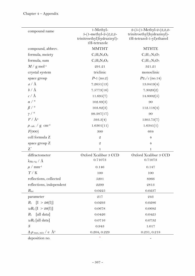
Chapter 4 – Appendix
- 367 -
compound name 1-Methyl- 5-(1-methyl-2-(2,2,2-
trinitroethyl)hydrazinyl)-1H-tetrazole
2-(5-(1-Methyl-2-(2,2,2-trinitroethyl)hydrazinyl)-1H-tetrazol-1-yl)ethanol
compound, abbrev. MMTHT MTHTE
formula, moiety C5H9N9O6 C5H11N9O7
formula, sum C5H9N9O6 C5H11N9O7
M / g mol-1 291.21 321.21
crystal system triclinic monoclinic
space group P-1 (no.2) P21/c (no.14)
a / Å 7.2651(13) 13.0419(4)
b / Å 7.5773(16) 7.3020(2)
c / Å 11.695(7) 14.8002(5)
α / ° 102.89(3) 90
β / ° 103.82(3) 112.118(4)
γ / ° 99.387(17) 90
V / Å3 593.3(4) 1305.73(7)
ρ calc. / g cm-3 1.6301(11) 1.6341(1)
F(000) 300 664
cell formula Z 2 4
space group Z 2 4
Z΄ 1 1
diffractometer Oxford Xcalibur 3 CCD Oxford Xcalibur 3 CCD
λMo Kα / Å 0.71073 0.71073
μ / mm-1 0.146 0.147
T / K 100 100
reflections, collected 5201 8966
reflections, independent 2299 2813
Rint 0.0225 0.0237
parameter 217 243
R1 [I > 2σ(I)] 0.0293 0.0286
wR2 [I > 2σ(I)] 0.0678 0.0682
R1 [all data] 0.0426 0.0425
wR2 [all data] 0.0716 0.0732
S 0.943 1.017
Δ ρ max, min / e Å3 0.204, 0.229 0.231, 0.218
deposition no. - -

Chapter 4 – Appendix
- 368 -
compound name 1-(4N-2,2,2-Trinitroethyl)-2,5-
hydroxymethyl- triazine
N3,N6-Bis- (2,2,2-trinitroethyl)- 3,6-diamino-1,2,4,5-
tetrazine compound, abbrev. THMT BTAT
formula, moiety C5H9N7O8 C6 H6 N12 O12
formula, sum C5H9N7O8 C6 H6 N12 O12
M / g mol-1 307.20 438.23
crystal system monoclinic orthorhombic
space group P21/n (no.14) Pna21
a / Å 9.1753(4) 23.3824(8)
b / Å 10.3168(4) 5.9271(2)
c / Å 13.0862(5) 11.1373(3)
α / ° 90.00 90
β / ° 105.975(4) 90
γ / ° 90.00 90
V / Å3 1190.90(9) 1543.52(9)
ρ calc. / g cm-3 1.7134(1) 1.8858(1)
F(000) 632 888
cell formula Z 4 4
space group Z 4 4
Z΄ 1 1
diffractometer Oxford Xcalibur 3 CCD Oxford Xcalibur 3 CCD
λMo Kα / Å 0.71073 0.71073
μ / mm-1 0.158 0.181
T / K 100 100
reflections, collected 7201 11060
reflections, independent 2702 2339
Rint 0.0313 0.0219
parameter 226 279
R1 [I > 2σ(I)] 0.0315 0.0269
wR2 [I > 2σ(I)] 0.0620 0.0685
R1 [all data] 0.0593 0.0329
wR2 [all data] 0.0690 0.0710
S 0.899 1.029
Δ ρ max, min / e Å3 0.256, 0.279 0.326, 0.200
deposition no. - CCDC - 699141
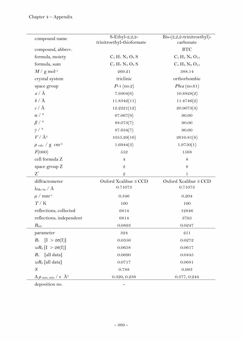
Chapter 4 – Appendix
- 369 -
compound name S-Ethyl-2,2,2-trinitroethyl-thioformate
Bis-(2,2,2-trinitroethyl)- carbonate
compound, abbrev. BTC
formula, moiety C5 H7 N3 O8 S C5 H4 N6 O15
formula, sum C5 H7 N3 O8 S C5 H4 N6 O15
M / g mol-1 269.21 388.14
crystal system triclinic orthorhombic
space group P-1 (no.2) Pbca (no.61)
a / Å 7.9304(6) 10.8828(2)
b / Å 11.8342(11) 11.4746(2)
c / Å 12.2221(12) 20.9073(4)
α / ° 67.067(9) 90.00
β / ° 88.073(7) 90.00
γ / ° 87.634(7) 90.00
V / Å3 1055.29(16) 2610.81(8)
ρ calc. / g cm-3 1.6944(3) 1.9750(1)
F(000) 552 1568
cell formula Z 4 8
space group Z 2 8
Z΄ 2 1
diffractometer Oxford Xcalibur 3 CCD Oxford Xcalibur 3 CCD
λMo Kα / Å 0.71073 0.71073
μ / mm-1 0.346 0.204
T / K 100 100
reflections, collected 6814 12846
reflections, independent 6814 3795
Rint 0.0893 0.0247
parameter 324 251
R1 [I > 2σ(I)] 0.0356 0.0272
wR2 [I > 2σ(I)] 0.0658 0.0617
R1 [all data] 0.0690 0.0445
wR2 [all data] 0.0717 0.0681
S 0.788 0.993
Δ ρ max, min / e Å3 0.320, 0.238 0.377, 0.244
deposition no. -

Chapter 4 – Appendix
- 370 -
compound name 2,2,2-Trinitroethyl- azidoformate
Bis-(2,2,2-trinitroethyl)-hydrazodicarboxylate,
Acetone, 1:1 compound, abbrev. TAF BTHC · Acetone
formula, moiety C3 H2 N6 O8 C6 H6 N8 O16, C3 H6 O
formula, sum C3 H2 N6 O8 C9 H12 N8 O17
M / g mol-1 250.11 504.27
crystal system monoclinic triclinic
space group P21/c (no.14) P-1 (no.2)
a / Å 7.3139(3) 10.1798(13)
b / Å 13.0430(6) 10.4185(17)
c / Å 18.8050(7) 11.256(3)
α / ° 90.00 63.448(19)
β / ° 91.273(4) 70.392(13)
γ / ° 90.00 89.865(12)
V / Å3 1793.46(13) 990.4(4)
ρ calc. / g cm-3 1.8526(1) 1.6910(7)
F(000) 1008 516.0
cell formula Z 8 2
space group Z 4 2
Z΄ 2 1
diffractometer Oxford Xcalibur 3 CCD Oxford Xcalibur 3 CCD
λMo Kα / Å 0.71073 0.71073
μ / mm-1 0.184 0.166
T / K 100 100
reflections, collected 8785 29458
reflections, independent 3244 6588
Rint 0.0657 0.0329
parameter 323 355
R1 [I > 2σ(I)] 0.0830 0.0366
wR2 [I > 2σ(I)] 0.1016 0.0849
R1 [all data] 0.1341 0.0718
wR2 [all data] 0.1166 0.1006
S 1.312 1.044
Δ ρ max, min / e Å3 0.374, 0.488 0.361, 0.294
deposition no. CCDC - 699137 CCDC - 699138
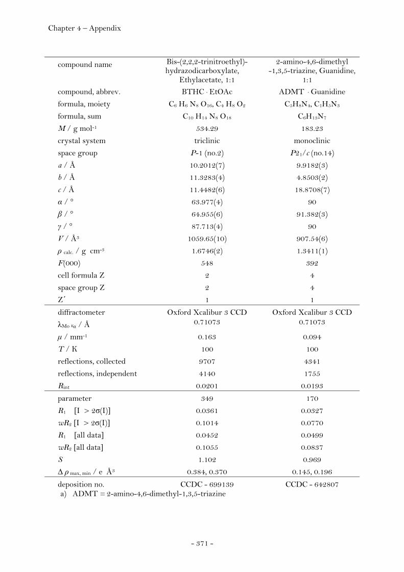
Chapter 4 – Appendix
- 371 -
compound name Bis-(2,2,2-trinitroethyl)-hydrazodicarboxylate,
Ethylacetate, 1:1
2-amino-4,6-dimethyl -1,3,5-triazine, Guanidine,
1:1 compound, abbrev. BTHC · EtOAc ADMT · Guanidine
formula, moiety C6 H6 N8 O16, C4 H8 O2 C5H8N4, C1H5N3
formula, sum C10 H14 N8 O18 C6H13N7
M / g mol-1 534.29 183.23
crystal system triclinic monoclinic
space group P-1 (no.2) P21/c (no.14)
a / Å 10.2012(7) 9.9182(3)
b / Å 11.3283(4) 4.8503(2)
c / Å 11.4482(6) 18.8708(7)
α / ° 63.977(4) 90
β / ° 64.955(6) 91.382(3)
γ / ° 87.713(4) 90
V / Å3 1059.65(10) 907.54(6)
ρ calc. / g cm-3 1.6746(2) 1.3411(1)
F(000) 548 392
cell formula Z 2 4
space group Z 2 4
Z΄ 1 1
diffractometer Oxford Xcalibur 3 CCD Oxford Xcalibur 3 CCD
λMo Kα / Å 0.71073 0.71073
μ / mm-1 0.163 0.094
T / K 100 100
reflections, collected 9707 4341
reflections, independent 4140 1755
Rint 0.0201 0.0193
parameter 349 170
R1 [I > 2σ(I)] 0.0361 0.0327
wR2 [I > 2σ(I)] 0.1014 0.0770
R1 [all data] 0.0452 0.0499
wR2 [all data] 0.1055 0.0837
S 1.102 0.969
Δ ρ max, min / e Å3 0.384, 0.370 0.145, 0.196
deposition no. CCDC - 699139 CCDC - 642807 a) ADMT = 2-amino-4,6-dimethyl-1,3,5-triazine
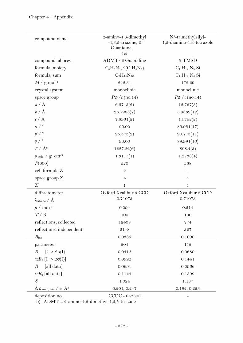
Chapter 4 – Appendix
- 372 -
compound name 2-amino-4,6-dimethyl -1,3,5-triazine, 2
Guanidine, 1:2
N5-trimethylsilyl- 1,5-diamino-1H-tetrazole
compound, abbrev. ADMT · 2 Guanidine 5-TMSD
formula, moiety C5H8N4, 2(C1H5N3) C4 H12 N6 Si
formula, sum C7H18N10 C4 H12 N6 Si
M / g mol-1 242.31 172.29
crystal system monoclinic monoclinic
space group P21/c (no.14) P21/c (no.14)
a / Å 6.5743(2) 12.767(3)
b / Å 23.7968(7) 5.9889(12)
c / Å 7.8931(2) 11.752(2)
α / ° 90.00 89.951(17)
β / ° 96.373(2) 90.773(17)
γ / ° 90.00 89.991(16)
V / Å3 1227.22(6) 898.4(3)
ρ calc. / g cm-3 1.3115(1) 1.2738(4)
F(000) 520 368
cell formula Z 4 4
space group Z 4 4
Z΄ 1 1
diffractometer Oxford Xcalibur 3 CCD Oxford Xcalibur 3 CCD
λMo Kα / Å 0.71073 0.71073
μ / mm-1 0.094 0.214
T / K 100 100
reflections, collected 12408 774
reflections, independent 2148 327
Rint 0.0385 0.1090
parameter 204 112
R1 [I > 2σ(I)] 0.0412 0.0680
wR2 [I > 2σ(I)] 0.0992 0.1441
R1 [all data] 0.0691 0.0966
wR2 [all data] 0.1144 0.1599
S 1.024 1.187
Δ ρ max, min / e Å3 0.201, 0.247 0.192, 0.223
deposition no. CCDC - 642808 - b) ADMT = 2-amino-4,6-dimethyl-1,3,5-triazine

Chapter 4 – Appendix
- 373 -
compound name N1,N5-bis(trimethylsilyl)-1,5-diamino-1H-tetrazole
N1-(propan-2-ylidene)- 1,5-diamino-1H-tetrazole
compound, abbrev. 1,5-BTMSD 1-PYD
formula, moiety C7 H20 N6 Si2 C4 H2 D6 N6
formula, sum C7 H20 N6 Si2 C4 H2 D6 N6
M / g mol-1 244.47 146.18
crystal system monoclinic monoclinic
space group P21/n (no.14) P21/c (no.14)
a / Å 14.6063(7) 7.4859(3)
b / Å 6.4066(3) 7.4334(2)
c / Å 15.0847(9) 11.9822(4)
α / ° 90.00 90.00
β / ° 100.568(5) 97.308(3)
γ / ° 90.00 90.00
V / Å3 1387.63(12) 661.34(4)
ρ calc. / g cm-3 1.1702(1) 1.4681(1)
F(000) 528 296
cell formula Z 4 4
space group Z 4 4
Z΄ 1 1
diffractometer Oxford Xcalibur 3 CCD Oxford Xcalibur 3 CCD
λMo Kα / Å 0.71073 0.71073
μ / mm-1 0.239 0.102
T / K 100 100
reflections, collected 13361 6449
reflections, independent 4562 2163
Rint 0.0433 0.0217
parameter 216 123
R1 [I > 2σ(I)] 0.0359 0.0342
wR2 [I > 2σ(I)] 0.0750 0.0839
R1 [all data] 0.0787 0.0500
wR2 [all data] 0.0931 0.0892
S 0.988 0.995
Δ ρ max, min / e Å3 0.487, 0.601 0.231, 251
deposition no. - -

Chapter 4 – Appendix
- 374 -
compound name N1,N1-bis-(trimethylsilyl)- 1,5-diamino-1H-tetrazole
N1,N1,N5-tris(trimethylsilyl)-
1,5-diamino-1H-tetrazole compound, abbrev. 1,1-BTMSD 1,1,5-TTMSD
formula, moiety C7 H20 N6 Si2 C10H28N6Si3
formula, sum C7 H20 N6 Si2 C10H28N6Si3
M / g mol-1 244.47 316.65
crystal system triclinic monoclinic
space group P-1 (no.2) P21/c (no.14)
a / Å 6.3649(10) 9.5490(4)
b / Å 8.0322(16) 17.1449(6)
c / Å 14.026(2) 11.8672(4)
α / ° 93.961(14) 90.00
β / ° 97.427(13) 94.643(4)
γ / ° 105.416(15) 90.00
V / Å3 681.5(2) 1936.48(12)
ρ calc. / g cm-3 1.1914(3) 1.0861(1)
F(000) 264 688
cell formula Z 2 4
space group Z 2 4
Z΄ 1 1
diffractometer Oxford Xcalibur 3 CCD Oxford Xcalibur 3 CCD
λMo Kα / Å 0.71073 0.71073
μ / mm-1 0.244 0.244
T / K 100 100
reflections, collected 4812 14750
reflections, independent 1870 4281
Rint 0.1030 0.0494
parameter 144 176
R1 [I > 2σ(I)] 0.0879 0.0403
wR2 [I > 2σ(I)] 0.1214 0.0813
R1 [all data] 0.1530 0.0818
wR2 [all data] 0.1425 0.1001
S 1.177 1.030
Δ ρ max, min / e Å3 0.411, 0.371 0.333, 0.274
deposition no. - -

Chapter 4 – Appendix
- 375 -
compound name 3,4,6-Trichlor- pyridazine
3,4,5,6-Tetrachlor-pyridazine
compound, abbrev. 3,4,6-TCP 3,4,5,6-TCP
formula, moiety C4 H Cl3 N2 C4 Cl4 N2
formula, sum C4 H Cl3 N2 C4 Cl4 N2
M / g mol-1 183.42 217.86
crystal system orthorhombic tetragonal
space group Pbca (no.61) P41212 (no.92)
a / Å 10.571(5) 5.2649(2)
b / Å 7.243(5) 5.2649(2)
c / Å 17.302(5) 52.351(2)
α / ° 90.0 90
β / ° 90.0 90
γ / ° 90.0 90
V / Å3 1324.7(12) 1451.10(10)
ρ calc. / g cm-3 1.8394(17) 1.9944(1)
F(000) 720 848
cell formula Z 8 8
space group Z 8 4
Z΄ 1 2
diffractometer Oxford Xcalibur 3 CCD Oxford Xcalibur 3 CCD
λMo Kα / Å 0.71073 0.71073
μ / mm-1 1.280 1.542
T / K 100 100
reflections, collected 12166 13078
reflections, independent 1278 1299
Rint 0.0264 0.0972
parameter 86 91
R1 [I > 2σ(I)] 0.0292 0.0770
wR2 [I > 2σ(I)] 0.0713 0.1411
R1 [all data] 0.0301 0.0772
wR2 [all data] 0.0720 0.1412
S 1.169 1.138
Δ ρ max, min / e Å3 0.206, 0.240 0.382, 0.383
deposition no. - -
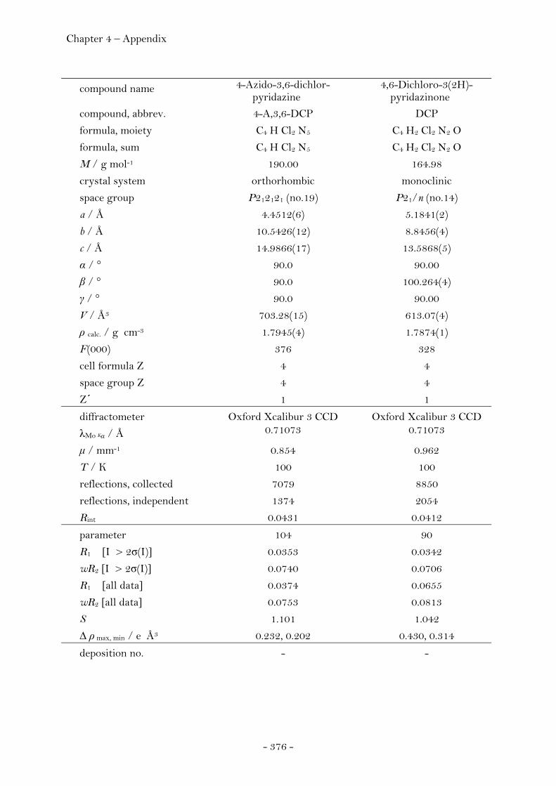
Chapter 4 – Appendix
- 376 -
compound name 4-Azido-3,6-dichlor-pyridazine
4,6-Dichloro-3(2H)-pyridazinone
compound, abbrev. 4-A,3,6-DCP DCP
formula, moiety C4 H Cl2 N5 C4 H2 Cl2 N2 O
formula, sum C4 H Cl2 N5 C4 H2 Cl2 N2 O
M / g mol-1 190.00 164.98
crystal system orthorhombic monoclinic
space group P212121 (no.19) P21/n (no.14)
a / Å 4.4512(6) 5.1841(2)
b / Å 10.5426(12) 8.8456(4)
c / Å 14.9866(17) 13.5868(5)
α / ° 90.0 90.00
β / ° 90.0 100.264(4)
γ / ° 90.0 90.00
V / Å3 703.28(15) 613.07(4)
ρ calc. / g cm-3 1.7945(4) 1.7874(1)
F(000) 376 328
cell formula Z 4 4
space group Z 4 4
Z΄ 1 1
diffractometer Oxford Xcalibur 3 CCD Oxford Xcalibur 3 CCD
λMo Kα / Å 0.71073 0.71073
μ / mm-1 0.854 0.962
T / K 100 100
reflections, collected 7079 8850
reflections, independent 1374 2054
Rint 0.0431 0.0412
parameter 104 90
R1 [I > 2σ(I)] 0.0353 0.0342
wR2 [I > 2σ(I)] 0.0740 0.0706
R1 [all data] 0.0374 0.0655
wR2 [all data] 0.0753 0.0813
S 1.101 1.042
Δ ρ max, min / e Å3 0.232, 0.202 0.430, 0.314
deposition no. - -
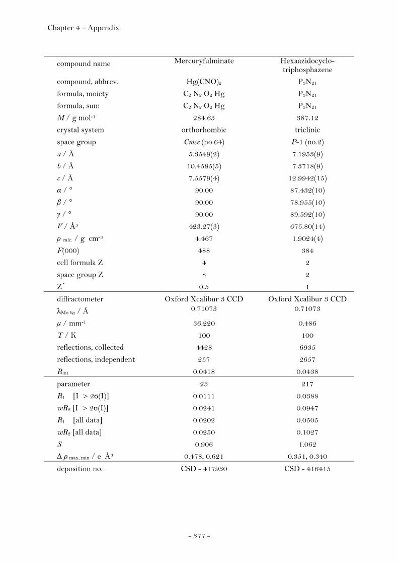
Chapter 4 – Appendix
- 377 -
compound name Mercuryfulminate Hexaazidocyclo- triphosphazene
compound, abbrev. Hg(CNO)2 P3N21
formula, moiety C2 N2 O2 Hg P3N21
formula, sum C2 N2 O2 Hg P3N21
M / g mol-1 284.63 387.12
crystal system orthorhombic triclinic
space group Cmce (no.64) P-1 (no.2)
a / Å 5.3549(2) 7.1953(9)
b / Å 10.4585(5) 7.3718(9)
c / Å 7.5579(4) 12.9942(15)
α / ° 90.00 87.432(10)
β / ° 90.00 78.955(10)
γ / ° 90.00 89.592(10)
V / Å3 423.27(3) 675.80(14)
ρ calc. / g cm-3 4.467 1.9024(4)
F(000) 488 384
cell formula Z 4 2
space group Z 8 2
Z΄ 0.5 1
diffractometer Oxford Xcalibur 3 CCD Oxford Xcalibur 3 CCD
λMo Kα / Å 0.71073 0.71073
μ / mm-1 36.220 0.486
T / K 100 100
reflections, collected 4428 6935
reflections, independent 257 2657
Rint 0.0418 0.0438
parameter 23 217
R1 [I > 2σ(I)] 0.0111 0.0388
wR2 [I > 2σ(I)] 0.0241 0.0947
R1 [all data] 0.0202 0.0505
wR2 [all data] 0.0250 0.1027
S 0.906 1.062
Δ ρ max, min / e Å3 0.478, 0.621 0.351, 0.340
deposition no. CSD - 417930 CSD - 416415

Chapter 4 – Appendix
- 378 -
compound name Chlortrinitromethane
compound, abbrev. Cl-TNM
formula, moiety C Cl N3 O6
formula, sum C Cl N3 O6
M / g mol-1 185.49
crystal system monoclinic
space group P21/c (no.14)
a / Å 10.3347(5)
b / Å 5.7397(1)
c / Å 10.3885(2)
α / ° 90.00
β / ° 106.53(1)
γ / ° 90.00
V / Å3 590.76(4)
ρ calc. / g cm-3 2.0856(1)
F(000) 368
cell formula Z 4
space group Z 4
Z΄ 1
diffractometer Oxford Xcalibur 3 CCD
λMo Kα / Å 0.71073
μ / mm-1 0.639
T / K 100
reflections, collected 6349
reflections, independent 1031
Rint 0.0916
parameter 100
R1 [I > 2σ(I)] 0.0305
wR2 [I > 2σ(I)] 0.0774
R1 [all data] 0.0323
wR2 [all data] 0.0792
S 1.056
Δ ρ max, min / e Å3 0.319, 0.348
deposition no. CSD - 705645
Chapter 4 – Appendix
- 379 -
4.8 Bibliography
Scientific Journals
16* The unique role of the nitro group in intramolecular interactions: chloronitromethanes, Laura Macaveiu, Michael Göbel, Thomas M. Klapötke, Jane S. Murray, and Peter Politzer,
Struct. Chem. 2010, 21, 139-146. 15 Does [I3]+ act as an '[I]+' donor to CH3CN and N2O? Structure of [H3CCN-I-
NCCH3]+[AsF6]-, Margaret-Jane Crawford, Michael Göbel, Thomas M. Klapötke, Konstantin
Karaghiosoff, and Jan Welch, Inorg. Chem. 2009, 48, 9983-9985. 14* Reaction force analyses of nitro-aci tautomerizations of trinitromethane, the elusive
trinitromethanol, picric acid and 2,4-dinitro-1H-imidazole, Jane S. Murray, Pat Lane, Michael Göbel, Thomas M. Klapötke, and Peter Politzer, Theor. Chem. Acc. 2009, 124, 355-363. 13* Chlorotrinitromethane and its exceptional short carbon chlorine bond, Michael Göbel, Boris H. Tchitchanov, Jane S. Murray, Peter Politzer and Thomas M.
Klapötke, Nature Chem. 2009, 1, 229-235. 12* Electrostatic intra- and intermolecular interactions and their significance on
structure, acidity and tautomerisation of trinitromethane. Jane S. Murray, Pat Lane, Michael Göbel, Thomas M. Klapötke, and Peter Politzer,
J. Chem. Phys. 2009, 130(10), 104304-1 – 104304-6. 11* (Feature Article / VIP)
Development and testing of energetic materials: the concept of high densities based on the trinitroethyl functionality, Michael Göbel and Thomas M. Klapötke, Adv. Funct. Mater. 2009, 19, 347-365. Note. * published as part of this thesis.
Chapter 4 – Appendix
- 380 -
10 Preparation and characterization of bis-[4-dimethylamino-2-
pyrimidyl]dichalcogenides (S, Se, Te): X-ray crystal structure of bis-[4-dimethylamino-2-pyrimidyl]diselenide and its physicochemical behaviour in microemulsion media,
Khuldip K. Bhasin, Ekta Arora, Khushwinder Kaur, Sung-Kyu Kang, Michael Göbel, Thomas M. Klapötke and S. K. Mehta,
Tetrahedron 2009, 65(1), 247-252. 9 (Cover Picture / VIP)
The synthesis of azadirachtin: a potent insect antifeedant, Steven V. Ley, Antonio Abad-Somovilla, James C. Anderson, Carles Ayats, Rolf
Bänteli, Edith Beckmann, Alistair Boyer, Maria G. Brasca, Abigail Brice, Howard B. Broughton, Brenda J. Burke, Ed Cleator, Donald Craig, Alastair A. Denholm, Ross M. Denton, Thomas Durand-Reville, Luca B. Gobbi, Michael Göbel, Brian Lawrence Gray, Robert B. Grossmann, Claire E. Gutteridge, Norbert Hahn, Sarah L. Harding, David C. Jennens, Lynn Jennens, Peter J. Lovell, Helen J. Lovell, Mary L. de la Puente, Hartmuth C. Kolb, Win-Jan Koot, Sarah L. Maslen, Catherine F. McCusker, Amos Mattes, Andrew R. Pape, Andrea Pinto, Dinos Santafianos, James S. Scott, Stephen C. Smith, Andrew Q. Somers, Christopher D. Spilling, Frank Stelzer, Peter L. Toogood, Richard M. Turner, Gemma E. Veitch, Anthony Wood, Cornelia Zumbrunn,
Chem. Eur. J. 2008, 14(34), 10683-10704. 8* (Cover Picture) Exceeding the oxygen content of liquid oxygen: bis(2,2,2-trinitroethyl)carbonate, Michael Göbel and Thomas M. Klapötke, Acta Crystallogr., Sect. C: Cryst. Struct. Commun. 2008, 64(2), o58-o60. 7 γ-FOX-7: Structure of a high energy density material immediately prior to
decomposition, Margaret J. Crawford, Jürgen Evers, Michael Göbel, Thomas M. Klapötke, Peter
Mayer, Gilbert Oehlinger, Jan M. Welch, Propellants, Explos., Pyrotech. 2007, 32(6), 478-495. 6* 2,2,2-Trinitroethanol, Michael Göbel and Thomas M. Klapötke, Acta Crystallogr., Sect. C: Cryst. Struct. Commun. 2007, 63(9), o562-o564. 5* First structural characterization of guanidine, HN=C(NH2)2, Michael Göbel and Thomas M. Klapötke, Chem. Commun. 2007, 3180-3182.
Chapter 4 – Appendix
- 381 -
4* (Cover Picture / open access) The crystal and molecular structure of mercury fulminate (Knallquecksilber), Wolfgang Beck, Jürgen Evers, Michael Göbel, Gilbert Oehlinger and Thomas M. Klapötke,
Z. Anorg. Allg. Chem. 2007, 633(9), 1417-1422. 3* (Cover Picture)
Potassium-, ammonium-, hydrazinium-, guanidinium-, aminoguanidinium-, diaminoguanidinium-, triaminoguanidinium- and melaminiumnitroformate – synthesis, characterization and energetic properties,
Michael Göbel and Thomas M. Klapötke, Z. Anorg. Allg. Chem. 2007, 633(7), 1006-1017. 2* (Cover Picture)
First structurally characterised binary P-N molecule: the highly energetic compound P3N21, Michael Göbel, Konstantin Karaghiosoff, Thomas M. Klapötke, Angew. Chem. 2006, 118(36), 6183-6186; Angew. Chem. Int. Ed. 2006, 45, 6037-6040.
1 Crystal structures of the potassium and silver salts of nitroform,
Michael Göbel, Thomas M. Klapötke, Peter Mayer, Z. Anorg. Allg. Chem. 2006, 632(6), 1043-1050.
Seminar Proceedings
9* (Cover Picture) Towards elucidating the exotic chemical reactivity of matter under extreme conditions,
Michael Göbel and Thomas M. Klapötke, Swiss Society for Crystallography (SGK/SSCr) Newsletter 2009, 78, 16-17. (ISSN 1662-5358 printed edition., ISSN 1662-534X electronic edition) 8* A structural isomer of CL-20:
synthesis, characterization and energetic properties of BTAT, Michael Göbel and Thomas M. Klapötke, New Trends in Research of Energetic Materials, Proceedings of the Seminar, 12th, Czech
Republic, Apr. 01-03, 2009 (Pt. 2), 542-547.
Chapter 4 – Appendix
- 382 -
7* An insensitive melt castable explosive with positive oxygen balance: synthesis, characterization and energetic properties of BTHC, Michael Göbel and Thomas M. Klapötke, New Trends in Research of Energetic Materials, Proceedings of the Seminar, 12th, Czech Republic, Apr. 01-03, 2009 (Pt. 2), 548-555.
6* From molecular structure to explosive performance parameters: properties of the
homologous series of guanidinium salts of 3,5-diaminopicirc acid, Michael Göbel, Thomas M. Klapötke, Anthony J. Bellamy, Luigi Cassioli, Alessandro E. Contini, New Trends in Research of Energetic Materials, Proceedings of the Seminar, 11th, Pardubice, Czech Republic, Apr. 9-11, 2008 (Pt. 2), 574-580.
5* Development and testing of high density energetic materials containing superior
amounts of oxygen, Michael Göbel and Thomas M. Klapötke,
New Trends in Research of Energetic Materials, Proceedings of the Seminar, 10th, Czech Republic, Apr. 09-11, 2008, 150-154.
4* Synthesis and characterization of N-trinitroethyl derivatives of nitrogen containing
compounds, Michael Göbel and Thomas M. Klapötke, New Trends in Research of Energetic Materials, Proceedings of the Seminar, 10th, Czech
Republic, Apr. 25-27, 2007, 149-153. 3* Synthesis and characterization of the oxygen rich energetic material melaminium
dinitrate (MDN), Roland Friedemann, Michael Göbel, Thomas M. Klapötke, Susanne Scheutzow New Trends in Research of Energetic Materials, Proceedings of the Seminar, 10th, Czech
Republic, Apr. 25-27, 2007 (Pt. 2), 875-876. 2 Stable salts of methylated 5-aminotetrazoles, Michael Göbel, Konstantin Karaghiosoff, Thomas M. Klapötke, Carlos Miró, Jan M.
Welch, New Trends in Research of Energetic Materials, Proceedings of the Seminar, 9th, Czech
Republic, Apr. 19-21, 2006, 202-213. 1* Guanidinium nitroformate salts: possible new oxidizers for high performance,
halogen free solid propellants, Michael Göbel, Thomas M. Klapötke, P. C. Thumbs, New Trends in Research of Energetic Materials, Proceedings of the Seminar, 9th, Czech
Republic, Apr. 19-21, 2006, 127-134.

Chapter 4 – Appendix
- 383 -
Letter 3 Danger with primary amines and methylene chloride,
Andreas Böhm, Michael Göbel, Thomas M. Klapötke, Susanne Scheutzow and Johann Weis, Chemistry in Australia 2007, 74(8), 2.
2 Gefahr bei primären Aminen und Dichlormethan,
Andreas Böhm, Michael Göbel, Thomas M. Klapötke, Susanne Scheutzow and Johann Weis, Nachr. Chem. 2007, 55, 864.
1 Visualisierung einzelner Viren auf ihrem Infektionsweg in lebende Zellen,
Michael Göbel, Thomas Endreß, Christoph Bräuchle, Laborwelt 2002, (4), 4-7.
Poster 8* Reaction force analysis of nitro / aci tautomerization,
Jane S. Murray, Pat Lane, Michael Göbel, Thomas M. Klapötke and Peter Politzer, 12th International Annual Conference on New Trends in Research of Energetic Materials,
Pardubice, Czech Republic, Apr. 01-03, 2009. 7* (Award)
A structural isomer of CL-20: synthesis, characterization and energetic properties of BTAT,
Michael Göbel and Thomas M. Klapötke, 12th International Annual Conference on New Trends in Research of Energetic Materials,
Pardubice, Czech Republic, Apr. 01-03, 2009. 6* An insensitive melt castable explosive with positive oxygen balance:
synthesis, characterization and energetic properties of BTHC, Michael Göbel and Thomas M. Klapötke,
12th International Annual Conference on New Trends in Research of Energetic Materials, Pardubice, Czech Republic, Apr. 01-03, 2009.
5* From molecular structure to explosive performance parameters: properties of the
homologous series of guanidinium salts of 3,5-diaminopicric acid, Michael Göbel, Thomas M. Klapötke, 11th International Annual Conference on New Trends in Research of Energetic Materials,
Pardubice, Czech Republic, April 09 – 11, 2008.
Chapter 4 – Appendix
- 384 -
4* Synthesis and characterization of the oxygen rich energetic material melaminium dinitrate (MDN),
Roland Friedemann, Michael Göbel, Thomas M. Klapötke, Susanne Scheutzow 10th International Annual Conference on New Trends in Research of Energetic Materials,
Pardubice, Czech Republic, April 25 – 27, 2007. 3* (Award)
P3N21, Michael Göbel, Thomas M. Klapötke, 13. Vortragstagung der Wöhler Vereinigung für Anorganische Chemie, Aachen, September
18 – 19, 2006. 2* New energetic materials: guanidinium nitroformate salts, Michael Göbel, Thomas M. Klapötke, Gordon Research Conference on Energetic Materials, New Hampshire, USA, June 18 –
23, 2006. 1 (Award)
Crystal structures of the potassium and silver salts of nitroform, Michael Göbel, Thomas M. Klapötke, Peter Mayer, 8th International Annual Conference on New Trends in Research of Energetic Materials,
Pardubice, Czech Republic, April 19 – 21, 2005.
Talks 11* (Martin Summerfield Best Paper Award)
High-nitrogen and high-oxygen explosives as possible replacements for RDX, Thomas M. Klapötke, Michael Göbel, Jörg Stierstorfer, 8th International Symposium on Special Topics in Chemial Propulsion, Cape Town, South
Africa, November 2 – 6, 2009. 10* (Invited Talk)
Energetic materials research – Kunst oder Wissenschaft? Michael Göbel,
Deutsches Krebsforschungszentrum (DKFZ), Heidelberger Life-Science Lab, Heidelberg, Germany, October 23, 2009.
9* (Invited Talk) Towards elucidating the exotic chemical reactivity of matter under extreme
conditions, Michael Göbel and Thomas M. Klapötke, Annual Meeting of the Swiss Society of Crystallography, Fribourg, Switzerland, September 8, 2009.
Chapter 4 – Appendix
- 385 -
8* Development and testing of energetic materials: The concept of high densities based on the trinitroethyl functionality, Michael Göbel and Thomas M. Klapötke, EDA Workshop on Energetic Materials with Higher Performance, EMHP and Formulation and Production of New Energetic Materials, FPNEM, FOI, Kista, Stockholm, Sweden, April 22, 2008.
7* Development and testing of high density energetic materials containing superior
amounts of oxygen, Michael Göbel and Thomas M. Klapötke,
11th International Annual Conference on New Trends in Research of Energetic Materials, Pardubice, Czech Republic, April 09 – 11, 2008.
6* (Award)
Synthesis and characterization of N-trinitroethyl derivatives of nitrogen containing compounds,
Michael Göbel and Thomas M. Klapötke, 10th International Annual Conference on New Trends in Research of Energetic Materials,
Pardubice, Czech Republic, April 25 – 27, 2007. 5* Recent aspects of academic and applied research in azide chemistry, Thomas M. Klapötke, Michael Göbel, Vikas Verma, IC07 – Conference of the Inorganic Chemistry Division Royal Australian Chemical Institute
– Hobart, Tasmania, February 4 – 8, 2007. 4* New highly energetic materials based on C-N and P-N heterocycles, Thomas M. Klapötke, Michael Göbel, Margaret J. Crawford, Jan M. Welch, Jörg
Stierstorfer, Vikas Verma, Jan J. Weigand, Anton Hammerl, IRIS – 11, Oulu, Finland, 30 July – 04. August, 2006. 3* Nitro compounds, tetrazoles and azides: the last five months at LMU munich, Margaret J. Crawford, Michael Göbel and Thomas M. Klapötke, University of Melbourne, Australia, May 22, 2006. 2 Stable salts of methylated 5-aminotetrazoles,
Michael Göbel, Konstantin Karaghiosoff, Thomas M. Klapötke, Carlos Miró, Jan M. Welch,
9th International Annual Conference on New Trends in Research of Energetic Materials, Pardubice, Czech Republic, April 19 – 21, 2006.
Chapter 4 – Appendix
- 386 -
1 (Award) Guanidinium nitroformate salts: possible new oxidizers for high performance, halogen free solid propellants,
Michael Göbel, Thomas M. Klapötke, 9th International Annual Conference on New Trends in Research of Energetic Materials,
Pardubice, Czech Republic, April 19 – 21, 2006.
Press / Books
10* Chemie der hochenergetischen Materialien, Klapötke, T.M. (Ed.), de Gruyter, Berlin,
2009, 132-137. 9* When atoms are getting close: shortest carbon-chlorine single bond detected until
now, Press release, Ludwig-Maximilian University Munich, May (4) 2009.
8* Explosive future, Materials Views 2009, March, p. A3. 7* Wenn Grün die Umwelt gefährdet,
Forschungsnewsletter EINSICHTEN, Ludwig-Maximilan University Munich, October 2008, p.61.
6* Explosives Geheimnis gelüftet,
Frankfurter Allgemeine Zeitung, September (26) 2007, 224, p. N2. 5* Das letzte Geheimnis des Knallquecksilbers,
Neue Zürcher Zeitung, September (5) 2007. 4* Mercury fulminate revealed,
Chemical & Engineering News, September (3) 2007, p. 10. 3* Trendbericht Festkörperchemie,
Nachr. Chem. 2007, 55, p. 244. 2* Trendbericht Anorganische Chemie,
Nachr. Chem. 2007, 55, p. 225. 1* High Energy Density Materials; Mingos, D.M.P; Klapötke, T.M., Eds.; Structure and
Bonding; Springer: Berlin Heidelberg, 2007; Vol. 125, pp. 77 & 96-99.
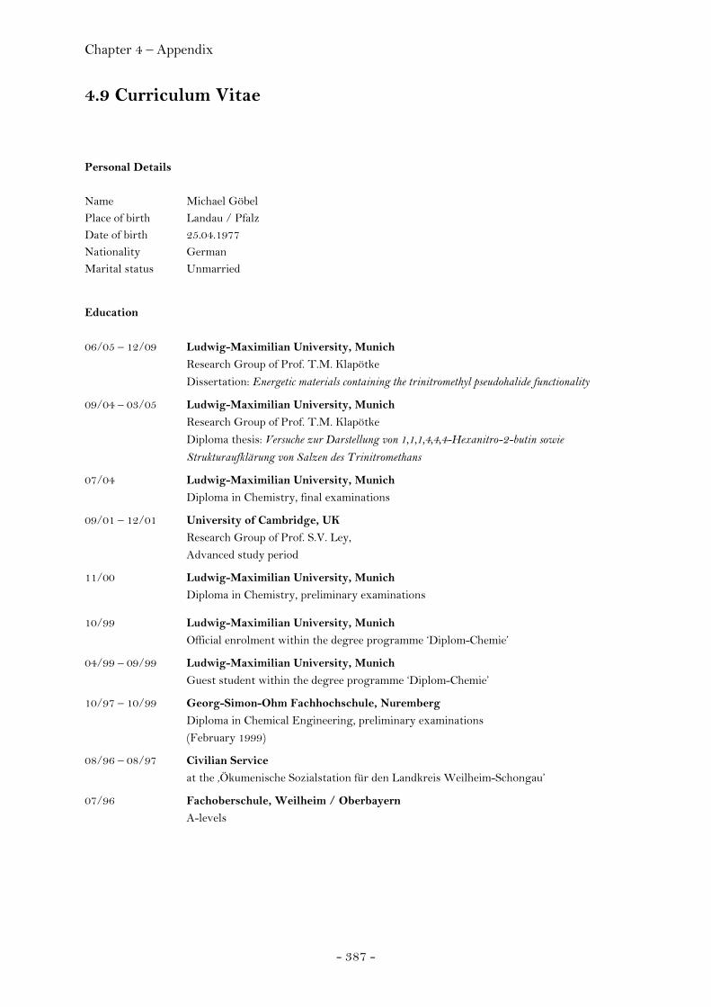
Chapter 4 – Appendix
- 387 -
4.9 Curriculum Vitae Personal Details Name Michael Göbel Place of birth Landau / Pfalz Date of birth 25.04.1977 Nationality German Marital status Unmarried Education
06/05 – 12/09 Ludwig-Maximilian University, Munich Research Group of Prof. T.M. Klapötke
Dissertation: Energetic materials containing the trinitromethyl pseudohalide functionality
09/04 – 03/05 Ludwig-Maximilian University, Munich Research Group of Prof. T.M. Klapötke
Diploma thesis: Versuche zur Darstellung von 1,1,1,4,4,4-Hexanitro-2-butin sowie Strukturaufklärung von Salzen des Trinitromethans
07/04 Ludwig-Maximilian University, Munich Diploma in Chemistry, final examinations
09/01 – 12/01 University of Cambridge, UK Research Group of Prof. S.V. Ley, Advanced study period
11/00 Ludwig-Maximilian University, Munich Diploma in Chemistry, preliminary examinations
10/99 Ludwig-Maximilian University, Munich Official enrolment within the degree programme ‘Diplom-Chemie’
04/99 – 09/99 Ludwig-Maximilian University, Munich Guest student within the degree programme ‘Diplom-Chemie’
10/97 – 10/99 Georg-Simon-Ohm Fachhochschule, Nuremberg Diploma in Chemical Engineering, preliminary examinations (February 1999)
08/96 – 08/97 Civilian Service at the ‚Ökumenische Sozialstation für den Landkreis Weilheim-Schongau’
07/96 Fachoberschule, Weilheim / Oberbayern A-levels

Chapter 4 – Appendix
- 388 -
4.10 Acknowledgements I am indebted to and thank Prof. Dr. Thomas M. Klapötke. Under his supervision it was possible to develop ideas and translate them into results. His always generous and supporting attitude together with a tremendous leap of faith towards my person is very much appreciated and I am most grateful and glad I had the opportunity to work in his group. I would like to express my gratitude to Prof. Dr. W. Beck. I feel honoured I had the chance to listen to and work with him on many topics including but not limited to mercury fulminate and I would like to thank him for being co-referee of this thesis. I am thankful to Prof. Dr. J. Evers, Prof. Dr. I.-P. Lorenz, Prof. Dr. P. Knochel and Prof. Dr. A. Kornath for their immediate attendance to act as examiners in my viva-voce. I am indebted for aid in the preparation of this thesis to many of my colleagues, friends and to many teachers in chemistry, including Dr. Anthony J. Bellamy, Dr. Richard Betz, Franziska Betzler, Dr. Sebastian M. Braun, Dr. habil. Margaret-Jane Crawford, Camilla Evangelisti, Dagmar Ewald, Dennis Fischer, Dr. Gerd Fischer, Niko Fischer, Roland Friedemann, Dr. Anton Hammerl, Stefan Huber, Prof. Dr. Konstantin Karaghiosoff, Marcos Kettner, Prof. Dr. Peter Klüfers, Prof. Dr. Bettina V. Lotsch, Franz Martin, Dr. Peter Mayer, Norbert Mayr, Dr. Carlòs Miro-Sabaté Richard Moll, Dr. Jane S. Murray, Anian Nieder, Dr. Oliver Oeckler, Gilbert Oehlinger, Alexander Penger, Dr. Kurt Pohlborn, Prof. Dr. Peter Politzer, Davin Piercey, Hendrik Radies, Sebastian Rest, Magdalena Rusan, Irene Scheckenbach, Dr. Matthias Scherr, Susanne Scheutzow, Prof. Dr. Wolfgang Schnick, Dr. Stefan Sproll, Dr. Franz X. Steemann, Dr. Jörg Stierstorfer, Karina Tarantik, Boris H. Tchitchanov, Peter C. Thumbs, Vikas Verma, Dr. Jan J. Weigand, Dr. Jan Welch, and I am glad to express my gratitude to them. The Cusanuswerk is thanked for a scholarship. Above all, I am indebted to my parents, grandparents and family who continuously encouraged and supported me.
Chapter 4 – Appendix
- 389 -
4.11 References (1) Official Energy Statistics from the U.S. Government, Energy Information
Administration (EIA), annual power data for 2007, http://www.eia.doe.gov/cneaf/electricity/epa/epat2p2.html
(2) J.B. Bdzil, T.D. Aslam, R. Henninger, J.J. Quirk, Los Alamos Science 2003, 28, 96.
(3) C.J. Wu, L.E. Fried, L.H. Yang, N. Goldman, S. Bastea, Nature Chem. 2009, 1, 57.
(4) M.R. Manaa, E.J. Reed, L.E. Fried, N. Goldman, J. Am. Chem. Soc. 2009, DOI: 10.1021/ja808196e
(5) W.C. Davies, Los Alamos Science 1981, 2, 48. (6) The New York Times, April 7, 2009, p. A1. (7) J. Akhavan (2004) The Chemistry of Explosives, 2nd edn. The Royal Society of
Chemistry, Great Britain, p. 41. (8) M. Dé Fourneaux, Overview of National Policies, ADPA IM Symposium,
Williamsburg, June 6-9, 1994. (9) UN Recommendations on the Transport of Dangerous Goods: series 7 Tests and
Criteria. (10) T.M. Klapötke (2007) Nichtmetallchemie. In: E. Riedel (ed.) Moderne Anorganische
Chemie, 3rd edn. Walter de Gruyter, Berlin.
(11) P. Noble, Jr., F.G. Borgardt, W.L. Reed, Chem. Rev. 1964, 64, 19. (12) a) M.J. Kamlet, NAVORD Report 6207, 14 Feb 1959, Naval Surface Weapons
Center; b) M.J. Kamlet, H.G. Adolph, J. Org. Chem. 1968, 33, 3073. (13) K.F. Mueller, R.H. Renner, W.H. Gilligan, H.G. Adolph, M.J. Kamlet, Combust. Flame
1983, 50, 341. (14) J. Akhavan, The Chemistry of Explosives, RSC Paperbacks, Cambridge, 1998.
(15) H. Bircher, Chimia 2004, 58(6), 357. (16) C.H. Johannson, P.A. Persson, Detonics of High Explosives. Academic Press, London &
New York, 1970. (17) I.B. Akst, Heat of Detonation, the Cylinder Test, and Performance in Munition, 9th
Symposium on Detonation, Portland, USA, 1989. (18) M.J. Kamlet, in a seminar at the White Oak Laboratory, NSWC, 1987. (19) T.M. Klapötke, J. Stierstorfer, Dalton Trans. 2009, 643. (20) M. DéFourneaux, About the Misuse of Detonation Velocities for the Characterisation of
High Explosives, 25th International Conference on ‘Energetic Materials – Analysis, Characterization and Test Techniques’ ICT, Karlsruhe (Germany), June 28 – July 1, 1994.
(21) A.J. Sanderson, Journal de Physique 1995, 5, C4-573.
(22) M.J. Kamlet, S.J. Jacobs, J. Chem. Phys. 1968, 48, 23.
(23) O. Schischkoff, Ann. Chem. 1857, 101, 216.
Chapter 4 – Appendix
- 390 -
(24) L.A. Kaplan, in Chemistry of the Nitro and Nitroso Groups, H. Feuer (Ed.), Interscience
Publishers, New York, 1970. (25) M.G.A. Shvekhgeimer, Russ. Chem. Rev. 1998, 67(1), 35.
(26) F.D. Chattaway, J. Chem. Soc. 1910, 97, 2099.
(27) P. Liang, Org. Syn. 1955, 3, 803; P. Liang, Org. Syn. 1921, 21, 105.
(28) K.J. Orton, P.V. McKie, J. Chem. Soc. 1920, 117, 283.
(29) T. Urbanski, Chemistry and Technology of Explosives 1964. I. Pergamon Press. pp. 589-594.
(30) Pictet, Genequand, Ber. 1903, 36, 2225.
(31) A. Hantzsch, Ber. 1906, 39, 2478.
(32) Pictet, Khotinsky, Ber. 1907, 40, 1163.
(33) a) Claessen, Ger. pat. 184.229; b) Claessen, Chem. Abstr. 1907, 1, 2524.
(34) a) R. Schenck, Ger. pat, 211.198, 211,199; b) R. Schenck, Chem. Abstr. 1909, 3, 2205. (35) a) Bayer & Co, Brit. pat. 24.299; b) Bayer & Co, Ger. pat. 224.057; c) Bayer & Co,
Chem. Abstr. 1911, 5, 2305; d) Berger, Compt. Rend. 1910, 151, 813; e) Berger, Bull. Soc. Chim. France 1911, 9, 26.
(36) W. Will, Chem. Ber. 1914, 47, 961.
(37) P.V. McKie, J. Soc. Chem. Ind. 1925, 44, 430T.
(38) P.V. McKie, J. Chem. Soc. 1927, 962.
(39) A. Langlet, N.V. Latypov, U. Wellmar, P. Goede, Propellants Explos. Pyrotech. 2004,
29(6), 344.
(40) J.G. Tschinkel, J. Ind. Eng. Chem. 1956, 48, 732.
(41) A.G. Anderson, Jr., R. Scotoni, Jr., E.J. Cowles, G. Fritz, J. Org. Chem. 1957, 22, 1193.
(42) C.W. Plummer, U.S. Patent 2.991.315, Chem. Abstr. 1962, 56, 2330e. (43) a) H. Meyer, Analyse und Konstitutionsermittlung organischer Verbindungen, Julius
Springer, Vienna 1938, 6th ed. p. 773; b) Ruzicka, Huyser, Pfeiffer, Seidel, Ann. 1929, 471, 21.
(44) T. Urbanski, Chemistry and Technology of Explosives 1964. I. Pergamon Press. p. 593.
(45) a) R. Schenck, Zeitsch. Angew. Chem. 1920, 33, 245; b) R. Schenck, Chem. Zeit. 1920, 44, 497; c)www.rsc.org/delivery/_ArticleLinking/DisplayArticleForFree.cfm?doi=CA9201805457&JournalCode=CA
(46) E. Rüst, A. Ebert, K. Egli, Unfälle beim chemischen Arbeiten, Rascher, 1948, p. 23. (47) H. Feuer, T. Kucera, J. Am. Chem. Soc. 1955, 77, 5740.
(48) T.N. Hall, Tetrahedron 1963, 19, 115.
(49) S.V. Lieberman, E.C. Wagner, J. Org. Chem. 1949, 14, 1001.
(50) E.R. Alexander, E.J. Underhill, J. Am. Chem. Soc. 1949, 71, 4014. (51) H. Feuer, W.A. Swarts, J. Org. Chem. 1962, 27, 1455.

Chapter 4 – Appendix
- 391 -
(52) V.I. Slovetskii, V.A. Tartakovskii, S.S. Novikov, Izv. Akad. Nauk SSSR, Otd. Khim. Nauk 1962, 1400.
(53) W. Beck, J. Evers, M. Göbel, G. Oehlinger, T.M. Klapötke, Z. Anorg. Allg. Chem.
2007, 1417. (54) W. Beck, T.M. Klapötke, J. Mol. Struct. 2008, 848, 94. (55) G.S. Hammond, W.D. Emmons, C.O. Parker, B.M. Graybill, J.H. Waters, N.F.
Hawthorne, Tetrahedron 1963, 19, 177.
(56) P.O. Tawney, U.S. Patent 3.040.105 1962; Chem. Abstr. 1962, 57, 13609.
(57) D.R. Levering, J. Org. Chem. 1962, 27, 2930.
(58) R.B. Kaplan, H. Shechter, J. Am. Chem. Soc. 1961, 83, 3535.
(59) C.W. Plummer, U.S. Patent 2,991,315 1961; Chem. Abstr. 1967, 67, 53650.
(60) S.S. Novikov, L.I. Khmelnitskii, O.V. Lebedev, Izv. Akad. SSSR 1960, 1783; Chem. Abstr. 1961, 55, 19832.
(61) A.V. Shastin, T.I. Godovikova, S.P. Golova, V.S. Kuz’min, L.I. Khmel’nitskii, B.L. Korsunskii, Mendeleev Commun. 1995, 5, 17.
(62) L. Hunter, J. Chem. Soc. 1923, 123, 543. (63) C.O. Parker, W.D. Emmons, A.S. Pangano, H.A. Rolewicz, K.S. McCallum,
Tetrahedron 1962, 17, 79.
(64) G. Darzens, M. Levy, Compt. Rend. 1949, 229, 1081.
(65) M.H. Gold, M.B. Frankel, G.B. Linden, K. Klager, J. Org. Chem. 1962, 27, 334.
(66) A.I. Shreibert, N.V. Elaskov, A.P. Khardin, V.I. Ermarchenko, Zh. Organ. Khim. 1967, 3, 1755.
(67) D.J. Glover, Tetrahedron 1963, 19, 219.
(68) F.G. Borgardt, A.K. Seeler, P. Noble, Jr., J. Org. Chem. 1966, 31, 2806.
(69) H. Feuer, E.H. White, M. Pier, J. Org. Chem. 1961, 26, 1639.
(70) E. Schmidt, Chem. Ber. 1919, 52, 400. (71) S. Iyer, N. Slagg, in: Structure and Reactivity. (1988) J.F. Liebman, A. Greenberg
(Eds.), VCH, New York, Chapter 7. (72) C.B. Storm, J.R. Stine, J.F. Kramer, in: Chemistry and Physics of Energetic Materials.
(1990) S.N. Bulusu (Ed.), Kluwer, Dordrecht, Chapter 27. (73) T.B. Brill, K. James, Chem. Rev. 1993, 93, 2667.
(74) B.M. Rice, J.J. Hare, J. Phys. Chem. A 2002, 106, 1770. (75) M. Pospisil, P. Vávra, M.C. Concha, J.S. Murray, P. Politzer, J. Mol. Model. 2009,
DOI 10.1007/s00894-009-0587-x. (76) J.F. Liebman, in: The Chemistry The Chemistry of Amino, Nitroso, Nitro and
Related Groups, Supplement F2, Chapter 8, 371. (77) B.T. Fedoroff, O.E. Sheffield, Encyclopedia of Explosives and Related Items 1962, 7, I38. (78) B.T. Fedoroff, O.E. Sheffield, Encyclopedia of Explosives and Related Items 1962, 9,
T266. (79) B.T. Fedoroff, O.E. Sheffield, Encyclopedia of Explosives and Related Items 1962, 7, I39.
Chapter 4 – Appendix
- 392 -
(80) T.S. Pivina, K.V. Sukhachev, N.S. Zefirov, Proceedings of the 5th International Conference of the ‘Groupe de Travail de Pyrotechnie’, p.71, June 6, 1993.
(81) Gaussian 03, Revision A.1, M.J. Frisch, G.W. Trucks, H.B. Schlegel, G.E. Scuseria, M.A. Robb, J.R. Cheeseman, J.A. Montgomery, Jr., T. Vreven, K.N. Kudin, J.C. Burant, J.M. Millam, S.S. Iyengar, J. Tomasi, V. Barone, B. Mennucci, M. Cossi, G. Scalmani, N. Rega, G.A. Petersson, H. Nakatsuji, M. Hada, M. Ehara, K. Toyota, R. Fukuda, J. Hasegawa, M. Ishida, T. Nakajima, Y. Honda, O. Kitao, H. Nakai, M. Klene, X. Li, J.E. Knox, H.P. Hratchian, J.B. Cross, C. Adamo, J. Jaramillo, R. Gomperts, R.E. Stratmann, O. Yazyev, A.J. Austin, R. Cammi, C. Pomelli, J.W. Ochterski, P.Y. Ayala, K. Morokuma, G.A. Voth, P. Salvador, J.J. Dannenberg, V.G. Zakrzewski, S. Dapprich, A.D. Daniels, M.C. Strain, O. Farkas, D.K. Malick, A.D. Rabuck, K. Raghavachari, J.B. Foresman, J.V. Ortiz, Q. Cui, A.G. Baboul, S. Clifford, J. Cioslowski, B.B. Stefanov, G. Liu, A. Liashenko, P. Piskorz, I. Komaromi, R.L. Martin, D.J. Fox, T. Keith, M.A. Al-Laham, C.Y. Peng, A. Nanayakkara, M. Challacombe, P.M.W. Gill, B. Johnson, W. Chen, M.W. Wong, C. Gonzalez, and J.A. Pople, Gaussian, Inc., Pittsburgh PA, 2003.
(82) C.B. Storm, J. Stine, J.F. Kramer, Sensitivity Relationships in Energetic Materials, Los Alamos Technical Report LA-UR-89-2936, 1989.
(83) M.J. Kamlet, 6th Symposium on detonation, San Diego, California, 1976.
(84) J.S. Murray, P. Politzer, Computational studies of energetic nitramines, Proceedings of the NATO advanced study institute on chemistry and physics of the molecular processes in energetic materials, 1989.
(85) H. Nefati, J.J. Legendre, C. Michot, Proceedings of the 5th International Conference of the ‘Groupe de Travail de Pyrotechnic’, p.79, June 6, 1993.
(86) A. Delpuech, J. Cherville, C. Michaud, 7th Symposium on Detonation, p.65, June 16, 1981.
(87) L. Brunet, J.M. Lombard, B. Blaise, L. Morin-Allory, Proceedings of the 5th International Conference of the ‘Groupe de Travail de Pyrotechnic’, p.89, June 6, 1993.
(88) J.S. Murray, P. Lane, P. Politzer, J. Mol. Phys. 1995, 85, 1.
(89) A. Meents, B. Dittrich, S.K.J. Johans, V. Thome, E.F. Weckert, Acta Cryst. 2008, B64, 42.
(90) B. Vogelsanger, Chimia 2004, 58(6), 402.
(91) T. Urbanski, in Chemistry and Technology of Explosives, Vol. 4, Technical University Warsaw, 1985.
(92) P. Folly, Chimia 2004, 58(6), 394. (93) G.B. Manelis; G.M. Nazin; Y.I. Rubtsov; V.A. Strunin, in Thermal Decomposition and
Combustion of Explosives and Propellants, Taylor & Francis Group, London and Berlin,
2003. (94) A. Hammerl, T. M. Klapötke, H. Nöth, M. Warchhold, G. Holl, M. Kaiser, U.
Ticmanis, Inorg. Chem. 2001, 40, 3570.
(95) R.N. Rogers, Thermochim. Acta 1975, 11, 131.

Chapter 4 – Appendix
- 393 -
(96) Systag AG (97) Joint Technical Bulletin TB 700-2 NAVSEAINST 8020.8B TO 11A-1-47 DLAR
8220.1, Department of Ammunition and Explosives Hazard Classification Procedures, Headquarters Departments of the Army, the Navy, the Air Force, and the Defense Logistics Agency, Washington DC (USA), January, 5th 1998.
(98) A. Hantzsch, A. Rinckenberger, Ber. 1899, 32, 628.
(99) O.E. Dragomir, Dissertation, Techn. Univ. Delft, 2009. (100) SciFinder Database Enquiry 02/2007 (101) H.S Jadhav, M.B. Talawar, D.D. Dhavale, S.N. Asthana, V.N. Krishnamurthy, Indian
Journal of Chemical Technology 2005, 12(2), 187.
(102) N.I. Golovina, L.O. Atovmyan, Zh. Strukt. Khim. (Russ.) 1967, 8, 307.
(103) A.M. Krishnan, P. Sjoberg, P. Politzer, J.H. Boyer, J. Chem. Soc. Perkins Trans. 2,
1989, 1237. (104) A. Gunasekaran, J.H. Boyer, Heteroatom Chemistry 1992, 3, 611.
(105) B. Dickens, Chem. Commun. 1967, 5, 246.
(106) N.I. Golovina, L.O. Atovmyan, Zh. Strukt. Khim. (Russ.)(J. Struct. Chem.) 1966, 7, 235. (107) N.V. Grigor'eva, N.V. Margolis, I.N. Shokhor, V.V. Mel'nikov, I.V. Tselinskii, Zh.
Strukt. Khim. (Russ. )(J. Struct. Chem.) 1966, 7, 278. (108) N.V. Grigor'eva, N.V. Margolis, I.N. Shokhor, I.V. Tselinskii, V.V. Mel'nikov, Zh.
Strukt. Khim. (Russ.)(J. Struct. Chem.) 1968, 9, 550.
(109) B. Dickens, J. Res. Nat. Bur. Stand. A, 1970 74, 309. (110) N.V. Podberezskaya, V.P. Doronina, V.V. Bakakin, I.I. Yakovlev, Zh. Strukt. Khim.
(Russ.)(J. Struct. Chem.) 1984, 25, 182.
(111) K.D. Scherfise, F. Weller, K. Dehnicke, Z. Naturforsch. B 1985, 40, 906.
(112) H.L. Ammon, C.S. Choi, A. Bashir-Hashemi, R.M. Moriarty, J.S. Khosrowshahi, Acta Crystallogr. 1989, C45, 319.
(113) H.L. Ammon, C.S. Choi, R.S. Damvarapu, S. Iyer, J. Alster, Acta Crystallogr. 1990,
C46, 295. (114) N.V. Podberezskaya, N.V. Pervukhina, V.P. Doronina, Zh. Strukt. Khim.(Russ.)(J.
Struct. Chem.) 1991, 32, 34. (115) H. Bock, R. Dienelt, H. Schodel, Z. Havlas, Chem.Commun. 1993, 1792.
(116) H. Schodel, R. Dienelt, H. Bock, Acta Crystallogr. 1994, C50, 1790.
(117) H. Bock, T. Hauck, C. Nather, Z. Havlas, Z. Naturforsch. 1994, 49, 1012.
(118) J.C. Bryan, M.N. Burnett, A.A. Gakh, Acta Crystallogr., 1998, C(54)9, 1229. (119) M. Göbel, T.M. Klapötke, P. Mayer, Z. Anorg. Allg. Chem. 2006, 632(6), 1043. (120) M. Göbel, T.M. Klapötke, P.C. Thumbs, New Trends in Research of Energetic Materials,
Proceedings of the Seminar, 9th, Pardubice, Czech Republic, Apr. 19-21, 2006 (2006), (Pt. 1), 127.
(121) A. Gunasekaran, J.H. Boyer, Heteroatom Chemistry 1992, 3, 611. (122) P.S. Dendage, D.B. Sarwade, S.N. Asthana, H. Singh, J. Energ. Mat. 2001, 19, 41.
Chapter 4 – Appendix
- 394 -
(123) A.M. Krishnan, P. Sjoberg, P. Politzer, J.H. Boyer, J. Chem. Soc. Perkins Trans. 2,
1989, 1237. (124) J.C. Bryan, M.N. Burnett, A.A. Gakh, Acta Crystallogr., 1998, C(54)9, 1229.
(125) H.M. Niemeyer, J. Mol. Struct. 1978, 50, 123.
(126) K.E. Edgecombe, R.J. Boyd, Can. J. Chem. 1983, 61, 45.
(127) B. Dickens, Chem. Commun. 1967, 5, 246.
(128) B. Dickens, J. Res. Nat. Bur. Stand. A, 1970 74, 309. (129) N.V. Podberezskaya, V.P. Doronina, V.V. Bakakin, I.I. Yakovlev, Zh. Strukt. Khim.
(Russ.)(J. Struct. Chem.) 1984, 25, 182.
(130) K.D. Scherfise, F. Weller, K. Dehnicke, Z. Naturforsch. B 1985, 40, 906.
(131) H.L. Ammon, C.S. Choi, R.S. Damvarapu, S. Iyer, J. Alster, Acta Crystallogr. 1990, C46, 295.
(132) N.V. Podberezskaya, N.V. Pervukhina, V.P. Doronina, Zh. Strukt. Khim.(Russ.)(J.
Struct. Chem.) 1991, 32, 34.
(133) H. Schodel, R. Dienelt, H. Bock, Acta Crystallogr. 1994, C50, 1790.
(134) H. Bock, T. Hauck, C. Nather, Z. Havlas, Z. Naturforsch. 1994, 49, 1012.
(135) J.C. Bryan, M.N. Burnett, A.A. Gakh, Acta Crystallographica, 1998, C(54)9, 1229. (136) M. Göbel, T.M. Klapötke, P.C. Thumbs, New Trends in Research of Energetic
Materials, Proceedings of the Seminar, 9th, Pardubice, Czech Republic, Apr. 19-21, 2006 (2006), (Pt. 1), 127.
(137) B. Dickens, Chem. Commun. 1967, 5, 246.
(138) B. Dickens, J. Res. Nat. Bur. Stand. A, 1970 74, 309.
(139) D.V. Jahagirdar, R.M. Kharwadkar, Ind. J. Chem. 1981, 20A, 635.
(140) J.C. Bryan, M.N. Burnett, A.A. Gakh, Acta Crystallogr., 1998, C(54)9, 1229. (141) Gaussian 03, Revision A.1, M.J. Frisch, G.W. Trucks, H.B. Schlegel, G.E. Scuseria,
M.A. Robb, J.R. Cheeseman, J.A. Montgomery, Jr., T. Vreven, K.N. Kudin, J.C. Burant, J.M. Millam, S.S. Iyengar, J. Tomasi, V. Barone, B. Mennucci, M. Cossi, G. Scalmani, N. Rega, G.A. Petersson, H. Nakatsuji, M. Hada, M. Ehara, K. Toyota, R. Fukuda, J. Hasegawa, M. Ishida, T. Nakajima, Y. Honda, O. Kitao, H. Nakai, M. Klene, X. Li, J.E. Knox, H.P. Hratchian, J.B. Cross, C. Adamo, J. Jaramillo, R. Gomperts, R.E. Stratmann, O. Yazyev, A.J. Austin, R. Cammi, C. Pomelli, J.W. Ochterski, P.Y. Ayala, K. Morokuma, G.A. Voth, P. Salvador, J.J. Dannenberg, V.G. Zakrzewski, S. Dapprich, A.D. Daniels, M.C. Strain, O. Farkas, D.K. Malick, A.D. Rabuck, K. Raghavachari, J.B. Foresman, J.V. Ortiz, Q. Cui, A.G. Baboul, S. Clifford, J. Cioslowski, B.B. Stefanov, G. Liu, A. Liashenko, P. Piskorz, I. Komaromi, R.L. Martin, D.J. Fox, T. Keith, M.A. Al-Laham, C.Y. Peng, A. Nanayakkara, M. Challacombe, P.M.W. Gill, B. Johnson, W. Chen, M.W. Wong, C. Gonzalez, and J.A. Pople, Gaussian, Inc., Pittsburgh PA, 2003.
(142) Test methods according to the UN Recommendations on the Transport of Dangerous Goods, Manual of Tests and Criteria, fourth revised edition, United
Chapter 4 – Appendix
- 395 -
Nations Publications, New York and Geneva, 2003, ISBN 92-1-139087-7, Sales No. E.03.VIII.2.; 13.4.2 Test 3(a)(ii) BAM Fallhammer.
(143) Y.A. Lebedev, E.A. Miroshnichenko, Y.K. Knobel, Termokhimiya nitrosoedinenii
[Thermochemistry of nitrocompounds], Nauka, Moscow, 1970, 168.
(144) T.S. Kon´kova, Yu.N. Matyushin, Russ. Chem. Bull. 1998, 47, 2371.
(145) M. Sućeska, Propellants, Explos., Pyrotech. 1991, 16, 197.
(146) M. Sućeska, Proc. of 30th Int. Annual Conference of ICT, June 29 – July 2, Karlsruhe,
Germany, 1999, 50.
(147) M. Sućeska, Propellants Explosives and Pyrotechniques 1999, 24, 280.
(148) M. Sućeska, Proc. of 32nd Int. Annual Conference of ICT, July 3-6, Karlsruhe, Germany,
2001, 110.
(149) M. Sućeska, Materials Science Forum 2004, 465, 325.
(150) P.S. Dendage, D.B. Sarwade, S.N. Asthana, H. Singh, J. Energ. Mat. 2001, 19, 41.
(151) J.H. Köhler, Explosivstoffe, 9. Ed., Wiley-VCH, New York, 1998, 344.
(152) M. Sućeska, Materials Science Forum 2004, 465, 325.
(153) J.H. Köhler, Explosivstoffe, 9. Ed., Wiley-VCH, New York, 1998, 166. (154) Test methods according to the UN Recommendations on the Transport of
Dangerous Goods, Manual of Tests and Criteria, fourth revised edition, United Nations Publications, New York and Geneva, 2003, ISBN 92-1-139087-7, Sales No. E.03.VIII.2.; 13.4.2 Test 3(a)(ii) BAM Fallhammer.
(155) HF-Vacuum-Pruefer, type VP 24. (156) B. Dickens, Chem. Commun. 1967, 5, 246.
(157) A. Gunasekaran, J.H. Boyer, Heteroatom Chemistry 1992, 3, 611.
(158) R. Tanbug, K. Kirschbaum, A.A. Pinkerton, J. Chem. Crystallogr. 1999, 29, 4. (159) R. Friedemann, M. Göbel, T.M. Klapötke, S. Scheutzow, New Trends in Research of
Energetic Materials, Proceedings of the Seminar, 10th, Czech Republic, Apr. 25-27, 2007, 875.
(160) M.L. Hobbs, M.R. Baer, Calibration of the BKW-EOS with a large product species data base and measured CJ properties, Proc. of the 10th Symp. (Int.) on Detonation, ONR 33395-12, Boston, MA, July 12-16 1993, 409.
(161) J.N. Varghese, A.M. O’Connell, E.N. Maslen, Acta Cryst. 1977, B33, 2102.
(162) R. Tanbug, K. Kirschbaum, A.A. Pinkerton, J. Chem. Crystallogr. 1999, 29, 4.
(163) A. Martin, A.A. Pinkerton, Acta Cryst. 1995, C51, 2174.
(164) Y. Wang, B. Wei, Q. Wang, J. Cryst. Spectrosc. Res. 1990, 20, 79. (165) U. Müller, H. Bärnighausen, Acta Cryst. 1970, B26, 1671.
(166) M. Göbel, T.M. Klapötke, Chem. Commun. 2007, 3180.
(167) H. Henke, H. Bärnighausen, Acta Cryst. 1972, B28, 1100.
(168) A. Dworkin, R. Naumann, C. Seigfred, J.M. Karty, J. Org. Chem. 2005, 70, 7605; and references therein.
(169) R.L. Livingston, C.N.R. Rao, J. Phys. Chem. 1960, 64, 756.
Chapter 4 – Appendix
- 396 -
(170) H. Henke, H. Bärnighausen, Acta Cryst. 1972, B28, 1100. (171) A.J. Bellamy, L. Cassioli, A.E. Contini, New Trends in Research of Energetic Materials,
Proceedings of the 8th Seminar (Part 1), Czech Republic, Apr. 19-21, 2005, 472.
(172) Experimental value, M. Sućeska, Materials Science Forum 2004, 465, 325.
(173) S.K. Bhattacharjee, H.L. Ammon, Acta Cryst. 1981, B37, 2082. (174) S. Sorriso, in:The Chemistry of Amino, Nitroso, Nitro and Related Groups, S. Patai
(Ed.), Supplement F, Part 1, Chapter 1. (175) A. Domenicano, A. Vaciago, C.A. Coulson, Acta Cryst. 1975, B31, 221.
(176) A. Domenicano, A. Vaciago, C.A. Coulson, Acta Cryst. 1975, B31, 1630.
(177) A. Domenicano, A. Vaciago, C.A. Coulson, Tetrahedron Lett. 1976, 13, 1029.
(178) L.S. Bartell, J. Chem. Phys. 1960, 32, 827.
(179) L.S. Bartell, Tetrahedron 1962, 17, 177.
(180) L.S. Bartell, J. Chem. Educ. 1968, 45, 754.
(181) O.L. Carter, A.T. McPhail, G.A. Sim, J. Chem. Soc. (A) 1966, 822.
(182) L. Nygaard, I. Bojesen, T. Pedersen, J. Rastrup-Andersen, J. Mol. Struct. 1968, 2, 209.
(183) R.J. Gillespie, R.S. Nyholm, Quart. Rev. 1957, 11, 339.
(184) R.J. Gillespie, J. Chem. Educ. 1963, 40, 295.
(185) R.J. Gillespie, J. Chem. Soc. 1963, 4672.
(186) R.J. Gillespie, Angew. Chem. Int. Ed. 1967, 6, 819.
(187) R.J. Gillespie, J. Chem. Educ. 1970, 47, 18. (188) R.J. Gillespie, Molecular Geometry, Van Nostrand-Reinhold, London, 1972.
(189) E.G. Cox, D.W.J. Cruickshank, J.A.S. Smith, Proc. R. Soc. Sect. A 1958, 247, 1.
(190) H.H. Cady, A.C. Larson, Acta Cryst. 1965, 18, 485.
(191) S.K. Bhattacharjee, H.L. Ammon, Acta Cryst. 1981, B37, 2082.
(192) M.D. Coburn, D.G. Ott, J. Heterocycl. Chem. 1990, 27, 1941. (193) J. Sauer, in : Comprehensive Heterocyclic Chemistry, Ch. 6.21 (194) S.F. Nelsen, Y. Kim, J. Org. Chem. 1991, 56, 1045.
(195) J.C.A. Boeyens, J. Cryst. Molec. Struct. 1978, 8, 317.
(196) J.D. Coates, L.A. Achenbach, Nat. Rev. Microbiol. 2004, 2, 569.
(197) a) M.J. Kamlet, NAVORD Report 6207, 14 Feb 1959, Naval Surface Weapons Center. b) M.J. Kamlet, H.G. Adolph, J. Org. Chem. 1968, 33, 3073. c) V. Grakauskas, K. Baum, US Patent 3423419, 1969. d) V. Grakauskas, K. Baum, J. Org. Chem. 1969, 34, 3927.
(198) a) T.M. Klapötke, C.M. Sabaté, Chem. Mater. 2008, 20(11), 3629. b) C. Darwich, T.M. Klapötke, C.M. Sabaté, Chem. Eur. J. 2008, 14, 5756. c) J. Stierstorfer, T.M. Klapötke,
A. Hammerl, B. Chapman, Z. Anorg. Allg. Chem. 2008, 634, 1051. d) M. Göbel, T.M.
Klapötke, Z. Anorg. Allg. Chem. 2007, 633(7), 1006. e) Y. Guo, H. Gao, B. Twamley,
J.M. Shreeve, Adv. Mat. 2007, 19, 2884. f) M. Göbel, K. Karaghiosoff, T.M. Klapötke,
Angew. Chem. Int. Ed. 2006, 45(36), 6037. g) M. Hiskey, A. Hammerl, G. Holl, T.M.
Chapter 4 – Appendix
- 397 -
Klapötke, K. Polborn, J. Stierstorfer, J.J. Weigand, Chem. Mater. 2005, 17, 3784. h)
H. Xue, J.M. Shreeve, Adv. Mat. 2005, 17, 2142.
(199) J. Hine, W.C. Bailey, J. Org. Chem. 1961, 26, 2098.
(200) Th. N. Hall, Tetrahedron 1963, 19(1), 115. (201) I.B. Starchenkov, V.G. Andrianov, A.F. Mishnev, Chemistry of Heterocyclic Compounds
1997, 33, 216. (202) A.B. Sheremetev, V.O. Kulagina, L.V. Batog, O.V. Lebedev, I.L. Yudin, T.S. Pivina,
Proceedings of the International Pyrotechnics Seminar 1996, 22, 377.
(203) A.V. Kalinin, E.T. Apasov, Y.A. Strelenko, S.L. Ioffe, V.A. Tartakovsky, Izvestiya Akademii Nauk, Seriya Khimicheskaya 1993, 4, 736.
(204) a) R. Schenck; G.A. Wetterholm, U.S. Pat. 2,731,460 (1956).
b) R. Schenck; G.A. Wetterholm, Chem. Abstr. 1956, 50, 7125. (205) A.A. Kozyro, V.V. Simirsky, A.P. Krasulin, V.M. Sevruk, G.J. Kabo, M.L. Gopanik,
Y.V. Grigotiev, Zh. Fiz. Khim. 1990, 64, 656.
(206) A. Gao, Y. Oyumi, T.B. Brill, Combust. Flame 1991, 83, 345.
(207) D. Moderhack, K.H. Goos, L. Preu, Chem. Ber. 1990, 123, 1575. (208) N.N. Makhova, A.B. Sheremetev, I.V. Ovchinnikov, I.L. Yudin, A.S. Ermakov, P.V.
Bulatov, D.B. Vinogradov, D.B. Lempert, G.B. Manelis, International Annual
Conference of ICT 2004, 140/1.
(209) X.X. Zhou, Y.Z. YU, Kogyo-kayaku 1991, 52(4), 251.
(210) T.P. Russell, T.M. Allen, Y.M. Gupta, Chem. Phys. Lett. 1997, 267, 351.
(211) R. Gilardi, C. George, J. L. Flipper-Anderson, Acta Cryst. 1988, C44, 1686.
(212) M.D. Coburn, D.G. Ott, J. Heterocycl. Chem. 1990, 27, 1941.
(213) Ger. Pat. 934 694, 1955; Chem. Abstr. 1959, 53, 17513b. (214) I.V. Ovchinnikov, A.S. Kulikov, M.A. Epishina, N.N. Makhova, V.A. Tartakovsky,
Russ. Chem. Bull. 2005, 54, 1346. (215) N.K. Sundholm, T.C. Richards, D.L. Schoene, Naugatuch Chem Div, US Rubber Co,
Progress Report (15 Feb. 1950 to 15 Apr. 1950), NORD 10121, p.4. (216) J.M. Dyke, G. Levita, A. Morris, J.S. Ogden, A.A. Dias, M. Algarra, J.P. Santos, M.L.
Costa, P. Rodrigues, M.M. Andrade, M.T. Barros, Chem. Eur. J. 2005, 11, 1665.
(217) R.C. Farmer, J. Chem. Soc. 1920, 1603.
(218) C.N. Hinshelwood, J. Chem. Soc. 1921, 721. (219) J.M. Rosen, J.C. Dacons, NOLTR 62-192, 15 Nov 1962, Naval Surface Weapons
Center. (220) Y.A. Lebedev, E.A. Miroshnichenko, Y.K. Knobel, Termokhimiya nitrosoedinenii
(Thermochemistry of nitrocompounds) 1970, 168.
(221) G.A. Petersson, T.G. Tensfeldt, J.A. Montgomery, Jr., J. Chem. Phys. 1991, 94, 6091.
(222) J.W. Ochterski, G.A. Petersson, J.A. Montgomery, Jr., J. Chem. Phys. 1996, 104, 2598.

Chapter 4 – Appendix
- 398 -
(223) J.A. Montgomery, Jr., M.J. Frisch, J.W. Ochterski, G.A. Petersson, J. Chem. Phys.
2000, 112, 6532. (224) L.A. Curtiss, K. Raghavachari, P.C. Redfern, J.A. Pople, J. Chem. Phys. 1997, 106(3),
1063. (225) M.S. Westwell; M.S. Searle; D.J. Wales; D. II. Williams, J. Am. Chem. Soc. 1995, 117,
5013. (226) J. Chao, F.D. Rossini, J. Chem. Eng. Data 1965, 10, 374.
(227) N.D. Lebedeva, V.L. Ryadnenko, I.N. Kuznetsova, Russ. J. Phys. Chem. (Engl. Transl.) 1968, 42, 962.
(228) C. Lenchitz, R.W. Velicky, G. Silvestro, L.P. Schlosberg, J. Chem. Thermodyn. 1971,
3, 689. (229) V.I. Pepekin, Y.N. Matyushin, Y.A. Lebedev, Thermochemistry of N-nitro- and N-
nitrosoamines of the alicyclic series, Bull. Acad. Sci. USSR, Div. Chem. Sci. 1974, 1707.
(230) G. Krien, H.H. Licht, J. Zierath, Thermochim. Acta 1973, 6, 465. (231) R.L. Simpson, P.A. Urtiew, D.L. Ornellas, G.L. Moody, K.J.Scribner, D.M. Hoffman,
Propellants, Explos., Pyrotech. 1997, 22, 249. (232) V.A. Ostrovskii, M.S. Pevzner, T.P. Kofman, I.V. Tselinskii, Targets Heterocycl. Syst.
1999 , 3, 467.
(233) M.F. Cheng, H.O. Ho, C.S. Lam, S. Chow, C.S. Li, J. Serb. Chem. Soc. 2002, 67(4), 257.
(234) M. Göbel, T.M. Klapötke, Acta Cryst. 2007, C63, o562.
(235) M. Göbel, T. M. Klapötke, Acta Cryst. 2008, C64, o58. (236) J. Köhler, R. Meyer, A. Homburg, Explosivstoffe, 10th ed. 2008, Wiley VCH,
Weinheim.
(237) M. Sućeska, Propellants, Explos., Pyrotech. 1991, 16, 197.
(238) M. Sućeska, Proc. of 30th Int. Annual Conference of ICT, June 29 – July 2, Karlsruhe,
Germany, 1999, 50.
(239) M. Sućeska, Propellants, Explos., Pyrotech. 1999, 24, 280.
(240) M. Sućeska, Proc. of 32nd Int. Annual Conference of ICT, July 3-6, Karlsruhe, Germany,
2001, 110.
(241) M. Sućeska, Materials Science Forum 2004, 465, 325.
(242) M.L. Hobbs, M.R. Baer, Calibration of the BKW-EOS With a Large product Species Data Base and Measured C-J Properties, Proc. of the 10th Symp. (International) on Detonation, ONR 33395-12, Boston, MA, July 12-16 1993, p. 409.
(243) EG A.14: Prüfverfahren nach Anhang I Teil A.14 der Richtlinie 92/69/EWG der Kommission vom 31. Juli 1992 zur Siebzehnten Anpassung der Richtlinie 67/548/EWG zur Angleichung der Rechts- und Verwaltungsvorschriften für die Einstufung, Verpackung und Kennzeichnung gefährlicher Stoffe an den technischen Fortschritt (ABI. EG Nr. L 383 S. 113 und Nr. L 383 A S. 1 (S. 87)) as well as
Chapter 4 – Appendix
- 399 -
Recommendations on the Transport of Dangerous Goods: Tests and criteria, 1990, United Nations, New York.
(244) A. Bondi, J. Phys. Chem. 1964, 68, 441.
(245) H.E. Ungnade, L.W. Kissinger, Tetrahdron 1963,19, 121.
(246) R.D. Shannon, Acta Cryst. 1976, A32, 751.
(247) M.D. Lind, Acta Cryst. 1970, B26, 590.
(248) F.H. Allen, J.P.M. Lommerse, V.J. Hoy, J.A.K. Howard, G.R. Desiraju, Acta Cryst. 1997, B53, 1006.
(249) J.P.M. Lommerse, A.J. Stone, R. Taylor, F.H. Allen, J. Am. Chem. Soc. 1996, 118, 3108.
(250) F.H. Allen, C.A. Baalham, J.P.M. Lommerse, P.R. Raithby, Acta Cryst. 1998, B54, 320. (251) I.B. Starchenkov, V.G. Andrianov, A.F. Mishnev, Chemistry of Heterocyclic Compounds
1997, 33, 216. (252) A.B. Sheremetev, V.O. Kulagina, L.V. Batog, O.V. Lebedev, I.L. Yudin, T.S. Pivina,
Proceedings of the International Pyrotechnics Seminar 1996, 22, 377. (253) A.V. Kalinin, E.T. Apasov, Y.A. Strelenko, S.L. Ioffe, V.A. Tartakovsky, Izvestiya
Akademii Nauk, Seriya Khimicheskaya 1993, 4, 736.
(254) R.L. Collin, W.N. Lipscomb, Acta Cryst. 1951, 4, 10.
(255) K.H. Linke, H.G. Kalker, Z. Anorg. Allg. Chem. 1977, 434 , 165.
(256) A. Héroux, F. Brisse, Acta Cryst. 1997, C53, 1318.
(257) Holleman-Wiberg, Lehrbuch der Anorganischen Chemie, 102nd ed., Walther de Gruyter,
Berlin, New York, 2007, p.2006. (258) R. Goddard, O. Heinemann, C. Kruger, Acta Cryst. 1997, C53, 590.
(259) C. Krieger, H. Fischer, F.A. Neugebauer, Acta Cryst. 1987, C43, 1320. (260) M.E. Hill, US Patent 3.375.266, 1956. (261) T.N. Hall, J. Org. Chem. 1968, 33, 4557.
(262) F.H. Allen, Acta Cryst. 2002, B58, 380.
(263) Holleman-Wiberg, Lehrbuch der Anorganischen Chemie, 101st ed., de Gruyter, Berlin
1995, p. 504. (264) P. Geißler, T.M. Klapötke, H.J. Kroth, Spectrochim. Acta 1995, Part A, 51A, 1075.
(265) F.H. Allen, Acta Cryst. 2002, B58, 380. (266) A. Héroux, F. Brisse, Acta Cryst. 1997, C53, 1318.
(267) F.H. Allen, Acta Cryst. 2002, B58, 380.
(268) M. Göbel, T.M. Klapötke, Adv. Funct. Mater. 2009, 19, 347.
(269) F.H. Allen, O. Kennard, Chem. Des. Autom. News 1993, 8, 31. (270) C.P. Butts, L. Eberson, K.L. Fulton, M.P. Hartshorn, W.T. Robinson, D.J.
Timmerman-Vaughan, Acta Chem. Scand. 1996, 50, 991.
(271) Adolf, 1996, private communication to the Cambridge Structural Database, refcode NABMIM.
(272) Y. Oyumi, A.L. Rheingold, T.B. Brill, Propellants, Explos., Pyrotech. 1987, 12, 46.
Chapter 4 – Appendix
- 400 -
(273) S.K. Bhattacharjee, H.L. Ammon, Acta Crystallogr. 1982, B38, 2503. (274) T.M. Klapötke, B. Krumm, M. Scherr, G. Spieß, F.X. Steemann, Z. Anorg. Allg. Chem.
2008, 634, 1244.
(275) Y. Oyumi, T. B. Brill, A.L. Rheingold, J. Phys. Chem. 1985, 89, 4824. (276) L.O. Atovmyan, N.I. Golovina, L.T. Eremenko, N.G. Zhitomirskaya, G.V. Oreshko,
M.A. Fadeev, Izv. Akad. Nauk SSSR, Ser. Khim. (Russ.) (Russ. Chem. Bull.) 1984, 549. (277) N.I. Golovina, L.O. Atovmyan, N.V. Chukanov, G.V. Oreshko, R.F. Trofimova, M.A.
Fadeev, L.T. Eremenko, Zh. Strukt. Khim. (Russ.) (J. Struct. Chem.) 1990, 31, 126. (278) L.O. Atovmyan, R.G. Gafurov, N.I. Golovina, L.T. Eremenko, B.S. Fedorov, Zh.
Strukt. Khim. (Russ.)(J. Stuct. Chem.) 1980, 21, 135. (279) L.T. Eremenko, L.O. Atovmyan, N.I. Golovina, G.V. Oreshko, M.A. Fadeev, Chem.
Commun. 1984, 709. (280) C. Jinhua, M. Shaoping, Z. Xiaotong, Jiegou Huaxue (Chin.) (Chinese J. Struct. Chem.)
1983, 2, 41.
(281) S.K. Bhattacharjee, H.L. Ammon, Acta Crystallogr. 1981, B37, 2082. (282) M.J.Frisch, G.W. Trucks, H.B. Schlegel, G.E. Scuseria, M.A.Robb, J.R. Cheeseman,
J.A. Montgomery Jr., T. Vreven, K.N.Kudin, J.C. Burant, J.M. Millam, S.S. Iyengar, J. Tomasi, V. Barone, B. Mennucci, M. Cossi, G. Scalmani, N. Rega, G.A.Petersson, H. Nakatsuji, M. Hada, M. Ehara, K. Toyota, R. Fukuda, J. Hasegawa, M. Ishida, T. Nakajima, Y. Honda, O. Kitao, H. Nakai, M. Klene, X. Li, J.E. Knox, H.P. Hratchian, J.B. Cross, V. Bakken, C. Adamo, J. Jaramillo, R. Gomperts, R.E. Stratmann, O.Yazyev, A.J. Austin, R. Cammi, C. Pomelli, J.W. Ochterski, P.Y.Ayala, K. Morokuma, G.A. Voth, P. Salvador, J.J. Dannenberg, V.G.Zakrzewski, S. Dapprich, A.D. Daniels, M.C. Strain, O. Farkas, D.K. Malick, A.D. Rabuck K. Raghavachari, J.B. Foresman, J.V. Ortiz, Q. Cui, A.G. Baboul, S. Clifford, J. Cioslowski, B.B. Stefanov, G. Liu, A. Liashenko, P. Piskorz, I. Komaromi, R.L. Martin, D.J. Fox, T. Keith, M.A. Al-Laham, C.Y.Peng, A. Nanayakkara, M. Challacombe, P.M.W. Gill, B. Johnson, W. Chen, M.W. Wong, C. Gonzalez, J.A. Pople. Gaussian 03, Revision D.01. Gaussian, Inc Wallingford CT. 2004.
(283) CrysAlis CCD, Oxford Diffraction Ltd., Version 1.171.27p5 beta (release 01-04-2005 CrysAlis171.NET.
(284) CrysAlis RED, Oxford Diffraction Ltd., Version 1.171.27p5 beta (release 01-04-2005 CrysAlis171.NET.
(285) A. Altomare, G. Cascarano, C. Giacovazzo, A. Guagliardi, J. Appl. Crystallogr. 1993, 26, 343.
(286) G.M. Sheldrick, SHELXL-97, University of Göttingen, 1997. (287) L.J. Farrugia, J. Appl. Crystallogr. 1999, 32, 837. (288) A.L. Spek, Platon, Utrecht University, Utrecht, The Netherlands, 1999. (289) NATO standardization agreement (STANAG) on explosives, impact sensitivity tests,
no. 4489, Ed. 1, Sept. 17, 1999.
Chapter 4 – Appendix
- 401 -
(290) WIWEB-Standardarbeitsanweisung 4-5.1.02, Ermittlung der Explosionsgefährlichkeit, hier der Schlagempfindlichkeit mit dem Fallhammer, Nov. 8, 2002.
(291) NATO standardization agreement (STANAG) on explosives, friction sensitivity tests, no. 4487, Ed. 1, Aug. 22, 2002.
(292) WIWEB-Standardarbeitsanweisung 4-5.1.03, Ermittlung der Explosionsgefährlichkeit oder der Reibeempfindlichkeit mit dem Reibeapparat, Nov. 8, 2002.
(293) R.C. West, S.M. Selby, Handbook of Chemistry and Physics, The Chemical Rubber Co., Cleveland, 1967, 48th edn, p. D22.
(294) N.S. Marans, R.P. Zelinski, J. Am. Chem. Soc. 1950, 72, 5329.
(295) H. Feuer, T.J. Kucera, J. Org. Chem. 1960, 25, 2069. (296) K.G. Ship, M.E. Hill, NAVORD 6752, 1960. (297) W.J. Murray, C.W. Sauer, US Patent 3.006.957, 1957; Chem. Abstr. 1962, 56, 2330c.
(298) H.E. Ungnade, L.W. Kissinger, Tetrahedron 1963, 19, 121.
(299) M.D. Coburn, D.G. Ott, J. Heterocycl. Chem. 1990, 27, 1941. (300) M.E. Hill, Preparation and Properties of Bis(Trinitroethyl)Carbonate, NAVORD 3656
(1953). (301) M.E. Hill, Development of the Synthesis of Bis(Trinitroethyl)Carbonate, BTNEC, A
New Oxygen Rich High Explosive, NAVORD 3469 (1955). (302) W.H. Gilligan, S.L. Stafford, Synthesis 1979, 8, 600.
(303) J.M. Welch, Dissertation, Ludwig-Maximilan Univ., 2008. (304) M.J. Crawford, J. Evers, M. Göbel, T.M. Klapötke, P. Mayer, G. Oehlinger, J.M.
Welch, Propellants, Explos., Pyrotech. 2007, 32(6), 478.
(305) M. Göbel, T.M. Klapötke, Z. Anorg. Allg. Chem. 2007, 633(7), 1006.
(306) A.T. Mixon, J. Cioslowski, J. Am. Chem. Soc. 1991, 113, 6760.
(307) J. Cioslowski, S.T. Mixon, E.D. Fleischmann, J. Am. Chem. Soc. 1991, 113, 4751. (308) M.A. Pietsch, M.B. Hall, J. Phys. Chem. 1994, 98, 11373. (309) K.Y. Mathews, D.W. Ball, J. Mol. Struct. (Theochem) 2009, 90(2), 15-20.
(310) S.J. Angyal, W.K. Warburton, J. Chem. Soc. 1951, 2492.
(311) R.W. Alder, M.R. Bryce, N.C. Goode, N. Miller, J. Owen, J. Chem. Soc., Perkin Trans. 1 1981, 2840.
(312) H.A. Staab, T. Saupe, Angew. Chem. 1988, 100, 895; Angew. Chem., Int. Ed. Engl. 1988, 27, 865.
(313) L. Pauling, The Nature of the Chemical Bond, 3rd ed.; Cornell University Press: Ithaca, NY, 1960, p.286.
(314) P. Gund, J. Chem. Educ. 1972, 49, 100.
(315) P. Kollman, J. McKelvey, P. Gund, J. Am. Chem. Soc. 1975, 97, 1640.
(316) J.F. Capitani, L. Pedersen, Chem. Phys. Lett. 1978, 54, 547.
(317) A.M. Sapse, L.J. Massa, J. Org. Chem. 1980, 45, 719.

Chapter 4 – Appendix
- 402 -
(318) T. Ohwada, A. Itai, T. Ohta, K. Shudo, J. Am. Chem. Soc. 1987, 109, 7036.
(319) N. Sreerama, S. Vishveshwara, J. Mol. Struct. 1989, 194, 61.
(320) M. L. Williams, J. E. Gready, J. Comput. Chem. 1989, 10, 35.
(321) K. B. Wiberg, J. Am. Chem. Soc. 1990, 112, 4177.
(322) A. Gobbi, G. Frenking, J. Am. Chem. Soc. 1993, 115, 2362.
(323) N. de Vries, C. E. Costello, A.G. Jones and A. Davidson, Inorg. Chem. 1990, 29, 1348.
(324) W.P. Fehlhammer, R. Metzner and W. Sperber, Chem. Ber. 1994, 127, 829. (325) P.J. Bailey, K.J. Grant, S. Pace, S. Parsons and L.J. Stewart,
J. Chem. Soc., Dalton Trans. 1997, 4263.
(326) H.J. Kabbe, K. Eiter, F. Möller, Liebigs Ann. Chem. 1967, 704, 140.
(327) M. Becker, M. Jansen, Acta Crystallogr. 1999, C55, IUC 9900094.
(328) A. Gobbi, G. Frenking, J. Am. Chem. Soc. 1993, 115, 2362. (329) V.A. Ostrovskii, G.I. Koldobskii, R.E. Trifonov, in: Comprehensive Heterocyclic
Chemistry, 2008, Ch. 6.07, 257. (330) F.R. Benson, in: Heterocyclic Compounds, R.C. Elderfield (Ed.), Wiley, New York,
1967, Vol. 8, p.8. (331) A. Hammerl, Dissertation, Ludwig-Maximilan Univ., Munich 2001.
(332) J.J. Weigand, Dissertation, Ludwig-Maximilan Univ., Munich 2005.
(333) J. Stierstorfer, Dissertation, Ludwig-Maximilan Univ., Munich 2009.
(334) P. Cmoch, L. Stefaniak, E. Melzer, S. Baloniak, G.A. Webb, Magn.Reson.Chem. 1999, 37, 493.
(335) J. Benito, Ernesto de Jesus, F. de la Mata, J. C. Flores, R. Gomez, Gomez-Sal, J.Organomet.Chem. 2002, 664, 258.
(336) R. M. Gauvin, C. Lorber, R. Choukroun, B. Donnadieu, J. Kress, Eur.J.Inorg.Chem.
2001, 2337. (337) Y. Hamada, Y. Yamamoto, H. Shimizu, J.Organomet.Chem. 1996, 510, 1.
(338) M. Schorr, W. Schmidt, Phosphorus, Sulfur Silicon Relat. Elem. 1992, 68, 25.
(339) W.Verboom, W.Visser, D.N.Reinhoudt, Synthesis 1981, 807.
(340) U. Wannagat, H.Bürger, C.Krüger, J.Pump, Z.Anorg.Allg.Chem. 1963, 321, 208. (341) A.S.Gordetsov, S.V.Zimina, L.N.Martynkova, E.M.Moseeva, S.E.Skobeleva,
T.K.Postnikova, V.L.Tsvetkova, R.P.Zaharova, Metalloorganicheskaya Khimiya 1992, 5, 811.
(342) J. Benito, Ernesto de Jesus, F. de la Mata, J. C. Flores, R. Gomez, Gomez-Sal, J.Organomet.Chem. 2002, 664, 258.
(343) Y. Hamada, Y. Yamamoto, H. Shimizu, J.Organomet.Chem. 1996, 510, 1. (344) R. M. Gauvin, C. Lorber, R. Choukroun, B. Donnadieu, J. Kress, Eur.J.Inorg.Chem.
2001, 2337. (345) U. Wannagat, H.Bürger, C.Krüger, J.Pump, Z.Anorg.Allg.Chem. 1963, 321, 208.
Chapter 4 – Appendix
- 403 -
(346) A.S.Gordetsov, S.V.Zimina, L.N.Martynkova, E.M.Moseeva, S.E.Skobeleva, T.K.Postnikova, V.L.Tsvetkova, R.P.Zaharova, Metalloorganicheskaya Khimiya 1992, 5, 811.
(347) U. Wannagat, H.Bürger, C.Krüger, J.Pump, Z.Anorg.Allg.Chem. 1963, 321, 208.
(348) F. Brotzel, Y.C. Chu, H. Mayr, J. Org. Chem. 2007, 72, 3679.
(349) M. Schorr, W. Schmidt, Phosphorus, Sulfur Silicon Relat. Elem. 1992, 68, 25. (350) J. Benito, Ernesto de Jesus, F. de la Mata, J. C. Flores, R. Gomez, Gomez-Sal,
J.Organomet.Chem. 2002, 664, 258.
(351) W. Broser, W. Harrer, Angew. Chem. 1965, 77, 1139.
(352) E. Boston, R. Maggi, K. Friedrich, M. Schlos, Eur. J. Inorg. Chem. 2001, 3985.
(353) M. Schorr, W. Schmidt, Phosphorus, Sulfur Silicon Relat. Elem. 1992, 68, 25.
(354) E. Boston, R. Maggi, K. Friedrich, M. Schlos, Eur. J. Inorg. Chem. 2001, 3985.
(355) I. Gronde, N.W. Mitzel, Z. Anorg. Allg. Chem. 2009, 635, 1313.
(356) A.S. Lyakhov, P.N. Gaponik, S.V. Voitekhovich, Acta Cryst. 2001, C57, 185.
(357) P.N. Gaponik, V.P. Karavai, Khimiya Geterotsiklicheskikh Soedinenii 1984, 12, 1683.
(358) R. N. Butler, Adv .Heterocyclic Chem., 1977, 21, 323.
(359) E. Cubero, M. Orozco, F. J. Luque, J. Am. Chem. Soc., 1998, 120, 4723.
(360) H. A. Dabbagh, W. Lwowski, J. Org. Chem., 2000, 65, 7284. (361) V. P. Krivopalov, A. U. Denisov, U. V. Gaitlov, V. I. Mamatiuk, Dokl. Akad. Nauk
SSSR, 1988, 300, 115.
(362) S. Baloniak, A. A. Katrusiak, Pol. J. Chem., 1994, 68, 683.
(363) A. S. Katrusiak, M. Gdaniec, A. A. Katrusiak, Pol. J. Chem., 1997, 71, 488.
(364) Itai, Kamiya, Chem. Pharm. Bull. (Tokyo) 1963, 11, 348. (365) R.D. Allan, J.R. Greenwood, T.W. Hambley, J.R. Hanrahan, D.E. Hibbs, S. Itani,
H.W. Tran, P. Turner, Org. Biomol. Chem. 2004, 2, 1782. (366) G. Pattison, G. Sandford, E.V.B. Wallace, D.S. Yufit, J.A.K. Howard, J.A.
Christopher, D.D. Miller, J. Heterocyclic Chem. 2008, 45, 143.
(367) Itai, Kamiya, Chem. Pharm. Bull. (Tokyo) 1963, 11, 348.
(368) C.J. Pennino, B.F. Goodrich Company (New York), U.S. Patent 2.846.433, 1958. (369) A. Dooms-Goossens, K. De Boulle, J. Snauwaert, H. Degreef, Contact Dermatitis 1986,
14, 64.
(370) R. Mizzoni, P. E. Spoerri, J. Am. Chem. Soc. 1954, 94, 2201. (371) P. Coad, R.A. Coad, S. Clough, J. Hyepock, R. Salisbury, C.Wilkins, J. Org. Chem.
1963, 28, 218. (372) R.D. Chambers, J.A.H. MacBride, W.K.R. Musgrave, J. Chem. Soc. 1968, 2116. (373) P. Coad, R.A. Coad, S. Clough, J. Hyepock, R. Salisbury, C.Wilkins, J. Org. Chem.
1963, 28, 218.
(374) R. Schönbeck, E. Kloimstein, Monatshefte für Chemie 1968, 99, 15.
(375) F. Kurzer: „Fulminic Acid in the History of Organic Chemistry“, J. Chem. Educ. 2000, 77, 851.
Chapter 4 – Appendix
- 404 -
(376) H. Wieland: „Die Knallsäure“, Sammlung Chemischer und Chemisch-Technischer Vorträge, Enke 1909, Vol. 14, p. 385.
(377) Ch. Grundmann, P. Grünanger: „The Nitrile Oxides“, Springer, Berlin 1971. (378) M. Winnewisser, B.P. Winnewisser: „Why it took 174 years to understand the
structure of fulminic acid?“, Chem. Listy 1976, 70, 785.
(379) J.H. Teles, G. Maier, B.A. Hess, L.J. Schaad, M. Winnewisser, B. Winnewisser, Chem. Ber. 1989, 122, 753.
(380) N.H. Toftlund, Dansk Kemi. 1996, 77, 17; Chem.Abstr. 1997, 121, 143725. (381) F. Kurzer: „The Life and Work of Edward Charles Howard FRS“, Annals of Science
1999, 56, 113.
(382) E. Howard, Philosophical Transactions R. Soc. 1800, 90 I, 204. (383) For a review of Liebig’s work on metal fulminates: W. Beck: „The First Chemical
Achievements and Publications by Justus von Liebig (1803-1873) and Some Further Developments in Metal Fulminates and Related Areas of Chemistry“, Eur. J. Inorg.
Chem. 2003, 4275.
(384) J. Liebig, J.L. Gay-Lussac: „Analyse der Fulminate d’argent“, Ann. Chim. Phys. 1824, XXV, 285; J. Liebig, J.L. Gay-Lussac: “Zerlegung des knallsauren Silberoxydes”,
Poggendorffs Annalen der Physik 1824, 1, 87.
(385) R. Scholl, Ber. Dtsch. Chem. Ges. 1990, 23, 3509.
(386) I.U. Nef, Ann. Chem. 1894, 280, 264.
(387) L. Wöhler, Ber. Dtsch. Chem. Ges. 1905, 38, 1351; 1910, 43, 754. (388) H. Wieland: „Die Knallsäure“, Sammlung Chemischer und Chemisch-Technischer
Vorträge, Enke 1909, Vol. 14, p. 385.
(389) Berthelot, Vielle, Comptes rendus 1880, 946. (390) H. Wieland: „Die Knallsäure“, Sammlung Chemischer und Chemisch-Technischer
Vorträge, Enke 1909, Vol. 14, p. 385.
(391) For Wieland’s work on fulminic acid see: F. Klages, Naturwissenschaften 1942, 30, 351.
(392) R. Huisgen, M. Christl, Angew. Chem. 1967, 79, 471; Angew. Chem. Int. Ed. Engl. 1967, 6, 456; Chem. Ber. 1973, 106, 3291; M. Christl, R. Huisgen 1973, 106, 3345.
(393) Berthelot, Vielle, Comptes rendus 1880, 946.
(394) H. Wieland, Ber. Dtsch. Chem. Ges. 1907, 40, 418; 1910, 43, 3362; also see:
L.Wöhler, Ber. Dtsch. Chem. Ges. 1910, 43, 754. (395) R. Knoll, „Das Knallquecksilber und ähnliche Sprengstoffe“, A. Hartleben’s Verlag,
Wien und Leipzig 1908. (396) A. Nobel, J. Soc. Arts 1875, 23, 611.
(397) J. Boileau in „Explosives“, Ullmanns Encyclopedia of Industrial Chemistry 2003, Wiley-Interscience.
(398) F.D. Miles, J. Chem. Soc. 1931, 2532; Strukturbericht Band II, 1928-1932, C. Hermann, O. Lohrmann, H. Philipp, Akademische Verlagsgesellschaft MBH Leipzig
Chapter 4 – Appendix
- 405 -
1937, S. 880; H.G. Otto, Zeitschrift für das gesamte Schieß- und Sprengstoffwesen 1943, 38, 85.
(399) A. Suzuki, J. Ind. Explosives Soc. Japan 1953, 14, 142; Chem. Abstr. 1955, 49, 58514; Z.
Iqbal, A.D. Yoffe, Proc. Roy. Acad. Soc. 1967, 302, 48.
(400) F.D. Miles, J. Chem. Soc. 1931, 2532; Strukturbericht Band II, 1928-1932, C. Hermann,
O. Lohrmann, H. Philipp, Akademische Verlagsgesellschaft MBH Leipzig 1937, S. 880; H.G. Otto, Zeitschrift für das gesamte Schieß- und Sprengstoffwesen 1943, 38, 85.
(401) International Centre for Diffraction Data, 12 campus Boulevard, Newton Square, PA, 19073-3273 USA, Powder Diffraction File Mercury Fulminate, 00-002-0287.
(402) M.E. Brown, G.M. Swallowe, Thermochimica Acta 1981, 49, 333.
(403) R.C. Seccombe, C.H.L. Kennard, J. Organomet. Chem. 1969, 18, 243; J. Hvoslef, Acta. Chem. Scand. 1958, 12, 1568; O. Reckeweg, A. Simon, Z. Naturforsch. 2002, 57b, 895.
(404) For a review of Liebig’s work on metal fulminates: W. Beck: „The First Chemical Achievements and Publications by Justus von Liebig (1803-1873) and Some Further Developments in Metal Fulminates and Related Areas of Chemistry“, Eur. J. Inorg.
Chem. 2003, 4275. (405) J. Liebig: „Über das Knallsilber und das Knallquecksilber“, Buchners Repertorium
der Pharmacie 1823, Vol. XV, p. 361; J. Liebig: „Über das Knall-Silber und Knall-
Quecksilber und über ihre, und anderer Knall-Metalle wahre Natur“, Gilbert Annalen der Physik und Chemie 1823, Vol. LXXV, p. 393; J. Liebig: „Sur l’Argent et le Mercure
fulminans“, Ann. Chim. Phys. 1823, XXIV, 294; presented by Gay-Lussac at the Royal
Academy in Paris, Sept. 1823. (406) W. Beck, K. Feldl, Angew. Chem. 1966, 78, 746; Angew. Chem. Int. Edn. Engl. 1966, 5,
722; W. Beck, P. Swoboda, K. Feldl, R. S. Tobias, Chem. Ber. 1971, 104, 533.
(407) M. Winnewisser, H.K. Bodensch, Z. Naturforsch. 1967, 22a, 1724; M. Winnewisser,
R. Schermaul, Berichte der Justus Liebig-Gesellschaft, Bd. 3, 1994; G. Schulze, O. Koja, B.P. Winnewisser, M. Winnewisser, J. Mol. Structure 2000, 307, 517 and references therein.
(408) For a review of Liebig’s work on metal fulminates: W. Beck: „The First Chemical Achievements and Publications by Justus von Liebig (1803-1873) and Some Further Developments in Metal Fulminates and Related Areas of Chemistry“, Eur. J. Inorg.
Chem. 2003, 4275.
(409) W. Beck, J. Organomet. Chem. Rev. Sect. A 1971, 7, 159.
(410) D. Britton, J.D. Dunitz, Acta Crystallogr. 1965, 19, 662; J.C. Barrick, D. Canfield, B. C.
Giessen, Acta Crystallogr. 1979, B35, 464; D. Britton, Acta Crystallogr. 1991, C47, 2646.
(411) W. Weigand, U. Nagel, W. Beck, J. Organomet. Chem. 1986, 314, C55. (412) W.P. Bosman, W. Bos, J.M.M. Smits, P.T. Beurskens, J.J. Bour, J.J. Steggerda, Inorg.
Chem. 1986, 25, 2093.
Chapter 4 – Appendix
- 406 -
(413) W. Ponikwar, P. Mayer, H. Pietrowski, P. Swoboda, C.J. Oetker, W. Beck, Z. Anorg. Allg. Chem. 2002, 628, 15.
(414) W. Ponikwar, E. Schuierer, W. Beck, Z. Anorg. Allg. Chem. 2000, 626, 1282.
(415) P. Mayer, W. Ponikwar, P. Swoboda, W. Beck, Z. Anorg. Allg. Chem. 2000, 626, 2038.
(416) W. Beck, T.M. Klapötke, Z. Naturforsch. 2001, 56b, 1376.
(417) L. Türker, S. Erkoc, J. Mol. Structure: THEOCHEM 2004, 712, 139. (418) J.R. Rodriguez-Carvajal, FullProf, A Program for Rietveld Refinement and Pattern
Matching Analysis, Abstracts of the Satellite Meeting on Powder Diffraction of the XV
Congress of the IUCR, p.127, Toulouse, France, 1990. (419) J.R. Rodriguez-Carvajal, FullProf, A Program for Rietveld Refinement and Pattern
Matching Analysis, Abstracts of the Satellite Meeting on Powder Diffraction of the XV
Congress of the IUCR, p.127, Toulouse, France, 1990, p. 1839. (420) P. Nockemann, U. Cremer, U. Ruschewitz and G. Meyer, Z. Anorg. Allg. Chem. 2003,
629, 2079.
(421) F.D. Miles, J. Chem. Soc. 1931, 2532; Strukturbericht Band II, 1928-1932, C. Hermann,
O. Lohrmann, H. Philipp, Akademische Verlagsgesellschaft MBH Leipzig 1937, S. 880; H.G. Otto, Zeitschrift für das gesamte Schieß- und Sprengstoffwesen 1943, 38, 85.
(422) G.M. Sheldrick, SHELXS-97, University of Göttingen, Germany, 1997. (423) Holleman-Wiberg, Lehrbuch der Anorganischen Chemie, 101st ed., de Gruyter, Berlin
1995, p.1842. (424) For a review of Liebig’s work on metal fulminates: W. Beck: „The First Chemical
Achievements and Publications by Justus von Liebig (1803-1873) and Some Further Developments in Metal Fulminates and Related Areas of Chemistry“, Eur. J. Inorg. Chem. 2003, 4275.
(425) W.P. Bosman, W. Bos, J.M.M. Smits, P.T. Beurskens, J.J. Bour, J.J. Steggerda, Inorg. Chem. 1986, 25, 2093.
(426) U. Nagel, K. Peters, H. G. von Schnering, W. Beck, J. Organomet. Chem. 1980, 185, 427.
(427) W. Ponikwar, P. Mayer, H. Pietrowski, P. Swoboda, C.J. Oetker, W. Beck, Z. Anorg.
Allg. Chem. 2002, 628, 15.
(428) P. Mayer, W. Ponikwar, P. Swoboda, W. Beck, Z. Anorg. Allg. Chem. 2000, 626, 2038. (429) P. Klüfers, H. Fuess, S. Haussühl, Z. Kristallogr. 1981, 156, 255.
(430) R.L DeKock, E.J. Baerends., J. Am. Chem. Soc. 1984, 106, 3387. (431) Holleman-Wiberg, Lehrbuch der Anorganischen Chemie, N. Wiberg, Walther de Gruyter,
Berlin, New York, 1995, p.1842. (432) U. Müller, Z. Anorg. Allg. Chem. 1973, 399, 183. (433) Houben-Weyl, Methoden der Organischen Chemie, Vol. VIII, Thieme, Stuttgart
1952, p. 355; E. Beckmann, Ber. Dtsch. Chem. Ges. 1886, 19, 993.
Chapter 4 – Appendix
- 407 -
(434) F.D. Miles, J. Chem. Soc. 1931, 2532; Strukturbericht Band II, 1928-1932, C. Hermann,
O. Lohrmann, H. Philipp, Akademische Verlagsgesellschaft MBH Leipzig 1937, S. 880; H.G. Otto, Zeitschrift für das gesamte Schieß- und Sprengstoffwesen 1943, 38, 85.
(435) a) Horstmann, S.; Irran, E.; Schnick, W., Angew. Chem. Int. Ed. 1997, 36(17), 1873. b)
K. Landskron, H. Huppertz, J. Senker, W. Schnick, Angew. Chem. 2001, 113, 2713;
Angew. Chem. Int. Ed. 2001, 40, 2643.
(436) E.H. Kober, H.F. Lederle, G.F. Ottmann, USA Patent US 32918645 1966. (437) X. Zeng, W. Wang, F. Liu, M. Ge, Z. Sun, D. Wang, European Journal of Inorganic
Chemistry 2006, (2), 416.
(438) P. Volgnandt, A. Schmidt, Z. Anorg. Allg. Chem. 1976, 425(2), 189. The authors state that phosphorus pentachloride reacts with excess of sodium azide not to phosphorus pentaazide as reported earlier, but to sodium hexaazidophosphate.
(439) Ch. Grundmann, R. Rätz, Z. Naturforschg. 1954, 10b, 116.
(440) F. Räuchle, M. Gayoso, Annales de Fisica 1970, 66, 241.
(441) J. Müller, H. Schröder, Z. Anorg. Allg. Chem. 1979, 450, 149.
(442) R. Haiges, S. Schneider, T. Schroer, K.O. Christe, Angew. Chem. Int. Edn. 2004, 43(37), 4919.
(443) I.C. Tornieporth-Oetting, T.M. Klapötke, Angew. Chem. 1995, 107, 559; Angew. Chem. Int. Edn. Engl. 1995, 34, 511.
(444) T.M. Klapötke, Chem. Ber. 1997, 130, 443.
(445) I.C. Tornieporth-Oetting, T.M. Klapötke in Combustion Efficiency and Air Quality,
(Hrsg.: I. Hargittai, T. Vidoczy ), Plenum Press, New York, 1995, S. 51. (446) B. Thomas, G. Seifert, G. Großmann, Z. Chem. 1980, 20, 217. (447) J.B. Foresman, A. Frisch, Exploring Chemistry with Electronic Structure Methods, 2nd
Ed., Gaussian, Inc. ISBN 0-9636769-3-8, 64. (448) G.J.Bullen, Journal of the Chemical Society 1971, [Section] A: Inorganic, Physical,
Theoretical, (10), 1450.
(449) T. Urbański (Ed.), in: Nitro Compounds 1964, Tetrahedron 20, Suppl. 1. (450) L.A. Kaplan, in: The Chemistry of the Nitro and Nitroso Groups, Part 2. H. Feuer
(Ed.) Wiley-Interscience, p. 289.
(451) T. Urbański, in: Chemistry and Technology of Explosives 1984, Vol 4. Pergamon Press, New York, p. 245.
(452) Ger. Pat. 934 694, 1955; Chem. Abstr. 1959, 53, 17513b.
(453) J.S. Murray, P. Lane, M. Göbel, T.M. Klapötke, P. Politzer P, J. Chem. Phys. 2009, 130, 104304.
(454) H. Morrison, B.H. Migdalof, J. Org. Chem. 1965, 30, 3996. (455) A.T. Nielsen, in: The Chemistry of the Nitro and Nitroso Groups, Part 1, H. Feuer,
(Ed.), Wiley-Interscience, New York, 1969, ch. 7. (456) S. Patai, (Ed.), The Chemistry of Amine, Nitroso and Nitro Compounds and Their
Derivatives, Wiley, New York, 1982.
Chapter 4 – Appendix
- 408 -
(457) M.L. McKee, J. Am. Chem. Soc. 1986, 108, 5784. (458) P.V. Bharatam, K. Lammertsma, in: Energetic Materials. Part 1. Decomposition,
Crystal and Molecular Properties, P. Politzer, J.S. Murray, (Eds.), Elsevier, Amsterdam, 2003, ch. 3.
(459) M.J. Kamlet, H.G. Adolph, Propell. Explos. 1979, 4, 30.
(460) R. Engelke, W.L. Earl, C.M. Rohlfing, J. Chem. Phys. 1986, 84, 142.
(461) P. Politzer, J.M. Seminario, A.G. Zacarías, Mol. Phys. 1996, 89, 1511.
(462) J.S. Murray, P. Lane, M. Göbel, T.M. Klapötke, P. Politzer, Theor. Chem. Acc. 2009, DOI 10.1007/s00214-009-0620-2.
(463) A.T. Nielsen, in: The Chemistry of the Nitro and Nitroso Groups, Part 1, H. Feuer, (Ed.), Wiley-Interscience, New York, 1969, ch. 7.
(464) L.W. Andrew, D.L. Hammick, J. Chem. Soc. 1934, 244. (465) S.S. Novikov, V.I. Slovetskii, V.A. Tartakovskii, S.A. Shevelev, A.A. Fainzil’berg,
Dokl. Akad. Nauk SSSR 1962, 146, 104; Chem. Abstr. 1963, 58, 3289.
(466) R. Hoffmann, H. Hopf, Angew. Chem. Int. Ed. 2008, 47, 4474.
(467) A.J. Stone, Science 2008, 321, 787. (468) P. Metrangolo, F. Meyer, T. Pilati, G. Resnati, G. Terraneo, Angew. Chem. Int. Edn.
2008, 47, 6114.
(469) P. Metrangolo, G. Resnati, Science 2008, 321, 918.
(470) L. Birckenbach, K. Huttner, W. Stein, Chem. Ber. 1929, 62, 2065. (471) V.I. Pepkin, Yu.N. Matyushin, G.Kh. Khisamutdinov, V.I. Slovetskii, A.A.
Fainzil’berg, Khimicheskaya Fizika 1993, 12, 1399. (472) G.Kh. Khisamutdinov, V.I. Slovetsky, M.Yu. Golub, S.A. Shevelev, A.A. Fainzil’berg,
Russ. Chem. Bull. 1997, 46, 324.
(473) C.O. Parker, W.E. Emmons, H.A. Rolewicz, K.S. McCallum,Tetrahedron 1962, 17, 79.
(474) A.K. Macbeth, D.D. Pratt, J. Chem. Soc. 1921, 119, 354.
(475) W. Will, Chem. Ber. 1914, 47, 961.
(476) D.V. Levchenkov, A.B. Kharitonkin, V.A. Shlyapochnikov, Russ. Chem. Bull. 2001, 50, 385.
(477) V.A. Shlyapochnikov, D.V. Levchenkov, A.B. Kharitonkin, Russ. Chem. Bull. 2001, 50, 1173.
(478) S.G. Vulfson, A.P. Timosheva, A.N. Vereshchagin, B.A. Arbuzov, J. Mol. Struct. 1977, 40, 225.
(479) V.A. Shlyapochnikov, A.A. Fainzil’berg, S.S. Novikov, Izvestiya Akademii Nauk SSSR
1961, 3, 519. (480) N.I. Sadova, N.I. Popik, L.V. Vilkov, Ju.A. Pankrushev, V.A. Shlyapochnikov, Chem.
Commun. 1973, 19, 708.
(481) N.I. Sadova, N.I. Popik, L.V. Vilkov, Zhurnal Strukturnoi Khimii 1976, 17, 298. (482) J. Demaison, G. Wlodarczak, H.D. Rudolph, In Advances in Molecular Structure
Research, Vol. 3; Hargittai, I.; Hargittai, M., Eds.; JAI Press: Greenwich, 1997, 1.
Chapter 4 – Appendix
- 409 -
(483) J. Demaison, L. Margulès, J.E. Boggs, Struct. Chem. 2003, 14, 159. (484) A.N. Shidlovskaya, Ya.K. Syrkin, S.S. Novikov, A.A. Fainzil’berg, V.V. Sevost’yanova,
V.I. Gulevskaya, Dokl. Akad. Nauk SSSR 1960, 132, 1367. (485) T.E. Levow, Union Carbide Corporation. Novel organofunctional silicon compounds
Substituted with Halogen and Process for making same. US Patent 3694480, 1972. (486) M.I. Dakhis, A.A. Levin, V.A. Shlyapochnikov, Zhurnal Strukturnoi Khimii 1971, 14,
162. (487) J.T. Larkins, J.M. Nicholson, F.E. Saalfeld, Organic Mass Spectrometry 1971, 5, 265.
(488) A.K. Macbeth, D.D. Pratt, J. Chem. Soc. 1921, 119, 354.
(489) M.F. Zimmer, E.E. Barcody, M. Schwartz, M.P. McAllister, J. Chem. Eng. Data 1964, 9, 527.
(490) A. Reed, P.v.R. Schleyer, J. Am. Chem. Soc. 1987, 109, 7362.
(491) A.E. Reed, L.A. Curtiss, F. Weinhold, Chem. Rev. 1988, 88, 899. (492) R.D. Harcourt, T.M. Klapötke, Pauling Three-electron bonds and increased valence
structures as components of the “Intellectual Heritage” of Qualitative Valence Bond Theory Research Trends 9, 11-22 (2006).
(493) J.R. Witt, D. Britton, C. Mahon, Acta Crystallogr. 1972, B28, 950. (494) D.R. Huntley, G. Markopoulos, P.M. Donovan, L.T. Scott, R. Hoffmann, Angew.
Chem. Int. Edn. 2005, 44, 7549.
(495) V. Schomaker, D.R. Stevenson, J. Am. Chem. Soc. 1941, 63, 37.
(496) H.O. Pritchard, H.A. Skinner, Chem. Rev. 1955, 55, 745.
(497) L.P. Hammet, Chem. Rev. 1935, 17, 125.
(498) L.P. Hammet, J. Am. Chem. Soc. 1937, 59, 96.
(499) R.W. Taft, In: Steric effects in organic chemistry, ed. By Newman, M.S., Chapt. 13,
Wiley, New York, 1956. (500) D. Datta, J. Phys. Org. Chem. 1991, 4, 96.
(501) J.E. Huheey, J. Org. Chem. 1966, 31, 2365.
(502) R.F. Stewart, J. Chem. Phys. 1972, 57, 1664. (503) Chemical applications of atomic and molecular electrostatic potentials (1981) Politzer, P.;
Truhlar, D.G. (eds) Plenum Press, New York. (504) R.F.W. Bader, M.T. Carroll, J.R. Cheeseman, C. Chang, J. Am. Chem. Soc. 1987, 109,
7968. (505) P. Politzer, P. Lane, M.C. Concha, Y. Ma, J.S. Murray, J. Mol. Model. 2007, 13, 305.
(506) J.S. Murray, P. Lane, P. Politzer, J. Mol. Model. 2009, 15, 723.
(507) T. Clark, M. Hennemann, J.S. Murray, P. Politzer, J. Mol. Model. 2007, 13, 291.
(508) P. Politzer, J.S. Murray, P. Lane, Int. J. Quant. Chem. 2007, 107, 3046. (509) T. Di Paolo, C. Sandorfy, Can. J. Chem. 1974, 52, 3612.
(510) R.E. Rosenfield, Jr., R. Parthasarathy, J.D. Dunitz, J. Am. Chem. Soc. 1977, 99, 4860.
(511) P. Murray-Rust, W.D.S. Motherwell, J. Am. Chem. Soc. 1979, 101, 4374.

Chapter 4 – Appendix
- 410 -
(512) P. Auffinger, F.A. Hays, E. Westhof, P. Shing Ho, Proc. Nat. Acad. Sci. USA 2004, 101, 16789.
(513) T. Brinck J.S. Murray, P. Politzer, Int. J. Quant. Chem., Quantum Biol. Symp. 1992, 19, 57.
(514) J.S. Murray, M.C. Concha, P. Lane, P. Hobza, P. Politzer, J. Mol. Model. 2008, 14, 699.
(515) W. Wang, N.B. Wang, W. Zheng, A. Tian, J. Phys. Chem. A 2004, 108, 1799.
(516) J. Hine, W.C. Bailey, Jr., J. Org. Chem. 1960, 26, 2098.
(517) A. Brändström, J. Chem. Soc., Perkin Trans. 2 1999, 1855. (518) L. Macaveiu, M. Göbel, T.M. Klapötke, J.S. Murray, P. Politzer, Struct. Chem. 2009, in press. (519) W. Caminati, E.B. Wilson, J. Mol. Spectrosc. 1980, 81, 507. (520) M. Göbel, B.H. Tchitchanov, J.S. Murray, P. Politzer, T.M. Klapötke, Nature Chem.
2009, 1, 229.
(521) C.B. Hübschle, P. Luger, J. Appl. Cryst. 2006, 39, 901.
(522) A.K. Macbeth, D.D. Pratt, J. Chem. Soc. 1921, 119, 354.