endovascular treatment of cerebral vasospasm
TRANSCRIPT
Endovascular Treatment ofCerebral VasospasmVasodilators and Angioplasty
Aditya S. Pandey, MDa, Augusto E. Elias, DDS, MDb,Neeraj Chaudhary, MD, MRCS, FRCRa,Byron G. Thompson, MDa, Joseph J. Gemmete, MD, FSIRc,*
KEYWORDS
� Vasospasm � Balloon angioplasty � Triple-H therapy � SAH � Mechanical and chemical angioplasty
KEY POINTS
� Calcium channel blockers, magnesium, 3-hydroxy-3-methylglutaryl coenzyme A (HMG CoA)reductase inhibitors (statins), fasudil, thrombolytics, endothelial receptor antagonists, and tirilazadhave all been used in attempts to prevent and treat vasospasm.
� Hypervolemia, hypertension, and hemodilution therapy (triple-H) has long been a mainstay of med-ical therapy in patients with aneurysmal SAH.
� Endovascular treatment with intra-arterial infusion of a vasodilator or balloon angioplasty is indi-cated in patients with symptomatic vasospasm refractory to medical therapy to prevent neurologicdeficits referable to the vascular territory of the angiographic vasospasm.
� Papaverine, verapamil, nimodipine, and nicardipine have been used as intra-arterial vasodilators totreat patients with symptomatic vasospasm refractory to medical therapy.
� Two different balloon technologies have been used for treatment of vasospasm: coronary balloonsand more compliant intracranial balloons.
INTRODUCTION
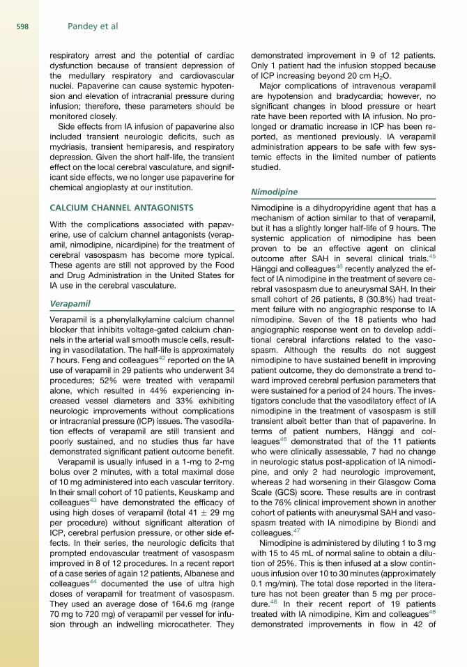
Cerebral vasospasm following aneurysmal sub-arachnoid hemorrhage (SAH) is a delayed, revers-ible narrowing of the intracranial vasculature thatoccurs most commonly 4 to 14 days after aneu-rysmal SAH and can lead to permanent ischemicinjury (Fig. 1). Although it has been the focus ofmuch research and clinical effort, vasospasmremains difficult to treat and is responsible for sig-nificant morbidity and mortality in patients withruptured cerebral aneurysms. Angiographic spasmoccurs in up to 70% of patients following SAH,and approximately half become symptomatic. In
a Division of Interventional Neuroradiology, Departmentigan Hospitals, 1500 East Medical Center Drive, Ann ArbNeuroradiology, Department of Radiology, University ofAnn Arbor, MI 48109-5030, USA; c Division of Interventioments of Radiology, Neurosurgery, and Otolaryngology,East Medical Center Drive, Ann Arbor, MI 48109-5030, U* Corresponding author.E-mail address: [email protected]
Neuroimag Clin N Am 23 (2013) 593–604http://dx.doi.org/10.1016/j.nic.2013.03.0081052-5149/13/$ – see front matter � 2013 Elsevier Inc. A
the past, mortality rates from vasospasm havebeen reported to range from 30% to 70%, with10% to 20% of patients experiencing severeneurologic deficits.1,2
With advancements in diagnostic and interven-tional technology, estimates of patients who aredisabled by vasospasm, or die because of it, rangefrom 5% to 9%, with vasospasm accounting for12% to 17% of all fatalities or cases of disability af-ter SAH.3,4 We have an incomplete understandingof the pathophysiology of vasospasm, and it isdifficult to predict which patients will developvasospasm after SAH. The Hunt and Hess grade,Fisher score, hypertension, smoking, cocaine
s of Neurosurgery and Radiology, University of Mich-or, MI 48109-5030, USA; b Division of InterventionalMichigan Hospitals, 1500 East Medical Center Drive,nal Neuroradiology and Cranial Base Surgery, Depart-University of Michigan Hospitals, UH B1 D328, 1500SA
ll rights reserved. neuroimaging.theclinics.com
Pandey et al594

=
Endovascular Treatment of Cerebral Vasospasm 595
use, age range of 40 to 59 years, and early rise inthe middle cerebral artery blood flow on transcra-nial Doppler have been shown to be independentrisk factors for vasospasm.4 Cerebral salt wastinghas also been associated with vasospasm. Thisarticle discusses the multiple medical and endo-vascular therapies that are used to prevent or treatvasospasm.
VASOSPASM PROPHYLAXIS AND MEDICALTREATMENT
Prophylactic treatment for cerebral vasospasmfollowing aneurysmal SAH is controversial andvaries among institutions. Calcium channelblockers, magnesium, 3-hydroxy-3-methylglutarylcoenzyme A (HMG CoA) reductase inhibitors(statins), fasudil, thrombolytics, endothelial recep-tor antagonists, and tirilazad have all been used inattempts to prevent and treat vasospasm. In addi-tion, triple-H (hypervolemia, hypertension, and he-modilution) therapy has been a mainstay ofvasospasm treatment for years. At our institution,we start all patients with SAH with triple H assoon as the aneurysm has been secured, as longas there are no medical contraindications, suchas congestive heart failure or severe pulmonarydisease. We use prophylactic nimodipine andmagnesium sulfate and are considering using sta-tins as new data emerge.
Calcium Channel Antagonists
Calcium channel antagonists have been shown todecrease the overall incidence of cerebral infarc-tion after SAH by 34% and the incidence of pooroutcomes by 40%.5 The physiologic reasoningbehind the use of calcium channel blockers isthat the central event in vascular smooth musclecontraction is the influx of calcium into cells, whichhas been shown to occur after SAH.6 This, in turn,leads to several downstream events, including freeradical formation, production of vasoconstrictingprostaglandins, and activation of the myosin light
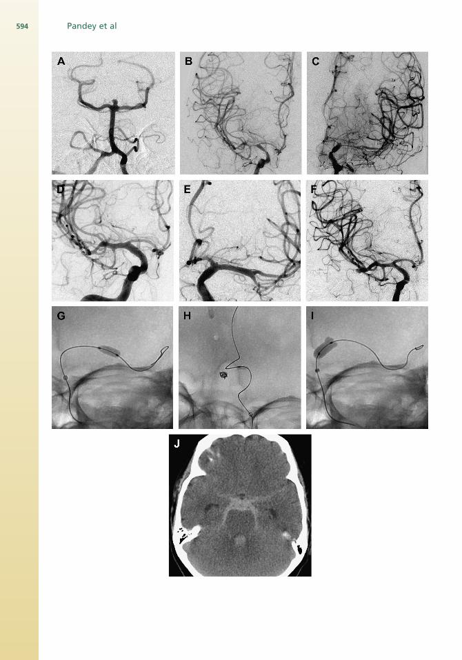
Fig. 1. (A) Anterior posterior (AP) left vertebral angiogramrotid angiogram 10 days after hemorrhage demonstrates danterior cerebral artery, and middle cerebral artery. (C) APrhage demonstrates diffuse vasospasm involving the intercerebral artery. (D) AP right internal carotid angiogram afshows angiographic resolution of the vasospasm. (E) AP lefand infusion of IA nicardipine shows angiographic resolangiogram same patient shows an additional middle cereshows a hyperform balloon in the left middle cerebral artballoon in the left A1 segment. (I) Spot AP fluoroscopic imcarotid artery, note a small portion of the balloon has herndemonstrates diffuse high attenuation material within th
chain kinase that causes smooth muscle contrac-tion. The most common calcium channel blockerused after SAH is nimodipine, which has a certaindegree of specificity for the cerebral vessels.Multiple trials have shown its efficacy in improvingoutcomes, although it may not improve angio-graphic outcome.5,7–12
Two randomized controlled trials (RCTS) usingnicardipine for treatment of and prevention ofcerebral vasospasm have been performed. Thelargest RCT treated 449 patients with intravenous(IV) nicardipine (0.15 mg/kg/h) and 457 patientwith placebo.13 Patients treated with nicardipinehad significantly reduced vasospasm comparedwith placebo (P<.001). Clinical outcomes weresimilar between the 2 groups at 3 months.Another small study that randomized 16 patientsto receive nicardipine prolonged-release im-plants placed into the basal cisterns at the timeof surgical clip placement demonstrated signifi-cantly reduced angiographic vasospasm inci-dence (P<.05) and improved modified RankinScale scores at 1 year.14
MagnesiumObstetricians have used magnesium to treateclampsia, as magnesium is thought to altercalcium physiology and thus alter vascular tonein the uterine circulation. Magnesium competeswith calcium-binding sites, thus preventing mus-cular contraction and allowing vascular musclerelaxation. Level 1 evidence now exists for theusage of magnesium sulfate in the preventionof cerebral vasospasm. Westermaier and col-leagues15 have shown that magnesium sulfatesignificantly reduces cerebral ischemic eventsafter SAH.16–19
StatinsFive RCTS have used statins for the preventionand treatment of cerebral vasospam followingSAH.20–24 Three of the trials have shown a benefitfor statins in reducing vasospasm, delayedischemic neurologic deficits (DINDs), and
shows a basilar tip aneurysm. (B) AP right internal ca-iffuse vasospasm involving the internal carotid artery,left internal carotid angiogram 10 days after hemor-
nal carotid artery, anterior cerebral artery, and middleter balloon angioplasty and infusion of IA nicardipinet internal carotid angiogram after balloon angioplastyution of the vasospasm. (F) AP right internal carotidbral artery aneurysm. (G) Spot AP fluoroscopic imageery. (H) Spot AP fluoroscopic images shows a gatewayages shows a hyperform balloon in the distal internaliated into the left A1 segment. (J) Axial CT of the heade subarachnoid space consistent with hemorrhage.
Pandey et al596
mortality.20–22 However, 2 of the more recent trialshave shown no significant difference in reducingvasospasm and clinical outcome.23,24
FasudilFasudil acts as a calcium channel blocker viathe Rho-kinase signaling pathway inhibition. It an-tagonizes the vasoconstrictive effects of endo-thelin by dilating cerebral arteries in an animalvasospasm model.25 Fasudil has been studied in2 RCTs.25,26 Shibuya and colleagues26 showedsignificant reductions in angiographic and symp-tomatic vasospasm, hypodensities on computedtomography (CT) scan, and poor outcome withuse of the drug. In another study, Zhao and col-leagues25 compared IV fasudil to IV nimodipineand found no significant differences in vasospasmincidence or clinical outcome.
ThrombolyticsTwo RCTs have been performed using thrombo-lytic therapy for the prevention or treatment of ce-rebral vasospasm.27,28 Findlay27 in an RCT of 100patients who received either a 1-time intracisternalbolus of 10 mg of tissue plasminogen activator(t-PA) or placebo at the time of surgical clip place-ment found a significant reduction in severe vaso-spasm without improvement in outcome. Hamadaand colleagues,28 in another randomizedcontrolled trial (RCT) of 110 patients who receivedeither intrathecal urokinase (60,000 Internationalunits (IU) over 20 minutes) or no thrombolytic ther-apy following aneurysm coiling, showed a signifi-cant reduction in symptomatic vasospasm withimproved outcome.
Endothelin receptor antagonistsA subclass of drugs has been developed to inhibitthe binding of endothelin I, a potent vasoconstric-tive peptide, to its target receptors of vascularsmooth muscle cells. Two drugs in this class, cla-zosentan and TAK-044, have been studied inRCTs for the treatment and prevention of cerebralvasospasm following aneurysmal SAH.29–31 Shawand colleagues,29 in study of 402 patients whowere randomized to IV TAK-044 or placebo, founda decreased incidence of delayed cerebral in-farction without improvement in the GlasgowOutcome Scale. Furthermore, Vajkoczy and col-leagues,30 in a small RCT comparing patientsreceiving IV clazosentan to placebo, found asignificant reduction in cerebral vasospasm inci-dence and severity. Fifty percent of the crossoverplacebo patients had reversal of vasospasmfollowing the initiation of clazosentan. The Clazo-sentan to Overcome Neurologic Ischemia andInfarction Occurring After Subarachnoid Hemor-rhage (CONSCIOUS-I) trial randomized 413
patients to either dose-escalated clazosentan orplacebo. Significant dose-dependent reductionsin moderate and severe vasospasm were notedwith increased pulmonary complications, hypo-tension, and anemia.31
TRIPLE-H THERAPY
Hypervolemia, hypertension, and hemodilutiontherapy (triple-H) has long been a mainstay ofmedical therapy in patients with aneurysmalSAH. The rationale behind triple-H therapy is thatmaintenance of high circulating blood volume,increased perfusion pressures, and decreasedblood viscosity will enhance cerebral blood flowin the setting of vasoconstriction. Although inhealthy adults, changes in cardiac output do notchange the local cerebral flow, they do affect cere-bral blood flow in patients suffering from cerebralvasospasm. Our goal is to maintain euvolemia asdefined by the central venous/wedge pressurethat allows for the highest cardiac output.The vast majority of our patients with SAH
receive central lines so that central venouspressures can be closely monitored to achieveoptimized volume expansion without causing pul-monary edema. We prefer central venous pres-sures in the range of 8 to 12 mm Hg, but theseare highly individualized andmust be closely moni-tored in relation to the clinical findings in each pa-tient. Patients with cardiopulmonary disease mayneed to be evaluated with a Swan Ganz catheter,as the ideal venous pressure in these patientsneeds to be related to the ideal cardiac output.32
One series of 184 patients reported a 13% risk ofdevice-related sepsis in patients with pulmonaryartery catheters, as well as a 2.0% risk of conges-tive heart failure, 1.3% risk of subclavian veinthrombosis, and 1.0% risk of pneumothorax.32
Most patients with SAH require invasive bloodpressure monitoring. In the post-clipping or post-coiling vasospasm period, we often allow patientsto autoregulate with systolic blood pressure in therange of 200 mm Hg. The role of red blood celltransfusion is not well studied, but transfusioncould certainly increase the oxygen carrying ca-pacity. Three RCTs have evaluated the efficacyof triple-H therapy.33–35
ENDOVASCULAR THERAPY
Endovascular treatment with intra-arterial (IA) infu-sion of a vasodilator or balloon angioplasty is indi-cated in patients with symptomatic vasospasmrefractory to medical therapy to prevent neurologicdeficits referable to the vascular territory of theangiographic vasospasm. Any patient who is a
Endovascular Treatment of Cerebral Vasospasm 597
candidate for cerebral angioplasty in the context ofcerebral vasospasm must be evaluated with a CTscan of the head to rule out hemorrhage and thepresence of hypodensity in the vascular territoryto undergo angioplasty. A vessel diameter reduc-tion between 25% and 50% from the initial angio-graphic diameter is usually treated with IA infusionof vasodilators. Vessel diameter reductionsgreater than 50% from the initial angiographicdiameter are treated with a combination of me-chanical and chemical angioplasty. The timing ofendovascular intervention for vasospasm is crit-ical, and a 2-hour window from the time of symp-toms may exist for restoration of blood flow toultimately improve the patient’s outcome.36
INTRA-ARTERIAL VASODILATORSPapaverine
The most studied IA pharmacologic agent to dateis papaverine. It is an opium alkaloid that is thoughtto alter adenosine 30,50–cyclic monophosphatelevels in smooth muscles (Table 1).37 The half-lifeis approximately 2 hours. In a review of IA agentsfor treatment of cerebral vasospasm, Hoh andOgilvy38 reported that papaverine produced clin-ical improvement in only 43% of the treated pa-tients. The effectiveness of treatment was shortgiven its half-life; therefore, multiple treatmentswere required, which led to a variable andincreased risk of complications. Platz and col-leagues39more recently reported a case of sponta-neous hemorrhage following IA use of papaverine.They hypothesized that local increased levels ofinfused papaverine possibly led to a blood–brainbarrier (BBB) breakdownwith subsequent intracra-nial hemorrhage. Furthermore, there has been arecent report from Pennings and colleagues.40
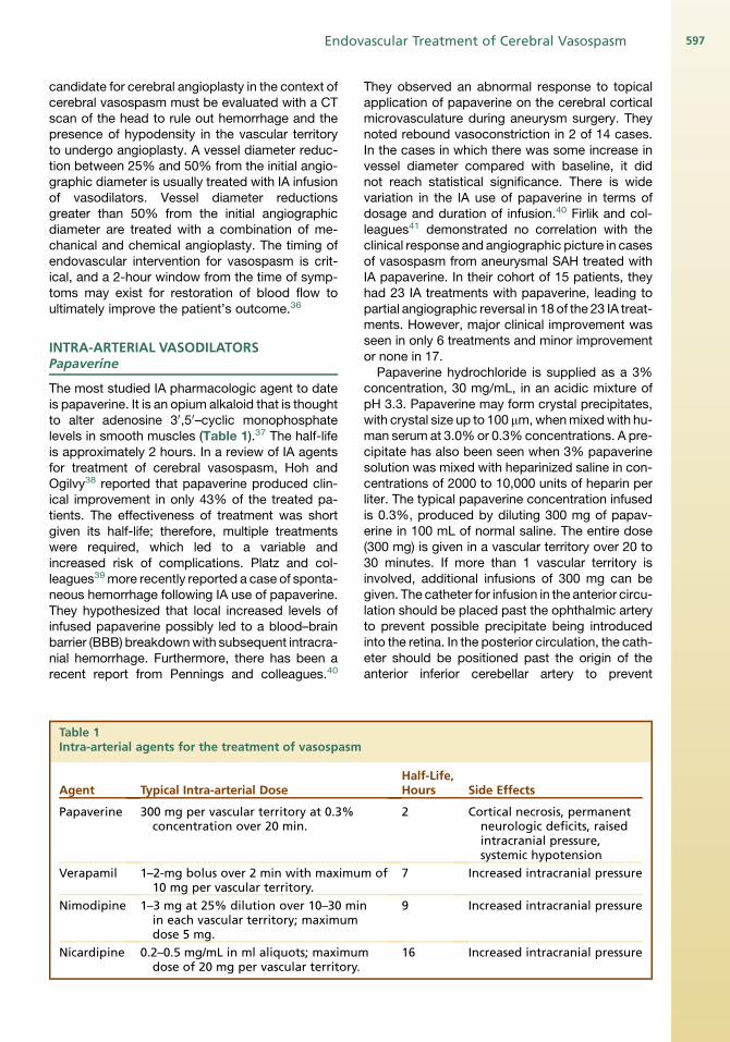
Table 1Intra-arterial agents for the treatment of vasospasm
Agent Typical Intra-arterial Dose
Papaverine 300 mg per vascular territory at 0.3%concentration over 20 min.
Verapamil 1–2-mg bolus over 2 min with maximu10 mg per vascular territory.
Nimodipine 1–3 mg at 25% dilution over 10–30 miin each vascular territory; maximumdose 5 mg.
Nicardipine 0.2–0.5 mg/mL in ml aliquots; maximumdose of 20 mg per vascular territory.
They observed an abnormal response to topicalapplication of papaverine on the cerebral corticalmicrovasculature during aneurysm surgery. Theynoted rebound vasoconstriction in 2 of 14 cases.In the cases in which there was some increase invessel diameter compared with baseline, it didnot reach statistical significance. There is widevariation in the IA use of papaverine in terms ofdosage and duration of infusion.40 Firlik and col-leagues41 demonstrated no correlation with theclinical response and angiographic picture in casesof vasospasm from aneurysmal SAH treated withIA papaverine. In their cohort of 15 patients, theyhad 23 IA treatments with papaverine, leading topartial angiographic reversal in 18 of the 23 IA treat-ments. However, major clinical improvement wasseen in only 6 treatments and minor improvementor none in 17.
Papaverine hydrochloride is supplied as a 3%concentration, 30 mg/mL, in an acidic mixture ofpH 3.3. Papaverine may form crystal precipitates,with crystal size up to 100 mm,whenmixedwith hu-man serum at 3.0%or 0.3%concentrations. A pre-cipitate has also been seen when 3% papaverinesolution was mixed with heparinized saline in con-centrations of 2000 to 10,000 units of heparin perliter. The typical papaverine concentration infusedis 0.3%, produced by diluting 300 mg of papav-erine in 100 mL of normal saline. The entire dose(300 mg) is given in a vascular territory over 20 to30 minutes. If more than 1 vascular territory isinvolved, additional infusions of 300 mg can begiven. The catheter for infusion in the anterior circu-lation should be placed past the ophthalmic arteryto prevent possible precipitate being introducedinto the retina. In the posterior circulation, the cath-eter should be positioned past the origin of theanterior inferior cerebellar artery to prevent
Half-Life,Hours Side Effects
2 Cortical necrosis, permanentneurologic deficits, raisedintracranial pressure,systemic hypotension
m of 7 Increased intracranial pressure
n 9 Increased intracranial pressure
16 Increased intracranial pressure
Pandey et al598
respiratory arrest and the potential of cardiacdysfunction because of transient depression ofthe medullary respiratory and cardiovascularnuclei. Papaverine can cause systemic hypoten-sion and elevation of intracranial pressure duringinfusion; therefore, these parameters should bemonitored closely.Side effects from IA infusion of papaverine also
included transient neurologic deficits, such asmydriasis, transient hemiparesis, and respiratorydepression. Given the short half-life, the transienteffect on the local cerebral vasculature, and signif-icant side effects, we no longer use papaverine forchemical angioplasty at our institution.
CALCIUM CHANNEL ANTAGONISTS
With the complications associated with papav-erine, use of calcium channel antagonists (verap-amil, nimodipine, nicardipine) for the treatment ofcerebral vasospasm has become more typical.These agents are still not approved by the Foodand Drug Administration in the United States forIA use in the cerebral vasculature.
Verapamil
Verapamil is a phenylalkylamine calcium channelblocker that inhibits voltage-gated calcium chan-nels in the arterial wall smooth muscle cells, result-ing in vasodilatation. The half-life is approximately7 hours. Feng and colleagues42 reported on the IAuse of verapamil in 29 patients who underwent 34procedures; 52% were treated with verapamilalone, which resulted in 44% experiencing in-creased vessel diameters and 33% exhibitingneurologic improvements without complicationsor intracranial pressure (ICP) issues. The vasodila-tion effects of verapamil are still transient andpoorly sustained, and no studies thus far havedemonstrated significant patient outcome benefit.Verapamil is usually infused in a 1-mg to 2-mg
bolus over 2 minutes, with a total maximal doseof 10 mg administered into each vascular territory.In their small cohort of 10 patients, Keuskamp andcolleagues43 have demonstrated the efficacy ofusing high doses of verapamil (total 41 � 29 mgper procedure) without significant alteration ofICP, cerebral perfusion pressure, or other side ef-fects. In their series, the neurologic deficits thatprompted endovascular treatment of vasospasmimproved in 8 of 12 procedures. In a recent reportof a case series of again 12 patients, Albanese andcolleagues44 documented the use of ultra highdoses of verapamil for treatment of vasospasm.They used an average dose of 164.6 mg (range70 mg to 720 mg) of verapamil per vessel for infu-sion through an indwelling microcatheter. They
demonstrated improvement in 9 of 12 patients.Only 1 patient had the infusion stopped becauseof ICP increasing beyond 20 cm H2O.Major complications of intravenous verapamil
are hypotension and bradycardia; however, nosignificant changes in blood pressure or heartrate have been reported with IA infusion. No pro-longed or dramatic increase in ICP has been re-ported, as mentioned previously. IA verapamiladministration appears to be safe with few sys-temic effects in the limited number of patientsstudied.
Nimodipine
Nimodipine is a dihydropyridine agent that has amechanism of action similar to that of verapamil,but it has a slightly longer half-life of 9 hours. Thesystemic application of nimodipine has beenproven to be an effective agent on clinicaloutcome after SAH in several clinical trials.45
Hanggi and colleagues46 recently analyzed the ef-fect of IA nimodipine in the treatment of severe ce-rebral vasospasm due to aneurysmal SAH. In theirsmall cohort of 26 patients, 8 (30.8%) had treat-ment failure with no angiographic response to IAnimodipine. Seven of the 18 patients who hadangiographic response went on to develop addi-tional cerebral infarctions related to the vaso-spasm. Although the results do not suggestnimodipine to have sustained benefit in improvingpatient outcome, they do demonstrate a trend to-ward improved cerebral perfusion parameters thatwere sustained for a period of 24 hours. The inves-tigators conclude that the vasodilatory effect of IAnimodipine in the treatment of vasospasm is stilltransient albeit better than that of papaverine. Interms of patient numbers, Hanggi and col-leagues46 demonstrated that of the 11 patientswho were clinically assessable, 7 had no changein neurologic status post-application of IA nimodi-pine, and only 2 had neurologic improvement,whereas 2 had worsening in their Glasgow ComaScale (GCS) score. These results are in contrastto the 76% clinical improvement shown in anothercohort of patients with aneurysmal SAH and vaso-spasm treated with IA nimodipine by Biondi andcolleagues.47
Nimodipine is administered by diluting 1 to 3 mgwith 15 to 45 mL of normal saline to obtain a dilu-tion of 25%. This is then infused at a slow contin-uous infusion over 10 to 30minutes (approximately0.1 mg/min). The total dose reported in the litera-ture has not been greater than 5 mg per proce-dure.48 In their recent report of 19 patientstreated with IA nimodipine, Kim and colleagues48
demonstrated improvements in flow in 42 of
Endovascular Treatment of Cerebral Vasospasm 599
53 procedures and an improvement in clinicaloutcome after 23. They used nimodipine in a10% concentration infused slowly at 0.1 mg nimo-dipine per minute. The main systemic complica-tions of nimodipine include hypotension, rash,diarrhea, and bradycardia. No significant changesin heart rate, blood pressure, or ICP have been re-ported in the literature from IA administration.
Nicardipine
Nicardipine is also a dihydropyridine agent that hasan even longer half-life of almost 16 hours. It has amore selective effect on vascular smooth musclethan cardiac muscle. Several studies have demon-strated that continuous IV nicardipine infusionsignificantly decreases the incidence of symptom-atic, angiographic, and transcranial Doppler vaso-spasm, but the efficacy is limited by prolongedhypotension, pulmonary edema, and renal dys-function. Badjatia and colleagues49 previouslydemonstrated that theuseof IAnicardipine inducesmore sustained reversal of vasospasm than doespapaverine. They demonstrated a 42.1% neuro-logic improvement in patients following treatmentwith IA nicardipine. Although this is encouraging,the investigators also highlight the incidence ofincreased ICP following treatment in 6 patients.Tejada and colleagues50 more recently showedthat a higher-dose regimen of IA nicardipine ismore efficacious. They used a total dose of 10 to40mg in each patient. GCS andGlasgowOutcomeScale (GOS) scores in 10 of 11 patients who weretreated with the high-dose regimen improved. Theinvestigators also demonstrated a low complica-tion rate and a sustained clinical outcome benefitwith a GOS score of 1 or 2 in 9 of 10 patients withat least 2-month follow-up.
Nicardipine is usually administered intra-arterially by diluting with normal saline to a concen-tration of 0.2 mg/mL and infusing in 1-mL aliquotsto a maximum dose between 2.5 and 20.0 mg pervessel.50 At our institution we recently adopted amore aggressive approach, and use a 1-mg/mLdosage concentration infusing in 3-mL aliquotswith a maximum dosage of 20 mg per vessel. Pro-longed hypotension, pulmonary edema, and renaldysfunction have been reported following the IVadministration of nicardipine; however, these find-ings have not been reported with IA administra-tion.50 The main adverse effect reported in theliterature is an increase in ICP, which usually canbe controlled with ventricular drainage. Tejadaand colleagues demonstrated the safe use of ahigh dose of IA nicardipine in their series of 11 pa-tients with vasospasm.50 However, ICP monitoringwas possible in only 2 of the 11patientswhodid not
demonstrate any change during IA infusion ofnicardipine.
Other Agents
There are other pharmaceutical agents withvarious other mechanisms of vasodilatation,namely magnesium sulfate, HMG CoA reductaseinhibitors, nitric oxide donors, and endothelin-1 an-tagonists. The neuroprotective and vasoprotectiveproperties of magnesium sulfate have been welldocumented in the literature. Shah and col-leagues51 recently evaluated a cohort of patientsin whom magnesium sulfate was used in conjunc-tion with nicardipine for IA treatment of aneurysmalSAH-related vasospasm. They observed that thisagent was well tolerated by the patients, with noadverse effects on ICP. They did not observe anystatistically significant difference in clinical out-come improvement in comparison with otherstudies with cohorts treated only with nicardipine.
TRANSLUMINAL BALLOON ANGIOPLASTY
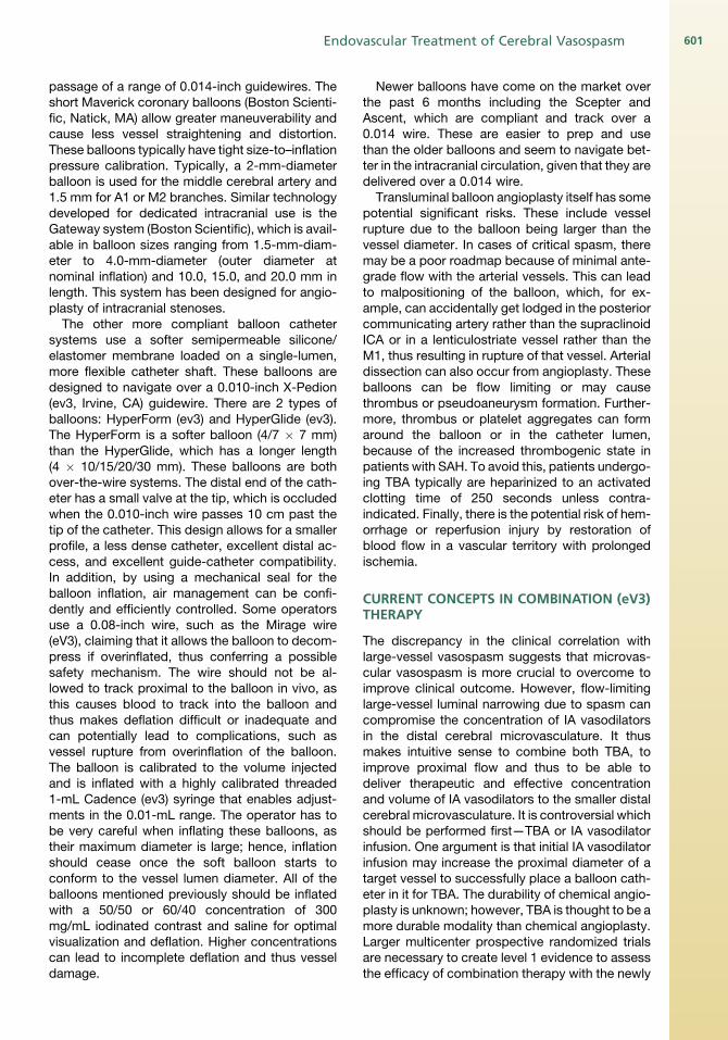
The mechanism of cerebral blood vessel narrow-ing in aneurysmal SAH is not clearly understood.Inflammatory changes mediating smooth musclecontraction, changes in the linkage of the proteinmatrix, and collagen deposition in the vessel wallhave all been implicated as a possible cause ofblood vessel narrowing in SAH related to aneu-rysm rupture (Table 2).
Mechanical dilation of intracranial vessels wasperformed first in 1984 by Zubkov and col-leagues.52 They reported their results in selectedpatients with large-vessel spasm. Balloons forintracranial angioplasty have developed consider-ably since then. The most significant limitation ofthese angioplasty balloons remains the inabilityto treat spasm affecting the distal cerebral vascu-lature. The current thinking is that transluminalballoon angioplasty (TBA) acts by stretching thevessel wall, leading to morphologic and functionalchanges in the smooth muscle fibers, resulting inimpairment of contractility. At a cellular level ithas been shown that there is fragmentation ofthe collagen matrix and flattening of the endothe-lial cells, resulting in permanent restoration ofvessel diameter. This has been demonstrated tobe durable in both canine and primate models.
Eskridge and colleagues53 reported 50 patientswith 170 treated arterial segments. They demon-strated sustained neurologic improvement in only61% of patients within 72 hours following angio-plasty. Rosenwasser and colleagues36 sought toidentify anoptimal time frame for treatment of vaso-spasm from aneurysmal SAH. They demonstratedthat 61% of patients treated within 2 hours had
Table 2Balloons used for transluminal balloon angioplasty
BalloonCatheters Technique Dimensions Manufacturer Qualities
Hyperform Over-the-wireballoon catheterwith single lumen(0.010 expedion wire)
Balloon diameter:4 mm, 7 mm
Balloon length:7 mm
ev3, Irvine, CA Readilyconformable
Hyperglide Over-the-wireballoon catheterwith single lumen(0.010 expedion wire)
Balloon diameter:4 mm, 5 mm
Balloon lengths(by diameter):4/10, 4/15, 4/20,4/30, 5/15, 5/20
ev3 Compliant balloon,rigid in longerlength
Gateway Over-the-wire ballooncatheter with separatelumen for inflation,tracks over a 0.014 wire
Balloon diameter:1.5, 2.0, 2.25, 3.0,3.5, 4 mm
Balloon lengths(by diameter):1.5/9, 2.0/9, and 15,20 mm in the otherdimensions
(StrykerNeurovascular,Fremont, CA)
Comparatively rigidbut available insmaller diameters
Scepter C Over-the-wire ballooncatheter with separatelumen for inflation,tracks over a 0.014 wire
Balloon diameter:4.0 mm
Balloon Lengths(10, 15, 20 mm)
(Microvention,Inc, Tustin, CA)
Compliant ballooncatheter
Scepter XC Over-the-wire ballooncatheter with separatelumen for inflation,tracks over a 0.014 wire
Balloon diameter:4.0 mm
Balloon Length:11 mm
Microvention X-tra compliantocclusion balloon
Ascent Over-the-wire ballooncatheter with separatelumen for inflation,tracks over a 0.014 wire
Balloon diameter:4.0, 6.0 mm
Balloon lengths(by diameter): 4.0/7,10, 15 and 6.0/9
(CodmanNeurovascular,Raynham, MA)
Compliant ballooncatheter
Pandey et al600
angiographic improvement in 90% of cases and asustained clinical improvement in 70% of cases;in contrast, the other 39% of cases treated morethan 2 hours later achieved 88% angiographicimprovement but sustained only a 40% clinicalimprovement.6 In summary, TBAhas shownbenefitin restoring antegrade flow in the treated cerebralvascular territory and some improvement in patientoutcomes. Although anecdotal reports suggestthat TBA provides durable relief of vasospasm, noRCTs using therapeutic angioplasty alone havebeen published to date. The only RCT of TBAcompared prophylactic TBA or no prophylactictreatment within 96 hours of aneurysm rupture.54
Patients undergoing prophylactic TBA experi-enced a nonsignificant reduction inDIND incidence(P 5 .30). A significant decrease in therapeutic an-gioplasty (P5 .03) was observed, however, for pa-tients who had prophylactic TBA compared withcontrols. There is still a lack of level 1 evidence
supporting balloon angioplasty for treatment of ce-rebral vasospasm in the literature, however.Typical locations of vessels amenable to TBA
are vertebral, basilar, supraclinoid internal carotidartery (ICA), and M1 segments. Less common lo-cations, because of their small diameter, are theposterior communicating, A1, M2, and P1 arteries.In general, TBA in vessels with a diameter smallerthan 2 mm may have a higher risk of vesselrupture. Hence, the posterior inferior communi-cating artery (PICA), anterior inferior communi-cating artery (AICA), A2, M3, and P2 arterialbranches are usually avoided altogether.Two different balloon technologies have been
used for treatment of vasospasm: coronary bal-loons and more compliant intracranial balloons.The coronary balloon catheter technology com-prises a stiff balloon membrane composed ofpolyethylene or nylon and a double-lumen shaft,one lumen for balloon inflation and the other for
Endovascular Treatment of Cerebral Vasospasm 601
passage of a range of 0.014-inch guidewires. Theshort Maverick coronary balloons (Boston Scienti-fic, Natick, MA) allow greater maneuverability andcause less vessel straightening and distortion.These balloons typically have tight size-to–inflationpressure calibration. Typically, a 2-mm-diameterballoon is used for the middle cerebral artery and1.5 mm for A1 or M2 branches. Similar technologydeveloped for dedicated intracranial use is theGateway system (Boston Scientific), which is avail-able in balloon sizes ranging from 1.5-mm-diam-eter to 4.0-mm-diameter (outer diameter atnominal inflation) and 10.0, 15.0, and 20.0 mm inlength. This system has been designed for angio-plasty of intracranial stenoses.
The other more compliant balloon cathetersystems use a softer semipermeable silicone/elastomer membrane loaded on a single-lumen,more flexible catheter shaft. These balloons aredesigned to navigate over a 0.010-inch X-Pedion(ev3, Irvine, CA) guidewire. There are 2 types ofballoons: HyperForm (ev3) and HyperGlide (ev3).The HyperForm is a softer balloon (4/7 � 7 mm)than the HyperGlide, which has a longer length(4 � 10/15/20/30 mm). These balloons are bothover-the-wire systems. The distal end of the cath-eter has a small valve at the tip, which is occludedwhen the 0.010-inch wire passes 10 cm past thetip of the catheter. This design allows for a smallerprofile, a less dense catheter, excellent distal ac-cess, and excellent guide-catheter compatibility.In addition, by using a mechanical seal for theballoon inflation, air management can be confi-dently and efficiently controlled. Some operatorsuse a 0.08-inch wire, such as the Mirage wire(eV3), claiming that it allows the balloon to decom-press if overinflated, thus conferring a possiblesafety mechanism. The wire should not be al-lowed to track proximal to the balloon in vivo, asthis causes blood to track into the balloon andthus makes deflation difficult or inadequate andcan potentially lead to complications, such asvessel rupture from overinflation of the balloon.The balloon is calibrated to the volume injectedand is inflated with a highly calibrated threaded1-mL Cadence (ev3) syringe that enables adjust-ments in the 0.01-mL range. The operator has tobe very careful when inflating these balloons, astheir maximum diameter is large; hence, inflationshould cease once the soft balloon starts toconform to the vessel lumen diameter. All of theballoons mentioned previously should be inflatedwith a 50/50 or 60/40 concentration of 300mg/mL iodinated contrast and saline for optimalvisualization and deflation. Higher concentrationscan lead to incomplete deflation and thus vesseldamage.
Newer balloons have come on the market overthe past 6 months including the Scepter andAscent, which are compliant and track over a0.014 wire. These are easier to prep and usethan the older balloons and seem to navigate bet-ter in the intracranial circulation, given that they aredelivered over a 0.014 wire.
Transluminal balloon angioplasty itself has somepotential significant risks. These include vesselrupture due to the balloon being larger than thevessel diameter. In cases of critical spasm, theremay be a poor roadmap because of minimal ante-grade flow with the arterial vessels. This can leadto malpositioning of the balloon, which, for ex-ample, can accidentally get lodged in the posteriorcommunicating artery rather than the supraclinoidICA or in a lenticulostriate vessel rather than theM1, thus resulting in rupture of that vessel. Arterialdissection can also occur from angioplasty. Theseballoons can be flow limiting or may causethrombus or pseudoaneurysm formation. Further-more, thrombus or platelet aggregates can formaround the balloon or in the catheter lumen,because of the increased thrombogenic state inpatients with SAH. To avoid this, patients undergo-ing TBA typically are heparinized to an activatedclotting time of 250 seconds unless contra-indicated. Finally, there is the potential risk of hem-orrhage or reperfusion injury by restoration ofblood flow in a vascular territory with prolongedischemia.
CURRENT CONCEPTS IN COMBINATION (eV3)THERAPY
The discrepancy in the clinical correlation withlarge-vessel vasospasm suggests that microvas-cular vasospasm is more crucial to overcome toimprove clinical outcome. However, flow-limitinglarge-vessel luminal narrowing due to spasm cancompromise the concentration of IA vasodilatorsin the distal cerebral microvasculature. It thusmakes intuitive sense to combine both TBA, toimprove proximal flow and thus to be able todeliver therapeutic and effective concentrationand volume of IA vasodilators to the smaller distalcerebral microvasculature. It is controversial whichshould be performed first—TBA or IA vasodilatorinfusion. One argument is that initial IA vasodilatorinfusion may increase the proximal diameter of atarget vessel to successfully place a balloon cath-eter in it for TBA. The durability of chemical angio-plasty is unknown; however, TBA is thought to be amore durable modality than chemical angioplasty.Larger multicenter prospective randomized trialsare necessary to create level 1 evidence to assessthe efficacy of combination therapy with the newly
Pandey et al602
available technology with compliant balloons andlonger-acting IA vasodilators.
FUTURE ENDOVASCULAR THERAPIES
Our understanding of the phenomenon of vaso-spasm due to aneurysmal SAH is still evolving. Abetter understanding of the pathophysiologicinsult to the brain at ictus will enable us to under-stand the phenomenon of vasospasm better andwill enable better design in the therapy to improvepatient outcome. Novel methods of production ofanimal vasospasm models are needed to evaluatevarious IA or TBA therapies to ultimately improvepatient outcome. Animal models will play a keyrole in evaluating different variables in the under-standing of cerebral vasospasm.
SUMMARY
The jury is still out on which IA endovascular phar-maceutical agent exclusively or in conjunction withTBA is the optimal treatment of cerebral vaso-spasm. There is a trend toward some benefitprovided by the longer-acting calcium channelblockers in combination with TBA. Newer agentson the horizon specifically targeting the vascularendothelium without a wide spectrum of side ef-fects need to be tried and tested in larger prospec-tive randomized patient cohorts. Improved animalmodels of vasospasm are being validated instudies. These models should form the corner-stone of optimizing treatment by helping to developa better understanding of the phenomenon of cere-bral vasospasm due to SAH from a rupturedaneurysm.
REFERENCES
1. Hop JW, Rinkel GJ, Algra A, et al. Case-fatality
rates and functional outcome after subarachnoid
hemorrhage: a systematic review. Stroke 1997;28:
660–4.
2. Komotar RJ, Zacharia BE, Valhora R, et al.
Advances in vasospasm treatment and prevention.
J Neurol Sci 2007;261:134–42.
3. Roos YB, de Haan RJ, Beenen LF, et al. Complica-
tions and outcome in patients with aneurysmal sub-
arachnoid haemorrhage: a prospective hospital
based cohort study in the Netherlands. J Neurol
Neurosurg Psychiatry 2000;68:337–41.
4. Zwienenberg-Lee M, Hartman J, Rudisill N, et al.
Endovascular management of cerebral vaso-
spasm. Neurosurgery 2006;59:S139–47 [discus-
sion: S3–13].
5. Pickard JD, Murray GD, Illingworth R, et al. Effect
of oral nimodipine on cerebral infarction and
outcome after subarachnoid haemorrhage: British
aneurysm nimodipine trial. BMJ 1989;298:636–42.
6. Rothoerl RD, Ringel F. Molecular mechanisms of
cerebral vasospasm following aneurysmal SAH.
Neurol Res 2007;29:636–42.
7. Allen GS, Ahn HS, Preziosi TJ, et al. Cerebral arte-
rial spasm—a controlled trial of nimodipine in pa-
tients with subarachnoid hemorrhage. N Engl J
Med 1983;308:619–24.
8. Neil-Dwyer G, Mee E, Dorrance D, et al. Early inter-
vention with nimodipine in subarachnoid haemor-
rhage. Eur Heart J 1987;8(Suppl K):41–7.
9. Ohman J, Servo A, Heiskanen O. Long-term effects
of nimodipine on cerebral infarcts and outcome
after aneurysmal subarachnoid hemorrhage and
surgery. J Neurosurg 1991;74:8–13.
10. Petruk KC, West M, Mohr G, et al. Nimodipine treat-
ment in poor-grade aneurysm patients. Results of a
multicenter double-blind placebo-controlled trial.
J Neurosurg 1988;68:505–17.
11. Philippon J, Grob R, Dagreou F, et al. Prevention of
vasospasm in subarachnoid haemorrhage. A
controlled study with nimodipine. Acta Neurochir
(Wien) 1986;82:110–4.
12. Rinkel GJ, Feigin VL, Algra A, et al. Calcium antag-
onists for aneurysmal subarachnoid haemorrhage.
Cochrane Database Syst Rev 2005;(1):CD000277.
13. Haley EC Jr, Kassell NF, Torner JC. A randomized
controlled trial of high-dose intravenous nicardipine
in aneurysmal subarachnoid hemorrhage. A report
of the Cooperative Aneurysm Study. J Neurosurg
1993;78:537–47.
14. Barth M, Capelle HH, Weidauer S, et al. Effect of
nicardipine prolonged-release implants on cere-
bral vasospasm and clinical outcome after severe
aneurysmal subarachnoid hemorrhage: a prospec-
tive, randomized, double-blind phase IIa study.
Stroke 2007;38:330–6.
15. Westermaier T, Stetter C, Vince GH, et al. Prophy-
lactic intravenous magnesium sulfate for treatment
of aneurysmal subarachnoid hemorrhage: a ran-
domized, placebo-controlled, clinical study. Crit
Care Med 2010;38:1284–90.
16. Muroi C, Terzic A, Fortunati M, et al. Magnesium
sulfate in the management of patients with aneu-
rysmal subarachnoid hemorrhage: a randomized,
placebo-controlled, dose-adapted trial. Surg Neu-
rol 2008;69:33–9 [discussion: 9].
17. van den Bergh WM, Algra A, van Kooten F, et al.
Magnesium sulfate in aneurysmal subarachnoid
hemorrhage: a randomized controlled trial. Stroke
2005;36:1011–5.
18. Veyna RS, Seyfried D, Burke DG, et al. Magnesium
sulfate therapy after aneurysmal subarachnoid
hemorrhage. J Neurosurg 2002;96:510–4.
19. Wong GK, Chan MT, Boet R, et al. Intravenous
magnesium sulfate after aneurysmal subarachnoid
Endovascular Treatment of Cerebral Vasospasm 603
hemorrhage: a prospective randomized pilot study.
J Neurosurg Anesthesiol 2006;18:142–8.
20. Lynch JR, Wang H, McGirt MJ, et al. Simvastatin re-
duces vasospasm after aneurysmal subarachnoid
hemorrhage: results of a pilot randomized clinical
trial. Stroke 2005;36:2024–6.
21. McGirt MJ, Woodworth GF, Pradilla G, et al. Gal-
braith Award: simvastatin attenuates experimental
cerebral vasospasm and ameliorates serum
markers of neuronal and endothelial injury in pa-
tients after subarachnoid hemorrhage: a dose-
response effect dependent on endothelial nitric
oxide synthase. Clin Neurosurg 2005;52:371–8.
22. Tseng MY, Czosnyka M, Richards H, et al. Effects
of acute treatment with pravastatin on cerebral
vasospasm, autoregulation, and delayed ischemic
deficits after aneurysmal subarachnoid hemor-
rhage: a phase II randomized placebo-controlled
trial. Stroke 2005;36:1627–32.
23. Chou SH, Smith EE, Badjatia N, et al. A random-
ized, double-blind, placebo-controlled pilot study
of simvastatin in aneurysmal subarachnoid hemor-
rhage. Stroke 2008;39:2891–3.
24. Vergouwen MD, Meijers JC, Geskus RB, et al. Bio-
logic effects of simvastatin in patients with aneu-
rysmal subarachnoid hemorrhage: a double-blind,
placebo-controlled randomized trial. J Cereb
Blood Flow Metab 2009;29:1444–53.
25. Zhao J, Zhou D, Guo J, et al. Effect of fasudil hy-
drochloride, a protein kinase inhibitor, on cerebral
vasospasm and delayed cerebral ischemic symp-
toms after aneurysmal subarachnoid hemorrhage.
Neurol Med Chir (Tokyo) 2006;46:421–8.
26. Shibuya M, Suzuki Y, Sugita K, et al. Effect of AT877
on cerebral vasospasm after aneurysmal sub-
arachnoid hemorrhage. Results of a prospective
placebo-controlled double-blind trial. J Neurosurg
1992;76:571–7.
27. Findlay JM. A randomized trial of intraoperative, in-
tracisternal tissue plasminogen activator for the
prevention of vasospasm. Neurosurgery 1995;37:
1026–7.
28. Hamada J, Kai Y, Morioka M, et al. Effect on cere-
bral vasospasm of coil embolization followed by
microcatheter intrathecal urokinase infusion into
the cisterna magna: a prospective randomized
study. Stroke 2003;34:2549–54.
29. Shaw MD, Vermeulen M, Murray GD, et al. Efficacy
and safety of the endothelin, receptor antagonist
TAK-044 in treating subarachnoid hemorrhage: a
report by the Steering Committee on behalf of the
UK/Netherlands/Eire TAK-044 Subarachnoid Hae-
morrhageStudyGroup. JNeurosurg2000;93:992–7.
30. Vajkoczy P, Meyer B, Weidauer S, et al. Clazosentan
(AXV-034343), a selective endothelin A receptor
antagonist, in the prevention of cerebral vasospasm
following severe aneurysmal subarachnoid
hemorrhage: results of a randomized, double-
blind, placebo-controlled, multicenter phase IIa
study. J Neurosurg 2005;103:9–17.
31. Macdonald RL, Kassell NF, Mayer S, et al. Clazo-
sentan to overcome neurological ischemia and
infarction occurring after subarachnoid hemor-
rhage (CONSCIOUS-1): randomized, double-
blind, placebo-controlled phase 2 dose-finding
trial. Stroke 2008;39:3015–21.
32. Rosenwasser RH, Jallo JI, Getch CC, et al. Compli-
cations of Swan-Ganz catheterization for hemody-
namic monitoring in patients with subarachnoid
hemorrhage. Neurosurgery 1995;37:872–5 [dis-
cussion: 5–6].
33. Egge A, Waterloo K, Sjoholm H, et al. Prophylactic
hyperdynamic postoperative fluid therapy after
aneurysmal subarachnoid hemorrhage: a clinical,
prospective, randomized, controlled study. Neuro-
surgery 2001;49:593–605 [discussion: 6].
34. Lennihan L, Mayer SA, Fink ME, et al. Effect of hy-
pervolemic therapy on cerebral blood flow after
subarachnoid hemorrhage: a randomized con-
trolled trial. Stroke 2000;31:383–91.
35. Rosenwasser RH, Delgado TE, Buchheit WA, et al.
Control of hypertension and prophylaxis against
vasospasm in cases of subarachnoid hemorrhage:
a preliminary report. Neurosurgery 1983;12:658–61.
36. Rosenwasser RH, Armonda RA, Thomas JE, et al.
Therapeutic modalities for the management of ce-
rebral vasospasm: timing of endovascular options.
Neurosurgery 1999;44:975–9 [discussion: 979–80].
37. Macdonald RL, Weir BK, Young JD, et al. Cytoskel-
etal and extracellular matrix proteins in cerebral
arteries following subarachnoid hemorrhage in
monkeys. J Neurosurg 1992;76:81–90.
38. Hoh BL, Ogilvy CS. Endovascular treatment of cere-
bral vasospasm: transluminal balloon angioplasty,
intra-arterial papaverine, and intra-arterial nicardi-
pine. Neurosurg Clin N Am 2005;16:501–16, vi.
39. Platz J, Barath K, Keller E, et al. Disruption of the
blood-brain barrier by intra-arterial administration
of papaverine: a technical note. Neuroradiology
2008;50:1035–9.
40. Pennings FA, Albrecht KW, Muizelaar JP, et al.
Abnormal responses of the human cerebral micro-
circulation to papaverin during aneurysm surgery.
Stroke 2009;40:317–20.
41. Firlik KS, Kaufmann AM, Firlik AD, et al. Intra-arte-
rial papaverine for the treatment of cerebral vaso-
spasm following aneurysmal subarachnoid
hemorrhage. Surg Neurol 1999;51:66–74.
42. Feng L, Fitzsimmons BF, Young WL, et al. Intraarte-
rially administered verapamil as adjunct therapy for
cerebral vasospasm: safety and 2-year experi-
ence. AJNR Am J Neuroradiol 2002;23:1284–90.
43. KeuskampJ,MuraliR,ChaoKH.High-dose intraarte-
rial verapamil in the treatment of cerebral vasospasm
Pandey et al604
after aneurysmal subarachnoid hemorrhage. J Neu-
rosurg 2008;108:458–63.
44. Albanese E, Russo A, Quiroga M, et al. Ultrahigh-
dose intraarterial infusion of verapamil through an
indwelling microcatheter for medically refractory
severe vasospasm: initial experience. Clinical
article. J Neurosurg 2010;113:913–22.
45. Barker FG 2nd, Ogilvy CS. Efficacy of prophylactic
nimodipine for delayed ischemic deficit after sub-
arachnoid hemorrhage: a metaanalysis. J Neuro-
surg 1996;84:405–14.
46. Hanggi D, Beseoglu K, Turowski B, et al. Feasibility
and safety of intrathecal nimodipine on posthae-
morrhagic cerebral vasospasm refractory to medi-
cal and endovascular therapy. Clin Neurol
Neurosurg 2008;110:784–90.
47. Biondi A, Ricciardi GK, Puybasset L, et al. Intra-
arterial nimodipine for the treatment of symptomatic
cerebral vasospasm after aneurysmal subarach-
noid hemorrhage: preliminary results. AJNR Am J
Neuroradiol 2004;25:1067–76.
48. Kim JH, Park IS, Park KB, et al. Intraarterial nimodi-
pine infusion to treat symptomatic cerebral vaso-
spasm after aneurysmal subarachnoid hemorrhage.
J Korean Neurosurg Soc 2009;46:239–44.
49. Badjatia N, Topcuoglu MA, Pryor JC, et al. Prelimi-
nary experience with intra-arterial nicardipine as a
treatment for cerebral vasospasm. AJNR Am J
Neuroradiol 2004;25:819–26.
50. Tejada JG, Taylor RA, Ugurel MS, et al. Safety and
feasibility of intra-arterial nicardipine for the treat-
ment of subarachnoid hemorrhage-associated
vasospasm: initial clinical experience with high-
dose infusions. AJNR Am J Neuroradiol 2007;28:
844–8.
51. Shah QA, Memon MZ, Suri MF, et al. Super-selec-
tive intra-arterial magnesium sulfate in combination
with nicardipine for the treatment of cerebral vaso-
spasm in patients with subarachnoid hemorrhage.
Neurocrit Care 2009;11:190–8.
52. Zubkov YN, Nikiforov BM, Shustin VA. Balloon cath-
eter technique for dilatation of constricted cerebral
arteries after aneurysmal SAH. Acta Neurochir
(Wien) 1984;70:65–79.
53. Eskridge JM, McAuliffe W, Song JK, et al. Balloon
angioplasty for the treatment of vasospasm: results
of first 50 cases. Neurosurgery 1998;42:510–6 [dis-
cussion: 6–7].
54. Zwienenberg-Lee M, Hartman J, Rudisill N, et al.
Effect of prophylactic transluminal balloon angio-
plasty on cerebral vasospasm and outcome in
patients with Fisher grade III subarachnoid hemor-
rhage: results of a phase II multicenter, random-
ized, clinical trial. Stroke 2008;39:1759–65.