endometrium as control of endometriosis in ... - mdpi
TRANSCRIPT
�����������������
Citation: Szegeczki, V.; Fazekas, L.;
Kulcsár, M.; Reglodi, D.; Török, P.;
Orlik, B.; Laganà, A.S.; Jakab, A.;
Juhasz, T. Endometrium as Control of
Endometriosis in Experimental
Research: Assessment of Sample
Suitability. Diagnostics 2022, 12, 970.
https://doi.org/10.3390/
diagnostics12040970
Academic Editor: Jeong Sook Kim
Received: 25 January 2022
Accepted: 7 April 2022
Published: 12 April 2022
Publisher’s Note: MDPI stays neutral
with regard to jurisdictional claims in
published maps and institutional affil-
iations.
Copyright: © 2022 by the authors.
Licensee MDPI, Basel, Switzerland.
This article is an open access article
distributed under the terms and
conditions of the Creative Commons
Attribution (CC BY) license (https://
creativecommons.org/licenses/by/
4.0/).
diagnostics
Article
Endometrium as Control of Endometriosis in ExperimentalResearch: Assessment of Sample SuitabilityVince Szegeczki 1, László Fazekas 1, Máté Kulcsár 1, Dora Reglodi 2, Péter Török 3, Brigitta Orlik 4,Antonio Simone Laganà 5 , Attila Jakab 3 and Tamas Juhasz 1,*
1 Department of Anatomy, Histology and Embryology, Faculty of Medicine, University of Debrecen,Nagyerdei krt. 98, H-4032 Debrecen, Hungary; [email protected] (V.S.);[email protected] (L.F.); [email protected] (M.K.)
2 Department of Anatomy, PTE-MTA PACAP Research Team, Szentagothai Research Center, Medical School,University of Pécs, Szigeti út 12, H-7624 Pécs, Hungary; [email protected]
3 Department of Obstetrics and Gynecology, Faculty of Medicine, University of Debrecen, Egyetem tér 1,H-4032 Debrecen, Hungary; [email protected] (P.T.); [email protected] (A.J.)
4 Department of Pathology, Faculty of Medicine, University of Debrecen, Egyetem tér 1,H-4032 Debrecen, Hungary; [email protected]
5 Unit of Gynecologic Oncology, ARNAS “Civico–Di Cristina–Benfratelli”, Department of Health Promotion,Mother and Child Care, Internal Medicine and Medical Specialties (PROMISE), University of Palermo,90127 Palermo, Italy; [email protected]
* Correspondence: [email protected]; Tel.: +36-52-255-567; Fax: +36-52-255-115
Abstract: Endometriosis is a chronic gynecological disease that causes numerous severe symptoms inaffected women. Revealing alterations of the molecular processes in ectopic endometrial tissue is thecurrent policy for understanding the pathomechanisms and discovering potential novel therapeutictargets. Examining molecular processes of eutopic endometrium is likely to be a convenient methodto compare it with the molecular alterations observed in ectopic tissues. The aim of the presentstudy was to determine what proportion of the surgically resected eutopic endometrial samplesis suitable for further experiments so that these can be comparable with endometriosis. Finalhospital reports and histopathology reports of a 3-year-long period (1162 cases) were analysed. Theapplication of a retrospective screening method promoted the categorization of these cases, andquantification of the categorized cases was accomplished. In addition, results obtained from culturedendometrium samples were also detailed. Only a small number of the harvested endometrial sampleswas suitable for further molecular analysis, while preoperative screening protocol could enlargethis fraction. Applying clinical and histopathological selection and exclusion criteria for tissuescreening and histopathological examination of samples could ensure the comparability of healthyendometrium with endometriosis. The present study could be useful for researchers who intendto perform molecular experiments to compare endometriosis with the physiological processes ofthe endometrium.
Keywords: endometrium; curettage; hysteroscopy; endometrial sampling; in vitro endometrialculturing
1. Introduction
Endometriosis is a gynaecological disease described as the formation of endometrial-like tissue outside the uterine cavity [1]. Although the definite origin and pathogenesisof the disease are still unknown [2], several theories exist describing the developmentof the lesions [3]. In addition to the widely accepted retrograde menstruation theory,embryonic rest, lympho-vascular metastasis, and coelomic metaplasia hypotheses havealso been settled in the last decade [4]. In recent studies, stem cell-originated endometriosishas also been suggested [5]: ectopic tissue occurs from endogenous stem cells in theendometrium or from bone-marrow-derived stem cells that differentiate into endometrial
Diagnostics 2022, 12, 970. https://doi.org/10.3390/diagnostics12040970 https://www.mdpi.com/journal/diagnostics
Diagnostics 2022, 12, 970 2 of 15
cells [6]. Various molecular alterations have been described in endometriosis that mayaffect several emerging downstream pathways such as transcriptional regulation, cell cycleregulation, and cell adhesion [7,8].
The endometrium is a multicellular tissue that forms the inner layer of the uterus.Different processes that lead to the development of endometriosis can result from phys-iological alterations observed in the endometrium [8]. Hence, it may be hypothesizedthat eutopic endometrium can be an appropriate control to detect the different molecularchanges observed in the ectopic tissue. However, due to many reasons, it is not easy toharvest healthy human endometrium, and careful consideration is needed to reveal thedifferent molecular processes of this tissue [9]. Therefore, the aim of this present study isto assess the suitability of the clinically extracted endometrial samples for further exper-imental research. To answer this question, more than 1000 cases of a 3-year-long periodwere examined, where endometrial harvesting happened. In addition, with the help ofeight cultured samples, examples of possible general pitfalls during the selection of suitablesamples are shown.
2. Materials and Methods2.1. Collection of Data
Lists from a medical database of patients provided by the Medical Record Office (Uni-versity of Debrecen) were studied (Research ethics committee approval number: H.0180-2020). Cases based on this list were collected from the period between January 2017 andMarch 2020, when samples of endometrial biopsies and curettages were extracted by thesurgeons of the Obstetrics and Gynaecology Clinic. These tissues were submitted with anICD (international classification of diseases) code of irregular menstrual bleeding to thePathology Department for histopathological analysis. In total, the final hospital reports andhistopathology reports of 1162 cases were analysed. The following entries were collected:the age of the patient, the appropriate surgical indication, the type of surgery, and finally,the diagnosis in the histopathology report.
2.2. Grouping of Histopathological Diagnoses
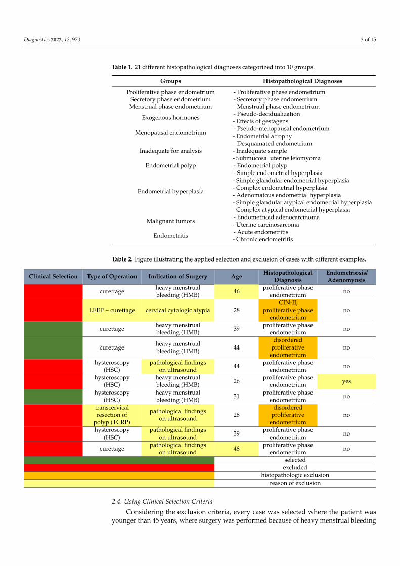
In total, 21 different diagnoses were described in the histopathology reports. Firstly,all cases were separated into these 21 groups. To make it easier to handle the differentcases, all diagnoses were categorized into 10 groups based on their similarities. These werethe following: proliferative phase endometrium, secretory phase endometrium, menstrualphase endometrium, group for effects of exogenous hormones, menopausal endometrium,samples inadequate for analysis, endometrial polyp, endometrial hyperplasia, malignanttumors, and endometritis (Table 1).
2.3. Using Clinical Exclusion Criteria
In freshly extracted samples, the diagnosis included in the histopathology report wasunknown at the time the tissue was submitted to a laboratory for in vitro tissue culturing.Therefore, all samples suggesting underlying pathological processes of the uterus must beeliminated. Every case suitable for further molecular analysis was selected. The followingaspects constituted the basis of exclusion: 1. every sample was excluded in which the patientwas older than 45 years; 2. surgery was performed because of an established pathologicalprocess of the uterus or the type of operation was not only aimed at the extraction of theendometrium; 3. endometriosis or pathological conditions can be revealed in the anamnesisof the patient. The following cases of surgical indication were excluded: postmenopausalbleeding, perimenopausal bleeding, cervical atypia, IUD removal, pathological findings onultrasound, and preoperative curettage before excision of an intrauterine tumor (Table 2).
Diagnostics 2022, 12, 970 3 of 15
Table 1. 21 different histopathological diagnoses categorized into 10 groups.
Groups Histopathological Diagnoses
Proliferative phase endometrium - Proliferative phase endometriumSecretory phase endometrium - Secretory phase endometriumMenstrual phase endometrium - Menstrual phase endometrium
Exogenous hormones - Pseudo-decidualization- Effects of gestagens
Menopausal endometrium - Pseudo-menopausal endometrium- Endometrial atrophy
Inadequate for analysis- Desquamated endometrium- Inadequate sample- Submucosal uterine leiomyoma
Endometrial polyp - Endometrial polyp
Endometrial hyperplasia
- Simple endometrial hyperplasia- Simple glandular endometrial hyperplasia- Complex endometrial hyperplasia- Adenomatous endometrial hyperplasia- Simple glandular atypical endometrial hyperplasia- Complex atypical endometrial hyperplasia
Malignant tumors - Endometrioid adenocarcinoma- Uterine carcinosarcoma
Endometritis - Acute endometritis- Chronic endometritis
Table 2. Figure illustrating the applied selection and exclusion of cases with different examples.
Clinical Selection Type of Operation Indication of Surgery Age HistopathologicalDiagnosis
Endometriosis/Adenomyosis
curettage heavy menstrualbleeding (HMB) 46 proliferative phase
endometrium no
LEEP + curettage cervical cytologic atypia 28CIN-II,
proliferative phaseendometrium
no
curettage heavy menstrualbleeding (HMB) 39 proliferative phase
endometrium no
curettage heavy menstrualbleeding (HMB) 44
disorderedproliferative
endometriumno
hysteroscopy(HSC)
pathological findingson ultrasound 44 proliferative phase
endometrium no
hysteroscopy(HSC)
heavy menstrualbleeding (HMB) 26 proliferative phase
endometrium yes
hysteroscopy(HSC)
heavy menstrualbleeding (HMB) 31 proliferative phase
endometrium no
transcervicalresection of
polyp (TCRP)
pathological findingson ultrasound 28
disorderedproliferative
endometriumno
hysteroscopy(HSC)
pathological findingson ultrasound 39 proliferative phase
endometrium no
curettage pathological findingson ultrasound 48 proliferative phase
endometrium no
selectedexcluded
histopathologic exclusionreason of exclusion
2.4. Using Clinical Selection Criteria
Considering the exclusion criteria, every case was selected where the patient wasyounger than 45 years, where surgery was performed because of heavy menstrual bleeding
Diagnostics 2022, 12, 970 4 of 15
(menorrhagia, abnormal menstruation, irregular periods), and finally, where an operationwas aimed at the extraction of the eutopic endometrium. The following types of operationwere selected: D&C (dilation and curettage) scraping procedures, such as curettage, frac-tional curettage (F&C), abrasion and fractional abrasion, in addition to hysteroscopy withendometrial biopsy (HSC) and transcervical resection of endometrium (TCRE). Scraping(D&C) and hysteroscopy with endometrial biopsy (HSC) procedures were also examinedseparately from each other (Table 2).
2.5. Using Histopathological Selection and Exclusion Criteria
Knowing the diagnoses of the histopathology report, only proliferative and secretoryphase endometria were accepted as appropriate control samples for experimental research.Considering these diagnoses only, every case was excluded where the histopathologyreport described a condition that could affect the signalling pathways of the physiologicalendometrium: disordered proliferative endometrium, adenomyosis, and cervical intraep-ithelial neoplasia (CIN) (Table 2).
2.6. Tissue Culturing
Endometrial tissues harvested by biopsy during HSC procedures were obtained fromthe Department of Gynaecology. Tissues were processed for histopathological analysis withICD codes different from irregular menstrual bleeding. Evaluation of the uterine cavitywas performed as part of diagnostic hysteroscopy. No other uterine pathologies could bedetected preoperatively. Every patient was younger than 45 years. These samples wereminced into 60 × 15 mm cell culture dishes (Eppendorf, North America, Inc., New York,NY, USA) and were fixed to a 15 µL matrigel drop (Cultrex® BME, Type 2). Dishes werefilled with 4.5 g/L Glucose DMEM (Lonza, Bend, OR, USA), and the medium was changedevery day. Tissues were divided into control and hormone-treated groups. Knowing thefirst day of the last menstruation (LMP), samples were maintained until the 24th day of themenstrual cycle. During the whole culturing period, protocol against tissue contaminationwas kept.
2.7. Hormone Application
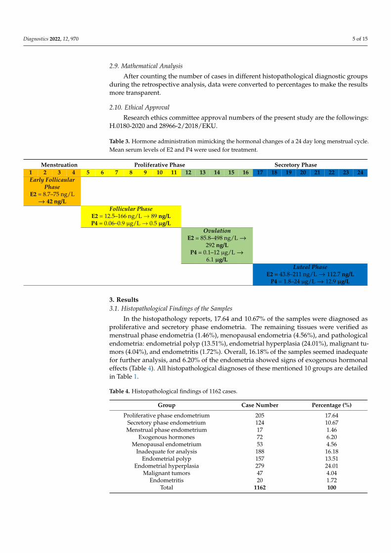
For hormone administration, 17β-estradiol (E2) and progesterone (P4) solutions(Sigma-Aldrich, St. Louis, MO, USA) were applied to the medium of the hormone-treatedgroups. Given the first day of the LMP, whenever the endometrial sample was submittedto the laboratory, the relevant day of the menstrual cycle could be calculated. Four differentconcentrations of E2 and P4 solutions were available. This way, the final concentrationsof hormones in the medium were the mean serum levels observed in the early follicular,follicular, ovulatory, and luteal phases of the ovarian cycle. Until the last day of hormoneadministration, hormone concentrations were changed when the sample reached anotherphase of the ovarian cycle (Table 3). The aim of this procedure was to imitate the in vivohormonal changes of the menstrual cycle.
2.8. H&E Staining
After the last day of the treatment period, tissues were fixed in 4% paraformaldehyde(PFA) solution for 6 h at least. On the day of sample submission, one part of the removedtissue (‘Fresh’) was fixed directly after the operation. Following paraffinization, histologicalslides were sectioned to StarFrost® microscope slides (Knittel Glass, Brunswick, Germany).After deparaffinization, slides were stained with haematoxylin (VWR International, Radnor,PA, USA) and eosin (Amresco, Fountain Parkway Solon, OH, USA) histological stainingmethod. Photomicrographs with 10×, 20× and 40×magnifications were taken from thestained slides with a light microscope (BX-53 Microscope, Olympus Microscopes).
Diagnostics 2022, 12, 970 5 of 15
2.9. Mathematical Analysis
After counting the number of cases in different histopathological diagnostic groupsduring the retrospective analysis, data were converted to percentages to make the resultsmore transparent.
2.10. Ethical Approval
Research ethics committee approval numbers of the present study are the followings:H.0180-2020 and 28966-2/2018/EKU.
Table 3. Hormone administration mimicking the hormonal changes of a 24 day long menstrual cycle.Mean serum levels of E2 and P4 were used for treatment.
Menstruation Proliferative Phase Secretory Phase1 2 3 4 5 6 7 8 9 10 11 12 13 14 15 16 17 18 19 20 21 22 23 24Early Follicaular
PhaseE2 = 8.7–75 ng/L
→ 42 ng/LFollicular Phase
E2 = 12.5–166 ng/L→ 89 ng/LP4 = 0.06–0.9 µg/L→ 0.5 µg/L
OvulationE2 = 85.8–498 ng/L →
292 ng/LP4 = 0.1–12 µg/L →
6.1 µg/LLuteal Phase
E2 = 43.8–211 ng/L → 112.7 ng/LP4 = 1.8–24 µg/L → 12.9 µg/L
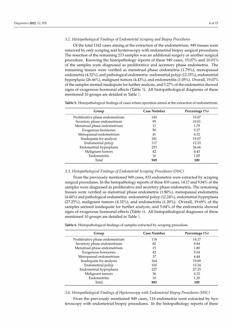
3. Results3.1. Histopathological Findings of the Samples
In the histopathology reports, 17.64 and 10.67% of the samples were diagnosed asproliferative and secretory phase endometria. The remaining tissues were verified asmenstrual phase endometria (1.46%), menopausal endometria (4.56%), and pathologicalendometria: endometrial polyp (13.51%), endometrial hyperplasia (24.01%), malignant tu-mors (4.04%), and endometritis (1.72%). Overall, 16.18% of the samples seemed inadequatefor further analysis, and 6.20% of the endometria showed signs of exogenous hormonaleffects (Table 4). All histopathological diagnoses of these mentioned 10 groups are detailedin Table 1.
Table 4. Histopathological findings of 1162 cases.
Group Case Number Percentage (%)
Proliferative phase endometrium 205 17.64Secretory phase endometrium 124 10.67Menstrual phase endometrium 17 1.46
Exogenous hormones 72 6.20Menopausal endometrium 53 4.56
Inadequate for analysis 188 16.18Endometrial polyp 157 13.51
Endometrial hyperplasia 279 24.01Malignant tumors 47 4.04
Endometritis 20 1.72Total 1162 100
Diagnostics 2022, 12, 970 6 of 15
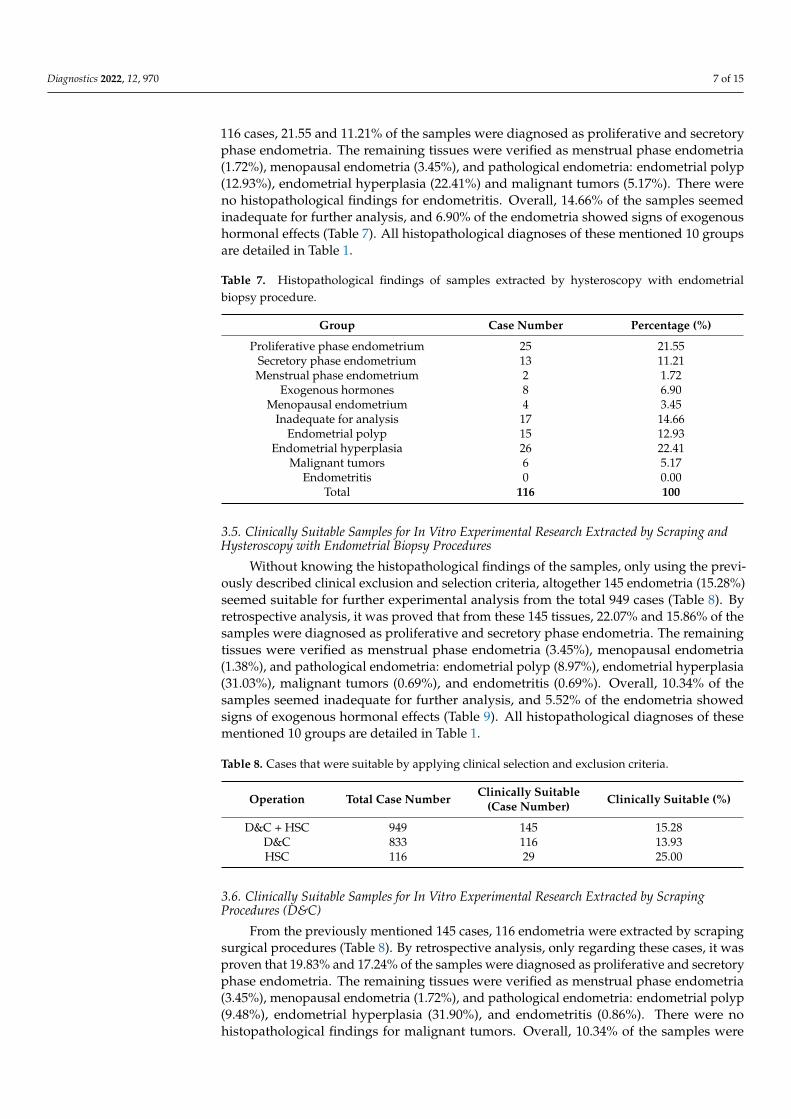
3.2. Histopathological Findings of Endometrial Scraping and Biopsy Procedures
Of the total 1162 cases aiming at the extraction of the endometrium, 949 tissues wereremoved by only scraping and hysteroscopy with endometrial biopsy surgical procedures.The resection of the remaining 213 samples was an additional surgery or another surgicalprocedure. Knowing the histopathology reports of these 949 cases, 15.07% and 10.01%of the samples were diagnosed as proliferative and secretory phase endometria. Theremaining tissues were verified as menstrual phase endometria (1.79%), menopausalendometria (4.32%), and pathological endometria: endometrial polyp (12.33%), endometrialhyperplasia (26.66%), malignant tumors (4.43%), and endometritis (1.05%). Overall, 19.07%of the samples seemed inadequate for further analysis, and 5.27% of the endometria showedsigns of exogenous hormonal effects (Table 5). All histopathological diagnoses of thesementioned 10 groups are detailed in Table 1.
Table 5. Histopathological findings of cases where operation aimed at the extraction of endometrium.
Group Case Number Percentage (%)
Proliferative phase endometrium 143 15.07Secretory phase endometrium 95 10.01Menstrual phase endometrium 17 1.79
Exogenous hormones 50 5.27Menopausal endometrium 41 4.32
Inadequate for analysis 181 19.07Endometrial polyp 117 12.33
Endometrial hyperplasia 253 26.66Malignant tumors 42 4.43
Endometritis 10 1.05Total 949 100
3.3. Histopathological Findings of Endometrial Scraping Procedures (D&C)
From the previously mentioned 949 cases, 833 endometria were extracted by scrapingsurgical procedures. In the histopathology reports of these 833 cases, 14.17 and 9.84% of thesamples were diagnosed as proliferative and secretory phase endometria. The remainingtissues were verified as menstrual phase endometria (1.80%), menopausal endometria(4.44%) and pathological endometria: endometrial polyp (12.24%), endometrial hyperplasia(27.25%), malignant tumors (4.32%), and endometritis (1.20%). Overall, 19.69% of thesamples seemed inadequate for further analysis, and 5.04% of the endometria showedsigns of exogenous hormonal effects (Table 6). All histopathological diagnoses of thesementioned 10 groups are detailed in Table 1.
Table 6. Histopathological findings of samples extracted by scraping procedure.
Group Case Number Percentage (%)
Proliferative phase endometrium 118 14.17Secretory phase endometrium 82 9.84Menstrual phase endometrium 15 1.80
Exogenous hormones 42 5.04Menopausal endometrium 37 4.44
Inadequate for analysis 164 19.69Endometrial polyp 102 12.24
Endometrial hyperplasia 227 27.25Malignant tumors 36 4.32
Endometritis 10 1.20Total 883 100
3.4. Histopathological Findings of Hysteroscopy with Endometrial Biopsy Procedures (HSC)
From the previously mentioned 949 cases, 116 endometria were extracted by hys-teroscopy with endometrial biopsy procedures. In the histopathology reports of these
Diagnostics 2022, 12, 970 7 of 15
116 cases, 21.55 and 11.21% of the samples were diagnosed as proliferative and secretoryphase endometria. The remaining tissues were verified as menstrual phase endometria(1.72%), menopausal endometria (3.45%), and pathological endometria: endometrial polyp(12.93%), endometrial hyperplasia (22.41%) and malignant tumors (5.17%). There wereno histopathological findings for endometritis. Overall, 14.66% of the samples seemedinadequate for further analysis, and 6.90% of the endometria showed signs of exogenoushormonal effects (Table 7). All histopathological diagnoses of these mentioned 10 groupsare detailed in Table 1.
Table 7. Histopathological findings of samples extracted by hysteroscopy with endometrialbiopsy procedure.
Group Case Number Percentage (%)
Proliferative phase endometrium 25 21.55Secretory phase endometrium 13 11.21Menstrual phase endometrium 2 1.72
Exogenous hormones 8 6.90Menopausal endometrium 4 3.45
Inadequate for analysis 17 14.66Endometrial polyp 15 12.93
Endometrial hyperplasia 26 22.41Malignant tumors 6 5.17
Endometritis 0 0.00Total 116 100
3.5. Clinically Suitable Samples for In Vitro Experimental Research Extracted by Scraping andHysteroscopy with Endometrial Biopsy Procedures
Without knowing the histopathological findings of the samples, only using the previ-ously described clinical exclusion and selection criteria, altogether 145 endometria (15.28%)seemed suitable for further experimental analysis from the total 949 cases (Table 8). Byretrospective analysis, it was proved that from these 145 tissues, 22.07% and 15.86% of thesamples were diagnosed as proliferative and secretory phase endometria. The remainingtissues were verified as menstrual phase endometria (3.45%), menopausal endometria(1.38%), and pathological endometria: endometrial polyp (8.97%), endometrial hyperplasia(31.03%), malignant tumors (0.69%), and endometritis (0.69%). Overall, 10.34% of thesamples seemed inadequate for further analysis, and 5.52% of the endometria showedsigns of exogenous hormonal effects (Table 9). All histopathological diagnoses of thesementioned 10 groups are detailed in Table 1.
Table 8. Cases that were suitable by applying clinical selection and exclusion criteria.
Operation Total Case Number Clinically Suitable(Case Number) Clinically Suitable (%)
D&C + HSC 949 145 15.28D&C 833 116 13.93HSC 116 29 25.00
3.6. Clinically Suitable Samples for In Vitro Experimental Research Extracted by ScrapingProcedures (D&C)
From the previously mentioned 145 cases, 116 endometria were extracted by scrapingsurgical procedures (Table 8). By retrospective analysis, only regarding these cases, it wasproven that 19.83% and 17.24% of the samples were diagnosed as proliferative and secretoryphase endometria. The remaining tissues were verified as menstrual phase endometria(3.45%), menopausal endometria (1.72%), and pathological endometria: endometrial polyp(9.48%), endometrial hyperplasia (31.90%), and endometritis (0.86%). There were nohistopathological findings for malignant tumors. Overall, 10.34% of the samples were
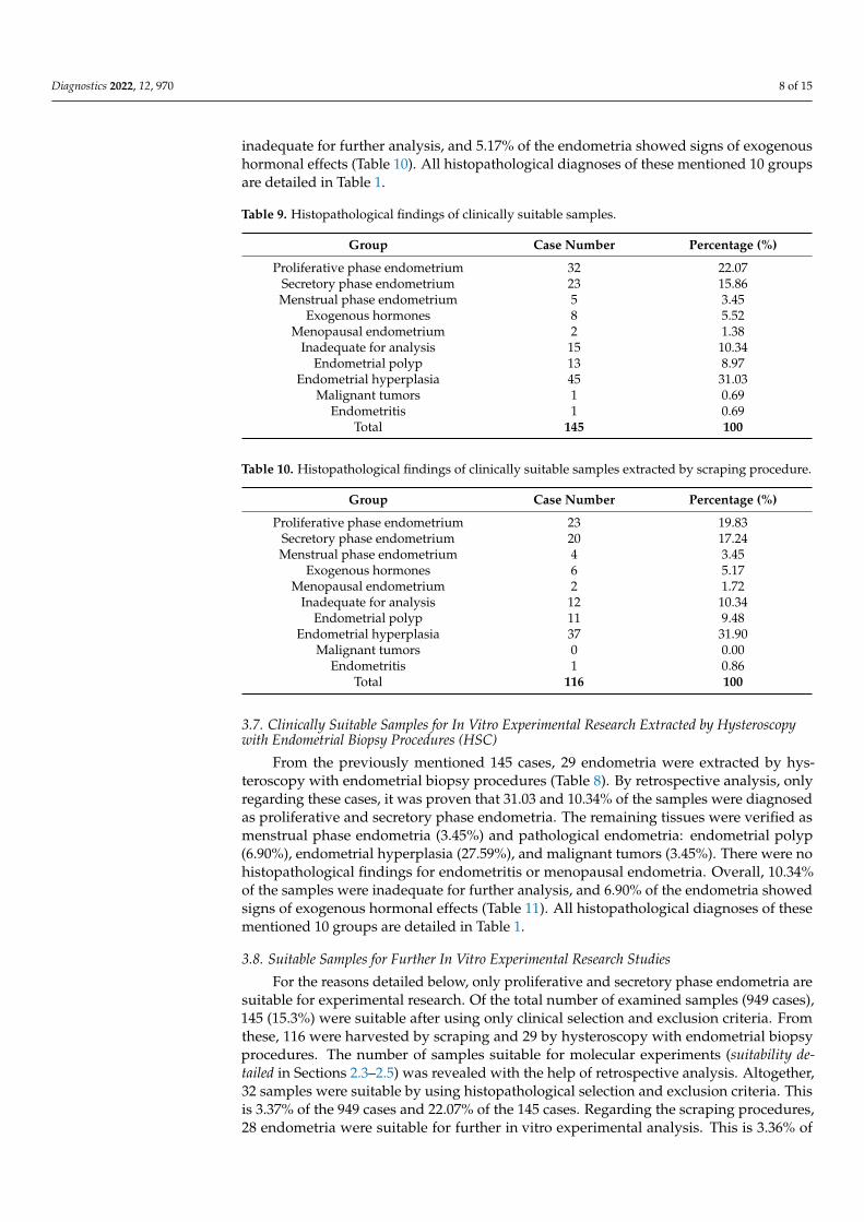
Diagnostics 2022, 12, 970 8 of 15
inadequate for further analysis, and 5.17% of the endometria showed signs of exogenoushormonal effects (Table 10). All histopathological diagnoses of these mentioned 10 groupsare detailed in Table 1.
Table 9. Histopathological findings of clinically suitable samples.
Group Case Number Percentage (%)
Proliferative phase endometrium 32 22.07Secretory phase endometrium 23 15.86Menstrual phase endometrium 5 3.45
Exogenous hormones 8 5.52Menopausal endometrium 2 1.38
Inadequate for analysis 15 10.34Endometrial polyp 13 8.97
Endometrial hyperplasia 45 31.03Malignant tumors 1 0.69
Endometritis 1 0.69Total 145 100
Table 10. Histopathological findings of clinically suitable samples extracted by scraping procedure.
Group Case Number Percentage (%)
Proliferative phase endometrium 23 19.83Secretory phase endometrium 20 17.24Menstrual phase endometrium 4 3.45
Exogenous hormones 6 5.17Menopausal endometrium 2 1.72
Inadequate for analysis 12 10.34Endometrial polyp 11 9.48
Endometrial hyperplasia 37 31.90Malignant tumors 0 0.00
Endometritis 1 0.86Total 116 100
3.7. Clinically Suitable Samples for In Vitro Experimental Research Extracted by Hysteroscopywith Endometrial Biopsy Procedures (HSC)
From the previously mentioned 145 cases, 29 endometria were extracted by hys-teroscopy with endometrial biopsy procedures (Table 8). By retrospective analysis, onlyregarding these cases, it was proven that 31.03 and 10.34% of the samples were diagnosedas proliferative and secretory phase endometria. The remaining tissues were verified asmenstrual phase endometria (3.45%) and pathological endometria: endometrial polyp(6.90%), endometrial hyperplasia (27.59%), and malignant tumors (3.45%). There were nohistopathological findings for endometritis or menopausal endometria. Overall, 10.34%of the samples were inadequate for further analysis, and 6.90% of the endometria showedsigns of exogenous hormonal effects (Table 11). All histopathological diagnoses of thesementioned 10 groups are detailed in Table 1.
3.8. Suitable Samples for Further In Vitro Experimental Research Studies
For the reasons detailed below, only proliferative and secretory phase endometria aresuitable for experimental research. Of the total number of examined samples (949 cases),145 (15.3%) were suitable after using only clinical selection and exclusion criteria. Fromthese, 116 were harvested by scraping and 29 by hysteroscopy with endometrial biopsyprocedures. The number of samples suitable for molecular experiments (suitability de-tailed in Sections 2.3–2.5) was revealed with the help of retrospective analysis. Altogether,32 samples were suitable by using histopathological selection and exclusion criteria. Thisis 3.37% of the 949 cases and 22.07% of the 145 cases. Regarding the scraping procedures,28 endometria were suitable for further in vitro experimental analysis. This is 3.36% of
Diagnostics 2022, 12, 970 9 of 15
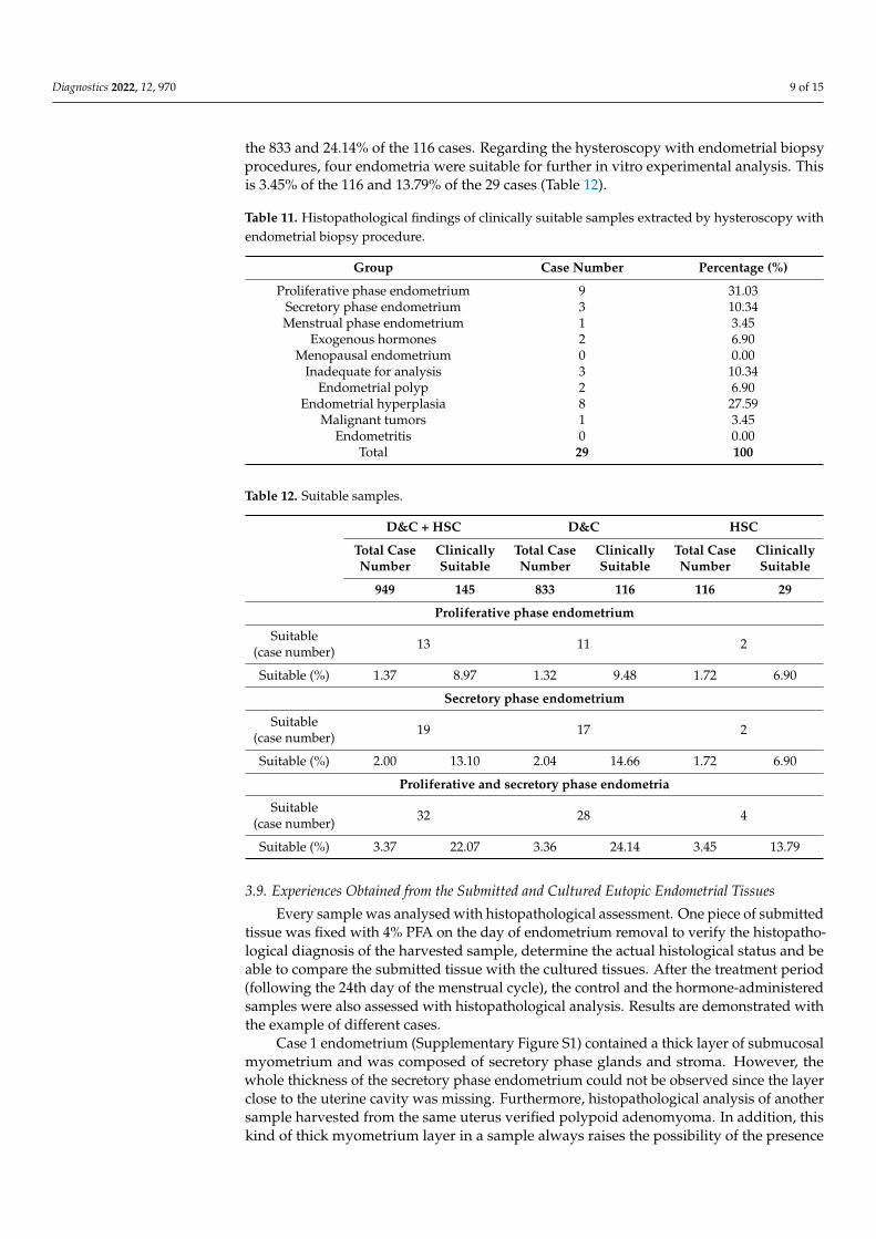
the 833 and 24.14% of the 116 cases. Regarding the hysteroscopy with endometrial biopsyprocedures, four endometria were suitable for further in vitro experimental analysis. Thisis 3.45% of the 116 and 13.79% of the 29 cases (Table 12).
Table 11. Histopathological findings of clinically suitable samples extracted by hysteroscopy withendometrial biopsy procedure.
Group Case Number Percentage (%)
Proliferative phase endometrium 9 31.03Secretory phase endometrium 3 10.34Menstrual phase endometrium 1 3.45
Exogenous hormones 2 6.90Menopausal endometrium 0 0.00
Inadequate for analysis 3 10.34Endometrial polyp 2 6.90
Endometrial hyperplasia 8 27.59Malignant tumors 1 3.45
Endometritis 0 0.00Total 29 100
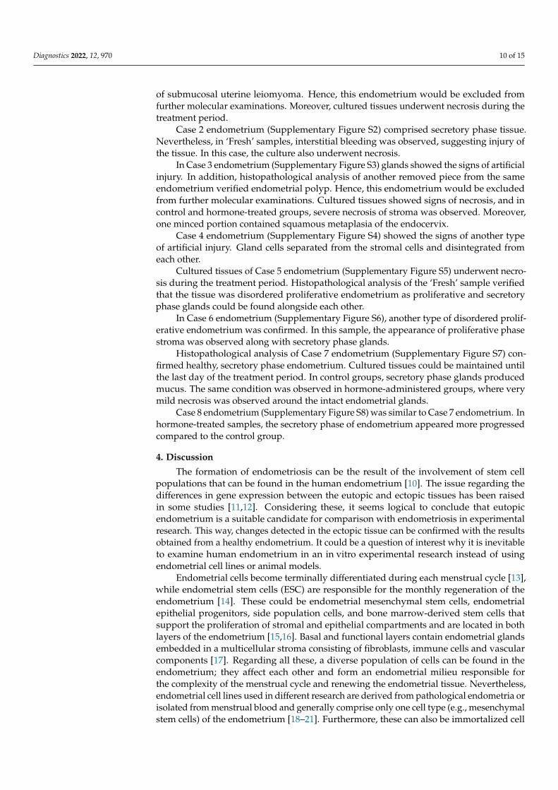
Table 12. Suitable samples.
D&C + HSC D&C HSC
Total CaseNumber
ClinicallySuitable
Total CaseNumber
ClinicallySuitable
Total CaseNumber
ClinicallySuitable
949 145 833 116 116 29
Proliferative phase endometrium
Suitable(case number) 13 11 2
Suitable (%) 1.37 8.97 1.32 9.48 1.72 6.90
Secretory phase endometrium
Suitable(case number) 19 17 2
Suitable (%) 2.00 13.10 2.04 14.66 1.72 6.90
Proliferative and secretory phase endometria
Suitable(case number) 32 28 4
Suitable (%) 3.37 22.07 3.36 24.14 3.45 13.79
3.9. Experiences Obtained from the Submitted and Cultured Eutopic Endometrial Tissues
Every sample was analysed with histopathological assessment. One piece of submittedtissue was fixed with 4% PFA on the day of endometrium removal to verify the histopatho-logical diagnosis of the harvested sample, determine the actual histological status and beable to compare the submitted tissue with the cultured tissues. After the treatment period(following the 24th day of the menstrual cycle), the control and the hormone-administeredsamples were also assessed with histopathological analysis. Results are demonstrated withthe example of different cases.
Case 1 endometrium (Supplementary Figure S1) contained a thick layer of submucosalmyometrium and was composed of secretory phase glands and stroma. However, thewhole thickness of the secretory phase endometrium could not be observed since the layerclose to the uterine cavity was missing. Furthermore, histopathological analysis of anothersample harvested from the same uterus verified polypoid adenomyoma. In addition, thiskind of thick myometrium layer in a sample always raises the possibility of the presence
Diagnostics 2022, 12, 970 10 of 15
of submucosal uterine leiomyoma. Hence, this endometrium would be excluded fromfurther molecular examinations. Moreover, cultured tissues underwent necrosis during thetreatment period.
Case 2 endometrium (Supplementary Figure S2) comprised secretory phase tissue.Nevertheless, in ‘Fresh’ samples, interstitial bleeding was observed, suggesting injury ofthe tissue. In this case, the culture also underwent necrosis.
In Case 3 endometrium (Supplementary Figure S3) glands showed the signs of artificialinjury. In addition, histopathological analysis of another removed piece from the sameendometrium verified endometrial polyp. Hence, this endometrium would be excludedfrom further molecular examinations. Cultured tissues showed signs of necrosis, and incontrol and hormone-treated groups, severe necrosis of stroma was observed. Moreover,one minced portion contained squamous metaplasia of the endocervix.
Case 4 endometrium (Supplementary Figure S4) showed the signs of another typeof artificial injury. Gland cells separated from the stromal cells and disintegrated fromeach other.
Cultured tissues of Case 5 endometrium (Supplementary Figure S5) underwent necro-sis during the treatment period. Histopathological analysis of the ‘Fresh’ sample verifiedthat the tissue was disordered proliferative endometrium as proliferative and secretoryphase glands could be found alongside each other.
In Case 6 endometrium (Supplementary Figure S6), another type of disordered prolif-erative endometrium was confirmed. In this sample, the appearance of proliferative phasestroma was observed along with secretory phase glands.
Histopathological analysis of Case 7 endometrium (Supplementary Figure S7) con-firmed healthy, secretory phase endometrium. Cultured tissues could be maintained untilthe last day of the treatment period. In control groups, secretory phase glands producedmucus. The same condition was observed in hormone-administered groups, where verymild necrosis was observed around the intact endometrial glands.
Case 8 endometrium (Supplementary Figure S8) was similar to Case 7 endometrium. Inhormone-treated samples, the secretory phase of endometrium appeared more progressedcompared to the control group.
4. Discussion
The formation of endometriosis can be the result of the involvement of stem cellpopulations that can be found in the human endometrium [10]. The issue regarding thedifferences in gene expression between the eutopic and ectopic tissues has been raisedin some studies [11,12]. Considering these, it seems logical to conclude that eutopicendometrium is a suitable candidate for comparison with endometriosis in experimentalresearch. This way, changes detected in the ectopic tissue can be confirmed with the resultsobtained from a healthy endometrium. It could be a question of interest why it is inevitableto examine human endometrium in an in vitro experimental research instead of usingendometrial cell lines or animal models.
Endometrial cells become terminally differentiated during each menstrual cycle [13],while endometrial stem cells (ESC) are responsible for the monthly regeneration of theendometrium [14]. These could be endometrial mesenchymal stem cells, endometrialepithelial progenitors, side population cells, and bone marrow-derived stem cells thatsupport the proliferation of stromal and epithelial compartments and are located in bothlayers of the endometrium [15,16]. Basal and functional layers contain endometrial glandsembedded in a multicellular stroma consisting of fibroblasts, immune cells and vascularcomponents [17]. Regarding all these, a diverse population of cells can be found in theendometrium; they affect each other and form an endometrial milieu responsible forthe complexity of the menstrual cycle and renewing the endometrial tissue. Nevertheless,endometrial cell lines used in different research are derived from pathological endometria orisolated from menstrual blood and generally comprise only one cell type (e.g., mesenchymalstem cells) of the endometrium [18–21]. Furthermore, these can also be immortalized cell
Diagnostics 2022, 12, 970 11 of 15
lines [22]. Both the cell lines trying to model the processes of endometriosis [23] as wellas the endometrial cell lines demonstrate one or several aspects of the physiology ofthe endometrium but are not applicable for understanding the entire complexity of thetissue. In addition, there is an absence of cell-cell interactions in these models, which arecharacteristic of the complex microenvironment of the endometrium [20].
One of the main problems with using animal models to investigate the processesof the endometrium is that most laboratory animals have an oestrus cycle instead of amenstrual cycle, except for menstruating primates [24,25]. The latter animals are expensiveto house, and the use of them for routine screenings is unethical. [26]. Another controversyregarding the use of animals is that endometriosis only develops spontaneously in humansand menstruating primates [26]. Although several models have been used to examinethe pathophysiology of the disease [27–30], they have limitations and can not mimic orreproduce all aspects of endometriosis [29,30].
As endometrial cell lines and animal models are not capable of fully reflecting thephysiological characteristics of the endometrium [20], examining the human endometriumitself can be a convenient decision to reveal the physiological or pathological molecularprocesses of this tissue as they consist of the same cells. Histopathological diagnosis ofendometriosis consists of identifying two or more of the following cell types: endometrialgland cells, endometrial epithelial cells, endometrial stromal cells and hemosiderin-ladenmacrophages [1]. The endometrial complexity and extensive capability of the tissue totransform are hallmarked by the diversity of cell types in the tissue, including epithelial,stromal, vascular (endothelium, pericytes and vascular smooth muscle), and immunecells [17,31,32]. Histologically, the endometrium is divided into a basal and a functionallayer [33]. Because of the hormonal changes (different blood levels of oestrogen andprogesterone), the endometrium undergoes cyclic episodes of proliferation, differentiation,and, in the absence of embryo implantation, the shedding of the functional layer [17].Regeneration of endometrium after menstruation is provided by endometrial stem cells [34].It has been found that these stem cells, found in both layers of the endometrium, can bedistributed by retrograde menstrual efflux, and may contribute to the establishment ofectopic endometrium [35]. In recent studies, stem cell-originated endometriosis has alsobeen suggested [5]: ectopic tissue occurs from endogenous stem cells in the endometriumor from bone-marrow-derived stem cells that differentiate into endometrial cells [6].
Harvesting healthy endometrium is a challenging process that also requires ethicalconsideration. Extraction of this sample from the uterine cavity is only possible by appropri-ate surgical indication. Moreover, operations are generally performed due to a presupposedunderlying pathological process in the uterus. Three main surgical procedures providethe extraction of the endometrium: scraping (curettage), hysteroscopy, and hysterectomyprocedures [9,36].
The aim of our present retrospective analysis was to determine what proportion of thesurgically removed human eutopic samples are suitable as appropriate controls to revealmolecular alterations observed in ectopic tissues. This study covers the cases of a 3-year-long period and intends to reveal what the chances are that a healthy, surgically removedendometrial sample submitted to a laboratory is eligible to be processed for further in vitroexperimental research. A list of 1162 final hospital reports and histopathology reports wasexamined where endometria have been submitted to histopathological analysis with anICD code of irregular menstrual bleeding. This is one of the most commonly used codessubmitted to the Pathology Departments together with endometrial samples. Altogether949 operations were aimed at endometrial harvesting only. The remaining 213 caseswere excluded, these being hysterectomy procedures or operations where endometrialharvesting was only a complementary part of another surgical procedure. In most cases, ahysterectomy is performed because of a confirmed pathological condition [9]. Hence, inthe present research, only endometria extracted by scraping and biopsy procedures wereexamined. It has been described that in pathological endometria, numerous molecularalterations can be observed (e.g., in endometrial hyperplasia and tumors) [37–40]. For
Diagnostics 2022, 12, 970 12 of 15
this reason, only healthy endometria can be an appropriate choice to examine molecularprocesses in a control experiment. In addition, it has been described that perimenopausalendometrium has altered gene expression compared to the premenopausal tissue [41].Thus, as early menopause occurs at or before the age of 45 [42], every sample was excludedwhere the patient was older than 45 years.
At the time the endometrial tissue is submitted to an experimental research laboratorydirectly after the surgical procedure, histopathological diagnosis is unknown. In this case,based on our research, the chance of examining pathological endometrium is more than96%. Hence, it should be considered to use preliminary clinical exclusion and selectioncriteria to decrease the probability of getting pathological tissue. With the help of these, itwas shown that the chance of examining a healthy endometrium is more than 22%.
The reason for examining the D&C and HSC cases separately is that endometrialsamples harvested with these procedures are examined in different types of molecularresearch. Tissues removed with scraping procedures mainly contain the functional layerand are suitable for fertility and perimenopausal research, while endometria harvestedby hysteroscopy with endometrial biopsy also contain the basalis layer, and are suitablefor stem cell examinations [9]. At the time of sample submission, the chance of examiningphysiological endometrium is a little more than 3% in the case of scraping and hysteroscopywith endometrial biopsy procedures also. With the help of using clinical exclusion andselection criteria, the chance of examining healthy endometrium is more than 24% re-garding the scraping procedure and more than 13% for hysteroscopy with endometrialbiopsy procedure. The proportion of endometrial samples with pathological findings wasalso revealed, although it was not the aim of the present research, contrary to previousstudies [36,43].
To examine molecular processes of a healthy endometrium, the use of samples ex-tracted with surgical indications other than menstrual disorders should be considered.Endometrium harvested during TCRS (transcervical resection of the uterine septum) ordiagnostic hysteroscopy could be good candidate methods; nevertheless, histopathologicalfindings observed in these kinds of samples have not been described yet. Removing theuterus of brain-dead women who are candidates for organ donation would also be a possi-bility to extract endometrium; however, it is supposed that these uteri would be used totreat absolute uterine factor infertility patients in the future [44]. The main limitation of thepresent research was that only samples submitted with an ICD code of irregular menstrualbleeding were examined, and endometria submitted with different ICD codes were not.
Regarding cultured samples, it was tested whether it is possible to maintain a mincedpiece of endometrium under laboratory circumstances. Hormonal changes in the men-strual cycle were mimicked with estradiol and progesterone administration. As a normalmenstrual cycle lasts between 25 and 35 days [45], the length of the treatment period was24 days and samples were fixed in formalin solution on the 25th day. Analysis of eightcases revealed the problems and difficulties that a molecular researcher faces while doingexperiments with eutopic endometrium. First of all, submitted samples can be injuredartificially at the time of removal. Harvested tissue can sustain mechanical injuries andinterstitial bleedings during the operation. It is unlikely that these injuries are dependenton individual surgeons. Moreover, except for the layers of the endometrium, samples canbe composed of other cell types, such as smooth muscle cells of submucosal myometriumor epithelial cells of the endocervix. These common pitfalls have also been described inprevious studies [9]. Furthermore, in spite of applying clinical selection and exclusioncriteria, during histopathological analysis, pathological conditions can be revealed, suchas disordered proliferative endometrium or endometrial polyp. Regarding our results,healthy endometria outlive the culturing procedures and can be used for further analysis.
5. Conclusions
In summary, since the possibility of harvesting pathological tissue is very high, it is amajor challenge for experimental researchers to achieve reliable results from human eutopic
Diagnostics 2022, 12, 970 13 of 15
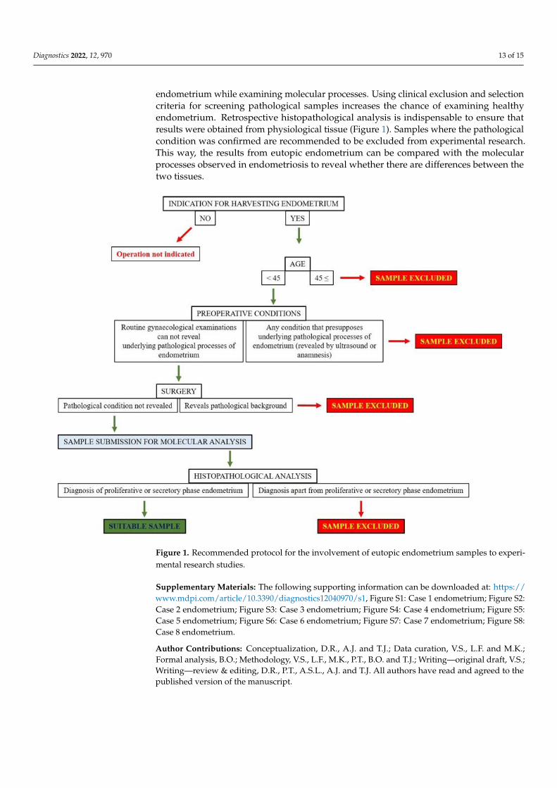
endometrium while examining molecular processes. Using clinical exclusion and selectioncriteria for screening pathological samples increases the chance of examining healthyendometrium. Retrospective histopathological analysis is indispensable to ensure thatresults were obtained from physiological tissue (Figure 1). Samples where the pathologicalcondition was confirmed are recommended to be excluded from experimental research.This way, the results from eutopic endometrium can be compared with the molecularprocesses observed in endometriosis to reveal whether there are differences between thetwo tissues.
Diagnostics 2022, 12, 970 14 of 16
Figure 1. Recommended protocol for the involvement of eutopic endometrium samples to experi-
mental research studies.
Supplementary Materials: The following supporting information can be downloaded at:
https://www.mdpi.com/article/10.3390/diagnostics12040970/s1, Figure S1: Case 1 endometrium;
Figure S2: Case 2 endometrium; Figure S3: Case 3 endometrium; Figure S4: Case 4 endometrium;
Figure S5: Case 5 endometrium; Figure S6: Case 6 endometrium; Figure S7: Case 7 endometrium;
Figure S8: Case 8 endometrium.
Author Contributions: Conceptualization, D.R., A.J. and T.J.; Data curation, V.S., L.F. and M.K.;
Formal analysis, B.O.; Methodology, V.S., L.F., M.K., P.T., B.O. and T.J.; Writing—original draft,
V.S.; Writing—review & editing, D.R., P.T., A.S.L., A.J. and T.J. All authors have read and agreed to
the published version of the manuscript.
Funding: Research was supported by the ÚNKP-17-2 New National Excellence Program of the Min-
istry for Innovation and Technology, by the ÚNKP-19-3 New National Excellence Program of the
Ministry for Innovation and Technology, by the ÚNKP-20-3-II New National Excellence Program of
the Ministry for Innovation and Technology from the source of the National Research, Development
and Innovation Fund. The research was supported by the EFOP-3.6.3-VEKOP-16-2017-00009 project
co-financed by EU and the European Social Fund. The research was supported by NKFIHK139396
and by MTA-TKI 14016.
Institutional Review Board Statement: The study was conducted in accordance with the
Declaration of Helsinki, and approved by the Institutional Review Board (or Ethics Com-
mittee) of University of Debrecen: research ethics committee approval numbers of the pre-
sent study are the followings: H.0180-2020 and 28966-2/2018/EKU. Informed Consent Statement: Informed consent was obtained from all subjects involved in the
study.
Data Availability Statement: Not applicable.
Acknowledgments: Special thanks to our laboratory assistant, Krisztina Bíróné Barna for her hon-
ourable work. We thank Lili Schwieters for proofreading the manuscript.
Figure 1. Recommended protocol for the involvement of eutopic endometrium samples to experi-mental research studies.
Supplementary Materials: The following supporting information can be downloaded at: https://www.mdpi.com/article/10.3390/diagnostics12040970/s1, Figure S1: Case 1 endometrium; Figure S2:Case 2 endometrium; Figure S3: Case 3 endometrium; Figure S4: Case 4 endometrium; Figure S5:Case 5 endometrium; Figure S6: Case 6 endometrium; Figure S7: Case 7 endometrium; Figure S8:Case 8 endometrium.
Author Contributions: Conceptualization, D.R., A.J. and T.J.; Data curation, V.S., L.F. and M.K.;Formal analysis, B.O.; Methodology, V.S., L.F., M.K., P.T., B.O. and T.J.; Writing—original draft, V.S.;Writing—review & editing, D.R., P.T., A.S.L., A.J. and T.J. All authors have read and agreed to thepublished version of the manuscript.
Diagnostics 2022, 12, 970 14 of 15
Funding: Research was supported by the ÚNKP-17-2 New National Excellence Program of theMinistry for Innovation and Technology, by the ÚNKP-19-3 New National Excellence Program of theMinistry for Innovation and Technology, by the ÚNKP-20-3-II New National Excellence Program ofthe Ministry for Innovation and Technology from the source of the National Research, Developmentand Innovation Fund. The research was supported by the EFOP-3.6.3-VEKOP-16-2017-00009 projectco-financed by EU and the European Social Fund. The research was supported by NKFIHK139396and by MTA-TKI 14016.
Institutional Review Board Statement: The study was conducted in accordance with the Declarationof Helsinki, and approved by the Institutional Review Board (or Ethics Committee) of Universityof Debrecen: research ethics committee approval numbers of the present study are the followings:H.0180-2020 and 28966-2/2018/EKU.
Informed Consent Statement: Informed consent was obtained from all subjects involved in the study.
Data Availability Statement: Not applicable.
Acknowledgments: Special thanks to our laboratory assistant, Krisztina Bíróné Barna for her hon-ourable work. We thank Lili Schwieters for proofreading the manuscript.
Conflicts of Interest: The authors declare that there are no conflicts of interest that could be perceivedas prejudicing the impartiality of the research reported.
References1. Hsu, A.L.; Khachikyan, I.; Stratton, P. Invasive and noninvasive methods for the diagnosis of endometriosis. Clin. Obstet. Gynecol.
2010, 53, 413–419. [CrossRef] [PubMed]2. Rolla, E. Endometriosis: Advances and controversies in classification, pathogenesis, diagnosis, and treatment. F1000Res 2019,
8, 529. [CrossRef] [PubMed]3. Lagana, A.S.; Vitale, S.G.; Salmeri, F.M.; Triolo, O.; Ban Frangez, H.; Vrtacnik-Bokal, E.; Stojanovska, L.; Apostolopoulos, V.;
Granese, R.; Sofo, V. Unus pro omnibus, omnes pro uno: A novel, evidence-based, unifying theory for the pathogenesis ofendometriosis. Med. Hypotheses 2017, 103, 10–20. [CrossRef] [PubMed]
4. Chang, J.H.; Au, H.K.; Lee, W.C.; Chi, C.C.; Ling, T.Y.; Wang, L.M.; Kao, S.H.; Huang, Y.H.; Tzeng, C.R. Expression of thepluripotent transcription factor OCT4 promotes cell migration in endometriosis. Fertil. Steril. 2013, 99, 1332–1339.e5. [CrossRef][PubMed]
5. Lagana, A.S.; Salmeri, F.M.; Vitale, S.G.; Triolo, O.; Gotte, M. Stem cell trafficking during endometriosis: May epigenetics play apivotal role? Reprod. Sci. 2018, 25, 978–979. [CrossRef] [PubMed]
6. Macer, M.L.; Taylor, H.S. Endometriosis and infertility: A review of the pathogenesis and treatment of endometriosis-associatedinfertility. Obstet. Gynecol. Clin. N. Am. 2012, 39, 535–549. [CrossRef]
7. Bouquet De Joliniere, J.; Ayoubi, J.M.; Gianaroli, L.; Dubuisson, J.B.; Gogusev, J.; Feki, A. Endometriosis: A new cellular andmolecular genetic approach for understanding the pathogenesis and evolutivity. Front. Surg. 2014, 1, 16. [CrossRef]
8. Lagana, A.S.; Garzon, S.; Gotte, M.; Vigano, P.; Franchi, M.; Ghezzi, F.; Martin, D.C. The pathogenesis of endometriosis: Molecularand cell biology insights. Int. J. Mol. Sci. 2019, 20, 5615. [CrossRef]
9. Maclean, A.; Kamal, A.; Adishesh, M.; Alnafakh, R.; Tempest, N.; Hapangama, D.K. Human uterine biopsy: Research value andcommon pitfalls. Int. J. Reprod. Med. 2020, 2020, 9275360. [CrossRef]
10. Sourial, S.; Tempest, N.; Hapangama, D.K. Theories on the pathogenesis of endometriosis. Int. J. Reprod. Med. 2014, 2014, 179515.[CrossRef]
11. Borghese, B.; Mondon, F.; Noel, J.C.; Fayt, I.; Mignot, T.M.; Vaiman, D.; Chapron, C. Gene expression profile for ectopic versuseutopic endometrium provides new insights into endometriosis oncogenic potential. Mol. Endocrinol. 2008, 22, 2557–2562.[CrossRef]
12. Gabriel, M.; Fey, V.; Heinosalo, T.; Adhikari, P.; Rytkonen, K.; Komulainen, T.; Huhtinen, K.; Laajala, T.D.; Siitari, H.;Virkki, A.; et al. A relational database to identify differentially expressed genes in the endometrium and endometriosis lesions.Sci. Data 2020, 7, 284. [CrossRef] [PubMed]
13. Evans, J.; Salamonsen, L.A.; Winship, A.; Menkhorst, E.; Nie, G.; Gargett, C.E.; Dimitriadis, E. Fertile ground: Human endometrialprogramming and lessons in health and disease. Nat. Rev. Endocrinol. 2016, 12, 654–667. [CrossRef] [PubMed]
14. Gargett, C.E.; Schwab, K.E.; Zillwood, R.M.; Nguyen, H.P.; Wu, D. Isolation and culture of epithelial progenitors and mesenchymalstem cells from human endometrium. Biol. Reprod. 2009, 80, 1136–1145. [CrossRef] [PubMed]
15. Cousins, F.L.; Dorien, F.O.; Gargett, C.E. Endometrial stem/progenitor cells and their role in the pathogenesis of endometriosis.Best Pract. Res. Clin. Obstet. Gynaecol. 2018, 50, 27–38. [CrossRef] [PubMed]
16. Maruyama, T. Endometrial stem/progenitor cells. J. Obstet. Gynaecol. Res. 2014, 40, 2015–2022. [CrossRef]17. Gibson, D.A.; Simitsidellis, I.; Collins, F.; Saunders, P.T.K. Endometrial intracrinology: Oestrogens, androgens and endometrial
disorders. Int. J. Mol. Sci. 2018, 19, 3276. [CrossRef]
Diagnostics 2022, 12, 970 15 of 15
18. Du, X.; Yuan, Q.; Qu, Y.; Zhou, Y.; Bei, J. Endometrial mesenchymal stem cells isolated from menstrual blood by adherence. StemCells Int. 2016, 2016, 3573846. [CrossRef]
19. Musina, R.A.; Belyavski, A.V.; Tarusova, O.V.; Solovyova, E.V.; Sukhikh, G.T. Endometrial mesenchymal stem cells isolated fromthe menstrual blood. Bull. Exp. Biol. Med. 2008, 145, 539–543. [CrossRef]
20. Nikolakopoulou, K.; Turco, M.Y. Investigation of infertility using endometrial organoids. Reproduction 2021, 161, R113–R127.[CrossRef]
21. Noumoff, J.; Haydock, S.W.; Sachdeva, R.; Heyner, S.; Pritchard, M.L. Characteristics of cell lines derived from normal andmalignant endometrial tissue. Gynecol. Oncol. 1987, 27, 141–149. [CrossRef]
22. Holdsworth-Carson, S.J.; Colgrave, E.M.; Donoghue, J.F.; Fung, J.N.; Churchill, M.L.; Mortlock, S.; Paiva, P.; Healey, M.;Montgomery, G.W.; Girling, J.E.; et al. Generation of immortalized human endometrial stromal cell lines with differentendometriosis risk genotypes. Mol. Hum. Reprod. 2019, 25, 194–205. [CrossRef] [PubMed]
23. Fan, H. In-vitro models of human endometriosis. Exp. Ther. Med. 2020, 19, 1617–1625. [PubMed]24. Ajayi, A.F.; Akhigbe, R.E. Staging of the estrous cycle and induction of estrus in experimental rodents: An update. Fertil. Res.
Pract. 2020, 6, 5. [CrossRef]25. Sato, J.; Nasu, M.; Tsuchitani, M. Comparative histopathology of the estrous or menstrual cycle in laboratory animals. J. Toxicol.
Pathol. 2016, 29, 155–162. [CrossRef]26. Simitsidellis, I.; Gibson, D.A.; Saunders, P.T.K. Animal models of endometriosis: Replicating the aetiology and symptoms of the
human disorder. Best Pract. Res. Clin. Endocrinol. Metab. 2018, 32, 257–269. [CrossRef]27. Grummer, R. Animal models in endometriosis research. Hum. Reprod. Update 2006, 12, 641–649. [CrossRef]28. Story, L.; Kennedy, S. Animal studies in endometriosis: A review. ILAR J. 2004, 45, 132–138. [CrossRef]29. Taniguchi, F.; Wibisono, H.; Mon Khine, Y.; Harada, T. Animal models for research on endometriosis. Front. Biosci. 2021, 13, 37–53.30. Tirado-Gonzalez, I.; Barrientos, G.; Tariverdian, N.; Arck, P.C.; Garcia, M.G.; Klapp, B.F.; Blois, S.M. Endometriosis research:
Animal models for the study of a complex disease. J. Reprod. Immunol. 2010, 86, 141–147. [CrossRef]31. Critchley, H.O.D.; Maybin, J.A.; Armstrong, G.M.; Williams, A.R.W. Physiology of the endometrium and regulation of menstrua-
tion. Physiol. Rev. 2020, 100, 1149–1179. [CrossRef] [PubMed]32. Simitsidellis, I.; Saunders, P.T.K.; Gibson, D.A. Androgens and endometrium: New insights and new targets. Mol. Cell Endocrinol.
2018, 465, 48–60. [CrossRef] [PubMed]33. Gibson, D.A.; Simitsidellis, I.; Collins, F.; Saunders, P.T.K. Androgens, oestrogens and endometrium: A fine balance between
perfection and pathology. J. Endocrinol. 2020, 246, R75–R93. [CrossRef] [PubMed]34. Tempest, N.; Jansen, M.; Baker, A.M.; Hill, C.J.; Hale, M.; Magee, D.; Treanor, D.; Wright, N.A.; Hapangama, D.K. Histological 3D
reconstruction and in vivo lineage tracing of the human endometrium. J. Pathol. 2020, 251, 440–451. [CrossRef]35. Proestling, K.; Birner, P.; Balendran, S.; Nirtl, N.; Marton, E.; Yerlikaya, G.; Kuessel, L.; Reischer, T.; Wenzl, R.; Streubel, B.; et al.
Enhanced expression of the stemness-related factors OCT4, SOX15 and TWIST1 in ectopic endometrium of endometriosis patients.Reprod. Biol. Endocrinol. 2016, 14, 81. [CrossRef]
36. McCluggage, W.G. My approach to the interpretation of endometrial biopsies and curettings. J. Clin. Pathol. 2006, 59, 801–812.[CrossRef]
37. Lax, S.F. Molecular genetic changes in epithelial, stromal and mixed neoplasms of the endometrium. Pathology 2007, 39, 46–54.[CrossRef]
38. Llobet, D.; Pallares, J.; Yeramian, A.; Santacana, M.; Eritja, N.; Velasco, A.; Dolcet, X.; Matias-Guiu, X. Molecular pathologyof endometrial carcinoma: Practical aspects from the diagnostic and therapeutic viewpoints. J. Clin. Pathol. 2009, 62, 777–785.[CrossRef]
39. Yeramian, A.; Moreno-Bueno, G.; Dolcet, X.; Catasus, L.; Abal, M.; Colas, E.; Reventos, J.; Palacios, J.; Prat, J.; Matias-Guiu, X.Endometrial carcinoma: Molecular alterations involved in tumor development and progression. Oncogene 2013, 32, 403–413.[CrossRef]
40. Zauber, P.; Denehy, T.R.; Taylor, R.R.; Ongcapin, E.H.; Marotta, S.; Sabbath-Solitare, M. Strong correlation between molecularchanges in endometrial carcinomas and concomitant hyperplasia. Int. J. Gynecol. Cancer 2015, 25, 863–868. [CrossRef]
41. Erikson, D.W.; Barragan, F.; Piltonen, T.T.; Chen, J.C.; Balayan, S.; Irwin, J.C.; Giudice, L.C. Stromal fibroblasts from peri-menopausal endometrium exhibit a different transcriptome than those from the premenopausal endometrium. Biol. Reprod. 2017,97, 387–399. [CrossRef]
42. Shuster, L.T.; Rhodes, D.J.; Gostout, B.S.; Grossardt, B.R.; Rocca, W.A. Premature menopause or early menopause: Long-termhealth consequences. Maturitas 2010, 65, 161–166. [CrossRef] [PubMed]
43. Inal, Z.O.; Inal, H.A.; Kucukosmanoglu, I.; Kucukkendirci, H. Assessment of endometrial sampling and histopathological results:Analysis of 4247 cases. Eurasian J. Med. 2017, 49, 44–47. [CrossRef] [PubMed]
44. Brannstrom, M. Uterus transplantation and beyond. J. Mater. Sci. Mater. Med. 2017, 28, 70. [CrossRef] [PubMed]45. Bull, J.R.; Rowland, S.P.; Scherwitzl, E.B.; Scherwitzl, R.; Danielsson, K.G.; Harper, J. Real-world menstrual cycle characteristics of
more than 600,000 menstrual cycles. NPJ Digit. Med. 2019, 2, 83. [CrossRef]