promoter methylation regulates estrogen receptor 2 in human endometrium and endometriosis
TRANSCRIPT

1
Title: Promoter methylation regulates estrogen receptor 2 (ESR2) in endometrium and 1
endometriosis 2
3
Short title: ESR2 methylation in endometrial and endometriotic cells 4
5
Summary sentence: Estrogen receptor 2, expressed in strikingly higher levels in endometriosis 6
compared with normal endometrium, is regulated primarily by methylation of a CpG island in its 7
promoter. 8
9
Keywords and Topic Categary: 10
Keywords: DNA methylation, ESR2, endometriosis, endometrium, CpG island 11
Topic Categary: Female Reproductive Tract 12
13
Authors: Qing Xue 3, 6, Zhihong Lin 3, You-Hong Cheng 3, Chiang-Ching Huang 4, Erica Marsh 14
3, 5, Ping Yin 3, Magdy P Milad 5, Edmond Confino 5, Scott Reierstad 3, Joy Innes 3, and Serdar E 15
Bulun 2, 3, 5 16
Division of Reproductive Biology Research 3, Department of Obstetrics and Gynecology, 17
Department of Preventive Medicine 4, Division of Reproductive Endocrinology and Infertility 5, 18
Department of Obstetrics and Gynecology, Feinberg School of Medicine, Northwestern 19
University, Chicago, Illinois 60611, USA; Department of Obstetrics and Gynecology, First 20
Hospital of Peking University, Beijing, P. R. China 6. 21
22 1 Grant support: Supported by the NIH/NICHD grant U54-HD40093. 23
24
2 Correspondence: Serdar E. Bulun, M.D., Division of Reproductive Biology Research , 25
Department of Obstetrics and Gynecology, Northwestern University, 303 E. Superior Street, 26
BOR Papers in Press. Published on July 11, 2007 as DOI:10.1095/biolreprod.107.061804
Copyright 2007 by The Society for the Study of Reproduction.

2
Suite 4-250, Chicago, Illinois 60611. Tel: 312-503-0520; Fax: 312-503-0095; E-mail: s-27
29
ABSTRACT 30
Steroid receptors in the stromal cells of endometrium and its disease counterpart tissue 31
endometriosis play critical physiological roles. We found that mRNA and protein levels of 32
estrogen receptor 2 (ESR2) were strikingly higher, whereas levels of estrogen receptor 1 33
(ESR1), total progesterone receptor (PGR), and progesterone receptor B (PGR B) were 34
significantly lower in endometriotic vs. endometrial stromal cells. Because ESR2 displayed 35
the most striking levels of differential expression between endometriotic and endometrial 36
cells, and the mechanisms for this difference are unknown, we tested the hypothesis that 37
alterations in DNA methylation is a mechanism responsible for severely increased ESR2 38
mRNA levels in endometriotic cells. We identified a CpG island occupying the promoter 39
region (-197/+359) of the ESR2 gene. Bisulfite sequencing of this region showed significantly 40
higher methylation in primary endometrial cells (n=8 subjects) vs. endometriotic cells (n=8 41
subjects). The demethylating agent 5-aza-2’-deoxycytidine significantly increased ESR2 42
mRNA levels in endometrial cells. Mechanistically, we employed serial deletion mutants of 43
the ESR2 promoter fused to the luciferase reporter gene and transiently transfected into 44
both endometriotic and endometrial cells. We demonstrated that the critical region (-45
197/+372) that confers promoter activity also bears the CpG island, and the activity of the 46
ESR2 promoter was strongly inactivated by in vitro methylation. Taken together, 47
methylation of a CpG island at the ESR2 promoter region is a primary mechanism 48
responsible for differential expression of ESR2 in endometriosis and endometrium. These 49
findings may be applied to a number of areas ranging from diagnosis to the treatment of 50
endometriosis. 51
52

3
INTRODUCTION 53
Endometriosis is defined as the presence of endometrium-like tissue outside of the uterine 54
cavity. It is a common gynecological condition affecting 1 in 10 women in the reproductive age 55
group (1). Endometriosis is associated with severely painful menstruation, chronic pelvic pain, 56
and infertility (1, 2). Although the etiology and exact mechanism for the development of 57
endometriosis is unclear, there is a large body of laboratory and circumstantial evidence that 58
suggests a crucial role for estrogen in the establishment and maintenance of this disease (3-5). 59
Despite its sensitivity to estrogen, endometriosis appears to contain a unique complement of 60
steroid hormone receptors compared with that of its normal tissue counterpart, the eutopic 61
endometrium. For example, a number of investigators reported markedly higher levels of estrogen 62
receptor 2 (ESR2) and lower levels of estrogen receptor 1 (ESR1) in human endometriotic tissues 63
and primary stromal cells compared with eutopic endometrial tissues and cells (6, 7). Moreover, 64
the levels of both isoforms of progesterone receptor (PGR), particularly progesterone receptor B 65
(PGR B), are significantly lower in endometriosis compared to eutopic endometrium (8, 9). The 66
classical human ESR1 was cloned in 1986, and a second estrogen receptor, ESR2 was cloned 67
from rat prostate and human testis in 1996 (10-12). Both ESRs act as transcription factors and are 68
believed to play a key role in endometrial and endometriosis growth regulation. 69
Hypermethylation of a CpG island has been associated with the transcriptional inactivation of 70
genes. Recently, key nuclear receptor genes such as ESR1, ESR2, and PGR were shown to be 71
regulated by methylation of their promoter regions in breast, prostate and endometrial cancer 72
tissues (13-15). To assess the relative expression levels of these nuclear receptors and the DNA 73
methylation mechanism in endometrium and endometriosis, an in vitro model of primary stromal 74
cells from these two tissue sources was developed. 75
Currently, the biological roles of ESR2 in endometrium and endometriosis are not well 76
understood. We chose to investigate the molecular mechanism responsible for differential 77
expression of ESR2 for two reasons. First, the most striking difference between endometriosis and 78

4
endometrium was observed with respect to ESR2 levels compared with other steroid receptors, 79
and ESR2 mRNA levels were very low or nearly undetectable in the endometrial stromal cells. 80
Second, an ESR2-selective compound was shown to be therapeutic in a rodent endometriosis 81
model (16, 17). At present, no evidence has been provided to indicate whether DNA methylation 82
is causally linked to differential ESR2 expression in endometriotic stromal cells and endometrial 83
stromal cells. Direct evidence in support of the cytosine methylation of specific 5' CpGs that 84
leads to transcriptional inactivation has not been reported. 85
86
MATERIALS AND METHODS 87
Subjects and primary cell culture 88
Eutopic endometrium from disease-free subjects (n=8) and ectopic endometrium from the cyst 89
walls of ovarian endometriomas (defined as a cystic ovarian lesion comprised of endometrium-90
like tissue in its cyst wall and bloody fluid filled in this cyst) (n=8) were obtained immediately 91
after surgery. The mean ages of subjects in each group were 42±3 to 39±3, and there were no 92
significant differences between the two groups with respect to age or cycle phase. Moreover, we 93
obtained 3 paired samples of ovarian endometriomas and eutopic endometrium from the same 94
subjects. None of the patients had received any pre-operative hormonal therapy. All samples were 95
histologically confirmed, and the phase of the menstrual cycle was determined by preoperative 96
history and histological examination. Written informed consent was obtained before surgical 97
procedures, including a consent form and protocol approved by the Institutional Review Board of 98
Northwestern University. Stromal cells were isolated from these two types of tissues using a 99
protocol previously reported by Ryan et al with minor modifications (18, 19). Briefly, tissues 100
were rinsed with sterile PBS, minced finely, and digested with collagenase (Sigma, St. Louis, 101
MO) and DNase (Sigma) at 37 oC for 60 min. Stromal cells were separated from epithelial cells 102
by filtration through a 70- and 20-µm sieve, then were suspended in DMEM/F12 1:1 103

5
(GIBCO/BRL, Grand Island, NY) containing 10% FBS and in a humidified atmosphere with 5% 104
CO2 at 37 oC. 105
106
RNA extraction and quantitative analysis by real-time RT PCR 107
Total RNA was isolated from stromal cells, using TRIzol reagent (Sigma), following the 108
manufacturer’s protocol. Total RNA was first treated with DNase І (Ambion, Austin, TX) to 109
remove contaminating genomic DNA from the RNA samples, then one µg of RNA was used to 110
generate cDNA with the Superscript Ш First-Strand Synthesis System (Invitrogen, Carlsbad, 111
CA). Real-time quantitative PCR was performed with the ABI 7900 Sequence Detection System 112
and the ABI Taqman Gene Expression system (purchased from Applied Biosystems, Foster City, 113
CA) for ESR1, ESR2 and eukaryotic 18S rRNA (18S). The SYBR Green assay was used for total 114
PGR, PGR B and 18S. 18S values were used for normalization. Primers used for SYBR Green 115
assay were: total PGR, forward: 5'- GTCCTTACCTGTGGGAGCTG-3', reverse: 5'- CAACAGC- 116
ATCCAGTGCTCTC -3'. PGR B, forward: 5'- GTACGGAGCCAGCAGAAGTC -3', reverse, 5'-117
TCTCTGGCATCAAACTCGTG-3'. 18S, forward: 5'- AGGAATTCCCAGTAAGTGCG -3', 118
reverse: 5'-GCCTCACTAAACCATCCAA -3'. Relative quantification of mRNA species was 119
performed using the comparative threshold cycles (CT) method. In brief, comparative threshold 120
cycles (CT) was used to determine the mRNA level normalized to the average mRNA level in 121
endometrial stromal cells. Thus, mRNA levels were expressed as an n-fold difference. For each 122
sample, the gene CT value was normalized using the formula: ∆ CT = CT gene ─ CT 18S. To 123
determine relative expression levels, the following formula was used: ∆ ∆CT = ∆CT sample ─ ∆CT 124
calibrator. This value was used to plot the gene expression employing the formula 2-∆ ∆CT. 125
126
Bisulfite modification and sequencing analysis 127
Genomic DNA was extracted from the primary stromal cells by using DNeasy Tissue kit 128
(Qiagen, Valencia, CA). 500 ng DNA was treated with sodium bisulfite following the 129

6
manufacturer’s protocol (Zymo Research, Orange, CA). Purified DNA was dissolved in 10 µl M-130
Elution Buffer. For PCR amplification, 3 µl of bisulfite-modified DNA was added to a final 131
volume of 20 µl. AmpliTaq Gold PCR Master Mix (Applied Biosystems) was used for all PCR 132
amplifications. PCR amplifications were performed using the following primers for ESR2: 133
forward: 5'- ATTATTTTTGTGGGTGGATTAGGAG -3', and reverse: 5'- AACCCCTTCTTCC- 134
TTTTAAAAACC -3'. The thermal cycle conditions were as follows: 95 oC for 10 min followed 135
by 40 cycles of denaturation at 95 oC 30 s, annealing at 50 oC for 2 min, and elongation at 72 oC 136
for 2 min, then followed by an incubation at 72 oC for 7 min. PCR products (166bp) were gel-137
purified and cloned into the pGEM-Teasy vector (Promega, Madison, WI). Following 138
transformation, 6 to 8 clones with the correct insert were randomly picked for each PCR, and 139
were sequenced using an Applied Biosystems 377 instrument. 140
141
5-Aza-2’-deoxycytidine (5-aza-dC) treatments 142
At approximately 40% confluence, endometrial stromal cells were placed in serum-free 143
DMEM/F12 for 24 hours, and then treated with 20 µM DNA methyltransferase inhibitor, 5-aza-144
dC (Sigma) for 5 days, and medium was changed each day. Total RNA was isolated from the 145
treated cells using TRIzol reagent. All experiments were conducted in triplicate and repeated 146
three times in primary cultured cells from at least 3 different subjects. 147
148
Plasmid construction 149
Reporter plasmid vectors containing the ESR2 promoter sequences were constructed by PCR 150
cloning. Genomic DNA from endometriotic stromal cells was used as the template for 151
amplification. The primers were: reverse primer 5'- GATATCTTAGCACAATCAACCCAGAG- 152
C -3' (position +564 relative to the transcription start site), forward primers 5'- GGTACCTTCCC-153
AGTGACCTCTTGA -3' (-525), 5'- GGTACCTGTGCGCCACTATCCTTG -3' (-197), 5'- GGT- 154
7
ACCTGTTTGAAATCCTGCGGTGAG -3' (+372). Restriction sites (KpnІ site for forward 155
primers and EcoRV site for reverse primers) were added to the 5'-end of primers, and promoter 156
sequences were amplified using TAKARA LA Taq™ with GC buffer (TaKaRa, Otsu, Japan). 157
PCR products were cloned into a modified pGL4 vector-SV40. The SV40 minimal promoter 158
was digested with Bgl II and HindIII from pGL2-promoter vector and cloned into Bgl II and 159
HindIII digested pGL4.10 vector (Promega). The final plasmids containing ESR2 promoter 160
sequences were -525/+564, -197/+564, and +372/+564. 161
162
Transfection and luciferase reporter gene assay 163
Transfection experiments of endometrial and endometriotic stromal cells were performed 164
using FuGENE 6 transfection reagent (Roche Applied Science, Indianapolis, IN) according to the 165
manufacturer’s protocol. Briefly, the cells were grown in 24-well tissue culture plates so that the 166
cell layer was 50-60% confluent on the day of transfection. For each well, OPTI-MEM І 167
containing 1.5 µl of FuGENE 6 was mixed with 240 ng of reporter plasmid and 60 ng or 80 ng of 168
pSV-β-galactosidase vector (Promega) for endometrial or endometriotic stromal cells. The cells 169
were harvested 48 hours after transfection, and the luciferase activity was measured using a 170
luciferase assay system (Promega). β-galactosidase activity was used to normalize transfection 171
efficiency. All of the experiments were repeated three times in triplicate. 172
173
In vitro methylation of reporter plasmids 174
In vitro methylation assays were carried out according to the methods described by Robertson 175
(20) and Singal (21). Briefly, region-specific methylation was carried out on the ESR2 promoter 176
fragments of -525/+564 and -197/+564 after excision and isolation. DNA was incubated with SssІ 177
CpG methylase (New England Biolabs, Ipswich, MA) in the presence (methylated) or absence 178
(mock-methylated) of S-adenosylmethionine, as recommended by the manufacturer for 2 hours. 179
Methylated and mock-methylated fragments were religated into their respectively unmethylated 180

8
vectors. All constructs were sequenced to confirm the correct region of the ESR2 gene, and the 181
efficiency of the methylation was determined through methylation-sensitive and methylation–182
insensitive restriction enzyme digestion with HpaІІ and MspІ. 183
184
Western blot analysis 185
Cell were washed with ice-cold PBS and suspended in the protein extraction reagent (Pierce, 186
Rockford, IL). Lysates were cleared by centrifugation at 13,000 rpm for 10 min. Equal amounts 187
of protein (15 µg) were resolved on 4-15% Tris-HCL gels, transferred onto nitrocellulose 188
membranes, and incubated with anti-human ESR1 or ESR2 antibodies diluted 1:100 or 1: 2000 189
(purchased from Calbiochem, Darmstadt, Germany and Upstate, Chicago, IL). Anti-ACTB 190
antibody was used as a loading control. Detection was performed using a supersignal west femto 191
maximum sensitivity substrate system (Pierce). Band intensity of protein expression was 192
quantified using the Quantity one analysis Software (Bio-Rad Laboratories, Los Angeles, CA). 193
194
Statistical analysis 195
For mRNA levels and luciferase assays, the values are expressed as means ± SEM of 196
measurements for primary cells cultured in triplicate. The results were representative of at least 197
three independent experiments. Percent methylation of each clone obtained from each of the 8 198
patients in each group was treated as a single value for the statistical analysis of bisulfite 199
sequencing. The data were analyzed using Student’s t-test with statistical significance at the level 200
of P<0.05. Spearman’s rank correlation coefficient was calculated for the correlation between 201
ESR2 mRNA levels and percent methylation, and a permutation test was used to assess its 202
statistical significance. 203
204
RESULTS 205

9
ESR1, ESR2, total PGR, and PGR B mRNA levels in endometrial and endometriotic stromal 206
cells 207
Real-time RT-PCR was used to quantify the mRNA levels of nuclear receptors in endometrial 208
(n=8 subjects) and endometriotic (n=8 subjects) stromal cells. ESR1 mRNA levels were 209
somewhat lower (7-fold, P=0.037) in endometriotic stromal cells compared to endometrial 210
stromal cells. ESR2 mRNA was strikingly higher (approximately 34-fold, P=0.015) in 211
endometriotic stromal cells, whereas it was much lower or nearly absent in endometrial stromal 212
cells. Thus, the ratios of ESR1 to ESR2 were on average 841 and 21 in endometrial and 213
endometriotic stromal cells (P<0.001). Total PGR and PGR B mRNA levels in endometriotic 214
stromal cells were also significantly lower than those in endometrial stromal cells (P=0.027 and 215
P=0.029) (Fig. 1). Western blot showed that ESR2 protein levels in endometriotic cells (n=8 216
subjects) were significantly higher compared to endometrial cells (n=8 subjects), wherease ESR1 217
protein levels in endometriotic cells were significantly lower compared to endometrial cells (P< 218
0.05, Fig 1F). We also compared ESR1 and ESR2 expression in matched endometrial vs. 219
endometriotic stromal cells obtained simultaneously from separate groups of 3 subjects (P< 0.05, 220
Fig 2). Both mRNA and protein levels of ESR1 and ESR2 were significantly different in these 221
two groups similar to the findings illustrated in Fig 1. 222
223
DNA methylation profile of the ESR2 promoter region 224
Among the four steroid receptors that we examined, ESR2 mRNA levels displayed the highest 225
and strikingly differential expression between the two homologous cell types. Since this 226
observation made promoter methylation a likely mechanism for the regulation of ESR2 in 227
endometriosis vs. endometrium, we pursued this line of investigation. We identified an 228
approximately 550-bp classic CpG island (-197/+359) within the promoter and its downstream 229
untranslated exon 0N region of the ESR2 gene. Methylation status of ESR2 promoter region was 230
determined by bisulfite genomic sequencing. The detailed CpG methylation status of endometrial 231

10
and endometriotic stromal cells was shown in Fig 3. There was a statistically significant 232
difference in the methylation status within this region (-189/-24) (P<0.0001). It was heavily 233
methylated in the majority of endometrial stromal cells (n=8) that expressed lower levels of ESR2 234
and largely unmethylated in endometriotic stromal cells (n=8) that expressed higher levels of 235
ESR2 mRNA. Also significant negative correlation was found between percent methylation of 236
ESR2 promoter region and ESR2 mRNA expression (in logarithmc scale) among 8 endometrial 237
stromal cells and 8 endometriotic stromal cells. (Fig 3D, Spearman’s rank correlation coefficient -238
0.89, permutation test, P<0.001) 239
240
Induction of ESR2 mRNA expression by 5-aza-dC 241
To determine the correlation between DNA methylation and down-regulation of the ESR2 242
gene or its nearly silencing, the endometrial stromal cells (with hypermethylation of ESR2 243
promoter) were treated with demethylating agent 5-aza-dC. The level of ESR2 mRNA was 244
measured using real-time RT-PCR. As shown in Fig 4, the treatment in endometrial stromal cells 245
with 5-aza-dC significantly increased ESR2 mRNA levels (P=0.025). 246
247
Regulation of ESR2 promoter activity by methylation of ESR2 248
To elucidate the critical region in the ESR2 gene 5'-flanking sequence, which regulates 249
promoter activity, we transfected serial deletion mutants (-525/+564, -197/+564, +372/+564) of 250
the ESR2 promoter region fused to the luciferase reporter gene into endometriotic and 251
endometrial stromal cells. The relative luciferase activities of the reporter gene constructs were 252
determined in triplicate. We did not detect a significant difference in luciferase activity between -253
525/+564 and -197/+564 constructs, whereas the +372/+564 construct exhibited significant 254
decreases (60.1% and 48.6%) in ESR2 promoter activity compared with the -197/+564 construct 255
in endometriotic or endometrial cells (Fig 5A, B). This indicated that the -197/+372bp region 256

11
containing the CpG island is critical for baseline promoter activity in both endometriotic and 257
endometrial cells (P<0.01). 258
Next, in vitro methylation analysis was performed to determine whether ESR2 promoter 259
activity was regulated by the methylation of the ESR2 CpG island. Fig 5C and D showed that in 260
vitro methylation of the CpG island in the -525/+564 or -197/+564 luciferase constructs 261
significantly reduced ESR2 promoter activity in both cell types (student’s t-test, P<0.001, and 262
P<0.01). 263
264
DISCUSSION 265
Development and progression of endometriosis depends on the presence of estrogen (22, 23). 266
However, the biological influence of estrogen on target organs is modulated by changes in tissue 267
hormone levels and the local distribution of its receptors ESR1 and ESR2. Studies in knockout 268
mice and its nonidentical tissue distribution compared to ESR1 would suggest that ESR2 has a 269
biological function distinct from that of ESR1 (24). We demonstrated that ESR1 expression was 270
down-regulated and ESR2 was up-regulated in endometriotic stromal cells compared with 271
endometrial stromal cells, which confirmed previous reports (7, 25). This raises the possibility 272
that at least some of the critical functions of estradiol are mediated by ESR2. Recently uncovered 273
biological roles of ESR2 regulating inflammatory processes in autoimmune diseases and 274
endometriosis lend further credence to these findings (16, 17). In fact, an ESR2-selective drug has 275
been shown to treat endometriosis in a rodent model. This highly selective ESR2 agonist, ERB-276
041 is found to be inactive on classic estrogenic targets such as the uterus, mammary gland and 277
bone. However, it has potent anti-inflammatory activity in two in vivo models: the HLA-B27 278
transgenic rat and Lewis rat adjuvant-induced arthritis. In a rodent model of endometriosis, the 279
beneficial actions of this compound were interpreted to be independent of ESR2 in the 280
lesion.(16). ESR2 has also been shown to induce cell proliferation and may cause growth of 281
endometriosis via this mechanism (26). 282

12
ESR2 is regulated by two alternatively used promoters (exon 0N and 0K), upstream of a 283
common coding region. Both promoter regions contain the CpG islands. We elected to evaluate 284
the DNA methylation status of the CpG island, located at the proximal promoter-exon 0N, 285
because this region was shown to be differentially methylated previously in normal vs. malignant 286
breast lesions (27, 28). 287
Differences in the ESR1 to ESR2 ratio between endometriotic and endometrial stromal cells 288
could have important functional implications, since these ESRs have different ligand binding 289
characteristics (29, 30). It also has been proposed that heterodimers of ESR1 and ESR2 can 290
associate with estrogen responsive elements in vitro (31). Because it was reported that one 291
possible role of ESR2 is to modulate ESR1 activity, the relative expression levels of two ESR 292
subtypes are an important determinant of target genes regulated by estrogens and anti-estrogens 293
(32). Therefore, it is conceivable that the set of estrogen-target genes vary significantly in 294
endometriotic vs. endometrial stromal cells. 295
We observed a clear inverse relationship between the extent of methylation in ESR2 promoter 296
CpG island and its mRNA levels in endometrial and endometriotic stromal cells. This was 297
verified mechanistically using treatments with demethylating agent and isolation of the regulatory 298
region subject to methylation by assaying promoter activity. This is consistent with a large body 299
of literature showing that DNA methylation at the transcription regulatory region is generally 300
associated with gene silencing or down-regulation (33-35). In general, DNA methylation-301
mediated control of gene expression may be a major mechanism for the regulation of steroid 302
receptor mRNA levels in various tissues in view of accumulating published evidence (13-15). 303
It has been demonstrated that DNA methylation can interfere with protein-DNA interaction, 304
recruitment of histone deacetylases, and the induction of chromatin condensation necessary for 305
gene inactivation (36, 37). Methylation can directly interfere with the DNA binding of certain 306
transcriptional factors. Also, some methyl-CpG binding proteins are shown to bind to methylated 307
DNA and alter its DNA conformation, thus affecting the binding of various transcriptional 308

13
regulators (38, 39). These molecular alterations associated with the methylation of the ESR2 309
promoter may be responsible for its repression in endometrial stromal cells. Also, the expression 310
of ESR2 in the stromal cells of endometriosis may be regulated by factors other than methylation. 311
For example, sequence analysis of the 5'-flanking region of the ESR2 promoter 0N has shown the 312
presence of several consensus transcriptional factor binding sites and cis-regulatory elements 313
(40). 314
This is the first demonstration of a methylation-dependent mechanism responsible for 315
strikingly elevated levels of ESR2 in endometriosis. This finding may have several clinical 316
applications. Because the methylation of a specific gene can be detected in DNA from diagnosis 317
biopsies (41), ESR2 methylation status could be a potentially helpful adjunct to morphological 318
criteria for the diagnosis of endometriosis. Moreover, testing for ESR2 promoter methylation in 319
endometriotic lesions may identify patients who are candidates for treatment with ESR2-selective 320
compounds. Finally, new drugs that regulate methylation may be used as potential therapeutics 321
for endometriosis. 322
323
REFERENCES 324
14
1. Wheeler JM 1989 Epidemiology of endometriosis-associated infertility. The 325 Journal of reproductive medicine 34:41-46 326
2. Eskenazi B, Warner ML 1997 Epidemiology of endometriosis. Obstetrics and 327 gynecology clinics of North America 24:235-258 328
3. Giudice LC, Kao LC 2004 Endometriosis. Lancet 364:1789-1799 329 4. Thomas EJ 1995 Endometriosis, 1995--confusion or sense? International 330
journal of gynaecology and obstetrics: the official organ of the International 331 Federation of Gynaecology and Obstetrics 48:149-155 332
5. Haber GM, Behelak YF 1987 Preliminary report on the use of tamoxifen in 333 the treatment of endometriosis. American journal of obstetrics and 334 gynecology 156:582-586 335
6. Brandenberger AW, Lebovic DI, Tee MK, Ryan IP, Tseng JF, Jaffe RB, 336 Taylor RN 1999 Oestrogen receptor (ER)-alpha and ER-beta isoforms in 337 normal endometrial and endometriosis-derived stromal cells. Molecular 338 human reproduction 5:651-655 339
7. Fujimoto J, Hirose R, Sakaguchi H, Tamaya T 1999 Expression of oestrogen 340 receptor-alpha and -beta in ovarian endometriomata. Molecular human 341 reproduction 5:742-747 342
8. Attia GR, Zeitoun K, Edwards D, Johns A, Carr BR, Bulun SE 2000 343 Progesterone receptor isoform A but not B is expressed in endometriosis. The 344 Journal of clinical endocrinology and metabolism 85:2897-2902 345
9. Bulun SE, Cheng YH, Yin P, Imir G, Utsunomiya H, Attar E, Innes J, Julie 346 Kim J 2006 Progesterone resistance in endometriosis: link to failure to 347 metabolize estradiol. Molecular and cellular endocrinology 248:94-103 348
10. Kuiper GG, Enmark E, Pelto-Huikko M, Nilsson S, Gustafsson JA 1996 349 Cloning of a novel receptor expressed in rat prostate and ovary. Proceedings 350 of the National Academy of Sciences of the United States of America 93:5925-351 5930 352
11. Mosselman S, Polman J, Dijkema R 1996 ER beta: identification and 353 characterization of a novel human estrogen receptor. FEBS letters 392:49-53 354
12. Green S, Walter P, Kumar V, Krust A, Bornert JM, Argos P, Chambon P 355 1986 Human oestrogen receptor cDNA: sequence, expression and homology 356 to v-erb-A. Nature 320:134-139 357
13. Sasaki M, Dharia A, Oh BR, Tanaka Y, Fujimoto S, Dahiya R 2001 358 Progesterone receptor B gene inactivation and CpG hypermethylation in 359 human uterine endometrial cancer. Cancer research 61:97-102 360
14. Ferguson AT, Vertino PM, Spitzner JR, Baylin SB, Muller MT, Davidson 361 NE 1997 Role of estrogen receptor gene demethylation and DNA 362 methyltransferase.DNA adduct formation in 5-aza-2'deoxycytidine-induced 363 cytotoxicity in human breast cancer cells. The Journal of biological 364 chemistry 272:32260-32266 365
15. Zhu X, Leav I, Leung YK, Wu M, Liu Q, Gao Y, McNeal JE, Ho SM 2004 366 Dynamic regulation of estrogen receptor-beta expression by DNA 367 methylation during prostate cancer development and metastasis. The 368 American journal of pathology 164:2003-2012 369

15
16. Harris HA, Bruner-Tran KL, Zhang X, Osteen KG, Lyttle CR 2005 A 370 selective estrogen receptor-beta agonist causes lesion regression in an 371 experimentally induced model of endometriosis. Human reproduction 372 (Oxford, England) 20:936-941 373
17. Harris HA 2007 Estrogen receptor-beta: recent lessons from in vivo studies. 374 Molecular endocrinology (Baltimore, Md 21:1-13 375
18. Noble LS, Takayama K, Zeitoun KM, Putman JM, Johns DA, Hinshelwood 376 MM, Agarwal VR, Zhao Y, Carr BR, Bulun SE 1997 Prostaglandin E2 377 stimulates aromatase expression in endometriosis-derived stromal cells. The 378 Journal of clinical endocrinology and metabolism 82:600-606 379
19. Ryan IP, Schriock ED, Taylor RN 1994 Isolation, characterization, and 380 comparison of human endometrial and endometriosis cells in vitro. The 381 Journal of clinical endocrinology and metabolism 78:642-649 382
20. Robertson KD, Ambinder RF 1997 Mapping promoter regions that are 383 hypersensitive to methylation-mediated inhibition of transcription: 384 application of the methylation cassette assay to the Epstein-Barr virus major 385 latency promoter. Journal of virology 71:6445-6454 386
21. Singal R, Ferris R, Little JA, Wang SZ, Ginder GD 1997 Methylation of the 387 minimal promoter of an embryonic globin gene silences transcription in 388 primary erythroid cells. Proceedings of the National Academy of Sciences of 389 the United States of America 94:13724-13729 390
22. Attar E, Bulun SE 2006 Aromatase and other steroidogenic genes in 391 endometriosis: translational aspects. In: Human reproduction update; 49-56 392
23. Bulun SE, Lin Z, Imir G, Amin S, Demura M, Yilmaz B, Martin R, 393 Utsunomiya H, Thung S, Gurates B, Tamura M, Langoi D, Deb S 2005 394 Regulation of aromatase expression in estrogen-responsive breast and 395 uterine disease: from bench to treatment. Pharmacological reviews 57:359-396 383 397
24. Krege JH, Hodgin JB, Couse JF, Enmark E, Warner M, Mahler JF, Sar M, 398 Korach KS, Gustafsson JA, Smithies O 1998 Generation and reproductive 399 phenotypes of mice lacking estrogen receptor beta. Proceedings of the 400 National Academy of Sciences of the United States of America 95:15677-401 15682 402
25. Fazleabas AT, Brudney A, Chai D, Langoi D, Bulun SE 2003 Steroid 403 receptor and aromatase expression in baboon endometriotic lesions. Fertility 404 and sterility 80 Suppl 2:820-827 405
26. Poelzl G, Kasai Y, Mochizuki N, Shaul PW, Brown M, Mendelsohn ME 2000 406 Specific association of estrogen receptor beta with the cell cycle spindle 407 assembly checkpoint protein, MAD2. Proceedings of the National Academy 408 of Sciences of the United States of America 97:2836-2839 409
27. Rody A, Holtrich U, Solbach C, Kourtis K, von Minckwitz G, Engels K, 410 Kissler S, Gatje R, Karn T, Kaufmann M 2005 Methylation of estrogen 411 receptor beta promoter correlates with loss of ER-beta expression in 412 mammary carcinoma and is an early indication marker in premalignant 413 lesions. Endocrine-related cancer 12:903-916 414

16
28. Zhao C, Lam EW, Sunters A, Enmark E, De Bella MT, Coombes RC, 415 Gustafsson JA, Dahlman-Wright K 2003 Expression of estrogen receptor 416 beta isoforms in normal breast epithelial cells and breast cancer: regulation 417 by methylation. Oncogene 22:7600-7606 418
29. Kuiper GG, Carlsson B, Grandien K, Enmark E, Haggblad J, Nilsson S, 419 Gustafsson JA 1997 Comparison of the ligand binding specificity and 420 transcript tissue distribution of estrogen receptors alpha and beta. 421 Endocrinology 138:863-870 422
30. Tong W, Perkins R, Xing L, Welsh WJ, Sheehan DM 1997 QSAR models for 423 binding of estrogenic compounds to estrogen receptor alpha and beta 424 subtypes. Endocrinology 138:4022-4025 425
31. Cowley SM, Hoare S, Mosselman S, Parker MG 1997 Estrogen receptors 426 alpha and beta form heterodimers on DNA. The Journal of biological 427 chemistry 272:19858-19862 428
32. Hall JM, McDonnell DP 1999 The estrogen receptor beta-isoform (ERbeta) 429 of the human estrogen receptor modulates ERalpha transcriptional activity 430 and is a key regulator of the cellular response to estrogens and antiestrogens. 431 Endocrinology 140:5566-5578 432
33. Tomikawa J, Fukatsu K, Tanaka S, Shiota K 2006 DNA methylation-433 dependent epigenetic regulation of dimethylarginine 434 dimethylaminohydrolase 2 gene in trophoblast cell lineage. The Journal of 435 biological chemistry 281:12163-12169 436
34. Song SH, Jong HS, Choi HH, Inoue H, Tanabe T, Kim NK, Bang YJ 2001 437 Transcriptional silencing of Cyclooxygenase-2 by hyper-methylation of the 5' 438 CpG island in human gastric carcinoma cells. Cancer research 61:4628-4635 439
35. Shao G, Berenguer J, Borczuk AC, Powell CA, Hei TK, Zhao Y 2006 440 Epigenetic inactivation of Betaig-h3 gene in human cancer cells. Cancer 441 research 66:4566-4573 442
36. Garinis GA, Patrinos GP, Spanakis NE, Menounos PG 2002 DNA 443 hypermethylation: when tumour suppressor genes go silent. Human genetics 444 111:115-127 445
37. Johnson CA, Turner BM 1999 Histone deacetylases: complex transducers of 446 nuclear signals. Seminars in cell & developmental biology 10:179-188 447
38. Jones PL, Veenstra GJ, Wade PA, Vermaak D, Kass SU, Landsberger N, 448 Strouboulis J, Wolffe AP 1998 Methylated DNA and MeCP2 recruit histone 449 deacetylase to repress transcription. Nature genetics 19:187-191 450
39. Kokura K, Kaul SC, Wadhwa R, Nomura T, Khan MM, Shinagawa T, 451 Yasukawa T, Colmenares C, Ishii S 2001 The Ski protein family is required 452 for MeCP2-mediated transcriptional repression. The Journal of biological 453 chemistry 276:34115-34121 454
40. Li LC, Yeh CC, Nojima D, Dahiya R 2000 Cloning and characterization of 455 human estrogen receptor beta promoter. Biochemical and biophysical 456 research communications 275:682-689 457
41. Patel A, Groopman JD, Umar A 2003 DNA methylation as a cancer-specific 458 biomarker: from molecules to populations. Annals of the New York 459 Academy of Sciences 983:286-297 460

17
461 Figure legend 462 463 FIG. 1. Expression levels of ESR1, ESR2, total PGR, and PGR B in endometrial (n=8) and 464
endometriotic (n=8) stromal cells. A, B, D, E, ESR1, ESR2, total PGR and PGR B mRNA levels 465
were quantified using real-time PCR. The values were first normalized to 18S, and expressed as 466
fold-difference of the average value found in endometrial stromal cells. C, the ratio of ESR1 to 467
ESR2 in primary endometrial (n=8) and endometriotic (n=8) stromal cells. F, Protein levels 468
determined by western blot of ESR1 and ESR2 in endometrial and endometriotic stromal cells (8 469
subjects in each group), Student’s t test, P< 0.05. 470
471
FIG. 2. Expression levels of ESR1 and ESR2 in 3 paired endometrial and endometriotic stromal 472
cells. A, B, ESR1 and ESR2 mRNA levels were quantified using real-time PCR. The values were 473
expressed as fold-difference of the average value found in endometrial stromal cells. C, Protein 474
levels determined by western blot of ESR1 and ESR2 in endometrial and endometriotic stromal 475
cells (3 subjects in each group), Student’s t test, P< 0.05. 476
477
FIG. 3. DNA methylation status of ESR2 promoter region in endometrial and endometriotic 478
stromal cells. A, a schematic diagram indicating the classic CpG island on ESR2 5'-flanking 479
region. The transcription start site (TSS) is indicated as +1. Upper black bar, predicted CpG 480
island; lower black bar, bisulfite sequencing fragment containing the promoter region. B, 481
methylation status of 13 CpG sites in ESR2 promoter region obtained from bisulfite sequencing in 482
endometrial and endometriotic stromal cell. Open and filled circles represent unmethylated and 483
methylated cytosines, respectively. The numbers indicate the positions of cytosine residues of 484
CpGs relative to the transcription start site (+1); and the numbers 1 to 8 on each side represent 485
subjects, from whom primary stromal cells were obtained. Cells were obtained from a total of 16 486
subjects. C. Percent methylation of ESR2 promoter region in endometrial and endometriotic cells, 487
18
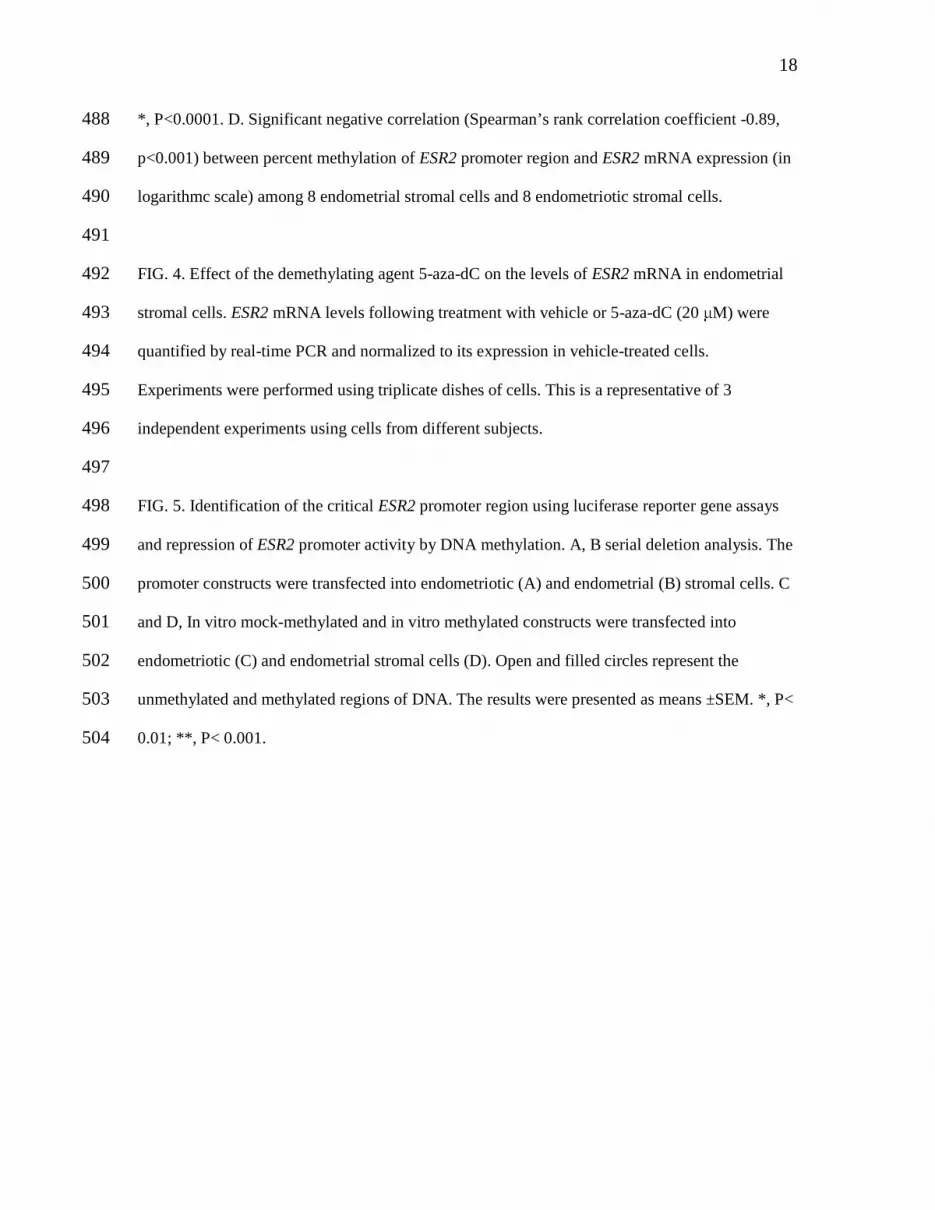
*, P<0.0001. D. Significant negative correlation (Spearman’s rank correlation coefficient -0.89, 488
p<0.001) between percent methylation of ESR2 promoter region and ESR2 mRNA expression (in 489
logarithmc scale) among 8 endometrial stromal cells and 8 endometriotic stromal cells. 490
491
FIG. 4. Effect of the demethylating agent 5-aza-dC on the levels of ESR2 mRNA in endometrial 492
stromal cells. ESR2 mRNA levels following treatment with vehicle or 5-aza-dC (20 µM) were 493
quantified by real-time PCR and normalized to its expression in vehicle-treated cells. 494
Experiments were performed using triplicate dishes of cells. This is a representative of 3 495
independent experiments using cells from different subjects. 496
497
FIG. 5. Identification of the critical ESR2 promoter region using luciferase reporter gene assays 498
and repression of ESR2 promoter activity by DNA methylation. A, B serial deletion analysis. The 499
promoter constructs were transfected into endometriotic (A) and endometrial (B) stromal cells. C 500
and D, In vitro mock-methylated and in vitro methylated constructs were transfected into 501
endometriotic (C) and endometrial stromal cells (D). Open and filled circles represent the 502
unmethylated and methylated regions of DNA. The results were presented as means ±SEM. *, P< 503
0.01; **, P< 0.001. 504

0
1
2
3
4
5
6
7
endometrial endometriotic
Fol
d di
ffer
ence
in to
tal P
GR
mR
NA
P=0.027D
0
1
2
3
4
5
6
7
endometrial endometriotic
Fol
d di
ffer
ence
in to
tal P
GR
mR
NA
P=0.027D
0
2
4
6
8
10
12
endometrial endometriotic
Fol
d d
iffer
ence
in P
GR
B m
RN
A
0
2
4
6
8
10
12
endometrial endometriotic
Fol
d d
iffer
ence
in P
GR
B m
RN
A
P=0.029E
0
2
4
6
8
10
12
endometrial endometriotic
Fol
d d
iffer
ence
in P
GR
B m
RN
A
0
2
4
6
8
10
12
endometrial endometriotic
Fol
d d
iffer
ence
in P
GR
B m
RN
A
P=0.029E
0
200
400
600
800
1000
1200
endometrial endometriotic
Fol
d d
iffe
renc
e in
ESR
1 t
o E
SR
2 r
atio
CP<0.001
0
200
400
600
800
1000
1200
endometrial endometriotic
Fol
d d
iffe
renc
e in
ESR
1 t
o E
SR
2 r
atio
CP<0.001
0
10
20
30
40
50
60
70
endometrial endometrioticF
old
diff
eren
ce in
ES
R2
mR
NA
0
10
20
30
40
50
60
70
endometrial endometrioticF
old
diff
eren
ce in
ES
R2
mR
NA
P=0.015B
0
10
20
30
40
50
60
70
endometrial endometrioticF
old
diff
eren
ce in
ES
R2
mR
NA
0
10
20
30
40
50
60
70
endometrial endometrioticF
old
diff
eren
ce in
ES
R2
mR
NA
P=0.015B
0
2
4
6
8
10
12
endometrial endometrioticFol
d di
ffer
ence
in E
SR
1 m
RN
A
P=0.037A
0
2
4
6
8
10
12
endometrial endometrioticFol
d di
ffer
ence
in E
SR
1 m
RN
A
P=0.037A
FIG.1.
ESR2
ACTB
ESR1
endometrial cells endometriotic cellsF
Subjects 1 2 3 4 1 2 3 4
Subjects 5 6 7 8 5 6 7 8
ESR1
ESR2
59 kDa
59 kDa
44 kDa
44 kDa
69 kDa
69 kDa
ESR2
ESR1
cellsF
Subjects 1 2 3 4 1 2 3 4
Subjects 5 6 7 8 5 6 7 8
ESR1
ESR2
59 kDa
59 kDa
44 kDa
44 kDa
69 kDa
69 kDa
ACTB
ESR2
ACTB
ESR1
endometrial cells endometriotic cellsF
Subjects 1 2 3 4 1 2 3 4
Subjects 5 6 7 8 5 6 7 8
ESR1
ESR2
59 kDa
59 kDa
44 kDa
44 kDa
69 kDa
69 kDa
ESR2
ESR1
cellsF
Subjects 1 2 3 4 1 2 3 4
Subjects 5 6 7 8 5 6 7 8
ESR1
ESR2
59 kDa
59 kDa
44 kDa
44 kDa
69 kDa
69 kDa
ACTB

FIG.2.
69 kDa
59 kDa
44 kDa
ESR1
ESR2
ACTB
endometrial endometriotic endometrial endometriotic endometrial endometriotic
3 paired stromal cells1 2 3
C
69 kDa
59 kDa
44 kDa
ESR1
ESR2
ACTB
endometrial endometriotic endometrial endometriotic endometrial endometriotic
3 paired stromal cells1 2 3
C
020406080
100120140160180200
endometrial endometrioticFol
d di
ffer
ence
in E
SR2
mR
NA
P=0.017B
020406080
100120140160180200
endometrial endometrioticFol
d di
ffer
ence
in E
SR2
mR
NA
P=0.017B
0
5
10
15
20
25
30
35
endometrial endometrioticFol
d d
iffe
ren
ce in
ES
R1
mR
NA
P=0.032
A
0
5
10
15
20
25
30
35
endometrial endometrioticFol
d d
iffe
ren
ce in
ES
R1
mR
NA
P=0.032
A

-20
-18
-16
-14
-12
-10
-8
-6
-4
-2
0
0 20 40 60 80 100
% methylation
loga
rith
m o
f m
RN
A le
vels
D
Spearman’s correlation coefficient= -0.89
P<0.001
-20
-18
-16
-14
-12
-10
-8
-6
-4
-2
0
0 20 40 60 80 100
% methylation
loga
rith
m o
f m
RN
A le
vels
D
Spearman’s correlation coefficient= -0.89
P<0.001
Exon 0N
Exon 0N
TSS +1
100bp
Bisulfite sequence
Predicted CpG island
Exon 1
A
Exon 0N
Exon 0N
TSS +1
100bp
Bisulfite sequence
Predicted CpG island
Exon 1
A
FIG.3.
-163 -158 -133 -131 -129 -126 -123 -119 -107 -104 -90 -74 -48 -163 -158 -133 -131 -129 -126 -123 -119 -107 -104 -90 -74 -48
1
2
3
4
5
6
7
8
1
2
3
4
5
6
7
8
B Endometrial stromal cells Endometriotic stromal cells-163 -158 -133 -131 -129 -126 -123 -119 -107 -104 -90 -74 -48 -163 -158 -133 -131 -129 -126 -123 -119 -107 -104 -90 -74 -48
1
2
3
4
5
6
7
8
1
2
3
4
5
6
7
8
B Endometrial stromal cells Endometriotic stromal cells
0
10
20
30
40
50
60
70
endometrial endometriotic
% m
eth
ylat
ion
54.25%
12.69%
*C
0
10
20
30
40
50
60
70
endometrial endometriotic
% m
eth
ylat
ion
54.25%
12.69%
*C

FIG.4.
0
0.5
1
1.5
2
2.5
3
Vehicle 5-aza-dC
Fol
d d
iffer
ence
in E
SR
2 m
RN
A
P=0.025
0
0.5
1
1.5
2
2.5
3
Vehicle 5-aza-dC
Fol
d d
iffer
ence
in E
SR
2 m
RN
A
P=0.025

0 1 2 3 4 5
A
Relative luciferase activity
*
Luc
Luc
Luc
Luc
-525 +564
-197
+372
pGL4
0 1 2 3 4 5
A
Relative luciferase activity
*
Luc
Luc
Luc
Luc
-525 +564
-197
+372
pGL4
LucLuc
LucLuc
LucLucLuc
LucLucLuc
-525 +564
-197
+372
pGL4
FIG.5.
0 1 2 3 4
-525Lu cLu c
methylated Lu cLu c
-197 Lu cLu c
methylated LucLuc
LucLuc
+564
pGL4
unmethylated
unmethylated-525
Lu cLu c-525
Lu cLu c
methylated Lu cLu cmethylated Lu cLu c
-197 Lu cLu c-197 Lu cLu c
methylated LucLucmethylated LucLuc
LucLucLucLuc
+564
pGL4
unmethylated
unmethylated
*
**
C
Relative luciferase activity0 1 2 3 4
-525Lu cLu c
methylated Lu cLu c
-197 Lu cLu c
methylated LucLuc
LucLuc
+564
pGL4
unmethylated
unmethylated-525
Lu cLu c-525
Lu cLu c
methylated Lu cLu cmethylated Lu cLu c
-197 Lu cLu c-197 Lu cLu c
methylated LucLucmethylated LucLuc
LucLucLucLuc
+564
pGL4
unmethylated
unmethylated
*
**
C
Relative luciferase activity
0 0.5 1 1.5 2 2.5Relative luciferase activity
B
*
Luc
Luc
Luc
Luc
-525 +564
-197
+372
pGL4
LucLuc
LucLuc
LucLucLuc
LucLucLuc
-525 +564
-197
+372
pGL4
0 0.5 1 1.5 2 2.5Relative luciferase activity
B
*
Luc
Luc
Luc
Luc
-525 +564
-197
+372
pGL4
LucLuc
LucLuc
LucLucLuc
LucLucLuc
-525 +564
-197
+372
pGL4
0 0.5 1 1.5 2 2.5
**
**
D
Relative luciferase activity
-525L u cL u c
methylated L u cL u c
-197 L u cL u c
methylated L ucL uc
L ucL uc
+564
pGL4
unmethylated
unmethylated
0 0.5 1 1.5 2 2.5
**
**
D
Relative luciferase activity
-525L u cL u c
methylated L ucL uc
-197 L u cL u c
methylated L ucL uc
L ucL uc
+564
pGL4
unmethylated
unmethylated-525
L u cL u c-525
L u cL u c
methylated L ucL ucmethylated L ucL uc
-197 L u cL u c-197 L u cL u c
methylated L ucL ucmethylated L ucL uc
L ucL ucL ucL uc
+564
pGL4
unmethylated
unmethylated