emerging novel roles of neuropeptide y in the retina: from neuromodulation to neuroprotection
TRANSCRIPT

Progress in Neurobiology 112 (2014) 70–79
Emerging novel roles of neuropeptide Y in the retina:From neuromodulation to neuroprotection
Ana Santos-Carvalho a,b, Ana Rita Alvaro a,c, Joao Martins a,d,Antonio Francisco Ambrosio a,d,e, Claudia Cavadas a,b,*a CNC-Center for Neuroscience and Cell Biology, University of Coimbra, Largo Marques de Pombal, 3004-517 Coimbra, Portugalb Faculty of Pharmacy, University of Coimbra, Polo das Ciencias da Saude, Azinhaga de Santa Comba, 3000-548 Coimbra, Portugalc Department of Biology and Environment, University of Tras-os-Montes and Alto Douro, Apartado 1013, 5001-801 Vila Real, Portugald Centre of Ophthalmology and Vision Sciences, IBILI, Faculty of Medicine, University of Coimbra, Azinhaga de Santa Comba, Celas, 3000-548 Coimbra,
Portugale AIBILI-Association for Innovation and Biomedical Research on Light and Image, Azinhaga Santa Comba, Celas, 3000-548 Coimbra, Portugal
Contents
1. The retina . . . . . . . . . . . . . . . . . . . . . . . . . . . . . . . . . . . . . . . . . . . . . . . . . . . . . . . . . . . . . . . . . . . . . . . . . . . . . . . . . . . . . . . . . . . . . . . . . . . . . . . . 71
1.1. Visual pathways in the retina . . . . . . . . . . . . . . . . . . . . . . . . . . . . . . . . . . . . . . . . . . . . . . . . . . . . . . . . . . . . . . . . . . . . . . . . . . . . . . . . . . 71
1.2. Neurotransmitters in the retina . . . . . . . . . . . . . . . . . . . . . . . . . . . . . . . . . . . . . . . . . . . . . . . . . . . . . . . . . . . . . . . . . . . . . . . . . . . . . . . . . 71
1.3. Neuropeptides in the retina . . . . . . . . . . . . . . . . . . . . . . . . . . . . . . . . . . . . . . . . . . . . . . . . . . . . . . . . . . . . . . . . . . . . . . . . . . . . . . . . . . . . 71
2. Neuropeptide Y (NPY) and NPY receptors in the retina. . . . . . . . . . . . . . . . . . . . . . . . . . . . . . . . . . . . . . . . . . . . . . . . . . . . . . . . . . . . . . . . . . . . 72
2.1. Localization of NPY and NPY receptors in the retina . . . . . . . . . . . . . . . . . . . . . . . . . . . . . . . . . . . . . . . . . . . . . . . . . . . . . . . . . . . . . . . . 73
2.2. NPY and retinal development. . . . . . . . . . . . . . . . . . . . . . . . . . . . . . . . . . . . . . . . . . . . . . . . . . . . . . . . . . . . . . . . . . . . . . . . . . . . . . . . . . . 74
3. Modulatory effects of NPY in the retina . . . . . . . . . . . . . . . . . . . . . . . . . . . . . . . . . . . . . . . . . . . . . . . . . . . . . . . . . . . . . . . . . . . . . . . . . . . . . . . . 75
4. Potential role of NPY in cell proliferation, differentiation and neuroprotection in the retina . . . . . . . . . . . . . . . . . . . . . . . . . . . . . . . . . . . . . . 75
5. NPY involvement in retinal pathologies . . . . . . . . . . . . . . . . . . . . . . . . . . . . . . . . . . . . . . . . . . . . . . . . . . . . . . . . . . . . . . . . . . . . . . . . . . . . . . . . 76
A R T I C L E I N F O
Article history:
Received 18 October 2012
Received in revised form 14 October 2013
Accepted 15 October 2013
Available online 30 October 2013
Keywords:
Neuropeptide Y
NPY receptors
Retina
Retinal neural cells
Neuroprotection
Neuromodulation
A B S T R A C T
Neuropeptide Y (NPY) and NPY receptors are widely expressed in the central nervous system, including
the retina. Retinal cells, in particular neurons, astrocytes, and Muller, microglial and endothelial cells
express this peptide and its receptors (Y1, Y2, Y4 and/or Y5). Several studies have shown that NPY is
expressed in the retina of various mammalian and non-mammalian species. However, studies analyzing
the distribution of NPY receptors in the retina are still scarce. Although the physiological roles of NPY in
the retina have not been completely elucidated, its early expression strongly suggests that NPY may be
involved in the development of retinal circuitry. NPY inhibits the increase in [Ca2+]i triggered by elevated
KCl in retinal neurons, protects retinal neural cells against toxic insults and induces the proliferation of
retinal progenitor cells. In this review, we will focus on the roles of NPY in the retina, specifically
proliferation, neuromodulation and neuroprotection. Alterations in the NPY system in the retina might
contribute to the pathogenesis of retinal degenerative diseases, such as diabetic retinopathy and
glaucoma, and NPY and its receptors might be viewed as potentially novel therapeutic targets.
� 2013 Elsevier Ltd. All rights reserved.
Abbreviations: BrdU, 5-bromo-20-deoxyuridine; [Ca2+]i, intracellular calcium; CNS, central nervous system; DPP-IV, dipeptidyl peptidase IV; E18, embrionic day 18; ERK,
extracellular signal-regulated kinases; GABA, g-aminobutyric acid; GAD 65, glutamic acid decarboxylase 65; GAT-1, GABA transporter 1; GCL, ganglion cell layer; hESC,
human embryonic stem cells; iGluRs, ionotropic glutamate receptors; INL, inner nuclear layer; IPL, inner plexiform layer; Leu, leucine; MAPK, mitogen activated protein
kinase; MDMA, 3,4-methylenedioxymethamphetamine; mGLuRs, metabotropic glutamate receptors; mRNA, messenger ribonucleic acid; NO, nitric oxide; NOS–sGC, nitric
Contents lists available at ScienceDirect
Progress in Neurobiology
jo u rn al ho m epag e: ww w.els evier . c om / lo cat e/pn eu ro b io
oxide synthase – soluble guanylyl cyclase; NPY, neuropeptide Y; NPY–IR, neuropeptide Y immunoreactivity; NPY Y1, NPY receptor type 1; ONL, outer nuclear layer; OPL, outer
plexiform layer; P7, postnatal day 7; PKA, protein kinase A; POS, photoreceptor outer segment; Pro, proline; RPE, retinal pigmented epithelium.
* Corresponding author at: Faculty of Pharmacy, University of Coimbra, Polo das Ciencias da Saude, Azinhaga de Santa Comba, 3000-548 Coimbra, Portugal.
Tel.: +351 963928766; fax: +351 239 488 503.
E-mail address: [email protected] (C. Cavadas).
0301-0082/$ – see front matter � 2013 Elsevier Ltd. All rights reserved.
http://dx.doi.org/10.1016/j.pneurobio.2013.10.002
A. Santos-Carvalho et al. / Progress in Neurobiology 112 (2014) 70–79 71
6. Conclusions . . . . . . . . . . . . . . . . . . . . . . . . . . . . . . . . . . . . . . . . . . . . . . . . . . . . . . . . . . . . . . . . . . . . . . . . . . . . . . . . . . . . . . . . . . . . . . . . . . . . . . 77
Acknowledgements . . . . . . . . . . . . . . . . . . . . . . . . . . . . . . . . . . . . . . . . . . . . . . . . . . . . . . . . . . . . . . . . . . . . . . . . . . . . . . . . . . . . . . . . . . . . . . . . 77
References . . . . . . . . . . . . . . . . . . . . . . . . . . . . . . . . . . . . . . . . . . . . . . . . . . . . . . . . . . . . . . . . . . . . . . . . . . . . . . . . . . . . . . . . . . . . . . . . . . . . . . . 77
1. The retina
1.1. Visual pathways in the retina
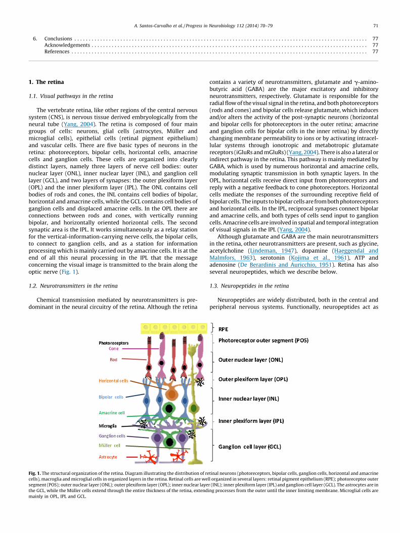
The vertebrate retina, like other regions of the central nervoussystem (CNS), is nervous tissue derived embryologically from theneural tube (Yang, 2004). The retina is composed of four maingroups of cells: neurons, glial cells (astrocytes, Muller andmicroglial cells), epithelial cells (retinal pigment epithelium)and vascular cells. There are five basic types of neurons in theretina: photoreceptors, bipolar cells, horizontal cells, amacrinecells and ganglion cells. These cells are organized into clearlydistinct layers, namely three layers of nerve cell bodies: outernuclear layer (ONL), inner nuclear layer (INL), and ganglion celllayer (GCL), and two layers of synapses: the outer plexiform layer(OPL) and the inner plexiform layer (IPL). The ONL contains cellbodies of rods and cones, the INL contains cell bodies of bipolar,horizontal and amacrine cells, while the GCL contains cell bodies ofganglion cells and displaced amacrine cells. In the OPL there areconnections between rods and cones, with vertically runningbipolar, and horizontally oriented horizontal cells. The secondsynaptic area is the IPL. It works simultaneously as a relay stationfor the vertical-information-carrying nerve cells, the bipolar cells,to connect to ganglion cells, and as a station for informationprocessing which is mainly carried out by amacrine cells. It is at theend of all this neural processing in the IPL that the messageconcerning the visual image is transmitted to the brain along theoptic nerve (Fig. 1).
1.2. Neurotransmitters in the retina
Chemical transmission mediated by neurotransmitters is pre-dominant in the neural circuitry of the retina. Although the retina
Fig. 1. The structural organization of the retina. Diagram illustrating the distribution of re
cells), macroglia and microglial cells in organized layers in the retina. Retinal cells are wel
segment (POS); outer nuclear layer (ONL); outer plexiform layer (OPL); inner nuclear laye
the GCL, while the Muller cells extend through the entire thickness of the retina, extendi
mainly in OPL, IPL and GCL.
contains a variety of neurotransmitters, glutamate and g-amino-butyric acid (GABA) are the major excitatory and inhibitoryneurotransmitters, respectively. Glutamate is responsible for theradial flow of the visual signal in the retina, and both photoreceptors(rods and cones) and bipolar cells release glutamate, which inducesand/or alters the activity of the post-synaptic neurons (horizontaland bipolar cells for photoreceptors in the outer retina; amacrineand ganglion cells for bipolar cells in the inner retina) by directlychanging membrane permeability to ions or by activating intracel-lular systems through ionotropic and metabotropic glutamatereceptors (iGluRs and mGluRs) (Yang, 2004). There is also a lateral orindirect pathway in the retina. This pathway is mainly mediated byGABA, which is used by numerous horizontal and amacrine cells,modulating synaptic transmission in both synaptic layers. In theOPL, horizontal cells receive direct input from photoreceptors andreply with a negative feedback to cone photoreceptors. Horizontalcells mediate the responses of the surrounding receptive field ofbipolar cells. The inputs to bipolar cells are from both photoreceptorsand horizontal cells. In the IPL, reciprocal synapses connect bipolarand amacrine cells, and both types of cells send input to ganglioncells. Amacrine cells are involved in spatial and temporal integrationof visual signals in the IPL (Yang, 2004).
Although glutamate and GABA are the main neurotransmittersin the retina, other neurotransmitters are present, such as glycine,acetylcholine (Lindeman, 1947), dopamine (Haeggendal andMalmfors, 1963), serotonin (Kojima et al., 1961), ATP andadenosine (De Berardinis and Auricchio, 1951). Retina has alsoseveral neuropeptides, which we describe below.
1.3. Neuropeptides in the retina
Neuropeptides are widely distributed, both in the central andperipheral nervous systems. Functionally, neuropeptides act as
tinal neurons (photoreceptors, bipolar cells, ganglion cells, horizontal and amacrine
l organized in several layers: retinal pigment epithelium (RPE); photoreceptor outer
r (INL); inner plexiform layer (IPL) and ganglion cell layer (GCL). The astrocytes are in
ng processes from the outer until the inner limiting membrane. Microglial cells are
A. Santos-Carvalho et al. / Progress in Neurobiology 112 (2014) 70–7972
neurotransmitters and/or neuromodulators through the activationof specific receptors to modulate the functional properties ofneurons, such as their membrane excitability or their signaltransduction pathways (Bagnoli et al., 2003). Over the last decades,several neuropeptides, which were highly conserved duringevolution, have been discovered in the eye. Substance P was thefirst peptide described in the retina and is also present inperipherally innervated tissues of the eye (Duner et al., 1954;Stone et al., 1987). The interest was extended to investigate thepresence and distribution of other neuropeptides includingcalcitonin gene-related peptide (CGRP) (Kiyama et al., 1985),vasoactive intestinal polypeptide (VIP) (Loren et al., 1980),pituitary adenylate cyclase-activating polypeptide (PACAP) (Onaliand Olianas, 1994), cholecystokinin (CCK) (Yamada et al., 1981),somatostatin (Rorstad et al., 1979), galanin (Hokfelt et al., 1992),neurokinin A and B (Schmid et al., 2006), corticotrophin-releasingfactor (CRF) (Kiyama et al., 1984), angiotensin II (Senanayake et al.,2007), secretoneurin (Overdick et al., 1996), and neuropeptide Y(NPY) (Bruun et al., 1984). In this review we will focus on the NPYsystem and its role in the retina.
2. Neuropeptide Y (NPY) and NPY receptors in the retina
NPY is a member of a peptide family named NPY family or ‘‘PP-fold’’ family that also includes peptide YY (PYY) and pancreaticpolypeptide (PP) (Michel et al., 1998). NPY is a 36-amino acidpeptide that possesses an amidated C-terminal residue and a largenumber of tyrosine residues (which are abbreviated by the letter Y)included in both ends of the molecule. NPY was first isolatedfrom the pig brain in Tatemoto et al. (1982). NPY is one ofthe neuropeptides with the highest degree of phylogeneticpreservation, while the PP differs considerably between species
Table 1NPY-IR localization in the retina of several non-mammalian species.
Species NPY-IR localization in the re
Non Mammals
Fishes
Blue acara (Aequidens pulcher) Amacrine cells and IPL
Carp Cell bodies of amacrine cells
Gilthead seabream (Sparus aurata L.) Amacrine cells
Goldfish Cell bodies of amacrine in IN
Killifish (Fundulus heteroclitus) Amacrine cell fibers in IPL
Lamprey (Lampreta fluviatilis) Pyriform subclass of amacrin
Skates (Raja clavata, Raja radiate and Raja oscellata) Amacrine cells in the innerm
Squid No NPY-IR
Trout Cell bodies of amacrine cells
Zebrafish Amacrine cells
Anurans
Frogs (Bufo marinus and Xenopus laevis) Cell bodies of amacrine cells
Bipolar-like cell bodies in th
Processes ramifying in three
Muller cells within the INL a
Co-localization of GABA in al
Reptiles
Lizards (Pogona vitticeps and Varanus gouldii) Amacrine cells: type A and t
Turtle Three types of amacrine cell
processes at GCL; type B, at
retina
Bipolar cells
Birds
Chicken Cell bodies of amacrine cells
processes in the IPL
Pigeon Cell bodies of amacrine cells
(Larhammar et al., 1992). The NPY gene is located on humanchromosome 7 at the locus 7p15.1 (Cerda-Reverter and Larham-mar, 2000). In mouse, it is located in chromosome 6, locus 6 B3; 626.0 cM while in rat it is localized in chromosome 4, locus 4q24(Pruitt et al., 2012). The prepro-NPY generated after translation isdirected into the endoplasmic reticulum, where a 28 amino acidpeptide is removed and Pro-NPY produced. This NPY precursor,Pro-NPY, is a 69 amino acid peptide formed by NPY1–39 where thecarboxylic group is flanked by a group of 33 amino acids called theC-flanking peptide of NPY (CPON). The following processing step isthe cleavage of the precursor Pro-NPY at a dibasic site byprohormone convertases, which generates NPY1–39 and CPON.Then, a truncation at the C-terminal end by a carboxypeptidase B(CPB) generates NPY1–37, which is a substrate for the enzymepeptidylglycine alpha-amidating monooxygenase (PAM) and leadsto the biologically active amidated NPY1–36 (NPY) (Medeiros andTurner, 1996). NPY can be further cleaved by two enzymes,dipeptidyl peptidase IV (DPP-IV) and aminopeptidase P (AmP)(Medeiros and Turner, 1994, 1996).
All known NPY receptors belong to the large super-family of G-protein-coupled, heptahelical receptors (Michel et al., 1998). TheNPY family receptors are the same for all members of the NPYfamily (NPY, PP, PYY) and are comprised of the receptor subtypesNPY Y1, Y2, Y4, Y5 and Y6 (Silva et al., 2005; Xapelli et al., 2008).
Generally, NPY receptors use similar signal transductionpathways, acting via pertussis toxin-sensitive G-proteins, i.e.,members of the Gi and Go family. Thus, inhibition of adenylylcyclase upon NPY receptor activation is found in almost everytissue and cell type investigated (Michel, 1991; Olasmaa andTerenius, 1986). However, the inhibition of adenylyl cyclase cannotprobably explain all functional responses observed upon stimula-tion of NPY receptors (Michel et al., 1998). Additional signaling
tina References
Negishi and Wagner (1995)
in INL and processes in the IPL Bruun et al. (1986)
Pirone et al. (2008)
L and cell processes in two layers in IPL Bruun et al. (1986),
Osborne et al. (1985),
and Muske et al. (1987)
Subhedar et al. (1996)
e cells Negishi et al. (1986) and
Rawitch et al. (1992)
ost part of INL and fibers in IPL Bruun et al. (1985)
Osborne et al. (1986)
in INL and processes in IPL Bruun et al. (1986)
Mathieu et al. (2002)
in INL and cell processes in IPL Bruun et al. (1986, 1991),
Hiscock and Straznicky (1989, 1990),
Osborne et al. (1985),
and Zhu and Gibbins (1995, 1996)
e middle of INL and sparsely in GCL
sublayers in IPL
nd processes in IPL
l NPY-IR amacrine cells of anuran retina
ype B in INL and displaced at GCL Straznicky and Hiscock (1994)
s: type A, at INL, IPL and occasional
INL and IPL; type C, at the periphery of
Isayama and Eldred (1988),
Isayama et al. (1988),
and Wetzel and Eldred (1997)
in the middle and innermost INL and Bruun et al. (1986)
in INL and processes in the IPL Bruun et al. (1986) and
Verstappen et al. (1986)
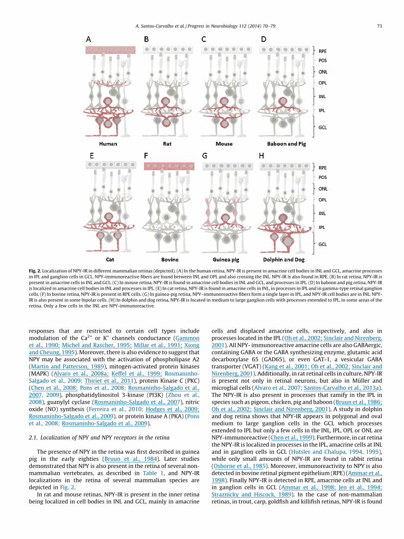
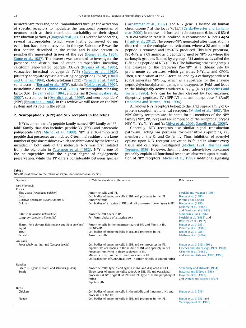
Fig. 2. Localization of NPY-IR in different mammalian retinas (depicted). (A) In the human retina, NPY-IR is present in amacrine cell bodies in INL and GCL, amacrine processes
in IPL and ganglion cells in GCL. NPY-immunoreactive fibers are found between INL and OPL and also crossing the INL. NPY-IR is also found in RPE. (B) In rat retina, NPY-IR is
present in amacrine cells in INL and GCL. (C) In mouse retina, NPY-IR is found in amacrine cell bodies in INL and GCL, and processes in IPL. (D) In baboon and pig retina, NPY-IR
is localized in amacrine cell bodies in INL and processes in IPL. (E) In cat retina, NPY-IR is found in amacrine cells in INL, in processes in IPL and in gamma-type retinal ganglion
cells. (F) In bovine retina, NPY-IR is present in RPE cells. (G) In guinea-pig retina, NPY–immunoreactive fibers form a single layer in IPL, and NPY-IR cell bodies are in INL. NPY-
IR is also present in some bipolar cells. (H) In dolphin and dog retina, NPY-IR is located in medium to large ganglion cells with processes extended to IPL, in some areas of the
retina. Only a few cells in the INL are NPY-immunoreactive.
A. Santos-Carvalho et al. / Progress in Neurobiology 112 (2014) 70–79 73
responses that are restricted to certain cell types includemodulation of the Ca2+ or K+ channels conductance (Gammonet al., 1990; Michel and Rascher, 1995; Millar et al., 1991; Xiongand Cheung, 1995). Moreover, there is also evidence to suggest thatNPY may be associated with the activation of phospholipase A2(Martin and Patterson, 1989), mitogen-activated protein kinases(MAPK) (Alvaro et al., 2008a; Keffel et al., 1999; Rosmaninho-Salgado et al., 2009; Thiriet et al., 2011), protein Kinase C (PKC)(Chen et al., 2008; Pons et al., 2008; Rosmaninho-Salgado et al.,2007, 2009), phosphatidylinositol 3-kinase (PI3K) (Zhou et al.,2008), guanylyl cyclase (Rosmaninho-Salgado et al., 2007), nitricoxide (NO) synthesis (Ferreira et al., 2010; Hodges et al., 2009;Rosmaninho-Salgado et al., 2009), or protein kinase A (PKA) (Ponset al., 2008; Rosmaninho-Salgado et al., 2009).
2.1. Localization of NPY and NPY receptors in the retina
The presence of NPY in the retina was first described in guineapig in the early eighties (Bruun et al., 1984). Later studiesdemonstrated that NPY is also present in the retina of several non-mammalian vertebrates, as described in Table 1, and NPY-IRlocalizations in the retina of several mammalian species aredepicted in Fig. 2.
In rat and mouse retinas, NPY-IR is present in the inner retinabeing localized in cell bodies in INL and GCL, mainly in amacrine
cells and displaced amacrine cells, respectively, and also inprocesses located in the IPL (Oh et al., 2002; Sinclair and Nirenberg,2001). All NPY- immunoreactive amacrine cells are also GABAergic,containing GABA or the GABA synthesizing enzyme, glutamic aciddecarboxylase 65 (GAD65), or even GAT-1, a vesicular GABAtransporter (VGAT) (Kang et al., 2001; Oh et al., 2002; Sinclair andNirenberg, 2001). Additionally, in rat retinal cells in culture, NPY-IRis present not only in retinal neurons, but also in Muller andmicroglial cells (Alvaro et al., 2007; Santos-Carvalho et al., 2013a).The NPY-IR is also present in processes that ramify in the IPL inspecies such as pigeon, chicken, pig and baboon (Bruun et al., 1986;Oh et al., 2002; Sinclair and Nirenberg, 2001). A study in dolphinand dog retina shows that NPY-IR appears in polygonal and ovalmedium to large ganglion cells in the GCL which processesextended to IPL but only a few cells in the INL, IPL, OPL or ONL areNPY-immunoreactive (Chen et al., 1999). Furthermore, in cat retinathe NPY-IR is localized in processes in the IPL, amacrine cells at INLand in ganglion cells in GCL (Hutsler and Chalupa, 1994, 1995),while only small amounts of NPY-IR are found in rabbit retina(Osborne et al., 1985). Moreover, immunoreactivity to NPY is alsodetected in bovine retinal pigment epithelium (RPE) (Ammar et al.,1998). Finally NPY-IR is detected in RPE, amacrine cells at INL andin ganglion cells in GCL (Ammar et al., 1998; Jen et al., 1994;Straznicky and Hiscock, 1989). In the case of non-mammalianretinas, in trout, carp, goldfish and killifish retinas, NPY-IR is found
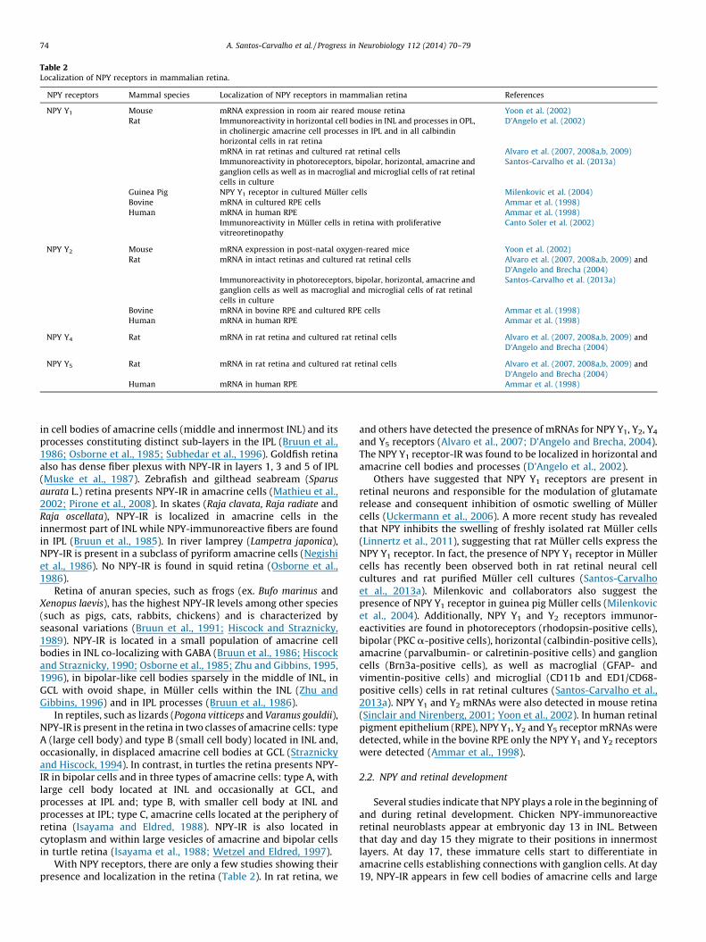
Table 2Localization of NPY receptors in mammalian retina.
NPY receptors Mammal species Localization of NPY receptors in mammalian retina References
NPY Y1 Mouse mRNA expression in room air reared mouse retina Yoon et al. (2002)
Rat Immunoreactivity in horizontal cell bodies in INL and processes in OPL,
in cholinergic amacrine cell processes in IPL and in all calbindin
horizontal cells in rat retina
D’Angelo et al. (2002)
mRNA in rat retinas and cultured rat retinal cells Alvaro et al. (2007, 2008a,b, 2009)
Immunoreactivity in photoreceptors, bipolar, horizontal, amacrine and
ganglion cells as well as in macroglial and microglial cells of rat retinal
cells in culture
Santos-Carvalho et al. (2013a)
Guinea Pig NPY Y1 receptor in cultured Muller cells Milenkovic et al. (2004)
Bovine mRNA in cultured RPE cells Ammar et al. (1998)
Human mRNA in human RPE Ammar et al. (1998)
Immunoreactivity in Muller cells in retina with proliferative
vitreoretinopathy
Canto Soler et al. (2002)
NPY Y2 Mouse mRNA expression in post-natal oxygen-reared mice Yoon et al. (2002)
Rat mRNA in intact retinas and cultured rat retinal cells Alvaro et al. (2007, 2008a,b, 2009) and
D’Angelo and Brecha (2004)
Immunoreactivity in photoreceptors, bipolar, horizontal, amacrine and
ganglion cells as well as macroglial and microglial cells of rat retinal
cells in culture
Santos-Carvalho et al. (2013a)
Bovine mRNA in bovine RPE and cultured RPE cells Ammar et al. (1998)
Human mRNA in human RPE Ammar et al. (1998)
NPY Y4 Rat mRNA in rat retina and cultured rat retinal cells Alvaro et al. (2007, 2008a,b, 2009) and
D’Angelo and Brecha (2004)
NPY Y5 Rat mRNA in rat retina and cultured rat retinal cells Alvaro et al. (2007, 2008a,b, 2009) and
D’Angelo and Brecha (2004)
Human mRNA in human RPE Ammar et al. (1998)
A. Santos-Carvalho et al. / Progress in Neurobiology 112 (2014) 70–7974
in cell bodies of amacrine cells (middle and innermost INL) and itsprocesses constituting distinct sub-layers in the IPL (Bruun et al.,1986; Osborne et al., 1985; Subhedar et al., 1996). Goldfish retinaalso has dense fiber plexus with NPY-IR in layers 1, 3 and 5 of IPL(Muske et al., 1987). Zebrafish and gilthead seabream (Sparus
aurata L.) retina presents NPY-IR in amacrine cells (Mathieu et al.,2002; Pirone et al., 2008). In skates (Raja clavata, Raja radiate andRaja oscellata), NPY-IR is localized in amacrine cells in theinnermost part of INL while NPY-immunoreactive fibers are foundin IPL (Bruun et al., 1985). In river lamprey (Lampetra japonica),NPY-IR is present in a subclass of pyriform amacrine cells (Negishiet al., 1986). No NPY-IR is found in squid retina (Osborne et al.,1986).
Retina of anuran species, such as frogs (ex. Bufo marinus andXenopus laevis), has the highest NPY-IR levels among other species(such as pigs, cats, rabbits, chickens) and is characterized byseasonal variations (Bruun et al., 1991; Hiscock and Straznicky,1989). NPY-IR is located in a small population of amacrine cellbodies in INL co-localizing with GABA (Bruun et al., 1986; Hiscockand Straznicky, 1990; Osborne et al., 1985; Zhu and Gibbins, 1995,1996), in bipolar-like cell bodies sparsely in the middle of INL, inGCL with ovoid shape, in Muller cells within the INL (Zhu andGibbins, 1996) and in IPL processes (Bruun et al., 1986).
In reptiles, such as lizards (Pogona vitticeps and Varanus gouldii),NPY-IR is present in the retina in two classes of amacrine cells: typeA (large cell body) and type B (small cell body) located in INL and,occasionally, in displaced amacrine cell bodies at GCL (Straznickyand Hiscock, 1994). In contrast, in turtles the retina presents NPY-IR in bipolar cells and in three types of amacrine cells: type A, withlarge cell body located at INL and occasionally at GCL, andprocesses at IPL and; type B, with smaller cell body at INL andprocesses at IPL; type C, amacrine cells located at the periphery ofretina (Isayama and Eldred, 1988). NPY-IR is also located incytoplasm and within large vesicles of amacrine and bipolar cellsin turtle retina (Isayama et al., 1988; Wetzel and Eldred, 1997).
With NPY receptors, there are only a few studies showing theirpresence and localization in the retina (Table 2). In rat retina, we
and others have detected the presence of mRNAs for NPY Y1, Y2, Y4
and Y5 receptors (Alvaro et al., 2007; D’Angelo and Brecha, 2004).The NPY Y1 receptor-IR was found to be localized in horizontal andamacrine cell bodies and processes (D’Angelo et al., 2002).
Others have suggested that NPY Y1 receptors are present inretinal neurons and responsible for the modulation of glutamaterelease and consequent inhibition of osmotic swelling of Mullercells (Uckermann et al., 2006). A more recent study has revealedthat NPY inhibits the swelling of freshly isolated rat Muller cells(Linnertz et al., 2011), suggesting that rat Muller cells express theNPY Y1 receptor. In fact, the presence of NPY Y1 receptor in Mullercells has recently been observed both in rat retinal neural cellcultures and rat purified Muller cell cultures (Santos-Carvalhoet al., 2013a). Milenkovic and collaborators also suggest thepresence of NPY Y1 receptor in guinea pig Muller cells (Milenkovicet al., 2004). Additionally, NPY Y1 and Y2 receptors immunor-eactivities are found in photoreceptors (rhodopsin-positive cells),bipolar (PKC a-positive cells), horizontal (calbindin-positive cells),amacrine (parvalbumin- or calretinin-positive cells) and ganglioncells (Brn3a-positive cells), as well as macroglial (GFAP- andvimentin-positive cells) and microglial (CD11b and ED1/CD68-positive cells) cells in rat retinal cultures (Santos-Carvalho et al.,2013a). NPY Y1 and Y2 mRNAs were also detected in mouse retina(Sinclair and Nirenberg, 2001; Yoon et al., 2002). In human retinalpigment epithelium (RPE), NPY Y1, Y2 and Y5 receptor mRNAs weredetected, while in the bovine RPE only the NPY Y1 and Y2 receptorswere detected (Ammar et al., 1998).
2.2. NPY and retinal development
Several studies indicate that NPY plays a role in the beginning ofand during retinal development. Chicken NPY-immunoreactiveretinal neuroblasts appear at embryonic day 13 in INL. Betweenthat day and day 15 they migrate to their positions in innermostlayers. At day 17, these immature cells start to differentiate inamacrine cells establishing connections with ganglion cells. At day19, NPY-IR appears in few cell bodies of amacrine cells and large

A. Santos-Carvalho et al. / Progress in Neurobiology 112 (2014) 70–79 75
cells in GCL (Prada Oliveira et al., 2003). In zebrafish, NPY-IRappears in amacrine cells at embryonic day 15, suggesting itsinvolvement in retinal synaptogenesis during ontogeny (Mathieuet al., 2002).
In Xenopus laevis retina, NPY-IR appears early in larval life. Thedendritic maturation of NPY-IR amacrine cells occurs later duringlarval development than in cell bodies, and just before metamor-phosis. In the adult retina of this frog, NPY-immunoreactivity (IR) ispresent in a wide field of amacrine cells in the INL and GCL (Hiscockand Straznicky, 1990). In the retina of blue acara (Aequidens
pulcher), NPY-immunoreactive amacrine cells appear in IPL aroundhatching, at days 3–4 (Negishi and Wagner, 1995).
During cat retina development, NPY-IR is detected in centralretina within the GCL at embryonic day 46 and amacrine cellswithin INL at embryonic day 50. Cat NPY-IR in amacrine populationreaches adult levels at P7, while NPY-IR in ganglion cell populationshows an extended development, with new cells expressing NPYuntil the third post-natal week (Hutsler and Chalupa, 1995).
Regarding the developing human retina, NPY-immunoreactiveamacrine cells are found around 14 weeks of gestation (Jotwaniet al., 1994). Another study indicates the presence of round andpear-shaped NPY-immunoreactive amacrine cells in INL after 15weeks of gestation. NPY positive-ganglion cells were only detectedat 17 weeks of gestation. NPY-immunoreactive amacrine andganglion cells are located in INL and GCL, respectively, at 26–28weeks of gestation. Later, by 38–40 weeks of gestation, NPY-immunoreactive cells are present in INL, GCL and IPL (Jen et al.,1994).
In rats, NPY-IR appears in the retina in small quantities in GCLonly at E18, and increases over pre- and postnatal development.Subsequently, at eye opening (P13) NPY-IR markedly increases inINL and GCL, but falls during maturation until adult levels formingtwo subpopulations in INL and GCL. This transient increase at eyeopening may have a role in modulating the developing retinacircuitry (Ferriero and Sagar, 1989).
In conclusion, several studies report the presence of NPY inundifferentiated retinal cells indicating a putative role of NPY inretinal development (Ferriero and Sagar, 1989; Hiscock andStraznicky, 1990; Hutsler and Chalupa, 1995; Jen et al., 1994;Jotwani et al., 1994; Mathieu et al., 2002; Negishi and Wagner,1995; Prada Oliveira et al., 2003).
3. Modulatory effects of NPY in the retina
A fine tuning neuromodulator has the capacity to exert subtleinfluence on synapse activity by changing receptor activation ofother neurotransmitters or neuromodulators as well as its ownreceptors. NPY co-localizes with other neurotransmitters indifferent areas of the CNS (Allen et al., 1983; Hendry et al.,1984; McDonald, 1996; Silva et al., 2005) and modulates therelease of several neurotransmitters (Silva et al., 2005), inhibitingthe release of glutamate, aspartate, growth hormone, epinephrineand acetylcholine (Bitran et al., 1999; Bleakman et al., 1992; Greberet al., 1994; Gu et al., 1983; Hastings et al., 2004; Martire et al.,1995; Potter, 1987; Rettori et al., 1990b; Rodi et al., 2003;Schwertfeger et al., 2004; Silva et al., 2001, 2003; Tsuda et al.,1995), and enhancing the release of somatostatin and dopamineand the production of nitric oxide (Ault and Werling, 1999; Bitranet al., 1999; Rettori et al., 1990a). Therefore, NPY may play a fine-tuning modulator in the nervous system (Grandt et al., 1996;Magni, 2003; Mazzocchi et al., 1996; Prod’homme et al., 2006).Some studies have also suggested that NPY may be a neuromo-dulator in the retina. NPY modulates the intracellular calciumconcentration ([Ca2+]i) in rat retinal neurons. NPY inhibits thedepolarization-evoked Ca2+ influx into rod bipolar cells throughthe activation of NPY Y2 receptors (D’Angelo and Brecha, 2004).
NPY also inhibits the KCl-evoked increase in [Ca2+]i in cultured ratretinal neurons through the activation of NPY Y1, Y4 and Y5
receptors (Alvaro et al., 2009). On the other hand, when appliedexogenously, NPY stimulates the release of [3H]-glycine, [3H]-dopamine, [3H]-5-hydroxytryptamine and [3H]-choline chloride-derived radioactivity in the rabbit retina and of [3H]-GABA, [3H]-5-hydroxytryptamine and [3H]-choline chloride-derived radioactiv-ity in chicken retina (Bruun and Ehinger, 1993). These results andthe presence of NPY in amacrine cells (in the INL) and displacedamacrine cells (in the GCL), which may connect with otheramacrine cell subtypes and ganglion cells that are not immunore-active for NPY, suggest that NPY may also play a role as aneuromodulator in the inner retinal layers (D’Angelo et al., 2002;Oh et al., 2002).
The ablation of NPY-immunoreactive amacrine cells causesalteration of receptive field surround size of ganglion cells,suggesting that NPY-immunoreactive amacrine cells are involvedin tuning ganglion cells to low spatial frequencies/large spatialpatterns (Sinclair et al., 2004).
In conclusion, NPY may affect neurotransmission betweendifferent retinal neurons (photoreceptors, and bipolar, ganglion,horizontal and amacrine cells), which depends on [Ca2+]i regula-tion, and therefore NPY may exert a relevant fine tuningneuromodulatory role in retinal cells.
4. Potential role of NPY in cell proliferation, differentiation andneuroprotection in the retina
In vitro and in vivo studies suggest that NPY has pro-neurogenicproperties in the olfactory epithelium, subventricular zone (SVZ)and subgranular zone (SGZ) of dentate gyrus (Agasse et al., 2008;Decressac et al., 2011; Hansel et al., 2001b; Howell et al., 2005,2007; Rodrigo et al., 2010). In addition, in the CNS, NPY inducesalterations in the rostral migratory stream, differentiation ofprogenitor cells into distinct interneuronal subsets in the olfactorybulb (Stanic et al., 2008), migration of newly generated neurons tothe striatum and the olfatory bulb and also increases the number ofcells in the rostral migratory stream, olfactory bulb and striatum(Decressac et al., 2009). These NPY effects on neural cellproliferation and differentiation are mediated by the NPY Y1-receptor activation (Agasse et al., 2008; Decressac et al., 2011;Hansel et al., 2001b; Howell et al., 2003; Rodrigo et al., 2010; Stanicet al., 2008). The involvement of Y2 receptor in these NPY effects iscontroversial (Decressac et al., 2011; Stanic et al., 2008). Theneurogenic effect of NPY requires ERK1/2 activation (Agasse et al.,2008; Hansel et al., 2001a; Howell et al., 2005) while NPYpromoting effect on neuronal differentiation and axonal sproutingis mediated through the activation of the SAPK/JNK pathway(Agasse et al., 2008). These studies suggest that secreted NPY mayact locally in an autocrine/paracrine manner, at least in thehippocampus, to stimulate proliferation or neuronal differentia-tion, either at an equal or even greater level than other trophic/growth factors, such as ciliary neurotrophic factor, vascularendothelial growth factor, and transforming growth factor(Decressac et al., 2011; Emsley and Hagg, 2003; Jin et al., 2002).
In the retina, it has been shown that NPY induces proliferationof retinal glial (Muller) cells mediated by NPY Y1 receptoractivation, through ERK 1/2, and partially, p38 pathways (Mile-nkovic et al., 2004). However, this proliferative effect on Mullercells is biphasic: at lower concentrations (0.1 ng/mL and 1 ng/mL)NPY decreases the cell proliferation rate, while at higherconcentration (100 ng/mL) increases Muller cell proliferation(Milenkovic et al., 2004).
It was accepted that the mature mammalian retina lackedregenerative capacity (Tropepe et al., 2000). However, manystudies in fish, amphibians, birds, rodents and humans have

A. Santos-Carvalho et al. / Progress in Neurobiology 112 (2014) 70–7976
identified neural progenitors in the adult eye with capacity togenerate all retinal cell types (Ahmad, 2001; Cepko et al., 1996; Coleset al., 2004; Johns, 1977; Martinez-Navarrete et al., 2008; Reh andFischer, 2001; Straznicky and Gaze, 1971; Tropepe et al., 2000; Xuet al., 2007). The identification and characterization of neuralprogenitors stem cells in the eye may open new avenues for thetreatment of several ocular diseases characterized by neuronaldeath, such as retinitis pigmentosa, age-related macular degenera-tion, diabetic retinopathy and glaucoma (Ahmad, 2001; Bernardoset al., 2007; Ohta et al., 2008; Ooto et al., 2004; Tropepe et al., 2000).
Some studies suggest that NPY might have an important role onprogenitor cells proliferation and/or differentiation in the nervoustissue (Baptista et al., 2012; Doyle et al., 2012; Thiriet et al., 2011).In cultured rat retinal cells, we have shown that NPY stimulates theproliferation of neuronal progenitor cells (BrdU+/nestin+ cells),which means that NPY promotes the proliferation of committedneural immature cells, with this effect being mediated by theactivation of the nitric oxide synthase – soluble guanylyl cyclase(NOS–sGC) and ERK 1/2 signaling pathways (Alvaro et al., 2008a).Additionally, NPY, through Y1 and Y5 receptor activation, has thepotential of maintaining self-renewal and pluripotency of humanembryonic stem cells (hESC) (Son et al., 2011). NPY signaling can beuseful in the development of defined and xeno-free cultureconditions for the large-scale propagation of undifferentiatedhESCs (Son et al., 2011). Thus, the NPY system is a putative target todevelop new strategies to increase retinal progenitor cellsproliferation.
Neuroprotection is an important strategy to prevent cell deathoccurring in neurological disorders. The neuroprotective effects ofNPY against excitotoxicity are well known both in different brainregions, and also in the retina (Alvaro et al., 2008b, 2009; Silvaet al., 2003, 2005; Smialowska et al., 2009; Xapelli et al., 2006,2007). In rat and mouse organotypic hippocampal cultures, NPY isable to reduce cell death induced by glutamate receptor agonists,through activation of NPY Y1, Y2 and/or Y5 receptors (Silva et al.,2003; Smialowska et al., 2009; Xapelli et al., 2007). NPY also exertsa neuroprotective effect against toxicity (necrosis and apoptosis)induced by 3,4-methylenedioxymethamphetamine (MDMA) in ratretinal neural cell culture (neurons, astrocytes, Muller cells (GFAP-positive cells) and microglial cells) (Alvaro et al., 2008b). Howeverthe mechanism underlying this neuroprotective effect of NPY
Table 3Roles of NPY receptors in mammalian retina.
NPY receptors Mammal species Function of NPY receptors in mammalian re
NPY Y1 Rat Receptor activation inhibits the increase in [
Proliferation of rat retinal cells in culture
Receptor activation in neurons increases glut
receptors of Muller cells and inhibits their o
Guinea Pig NPY has a biphasic effect on Muller cells pro
NPY Y2 Mouse NPY Y2 mRNA expression increase in post-na
have a dual role, vasoconstriction and angio
retinopathy
Rat Inhibition of voltage-dependent Ca2+ influx
NPY Y2 receptor antisense oligonucleotide p
neovascularization
Proliferation of rat retinal cells in culture
Receptor activation inhibits the necrotic cell d
in culture
Bovine NPY signaling in cultured bovine RPE occurs
NPY Y4 Rat Receptor activation inhibits the increase in [
Receptor activation inhibits the necrotic cell d
in culture
NPY Y5 Rat Receptor activation inhibits the increase in [
Proliferation of rat retinal cells in culture
Receptor activation inhibits the necrotic and
rat retinal cells in culture
against MDMA toxicity has not yet been clarified (Alvaro et al.,2008b). As mentioned above, we have shown that NPY inhibits theincrease in [Ca2+]i in rat retinal neurons through the activation ofNPY Y1, Y4, and Y5 receptor subtypes. Since sustained elevatedcytosolic [Ca2+]i levels have been linked to cell death, this inhibitoryeffect of NPY may also contribute to its neuroprotective effect inthese cells (Alvaro et al., 2009). More recently, we also showed thatNPY has a protective role against glutamate-induced toxicity in ratretinal cells (in vitro and in an animal model) (Santos-Carvalho et al.,2013b). In rat retinal neural cell cultures, the activation of NPY Y2, Y4
and Y5 receptors inhibited necrotic cell death, while apoptotic celldeath was only prevented by the activation of NPY Y5 receptor.Moreover, NPY neuroprotective effect was mediated by theactivation of PKA and p38K. In the animal model, NPY inhibitedthe increase in the number of apoptotic cell death induced byglutamate (Santos-Carvalho et al., 2013b).
In retinal slices, it has also been shown that NPY, through therelease of glutamate and ATP, inhibits osmotic swelling of Mullercells. This inhibitory effect was mediated by NPY Y1, but not NPY Y2
or Y5 receptors, expressed in retinal neurons. This glial volumeregulation may contribute to the neuroprotective effects of NPY inthe retina (Uckermann et al., 2006). Subsequently, the same groupfound that this neuroprotective effect of NPY was also detected infreshly isolated rat Muller cells, which suggests that NPY receptorsof rat Muller cells were directly activated (Linnertz et al., 2011),and that rat Muller cells express NPY Y1 receptor. Thus, NPYreceptor agonists might be viewed as putative therapeutic drugsagainst neural cell degeneration occurring in several retinaldegenerative diseases, such as glaucoma and diabetic retinopathy.
5. NPY involvement in retinal pathologies
A genetic study in a Finnish population has shown that aLeu7Pro polymorphism in the NPY gene (substitution of a leucineto proline in human prepro-NPY) is associated with an increasedpredisposition to develop diabetic retinopathy (DR) in Type 2diabetic patients (Koulu et al., 2004; Niskanen et al., 2000), andcould be used to predict earlier onset of type 2 diabetes andretinopathy (Jaakkola et al., 2006). In contrast, it has been shownthat this polymorphism is not a risk factor for exudative age relatedmacular degeneration (Kaarniranta et al., 2007).
tina References
Ca2+]i in rat retinal neurons Alvaro et al. (2009)
Alvaro et al. (2008a,b)
amate release that activates glutamate mGlu
smotic swelling
Uckermann et al. (2006)
liferation through NPY Y1 receptor activation Milenkovic et al. (2004)
tal oxygen-reared mice, suggesting that may
genesis, in the evolution of oxygen-induced
Yoon et al. (2002)
into rod bipolar cell terminals D’Angelo and Brecha (2004)
revents hyperoxia-induced retinal Koulu et al. (2004)
Alvaro et al. (2008a,b)
eath induced by glutamate in rat retinal cells Santos-Carvalho et al. (2013b)
mainly through NPY Y2 receptor Ammar et al. (1998)
Ca2+]i in rat retinal neurons Alvaro et al. (2009)
eath induced by glutamate in rat retinal cells Santos-Carvalho et al. (2013b)
Ca2+]i in rat retinal neurons Alvaro et al. (2009)
Alvaro et al. (2008a,b)
apoptotic cell death induced by glutamate in Santos-Carvalho et al. (2013b)
A. Santos-Carvalho et al. / Progress in Neurobiology 112 (2014) 70–79 77
Retinal NPY and NPY Y2 receptor expression are increased inoxygen-reared animals compared with room-air reared ones, inthe hyperoxic vasoconstrictive phase (P12) and the period ofretinal neovascularization (P17) of the development of oxygen-induced retinopathy of this mouse model. Therefore, NPY and NPYY2 receptor could be associated with angiogenesis and vasocon-striction in this mouse model of oxygen-induced retinopathy(Yoon et al., 2002).
In another study, Koulu and collaborators, using Y2�/�mice and
rats treated with NPY Y2 receptor mRNA targeted antisenseoligonucleotide, demonstrated that the NPY Y2 receptor plays animportant role in hyperoxia-induced retinal neovascularization(Koulu et al., 2004). Thus, they corroborate the contribution of NPYY2 receptor in neovascularization processes in the progression ofdiabetic retinopathy and the contribution of NPY gene in type 2diabetes diabetic retinopathy (Koulu et al., 2004).
However, more recently, another group found decreased levels ofNPY and NPY Y2 receptor in a similar model of oxygen-inducedretinopathy to that used by Yoon and collaborators (Schmid et al.,2012). The authors justify this discrepancy by the fact that Yoon et al.measured mRNA levels by quantitative RT-PCR while Schmidt et al.measured protein levels by radioimmunoassay. They suggested,therefore, thatNPYisamediatorofphysiologicalbutnotpathologicalangiogenesis,thusexplainingtheabsenceofthispeptideinabnormalvessel formation in retinopathy (Schmid et al., 2012).
In humans, retinas of patients with proliferative vitreoretino-pathy present NPY Y1 receptor immunoreactivity in reactive andproliferating Muller cells. This immunoreactivity was not detectedin normal human retina. Therefore, the presence of this receptormay be related to the proliferation of Muller cells, the regrowth ofproliferative vitreoretinopathy membranes, and the consequentsecondary retinal detachments (Canto Soler et al., 2002).
In conclusion, the available data suggests a relevant role of NPYon the development of some retinal disease, but further studies areneeded to clarify the mechanisms involved.
6. Conclusions
NPY and NPY receptors are expressed in the retina of severalspecies, in neurons, astrocytes, and Muller and microglial cells.Activation of NPY receptors appears to mediate several, potentiallyimportant effects in the retina, including cell proliferation,neurotransmitter modulation and neuroprotection summarizedin Table 3. Future studies are likely to uncover several furtherfunctions of NPY in retinal physiology and pathophysiology.
Acknowledgments
This work was supported by the Portuguese Foundationfor Science and Technology (FCT), FEDER and COMPETE (SFRH/BD/45311/2008, PTDC/SAU-NEU/73119/2006; PTDC/SAU-NEU/099075/2008; PTDC/NEU-OSD/1113/2012; PEst-/SAU/LA0001/2011; and PEst-C/SAU/UI3282/2011).
References
Agasse, F., Bernardino, L., Kristiansen, H., Christiansen, S.H., Ferreira, R., Silva, B.,Grade, S., Woldbye, D.P., Malva, J.O., 2008. Neuropeptide Y promotes neurogen-esis in murine subventricular zone. Stem Cells (Dayton, Ohio) 26, 1636–1645.
Ahmad, I., 2001. Stem cells: new opportunities to treat eye diseases. Invest.Ophthalmol. Vis. Sci. 42, 2743–2748.
Allen, Y.S., Adrian, T.E., Allen, J.M., Tatemoto, K., Crow, T.J., Bloom, S.R., Polak, J.M.,1983. Neuropeptide Y distribution in the rat brain. Science 221, 877–879.
Alvaro, A.R., Martins, J., Araujo, I.M., Rosmaninho-Salgado, J., Ambrosio, A.F., Cava-das, C., 2008a. Neuropeptide Y stimulates retinal neural cell proliferationinvolvement of nitric oxide. J. Neurochem. 105, 2501–2510.
Alvaro, A.R., Martins, J., Costa, A.C., Fernandes, E., Carvalho, F., Ambrosio, A.F.,Cavadas, C., 2008b. Neuropeptide Y protects retinal neural cells against celldeath induced by ecstasy. Neuroscience 152, 97–105.
Alvaro, A.R., Rosmaninho-Salgado, J., Ambrosio, A.F., Cavadas, C., 2009. Neuropep-tide Y inhibits [Ca2+]i changes in rat retinal neurons through NPY Y1, Y4, and Y5receptors. J. Neurochem. 109, 1508–1515.
Alvaro, A.R., Rosmaninho-Salgado, J., Santiago, A.R., Martins, J., Aveleira, C., Santos,P.F., Pereira, T., Gouveia, D., Carvalho, A.L., Grouzmann, E., Ambrosio, A.F.,Cavadas, C., 2007. NPY in rat retina is present in neurons, in endothelial cellsand also in microglial and Muller cells. Neurochem. Int. 50, 757–763.
Ammar, D.A., Hughes, B.A., Thompson, D.A., 1998. Neuropeptide Y and the retinalpigment epithelium: receptor subtypes, signaling, and bioelectrical responses.Invest. Ophthalmol. Vis. Sci. 39, 1870–1878.
Ault, D.T., Werling, L.L., 1999. Phencyclidine and dizocilpine modulate dopaminerelease from rat nucleus accumbens via sigma receptors. Eur. J. Pharmacol. 386,145–153.
Bagnoli, P., Dal Monte, M., Casini, G., 2003. Expression of neuropeptides andtheir receptors in the developing retina of mammals. Histol. Histopathol. 18,1219–1242.
Baptista, S., Bento, A.R., Goncalves, J., Bernardino, L., Summavielle, T., Lobo, A.,Fontes-Ribeiro, C., Malva, J.O., Agasse, F., Silva, A.P., 2012. Neuropeptide Ypromotes neurogenesis and protection against methamphetamine-inducedtoxicity in mouse dentate gyrus-derived neurosphere cultures. Neuropharma-cology 62, 2413–2423.
Bernardos, R.L., Barthel, L.K., Meyers, J.R., Raymond, P.A., 2007. Late-stage neuronalprogenitors in the retina are radial Muller glia that function as retinal stem cells.J. Neurosci. 27, 7028–7040.
Bitran, M., Tapia, W., Eugenin, E., Orio, P., Boric, M.P., 1999. Neuropeptide Y inducedinhibition of noradrenaline release in rat hypothalamus: role of receptorsubtype and nitric oxide. Brain Res. 851, 87–93.
Bleakman, D., Harrison, N.L., Colmers, W.F., Miller, R.J., 1992. Investigations intoneuropeptide Y-mediated presynaptic inhibition in cultured hippocampal neu-rones of the rat. Br. J. Pharmacol. 107, 334–340.
Bruun, A., Ehinger, B., 1993. NPY-induced neurotransmitter release from the rabbitand chicken retina. Acta Ophthalmol. (Copenh.) 71, 590–596.
Bruun, A., Ehinger, B., Ekman, R., 1991. Characterization of neuropeptide Y-likeimmunoreactivity in vertebrate retina. Exp. Eye Res. 53, 539–543.
Bruun, A., Ehinger, B., Sundler, F., Tornqvist, K., Uddman, R., 1984. Neuropeptide Yimmunoreactive neurons in the guinea-pig uvea and retina. Invest. Ophthalmol.Vis. Sci. 25, 1113–1123.
Bruun, A., Ehinger, B., Sytsma, V., Tornqvist, K., 1985. Retinal neuropeptides in theskates, Raja clavata, R. radiata, R. oscellata (Elasmobranchii). Cell Tissue Res. 241,17–24.
Bruun, A., Tornqvist, K., Ehinger, B., 1986. Neuropeptide Y (NPY) immunoreactiveneurons in the retina of different species. Histochemistry 86, 135–140.
Canto Soler, M.V., Gallo, J.E., Dodds, R.A., Hokfelt, T., Villar, M.J., Suburo, A.M., 2002.Y1 receptor of neuropeptide Y as a glial marker in proliferative vitreoretino-pathy and diseased human retina. Glia 39, 320–324.
Cepko, C.L., Austin, C.P., Yang, X., Alexiades, M., Ezzeddine, D., 1996. Cell fatedetermination in the vertebrate retina. Proc. Natl. Acad. Sci. U. S. A. 93, 589–595.
Cerda-Reverter, J.M., Larhammar, D., 2000. Neuropeptide Y family of peptides:structure, anatomical expression, function, and molecular evolution. Biochem.Cell Biol. 78, 371–392.
Chen, S.-T., Shen, C.-L., Wang, J.-P., Chou, L.-S., 1999. A comparative study ofneuropeptide Y-immunoreactivity in the retina of dolphin and several othermammalian species. Zool. Stud. 38, 416–422.
Chen, J., Zhang, Y., Shen, P., 2008. A protein kinase C activity localized to neuropep-tide Y-like neurons mediates ethanol intoxication in Drosophila melanogaster.Neuroscience 156, 42–47.
Coles, B.L., Angenieux, B., Inoue, T., Del Rio-Tsonis, K., Spence, J.R., McInnes, R.R.,Arsenijevic, Y., van der Kooy, D., 2004. Facile isolation and the characterizationof human retinal stem cells. Proc. Natl. Acad. Sci. U. S. A. 101, 15772–15777.
D’Angelo, I., Brecha, N.C., 2004. Y2 receptor expression and inhibition of voltage-dependent Ca2+ influx into rod bipolar cell terminals. Neuroscience 125,1039–1049.
D’Angelo, I., Oh, S.J., Chun, M.H., Brecha, N.C., 2002. Localization of neuropeptide Y1receptor immunoreactivity in the rat retina and the synaptic connectivity of Y1immunoreactive cells. J. Comp. Neurol. 454, 373–382.
De Berardinis, E., Auricchio, G., 1951. Hydrolysis of adenosinetriphosphoric acid(ATP) in the retina and its biological significance. Ann. Ottalmol. Clin. Ocul. 77,430–453.
Decressac, M., Prestoz, L., Veran, J., Cantereau, A., Jaber, M., Gaillard, A., 2009.Neuropeptide Y stimulates proliferation, migration and differentiation of neuralprecursors from the subventricular zone in adult mice. Neurobiol. Dis. 34,441–449.
Decressac, M., Wright, B., David, B., Tyers, P., Jaber, M., Barker, R.A., Gaillard, A., 2011.Exogenous neuropeptide Y promotes in vivo hippocampal neurogenesis. Hip-pocampus 21, 233–238.
Doyle, K.L., Hort, Y.J., Herzog, H., Shine, J., 2012. Neuropeptide Y and peptide YY havedistinct roles in adult mouse olfactory neurogenesis. J. Neurosci. Res. 90, 1126–1135.
Duner, H., Von Euler, U.S., Pernow, B., 1954. Catechol amines and substance P in themammalian eye. Acta Physiol. Scand. 31, 113–114.
Emsley, J.G., Hagg, T., 2003. Endogenous and exogenous ciliary neurotrophic factorenhances forebrain neurogenesis in adult mice. Exp. Neurol. 183, 298–310.
Ferreira, R., Xapelli, S., Santos, T., Silva, A.P., Cristovao, A., Cortes, L., Malva, J.O., 2010.Neuropeptide Y modulation of interleukin-1 beta (IL-1{beta})-induced nitricoxide production in microglia. J. Biol. Chem. 285, 41921–41934.
A. Santos-Carvalho et al. / Progress in Neurobiology 112 (2014) 70–7978
Ferriero, D.M., Sagar, S.M., 1989. Development of neuropeptide Y-immunoreactiveneurons in the rat retina. Brain Res. Dev. Brain Res. 48, 19–26.
Gammon, C.M., Lyons, S.A., Morell, P., 1990. Modulation by neuropeptides ofbradykinin-stimulated second messenger release in dorsal root ganglion neu-rons. Brain Res. 518, 159–165.
Grandt, D., Schimiczek, M., Rascher, W., Feth, F., Shively, J., Lee, T.D., Davis, M.T.,Reeve Jr., J.R., Michel, M.C., 1996. Neuropeptide Y 3–36 is an endogenous ligandselective for Y2 receptors. Regul. Pept. 67, 33–37.
Greber, S., Schwarzer, C., Sperk, G., 1994. Neuropeptide Y inhibits potassium-stimulated glutamate release through Y2 receptors in rat hippocampal slicesin vitro. Br. J. Pharmacol. 113, 737–740.
Gu, J., Polak, J.M., Adrian, T.E., Allen, J.M., Tatemoto, K., Bloom, S.R., 1983. Neuro-peptide tyrosine (NPY) – a major cardiac neuropeptide. Lancet 1, 1008–1010.
Haeggendal, J., Malmfors, T., 1963. Evidence of dopamine-containing neurons in theretina of rabbits. Acta Physiol. Scand. 59, 295–296.
Hansel, D.E., Eipper, B.A., Ronnett, G.V., 2001a. Neuropeptide Y functions as aneuroproliferative factor. Nature 410, 940–944.
Hansel, D.E., Eipper, B.A., Ronnett, G.V., 2001b. Regulation of olfactory neurogenesisby amidated neuropeptides. J. Neurosci. Res. 66, 1–7.
Hastings, J.A., Morris, M.J., Lambert, G., Lambert, E., Esler, M., 2004. NPY and NPY Y1receptor effects on noradrenaline overflow from the rat brain in vitro. Regul.Pept. 120, 107–112.
Hendry, S.H., Jones, E.G., DeFelipe, J., Schmechel, D., Brandon, C., Emson, P.C., 1984.Neuropeptide-containing neurons of the cerebral cortex are also GABAergic.Proc. Natl. Acad. Sci. U. S. A. 81, 6526–6530.
Hiscock, J., Straznicky, C., 1989. Neuropeptide Y-like immunoreactive amacrine cellsin the retina of Bufo marinus. Brain Res. 494, 55–64.
Hiscock, J., Straznicky, C., 1990. Neuropeptide Y- and substance P-like immunore-active amacrine cells in the retina of the developing Xenopus laevis. Brain Res.Dev. Brain Res. 54, 105–113.
Hodges, G.J., Jackson, D.N., Mattar, L., Johnson, J.M., Shoemaker, J.K., 2009. Neuro-peptide Y and neurovascular control in skeletal muscle and skin. Am. J. Physiol.Regul. Integr. Comp. Physiol. 297, R546–R555.
Hokfelt, T., Aman, K., Arvidsson, U., Bedecs, K., Ceccatelli, S., Hulting, A.L., Langel, U.,Meister, B., Pieribone, V., Bartfai, T., 1992. Galanin message-associated peptide(GMAP)- and galanin-like immunoreactivities: overlapping and differentialdistributions in the rat. Neurosci. Lett. 142, 139–142.
Howell, O.W., Doyle, K., Goodman, J.H., Scharfman, H.E., Herzog, H., Pringle, A., Beck-Sickinger, A.G., Gray, W.P., 2005. Neuropeptide Y stimulates neuronal precursorproliferation in the post-natal and adult dentate gyrus. J. Neurochem. 93,560–570.
Howell, O.W., Scharfman, H.E., Herzog, H., Sundstrom, L.E., Beck-Sickinger, A., Gray,W.P., 2003. Neuropeptide Y is neuroproliferative for post-natal hippocampalprecursor cells. J. Neurochem. 86, 646–659.
Howell, O.W., Silva, S., Scharfman, H.E., Sosunov, A.A., Zaben, M., Shatya, A.,McKhann 2nd, G., Herzog, H., Laskowski, A., Gray, W.P., 2007. NeuropeptideY is important for basal and seizure-induced precursor cell proliferation in thehippocampus. Neurobiol. Dis. 26, 174–188.
Hutsler, J.J., Chalupa, L.M., 1994. Neuropeptide Y immunoreactivity identifies aregularly arrayed group of amacrine cells within the cat retina. J. Comp. Neurol.346, 481–489.
Hutsler, J.J., Chalupa, L.M., 1995. Development of neuropeptide Y immunoreactiveamacrine and ganglion cells in the pre- and postnatal cat retina. J. Comp. Neurol.361, 152–164.
Isayama, T., Eldred, W.D., 1988. Neuropeptide Y-immunoreactive amacrine cells inthe retina of the turtle Pseudemys scripta elegans. J. Comp. Neurol. 271, 56–66.
Isayama, T., Polak, J., Eldred, W.D., 1988. Synaptic analysis of amacrine cells withneuropeptide Y-like immunoreactivity in turtle retina. J. Comp. Neurol. 275,452–459.
Jaakkola, U., Pesonen, U., Vainio-Jylha, E., Koulu, M., Pollonen, M., Kallio, J., 2006. TheLeu7Pro polymorphism of neuropeptide Y is associated with younger age ofonset of type 2 diabetes mellitus and increased risk for nephropathy in subjectswith diabetic retinopathy. Exp. Clin. Endocrinol. Diabetes 114, 147–152.
Jen, P.Y., Li, W.W., Yew, D.T., 1994. Immunohistochemical localization of neuropep-tide Y and somatostatin in human fetal retina. Neuroscience 60, 727–735.
Jin, K., Zhu, Y., Sun, Y., Mao, X.O., Xie, L., Greenberg, D.A., 2002. Vascular endothelialgrowth factor (VEGF) stimulates neurogenesis in vitro and in vivo. Proc. Natl.Acad. Sci. U. S. A. 99, 11946–11950.
Johns, P.R., 1977. Growth of the adult goldfish eye. III. Source of the new retinal cells.J. Comp. Neurol. 176, 343–357.
Jotwani, G., Itoh, K., Wadhwa, S., 1994. Immunohistochemical localization of tyro-sine hydroxylase, substance P, neuropeptide-Y and leucine-enkephalin in de-veloping human retinal amacrine cells. Brain Res. Dev. Brain Res. 77, 285–289.
Kaarniranta, K., Holopainen, J.M., Karvonen, M.K., Koulu, M., Kallio, J., Pesonen, U.,Terasvirta, M., Uusitalo, H., Immonen, I., 2007. Leucine 7-proline 7 polymor-phism in the signal peptide of neuropeptide Y is not a risk factor for exudativeage-related macular degeneration. Acta Ophthalmol. Scand. 85, 188–191.
Kang, W.S., Lim, M.Y., Lee, E.J., Kim, I.B., Oh, S.J., Brecha, N.C., Park, C.B., Chun, M.H.,2001. Light- and electron-microscopic analysis of neuropeptide Y-immunore-active amacrine cells in the guinea pig retina. Cell Tissue Res. 306, 363–371.
Keffel, S., Schmidt, M., Bischoff, A., Michel, M.C., 1999. Neuropeptide-Y stimulationof extracellular signal-regulated kinases in human erythroleukemia cells. J.Pharmacol. Exp. Ther. 291, 1172–1178.
Kiyama, H., Katayama, Y., Hillyard, C.J., Girgis, S., MacIntyre, I., Emson, P.C.,Tohyama, M., 1985. Occurrence of calcitonin gene-related peptide in thechicken amacrine cells. Brain Res. 327, 367–369.
Kiyama, H., Shiosaka, S., Kuwayama, Y., Shibasaki, T., Ling, N., Tohyama, M., 1984.Corticotropin-releasing factor in the amacrine cells of the chicken retina. BrainRes. 298, 197–200.
Kojima, K., Iida, M., Majima, Y., Okada, S., Yoshida, N., Kiribuchi, K., 1961. Histo-chemical studies on monoamine oxydase (serotonin) in the retina. Nihon GankaKiyo 12, 861–866.
Koulu, M., Movafagh, S., Tuohimaa, J., Jaakkola, U., Kallio, J., Pesonen, U., Geng, Y.,Karvonen, M.K., Vainio-Jylha, E., Pollonen, M., Kaipio-Salmi, K., Seppala, H., Lee,E.W., Higgins, R.D., Zukowska, Z., 2004. Neuropeptide Y and Y2-receptor areinvolved in development of diabetic retinopathy and retinal neovascularization.Ann. Med. 36, 232–240.
Larhammar, D., Blomqvist, A.G., Yee, F., Jazin, E., Yoo, H., Wahlested, C., 1992.Cloning and functional expression of a human neuropeptide Y/peptide YYreceptor of the Y1 type. J. Biol. Chem. 267, 10935–10938.
Lindeman, V.F., 1947. The cholinesterase and acetylcholine content of the chickretina, with especial reference to functional activity as indicated by the pupil-lary constrictor reflex. Am. J. Physiol. 148, 40–44.
Linnertz, R., Wurm, A., Pannicke, T., Krugel, K., Hollborn, M., Hartig, W., Iandiev, I.,Wiedemann, P., Reichenbach, A., Bringmann, A., 2011. Activation of voltage-gated Na(+) and Ca(2)(+) channels is required for glutamate release from retinalglial cells implicated in cell volume regulation. Neuroscience 188, 23–34.
Loren, I., Tornqvist, K., Alumets, J., 1980. VIP (vasoactive intestinal polypeptide)-immunoreactive neurons in the retina of the rat. Cell Tissue Res. 210, 167–170.
Magni, P., 2003. Hormonal control of the neuropeptide Y system. Curr. Protein Pept.Sci. 4, 45–57.
Martin, S.E., Patterson, R.E., 1989. Coronary constriction due to neuropeptide Y:alleviation with cyclooxygenase blockers. Am. J. Physiol. 257, H927–H934.
Martinez-Navarrete, G.C., Angulo, A., Martin-Nieto, J., Cuenca, N., 2008. Gradualmorphogenesis of retinal neurons in the peripheral retinal margin of adultmonkeys and humans. J. Comp. Neurol. 511, 557–580.
Martire, M., Pistritto, G., Mores, N., Agnati, L.F., Fuxe, K., 1995. Presynaptic A2-adrenoceptors and neuropeptide Y Y2 receptors inhibit [3H]noradrenalinerelease from rat hypothalamic synaptosomes via different mechanisms. Neu-rosci. Lett. 188, 9–12.
Mathieu, M., Tagliafierro, G., Bruzzone, F., Vallarino, M., 2002. Neuropeptide tyro-sine-like immunoreactive system in the brain, olfactory organ and retina ofthe zebrafish, Danio rerio, during development. Brain Res. Dev. Brain Res. 139,255–265.
Mazzocchi, G., Malendowicz, L.K., Macchi, C., Gottardo, G., Nussdorfer, G.G., 1996.Further investigations on the effects of neuropeptide Y on the secretion andgrowth of rat adrenal zona glomerulosa. Neuropeptides 30, 19–27.
McDonald, A.J., 1996. Localization of AMPA glutamate receptor subunits in sub-populations of non-pyramidal neurons in the rat basolateral amygdala. Neu-rosci. Lett. 208, 175–178.
Medeiros, M.D., Turner, A.J., 1994. Processing and metabolism of peptide-YY:pivotal roles of dipeptidylpeptidase-IV, aminopeptidase-P, and endopepti-dase-24.11. Endocrinology 134, 2088–2094.
Medeiros, M.D., Turner, A.J., 1996. Metabolism and functions of neuropeptide Y.Neurochem. Res. 21, 1125–1132.
Michel, M.C., 1991. Receptors for neuropeptide Y: multiple subtypes and multiplesecond messengers. Trends Pharmacol. Sci. 12, 389–394.
Michel, M.C., Beck-Sickinger, A., Cox, H., Doods, H.N., Herzog, H., Larhammar, D.,Quirion, R., Schwartz, T., Westfall, T., 1998. XVI. International Union of Phar-macology recommendations for the nomenclature of neuropeptide Y, peptideYY, and pancreatic polypeptide receptors. Pharmacol. Rev. 50, 143–150.
Michel, M.C., Rascher, W., 1995. Neuropeptide Y: a possible role in hypertension? J.Hypertens. 13, 385–395.
Milenkovic, I., Weick, M., Wiedemann, P., Reichenbach, A., Bringmann, A., 2004.Neuropeptide Y-evoked proliferation of retinal glial (Muller) cells. Graefes Arch.Clin. Exp. Ophthalmol. 242, 944–950.
Millar, B.C., Weis, T., Piper, H.M., Weber, M., Borchard, U., McDermott, B.J., Balasu-bramaniam, A., 1991. Positive and negative contractile effects of neuropeptide Yon ventricular cardiomyocytes. Am. J. Physiol. 261, H1727–H1733.
Muske, L.E., Dockray, G.J., Chohan, K.S., Stell, W.K., 1987. Segregation of FMRFamide-immunoreactive efferent fibers from NPY-immunoreactive amacrinecells in goldfish retina. Cell Tissue Res. 247, 299–307.
Negishi, K., Kiyama, H., Kato, S., Teranishi, T., Hatakenaka, S., Katayama, Y., Miki, N.,Tohyama, M., 1986. An immunohistochemical study on the river lampreyretina. Brain Res. 362, 389–393.
Negishi, K., Wagner, H.J., 1995. Differentiation of photoreceptors, glia, and neuronsin the retina of the cichlid fish Aequidens pulcher; an immunocytochemicalstudy. Brain Res. Dev. Brain Res. 89, 87–102.
Niskanen, L., Voutilainen-Kaunisto, R., Terasvirta, M., Karvonen, M.K., Valve, R.,Pesonen, U., Laakso, M., Uusitupa, M.I., Koulu, M., 2000. Leucine 7 to proline 7polymorphism in the neuropeptide Y gene is associated with retinopathy intype 2 diabetes. Exp. Clin. Endocrinol. Diabetes 108, 235–236.
Oh, S.J., D’Angelo, I., Lee, E.J., Chun, M.H., Brecha, N.C., 2002. Distribution andsynaptic connectivity of neuropeptide Y-immunoreactive amacrine cells inthe rat retina. J. Comp. Neurol. 446, 219–234.
Ohta, K., Ito, A., Tanaka, H., 2008. Neuronal stem/progenitor cells in the vertebrateeye. Dev. Growth Differ. 50, 253–259.
Olasmaa, M., Terenius, L., 1986. Neuropeptide Y receptor interaction with beta-adrenoceptor coupling to adenylate cyclase. Prog. Brain Res. 68, 337–341.
Onali, P., Olianas, M.C., 1994. PACAP is a potent and highly effective stimulator ofadenylyl cyclase activity in the retinas of different mammalian species. BrainRes. 641, 132–134.
A. Santos-Carvalho et al. / Progress in Neurobiology 112 (2014) 70–79 79
Ooto, S., Akagi, T., Kageyama, R., Akita, J., Mandai, M., Honda, Y., Takahashi, M., 2004.Potential for neural regeneration after neurotoxic injury in the adult mamma-lian retina. Proc. Natl. Acad. Sci. U. S. A. 101, 13654–13659.
Osborne, N.N., Beaton, D.W., Boyd, P.J., Walker, R.J., 1986. Substance P-like immu-noreactivity in the retina and optic lobe of the squid. Neurosci. Lett. 70, 65–68.
Osborne, N.N., Patel, S., Terenghi, G., Allen, J.M., Polak, J.M., Bloom, S.R., 1985.Neuropeptide Y (NPY)-like immunoreactive amacrine cells in retinas of frog andgoldfish. Cell Tissue Res. 241, 651–656.
Overdick, B., Kirchmair, R., Marksteiner, J., Fischer-Colbrie, R., Troger, J., Winkler, H.,Saria, A., 1996. Presence and distribution of a new neuropeptide, secretoneurin,in human retina. Peptides 17, 1–4.
Pirone, A., Lenzi, C., Marroni, P., Betti, L., Mascia, G., Giannaccini, G., Lucacchini, A.,Fabiani, O., 2008. Neuropeptide Y in the brain and retina of the adult teleostgilthead seabream (Sparus aurata L.). Anat. Histol. Embryol. 37, 231–240.
Pons, J., Kitlinska, J., Jacques, D., Perreault, C., Nader, M., Everhart, L., Zhang, Y.,Zukowska, Z., 2008. Interactions of multiple signaling pathways in neuropep-tide Y-mediated bimodal vascular smooth muscle cell growth. Can. J. Physiol.Pharmacol. 86, 438–448.
Potter, E., 1987. Presynaptic inhibition of cardiac vagal postganglionic nerves byneuropeptide Y. Neurosci. Lett. 83, 101–106.
Prada Oliveira, J.A., Verastegui Escolano, C., Gomez Luy, C., Collantes Ruiz, J., 2003.Ontogenic attendance of neuropeptides in the embryo chicken retina. Histol.Histopathol. 18, 1013–1026.
Prod’homme, T., Weber, M.S., Steinman, L., Zamvil, S.S., 2006. A neuropeptide inimmune-mediated inflammation, Y? Trends Immunol. 27, 164–167.
Pruitt, K.D., Tatusova, T., Brown, G.R., Maglott, D.R., 2012. NCBI reference sequences(RefSeq): current status, new features and genome annotation policy. NucleicAcids Res. 40, D130–D135.
Rawitch, A.B., Pollock, H.G., Brodin, L., 1992. A neuropeptide Y (NPY)-related peptideis present in the river lamprey CNS. Neurosci. Lett. 140, 165–168.
Reh, T.A., Fischer, A.J., 2001. Stem cells in the vertebrate retina. Brain Behav. Evol. 58,296–305.
Rettori, V., Milenkovic, L., Aguila, M.C., McCann, S.M., 1990a. Physiologically signifi-cant effect of neuropeptide Y to suppress growth hormone release by stimu-lating somatostatin discharge. Endocrinology 126, 2296–2301.
Rettori, V., Milenkovic, L., Riedel, M., McCann, S.M., 1990b. Physiological role ofneuropeptide Y (NPY) in control of anterior pituitary hormone release in the rat.Endocrinol. Exp. 24, 37–45.
Rodi, D., Mazzuferi, M., Bregola, G., Dumont, Y., Fournier, A., Quirion, R., Simonato,M., 2003. Changes in NPY-mediated modulation of hippocampal [3H]D-aspar-tate outflow in the kindling model of epilepsy. Synapse 49, 116–124.
Rodrigo, C., Zaben, M., Lawrence, T., Laskowski, A., Howell, O.W., Gray, W.P., 2010.NPY augments the proliferative effect of FGF2 and increases the expression ofFGFR1 on nestin positive postnatal hippocampal precursor cells, via the Y1receptor. J. Neurochem. 113, 615–627.
Rorstad, O.P., Brownstein, M.J., Martin, J.B., 1979. Immunoreactive and biologicallyactive somatostatin-like material in rat retina. Proc. Natl. Acad. Sci. U. S. A. 76,3019–3023.
Rosmaninho-Salgado, J., Alvaro, A.R., Grouzmann, E., Duarte, E.P., Cavadas, C., 2007.Neuropeptide Y regulates catecholamine release evoked by interleukin-1beta inmouse chromaffin cells. Peptides 28, 310–314.
Rosmaninho-Salgado, J., Araujo, I.M., Alvaro, A.R., Mendes, A.F., Ferreira, L., Grouz-mann, E., Mota, A., Duarte, E.P., Cavadas, C., 2009. Regulation of catecholaminerelease and tyrosine hydroxylase in human adrenal chromaffin cells by inter-leukin-1beta: role of neuropeptide Y and nitric oxide. J. Neurochem. 109,911–922.
Santos-Carvalho, A., Aveleira, C.A., Elvas, F., Ambrosio, A.F., Cavadas, C., 2013a.Neuropeptide Y receptors Y1 and Y2 are present in neurons and glial cells in ratretinal cells in culture. Invest. Ophthalmol. Vis. Sci. 54, 429–443.
Santos-Carvalho, A., Elvas, F., Alvaro, A.R., Ambrosio, A.F., Cavadas, C., 2013b.Neuropeptide Y receptors activation protects rat retinal neural cells againstnecrotic and apoptotic cell death induced by glutamate. Cell Death Dis. 4, e636.
Schmid, E., Leierer, J., Kieselbach, G., Teuchner, B., Kralinger, M., Fischer-Colbrie, R.,Krause, J.E., Nguyen, Q.A., Haas, G., Stemberger, K., Troger, J., 2006. Neurokinin Aand neurokinin B in the human retina. Peptides 27, 3370–3376.
Schmid, E., Nogalo, M., Bechrakis, N.E., Fischer-Colbrie, R., Tasan, R., Sperk, G.,Theurl, M., Beer, A.G., Kirchmair, R., Herzog, H., Troger, J., 2012. Secretoneurin,substance P and neuropeptide Y in the oxygen-induced retinopathy in C57Bl/6Nmice. Peptides 37, 252–257.
Schwertfeger, E., Klein, T., Vonend, O., Oberhauser, V., Stegbauer, J., Rump, L.C., 2004.Neuropeptide Y inhibits acetylcholine release in human heart atrium byactivation of Y2-receptors. Naunyn-Schmiedeberg’s Arch. Pharmacol. 369,455–461.
Senanayake, P., Drazba, J., Shadrach, K., Milsted, A., Rungger-Brandle, E., Nishiyama,K., Miura, S., Karnik, S., Sears, J.E., Hollyfield, J.G., 2007. Angiotensin II andits receptor subtypes in the human retina. Invest. Ophthalmol. Vis. Sci. 48,3301–3311.
Silva, A.P., Carvalho, A.P., Carvalho, C.M., Malva, J.O., 2001. Modulation of intracel-lular calcium changes and glutamate release by neuropeptide Y1 and Y2receptors in the rat hippocampus: differential effects in CA1, CA3 and dentategyrus. J. Neurochem. 79, 286–296.
Silva, A.P., Pinheiro, P.S., Carvalho, A.P., Carvalho, C.M., Jakobsen, B., Zimmer, J.,Malva, J.O., 2003. Activation of neuropeptide Y receptors is neuroprotectiveagainst excitotoxicity in organotypic hippocampal slice cultures. FASEB J. 17,1118–1120.
Silva, A.P., Xapelli, S., Grouzmann, E., Cavadas, C., 2005. The putative neuroprotec-tive role of neuropeptide Y in the central nervous system. Curr. Drug TargetsCNS Neurol. Disord. 4, 331–347.
Sinclair, J.R., Jacobs, A.L., Nirenberg, S., 2004. Selective ablation of a class of amacrinecells alters spatial processing in the retina. J. Neurosci. 24, 1459–1467.
Sinclair, J.R., Nirenberg, S., 2001. Characterization of neuropeptide Y-expressingcells in the mouse retina using immunohistochemical and transgenic techni-ques. J. Comp. Neurol. 432, 296–306.
Smialowska, M., Domin, H., Zieba, B., Kozniewska, E., Michalik, R., Piotrowski, P.,Kajta, M., 2009. Neuroprotective effects of neuropeptide Y-Y2 and Y5 receptoragonists in vitro and in vivo. Neuropeptides 43, 235–249.
Son, M.Y., Kim, M.J., Yu, K., Koo, D.B., Cho, Y.S., 2011. Involvement of neuropeptide Yand its Y1 and Y5 receptors in maintaining self-renewal and proliferation ofhuman embryonic stem cells. J. Cell. Mol. Med. 15, 152–165.
Stanic, D., Paratcha, G., Ledda, F., Herzog, H., Kopin, A.S., Hokfelt, T., 2008.Peptidergic influences on proliferation, migration, and placement of neuralprogenitors in the adult mouse forebrain. Proc. Natl. Acad. Sci. U. S. A. 105,3610–3615.
Stone, R.A., Kuwayama, Y., Laties, A.M., 1987. Regulatory peptides in the eye.Experientia 43, 791–800.
Straznicky, C., Hiscock, J., 1989. Neuropeptide Y-like immunoreactivity in neuronsof the human retina. Vis. Res. 29, 1041–1048.
Straznicky, C., Hiscock, J., 1994. Neuropeptide Y-immunoreactive neurons in theretina of two Australian lizards. Arch. Histol. Cytol. 57, 151–160.
Straznicky, K., Gaze, R.M., 1971. The growth of the retina in Xenopus laevis: anautoradiographic study. J. Embryol. Exp. Morphol. 26, 67–79.
Subhedar, N., Cerda, J., Wallace, R.A., 1996. Neuropeptide Y in the forebrain andretina of the killifish, Fundulus heteroclitus. Cell Tissue Res. 283, 313–323.
Tatemoto, K., Carlquist, M., Mutt, V., 1982. Neuropeptide Y–a novel brain peptidewith structural similarities to peptide YY and pancreatic polypeptide. Nature296, 659–660.
Thiriet, N., Agasse, F., Nicoleau, C., Guegan, C., Vallette, F., Cadet, J.L., Jaber, M., Malva,J.O., Coronas, V., 2011. NPY promotes chemokinesis and neurogenesis in the ratsubventricular zone. J. Neurochem. 116, 1018–1027.
Tropepe, V., Coles, B.L., Chiasson, B.J., Horsford, D.J., Elia, A.J., McInnes, R.R., van derKooy, D., 2000. Retinal stem cells in the adult mammalian eye. Science 287,2032–2036.
Tsuda, K., Tsuda, S., Goldstein, M., Masuyama, Y., 1995. Sodium ions attenuate theinhibitory effects of neuropeptide Y on norepinephrine release in rat hypothal-amus. Am. J. Hypertens. 8, 1135–1140.
Uckermann, O., Wolf, A., Kutzera, F., Kalisch, F., Beck-Sickinger, A.G., Wiedemann, P.,Reichenbach, A., Bringmann, A., 2006. Glutamate release by neurons evokes apurinergic inhibitory mechanism of osmotic glial cell swelling in the rat retina:activation by neuropeptide Y. J. Neurosci. Res. 83, 538–550.
Verstappen, A., Van Reeth, O., Vaudry, H., Pelletier, G., Vanderhaeghen, J.J., 1986.Demonstration of a neuropeptide Y (NPY)-like immunoreactivity in the pigeonretina. Neurosci. Lett. 70, 193–197.
Wetzel, R.K., Eldred, W.D., 1997. Specialized neuropeptide Y- and glucagon-likeimmunoreactive amacrine cells in the peripheral retina of the turtle. Vis.Neurosci. 14, 867–877.
Xapelli, S., Agasse, F., Ferreira, R., Silva, A.P., Malva, J.O., 2006. Neuropeptide Y as anendogenous antiepileptic, neuroprotective and pro-neurogenic peptide. RecentPat. CNS Drug Discov. 1, 315–324.
Xapelli, S., Bernardino, L., Ferreira, R., Grade, S., Silva, A.P., Salgado, J.R., Cavadas, C.,Grouzmann, E., Poulsen, F.R., Jakobsen, B., Oliveira, C.R., Zimmer, J., Malva, J.O.,2008. Interaction between neuropeptide Y (NPY) and brain-derived neuro-trophic factor in NPY-mediated neuroprotection against excitotoxicity: a rolefor microglia. Eur. J. Neurosci. 27, 2089–2102.
Xapelli, S., Silva, A.P., Ferreira, R., Malva, J.O., 2007. Neuropeptide Y can rescueneurons from cell death following the application of an excitotoxic insultwith kainate in rat organotypic hippocampal slice cultures. Peptides 28,288–294.
Xiong, Z., Cheung, D.W., 1995. ATP-dependent inhibition of Ca2+-activated K+
channels in vascular smooth muscle cells by neuropeptide Y. Pflugers Arch.431, 110–116.
Xu, H., Sta Iglesia, D.D., Kielczewski, J.L., Valenta, D.F., Pease, M.E., Zack, D.J., Quigley,H.A., 2007. Characteristics of progenitor cells derived from adult ciliary body inmouse, rat, and human eyes. Invest. Ophthalmol. Vis. Sci. 48, 1674–1682.
Yamada, T., Brecha, N., Rosenquist, G., Basinger, S., 1981. Cholecystokinin-likeimmunoreactivity in frog retina: localization, characterization, and biosynthe-sis. Peptides 2 (Suppl. 2) 93–97.
Yang, X.L., 2004. Characterization of receptors for glutamate and GABA in retinalneurons. Prog. Neurobiol. 73, 127–150.
Yoon, H.Z., Yan, Y., Geng, Y., Higgins, R.D., 2002. Neuropeptide Y expression ina mouse model of oxygen-induced retinopathy. Clin. Exp. Ophthalmol. 30,424–429.
Zhou, Z., Zhu, G., Hariri, A.R., Enoch, M.A., Scott, D., Sinha, R., Virkkunen, M., Mash,D.C., Lipsky, R.H., Hu, X.Z., Hodgkinson, C.A., Xu, K., Buzas, B., Yuan, Q., Shen, P.H.,Ferrell, R.E., Manuck, S.B., Brown, S.M., Hauger, R.L., Stohler, C.S., Zubieta, J.K.,Goldman, D., 2008. Genetic variation in human NPY expression affects stressresponse and emotion. Nature 452, 997–1001.
Zhu, B.S., Gibbins, I., 1995. Synaptic circuitry of neuropeptide-containing amacrinecells in the retina of the cane toad, Bufo marinus. Vis. Neurosci. 12, 919–927.
Zhu, B.S., Gibbins, I., 1996. Muller cells in the retina of the cane toad, Bufo marinus,express neuropeptide Y-like immunoreactivity. Vis. Neurosci. 13, 501–508.