electronic properties and compton profiles of molybdenum dichalcogenides
TRANSCRIPT
The Extracytoplasmic Domain of the Mycobacteriumtuberculosis Ser/Thr Kinase PknB Binds SpecificMuropeptides and Is Required for PknB LocalizationMushtaq Mir1, Jinkeng Asong2, Xiuru Li2, Jessica Cardot2, Geert-Jan Boons2, Robert N. Husson1*
1 Division of Infectious Diseases, Children’s Hospital Boston and Harvard Medical School, Boston, Massachusetts, United States of America, 2 Department of Chemistry and
the Complex Carbohydrate Research Center, University of Georgia, Athens, Georgia, United States of America
Abstract
The Mycobacterium tuberculosis Ser/Thr kinase PknB has been implicated in the regulation of cell growth and morphology inthis organism. The extracytoplasmic domain of this membrane protein comprises four penicillin binding protein and Ser/Thrkinase associated (PASTA) domains, which are predicted to bind stem peptides of peptidoglycan. Using a comprehensivelibrary of synthetic muropeptides, we demonstrate that the extracytoplasmic domain of PknB binds muropeptides in amanner dependent on the presence of specific amino acids at the second and third positions of the stem peptide, and onthe presence of the sugar moiety N-acetylmuramic acid linked to the peptide. We further show that PknB localizes stronglyto the mid-cell and also to the cell poles, and that the extracytoplasmic domain is required for PknB localization. In contrastto strong growth stimulation by conditioned medium, we observe no growth stimulation of M. tuberculosis by a syntheticmuropeptide with high affinity for the PknB PASTAs. We do find a moderate effect of a high affinity peptide on resuscitationof dormant cells. While the PASTA domains of PknB may play a role in stimulating growth by binding exogenouspeptidoglycan fragments, our data indicate that a major function of these domains is for proper PknB localization, likelythrough binding of peptidoglycan fragments produced locally at the mid-cell and the cell poles. These data suggest amodel in which PknB is targeted to the sites of peptidoglycan turnover to regulate cell growth and cell division.
Citation: Mir M, Asong J, Li X, Cardot J, Boons G-J, et al. (2011) The Extracytoplasmic Domain of the Mycobacterium tuberculosis Ser/Thr Kinase PknB Binds SpecificMuropeptides and Is Required for PknB Localization. PLoS Pathog 7(7): e1002182. doi:10.1371/journal.ppat.1002182
Editor: William R. Bishai, Johns Hopkins School of Medicine, United States of America
Received December 7, 2010; Accepted June 12, 2011; Published July 28, 2011
Copyright: � 2011 Mir et al. This is an open-access article distributed under the terms of the Creative Commons Attribution License, which permits unrestricteduse, distribution, and reproduction in any medium, provided the original author and source are credited.
Funding: This work was supported by National Institutes of Health Grants R21AI062275 and R01AI059702 to RNH, and 2R01GM061761 to G-JB. The funders hadno role in study design, data collection and analysis, decision to publish, or preparation of the manuscript.
Competing Interests: The authors have declared that no competing interests exist.
* E-mail: [email protected]
Introduction
Bacterial cell growth and cell division are highly regulated
processes, requiring the coordination of multiple activities within the
cell. DNA replication and chromosome segregation for example,
must occur at the correct time and in the correct location, and be
coordinated with septum formation and cytokinesis. The molecules
involved in septum formation and the sequence in which they are
recruited to the division site have been the subject of intense
investigation in the model organisms Bacillus subtilis and Escherichia
coli, and the identities and functions of many bacterial cell division
proteins have been elucidated [1,2]. In addition to divisome
assembly and DNA segregation, bacterial growth and cell division
require remodeling of the peptidoglycan (PGN) mesh that forms the
cell wall [3]. The enzymes and the sequence of reactions involved in
cell wall synthesis are relatively well understood as are the enzymatic
activities of many of the PGN hydrolases that can degrade this
polymer [4,5]. In the model organism B. subtilis, the mechanisms by
which cell wall hydrolases are regulated to achieve morphogenesis
are at least partially understood [6]. In other bacteria, including the
slow growing actinomycete Mycobacterium tuberculosis, less is known
about the regulation of PGN synthesis and hydrolysis, how these
opposing processes are balanced, and how they are coordinated
with other cell processes in growing and dividing vs. non-growing
dormant cells.
Because of the apparent ability of M. tuberculosis to become
dormant in the human host, leading to asymptomatic latent
infection, there has been great interest in understanding how cell
growth and cell division are regulated in this organism [7]. A
longstanding observation that ‘‘spent’’ or ‘‘conditioned’’ medium,
i.e. filter-sterilized supernatant from bacterial cultures grown in
liquid medium, is able to stimulate growth of dormant cells, led to
the identification of a resuscitation promoting factor (Rpf) by
purifying from spent medium a component that was able to
stimulate growth of the actinomycete Micrococus luteus [8]. Rpf is
small protein that has homologues in other actinobacteria, including
M. tuberculosis, which has five rpf genes [9]. Functional studies of
these genes in M. tuberculosis have shown that individually they are
not required for resuscitation of dormant M. tuberculosis cells and
single rpf mutant strains do not have other growth or morphologic
phenotypes. When two or more rpf genes are inactivated, however,
growth or resuscitation defects are observed [10,11,12]. The recent
demonstration that the Rpf’s are PGN hydrolases suggests that
growth stimulation of dormant cells may result from the enzymatic
activity of these secreted proteins, possibly through alterations in
PGN structure or through the interaction of PGN degradation
products with the bacterial cell surface [13].
A domain found to occur in the extracytoplasmic regions of
penicillin binding proteins and serine/threonine kinases (PASTA
domain) was identified by bioinformatic analysis and predicted to
PLoS Pathogens | www.plospathogens.org 1 July 2011 | Volume 7 | Issue 7 | e1002182
bind to the stem peptide of un-crosslinked PGN precursors, based
on the structure of the PASTA-containing penicillin binding
protein PBP2X of Streptococcus pneumoniae bound to a cephalosporin
antibiotic [14]. Recently the PASTA domain of a Ser/Thr kinase
of B. subtilis was shown to bind both intact and hydrolyzed PGN
[15]. Incubation of B. subtilis spores with PGN stimulated spore
germination and increased Ser/Thr phosphorylation. Some
specificity with respect to the source of PGN and these functional
effects was observed, suggesting a preference for meso-diamino-
pimelic acid (m-DAP)-containing PGN in stimulating spore
germination in this organism.
The M. tuberculosis genome encodes two proteins that contain
PASTA domains, the Ser/Thr protein kinase PknB (Rv0014c)
whose extracytoplasmic region comprises four PASTA domains,
and the bifunctional penicillin binding protein PBP2 (PonA2,
Rv3682), which has a single PASTA domain at the extreme
carboxy-terminus of the protein distal to the extracytoplasmic
transpeptidase and transglycosylase-containing regions [16]. In
this work we investigated the quantitative binding of a series of
synthetic muropeptides to the extracytoplasmic region of PknB.
We identified specific features of these molecules that are required
for high affinity binding, and investigated the functional effects of
these compounds in vivo on mycobacterial growth, morphology
and the localization of PknB. We determined that PknB is strongly
localized to septum and less strongly to the cell poles, the sites of
active PGN synthesis in mycobacteria, and that the PASTA
domains of PknB are required for its localization.
Results
Binding of PGN fragments to the extracytoplasmicdomain of PknB
The region of pknB that encodes the extracytoplasmic domain of
PknB (ED-PknB) was amplified by PCR, cloned and ED-PknB was
expressed in Escherichia coli as an N-terminal Glutathione-S-
transferase (GST) fusion protein. The ED-PknB comprises 4
PASTA motifs that share limited sequence similarity aside from
the key residues that define the motif (Figure S1). Soluble
recombinant GST-ED-PknB was affinity purified to .95% purity
and after removal of the GST tag was used in subsequent binding
experiments (Figure S2).
A series of PGN fragments (muropeptides) were synthesized as
tri-, tetra- and penta-peptides linked to N-acetylmuramic acid
(MurNAc) or as unlinked peptides. Amino acids characteristic of
PGN stem peptides from Gram-positive bacteria, Gram-negative
bacteria or actinobacteria were incorporated into different
compounds. Modifications of amino acid side chains that
correspond to PGN modifications that are found in vivo were also
included in the compound series (Figure 1). These compounds
were then used in surface plasmon resonance (Biacore) experi-
ments to measure binding affinities of the muropeptides to ED-
PknB. To obtain kinetic and thermodynamic parameters, a range
of compound concentrations was assayed and kinetic analysis was
performed using Biacore Software. An example of a set of
sensorgrams for a compound with a relatively low KD is shown in
Figure 2. Sensorgrams for the other compounds tested are shown
in Figure S3. Table S1 shows detailed kinetic parameters obtained
from these experiments.
As shown in Table 1, these experiments demonstrated moder-
ately strong binding of several PGN fragments that have DAP at the
third position of the stem peptide. N-acetylation of the amino group
of DAP as in compound 6e (MTrP-DAP (amide/acid) NHAc),
which is designed to mimic branching of the PGN subunits within
the PGN polymeric structure, resulted in a six-fold decrease in
binding compared to compound 6a. The MurNAc-pentapeptide,
compound 7, corresponding to newly synthesized PGN prior to
remodeling, bound strongly though about two-fold less than the
corresponding MurNAc-tetrapeptide (6a).
In addition to preference for DAP at the third position of the
stem peptide, another clear result of these experiments is the
requirement for amidation of D-isoglutamate (D-iGlu) to D-
isoglutamine (D-iGln) at the second position, in order to achieve
high affinity binding. Compound 6a, which contains both D-iGln
and DAP at the second and third positions, respectively, exhibited
the highest affinity, while compound 6d, which is identical except
for D-iGlu at the second position, bound four-fold less strongly.
Similarly, compound 6c, which also bound with a relatively high
affinity (KD = 15 mM), contains D-iGln together with amidation of
the carboxyl group of DAP. In contrast, a similar compound, (6b),
that has D-iGlu at the second position instead of D-iGln did not
show measurable binding. The importance of this residue is
further underscored by the finding that among the Lys-containing
compounds, the only one that showed detectable interaction was
compound 2a, the muramyl tetrapeptide incorporating a D-iGln
moiety. While the data indicate a preference for DAP at the third
position, the e-carboxylic acid group that is a major feature that
distinguishes DAP from Lys is not an essential requirement for
binding. To determine whether the MurNAc moiety was
important for binding, compounds 4 and 8, pentapeptides not
linked to MurNAc and containing either Lys or DAP at the third
position, respectively, were tested. Neither compound showed
significant interaction, indicating an important contribution of
MurNAc in binding to the PknB PASTA domains.
Muropeptides stimulate resuscitation of dormant M.tuberculosis cells
The Rpf’s have been shown to have PGN hydrolytic activity,
and are thought to cleave the ß-1–4 glycosidic linkage between N-
acetylmuramic acid and N-acetylglucosamine [13]. This muralytic
activity has been shown to be essential for the resuscitation activity
of these Rpf proteins, but the mechanism remains uncertain. To
determine whether muropeptides that bind to the PASTA
Author Summary
Regulation of growth by Mycobacterium tuberculosis isimportant in the pathogenesis of tuberculosis (TB),including asymptomatic latent TB infection and active TBdisease. The M. tuberculosis kinase PknB regulates cellgrowth and cell division by phosphorylating proteinsinvolved in these processes to modify their function. Theactivity of PknB is thought to respond to extracellularstimuli by binding specific molecules with its extracyto-plasmic domain. In this work we show that cell wallfragments bind to this domain, and that strong bindingrequires that these interacting molecules have specificmolecular features. We demonstrate that a peptidoglycanfragment that binds strongly can stimulate growth ofdormant bacteria, but that it does not affect growth ofnon-dormant bacteria. We also show that PknB localizes tothe site of cell division and to the growing tip of thebacterium, where cell wall synthesis and degradationoccur, and that the extracytoplasmic domain is requiredfor this localization. These findings indicate that a majorfunction of the extracytoplasmic domain of PknB is toplace it at the sites of cell wall turnover, and suggest amodel by which PknB can regulate growth and celldivision, and thereby contribute to the pathogenesis of TB.
Muropeptides Bind and Localize PknB
PLoS Pathogens | www.plospathogens.org 2 July 2011 | Volume 7 | Issue 7 | e1002182
Figure 1. Structures of synthetic muropeptides used in the binding and phenotypic assays. Lys-containing compounds are typical ofGram-positive bacteria and DAP-containing compounds are typical of Gram-negative bacteria and Actinomycetes, including mycobacteria. Variationsin substituents are indicated in red, and the specific variations are listed immediately below the structure.doi:10.1371/journal.ppat.1002182.g001
Muropeptides Bind and Localize PknB
PLoS Pathogens | www.plospathogens.org 3 July 2011 | Volume 7 | Issue 7 | e1002182
domains of ED-PknB can stimulate resuscitation of dormant M.
tuberculosis cells, we utilized an established M. tuberculosis dormancy
and resuscitation model [17]. In this assay, M. tuberculosis cells are
incubated under hypoxic conditions for several months, at which
point the number of cells capable of resuming growth in liquid
culture is markedly decreased. In this assay, addition of sterile
spent medium ‘‘resuscitates’’ dormant cells, leading to an increase
in the number of cells that can grow on solid or in liquid medium.
In two independent experiments, we took M. tuberculosis
stationary phase cultures that had been incubated under hypoxic
conditions for 6 or 9 months, and performed this resuscitation
assay. In addition to cells incubated in Sauton’s medium alone,
cells were incubated with a synthetic muropeptide with a high
affinity for ED-PknB (6c in Figure 1), a muropeptide with low
affinity for ED-PknB (3b in Figure 1), or with sterile conditioned
medium as a positive control. The muropeptides were used at a
concentration of 10 times the KD of the high affinity compound as
determined in the SPR experiments. Using most probable number
analysis [18], which has been used to analyze results from this
assay, we observed three and nine-fold increases in the viability of
cells that were incubated with the high affinity muropeptide in the
two independent experiments. No increase in viability was
observed for cells incubated with the low affinity peptide. The
cells incubated with sterile spent medium showed a much stronger
resuscitation phenotype, with 14 and 100-fold increased viability
relative to the cells incubated in fresh medium alone (Table 2).
The original identification of Rpf in M. luteus was based on the
observation that stationary phase cells show decreased viability
when plated or diluted to low density in liquid medium, but that
addition of sterile conditioned medium stimulates growth [8]. A
similar phenomenon is observed when mycobacteria are inocu-
lated at low density. To determine whether synthetic muropeptides
stimulate growth when stationary phase cells are inoculated at low
density, cells from cultures of M. tuberculosis (O.D600 of 2.4–3.6)
were washed and diluted 10,000-fold in minimal medium with or
without the addition of the high or low affinity muropeptide. As
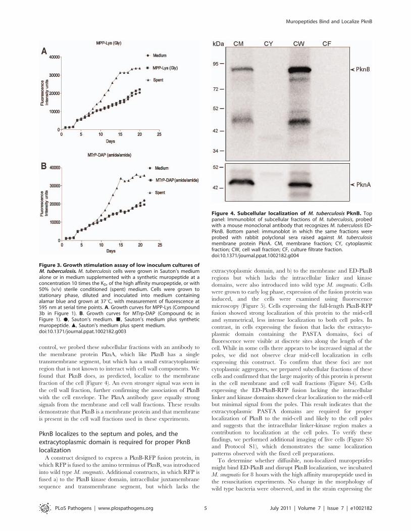
shown in Figure 3, no growth stimulation by either muropeptide
was observed in this assay. In contrast, strong growth stimulation
by conditioned medium was observed.
PknB is present in the cell envelope of M. tuberculosisBased on its sequence, PknB is predicted to have a single
transmembrane segment, with an intracellular kinase domain and
an extracytoplasmic region that incorporates the four PASTA
domains [16]. To determine whether PknB is a membrane protein
and in which subcellular fraction(s) PknB is located, we performed
immunoblotting with a PknB-specific monoclonal antibody. As a
Figure 2. Sensorgrams of a compound with relatively highbinding affinity for ED-PknB. The sensorgrams show the simulta-neous concentration-dependent kinetic analysis of two-fold serialdilutions of MTrP-DAP (amide/acid) (Compound 6c in Figure 1) atconcentrations from 1.56 mM to 100 mM. ED-PknB was bound to thesensor chip and at time 0 the muropeptide was flowed over the chip,followed by a buffer only dissociation step, as described in the Materialsand Methods section. Positive deflection of the curve indicates bindingin RU (resonance units). The primary data are shown in red. The datawere fitted with a two-state binding model (black lines). Thecorresponding residual values, which are the signal remaining afterthe data are fitted to the kinetic model, are plotted below thesensorgrams.doi:10.1371/journal.ppat.1002182.g002
Table 1. Affinity of synthetic muropeptides for theextracytoplasmic domain of M. tuberculosis PknB.
Analyte KD (mM)
MTP-Lys (amide) 1 .500
MTrP-Lys (amide) 2a 21.5
MTrP-Lys (amide) NHAc 2b .500
MTrP-Lys (Gly) 2c . 500
MPP-Lys (D-Ala) 3a . 500
MPP-Lys (Gly) 3b .500
Peptide 4 (amide) . 500
MTP-DAP (amide/acid) 5 21.8
MTrP-DAP (amide/acid) 6a 12.7
MTrP-DAP (acid/amide) 6b .100
MTrP-DAP (amide/amide) 6c 14.9
MTrP-DAP (acid/acid) 6d 53.6
MTrP-DAP(amide/acid)NHAc 6e 73.8
MPP-DAP (amide/acid) 7 25.1
Peptide 8 (amide/amide) .500
Recombinant ED-PknB was immobilized on NHS-activated groups of a CM-5sensor chip surface (5,000 RU) and titration experiments were performed withthe synthetic compounds 1–8 (Figure 1). The binding constants of allcompounds were determined by fitting the data using a two-state bindingmodel. Kinetic binding parameters and the sensorgrams for the kinetic analysesare presented in Table S1 and Figure S3. MTP, muramyl-tripeptide; MTrP,muramyl-tetra peptide; MPP, muramyl-pentapeptide.doi:10.1371/journal.ppat.1002182.t001
Table 2. Resuscitation of dormant M. tuberculosis cultures.
Additive to Culture Medium Fold increase*
Experiment 1:6 month old dormant culture
MTrP-DAP (amide/amide) (6c) 9
MPP-Lys (Gly) (3b) 0.9
50% spent medium 100
Experiment 2:9 month old dormant culture
MTrP-DAP (amide/amide) (6c) 3
MPP-Lys (Gly) (3b) 1
50% spent medium 13.6
*Fold increase in viable cell number relative to cultures grown in Sauton’smedium without additive.doi:10.1371/journal.ppat.1002182.t002
Muropeptides Bind and Localize PknB
PLoS Pathogens | www.plospathogens.org 4 July 2011 | Volume 7 | Issue 7 | e1002182
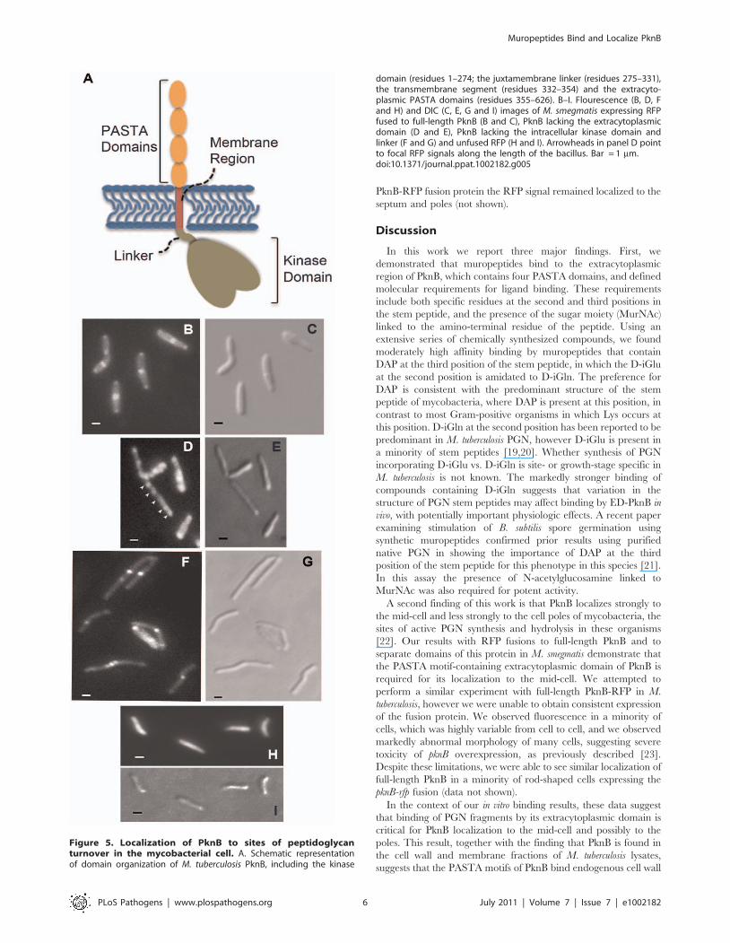
control, we probed these subcellular fractions with an antibody to
the membrane protein PknA, which like PknB has a single
transmembrane segment, but which has a small extracytoplasmic
region that is not known to interact with cell wall components. We
found that PknB does, as predicted, localize to the membrane
fraction of the cell (Figure 4). An even stronger signal was seen in
the cell wall fraction, further confirming the association of PknB
with the cell envelope. The PknA antibody gave equally strong
signals from the membrane and cell wall fractions. These results
demonstrate that PknB is a membrane protein and that membrane
is present in the cell wall fractions used in these experiments.
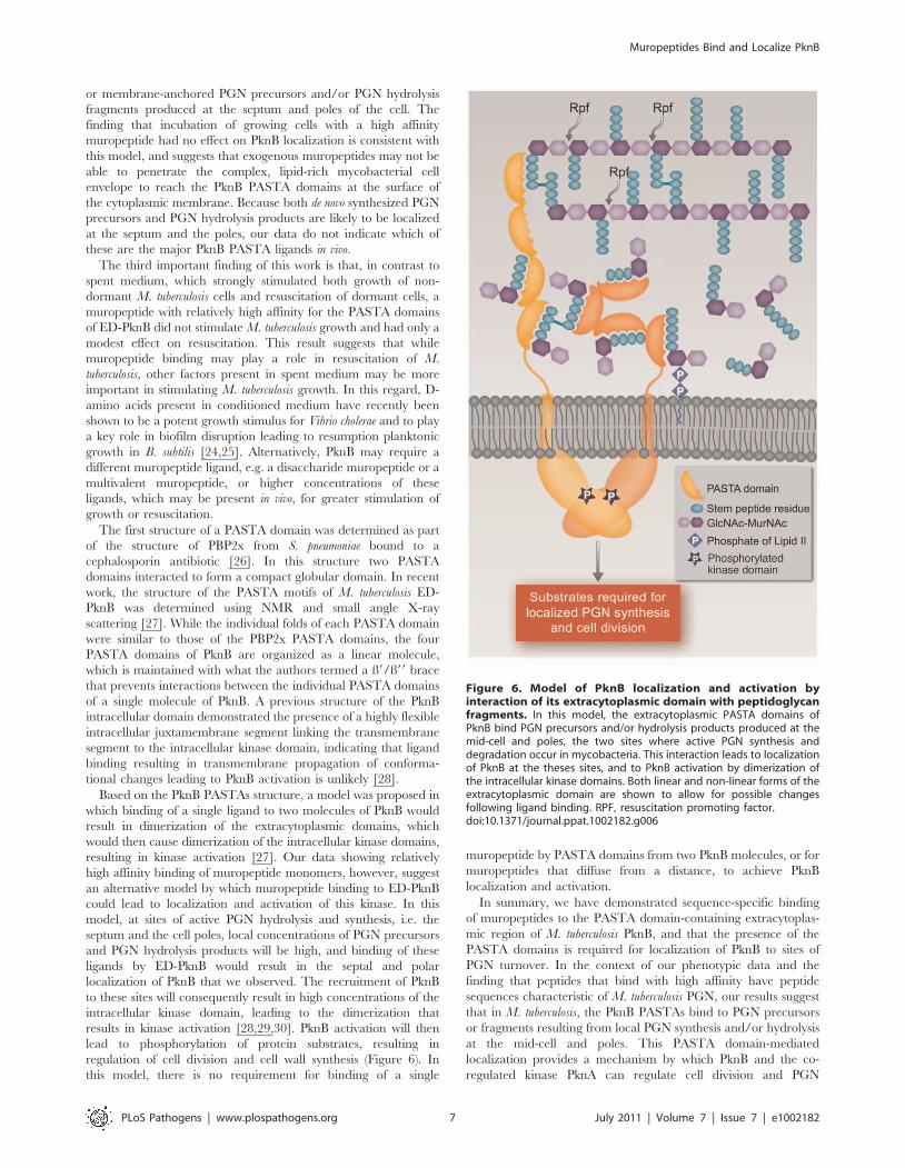
PknB localizes to the septum and poles, and theextracytoplasmic domain is required for proper PknBlocalization
A construct designed to express a PknB-RFP fusion protein, in
which RFP is fused to the amino terminus of PknB, was introduced
into wild type M. smegmatis. Additional constructs, in which RFP is
fused a) to the PknB kinase domain, intracellular juxtamembrane
sequence and transmembrane segment, but which lacks the
extracytoplasmic domain, and b) to the membrane and ED-PknB
regions but which lacks the intracellular linker and kinase
domains, were also introduced into wild type M. smegmatis. Cells
were grown to early log phase, expression of the fusion protein was
induced, and the cells were examined using fluorescence
microscopy (Figure 5). Cells expressing the full-length PknB-RFP
fusion showed strong localization of this protein to the mid-cell
and symmetrical, less intense localization to both cell poles. In
contrast, in cells expressing the fusion that lacks the extracyto-
plasmic domain containing the PASTA domains, foci of
fluorescence were visible at discrete sites along the length of the
cell. While in some cells there appears to be increased signal at the
poles, we did not observe clear mid-cell localization in cells
expressing this construct. To confirm that these foci are not
cytoplasmic aggregates, we prepared subcellular fractions of these
cells and confirmed that the large majority of this protein is present
in the cell membrane and cell wall fractions (Figure S4). Cells
expressing the ED-PknB-RFP fusion lacking the intracellular
linker and kinase domains showed clear localization to the mid-cell
but minimal signal from the poles. This result indicates that the
extracytoplasmic PASTA domains are required for proper
localization of PknB to the mid-cell and likely to the cell poles
and suggests that the intracellular linker-kinase region makes a
contribution to localization at the cell poles. To verify these
findings, we performed additional imaging of live cells (Figure S5
and Protocol S1), which demonstrates the same localization
patterns observed with the fixed cell preparations.
To determine whether diffusible, non-localized muropeptides
might bind ED-PknB and disrupt PknB localization, we incubated
M. smegmatis for 8 hours with the high affinity muropeptide used in
the resuscitation experiments. No change in the morphology of
wild type bacteria were observed, and in the strain expressing the
Figure 3. Growth stimulation assay of low inoculum cultures ofM. tuberculosis. M. tuberculosis cells were grown in Sauton’s mediumalone or in medium supplemented with a synthetic muropeptide at aconcentration 10 times the KD, of the high affinity muropeptide, or with50% (v/v) sterile conditioned (spent) medium. Cells were grown tostationary phase, diluted and inoculated into medium containingalamar blue and grown at 37uC, with measurement of fluorescence at595 nm at serial time points. A. Growth curves for MPP-Lys (Compound3b in Figure 1). B. Growth curves for MTrp-DAP (Compound 6c inFigure 1). N, Sauton’s medium. &, Sauton’s medium plus syntheticmuropeptide. m, Sauton’s medium plus spent medium.doi:10.1371/journal.ppat.1002182.g003
Figure 4. Subcellular localization of M. tuberculosis PknB. Toppanel: Immunoblot of subcellular fractions of M. tuberculosis, probedwith a mouse monoclonal antibody that recognizes M. tuberculosis ED-PknB. Bottom panel: immunoblot in which the same fractions wereprobed with rabbit polyclonal sera raised against M. tuberculosismembrane protein PknA. CM, membrane fraction; CY, cytoplasmicfraction; CW, cell wall fraction; CF, culture filtrate fraction.doi:10.1371/journal.ppat.1002182.g004
Muropeptides Bind and Localize PknB
PLoS Pathogens | www.plospathogens.org 5 July 2011 | Volume 7 | Issue 7 | e1002182
PknB-RFP fusion protein the RFP signal remained localized to the
septum and poles (not shown).
Discussion
In this work we report three major findings. First, we
demonstrated that muropeptides bind to the extracytoplasmic
region of PknB, which contains four PASTA domains, and defined
molecular requirements for ligand binding. These requirements
include both specific residues at the second and third positions in
the stem peptide, and the presence of the sugar moiety (MurNAc)
linked to the amino-terminal residue of the peptide. Using an
extensive series of chemically synthesized compounds, we found
moderately high affinity binding by muropeptides that contain
DAP at the third position of the stem peptide, in which the D-iGlu
at the second position is amidated to D-iGln. The preference for
DAP is consistent with the predominant structure of the stem
peptide of mycobacteria, where DAP is present at this position, in
contrast to most Gram-positive organisms in which Lys occurs at
this position. D-iGln at the second position has been reported to be
predominant in M. tuberculosis PGN, however D-iGlu is present in
a minority of stem peptides [19,20]. Whether synthesis of PGN
incorporating D-iGlu vs. D-iGln is site- or growth-stage specific in
M. tuberculosis is not known. The markedly stronger binding of
compounds containing D-iGln suggests that variation in the
structure of PGN stem peptides may affect binding by ED-PknB in
vivo, with potentially important physiologic effects. A recent paper
examining stimulation of B. subtilis spore germination using
synthetic muropeptides confirmed prior results using purified
native PGN in showing the importance of DAP at the third
position of the stem peptide for this phenotype in this species [21].
In this assay the presence of N-acetylglucosamine linked to
MurNAc was also required for potent activity.
A second finding of this work is that PknB localizes strongly to
the mid-cell and less strongly to the cell poles of mycobacteria, the
sites of active PGN synthesis and hydrolysis in these organisms
[22]. Our results with RFP fusions to full-length PknB and to
separate domains of this protein in M. smegmatis demonstrate that
the PASTA motif-containing extracytoplasmic domain of PknB is
required for its localization to the mid-cell. We attempted to
perform a similar experiment with full-length PknB-RFP in M.
tuberculosis, however we were unable to obtain consistent expression
of the fusion protein. We observed fluorescence in a minority of
cells, which was highly variable from cell to cell, and we observed
markedly abnormal morphology of many cells, suggesting severe
toxicity of pknB overexpression, as previously described [23].
Despite these limitations, we were able to see similar localization of
full-length PknB in a minority of rod-shaped cells expressing the
pknB-rfp fusion (data not shown).
In the context of our in vitro binding results, these data suggest
that binding of PGN fragments by its extracytoplasmic domain is
critical for PknB localization to the mid-cell and possibly to the
poles. This result, together with the finding that PknB is found in
the cell wall and membrane fractions of M. tuberculosis lysates,
suggests that the PASTA motifs of PknB bind endogenous cell wall
Figure 5. Localization of PknB to sites of peptidoglycanturnover in the mycobacterial cell. A. Schematic representationof domain organization of M. tuberculosis PknB, including the kinase
domain (residues 1–274; the juxtamembrane linker (residues 275–331),the transmembrane segment (residues 332–354) and the extracyto-plasmic PASTA domains (residues 355–626). B–I. Flourescence (B, D, Fand H) and DIC (C, E, G and I) images of M. smegmatis expressing RFPfused to full-length PknB (B and C), PknB lacking the extracytoplasmicdomain (D and E), PknB lacking the intracellular kinase domain andlinker (F and G) and unfused RFP (H and I). Arrowheads in panel D pointto focal RFP signals along the length of the bacillus. Bar = 1 mm.doi:10.1371/journal.ppat.1002182.g005
Muropeptides Bind and Localize PknB
PLoS Pathogens | www.plospathogens.org 6 July 2011 | Volume 7 | Issue 7 | e1002182
or membrane-anchored PGN precursors and/or PGN hydrolysis
fragments produced at the septum and poles of the cell. The
finding that incubation of growing cells with a high affinity
muropeptide had no effect on PknB localization is consistent with
this model, and suggests that exogenous muropeptides may not be
able to penetrate the complex, lipid-rich mycobacterial cell
envelope to reach the PknB PASTA domains at the surface of
the cytoplasmic membrane. Because both de novo synthesized PGN
precursors and PGN hydrolysis products are likely to be localized
at the septum and the poles, our data do not indicate which of
these are the major PknB PASTA ligands in vivo.
The third important finding of this work is that, in contrast to
spent medium, which strongly stimulated both growth of non-
dormant M. tuberculosis cells and resuscitation of dormant cells, a
muropeptide with relatively high affinity for the PASTA domains
of ED-PknB did not stimulate M. tuberculosis growth and had only a
modest effect on resuscitation. This result suggests that while
muropeptide binding may play a role in resuscitation of M.
tuberculosis, other factors present in spent medium may be more
important in stimulating M. tuberculosis growth. In this regard, D-
amino acids present in conditioned medium have recently been
shown to be a potent growth stimulus for Vibrio cholerae and to play
a key role in biofilm disruption leading to resumption planktonic
growth in B. subtilis [24,25]. Alternatively, PknB may require a
different muropeptide ligand, e.g. a disaccharide muropeptide or a
multivalent muropeptide, or higher concentrations of these
ligands, which may be present in vivo, for greater stimulation of
growth or resuscitation.
The first structure of a PASTA domain was determined as part
of the structure of PBP2x from S. pneumoniae bound to a
cephalosporin antibiotic [26]. In this structure two PASTA
domains interacted to form a compact globular domain. In recent
work, the structure of the PASTA motifs of M. tuberculosis ED-
PknB was determined using NMR and small angle X-ray
scattering [27]. While the individual folds of each PASTA domain
were similar to those of the PBP2x PASTA domains, the four
PASTA domains of PknB are organized as a linear molecule,
which is maintained with what the authors termed a ß9/ß99 brace
that prevents interactions between the individual PASTA domains
of a single molecule of PknB. A previous structure of the PknB
intracellular domain demonstrated the presence of a highly flexible
intracellular juxtamembrane segment linking the transmembrane
segment to the intracellular kinase domain, indicating that ligand
binding resulting in transmembrane propagation of conforma-
tional changes leading to PknB activation is unlikely [28].
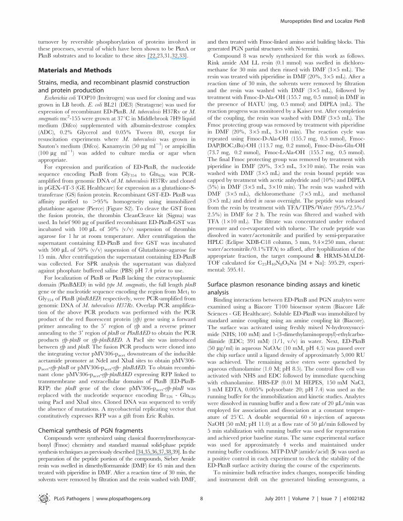
Based on the PknB PASTAs structure, a model was proposed in
which binding of a single ligand to two molecules of PknB would
result in dimerization of the extracytoplasmic domains, which
would then cause dimerization of the intracellular kinase domains,
resulting in kinase activation [27]. Our data showing relatively
high affinity binding of muropeptide monomers, however, suggest
an alternative model by which muropeptide binding to ED-PknB
could lead to localization and activation of this kinase. In this
model, at sites of active PGN hydrolysis and synthesis, i.e. the
septum and the cell poles, local concentrations of PGN precursors
and PGN hydrolysis products will be high, and binding of these
ligands by ED-PknB would result in the septal and polar
localization of PknB that we observed. The recruitment of PknB
to these sites will consequently result in high concentrations of the
intracellular kinase domain, leading to the dimerization that
results in kinase activation [28,29,30]. PknB activation will then
lead to phosphorylation of protein substrates, resulting in
regulation of cell division and cell wall synthesis (Figure 6). In
this model, there is no requirement for binding of a single
muropeptide by PASTA domains from two PknB molecules, or for
muropeptides that diffuse from a distance, to achieve PknB
localization and activation.
In summary, we have demonstrated sequence-specific binding
of muropeptides to the PASTA domain-containing extracytoplas-
mic region of M. tuberculosis PknB, and that the presence of the
PASTA domains is required for localization of PknB to sites of
PGN turnover. In the context of our phenotypic data and the
finding that peptides that bind with high affinity have peptide
sequences characteristic of M. tuberculosis PGN, our results suggest
that in M. tuberculosis, the PknB PASTAs bind to PGN precursors
or fragments resulting from local PGN synthesis and/or hydrolysis
at the mid-cell and poles. This PASTA domain-mediated
localization provides a mechanism by which PknB and the co-
regulated kinase PknA can regulate cell division and PGN
Figure 6. Model of PknB localization and activation byinteraction of its extracytoplasmic domain with peptidoglycanfragments. In this model, the extracytoplasmic PASTA domains ofPknB bind PGN precursors and/or hydrolysis products produced at themid-cell and poles, the two sites where active PGN synthesis anddegradation occur in mycobacteria. This interaction leads to localizationof PknB at the theses sites, and to PknB activation by dimerization ofthe intracellular kinase domains. Both linear and non-linear forms of theextracytoplasmic domain are shown to allow for possible changesfollowing ligand binding. RPF, resuscitation promoting factor.doi:10.1371/journal.ppat.1002182.g006
Muropeptides Bind and Localize PknB
PLoS Pathogens | www.plospathogens.org 7 July 2011 | Volume 7 | Issue 7 | e1002182
turnover by reversible phosphorylation of proteins involved in
these processes, several of which have been shown to be PknA or
PknB substrates and to localize to these sites [22,23,31,32,33].
Materials and Methods
Strains, media, and recombinant plasmid constructionand protein production
Escherichia coli TOP10 (Invitrogen) was used for cloning and was
grown in LB broth. E. coli BL21 (DE3) (Stratagene) was used for
expression of recombinant ED-PknB. M. tuberculosis H37Rv or M.
smegmatis mc2-155 were grown at 37uC in Middlebrook 7H9 liquid
medium (Difco) supplemented with albumin-dextrose complex
(ADC), 0.2% Glycerol and 0.05% Tween 80, except for
resuscitation experiments where M. tuberculosis was grown in
Sauton’s medium (Difco). Kanamycin (50 mg ml21) or ampicillin
(100 mg ml21) was added to culture media or agar when
appropriate.
For expression and purification of ED-PknB, the nucleotide
sequence encoding PknB from Gly354 to Gln626 was PCR-
amplified from genomic DNA of M. tuberculosis H37Rv and cloned
in pGEX-4T-3 (GE Healthcare) for expression as a glutathione-S-
transferase (GS) fusion protein. Recombinant GST-ED- PknB was
affinity purified to .95% homogeneity using immobilized
glutathione agarose (Pierce) (Figure S2). To cleave the GST from
the fusion protein, the thrombin CleanCleave kit (Sigma) was
used. In brief 900 mg of purified recombinant ED-PknB-GST was
incubated with 100 mL of 50% (v/v) suspension of thrombin
agarose for 1 hr at room temperature. After centrifugation the
supernatant containing ED-PknB and free GST was incubated
with 500 mL of 50% (v/v) suspension of Glutathione-agarose for
15 min. After centrifugation the supernatant containing ED-PknB
was collected. For SPR analysis the supernatant was dialyzed
against phosphate buffered saline (PBS) pH 7.4 prior to use.
For localization of PknB or PknB lacking the extracytoplasmic
domain (PknBDED) in wild type M. smegmatis, the full length pknB
gene or the nucleotide sequence encoding the region from Met1 to
Gly354 of PknB (pknBDED) respectively, were PCR-amplified from
genomic DNA of M. tuberculosis H37Rv. Overlap PCR amplifica-
tion of the above PCR products was performed with the PCR
product of the red fluorescent protein (rfp) gene using a forward
primer annealing to the 59 region of rfp and a reverse primer
annealing to the 39 region of pknB or PknBDED to obtain the PCR
products rfp-pknB or rfp-pknBDED. A PacI site was introduced
between rfp and pknB. The fusion PCR products were cloned into
the integrating vector pMV306-pacet downstream of the inducible
acetamide promoter at NdeI and XbaI sites to obtain pMV306-
pacet-rfp-pknB or pMV306-pacet-rfp- pknBDED. To obtain recombi-
nant clone pMV306-pacet-rfp-pknBDKD expressing RFP linked to
transmembrane and extracellular domains of PknB (ED-PknB-
RFP) the pknB gene of the clone pMV306-pacet-rfp-pknB was
replaced with the nucleotide sequence encoding Ile326 - Gln626
using PacI and XbaI sites. Cloned DNA was sequenced to verify
the absence of mutations. A mycobacterial replicating vector that
constitutively expresses RFP was a gift from Eric Rubin.
Chemical synthesis of PGN fragmentsCompounds were synthesized using classical fluorenylmethoxycar-
bonyl (Fmoc) chemistry and standard manual solid-phase peptide
synthesis techniques as previously described [34,35,36,37,38,39]. In the
preparation of the peptide portion of the compounds, Sieber Amide
resin was swelled in dimethylformamide (DMF) for 45 min and then
treated with piperidine in DMF. After a reaction time of 30 min, the
solvents were removed by filtration and the resin washed with DMF,
and then treated with Fmoc-linked amino acid building blocks. This
generated PGN partial structures with N-termini.
Compound 8 was newly synthesized for this work as follows.
Rink amide AM LL resin (0.1 mmol) was swelled in dichloro-
methane for 30 min and then rinsed with DMF (365 mL). The
resin was treated with piperidine in DMF (20%, 365 mL). After a
reaction time of 30 min, the solvents were removed by filtration
and the resin was washed with DMF (365 mL), followed by
treatment with Fmoc-D-Ala-OH (155.7 mg, 0.5 mmol) in DMF in
the presence of HATU (mg, 0.5 mmol) and DIPEA (mL). The
reaction progress was monitored by a Kaiser test. After completion
of the coupling, the resin was washed with DMF (365 mL). The
Fmoc protecting group was removed by treatment with piperidine
in DMF (20%, 365 mL, 3610 min). The reaction cycle was
repeated using Fmoc-D-Ala-OH (155.7 mg, 0.5 mmol), Fmoc-
DAP(BOC,tBu)-OH (113.7 mg, 0.2 mmol), Fmoc-D-iso-Gln-OH
(73.7 mg, 0.2 mmol), Fmoc-L-Ala-OH (155.7 mg, 0.5 mmol).
The final Fmoc protecting group was removed by treatment with
piperidine in DMF (20%, 365 mL, 3610 min). The resin was
washed with DMF (365 mL) and the resin bound peptide was
capped by treatment with acetic anhydride and (10%) and DIPEA
(5%) in DMF (365 mL, 3610 min). The resin was washed with
DMF (365 mL), dichloromethane (765 mL), and methanol
(365 mL) and dried in vacuo overnight. The peptide was released
from the resin by treatment with TFA/TIPS/Water (95%/2.5%/
2.5%) in DMF for 2 h. The resin was filtered and washed with
TFA (1610 mL). The filtrate was concentrated under reduced
pressure and co-evaporated with toluene. The crude peptide was
dissolved in water/acetonitrile and purified by semi-preparative
HPLC (Eclipse XDB-C18 column, 5 mm, 9.46250 mm, eluent:
water/acetonitrile/0.1%TFA) to afford, after lyophilization of the
appropriate fraction, the target compound 8. HRMS-MALDI-
TOF calculated for C23H40N8O9Na [M + Na]: 595.29, experi-
mental: 595.41.
Surface plasmon resonance binding assays and kineticanalysis
Binding interactions between ED-PknB and PGN analytes were
examined using a Biacore T100 biosensor system (Biacore Life
Sciences - GE Healthcare). Soluble ED-PknB was immobilized by
standard amine coupling using an amine coupling kit (Biacore).
The surface was activated using freshly mixed N-hydroxysucci-
mide (NHS; 100 mM) and 1-(3-dimethylaminopropyl)-ethylcarbo-
diimide (EDC; 391 mM) (1/1, v/v) in water. Next, ED-PknB
(50 mg/ml) in aqueous NaOAc (10 mM, pH 4.5) was passed over
the chip surface until a ligand density of approximately 5,000 RU
was achieved. The remaining active esters were quenched by
aqueous ethanolamine (1.0 M; pH 8.5). The control flow cell was
activated with NHS and EDC followed by immediate quenching
with ethanolamine. HBS-EP (0.01 M HEPES, 150 mM NaCl,
3 mM EDTA, 0.005% polysorbate 20; pH 7.4) was used as the
running buffer for the immobilization and kinetic studies. Analytes
were dissolved in running buffer and a flow rate of 20 mL/min was
employed for association and dissociation at a constant temper-
ature of 25uC. A double sequential 60 s injection of aqueous
NaOH (50 mM; pH 11.0) at a flow rate of 50 ml/min followed by
5 min stabilization with running buffer was used for regeneration
and achieved prior baseline status. The same experimental surface
was used for approximately 4 weeks and maintained under
running buffer conditions. MTP-DAP (amide/acid) (5) was used as
a positive control in each experiment to check the stability of the
ED-PknB surface activity during the course of the experiments.
To minimize bulk refractive index changes, nonspecific binding
and instrument drift on the generated binding sensorgrams, a
Muropeptides Bind and Localize PknB
PLoS Pathogens | www.plospathogens.org 8 July 2011 | Volume 7 | Issue 7 | e1002182
double referencing of the data was performed. First, bulk refractive
index change effects were minimized by preparing all analytes in
the HBS-EP buffer. Then, the binding responses over the
reference surface were subtracted from the active surface to
correct for nonspecific binding. A blank analyte run of running
buffer alone was also subtracted from the specific binding
sensorgrams to minimize instrument noise. Using Biacore T100
evaluation software, the response curves of various analyte
concentrations were globally fitted to the two-state binding model
[40].
Conditioned medium preparationConditioned medium was prepared as previously described
[41]. Briefly, supernatant was obtained from M. tuberculosis H37Rv
culture grown in ADC-supplemented Sauton’s medium containing
0.05% Tween 80 at 37uC with shaking to an optical density at
600 nm (OD600) of 1.2. After centrifugation (4000 rpm, 10 min),
the supernatant was sterilized by passage through 0.22 mm filter,
tested for sterility, and used for the resuscitation experiments.
Dormancy and resuscitation assayTo obtain non-culturable dormant bacilli, Mycobacterium tubercu-
losis H37Rv was grown under long-term oxygen starvation in
broth growth medium (Sauton’s medium containing 0.05% tween
80 and supplemented with ADC) as previously described [10]. In
brief, M. tuberculosis was initially grown to late stationary phase at
37uC with shaking. From this initial culture, 100 mL was
subcultured into 20 ml of growth medium and grown to an
optical density at 600 nm (OD600) of 1.8 to 2. Finally 100 mL was
inoculated into 75 ml of growth medium containing 1.5 mg/ml
methylene blue in a sealed 250-ml flask and grown with shaking at
37uC for 6 or 9 months. Methylene blue became colorless by 10
days of incubation, indicating oxygen depletion.
For resuscitation experiments muropeptides were dissolved in
sterile Sauton’s medium to a concentration of 20 times the binding
constant (KD) of the high affinity compound (6c) The dormant
culture was serially diluted (1021 to 1026) in growth medium.
From each dilution 4 sets of triplicate 100 mL culture were
aliquoted in wells of 96 well plates (one set each for muropeptides
(2 muropeptides tested), growth medium and spent medium).
100 mL of growth medium, muropeptide, or spent medium was
added to each well of the corresponding set. The final
concentration of muropeptide was 10 times the KD of the high
affinity compound, and of the spent medium was 50%. Plates were
incubated at 37uC. Drying was prevented by maintaining sterile
water in outer wells of the plate. After 2 months the wells with
visibly turbid growth were recorded and MPN values were
calculated as previously described [42].
Growth stimulation assayTo investigate the effect of muropeptides on growth initiation of
low inoculum cultures of M. tuberculosis, stationary phase (O.D600
of 2.4–3.6) cultures grown in Sauton’s medium supplemented with
ADC and 0.05% Tween 80 were passed through five micron pore
filter (Millipore) to remove clumps. To obtain a single cell
suspension, the culture was passed five times through a 27K gauge
needle followed by washing three times with Sauton’s medium.
100 ml of a 1024 dilution was inoculated into wells of a 96 well
plate for a final volume of 200 ml. 1x alamar Blue was included in
each well. As above, the final concentration of the muropeptides
was 10 times the KD of the high affinity compound, and of the
spent medium was 50%. Each condition was tested in duplicate.
Growth was monitored in each well by measuring fluorescence
using excitation of 550 nm and emission of 595 nm and plotted as
fluorescence intensity units versus time in days.
MicroscopyFor cellular localization of RFP fusions to intact PknB or its
domains, the corresponding plasmid expressing the fusion under
control of the acetamidase promoter was electroporated into M.
smegmatis cells. The resulting strains were grown in Middlebrook
7H9 liquid medium supplemented with ADC, 0.2% Glycerol and
0.05% Tween 80 to mid-log phase, followed by induction with
0.2% acetamide for 6 hrs. Cells were fixed in 4% paraformalde-
hyde at 37uC for 30 min followed by incubation with 50 mM
ammonium chloride for 5 min at room temperature. Cells were
transferred onto a glass slide, air-dried and one drop of Prolong
Gold antifade reagent (Invitrogen) was applied before covering
with a coverslip. After 24 hrs of curing in the dark, cells were
observed using a Zeiss Axiophot microscope with a 63x differential
interference contrast (DIC) oil immersion objective and red
fluorescence filter. Images were captured by a Spot cooled CCD
camera (Diagnostic Instruments), acquired with Spot software and
processed by Adobe Photoshop CS2.
Western blottingFor cellular localization of native PknB in wild type M.
tuberculosis cells, 60 mg total protein of cytosol, cell wall, cell
membrane and culture filtrate fractions, prepared at Colorado
Statue University and obtained from the Biodefense and Emerging
Infections Research Resources Repository, was fractionated on
10% SDS-PAGE and transferred to a PVDF membrane. The blot
was blocked in Tris-buffered saline containing 0.1% Tween 20
(TBST) and 5% milk for 1 hr at room temperature. The blot was
incubated overnight at 4uC with 1:10,000 dilutions of either a
mouse monoclonal antibody raised against extracytoplasmic
domain of PknB or with a rabbit polyclonal antibody against
PknA. After thorough washing with TBST, the blot was incubated
with a 1:10,000 dilution of HRP-conjugated secondary antibodies
(Cell Signaling) for 3 hrs at room temperature. Finally after 3
washes with TBST the blot was developed with Lumiglo (Cell
Signaling) and the blot image was obtained on a Kodak Image
Station 4000.
Accession numbersPknA: P65726
PknB: POA5S4
Supporting Information
Figure S1 T-Coffee alignment of the four PASTAdomains of M. tuberculosis PknB and the single PASTAdomain of PBP2. Residues/positions corresponding to those
that are conserved in PASTA domains from multiple bacterial
species, according to reference 14, are highlighted in blue.
(TIF)
Figure S2 SDS-PAGE gel showing expression and puri-fication of ED-PknB. M, molecular weight markers: UI, lysate
from uninduced cultures; I, lysate from induced cultures, The
purified protein following removal of the GST tag, shown in the
last lane on the right, was used in the binding experiments.
(TIF)
Figure S3 Sensorgrams of compounds tested in theBiacore binding experiments. The sensorgrams show the
simultaneous concentration-dependent kinetic analysis of two-fold
serial dilutions of each compound. ED-PknB was bound to the
Muropeptides Bind and Localize PknB
PLoS Pathogens | www.plospathogens.org 9 July 2011 | Volume 7 | Issue 7 | e1002182
sensor chip and at time 0 the muropeptide was flowed over the
chip as described in the Materials and Methods section. Positive
deflection of the curve indicates binding in RU (resonance units).
The primary data are shown in red. For compounds that showed
significant binding the data were fitted with a two-state binding
model (black lines) and the corresponding residual values, which
are the signal remaining after the data are fitted to the kinetic
model, are plotted below the sensorgrams. Sensorgrams for
individual muropeptides are shown in A) MTP-Lys (amide) (1);
B) MTrP-Lys (amide) (2a); C) MTrP-Lys (amide) NHAc (2b); D)
MTrP-Lys (Gly) (2c); E) MPP-Lys (D-Ala) (3a); F) MPP-Lys (Gly)
(3b); G) Peptide (amide) (4); H) MTP-DAP (amide/acid) (5); I)
MTrP-DAP (amide/acid) (6a); J) MTrP-DAP (acid/amide) (6b);
K) MTrP-DAP (acid/acid) (6d); L) MTrP-DAP(amide/acid)NHAc
(6e); M) MPP-DAP (amide/acid) (7); N) Peptide (amide/amide) (8).
(PDF)
Figure S4 Immunoblot of subcellular fractions of M.smegmatis showing localization of the RFP-PknB kinasedomain fusion. M. smegmatis was grown to mid log phase,
acetamide was added at a concentration of 0.2% for 8 hours. Cells
were harvested, lysed with a French Press and sub-cellular
fractions were isolated using the protocol developed by the TB
Research Materials Contract at Colorado State University (http://
www.cvmbs.colostate.edu/mip/tb/pdf/scf.pdf). RH615, M. smeg-
matis expressing the RFP-PknB kinase domain fusion under control
of the inducible acetamidase promoter; CM, cytoplasmic mem-
brane fraction; CY, cytoplasm; CW, Cell wall fraction. Though
some fusion protein is present in the cytoplasm, the majority is in
the cell wall and cell membrane fractions, as is native M. smegmatis
PknB.
(TIF)
Figure S5 Live cell imaging of M. smegmatis. Cells
expressing RFP fused to a) full-length PknB, b) to the kinase
domain, linker and transmembrane segment, or c) to ED-PknB
and the transmembrane segment. Fluorescence images are shown
on the left and DIC images on the right. Bar = 1 mm.
(TIF)
Table S1 Kinetic binding parameters for the interaction of
synthetic muropeptides with the PASTA domains of M. tuberculosis
PknB.
(DOC)
Protocol S1 Methods for live cell imaging of M. smegmatis
expressing RFP-PknB fusions.
(DOCX)
Acknowledgments
We thank and Michael Chao and Eric Rubin for the RFP expression
vector, Jessica Wagner and the Harvard Digestive Diseases Center Imaging
Core for assistance with microscopy and Margreet Wolfert for help with
the figures. M. tuberculosis subcellular fractions were obtained through
NIAID Contract No. HHSN266200400091C, entitled "Tuberculosis
Vaccine Testing and Research Materials," awarded to Colorado State
University. Antibodies to PknA were obtained from Vertex Pharmaceu-
ticals Incorporated.
Author Contributions
Conceived and designed the experiments: MM JA G-JB RNH. Performed
the experiments: MM JA XL JC. Analyzed the data: MM JA XL JC G-JB
RNH. Contributed reagents/materials/analysis tools: MM RNH G-JB.
Wrote the paper: MM JA G-JB RNH. Revision of manuscript: MM G-JB
RNH.
References
1. Goehring NW, Beckwith J (2005) Diverse paths to midcell: assembly of the
bacterial cell division machinery. Curr Biol 15: R514–526.
2. Errington J, Daniel RA, Scheffers DJ (2003) Cytokinesis in bacteria. Microbiol
Mol Biol Rev 67: 52–65.
3. Blackman SA, Smith TJ, Foster SJ (1998) The role of autolysins during
vegetative growth of Bacillus subtilis 168. Microbiology 144(Pt 1): 73–82.
4. Vollmer W, Blanot D, de Pedro MA (2008) Peptidoglycan structure and
architecture. FEMS Microbiol Rev 32: 149–167.
5. Vollmer W, Joris B, Charlier P, Foster S (2008) Bacterial peptidoglycan (murein)
hydrolases. FEMS Microbiol Rev 32: 259–286.
6. Morlot C, Uehara T, Marquis KA, Bernhardt TG, Rudner DZ (2010) A highly
coordinated cell wall degradation machine governs spore morphogenesis in
Bacillus subtilis. Genes Dev 24: 411–422.
7. Chao MC, Rubin EJ (2010) Letting sleeping dos lie: does dormancy play a role
in tuberculosis? Annu Rev Microbiol 64: 293–311.
8. Mukamolova GV, Kaprelyants AS, Young DI, Young M, Kell DB (1998) A
bacterial cytokine. Proc Natl Acad Sci U S A 95: 8916–8921.
9. Camus JC, Pryor MJ, Medigue C, Cole ST (2002) Re-annotation of the genome
sequence of Mycobacterium tuberculosis H37Rv. Microbiology 148: 2967–2973.
10. Downing KJ, Mischenko VV, Shleeva MO, Young DI, Young M, et al. (2005)
Mutants of Mycobacterium tuberculosis lacking three of the five rpf-like genes are
defective for growth in vivo and for resuscitation in vitro. Infect Immun 73:
3038–3043.
11. Kana BD, Gordhan BG, Downing KJ, Sung N, Vostroktunova G, et al. (2008)
The resuscitation-promoting factors of Mycobacterium tuberculosis are required for
virulence and resuscitation from dormancy but are collectively dispensable for
growth in vitro. Mol Microbiol 67: 672–684.
12. Russell-Goldman E, Xu J, Wang X, Chan J, Tufariello JM (2008) A
Mycobacterium tuberculosis Rpf double-knockout strain exhibits profound defects
in reactivation from chronic tuberculosis and innate immunity phenotypes.
Infect Immun 76: 4269–4281.
13. Mukamolova GV, Murzin AG, Salina EG, Demina GR, Kell DB, et al. (2006)
Muralytic activity of Micrococcus luteus Rpf and its relationship to physiologicalactivity in promoting bacterial growth and resuscitation. Mol Microbiol 59: 84–98.
14. Yeats C, Finn RD, Bateman A (2002) The PASTA domain: a beta-lactam-
binding domain. Trends Biochem Sci 27: 438–440.
15. Shah IM, Laaberki MH, Popham DL, Dworkin J (2008) A eukaryotic-like Ser/
Thr kinase signals bacteria to exit dormancy in response to peptidoglycan
fragments. Cell 135: 486–496.
16. Cole ST, Brosch R, Parkhill J, Garnier T, Churcher C, et al. (1998) Deciphering
the biology of Mycobacterium tuberculosis from the complete genome sequence.
Nature 393: 537–544.
17. Mukamolova GV, Turapov OA, Young DI, Kaprelyants AS, Kell DB, et al.
(2002) A family of autocrine growth factors in Mycobacterium tuberculosis. Mol
Microbiol 46: 623–635.
18. Mukamolova GV, Yanopolskaya ND, Kell DB, Kaprelyants AS (1998) On
resuscitation from the dormant state of Micrococcus luteus. Antonie Van
Leeuwenhoek 73: 237–243.
19. Mahapatra S, Crick DC, McNeil MR, Brennan PJ (2008) Unique structural
features of the peptidoglycan of Mycobacterium leprae. J Bacteriol 190: 655–661.
20. Lavollay M, Arthur M, Fourgeaud M, Dubost L, Marie A, et al. (2008) The
peptidoglycan of stationary-phase Mycobacterium tuberculosis predominantly
contains cross-links generated by L,D-transpeptidation. J Bacteriol 190:
4360–4366.
21. Lee M, Hesek D, Shah IM, Oliver AG, Dworkin J, et al. (2010) Synthetic
peptidoglycan motifs for germination of bacterial spores. Chembiochem: a
Chembiochem 11: 2525–2529.
22. Kang CM, Nyayapathy S, Lee JY, Suh JW, Husson RN (2008) Wag31, a
homologue of the cell division protein DivIVA, regulates growth, morphology
and polar cell wall synthesis in mycobacteria. Microbiology 154: 725–735.
23. Kang CM, Abbott DW, Park ST, Dascher CC, Cantley LC, et al. (2005) The
Mycobacterium tuberculosis serine/threonine kinases PknA and PknB: substrate
identification and regulation of cell shape. Genes Dev 19: 1692–1704.
24. Lam H, Oh DC, Cava F, Takacs CN, Clardy J, et al. (2009) D-amino acids govern
stationary phase cell wall remodeling in bacteria. Science 325: 1552–1555.
25. Kolodkin-Gal I, Romero D, Cao S, Clardy J, Kolter R, et al. (2010) D-amino
acids trigger biofilm disassembly. Science 328: 627–629.
26. Dessen A, Mouz N, Gordon E, Hopkins J, Dideberg O (2001) Crystal structure
of PBP2x from a highly penicillin-resistant Streptococcus pneumoniae clinical isolate:
a mosaic framework containing 83 mutations. J Biol Chem 276: 45106–45112.
27. Barthe P, Mukamolova GV, Roumestand C, Cohen-Gonsaud M (2010) The
structure of PknB extracellular PASTA domain from Mycobacterium tuberculosis
suggests a ligand-dependent kinase activation. Structure 18: 606–615.
28. Young TA, Delagoutte B, Endrizzi JA, Falick AM, Alber T (2003) Structure of
Mycobacterium tuberculosis PknB supports a universal activation mechanism for
Ser/Thr protein kinases. Nat Struct Biol 10: 168–174.
29. Mieczkowski C, Iavarone AT, Alber T (2008) Auto-activation mechanism of the
Mycobacterium tuberculosis PknB receptor Ser/Thr kinase. Embo J 27: 3186–3197.
Muropeptides Bind and Localize PknB
PLoS Pathogens | www.plospathogens.org 10 July 2011 | Volume 7 | Issue 7 | e1002182
30. Ortiz-Lombardia M, Pompeo F, Boitel B, Alzari PM (2003) Crystal structure of
the catalytic domain of the PknB serine/threonine kinase from Mycobacterium
tuberculosis. J Biol Chem 278: 13094–13100.
31. Prisic S, Dankwa S, Schwartz D, Chou MF, Locasale JW, et al. (2010) Extensive
phosphorylation with overlapping specificity by Mycobacterium tuberculosis serine/threonine protein kinases. Proc Natl Acad Sci U S A 107: 7521–7526.
32. Dasgupta A, Datta P, Kundu M, Basu J (2006) The serine/threonine kinasePknB of Mycobacterium tuberculosis phosphorylates PBPA, a penicillin-binding
protein required for cell division. Microbiology 152: 493–504.
33. Sureka K, Hossain T, Mukherjee P, Chatterjee P, Datta P, et al. (2010) Novelrole of phosphorylation-dependent interaction between FtsZ and FipA in
mycobacterial cell division. PLoS ONE 5: e8590.34. Chowdhury A, Boons GJ (2005) The synthesis of diaminopimelic acid containing
peptidoglycan fragments using metathesis cross coupling. Tetrahedron Lett 46:1675–1678.
35. Chowdhury A, Siriwardena A, Boons GJ (2002) A highly convergent approach
for the synthesis of disaccharide repeating units of peptidoglycan. TetrahedronLett 43: 7805–7807.
36. Guan R, Roychowdhury A, Ember B, Kumar S, Boons GJ, et al. (2004)Structural basis for peptidoglycan binding by peptidoglycan recognition
proteins. Proc Natl Acad Sci U S A 101: 17168–17173.
37. Kumar S, Roychowdhury A, Ember B, Wang Q, Guan R, et al. (2005) Selective
recognition of synthetic lysine and meso-diaminopimelic acid-type peptidoglycan
fragments by human peptidoglycan recognition proteins Ia and S. J Biol Chem
280: 37005–37012.
38. Roychowdhury A, Wolfert MA, Boons GJ (2005) Synthesis and proinflamma-
tory properties of muramyl tripeptides containing lysine and diaminopimelic
acid moieties. Chembiochem 6: 2088–2097.
39. Swaminathan CP, Brown PH, Roychowdhury A, Wang Q, Guan R, et al. (2006)
Dual strategies for peptidoglycan discrimination by peptidoglycan recognition
proteins (PGRPs). Proc Natl Acad Sci U S A 103: 684–689.
40. Asong J, Wolfert MA, Maiti KK, Miller D, Boons GJ (2009) Binding and cellular
activation studies reveal that Toll-like receptor 2 can differentially recognize
peptidoglycan from Gram-positive and Gram-negative bacteria. J Biol Chem
284: 8643–8653.
41. Sun Z, Zhang Y (1999) Spent culture supernatant of Mycobacterium tuberculosis
H37Ra improves viability of aged cultures of this strain and allows small inocula
to initiate growth. J Bacteriol 181: 7626–7628.
42. de Man J (1975) The probability of most probable numbers. Eur J Appl
Microbiol 1: 67–78.
Muropeptides Bind and Localize PknB
PLoS Pathogens | www.plospathogens.org 11 July 2011 | Volume 7 | Issue 7 | e1002182