effects of ursodeoxycholic acid on conjugated bile acids and progesterone metabolites in serum and...
TRANSCRIPT
Journal of Hepatology 1997; 21: 1029-1040 Printed in Denmark . AN rights reserved Munksgaard ’ Copenhagen
Copyright 0 European Association for the Study of the Liver 1997
Journal of Hepatology ISSN 0168-8278
Effects of ursodeoxycholic acid on conjugated bile acids and progesterone metabolites in serum and urine of patients with intrahepatic cholestasis
of pregnancy
Ling-Jie Meng’, Humberto Reyes2, Jonquin Palma2, Ismael Hernandez2, Jose Ribalta2 and Jan Sjovall’
‘Department of Medical Biochemistry and Biophysics, Karolinska Institute, Stockholm, Sweden and 2Departments of Medicine and Experimental Medicine, Campus Oriente, University of Chile, School of Medicine. Santiago de Chile
Background/Aims and Methods: The mechanism(s) behind the effects of ursodeoxycholic acid on serum steroid sulphate profiles in patients with intrahepatic cholestasis of pregnancy is not clear. Conjugated pro- gesterone metabolites and bile acids have therefore been analysed in serum and mine of patients with in- trahepatic cholestasis of pregnancy before and during treatment with ursodeoxycholic acid using chromato- graphic and mass spectrometric methods. Results: The concentration of glycine-/taurine-conju- gated bile acids decreased from 8.923 pmol/I (mean* SEM) before treatment to 1.8&0.6/rmol/l during treat- ment with ursodeoxycholic acid. The total bile acid ex- cretion in urine decreased from 56&14 to 32k5.6 mol/g creatinine. The proportion of cholic acid in serum and urine, and of l/l-, 2p- and 6a-hydroxylated cholic acids in urine decreased markedly during urso- deoxycholic acid while the percentages of 3a,l2a-dihy- droxy-3-oxo+cholenoic acid and chenodeoxycholic acid were unchanged. The levels in serum and excretion in urine of sulphated steroids decreased during urso- deoxycholic acid, by 499% for disulphates and 33- 35% for monosulphates. The ratios of 3a- to 3/l- hydroxysteroid disulphates were lowered by ursodeoxy- cholic acid from 1.1 (mean) to 0.68 in serum, and from 1.2 to 0.70 in urine. The corresponding ratios for monosulphates before and during ursodeoxycholic acid were 6.9 and 4.5, respectively, in serum, and 21 and 5.2, respectively, in urine. The major monosulphates in
T” LEVELS and patterns of conjugated bile acids
and steroids in serum and urine are markedly dif-
Received 9 May: revised 1 July; accepted 2 July 1997
Correspondence: Jan Sjovall, Department of Medical Biochemistry and Biophysics, Karolinska Institutet, S-171 77 Stockholm, Sweden. Tel: 46-8-7287700. Fax: 46-8-339004. e-mail:[email protected].
urine, dominated by 5a-pregnane_3a, 20a-diol, were also conjugated with N-acetylglucosamine. The ex- cretion of these double conjugates decreased from 27Hl.4 to 15k5.3 Fol/g creatinine during urso- deoxycholic acid. In contrast to sulphated steroids, the concentrations of glucuronides were unchanged in serum and their excretion in urine tended to increase during ur- sodeoxycholic acid. The metabolism of ursodeoxycholic acid was similar to that described in nonpregnant sub- jects. In addition to metabolites hydroxylated in the l/l-, 5fi, 6a//3 and 22-positions, a 4-hydroxy-ursodeoxycholic acid was tentatively identified. This occurred predom- inantly as a double conjugate with glycine/taurine and glucuronic acid, as did other 4-hydroxylated bile acids of probable foetal origin. Conclusions: The results are compatible with the con- tention that ursodeoxycholic acid stimulates the biliary excretion of sulphated progesterone metabolites, par- ticularly those with a 3a-hydroxy-5a(H) configuration and disulphates. The effect(s) appears to be indepen- dent of the stimulation of bile acid secretion. An effect of ursodeoxycholic acid on the reductive metabolism of progesterone cannot be excluded.
Key words: Bile acids; Chromatography; Glucuronides; Intrahepatic cholestasis of pregnancy; Mass spec- trometry; N-acetylglucosaminides; Progesterone metabolites; Serum; Sulphates; Urine; Ursodeoxy- cholic acid.
ferent from normal in patients with intrahepatic chol- estasis of pregnancy (ICP) (l-9). Oral administration of ursodeoxycholic acid (UDCA) improves the clinical condition (10) and reduces the concentrations of bile acids and sulphated steroids in serum towards normal values (11). The mechanism(s) behind the effects on the steroids is not clear. In order to gain further information on this point, we have performed comprehensive analy-
1029
L. J. Meng et cd.
ses of steroids and bile acids in both serum and urine of patients with ICP before and during oral administration of UDCA. The results described in this paper support the contention that UDCA or some of its metabolites improves the biliary secretion of sulphated steroids with a 3a-hydroxy-Sa(H) configuration.
Materials and Methods Patients The study was performed on seven patients with early onset of pruritus, five with ICP and two with uncertain diagnosis. They were hospitalised and treated with UDCA, 1 g per day (12-16 mg * kg-’ - day- ‘) until de- livery. The clinical observations at the time of collec- tion of serum and urine samples are given in Table 1. Patients 1 to 5 were part of a double-blind placebo- controlled study of the clinical effects of UDCA in this disease (12), and they fulfilled several pre-established criteria: pruritus had appeared between the 21st and 32nd week of pregnancy without any other possible ex- planation besides cholestasis of pregnancy; simul- taneously, serum alanine and/or aspartate aminotrans- ferase were above the upper limits observed in normal pregnancies, and serum total bile salts were also abnor- mally high, estimated by an enzymatic method using a commercially available kit (Merckotest@). Patients 6 and 7 also had pruritus of pregnancy of early onset,
TABLE 1
Clinical data and liver function tests in the seven patients before and during the administration of UDCA
Parametera Patientsb
1 2 3 4 5 6 7
Gestational weeks 30 34 34 35 34 27 35
Days of treatment 14 21 21 28 21 14 14 Pruritus severity (O-4) 3 3 3 3 3 3 3
Treated’ 1 0 0 1 1 2 3
Bilirubin, total 0.9 1.3 0.6 0.6 0.7 0.2 0.3 (mg/dl;<l.l)
Treated’ 0.6 0.7 0.2 0.2 0.2 0.5 0.1 S-ALAT (U/1;<40) 112 278 205 120 104 7 14
Treated’ 24 66 16 19 15 7 8
S-ALP (U/l;< 117) 190 167 207 271 183 59 141
Treated’ 170 179 120 293 131 60 146 Cholesterol, total 186 268 229 352 280 268 265 (mg/dl; <200)
Treated’ 132 262 237 362 205 262 234
a S-ALAT: serum alanine aminotransferase; S-ALP: serum total al- kaline phosphatases. In parentheses, units of measurement followed by the upper limit acceptable in fasting blood samples obtained from nonpregnant healthy women in the comparable age range.
b Patients nos. l-5 with ICP participated in the double-blind, pla- cebo-controlled study; patients nos. 6 and 7 with pruritus were given UDCA openly.
c Values obtained after treatment with UDCA or placebo for the period indicated.
with similar severity, but in them serum levels of aminotransferases were normal; they received an ident- ical treatment with UDCA, but in an open fashion.
In all these patients, the remaining pruritus disap- peared and liver function tests normalised during the week following delivery.
Blood and urine samples
Fasting morning serum samples and 12-h overnight urine collection samples were obtained on the same day, before and after 2 to 4 weeks of UDCA adminis- tration. They were frozen immediately at -20°C until analysis.
Informed consent was obtained from all the patients and permission was granted from their attending ob- stetricians and the local ethical committee.
Analytical procedures A detailed description of the analytical method has been published (13). A solid-phase method was used to ex- tract bile acids and steroids from 10 ml urine and l-4 ml serum. Unconjugated bile acids and different groups of conjugated bile acids and steroids were then separated by anion exchange chromatography. The groups of con- jugated compounds were qualitatively analysed by fast bombardment mass spectrometry (FABMS). After hy- drolysis and solvolysis, the liberated bile acids and neu- tral steroids were purified by passage through the anion exchanger. A separate procedure was employed to ana- lyse glucuronides of aminoacyl amidated bile acids (GlcA-G/T) in the urine of patient nos. 1 and 2 during the treatment with UDCA. The urine extract was separ- ated on the same anion exchange column (13). However, elution was performed first with 0.4 M formic acid in 95% methanol and then with 0.5 M acetic acid-potas- sium hydroxide in 72% aqueous methanol, apparent pH 10, to obtain the bile acids doubly conjugated with glu- curonic acid and amino acid. Following neutralisation and solid-phase extraction, the double conjugates were hydrolysed with cholylglycine hydrolase (13). The hy- drolysate was extracted and glucuronides were isolated by ion exchange chromatography. After washing with 0.1 M acetic acid in 95% methanol, the glucuronidated bile acids were eluted with 0.4 M formic acid in 95%) methanol. They were then hydrolysed with Helixpomat- ia digestive juice (13). The liberated unconjugated bile acids were isolated by ion exchange chromatography
(13). The steroids and bile acids were analysed by gas-
liquid chromatography, following derivatisation (13). Bile acids were methylated and the methyl esters and the steroids were converted into trimethylsilyl ethers (13). n-Dotriacontane (C3,J and n-hexatriacontane
1030
Steroids and bile acids in ICP during UDCA
(Cs,) were added as internal standards prior to deriv- atisation. A GLC temperature programme was used to permit detection of compounds with retention indices (RI) between 2300 and 4500, i.e. covering a range from Cis steroids to sugar conjugates of C2i steroids. The method of derivatisation excluded several glucocort- icoid metabolites since emphasis was put on the analy- sis of progesterone metabolites. The identification of steroids and bile acids was based on mass spectra and retention times, as compared with those of reference compounds or data from the literature. Quantification was based on comparison of peak areas given by the individual compounds with the peak area given by the internal standards. Bile acids with a 7p-hydroxy group are known to occur in forms conjugated with N-acetyl- glucosamine. The derivatives of these conjugates have been previously analysed after removal of the glycinel taurine moiety. However, the batches of cholylglycine hydrolase used in the present study were unable to hy- drolyse these double conjugates. Therefore, the occur- rence of N-acetylglucosaminides of UDCA in urine during treatment was evaluated from the FAB spectra of the urine extracts and the fractions from the anion exchanger. The relative intensities of ions at m/z 65 1 and 701, corresponding to N-acetylglucosami- nides of glycine- and taurine-conjugated dihydroxycho- lanoic acids, were monitored.
TABLE 2
The effects of treatment with UDCA on the bile acids in five patients (nos. l-5) with ICP The values include potential metabolites of UDCA but not UDCA itself or its conjugates with N-acetylglucosa- mine
Mode of Before During Changeb conjugationa UDCA UDCA %
mean?SEM mean? SEM
Serum /fIllOWl firnoW G/T 8.9-r-3 1.820.6 -80 (4)
Urine /nnol” %d pmol” %d U 3.6e1.4 6.4 1.8k0.3 5.6 -50 (3) G/T 2255.4 39 9.752.0 30 -56 (5) S,S-GiT 2827.3 50 llk3.7 34 -61 (5) GlcA 1.821.0 3.2 4.121.4 13 +128 S-GlcA 0.21?0.1 0.4 0.13kO.l 0.4 -38 (3) GlcA-GfP n.d. 6.6; 3.2 15 Total (urine) 562 14 3225.6
a U: unconjugated; G/T: aminoacyl amidated; S,S-G/T: sulphated and both sulphated and aminoacyl amidated; GlcA: glucuronidat- ed; S-GlcA: both sulphated and glucuronidated; GlcA-G/T: both glucuronidated and aminoacyl amidated. n.d.: not determined.
b Number of patients showing a decrease given in parentheses. c WoVg creatinine. d Percentage of the total urinary bile acids analysed, excluding
UDCA. e The data are from patients nos. 1 and 2.
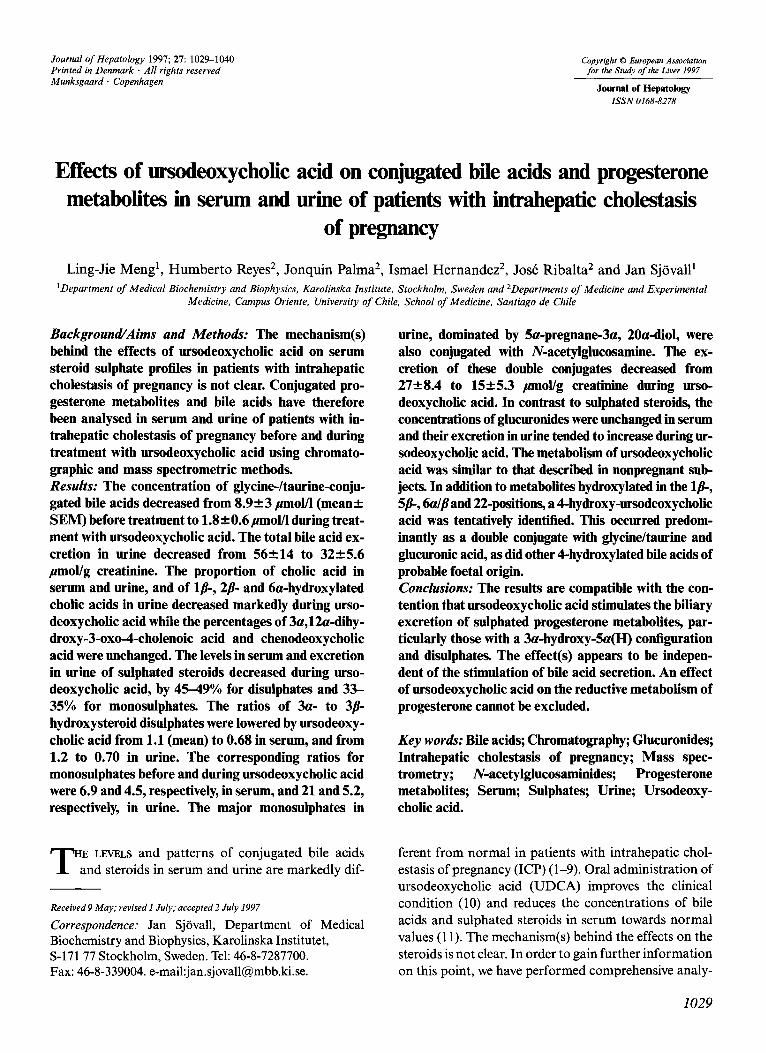
& E4 Tetrols 3-0x0-4 CA CDCA DCA LCA
Fig. 1. Effects of UDCA administration on major urinary bile acids in the five patients (nos. l-5) with ICP. The re- sults are expressed as mean 2 SEM; the hatched bars repre- sent data before treatment, the filled bars data during treat- ment. Tetrols: the sum of tetrahydroxy bile acids; 3-0x0-4: 3a,12a-dihydroxy-3-oxo-4-cholenoic acid; CA: cholic; CDCA: chenodeoxycholic; DCA: deoxycholic; LCA: litho- cholic acids.
Results Bile acids
The levels of conjugated bile acids in serum decreased in patients nos. 2-4 during treatment with UDCA. Pa- tients nos. 1 and 5 had normal concentrations of endo- genous bile acids in serum: 2.1 and 2.9 pmol/l, respec- tively, before, and 4.1 and 2 pmol, respectively, during the administration of UDCA. UDCA was present in the serum of all patients during the treatment, at con- centrations of 0.14.8 pmolll. The mean ratios of cholic:chenodeoxycholic:deoxycholic acids in patients nos. 2-5 were 2.6:1:0.2 before treatment, as compared to 1: 1:0.9 in normal pregnancy (l), and 4: 1 :O. 1 in more advanced ICP (1). These ratios were 0.7: 1:0.2 in patient no. 1. During administration of UDCA, the mean ra- tios were 0.5:1:0.3 in the five patients.
Five groups of bile acids in urine were analysed, dif- fering by the mode of conjugation: unconjugated (U), aminoacyl amidated (G/T), glucuronidated (GlcA), sulphated and both sulphated and aminoacyl amidated (S,S-G/T), and both glucuronidated and sulphated (S- GlcA). In addition, bile acids doubly conjugated with glucuronic acid and amino acid (GlcA-G/T) were ana- lysed in patients nos. 1 and 2 during treatment with UDCA, when FAB spectra indicated the appearance of substantial amounts of such conjugates.
A total of 45 bile acids was found in the urine of the patients before administration of UDCA. They were identified or partly characterised as described in pre- vious studies (13,15). The mean urinary excretion of total bile acids in each group of conjugates in the five
1031
L. J. Meng et al.
patients is shown in Table 2 (UDCA is not included in these values). As seen in this Table, bile acids in the &S-G/T and G/T groups constituted the largest frac- tion. Their percentage decreased during the adminis- tration of UDCA when the total bile acid excretion decreased (except in patient no.1 whose total bile acid excretion (but not the composition) in urine remained the same before and during UDCA).
The fraction of glucuronidated bile acids, containing mainly 6-hydroxylated bile acids before administration of UDCA, increased in all patients during administra- tion of UDCA. This was due to the appearance of putative metabolites of UDCA in this fraction. Peaks of bile acids conjugated with both glucuronic acid and amino acid also appeared in the FAB spectra of urine
TABLE 3
The effects of treatment with UDCA upon the progesterone metabo- lites in five patients (nos. l-5) with ICP
Mode of conjugation
Before UDCA meanCSEM
SWWl pmol/l %b
Monosulphates 2358 50 Disulphates 2058 43
Glucuronides 2.820.9 6
Total 46e-15
Urine Monosulphatesd Disulphatesd Glucuronidesd Total
,umoF % 39210 13 3521 12
224241 75 298238
During UDCA meaniSEM
Change” %
,umol/l %b 1527 51 -35 (5) 1125 38 -45 (5)
3kO.7 10 +7 29tll
,umoF % 26-t-6 9 -33 (5) 1822 6 -49 (5)
249z33 85 +11 293?34
a Number of patients showing a decrease given in parentheses. b Percentage of all the steroids analysed. c pmolig creatinine. d The predominant part but not all the steroids analysed are pro-
gesterone metabolites.
TABLE 4
Ratios (mean (range)) of sulphated 3a- to 3P-hydroxysteroids in serum and urine of five patients (nos. l-5) with ICP before and dur- ing UDCA
Steroids ICP
Before UDCA
Healthy”
During UDCA
Disulphates In serum 1.1 (0.83-1.7) 0.68 (0.32-1.0) 0.5 (0.3-0.7) In urine 1.2 (0.91-1.7)b 0.70 (0.42-1.3)b 0.9 (1.9-2.1)b
Monosulphates In serum 6.9 (3.5-10) 4.5 (2.3-7.6) 2.3 (2.1-2.6) In urine 21 (8.9-33) 5.2 (3.4-6.3) 2.0 (1.9-2.1)
a The values are from three healthy pregnant women at 36-38 weeks of gestation in a former study (15).
b The sulphates of the following steroids were included in the calcu- lations: SC&‘)-pregnane-3a(, 20a-diols, Sa-pregnane-3a(P), 21- diol-20-one, and 5a-pregnane-3a, 20a, 21-trio].
c All sulphated N-acetylglucosaminides were included in the calcu- lation.
collected during treatment with UDCA. This group of conjugates was therefore analysed by GC/MS of samples from two patients (nos. 1 and 2) during treat- ment. It constituted a mean of 15% of the total bile acids in these patients (UDCA not included).
The relative composition of bile acids in urine changed towards normal during treatment with UDCA (Fig. 1). The proportions of cholic acid and particularly of its lp, 2/3, 5/?- (tentative identification) and 6a-hydroxylated products were markedly reduced in all five patients during administration of UDCA. Eight additional bile acids (not including conjugates with N-acetylglucosamine) appeared, which were as- sumed to be metabolites of UDCA (see below). The percentage of 7a, 12a-dihydroxy-3-oxo4-cholenoic acid remained essentially unchanged, as did those of the common bile acids in the sulphated fraction, chenodeoxycholic, deoxycholic and lithocholic acids. However, the absolute excretion of these acids de- creased, contributing to the marked reduction of uri- nary bile acid secretion seen in Table 2. The excretion of lithocholic acid in urine decreased from 4.621.7 pmollg creatinine (mean?SEM) before UDCA to 2.850.8 during UDCA. The three 3-hydroxyandros- tane- 17@-carboxylic acid isomers previously identified in urine of patients with ICP (15) were only found in trace amounts in the present patients; probably these patients were in an earlier stage of ICI?
Progesterone metabolites
Three groups of progesterone metabolites in serum and urine were analysed, differing with respect to their mode of conjugation, i.e., monosulphates, disulphates and glucuronides. Eleven progesterone metabolites in serum and 44 steroids in urine, not all being metabolites of pro- gesterone, were analysed (15). Sulphated steroids pre- dominated in serum and glucuronides in urine, both be- fore and during administration of UDCA. The effects of UDCA are shown in Tables 3 and 4 and Fig. 2. In gen- eral, the levels and patterns of sulphated steroids changed towards normal in patients nos. l-5. The con- centrations in serum and excretion in urine of total mono- and disulphates decreased to similar extents, the decrease of disulphates being slightly larger, 45-49(X than that of monosulphates, 33-35%. In contrast, the levels of glucuronides in serum were not affected and their excretion in urine tended to increase (Table 3).
Confirming the results of a previous study (1 l), the pattern of sulphated steroids in plasma changed in a characteristic way during administration of UDCA. Thus, the levels of mono- and disulphated Sal/?-preg- nane-3cz. 20a-diols and 5a-pregnane-3a, 20a, 21 -trio1 decreased markedly, while Sa-pregnane-3/3, 20a-diol
1032
Steroids and bile acids in ICP during UDCA
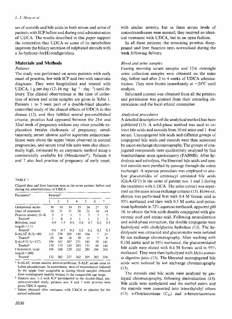
UDCA - + - + - + - + - + Patient no. 1 2 3 4 5
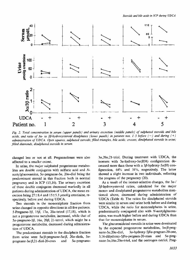
Fig. 2. Total concentration in serum (upper panels) and urinary excretion (middle panels) of sulphated steroids and bile acids, and ratio of 3a- to 3j6hydroxysteroid disulphates (lower panels) in patients nos. l-5 before (-) and during (+) administration of UDCA. Open squares, sulphated steroids;$lled triangles, bile acids; crosses, disulphated steroids in urine; filled diamonds, disulphated steroids in serum.
changed less or not at all. Pregnanolones were also affected to a smaller extent.
In urine, the major sulphated progesterone metabo- lites are double conjugates with sulfuric acid and N- acetylglucosamine, 5a-pregnane-3a, 20a-diol being the predominant steroid in this fraction both in normal pregnancy and in ICP (15,16). The urinary excretion of these double conjugates decreased markedly in all patients during administration of UDCA, the mean ex- cretion being 27 + 8.4 and 15 + 5.3 pmollg creatinine, re- spectively, before and during UDCA.
Two steroids in the monosulphate fraction from urine changed in opposite directions in all five patients. 5-Pregnene-3P, 15/3, 17a, 20a-tetrol (17,18), which is not a progesterone metabolite, increased, while that of Sa-pregnane-3P, 16a, 208, 21-tetrol, which might be a progesterone metabolite, decreased during administra- tion of UDCA.
The predominant steroids in the disulphate fraction from urine were 5a//?-pregnane-3a/p, 20a-diols, 5a- pregnane-3al/?,2 1 -dial-20-ones and Sa-pregnane-
3a,20a,2 1-triol. During treatment with UDCA, the isomers with 3a-hydroxy-Sa/P(H) configuration de-
creased more than those with a 3/3-hydroxy-Sa(H) con- figuration, 64% and 31%, respectively. The latter showed a slight increase in two individuals, reflecting the progress of the pregnancy (20).
As a result of the isomer-selective changes, the 3a-/ 3/?-hydroxysteroid ratios, calculated for the major mono- and disulphated progesterone metabolites men- tioned above, decreased during administration of UDCA (Table 4). The ratios for disulphated steroids were similar in serum and urine both before and during UDCA, while the ratio for monosulphates in urine, predominantly conjugated also with N-acetylglucosa- mine, was much higher before and during UDCA than that for monosulphates in serum.
The glucuronidated steroids in urine were dominated by the expected progesterone metabolites, Sal/?-preg- nane-3a,20a-diol, 3a-hydroxy-5/I/a-pregnan-20-one, 3,16-dihydroxy-5P/a-pregnan-20-ones and Sa-preg- nane-3a, 16a,20a-triol, and the oestrogen estriol. Preg-
1033
L. J. Meng et al.
nanolone and pregnanediol isomers with a 3p, 5a(H) configuration and 5a-pregnane-3a,20a,2l-triol were not present in glucuronidated form. The patterns of pro- gesterone metabolites were similar before and during ad- ministration of UDCA, and ratios between the predomi- nant 5/?(H) steroids and 5a(H) steroids did not change.
Estriol constituted 13 +- 2.8% (mean? SEM) and 15&0.8%, respectively, of the total glucuronidated ster- oids analysed before and during treatment with UDCA. Its excretion was 3Ok5.4 pmollg creatinine be- fore treatment and 3727.1 during treatment. While this difference is not statistically significant, a slight increase in the estriol excretion during treatment was observed in all patients. This may reflect the progress of the pregnancy or a restoration of the enterohepatic circulation of estriol conjugates (21-24).
parallel in all patients with ICP (Fig. 2). Furthermore, the excretion of total bile acids or steroid sulphates in urine was not always a reflection of the levels of the compounds in serum (Fig. 2). Patients nos. 1 and 5 were particularly interesting. Their serum bile acids were low, as in normal pregnancy, while the urinary ex- cretion was increased. The cholestasis (Table 1) ap- peared to be best reflected in the elevated levels of sul- phated steroids and in the increased ratio of 3a- to 3p- hydroxysteroids.
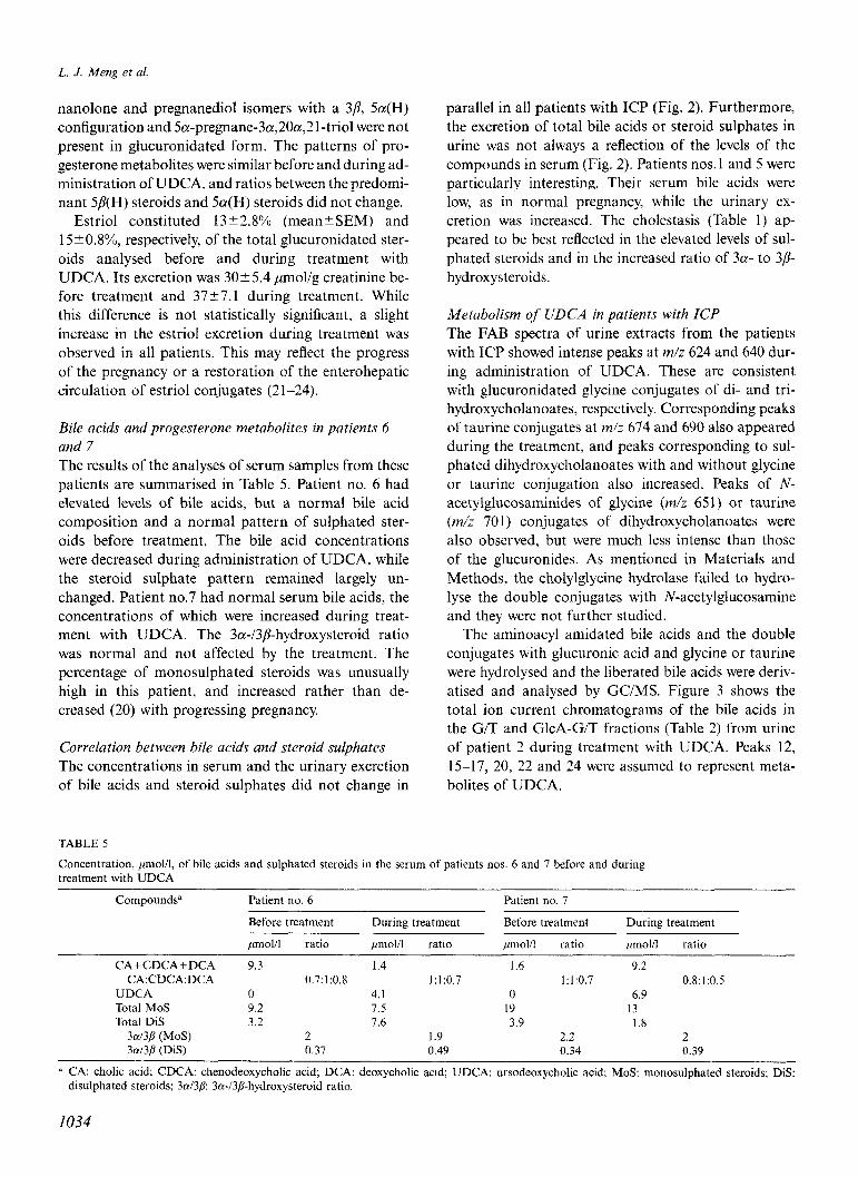
Bile acids and progesterone metabolites in patients 6 and 7 The results of the analyses of serum samples from these patients are summarised in Table 5. Patient no. 6 had elevated levels of bile acids, but a normal bile acid composition and a normal pattern of sulphated ster- oids before treatment, The bile acid concentrations were decreased during administration of UDCA, while the steroid sulphate pattern remained largely un- changed. Patient no.7 had normal serum bile acids, the concentrations of which were increased during treat- ment with UDCA. The 3&3P-hydroxysteroid ratio was normal and not affected by the treatment. The percentage of monosulphated steroids was unusually high in this patient, and increased rather than de- creased (20) with progressing pregnancy.
Metabolism of UDCA inpatients with ICP The FAB spectra of urine extracts from the patients with ICP showed intense peaks at m/z 624 and 640 dur- ing administration of UDCA. These are consistent with glucuronidated glycine conjugates of di- and tri- hydroxycholanoates, respectively. Corresponding peaks of taurine conjugates at m/z 674 and 690 also appeared during the treatment, and peaks corresponding to sul- phated dihydroxycholanoates with and without glycine or taurine conjugation also increased. Peaks of N- acetylglucosaminides of glycine (m/z 651) or t&urine (m/z 701) conjugates of dihydroxycholanoates were also observed, but were much less intense than those of the glucuronides. As mentioned in Materials and Methods, the cholylglycine hydrolase failed to hydro- lyse the double conjugates with N-acetylglucosamine and they were not further studied.
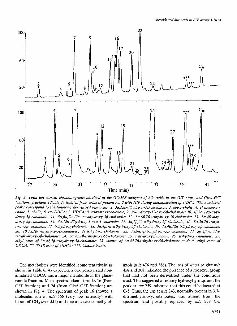
The aminoacyl amidated bile acids and the double conjugates with glucuronic acid and glycine or taurine were hydrolysed and the liberated bile acids were deriv- atised and analysed by GCIMS. Figure 3 shows the total ion current chromatograms of the bile acids in the G/T and GlcA-G/T fractions (Table 2) from urine
Correlation between bile acids and steroid sulphates of patient 2 during treatment with The concentrations in serum and the urinary excretion 15-17, 20, 22 and 24 were assumed of bile acids and steroid sulphates did not change in bolites of UDCA.
TABLE 5
Concentration, ymol/l, of bile acids and sulphated steroids in the serum of patients nos. 6 and 7 before and during treatment with UDCA
UDCA. Peaks 12, to represent meta-
Compounds”
CA+CDCA+DCA CA:CDCA:DCA
UDCA Total MoS Total DiS
3a/3/I (MoS) 3a134 (DiS)
Patient no. 6 Patient no. 7
Before treatment During treatment Before treatment During treatment
,umol/l ratio pmol/l ratio ~mol/l ratio ,umol/i ratio
9.3 1.4 1.6 9.2 0.7: 1:0.8 1:1:0.7 1:1:0.7 0.8:1:0.5
0 4.1 0 6.9 9.2 7.5 19 13 3.2 7.6 3.9 1.8
2 1.9 2.2 2 0.37 0.49 0.34 0.39
a CA: cholic acid; CDCA: chenodeoxycholic acid; DCA: deoxycholic acid; UDCA: ursodeoxycholic acid; MoS: monosulphated steroids; DiS: disulphated steroids; 3a/3/?: 3a-/3P-hydroxysteroid ratio.
1034
IOC h
60
2 20.
6
lo(
6C
20
7 9 16
Steroids and bile acids in ICP during UDCA
C36
24 t c36
27 29 31 33 35 31 39 41
Time (min)
Fig. 3. Total ion current chromatograms obtained in the GC/MS analyses of bile acids in the G/T (top) and GlcA-G/T (bottom) fractions (Table 2) isolatedfrom urine of patient no. 2 with ICP during administration of UDCA. The numbered peaks correspond to the following derivatised bile acids: 2. 3a,I2P-dihydroxy-_5/?-cholanoic; 3. deoxycholic; 4. chenodeoxy- cholic; 5. cholic; 6. iso-UDCA; 7. UDCA; 8. trihydroxycholanoic; 9. 3a-hydroxy-12-oxo-5/3-cholanoic; IO. 1/3,3a,IZa-trihy- droxy-5p-cholanoic; II. 3a,6a, 7a,12a-tetrahydroxy-5/3-cholanoic; 12. 3a,6P,78-trihydroxy-5P-cholanoic; 13. 3a,4p-dihy- droxy-.5/3-cholanoic; 14: 3a,l2a-dihydroxy-3-oxo-4-cholenoic; 15. 3a,7/3,22-trihydroxy-5P-cholanoic; 16. 3a,sp,7P-trihyd- roxy-.5@holanoic; 17. trihydroxycholanoic; 18. 3a,4/$7a-trihydroxy-5/3-cholanoic; 19. 3a,4/3,12a-trihydroxy-5/3-cholanoic; 20. 1/3,3a,7P-trihydroxy-5P-cholanoic; 21. trihydroxycholanoic; 22. 3a,6a,7/3-trihydroxy-5B-cholanoic; 23. 3a,4/3,7a,12a- tetrahydroxy-5P-cholanoic; 24. 3a,4<, 7/?-trihydroxy-5<-cholanoic; 25. trihydroxycholanoic; 26. trihydroxycholanoic; 27. ethyl ester of 3a,4<,7P-trihydroxy-5/3-cholanoic; 28. isomer of 3a,4&7/3-trihydroxy-S@holanoic acid; *. ethyl ester of UDCA; **. TMS ester of UDCA; ***. Contaminants.
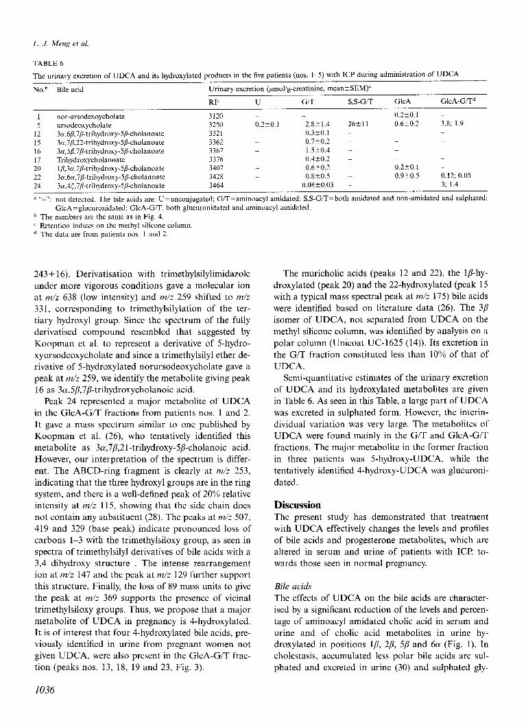
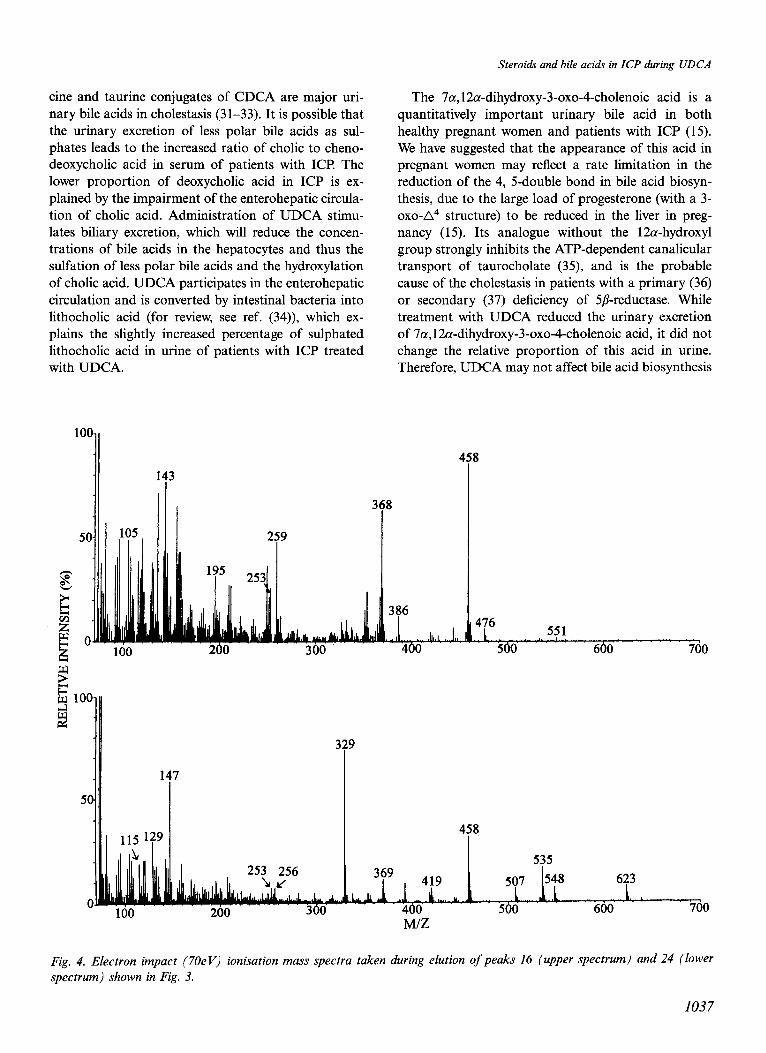
The metabolites were identified, some tentatively, as anols (m/z 476 and 386). The loss of water to give m/z
shown in Table 6. As expected, a 6a-hydroxylated non- 458 and 368 indicated the presence of a hydroxyl group amidated UDCA was a major metabolite in the glucu- that had not been derivatised under the conditions ronide fraction. Mass spectra taken at peaks 16 (from used. This suggested a tertiary hydroxyl group, and the G/T fraction) and 24 (from GlcA-G/T fraction) are peak at m/z 259 indicated that this could be located at shown in Fig. 4. The spectrum of peak 16 showed a C-5. Thus, the ion at m/z 243, normally present in 3,7- molecular ion at m/z 566 (very low intensity) with ditrimethylsiloxycholanoates, was absent from the losses of CHs (m/z 551) and one and two trimethylsil- spectrum and possibly replaced by m/z 259 (i.e.
1035
L. J. Meng et al
TABLE 6
The urinary excretion of UDCA and its hydroxylated products in the five patients (nos. l-5) with ICP during administration of UDCA
No.~ Bile acid Urinary excretion &mol/g-creatinine, mean+SEM)”
RI’ U G/T S,S-G/T GlcA GlcA-GITd
1 nor-ursodexoycholate 3120 ._ ._ 0.2?0.1
5 ursodeoxycholate 3250 0.220.1 2.8kl.4 26+11 0.6t0.2 3.8; 1.9
12 3a,6~,7~-trihydroxy-5~-cholanoate 3321 _ 0.3-cO.l -
15 3n,7/?,22-trihydroxy-S/3-cholanoate 3362 0.720.2 -
16 3a,5P,‘i/%trihydroxy-SD-cholanoate 3367 _ 1.520.4 - _ _
17 Trihydroxycholanoate 3376 0.4t0.2 - _ _ _
20 lp,3n,7p-trihydroxy-5/?-cholanoate 3407 _ 0.6-+0.2 - 0.2t0.1 _
22 3cc,6a,7&trihydroxy-5p-cholanoate 3428 _ 0.8-+0.5 - 0.920.5 0.12; 0.05
24 3a,4t,7P-trihydroxy-5/kholanoate 3464 _ 0.08~0.03 3; 1.4
a “-“: not detected. The bile acids are: U=unconjugated; G/T=aminoacyl amidated; S,S-G/T=both amidated and non-amidated and sulphated; GlcA=glucuronidated: GlcA-G/T: both glucuronidated and aminoacyl amidated.
b The numbers are the same as in Fig. 4. c Retention indices on the methyl silicone column. d The data are from patients nos. 1 and 2.
243+ 16). Derivatisation with trimethylsilylimidazole under more vigorous conditions gave a molecular ion at m/z 638 (low intensity) and m/z 259 shifted to m/z 331, corresponding to trimethylsilylation of the ter- tiary hydroxyl group. Since the spectrum of the fully derivatised compound resembled that suggested by Koopman et al. to represent a derivative of 5-hydro- xyursodeoxycholate and since a trimethylsilyl ether de- rivative of 5-hydroxylated norursodeoxycholate gave a peak at m/z 259, we identify the metabolite giving peak 16 as 3a,5/?,7P-trihydroxycholanoic acid.
Peak 24 represented a major metabolite of UDCA in the GlcA-G/T fractions from patients nos. 1 and 2. It gave a mass spectrum similar to one published by Koopman et al. (26) who tentatively identified this metabolite as 3a,7/?,21-trihydroxy-5/?-cholanoic acid. However, our interpretation of the spectrum is differ- ent. The ABCD-ring fragment is clearly at m/z 253, indicating that the three hydroxyl groups are in the ring system, and there is a well-defined peak of 20”/0 relative intensity at m/z 115, showing that the side chain does not contain any substituent (28). The peaks at m/z 507, 419 and 329 (base peak) indicate pronounced loss of carbons l-3 with the trimethylsiloxy group, as seen in spectra of trimethylsilyl derivatives of bile acids with a 3,4dihydroxy structure . The intense rearrangement ion at m/z 147 and the peak at m/z 129 further support this structure. Finally, the loss of 89 mass units to give the peak at m/z 369 supports the presence of vicinal trimethylsiloxy groups. Thus, we propose that a major metabolite of UDCA in pregnancy is 4-hydroxylated. It is of interest that four 4-hydroxylated bile acids, pre- viously identified in urine from pregnant women not given UDCA, were also present in the GlcA-G/T frac- tion (peaks nos. 13, 18, 19 and 23, Fig. 3).
The muricholic acids (peaks 12 and 22), the Ip-hy- droxylated (peak 20) and the 22-hydroxylated (peak 15 with a typical mass spectral peak at m/z 175) bile acids were identified based on literature data (26). The 3p isomer of UDCA, not separated from UDCA on the methyl silicone column, was identified by analysis on a polar column (Unicoat UC-1625 (14)). Its excretion in the G/T fraction constituted less than 10% of that of UDCA.
Semi-quantitative estimates of the urinary excretion of UDCA and its hydroxylated metabolites are given in Table 6. As seen in this Table, a large part of UDCA was excreted in sulphated form. However, the interin- dividual variation was very large. The metabolites of UDCA were found mainly in the G/T and GlcA-G/T fractions. The major metabolite in the former fraction in three patients was 5-hydroxy-UDCA, while the tentatively identified 4-hydroxy-UDCA was glucuroni- dated.
Discussion The present study has demonstrated that treatment with UDCA effectively changes the levels and profiles of bile acids and progesterone metabolites, which are altered in serum and urine of patients with ICP to- wards those seen in normal pregnancy.
Bile acids
The effects of UDCA on the bile acids are character- ised by a significant reduction of the levels and percen- tage of aminoacyl amidated cholic acid in serum and urine and of cholic acid metabolites in urine hy- droxylated in positions lp, 2/?, 5p and 6a (Fig. 1). In cholestasis, accumulated less polar bile acids are sul- phated and excreted in urine (30) and sulphated gly-
1036
tine and taurine conjugates of CDCA are major uri- nary bile acids in cholestasis (31-33). It is possible that the urinary excretion of less polar bile acids as sul- phates leads to the increased ratio of cholic to cheno- deoxycholic acid in serum of patients with ICP The lower proportion of deoxycholic acid in ICP is ex- plained by the impairment of the enterohepatic circula- tion of cholic acid. Administration of UDCA stimu- lates biliary excretion, which will reduce the concen- trations of bile acids in the hepatocytes and thus the sulfation of less polar bile acids and the hydroxylation of cholic acid. UDCA participates in the enterohepatic circulation and is converted by intestinal bacteria into lithocholic acid (for review, see ref. (34)), which ex- plains the slightly increased percentage of sulphated lithocholic acid in urine of patients with ICP treated with UDCA.
Steroids and bile acids in ICP during UDCA
The 7a, 12a-dihydroxy-3-oxo4-cholenoic acid is a quantitatively important urinary bile acid in both healthy pregnant women and patients with ICP (15). We have suggested that the appearance of this acid in pregnant women may reflect a rate limitation in the reduction of the 4, 5-double bond in bile acid biosyn- thesis, due to the large load of progesterone (with a 3- oxo-A4 structure) to be reduced in the liver in preg- nancy (15). Its analogue without the 12a-hydroxyl group strongly inhibits the ATP-dependent canalicular transport of taurocholate (35), and is the probable cause of the cholestasis in patients with a primary (36) or secondary (37) deficiency of 5/Sreductase. While treatment with UDCA reduced the urinary excretion of 7a, 12a-dihydroxy-3-oxo4-cholenoic acid, it did not change the relative proportion of this acid in urine. Therefore, UDCA may not affect bile acid biosynthesis
386
3
660 700
458
507 623
500 600 760 M/Z
Fig. 4. Electron impact (70eV) ionisation mass spectra taken during elution of peaks 16 (upper spectrum) and 24 (lower spectrum) shown in Fig. 3.
1037
L. J. Meng et al.
at the step of S/?-reduction. The relief of cholestasis by UDCA has been suggested to be due to stimulation of vesicular exocytosis resulting in mobilisation of an increased number of transport proteins to the canalicu- lar membrane (3840). If this is the case in ICP the effect is transient, since cholestasis reappeared when the administration of UDCA was discontinued (11). Although the formation of 701, 12a-dihydroxy-3-oxo-4- cholenoic acid is not correlated to the appearance of ICI’, it may be related to the mild physiological chol- estasis in pregnancy (see a review in (41)) and the possibility exists that it may be an aetiological factor in some women due to genetic polymorphism of cana- licular transport proteins.
Double conjugates of bile acids with glucuronic acid and glycine or taurine were analysed for the first time in pregnant women. It is of interest that a number of 4-hydroxylated bile acids were present in this conju- gated form (Fig. 3). Such bile acids have been found in foetal bile (29,42), amniotic fluid (43) and in infant urine (44). Recently, they were found in urine of preg- nant women and suggested to be of foetal origin (15) based on the above reports (29,4244) and our failure to find any of them in the urine of healthy women dur- ing the menstrual cycle (unpublished results). If 4-hy- droxylation is a unique feature of bile acid metabolism in early human life (29,42), analysis of urinary bile acids doubly conjugated with glucuronic acid and gly- tine or taurine may provide information about the condition of the foetus.
Steroids
The most striking change in progesterone metabolism in ICP is the increased concentration of sulphated metabolites, particularly those with a 3cx-sulphoxy- 5a(H) configuration, in maternal serum and urine (Table 3, (refs. 6,8,9,15)) as well as in cord plasma (45). Administration of UDCA induced a change of these steroids towards normal (Tables 3 and 4). In contrast, glucuronidated metabolites were not significantly affected, either by ICP (11,15) or by administration of UDCA (Table 3). Sulfated progesterone metabolites are excreted in bile (8,46) and are hydrolysed by intesti- nal bacteria and excreted as unconjugated steroids in faeces (7,23,47). It has been suggested that part of the unconjugated steroids is also reabsorbed and conju- gated with glucuronic acid in the intestinal mucosa for final elimination in urine (48). This is supported by the findings that administration of antibiotics leads to a large increase of sulphated progesterone metabolites in faeces (7,23) and a decrease of glucuronidated metabo- lites in urine (49). Thus, part of the glucuronidated 5a- reduced metabolites in urine of healthy pregnant
women may be formed from sulphated biliary metabo- lites. This pathway would be inhibited in cholestasis and stimulated by UDCA to produce the slight in- crease of glucuronides in urine during administration of UDCA (Table 3).
An important question for the understanding of the pathogenesis of ICP is whether the abnormal profile of sulphated progesterone metabolites characterised by an increased ratio of 3a- to 3/$hydroxylated steroids is primary or secondary to the cholestasis. Previous studies with deuterium-labelled steroid sulphates have clearly shown that the SLY- and 3P-hydroxysteroid sul- phates are not interconverted (50,51). Therefore, the increased 3&3ghydroxysteroid ratio is not due to the effect of ICP on an enterohepatic circulation (8). Consequently, it is either due to a relative increase in the formation of 3a-hydroxy isomers from progester- one, or is secondary to an impaired biliary excretion of these isomers (8).
The levels of total sulphated 3/I-hydroxysteroid iso- mers in serum are higher than those of 3a isomers in late pregnancy (20) in spite of a lower production rate of 38 isomers (50,51). This may be due to the longer half-life of 5a-pregnane-3p, 20a-diol disulphate (50,51) and a slower biliary elimination of 3a isomers. Thus, the proportion of 3/?-hydroxysteroid sulphates in bile (46) is lower than that in serum of healthy pregnant women. It is conceivable that the reduced bile flow in ICP could lead to an accumulation in serum and an increased urinary excretion of predominantly 3a- hydroxysteroid sulphates, i.e. the abnormal pattern in ICP would be secondary to cholestasis. However, this is not in agreement with the data for patients 6 and 7. They had elevated serum bile acids before (patient no. 6) or during (patient no. 7) treatment, reflecting a mild cholestasis, while the ratios of 3a- to 3P-hydroxysteroid sulphates were normal and not influenced by UDCA. Therefore, the alternative explanation that a change in the reductive metabolism of progesterone is a primary event in ICP cannot be excluded. This is also sup- ported by the observation of abnormal steroid sul- phate patterns in two patients prior to the development of ICP (6) and by the report that the abnormal steroid sulphate pattern characteristic of ICP is not seen in patients with cholestasis due to viral hepatitis (9). Further studies are needed to resolve the question whether the changes in steroid sulphates in ICP are primary or secondary, and whether UDCA can induce a recruitment of transport proteins to the canalicular membrane (40) for excretion of steroid sulphates.
Patients nos. 6 and 7 were initially thought to have ICP but were excluded from the double-blind study because of normal values for serum alanine amino-
1038
transferase. Our analyses showed that the ratios of sul- phated 3a- to 3/z-hydroxysteroids were normal. The pruritus in these patients is therefore unlikely to be due to ICP, and the results support the contention that the ratio of 3a- to 3P-hydroxysteroid sulphates in serum is a selective parameter for the diagnosis of ICP, es- pecially in the early stage of the disease (6).
Metabolism of UDCA The metabolism of UDCA has been extensively studied in health and disease. Conjugation with N- acetylglucosamine is a selective pathway for 7p-hy- droxylated bile acids (14,52) and was also observed by FAB mass spectrometry of urine samples from the pa- tients with ICI? UDCA has been found to be hy- droxylated at carbon atoms 1, 5, 6, 12, 21, 22 and 23 in humans (26,53-56). We found metabolites hy- droxylated in these positions, except for C-12, C-21 and C-23, which may have been minor components. Hydroxylation at C-5 of UDCA in humans has been reported in two studies (26,54), and 5P-hydroxylation of nor-UDCA and nor-CDCA occurs in rodents (27). Our data strongly support the 5-hydroxylation, which gave the predominant hydroxylated metabolite in the urine from three of the patients.
We have tentatively identified 4-hydroxy-UDCA as a double conjugate with glucuronic acid and glycine or taurine. As mentioned above, other 4-hydroxylated bile acids are also conjugated in this way. Since 4-hydroxyl- ation has been suggested to be a unique feature of bile acid metabolism in early human life (29,42), it will be of interest to investigate whether the tentative 4-hy- droxy-UDCA is a metabolite of UDCA in the foetus.
Acknowledgements This work was supported by grants from the Swedish Medical Research Council (no. 03x-219), Karolinska Institutet and Fondecyt (Chile) (no. 1940420).
References 1.
2.
3.
4.
5.
Sjiivall K, Sjovall J. Serum bile acid levels in pregnancy with pruritus. Clin Chim Acta 1966; 13: 207-l 1. Heikkinen J, Maentausta 0, Yliistalo P Janne 0. Serum bile acid levels in intrahepatic cholestasis of pregnancy during treatment with phenobarbital or cholestyramine. Eur J Ob- stet Gyn R B 1982; 14: 153-62. Bacq Y, Myara A, Brechot M-C, Hamon C, Studer E, Trivin F, et al. Serum conjugated bile acid profile during intrahepat- ic cholestasis of pregnancy. J Hepatol 1995; 22: 6670. Thomassen PA. Urinary bile acids during development of recurrent cholestasis of pregnancy. Eur J Clin Invest 1979; 9: 417-23. Thomassen PA. Urinary bile acids in late pregnancy and in” recurrent cholestasis of pregnancy. Eur J Clin Invest 1979; 9: 425-32.
Steroids and bile acids in ICP during UDCA
6. Sjijvall J, Sjbvall K. Steroid sulphates in plasma from preg- nant women with pruritus and elevated plasma bile acid levels. Ann Clin Res 1970; 2: 321-37.
7. Eriksson H, Gustafsson J-A, Sjiivall K. Excretion of neutral steroids in urine and faeces of women with intrahepatic chol- estasis of pregnancy. Steroids Lipids Res 1972; 3: 30-48.
8. Laatikainen T, Karjalainen 0, Janne 0. Excretion of progester- one metabolites in urine and bile of pregnant women with in- trahepatic cholestasis. J Steroid Biochem 1973; 4: 641-8.
9. Giusti G, Piccinino E Ricciardi I, Delrio G, Sagnelli E, Man- zillo G. Abnormal steroid sulphates in plasma of women with intrahepatic cholestasis of pregnancy. Acta Hepato-Gastro- enter01 1979; 26: 203-6.
10. Palma J, Reyes H, Ribalta J, Iglesias J, Gonzales MC, Her- nandez I, et al. Effects of ursodeoxycholic acid in patients with intrahepatic cholestasis of pregnancy. Hepatology 1992; 15: 1043-7.
11
12
Meng LJ, &yes H, Palma I, Hernandez I, Ribalta J, Sjovall J. Profiles of steroids and bile acids in plasma of patients with intrahepatic cholestasis of pregnancy - effect of ursodeoxy- cholic acid therapy. In: van Berge Henegouwen GP, van Hoek B, De Groote J, Matern S, Stochbrtigger RW, editors. Choles- tatic Liver Diseases. Dordrecht: Kluwer Academic Pub- lishers: 1994. p. 45-9. Palma J, Reyes H, Ribalta J, Hernandez I, Sandoval S, Almu- na R, et al. Ursodeoxycholic acid therapy in cholestasis of pregnancy. In: Reyes HB, Leuschner U, Arias IM, editors. Pregnancy, Sex Hormones and the Liver. Dordrecht/Boston/ London: Kluwer Academic Publishers; 1995. p. 223-7.
13. Meng LJ, Sjiivall J. Method for combined analysis of profiles of conjugated progesterone metabolites and bile acids in serum and urine of pregnant women. J Chromatogr B Biom- ed Appl 1997; 688: 11-26.
14. Marschall HU, Matern H, Wietholtz H, Egestad B, Matern S, Sjijvall J. Bile acid N-Acetylglucosaminidation - in vivo
and in vitro evidence for a selective conjugation reaction of 7P-hydroxylated bile acids in humans. J Clin Invest 1992; 89: 1981-7.
15. Meng LJ, Reyes H, Palma I, Hernandez I, Ribalta J, Sjiivall J. Profiles of bile acids and progesterone metabolites in the urine and serum of women with intrahepatic cholestasis of pregnancy. J Hepatol 1997; in press.
16. Meng LJ, Griffiths WJ, Sjiivall J. The identification of novel steroid N-acetylglucosaminides in the urine of pregnant women. J Steroid Biochem Mol Biol 1996; 58: 585-98.
17. Janne OA, Vihko RK. Neutral steroids in urine during preg- nancy. Identification of sulphate-conjugated 3p, 1 Sa-dihy- droxy-5a-pregnan-20-one, 3/?, 1 Sa-dihydroxy-5P-pregnan-20- one and 5-pregnene-3P, 15/?, 17a,20a-tetrol. Eur J Biochem 1970; 17: 13440.
18. Kraan GP Wolthers BG, van der Molen JC, Nagel GT, Dray- er NM, Joannou GE. New identified 15/?-hydroxylated 21- deoxy-pregnanes in congenital adrenal hyperplasia due to 21- hydroxylase deficiency. J Steroid Biochem Mol Biol 1993; 45: 421-34.
20
21
19. Eriksson H. Gustafsson J-A. Excretion of steroid hormones in adults: steroids in urine from a pregnant woman. Eur J Biochem 1970; 16: 268-77. Sjiivall K. Gas chromatographic determination of steroid sulphates in plasma during pregnancy. Ann Clin Res 1970; 2: 393408. I)‘
Tikkanen MJ, Pulkkinen MO, Adlercreutz H. Effect of ampi- cillin treatment on the urinary excretion of estriol conjugates in pregnancy. J Steroid Biochem 1973; 4: 43940.
1039
L. J. Meng et al
22. Adlercreutz H, Martin F, Tikkanen MJ, Pulkkinen M. Effect of ampicillin administration on the excretion of twelve oes- trogens in pregnancy urine. Acta Endocr Copenh 1975; 80: 551-7.
23. Martin F, Peltonen J, Laatikainen T, Pulkkinen M, Ad- lercreutz H. Excretion of progesterone metabolites and estriol in faeces from pregnant women during ampicillin administra- tion. J Steroid Biochem 1975; 6: 133946.
24. Adlercreutz H, Martin F, Lehtinen T, Tikkanen MJ, Pulkkin- en MO. Effect of ampicillin administration on plasma conju- gated and unconjugated estrogen and progesterone levels in pregnancy. Am J Obstet Gynecol 1977; 128: 266-71.
25. Marschall HU, Griffiths WJ, Gotze U, Zhang J, Wietholtz H, Busch N, et al. The major metabolites of ursodeoxycholic acid in human urine are conjugated with N-acetylglucosa- mine. Hepatology 1994; 20: 845-53.
26. Koopman BJ, Wolthers BG, van der Molen JG, Nagel GT, Kruizinga W. Abnormal urinary bile acids in a patient suffer- ing from cerebrotendinous xanthomatosis during oral admin- istration of ursodeoxycholic acid. Biochim Biophys Acta 1987; 917: 238846.
27. Schteingart CD, Hagey LR, Setchell KDR, Hofmann AE SD- Hydroxylation by the liver: identification of 3,5,7-trihydroxy nor-bile acids as new major biotransformation products of 3,7-dihydroxy nor-bile acids in rodents. J Biol Chem 1993; 268: 1123946.
28. Sjiivall J, Lawson AM, Setchell KDR. Mass spectrometry of bile acids. Meth Enzymol 1985; 111: 63-l 13.
29. Dumaswala R, Setchell KDR, Zimmer-Nechemias L, Iida T, Goto J, Nambara T. Identification of 3a,4/$7ol_trihydroxy- 5p-cholanoic acid in human bile: reflection of a new pathway in bile acid metabolism in humans. J Lipid Res 1989: 30: 8477 56.
30. Hofmann AI? Bile acids. In: Arias IM, Boyer JL, Faust0 N, Jakoby WB, Schachter DA, Shafritz DA, editors. The Liver: Biology and Pathobiology, 3rd ed. New York: Raven Press; 1994. p. 6777718.
31. van Berge Henegouwen GP Brandt K-H, Eyssen H, Par- mentier G. Sulfated and unsulfated bile acids in serum, bile and urine of patients with cholestasis. Gut 1976; 17: 861-9.
32. Stiehl A. Disturbances of bile acid metabolism in cholestasis. Clin Gastroenterol 1977; 6: 45-67.
33. Back P Urinary bile acids. In: Setchell KDR, Kritchevsky D, Nair PP editors. The Bile Acids: Chemistry, Physiology, and Metabolism, Vol 4. New York and London: Plenum Press; 1988. p. 404-40.
34. Hofmann AF. Pharmacology of ursodeoxycholic acid, an en- terohepatic drug. Stand J Gastroenterol 1994; 29 (Suppl): l- 15.
35. Stieger B, Zhang J, O’Neill B, Sjiivall J, Meier PJ. Differential interaction of bile acids from patients with inborn errors of bile acid synthesis with hepatocellular bile acid transporters. Eur J Biochem 1997; 244: 3944.
36. Setchell KDR, Suchy FJ, Welsh MB, Zimmer-Nechemias L, Heubi J, Balistreri WE n4-3-Oxosteroid SD-reductase de- ficiency described in identical twins with neonatal hepatitis. J Clin Invest 1988; 82: 2148-57.
37. Clayton PT, Pate1 E, Lawson AM, Carruthers RA, Tanner MS, Strandvik B, et al. 3-0x0-A4 bile acids in liver disease. Lancet 1988; i: 12834.
38. Beuers U, Throckmorton DC, Anderson MS, Isales CM, Thasler W, Kullak-Ublick GA, et al. Tauroursodeoxycholic acid activates protein kinase C in isolated rat hepatocytes. Gastroenterology 1996; 110: 1553-63.
39. Beuers U, Nathanson MH, Isales CM, Boyer JL. Taurourso- deoxycholic acid stimulates hepatocellular exocytosis and mobilizes extracellular Caf+ mechanisms defective in chol- estasis. J Clin Invest 1993; 92: 298493.
40. Boyer JL, Soroka CJ. Vesicle targeting to the apical domain regulates bile excretory function in isolated rat hepatocyte couplets. Gastroenterology 1995; 109: 1600-l 1.
41. Kreek MJ. Female sex steroids and cholestasis. Semin Liver Dis 1987; 7: 8-23.
42. Setchell KD, Dumaswala R, Colombo C, Ronchi M. Hepatic bile acid metabolism during early development revealed from the analysis of human fetal gallbladder bile. J Biol Chem 1988; 263: 1663744.
43. Nakagawa M, Setchell KDR. Bile acid metabolism in early life: studies of amniotic fluid. J Lipid Res 1990; 31: 1089-98.
44. Strandvik B, Wahlen E, Wikstrom SA. The urinary bile acid excretion in healthy premature and full-term infants during the neonatal period. Stand J Clin Lab Invest 1994; 54: l-10.
45. Laatikainen TJ, Peltonen JI, Nylander PL. Effect of maternal intrahepatic cholestasis on fetal steroid metabolism. J Clin Invest 1974; 53: 1709-15.
46. Laatikainen T, Karjalainen 0. Excretion of conjugates of neutral steroids in human bile during late pregnancy. Acta Endocr Copenh 1972; 60: 775-88.
47. Eriksson H, Gustafsson J-A, Sjbvall J. Excretion of steroid hormones in adults - Cis and C2, steroids in faeces from pregnant women. Eur J Biochem 1969; 12: 520-6.
48. Adlercreutz H, Martin E Biliary excretion and intestinal me- tabolism of progesterone and estrogens in man. J Steroid Bio- them 1980; 13: 23144.
49. Martin F, Peltonen J, Laatikainen T. Tikkanen M, Pulkkinen M. Excretion of unconjugated and conjugated progesterone metabolites in pregnancy urine during ampicillin administra- tion Clin Chim Acta 1974; 55: 71-80.
50. Anderson RA, Baillie TA. Axelson M, Cronholm T, Sjovall K, Sjiivall J. Stable isotope studies on steroid metabolism and kinetics: sulphates of 3cc-hydroxy-5a-pregnane derivatives in human pregnancy. Steroids 1990; 55: 443-57.
51. Baillie TA, Curstedt T, Sjovall K, Sjiivall J. Production rates and metabolism of sulphates of 3/%hydroxy-Sa-pregnane de- rivatives in pregnant women. J Steroid Biochem 1980; 13: 1473-88.
52. Marschall HU. Griffiths WJ, Zhang J, Wietholtz H. Matern H, Matern S, et al. Positions of conjugation of bile acids with glucose and N-acetylglucosamine in vitro. J Lipid Res 1994; 35: 1599-610.
53. Stiehl A, Rudolph G, Raedsch R, Moller B, Hopf U, Lotterer E, et al. Ursodeoxycholic acid-induced changes of plasma and urinary bile acids in patients with primary biliary cir- rhosis. Hepatology 1990; 12: 492-7.
54. Fischer S, Neubrand M, Paumgartner G. Biotransformation of orally administered ursodeoxycholic acid in man as ob- served in gallbladder bile, serum and urine. Eur J Clin Invest 1993; 23: 28-36.
55. Batta AK, Arora R, Salen G, Tint GS, Eskreis D, Katz S. Characterization of serum and urinary bile acids in patients with primary biliary cirrhosis by gas-liquid chromatogra- phymass spectrometry: effect of ursodeoxycholic acid treat- ment. J Lipid Res 1989; 30: 1953-62.
56. Batta AK, Salen G, Arora R, Shefer S, Tint GS, Abroon J, et al. Effect of ursodeoxycholic acid on bile acid metabolism in primary biliary cirrhosis. Hepatology 1989; 10: 414-9.
1040