effects of dietary l-arginine or n-carbamylglutamate
TRANSCRIPT
HAL Id: hal-01004190https://hal.archives-ouvertes.fr/hal-01004190
Submitted on 29 May 2020
HAL is a multi-disciplinary open accessarchive for the deposit and dissemination of sci-entific research documents, whether they are pub-lished or not. The documents may come fromteaching and research institutions in France orabroad, or from public or private research centers.
L’archive ouverte pluridisciplinaire HAL, estdestinée au dépôt et à la diffusion de documentsscientifiques de niveau recherche, publiés ou non,émanant des établissements d’enseignement et derecherche français ou étrangers, des laboratoirespublics ou privés.
Distributed under a Creative Commons Attribution| 4.0 International License
Effects of dietary L-arginine or N-carbamylglutamatesupplementation during late gestation of sows on the
miR-15b/16, miR-221/222, VEGFA and eNOSexpression in umbilical vein
X. D. Liu, X. Wu, Y. L. Yin, Y. Q. Liu, M. M. Geng, H. S. Yang, FrancoisBlachier, G. Y. Wu
To cite this version:X. D. Liu, X. Wu, Y. L. Yin, Y. Q. Liu, M. M. Geng, et al.. Effects of dietary L-arginine or N-carbamylglutamate supplementation during late gestation of sows on the miR-15b/16, miR-221/222,VEGFA and eNOS expression in umbilical vein. Amino Acids, Springer Verlag, 2012, 42 (6), pp.2111- 2119. �10.1007/s00726-011-0948-5�. �hal-01004190�
ORIGINAL ARTICLE
Effects of dietary L-arginine or N-carbamylglutamatesupplementation during late gestation of sows on the miR-15b/16,miR-221/222, VEGFA and eNOS expression in umbilical vein
X. D. Liu • X. Wu • Y. L. Yin • Y. Q. Liu •
M. M. Geng • H. S. Yang • Francois Blachier •
G. Y. Wu
Received: 3 April 2011 / Accepted: 4 May 2011 / Published online: 3 June 2011
� The Author(s) 2011. This article is published with open access at Springerlink.com
Abstract Placental vascular formation and blood flow are
crucial for fetal survival, growth and development, and
arginine regulates vascular development and function. This
study determined the effects of dietary arginine or N-car-
bamylglutamate (NCG) supplementation during late
gestation of sows on the microRNAs, vascular endothelial
growth factor A (VEGFA) and endothelial nitric oxide
synthase (eNOS) expression in umbilical vein. Twenty-
seven landrace 9 large white sows at day (d) 90 of ges-
tation were assigned randomly to three groups and fed the
following diets: a control diet and the control diet supple-
mented with 1.0% L-arginine or 0.10% NCG. Umbilical
vein of fetuses with body weight around 2.0 kg (oversized),
1.5 kg (normal) and 0.6 kg (intrauterine growth restriction,
IUGR) were obtained immediately after farrowing for
miR-15b, miR-16, miR-221, miR-222, VEGFA and eNOS
real-time PCR analysis. Compared with the control
diets, dietary Arg or NCG supplementation enhanced the
reproductive performance of sows, significantly increased
(P \ 0.05) plasma arginine and decreased plasma VEGF
and eNOS (P \ 0.05). The miR-15b expression in the
umbilical vein was higher (P \ 0.05) in the NCG-supple-
mented group than in the control group. There was a trend
in that the miR-222 expression in the umbilical vein of the
oversized fetuses was higher (0.05 \ P \ 0.1) than in the
normal and IUGR fetuses. The expression of eNOS in both
Arg-supplemented and NCG-supplemented group were
lower (P \ 0.05) than in the control group. The expression
of VEGFA was higher (P \ 0.05) in the NCG-supple-
mented group than in the Arg-supplemented and the con-
trol group. Meanwhile, the expression of VEGFA of the
oversized fetuses was higher (P \ 0.05) than the normal
and IUGR fetuses. In conclusion, this study demonstrated
that dietary Arg or NCG supplementation may affect
microRNAs (miR-15b, miR-222) targeting VEGFA and
eNOS gene expressions in umbilical vein, so as to regulate
the function and volume of the umbilical vein, provide
more nutrients and oxygen from the maternal to the fetus
tissue for fetal development and survival, and enhance the
reproductive performance of sows.
Keywords L-Arginine � N-Carbamylglutamate �Umbilical vein � VEGFA � ENOS � MicroRNAs
Abbreviations
Arg L-Arginine
eNOS Endothelial nitric oxide synthase
iNOS Inducible nitric oxide synthase
IUGR Intrauterine growth restriction
NAG N-Acetylglutamate
NCG N-Carbamylglutamate
NO Nitric oxide
RT-PCR Real-time polymerase chain reaction
VEGFA Vascular endothelial growth factor A
X. D. Liu � X. Wu (&) � Y. L. Yin (&) �Y. Q. Liu � M. M. Geng � H. S. Yang � G. Y. Wu
Research Center for Healthy Breeding of Livestock and Poultry,
Hunan Engineering and Research Center of Animal and Poultry
Science and Key Laboratory for Agro-ecological Processes
in Subtropical Region, Institute of Subtropical Agriculture,
Chinese Academy of Sciences, Hunan 410125, China
e-mail: [email protected]
Y. L. Yin
e-mail: [email protected]
F. Blachier
INRA, CNRH-IdF, Paris, France
G. Y. Wu
Department of Animal Science, Texas A&M University,
College Station, TX 77843, USA
123
Amino Acids (2012) 42:2111–2119
DOI 10.1007/s00726-011-0948-5
Introduction
High embryonic loss and fetal deaths during gestation
limited the number of piglets born at farrowing in sows
(Pope 1994; Mateo et al. 2007). Higher fetal growth rates
may require an increased provision of nutrients for sup-
porting the metabolic needs of both the sow and her fetuses
(Wu et al. 2004; Kim et al. 2005). Adequate vasculogenesis
and angiogenesis of the maternal vasculature is important
for providing adequate maternal oxygen/nutrients and
blood flow to the placenta. Placental angiogenesis supports
the required blood flow on the fetal side necessary for fetal
growth and development. Therefore, vasculogenesis and
angiogenesis are essential for proper placental develop-
ment (Wu et al. 2004; Demir et al. 2007; Arroyo and Winn
2008). Umbilical venous blood flow is crucial for fetal
growth and development (Barbera et al. 1999; Ferrazzi
et al. 2000; Boiti et al. 2002).
The vascular endothelial growth factor (VEGF) proteins
are mostly known to regulate the processes of vasculogen-
esis and angiogenesis (Hanahan 1997; Otrock et al. 2007;
Demir et al. 2007; Arroyo and Winn 2008; Yao et al. 2011).
As a potent endothelial survival factor, VEGF induces
vasodilation and facilitates blood flow by increasing nitric
oxide (NO) production (Hood et al. 1998; Otrock et al.
2007). MicroRNAs (miRNAs), about 22-nucleotide, non-
coding RNAs, have been shown to be involved in various
biological processes in animals (Ambros 2004; Kloos-
terman and Plasterk 2006), including angiogenesis regula-
tion (Kuehbacher et al. 2008; Anand et al. 2010). Recently,
it is reported that miR-15b, miR-16, miR-221 and miR-222
target VEGFA (Hua et al. 2006; Karaa et al. 2009) and
eNOS (Poliseno et al. 2006; Suarez et al. 2007) expressions
in angiogenesis.
Arginine (Arg) can enhance the reproductive performance
of pigs (Mateo et al. 2007; Wu et al. 2007) and also regulate
angiogenesis (Raghavan and Dikshit 2004). In addition,
N-carbamylglutamate (NCG) increases the endogenous syn-
thesis of Arg (Frank et al. 2007; Wu et al. 2010).
Therefore, we hypothesized that dietary supplementation
with Arg or NCG may enhance the reproductive performance
of sows and the potential mechanisms are that microNRAs
(miR-15b, miR-16, miR-221 and miR-222) target VEGFA
and eNOS gene expression in fetal umbilical vein so as to
regulate the function and volume of the umbilical vein,
thereby providing more nutrients and oxygen from the
maternal to the fetus tissue for fetal development and survival.
Materials and methods
This study was performed in accordance with the Chinese
guidelines for animal welfare and approved by the Animal
Care and Use Committee of the Institute of Subtropical
Agriculture, the Chinese Academy of Sciences (Yin et al.
1993).
Animals and experimental design
A total of 27 Large White 9 Landrace crossbred sows at d
90 of gestation with initial body weight (BW) of
187 ± 5 kg, parity of 3.2 ± 0.7 and similar reproductive
performance at last parity were chosen and assigned to
three groups randomly: a control group (fed a corn- and
soybean meal-based diet) and two treatment groups (fed a
corn- and soybean meal-based diet supplemented with
1.0% L-Arg�HCl (Arg) or 0.1% NCG) (Table 1). L-Arg�HCl
was obtained from Ajinomoto Inc. (Tokyo, Japan) and
NCG was provided by the Institute of Subtropical Agri-
culture, the Chinese Academy of Sciences.
The dose of Arg was based on previous study (Mateo et al.
2007), and the dose of NCG was based on our own study.
The sows were housed individually in gestation crates
(2.0 9 0.6 m, concrete floor) and transferred to individual
farrowing crates (2.2 9 1.5 m) at d 107 of gestation. The
sows were provided 2 kg diet (on an as-fed basis) daily as
two equal-sized meals (08:00 and 16:30 h) during the
entire gestation period. All the diets provided 13.5 MJ
metabolizable energy/kg and 14.7 crude protein (on as-fed
basis). All the sows had free access to drinking water.
Sample collection
Blood samples were collected 2 h after feeding via jugular
venepuncture into heparinized tubes on d 110 of gestation.
Samples were centrifuged at 2,0009g, 15 min at 4�C
(Mateo et al. 2007; Yin et al. 2010). Plasma was transferred
to 1.5 microcentrifuge tubes and stored at -20�C until
analysis (Geng et al. 2011). The total number of piglets and
their BW at birth were recorded. The piglets were classified
as born alive or dead as previously described (Mateo et al.
2007). The number of mummified fetuses (early or middle
gestation deaths) was neglected.
The umbilical veins of piglets with BW of about 2.0 kg
(oversized), 1.5 kg (normal) and 0.6 kg (IUGR) were
obtained immediately after farrowing. Samples were of
length about 5 cm, 10 cm from the body, and washed with
4�C PBS (RNA free). Then the samples were collected into
1.5 microcentrifuge tubes (RNA free) with RNAlater
(Applied Biosystems, Valencia, CA, USA) in it and stored
at -20�C for RT-PCR analyses (Wu et al. 2010).
Chemical analyses
Plasma samples were assayed for biochemical indices
using Beckman Coulter CX4 Pro. (Beckman, USA),
2112 X. D. Liu et al.
123
standards obtained from Beckman (Beckman, USA) (Tang
et al. 2005). Plasma concentrations of free amino acids
were analyzed by Amino Acid Analyzer, Hitachi L8800
(Hitachi, Japan), and amino acid standards were obtained
from Sigma Chemical (Kong et al. 2009). Plasma con-
centrations of VEGF and eNOS were analyzed using
enzyme-linked immunosorbent assay (ELISA) from R & D
system (Minneapolis, MN, USA) and an ELISA plate
reader (BioTek, USA) (Deng et al. 2010). Concentrations
of hormones were analyzed by radioimmunoassay (Jiuding,
China).
Real-time PCR analyses
RT-PCR for VEGFA and eNOS in fetal umbilical vein
Total RNA was isolated by Trizol (Invitrogen, USA, Karaa
et al. 2009) and treated with DNase. Reverse transcription
was performed using AMV Reverse Transcriptase Kit
(Promega, USA). mRNA levels for VEGFA and eNOS
were determined by a standard real-time polymerase chain
reaction (RT-PCR) method. RT-PCR was performed with
the total RNA using TaKaRa one-step RNA PCR Kit
(TaKaRa Bio Inc, Japan). The primer pairs for VEGFA,
eNOS and GAPDH are presented in Table 2. GAPDH was
used as the housekeeping gene, whose mRNA levels in the
fetal umbilical vein did not differ among the groups. The
RT-PCR conditions were: 10 min pre-denaturation at
95�C, and then 15 s denaturation at 94�C, and 30 s
annealing at 60�C for 40 cycles. The relative quantification
of gene amplification by RT-PCR was performed using
cycle threshold (CT) values. The comparative CT value
method was employed to quantitate expression levels for
VEGFA and eNOS relative to those for GAPDH. The final
PCR product was visualized in a 2% agarose gel.
RT-PCR for miR-15b, miR-16, miR-221 and miR-222
Expression of mature miRNAs was measured using mi-
Script PCR System (Qiagen, Hilden, Germany) (Chen et al.
Table 1 Composition of
gestation diets, on an as-fed
basis (%)
a The premix provided the
following per kg of diet: Fe
(FeSO4�H2O) 80 mg, Mn
(MnSO4�5H2O) 45 mg, Zn
(ZnO) 100 mg, Cu
(CuSO4�5H2O) 20 mg, I (KI)
0.70 mg, Se (Na2SeO3�H2O)
0.25 mg, vitamin A 10,000 IU,
vitamin D3 2,500 IU, vitamin E
100 IU, vitamin K 10 IU,
vitamin B2 10 mg, vitamin B6
1 mg, vitamin B12 50 lg, biotin
80 lg, folic acid 5 mg, nicotinic
acid 15 mg, choline chloride
1,500 mg
Item Treatment
Control Arg NCG
Ingredient
Corn 70.30 70.30 70.30
Soybean meal 12.00 12.00 12.00
Wheat middling 4.00 3.00 3.90
Rice bran meal 3.00 3.00 3.00
Puffed soybean 3.00 3.00 3.00
Salt 0.35 0.35 0.35
Potassium chloride 0.75 0.75 0.75
Vitamin–mineral premixa 3.00 3.00 3.00
Dicalcium phosphate 2.20 2.20 2.20
Limestone 0.50 0.50 0.50
Soy oil 0.50 0.50 0.50
Mold-inhibitor 0.09 0.09 0.09
L-Lys�HCl 0.20 0.20 0.20
DL-Met 0.06 0.06 0.06
L-Thr 0.05 0.05 0.05
L-Arginine�HCl – 1.00 –
NCG – – 0.10
Nutrient composition
DM (%) 88.2 88.2 88.2
ME (MJ/kg) 13.5 13.5 13.5
CP (%) 14.7 14.7 14.7
Lys (%) 0.78 0.78 0.78
Met ? Cys (%) 0.69 0.69 0.69
Ca (%) 0.92 0.92 0.92
Total P (%) 0.67 0.67 0.67
Available P (%) 0.45 0.45 0.45
Effects of dietary L-arginine or NCG supplementation during late gestation 2113
123

2005). The miScript PCR System comprises the following
components: miScript Reverse Transcription Kit, miScript
SYBR Green PCR Kit, miScript Primer Assay.
Total RNA was extracted as described above. After
cDNA synthesis, the cDNA serves as the template for real-
time PCR analysis using a miScript Primer Assay in
combination with the miScript SYBR Green PCR Kit.
Mature miRNAs are amplified using the miScript Universal
Primer together with the miRNA-specific primer (the
miScript Primer Assay). Primers for sus scrofa miR-15b,
miR-16, miR-221 and miR-222 (miRBase, http://www.
mirbase.org) were designed by Qiagen. 5S rRNA (forward
primer: 50-gcccgatctcgtctgatct-30, reverse primer: 50-agcct
acagcacccggtatt-30) was used as the referent for miRNAs
expression for its constant expression level across all
samples and suitable size. The amplification protocol was
as follows: 95�C for 5 min, 50 cycles of denaturation
at 94�C/15 s, annealing temperature of 55�C/30 s, and
extension at 70�C/30 s. Real-time analysis of PCR ampli-
fication was performed on an Applied Biosystems 7900HT
Sequence Detection System and analyzed with an SDS 2.3
Software (Applied Biosystems).
The final PCR product was visualized in a 2% agarose
gel. All the procedures above followed the instructions of
each manufacturer.
Statistical analyses
The relative quantification of gene amplification by
RT-PCR was performed using cycle threshold (CT) values.
The comparative CT value method was employed to
quantitate expression levels for VEGFA and eNOS relative
to those for GAPDH. The DDCT method is used for relative
quantification when working with the miScript PCR
System. This comparative method relies on comparing the
differences in CT values obtained with normal versus
experimental samples. The threshold cycle (CT) obtained
with the miScript PCR Control (5S rRNA) is used to
normalize the data.
Values are presented as the mean ± SEM. Data of gene
and miRNAs expression were analyzed using the GLM and
the others using the one-way ANOVA (SAS 9.1.3, SAS
Inc., USA). In case of a P value \ 0.05, the result was
regarded as statistically significant, while 0.05 B P \ 0.1
was considered as a trend.
Results
Gestation performance
The reproductive performance of sows fed diets supple-
mented with Arg or NCG can be seen in Table 3. The total
number of piglets born, birth weight of all piglets born or
born alive, and litter birth weight of all piglets born did not
differ between the three groups of sows. However, there
was a trend (0.05 \ P \ 0.1) toward an increase in the
number of piglets born alive for sows fed the Arg or NCG-
supplemented diet compared with sows fed the control diet.
The litter birth weight of all piglets born alive were
15% higher (P \ 0.05) for Arg-supplemented sows and
14% (P \ 0.05) higher for NCG-supplemented sows, both
Table 2 Primers for VEGFA,
eNOS and GAPDH
F forward, R reverse
Target gene Primer sequences Products Annealing
temperature (�C)
VEGFA F: CAACGACGAAGGTCTGGAGTG 155 60
R: GCCTCGCTCTATCTTTCTTTGG
eNOS F: ACGCCCGTCTTCCACCA 193 60
R: ACGCCTTCACTCGCTTCG
GAPDH F: GTGCTGAGTATGTCGTGGAGTC 196 60
R: CAGTTGGTGGTACAGGAGGC
Table 3 Reproductive
performance of sows
In the same row, values with
different letter mean significant
difference (P \ 0.05)
Items Control Arg NCG SEM
Total piglets born per litter (n) 11.22 11.38 11.33 0.26
Total piglets born alive per litter (n) 9.78 10.87 10.77 0.24
Birth weight of all piglets born (kg) 1.44 1.50 1.49 0.02
Birth weight of all piglets born alive (kg) 1.45 1.51 1.49 0.02
Litter birth weight of all piglets born (kg) 16.09 16.87 16.83 0.25
Litter birth weight of all piglets born alive (kg) 14.12b 16.26a 16.04a 0.29
Piglets born dead per litter (n) 1.44a 0.50b 0.56b 0.14
2114 X. D. Liu et al.
123
compared with the control group. The number of piglets
born dead were 65% lower (P \ 0.05) for the Arg-sup-
plemented sows and 61% lower (P \ 0.05) for the NCG-
supplemented sows, both compared with the control group.
The days from weaning to estrus of sows did not differ
between the three groups (data not shown).
Plasma biochemical assays
Concentrations of glucose, ammonia, albumin, total pro-
tein, Ca2?, Cu2? and Mg2? in plasma did not differ
between the three groups (Table 4). Concentrations of
phosphorus and Zn2? were both higher (P \ 0.05) in Arg
or NCG -supplemented sows than in the control group of
sows (Table 4). There was a trend (0.05 \ P \ 0.1) toward
the decrease in the concentrations of urea nitrogen for Arg-
supplemented sows compared with the control group of
sows.
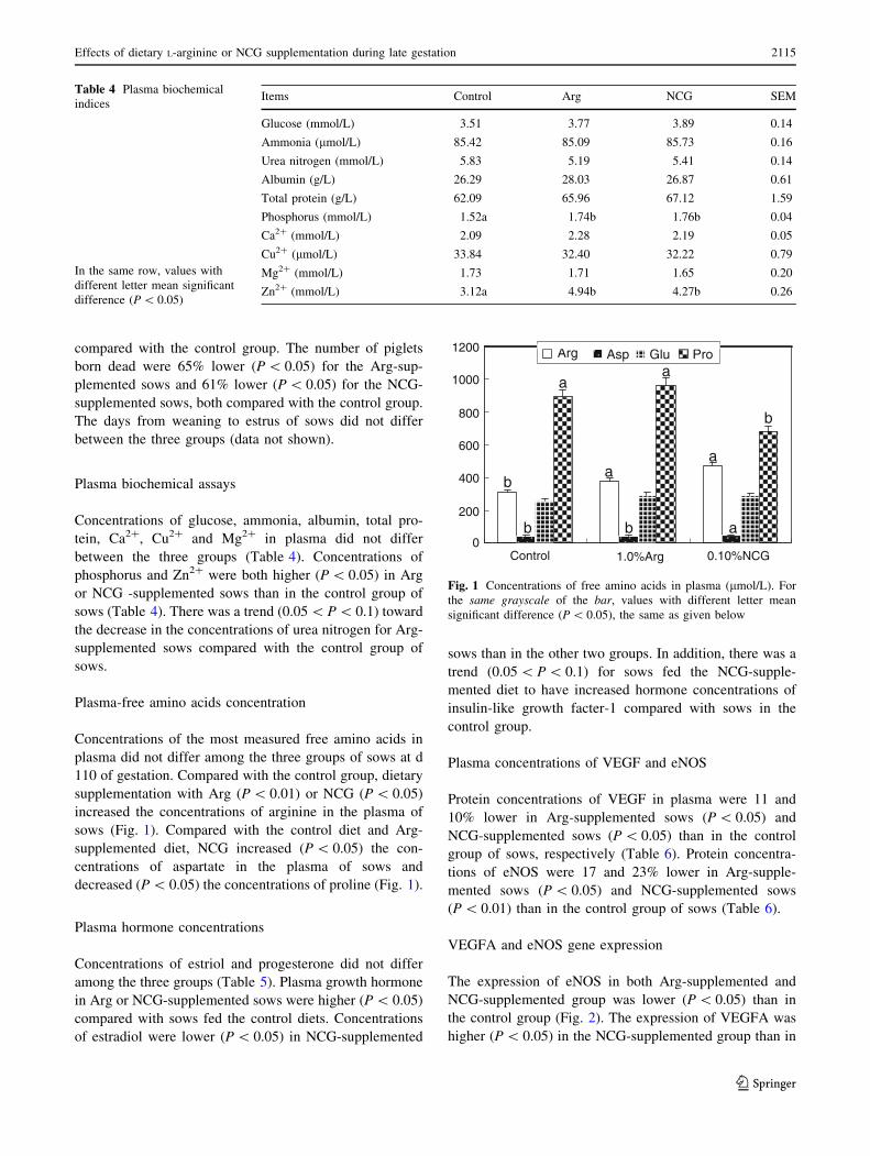
Plasma-free amino acids concentration
Concentrations of the most measured free amino acids in
plasma did not differ among the three groups of sows at d
110 of gestation. Compared with the control group, dietary
supplementation with Arg (P \ 0.01) or NCG (P \ 0.05)
increased the concentrations of arginine in the plasma of
sows (Fig. 1). Compared with the control diet and Arg-
supplemented diet, NCG increased (P \ 0.05) the con-
centrations of aspartate in the plasma of sows and
decreased (P \ 0.05) the concentrations of proline (Fig. 1).
Plasma hormone concentrations
Concentrations of estriol and progesterone did not differ
among the three groups (Table 5). Plasma growth hormone
in Arg or NCG-supplemented sows were higher (P \ 0.05)
compared with sows fed the control diets. Concentrations
of estradiol were lower (P \ 0.05) in NCG-supplemented
sows than in the other two groups. In addition, there was a
trend (0.05 \ P \ 0.1) for sows fed the NCG-supple-
mented diet to have increased hormone concentrations of
insulin-like growth facter-1 compared with sows in the
control group.
Plasma concentrations of VEGF and eNOS
Protein concentrations of VEGF in plasma were 11 and
10% lower in Arg-supplemented sows (P \ 0.05) and
NCG-supplemented sows (P \ 0.05) than in the control
group of sows, respectively (Table 6). Protein concentra-
tions of eNOS were 17 and 23% lower in Arg-supple-
mented sows (P \ 0.05) and NCG-supplemented sows
(P \ 0.01) than in the control group of sows (Table 6).
VEGFA and eNOS gene expression
The expression of eNOS in both Arg-supplemented and
NCG-supplemented group was lower (P \ 0.05) than in
the control group (Fig. 2). The expression of VEGFA was
higher (P \ 0.05) in the NCG-supplemented group than in
Table 4 Plasma biochemical
indices
In the same row, values with
different letter mean significant
difference (P \ 0.05)
Items Control Arg NCG SEM
Glucose (mmol/L) 3.51 3.77 3.89 0.14
Ammonia (lmol/L) 85.42 85.09 85.73 0.16
Urea nitrogen (mmol/L) 5.83 5.19 5.41 0.14
Albumin (g/L) 26.29 28.03 26.87 0.61
Total protein (g/L) 62.09 65.96 67.12 1.59
Phosphorus (mmol/L) 1.52a 1.74b 1.76b 0.04
Ca2? (mmol/L) 2.09 2.28 2.19 0.05
Cu2? (lmol/L) 33.84 32.40 32.22 0.79
Mg2? (mmol/L) 1.73 1.71 1.65 0.20
Zn2? (mmol/L) 3.12a 4.94b 4.27b 0.26
0
200
400
600
800
1000
1200
Control 1.0%Arg 0.10%NCG
Arg Asp Glu Pro
a
b
aa
b
a
b b a
Fig. 1 Concentrations of free amino acids in plasma (lmol/L). For
the same grayscale of the bar, values with different letter mean
significant difference (P \ 0.05), the same as given below
Effects of dietary L-arginine or NCG supplementation during late gestation 2115
123
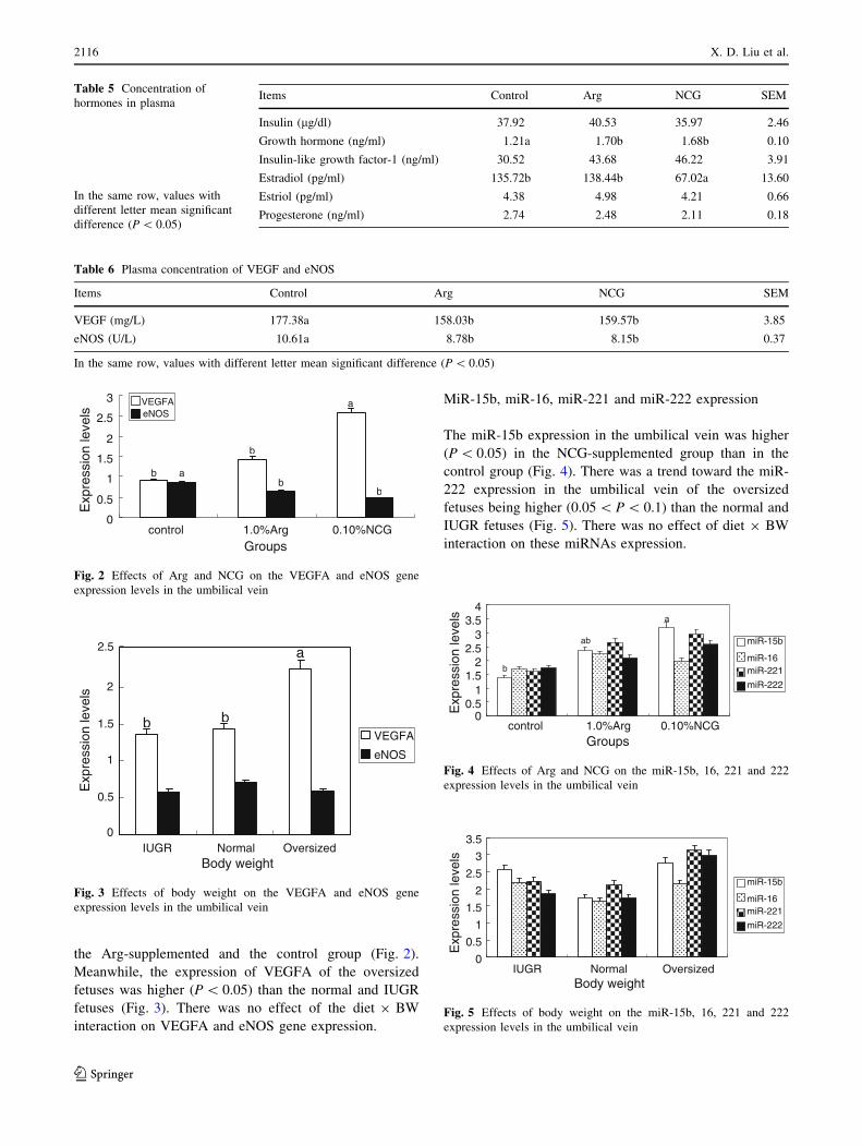
the Arg-supplemented and the control group (Fig. 2).
Meanwhile, the expression of VEGFA of the oversized
fetuses was higher (P \ 0.05) than the normal and IUGR
fetuses (Fig. 3). There was no effect of the diet 9 BW
interaction on VEGFA and eNOS gene expression.
MiR-15b, miR-16, miR-221 and miR-222 expression
The miR-15b expression in the umbilical vein was higher
(P \ 0.05) in the NCG-supplemented group than in the
control group (Fig. 4). There was a trend toward the miR-
222 expression in the umbilical vein of the oversized
fetuses being higher (0.05 \ P \ 0.1) than the normal and
IUGR fetuses (Fig. 5). There was no effect of diet 9 BW
interaction on these miRNAs expression.
Table 5 Concentration of
hormones in plasma
In the same row, values with
different letter mean significant
difference (P \ 0.05)
Items Control Arg NCG SEM
Insulin (lg/dl) 37.92 40.53 35.97 2.46
Growth hormone (ng/ml) 1.21a 1.70b 1.68b 0.10
Insulin-like growth factor-1 (ng/ml) 30.52 43.68 46.22 3.91
Estradiol (pg/ml) 135.72b 138.44b 67.02a 13.60
Estriol (pg/ml) 4.38 4.98 4.21 0.66
Progesterone (ng/ml) 2.74 2.48 2.11 0.18
Table 6 Plasma concentration of VEGF and eNOS
Items Control Arg NCG SEM
VEGF (mg/L) 177.38a 158.03b 159.57b 3.85
eNOS (U/L) 10.61a 8.78b 8.15b 0.37
In the same row, values with different letter mean significant difference (P \ 0.05)
0
0.5
1
1.5
2
2.5
3
control 1.0%Arg 0.10%NCGGroups
Exp
ress
ion
leve
ls VEGFA
eNOS
a
b
a
b
bb
Fig. 2 Effects of Arg and NCG on the VEGFA and eNOS gene
expression levels in the umbilical vein
0
0.5
1
1.5
2
2.5
IUGR Normal OversizedBody weight
Exp
ress
ion
leve
ls
VEGFA
eNOS
a
b b
Fig. 3 Effects of body weight on the VEGFA and eNOS gene
expression levels in the umbilical vein
00.5
11.5
22.5
33.5
4
control 1.0%Arg 0.10%NCGGroups
Exp
ress
ion
leve
ls
a
ab
b
miR-15b
miR-16miR-221
miR-222
Fig. 4 Effects of Arg and NCG on the miR-15b, 16, 221 and 222
expression levels in the umbilical vein
0
0.5
1
1.5
2
2.5
3
3.5
IUGR Normal OversizedBody weight
Exp
ress
ion
leve
ls
miR-15bmiR-16miR-221miR-222
miR-15b
miR-16miR-221
miR-222
Fig. 5 Effects of body weight on the miR-15b, 16, 221 and 222
expression levels in the umbilical vein
2116 X. D. Liu et al.
123
Discussion
Maternal nutrition and oxygen play a key role in regulating
fetal survival, growth and development (Wu et al. 2004).
Malnutrition is known to be a major cause of pregnancy
complications, such as intrauterine growth restriction
(IUGR) or even worse, such as embryonic loss and fetal
deaths during gestation. Thus, providing the pregnant dam
with proper nutrition is vital for the fetus (Snoeck et al.
1990; Hoet and Hanson 1999; McPherson et al. 2004).
Various evidences have substantiated the importance of
arginine in the survival, growth, and development of fetal
pigs (Wu et al. 2004, 2007). Furthermore, amino acid
malnutrition in gestating sows results in lower concentra-
tions of arginine in the placenta and fetal plasma (Wu et al.
1998), as well as reduced the synthesis of NO (the endo-
thelium-derived relaxing factor) from L-arginine (Wu et al.
2009) and the synthesis of polyamines (Pegg 1986).
Impaired placental synthesis of both NO and polyamines is
considered a major factor contributing to IUGR (Wu et al.
2004, 2006). Additionally, previous studies showed that
uterine uptake of arginine may not be sufficient to meet
fetal growth requirements during late gestation in pigs (Wu
et al. 1999). NCG is a safe and metabolically stable analog
of NAG (Wu et al. 2009; Gessler et al. 2010) and increases
the endogenous synthesis of Arg (Frank et al. 2007; Wu
et al. 2009).
The results of this study showed that Arg or NCG
supplementation to gestation diets for late pregnant sows
improved pregnancy outcome, decreased plasma urea
concentrations and increased the plasma concentrations of
free arginine of sows at d 110 of gestation. Mateo et al.
(2007) also reported the similar results. This suggested that
both Arg and NCG supplementation provided better
nutrients to sows, and therefore probably improved the
uterine environment for fetal growth and development.
Additionally, arginine is not only required for protein
synthesis and ammonia detoxification, but is also a pre-
cursor of many metabolically important molecules,
including proline, ornithine, polyamines and NO (Wu and
Morris 1998; Kim et al. 2007).
However, proper nutrition for sows cannot guarantee
good reproductive efficiency. The placenta is responsible
for the exchange of nutrients and oxygen from the mother
to the fetus. Adequate vasculogenesis and angiogenesis of
the maternal vasculature are important for providing ade-
quate maternal nutrients/oxygen and blood flow to the
placenta. Placental vascular formation and function are
important for fetal growth and development. Proper
development of the placenta is critical for a successful
pregnancy, mediates important steps, such as maternal
blood flow to the placenta and delivery of nutrients to the
fetus, and ensures the exchange of nutrients/oxygen and
blood flow necessary for fetal growth (Arroyo and Winn
2008).
Also, umbilical venous blood flow is crucial for fetal
growth and development (Barbera et al. 1999; Ferrazzi
et al. 2000; Boiti et al. 2002). Pathologic umbilical vein
leads to pregnancy complications too (Klaritsch et al. 2008;
Koech et al. 2008). Vascular growth is necessary to
increase placental fetal blood flow over gestation. Poor
vascular development is known to cause intrauterine
embryonic death characterized by low vascular density in
the placental villi along with fibrosis and other deficiencies.
The VEGF proteins are the most studied family of growth
factors known to regulate the processes of vasculogenesis
and angiogenesis. VEGFA (also known as VEGF), aside
from being a potent endothelial survival factor, is also
known to induce vasodilation by increasing nitric oxide
(NO) production, another function which facilitates blood
flow. ENOS is critical in the regulation of vascular function
(Lu et al. 2011) and can generate both nitric oxide (NO)
and superoxide (O2-), which are key mediators of cellular
signaling (Chen et al. 2010).
MicroRNAs (miRNAs), about 22-nucleotide, non-cod-
ing RNAs, have come into focus as a powerful mechanism
to regulate angiogenesis (Dews et al. 2006; Urbich et al.
2008). It has been demonstrated that miR-221 and miR-222
block endothelial cell migration, proliferation and angio-
genesis and indirectly regulate the expression of endothe-
lial nitric oxide synthase (Poliseno et al. 2006). In addition,
miR-221 and miR-222 inhibit cell proliferation and reduce
the expression of c-Kit in hematopoietic progenitor cells—
a process that can contribute to vessel growth (Kuehbacher
2007). Two other miRNAs that might be involved in
angiogenesis are miR-15b and miR-16. MiR-15b and miR-
16 have been shown to control the expression of VEGF
(Hua et al. 2006). Data indicate that hypoxia-induced
reduction of miR-15b and miR-16 contributes to an
increase in VEGF. Some other miRNAs also regulate
angiogenesis and vascular function (Anand et al. 2010).
In this study, the levels of gene expression of VEGFA
and eNOS in umbilical vein and decrease in the plasma
concentrations of VEGFA and eNOS in both the Arg- and
NCG-supplemented groups may be a feedback regulatory
mechanism of arginine-produced NO in the fetal umbilical
vein and placenta compared with the control group. This is
supported by high expression of miR-15b and miR-222 in
the umbilical vein of dietary Arg- or NCG-supplemented
groups.
The arginine treatment may enhance placental angio-
genesis and growth during early-to mid-gestation, thereby
promoting an optimal intrauterine environment throughout
pregnancy (Wu et al. 2004). Therefore, it is possible that
dietary supplementation with arginine increases the syn-
thesis of NO in the placenta and fetus, as reported for adult
Effects of dietary L-arginine or NCG supplementation during late gestation 2117
123
rats (Wu and Morris 1998; Kohli et al. 2004). The outcome
would be to enhance placental angiogenesis and growth
(including vascular growth), utero-placental blood flow, the
transfer of nutrients from mother to fetus, and, therefore,
fetal survival, growth and development (Kwon et al. 2004;
Wu et al. 2004, 2006).
Although the majority of the conceptus loss occurs
during the peri-implantation period, there is evidence that
significant losses also occur during later gestation (Wilson
2002). Piglets born dead per litter were significantly
decreased both in Arg-supplemented and NCG-supple-
mented groups in this study, which is similar to the study
reported previously (Mateo et al. 2007). This may suggest
that dietary Arg or NCG supplementation also regulated
placenta vascular functions.
Notably, we found that plasma concentrations of phos-
phorus and Zn2? were higher both in sows of the Arg-sup-
plemented and NCG-supplemented groups, indicating that
Arg or NCG supplementation increases protein synthesis of
fetus (Castillo-Duran and Weisstaub 2003; Frank et al.
2007). This is in agreement with the findings reported pre-
viously (Mateo et al. 2007). This study showed that dietary
Arg or NCG supplementation increased litter piglets born
alive and litter birth weight of all piglets born alive, while
there were not differences in the average birth weights of all
piglets born or of piglets born alive between groups. Fur-
thermore, plasma concentrations of growth hormone were
higher in sows of Arg-supplemented and NCG-supple-
mented groups than in sows of the control group.
In summary, supplementing dietary Arg or NCG during
late gestation enhanced the reproductive performance of
sows. Also, Arg or NCG treatment improved efficiency in
the utilization of dietary nutrients; we propose that Arg or
NCG treatment may effect the expression of miRNA-15b
and miRNA-222, thereby controlling its target, VEGFA
and eNOS, respectively, gene expression in the umbilical
vein. Thus, Arg may regulate angiogenesis and vascular
development and functions of umbilical vein and placenta,
providing more nutrients and oxygen from mother to
fetuses for fetal survival, growth and development. How-
ever, it is necessary to determine how arginine regulate
fetal survival, growth and development through
microRNAs.
Acknowledgments This research was jointly supported by
grants from the National Basic Research Program of China
(2009CB118806), the Chinese Academy of Sciences and Knowledge
Innovation Project (KZCX2-EW-412, KZCX2-EW-QN411), NSFC
(30901040, 30901041, 30928018, 30828025, 30771558).
Open Access This article is distributed under the terms of the
Creative Commons Attribution Noncommercial License which per-
mits any noncommercial use, distribution, and reproduction in any
medium, provided the original author(s) and source are credited.
References
Ambros V (2004) The functions of animal microRNAs. Nature
431:350–355
Anand S, Majeti BK, Acevedo LM et al (2010) MicroRNA-132–
mediated loss of p120RasGAP activates the endothelium to
facilitate pathological angiogenesis. Nat Med 16(8):909–914
Arroyo JA, Winn VD (2008) Vasculogenesis and angiogenesis in the
IUGR placenta. Semin Perinatol 32:172–177
Barbera A, Galan HL, Ferrazzi E et al (1999) Relationship of
umbilical vein blood flow to growth parameters in the human
fetus. Am J Obstet Gynecol 181:174–179
Boiti S, Struijk PC, Ursem NTC et al (2002) Umbilical venous
volume flow in the normally developing and growth-restricted
human fetus. Ultrasound Obstet Gynecol 19:344–349
Castillo-Duran C, Weisstaub G (2003) Zinc supplementation and
growth of the fetus and low birth weight infant. J Nutr
133:1494S–1497S
Chen C, Ridzon DA, Broomer AJ et al (2005) Real-time quantifica-
tion of microRNAs by stem-loop RT-PCR. Nucleic Acids Res
33(20):e179
Chen CA, Wang TY, Varadharaj S et al (2010) S-glutathionylation
uncouples eNOS and regulates its cellular and vascular function.
Nature 468:1115–1118
Demir R, Seval Y, Huppertz B (2007) Vasculogenesis and angio-
genesis in the early human placenta. Acta Histochem
109:257–265
Deng J, Wu X, Li TJ et al (2010) Dietary amylose and amylopectin
ratio and resistant starch content affects plasma glucose, lactic
acid, hormone levels and protein synthesis in splanchnic tissues.
J Anim Physiol Anim Nutr 94:220–226
Dews M, Homayouni A, Yu DN et al (2006) Augmentation of tumor
angiogenesis by a Myc-activated microRNA cluster. Nat Genet
38:1060–1065
Ferrazzi E, Rigano S, Bozzo M et al (2000) Umbilical vein blood flow in
growth-restricted fetuses. Ultrasound Obstet Gynecol 16:432–438
Frank J, Escobar J, Nguyen HV et al (2007) Oral N-carbamylgluta-
mate supplementation increases protein synthesis in skeletal
muscle of piglets. J Nutr 137:315–319
Geng M, Li T, Kong X et al (2011) Reduced expression of intestinal
N-acetyglutamate synthase in suckling piglets: a novel molecular
mechanism for arginine as a nutritionally essential amino acid
for neonates. Amino Acids 40:1513–1522
Gessler P, Buchal P, Schwenk HU et al (2010) Favourable long-term
outcome after immediate treatment of neonatal hyperammone-
mia due to N-acetylglutamate synthase deficiency. Eur J Pediatr
169:197–199
Hanahan D (1997) Signaling vascular morphogenesis and mainte-
nance. Science 277:48–50
Hoet JJ, Hanson M (1999) Intrauterine nutrition: its importance
during critical periods for cardiovascular and endocrine devel-
opment. J Physiol 514:617–627
Hood JD, Meininger CJ, Ziche M et al (1998) VEGF upregulates
ecNOS message, protein, and NO production in human endo-
thelial cells. Am J Physiol 274:H1054–H1058
Hua Z, Lv Q, Ye WB et al (2006) MiRNA-directed regulation of
VEGF and other angiogenic factors under hypoxia. PLoS ONE
1(1):e116
Karaa ZS, Iacovoni JS, Bastide A et al (2009) The VEGF IRESes are
differentially susceptible to translation inhibition by miR-16.
RNA 15:249–254
Kim SW, Wu G, Baker DH (2005) Amino acid nutrition of breeding
sows during gestation and lactation. Pigs News Inform 26:N89–
N99
2118 X. D. Liu et al.
123
Kim SW, RD Mateo, Y-L Yin et al (2007) Functional amino acids
and fatty acids for enhancing production performance of sows
and piglets. Asian-Aust J Anim Sci 20:295–306
Klaritsch P, Haeusler M, Karpf E et al (2008) Spontaneous
intrauterine umbilical artery thrombosis leading to severe fetal
growth restriction. Placenta 29:374–377
Kloosterman WP, Plasterk R (2006) The diverse functions of
MicroRNAs in animal development and disease. Dev Cell
11:441–450
Koech A, Ndungu B, Gichangi P (2008) Structural changes in
umbilical vessels in pregnancy induced hypertension. Placenta
29:210–214
Kohli R, Meininger CJ, Haynes TE et al (2004) Dietary L-arginine
supplementation enhances endothelial nitric oxide synthesis in
streptozotocin-induced diabetic rats. J Nutr 134:600–608
Kong XF, Wu GY, Yin YL et al (2007) Dietary supplementation with
Chinese herbal ultra-fine powder enhances cellular and humoral
immunity in early weaned piglets. Livest Sci 108:94–98
Kong XF, Yin YL, He QH et al (2009) Dietry supplementation with
Chinese herbal powder enhances ileal digestibilies and serum
concentrations of amino acids in young pigs. Amino Acids
37:573–582
Kuehbacher A, Urbich C, Dimmeler S (2008) Targeting microRNA
expression to regulate angiogenesis. Trends Pharmacol Sci
29(1):12–15
Kwon H, Wu G, Meininger CJ et al (2004) Developmental changes in
nitric oxide synthesis in the ovine placenta. Biol Reprod
70:679–686
Lu Y, Xiong Y, Huo YQ et al (2011) Grb-2-associated binder 1
(Gab1) regulates postnatal ischemic and VEGF-induced angio-
genesis through the protein kinase A-endothelial NOS pathway.
PNAS 108(7):2957–2962
Mateo RD, Wu G, Fuller W et al (2007) Dietary L-Arginine
supplementation enhances the reproductive performance of gilts.
J Nutr 137:652–656
McPherson RL, Ji F, Wu G et al (2004) Growth and compositional
changes of fetal tissues in pigs. J Anim Sci 82:2534–2540
Otrock ZK, Makarem JA, Shamseddine AI (2007) Vascular endo-
thelial growth factor family of ligands and receptors: review.
Blood Cells Mol Dis 38:258–268
Pegg AE (1986) Recent advances in the biochemistry of polyamines
in eukaryotes. Biochem J 234:249–262
Poliseno L, Tuccoli A, Mariani L et al (2006) MicroRNAs modulate
the angiogenic properties of HUVECs. Blood 108:3068–3071
Pope WF (1994) Embryonic mortality in swine. In: Geisert RD (ed).
Embryonic mortality in domestic species. CRC Press Boca
Raton, pp 53–78
Raghavan S, Dikshit M (2004) Vascular regulation by the L-arginine
metabolites, nitric oxide and agmatine. Pharmacol Res
49:397–414
Snoeck A, Remacle C, Reusens B et al (1990) Effect of a low protein
diet during pregnancy on the fetal rat endocrine pancreas. Biol
Neonate 57:107–118
Suarez Y, Fernandez-Hernando C, Pober JS et al (2007) Dicer
dependent microRNAs regulate gene expression and functions in
human endothelial cells. Circ Res 100:1164–1173
Tang ZR, Yin LY, Nyachoti CM et al (2005) Effect of dietary
supplementation of chitosan and galacto-mannan-oligosacchar-
ide on serum parameters and the insulin-like growth factor-I
mRNA expression in early-weaned piglets. Domest Anim
Endocrinol 28:430–441
Urbich C, Kuehbacher A, Dimmeler S (2008) Role of microRNAs in
vascular diseases, inflammation, and angiogenesis. Cardiovasc
Res 79(4):581–588
Wilson ME (2002) Role of placental function in mediating conceptus
growth and survival. J Anim Sci 80(Suppl 2):E195–E201
Wu G, Morris SM (1998) Arginine metabolism: nitric oxide and
beyond. Biochem J 336:1–17
Wu G, Pond WG, Ott T et al (1998) Maternal dietary protein
deficiency decreases amino acid concentrations in fetal plasma
and allantoic fluid of pigs. J Nutr 128(5):894–902
Wu G, Ott TL, Knabe DA et al (1999) Amino acid composition of the
fetal pig. J Nutr 129:1031–1038
Wu G, Bazer FW, Cudd TA et al (2004) Maternal nutrition and fetal
development. J Nutr 134:2169–2172
Wu G, Bazer FW, Wallace JM et al (2006) Intrauterine growth
retardation: implications for the animal sciences. J Anim Sci
84:2316–2337
Wu G, Bazer FW, Davis TA et al (2007) Important roles for the
arginine family of amino acids in swine nutrition and production.
Livest Sci 112:8–22
Wu G, Bazer FW, Davis TA et al (2009) Arginine metabolism and
nutrition in growth, health and disease. Amino Acids 37:153–168
Wu X, Ruan Z, Gao YL et al (2010) Dietary supplementation withL-arginine or N-carbamylglutamate enhances intestinal growth
and heat shock protein-70 expression in weanling pigs fed a
corn- and soybean meal-based diet. Amino Acids 39:831–839
Wu X, Yin YL, Li TJ et al (2010) Dietary protein, energy and
arginine affect LAT1 expression in forebrain white matter
differently. Animal 4:1518–1521
Yao K, Guan S, Li TJ et al (2011) Dietary L-arginine supplementation
enhances intestinal development and expression of vascular
endothelial growth factor in weanling piglets. Bri J Nutr
105:703–709
Yin YL, Zhong HY, Huang RL et al (1993) Nutritive value of
feedstuffs and diets for pigs. I. Chemical composition, apparent
ileal and fecal digestibility. Anim Feed Sci Technol 44:1–27
Yin YL, Yao K, Liu ZJ et al (2010) Supplementing L-leucine to a low-
protein diet increases tissue protein synthesis in weanling pigs.
Amino Acids 39:1477–1486
Effects of dietary L-arginine or NCG supplementation during late gestation 2119
123