effect of seed-borne fusarium species on constituents of
TRANSCRIPT
Jour nal of P lant Protect ion R esearc h ISSN 1427-4345
Effect of seed-borne Fusarium species on constituents of essential oils from seeds of black cumin populations
Nima Khaledi* , Farshid Hassani
Seed and Plant Certification and Registration Institute, Agricultural Research, Education and Extension Organization (AREEO), Karaj, Iran
AbstractThe seed is one of the most important inputs of agricultural products and its quality and health can be affected by seed-borne fungi. Seed-borne fungal pathogens are a major threat to black cumin production and cause considerable yield losses every year worldwide. The aim of this study was to identify seed-borne fungi, the effects of natural fungal infected seeds on some seed quality indicators, and also to investigate cell wall degrading enzymes (CWDEs), pathogenicity and aggressiveness of the isolates obtained from seeds. The con-stituents of essential oils (EOs) from seeds of Iranian and Syrian black cumin popula-tions were identified and their effect on [isolated] seed-borne Fusarium isolates. A total of 17 isolates were identified based on morphological and molecular characteristics of Fusar‑ium oxysporum and F. solani species. The results of the standard germination test showed that there was a significant difference between the studied seed populations in the germi-nation and vigor indices. Our results indicated that most of the identified isolates were in the seed coat, while a few isolates of F. oxysporum were located in embryos. The results of the pathogenicity test showed that about 42% of the isolates were pathogenic and 58% of the isolates were non-pathogenic. Different levels of pathogenicity and aggressiveness were observed for various isolates of Fusarium species. All Fusarium isolates were not capable of producing CWDEs as pathogenicity factors. Analyzing the activity of CWDEs, includ-ing cellulase, pectinase, xylanase and lipase produced by the Fusarium isolates, revealed that activity levels of CWDEs are positive and are correlated with variations in patho-genicity and aggressiveness of seed-borne fungal isolates on seeds. The EOs were identified by gas chromatography-mass spectrometry and the major constituents were identified as ρ-cymene, trans-anethole, thymoquinone, limonene, carvacrol and α-thujene. The results showed that the compounds ρ-cymene, limonene, carvacrol, thymoquinone and trans-anethole had antifungal effects against F. oxysporum isolate. It seems that the percentage of carvacrol and limonene composition in the EOs components can affect the presence of the seed-borne Fusarium. This is the first report on the effect of EO compositions of black cu-min seed populations on seed-borne Fusarium isolated from the same seeds. The findings of this research showed that the amounts and types of constituents of EOs of black cumin seed populations are different and they can affect the abundance of seed-borne fungi and their level of pathogenicity and aggressiveness.
Keywords: black cumin, carvacrol, cell wall degrading enzymes, limonene, pathogenicity, seed-borne
ORIGINAL ARTICLE
Vol. 61, No. 3: 229–242, 2021
DOI: 10.24425/jppr.2021.137945
Received: November 30, 2020Accepted: March 12, 2021
*Corresponding address: [email protected]
Responsible Editor:Chetan Keswani
Introduction
Black cumin (Nigella sativa L.) belonging to the Ra-nunculaceae family, is an important aromatic plant with medicinal properties (Ahmad et al. 2013). Black cumin is cultivated on about 24.2 ha with a pro-
duction of 30,700 t in Iran (Anonymous 2019). There are various reports about how abiotic and biotic stress affect the germination and seedling growth of black cumin (Elwakil and Ghoneem 1999; Ahma-
Journal of Plant Protection Research 61 (3), 2021230
dian et al. 2015; Papastylianou et al. 2018). Black cu-min seeds are economically important in herbal medi-cines commonly employed in Iran. In previous studies, different biological activities, including antibacterial, antifungal and antioxidant, antiviral and ant para-sitic potentials of this plant, have been demonstrated (Chaieb et al. 2011; Ozdemir et al. 2018).
Seed health plays an important role in increasing the quantity and quality of yield. Seed health refers specifically to the severity and incidence of pathogens in seeds which play a vital role in carrying pathogens (Mahapatra et al. 2019). Fungal pathogens are some of the most important living factors affecting seed health that may be internally associated with seeds or exter-nally seed-borne (Singh et al. 2011). Seed-borne fun-gal pathogens have some deleterious effects on seeds such as reducing seed viability, seed necrosis, seed rot, rotting, reduction or elimination of vigor and germi-nation capacity, variation in plant morphology, wilting and blight of seedlings (Browne and Cooke 2005).
Black cumin production is seriously affected by various diseases like fusarium root rot and wilt which are devastating diseases that occur in major black cu-min growing areas of the world. Fusarium root rot and wilt of black cumin are caused by several species of Fusarium, but primarily they are caused by F. oxyspo‑rum and F. solani, and results in yield losses. So far, the presence of different species of Fusarium asso- ciated with black cumin have been identified through-out the world, including F. oxysporum, F. solani, F. camptoceras, F. lateritium and F. moniliforme (Elwakil and Ghoneem 1999; Karakaya and Erzurum 2002; Mo-hamed et al. 2017; Al-Sman et al. 2019). These fungi are considered to be soil and seed-borne pathogens. Sev-eral management strategies have been used to control diseases of black cumin such as planting uninfected and healthy seeds, and chemical and biological control (Al-Sman et al. 2019).
Fungal pathogens belonging to the Fusarium have no specialized structures for penetration into plant cells and enter the host via natural openings, e.g., through wounds, and penetrate the epidermal cell walls directly with short infection-hyphae (Pritsch et al. 2000; Wan-yoike et al. 2002). The plant cell wall is a dynamic phys-ical barrier, the first layer of plant defense (Underwood 2012), and fungal pathogens have evolved for overcom-ing physical defense barriers in the host plants. The ability to produce various cell wall degrading enzymes (CWDEs), as the major type of pathogenicity factors, is necessary for fungal pathogens to penetrate into cell walls and plasma membranes (Kikot et al. 2009). Aggressiveness is an important factor of pathogenic adaptability since it indicates the potential ability of pathogens to cause epidemics and induce the disease on a smaller scale of time to the host plants (Sacristan
and García-Arenal 2008; Lannou 2012). Pathogenicity and aggressiveness of Fusarium species are associated with various mechanisms such as production of extra-cellular enzymes and mycotoxin, which enable them to penetrate and invade host plant seedlings and to cause wilt disease symptoms (Cho et al. 2009). The secretion of CWDEs, such as polygalacturonases, pectate lyases, xylanases and proteases are important in the penetra-tion of F. oxysporum hyphae into the host (Michielse and Rep 2009). The increased production of CWDEs by phytopathogens is directly associated with their in-creased invasion in the plant host (Kikot et al. 2009; Noda et al. 2010).
Despite the economic and commercial importance of diseases in black cumin production, understanding the percentage of infected seeds in black cumin pop-ulations is limited. Thus, the objectives of this study were to (i) identify isolated seed-borne Fusarium spe-cies isolates, (ii) determine the effect of seed-borne Fusarium on germination and vigor indices, (iii) eval-uate pathogenicity and some factors affecting it, and also (iv) identify the constituents of EOs from seeds of black cumin populations and evaluate their effect on seed-borne Fusarium isolates.
Materials and Methods
Sample collection
Eight samples were collected from the main black cu-min-growing areas of two important black cumin-pro-ducing countries, Iran and Syria. Samples from each of the black cumin-growing regions were collected ac-cording to the International Seed Testing Association guidelines after harvest in 2018–2019 (ISTA 1986). A total of seven samples from Iran including Gorgan (36°52’ 3.1”N, 54°28’57.4”E), Bushehr (28°55’00.7”N, 50°58’15.4”E), Shahreza (31°58’28.0”N, 51°52’49.4”E), Fereidan (33°01’10.3”N, 50°14’27.1”E) and Semirom (31°24’57.5”N, 51°32’30.9”E), Sarvabad (35°18’39.6”N, 46°22’21.6”E), Nazarabad (35°59’25.9”N, 50°35’56.8”E) and also one sample from Syria’s export population were used in this study. The seed samples were packed in a paper envelope and stored in a freezer until used for subsequent studies.
Isolation and morphological identification of Fusarium
The agar plate method was used to detect seed-borne Fusarium associated with black cumin seeds. One hundred seeds of each seed lot were randomly se-lected, surface-sterilized in 1% sodium hypochlorite solution for 1 min, rinsed in sterile distilled water and
Nima Khaledi anf Farshid Hassani: Effect of seed-borne Fusarium species on constituents of essential oils… 231
left to air dry. Seeds were plated in Petri dishes contain-ing potato dextrose agar (PDA) amended with 25 mg · l–1 streptomycin sulphate. Petri dishes were incubated at 25 ± 1°C for 14 days under cool white fluorescent light with alternating cycles of 12 h light and 12 h dark-ness. The different fungal colonies on the PDA were re-trieved from the seeds, isolated and purified using the single spore technique and/or the hyphal tip method. Morphological identification was based on the key de-scribed by Leslie and Summerell (2006).
Molecular identification of isolated Fusarium
To confirm the morphological identification of the isolated Fusarium at the species level, conventional polymerase chain reaction (PCR) was performed us-ing specific primers (Table 1). DNA extraction was performed with a DNA extraction kit (Genomic DNA isolation kit; Pishgam Biotech, Iran) according to the manufacturer’s instructions. The reaction mixture and the PCR program were performed as described by Mishra et al. (2003). Amplification products were separated by electrophoresis (80–90 V, 60 mA, 100 W, 30 min) in 1.5% agarose gels in 1 Tris-acetate-EDTA (TAE) buffer and visualized by SYBR Green staining on a UV gel documentation system. Positive controls containing the DNA of F. oxysporum and F. solani in PCR reaction mixture and negative control with mo-lecular grade water were included (Rezaee et al. 2018).
Standard germination test
Germination and vigor tests were performed (on What-man filter paper in 9-cm Petri dishes) using a standard germination test (with 2 mm radicle growth) (ISTA 2013). Four replicates of 25 seeds from each genotype were sown in Petri dishes. The Petri dishes were placed in a germinator at 25 ± 1°C with an alternating 16/8 h light/dark photoperiod. After a 14-day incubation period, we analyzed the germination percentage, nor-mal and abnormal (deformed and diseased) seedlings, shoot and root lengths, and fresh and dry weights were determined (by placing them in the oven for 24 h at 75°C). Also, seedling length vigor index (SLVI) and seedling weight vigor index (SWVI) relations were cal-culated (Nautiyal 2009).
Location of isolates in black cumin seed
The location of identified isolates in black cumin seed was studied by employing the component plating tech-nique (Maden et al. 1975). Naturally infected black cumin seed samples were used for the study. Twenty seeds were washed four times with tap water, surface sterilized in 1% sodium hypochlorite for 3 min. These seeds were again washed with sterile water and soaked in water for 60 min and then the seeds were dissected aseptically using a sterile needle and forceps. The sepa-rated seed parts viz., seed coat and embryo were plated immediately before drying on PDA plates. The plates were incubated at 25 ± 1°C for 7 days. Then, they were examined under a stereo-binocular microscope for the presence of Fusarium spp. in different seed parts. The infection level of each part was evaluated according to the following formula (Müller et al. 2012):
Plant materials and extraction of EOs
An Iranian black cumin native population obtained from the Seed and Plant Certification and Registra-tion Institute of Karaj, Iran, was used for pathogenicity tests. The seeds were surface sterilized with 1% sodium hypochlorite for 30 s, rinsed three times with sterile distilled water. Five black cumin seeds were sown in each of the 15 cm-diameter plastic pots filled with ster-ile potting soil, which had been autoclaved at 121°C for a minimum of 30 min at 100 kPa (15 psi) on 2 succes-sive days and grown in a greenhouse (25 ± 3°C; 16/8 h light/dark photoperiod). The soil used in this experi-ment was a combination of peat moss, vermiculite and perlite at a ratio of 2 : 1 : 1 (v/v/v).
The extraction of essential oils (EOs) was performed in the Horticulture Laboratory at Ferdowsi University of Mashhad, Iran. The seeds were washed with distilled water and dried at room temperature in the shade and away from direct sunlight. Then, the dried leaves were crushed and plant tissues were passed through a sieve (10 mesh). For isolation of the EOs, 50 g of dried plant
Table 1. Primer sequences, product sizes and reference used for PCR identification of Fusarium species
Species Primer Sequences (5’-3’) Product size (bp) Reference
Fusarium oxysporumFOF1 ACATACCACTTGTTGCCTCG
340 Mishra et al. (2003)FOR1 CGCCAATCAATTTGAGGAACG
F. solaniFs4F ATCGGCCACGTCGACTCT
658 Delgado-Ortiz et al. (2016)Fs4R GGCGTCTGTTGATTGTTAGC
Infection level [%]
Total number of infected seed parts 100.Total number of infected parts
Essential oil [%] =
Extracted essential oil [g] 100.50 g sample of each seed
Journal of Plant Protection Research 61 (3), 2021232
materials were subjected to hydro-distillation for about 3 h, using a clevenger apparatus. The oil was dried over anhydrous Na2SO4, preserved in sealed glass bottles and protected from the light by wrapping in aluminum foil. It was stored at 4°C until used. Essential oils per-centage was determined using the following formula:
Infection level [%]
Total number of infected seed parts 100.Total number of infected parts
Essential oil [%] =
Extracted essential oil [g] 100.50 g sample of each seed
Inoculum preparation
Fungal inocula were produced on saltwater nutri-ent agar (SNA) medium as described by Müller et al. (2012). Conidial suspensions were diluted with sterile distilled water to a final concentration of 1 × 10⁵ co-nidia · ml–1 containing 0.05% (v/v) Tween 20.
Pathogenicity test and assessment of aggressiveness
To confirm Koch’s postulates, isolates were subjected to the pathogenicity test on black cumin under controlled conditions. Inoculation was carried out with 10 μl of spore suspension (1 × 10⁵ conidia · ml–1) amended with Tween 20, and sprayed onto the leaves of black cumin seedlings. Disease severity was estimated at 10 days postinoculation using a 0–3 disease scale (Al-Sman et al. 2019), and the disease index (DI) was calculated (Khaledi et al. 2017). Aggressiveness, as the quantita-tive component of pathogenicity, was investigated for each fungal isolate on seedlings using the methods described by Hassani et al. (2019) and Khaledi et al. (2017) based on determining hours post inoculation (hpi) for disease symptom appearance.
Enzymatic analyses of pathogenicity factors
Activity of several CWDEs (including cellulase, li-pase, pectinase and xylanase) was evaluated in this study within 10 days in vitro (37–38). The test for each enzyme had three replicates for each isolate and the experiment was repeated two times. Fungal cultures were grown for each fungal isolate in basic culture media of cellulase, xylanase, lipase and pectinase activities as described by Miller (1959), MacMillan and Voughin (1964), Abdel-Razik (1970), Ortega et al. (2013), respectively. Then, cellulase, xylanase, lipase and pectinase activities were investigated us-ing the methods of Wood and Bhat (1988), Khanna and Gauri (1993), Colowich (1995), and Ortega et al. (2013), respectively.
The absorbance of cellulase and xylanase activities were spectrophotometrically measured at 550 nm and 540 nm, respectively, and also the amount of reduc-ing sugar released was calculated from the standard curve of glucose. One unit of cellulase activity was defined as the amount of the enzyme that catalyzed 1.0 µmol of glucose per minute during the hydrolysis reaction. One unit of xylanase activity was defined as the amount of enzyme that liberates 1.0 µ mol of re-ducing sugars equivalent to xylose per minute under the assay conditions described by Colowich (1995) and Wood and Bhat (1988). The absorbance of pecti-nase activity was measured spectrophotometrically at 540 nm and a standard curve was drawn based on the absorbance in different concentrations (µg · ml–1) of D-galacturonic acid. Then, a unit of pectinase activ-ity was defined as the amount of enzyme that released 1 μmol of galacturonic acid per minute according to the standard curve. Lipase hydrolytic activity was measured spectrophotometrically at 440 nm with pni-trophenyl palmitate and one unit of lipase activity was defined as the amount of enzyme that releases 1 µmol of p-NPP per minute under the above-mentioned re-action conditions (Ortega et al. 2013).
Gas chromatography-mass spectrometry (GC-MS) analysis
GC-MS was performed in the Oil, Cereal and Food Analysis Laboratory at Ferdowsi University of Mash-had, Iran. Gas chromatography was performed in a Shimadzu GC-17A using a DB-5 MS capillary col-umn (30 m × 0.25 mm, film thickness 0.25 μm). Helium was used as a carrier gas at a flow rate of 1 ml · min–1 (split ratio 1 : 30) with an injection volume of 1 μl. Mass spectra were obtained in the electron im-pact (EI) mode at 70 eV in a full scan of range from m/z 50 to 650. GC-MS analysis was carried out using a Shimadzu QP 5050 operator. Retention indices were determined by using retention times of n-alkanes that had been injected after the oil under the same chroma-tographic conditions. The components of the EOs were identified by comparison of their retention indices with those published in the literature (Adams 2017).
Determination of antifungal activities of EOs and their main constituents
Minimum inhibitory concentrations of EOs and their main constituents were determined as described by Plodpai et al. (2013) with a few modifications. The PDA plates were amended with various concentrations of EOs and their main constituents (0–4,000 ppm). For enhancing the solubility, Tween-20, 0.05% (v/v) was added. Each plate was inoculated with a mycelial
Nima Khaledi anf Farshid Hassani: Effect of seed-borne Fusarium species on constituents of essential oils… 233
plug (10 mm diameter) of F. oxysporum FO7. All plates were incubated in triplicate for each concentration at 25 ± 1°C for 120 h. Plates with Tween-20 but with-out any EOs and their main constituents were used as control. Observation of fungal growth was done at time intervals of 12 h up to 120 h after incubation. The minimum inhibitory concentration (MIC) values were determined as the lowest concentrations of EOs and their main constituents that completely prevented vis-ible fungal growth. IC50 (the concentration that pro-duces a 50% inhibitory effect) values were graphically calculated from the dose-response curves based on measurements at various concentrations.
Nature of toxicity of EOs and their constituents
The nature of toxicity (fungistatic/fungicide) of the EOs and their constituents against F. oxysporum FO7 was determined as described by Thompson (1989). The inhibited fungal mycelia plugs of the oil treated sets were reinoculated into fresh medium and the re-vival of their growth was observed.
Statistical analysis
All experiments were set up in a completely rando mized design with four replicates and conducted three times. The data were analyzed by one-way analysis of variance (ANOVA) and comparison of means was carried out using the least significant difference (LSD) at the level of p < 0.05. Statistical analysis was performed using SAS software (version 9.2; SAS Institute, Cary, NC, USA).
Results
Morphological and molecular identification of isolated Fusarium
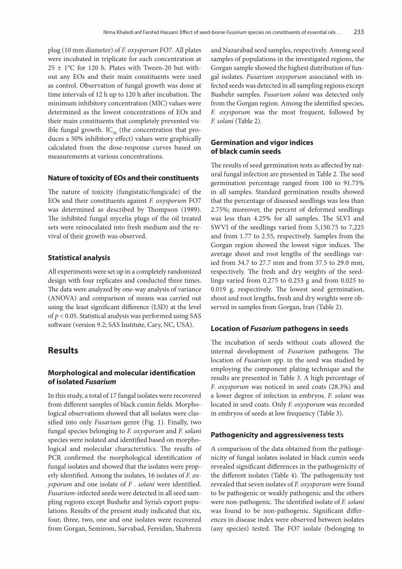
In this study, a total of 17 fungal isolates were recovered from different samples of black cumin fields. Morpho-logical observations showed that all isolates were clas-sified into only Fusarium genre (Fig. 1). Finally, two fungal species belonging to F. oxysporum and F. solani species were isolated and identified based on morpho-logical and molecular characteristics. The results of PCR confirmed the morphological identification of fungal isolates and showed that the isolates were prop-erly identified. Among the isolates, 16 isolates of F. ox‑ysporum and one isolate of F . solani were identified. Fusarium-infected seeds were detected in all seed sam-pling regions except Bushehr and Syria’s export popu-lations. Results of the present study indicated that six, four, three, two, one and one isolates were recovered from Gorgan, Semirom, Sarvabad, Fereidan, Shahreza
and Nazarabad seed samples, respectively. Among seed samples of populations in the investigated regions, the Gorgan sample showed the highest distribution of fun-gal isolates. Fusarium oxysporum associated with in-fected seeds was detected in all sampling regions except Bushehr samples. Fusarium solani was detected only from the Gorgan region. Among the identified species, F. oxysporum was the most frequent, followed by F. solani (Table 2).
Germination and vigor indices of black cumin seeds
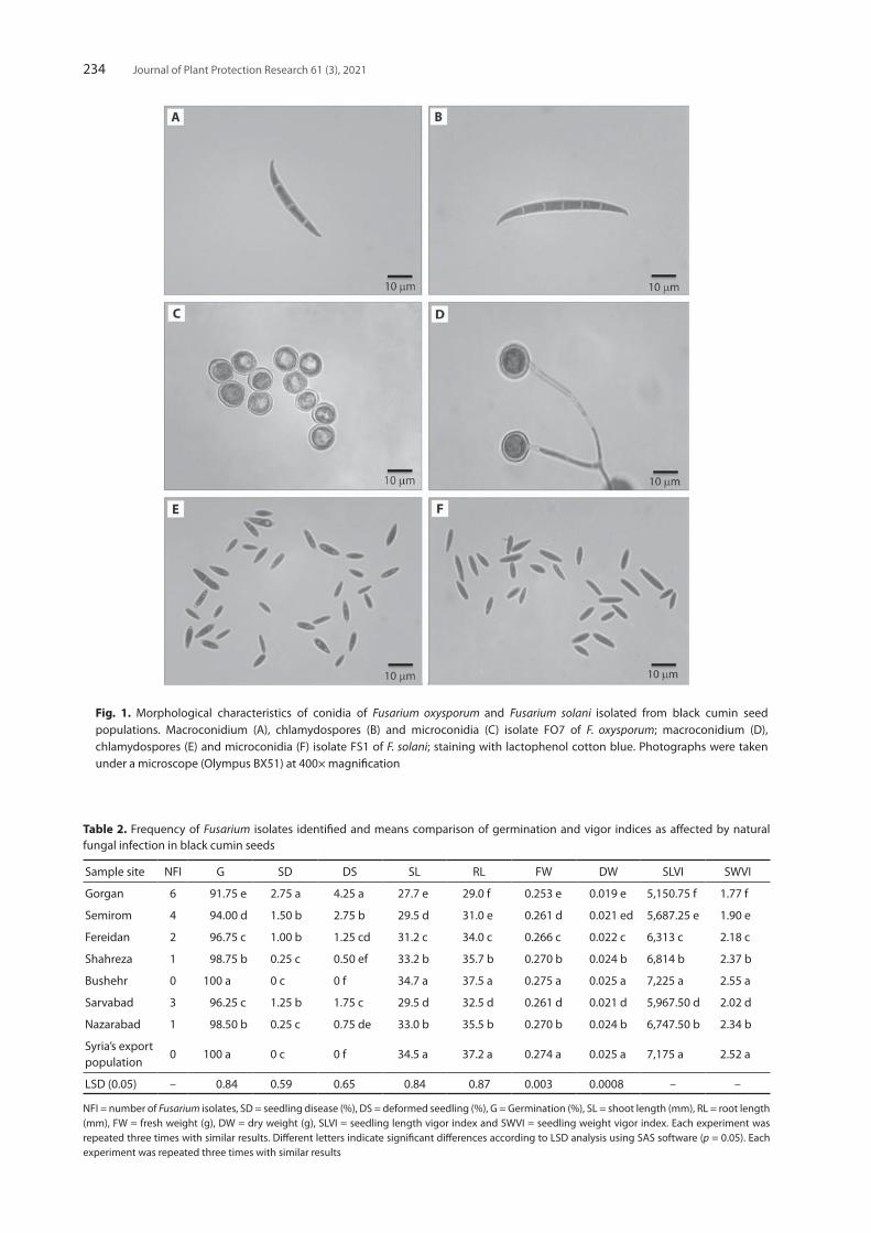
The results of seed germination tests as affected by nat-ural fungal infection are presented in Table 2. The seed germination percentage ranged from 100 to 91.75% in all samples. Standard germination results showed that the percentage of diseased seedlings was less than 2.75%; moreover, the percent of deformed seedlings was less than 4.25% for all samples. The SLVI and SWVI of the seedlings varied from 5,150.75 to 7,225 and from 1.77 to 2.55, respectively. Samples from the Gorgan region showed the lowest vigor indices. The average shoot and root lengths of the seedlings var-ied from 34.7 to 27.7 mm and from 37.5 to 29.0 mm, respectively. The fresh and dry weights of the seed-lings varied from 0.275 to 0.253 g and from 0.025 to 0.019 g, respectively. The lowest seed germination, shoot and root lengths, fresh and dry weights were ob-served in samples from Gorgan, Iran (Table 2).
Location of Fusarium pathogens in seeds
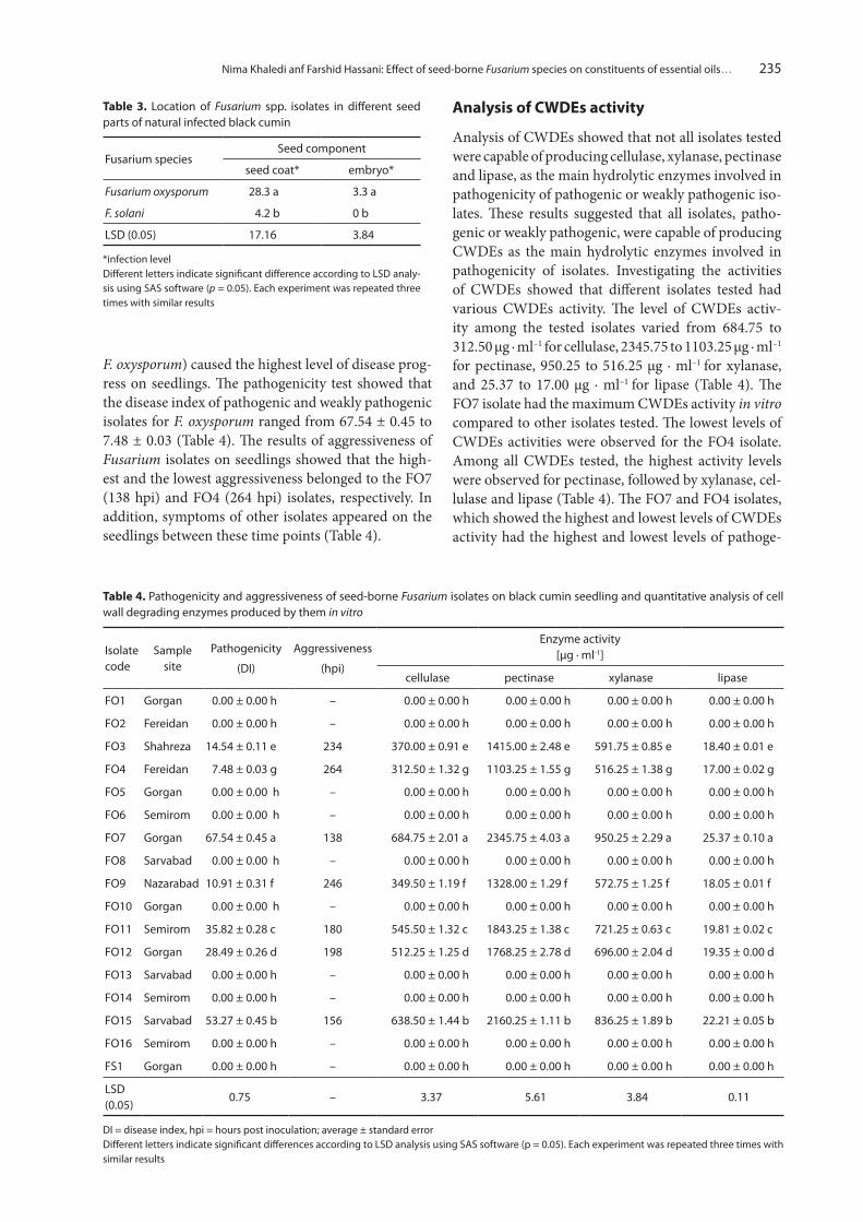
The incubation of seeds without coats allowed the internal development of Fusarium pathogens. The location of Fusarium spp. in the seed was studied by employing the component plating technique and the results are presented in Table 3. A high percentage of F. oxysporum was noticed in seed coats (28.3%) and a lower degree of infection in embryos. F. solani was located in seed coats. Only F. oxysporum was recorded in embryos of seeds at low frequency (Table 3).
Pathogenicity and aggressiveness tests
A comparison of the data obtained from the pathoge-nicity of fungal isolates isolated in black cumin seeds revealed significant differences in the pathogenicity of the different isolates (Table 4). The pathogenicity test revealed that seven isolates of F. oxysporum were found to be pathogenic or weakly pathogenic and the others were non-pathogenic. The identified isolate of F. solani was found to be non-pathogenic. Significant differ-ences in disease index were observed between isolates (any species) tested. The FO7 isolate (belonging to
Journal of Plant Protection Research 61 (3), 2021234
Table 2. Frequency of Fusarium isolates identified and means comparison of germination and vigor indices as affected by natural fungal infection in black cumin seeds
Sample site NFI G SD DS SL RL FW DW SLVI SWVI
Gorgan 6 91.75 e 2.75 a 4.25 a 27.7 e 29.0 f 0.253 e 0.019 e 5,150.75 f 1.77 f
Semirom 4 94.00 d 1.50 b 2.75 b 29.5 d 31.0 e 0.261 d 0.021 ed 5,687.25 e 1.90 e
Fereidan 2 96.75 c 1.00 b 1.25 cd 31.2 c 34.0 c 0.266 c 0.022 c 6,313 c 2.18 c
Shahreza 1 98.75 b 0.25 c 0.50 ef 33.2 b 35.7 b 0.270 b 0.024 b 6,814 b 2.37 b
Bushehr 0 100 a 0 c 0 f 34.7 a 37.5 a 0.275 a 0.025 a 7,225 a 2.55 a
Sarvabad 3 96.25 c 1.25 b 1.75 c 29.5 d 32.5 d 0.261 d 0.021 d 5,967.50 d 2.02 d
Nazarabad 1 98.50 b 0.25 c 0.75 de 33.0 b 35.5 b 0.270 b 0.024 b 6,747.50 b 2.34 b
Syria’s export population
0 100 a 0 c 0 f 34.5 a 37.2 a 0.274 a 0.025 a 7,175 a 2.52 a
LSD (0.05) – 0.84 0.59 0.65 0.84 0.87 0.003 0.0008 – –
NFI = number of Fusarium isolates, SD = seedling disease (%), DS = deformed seedling (%), G = Germination (%), SL = shoot length (mm), RL = root length (mm), FW = fresh weight (g), DW = dry weight (g), SLVI = seedling length vigor index and SWVI = seedling weight vigor index. Each experiment was repeated three times with similar results. Different letters indicate significant differences according to LSD analysis using SAS software (p = 0.05). Each experiment was repeated three times with similar results
Fig. 1. Morphological characteristics of conidia of Fusarium oxysporum and Fusarium solani isolated from black cumin seed populations. Macroconidium (A), chlamydospores (B) and microconidia (C) isolate FO7 of F. oxysporum; macroconidium (D), chlamydospores (E) and microconidia (F) isolate FS1 of F. solani; staining with lactophenol cotton blue. Photographs were taken under a microscope (Olympus BX51) at 400× magnification
Nima Khaledi anf Farshid Hassani: Effect of seed-borne Fusarium species on constituents of essential oils… 235
F. oxysporum) caused the highest level of disease prog-ress on seedlings. The pathogenicity test showed that the disease index of pathogenic and weakly pathogenic isolates for F. oxysporum ranged from 67.54 ± 0.45 to 7.48 ± 0.03 (Table 4). The results of aggressiveness of Fusarium isolates on seedlings showed that the high-est and the lowest aggressiveness belonged to the FO7 (138 hpi) and FO4 (264 hpi) isolates, respectively. In addition, symptoms of other isolates appeared on the seedlings between these time points (Table 4).
Analysis of CWDEs activity
Analysis of CWDEs showed that not all isolates tested were capable of producing cellulase, xylanase, pectinase and lipase, as the main hydrolytic enzymes involved in pathogenicity of pathogenic or weakly pathogenic iso-lates. These results suggested that all isolates, patho-genic or weakly pathogenic, were capable of producing CWDEs as the main hydrolytic enzymes involved in pathogenicity of isolates. Investigating the activities of CWDEs showed that different isolates tested had various CWDEs activity. The level of CWDEs activ-ity among the tested isolates varied from 684.75 to 312.50 µg · ml–1 for cellulase, 2345.75 to 1103.25 µg · ml–1
for pectinase, 950.25 to 516.25 µg · ml–1 for xylanase, and 25.37 to 17.00 µg · ml–1 for lipase (Table 4). The FO7 isolate had the maximum CWDEs activity in vitro compared to other isolates tested. The lowest levels of CWDEs activities were observed for the FO4 isolate. Among all CWDEs tested, the highest activity levels were observed for pectinase, followed by xylanase, cel-lulase and lipase (Table 4). The FO7 and FO4 isolates, which showed the highest and lowest levels of CWDEs activity had the highest and lowest levels of pathoge-
Table 3. Location of Fusarium spp. isolates in different seed parts of natural infected black cumin
Fusarium speciesSeed component
seed coat* embryo*
Fusarium oxysporum 28.3 a 3.3 a
F. solani 4.2 b 0 b
LSD (0.05) 17.16 3.84
*infection level Different letters indicate significant difference according to LSD analy-sis using SAS software (p = 0.05). Each experiment was repeated three times with similar results
Table 4. Pathogenicity and aggressiveness of seed-borne Fusarium isolates on black cumin seedling and quantitative analysis of cell wall degrading enzymes produced by them in vitro
Isolate code
Sample site
Pathogenicity
(DI)
Aggressiveness
(hpi)
Enzyme activity [µg ∙ ml-1]
cellulase pectinase xylanase lipase
FO1 Gorgan 0.00 ± 0.00 h – 0.00 ± 0.00 h 0.00 ± 0.00 h 0.00 ± 0.00 h 0.00 ± 0.00 h
FO2 Fereidan 0.00 ± 0.00 h – 0.00 ± 0.00 h 0.00 ± 0.00 h 0.00 ± 0.00 h 0.00 ± 0.00 h
FO3 Shahreza 14.54 ± 0.11 e 234 370.00 ± 0.91 e 1415.00 ± 2.48 e 591.75 ± 0.85 e 18.40 ± 0.01 e
FO4 Fereidan 7.48 ± 0.03 g 264 312.50 ± 1.32 g 1103.25 ± 1.55 g 516.25 ± 1.38 g 17.00 ± 0.02 g
FO5 Gorgan 0.00 ± 0.00 h – 0.00 ± 0.00 h 0.00 ± 0.00 h 0.00 ± 0.00 h 0.00 ± 0.00 h
FO6 Semirom 0.00 ± 0.00 h – 0.00 ± 0.00 h 0.00 ± 0.00 h 0.00 ± 0.00 h 0.00 ± 0.00 h
FO7 Gorgan 67.54 ± 0.45 a 138 684.75 ± 2.01 a 2345.75 ± 4.03 a 950.25 ± 2.29 a 25.37 ± 0.10 a
FO8 Sarvabad 0.00 ± 0.00 h – 0.00 ± 0.00 h 0.00 ± 0.00 h 0.00 ± 0.00 h 0.00 ± 0.00 h
FO9 Nazarabad 10.91 ± 0.31 f 246 349.50 ± 1.19 f 1328.00 ± 1.29 f 572.75 ± 1.25 f 18.05 ± 0.01 f
FO10 Gorgan 0.00 ± 0.00 h – 0.00 ± 0.00 h 0.00 ± 0.00 h 0.00 ± 0.00 h 0.00 ± 0.00 h
FO11 Semirom 35.82 ± 0.28 c 180 545.50 ± 1.32 c 1843.25 ± 1.38 c 721.25 ± 0.63 c 19.81 ± 0.02 c
FO12 Gorgan 28.49 ± 0.26 d 198 512.25 ± 1.25 d 1768.25 ± 2.78 d 696.00 ± 2.04 d 19.35 ± 0.00 d
FO13 Sarvabad 0.00 ± 0.00 h – 0.00 ± 0.00 h 0.00 ± 0.00 h 0.00 ± 0.00 h 0.00 ± 0.00 h
FO14 Semirom 0.00 ± 0.00 h – 0.00 ± 0.00 h 0.00 ± 0.00 h 0.00 ± 0.00 h 0.00 ± 0.00 h
FO15 Sarvabad 53.27 ± 0.45 b 156 638.50 ± 1.44 b 2160.25 ± 1.11 b 836.25 ± 1.89 b 22.21 ± 0.05 b
FO16 Semirom 0.00 ± 0.00 h – 0.00 ± 0.00 h 0.00 ± 0.00 h 0.00 ± 0.00 h 0.00 ± 0.00 h
FS1 Gorgan 0.00 ± 0.00 h – 0.00 ± 0.00 h 0.00 ± 0.00 h 0.00 ± 0.00 h 0.00 ± 0.00 h
LSD (0.05)
0.75 – 3.37 5.61 3.84 0.11
DI = disease index, hpi = hours post inoculation; average ± standard error Different letters indicate significant differences according to LSD analysis using SAS software (p = 0.05). Each experiment was repeated three times with similar results
Journal of Plant Protection Research 61 (3), 2021236
nicity and aggressiveness in bioassays on seedlings, re-spectively (Table 4). Analysis of CWDEs showed that cellulase and lipase play an important role in pathoge-nicity by degrading the plant cell wall.
Composition of the EOs
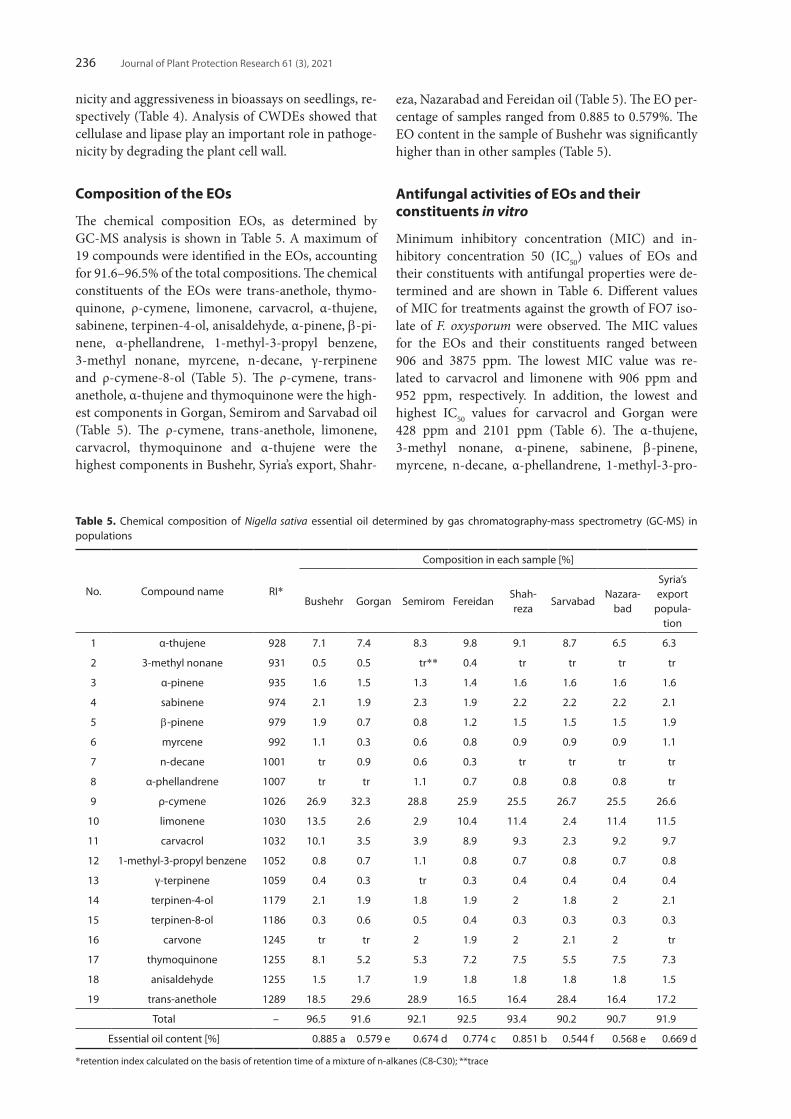
The chemical composition EOs, as determined by GC-MS analysis is shown in Table 5. A maximum of 19 compounds were identified in the EOs, accounting for 91.6–96.5% of the total compositions. The chemical constituents of the EOs were trans-anethole, thymo-quinone, ρ-cymene, limonene, carvacrol, α-thujene, sabinene, terpinen-4-ol, anisaldehyde, α-pinene, b-pi- nene, α-phellandrene, 1-methyl-3-propyl benzene, 3-methyl nonane, myrcene, n-decane, γ-rerpinene and ρ-cymene-8-ol (Table 5). The ρ-cymene, trans-anethole, α-thujene and thymoquinone were the high-est components in Gorgan, Semirom and Sarvabad oil (Table 5). The ρ-cymene, trans-anethole, limonene, carvacrol, thymoquinone and α-thujene were the highest components in Bushehr, Syria’s export, Shahr-
eza, Nazarabad and Fereidan oil (Table 5). The EO per-centage of samples ranged from 0.885 to 0.579%. The EO content in the sample of Bushehr was significantly higher than in other samples (Table 5).
Antifungal activities of EOs and their constituents in vitro
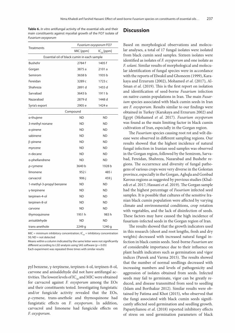
Minimum inhibitory concentration (MIC) and in-hibitory concentration 50 (IC50) values of EOs and their constituents with antifungal properties were de-termined and are shown in Table 6. Different values of MIC for treatments against the growth of FO7 iso-late of F. oxysporum were observed. The MIC values for the EOs and their constituents ranged between 906 and 3875 ppm. The lowest MIC value was re-lated to carvacrol and limonene with 906 ppm and 952 ppm, respectively. In addition, the lowest and highest IC50 values for carvacrol and Gorgan were 428 ppm and 2101 ppm (Table 6). The α-thujene, 3-methyl nonane, α-pinene, sabinene, b-pinene, myrcene, n-decane, α-phellandrene, 1-methyl-3-pro-
Table 5. Chemical composition of Nigella sativa essential oil determined by gas chromatography-mass spectrometry (GC-MS) in populations
No. Compound name RI*
Composition in each sample [%]
Bushehr Gorgan Semirom FereidanShah- reza
SarvabadNazara-
bad
Syria’s export
popula-tion
1 α-thujene 928 7.1 7.4 8.3 9.8 9.1 8.7 6.5 6.3
2 3-methyl nonane 931 0.5 0.5 tr** 0.4 tr tr tr tr
3 α-pinene 935 1.6 1.5 1.3 1.4 1.6 1.6 1.6 1.6
4 sabinene 974 2.1 1.9 2.3 1.9 2.2 2.2 2.2 2.1
5 b-pinene 979 1.9 0.7 0.8 1.2 1.5 1.5 1.5 1.9
6 myrcene 992 1.1 0.3 0.6 0.8 0.9 0.9 0.9 1.1
7 n-decane 1001 tr 0.9 0.6 0.3 tr tr tr tr
8 α-phellandrene 1007 tr tr 1.1 0.7 0.8 0.8 0.8 tr
9 ρ-cymene 1026 26.9 32.3 28.8 25.9 25.5 26.7 25.5 26.6
10 limonene 1030 13.5 2.6 2.9 10.4 11.4 2.4 11.4 11.5
11 carvacrol 1032 10.1 3.5 3.9 8.9 9.3 2.3 9.2 9.7
12 1-methyl-3-propyl benzene 1052 0.8 0.7 1.1 0.8 0.7 0.8 0.7 0.8
13 γ-terpinene 1059 0.4 0.3 tr 0.3 0.4 0.4 0.4 0.4
14 terpinen-4-ol 1179 2.1 1.9 1.8 1.9 2 1.8 2 2.1
15 terpinen-8-ol 1186 0.3 0.6 0.5 0.4 0.3 0.3 0.3 0.3
16 carvone 1245 tr tr 2 1.9 2 2.1 2 tr
17 thymoquinone 1255 8.1 5.2 5.3 7.2 7.5 5.5 7.5 7.3
18 anisaldehyde 1255 1.5 1.7 1.9 1.8 1.8 1.8 1.8 1.5
19 trans-anethole 1289 18.5 29.6 28.9 16.5 16.4 28.4 16.4 17.2
Total – 96.5 91.6 92.1 92.5 93.4 90.2 90.7 91.9
Essential oil content [%] 0.885 a 0.579 e 0.674 d 0.774 c 0.851 b 0.544 f 0.568 e 0.669 d
*retention index calculated on the basis of retention time of a mixture of n-alkanes (C8-C30); **trace
Nima Khaledi anf Farshid Hassani: Effect of seed-borne Fusarium species on constituents of essential oils… 237
pyl benzene, γ-terpinene, terpinen-4-ol, terpinen-8-ol, carvone and anisaldehyde did not have antifungal ac-tivities. The lowest levels of IC50 and MIC were obtained for carvacrol against F. oxysporum among the EOs and their constituents tested. Investigating fungistatic and/or fungicide activity revealed that the EOs, ρ-cymene, trans-anethole and thymoquinone had fungistatic effects on F. oxysporum. In addition, carvacrol and limonene had fungicide effects on F. oxysporum.
Discussion
Based on morphological observations and molecu-lar analyses, a total of 17 fungal isolates were isolated from black cumin seed samples. Sixteen isolates were identified as isolates of F. oxysporum and one isolate as F. solani. Similar results of morphological and molecu-lar identification of fungal species were in accordance with the reports of Elwakil and Ghoneem (1999), Kara-kaya and Erzurum (2002), Mohamed et al. (2017), Al-Sman et al. (2019). This is the first report on isolation and identification of seed-borne Fusarium infection on native cumin populations in Iran. The main Fusar‑ium species associated with black cumin seeds in Iran are F. oxysporum. Results similar to our findings were obtained in Turkey (Karakaya and Erzurum 2002) and Egypt (Mohamed et al. 2017). Fusarium oxysporum was found as the main limiting factor in black cumin cultivation of Iran, especially in the Gorgan region.
The Fusarium species causing root rot and wilt dis-ease were observed in different sampling regions. Our results showed that the highest incidence of natural fungal infection in Iranian seed samples was observed in the Gorgan region, followed by the Semirom, Sarva-bad, Fereidan, Shahreza, Nazarabad and Bushehr re-gions. The occurrence and diversity of fungal patho-gens of various crops were very diverse in the Golestan province, especially in the Gorgan, Aghqla and Gonbad Kavous regions as suggested by previous studies (Khal-edi et al. 2017; Hassani et al. 2019). The Gorgan sample had the highest percentage of Fusarium infected seed samples. It is possible that cultures of the sensitive Ira-nian black cumin population were affected by varying climate and environmental conditions, crop rotation with vegetables, and the lack of disinfection of seeds. These factors may have caused the high incidence of fusarium-infected seeds in the Gorgan region of Iran.
The results showed that the growth indicators used in this research (shoot and root lengths, fresh and dry weights) decreased with increased natural fungal in-fection in black cumin seeds. Seed-borne Fusarium are of considerable importance due to their influence on plant health indicators such as germination and vigor indices (Pareek and Varma 2015). The results showed that the number of normal seedlings decreased with increasing numbers and levels of pathogenicity and aggression of isolates obtained from seeds. Infected seeds may fail to germinate, vigor can be greatly re-duced, and disease transmitted from seed to seedling (Islam and Borthakur 2012). Similar results were ob-tained by Fatima and Khot (2015), who observed that the fungi associated with black cumin seeds signifi-cantly affected seed germination and seedling growth. Papastylianou et al. (2018) reported inhibitory effects of stress on seed germination parameters of black
Table 6. In vitro antifungal activity of the essential oils and their main constituents against mycelial growth of the FO7 isolate of Fusarium oxysporum
TreatmentsFusarium oxysporum FO7
MIC [ppm] IC50 [ppm]
Essential oil of black cumin in each sample
Bushehr 2784 f 1405 f
Gorgan 3875 a 2101 a
Semirom 3658 b 1935 b
Fereidan 3289 c 1723 c
Shahreza 2891 d 1455 d
Sarvabad 3643 b 1911 b
Nazarabad 2879 d 1448 d
Syria’s export 2905 e 1424 e
Compound
α-thujene ND ND
3-methyl nonane ND ND
α-pinene ND ND
sabinene ND ND
b-pinene ND ND
myrcene ND ND
n-decane ND ND
α-phellandrene ND ND
ρ-cymene 3640 b 1928 b
limonene 952 i 485 i
carvacrol 906 j 459 j
1-methyl-3-propyl benzene ND ND
γ-terpinene ND ND
terpinen-4-ol ND ND
terpinen-8-ol ND ND
carvone ND ND
thymoquinone 1951 h 983 h
anisaldehyde ND ND
trans-anethole 2249 g 1240 g
MIC = minimum inhibitory concentration; IC50 = inhibitory concentration 50; ND = not detected Means within a column indicated by the same letter were not significantly different according to LSD analysis using SAS software (p = 0.05)Each experiment was repeated three times with similar results
Journal of Plant Protection Research 61 (3), 2021238
cumin. Similarly, Ghiyasi et al. (2019) reported that germination and vigor indices were decreased under the influence of stress.
Some studies revealed location activities of some fungal pathogens in seed coats, endosperms, coty-ledons and embryonic axes. The results showed that a higher infection level of F. oxysporum most com-monly occurred in the seed coat (28.3%), followed by the embryo (3.3%). Fusarium solani was observed in the seed coat (4.2%). Similar results were obtained by Hassani et al. (2019), who reported that Fusarium sp. were more active in the seed coat than in the em-bryo. Among the species of Fusarium detected in black cumin seeds, only F. oxysporum was observed in the embryos of seeds. Previous studies have shown the lo-cation of fungi pathogens in naturally infected black cumin (Elwakil and Ghoneem 1999), guar (Pareek and Varma 2015) and wheat (Pathak and Zaidi 2013). This is the first time the location of Fusarium spp. in seeds has been identified.
Our results suggest that these pathogens can cause fusarium root rot and wilt of black cumin. A total of seven (about 42%) out of 17 isolates were pathogenic or weakly pathogenic on black cumin seedlings. The pathogenicity test revealed that seven isolates of F. oxy‑ sporum were found to be pathogenic or weakly patho-genic and the others were non-pathogenic. The path-ogenicity test on seedlings showed that two isolates isolated from Gorgan seed samples were pathogenic or weakly pathogenic, while in other samples only one isolate was pathogenic or weakly pathogenic. The fun-gal isolates recovered from seeds revealed significant differences in pathogenicity and aggressiveness be-tween Fusarium isolates. This finding is in agreement with the observations of other researchers (Purahong et al. 2012; Khaledi et al. 2017; Hassani et al. 2019). Aggressiveness is an important factor for determining the potential ability of isolates to cause pathogenic-ity and yield losses. More detailed knowledge about the extensive variability of aggressiveness is essential for understanding the interaction between plant and pathogen. Elwakil and Ghoneem (1999) reported that there was variation in severity and incidence of disease symptoms caused by different seed-borne F. oxyspo‑rum and F. solani isolates, which is in accordance with our observations. The results showed that there was a difference in levels of pathogenicity and aggressive-ness between fungal species and also between isolates of a species. Similar results were obtained by Amatulli et al. (2010), who observed a high variation in aggres-siveness among Fusarium spp. isolates on rice plants.
Pathogenicity and aggressiveness of these pathogen-ic fungi involve different mechanisms such as produc-tion of extracellular enzymes and mycotoxins (Ortega et al. 2013). Fusarium oxysporum and F. solani, as necro-trophic pathogens, utilize a variety of pathogenicity
factors especially production of extracellular enzymes throughout the infection process (Gibson et al. 2011; Khaledi et al. 2017). The secreted CWDEs produced by Fusarium species are important pathogenicity factors. Production of CWDEs such as cellulase, pectinase, xy-lanase and lipase are important and vital for Fusarium spp. establishment in plants (Upasani et al. 2017). The activities of CWDEs, as the main pathogenicity factors involved in the infection process of Fusarium isolates on black cumin seedlings, were evaluated in this study. Pathogenic isolates produced considerably higher levels of CWDEs than weakly pathogenic isolates. Fungal isolates without the ability of producing CWDEs were unable to cause severe disease in black cumin seedlings. Similar results were reported by Hub-balli et al. (2011), who reported that non-pathogenic isolates do not produce or produce less of the pectino-lytic enzymes.
Maximum activities of CWDEs studied were ob-served for the FO7 isolate, which showed the high-est levels of pathogenicity and aggressiveness. This is in agreement with other reports (Khaledi et al. 2017; Hassani et al. 2019). Determining the role of indi-vidual, secreted CWDEs in pathogenicity is difficult to establish because of functional redundancy, while the collective action of CWDEs such as carbon catabo-lite repression and nitrogen regulation is important for the infection process (Michielse and Rep 2009), as observed in this study. We found a positive relation-ship between both pathogenicity and aggressiveness on seedlings and CWDEs produced by fungal isolates in vitro. These findings were in accordance with the results obtained by Hassani et al. (2019) and Khaledi et al. (2017). This is the first report about a strong as-sociation between the amount of extracellular CWDEs activities and the pathogenicity of Fusarium isolates on black cumin seedlings. Similar to our observations, in chickpea – F. oxysporum (Upasani et al. 2017), in wheat – Fusarium spp. (Paccanaro et al. 2017), and also in palm oil – Fusarium spp. (Suwandi et al. 2018), secreted CWDEs increased pathogenicity and aggres-siveness of phytopathogens.
The main components identified in the EOs in-cluded ρ-cymene, trans-anethole, thymoquinone, limonene, carvacrol and α-thujene which is in accord-ance with Ahamad Bustamam et al. (2017), Gerige et al. (2009) and Wajs et al. (2008). Our observations showed a high percentage of ρ-cymene and trans-anethole in the EOs sampled from Iran (Minooe-ian Haghighi and Khosravi (2013) and Mojab et al. (2003). The N. sativa EO and its main constituents indicated antifungal activity. These results were simi-lar to those of other investigators. Sitara et al. (2008) showed that the N. sativa EO was responsible for its antifungal activity against phytopathogenic fungi such as F. oxysporum, F. moniliforme, F. nivali, F. semitectum,
Nima Khaledi anf Farshid Hassani: Effect of seed-borne Fusarium species on constituents of essential oils… 239
A. alternate, Drechslera hawiinesis, Aspergillus niger and A. flavus. Huang et al. (2010) reported that trans-anethole has antifungal activity against F. oxysporum and F. graminearum. Carvacrol antifungal activity also has been reported against phytopathogenic fungi such as F. oxysporum, F. solani, F. semitectum, F. sambuci‑num, F. nivale, F. equiseti, F. culmorum and F. acumi‑natum (Kordali et al. 2008). Thymoquinone has anti-fungal activity against pathogenic dermatophyte fungi such as F. oxysporum (Mahmoudvand et al. 2014). Also, limonene has antifungal activity against F. oxysporum, F. proliferatum and F. verticillioides (Dambolena et al. 2008; Chutia et al. 2009; Singh et al. 2010; Van Hung et al. 2013). However, p-cymene and α-thujene have weak or no antifungal activities (Kordali et al. 2008; Rammanee and Hongpattarakere 2011).
This is the first report on the effects of EOs and their constituents on mycelial growth inhibition of F. oxysporum. In our investigations, the constituents of carvacrol and limonene had the best inhibitory effects on the mycelia growth of F. oxysporum with a MIC val-ue of less than 1000 ppm in vitro. This is in accordance with the results obtained by Rahmouni et al. (2019), who reported that carvacrol is an effective fungicide on the growth of F. oxysporum at a very low concentra-tion. Marei et al. (2012) reported that reducing pectin methyl esterase, cellulase and polyphenol oxidase ac-tivity has a limonene effect on F. oxysporum. The mini-mum concentration of the EOs and their constituents required to inhibit mycelial growth of F. oxysporum differed. The IC50 and MFC values obtained for car-vacrol and limonene were considerably lower than the values obtained for the EOs tested.
It seems that the percentage of antifungal composi-tion of the EOs components can affect the presence of the seed-borne fusarium and/or the pathogenicity of them. The results showed that although ρ-cymene was the main component of the EOs of all Iranian popula-tions, relative amounts of carvacrol, limonene and thy-moquinone were more in oil from Bushehr, Fereidan, Nazarabad and Shahreza than in the oil from Gorgan, Semirom and Sarvabad. Furthermore, relative amounts of carvacrol, limonene and thymoquinone were higher than other constituents in the oil from Syria’s export population. These observations confirm that these compounds are effective on the presence of the seed-borne fusarium. The seed samples of Bushehr and Syria’s export did not have any seed-borne fusarium infection. The results of the GC-MS analysis showed that the total percentage of the antifungal composition of the EOs components of these samples was less than that in other samples.
Biotic and abiotic stresses may have an effect on EO content and production. We observed that the EO content was decreased due to increased Fusarium-infected seeds. However, the effect of genotype on
the EO content should not be ignored. Therefore, the quantity and quality of EO are controlled by genetic, biotic and abiotic factors (Mohammadnejad Ganji et al. 2017; Isah 2019). On the other hand, the geo-graphical region can also affect the EO composition and their antifungal and antibacterial properties (Kha-ledi and Hassani 2018).
In summary, this study indicated that seed-borne fusarium caused a reduction in seed quality indica-tors such as germination and vigor indices. Knowl-edge about phytopathogens and the diseases caused by them which affect the production and quality of black cumin seeds, especially seed-borne pathogens that can spread the disease to a new location and also pass it to the next-generation, can be helpful in selecting ef-fective strategies for controlling diseases. Identifying pathogenicity factors of Fusarium species especially CWDEs, and their association with pathogenicity and aggressiveness of these Fusarium can further our un-derstanding of how phytopathogens cause diseases. In addition, it seems that the composition of the EO in the seed of black cumin, especially carvacrol and limonene, can affect the presence of the seed-borne fusarium and/or the pathogenicity of them. It is sug-gested that carvacrol and limonene, after suitable for-mulation, could be used for the control of Fusarium sp. Moreover, they could be useful for plant breeders in selecting resistant native populations and other man-agement strategies to reduce destructive effects of the pathogens on the host plants.
Acknowledgements
The research was financially supported by Seed and Plant Certification and Registration Institute (SPCRI), Iran (Grant number: 124-08-08-021-98024-980892).
References
Abdel-Razik A.A. 1970. The parasitism of white Sclerotium cepivorum Berk, the incitant of white rot of onion. Ph.D. thesis, Faculty of Agriculture, Assiut University, Assiut, Egypt.
Adams R.P. 2017. Identification of Essential Oil Components by GAS Chromatography/Massspectrometry. 5th ed. Gruver, TX USA, Texensis, 698 pp.
Ahamad Bustamam M.S., Hadithon K.A., Mediani A., Abas F., Rukayadi Y., Lajis N., Shaari K., Ismail I.S. 2017. Stability study of Algerian Nigella sativa seeds stored under differ-ent conditions. Journal of Analytical Methods in Chemistry 2017: 1–12. DOI: https://doi.org/10.1155/2017/7891434
Ahmad A., Husain A., Mujeeb M., Khan S.A., Najmi A.K., Sid-dique N.A., Damanhouri Z.A., Anwar F. 2013. A review on therapeutic potential of Nigella sativa: A miracle herb. Asian Pacific Journal of Tropical Biomedicine 3: 337–352. DOI: https://doi.org/10.1016/S2221-1691(13)60075-1
Ahmadian A., Shiri Y., Froozandeh M. 2015. Study of germina-tion and seedling growth of black cumin (Nigella sativa L.)
Journal of Plant Protection Research 61 (3), 2021240
treated by hydro and osmopriming under salt stress con-ditions. Cercetari Agronomice in Moldova 2: 69–78. DOI: https://doi.org/10.1515/cerce-2015-0031
Al-Sman M.K., Abo-Elyousr K.A.M., Eraky A., El-Zawahry A. 2019. Efficiency of Pseudomonas spp. based formulation for controlling root rot disease of black cumin under green-house and field conditions. Archives of Phytopathology and Plant Protection 52: 1313–1325. DOI: https://doi.org/10.1080/03235408.2019.1707384
Amatulli M.T., Spadaro D., Gullino M.L., Garibaldi A. 2010. Molecular identification of Fusarium spp. associated with bakanae disease of rice in Italy and assessment of their pathogenicity. Plant Pathology 59: 839–844. DOI: https://doi.org/10.1111/j.1365-3059.2010.02319.x
Anonymous 2019. Agricultural Statistics. Volume 2. Ministry of Jihad-e-Agriculture, Programing and Economic, Statistics and Information Technology Office, 425 pp. (in Persian)
Browne R.A., Cooke B.M. 2005. A comparative assessment of potential components of partial disease resistance to Fusarium head blight using a detached leaf assay of wheat, barley and oats. European Journal of Plant Pathology 112: 247–258. DOI: https://doi.org/10.1007/s10658-005-2077-z
Chaieb K., Kouidhi B., Jrah H., Mahdouani K., Bakhrouf A. 2011. Antibacterial activity of Thymoquinone, an active principle of Nigella sativa and its potency to prevent bacterial biofilm formation. BMC Complementary Medicine and Therapies 1: 29. DOI: https://doi.org/10.1186/1472-6882-11-29
Cho Y., Kim K.H., Rota M.L., Scott D., Santopietro G., Calli-han M., Mitchell T.K., Lawrenc C.B. 2009. Identification of novel virulence factors associated with signal transduction pathways in Alternaria brassicicola. Molecular Microbiol-ogy 72: 1316–1333. DOI: https://doi.org/10.1111/j.1365-2958.2009.06689.x
Chutia M., Deka Bhuyan P., Pathak M.G., Sarma T.C., Boruah P. 2009. Antifungal activity and chemical composition of Citrus reticulata Blanco essential oil against phytopathogens from North East India. LWT-Food. Science and Technology 42: 777–780. DOI: https://doi.org/10.1016/j.lwt.2008.09.015
Colowich S.P. 1995. Methods in Enzymology. Academic Prees Inc., London.
Dambolena J., Lopez A., Canepa M., Theumer M., Zygadlo J. 2008. Rubinstein, H. Inhibitory effect of cyclic terpenes (limonene, menthol, menthone and thymol) on Fusarium verticillioides MRC 826 growth and fumonisin B1 biosyn-thesis. Toxicon 51: 37–44. DOI: https://doi.org/10.1016/j.toxicon.2007.07.005
Delgado-Ortiz J.C., Ochoa-Fuentes Y.M., Cerna-Chávez E., Beltrán-Beache M., Rodríguez-Guerra R., Aguirre-Uribe L.A., Vázquez-Martínezc O. 2016. Fusarium species associ-ated with basal rot of garlic in North. Central Mexico and its pathogenicity. Revista Argentina de Microbiología 48: 222–228. DOI: https://doi.org/10.1016/j.ram.2016.04.003
Elwakil M.A., Ghoneem K. 1999. Detection and location of seed-borne fungi of black cumin and their transmission in seed-lings. Pakistan Journal of Biological Sciences 2: 559–564. DOI: https://doi.org/2:559-564. 10.3923/pjbs.1999.559.564
Fatima S., Khot Y.C. 2015. Studies on fungal population of cum-in (Nigella sativa L.) from different parts of Marathwada. Journal of Multidisciplinary Research 2: 25–31.
Gerige S.J., Yadav M.K.G., Rao M., Ramanjaneyulu. 2009. GC-MS analysis of Nigella sativa seeds and antimicrobial ac-tivity of its volatile oil. Brazilian Archives of Biology and Technology 52: 1189–1192. DOI: https://doi.org/10.1590/S1516-89132009000500016
Ghiyasi M., Moghaddam S.S., Amirnia R., Damalas C.A. 2019. Chemical priming with salt and urea improves germination and seedling growth of black cumin (Nigella sativa L.) un-der osmotic stress. Journal of Plant Growth Regulation 38: 1170–1178. DOI: https://doi.org/10.1007/s00344-019-09922-z
Gibson D.M., King B.C., Hayes M.L., Bergstrom G.C. 2011. Plant pathogens as a source of diverse enzymes for ligno-cellulose digestion. Current Opinion in Microbiology 14: 264–270. DOI: https://doi.org/10.1016/j.mib.2011.04.002
Hassani F., Zare L., Khaledi N. 2019. Evaluation of germina-tion and vigor indices associated with fusarium-infected seeds in pre-basic seeds wheat fields. Journal of Plant Pro-tection Research 59: 69–85. DOI: https://doi.org/10.24425/jppr.2019.126037
Huang Y., Zhao J., Zhou L., Wang J., Gong Y., Chen X., Guo Z., Qi Wang Q., Jiang W. 2010. Antifungal activity of the es-sential oil of Illicium verum fruit and its main component trans-anethole. Molecules 15: 7558–7569. DOI: https://doi.org/10.3390/molecules15117558
Hubballi M., Sornakili A., Anand S.N.T., Raguchander T. 2011. Virulence of Alternaria alternata infecting noni associated with production of cell wall degrading enzymes. Journal of Plant Protection Research 51: 87–92. DOI: https://doi.org/10.1080/03235408.2019.1651580
Isah T. 2019. Stress and defense responses in plant secondary metabolites production. Biological Research 52: 39. DOI: https://doi.org/10.1186/s40659-019-0246-3
Islam N.F., Borthakur S.K. 2012. Screening of mycota associated with Aijung rice seed and their effects on seed germination and seedling vigour. Plant Pathology and Quarantine 2: 75–85. DOI: https://doi.org/10.5943/ppq/2/1/11
ISTA. 1986. International Seed Testing Association.1986. Hand-book on Seed Sampling. ISTA, Zurich, Switzerland, 61 pp.
ISTA. 2013. International Seed Testing Association. 2013. The germination test. In: “International Rules for Seed Testing”. ISTA, Bassersdorf, Switzerland, 56 pp. DOI: https://doi.org/10.15258/istarules.2017.05
Karakaya A, Erzurum K. 2002. Wilt disease of Nigella sativa in Turkey. Journal of Turkish Phytopathology 31: 43–47.
Khaledi N., Hassani F. 2018. Antifungal activity of the essential oil of Bunium persicum and its constituents on growth and pathogenesis of Colletotrichum lindemuthianum. Journal of Plant Protection Research 58: 431–441. DOI: https://doi.org/10.24425/jppr.2018.124646
Khaledi N., Taheri P., Falahati-Rastegar M. 2017. Identification, virulence factors characterization and analysis virulence to-gether with aggressiveness of Fusarium spp., causing wheat head blight in Iran. European Journal of Plant Pathology 147: 897–918. DOI: https://doi.org/10.1007/s10658-016-1059-7
Khanna S., Gauri A. 1993. Regulation, purification, and prop-erties of xylanase from Cellulomonas fimi. Enzyme and Microbial Technology 15: 990–995. DOI: https://doi.org/10.1016/0141-0229(93)90177-4
Kikot G.E., Hours R.A., Alconada T.M. 2009. Contribution of cell wall degrading enzymes to pathogenesis of Fusarium graminearum: A review. Journal of Basic Microbiology 49: 231–241. DOI: https://doi.org/10.1002/jobm.200800231
Kordali S., Cakir A., Ozer H., Cakmakci R., Kesdek M., Mete E. 2008. Antifungal, phytotoxic and insecticidal properties of essential oil isolated from Turkish Origanum acutidens and its three components, carvacrol, thymol and p-cymene. Bioresource Technology 99: 8788–8795. DOI: https://doi.org/10.1016/j.biortech.2008.04.048
Lannou C. 2012. Variation and selection of quantitative traits in plant pathogens. Annual Review of Phytopathology 50: 319–338. DOI: https://doi.org/10.1146/annurev-phyto-081211-173031
Leslie J.F., Summerell B.A. 2006. The Fusarium Laboratory Manual. 1st ed. Blackwell Publishing Ltd; Oxford, London.
MacMillan J.D., Voughin R.H. 1964. Purification and proper-ties of a polyglacturonic acid-transeliminase produced by Clastridium multiformentans. Biochemistry 3: 564–572.
Maden S., Singh D., Mathur S.B., Neergard P. 1975. Detection and location of seed borne inoculum of Ascochyta rabei and
Nima Khaledi anf Farshid Hassani: Effect of seed-borne Fusarium species on constituents of essential oils… 241
its transmission in chickpea. Seed Science and Technology 3: 667–671.
Mahapatra S.S., Arya A., Kesarwani A., Verma O. 2019. Influ-ence on oilseeds and legume seed physiology under insect pest and pathogenic infestation. Journal of Pharmacog-nosy and Phytochemistry 8: 671–676. DOI: http://dx.doi.org/10.3329/bjar.v39i2.20429
Mahmoudvand H., Sepahvand A., Jahanbakhsh S., Ezatpour B., Mousavi S.A.A. 2014. Evaluation of antifungal activities of the essential oil and various extracts of Nigella sativa and its main component, thymoquinone against pathogenic der-matophyte strains. Journal of Medical Mycology 24: 155–161. DOI: https://doi.org/10.1016/j.mycmed.2014.06.048
Marei G.I.K., Rasoul M.A.A., Abdelgaleil S.A. 2012. Compara-tive antifungal activities and biochemical effects of monot-erpenes on plant pathogenic fungi. Pesticide Biochemistry and Physiology 103: 56–61. DOI: https://doi.org/10.1016/j.pestbp.2012.03.004.
Michielse C.B., Rep M. 2009. Pathogen profile update: Fusarium oxysporum. Molecular Plant Pathology 10: 311–324. DOI: https://doi.org/10.1111/j.1364-3703.2009.00538.x
Miller G.L. 1959. Use of dinitrosalicylic acid reagent for determi-nation of reducing sugar. Analytical Chemistry 31: 426–428. DOI: https://doi.org/10.1021/ac60147a030
Minooeian Haghighi M.H., Khosravi A.R. 2013. Inhibition and destruction effects of Cuminum cyminum, Ziziphora cli‑nopodioides and Nigella sativa essences on Aspergillus cells. Journal of Babol University of Medical Sciences 15: 25–35.
Mishra P.K., Fox R.T.V., Culham A. 2003. Development of a PCR based assay for rapid and reliable identification of patho-genic Fusaria. FEMS Microbiology Letters 218: 329–332. DOI: https://doi.org/10.1111/j.1574-6968.2003.tb11537.x
Mohamed A.S.K., Kamal A.A.M., Amal E., Aida E. 2017. Isola-tion, identification and biomanagement of root rot of black cumin (Nigella sativa) using selected bacterial antagonists. International Journal of Phytopathology 6: 47–56. DOI: https://doi.org/10.1080/03235408.2019.1707384
Mohammadnejad Ganji S.M., Moradi H., Ghanbari A., Ak-barzadeh M. 2017. Quantity and quality of secondary me-tabolites in lavender plant under the influence of ecological factors. Nova Biologica Reperta 4: 166–172. DOI: https://doi.org/10.21859/acadpub.nbr.4.2.166
Mojab F., Nikavar B., Javidnia K., Roodgar Amoli M.A. 2003. Chemical composition of essential oil and black seed oil. Journal of Medicinal Plants 6: 21–26. DOI: https://doi.org/10.13040/IJPSR.0975-8232.7(11).4473-79
Müller M.E.H., Steier I., Köppen R., Siegel D., Proske M., Korn U., Koch M. 2012. Cocultivation of phytopathogenic Fusarium and Alternaria strains affects fungal growth and mycotoxin production. Journal of Applied Microbiology 113: 874–887. DOI: https://doi.org/10.1111/j.1365-2672.2012.05388.x
Nautiyal P.C. 2009. Seed and seedling vigor traits in ground-nut (Arachis hypogaea L.). Seed Science and Technology 37: 721–735. DOI: https://doi.org/10.15258/sst.2009.37.3.19
Noda J., Brito N., Gonzalez C. 2010. The Botrytis cinerea xyla-nase Xyn11A contributes to virulence with its necrotizing activity, not with its catalytic activity. BMC Plant Biology 10: 38. DOI: https://doi.org/10.1186/1471-2229-10-38
Ortega L.M., Kikot G.E., Astoreca A.L., Alconada T.M. 2013. Screening of Fusarium graminearum isolates for enzymes ex-tracellular and deoxynivalenol production. Journal of Mycol-ogy 2013: 1–7. DOI: https://doi.org/10.1155/2013/358140
Ozdemir N., Kantekin-Erdogan M.N., Tat T., Tekin A. 2018. Ef-fect of black cumin oil on the oxidative stability and sensory characteristics of mayonnaise. Journal of Food Science and Technology 55: 1562–1568. DOI: https://doi.org/10.1007/s13197-018-3075-4
Paccanaro M.C., Sella L., Castiglioni C., Giacomello F., Mar-tínez-Rocha A.L., D’Ovidio R., Schäfer W., Favaron F. 2017. Synergistic effect of different plant cell wall-degrading en-
zymes is important for virulence of Fusarium graminearum. Molecular Plant-Microbe Interactions 30: 886–895. DOI: https://doi.org/10.1094/MPMI-07-17-0179-R.
Papastylianou P., Bakogianni N.N., Travlos I., Roussis I. 2018. Sensitivity of seed germination to salt stress in black cumin (Nigella sativa L.). Notulae Botanicae Horti Agrobotanici Cluj-Napoca 46: 202–205. DOI: https://doi.org/10.15835/nbha46110861
Pareek V., Varma R. 2015. Fusarium solani a dominant seed borne pathogen in seeds of cluster bean grown in Rajasthan. Bioscience Biotechnology Research Communications 8: 29–34.
Pathak N., Zaidi R.K. 2013. Studies on seed-borne fungi of wheat in seed health testing programme. Archives of Phyto-pathology and Plant Protection 46: 389–401. DOI: https://doi.org/10.1080/03235408.2012.741978
Rezaee S., Gharanjik S., Mojerlou S. 2018. Identification of Fusarium solani f. sp. cucurbitae races using morphologi-cal and molecular approaches. Journal of Crop Protection 7: 161–170.
Plodpai P., Chuenchitt S., Petcharat V., Chakthong S., Vora-vuthikunchai S.P. 2013. Anti-Rhizoctonia solani activity by Desmos chinensis extracts and its mechanism of action. Crop Protection 43: 65–71. DOI: https://doi.org/10.1016/j.cropro.2012.09.004
Pritsch C., Muehlbauer G.J., Bushnell W.R., Somers D.A., Vance C.P. 2000. Fungal development and induction of defense response genes during early infection of wheat spikes by Fusarium graminearum. Molecular Plant-Microbe Interactions 13: 159–169. DOI: https://doi.org/10.1094/MPMI.2000.13.2.159
Purahong W., Alkadri D., Nipoti P., Pisi A., Lemmens M., Prodi A. 2012. Validation of a modified Petri-dish test to quantify aggressiveness of Fusarium graminearum in durum wheat. European Journal of Plant Pathology 132: 381–391. DOI: https://doi.org/10.1007/s10658-011-9883-2
Rahmouni A., Saidi R., Khaddor M., Pinto E., Joaquim Carlos Gomes E.D.S., Maouni A. 2019. Chemical composition and antifungal activity of five essential oils and their major components against Fusarium oxysporum f. sp. albedinis of Moroccan palm tree. Euro-Mediterranean Journal for Envi-ronmental Integration 4: 27. DOI: https://doi.org/10.1007/s41207-019-0117-x
Rammanee K., Hongpattarakere T. 2011. Effects of tropical cit-rus essential oils on growth, aflatoxin production, and ul-trastructure alterations of Aspergillus flavus and Aspergillus parasiticus. Food and Bioprocess Technology 4: 1050–1059. DOI: https://doi.org/10.1007/s11947-010-0507-1
Sacristan S., García-Arenal F. 2008. The evolution of virulence and pathogenicity in plant pathogen populations. Molecular Plant Pathology 9: 369–384. DOI: https://doi.org/10.1111/j.1364-3703.2007.00460.x
Singh J., Shikha S.S., Sinha A., Bose B. 2011. Studies on seed mycoflora of wheat (Triticum aestivum L.) treated with po-tassium nitrate and its effect on germination during storage. Research Journal of Seed Science 4: 148–156. DOI: https://doi.org/10.3923/rjss.2011.148.156
Singh P., Shukla R., Prakash B., Kumar A., Singh S., Mishra P.K., Dubey N.K. 2010. Chemical profile, antifungal, antiafla-toxigenic and antioxidant activity of Citrus maxima Burm, and Citrus sinensis (L.) Osbeck essential oils and their cyclic monoterpene, DL-limonene. Food and Chemical Toxicology 48: 1734–1740. DOI: https://doi.org/10.1016/j.fct.2010.04.001
Sitara U., Niaz I., Naseem J., Sultana N. 2008. Antifungal effect of essential oils on in vitro growth of pathogenic fungi. Paki-stan Journal of Botany 40: 409–414.
Suwandi S., Akino S., Kondo N. 2018. Enhanced virulence of Fusarium species associated with spear rot of oil palm fol-lowing recovery from osmotic stress. Mycology 9: 20–28. DOI: https://doi.org/10.1080/21501203.2017.1336497
Journal of Plant Protection Research 61 (3), 2021242
Thompson D.P. 1989. Fungitoxic activity of essential oil com-ponents on food storage fungi. Mycologia 81 (1): 151–153. DOI: https://doi.org/10.2307/3759462
Underwood W. 2012. The plant cell wall: A dynamic barrier against pathogen invasion. Frontiers in Plant Science 85: 1–6. DOI: https://doi.org/10.3389/fpls.2012.00085
Upasani M.L., Limaye B.M., Gurjar G.S., Kasibhatla S.M., Joshi R.R., Kadoo N.Y., Gupta V.S. 2017. Chickpea-Fusari‑um oxysporum interaction transcriptome reveals differential modulation of plant defense strategies. Scientific Reports 7: 7746. DOI: https://doi.org/10.1038/s41598-017-07114-x
Van Hung P., Chi P.T.L., Phi N.T.L. 2013. Comparison of anti-fungal activities of Vietnamese citrus essential oils. Natural Product Research 27: 506–508. DOI: https://doi.org/10.1080/14786419.2012.706293
Wajs A., Bonikowski R., Kalemba D. 2008. Composition of es-sential oil from seeds of Nigella sativa L. cultivated in Po-land. Flavour and Fragrance Journal 23: 126–132. DOI: https://doi.org/10.1002/ffj.1866
Wanyoike W.M., Kang Z., Buchenauer H. 2002. Importance of cell wall degrading enzymes produced by Fusarium graminearum during infection of wheat head. European Journal of Plant Pathology 108: 803–810. DOI: https://doi.org/10.1023/A:1020847216155
Wood T.M., Bhat M. 1988. Methods for measuring cellulase activities. Methods Enzymol 160: 87–112. DOI: https://doi.org/10.1016/0076-6879(88)60109-1