effect of chromium on the fatty acid composition of two strains of euglena gracilis
TRANSCRIPT
Environmental Pollution 141 (2006) 353e358www.elsevier.com/locate/envpol
Effect of chromium on the fatty acid compositionof two strains of Euglena gracilis
Iara Rocchetta a,*, Marcia Mazzuca c, Visitacion Conforti a, Laura Ruiz a,Vilma Balzaretti c, Marıa del Carmen Rıos de Molina b
a Departamento de Biodiversidad y Biologıa Experimental, Universidad de Buenos Aires, Pab. II, Ciudad Universitaria, 1428 Buenos Aires, Argentinab Departamento de Quımica Biologica, Facultad de Ciencias Exactas y Naturales, Universidad de Buenos Aires, Pab. II,
Ciudad Universitaria, 1428 Buenos Aires, Argentinac Departamento de Quımica, Facultad de Ciencias Naturales, Universidad de Patagonia, Comodoro Rivadavia, Chubut, Argentina
Received 6 February 2005; accepted 8 August 2005
Fatty acid evaluation in the presence of chromium in Euglena gracilis grown in different culture conditions.
Abstract
The effect of hexavalent chromium on fatty acid composition was studied in two strains of Euglena gracilis; UTEX 753 (from the CultureCollection of Algae of Texas University, USA) and MAT (isolated from a highly polluted River). Both were grown in photoauxotrophic andphotoheterotrophic conditions and exposed to two metal concentrations, one below and one above IC50. The high malondialdehyde (MDA)levels (3 to 7-fold) obtained with chromium concentration above IC50, suggested the existence of metal-induced lipid peroxidation. Total lipidcontent increased only with concentration below IC50, whereas it was inhibited by higher metal concentration. Photoheterotrophic control strainsexhibited a significantly higher proportion of saturated and polyunsaturated fatty acids. Polyunsaturated acids were most affected by chromium,especially those related to chloroplast structures. Ultra-structure studies showed clear thylakoid disorganization in all treated cells. The resultsindicate that hexavalent chromium affects levels of fatty acids, especially those related to photosynthetic activity.� 2005 Elsevier Ltd. All rights reserved.
Keywords: Euglena gracilis; Fatty acids; Hexavalent chromium; Heavy metal; Lipids
1. Introduction
Hexavalent chromium is a highly toxic heavy metal thatseriously affects environmental conditions in many aquatic eco-systems (Bagchi et al., 2002). The toxicity of this metal dependson its physicochemical properties. The oxidation properties ofion CrO4
�2 and its structural similarity to inorganic anions allowchromium to easily go through cell membranes, thus being analternative substrate in the sulfate transport system (Cieslak-Golonka, 1996; Haglund, 1997). The cytotoxic effects of thismetal on animals and plants are well documented, and its muta-genicity turn it into the source of different types of human can-cer (Vajpayee et al., 2001; Bagchi et al., 2002). Chromium
* Corresponding author. Fax: C54 11 4576 3384.
E-mail address: [email protected] (I. Rocchetta).
0269-7491/$ - see front matter � 2005 Elsevier Ltd. All rights reserved.
doi:10.1016/j.envpol.2005.08.035
discharges into surface water carried out by electroplating units,as well as textile, leather tanning, and paper industries increasedits concentration several times above natural levels.
It is well known that algal cells exposed to heavy metalsmay suffer serious morphological and biochemical alterations(Devars et al., 1998; Rai and Rai, 1998; Okamoto et al., 2001).The reported effects of hexavalent chromium on algal cells in-clude pigment content reduction, chloroplast disorganization,mitochondrial damage, and cytoskeleton alterations causingloss of mobility and cellular growth another mechanism of in-hibition is affecting DNA (Wang, 1999; Cervantes et al., 2001;Rocchetta et al., 2003).
Changes in the lipid composition of algae and plants havebeen attributed to the variations in environmental or cultureconditions, (Molina Grima et al., 1994; Harwood, 1995; Ram-alho et al., 1998; Chini Zittelli et al., 1999). Studies on the
354 I. Rocchetta et al. / Environmental Pollution 141 (2006) 353e358
effect of different growth conditions performed on fatty acidsfrom Euglena gracilis strains showed that exogenous carbonhas an enormous influence on lipid class and fatty acid com-position (Regnault et al., 1995; Barsanti et al., 2000).
It has been demonstrated that polyunsaturated fatty acidsare the main targets of free radicals in lipid peroxidation(Girotti, 2001). Einicker-Lamas et al. (1996) reported severalchanges in membrane lipid content when Euglena graciliscells were cultured in the presence of cadmium.
The unicellular protist Euglena gracilis is a useful modelto study cell damage caused by cytotoxic compounds suchas heavy metals (Coppellotti, 1989; Navarro et al., 1997).This microorganism grows in photoauxotrophic conditionslike most green euglenoids, but it is also able to develop inheterotrophic or photoheterotrophic conditions.
The aim of our study was to examine fatty acid compositionand content in two strains of E. gracilis; MAT (isolated fromthe highly polluted Matanza River, Buenos Aires, Argentina)and UTEX 753 (from the Culture Collection of Algae of TexasUniversity, USA). Strains were cultured under photoauxotro-phic and photoheterotrophic conditions and exposed to differ-ent chromium concentrations. Effects on morphology and totallipid content were analyzed, and lipid peroxidation was eval-uated in terms of MDA content.
2. Materials and methods
2.1. Microorganism and culture conditions
The strains of E. gracilis used were: UTEX 753, from the Culture Collec-
tion of Algae of Texas University, USA (generously provided by Dr Richard
Triemer), and MAT, isolated from the Matanza River, Buenos Aires, Argentina
(Ruiz et al., 2004). Experimental cultures were grown in two mineral media;
Cramer & Myers (C&M) and Buetow (with sodium acetate as carbon source)
(Buetow, 1982), at 24 G 1 �C, with cool-white fluorescent continuous light
(150 mE m�2 s�1 irradiance). Axenicity was monitored plating cultures in
a bacterial medium. A new culture was initiated 6 days before each experiment
in order to obtain an inoculum in exponential growth.
2.2. Metal toxicity assays
Experiments were performed on static cultures containing 150 ml culture
medium in 250 ml glass flasks, at 24 G 1 �C, and under cool-white fluores-
cent continuous light, with an irradiance of 150 mE m�2 s�1. Aliquots of
105 cells ml�1 from both stock cultures (MAT and UTEX) were inoculated
in each flask. K2Cr2O7 was added axenically from a 0.1 M stock solution.
The IC50 values obtained in a previous study for MAT and UTEX strains
cultured in Buetow medium were 24.6 mM Cr(VI) and 3.2 mM Cr(VI), respec-
tively. On the other hand, IC50 values observed in the strains grown in C&M
medium were 120.5 mM Cr(VI) for MAT and 90.4 mM Cr(VI) for UTEX.
Growth rates for the conditions assayed were quite different not only between
strains but also between culture media. Since IC50 values were also different,
we decided to express concentrations as a percentage (40% and 80%) respect
to the concentration necessary to obtain 50% growth (IC50). For MAT strain
cultured in Buetow medium, the concentrations corresponding to 40% and
80% were 9.8 mM and 19.7 mM, respectively, while for cells grown in C&M
medium, they were 48.2 mM for 40% and 96.4 mM for 80%. In the case of
UTEX, values were 1.3 mM and 2.6 mM for cells cultured in Buetow medium,
and 36.2 mM and 72.3 mM for samples cultured in C&M medium.
Cellular density was determined with a Neubauer chamber, with less than
10% error, P ! 0.05. Harvesting time was 96 h after start of metal stress for
all the cultures (U.S. Environmental Protection Agency, 1985).
2.3. Transmission electron microscopy
Cells collected by centrifugation at 4500 ! g for 20 min were fixed in
2.5% glutaraldehyde in 0.1 M cacodylate buffer. Then, they were post-fixed
in 1% osmium tetroxide in 0.1 M cacodylate buffer for 2 h, dehydrated in
an acetone series, and embedded in Spurr resin. Ultrathin sections were cut
with a diamond knife and stained with uranyl acetate and lead citrate. Later,
they were examined using a JEOL 100 CX-II electron microscope from the
Basic and Applied Investigation Center of Bahıa Blanca, Argentina.
2.4. Total lipid determination
Cultures cells were harvested by centrifugation for 15 min at 4500 ! g
and washed three times with 0.154 M phosphate buffer, pH 7. Total lipids
were extracted with chloroform:methanol (2:1 v/v) and quantified according
to the Bligh and Dyer method (1959).
2.5. Malondialdehyde (MDA) determination
Cultures were harvested by centrifugation for 15 min at 4500 ! g, and
washed three times with 0.154 M phosphate buffer, pH 7. Lipid peroxidation
levels were measured in terms of malondialdehyde (MDA) content determined
by the thiobarbituric acid reactive substances (TBARS) method (Vavilin et al.,
1998), according to Hodges et al. equations (1999).
2.6. Fatty acid determination
Dried biomass (15 mg) was exposed to direct transesterification with 1 ml
acetyl chloride in methanol 1:20 (v/v), according to Lepage and Roy (1984).
After tubes were cooled in water (25 �C), the reaction mixture was diluted
with 1 ml water, and extracted three times with 1 ml hexane. The hexane
phase was dried under gentle nitrogen stream, at atmospheric pressure and
room temperature. Then, fatty acid methyl esters were resuspended in
100 ml hexane and injected into the chromatograph. Composition assessment
was performed using a gas chromatograph (Hewlett Packard GC5890) with
a flame ionization detector, on an Innowax capillary column (30 m,
0.32 mm ID, 1 mm film thickness). Helium was used as carrier gas. The col-
umn was held at 150 �C for 3 min, and then temperature was increased
5 �C/min until reaching 280 �C (held for 15 min). Injection port and detector
temperatures were 250 �C and 280 �C, respectively. Determinations were car-
ried out in triplicate. A standard of fatty acid methyl ester mixture (Supelco
Inc., Supelco Park, Bellfonte) was run under identical conditions to identify
compounds on the basis of their retention times.
Fatty acid quantitation was performed using heptadecanoic acid (C17:0) as
internal standard. Thus, an aliquot of 125 mg heptadecanoic acid dissolved in
5 ml toluene was added to the biological samples before transesterification.
2.7. Statistical analysis
Quantitative data for fatty acids content represent the mean of three inde-
pendent experiments. The statistical significance of differences between con-
trols and treated cultures and between Buetow and C&M media was
determined by an analysis of variance (ANOVA). For MDA and total lipid
contents, mean and standard deviation were obtained from the duplicates of
each concentration. Each treatment was performed in duplicate and each assay
was repeated three times. Data were evaluated by an analysis of variance
(ANOVA). Values of P ! 0.005 were considered significant.
3. Results
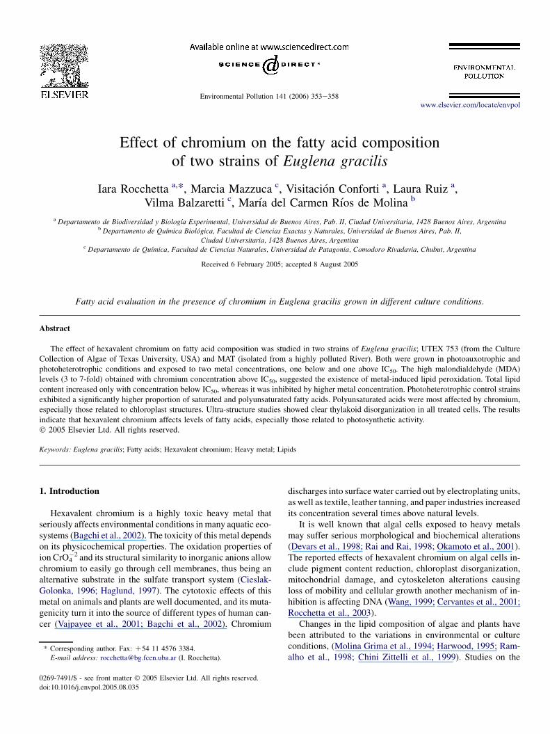
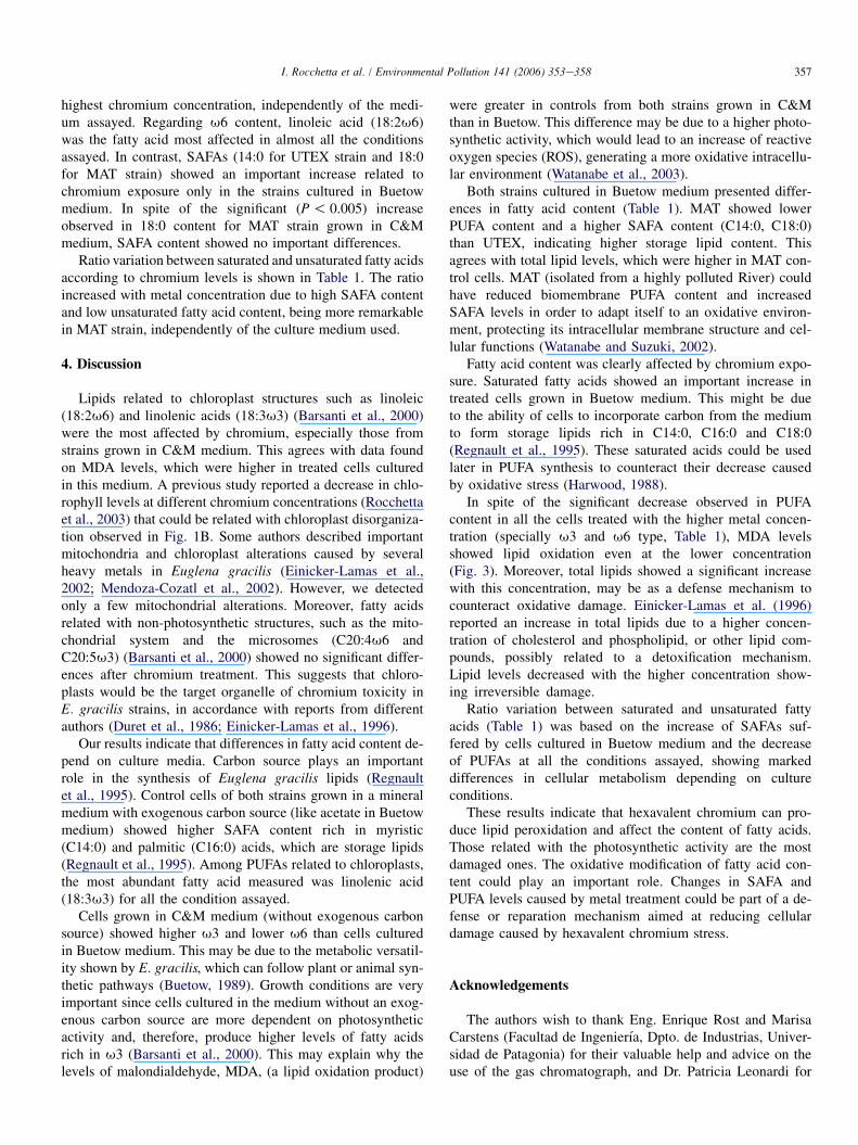
Fig. 1 shows ultrastructural morphology differences be-tween control cells (Fig. 1A) and cells grown in the presenceof the higher chromium concentration (Fig. 1B). Independentlyof the culture media used, the main damages observed in both
355I. Rocchetta et al. / Environmental Pollution 141 (2006) 353e358
strains were significant chloroplast thylakoid disorganizationand the presence of several vacuoles. Mitochondria, on the otherhand, did not seem to be affected by chromium.
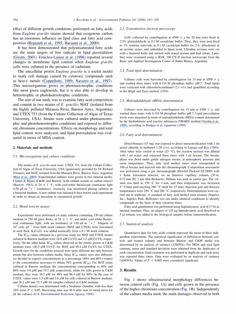
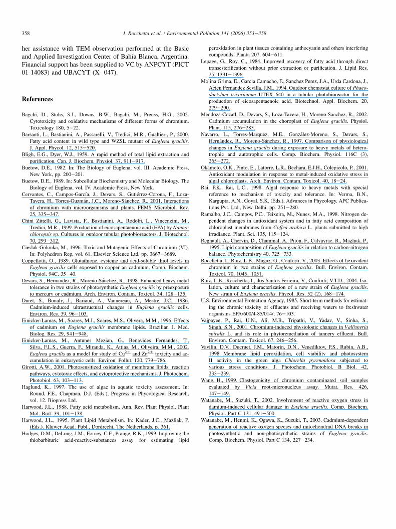
Fig. 2 describes the important variation of total lipid con-tent detected between controls grown in Buetow and C&Mmedia. Both control and treated cells cultured in Buetow(a mineral medium with sodium acetate as carbon source) pre-sented higher lipid levels than those cultured in C&M. Theywere greater in MAT strain. On the other hand, the lower chro-mium concentration increased total lipid content respect tocontrol cells, whereas the higher decreased it.
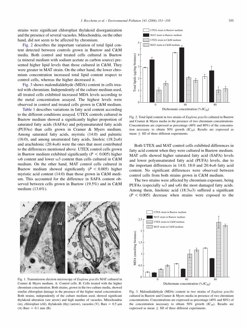
Fig. 3 shows malondialdehyde (MDA) content in cells trea-ted with chromium. Independently of the culture medium used,all treated cells exhibited increased MDA levels according tothe metal concentration assayed. The highest levels wereobserved in control and treated cells grown in C&M medium.
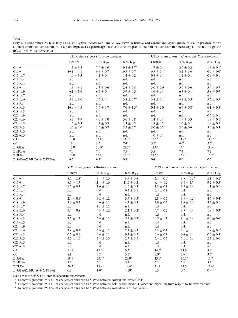
Table 1 describes variations in fatty acid content accordingto the different conditions assayed. UTEX controls cultured inBuetow medium showed a significantly higher proportion ofsaturated fatty acids (SAFAs) and polyunsaturated fatty acids(PUFAs) than cells grown in Cramer & Myers medium.Among saturated fatty acids, myristic (14:0) and palmitic(16:0), and among unsaturated fatty acids, linoleic (18:2u6)and arachidonic (20:4u6) were the ones that most contributedto the differences mentioned above. UTEX control cells grownin Buetow medium exhibited significantly (P ! 0.005) higheru6 content and lower u3 content than cells cultured in C&Mmedium. On the other hand, MAT control cells cultured inBuetow medium showed significantly (P ! 0.005) highermyristic acid content (14:0) than those grown in C&M medi-um. This accounted for the difference in SAFA content ob-served between cells grown in Buetow (19.5%) and in C&Mmedium (13.6%).
Fig. 1. Transmission electron microscopy of Euglena gracilis MAT cultured in
Cramer & Myers medium. A: Control cells. B: Cells treated with the higher
chromium concentration. Both strains, grown in the two culture media, showed
similar chloroplast damage in the presence of the higher metal concentration.
Both strains, independently of the culture medium used, showed significant
thylakoid alteration (see arrow) and high number of vacuoles. Mitochondria
(m), chloroplast (chl), thylakoids (thy) (arrow), vacuoles (V). Bars Z 0.5 mm
(A) Bars Z 0.1 mm (B).
Both UTEX and MAT control cells exhibited differences infatty acid content when they were cultured in Buetow medium.MAT cells showed higher saturated fatty acid (SAFA) levelsand lower polyunsaturated fatty acid (PUFA) levels, due tothe important differences in 14:0, 18:0 and 20:4u6 fatty acidcontent. No significant differences were observed betweencontrol cells from both strains grown in C&M medium.
The two strains were affected by chromium exposure, beingPUFAs (especially u3 and u6) the most damaged fatty acids.Among them, linolenic acid (18:3u3) suffered a significant(P ! 0.005) decrease when strains were exposed to the
0 40 800
1
2
3
4
5
Tot
al li
pids
(µg
/mg
biom
ass)
UTEX strain in Buetow medium
MAT strain in Buetow medium
UTEX strain in C&M medium
MAT strain in C&M medium
Dichromate concentration ( IC50)
Fig. 2. Total lipid content in two strains of Euglena gracilis cultured in Buetow
and Cramer & Myers media in the presence of two chromium concentrations.
Concentrations are expressed as percentage (40% and 80%) of the concentra-
tion necessary to obtain 50% growth (IC50). Results are expressed as
mean G SD of three different experiments.
0 40 800
0,5
1
1,5
2
2,5
MD
A (
nmol
/mg
biom
ass)
UTEX strain in Buetow medium
MAT strain in Buetow medium
UTEX strain in C&M medium
MAT strain in C&M medium
Dichromate concentration ( IC50)
Fig. 3. Malondialdehyde (MDA) content in two strains of Euglena graciliscultured in Buetow and Cramer & Myers media in presence of two chromium
concentrations. Concentrations are expressed as percentage (40% and 80%) of
the concentration necessary to obtain 50% growth (IC50). Results are
expressed as mean G SD of three different experiments.
356 I. Rocchetta et al. / Environmental Pollution 141 (2006) 353e358
Table 1
Fatty acid composition (% total fatty acids) of Euglena gracilis MAT and UTEX grown in Buetow and Cramer and Myers culture media, in presence of two
different chromium concentrations. They are expressed as percentage (40% and 80%) respect to the minimal concentration necessary to obtain 50% growth
(IC50). (n.d. Z not detectable)
UTEX strain grown in Buetow medium UTEX strain grown in Cramer and Myers medium
Control 40% IC50 80% IC50 Control 40% IC50 80% IC50
C14:0 4.5 G 0.4 7.9 G 1.9 9.4 G 1.7a 1.7 G 0.1b 2.9 G 0.2b 1.6 G 0.1b
C16:0 10.1 G 1.1 9.4 G 0.7 10.5 G 0.7 6.3 G 0.4b 8.2 G 1.0 6.6 G 0.6b
C16:1u7 1.0 G 0.3 1.1 G 0.1 1.4 G 0.2 0.8 G 0.1 1.1 G 0.1 0.9 G 0.1
C16:2u4 n.d. n.d. n.d. n.d. n.d. n.d.
C16:3u4 n.d. n.d. n.d. n.d. n.d. n.d.
C18:0 1.8 G 0.1 2.7 G 0.6 2.4 G 0.8 3.0 G 0.6 3.6 G 0.6 3.4 G 0.7
C18:1u9 6.1 G 0.6 6.5 G 0.1 5.9 G 0.5 4.6 G 0.1 6.3 G 0.1 5.8 G 0.9
C18:1u7 n.d. n.d. n.d. n.d. n.d. n.d.
C18:2u6 5.8 G 0.6 5.5 G 1.1 3.8 G 0.7a 3.8 G 0.1b 4.1 G 0.5 3.5 G 0.3
C18:3u4 n.d. n.d. n.d. n.d. n.d. n.d.
C18:3u3 10.9 G 1.5 8.6 G 1.7 7.6 G 1.5a 10.4 G 2.0 6.9 G 0.8a 6.1 G 0.8a
C18:4u3 n.d. n.d. n.d. n.d. n.d. n.d.
C20:1u9 n.d. n.d. n.d. n.d. n.d. 0.5 G 0.1
C20:4u6 5.3 G 0.9 4.0 G 1.0 3.6 G 0.8 1.4 G 0.1b 1.9 G 0.3b 1.9 G 0.1b
C20:4u3 1.2 G 0.3 1.3 G 0.3 1.3 G 0.3 1.5 G 0.2 1.6 G 0.1 1.9 G 0.8
C20:5u3 2.9 G 1.0 2.9 G 0.7 3.2 G 0.7 3.0 G 0.2 2.9 G 0.6 3.4 G 0.5
C22:5u3 n.d. n.d. n.d. n.d. n.d. n.d.
C22:6u3 n.d. n.d. n.d. 1.0 G 0.1 n.d. n.d.
u3 14.9 12.8 12.1a 16.2b 11.4a 11.4a
u6 11.1 9.5 7.4a 5.2b 6.0b 5.3b
S SAFA 15.0 20.0a 22.2a 11.0b 14.7b 11.5b
S MUFA 7.0 7.6 7.5 5.5 7.4 7.2
S PUFA 26.0 22.3 19.5a 21.7b 17.4b 16.8a
S SAFA/(S MUFA C S PUFA) 0.5 0.7a 0.8a 0.4 0.6 0.5
MAT strain grown in Buetow medium MAT strain grown in Cramer and Myers medium
Control 40% IC50 80% IC50 Control 40% IC50 80% IC50
C14:0 8.5 G 2.8c 9.1 G 0.5 8.9 G 0.4 2.4 G 0.6b 1.9 G 0.3b 2.1 G 0.2b
C16:0 8.6 G 1.7 11.1 G 0.6 11.0 G 0.2 8.4 G 1.2 9.8 G 1.7 9.2 G 0.5b
C16:1u7 1.2 G 0.3 1.0 G 0.1 1.0 G 0.3 1.3 G 0.1 1.5 G 0.4 1.1 G 0.1
C16:2u4 n.d. n.d. 0.2 G 0.1 0.4 G 0.1 n.d. n.d.
C16:3u4 n.d. n.d. n.d. n.d. n.d. n.d.
C18:0 2.4 G 0.1c 3.2 G 0.2 3.9 G 0.3a 2.8 G 0.7 3.4 G 0.5 4.5 G 0.4a
C18:1u9 4.0 G 0.2 4.0 G 0.1 4.7 G 0.5 3.9 G 0.5 3.9 G 0.3 4.7 G 0.1
C18:1u7 n.d. 1.2 G 0.2 n.d. n.d. n.d. n.d.
C18:2u6 5.4 G 0.8 4.3 G 0.2 2.4 G 0.2a 4.7 G 0.2 3.5 G 0.4 1.9 G 0.3a
C18:3u4 n.d. n.d. n.d. n.d. n.d. n.d.
C18:3u3 7.7 G 1.7 7.6 G 0.3 5.8 G 0.7a 10.5 G 1.1 8.1 G 0.4 6.0 G 0.6a
C18:4u3 n.d. n.d. n.d. n.d. n.d. n.d.
C20:1u9 n.d. n.d. n.d. n.d. n.d. n.d.
C20:4u6 2.8 G 0.5c 2.9 G 0.2 2.7 G 0.4 2.3 G 0.1 2.1 G 0.5 1.8 G 0.1b
C20:4u3 0.7 G 0.1 0.6 G 0.1 0.7 G 0.2 0.6 G 0.1 0.6 G 0.1 0.6 G 0.3
C20:5u3 3.5 G 1.0 2.8 G 0.3 2.7 G 0.5 3.4 G 0.5 3.2 G 0.5 2.2 G 0.6
C22:5u3 n.d. n.d. n.d. n.d. n.d. n.d.
C22:6u3 n.d. n.d. n.d. n.d. n.d. n.d.
u3 11.8 11.0 9.2a 14.6b 11.9 8.8a
u6 8.1 7.2 5.1a 7.0b 5.6b 3.6a,b
S SAFA 19.5c 23.4a 23.9a 13.6b 15.1b 15.7b
S MUFA 5.2 6.2 5.7 5.1 5.4 5.7
S PUFA 20.2c 18.4 14.4a 21.9 17.5 12.4a
S SAFA/(S MUFA C S PUFA) 0.8 1.0a 1.19a 0.5 0.7 0.9a
Data are mean G SD of three independent experiments.a Denotes significant (P ! 0.05) analysis of variance (ANOVA) between control and treated cells.b Denotes significant (P ! 0.05) analysis of variance (ANOVA) between both culture media, Cramer and Myers medium respect to Buetow medium.c Denotes significant (P ! 0.05) analysis of variance (ANOVA) between control cells of both strains.
357I. Rocchetta et al. / Environmental Pollution 141 (2006) 353e358
highest chromium concentration, independently of the medi-um assayed. Regarding u6 content, linoleic acid (18:2u6)was the fatty acid most affected in almost all the conditionsassayed. In contrast, SAFAs (14:0 for UTEX strain and 18:0for MAT strain) showed an important increase related tochromium exposure only in the strains cultured in Buetowmedium. In spite of the significant (P ! 0.005) increaseobserved in 18:0 content for MAT strain grown in C&Mmedium, SAFA content showed no important differences.
Ratio variation between saturated and unsaturated fatty acidsaccording to chromium levels is shown in Table 1. The ratioincreased with metal concentration due to high SAFA contentand low unsaturated fatty acid content, being more remarkablein MAT strain, independently of the culture medium used.
4. Discussion
Lipids related to chloroplast structures such as linoleic(18:2u6) and linolenic acids (18:3u3) (Barsanti et al., 2000)were the most affected by chromium, especially those fromstrains grown in C&M medium. This agrees with data foundon MDA levels, which were higher in treated cells culturedin this medium. A previous study reported a decrease in chlo-rophyll levels at different chromium concentrations (Rocchettaet al., 2003) that could be related with chloroplast disorganiza-tion observed in Fig. 1B. Some authors described importantmitochondria and chloroplast alterations caused by severalheavy metals in Euglena gracilis (Einicker-Lamas et al.,2002; Mendoza-Cozatl et al., 2002). However, we detectedonly a few mitochondrial alterations. Moreover, fatty acidsrelated with non-photosynthetic structures, such as the mito-chondrial system and the microsomes (C20:4u6 andC20:5u3) (Barsanti et al., 2000) showed no significant differ-ences after chromium treatment. This suggests that chloro-plasts would be the target organelle of chromium toxicity inE. gracilis strains, in accordance with reports from differentauthors (Duret et al., 1986; Einicker-Lamas et al., 1996).
Our results indicate that differences in fatty acid content de-pend on culture media. Carbon source plays an importantrole in the synthesis of Euglena gracilis lipids (Regnaultet al., 1995). Control cells of both strains grown in a mineralmedium with exogenous carbon source (like acetate in Buetowmedium) showed higher SAFA content rich in myristic(C14:0) and palmitic (C16:0) acids, which are storage lipids(Regnault et al., 1995). Among PUFAs related to chloroplasts,the most abundant fatty acid measured was linolenic acid(18:3u3) for all the condition assayed.
Cells grown in C&M medium (without exogenous carbonsource) showed higher u3 and lower u6 than cells culturedin Buetow medium. This may be due to the metabolic versatil-ity shown by E. gracilis, which can follow plant or animal syn-thetic pathways (Buetow, 1989). Growth conditions are veryimportant since cells cultured in the medium without an exog-enous carbon source are more dependent on photosyntheticactivity and, therefore, produce higher levels of fatty acidsrich in u3 (Barsanti et al., 2000). This may explain why thelevels of malondialdehyde, MDA, (a lipid oxidation product)
were greater in controls from both strains grown in C&Mthan in Buetow. This difference may be due to a higher photo-synthetic activity, which would lead to an increase of reactiveoxygen species (ROS), generating a more oxidative intracellu-lar environment (Watanabe et al., 2003).
Both strains cultured in Buetow medium presented differ-ences in fatty acid content (Table 1). MAT showed lowerPUFA content and a higher SAFA content (C14:0, C18:0)than UTEX, indicating higher storage lipid content. Thisagrees with total lipid levels, which were higher in MAT con-trol cells. MAT (isolated from a highly polluted River) couldhave reduced biomembrane PUFA content and increasedSAFA levels in order to adapt itself to an oxidative environ-ment, protecting its intracellular membrane structure and cel-lular functions (Watanabe and Suzuki, 2002).
Fatty acid content was clearly affected by chromium expo-sure. Saturated fatty acids showed an important increase intreated cells grown in Buetow medium. This might be dueto the ability of cells to incorporate carbon from the mediumto form storage lipids rich in C14:0, C16:0 and C18:0(Regnault et al., 1995). These saturated acids could be usedlater in PUFA synthesis to counteract their decrease causedby oxidative stress (Harwood, 1988).
In spite of the significant decrease observed in PUFAcontent in all the cells treated with the higher metal concen-tration (specially u3 and u6 type, Table 1), MDA levelsshowed lipid oxidation even at the lower concentration(Fig. 3). Moreover, total lipids showed a significant increasewith this concentration, may be as a defense mechanism tocounteract oxidative damage. Einicker-Lamas et al. (1996)reported an increase in total lipids due to a higher concen-tration of cholesterol and phospholipid, or other lipid com-pounds, possibly related to a detoxification mechanism.Lipid levels decreased with the higher concentration show-ing irreversible damage.
Ratio variation between saturated and unsaturated fattyacids (Table 1) was based on the increase of SAFAs suf-fered by cells cultured in Buetow medium and the decreaseof PUFAs at all the conditions assayed, showing markeddifferences in cellular metabolism depending on cultureconditions.
These results indicate that hexavalent chromium can pro-duce lipid peroxidation and affect the content of fatty acids.Those related with the photosynthetic activity are the mostdamaged ones. The oxidative modification of fatty acid con-tent could play an important role. Changes in SAFA andPUFA levels caused by metal treatment could be part of a de-fense or reparation mechanism aimed at reducing cellulardamage caused by hexavalent chromium stress.
Acknowledgements
The authors wish to thank Eng. Enrique Rost and MarisaCarstens (Facultad de Ingenierıa, Dpto. de Industrias, Univer-sidad de Patagonia) for their valuable help and advice on theuse of the gas chromatograph, and Dr. Patricia Leonardi for
358 I. Rocchetta et al. / Environmental Pollution 141 (2006) 353e358
her assistance with TEM observation performed at the Basicand Applied Investigation Center of Bahıa Blanca, Argentina.Financial support has been supplied to VC by ANPCYT (PICT01-14083) and UBACYT (X- 047).
References
Bagchi, D., Stohs, S.J., Downs, B.W., Bagchi, M., Preuss, H.G., 2002.
Cytotoxicity and oxidative mechanisms of different forms of chromium.
Toxicology 180, 5e22.
Barsanti, L., Bastianini, A., Passarelli, V., Tredici, M.R., Gualtieri, P., 2000.
Fatty acid content in wild type and WZSL mutant of Euglena gracilis.
J. Appl. Phycol. 12, 515e520.
Bligh, E.G., Dyer, W.J., 1959. A rapid method of total lipid extraction and
purification. Can. J. Biochem. Physiol. 37, 911e917.
Buetow, D.E., 1982. In: The Biology of Euglena, vol. III. Academic Press,
New York, pp. 200e201.
Buetow, D.E., 1989. In: Subcellular Biochemistry and Molecular Biology. The
Biology of Euglena, vol. IV. Academic Press, New York.
Cervantes, C., Campos-Garcıa, J., Devars, S., Gutierrez-Corona, F., Loza-
Tavera, H., Torres-Guzman, J.C., Moreno-Sanchez, R., 2001. Interactions
of chromium with microorganisms and plants. FEMS Microbiol. Rev.
25, 335e347.
Chini Zittelli, G., Lavista, F., Bastianini, A., Rodolfi, L., Vincenzini, M.,
Tredici, M.R., 1999. Production of eicosapentaenoic acid (EPA) by Nanno-
chloropsis sp. Cultures in outdoor tubular photobioreactors. J. Biotechnol.
70, 299e312.
Cieslak-Golonka, M., 1996. Toxic and Mutagenic Effects of Chromium (VI).
In: Polyhedron Rep, vol. 61. Elsevier Science Ltd, pp. 3667e3689.
Coppellotti, O., 1989. Glutathione, cysteine and acid-soluble thiol levels in
Euglena gracilis cells exposed to copper an cadmium. Comp. Biochem.
Physiol. 94C, 35e40.
Devars, S., Hernandez, R., Moreno-Sanchez, R., 1998. Enhanced heavy metal
tolerance in two strains of photosynthetic Euglena gracilis by preexposure
to mercury or cadmium. Arch. Environ. Contam. Toxicol. 34, 128e135.
Duret, S., Bonaly, J., Bariaud, A., Vannereau, A., Mestre, J.C., 1986.
Cadmium-induced ultrastructural changes in Euglena gracilis cells.
Environ. Res. 39, 96e103.
Einicker-Lamas, M., Soares, M.J., Soares, M.S., Olivera, M.M., 1996. Effects
of cadmium on Euglena gracilis membrane lipids. Brazilian J. Med.
Biolog. Res. 29, 941e948.
Einicker-Lamas, M., Antunes Mezian, G., Benavides Fernandes, T.,
Silva, F.L.S., Guerra, F., Miranda, K., Attias, M., Oliveira, M.M., 2002.
Euglena gracilis as a model for study of Cu2C and Zn2C toxicity and ac-
cumulation in eukaryotic cells. Environ. Pollut. 120, 779e786.
Girotti, A.W., 2001. Photosensitized oxidation of membrane lipids: reaction
pathways, cytotoxic effects, and cytoprotective mechanisms. J. Photochem.
Photobiol. 63, 103e113.
Haglund, K., 1997. The use of algae in aquatic toxicity assessment. In:
Round, F.E., Chapman, D.J. (Eds.), Progress in Phycological Research,
vol. 12. Biopress Ltd.
Harwood, J.L., 1988. Fatty acid metabolism. Ann. Rev. Plant Physiol. Plant
Mol. Biol. 39, 101e138.
Harwood, J.L., 1995. Plant Lipid Metabolism. In: Kader, J.C., Mazliak, P.
(Eds.). Kluwer Acad. Publ., Dordrecht, The Netherlands, p. 361.
Hodges, D.M., DeLong, J.M., Forney, C.F., Prange, R.K., 1999. Improving the
thiobarbituric acid-reactive-substances assay for estimating lipid
peroxidation in plant tissues containing anthocyanin and others interfering
compounds. Planta 207, 604e611.
Lepage, G., Roy, C., 1984. Improved recovery of fatty acid through direct
transesterification without prior extraction or purification. J. Lipid Res.
25, 1391e1396.
Molina Grima, E., Garcia Camacho, F., Sanchez Perez, J.A., Urda Cardona, J.,
Acien Fernandez Sevilla, J.M., 1994. Outdoor chemostat culture of Phaeo-
dactylum tricornutum UTEX 640 in a tubular photobioreactor for the
production of eicosapentaenoic acid. Biotechnol. Appl. Biochem. 20,
279e290.
Mendoza-Cozatl, D., Devars, S., Loza-Tavera, H., Moreno-Sanchez, R., 2002.
Cadmium accumulation in the choroplast of Euglena gracilis. Physiol.
Plant. 115, 276e283.
Navarro, L., Torres-Marquez, M.E., Gonzalez-Moreno, S., Devars, S.,
Hernandez, R., Moreno-Sanchez, R., 1997. Comparison of physiological
changes in Euglena gracilis during exposure to heavy metals of hetero-
trophic and autotrophic cells. Comp. Biochem. Physiol. 116C (3),
265e272.
Okamoto, O.K., Pinto, E., Latorre, L.R., Bechara, E.J.H., Colepicolo, P., 2001.
Antioxidant modulation in response to metal-induced oxidative stress in
algal chloroplasts. Arch. Environ. Contam. Toxicol. 40, 18e24.
Rai, P.K., Rai, L.C., 1998. Algal response to heavy metals with special
reference to mechanism of toxicity and tolerance. In: Verma, B.N.,
Kargupta, A.N., Goyal, S.K. (Eds.), Advances in Phycology. APC Publica-
tions Pvt. Ltd., New Delhi, pp. 251e280.
Ramalho, J.C., Campos, P.C., Teixeira, M., Nunes, M.A., 1998. Nitrogen de-
pendent changes in antioxidant system and in fatty acid composition of
chloroplast membranes from Coffea arabica L. plants submitted to high
irradiance. Plant. Sci. 135, 115e124.
Regnault, A., Chervin, D., Chammal, A., Piton, F., Calvayrac, R., Mazliak, P.,
1995. Lipid composition of Euglena gracilis in relation to carbon-nitrogen
balance. Phytochemistry 40, 725e733.
Rocchetta, I., Ruiz, L.B., Magaz, G., Conforti, V., 2003. Effects of hexavalent
chromium in two strains of Euglena gracilis. Bull. Environ. Contam.
Toxicol. 70, 1045e1051.
Ruiz, L.B., Rocchetta, I., dos Santos Ferreira, V., Conforti, V.T.D., 2004. Iso-
lation, culture and characterization of a new strain of Euglena gracilis.
New strain of Euglena gracilis. Phycol. Res. 52 (2), 168e174.
U.S. Environmental Protection Agency, 1985. Short-term methods for estimat-
ing the chronic toxicity of effluents and receiving waters to freshwater
organisms EPA/600/4-85/014/, 76e103.
Vajpayee, P., Rai, U.N., Ali, M.B., Tripathi, V., Yadav, V., Sinha, S.,
Singh, S.N., 2001. Chromium-induced physiologic changes in Vallisneria
spiralis L. and its role in phytoremediation of tannery effluent. Bull.
Environ. Contam. Toxicol. 67, 246e256.
Vavilin, D.V., Ducruet, J.M., Matorin, D.N., Venediktov, P.S., Rubin, A.B.,
1998. Membrane lipid peroxidation, cell viability and photosystem
II activity in the green alga Chlorella pyrenoidosa subjected to
various stress conditions. J. Photochem. Photobiol. B Biol. 42,
233e239.
Wang, H., 1999. Clastogenicity of chromium contaminated soil samples
evaluated by Vicia root-micronucleus assay. Mutat. Res. 426,
147e149.
Watanabe, M., Suzuki, T., 2002. Involvement of reactive oxygen stress in
damium-induced cellular damage in Euglena gracilis. Comp. Biochem.
Physiol. Part C 131, 491e500.
Watanabe, M., Henmi, K., Ogawa, K., Suzuki, T., 2003. Cadmium-dependent
generation of reactive oxygen species and mitochondrial DNA breaks in
photosynthetic and non-photosynthetic strains of Euglena gracilis.
Comp. Biochem. Physiol. Part C 134, 227e234.