editor's choice e management of chronic venous disease
TRANSCRIPT

Editor’s Choice e Management of Chronic Venous Disease
Clinical Practice Guidelines of the European Society for Vascular Surgery (ESVS)
C. Wittens, A.H. Davies, N. Bækgaard, R. Broholm, A. Cavezzi, S. Chastanet,
M. de Wolf, C. Eggen, A. Giannoukas, M. Gohel, S. Kakkos, J. Lawson, T. Noppeney, S. Onida, P. Pittaluga,
S. Thomis, I. Toonder, M. Vuylsteke,
ESVS Guidelines Committeeb P. Kolh, G.J. de Borst, N. Chakfé, S. Debus, R. Hinchliffe, I. Koncar, J. Lindholt,
M.V. de Ceniga, F. Vermassen, F. Verzini,
Document Reviewersc M.G. De Maeseneer, L. Blomgren, O. Hartung, E. Kalodiki, E. Korten, M. Lugli,
R. Naylor, P. Nicolini, A. Rosales
Keywords: Chronic venous disease, Venous disease, Varicose veins, CEAP, VCSS, Villalta, AVVQ, Vein Qol Sym,Duplex ultrasound, Plethysmography, Phlebography, MRV, CTV, Wound dressings, Compression,Ambulatory compression, Sclerotherapy, Thermal ablation, Non-thermal ablation, Laser,Radiofrequency ablation, Stripping, High ligation, Phlebectomy, Stenting, Endophlebectomy, AVfistula, Recurrent varicose veins, Venous malformation
TABLE OF CONTENTS
Abbreviations . . . . . . . . . . . . . . . . . . . . . . . . . . . . . . . . . . . . . . . . . . . . . . . . . . . . . . . . . . . . . . . . . . . . . . . . . . . . . . . . . . . . . . . . . . . . . . . . . . . . . . 680
Introduction . . . . . . . . . . . . . . . . . . . . . . . . . . . . . . . . . . . . . . . . . . . . . . . . . . . . . . . . . . . . . . . . . . . . . . . . . . . . . . . . . . . . . . . . . . . . . . . . . . . . . . . 681
Chapter 1: General Considerations . . . . . . . . . . . . . . . . . . . . . . . . . . . . . . . . . . . . . . . . . . . . . . . . . . . . . . . . . . . . . . . . . . . . . . . . . . . . . . . . . . . . . . 682
1.1. History . . . . . . . . . . . . . . . . . . . . . . . . . . . . . . . . . . . . . . . . . . . . . . . . . . . . . . . . . . . . . . . . . . . . . . . . . . . . . . . . . . . . . . . . . . . . . . . . . . . . . . 682
1.1.1. Pathophysiology . . . . . . . . . . . . . . . . . . . . . . . . . . . . . . . . . . . . . . . . . . . . . . . . . . . . . . . . . . . . . . . . . . . . . . . . . . . . . . . . . . . . . . . 682
1.1.2. Treatment . . . . . . . . . . . . . . . . . . . . . . . . . . . . . . . . . . . . . . . . . . . . . . . . . . . . . . . . . . . . . . . . . . . . . . . . . . . . . . . . . . . . . . . . . . . . . 682
1.1.3. Development in the last 50 years . . . . . . . . . . . . . . . . . . . . . . . . . . . . . . . . . . . . . . . . . . . . . . . . . . . . . . . . . . . . . . . . . . . . . . . . . . 683
1.2. Epidemiology . . . . . . . . . . . . . . . . . . . . . . . . . . . . . . . . . . . . . . . . . . . . . . . . . . . . . . . . . . . . . . . . . . . . . . . . . . . . . . . . . . . . . . . . . . . . . . . . 683
1.2.1. Risk factors . . . . . . . . . . . . . . . . . . . . . . . . . . . . . . . . . . . . . . . . . . . . . . . . . . . . . . . . . . . . . . . . . . . . . . . . . . . . . . . . . . . . . . . . . . 683
1.2.1.1. Age . . . . . . . . . . . . . . . . . . . . . . . . . . . . . . . . . . . . . . . . . . . . . . . . . . . . . . . . . . . . . . . . . . . . . . . . . . . . . . . . . . . . . . . . 683
1.2.1.2. Gender . . . . . . . . . . . . . . . . . . . . . . . . . . . . . . . . . . . . . . . . . . . . . . . . . . . . . . . . . . . . . . . . . . . . . . . . . . . . . . . . . . . . . . 683
1.2.1.3. Obesity . . . . . . . . . . . . . . . . . . . . . . . . . . . . . . . . . . . . . . . . . . . . . . . . . . . . . . . . . . . . . . . . . . . . . . . . . . . . . . . . . . . . . . 683
1.2.1.4. Family history . . . . . . . . . . . . . . . . . . . . . . . . . . . . . . . . . . . . . . . . . . . . . . . . . . . . . . . . . . . . . . . . . . . . . . . . . . . . . . . . . . 683
1.2.1.5. Ethnicity . . . . . . . . . . . . . . . . . . . . . . . . . . . . . . . . . . . . . . . . . . . . . . . . . . . . . . . . . . . . . . . . . . . . . . . . . . . . . . . . . . . . . 683
1.2.2. Prevalence of reflux . . . . . . . . . . . . . . . . . . . . . . . . . . . . . . . . . . . . . . . . . . . . . . . . . . . . . . . . . . . . . . . . . . . . . . . . . . . . . . . . . . . . 683
1.2.3. Progression of varicose veins . . . . . . . . . . . . . . . . . . . . . . . . . . . . . . . . . . . . . . . . . . . . . . . . . . . . . . . . . . . . . . . . . . . . . . . . . . . . . 684
1.3. Anatomy . . . . . . . . . . . . . . . . . . . . . . . . . . . . . . . . . . . . . . . . . . . . . . . . . . . . . . . . . . . . . . . . . . . . . . . . . . . . . . . . . . . . . . . . . . . . . . . . . . . . 684
1.3.1. The superficial veins of the lower extremity . . . . . . . . . . . . . . . . . . . . . . . . . . . . . . . . . . . . . . . . . . . . . . . . . . . . . . . . . . . . . . . . . . 684
1.3.2. The deep veins of the lower extremity . . . . . . . . . . . . . . . . . . . . . . . . . . . . . . . . . . . . . . . . . . . . . . . . . . . . . . . . . . . . . . . . . . . . . . 684
1.4. Physiology . . . . . . . . . . . . . . . . . . . . . . . . . . . . . . . . . . . . . . . . . . . . . . . . . . . . . . . . . . . . . . . . . . . . . . . . . . . . . . . . . . . . . . . . . . . . . . . . . . . 684
1.4.1. The relationship of capacitance/volume to pressure . . . . . . . . . . . . . . . . . . . . . . . . . . . . . . . . . . . . . . . . . . . . . . . . . . . . . . . . . . . . 685
1.4.2. The hydrostatic and dynamic pressure . . . . . . . . . . . . . . . . . . . . . . . . . . . . . . . . . . . . . . . . . . . . . . . . . . . . . . . . . . . . . . . . . . . . . . 685
1.4.3. The vein valves . . . . . . . . . . . . . . . . . . . . . . . . . . . . . . . . . . . . . . . . . . . . . . . . . . . . . . . . . . . . . . . . . . . . . . . . . . . . . . . . . . . . . . . . 685
1.4.4. The calf muscle and the foot pump . . . . . . . . . . . . . . . . . . . . . . . . . . . . . . . . . . . . . . . . . . . . . . . . . . . . . . . . . . . . . . . . . . . . . . . . . 685
1.4.5. Venous tone . . . . . . . . . . . . . . . . . . . . . . . . . . . . . . . . . . . . . . . . . . . . . . . . . . . . . . . . . . . . . . . . . . . . . . . . . . . . . . . . . . . . . . . . . . . 685
1.4.6. The venous pump: main transport system in the non-supine position . . . . . . . . . . . . . . . . . . . . . . . . . . . . . . . . . . . . . . . . . . . . . . . 685
a Writing Committee: Cees Wittens (Netherlands), Chair; Alun Davies (United Kingdom), Co-Chair; Niels Bækgaard (Denmark); Rikke Broholm (Denmark);
Attilio Cavezzi (Italy); Sylvain Chastanet (France); Mark de Wolf (Netherlands); Céline Eggen (Netherlands); Athanasios Giannoukas (Greece); Manjit Gohel
(United Kingdom); Stavros Kakkos (Greece/United Kingdom); James Lawson (Netherlands); Thomas Noppeney (Germany); Sarah Onida (United Kingdom); Paul
Pittaluga (France); Sarah Thomis (Belgium); Irwin Toonder (Netherlands); Marc Vuylsteke (Belgium).b ESVS Guidelines Committee: Philippe Kolh (Chair) (Belgium), Gert Jan de Borst (Co-Chair) (Netherlands), Nabil Chakfé (France), Sebastian Debus (Ger-
many), Rob Hinchliffe (United Kingdom), Igor Koncar (Serbia), Jes Lindholt (Denmark), Melina Vega de Ceniga (Spain), Frank Vermassen (Belgium), Fabio
Verzini (Italy).c Document Reviewers: Marianne De Maeseneer (Review Coordinator) (Belgium), Lena Blomgren (Sweden), Olivier Hartung (France), Evi Kalodiki (United
Kingdom), Eunice Korten (Netherlands), Marzia Lugli (Italy), Ross Naylor (United Kingdom), Philippe Nicolini (France), Antonio Rosales (Norway).DOI of original
article: http://dx.doi.org/10.1016/j.ejvs.2015.02.014
1078-5884/� 2015 European Society for Vascular Surgery. Published by Elsevier Ltd. All rights reserved.
http://dx.doi.org/10.1016/j.ejvs.2015.02.007
Eur J Vasc Endovasc Surg (2015) 49, 678e737
a a a a a a
aaaaaaaaa
a a a

1.5. Pathophysiology . . . . . . . . . . . . . . . . . . . . . . . . . . . . . . . . . . . . . . . . . . . . . . . . . . . . . . . . . . . . . . . . . . . . . . . . . . . . . . . . . . . . . . . . . . . . . . 685
1.5.1. Venous reflux and obstruction . . . . . . . . . . . . . . . . . . . . . . . . . . . . . . . . . . . . . . . . . . . . . . . . . . . . . . . . . . . . . . . . . . . . . . . . . . . . . 686
Chapter 2: Clinical Presentation of CVD . . . . . . . . . . . . . . . . . . . . . . . . . . . . . . . . . . . . . . . . . . . . . . . . . . . . . . . . . . . . . . . . . . . . . . . . . . . . . . . . . . 686
2.1. Clinical presentations . . . . . . . . . . . . . . . . . . . . . . . . . . . . . . . . . . . . . . . . . . . . . . . . . . . . . . . . . . . . . . . . . . . . . . . . . . . . . . . . . . . . . . . . . . 686
2.2. Classification of chronic venous disease . . . . . . . . . . . . . . . . . . . . . . . . . . . . . . . . . . . . . . . . . . . . . . . . . . . . . . . . . . . . . . . . . . . . . . . . . . . 686
2.2.1. Clinical Etiological Anatomical Pathophysiological (CEAP) classification . . . . . . . . . . . . . . . . . . . . . . . . . . . . . . . . . . . . . . . . . . . . 687
2.2.1.1. Clinical classification: C0eC6 . . . . . . . . . . . . . . . . . . . . . . . . . . . . . . . . . . . . . . . . . . . . . . . . . . . . . . . . . . . . . . . . . . . . . 687
2.2.1.2. Etiological classification: Ec, Ep, Es, En . . . . . . . . . . . . . . . . . . . . . . . . . . . . . . . . . . . . . . . . . . . . . . . . . . . . . . . . . . . . 687
2.2.1.3. Anatomical classification: As, Ap, Ad, An . . . . . . . . . . . . . . . . . . . . . . . . . . . . . . . . . . . . . . . . . . . . . . . . . . . . . . . . . . . . 687
2.2.1.4. Pathophysiological classification: Pr, Po, Pr/o, Pn . . . . . . . . . . . . . . . . . . . . . . . . . . . . . . . . . . . . . . . . . . . . . . . . . . . . . 687
2.2.1.5. Level of investigation . . . . . . . . . . . . . . . . . . . . . . . . . . . . . . . . . . . . . . . . . . . . . . . . . . . . . . . . . . . . . . . . . . . . . . . . . . . . 688
2.2.1.6. Applying the CEAP . . . . . . . . . . . . . . . . . . . . . . . . . . . . . . . . . . . . . . . . . . . . . . . . . . . . . . . . . . . . . . . . . . . . . . . . . . . . . 688
2.2.2. Venous Clinical Severity Score, Venous Segmental Disease Score, and Venous Disability Score . . . . . . . . . . . . . . . . . . . . . . . . . 688
2.2.2.1. Venous Clinical Severity Score: measure of severity . . . . . . . . . . . . . . . . . . . . . . . . . . . . . . . . . . . . . . . . . . . . . . . . . . . . 688
2.2.2.2. Venous Segmental Disease Score: pathophysiology and anatomy . . . . . . . . . . . . . . . . . . . . . . . . . . . . . . . . . . . . . . . . 688
2.2.2.3. Venous Disability Score: functional impact . . . . . . . . . . . . . . . . . . . . . . . . . . . . . . . . . . . . . . . . . . . . . . . . . . . . . . . . . . . 689
2.2.3. Villalta-Prandoni Scale . . . . . . . . . . . . . . . . . . . . . . . . . . . . . . . . . . . . . . . . . . . . . . . . . . . . . . . . . . . . . . . . . . . . . . . . . . . . . . . . . . . 689
2.3. Quality of life measures in venous disease . . . . . . . . . . . . . . . . . . . . . . . . . . . . . . . . . . . . . . . . . . . . . . . . . . . . . . . . . . . . . . . . . . . . . . . . . 689
2.3.1. Health related generic tools . . . . . . . . . . . . . . . . . . . . . . . . . . . . . . . . . . . . . . . . . . . . . . . . . . . . . . . . . . . . . . . . . . . . . . . . . . . . . . . 690
2.3.1.1. SF-36, Medical Outcomes Study 36 Item Short Form . . . . . . . . . . . . . . . . . . . . . . . . . . . . . . . . . . . . . . . . . . . . . . . . . . . 690
2.3.1.2. EuroQoL, 5D . . . . . . . . . . . . . . . . . . . . . . . . . . . . . . . . . . . . . . . . . . . . . . . . . . . . . . . . . . . . . . . . . . . . . . . . . . . . . . . . . 690
2.3.2. Disease specific tools . . . . . . . . . . . . . . . . . . . . . . . . . . . . . . . . . . . . . . . . . . . . . . . . . . . . . . . . . . . . . . . . . . . . . . . . . . . . . . . . . . . 690
2.3.2.1. Aberdeen Varicose Veins Questionnaire . . . . . . . . . . . . . . . . . . . . . . . . . . . . . . . . . . . . . . . . . . . . . . . . . . . . . . . . . . . . . 690
2.3.2.2. Chronic Venous Insufficiency Questionnaire . . . . . . . . . . . . . . . . . . . . . . . . . . . . . . . . . . . . . . . . . . . . . . . . . . . . . . . . . . 691
2.3.2.3. Venous Insufficiency Epidemiological and Economic study . . . . . . . . . . . . . . . . . . . . . . . . . . . . . . . . . . . . . . . . . . . . . . 691
Chapter 3: Diagnostics . . . . . . . . . . . . . . . . . . . . . . . . . . . . . . . . . . . . . . . . . . . . . . . . . . . . . . . . . . . . . . . . . . . . . . . . . . . . . . . . . . . . . . . . . . . . . . . . 691
3.1. Clinical examination . . . . . . . . . . . . . . . . . . . . . . . . . . . . . . . . . . . . . . . . . . . . . . . . . . . . . . . . . . . . . . . . . . . . . . . . . . . . . . . . . . . . . . . . . . . . 691
3.1.1. History . . . . . . . . . . . . . . . . . . . . . . . . . . . . . . . . . . . . . . . . . . . . . . . . . . . . . . . . . . . . . . . . . . . . . . . . . . . . . . . . . . . . . . . . . . . . . . 691
3.1.2. Physical examination . . . . . . . . . . . . . . . . . . . . . . . . . . . . . . . . . . . . . . . . . . . . . . . . . . . . . . . . . . . . . . . . . . . . . . . . . . . . . . . . . . . 692
3.2. Diagnostic tools . . . . . . . . . . . . . . . . . . . . . . . . . . . . . . . . . . . . . . . . . . . . . . . . . . . . . . . . . . . . . . . . . . . . . . . . . . . . . . . . . . . . . . . . . . . . . . 692
3.2.1. Definition of reflux . . . . . . . . . . . . . . . . . . . . . . . . . . . . . . . . . . . . . . . . . . . . . . . . . . . . . . . . . . . . . . . . . . . . . . . . . . . . . . . . . . . . . . 692
3.2.2. Handheld continuous wave Doppler . . . . . . . . . . . . . . . . . . . . . . . . . . . . . . . . . . . . . . . . . . . . . . . . . . . . . . . . . . . . . . . . . . . . . . . . 693
3.2.3. Duplex ultrasound examination . . . . . . . . . . . . . . . . . . . . . . . . . . . . . . . . . . . . . . . . . . . . . . . . . . . . . . . . . . . . . . . . . . . . . . . . . . . 693
3.2.3.1. Efficacy . . . . . . . . . . . . . . . . . . . . . . . . . . . . . . . . . . . . . . . . . . . . . . . . . . . . . . . . . . . . . . . . . . . . . . . . . . . . . . . . . . . . . . 693
3.2.3.2. Technique . . . . . . . . . . . . . . . . . . . . . . . . . . . . . . . . . . . . . . . . . . . . . . . . . . . . . . . . . . . . . . . . . . . . . . . . . . . . . . . . . . . . 695
3.2.3.3. Imaging recurrent disease . . . . . . . . . . . . . . . . . . . . . . . . . . . . . . . . . . . . . . . . . . . . . . . . . . . . . . . . . . . . . . . . . . . . . . . 695
3.2.4. Plethysmography and venous pressure measurements . . . . . . . . . . . . . . . . . . . . . . . . . . . . . . . . . . . . . . . . . . . . . . . . . . . . . . . . . 695
3.2.4.1. Strain-gauge plethysmography . . . . . . . . . . . . . . . . . . . . . . . . . . . . . . . . . . . . . . . . . . . . . . . . . . . . . . . . . . . . . . . . . . . . . 695
3.2.4.2. Photoplethysmography . . . . . . . . . . . . . . . . . . . . . . . . . . . . . . . . . . . . . . . . . . . . . . . . . . . . . . . . . . . . . . . . . . . . . . . . . . 695
3.2.4.3. Air-plethysmography . . . . . . . . . . . . . . . . . . . . . . . . . . . . . . . . . . . . . . . . . . . . . . . . . . . . . . . . . . . . . . . . . . . . . . . . . . . 696
3.2.4.4. Foot volumetry . . . . . . . . . . . . . . . . . . . . . . . . . . . . . . . . . . . . . . . . . . . . . . . . . . . . . . . . . . . . . . . . . . . . . . . . . . . . . . . . 696
3.2.5. Phlebography . . . . . . . . . . . . . . . . . . . . . . . . . . . . . . . . . . . . . . . . . . . . . . . . . . . . . . . . . . . . . . . . . . . . . . . . . . . . . . . . . . . . . . . . . . 696
3.2.6. Other imaging methods . . . . . . . . . . . . . . . . . . . . . . . . . . . . . . . . . . . . . . . . . . . . . . . . . . . . . . . . . . . . . . . . . . . . . . . . . . . . . . . . . 696
3.2.6.1. Scientific evidence . . . . . . . . . . . . . . . . . . . . . . . . . . . . . . . . . . . . . . . . . . . . . . . . . . . . . . . . . . . . . . . . . . . . . . . . . . . . . . 696
Chapter 4: Treatment Options in Chronic Venous Disease . . . . . . . . . . . . . . . . . . . . . . . . . . . . . . . . . . . . . . . . . . . . . . . . . . . . . . . . . . . . . . . . . . . 697
4.1. Dressings for venous ulcers . . . . . . . . . . . . . . . . . . . . . . . . . . . . . . . . . . . . . . . . . . . . . . . . . . . . . . . . . . . . . . . . . . . . . . . . . . . . . . . . . . . . . 697
4.2. Compression therapy . . . . . . . . . . . . . . . . . . . . . . . . . . . . . . . . . . . . . . . . . . . . . . . . . . . . . . . . . . . . . . . . . . . . . . . . . . . . . . . . . . . . . . . . . . . 698
4.2.1. Chronic venous disease without ulceration (C0eC4) . . . . . . . . . . . . . . . . . . . . . . . . . . . . . . . . . . . . . . . . . . . . . . . . . . . . . . . . . . . 698
4.2.2. Venous ulceration (C5eC6) . . . . . . . . . . . . . . . . . . . . . . . . . . . . . . . . . . . . . . . . . . . . . . . . . . . . . . . . . . . . . . . . . . . . . . . . . . . . . . . 698
4.2.2.1. Venous ulcer healing . . . . . . . . . . . . . . . . . . . . . . . . . . . . . . . . . . . . . . . . . . . . . . . . . . . . . . . . . . . . . . . . . . . . . . . . . . . . 698
4.2.2.2. Venous ulcer recurrence . . . . . . . . . . . . . . . . . . . . . . . . . . . . . . . . . . . . . . . . . . . . . . . . . . . . . . . . . . . . . . . . . . . . . . . . . 699
4.2.3. Intermittent pneumatic compression for venous ulceration . . . . . . . . . . . . . . . . . . . . . . . . . . . . . . . . . . . . . . . . . . . . . . . . . . . . . . . 699
4.2.4. Compression after venous intervention . . . . . . . . . . . . . . . . . . . . . . . . . . . . . . . . . . . . . . . . . . . . . . . . . . . . . . . . . . . . . . . . . . . . . . 699
4.3. Physiotherapy, leg elevation, and leg massage . . . . . . . . . . . . . . . . . . . . . . . . . . . . . . . . . . . . . . . . . . . . . . . . . . . . . . . . . . . . . . . . . . . . . . 700
4.3.1. Physiotherapy for leg ulceration . . . . . . . . . . . . . . . . . . . . . . . . . . . . . . . . . . . . . . . . . . . . . . . . . . . . . . . . . . . . . . . . . . . . . . . . . . . 700
4.3.2. Leg elevation . . . . . . . . . . . . . . . . . . . . . . . . . . . . . . . . . . . . . . . . . . . . . . . . . . . . . . . . . . . . . . . . . . . . . . . . . . . . . . . . . . . . . . . . . . 700
4.3.3. Leg massage . . . . . . . . . . . . . . . . . . . . . . . . . . . . . . . . . . . . . . . . . . . . . . . . . . . . . . . . . . . . . . . . . . . . . . . . . . . . . . . . . . . . . . . . . . 701
4.4. Medical treatment . . . . . . . . . . . . . . . . . . . . . . . . . . . . . . . . . . . . . . . . . . . . . . . . . . . . . . . . . . . . . . . . . . . . . . . . . . . . . . . . . . . . . . . . . . . . . 701
4.4.1. Chronic venous disease without ulceration (C0eC4) . . . . . . . . . . . . . . . . . . . . . . . . . . . . . . . . . . . . . . . . . . . . . . . . . . . . . . . . . . . 701
4.4.2. Venous ulceration (C5eC6) . . . . . . . . . . . . . . . . . . . . . . . . . . . . . . . . . . . . . . . . . . . . . . . . . . . . . . . . . . . . . . . . . . . . . . . . . . . . . . . 702
4.5. Sclerotherapy . . . . . . . . . . . . . . . . . . . . . . . . . . . . . . . . . . . . . . . . . . . . . . . . . . . . . . . . . . . . . . . . . . . . . . . . . . . . . . . . . . . . . . . . . . . . . . . . 702
4.6. Transcutaneous laser . . . . . . . . . . . . . . . . . . . . . . . . . . . . . . . . . . . . . . . . . . . . . . . . . . . . . . . . . . . . . . . . . . . . . . . . . . . . . . . . . . . . . . . . . . 703
4.7. Endovenous treatments . . . . . . . . . . . . . . . . . . . . . . . . . . . . . . . . . . . . . . . . . . . . . . . . . . . . . . . . . . . . . . . . . . . . . . . . . . . . . . . . . . . . . . . . . 704
4.7.1. Endovenous thermal ablation . . . . . . . . . . . . . . . . . . . . . . . . . . . . . . . . . . . . . . . . . . . . . . . . . . . . . . . . . . . . . . . . . . . . . . . . . . . . . 704
4.7.1.1. Great saphenous vein . . . . . . . . . . . . . . . . . . . . . . . . . . . . . . . . . . . . . . . . . . . . . . . . . . . . . . . . . . . . . . . . . . . . . . . . . . . 704
4.7.1.2. Small saphenous vein . . . . . . . . . . . . . . . . . . . . . . . . . . . . . . . . . . . . . . . . . . . . . . . . . . . . . . . . . . . . . . . . . . . . . . . . . . . 706
4.7.2. Mechanochemical endovenous ablation . . . . . . . . . . . . . . . . . . . . . . . . . . . . . . . . . . . . . . . . . . . . . . . . . . . . . . . . . . . . . . . . . . . . . 706
4.8. Surgery of the superficial veins . . . . . . . . . . . . . . . . . . . . . . . . . . . . . . . . . . . . . . . . . . . . . . . . . . . . . . . . . . . . . . . . . . . . . . . . . . . . . . . . . . . . 706
4.8.1. High ligation with/without stripping . . . . . . . . . . . . . . . . . . . . . . . . . . . . . . . . . . . . . . . . . . . . . . . . . . . . . . . . . . . . . . . . . . . . . . . . . 706
4.8.2. Phlebectomy . . . . . . . . . . . . . . . . . . . . . . . . . . . . . . . . . . . . . . . . . . . . . . . . . . . . . . . . . . . . . . . . . . . . . . . . . . . . . . . . . . . . . . . . . . 708
4.8.3. Ambulatory Selective Varices Ablation under Local anaesthesia . . . . . . . . . . . . . . . . . . . . . . . . . . . . . . . . . . . . . . . . . . . . . . . . . . . 708
679
4.8.4. Cure conservatrice et Hemodynamique de l’Insuffisance Veineuse en Ambulatoire (CHIVA) . . . . . . . . . . . . . . . . . . . . . . . . . . . . . 709
4.8.5. Powered phlebectomy . . . . . . . . . . . . . . . . . . . . . . . . . . . . . . . . . . . . . . . . . . . . . . . . . . . . . . . . . . . . . . . . . . . . . . . . . . . . . . . . . . . . 709
4.9. Treatment of deep vein pathology . . . . . . . . . . . . . . . . . . . . . . . . . . . . . . . . . . . . . . . . . . . . . . . . . . . . . . . . . . . . . . . . . . . . . . . . . . . . . . . . . 709
4.9.1. Treatment of chronic deep venous obstruction . . . . . . . . . . . . . . . . . . . . . . . . . . . . . . . . . . . . . . . . . . . . . . . . . . . . . . . . . . . . . . . . 710
4.9.1.1. Percutaneous transluminal angioplasty and stenting . . . . . . . . . . . . . . . . . . . . . . . . . . . . . . . . . . . . . . . . . . . . . . . . . . . 710
4.9.1.2. Open bypass procedures . . . . . . . . . . . . . . . . . . . . . . . . . . . . . . . . . . . . . . . . . . . . . . . . . . . . . . . . . . . . . . . . . . . . . . . . 711
4.9.2. Treatment of deep venous incompetence . . . . . . . . . . . . . . . . . . . . . . . . . . . . . . . . . . . . . . . . . . . . . . . . . . . . . . . . . . . . . . . . . . . . . 711
Chapter 5: Recurrent Varicose Veins . . . . . . . . . . . . . . . . . . . . . . . . . . . . . . . . . . . . . . . . . . . . . . . . . . . . . . . . . . . . . . . . . . . . . . . . . . . . . . . . . . . . 712
5.1. Etiology . . . . . . . . . . . . . . . . . . . . . . . . . . . . . . . . . . . . . . . . . . . . . . . . . . . . . . . . . . . . . . . . . . . . . . . . . . . . . . . . . . . . . . . . . . . . . . . . . . . . . 713
5.2. Risk factors . . . . . . . . . . . . . . . . . . . . . . . . . . . . . . . . . . . . . . . . . . . . . . . . . . . . .. . . . . . . . . . . . . . . . . . . . . . . . . . . . . . . . . . . . . . . . . . . . . . 714
5.3. Diagnosis of recurrent varicose veins . . . . . . . . . . . . . . . . . . . . . . . . . . . . . . . . . . . . . . . . . . . . . . . . . . . . . . . . . . . . . . . . . . . . . . . . . . . . . 714
5.4. Treatment of recurrent varicose veins . . . . . . . . . . . . . . . . . . . . . . . . . . . . . . . . . . . . . . . . . . . . . . . . . . . . . . . . . . . . . . . . . . . . . . . . . . . . . 714
Chapter 6: Congenital Venous Malformations . . . . . . . . . . . . . . . . . . . . . . . . . . . . . . . . . . . . . . . . . . . . . . . . . . . . . . . . . . . . . . . . . . . . . . . . . . . . . 715
6.1. Pathophysiology . . . . . . . . . . . . . . . . . . . . . . . . . . . . . . . . . . . . . . . . . . . . . . . . . . . . . . . . . . . . . . . . . . . . . . . . . . . . . . . . . . . . . . . . . . . . . . . 715
6.2. Classification . . . . . . . . . . . . . . . . . . . . . . . . . . . . . . . . . . . . . . . . . . . . . . . . . . . . . . . . . . . . . . . . . . . . . . . . . . . . . . . . . . . . . . . . . . . . . . . . . 715
6.2.1. International Society for the Study of Vascular Anomalies classification . . . . . . . . . . . . . . . . . . . . . . . . . . . . . . . . . . . . . . . . . . . . . 715
6.2.2. Hamburg classification . . . . . . . . . . . . . . . . . . . . . . . . . . . . . . . . . . . . . . . . . . . . . . . . . . . . . . . . . . . . . . . . . . . . . . . . . . . . . . . . . . . 715
6.2.3. Puig classification . . . . . . . . . . . . . . . . . . . . . . . . . . . . . . . . . . . . . . . . . . . . . . . . . . . . . . . . . . . . . . . . . . . . . . . . . . . . . . . . . . . . . . 715
6.3. Venous malformations . . . . . . . . . . . . . . . . . . . . . . . . . . . . . . . . . . . . . . . . . . . . . . . . . . . . . . . . . . . . . . . . . . . . . . . . . . . . . . . . . . . . . . . . . . 715
6.4. Syndromes . . . . . . . . . . . . . . . . . . . . . . . . . . . . . . . . . . . . . . . . . . . . . . . . . . . . . . . . . . . . . . . . . . . . . . . . . . . . . . . . . . . . . . . . . . . . . . . . . . 717
6.4.1. Klippel-Trenaunay syndrome . . . . . . . . . . . . . . . . . . . . . . . . . . . . . . . . . . . . . . . . . . . . . . . . . . . . . . . . . . . . . . . . . . . . . . . . . . . . . 717
6.4.1.1. Etiology . . . . . . . . . . . . . . . . . . . . . . . . . . . . . . . . . . . . . . . . . . . . . . . . . . . . . . . . . . . . . . . . . . . . . . . . . . . . . . . . . . . . . . 717
6.4.1.2. Clinical characteristics . . . . . . . . . . . . . . . . . . . . . . . . . . . . . . . . . . . . . . . . . . . . . . . . . . . . . . . . . . . . . . . . . . . . . . . . . . . 718
6.4.1.3. Diagnosis . . . . . . . . . . . . . . . . . . . . . . . . . . . . . . . . . . . . . . . . . . . . . . . . . . . . . . . . . . . . . . . . . . . . . . . . . . . . . . . . . . . . 718
6.4.1.4. Management . . . . . . . . . . . . . . . . . . . . . . . . . . . . . . . . . . . . . . . . . . . . . . . . . . . . . . . . . . . . . . . . . . . . . . . . . . . . . . . . . 718
6.4.2. Parkes-Weber syndrome . . . . . . . . . . . . . . . . . . . . . . . . . . . . . . . . . . . . . . . . . . . . . . . . . . . . . . . . . . . . . . . . . . . . . . . . . . . . . . . . . 719
6.4.2.1. Etiology . . . . . . . . . . . . . . . . . . . . . . . . . . . . . . . . . . . . . . . . . . . . . . . . . . . . . . . . . . . . . . . . . . . . . . . . . . . . . . . . . . . . . . 719
6.4.2.2. Clinical characteristics . . . . . . . . . . . . . . . . . . . . . . . . . . . . . . . . . . . . . . . . . . . . . . . . . . . . . . . . . . . . . . . . . . . . . . . . . . . 719
6.4.2.3. Diagnosis . . . . . . . . . . . . . . . . . . . . . . . . . . . . . . . . . . . . . . . . . . . . . . . . . . . . . . . . . . . . . . . . . . . . . . . . . . . . . . . . . . . . 719
6.4.2.4. Management . . . . . . . . . . . . . . . . . . . . . . . . . . . . . . . . . . . . . . . . . . . . . . . . . . . . . . . . . . . . . . . . . . . . . . . . . . . . . . . . . 719
References . . . . . . . . . . . . . . . . . . . . . . . . . . . . . . . . . . . . . . . . . . . . . . . . . . . . . . . . . . . . . . . . . . . . . . . . . . . . . . . . . . . . . . . . . . . . . . . . . . . . . . . 721
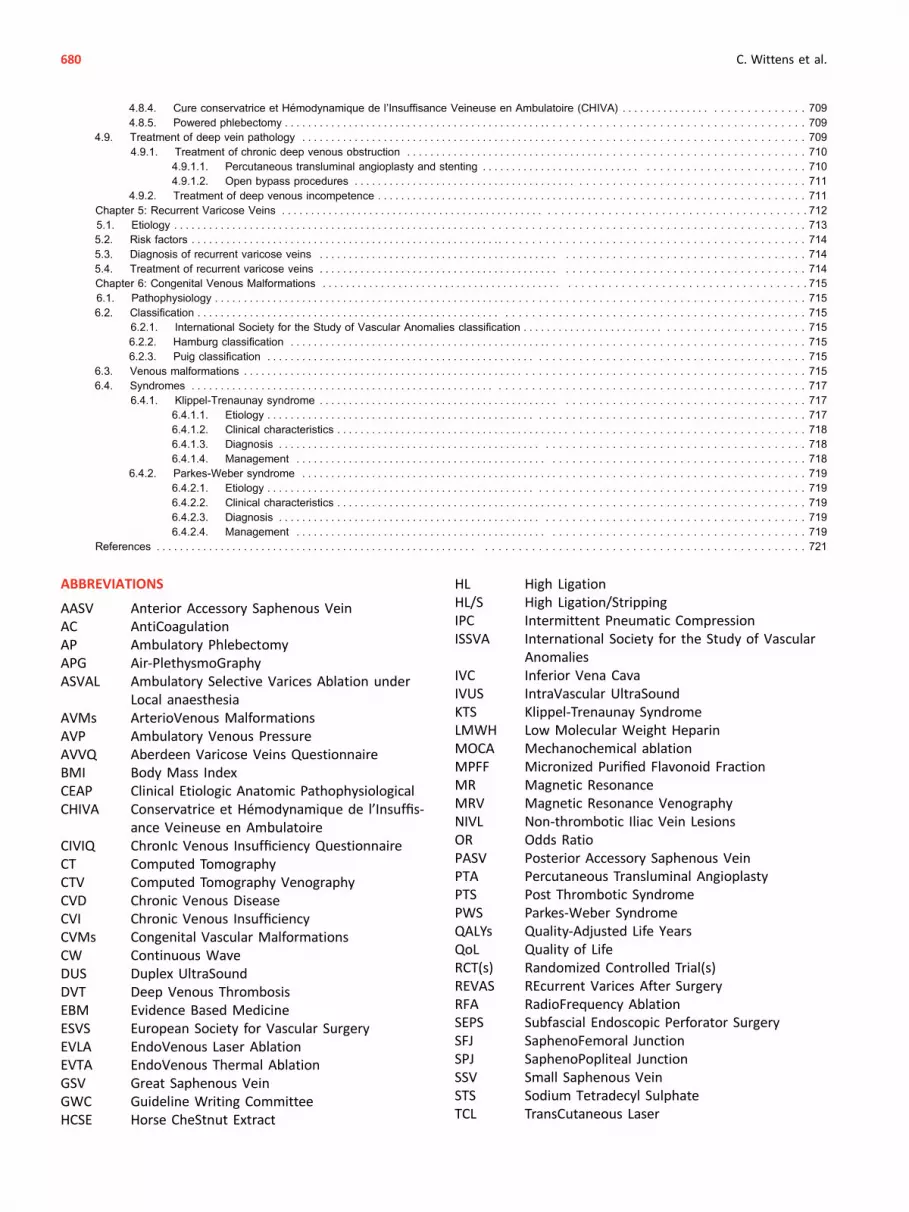
ABBREVIATIONS
AASV Anterior Accessory Saphenous VeinAC AntiCoagulationAP Ambulatory PhlebectomyAPG Air-PlethysmoGraphyASVAL Ambulatory Selective Varices Ablation under
Local anaesthesiaAVMs ArterioVenous MalformationsAVP Ambulatory Venous PressureAVVQ Aberdeen Varicose Veins QuestionnaireBMI Body Mass IndexCEAP Clinical Etiologic Anatomic PathophysiologicalCHIVA Conservatrice et Hémodynamique de l’Insuffis-
ance Veineuse en AmbulatoireCIVIQ ChronIc Venous Insufficiency QuestionnaireCT Computed TomographyCTV Computed Tomography VenographyCVD Chronic Venous DiseaseCVI Chronic Venous InsufficiencyCVMs Congenital Vascular MalformationsCW Continuous WaveDUS Duplex UltraSoundDVT Deep Venous ThrombosisEBM Evidence Based MedicineESVS European Society for Vascular SurgeryEVLA EndoVenous Laser AblationEVTA EndoVenous Thermal AblationGSV Great Saphenous VeinGWC Guideline Writing CommitteeHCSE Horse CheStnut Extract
HL High LigationHL/S High Ligation/StrippingIPC Intermittent Pneumatic CompressionISSVA International Society for the Study of Vascular
AnomaliesIVC Inferior Vena CavaIVUS IntraVascular UltraSoundKTS Klippel-Trenaunay SyndromeLMWH Low Molecular Weight HeparinMOCA Mechanochemical ablationMPFF Micronized Purified Flavonoid FractionMR Magnetic ResonanceMRV Magnetic Resonance VenographyNIVL Non-thrombotic Iliac Vein LesionsOR Odds RatioPASV Posterior Accessory Saphenous VeinPTA Percutaneous Transluminal AngioplastyPTS Post Thrombotic SyndromePWS Parkes-Weber SyndromeQALYs Quality-Adjusted Life YearsQoL Quality of LifeRCT(s) Randomized Controlled Trial(s)REVAS REcurrent Varices After SurgeryRFA RadioFrequency AblationSEPS Subfascial Endoscopic Perforator SurgerySFJ SaphenoFemoral JunctionSPJ SaphenoPopliteal JunctionSSV Small Saphenous VeinSTS Sodium Tetradecyl SulphateTCL TransCutaneous Laser
680 C. Wittens et al.

INTRODUCTION
Members of this Guideline Writing Committee (GWC) wereselected by the European Society for Vascular Surgery(ESVS) to represent physicians involved in management ofpatients with chronic venous disease (CVD). The membersof the GWC have provided disclosure statements of allrelationships that might be perceived as real or potentialsources of conflicts of interest. These disclosure forms arekept on file at the headquarters of the ESVS. The GWCreport received neither financial support nor support fromthe ESVS or any pharmaceutical, device, or surgicalindustry.
The ESVS guideline committee was responsible for theendorsement process of this guideline. All expertsinvolved in the GWC have approved the final document.The guideline document was reviewed and approvedby the EJVES editorial board and ESVS guidelinecommittee.
THE PURPOSE OF THESE GUIDELINES
The ESVS has developed clinical practice guidelines for thecare of patients with CVD in the lower extremities.
The aim of this document is to assist physiciansin selecting the best management strategy for patientswith CVD. This guideline, established by members of theGWC, who are members of the ESVS or non-members with specific expertise in the field, is basedon scientific evidence completed with expert opinion onthe matter. By summarizing and evaluating all availableevidence in the field, recommendations for the evaluationand treatment of patients with CVD have beenformulated.
Guidelines have the purpose of promoting a standard ofcare according to specialists in the field, in this case repre-sented by members of the ESVS. However, under nocircumstance should this guideline be seen as the legalstandard of care in all patients. As the word guideline statesin itself, the document is a guiding principle, but the caregiven to a single patient is always dependent on the indi-vidual patient (symptom variability, comorbidities, age, levelof activity, etc.), treatment setting (techniques available),and other factors.
The recommendations are valid only at the time ofpublication, as technology and disease knowledge in thisfield changes rapidly and expanding recommendations canbecome outdated. It is an aim of the ESVS to revise theguidelines when important new insights in the evaluationand management of CVD become available.
METHODOLOGY
Strategy
The GWC was convened in 2011 at the annual ESVS meetingin Athens. At that meeting the tasks in creating the guide-line were evaluated and distributed among the committeemembers. The final version of the guideline was submittedon December 22, 2014.
Literature search and selection
A clinical librarian performed the literature search for thisguideline systematically in PubMed, Embase, Cinahl, andthe Cochrane Library up to January 1, 2013. Referencechecking and handsearch by the guideline committeemembers added other relevant literature.
The members of the GWC performed the literature se-lection based on information provided in the title and ab-stract of the retrieved studies.
Criteria for search and selection were:
Several relevant articles published after the searchdate or in another foreign language were included, but onlyif they were of paramount importance to this guideline.
Weighing the evidence
To define the current guidelines, members of theGWC reviewed and summarized the selected literature.Conclusions were drawn based on the scientificevidence.
The guidelines in this document are based on the Euro-pean Society of Cardiology grading system. For eachrecommendation, the letter A, B, or C marks the level of
Language: English, German, and French
Level of
evidence:
Selection of the literature was performed
following the pyramid of evidence, with
aggregated evidence in the top of the pyramid
(systematic reviews, meta-analysis), then
randomized controlled trials, then observational
studies. Single case reports, animal studies, and in
vitro studies in the bottom of the pyramid were
excluded, leaving expert opinions at the bottomof
the pyramid. The level of evidence per section in
the guideline is dependent on the level of
evidence available on the specific subject.
Sample size: If there were large studies available, with a
minimum of 15 subjects per research group, only
these were included. If not available, smaller
studies were also included.
TIPP TransIlluminated Powered PhlebectomyUGFS Ultrasound Guided Foam SclerotherapyVCSS Venous Clinical Severity ScoreVDS Venous Disability Score
VEINES Venous Insufficiency Epidemiological and Eco-nomic Study
VMs Venous MalformationsVSDS Venous Segmental Disease Score
681

current evidence (Table 1). Weighing the level of evidenceand expert opinion, every recommendation is subsequentlymarked as either class I, IIa, IIb, or III (Table 2). The lowerthe class number, the more proven is the efficacy and safetyof a certain procedure.
CHAPTER 1: GENERAL CONSIDERATIONS
The term CVD has been used to describe both visual andfunctional manifestations of abnormalities in the peripheralvenous system. It can be defined as “(any) morphologicaland functional abnormalities of the venous system of longduration manifest either by symptoms and/or signs indi-cating the need for investigation and/or care.”1
The prevalence of CVD in the adult population has beenreported to be as high as 60%, particularly affecting pop-ulations in the developed world.2,3 It has become clear thatCVD is an important cause of patient distress and signifi-cantly impacts on healthcare resources.4,5
Although a complete understandingof thepathophysiologyof CVD remains elusive, chronic venous hypertension iswidelyaccepted as the predominant cause of advanced venous skinchanges and ulceration. A sound understanding of the diseaseprocess and its clinical presentations is paramount in assess-ment and management of the patient with CVD.
1.1. History
1.1.1. Pathophysiology
In ancient times, venous problems were described occasion-ally. Hippocrates (460e377 before Christ) stated that an up-right position was inappropriate for a leg with ulceration,assumingly not knowing the real background at that time. In1544, a Spanish anatomist, Vassaseus, gave a description ofvenous valves and their function.6 At the beginning of theseventeenth century, Harvey published his contribution to theunderstandingof thephysiologyof the venous circulation, andMalpighi demonstrated the existence of capillaries andthereby clarified the final connection in the circulatory sys-tem.7At the same time, Brodie described symptoms and signsof chronic venous insufficiency (CVI).8 In 1670, Lowerdescribed venous return as a result of the arterial propagatingpulsation (“vis a tergo”), and also described the musclepump.7 The pressure changes caused by thoracoabdominalrespiration, enhancing the venous returne “vis a fronte”e tothe heart, were described in 1710 by Valsalva.7
In 1891, the classical test was invented to differentiate be-tween superficial and deep reflux/retrograde flow by Tren-delenburg, and5 years later a test to verify patencyof the deepveins was proposed by Perthes, both tests using compressionof the limb.7Homans pointed out that ulcerationwas differentin behaviour dependent on whether it was a result of super-ficial or deep disease.9 Linton introduced the concept ofambulatory venous hypertension as the fundamental patho-physiologic theory for terminal and distinct CVD.10
1.1.2. Treatment
Hippocrates recommended puncture of varicose veins fol-lowed by compression.8 Four-hundred years later, Celsusperformed an avulsion technique with hooks of varicoseveins.The French surgeon Pravaz has been given credit for thedesign of the syringe and needle technique for vascular in-jection in 1831, and later Pétrequin introduced the method ofsclerotherapy for varicose veins. After unsatisfactory resultsby Smith in 1939, the technique was discredited for manyyears.11 In 1944, Orbach introduced the so called “air-block”technique to avoid dilution of the injected sclerosant and, atthe same time, create close contact with the endothelium,which indeedwas a step forward and also a precursor towardsfoam sclerotherapy.12 Trendelenburg proposed great saphe-nous vein (GSV) ligature atmid-thigh in 1891 as being a step tocontrol distal varicosities.13 The most used methods havebeen the external stripping byMayo and the Babcock methodwith the intraluminal technique, both at the beginning of thetwentieth century, and later pin-stripping by Oesch in 1963.14
Muller revisited the accompanying hook phlebectomy in 1956through minimal incisions.15
Elastic stockings were invented in 1930 as a result of thepersonal experience of Jobst, an engineer, who himselfsuffered from a venous ulceration. While bathing in hispool, he noticed that his symptoms were less pronounced,coming to the conclusion that the increasing depth of thewater was the secret of the “healing” component. Thus,graduated compression stockings were invented.16
Table 1. Levels of evidence.
Table 2. Classes of recommendations.
682 C. Wittens et al.

1.1.3. Development in the last 50 years
Bypass procedures were popularized as the May-Husnioperation at the femoral level,17 and the Palma operationfor iliac occlusion.18 Gloviczki presented experimental workon abdominal bypass surgery with prosthetic grafts andarteriovenous fistulae some years later.19 Eklöf suggested thebenefit of using an arteriovenous fistula after iliac throm-bectomy.20 At the same time, the pioneers Kistner and Rajuperformed valve reconstructions and valve transfer.21,22
Hauer introduced subfascial endoscopic perforator surgery(SEPS) in 1985.23 Balloon dilatation and implantation of stentsin the venous system was published for the first time in 1991by Okrent using ballooning and in 1994 by Semba, who usedthe more durable stenting technique. Both procedures wereused as additional treatments to catheter directed throm-bolysis at the ilio-femoral level.24,25 Stenting of iliac obstruc-tion in patients with CVI was popularized by Néglen in 2000 ina large scale study.26
The endovenous procedures for varicose veins weredeveloped in the 1990s as thermal, chemical, and mecha-nochemical vein ablation for truncal varicose disease butwere based on the initial work of electro puncture andcauterizations of varicose veins dating back to the 1960s.
1.2. Epidemiology
Clinical reporting, usually indicated as the C of the CEAPclassification (from C0 to C6, see further 2.2.1) makes itpossible to report prevalence numbers for each clinical class aswell as progression rates through the clinical classes over timeand relationship to gender, age, obesity, and other risk factors.The prevalences of CVD differ according to these risk factors.The newest and most comprehensive epidemiologic studiesfrom this century will be presented here. Telangiectasiae (alsoknown as spider veins) (C1) have been reported to affect up to80% of the population.2 Varicose veins (C2) are also extremelycommon,with a variable reported incidence ranging from 20%to 64%.2,27e29 The more advanced stages of venous disease,CVI (C3eC6), appear to affect about 5% of the population,with the prevalence of the end stages of CVI (active and healedvenous ulcers, C5þC6) estimated at 1e2%.30
1.2.1. Risk factors
1.2.1.1. Age. Several studies have revealed older age as themost important risk factor for varicose veins and CVI. In theSan Diego study, older age showed a significant odds ratio(OR) up to 2.42 for varicose veins and up to 4.85 for CVI.31
In the Bonn Vein study, the most important risk factor forvaricose veins and CVI was older age (OR in the age 70e79years were 15.9 for varicose veins and 23.3 for CVI).32
1.2.1.2. Gender. C2 disease is more common in femaleadults than male adults: 13.9e46.3% females and 11.4e29.3% males based on 50,974 persons with most between16 and 90 years in the five classical studies from Europe andthe USA.31e35 In the same studies, C3 varied from 4.5% to13.6% and the prevalence of C4eC6 varied from 3.6% to12%.31e33,35 A similar prevalence of C2 was found in womenwho had never been pregnant, and in men.36 In the samestudies, the influence of gender on C0eC1 is inconclusive.
However, it has to be mentioned that in the Edinburgh Veinstudy, varicose veins (C2) were more common among malesubjects in the general population.29
The incidence of varicose veins per year is 2.6% in womenand 1.9% in men.37 The gender influence diminishes withage.38 No obvious gender difference is shown concerningCVI.32,34,35
Oral hormone replacement and contraceptives do not in-crease the risk of varicose veins.32,39 The number of preg-nancies increased the OR from 1.3 to 2.2 for development ofvaricose veins.32 Another recent large scale study could notdemonstrate change in GSV reflux following pregnancies.40
Half of the general population in the Bonn Vein studyreported venous symptoms, 49.1% of the males and 62.1%of the females, and the prevalence increased with age.41
Symptoms were more frequently reported in limbs withdeep venous involvement compared with superficial, andwere also more frequent in women.31
In a recent global collection of prospective epidemiologicdata on chronic venous disorder in 91,545 subjects includingareas outside Europe and the USA, almost the same obser-vations weremade, but on a larger scale. Symptomatic C0wasmore frequent in men and C2eC3 more frequent in women,but C4eC6 did not differ between men and women.27
1.2.1.3. Obesity. A body mass index (BMI) greater than 30increases the risk for CVI significantly, with ORs for men andwomen of 6.5 and 3.1, respectively.41 Another study founda positive correlation between a BMI of more than 30 andvaricose veins (OR 5.8) in postmenopausal women.42 Otherauthors found an association between severe obesity (BMI40 or more) and increasing limb symptoms withoutanatomic evidence of venous disease, suggesting that theobesity itself contributed to the venous insufficiency.43
Similar findings were published in a larger scale investiga-tion with a threshold of BMI of 25.44
1.2.1.4. Family history. Many studies have shown a corre-lation between a positive family history for varicose veins orvenous disease and the risk of varicose veins.32 A cohortstudy revealed that a family history of hospital treatment forvaricose veins was associatedwith an increased risk of similartreatment among relatives.45 Responsible genetic distur-bances have not been found to explain the obvious heredity.Genome-wide association studies should be considered tofurther unravel the genetic basis of venous disease.46
1.2.1.5. Ethnicity. For many years, prevalence studies havebeen based on figures and numbers from the western world.Data from Europe, Latin America, theMiddle East, and the FarEast are now available in the large scale Vein Consult Programwith 91,545 subjects over 18 years of age. C1eC6 involved63.9% of the subjects. The incidence of C2 was significantlylower in theMiddle East, whereas C1 was significantly higher.C5 and C6 were unequally distributed in the regions.27
1.2.2. Prevalence of reflux
In the Edinburgh Vein study with 1,566 subjects, the aimwas to correlate venous reflux with clinical features. Refluxwas defined as reversed flow longer than 0.5 seconds. Noreflux was found in 36.5% of the patients. One third of the
Management of Chronic Venous Disease 683

subjects had incompetence limited to the superficial sys-tem. The frequency of reflux in both superficial and deepsegments increased with the clinical severity of disease. CVIincreased with age. Symptoms were strongly related to theseverity of CVI.47 Pattern of reflux has also been examinedin the Bonn Vein study with 3,072 subjects.48 Pathologicalreflux was defined as longer than 0.5 seconds. The preva-lence of superficial reflux was significantly higher in women,whereas deep venous reflux was significantly higher in men.Both types correlated with progression in C stages, but onlysuperficial reflux showed a marked increase with age.48
1.2.3. Progression of varicose veins
The prevalence of C6 disease varies from 0.1 to 0.5%.32,33
However, this does not reveal the rate of progression fromlower to higher C classes. A study including 116 limbs withvaricose veins used a second duplex scan a median 19monthsafter the initial examination in the period waiting for surgery.Approximately one-third of the patients had progression, andin 95% of the patients the changes were noted after 6 monthsor more.49 In the large scale Bonn Vein study, the progressionrate from varicose veins to CVI was 4% per year.50
1.3. Anatomy
1.3.1. The superficial veins of the lower extremity
The full length of the GSV is covered by a connective tissuelamina called the “saphenous fascia,” and typically lies inthe saphenous compartment.51 On B-mode ultrasound itresembles an “Egyptian eye” in transverse scan with thesaphenous fascia easily being identified.52 In the GSVcompartment there is usually only one truncal vein. Veryrarely (in 1% of patients) the GSV is duplicated, whichmeans two veins are situated in the same saphenouscompartment.53
A few millimetres distal to the saphenofemoral junction(SFJ), the GSV has a terminal valve, and a few centimetresdistal to that valve there is often another valve, called thepre-terminal valve.54,55 Important tributaries (i.e. superficialcircumflex iliac, superficial epigastric, and superficialexternal pudendal veins) join the GSV between thesevalves. The anterior accessory saphenous vein (AASV) andthe posterior accessory saphenous vein (PASV) arefrequently present and run parallel to the GSV in the thighin their own saphenous compartment.
The SSV ascends upwards on the posterior aspect of thecalf between the two heads of the gastrocnemius muscle. Inthe popliteal fossa, the main trunk of the SSV frequentlydrains into the popliteal vein. Often, a cranial extension ofthe SSV, called the “thigh extension,” continues upwardsand uncommonly the SSV does not drain into the poplitealfossa but instead continues cranially and eventually emptiesinto the femoral vein or the GSV. Veins connecting the GSVand SSV are called “intersaphenous veins.” A particularintersaphenous vein is the Giacomini vein running from theSSV in the popliteal fossa to the GSV.55 The SSV lies in itsown saphenous compartment, delineated by the superficialfascia and the muscular fascia.56,57
Perforating veins are variable in arrangement and distri-bution, and are numerous (more than 100 in each limb). Themedial perforating veins are most significant but their rolein CVI and venous ulcers is not well defined.58e61
1.3.2. The deep veins of the lower extremity
Venous blood from the foot drains through the deep plantarvenous arch, which at the medial malleolus becomes theposterior tibial veins.62 On the dorsum of the foot the deepdorsal digital veins drain into the dorsal metatarsal veins. Thedorsalis pedis vein located on the dorsumof the foot becomesthe anterior tibial veins at the ankle. The tibioperoneal trunkand the anterior tibial veins join and form the popliteal vein inthe popliteal fossa.
The main tributaries of the popliteal vein are the gastroc-nemius veins, the tibial veins, and the SSV, although thegastrocnemius veins may join the SSV before joining thepopliteal vein. The saphenopopliteal junction (SPJ) is oftenlocated within 5 cm of the popliteal skin crease, but this levelvaries.
The popliteal vein continues in a cephalad direction, andascends in the adductor canal becoming the femoral vein(the previously used term “superficial femoral vein” hasbeen abandoned).63 Approximately 10 cm below theinguinal ligament, the femoral vein joins with the deepfemoral vein to form the common femoral vein. The com-mon femoral vein is situated medially to the correspondingartery and it ends at the inguinal ligament. The vein receivesthe GSV at the SFJ. Both the popliteal and the femoral veinmay be duplicated in segments of various lengths.64,65
Above the inguinal ligament the common femoral veincontinues as the external iliac vein, and at the junction ofthe internal and external iliac veins anterior to the sacroiliacjoint they form the common iliac vein.
As well as the superficial veins, the deep veins containvalves. The frequency of valves increases from the moreproximal veins to the more distal. The calf veins containnumerous valves, whereas the femoral and popliteal veinshave only one or two valves.66,67 Additional valves are seen,however, in the femoral vein near the junction with the deepfemoral vein. The common femoral vein usually contains onlyone valve. Cranial to the SFJ, there is only one or no valve. Inthe common iliac vein, valves are practically absent or rudi-mentary, andvalves are absent in the inferior venacava (IVC).66
1.4. Physiology
The venous circulation is a low pressure, low velocity, largevolume, low resistance vascular system. The primary func-tion of the venous system is to return blood to the heart.Venous return is influenced by the interaction between acentral pump (the heart), pressure gradients, the peripheralvenous pump, and competent valves in patent veins. In anupright position these factors work together to overcomethe hydrostatic pressure induced by gravity, which is quitedifferent in the supine position. Furthermore, the system ischaracterized by its capacitance, which allows pronouncedfluid variations. Finally, the system has an impact on theregulation of body temperature.
684 C. Wittens et al.

In steady state, the venous return equals the cardiacoutput. The venous system contains at least 60% of totalresting blood volume, with half of this being in the post-capillary venules in the lower extremity. About 25% re-sides in the splanchnic circulation.68,69
1.4.1. The relationship of capacitance/volume to pressure
Variations in venous blood volume of up to 10e20% aretolerated.68,70 A simple shift from a supine to an uprightposition can be responsible for a 10% volume change in thelower extremity.69An increase in capacitance is normal late inthe day after standing or sitting, and almost 20% of normalvolunteers will demonstrate valvular dysfunction.71
The system has a unique function based on the veincompliance.Tomaintain anacceptable lowpositivepressure of5 mmHg, the veins become flaccid, and the pressure can evenbe negative with minimal volume. In contrast, a considerableincrease in volume will result in only a relatively modestchange in pressure. A change in vein shape from elliptic tocircular indicates high volume and high pressure. In otherwords: over a normal pressure range of 5e25 mmHg, volumecan change remarkably without affecting either flow orpressure.70
1.4.2. The hydrostatic and dynamic pressure
In the non-supine situation, gravity exercises a hydrostaticinfluence on the venous system.The hydrostatic pressure at agiven anatomical point is determined by measuring the ver-tical distance between the heart and the point of interest.72 Inthe upright position, the hydrostatic pressure, measured in adorsal foot vein, is determined by the blood column betweenthe right atrium and the foot. For example in a person 175 cmtall, the venous pressure at the foot may reach approximately95mmHg, with the pressure at the groin being 30e35mmHg,dependent on the anthropometric shape of the body.
The dynamic pressure is basically caused by propagation ofarterial pulsation from the pumping heart. Through pre-capillary arterial vasoconstriction - among other factors -most of the dynamic pressure is decreased, resulting in apressure of 12e18 mmHg in the venous side of the capillary.The atrial pressure of 4e7mmHg causes the resulting dynamicpressure gradient to facilitate return of blood to the heart inthe supine position. The respiratory influence is positive forvenous return. Inspiration creates a negative pressure in thethoracic cavity, creating a kind of “suction” of blood, whileincreased abdominal pressure during inspiration reduces flowin the abdomen. During expiration the opposite flowpattern isseen. This mechanism is mostly seen in the supine position.73
1.4.3. The vein valves
The valves divide the column of blood into segments andprevent retrograde flow.74 The greater number of valves in theinfrapopliteal segment suggests their greater functionalimportance at this level.75 A normal valve can resist a pressureabove 300 mmHg, but reflux will occur at a higher pressure. Inpatients with superficial or deep vein valvular imcompetencereflux develops at a much lower pressure because of valvedisease and/or vein dilatation.73 In the presence of normalvalve function the blood is conducted from the superficial
veins to the deep veins through the perforating system. Anexception is the perforating veins in the foot, where bidirec-tional flow is normal.75 One study has described the valvescreating jet streams in the venous system.76Thisflowpattern islater described as helical, especially at venous junctions.77
1.4.4. The calf muscle and the foot pump
These pumps act together during walking. Intramuscularpressure can increase up to 200e300 mmHg, creating apressure three times higher in the muscle veins than in thesuperficial veins, thus creating a pressure gradient craniallyand from the calf.78 During relaxation the blood is directedfrom the superficial veins to the deep veins, with the lowestpressure at this stage. The foot pump is quite different infunction with elongation of the plantar veins during walking,thus squeezing the blood antegradely.79 The compression ofthe plantar venous plexus during walking is a primer of the calfpump.62Half of the blood can be ejected upwards in one singlecontraction.80,81 The contribution of thigh muscle contractionis minimal compared with the above mentioned pumps.81
1.4.5. Venous tone
Venous tone is managed by the muscle layer in the veinwall. Several mechanisms, such as sympathetic-adrenergicnerve activity, circulating vasoactive substances, and localmetabolites will stimulate it.73
1.4.6. The venous pump: main transport system in the
non-supine position
In an upright position venous return is still influenced by thedynamic effect from the heart. The increase in hydrostaticpressure is the same in both the arteries and veins. Fortu-nately the potent veno-arterial reflex, activated by the venousdilatation, involves an arteriolar constriction restricting thearterial blood flow by 50%.73,82 Even in a so called relaxedstanding position therewill bemuscle contractions, whichwilldiminish the capillary pressure distally in the extremity. Withuse of the muscle pumps and the valves, together called thevenous pump, the pressure distally will be decreased toapproximately 30mmHg during walking or tiptoe/heel raisingmanoeuvres. This pressure is called the ambulatory venouspressure (AVP), which can bemonitored through a needle in afoot vein. Measuring AVP is potentially meaningful. It hasbeen shown that no ulceration was observed in limbs withAVP less than 30 mmHg, but there was 100% incidence withAVP above 90 mmHg.83
1.5. Pathophysiology
The pathophysiology of CVD is characterized by reflux,obstruction, or a combination of both. This results in reducedability to empty the leg veins efficiently during exercise, whichmeans theAVP remains high and this eventually leads to all theclinical features of venous hypertension. Apart from reflux andobstruction, other underlying factors may compromiseadequate venous emptying, such as failure of the calf and footmuscle pump (decreased mobility of the ankle joint and otherneuromuscular problems).80,83,84Whereas most patients withuncomplicated varicose veins (C2) still have normal venous
Management of Chronic Venous Disease 685
pressures during ambulation, all those with more advancedstages of CVD progressively develop venous hypertension,characterized by symptoms and signs of CVI (C3eC6). Theclinical manifestations of CVI are oedema and skin changes,from hyperpigmentation, eczema, atrophie blanche and lip-odermatosclerosis to venous ulcers.
Deep vein valve incompetence will result in minor or noreduction in AVP, and venous obstruction will even elevatethe pressure during calf contractions, both representingambulatory venous hypertension.85,86 Outflow obstructionat ilio-femoral level with or without valvular incompetencein the femoral and/or popliteal vein can lead to venousclaudication described as a “bursting” pain while walking,only relieved by rest or even better by elevation. In multi-level post-thrombotic obstruction, the iliac vein lesions arethe key pathology as infrainguinal obstructions are bettertolerated because of adequate collateralization.87 Thepathophysiological combination of reflux and obstruction issignificantly more common in patients with venous ulcera-tion than in those with less advanced stages of CVD.88
1.5.1. Venous reflux and obstruction
In incompetent superficial veins, reflux is primarily caused byvein wall abnormalities.89,90 Varicose veins contain anincreased amount of collagen and decreased number ofsmooth muscle cells and elastin leading to disorganization ofmuscle components, disruption of elastic fibres, andfibrosis.91e93 The weakness of the vein wall results in dilata-tion and enlargement of the valve ring, making the valveunable to work sufficiently, with reflux as the consequence.94
The reflux can be axial or segmental. For many years, it hasbeen accepted that this process starts cranially, mainly at thelevel of the SFJ or SPJ, and from there extends to the maintrunks and further to the superficial tributaries. This is the socalled “descending” pathophysiological theory. More recentresearch has proposed a rather multifocal origin of varicoseveins, which states that, first, tributaries become dilated andincompetent, and only thereafter the main trunks, andeventually the junctions. This corresponds with the“ascending” theory of varicose vein development.95
The pathology in the deep veins is more complex. Acuteobstruction occurs in the case of deep vein thrombosis.This isnot discussed further in the present guideline. Chronicobstruction, resulting in increase of resistance to blood flow, ismainly caused by post-thrombotic changes consisting of ste-nosis, occlusion, intraluminal synechia, and increased rigidityof the vein wall, or any combination of these abnormalities.96
Valves may be damaged and collaterals will develop at anyplace parallel to a deep obstruction, and even these can beincompetent. Chronic deep venous incompetence occurs in80% of cases because of post-thrombotic valvular changes,and in 20% because of primary valvular incompetence.75
Ilio-femoral venous occlusion is less likely to recanalizecomparedwith other venous segments. Almost two thirds willremain more or less obstructed with variable collateraliza-tion.97Obstruction in combinationwith refluxoccurs in 55% ofsymptomatic patients.97,98 In patients with ulceration, thecause is distributed almost equally between superficial and
deep venous incompetence.99 Perforator incompetence hasproven to be a significant factor in the determination of CVDseverity.100
CHAPTER 2: CLINICAL PRESENTATION OF CVD
2.1. Clinical presentation
The symptoms of CVD are extremely variable and causesignificant morbidity to patients, negatively impacting onquality of life (QoL).101,102 Self reported symptoms areworse in women.5,35 Patients present with heaviness,tiredness, itching of the skin, nocturnal cramps, andthrobbing and aching of the legs, which is exacerbated byprolonged standing.16 These symptoms can interfere withday to day activities and work, particularly in patients whoneed to stand for prolonged periods of time. Symptoms areworse at the end of the day, and symptomatic relief may beachieved by leg elevation, mobilization, and exercise.
In patients with chronic outflow obstruction, venous clau-dication may typically occur during walking or climbing stairs.
Superficial veins can thrombose, resulting in painfulthrombophlebitis and localized cellulitis. Deep venousthrombosis, particularly if found in the ilio-femoral segment,may lead to the development of venous claudication, abursting pain affecting the buttocks, thighs, or legs whenwalking, requiring rest and leg elevation to achieve symp-tomatic relief.
Uncommonly, bleeding can be a presentation of CVD. Thisis commonly associated with a traumatized superficial var-icosity, but significant bleeding can also arise from an areaof ulceration. The resulting blood loss may be profound andeven life threatening.103
Studies have demonstrated that clinical signs correlatewith patterns of venous reflux as identified by duplex ul-trasound (DUS) examination. This is true for the superficialvenous system (including both great and small saphe-nous)104 and the deep venous system.47 There is evidencesuggesting that clinical signs of disease also correlate withGSV vein diameter, with increasing diameter being associ-ated with greater disease severity.105
QoL scores also correlatewith disease severity. Patientswithmore severe signs and symptoms report worse QoL scores.106
Clinical recurrence of varicose veinsmay present in a similarfashion to primary superficial venous disease. A multicentrestudy was performed to assess the presence of recurrence inpatients who had undergone previous varicose vein sur-gery.107 Following the CEAP classification,123 the vast majorityhad recurrence associated with oedema (C3) (70.9%), while29.1% had skin changes (C4). Varicose veins were present in24.6% (C2), in 43% two clinical classes were present, and in24% four classes were present. There was a mixture of C0eC6classes, from reticular veins and telangiectasiae, to varicoseveins, oedema, hyperpigmentation, and ulceration.
2.2. Classification of chronic venous disease
The diverse nature of presenting signs and symptoms ofpatients with CVD means that objective classification ofdisease severity presents a significant challenge.
686 C. Wittens et al.

Classification of CVD may be performed using clinical,anatomical, haemodynamic, or patient reported criteria. Acomprehensive classification system would ideally take intoconsideration all of these factors.
Dramatic variations and inconsistencies in the assess-ment of disease severity have made it difficult to interpretand compare published reports in the literature. The chal-lenge of inconsistent reporting and the recognition thatthere was a need for a uniform, applicable and standardizedclassification system for venous disease, was the mainmotivation for the development of classifications, particu-larly the CEAP classification.
2.2.1. Clinical Etiological Anatomical Pathophysiological
(CEAP) classification
The CEAP classification was published in 1994 by an inter-national ad hoc committee of the American Venous Forumand endorsed by the Society for Vascular Surgery.108
Following the meeting, it was published in 26 journalsand books and in nine languages, making it a truly universaldocument in the field of CVD. It was revised in 2004 and is awidely endorsed classification system for clinical papersreporting on CVD (Table 3).109
The CEAP classification system was developed to takeinto account not only clinical (C) aspects of venous disease,but also etiological (E), anatomical (A), and pathophysio-logical (P) components, enabling a more comprehensiveassessment of the severity of venous disease. The CEAPclassification system has largely replaced the previousseverity tools, allowing a standardized approach to the signsand symptoms of CVD and enabling correlation betweendifferent studies and reports. Nonetheless, CEAP has been
reported as having moderate inter-observer reproducibilitywhen deciding medical indication for treatment.110
2.2.1.1. Clinical classification: C0eC6. Clinical signs formthe basis of the clinical component of CEAP, which is scoredfrom 0 (no evidence of venous disease) to 6 (active ulcer-ation). Although increasing C classification is generallyconsidered to represent increasing disease severity, thisshould not be considered a linear progression or severityscore. Unlike the Widmer and Porter classifications, theCEAP classification allows more detail to be recorded.Symptoms of CVD, including aching, pain, tightness, skinirritation, heaviness, and muscle cramps are denoted by theletter S in subscript, for example C2S (symptomatic) or C2A(asymptomatic). Even if skin changes have occurred, a pa-tient may be asymptomatic, for example C5A.2.2.1.2. Etiological classification: Ec, Ep, Es, En. Assessmentand management of CVD varies depending on the under-lying etiological process. The CEAP classification recognizesand records three different causative factors: congenital(Ec), primary (Ep), and secondary or post-thrombotic (Es). Incases where no etiology is found, (En) is used.
Congenital factors are present from birth, and are relatedto disorders in the development of the venous system.Klippel-Trenaunay syndrome (KTS), Parkes-Weber syndrome(PWS), and vascular malformations are examples ofcongenital anomalies.
Primary venous disease commonly results in superficialvenous incompetence, particularly located at the connect-ing points between deep and superficial veins, SFJ, SPJ, orperforating veins. Incompetence (or reflux) of the superfi-cial venous system may result in venous hypertension andthe development of signs and symptoms of CVD.
Secondary venous disease usually occurs as a result ofprevious deep venous thrombosis, although trauma andintra-abdominal masses may also result in impaired venousdrainage and the development of CVD.2.2.1.3. Anatomical classification: As, Ap, Ad, An. Theanatomical classification allows accurate description of thelocation of venous disease. The classification recognizessuperficial (As), perforating (Ap), and deep (Ad) venoussystems as the site of venous incompetence.
This can be inferred with the aid of clinical tests and thehandheld Doppler probe, but determined much more reli-ably with DUS examination. Where examination cannotidentify the location of venous incompetence, the patient isclassified as (An). Superficial disease may affect either thegreat or small saphenous systems. Clinical examination andDUS imaging can provide detailed information to enabletargeted assessment and management planning.2.2.1.4. Pathophysiological classification: Pr, Po, Pr/o, Pn.
The pathophysiological mechanism for CVD has been definedas reflux (Pr), obstruction (Po), both (Pr/o), or not identified(Pn). In the advanced CEAP classification, the venous systemhas been described as 18 named (and numbered) venoussegments, which could be included in the classification toprovide a detailed description of CVD in each leg, in an indi-vidual patient. Although the detailed elaboration in theadvanced CEAP may seem unnecessarily complex or
Table 3. CEAP Classification.
C: Clinical Classification
C0: no visible or palpable signs of venous disease
C1: telangectasia or reticular veins
C2: varicose veins
C3: oedema
C4a: hyperpigmentation or eczema
C4b: lipodermatosclerosis or atrophie blanche
C5: healed venous ulcer
C6: active venous ulcer
s: symptomatic, including ache, pain, tightness, skin irritation,
heaviness, muscle cramps
a: asymptomatic
E: Etiological Classification
Ec: congenital
Ep: primary (undeterminate cause)
Es: secondary (e.g. post thrombotic)
En: no venous cause identified
A: Anatomical Classification
As: superficial veins
Ap: perforator veins
Ad: deep veins
An: no venous location identified
P: Pathophysiological Classification
Pr: reflux
Po: obstruction
Pr,o: reflux and obstruction
Pn: no venous pathophysiology identifiable
Management of Chronic Venous Disease 687
intimidating, it is the only classification to provide a widelyaccepted and understandable description of all aspects ofCVD.2.2.1.5. Level of investigation. The diagnostic evaluation ofvenous disease can be classified as111:
� Level 1: history and examination, with or withouthandheld Doppler assessment
� Level 2: non-invasive imaging with colour venous duplexand plethysmography, if available
� Level 3: invasive or complex imaging, including veno-graphy, computerized tomography, or MR imaging.
2.2.1.6. Applying the CEAP. The CEAP classification is widelyaccepted as the best available (and most widely used)classification system, and should be used by investigatorsreporting on CVD.112 It is important to realize that this is ameasure that can be repeated to classify changes in pa-tient’s clinical presentation. It should be initialized at thefirst patient encounter and revised on follow up. Many ofthe limitations of CEAP have been addressed during re-visions, resulting in updated terminology and amendeddefinitions.109 However, there are aspects that are not takeninto account by this classification system, including mixedarterial/venous disease, venous neuropathy, venous clau-dication, corona phlebectatica, and obesity.16 Furthermore,it has been acknowledged that CEAP cannot be used as areliable technique to rationalize patient treatment.113,114
Nevertheless, the CEAP classification is currently the mostcommonly used assessment tool for venous disease.105,106
2.2.2. Venous Clinical Severity Score, Venous Segmental
Disease Score, and Venous Disability Score
Although the CEAP classification provides a descriptiveclassification tool for patients with CVD, there have beencriticisms that it lacks responsiveness in the long term andwith repeated evaluation of patients. Three other clinical
tools have been described to address some of thesecriticisms.2.2.2.1. Venous Clinical Severity Score: measure of severity.
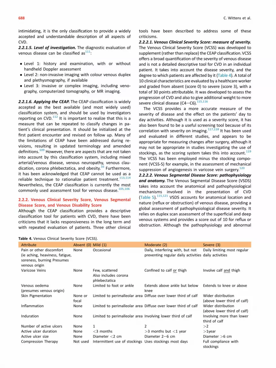
The Venous Clinical Severity Score (VCSS) was developed tosupplement (rather than replace) the CEAP classification.VCSSoffers a broad quantification of the severity of venous diseaseand is not a detailed descriptive tool for CVD in an individualpatient. It takes into account the disease severity, and thedegree to which patients are affected by it (Table 4). A total of10 clinical characteristics are evaluated by a healthcare workerand graded from absent (score 0) to severe (score 3), with atotal of 30 points attributable. It was developed to assess theprogression of CVD and also to give additional weight to moresevere clinical disease (C4eC6).115,116
The VCSS provides a more accurate measure of theseverity of disease and the effect on the patients’ day today activities. Although it is used as a severity score, it hasalso been found to be a useful screening tool because of itscorrelation with severity on imaging.117,118 It has been usedand evaluated in different studies, and appears to beappropriate for measuring changes after surgery, although itmay not be appropriate in studies investigating the use ofstockings, as the scoring system takes this into account.119
The VCSS has been employed minus the stocking compo-nent (VCSS-S) for example, in the assessment of mechanicalsuppression of angiogenesis in varicose vein surgery.120
2.2.2.2. Venous Segmental Disease Score: pathophysiology
and anatomy. The Venous Segmental Disease Score (VSDS)takes into account the anatomical and pathophysiologicalmechanisms involved in the presentation of CVD(Table 5).115,121 VSDS accounts for anatomical location andnature (reflux or obstruction) of venous disease, providing aglobal assessment of pathophysiological disease severity. Itrelies on duplex scan assessment of the superficial and deepvenous systems and provides a score out of 10 for reflux orobstruction. Although the pathophysiology and abnormal
Table 4. Venous Clinical Severity Score (VCSS).
Attribute Absent (0) Mild (1) Moderate (2) Severe (3)
Pain or other discomfort
(ie aching, heaviness, fatigue,
soreness, burning Presumes
venous origin
None Occasional Daily, interfering with, but not
preventing regular daily activities
Daily limiting most regular
daily activities
Varicose Veins None Few, scattered
Also includes corona
phlebectatica
Confined to calf or thigh Involve calf and thigh
Venous oedema
(presumes venous origin)
None Limited to foot or ankle Extends above ankle but below
knee
Extends to knee or above
Skin Pigmentation None or
focal
Limited to perimalleolar area Diffuse over lower third of calf Wider distribution
(above lower third of calf)
Inflammation None Limited to perimalleolar area Diffuse over lower third of calf Wider distribution
(above lower third of calf)
Induration None Limited to perimalleolar area Involving lower third of calf Involving more than lower
third of calf
Number of active ulcers None 1 2 >2
Active ulcer duration None <3 months >3 months but <1 year >1year
Active ulcer size None Diameter <2 cm Diameter 2e6 cm Diameter >6 cm
Compression Therapy Not used Intermittent use of stockings Uses stockings most days Full compliance with
stockings
688 C. Wittens et al.

venous segments can be described accurately using theadvanced CEAP classification, VSDS attributes differentscores to different venous segments to indicate the level ofoverall impact on venous function.
Reflux describes all valves in a specific segment as incom-petent. Obstruction describes a total occlusion at a point in theinvestigated segment or a >50% stenosis in at least half thesegment. Importantly, traumatic obstruction, ligation, orexcision of deep venous segments count as thrombosis.However, the same is not true for superficial veins. Perforatorinterruption and saphenous ligation/ablation count as areduction of the reflux score, not as an obstruction score.
VSDS was found to correlate with clinical scores, with themagnitude of reflux correlating with symptom severity.119
2.2.2.3. Venous Disability Score: functional impact. TheVenous Disability Score (VDS) provides a simple measure ofthe functional impact of CVD, using a 4 point scale (0e3;Table 6).115 This evaluates the effect of CVD on daily ac-tivities. VDS has been validated against the CEAP as ameasure of disease severity, and has been used as a
measure of change following venous surgery.119 As withVCSS, VDS is designed to complement the CEAP classifica-tion by providing greater detail on the level of disabilityexperienced by the patient.
2.2.3. Villalta-Prandoni Scale
The Villalta-Prandoni Scale was described in the 1990s toclassify the severity of post-thrombotic syndrome (PTS), acomplication of deep venous thrombosis.122 Essentially, thescale consists of five symptoms (patient rated) and six physicalsigns (clinician rated), with each of the 11 factors scored out of3 (total score out of 33; Table 7). A score of >14, or thepresence of venous ulceration, indicates severe PTS.
TheVillalta-Prandoni Scale is specific to the post-thromboticlimb and is a reliable, valid measure of PTS in patients withconfirmed deep venous thrombosis (DVT).123 It also correlateswell with patient perceived health burden and QoL scores. Adrawback of this scale is that it does not take into accountvenous claudication or venous ulcer severity, as the presenceof a venous ulcer is given a fixed score irrespective of severity.
2.3. Quality of life measures in venous disease
The burden of CVD lies with the patients, with up to 30%displaying symptoms suggestive of a depressive illness.124
Assessment of QoL in patients with CVD is integral to a com-plete and thoroughevaluationof their disease status. Evidenceshows that increasing clinical severity correlates strongly withdeterioration in QoL measures, both general and diseasespecific.113 Similarly, clinical improvement correlates withprogression in QoL measures.125 Clinical classification systems
Table 5. Venous Segmental Disease Score (VSDS).
Reflux Obstruction
½ Small saphenous
1 Great saphenous 1 Great saphenous (if thrombosed from groin to
below knee)
½ Thigh perforators
1 Calf perforators
2 Calf veins, multiple (Posterior Tibial only ¼ 1) 1 Calf veins, multiple
2 Popliteal vein 2 Popliteal vein
1 Femoral vein 1 Femoral vein
1 Profunda femoris vein 1 Profunda femoris vein
1 Common femoral vein and above 2 Common femoral vein
1 Iliac vein
1 Inferior Vena Cava
10 Maximum reflux score 10 Maximum obstruction score
Table 6. Venous Disability Score (VDS).
0 e Asymptomatic
1 e Symptomatic but able to carry out usual activities without
compressive therapy
2 e Able to carry out usual activities only with compression
and/or limb elevation
3 e Unable to carry out usual activities even with compression
and/or limb elevation
Usual activities: defined as patient activities before the onset
of disability from venous disease
Management of Chronic Venous Disease 689

are in place to assess the severity of CVD. QoL tools areavailable to assess patient reported outcomes. The ideal QoLtool should be generally applicable to any disease process,irrespective of severity, outcome measures, or geographiclocation.16 The tool should be valid (i.e. measure what isintended), reliable (i.e. provide the same measurements for asingle individual despite different conditions), and responsive(i.e. sensitive to assess change e.g. after treatment). Ideally, itshould also assess all aspects ofQoL, including physical,mentaland social wellbeing. A number of global QoL instrumentsexist; however, they lack sensitivity to changing clinical con-ditions. Health related measures are used instead. A largenumber of tools have been developed and are in widespreaduse. There have been greater efforts to standardize the use ofQoL assessments in recent years.
Generic and disease specific instruments measuring healthrelated QoL in patients with CVD are discussed below.
2.3.1. Health related generic tools
2.3.1.1. SF-36, Medical Outcomes Study 36 Item Short
Form. The SF-36 form is a widely used, generic QoLassessment tool with both physical and mental domains,providing a global assessment of patient wellbeing(Table 8).126 The physical component of this patientcompleted questionnaire has been shown to correlate withvenous disease severity. Studies have shown that all sub-domains of the physical component (physical role, pain,physical functioning, and general health perception) corre-late significantly with disease severity as measured by theCEAP classification. This is not true for the mental compo-nent, as correlations with vitality127 and mental health128
are weak and inconsistent.
2.3.1.2. EuroQoL, 5D. The EuroQoL group is a multinational,multicentre, and multidisciplinary network of researchersdedicated to the measurement of health status. The Euro-Qol questionnaire was devised in the 1990s with the aim ofdeveloping a standardized, simple, and generic measure ofhealth for clinical and economic appraisal.129 It consists of adescriptive part, evaluating five dimensions (EuroQol e 5D),and a vertical, visual analogue scale (VAS), recording therespondent’s self-rated health (EuroQol e VAS).
Together, the EuroQol, 5D and EuroQoL-VAS, provide acomprehensive measure of health state. This tool isparticularly useful for measuring utility or quality-adjustedlife years (QALYs; a measure of disease burden), and hasbeen used as a QoL measure in the assessment of patientswith symptomatic varicose veins (Table 9).124
2.3.2. Disease specific tools
2.3.2.1. Aberdeen Varicose Veins Questionnaire. TheAberdeen Varicose Veins Questionnaire (AVVQ), is a patientcompleted QoL assessment tool comprising 13 questionswith domains including physical symptoms, social effect,and cosmesis (Table 10).130 Each question is graded in
Table 7. Villalta-Prandoni Scale.
[severity scoring: none (0), mild (1), moderate (2), severe (3)]. Each
sign/symptom is scored 0e3; scores are added to obtain the final
result (maximum of 33).
5 Venous symptoms - Pain- Cramping- Heaviness- Pruritus- Paraesthesia
6 Clinical signs - Oedema- Induration- Hyperpigmentation- Venous ectasia- Redness- Calf tenderness
Severity of post thrombotic
syndrome (PTS)- No PTS <5
- Mild PTS 5-9
- Moderate PTS 10-14
- Severe PTS >14 orvenousulceration
- Total pointsrange 0e33
Table 8. SF-36.
- Physical function- Role physical- Bodily pain- General health
Physical component
- Mental health- Role emotional- Social function- Vitality
Mental component
Table 9. EuroQoL e 5D.
1. Mobility - No problems- Some problems- Bed bound
2. Self care - No problems- Some problems washingor dressing
- Unable to wash or dress
3. Usual activities - No problems- Some problems- Unable to perform
4. Pain/discomfort - None- Moderate- Extreme
5. Anxiety/depression - None- Moderately- Extremely
Euro e QoL VAS
Perceived health Visual analogue scale
0 (worst state)e100 (best state)
690 C. Wittens et al.

terms of severity/presence or absence, and the results arecollated into the Aberdeen Varicose Veins SymptomSeverity Score from 0 to 100, where the higher the score,the worse the QoL.
The AVVQ has been validated as a measure of healthoutcome in patients with varicose veins against the SF-36questionnaire.131 It was found to be reliable, with signifi-cant association with patient symptoms. Many consider theresponsiveness and sensitivity of the AVVQ to be greaterthan generic QoL questionnaires. However, generic QoL toolsallow simpler calculation of health utility (QALYs), which is anecessity for meaningful health economy comparisons.2.3.2.2. Chronic Venous Insufficiency Questionnaire.
Developed in 1996 in France, the Chronic Venous Insuffi-ciency Questionnaire (CIVIQ) is a 20-item self reporting QoLtool covering four dimensions: physical, psychological, socialfunctioning, and pain (Table 11).132 The items are graded ona 5 point Likert scale.133 The questionnaire has been vali-
dated in its French version, as well as in a number of otherlanguages.134,135
In 2010 psychometric validation was carried out, revali-dating the questionnaire and providing evidence for its
consistency, reliability, and value in assessing changes inQoL after treatment.136
2.3.2.3. Venous Insufficiency Epidemiological and Economic
study. The Venous Insufficiency Epidemiological and Eco-nomic study (VEINES) was an international, prospectivecohort study evaluating the epidemiology and outcomes ofCVD.137 As part of this project, a validated venous diseasespecific QoL and symptom measure was developed (VEINESQoL/Sym; Table 12).138 The aim of this tool was to provide anassessment of QoL and symptoms across the range of con-ditions in CVD (including telangiectasia, varicose veins,oedema, skin changes, and leg ulcers). Psychometric testingrevealed the questionnaire to be acceptable, reliable, andvalid in four different language versions, as well as demon-strating correlation with both SF-36 and C class. The VEINESQoL/Symwas also found to be reliable and valid as a measureof QoL and symptoms in patients with acute DVT.139
CHAPTER 3: DIAGNOSTICS
Introduction
This chapter describes the value of available diagnostic toolsused in patients with CVD. It describes the physical exami-nation and additional tests including continuous wave [CW]Doppler, duplex ultrasound [DUS], phlebography, plethys-mography, venous pressure measurement, and modern im-aging techniques such as magnetic resonance venography[MRV] and computed tomography venography [CTV], as wellas describing clinical and radiological diagnostic criteria ofrecurrent disease.
In the diagnostic work up the nature of the problem andthe severity of the disease should be determined.
3.1. Clinical examination
3.1.1. History
Scientific evidence. Patients with varicose veins and/orsigns of CVD should be asked, prior to any clinical or
Table 10. AVVQ.
1. Distribution of veins
2. Duration of pain
3. Duration of analgesia
4. Degree of ankle swelling
5. Use of support stockings
6. Extent of itching
7. Presence of discolouration
8. Presence of rash or eczema
9. Presence of skin ulcer
10. Degree of concern at appearance
11. Influence on choice of clothes
12. Interference with work/household jobs
13. Interference with leisure
(score 0e100; 0 best, 100 worst)
Management of Chronic Venous Disease 691

diagnostic investigation, about symptoms suggestive ofvenous pathology.140 This applies also to patients withrecurrent varicose veins following intervention, who maypresent with characteristic symptoms of CVD. Possiblethromboembolic antecedents should be investigated,together with any allergy, medication (oral contraceptivesprimarily), and concomitant relevant diseases includingheart and renal failure, which may influence CVD.140 Finally,the number and timing of pregnancies should be noted.141
A differential diagnosis is very important. Even in thepresence of trunk varices, many lower limb symptoms couldhave a non-venous cause.5,142
3.1.2. Physical examination
Scientific evidence. Patients with CVD are examined in aphysiological upright standing position. Both legs should beexamined completely.When signs of severe CVD or secondary(e.g. post-thrombotic) varices are present, the abdominal re-gion should be inspected for the possible presence of venouscollaterals. Venous collaterals on the lower abdomen, flanks,and pubic region are pathognomonic of iliac or ilio-cavaloutflow obstruction.
Corona phlebectatica paraplantaris should be noted asthis may indicate advanced venous stasis.143
In recurrent disease, it is important to bear in mind thepatient’s pre-operative state and assess any amelioration orworsening in signs such as skin changes or ulceration.
During physical examination, it is important to consideralternative pathology such as signs of arterial insufficiency,orthopaedic, rheumatological, or neurological pathology(muscle pump function). The main circumferences of bothlegs should be measured when indicated (e.g. phlebolym-phedema, suspicion of vascular malformations).
Traditional clinical tests such as Trendelenburg, Perthes, andothers have proven unreliable and have no place in the map-ping of venous incompetence in general, and of varicose veinsin particular.144,145
3.2. Diagnostic tools
3.2.1. Definition of reflux
Scientific evidence. In a study using DUS, reflux times in thevarious venous segments of the lower extremity were
examined.146 Distinctions were made between the iliacveins, the femoro-popliteal axis, deep veins in the calf, andsuperficial and perforating veins. Both normal subjects andpatients with known CVD were studied and compared,including the differences between supine and upright ex-amination by DUS.
When the duration of retrograde flow in patients withCVD was compared with healthy subjects, there was a sig-nificant (p < .0001) difference for all segments in theaffected leg. The cut off values defining venous incompe-tence (reflux) during ultrasound examination are set atretrograde flow longer than 0.5 s in the superficial venous
system, the deep femoral vein, and the calf veins, longerthan 1 s in the common femoral, femoral vein, and poplitealvein, and longer than 0.35 s in perforating veins.146
An additional finding of this study is that an erect posi-tion is the only reliable way to detect reflux.146
Previous international consensus held 0.5 s as a cut offvalue in all leg vein segments, but this appears to varywith the type of venous segment. The present consensusrecommends 1 s as the cut off duration for reflux infemoral and popliteal vein, whereas above 0.5 s isconsidered reflux in saphenous veins, lower leg veins, andperforators.146
The GSV, AASV, PASV, thigh extension, and SSV all sit-uated in their saphenous compartment, are the main su-perficial conduits to be imaged for morphology and testedfor possible reflux, and its segmental distribution.147 Mainthigh or lower leg perforators, mostly on the medialaspect of the limb, should be examined with diametersmeasured at fascia level. Perforators should also be testedfor their inward and/or outward flow during distal calfcompression (systole) and release (diastole).147 Saphenousdiameter should be measured at specific locations: theGSV 3 cm below the saphenofemoral junction, at mid-thigh, at the knee, and lower leg; the AASV 3 cm belowthe SFJ and at mid-thigh when still lying in its saphenouscompartment; and the SSV 3 cm below the SPJ.148 Theterminal and pre-terminal valves of GSV must be testedfor their function, as Cappelli demonstrated that GSV
692 C. Wittens et al.

diameter at proximal thigh is strictly related to the pres-ence or absence of reflux at the terminal valve and at theiliac-femoral valve.149,150
Assessment of venous reflux and of perforators is largelybased on DUS. Limitations diminish for the abdominal andor pelvic veins. Antegrade flow and reflux are elicitedthrough the creation of a pressure gradient, with specificmanoeuvres (e.g. Valsalva manoeuvre and compression/release manoeuvre). Patency of the main deep veins of the
lower limbs (common femoral vein, femoral vein, poplitealvein, gastrocnemius veins, peroneal and tibial veins) shouldbe highlighted, especially when detection of deep veinthrombosis or its sequelae (post-thrombotic syndrome) isrequired.
3.2.2. Handheld continuous wave Doppler
Scientific evidence. Doppler examination is a non-invasiveprocedure using ultrasound information to determinevenous flow. CW Doppler provides no information onvenous morphology, therefore it is unsuitable for thedetermination of any anatomical component of any venousdisease. Reliability of CW Doppler examination in detectingobstruction/reflux in deep veins is extremely low inabdominal and lower leg veins. Research has shown thatpre-operative planning on the basis of CW Doppler alone,instead of DUS, results in inadequate treatment in a sig-nificant proportion of patients.151 In a study of 40 patientsit was found that DUS is much more reliable than bothphysical examination and CW Doppler, with little differencein reliability between the last two.152
3.2.3. Duplex ultrasound examination
3.2.3.1. Efficacy. Scientific evidence. DUS examination isbased on a combination of ultrasound imaging and pulsedwave Doppler with which information can be obtained onboth the anatomy and the hemodynamic features of thevenous system. Additional colour flow imaging is routinelyemployed to quicken and improve DUS accuracy.
Anatomy, valvular incompetence, and venous obstructioncan be easily detected using DUS.153e157 With DUS it is alsopossible to investigate the deep venous system in mostsegments with adequate accuracy.158 In addition to goodreproducibility, the non-invasive nature of duplex scanningis a great asset. With the development of this technique,invasive tests such as phlebography have been reserved fora few selective indications (see 3.2.5), as well as makingformer non-invasive devices such as handheld Dopplerobsolete. DUS examination should be considered as thegold standard in the diagnosis of CVD.
However, DUS investigation has a lower reliability to elicitpatency, obstruction, or occlusion of deep veins in the lowerleg, whereas a higher accuracy has been reported in the
Table 11. CIVIQ e 20.
In the past four weeks, to what extent did your leg
problems interfere with./cause you.
Physical Items 1. Climbing stairs2. Crouching/Kneeling3. Walking briskly4. Doing the housework
Psychological
Items1. Feeling on edge2. Becoming tired easily3. Feeling like a burden to people4. Needing to take precautions5. Embarrassment to show one’s legs6. Being easily irritable7. Feeling handicapped8. Having difficulty getting going
in the morning9. Not feeling like going out
Social items 1. Going out in the evening2. Practicing a sport3. Travelling by car/bus/plane
Pain items 1. Pain in the ankles or legs2. Interference with work or daily
activities3. Interference with sleeping4. Interference with standing for
a long time
Management of Chronic Venous Disease 693

Table 12. VEINES QoL/Sym.
During the last 4 weeks how often did you have:
1. Heavy legs2. Aching legs3. Swelling4. Night cramps5. Heat or burning sensation6. Restless legs7. Throbbing8. Itching9. Tingling sensation
- Every day- Several times a week- Once a week- <Once a week- Never
At what time of day is the problem most intense? - On walking- At midday- At the end of the day- During the night- At any time of day- Never
Compared to one year ago, how would you rate your leg problem now? - Much better- Somewhat better- About the same- Somewhat worse- Much worse- I did not have a problem last year
Does your leg problem limit you in the following activities?
1. Daily activities at work2. Daily activities at home (housework)3. Standing for long periods4. Sitting for long periods
- I do not work- Yes, a lot- Yes, a little- Not limited at all
During the past 4 weeks, have you had any of the following at work/during your
day as a result of your leg problem?- Yes- No
1. Cut down the amount of time you spent at work/doing activities2. Accomplished less than you would like3. Limited in the kind of work or other activities4. Difficulty performing the work or other activities
During the past 4 weeks, to what extent has your leg problem interfered with your
normal social activities with family, friends, neighbours or groups?- Not at all- Slightly- Moderately- Quite a bit- Extremely
How much leg pain have you had during the past 4 weeks? - None- Very mild- Mild- Moderate- Severe- Very severe
How have you felt over the past 4 weeks as a result of your leg problem?
694 C. Wittens et al.

femoro-popliteal segment.159,160 Transvaginal DUS evalua-tion of pelvic veins is not yet fully defined.161,162
Additional colour flow imaging resulted in facilitation ofthe overall approach to the insonated segment to assesspatency and competence. In the post-thrombotic syndrome,DUS identifies residual obstruction, persistent occlusion,and valvular incompetence in the affected segments, withgreater accuracy in the infrainguinal areas. Phlebography orMRV/CTV have a definite role in the diagnostic work up ofthe veins in the abdominal/pelvic area.
With the introduction of duplex ultrasound, other non-invasive techniques such as Doppler and plethysmography(except certain parameters of air-plethysmography) havelost most of their value, and are no longer used in theroutine evaluation of CVD.
DUS is an ideal tool for follow up. DUS performed 1 yearafter surgery focusing on detection of neovascularization,has a high accuracy in predicting recurrence at the SFJ after5 years.163 DUS is also used in the pre-operative assessmentof patients undergoing surgery, as recommended by theclinical practice guidelines of the Society for Vascular Sur-gery and the American Venous Forum.140
3.2.3.2. Technique. The extent of DUS depends on thesymptoms. In severe CVD, after a deep vein thrombosis orpersistent or rapidly recurrent varicosities, it is important tofully scan the deep system including the iliac tract. Withprimary varicose veins it is sufficient to scan below the groinin most cases. When pelvic vein incompetence and orobstruction is suspected, complementary scanning in thepelvic area is recommended. Alternative methods (e.g. CTV,MRV, and especially phlebography) are to be used for amore complete diagnosis and for planning possibletreatment.
The five major components that define a complete DUSexamination are: anatomical information, flow visualization(presence or absence of reflux), provocation manoeuvresfor flow augmentation, morphology (patency or oblitera-tion), and compressibility (thrombosis diagnosis).
Standardized DUS examination to determine reflux insuperficial and perforating veins must be performed withthe patient in a physiological standing position with externalrotation of the examined limb in a relaxed position, whilesupporting the weight on the contralateral limb. Investiga-tion of patency of iliac and patency and incompetence ofcommon femoral veins should be performed with the pa-tient in supine position, whereas femoral and popliteal veinsegments should be investigated with the patient standing
as for competence, although patency also can be elicited insupine and prone position, respectively.147 Lower leg deepveins should to be examined with relaxed calf muscles.
High frequency broadband linear transducers are generallyused to investigate the lower limbs, whereas low frequencybroadband curved array transducers are used when investi-gating deeper veins (e.g. obese patients, abdominal veins).An appropriate transducer for transvaginal ultrasound maybe used in case of suspected pelvic vein incompetence. Anadequate pulse repetition frequency setting is required todetect low velocities (5e10 cm/s) and/or reflux.147
3.2.3.3. Imaging recurrent disease. Recurrent disease canbe reliably assessed by DUS. DUS can provide the necessaryanatomical and functional information about the nature ofrecurrence and is a fundamental component of the assess-ment of the lower limb after venous intervention.148 Studiesreporting on recurrent varicose veins largely use DUS ex-amination in their assessment of the venous system.164e167
As recurrent disease will be most likely secondary to inter-vention in the GSVor SSV systems, DUS is an appropriate, lowrisk imaging modality to assess these patients.
It is important to understand that DUS examination diag-nosed recurrence may be present in the absence of clinicalrecurrence. Patients may have recurrent reflux identifiedduring DUS examination that does not cause any clinicalsymptoms. The 5 year recurrence rate on DUS has been re-ported at 64%, with a clinical recurrence rate of 4%.168,170
3.2.4. Plethysmography and venous pressure measure-
ments
3.2.4.1. Strain-gauge plethysmography. Scientific evi-
dence. This was first described in 1953 by Whitney, and thendeveloped as a method for indirect venous function mea-surement.171 It was then developed for DVT detection or forquantification of post-thrombotic syndrome (PTS). By deter-mining a pressure volume relationship, this technique cantranslate the volume reduction into a pressure decrease.172
3.2.4.2. Photoplethysmography. Scientific evidence. Theprinciple of photoplethysmography (also known as lightreflection rheography), in which the transmission of lightreflection in the subdermal venous plexus is detected as ameasure of the change of blood volume in the skin, wasdescribed in 1937 by Hertzman.173 This technique wasinitially used for arterial research, but adapted by Abra-mowitz in 1979 for venous applications.174 In the 1980s,Wienert and Blazek developed the technique towards astandardized digital form.175,176
Table 12-continued
1. Concern about the appearance of your leg(s)?2. Irritable?3. Burden to your family or friends?4. Worried about bumping into things?5. Has the appearance of your leg(s) influenced your choice of clothing?
- All of the time- Most of the time- A good bit of the time- Some of the time- A little of the time- None of the time
Management of Chronic Venous Disease 695

The main parameter of venous plethysmography is therefill time. There is a good correlation between the refilltime and direct venous pressure measurement and photo-plethysmography.173,177 However, there is no good rela-tionship between the refill time and the degree of venousdisease.178,179
3.2.4.3. Air-plethysmography. Scientific evidence. Usingair-plethysmography (APG), volume changes can bemeasured, with venous filling index (VFI) being the mostimportant parameter.180,181 Also reflux quantification andejection fraction have been assessed and APG parametersgive an overall evaluation of the functional impairment ofthe limbs as to venous obstruction/valvular incompetence,quantifying calf pump dysfunction as well.85
Since the advance of DUS and other alternative methods,plethysmographical examination techniques have beenconsidered of less importance in routine investigation, andthey are no longer considered as stand alone diagnostic toolsin patients with CVD. Plethysmography may be considered forassessment of quantitative parameters related to venousfunction for research purposes and post-treatment follow up.3.2.4.4. Foot volumetry. Foot volumetry is performed in thestanding position by immersing the leg in a container withwater.182 The expelled volume (EV) in mL and the refillingrate (Q; mL/100 mL � minute), as well as the total footvolume (mL), are measured. It has been used to assesscompression treatment in venous insufficiency and to pre-dict results of interventions on the superficial venous sys-tem in case of venous ulceration.
3.2.5. Phlebography
Scientific evidence. The indication for using phlebography inCVD patients with varicose veins has decreased significantlywith the advent of DUS. In the evaluation of superficial,perforating, and deep vein incompetence, DUS is at least asreliable as phlebography.155,156 However, in the diagnosis ofpelvic vein obstruction or incompetence (gonadal veins,iliac veins) and of vascular malformations, when alternativeimaging techniques are inconclusive, phlebography canrepresent a necessary investigation.
In the presence of vascular malformation, complex post-thrombotic cases, or cases of complex recurrent varicoseveins, phlebography may help to elicit possible abdominaland/or pelvic vein involvement.
3.2.6. Other imaging methods
3.2.6.1. Scientific evidence. Both CTV and MRV haveevolved significantly in recent years and it is now possible toobtain detailed three dimensional reconstructions of thevenous system.183e188 Ilio-caval and pelvic venous pathol-ogy (post-thrombotic obstruction, venous compression/stenosis like Nutcracker syndrome or May-Thurner syn-drome and pelvic varicocele) can be reliably identified.189e193 However, there is insufficient scientific evidence toadequately judge the true effectiveness of both techniquesfor visualization of the venous vasculature given the het-erogeneity of the published studies. Furthermore, in vari-cose vein disease the use of CTV and MRV should be limitedto the specific indications mentioned above.
696 C. Wittens et al.

In patients with (severe) renal impairment, administra-tion of intravenous contrast in both MRV and CTV exami-nations may be contraindicated. A disadvantage of the MRVexamination is that the acquisition time is significantlylonger compared with CTV. Unlike MRV, however, CTV in-volves exposing the patient to radiation. Both techniquescan be used to diagnose deep venous obstruction, but MRVhas the advantage of displaying more heterogenic infor-mation of intravascular abnormalities, which may be usefulin determining the therapeutic (endovascular) options.183
The decision of whether to perform MRV or CTV is stillmainly dependent on the local expertise in performing andevaluating these studies.
Complementary to the aforementioned techniques,intravascular ultrasound (IVUS) is suitable for deter-mining ilio-caval venous compression.169,194e198 Toassess chronic venous obstruction or incompetence andfor planning a deep venous reconstruction, DUS exami-nation, CTV or MRV, ascending and descending phle-bography and in selected cases IVUS have to beperformed. In addition, venous pressure measurementcan be performed to clarify whether the venous collat-erals are capable of adequately reducing ambulatoryvenous hypertension.154,169,194,199e203
CHAPTER 4: TREATMENT OPTIONS IN CHRONIC
VENOUS DISEASE
Introduction
A variety of treatment methods are currently available forpatients with CVD, and these are presented in detail below.
4.1. Dressings for venous ulcers
Scientific evidence. A large number of types of wounddressing are in current use for venous ulcers. Two separateCochrane reviews concluded that alginate and foam dress-ings do not increase venous ulcer healing rates and thatmore research is needed before recommending them.204,205
Similar results have been shown for hydrocolloid dress-ings.206 There is also no evidence to support the routine useof silver donating dressings beneath compression forvenous ulceration.207
Another Cochrane review showed that the use of topicalcadexomer iodine was more effective than standard care inachieving complete healing when added to compressiontherapy (risk ratio 6.7, p ¼ .011). However, use of compres-sion with povidone iodine versus hydrocolloid dressings wasequivalent in achieving complete wound healing.208
Management of Chronic Venous Disease 697

Zinc oxide impregnated paste bandages have been shownto achieve better ulcer healing compared with alginatedressings and the zinc oxide stockingette.209 Furtherresearch is needed to investigate the role of zinc dressingson wound healing.
Local allergic side effects might limit the use of topicalantimicrobial dressings.
4.2. Compression therapy
Introduction. Compression therapy, despite significant im-provements in dressing materials and other methods, re-mains the cornerstone of conservative treatment. This isbecause of its ease of use, non-invasive nature, and alsoefficacy in managing venous hypertension, the main path-ophysiological mechanism of CVD.
The most common forms of therapeutic leg compressionare elastic stockings, including tights, non-elastic and elasticbandages (short and long stretch), and intermittent pneu-matic compression. The mechanisms of action includecompression of superficial and deep veins and improvementof the muscle pump function, both leading to reduction ofambulatory venous pressure and reduction of oedema.
4.2.1. Chronic venous disease without ulceration (C0eC4)
Scientific evidence. Elastic stockings in the form of graduatedcompression have been the cornerstone of conservativemanagement of CVD C0eC4 for decades. They compressvaricose veins, reduce venous reflux, and improve calf musclepump function. More recently it has been shown that pro-gressive graduated compressive stockings (higher pressure atthe calf comparedwith the ankle) aremore effective than theusual degressive graduated compressive stockings (higherpressure at the ankle) in improving pain and lower legsymptoms. Furthermore, they were easier to apply.210 Thesebeneficial effects might be related to the observation thatprogressive graduated compressive stockings have a morepronounced effect on venous pumping function than
graduated elastic compression.211 Higher pressure at calflevel duringwalking also seems to be the reasonwhy inelasticbandages are haemodynamically more effective than stock-ings, which give way during muscle contraction.212 Despitethe popularity of elastic stockings, evidence of their efficacy isunclear, based on the lack of randomized controlled trials forboth superficial venous incompetence and the post-thrombotic leg.213,214
However, there is ample lower quality evidence basedon non-RCTs and clinical experience, to suggest their usebecause they improve patient symptoms213 and theyimprove patients’ QoL.215 In general, there are issuesputting on and removing elastic stockings, especially inthe elderly, which along with the sensation of warmthand deterioration of pre-existing pruritis associated withvenous eczema, could account for the suboptimal patientcompliance.216 Additional issues include potential skindamage, and contraindications to use including periph-eral arterial disease. Also, elastic stockings require properfitting and, to remain effective, should be replaced atintervals of 3e4 months, according to the manufacturers’instructions.
4.2.2. Venous ulceration (C5eC6)
4.2.2.1. Venous ulcer healing. Scientific evidence.
Compression bandages have been shown to improve healingrate of ulcers compared with standard care withoutcompression.217e219 An alternative to traditional bandagingis non-elastic compression, including the Unna boot (acompression dressing impregnated with zinc oxide paste)and a non-elastic compression system based on adjustableVelcro bands that can be changed and adjusted daily elimi-nating odorous secretions.220 However, involving an elasticcomponent in a bandage system seems more effective interms of wound healing than when it is not involved.219
In a RCT, sustained compression of at least 40 mmHg witha four layer compression bandage over a week has been
698 C. Wittens et al.

shown to be more effective than lower grades ofcompression.221 However, other bandage types applyingsustained high pressure compression (including two orthree layer compression bandages, the Unna boot, pastebandages, or high compression stockings) have been shownby the vast majority of studies to be equally effective asfour layer compression bandages.222e230 This is likely to bethe result of improved properties of the former materials(including adhesiveness of bandaging material allowing it tobe kept in place) and familiarity of use.231 However, use of afour layer bandage does result in faster healing of ulcerscompared with a short stretch bandage.219
Until several trials with conclusive results favouring oneparticular compression system are published, the initialtreatment modality should be the one with which theperson applying the compression is most familiar. However,the differential properties of the various bandage typesdeserve further investigation.232
4.2.2.2. Venous ulcer recurrence. Scientific evidence.
Compression is important to prevent ulcer recurrence andmost benefit is seen from high compression; however, thisis more likely to be associated with patient intolerance.233
Following healing of the ulcer, compression bandagesshould be replaced by elastic stockings. The latter could bethe definitive treatment, particularly in patients with deepvein occlusion or gross incompetence not amenable towidely accepted surgical or interventional solutions. Pa-tients should wear the highest level of compression that iscomfortable, preferably 25e35 mmHg at ankle level.234 Inthe ESCHAR trial, compression in the form of four layerbandaging was shown to be equally effective with surgeryand compression in achieving healing of venous ulcers,although long-term effects were less durable in terms ofrecurrence compared with surgery.235
Another trial reached similar conclusions but surgerycombined with ambulatory compression therapy was moredurable than compression alone in patients with medialand/or recurrent ulceration, who should receive superficialand perforating vein surgery.236
4.2.3. Intermittent pneumatic compression for venous
ulceration
Scientific evidence. Intermittent pneumatic compression(IPC) improves venous flow significantly in patients usingelastic bandages,237 while, in combination with elastic stock-ings, it has been shown to increase healing rates and overallhealing of venous ulcers compared with elastic stockingsalone.238 Three subsequent trials either failed to demonstrateany benefit when IPC was used in conjunction with compres-sion,239 or this was marginally significant.240,241 This raisesconcerns regarding its efficacy.242 However, in a trial thatincluded patients with venous ulcers not using backgroundcompression, rapid IPC (one short compression period of 6.5 sthree times a minute) healed 86% of venous ulcers comparedwith 61% with slow IPC (one long compression period of 90 severy 3 minutes) at 6 month follow up, p ¼ .003.243
It has been proposed that IPC should be provided only forpatients with refractory oedema and significant leg ulcera-
tion after a 6 month treatment course, with standardmethods such as compression stockings, has failed.244
IPC is also effective in CVI and higher compressionpressures have been reported to be associated with greaterleg volume reduction in patients with chronic venousoedema.245 IPC is able to provide symptom relief in patientswith PTS.246,247
More research is needed to establish which kind ofcompression may, in combination with IPC, be most bene-ficial in ulcer healing. Also the IPC impact should be studiedin relation to ulcer characteristics (duration since onset,surface area and depth).
4.2.4. Compression after venous intervention
Scientific evidence. A meta-analysis has shown that shortduration (about a week) compression after varicose veinsurgery is as good regarding post-operative pain, leg vol-ume, incidence of complications and absence from work aslonger use (3e6 weeks) of compression.248 Additionally,after EVLA, use of high compression profile (35 mmHg)elastic stockings for 7 days significantly reduced pain and
Management of Chronic Venous Disease 699

improved both physical function and vitality whencompared with use for 2 days.249
Similarly, after foam sclerotherapy, compressionbandaging for 24 hours and thromboembolus deterrentstockings for a fortnight were equivalent to 5 days ofbandaging, confirming results shown by a previous studywith a similar design.250,251 However, use of low compres-sion profile elastic stockings after foam sclerotherapy oflarger veins had no effect on efficacy, side effects, andsatisfaction scores compared with a control group withoutcompression.252 Following superficial vein surgery forvenous ulceration, especially after GSV stripping to the kneeor residual deep or superficial venous reflux, continuationof compression could be considered.
Use of elastic stockings (23e32 mmHg) for 3 weeks aftersclerotherapy for leg telangiectasias, compared with nosuch intervention, was associated with an improved efficacyas determined by clinical vessel disappearance.253
Further studies are needed to establish the duration ofcompression as well as the type of compression (full lengthor knee length) following superficial vein surgery forulceration.
4.3. Physiotherapy, leg elevation, and leg massage
4.3.1. Physiotherapy for leg ulceration
Scientific evidence. A reduced range of joint mobility hasbeen described in CVD, correlating with the disease severity(CEAP clinical class) and haemodynamic changes on airplethysmography.254 A fixed ankle joint and reduced rangeof movement have been shown to be independent pa-rameters associated with non-healing of venous ulcers,indicating that these findings are related to the impairmentof the calf muscle pump.255 Furthermore, calf muscle pump
function and dynamic calf muscle strength can be improvedwith exercise,256,257 as can the range of ankle movement.258
One small underpowered RCT showed a non-significanttrend for reduced ulcer size with physiotherapy comparedwith no intervention.259 Another RCT showed that thegroup of patients who received lifestyle counselling hadincreased physical activity and reduced wound dayscompared with the control group, but a larger RCT of 40patients failed to demonstrate any effect of home basedprogressive resistance exercise on ulcer healingparameters.260,261
Properly powered studies are needed on the effect ofsupervised exercise on healing rates of venous ulcers.
4.3.2. Leg elevation
Scientific evidence. Leg elevation has been used for a longtime and is still recommended to patients to amelioratevenous stasis, provide symptomatic relief, reduce leg oedema,and promote healing of ulcers in patients with CVD.262,263
Leg elevation in patients with CVD classified as C3eC6has been shown to reduce leg volume and venous pressure,and to enhance the microcirculatory flow velocity in lip-odermatosclerotic skin.264e266
In the past, elevation has been used as the main measureto heal venous leg ulcers or as an adjunctive measure inpatients wearing elastic bandages.267,268 It is still practicedin combination with antibiotics and debridement in cases ofinfected ulcers where compression cannot be toleratedbecause of pain associated with cellulitis in the surroundingareas. In a retrospective study, elevation was a statisticallysignificant predictor of ulcer free survival among othervariables, while in a structured education programme, pa-tients who were allocated to the study group spent more
700 C. Wittens et al.

time with their legs elevated each day and had a reducedulcer recurrence rate.269,270 Others reported that in thepresence of compression, there was no correlation betweenmedian leg elevation per 24 hours and percentage decreasein ulcer size.265 Although still recommended, treatmentcannot rely on this otherwise simple advice, because pa-tient compliance is poor and ulcer recurrence is extremelycommon even after a period of prolonged hospitaliza-tion.271,272 Finally, leg elevation has been used beforebandages are applied and could also be used to reduce legswelling before elastic stockings are fitted.273
Prospective, randomized studies should be performedinvestigating the role of leg elevation in healing venous legulcers. But even with the lack of strong evidence, practiceand physiology suggest that leg elevation is helpful.
4.3.3. Leg massage
Scientific evidence. This method can be part of a multi-component regimen aimed at reducing tissue oedema byapplying deep massage around the ulcer area, before formalcompression is applied, or as an adjunct. Leg massage entailsa form of light massage over elastic compression with astocking.267,274 Further studies are necessary to assess therole of leg massage in patients with venous oedema, but IPChas largely replaced leg massage.
4.4. Medical treatment
Introduction. Medical treatment has been used for de-cades, but its place as a treatment modality for CVD is atopic of continuing debate. Venoactive drugs are widelyprescribed in some countries but are not available in others.They can be classified into two groups: natural and syn-thetic drugs, such as naftazone and calcium dobesilate,respectively. The main modes of action of venoactive drugsare to decrease capillary permeability, diminish release ofinflammatory mediators, or improve venous tone. Non-venoactive drugs like pentoxifylline, reduce white cell acti-vation, and acetylsalicylic acid inhibits platelet function andalso has an anti-inflammatory effect.
4.4.1. Chronic venous disease without ulceration (C0eC4)
Scientific evidence. In 2005, a Cochrane review of 110publications was published with 44 studies (CEAP classi-fication C0eC4) considered valid. This review concluded
that there was insufficient evidence to support the use ofall venoactive drugs in the treatment of CVD. However, italso showed that micronized purified flavonoid fraction(MPFF) was the most effective at reducing symptoms ofoedema and restless legs. Calcium dobesilate reducedcramps and restless legs. MPFF helped in healingvenous ulcerations and was also useful in treatment ofcramps and swelling. Rutosides decreased venousoedema.275,276
A Cochrane review of 17 randomized controlled trials(RCTs) showed that horse chestnut extract (HCSE) waseffective at decreasing oedema, pain, and itching.277
Numerous studies have been conducted especially withMPFF, showing that these products lead to improvementof symptoms by increasing the venous tone.278e282 The
RELIEF study included 5,052 CVD patients with clinicalclass C0 to C4. They were divided into two groups: withand without reflux. All patients received MPFF over 6months. Assessment was made of presence and/orseverity of pain, heaviness, sensation of swelling, andcramps in lower limbs. Oedema was measured using theLeg-O-Meter. In both treatment groups, MPFF significantlyreduced symptoms and signs, and reduced ankleoedema.125
Another RCT compared MPFF with placebo and couldonly show a difference in night cramps without changingother symptoms of CVD.283
Calcium dobesilate is a synthetic venoactive drugwhich has been evaluated in a few RCTs and one meta-analysis published in 2004. A recent large RCT with 509patients could not show a difference between its studygroups.284 However, a double blind, placebo controlledtrial showed that calcium dobesilate reduces leg oedemaand improves the symptoms of objectively diagnosedCVD independent of the concomitant usage ofcompression stockings.285
The effect of Red vine leaf extract was evaluated in aprospective randomized trial including 248 patients. Efficacyendpoints were changes in limb volume determined bywater displacement volumetry, clinical CVD symptomsassessed on a visual analogue scale and global efficacyevaluations. Red vine leaf extract reduced lower limb
Management of Chronic Venous Disease 701

volume and CVD related symptoms significantly more thanplacebo.286
4.4.2. Venous ulceration (C5eC6)
Scientific evidence. A Cochrane database review showedthat pentoxifylline is effective as an adjuvant therapy tocompression, but it also appeared to be effective in theabsence of compression.287 Other studies showed anaccelerating healing time when patients were treated withpentoxifylline,288,289 in addition to wound care.290
Other products, like acetylsalicylic acid and oral zinc, arenot routinely recommended for the promotion of healingvenous leg ulcers.291e294 Some studies showed an acceler-ation of healing in a patient group treated with acetylsali-cylic acid, but these were small studies and there was large
variation in the number and size of ulcers. Further researchis necessary on this subject.291e293
The effectiveness of routine use of systemic antibioticsfor venous ulcers could not be demonstrated in a Cochranereview based on 25 RCTs.208 However, in light of theincreasing problem of bacterial resistance to antibiotics,current guidelines recommend that antibacterial prepara-tions should only be used in cases of clinical infection andnot for bacterial colonization.
There is some evidence that sulodexide can support ulcerhealing in combination with compression therapy.295
Although it was thought likely that HCSE would attenuatethe pathogenesis of venous insufficiency and, in turn,facilitate venous ulcer healing, a study by Leach did notsupport this.296 A possible reason is that reducing oedemaalone is insufficient to treat venous ulcers.
The time to complete ulcer healing was compared in astudy of patients taking MPFF versus placebo.297 There wasa significant reduction in healing time in the treated groupcompared with the placebo group. Other symptoms, likeheavy sensation of the legs, were evaluated and were alsoshown to be reduced. Other studies have confirmed these
findings.298,299 However, according to a Cochrane review,the studies mentioned above showed shortcomings interms of blinding and allocation.300
In 2005, a meta-analysis of five RCTs was performed (723patients with venous ulcers). This showed that at 6 months,the chance of healing certain subtypes of ulcers was 32%higher in patients treated with MPFF as an adjunctive treat-ment in comparison with conventional therapy alone.301
A more recent review concluded that in prospectiverandomized studies, MPFF and other flavonoid de-rivatives, and pentoxifylline have demonstrated clinicalbenefits in patients with CVD (clinical class C4eC6).284
Therefore pharmacotherapy should be part of a rangeof treatment options in the modern management ofpatients with CVD.
4.5. Sclerotherapy
Introduction. Sclerotherapy involves injection of dilatedveins, including major refluxing trunks or tributary varicos-ities, venules, or telangiectasias, with liquid or foamchemical agents to damage the endothelium and eventuallyablate the veins.
Scientific evidence. A number of chemical agents e scle-rosants - with a variety of mechanisms of action have beenused to damage and denude the vessel endothelium,including polidocanol, sodium tetradecyl sulphate (STS),morrhuate sodium, glycerin, and hypertonic saline, eitheras pure agents or in the form of foam, in increasing con-centrations according to the size of vein being treated.Some of these have been mixed with lidocaine to reducepain during injection.302,303 Post-procedural compression isan integral part of this type of treatment, which can beused to treat the entire spectrum of refluxing superficialveins, including the saphenous veins, and telangiectasiaswith a great degree of efficacy and safety.304 The reader isreferred to section 4.2.4. for further information. However,patients should be informed of possible side effects (such
702 C. Wittens et al.

as skin necrosis, telangiectatic matting, hyperpigmentation,and also allergic reactions) and complications (such as deepvein thrombosis, pulmonary embolism, or cerebral embolicevent for foam sclerotherapy in the presence of a right toleft cardiac shunt).305,306
Liquid sclerotherapy is more effective than placebo in-jection for telangiectasias and reticular and/or varicoseveins.307,308 However, in two Cochrane reviews, there is noevidence suggesting superior efficacy of any one sclerosantover another.309,310 Lower concentrations of the sclerosantagent are used for smaller veins. In addition to effective-ness,307,311 sclerotherapy is less time consuming and is aneasily repeatable treatment. It provides faster recovery withless pain and is relatively inexpensive compared with sur-gery or endovenous thermal ablation (EVTA). However,varicose vein recurrence, as high as 90% after 6 years,312
and the need for additional treatment (more oftencompared with surgery) are inherent major problems,312,313
as summarized also by a Cochrane review.314 This is true notonly for the major saphenous trunks, but also for varicosetributaries if the source of incompetence has not beeneliminated.315 On the other hand, good long-term results inthe primary treatment of isolated varicose veins, notassociated with saphenous vein incompetence have beendemonstrated.312 Treatment of recurrent varicose veinsfollowing surgery is another indication, by avoiding themorbidity (40% in one study) associated with redosurgery.316
Foam sclerotherapy, usually guided by DUS (ultrasoundguided foam sclerotherapy, UGFS), has been shown to bemore effective than liquid sclerotherapy because of theenhanced sclerosing properties of the foam form of thesclerosant.317e320 Foam sclerotherapy has been used as
an adjuvant treatment of varicose tributaries followingendovenous ablation of the main refluxing trunk,321 orhigh ligation (HL),322 or to treat recurrent varicoseveins.323,324 It is also widely used to treat telangiectasiasand reticular veins, although there is no evidence thatfoam sclerotherapy is better than liquid sclerotherapy inthis setting.
UGFS is characterized by a higher recanalization ratewhen treating veins larger than 5e7 mm in diameter.325e327
Foam sclerotherapy is associated with a higher recanaliza-tion rate and similar results in terms of symptoms and QoLassessment when compared with surgery, laser or radio-frequency endovenous ablation for treating GSV reflux.328e331 Foam sclerotherapy proved significantly cheaper329 andit may be considered an attractive minimally invasivealternative to surgery to treat superficial incompe-tence,332,333 particularly in elderly and frail patients andespecially in those with venous leg ulcers.334,335 Repeattreatment may be considered an integral part of UGFS insome of the treated patients and the additional costincurred must be integrated into the global cost when it iscompared with the other techniques (surgery or thermalablation). Foam sclerotherapy is becoming an increasinglypopular choice in the management of patients with recur-rent varicose veins.315,324,332 Long catheter foam sclero-therapy has been introduced recently and its short-termefficacy is equal or superior to the usual UGFS for GSVreflux.336e339 Long-term results are awaited.
4.6. Transcutaneous laser
Introduction. Surface transcutaneous laser (TCL) has beenused for the treatment of telangiectasias and reticular veinssince the 1970s. The mechanism behind this treatment is to
Management of Chronic Venous Disease 703

cause endothelial injury of the vein by heating the hae-moglobin and eventually to obliterate the lumen. Theadvent of laser technology with delivery of sufficientlycontrolled energy has enabled achievement of pan-endothelial necrosis without affecting more superficialstructures such as the epidermal layer. High intensity pulsedlight therapy, which was introduced in 1990, represents afurther development in the field as it allows treatment ofreticular veins by emitting a spectrum of light rather than awavelength to obliterate the vein.
Scientific evidence. TCL is an effective treatment for tel-angiectasias and reticular veins, although not capable ofmaking them to disappear completely.340 Laser light isemitted from the laser equipment and transmitted throughthe skin to the targeted vessel, where it is selectivelyabsorbed by the oxyhaemoglobin and converted to thermalenergy leading to heating of the telangiectatic vein, coag-ulation, and vessel destruction. Different settings of laserare used for blood vessels of different diameter and skintype and/or colour.341,342
TCL is less effective than sclerotherapy at managing legtelangiectasias, requires more treatment sessions,343,344 andis more expensive. However, particular indications include: 1.known allergy to sclerosant solutions, 2. needle phobia, 3.telangiectatic matting after sclerotherapy, 4. failure ofsclerotherapy,345 and 5. vessel size less than 0.5 mm.
Recent technical modifications have been reported tosignificantly improve the efficacy of TCL.346,347 Indocyaninegreen (ICG)-augmented diode laser therapy has been shownto be superior to pulsed dye laser and the diode laserwithout ICG.346 Similarly, improved results have beendemonstrated in combination with sclerotherapy.348
More studies are needed to establish which telangiectaticveins, and at what site in the lower limb, are bettermanaged with TCL. Also, the role of elastic compressionafter TCL should be further investigated.
4.7. Endovenous treatments
Introduction. Endovenous techniques in the treatment ofsaphenous vein incompetence have become very popular asa minimally invasive alternative to classical surgery (HL andstripping). In countries where reimbursement is available,the vast majority of patients are treated endovenously. Itwas Carlos Boné in 1999, who first treated patients withendovenous laser.349 In the past 10 years, endovenoustechniques have evolved quickly and performance hasimproved.
The two most frequently used techniques are endove-nous laser ablation (EVLA) and radiofrequency ablation
(RFA). Another endovenous thermal ablation (EVTA) tech-nique is steam ablation.350 Other more recently introducedtechniques are mechanochemical ablation (MOCA)351 andinjection of cyanoacrylate glue.352
The use of EVTA (EVLA, RFA, Steam) techniques requiresinjection of tumescent liquid around the target vein. Thepurpose of this is to protect the perivenous tissue from theheat created during treatment. The tumescent liquid acts asa heat sink.353 Other purposes of the tumescent liquid in-jection are to create spasm of the vein and to obtain localcompression and anaesthesia. The new non-thermal abla-tion techniques (MOCA and glue injection) can be per-formed without injection of tumescence during theablation.
The scientific evidence for cyanoacrylate glue injection istoo little to warrant further attention in this chapter becauseof the lack of publications. Steam ablation seems to giveresults comparable with other thermal ablation techniques at1 year follow up,354 but more studies are necessary toconfirm those results in the medium and long term.
4.7.1. Endovenous thermal ablation
Introduction. To evaluate the two most commonly usedEVTA techniques, EVLA and RFA, six meta-analyses,355e360
31 RCTs,328,361e390 six non-randomized comparative clin-ical trials,391e396 seven prospective clinical trials,397e403 andone technical review article were selected.404
4.7.1.1. Great saphenous vein. Technique. The technique issimilar for all EVTA methods. The procedure is performedpercutaneously with ultrasound guidance. A laserfibre orRFA catheter is inserted and positioned 1e2 cm distal tothe SFJ, and tumescent liquid is injected around thesaphenous vein. A very dilute solution of local anaestheticcombined with bicarbonate and epinephrine is used for thispurpose. It is important to empty the vein of blood asmuch as possible. While withdrawing the catheter or fibre,energy is emitted intraluminally to cause irreversible ther-
mal destruction of the endothelium of the vein wall.Compression post intervention is recommended (see sec-tion 4.2.4.), but the duration of compression is not clearlydefined.405,406
Effectiveness. Occlusion rates of EVLA vary between 77%365
and 99%362 at 1 year. Compared with surgery (HL andstripping), most studies report no significant difference invaricose vein recurrence (clinical) or recurrent reflux (ac-cording to DUS) after treatment.328,356,364e366,368e370,391
Other studies report a higher recurrence rate after sur-gery (20% vs. 4% at 1 year)362 or after EVLA (7.4% vs. 0% at2 years).363 Neovascularization at the SFJ is an important
704 C. Wittens et al.

cause of varicose vein recurrence after treatment. This wasseen more commonly after surgery, although this was notsignificantly different,359 compared with EVTA.366,372
Compared with RFA, surgery seems to be more effi-cient373,376 orequally effective.371,372 In cases in which sur-gery was more efficient, RFA was performed using the older“Closure Plus” catheter (VNUS Med Tech, San Jose, CA,USA). Meta-analyses show that RFA and EVLA are aseffective as surgery in the treatment of GSV incompe-tence.355,357,359 The first published meta-analysis on EVTAreports superior results of EVLA compared with surgery andRFA.355 This study, however, only includes trials using eitherEVLA with lower wavelength or RFA with the “Closure Plus”catheter, and uses heterogeneous definitions of treatmentfailure.Safety. No difference in safety including the risk of venousthromboembolism could be determined, comparing EVTAand surgical treatment for saphenous veinincompetence.367,391
The reported incidence of DVT varies between 0.2% and1.3% in EVTA, and seems to be higher in patients treatedwith RFA compared with EVLA.357 The latter meta-analysismainly included the old RFA techniques and EVLA withbare tip fibres and lower wavelength. Development ofthrombus extension at the SFJ (often called endovenousheat induced thrombosis) can be found in a limitednumber of cases. Case series report a frequency varyingbetween 0.3% and 7.8% of patients after EVTA.395,397e399
When thrombus extends into the common femoral veintreatment by anticoagulation (AC) is advised. Pulmonaryembolism was reported in 0.0% to 3% after EVLA.395,398
Nevertheless, the role of routine use of pharmacologicprophylaxis remains uncertain and more studies arenecessary.
Thrombo-prophylaxis can be prescribed for high risk pa-tients (previous venous thromboembolism, documentedthrombophilia, obesity, immobilized patients, patients withneoplasm, and older patients).407,408 However, risk factorsshould be weighted for each individual patient using aspecific risk assessment score such as the Caprini score.409
Patients can be treated in an outpatient setting underlocal tumescent anaesthesia, which permits early ambula-tion reducing the risk of possible thromboemboliccomplications.Side effects. Post-operative complications in EVTA arelimited. Reported complications are thrombophlebitis(7%),359 thermal skin injury (<1%),359 bruising, hyperpig-mentation (5%),359 paresthesia (1e2%),364,369 and haema-tomas (0e7%).364,369,374,378 Compared with EVTA, surgicallytreated patients more frequently develop complications,such as wound infection (2e6% for surgery vs. 0% forEVTA)356 and haematomas (5% for surgery vs. 2% forEVTA).359 Patients treated with EVTA have, on average, lesspain compared with patients treated surgi-cally.356,365,369,374,377,378 Post EVTA as opposed to surgery,swelling and bruising is reduced.391 This results in quickerrecovery357,362,375,376 and faster return to normal activ-ities.364,365,374,378,392 Endovenously treated patients also
seem to have an equal364,366,367 or better361 QoL aftertreatment. The risk of superficial thrombophlebitis appearsto be significantly higher (2.3 times) in RFA than insurgery.356
EVLA: different wavelengths and fibres. To reduce possibleside effects of EVLA (pain, bruising, and haematoma),higher laser wavelengths have been introduced anddifferent fibre tips have been developed. Theoretically, lightof lower wavelength lasers (810, 940, 980, 1320 nm) is lessspecifically absorbed by their chromophores (haemoglobin,water, proteins) compared with the light of higher wave-length lasers (1320, 1470, 1500 nm).404
The clinical use of higher wavelength lasers shouldresult in equal occlusion rates (97e100%) and less post-operative pain.396,402,403 There is only one randomizedclinical trial comparing the use of lower and higherwavelength lasers.379 Also, most studies reporting resultsof higher wavelengths use a lower energy level whichmakes it difficult to draw any conclusions regardingoutcome and potential side effects. As the use of a barefibre creates unequal energy delivery at the vein wallresulting in local vein wall perforations and perivenoustissue destruction, new fibres have beendesigned.380,400,404 The purpose of these new fibre designsis to increase the heated surface area, resulting in lowerenergy density. The use of the these new fibres, Never-touch (AngioDynamics, Latham, NY, USA),393 Radial fibre(Biolitec, Wien, Austria),394,400 and Tulip fibre (Tobrix,Waalre, the Netherlands),380 may be safe and effective inthe treatment of saphenous vein reflux.
There is a lack of RCTs comparing these fibres with astandard bare fibre. Only one RCT reports fewer side effectsand an equal occlusion rate using a Tulip fibre comparedwith a bare fibre.380
RFA versus EVLA. EVLA and RFA have the same occlusionrates, but patients treated with RFA have less post-operative pain and bruising.328,381e384 This can result inan equal383 or faster328 return to normal activities. In trialscomparing EVLA and RFA, however, the ClosureFast cath-eter was compared with lower wavelength lasers using abare fibre. No trials have been published comparing the useof ClosureFast with higher wavelength lasers and new fibretip design.
Consideration must be given to the fact that in the lastdecade the technique and the fibres or catheters used haveimproved. The more recently introduced ClosureFast cath-eter (VNUS Med Tech, San Jose, CA, USA), although no RCTsare available, seems to be more effective, faster (shorteroperative time), and induces fewer side effects comparedwith the older “Closure Plus” catheter.401
UGFS versus EVTA/surgery. The advantage of foam sclero-therapy is its simplicity. There is no need to inject tumescentliquid, the treatment is cheap, and easy to repeat ifnecessary.
Complications of UGFS include hyperpigmentation,thrombophlebitis, matting, and pain at the injection site.Also, some neurologic events, such as visual disturbances,migraine, and stroke, have been reported.410e413
Management of Chronic Venous Disease 705

Myers et al. reported primary and secondary success ratesof 52.4% and 76.8% respectively.327 Compared with surgery,UGFS is less efficient328,329,355,356,414,415 but has significantlyfewer side effects (less pain, better post-operative QoL, fasterreturn to normal activities).322,328,329,356,414e416
Occlusion rates of saphenous veins treated with UGFSseem to be inferior to those of veins treated withEVTA.355,356,361 Compared with EVLA, patients treated withUGFS have somewhat less post-operative pain, but no dif-ference could be found compared with RFA.328
The results (occlusion rates and side effects) of EVTA in thetreatment of saphenous vein reflux also depend on a goodtechnique (e.g. detailed pre-operative ultrasound, correctpositioning of the catheter or the fibre tip, targeting injectionof tumescent fluid, regular pull back speed). The vein to betreated should be emptied of blood far as possible.
4.7.1.2. Small saphenous vein. Scientific evidence. EVTA ofthe SSV has excellent early and mid-term results.386,388,389
Access at the lateral malleolus results in a higher pares-thesia rate compared with mid-calf access, because of theproximity of the sural nerve to the SSV in the distal part of thecalf.386 EVLA of the SSV is associated with a significantlyhigher incidence of sensory disturbance compared with EVLAof the GSV.387 SPJ ligation and stripping of the SSV often failsbecause of the complex anatomy.360
Compared with surgery, EVTA seems to be more efficientand results in fewer post-operative side effects (less pares-thesia, pain, and a faster return to normal activities).388
UGFS for the treatment of SSV incompetence can be analternative to EVTA and surgery. Two articles on UGFSshowed success rates varying from 82% to 100% (follow up1.5e60 months). Thrombophlebitis (5%) and hyperpig-mentation (24%) were common complications.326,360,417
There are no RCTs comparing foam sclerotherapy withsurgery or EVTA in treatment of SSV incompetence.
4.7.2. Mechanochemical endovenous ablation
Introduction. Recently, a new hybrid (dual injury) techniquehas been developed (ClariVein Vascular Insights, Quincy, MA,USA). Endomechanical abrasion is produced by the tip of the
catheter’s rotating wire (mechanical component) and endo-venous chemical ablation via the simultaneous injection ofsclerosant over the rotating wire (chemical component). Thistechnique is under development as the optimal dosage ofsclerosans still needs to be determined.Scientific evidence. To date, there have been only twocohort studies including a small number of patients treatedwith MOCA.351,418 The occlusion rate at 6 months is 96.7%and side effects seem to be minimal.
4.8. Surgery of the superficial veins
Introduction. For many years the gold standard for treat-ment for CVD patients with superficial venous incompe-tence was surgery.
4.8.1. High ligation with/without stripping
Scientific evidence. It has been reported that surgicaltreatment of varicose veins is superior to conservativemanagement. In the REACTIV trial,419 the results of sur-gery were compared with results of compression alone in246 patients with uncomplicated varicose veins, thesurgical treatment comprising high ligation/stripping (HL/S) of the GSV and multiple phlebectomies.420 At 2-yearfollow up the results showed more symptomatic relief,better cosmetic results, and much improved QoL forsurgery compared with conservative management bycompression.
In a prospective cohort study, 203 consecutive patientswho underwent varicose vein surgery were monitored, andreported an improvement in QoL 2 years after surgery, witha significant improvement in the health related AVVQ scoreand the SF-36 score.421 In addition, improvement in QoLfrom varicose vein surgery has been shown to be statisti-cally significant and clinically relevant.422
Comparative studies relating to traditional surgicaltreatment compare isolated HL and HL/S.423e426 In a ran-domized trial in 100 patients and 133 legs, HL wascompared with HL/S.424 The need for reoperation was 6% in
706 C. Wittens et al.

patients who underwent HL/S versus 20% in those patientswho underwent HL alone (p < .02). In another study it wasshown that the HL group and the sclerotherapy group withintention to close the GSV, had significantly higher recur-rence rates than HL/S at 4 years.425 Cosmetic results, bothjudged by the patient and the surgeon, were significantlybetter (p < .05) in the stripped limbs than in the limbs withHL and sclerotherapy.426 They also found that CW Dopplerevidence of reflux of the saphenous vein was less frequent(p < 0.001) after the stripping operation. Better resultsafter aggressive and extensive surgical treatment of therefluxing veins and the sources of the reflux were reported.A randomized trial showed a significant reduction of risk ofre-operation in a HL/S group compared with a HL alonegroup at 11 years of follow up, even if there was no dif-ference for the rate of visible recurrent veins.423
In a RCT it was shown that patients in whom the stump ofthe GSV had been invaginated with a non-absorbable suture,had less neovascularization in comparison with those whohad the endothelium of the stump exposed.427 According toa single centre prospective cohort study, interposition of ananatomical barrier, by closing the cribriform fascia after SFJligation, also reduced ultrasound detected neovascularizationat the SFJ after 1 year.428 In a RCT of 389 limbs by van Rij,polytetrafluoroethylene (PTFE) patch coverage of the SFJcreated a significant reduction of recurrence at 3 year followup by reducing neovascularization at the groin.120 The po-tential benefit of inserting a PTFE patch in recurrent varicosevein operations could not be proven in another small RCT.429
Traditional HL/S has evolved towards a less invasivetechnique by invagination under tumescent local anaes-thesia, giving post-operative results as good as endovenousablation techniques or foam sclerotherapy in terms of
haematoma, pain, and QoL as shown by Rasmussen in arandomized control study.328 A non-comparative prospectivestudy showed that the use of isotonic sodium bicarbonate asexcipient for tumescent anaesthesia reduced intra-operativepain and improved cost-effectiveness of surgery.430
As endovenous procedures have shown good resultswithout HL, stripping of the saphenous vein has also beendescribed without HL. A retrospective study showed thatstripping of the GSVwithout HL led to low neovascularizationand SFJ reflux rates at 3 year follow up (0.9% and 1.8%,respectively).431 In a RCT at 8 year follow up, a group treatedby stripping without HL had significantly less recurrence thana group treated by HL/S (9.8% vs. 29%, p ¼ .014).432
Several RCTs have been performed comparing HL/s withendovenous techniques (EVLA, RFA, foam sclerotherapy).Among them, Rasmussen published a four arm RCTcomparing HL/S under tumescent local anaesthesia byinvagination stripping with EVLA, RFA, and foam sclero-therapy. The results showed that the post-operative averagepain scores at 10 days were significantly lower in the groupstreated with RFA and foam sclerotherapy compared withHL/S and EVLA, with a shorter time to resumption of normalactivities and work. But the VCSS, the AVVQ scores, and theclinical recurrence were not different between HL/S and theother treatments at 3 years. The conclusion was thatbeyond a higher pain score in the post-operative period anda higher total cost for HL/S and EVLA, the efficiency of thefour modalities was not significantly different.433
There is no evidence in the literature for continuing in-dications for HL/S, unless there is lack of availability of theendovenous techniques for financial or other reasons. Onthe other hand, HL/S is not inferior compared with the newmodalities of treatment in the mid term.
Management of Chronic Venous Disease 707

4.8.2. Phlebectomy
Scientific evidence. The technique of ambulatory phlebec-tomy (AP; stab or hook or mini-phlebectomy) was describedby Muller in 1966 with removal or avulsion of varicose veinsthrough small incisions performed with hooks and for-ceps.15 Numerous authors have reported that ambulatoryphlebectomy is a safe and immediately effective procedurefor the treatment of varicose veins,315,434e438 which can bedone under local anaesthesia in an office based setting.
Phlebectomy can be considered as an adjunctive treat-ment in association with stripping or endovenous ablation ofthe main refluxing truncal vein,439e442 or as the exclusiveprocedure for the treatment of varicose veins.15,315,435,443,444
A randomized study showed that ambulatory phlebec-tomy compared with liquid sclerotherapy plus compressionfor accessory vein incompetence caused significantly fewerrecurrences at 1 and 2 years of follow up (respectively, 1/48vs. 12/48 and 1/48 vs. 18/48 p < .001).315
Some publications advocate limiting the treatment tothermal ablation of the SV without treating the tributaryvaricose veins.445,446 According to these papers, secondarytreatment (foam sclerotherapy, phlebectomy, stripping) forresidual varicose veins was only necessary in 17e62.5%after a short follow up period (3e9 months). On the con-trary, another prospective study in 67 patients showed thatthe performance of concomitantly performed phlebectomywith an EVLA of the saphenous vein obviates the need forsubsequent procedures.447 The authors found that anadditional treatment by sclerotherapy or by phlebectomywas performed in only 4% and 1% at 1 and 12 weeks. In arandomized study of 50 patients between EVLA alone andEVLA with concomitant ambulatory phlebectomy (EVLA/AP), it was shown that at 3 months in the EVLA/AP groupthe requirement for a secondary procedure was less com-mon (1/25 vs. 16/24, p < .001), the VCSS was lower(0(0 þ 1) vs. 2 (0e2), p < .001), and the AVVQ score wasbetter (7.9 vs. 13.5 p < .001).448
4.8.3. Ambulatory Selective Varices Ablation under Local
anaesthesia
Introduction. The Ambulatory Selective Varices Ablationunder Local anaesthesia (ASVAL) method, consisting of singlephlebectomies with preservation of the saphenous trunk, isbased on the concept of ascending or multifocal evolution ofvaricose veins. According to this pathophysiological concept,progression of the disease could begin in subcuticular veins,outside the saphenous compartment, creating a dilated andrefluxing venous network. When this refluxing network be-comes large enough, it could create a “filling” effect in the
intrafascial saphenous vein, leading to decompensation ofthe saphenous vein wall, moving on to eventually reach thesaphenofemoral or saphenopopliteal junction.Scientific evidence. The theory for the development ofvaricose veins is historically based on the descending theory.Anatomical, DUS examination, and morphological informa-tion confirmed the theory that the reflux begins at thesaphenous junctions and progresses downwards through thesaphenous axis leading to venous hypertension, wall dilata-tion, and dilatation of tributaries which become varicoseveins. Cotton reported in an anatomical study that normalveins have a significantly higher number of valves than vari-cose veins.90 Cooper assessed in a retrospective analysis ofvenous DUS examinations that the incompetence of the SFJhas a major influence on the extension of the reflux and thatthe venous incompetence was most commonly proximal inpresence of varicose veins.449 Takase described numerousalterations in structure of valves induced by venous hyper-tension in association with varicose veins.450
However, numerous publications challenge the theory ofdescending disease progression, citing the possibility of localor multifocal early distal evolution, sometimes ascending oranterograde, based on precise and detailed DUS in-vestigations.49,431,451e456 The fact that the terminal valve atthe SFJ is frequently competent (in >50% of cases) in thepresence of truncal reflux has been clearly proven.449,457e459
The disappearance of reflux in the GSV following phle-bectomy460e463 or after thermal ablation of an incompetenttributary464 has been reported in the literature. Quill andFegan also found such reversibility of the saphenous refluxfollowing sclerotherapy.465 The diameter of the GSV wasobserved to be reduced after ablating a refluxingtributary.466
In addition, the evolution of CVD, whether ascendingor descending, remains largely unknown, as prospectivelongitudinal studies with a lengthy period of observationand a large sample size, are missing and RCTs are needed.
A retrospective cohort of 303 limbs treated by ASVAL re-ported complete abolition of the saphenous reflux after 1, 2,3, and 4 years in 69.2%, 68.7%, 68.0%, and 66.3% of cases,respectively, and reduction in SFJ vein diameter, markedsymptom relief, and low recurrence rates.431 In this study, thecohort of patients operated onwith ASVALwas younger,morefrequently asymptomatic, with a less extensive reflux, andlower average diameter of the SFJ comparedwith the patientswho underwent stripping during the same period of time.
In a prospective study including 94 patients with largevaricose tributaries and GSV reflux assessed by DUS
708 C. Wittens et al.

examination, single phlebectomies led to abolition of theGSV reflux in 50% of the cases, with a significant reductionof the GSV diameter and a significant improvement of theAVVQ score.467
4.8.4. Cure conservatrice et Hémodynamique de l’Insuf-
fisance Veineuse en Ambulatoire (CHIVA)
Introduction. CHIVA was described by Franceschi in 1988. Itaims to improve the haemodynamics of the superficialvenous network by splitting the column of hydrostaticpressure and disconnecting venovenous shunts by inter-rupting the incompetent trunks at strategic levels (SFJ,perforating veins) depending on a precise pre-operativeDUS examination, to obtain a well-drained superficialvenous system with low pressure and high flow.Scientific evidence. One RCT, concerned with treatment ofvenous ulcers, showed that the group treated by CHIVA hada similar healing rate (100% vs. 96%) and a lower recurrencerate (9% vs. 38%) at 3 years follow up than the grouptreated by compression.468
The clinical and DUS recurrence of varicose veins wasstudied in two RCTs: the results were in favour of CHIVAversus HL/S in both studies, 8% vs. 35% (p < .004) at 10year follow up in one study469 and 31.1% vs. 52.7%(p < .001) at 5 year follow up in the other.470
Some observations must be noted: in the paper byCarandina, there is an important bias concerning therandomization and follow up.469 In another study allstripping procedures were done under general orepidural anaesthesia whereas the CHIVA treatments wereperformed under local anaesthesia, which acted as aconfounder for the evaluation of the post-operative sideeffects.470 The most serious limiting concerns in bothstudies were how “failure” by recurrence was defined: itis unclear if the presence of visible recurrent varicoseveins or the presence of refluxing veins during the DUSevaluation or both were considered to define the failureof the treatment.
4.8.5. Powered phlebectomy
Introduction. Transilluminated powered phlebectomy (TIPP)consists of removal of large varicose vein clusters, with adecrease in the number of incisions needed and a faster
performance of the procedure, often combined withsaphenous vein ablation procedures or HL/S.Scientific evidence. A published series of 114 patients, in aprospective, non-comparative, multicentre, pilot study re-ported the safety and efficacy of TIPP.471 The patients, anindependent nurse, and the surgeon subjectively scored theevaluation of outcomes in this study.
Other authors have stated in observational studies that alearning curve exists to minimize bruising and that otherlocal complications may vary.472e474 Complicationsobserved include haematoma (4.9e95%), paresthesias andnerve injury (9.5e39%), skin perforation (1.2e5%), super-ficial phlebitis (2.4e13%), swelling (5e17.5%), and hyper-pigmentation (1.2e3.3%).475,476
More recently, a new system with important technicalmodifications (lower oscillation frequency, larger volume oftumescence, dermal drainage, flushing of hematoma, andresidual tissue fragments), has improved the results of TIPPwith a lower rate of complications.477
A limited number of studies with many limitations (pilotstudy, non-randomized, observational) have been publishedand clearly lack data to show a significant advantage of TIPPover phlebectomy. It seems that the newer generationsystem and modified technique enable TIPP to be lessinvasive. Further studies are required to confirm the po-tential benefits of TIPP.
4.9. Treatment of deep vein pathology
Introduction. Annually one to two adults per 1,000 in theWestern population develop a DVT of the lower extremity.Treatments using low molecular weight heparin (LMWH) orunfractionated heparin and vitamin K antagonists in theacute phase will not be discussed here. Despite adequate
Management of Chronic Venous Disease 709

treatment according to this regimen, residual venous le-sions occur in a substantial number of these patients (20e50%). The incidence of residual lesions is greater after arecurrent DVT in the ipsilateral leg aggravating venous hy-pertension because of deep venous obstruction andvalvular incompetence. The effect of this is the develop-ment of oedema and dysfunction of the (micro)circulation,which can lead to the clinical characteristics of post-thrombotic syndrome (PTS). Also in well or partiallyrecanalized veins, residual venous obstruction may beimportant, because the scarred venous outflow causesincreased outflow resistance, especially during and afteractivity. As a result of chronic venous obstruction, collat-erals develop, which partly take over the outflow capacityof the thrombosed venous segments. However, these col-laterals often feature a relatively low outflow capacity, andare mainly used when venous pressure rises significantly.These collaterals are clinically visible at the level of thethigh, lower abdomen, and pubis. Less commonly, thecause of obstruction is extrinsic compression on the vein,for example by a malignant tumour. There are also specificiliac vein compression syndromes caused by non-thrombotic iliac vein lesions (NIVL) such as May-Thurnersyndrome, in which the left common iliac vein is com-pressed between the right iliac artery and the spine. Thereare several forms of congenital deep venous anomaliescausing obstruction of the outflow tract. One severeanomaly is atresia (congenital absence) of the IVC in whichthe sub-hepatic segment of the IVC has not developed. Thismay cause CVD because of outflow obstruction, oftencomplicated by recurrent ilio-femoral DVT and subsequentpost-thrombotic problems.478
In patients who have suffered from extensive DVT, apartfrom residual venous obstruction, deep venous reflux mayoccur, secondary to valvular damage. However, deep venousreflux can also be induced by a very important proximaloutflow obstruction, causing dilatation of the deep veins inthe leg, and eventually failure of the valves to closecompletely. The increased hydrostatic pressure leads tovenous hypertension, damage of the microcirculation, andsymptoms and signs of CVD. In some patients there may bea congenital hypo- or aplasia of valves in the deep venoussystem. This form of deep venous reflux is very rare.
The current standard treatment for symptoms and signsof chronic venous obstruction or deep venous reflux iscompression therapy. However, its effectiveness may besuboptimal in selected cases, especially when compressiontherapy has limited effect on venous symptoms or thecompliance is poor.
4.9.1. Treatment of chronic deep venous obstruction
This can be performed either by percutaneous transluminalangioplasty (PTA) and stenting, or by surgical correction,usually bypass.4.9.1.1. Percutaneous transluminal angioplasty and stent-
ing. Introduction. The endovenous treatment of iliocavalobstruction includes traversing the obstruction with a guidewire (occlusions need to be recanalized, i.e., a new lumen
created), followed by placement of a stent to cover theobstructed vein segment. Self-expandable stents should beused in the veins, ideally with a high radial force and suf-ficient flexibility. The aim is for the stent to support the veinwall and prevent recoil/collapse, and thereby to maintainpatency in the long-term. The largest longer term experi-ence reported from multiple centres utilizes a braided stentmade of elgiloy (main content cobalt, chromium andnickel). Although clinical results are favourable, these stentshave potential design inherited disadvantages, mainlyforeshortening and rigidity, and could possibly beimproved. Recently, dedicated venous stents of nitinol(nickel and titanium) have therefore been designed tospecifically treat venous obstructions with these disadvan-tages in mind. Nitinol is commonly used in the arterialsystem and is known for its superelasticity and shapememory (recovers its designed form at body temperature).Clinical results using dedicated venous stents and com-parison between different types of stent are lacking. Theoptimal design and material of a venous stent is presentlynot known.
In the arterial system, in most cases it is sufficient to usePTA alone. In the venous system, PTA must always becomplemented by a stent placement to avoid collapse ofthe vein.26,479,480
Scientific evidence. Sixteen articles have been identifiedrelated to PTA and stenting.479e494 Fifteen articles are non-comparative in nature, with only four studies having aprospective design. The methodology is generally moder-ately clearly described and follow up varies from 2 to 144months. Often there is no Kaplan-Meier survival analysisperformed to assess patency rates; however, some studieswith longer follow up report Kaplan-Meier data. Potentialconfounding factors related to clinical outcome are oftennot described, frequently additional treatment of the su-perficial system is performed during follow up. One study isa retrospective, comparative study.482 This study has a largeproportion of C6 patients, particularly in the interventiongroup, with a short follow up. There is a possible selectionbias, as the inclusion and exclusion criteria are poorlydescribed and it is not clear why patients are included ineither the intervention or control groups.
In seven studies with a total of 426 legs there was NIVL,often May-Thurner syndrome, and post-thrombotic pathol-ogy present.479,480,482,483,489,492 Technical success was ach-ieved in 87e100% of cases, with an average follow up of 45months (range 4e120 months). Primary* patency was 78%(59e94%), assisted primary patency** 83% (63e90%), andsecondary patency*** 93% (72e100%). Ulcer healing ratesranged from 47% to 100%,479,482,483,487,492 with 8% to 17%ulcer recurrence rates; patients with patent reconstructionperformed generally better.479,487 Reduction of pain wasobserved in 48% of the patients and 61% showed reductionof oedema.490 In four studies, with a total of 1,000 lowerextremities, NIVL was specifically assessed.484,485,487,493
Technical success was achieved in 96e100% of cases, witha follow up of 59 months (6e72 months). Primary* patencywas 85% (79e99%), and assisted primary** and secondary
710 C. Wittens et al.

patencies*** were 100%. Rates of ulcer healing ranged from82% to 85%,485,493 with 5% to 8% recurrence.485,487 Therewas statistically significant improvement at all points of theCIVIQ,487 and in the VAS and QoL scores.487,493 Oedemadecreased in 32% to 89% of cases,485,487,493 and hyperpig-mentation improved in 87%.485
In six studies with a total of 921 legs, secondary (post-thrombotic) obstruction was specifically examined.481,486e488,490,491 Technical success was achieved in 93e100% ofcases, with a mean follow up of 46 months (2e72 months).Primary patency* was 57% (50e80%), assisted primarypatency** 80% (76e82%), and secondary patency*** 86%(82e90%). Ulcer healing ranged from 63% to 67%,486,490 with0 to 8% recurrence.487,490 There was statistically significantimprovement at all points of the CIVIQ487 and VCSS491 scores.Oedema decreased in 32e51% of cases.486,487,490
* Primary patency: the vein is open without any
additional intervention.
** Assisted primary patency: the vein is patent, but an
additional intervention was needed to keep the vein
patent.
*** Secondary patency: the vein is patent after one or
more additional interventions were required to treat
an occlusion of the vein.
Stent placement is safe with low mortality and morbidity.Stents in the external iliac vein may cross the inguinal lig-ament to “land” in the common femoral vein. It has beenshown that in two thirds of patients with post-thromboticdisease it is necessary to implant stents down to thegroin below the inguinal ligament to improve inflow intothe reconstructed iliac veins.487,491 Interventions distal tothe groin, including endophlebectomy or stenting furtherdown into the femoral vein or profunda femoral vein arenot yet validated.4.9.1.2. Open bypass procedures. Scientific evidence. Twostudies from the same institution were identified, with
different types of surgical bypasses, that is femoro-femoralbypass (Palma procedure, using GSV or polytetrafluoro-ethylene bypass), femoro-iliac-inferior vena cava bypass, spiralvein grafts, and femoral vein patch angioplasty.495,496 Themost recent report also includes the data of the first publi-cation and data about hybrid reconstructions (endo-phlebectomy, patch angioplasty, stenting).495 Both studies areretrospective in nature and often include patients who havealready had previous venous interventions, which are notclearly described. A second problem with these studies is thatdifferent procedures were performed. Results are not alwaysentirely clear, although Kaplan-Meier analyses were calcu-lated. In these studies with a total of 110 legs and a follow upof 31 to 41 months, there was 89% technical success.495 Pri-mary patency of 42% and secondary patency of 56% wereachieved at 60 months. Ulcer healing was not registered.
AbuRahma reported a low clinical success and primarypatency rate for saphenopopliteal venovenous bypasses(May-Husni procedures) of 56% after 66 months.497
New externally reinforced, ringed, grafts with large di-ameters and coating may affect future results in a positivefashion. Selectively constructing an AV-fistula may also in-fluence patency positively.
4.9.2. Treatment of deep venous incompetence
Introduction. Deep venous incompetence can be divided intoprimary and secondary forms. In primary incompetence, thestructure of the valve sometimes remains intact and istherefore suitable for external or internal repair (valvulo-plasty). More commonly there is valvular incompetencecaused by DVT. This secondary, post-thrombotic incompe-tence is caused by a destruction of the valve as a result of theinflammatory response in the thrombus, vein wall, and valve.There are twooptions for reconstruction.One is to replace theaffected valve by transposition or transplantation of a veinsegment containing a valve; the second is to create a neovalvefrom the thickened vein wall.
Management of Chronic Venous Disease 711

Scientific evidence. A total of 24 articles were identifiedthat describe the non-conservative treatment of deepvenous reflux.498e520 There were 19 retrospective, oneprospective case series, and four prospective cohortstudies. In addition, one meta-analysis520 was performed. Awide variety of surgical techniques were described,including: internal valvuloplasty, external valvuloplasty,valvuloplasty through external “banding”, creation of a newvalve from the local vessel wall (“neovalve”), transpositionof the incompetent vein on to a competent vein, trans-
plantation of an autologous valve bearing vein segment toan incompetent vein in the leg, and other less commontechniques of valve repair. Often, these operations werecombined with surgical treatment of incompetent superfi-cial or perforating veins during or just before or after deepvenous surgery, which makes the interpretation of the re-sults rather difficult. In general, very heterogeneous pop-ulations, often with only small patient numbers (between20 and 50), were described or patient history and de-mographics were simply not mentioned. Outcome mea-sures used were heterogenous. In conclusion, it can bestated that the durability of all of these techniques cannotbe derived from the available literature.Summary. In general, good short-term results aredescribed in all the above mentioned studies for alltechniques. Technical success of the operation is close to100%, when it is described. Competence of the replacedor repaired valve during follow up is variable between35% and 100% at a follow up of up to 144 months.498e520
There seems to be a trend in the literature, that valvulo-plasty and creating a neovalve leads to better results thantransposition or valve transplantation. Success rates of70% are reported after valvuloplasty in primary incom-petence versus 50% after transposition or transplantationin post-thrombotic patients at 60 months.521 Overall, ulcerhealing varies between 54% and 100% up to 5 years.
As no comparative studies have been performed, it isnot possible to indicate which valve (re)constructiontechniques or other invasive treatments should be per-formed and at what timing, in patients with deep venousincompetence.
Most patients with deep venous incompetence can betreated with conservative measures alone. A surgicalcorrection is indicated only when, despite the use ofadequate compression therapy and a strict adherence tolifestyle advice, severe symptoms persist, such as leg ulcers.Surgical repair of deep venous incompetence cannot beconsidered as a replacement therapy, but it is performed inaddition to conservative measures and surgical treatment ofsuperficial venous incompetence, and, if necessary, perfo-rating vein incompetence.
In addition to all invasive treatment options, compressiontherapy is mandatory, presumably lifelong. Patients withpost-thrombotic changes may continue to suffer fromvenous reflux (e.g. calf veins) and/or obstruction.Compression therapy improves venous outflow and mayenhance the patency of the reconstruction.
Therapeutic anticoagulation (AC) with coumadins orLMWH is necessary after PTA and stenting, at least forpatients with chronic obstruction after DVT. The duration ofAC is unclear and should last at least 3 months. Some au-thors recommend lifelong AC. LMWH and antiplatelet drugsare recommended by some authors for treatment after PTAand stenting of the deep venous system in NIVL patients.
CHAPTER 5: RECURRENT VARICOSE VEINS
Introduction
Varicose vein recurrence following surgical intervention is acommon problem for both patients and clinicians. Whetherafter open surgery or endovenous intervention, mid-termrecurrence rates remain in the region of up to 35% at 2year follow up, and 65% at 11 year follow up.423,522e524
According to an international consensus meeting held onrecurrent varices after surgery (REVAS), the definition ofrecurrent varicose veins is as follows: “the existence ofvaricose veins in a lower limb previously operated on forvaricosities, with or without adjuvant therapies, which in-cludes true recurrences, residual veins and new varices, as aresult of disease progression.”107,525,526
In the more recently published “Vein Term Update,” anew acronym has been introduced to describe both
712 C. Wittens et al.

recurrent varices (i.e. those presenting de novo in an areapreviously treated successfully) and residual varices (i.e.those remaining after treatment).1 PREVAIT stands forPREsence of Varices (residual or recurrent) after InTerven-tion and defines recurrent varicosities following both sur-gery and endovenous intervention (thermal, ultrasoundguided foam sclerotherapy [UGFS]).
Recurrence can be classified according to clinical criteriaand duplex ultrasound (DUS) examination. Clinical recur-rence has to be further defined by DUS examination.
5.1. Etiology
The etiology of recurrent varicose veins is still poorly un-derstood. It was originally thought that these were largelytactical and technical errors resulting from poorly plannedor completed procedures.107,168 There is, however, evidencesuggesting that factors such as neovascularization may beinvolved in recurrence.167,524,527
The sources of reflux feeding recurrent varicosities,assessed by venous DUS, can be multiple, but have beenreported most commonly at the SFJ.107 A multicentre studyidentified incompetence at the level of the junction in 47%of their patients: 10% had no identifiable source of reflux;75% of legs assessed had incompetent perforator veins,whereas in 17% it was of pelvic or abdominal origin.
Pelvic vein reflux has been identified as an importantcontributing factor to varicose vein recurrence.528 It isparticularly common in multiparous women, having been re-ported in 44% of parous and 5% of nulliparous asymptomaticwomen.529 Pregnancy increases blood volume and causescompression of the pelvic veins secondary to the graviduterus, resulting in pelvic vein incompetence.530 In addition tomechanical compression, hormonal effects may also beimplicated in the development of varicose veins in pregnancy,with both oestrogen and progesterone receptors present innormal and varicose vein walls.531e533 Venous obstruction,secondary to DVT, or direct compression, secondary to pelvicpathology, may also contribute to varicose vein recurrence.
A prospective study of 113 limbs identified four maincauses of recurrence (Table 13).166
Tactical error. Tactical error refers to errors in pre-operativeplanning. This includes erroneous identification of the reflux-ing segment. With the advent of improved pre-operative im-aging in the form of venous DUS examination and withminimally invasive ultrasound guided techniques providingtargeted treatment, human error should play a lesser part inthe development of recurrence.534 Pre-operative DUS imaginghas been shown to improve the results of varicose vein sur-gery, based on correct identification of incompetence in theGSV, AASV, and/or SSV system.534 Tactical errors also includedecisions regarding the surgical approach by the treatingphysician that may result in recurrence.
There is evidence that stripping or ablating the GSV, asopposed to performing high ligation only, results in reducedrecurrence rates. A randomized trial of SFJ ligation � GSVstripping revealed recurrence rates at 2 years of 43% in theligation group and 25% in the ligation þ stripping group.522
In endovenous intervention, the persistence of a belowknee refluxing GSV segment (i.e. ablation of the above kneesegment only) was found to be associated with recurrence,reflux, and the need for further intervention.535 However,the increased risk of paraesthesiae and saphenous nervedamage following treatment of the below knee GSV shouldbe taken into account.536
When planning intervention in CVD, it is of paramountimportance to carefully discuss the risks and benefits ofeach option with the patient.
Technical error. Technical error refers to inaccurate treat-ment at the time of surgery or endovenous intervention.This may be related to poor access, poor visualization of thetarget vein segment and the SFJ or SPJ (in ultrasoundguided endovenous procedures), or inexperience.
Historically, a lack of experience by the operating surgeonhas been blamed for high recurrence rates. However, there isevidence against this, suggesting that human, or technicalerror, plays a lesser role in the development of recurrencecompared with neovascularization and diseaseprogression.166,537
Neovascularization. Neovascularization refers to the for-mation of new veins, typically at the site of the previousligation of the GSV or SSV. This results in reconnection of thedeep vein with a residual main saphenous trunk (if notstripped) or with superficial tributaries, eventually resultingin clinical recurrence.538 After GSV stripping, revasculariza-tion of the saphenectomy track has been observed, seen asmultiple tortuous venous channels in the track of the pre-viously stripped GSV.539
On DUS examination, neovascularization is visualized as anetwork of complex, tortuous vessels, connecting the end,or stump, of the GSV or SSV to a tributary in the thigh orcalf.148,164 It is important to note that different degrees ofDUS-detected neovascularization exist; neovascularization ispresent in 25e94% of recurrent varicose veins.540,541
However, assessment of neovascularization on DUS exami-nation can be challenging.
Table 13. Causes of recurrent varicose veins (least to most
common).
Tactical error e 4% Persistent venous reflux in
a saphenous vein secondary
to inadequate preoperative
evaluation and inappropriate
surgery
Technical error e 5.3% Persistent venous reflux due
to inadequate or incomplete
surgical technique
Neovascularisation e 13% Presence of reflux in a
previously ligated SFJ or SPJ
caused by development of
incompetent tortuous veins
linked to thigh (or calf)
varicosities
Disease progression e 15% Development of venous reflux
secondary to the natural
evolution of the disease
Management of Chronic Venous Disease 713

The mechanism for the development of angiogenesis andneovascularization is still unclear.524 Mediators such asgrowth factors (e.g. vascular endothelial growth factor),matrix metalloproteinases, and angiopoietin areinvolved.164 A number of theories have been postulated toexplain why neovascularization develops post-operatively. Itmay be part of the physiological healing process aftervenous surgery. In the track where the original vein wasstripped, the post-operative haematoma may organize, withdevelopment of capillaries, venules, and finally tortuousrecurrent veins. Altered venous haemodynamics may leadto its development. Or finally, it may be a physiologicalresponse to venous disconnection.
Either way, neovascularization has been found to accountfor a considerable proportion of recurrences and there isevidence suggesting it plays a greater role in open surgerythan in endovenous therapy.166 DUS examination detectedneovascularization in 18% of the surgery group and 1% ofthe Endovenous Laser Ablation (EVLA) group, while clinicalrecurrence rates were similar.167
However, the presence of DUS detected recurrence doesnot necessarily mean a new intervention is needed. The rateof re-operation following varicose vein surgery was shownto vary according to the nature of the recurrence.534 DUSdetected neovascularization seldom required re-intervention, whereas patients with recurrent or residualvaricose veins because of tactical or technical failure hadmore re-operations during a follow up period of 7 years.
Disease progression. Disease progression accounts for alarge proportion of recurrences.166 The progression may bemultifocal, ascending, or descending. According to theascending pathophysiological principle, superficial veinsmay dilate and become varicose over time. According to thedescending principle, ongoing reflux at the SFJ or SPJ and/orsaphenous trunks may cause superficial tributaries to dilateand become varicose. As a result of disease progression,incompetence can also develop in a previously untreatedsaphenous trunk, for instance in the SSV, after previoustreatment of the GSV, or in perforating veins.
Recurrent varicose veins remain a poorly understoodentity with several proposed etiologic factors. Their inci-dence and severity may be related to the original treatmentmodality; however, disease progression may also play amajor role. Further studies are required to increase
understanding of their development at a molecular level toenable the development of effective prevention and treat-ment strategies for recurrent varicose veins.
5.2. Risk factors
In addition to the aforementioned four factors, there is evi-dence suggesting that the original intervention has an impacton the likelihood of recurrence, although recurrence ratesare largely comparable among treatment modalities.166,542
5.3. Diagnosis of recurrent varicose veins
DUS is the preferred diagnostic approach to investigaterecurrent varicose veins (see 3.2.3.3).
5.4. Treatment of recurrent varicose veins
The management of recurrent varicose veins is a challengefor the clinician. Repeated intervention is associated withreduced patient satisfaction.166,543,544 Therefore, a lessinvasive approach, consisting of multiple phlebectomies oralternative techniques, has been advocated to replaceinvasive redo surgery including re-exploration of the groinor popliteal fossa.545,546 Extensive redo surgery should beperformed only in selected cases.
RFA and EVLA have been described as safe and effectiveoptions for treatment of recurrent varicose veins.377,547,548
The pattern of reflux feeding the recurrence can be frommultiple origins, and the SFJ is often involved as a source.107
Recurrence may also be secondary to incompetent perfo-rators or pelvic vein incompetence. If a truncal veinsegment is involved, this may be suitable for endovenousablation, which may re-establish competence in previouslyincompetent perforating veins.547 Ablation of the refluxingaxial segment has been reported to re-establish compe-tence in previously incompetent perforator veins.547
When compared with conventional surgery in the treat-ment of SSV recurrence, EVLA was found to have a highertechnical success rate and lower complication rate, particularlywith respect to sural nerve neuralgia (20% vs. 9%).549 RFA wasalso found to be superior to redo groin surgery, with signifi-cantly lower pain scores, bruising, and procedure times.377
UGFS has also been used successfully in the treatment ofrecurrence,323 although with lower success rates comparedwith laser ablation.
714 C. Wittens et al.

CHAPTER 6: CONGENITAL VENOUS
MALFORMATIONS
Introduction
CVD describes visual and functional abnormalities in theperipheral venous system, affecting most commonly, butnot exclusively, the lower limbs. Classification systems, suchas the CEAP classification,109 have been employed todifferentiate between congenital, primary, secondary, andunknown causes of CVD.
Congenital vascular malformations (CVMs) are present in avery small proportion of patients. However, despite a limitedincidence, the magnitude and severity of their clinical mani-festations can be striking. Congenital venous malformationsare disorders resulting from abnormalities in the developmentof the venous system. Depending on the developmental ab-erration, venous, arterial, and/or lymphatic channels may beinvolved, resulting in heterogeneous clinical presentations.550
In this chapter, the classificationofCVMs is discussed, payingparticular attention to congenital venous malformations, aswell as syndromes affecting the venous system, suchas Klippel-Trenaunay (KTS) and Parkes-Weber syndrome (PWS).
6.1. Pathophysiology
The pathophysiology of CVMs is unclear and may be sec-ondary to sporadic mutations or a possible hereditarycomponent, although familial inheritance is rare. CVMs arenon-degenerative and non-inflammatory in origin and canappear anywhere in the body.
Development of the circulatory and lymphatic systemsbegins in the third gestational week, with the formation ofperipheral and central blood islands from mesoblastic cells.In the first developmental stage, proliferation and fusion ofthese cells results in the creation of a primitive capillarynetwork. This is followed by retiform and plexiform stages asthe foetus develops. Ultimately, specific vessels enlarge tocontribute to the definitive circulatory system, while othersthat are present only in the embryonic stages regress.550
Aberrant development at any of these stages can result inCVMs. Importantly, if this occurs during the primitive capillarystage, the abnormal embryonic vessels remain in the form ofclusters and do not differentiate into definitive vessels.
Furthermore, they are separate from themain venous trunkand are therefore defined as extratruncular.550 If the defectoccurs at a later stage, itmay lead to abnormalities in “named”vessels, with resulting hypoplasia, aplasia, obstruction, ordilatation. These are directly involved with the truncal venoussystem, and are therefore classified as truncular. This distinc-tion is important in the classification of CVMs.
6.2. Classification
Introduction. CVMs are challenging to understand anddefine. A number of contributors have attempted to classifythese lesions, aiming to develop a universal system tostandardize their diagnosis and description, as well as toimprove communication among specialists reporting in theliterature. Different contributors have employed differing
standards for their classifications, including embryological,anatomical, clinical, and haemodynamic criteria, resulting inheterogenous classification systems.550 The InternationalSociety for the Study of Vascular Anomalies (ISSVA) aims tofurther increase knowledge of the pathogenesis, diagnosis,and treatment of patients with CVMs. Among its roles isreview of specific classification systems.551
6.2.1. International Society for the Study of Vascular
Anomalies classification
Mulliken and Glowacki first introduced a biological classifi-cation based on pathological characteristics of the endo-thelium and the natural course of the vascular lesion.552
This system made the distinction between vascular tu-mours (of which haemangioma is the most common) andvascular malformations (Table 14).
This classification was redefined553 and adopted by theISSVA. Again, the distinction was made between hae-mangiomata and vascular malformations, which were sub-divided into single system and combined lesions (e.g.arterial and/or venous, and/or lymphatic; Table 15).
6.2.2. Hamburg classification
The Hamburg classification was developed in 1988554 andapproved by the ISSVA. This classification system describedCVMs in terms of their anatomical and clinical characteris-tics (i.e. arterial, venous, shunt, or combined) and theembryological stage during which the malformation beganto develop (truncular or extratruncular).
This classification system does not take into accounthaemangiomata or lymphatic malformations, but adequatelydescribes lesions according to clinical and anatomical char-acteristics and facilitates communication between differentspecialties as a result of its descriptive nature (Table 16).
6.2.3. Puig classification
A new classification system was also developed by Puig,describing the subgroup of venous malformations (VMs) interms of their anatomical characteristics and pattern ofvenous drainage (Table 17).555 The authors used this clas-sification system as a basis for interventional therapy,particularly in assessing the suitability of paediatric patientsfor sclerotherapy.
6.3. Venous malformations
Venous malformations (VMs) are a subtype of CVMs. Theyhave an estimated incidence of 1e2/10,000 births, a
Table 14. Haemangiomata and Vascular Malformations.
Haemangiomata
Vascular Malformations
- Capillary
- Venular
- Venous
- Lymphatic
- Arteriovenous
- Combined
B Venous-lymphatic
B Venous-venular
Management of Chronic Venous Disease 715

prevalence of 1%, and are the most common type ofCVM.556
Etiology. The majority of isolated VM’s appear on a sporadicbasis with no hereditary component,557 although rareautosomal dominant conditions such as familial cutaneo-mucosal VM or glomuvenous malformation exist.556
Clinical characteristics. VMs may not be apparent frombirth, developing in later childhood or adulthood. Oftenrapid growth occurs in puberty. The location of VMs isrelatively equally distributed: 40% occur in the head andneck area, 40% in the extremities, and 20% in the trunk.558
VMs present as soft, compressible, blue-tinged massesthat, like veins, may change in size according to position orexpand with the Valsalva manoeuvre.558 Superficial ecchy-moses, teleangiectasiae or varicosities may be present. Thereis no evidence of pulsatility, thrill, raised temperature, orhyperaemia, differentiating them from arteriovenous mal-formations (AVMs). VMs are most commonly found in theskin and subcutaneous tissues; however, they can involve the
underlying muscle, bone, and viscera. If large in size, theymay cause pressure effects on surrounding structures andplanes, including bone, muscle, and subcutaneous tissue.
Importantly, VMs may be located intraorally and maycause bleeding, speech impediments, and airway obstruc-tion. VMs are also at risk of thrombosis and there is evi-dence that the presence of VMs is associated with localizedintravascular coagulopathic features.559
Diagnosis. DUS examination is the preferred initial imagingmodality in the examination of VMs. It is safe, widelyavailable, rapid, non-invasive, and low cost. Patients withVMs are young, so avoiding ionizing radiation is paramount.DUS allows differentiation between high flow (AVMs) andlow flow (VMs, lymphatic malformations) CVMs. VMs areusually compressible, heterogeneous masses, and appear ashypoechoic, heterogenoeous lesions in the majority of cases(82%). In a study of 51 soft tissue VMs, 16% had an iden-tifiable pathognomic phlebolith on DUS.560
VMs may be difficult to differentiate from adjacent fattystructures. Fat suppression techniques, such as inversionrecovery sequences (Short Inversion Recovery, STIR), maybe helpful in characterizing lesions.
CT is of limited use because it requires ionizing radiationand because of its poor lesion characterization, particularlyin cases where the VM is deep and adjacent to underlyingstructures.556 In addition, the use of contrast CT may un-derestimate the true lesion extent.558
MR imaging, with its superior soft tissue and lesioncharacterization, is the preferred imaging modality to definelesion extent and the relationship with adjacent
Table 15. Tumours and Vascular Malformations.
Tumours
Haemangiomata Superficial (capillary of strawberry)
Deep (cavernous)
Combined
Others Kaposiform haemangioendothelioma
Tufted angioma
Haemangiopericytoma
Spindle-cell haemangioendothelioma
Glomangiomas
Pyogenic granuloma
Kaposi sarcoma
Angiosarcoma
Vascular Malformations
Single Capillary (C) (port wine stain, naevus
flammeus)
Venous (V)
Lymphatic (L) (lymphangioma, cystic
hygroma)
Arterial (A)
Combined Arteriovenous fistula (AVF)
Arteriovenous malformation (AVM)
Capillary Lymphatic Venous
Malformation (CLVM) [(associated
with Klippel-Trenaunay Syndrome (KTS)]
- Capillary Venous Malformations
(CVM)
- Lymphatic Venous Malformation
(LVM)
Capillary, arteriovenous malformation
(CAVM)
Capillary, lymphatico - arteriovenous
malformation (CLAVM)
Table 17. Puig Classification.
Type I Isolated malformation without peripheral
drainage
Type II Malformation that drains into normal veins
Type III Malformation that drains into dilated veins/
venous ectasia
Type IV Malformation that represents dysplastic venous
ectasia
Table 16. Modified Hamburg Classification.
Type of defect Truncular Extratruncular
Predominantly arterial Aplasia
Obstruction
Dilation
Infiltrating
Limited
Predominantly venous Aplasia
Obstruction
Dilation
Infiltrating
Limited
Arteriovenous shunting Superficial AV fistula
Deep AV fistula
Infiltrating
Limited
Mixed defects Arterial and venous
Haemolymphatic
Infiltrating
Limited
716 C. Wittens et al.

structures.556 Fat suppression techniques, such as inversionrecovery sequences (Short inversion Time Inversion Recov-ery, STIR), may be particularly helpful in characterizing le-sions difficult to differentiate from adjacent fattystructures.561
Direct percutaneous phlebograpy entails fine needlepuncturing of the VM and contrast injection under fluo-roscopy. It is the imaging modality of choice to diagnoseVMs in situations where previous imaging has been equiv-ocal556 and is used during sclerotherapy procedures as aninitial diagnostic evaluation.
Management
The management of VMs involves a number of disciplines,including surgery, dermatology, medicine or paediatrics, andradiology. Clear communication between members of thesedisciplines and a true multidisciplinary approach areextremely important in the assessment and management ofpatients with VMs.Conservative. VMs can be a chronic, non-life threateningcondition with a potentially variable clinical picture. Mildsymptoms are successfully managed with conservativemeasures, including limb elevation during sleep andavoidance of activities that may exacerbate symptoms. Pa-tients can have localized intravascular coagulopathy, whichmay lead to thrombosis and paroxysmal pain.559 Aspirin andprophylactic treatment with LMWH have been used inselected cases as an adjunct to conservative treatment,556
while elastic compression garments have been employedto provide symptomatic relief.562
Sclerotherapy. The rationale for using sclerotherapy to treatVMs is the same as for treating varicose veins: to causedamage to the venous endothelium. A sclerosing agent (e.g.ethanol, sodium tetradecyl sulphate [STS] or polidocanol), inliquid form or aerated to produce foam, is directly deliveredinto the venous lumen via fine needle injection under imageguidance (e.g. ultrasound, phlebography), allowing the agentto directly interact with the endothelium.556
QoL measures following sclerotherapy to VMs haverevealed improved symptoms following therapy.563 Patientswith localized VMs did better than those with diffuse mal-formations affecting whole muscles or compartments.
The choice of sclerosant agent is an important variable inboth the efficacy and complication rate profile of thedifferent agents. A large series reporting the use of ethanolin 87 patients over 399 sessions has revealed initial successrates of 95%, with no recurrence at follow up (average 18.2months).564
Minor and major complication rates were 12.4%: ery-thema, blistering, and localized skin ulceration or necrosis,as well as DVT and pulmonary embolism (reported in 1.25%and 0.25% of sessions, respectively) have all been reported.
Sodium tetradecyl sulphate (STS) has a more favourableside effect profile. A large series of 72 patients over 226sessions revealed no major complications. Minor compli-cations were present in approximately 3% of the sessions,including ulceration, skin necrosis, and transient sensorydeficits.565 According to this series, after treatment, 15% of
patients became asymptomatic, 28% rated the therapy asgood, 24% were improved, 28% unchanged, and 5.6% feltworse. Again, patients with infiltrative lesions reportedpoorer outcomes than those with localized lesions.
Polidocanol has the lowest side effect profile, with painand swelling being the most common complications.556 Aretrospective series of 19 patients with VMs found that,after treatment, patients reported a decrease in pain andsignificant reduction in the size of the lesion.566
A prospective study compared use of UGFS with ultra-sound guided liquid sclerotherapy in treatment of symp-tomatic VMs. UGFS required a significantly smaller volumeof sclerosant and had improved treatment outcomescompared with liquid sclerotherapy.567
Surgical. The decision to treat VMs surgically should bemultidisciplinary. The main indications for treatment arepersistent pain, functional impairment, or cosmetic implica-tions in head and neck lesions, as well as location posing arealistic risk to the patient (e.g. lesions affecting the airway).558
Surgical resection is appropriate in patients with focal,well-defined VMs that are thrombosed and limited to aspecific area. However, many VMs are infiltrative andinvolve more than one muscle group or fascial plane,resulting in extensive debulking. In these cases, sclero-therapy can be used as an adjunct.556 Truncular VMs withdeep venous abnormalities may require resection and deepvenous reconstruction. Extratruncular, diffuse VMs are besttreated with sclerotherapy.
Patients with VMs should be treated conservatively orwith sclerotherapy where possible.
6.4. Syndromes
Some forms of combined congenital vascular malformationshave been previously described as syndromes. The twomost important syndromes are KTS and PWS, and both willbe discussed in this chapter.
6.4.1. Klippel-Trenaunay syndrome
Maurice Klippel and Paul Trenaunay first described thiscondition in 1900, referring to it as “naevus vasculosusosteohypertrophicus” after observing two patients with skinlesions associated with asymmetric soft tissue and bonehypertrophy.568
Vascular malformations can be single or combineddepending on the components of the circulatory system thatare involved in the lesion.553 KTS is a syndrome characterizedby capillary, venous, and lymphatic abnormalities, withoutsignificant arteriovenous shunting. It is a rare condition,occurring in 1:20,000-1:40,000 live births with an unclearcause.569 Patients with this condition are characterized bythe clinical triad of port wine stain, varicose veins, and softtissue and/or bone hypertrophy (occasionally hypotrophy).6.4.1.1. Etiology. The genetic origin of KTS is still unclear.The literature on the subject is heterogenous, varying fromsporadic cases568 to single gene defects.569
With regards to the development of clinical signs andsymptoms, several theories have been proposed. It isthought that KTS presents as a consequence of a
Management of Chronic Venous Disease 717

mesodermal abnormality during foetal development,resulting in the maintenance of microscopic arteriovenouscommunications in the limb bud, leading to the develop-ment of naevi, hypertrophy, and superficial varices.570
Other developmental theories include primary obstruc-tion or atresia of the venous system (leading to increasedvenous pressure, chronic venous hypertension, limb hy-pertrophy, and varicose veins)571 and increased angiogen-esis during development.572
6.4.1.2. Clinical characteristics. Clinical findings are usuallypresent at birth, but in some cases they may not be fullyapparent.573 Clinical features are variable, ranging from mini-mal disease to disfigurement with a significant cosmeticimpact.Clinical signs: capillary malformation (naevus flammeus).
In KTS patients typical capillary malformations are seen,often described as “naevus flammeus.” There are charac-terized by reddish pink macules that are present from birthbut may not be visible until a few days after delivery.
Capillary malformations are present in KTS in up to 98% ofpatients, making them the most common associated clinicalabnormality.574 They are usually multiple, affecting the lowerlimb in 95% of cases, and can spread to the buttock or chest.Rarely, the entire side of the body may be affected.Clinical signs: varicose veins. Varicose veins are anotherclinical feature associated with KTS. Abnormal veins canmanifest as anomalous lateral veins or persistent embryonicveins. These are dilated and tortuous secondary to valvularincompetence or deep venous abnormalities. In the MayoClinic series, 72% of patients had atypical veins, the mostcommon abnormality being the persistence of a lateral em-bryonic vein.575 The authors advised that the presence of alarge, persistent, superficial vein in the lateral thigh that doesnot join the deep system should alert the clinician to thepossibility of KTS. A smaller proportion of patients had ananomalous medial vein or, more rarely, a suprapubic vein.
In addition to superficial venous abnormalities, the deepvenous system also may be anomalous. Venous ectasia,hypoplasia, aplasia, or the persistence of an embryologicalsciatic vein are all manifestations of deep venous abnor-malities in KTS.Clinical signs: lymphatic hyperplasia. Lymphatic hyperplasiais found in a large number of patients. This presents asvesicles containing clear fluid and may be associated withmarked lymphoedema, resulting in soft tissue hypertrophyand leg length discrepancy. In a large series from the MayoClinic, lymphatic hyperplasia was present in 67% of theirpatients.575 Importantly, the presence of this triad wasvariable. In 63% of patients, all three features were present,while the remaining patients had two or fewer. Further-more, most had lower limb malformations, mainly unilat-eral. Only 30% suffered from upper limb malformations.
Patients with KTS are at increased risk of developingsupeficial vein thrombosis, DVT and pulmonary embolism.569
Symptoms. The diagnosis of KTS is made according to thepresence of physical signs. However, patients may presentwith symptoms, most commonly swelling, pain, bleeding fromsuperficial varicosities, and superficial thrombophlebitis.575
Pain is a significant complaint, with up to 88% of patientsexperiencing varying degrees of discomfort.576 A number offactors causing pain and discomfort in patients with KTShave been identified, including varicose veins,CVI (C3eC6),cellulitis, superficial vein thrombosis, DVT, vascular malfor-mations, arthritis, and neuropathic pain.569
6.4.1.3. Diagnosis. The diagnosis of KTS is largely clinical. Asfor CVD, DUS examination is the gold standard to assess thesuperficial and deep venous systems in these patients. Thisenables assessment of aberrant venous anatomy, as well asconfirming the absence of any clinically significant arterio-venous shunting.
Plain radiographs are used to measure limb length in thefirst instance. Other helpful imaging modalities to assess forvenous abnormalities include MR imaging (useful todifferentiate between bone, fat, muscle hypertrophy, andlymphoedema), MR angiography and/or venography, CTimaging (helpful in assessing bony anatomy), and contraststudies (arterial and venous), which allow delineation of thedeep venous system and collaterals, as well as revealinglocalized vascular malformations.574
6.4.1.4. Management. KTS is a rare, complex disorderrequiring a multidisciplinary approach to its management.577
This involves a number of specialties, including paediatrics,general medicine, orthopaedic, plastic and vascular surgery,interventional radiology, cardiology, and physical therapy.578
The disorder is a mixed vascular malformation, with capil-lary, venous, and lymphatic components. There is no cure forthe syndrome, and treatment is directed towards symptom-atic management, secondary prevention of venous hyper-tension, and preservation of functional integrity of the legs.579
Patients often do well without intervention; however,absolute indications for treatment exist. These includehaemorrhage, infection, acute venous thromboembolism,and refractory ulceration.578
Conservative treatment. The treatment of VMs is similar tothat of other forms of CVD. Limb elevation, compressiontherapy (stockings or compression bandaging), and decon-gestive physical therapy, in the form of massage, all can bebeneficial in the management of the hypertrophied limbaffected by lymphoedema and CVI (C3eC6). Skin care isparamount in patients at risk of developing cutaneousmanifestations of CVD, including ulceration.580
Because of the increased risk of venous thromboembo-lism, AC may be considered in patients with recurrent epi-sodes of superficial or deep vein thrombosis.578
TCL therapy has been used in the treatment of naevusflammeus to lighten or, where possible, remove the cuta-neous abnormality.Surgical treatment. Surgical intervention is limited to thosepatients who are not candidates for conservative treatment.It is important to fully assess both the superficial and deepvenous systems by DUS to evaluate the extent of VMs andthe patency of the deep venous system prior to performingany surgical procedures.580
The Mayo Clinic has published the largest reported seriesof patients with KTS.575 According to their experience, themost common operations performed in these patients were
718 C. Wittens et al.

epiphysiodesis (i.e. fusion of the epiphyseal, or growth,plate, in a child to slow or halt the growth of a limb),stripping of refluxing truncal veins, debulking procedures,and amputations. Nonetheless, they advised that patientswith KTS should be treated conservatively where possible.
Abnormal veins and obvious varicosities can be removedwhere the deep venous system is competent and functional.Otherwise, compression therapy has been used successfullyto reduce swelling and signs of venous disease.580
Incompetence of the main superficial trunks in KTS patientsmay not only be addressed by stripping, but also with moremodern technology in the form of endovenous thermal (laseror radiofrequency) ablation or foam sclerotherapy.551,581
UGFS with STS and polidocanol has been used to treatsuperficial varicose tributaries with good cosmetic andfunctional results, with a reduction in pain levels and in thesize of varicosities.566,579
Endovascular RFA has also been employed to treatvenous insufficiency in patients with this syndrome. Pa-tients reported decreased leg pain, oedema, and varicosevein prominence after intervention.551,582 In selected cases,both treatment modalities can be used together for thetreatment of superficial venous disease.581
Open surgery is not only limited to ligationand stripping, butalso includes deep venous reconstructions or entrappedpopliteal vein release in those cases where embryological ab-normalities have left the patient with significant deep venousanomalies. Recurrence rates for treatment of VMs canbeup to15%, but clinical improvement in patients is significant andfurther interventions may be performed if required.580
Limb hypertrophy, resulting in leg length discrepancy,may be treated by epiphysiodesis in the growing childwhere the difference is predicted to exceed 2 cm. Veryrarely, amputation is required where the size of the limbaffects the patient’s day to day functioning.583
6.4.2. Parkes-Weber syndrome
PWS presents similarly to KTS but has a distinct patho-physiological mechanism. It was first described in 1907 bythe physician Frederick Parkes Weber. The incidence issporadic, with no apparent racial or gender predilection.6.4.2.1. Etiology. Unlike KTS, which is a low flow malfor-mation involving the capillary, venous, and lymphatic sys-tems, PWS is characterized by fast flow arteriovenousabnormalities. It is present from birth and most commonlyaffects the lower limbs. Persistent lateral thigh vein is un-common, and lymphatic and soft tissue malformations do
not usually occur.551 The presence of a fast flow fistula canincrease cardiac preload, leading to cardiac failure, andeven cutaneous ischaemia.
Originally thought tobea sporadic disease, there is evidencethat the RASA1 gene on chromosome 5 plays a significant rolein the development of PWS. This gene mediates growth, dif-ferentiation, and proliferation of several cell types, includingvascular endothelial cells.584,585Mutations result in multifocalcapillary malformations with fast flow vascular lesions.584
6.4.2.2. Clinical characteristics. PWS is characterized, likeKTS, by capillary cutaneous malformations and limb hyper-trophy, in addition to AVMs. The connections between ar-teries and veins are high flow, and can result in skeletal or softtissue hypertrophy. Local effects of arteriovenous fistulae areprominence and dilatation of the superficial veins secondaryto increased pressure. A bruit ormachinerymurmur is audibleand palpable throughout the cardiac cycle.
The presence of high flow fistulae prior to epiphysealunion results in regional effects, including increase in bonelength, girth, and in the temperature of the affected limb.
Systemic effects are the result of the large flow of bloodtravelling from the arterial to the venous system. This canresult in a marked increase in cardiac preload, leading tocongestive cardiac failure.586
6.4.2.3. Diagnosis. Similar to KTS, DUS examination can bevery helpful in characterizing arterial and venous anatomy,as well as in differentiating between high and low flowanomalies. Plain radiographs and MR imaging are helpful inassessing the hypertrophied limb. MR studies also provideassessment of lymphatic, venous, and soft tissue compo-nents of the affected limbs.587 Arteriography and venog-raphy can assess the circulatory anatomy and characterizearteriovenous fistulation.6.4.2.4. Management. Conservative management, wherepossible, is preferred. This includes prevention of traumaand damage to the affected limb as healing may beimpaired and the patient is at an increased risk of bleedingin the presence of AVMs. Lower limb swelling may bemanaged with limb elevation and compression therapy.Limb growth, similar to KTS, can be addressed with epi-physiodesis where marked.
Fast flow, significant AVMs associated with clinical signs(e.g. ulceration or congestive cardiac failure), are a potentialindication for intervention. This is in the form of arterialembolization or surgical resection of the lesion.585,588 Inextreme cases, amputation may be required where theaffected limb is markedly impinging on a patient’s QoL.
Management of Chronic Venous Disease 719
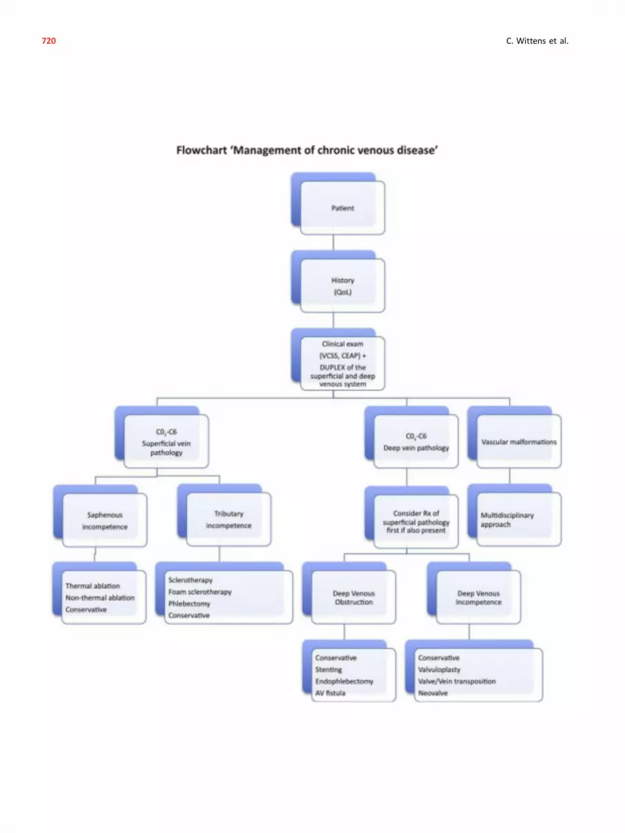
720 C. Wittens et al.

ACKNOWLEDGEMENT
The Writing Committee, the ESVS Review Committee andthe Document Reviewers would like to thank Cora Hazelzetfor her invaluable contribution in the preparation of thepresent guidelines.
REFERENCES
1 Eklöf B, Perrin M, Delis KT, Rutherford RB, Gloviczki P.
Updated terminology of chronic venous disorders: the VEIN-
TERM transatlantic interdisciplinary consensus document.
J Vasc Surg 2009;49:498e501.
2 Beebe-Dimmer JL, Pfeifer JR, Engle JS, Schottenfeld D. The
epidemiology of chronic venous insufficiency and varicose
veins. Ann Epidemiol 2005;15:175e84.
3 Robertson L, Evans C, Fowkes FG. Epidemiology of chronic
venous disease. Phlebology 2008;23:103e11.
4 Van den Oever R, Hepp B, Debbaut B, Simon I. Socio-eco-
nomic impact of chronic venous insufficiency. An under-
estimated public health problem. Int Angiol 1998;17:161e7.
5 Bradbury A, Evans C, Allan P, Lee A, Ruckley CV, Fowkes FG.What
are the symptoms of varicose veins? Edinburgh vein study cross
sectional population survey. Br Med J 1999;318:353e6.
6 Caggiati A, Bertocchi P. Regarding “fact and fiction sur-
rounding the discovery of the venous valves”. J Vasc Surg
2001;33:1317.
7 Caggiati A, Allegra C. Historical introduction. In: Bergan JJ, editor.
The vein book. London: Elsevier Academic Press; 2007. p. 1e14.
8 Illig KA, Rhodes JM, DeWeese J. Venous and lymphatic dis-
ease: a historical review. In: Gloviczki P, editor. Handbook of
venous disorders. London: Edward Arnold Publishers Ltd;
2009. p. 3e11.
9 Homans J. The etiology of treatment of varicose ulcer of the
leg. Surg Gyn Obstet 1917:300e11.
10 Linton RR. The post-thrombotic ulceration of the lower ex-
tremity: its etiology and surgical treatment. Ann Surg
1953;138:415e33.
11 Smith FL. Varicose veins, complications and results of treat-
ment of 5,000 patients. Milit Surg 1939;85:514e21.
12 Orbach EJ. Sclerotherapy of varicose veins: utilization of an
intravenous air block. Am J Surg 1944;LXVI:362e6.
13 Trendelenburg F. Über die Unterbindung der Vena
Saphena Magna bei Unterschenkel Varicen. Beitr Z Clin
Chir 1891:195e210.
14 Perrin M. Insuffisance veineuse superficielle: notions fonda-
mentales. Technique chirurgicales e Chirurgie vasculaire.
Paris: Elsevier Masson SAS; 2007. p. 43e161.
15 Muller R. Traitement des varices par la phlebectomie ambu-
latoire. Phlébologie 1966;19:277e9.
16 Bergan JJ. Conrad Jobst and the development of pressure
gradient therapy for venous disease. In: Bergan JJ, Yao JST,
editors. Surgery of the veins. Orlando: Grune & Stratton;
1985. p. 529e40.
17 Husni EA. In situ saphenopopliteal bypass graft for incompe-
tence of the femoral and popliteal veins. Surg Gynecol Obstet
1970;130:279e84.
18 Palma EC, Esperon R. Vein transplants and grafts in the sur-
gical treatment of the postphlebitic syndrome. J Cardiovasc
Surg (Torino) 1960;1:94e107.
19 Gloviczki P, Hollier LH, Dewanjee MK, Trastek VF, Hoffman EA,
Kaye MP. Experimental replacement of the inferior vena cava:
factors affecting patency. Surgery 1984;95:657e66.
20 Eklöf B, Albrechtson U, Einarsson E, Plate G. The temporary
arteriovenous fistula in venous reconstructive surgery. Int
Angiol 1985;4:455e62.
21 Kistner RL. Surgical repair of the incompetent femoral vein
valve. Arch Surg 1975;110:1336e42.
22 Raju S. Valvuloplasty and valve transfer. Int Angiol 1985;4:
419e24.
23 Hauer G. Die endoskopische subfasziale Diszision der Perfo-
ransvenenevorlaufige Mitteilung. Vasa 1985;14:59e61.
24 Okrent D, Messersmith R, Buckman J.Transcatheter fibrinolytic
therapy and angioplasty for left iliofemoral venous thrombosis.
J Vasc Interv Radiol 1991;2:195e7 [discussion 8e200].
25 Semba CP, Dake MD. Iliofemoral deep venous thrombosis:
aggressive therapy with catheter-directed thrombolysis.
Radiology 1994;191:487e94.
26 Neglen P, Berry MA, Raju S. Endovascular surgery in the
treatment of chronic primary and post-thrombotic iliac vein
obstruction. Eur J Vasc Endovasc Surg 2000;20:560e71.
27 Rabe E, Guex JJ, Puskas A, Scuderi A, Fernandez Quesada F.
Epidemiology of chronic venous disorders in geographically
diverse populations: results from the Vein Consult Program.
Int Angiol 2012;31:105e15.
28 Callam MJ. Epidemiology of varicose veins. Br J Surg 1994;81:
167e73.
29 Evans CJ, Fowkes FG, Ruckley CV, Lee AJ. Prevalence of vari-
cose veins and chronic venous insufficiency in men and
women in the general population: Edinburgh Vein Study.
J Epidemiol Community Health 1999;53:149e53.
30 Graham ID, Harrison MB, Nelson EA, Lorimer K, Fisher A.
Prevalence of lower-limb ulceration: a systematic review
of prevalence studies. Adv Skin Wound Care 2003;16:
305e16.
31 Criqui MH, Jamosmos M, Fronek A, Denenberg JO, Langer RD,
Bergan J, et al. Chronic venous disease in an ethnically diverse
population: the San Diego Population Study. Am J Epidemiol
2003;158:448e56.
32 Rabe E, Pannier-Fisher F, Bromen K. Bonner Venenstudie der
Deutschen Gesellschaft für Phlebologie e epidemiologische
Untersuchung zur Frage der Häufigkeit und Ausprägung von
chronischen Venenkrankheiten in der städtischen und länd-
lichen Wohnbevölkerung. Phlebologie 2003;32:1e14.
33 Jawien A, Grzela T, Ochwat A. Prevalence of chronic
insufficiency in men and women in Poland: multicenter
cross-sectional study in 40,095 patients. Phlebology
2003;18:110e21.
34 Carpentier PH, Maricq HR, Biro C, Poncot-Makinen CO,
Franco A. Prevalence, risk factors, and clinical patterns of
chronic venous disorders of lower limbs: a population-based
study in France. J Vasc Surg 2004;40:650e9.
35 Chiesa R, Marone EM, Limoni C, Volonte M, Schaefer E,
Petrini O. Chronic venous insufficiency in Italy: the 24-cities
cohort study. Eur J Vasc Endovasc Surg 2005;30:422e9.
36 Bromen K, Pannier-Fischer F, Stang A, Rabe E, Bock E,
Jockel KH. Lassen sich geschlechtsspezifische Unterschiede bei
Venenerkrankungen durch Schwangerschaften und Hormo-
neinnahme erklaren? Gesundheitswesen 2004;66:170e4.
37 Brand FN, Dannenberg AL, Abbott RD, Kannel WB. The
epidemiology of varicose veins: the Framingham Study. Am J
Prev Med 1988;4:96e101.
38 Hirai M, Naiki K, Nakayama R. Prevalence and risk factors of
varicose veins in Japanesewomen.Angiology 1990;41:228e32.
39 Jukkala TM, Mäkivaara LA, Juukkaala T, Hakama M, Laurikka J.
The effects of parity, oral contraceptive use and hormone
Management of Chronic Venous Disease 721

replacement therapy on the incidence of varicose veins.
J Obstet Gynaecol 2006:448e51.
40 Engelhorn CA, Cassou MF, Engelhorn AL, Salles-Cunha SX.
Does the number of pregnancies affect patterns of great
saphenous vein reflux in women with varicose veins? Phle-
bology 2010;25:190e5.
41 Rabe E. Vein Bonn Study. Phlebologie 2006:179e86.
42 Iannuzzi A, Panico S, Ciardullo AV, Bellati C, Cioffi V,
Iannuzzo G, et al. Varicose veins of the lower limbs and
venous capacitance in postmenopausal women: relationship
with obesity. J Vasc Surg 2002;36:965e8.
43 Padberg Jr F, Cerveira JJ, Lal BK, Pappas PJ, Varma S,
Hobson RW. Does severe venous insufficiency have a different
etiology in the morbidly obese? Is it venous? J Vasc Surg
2003;37:79e85.
44 Danielsson G, Eklöf B, Grandinetti A, Kistner RL. The influence
of obesity on chronic venous disease. Vasc Endovascular Surg
2002;36:271e6.
45 Zoller B, Ji J, Sundquist J, Sundquist K. Family history and risk
of hospital treatment for varicose veins in Sweden. Br J Surg
2012;99:948e53.
46 Krysa J, Jones GT, van Rij AM. Evidence for a genetic role in
varicose veins and chronic venous insufficiency. Phlebology
2012;27:329e35.
47 Ruckley CV, Evans CJ, Allan PL, Lee AJ, Fowkes FG. Chronic
venous insufficiency: clinical and duplex correlations. The
Edinburgh Vein Study of venous disorders in the general
population. J Vasc Surg 2002;36:520e5.
48 Maurins U, Hoffmann BH, Losch C, Jockel KH, Rabe E,
Pannier F. Distribution and prevalence of reflux in the su-
perficial and deep venous system in the general population e
results from the Bonn Vein Study, Germany. J Vasc Surg
2008;48:680e7.
49 Labropoulos N, Leon L, Kwon S, Tassiopoulos A, Gonzalez-
Fajardo JA, Kang SS, et al. Study of the venous reflux pro-
gression. J Vasc Surg 2005;41:291e5.
50 Pannier F, Rabe E.The relevance of the natural historyof varicose
veins and refunded care. Phlebology 2012;27(Suppl. 1):23e6.
51 Caggiati A. Fascial relationships of the long saphenous vein.
Circulation 1999;100:2547e9.
52 Caggiati A. The saphenous venous compartments. Surg Radiol
Anat 1999;21:29e34.
53 Oguzkurt L. Ultrasonographic anatomy of the lower extremity
superficial veins. Diagn Interv Radiol 2012;18:423e30.
54 Pang AS. Location of valves and competence of the great
saphenous vein above the knee. Ann Acad Med Singapore
1991;20:248e50.
55 Cavezzi A, Labropoulos N, Partsch H, Ricci S, Caggiati A,
Myers K, et al. Duplex ultrasound investigation of the veins in
chronic venous disease of the lower limbs e UIP consensus
document. Part II. Anatomy. Eur J Vasc Endovasc Surg
2006;31:288e99.
56 Caggiati A. Fascial relationships of the short saphenous vein.
J Vasc Surg 2001;34:241e6.
57 Schweighofer G, Muhlberger D, Brenner E. The anatomy of the
small saphenous vein: fascial and neural relations, saphenofe-
moral junction, and valves. J Vasc Surg 2010;51:982e9.
58 van Limborgh J, Hage RW. The systemic anatomy of the
perforating veins in the leg, especially Cockett’s veins. Phle-
bologie 1982;35:19e28.
59 Delis KT, Husmann M, Kalodiki E, Wolfe JH, Nicolaides AN. In
situ hemodynamics of perforating veins in chronic venous
insufficiency. J Vasc Surg 2001;33:773e82.
60 Delis KT, Ibegbuna V, Nicolaides AN, Lauro A, Hafez H. Prev-
alence and distribution of incompetent perforating veins in
chronic venous insufficiency. J Vasc Surg 1998;28:815e25.
61 Mozes G, Gloviczki P, Menawat SS, Fisher DR, Carmichael SW,
Kadar A. Surgical anatomy for endoscopic subfascial division
of perforating veins. J Vasc Surg 1996;24:800e8.
62 White JV, Katz ML, Cisek P, Kreithen J. Venous outflow of the
leg: anatomy and physiologic mechanism of the plantar
venous plexus. J Vasc Surg 1996;24:819e24.
63 Caggiati A, Bergan JJ, Gloviczki P, Jantet G, Wendell-Smith CP,
Partsch H, et al. Nomenclature of the veins of the lower limbs:
an international interdisciplinary consensus statement. J Vasc
Surg 2002;36:416e22.
64 Quinlan DJ, Alikhan R, Gishen P, Sidhu PS. Variations in lower
limb venous anatomy: implications for US diagnosis of deep
vein thrombosis. Radiology 2003;228:443e8.
65 Casella IB, Presti C, Yamazaki Y, Vassoler AA, Furuya LA,
Sabbag CD. A duplex scan-based morphologic study of the
femoral vein: incidence and patterns of duplication. Vasc Med
2010;15:197e203.
66 Banjo AO. Comparative study of the distribution of venous
valves in the lower extremities of black Africans and Cauca-
sians: pathogenetic correlates of prevalence of primary vari-
cose veins in the two races. Anat Rec 1987;217:407e12.
67 Muhlberger D, Morandini L, Brenner E. An anatomical study
of femoral vein valves near the saphenofemoral junction.
J Vasc Surg 2008;48:994e9.
68 Shepherd JT. Role of the veins in the circulation. Circulation
1966;33:484e91.
69 Rothe CF. Venous system: physiology of the capacitance
vessels. In: Shepherd JT, Abboud FM, Geiger SR, editors. The
Cardiovascular System, peripheral circulation and organ blood
flow, part I, Handbook of physiology. Bethesda: American
Physiological Society; 1983. p. 397e452.
70 Katz AI, Chen Y, Moreno AH. Flow through a collapsible tube.
Experimental analysis and mathematical model. Biophys J
1969;9:1261e79.
71 Katz ML, Comerota AJ, Kerr RP, Caputo GC. Variability of
venous-hemodynamics with daily activity. J Vasc Surg
1994;19:361e5.
72 Arnoldi CC. Venous pressure in the leg of healthy human
subjects at rest and during muscular exercise in the nearly
erect position. Acta Chir Scand 1965;130:570e83.
73 Stranden E. Ventömningens fysiologi. In: Norgren L, editor.
Vensjukdmar. Lund: Studentliteratur; 2004. p. 45e55.
74 Goldman MP, Fronek A. Anatomy and pathophysiology of
varicose veins. J Dermatol Surg Oncol 1989;15:138e45.
75 Meissner MH, Moneta G, Burnand K, Gloviczki P, Lohr JM,
Lurie F, et al. The hemodynamics and diagnosis of venous
disease. J Vasc Surg 2007;46(Suppl. S):4Se24S.
76 Lurie F, Kistner RL, Eklöf B, Kessler D. Mechanism of venous
valve closure and role of the valve in circulation: a new
concept. J Vasc Surg 2003;38:955e61.
77 Lurie F, Kistner RL. The relative position of paired valves at
venous junctions suggests their role in modulating three-
dimensional flow pattern in veins. Eur J Vasc Endovasc Surg
2012;44:337e40.
78 Alimi YS, Barthelemy P, Juhan C. Venous pump of the calf: a
study of venous and muscular pressures. J Vasc Surg 1994;20:
728e35.
79 Gardner AMN, Fox RH. The return of blood to the heart:
venous pumps in health and disease. London: John Libbey &
Co Ltd; 1989.
722 C. Wittens et al.

80 Araki CT, Back TL, Padberg FT, Thompson PN, Jamil Z, Lee BC,
et al. The significance of calf muscle pump function in venous
ulceration. J Vasc Surg 1994;20:872e7.
81 Ludbrook J. The musculovenous pumps of the human lower
limb. Am Heart J 1966;71:635e41.
82 Stranden E. Edema in venous insufficiency. In: Wittens C,
editor. Best practice in venous procedures. Turin: Ediziona
Minerva Medica; 2010. p. 131e40.
83 Nicolaides AN, Hussein MK, Szendro G, Christopoulos D,
Vasdekis S, Clarke H. The relation of venous ulceration with
ambulatory venous pressure measurements. J Vasc Surg
1993;17:414e9.
84 Plate G, Brudin L, Eklöf B, Jensen R, Ohlin P. Congenital vein
valve aplasia. World J Surg 1986;10:929e34.
85 Nicolaides AN. Investigations of chronic venous insufficiency:
a consensus statement (France March 5e9, 1997). Circulation
2000:E126e63.
86 Hosoi Y, Zukowski A, Kakkos SK, Nicolaides AN. Ambulatory
venous pressure measurements: new parameters derived
from a mathematic hemodynamic model. J Vasc Surg
2002;36:137e42.
87 Raju S, Fredericks R. Venous obstruction: an analysis of one
hundred thirty-seven cases with hemodynamic, venographic,
and clinical correlations. J Vasc Surg 1991;14:305e13.
88 Labropoulos N, Patel PJ, Tiongson JE, Pryor L, Leon Jr LR,
Tassiopoulos AK. Patterns of venous reflux and obstruction in
patients with skin damage due to chronic venous disease.
Vasc Endovascular Surg 2007;41:33e40.
89 Rose SS, Ahmed A. Some thoughts on the aetiology of vari-
cose veins. J Cardiovasc Surg (Torino) 1986;27:534e43.
90 Cotton LT. Varicose veins. Gross anatomy and development.
Br J Surg 1961;48:589e98.
91 Travers JP, Brookes CE, Evans J, Baker DM, Kent C, Makin GS,
et al. Assessment of wall structure and composition of vari-
cose veins with reference to collagen, elastin and smooth
muscle content. Eur J Vasc Endovasc Surg 1996;11:230e7.
92 Lowell RC, Gloviczki P, Miller VM. In vitro evaluation of
endothelial and smooth muscle function of primary varicose
veins. J Vasc Surg 1992;16:679e86.
93 Gandhi RH, Irizarry E, Nackman GB, Halpern VJ, Mulcare RJ,
Tilson MD. Analysis of the connective tissue matrix and pro-
teolytic activity of primary varicose veins. J Vasc Surg
1993;18:814e20.
94 Alexander CJ. The theoretical basis of varicose vein formation.
Med J Aust 1972;1:258e61.
95 Labropoulos N, Giannoukas AD, Delis K, Mansour MA,
Kang SS, Nicolaides AN, et al. Where does venous reflux start?
J Vasc Surg 1997;26:736e42.
96 Lurie F, Kistner R, Perrin M, Raju S, Neglen P, Maleti O.
Invasive treatment of deep venous disease. A UIP consensus.
Int Angiol 2010;29:199e204.
97 Johnson BF, Manzo RA, Bergelin RO, Strandness Jr DE. Rela-
tionship between changes in the deep venous system and the
development of the postthrombotic syndrome after an acute
episode of lower limb deep vein thrombosis: a one- to six-
year follow-up. J Vasc Surg 1995;21:307e12.
98 Johnson BF, Manzo RA, Bergelin RO, Strandness Jr DE. The site
of residual abnormalities in the leg veins in long-term follow-
up after deep vein thrombosis and their relationship to the
development of the post-thrombotic syndrome. Int Angiol
1996;15:14e9.
99 Nelzen O, Bergqvist D, Lindhagen A. Leg ulcer etiologye a cross
sectional population study. J Vasc Surg 1991;14:557e64.
100 Delis KT. Perforator vein incompetence in chronic venous
disease: a multivariate regression analysis model. J Vasc Surg
2004;40:626e33.
101 Darvall KA, Bate GR, Adam DJ, Bradbury AW. Generic health-
related quality of life is significantly worse in varicose vein
patients with lower limb symptoms independent of CEAP
clinical grade. Eur J Vasc Endovasc Surg 2012;44:341e4.
102 Palfreyman SJ, Drewery-Carter K, Rigby K, Michaels JA,
Tod AM. Varicose veins: a qualitative study to explore ex-
pectations and reasons for seeking treatment. J Clin Nurs
2004;13:332e40.
103 Fragkouli K, Mitselou A, Boumba VA, Siozios G,
Vougiouklakis GT, Vougiouklakis T. Unusual death due to a
bleeding from a varicose vein: a case report. BMC Res Notes
2012;5:488.
104 Chiesa R, Marone EM, Limoni C, Volonte M, Petrini O. Chronic
venous disorders: correlation between visible signs, symptoms,
andpresenceof functional disease. J Vasc Surg2007;46:322e30.
105 Navarro TP, Delis KT, Ribeiro AP. Clinical and hemodynamic
significance of the greater saphenous vein diameter in chronic
venous insufficiency. Arch Surg 2002;137:1233e7.
106 Kahn SR, M’Lan CE, Lamping DL, Kurz X, Berard A,
Abenhaim LA, et al. Relationship between clinical classifica-
tion of chronic venous disease and patient-reported quality of
life: results from an international cohort study. J Vasc Surg
2004;39:823e8.
107 Perrin MR, Labropoulos N, Leon Jr LR. Presentation of the
patient with recurrent varices after surgery (REVAS). J Vasc
Surg 2006;43:327e34.
108 Beebe HG, Bergan JJ, Bergqvist D, Eklöf B, Eriksson I,
Goldman MP, et al. Classification and grading of chronic
venous disease in the lower limbs. A consensus statement.
Eur J Vasc Endovasc Surg 1996;12:487e91 [discussion 91e2].
109 Eklöf B, Rutherford RB, Bergan JJ, Carpentier PH, Gloviczki P,
Kistner RL, et al. Revision of the CEAP classification for chronic
venous disorders: consensus statement. J Vasc Surg 2004;40:
1248e52.
110 Sinabulya H, Holmberg A, Blomgren L. Interobserver vari-
ability in the assessment of the clinical severity of superficial
venous insufficiency. Phlebology 2013. http://dx.doi.org/10.
1177/0268355513515474 [Epub].
111 Krishnan S, Nicholls SC. Chronic venous insufficiency: clinical
assessment and patient selection. Semin Intervent Radiol
2005;22:169e77.
112 Rabe E, Pannier F. Clinical, aetiological, anatomical and
pathological classification (CEAP): gold standard and limits.
Phlebology 2012;27(Suppl. 1):114e8.
113 Carradice D, Mazari FA, Samuel N, Allgar V, Hatfield J,
Chetter IC. Modelling the effect of venous disease on quality
of life. Br J Surg 2011;98:1089e98.
114 Shepherd AC, Gohel MS, Lim CS, Davies AH. A study to
compare disease-specific quality of life with clinical anatom-
ical and hemodynamic assessments in patients with varicose
veins. J Vasc Surg 2011;53:374e82.
115 Rutherford RB, Padberg Jr FT, Comerota AJ, Kistner RL,
Meissner MH, Moneta GL. Venous severity scoring: an adjunct
to venous outcome assessment. J Vasc Surg 2000;31:1307e
12.
116 Vasquez MA, Rabe E, McLafferty RB, Shortell CK, Marston WA,
Gillespie D, et al. Revision of the venous clinical severity
score: venous outcomes consensus statement: special
communication of the American Venous Forum Ad Hoc Out-
comes Working Group. J Vasc Surg 2010;52:1387e96.
Management of Chronic Venous Disease 723

117 Ricci MA, Emmerich J, Callas PW, Rosendaal FR, Stanley AC,
Naud S, et al. Evaluating chronic venous disease with a
new venous severity scoring system. J Vasc Surg 2003;38:
909e15.
118 Passman MA, McLafferty RB, Lentz MF, Nagre SB, Iafrati MD,
Bohannon WT, et al. Validation of Venous Clinical Severity
Score (VCSS) with other venous severity assessment tools
from the American Venous Forum, National Venous Screening
Program. J Vasc Surg 2011;54:2Se9S.
119 Kakkos SK, Rivera MA, Matsagas MI, Lazarides MK,
Robless P, Belcaro G, et al. Validation of the new venous
severity scoring system in varicose vein surgery. J Vasc Surg
2003;38:224e8.
120 van Rij AM, Jones GT, Hill BG, Amer M, Thomson IA,
Pettigrew RA, et al. Mechanical inhibition of angiogenesis at
the saphenofemoral junction in the surgical treatment of
varicose veins: early results of a blinded randomized
controlled trial. Circulation 2008;118:66e74.
121 Perrin M, Dedieu F, Jessent V, Blanc MP. Evaluation of the
new severity scoring system in chronic venous disease of the
lower limbs: an observational study conducted by French
angiologists. Phlebolymphology 2006;13:6e11.
122 Villalta S, Bagatella P, Piccioli A, Lensing A, Prins M,
Prandoni P. Assessment and validity and reproducibility of a
clinical scale for the post-thrombotic syndrome [abstract].
Haemostasis 1994:158a.
123 Kahn SR. Measurement properties of the Villalta scale to
define and classify the severity of the post-thrombotic syn-
drome. J Thromb Haemost 2009;7:884e8.
124 Sritharan K, Lane TR, Davies AH. The burden of depression in
patients with symptomatic varicose veins. Eur J Vasc Endo-
vasc Surg 2012;43:480e4.
125 Jantet G. Chronic venous insufficiency: worldwide results of
the RELIEF study. Reflux assEssment and quaLity of lIfe
improvEment with micronized Flavonoids. Angiology 2002;53:
245e56.
126 Ware Jr JE, Gandek B. Overview of the SF-36 Health Survey
and the International Quality of Life Assessment (IQOLA)
Project. J Clin Epidemiol 1998;51:903e12.
127 Kaplan RM, Criqui MH, Denenberg JO, Bergan J, Fronek A.
Quality of life in patients with chronic venous disease: San
Diego population study. J Vasc Surg 2003;37:1047e53.
128 Moura RM, Goncalves GS, Navarro TP, Britto RR, Dias RC.
Relationship between quality of life and the CEAP clinical
classification in chronic venous disease. Rev Bras Fisioter
2010;14:99e105.
129 EuroQol, Group. EuroQol e a new facility for the measure-
ment of health-related quality of life. Health Policy 1990;16:
199e208.
130 Garratt AM, Macdonald LM, Ruta DA, Russell IT,
Buckingham JK, Krukowski ZH. Towards measurement of
outcome for patients with varicose veins. Qual Health Care
1993;2:5e10.
131 Smith JJ, Garratt AM, Guest M, Greenhalgh RM, Davies AH.
Evaluating and improving health-related quality of life in pa-
tients with varicose veins. J Vasc Surg 1999;30:710e9.
132 Launois R, Reboul-Marty J, Henry B. Construction and vali-
dation of a quality of life questionnaire in chronic lower limb
venous insufficiency (CIVIQ). Qual Life Res 1996;5:539e54.
133 Likert R. A technique for the measurement of attitudes. Arch
Psychol 1932;22:1e55.
134 Erevnidou K, Launois R, Katsamouris A, Lionis C. Translation
and validation of a quality of life questionnaire for chronic
lower limb venous insufficiency into greek. Int Angiol
2004;23:394e9.
135 Biemans AA, van der Velden SK, Bruijninckx CM, Buth J,
Nijsten T. Validation of the chronic venous insufficiency
quality of life questionnaire in Dutch patients treated for
varicose veins. Eur J Vasc Endovasc Surg 2011;42:246e53.
136 Launois R, Mansilha A, Jantet G. International psychometric
validation of the Chronic Venous Disease quality of life
Questionnaire (CIVIQ-20). Eur J Vasc Endovasc Surg 2010;40:
783e9.
137 Abenhaim L, Kurz X. The VEINES study (VEnous Insufficiency
Epidemiologic and Economic Study): an international cohort
study on chronic venous disorders of the leg. VEINES Group.
Angiology 1997;48:59e66.
138 Lamping DL, Schroter S, Kurz X, Kahn SR, Abenhaim L. Eval-
uation of outcomes in chronic venous disorders of the leg:
development of a scientifically rigorous, patient-reported
measure of symptoms and quality of life. J Vasc Surg
2003;37:410e9.
139 Kahn SR, Lamping DL, Ducruet T, Arsenault L, Miron MJ,
Roussin A, et al. VEINES-QOL/Sym questionnaire was a reliable
and valid disease-specific quality of life measure for deep
venous thrombosis. J Clin Epidemiol 2006;59:1049e56.
140 Gloviczki P, Comerota AJ, Dalsing MC, Eklöf BG, Gillespie DL,
Gloviczki ML, et al. The care of patients with varicose veins
and associated chronic venous diseases: clinical practice
guidelines of the Society for Vascular Surgery and the
American Venous Forum. J Vasc Surg 2011;53:2Se48S.
141 Fowkes FG, Lee AJ, Evans CJ, Allan PL, Bradbury AW,
Ruckley CV. Lifestyle risk factors for lower limb venous reflux
in the general population: Edinburgh Vein Study. Int J Epi-
demiol 2001;30:846e52.
142 Cronenwett JL, Johnston KW. Patient clinical evaluation.
Rutherford’s Vascular Surgery. 7th ed. Philadelphia: Saunders;
2010. p. 204e15.
143 Uhl JF, Cornu-Thenard A, Carpentier PH, Widmer MT,
Partsch H, Antignani PL. Clinical and hemodynamic signifi-
cance of corona phlebectatica in chronic venous disorders.
J Vasc Surg 2005;42:1163e8.
144 Vrouenraets BC, Keeman JN. Fysische diagnostiek e de bandje-
sproeven bij varices.Ned Tijdschr Geneeskd 2000;144:1267e72.
145 Hoffmann WH, Toonder IM, Wittens CHA. Value of the Tren-
delenburg tourniquet test in the assessment of primary
varicose veins. Phlebology 2004;19:77e80.
146 Labropoulos N, Tiongson J, Pryor L, Tassiopoulos AK, Kang SS,
Ashraf Mansour M, et al. Definition of venous reflux in lower-
extremity veins. J Vasc Surg 2003;38:793e8.
147 Coleridge-Smith P, Labropoulos N, Partsch H, Myers K,
Nicolaides A, Cavezzi A. Duplex ultrasound investigation of the
veins in chronic venous disease of the lower limbs e UIP
consensus document. Part I. Basic principles. Eur J Vasc
Endovasc Surg 2006;31:83e92.
148 De Maeseneer M, Pichot O, Cavezzi A, Earnshaw J, van Rij A,
Lurie F, et al. Duplex ultrasound investigation of the veins of the
lower limbs after treatment for varicose veins e UIP consensus
document. Eur J Vasc Endovasc Surg 2011;42:89e102.
149 Cappelli M, Molino Lova R, Ermini S, Zamboni P. Hemody-
namics of the sapheno-femoral junction. Patterns of reflux
and their clinical implications. Int Angiol 2004;23:25e8.
150 Cappelli M, Molino Lova R, Ermini S, Giangrandi I, Giannelli F,
Zamboni P. Hemodynamics of the sapheno-femoral complex:
an operational diagnosis of proximal femoral valve function.
Int Angiol 2006;25:356e60.
724 C. Wittens et al.

151 Rautio T, Perala J, Biancari F, Wiik H, Ohtonen P, Haukipuro K,
et al. Accuracy of hand-held Doppler in planning the opera-
tion for primary varicose veins. Eur J Vasc Endovasc Surg
2002;24:450e5.
152 DePalma RG, Hart MT, Zanin L, Massarin EH. Phyical exami-
nation, Doppler ultrasound and colour flow duplex scanning:
guides to therapy for primary varicose veins. Phlebology
1993;8:7e11.
153 Baker SR, Burnand KG, Sommerville KM, Thomas ML,
Wilson NM, Browse NL. Comparison of venous reflux assessed
by duplex scanning and descending phlebography in chronic
venous disease. Lancet 1993;341:400e3.
154 Magnusson M, Kalebo P, Lukes P, Sivertsson R, Risberg B.
Colour Doppler ultrasound in diagnosing venous insufficiency.
A comparison to descending phlebography. Eur J Vasc Endo-
vasc Surg 1995;9:437e43.
155 Baldt MM, Bohler K, Zontsich T, Bankier AA, Breitenseher M,
Schneider B, et al. Preoperative imaging of lower extremity
varicose veins: color coded duplex sonography or venography.
J Ultrasound Med 1996;15:143e54.
156 Meyer T, Cavallaro A, Lang W. Duplex ultrasonography in the
diagnosis of incompetent Cockett veins. Eur J Ultrasound
2000;11:175e80.
157 Blomgren L, Johansson G, Bergqvist D. Randomized clinical
trial of routine preoperative duplex imaging before varicose
vein surgery. Br J Surg 2005;92:688e94.
158 Haenen JH, van Langen H, Janssen MC, Wollersheim H, van ’t
Hof MA, van Asten WN, et al. Venous duplex scanning of the
leg: range, variability and reproducibility. Clin Sci (Lond)
1999;96:271e7.
159 Lensing AW, Prandoni P, Brandjes D, Huisman PM, Vigo M,
Tomasella G, et al. Detection of deep-vein thrombosis by
real-time B-mode ultrasonography. N Engl J Med 1989;320:
342e5.
160 Mattos MA, Londrey GL, Leutz DW, Hodgson KJ, Ramsey DE,
Barkmeier LD, et al. Color-flow duplex scanning for the sur-
veillance and diagnosis of acute deep venous thrombosis.
J Vasc Surg 1992;15:366e75.
161 Holdstock JM, Dos Santos SJ, Harrison CC, Price BA,
Whiteley MS. Haemorrhoids are associated with internal iliac
vein reflux in up to one-third of women presenting with
varicose veins associated with pelvic vein reflux. Phlebology
2014 [Epub].
162 Park SJ, Lim JW, Ko YT, Lee DH, Yoon Y, Oh JH, et al. Diagnosis
of pelvic congestion syndrome using transabdominal and
transvaginal sonography. Am J Roentgenol 2004;182:683e8.
163 De Maeseneer MG, Vandenbroeck CP, Hendriks JM,
Lauwers PR, Van Schil PE. Accuracy of duplex evaluation one
year after varicose vein surgery to predict recurrence at the
sapheno-femoral junction after five years. Eur J Vasc Endovasc
Surg 2005;29:308e12.
164 Jiang P, van Rij AM, Christie R, Hill G, Solomon C, Thomson I.
Recurrent varicose veins: patterns of reflux and clinical
severity. Cardiovasc Surg 1999;7:332e9.
165 Labropoulos N, Touloupakis E, Giannoukas AD, Leon M,
Katsamouris A, Nicolaides AN. Recurrent varicose veins:
investigation of the pattern and extent of reflux with color
flow duplex scanning. Surgery 1996;119:406e9.
166 Kostas T, Ioannou CV, Touloupakis E, Daskalaki E,
Giannoukas AD, Tsetis D, et al. Recurrent varicose veins
after surgery: a new appraisal of a common and complex
problem in vascular surgery. Eur J Vasc Endovasc Surg
2004;27:275e82.
167 Theivacumar NS, Darwood R, Gough MJ. Neovascularisation
and recurrence 2 years after varicose vein treatment for
sapheno-femoral and great saphenous vein reflux: a com-
parison of surgery and endovenous laser ablation. Eur J Vasc
Endovasc Surg 2009;38:203e7.
168 Chapman-Smith P, Browne A. Prospective five-year study of
ultrasound-guided foam sclerotherapy in the treatment of
great saphenous vein reflux. Phlebology 2009;24:183e8.
169 Labropoulos N, Borge M, Pierce K, Pappas PJ. Criteria for
defining significant central vein stenosis with duplex ultra-
sound. J Vasc Surg 2007;46:101e7.
170 Phillips GW, Paige J, Molan MP. A comparison of colour duplex
ultrasound with venography and varicography in the assess-
ment of varicose veins. Clin Radiol 1995;50:20e5.
171 Whitney RJ. The measurement of volume changes in human
limbs. J Physiol 1953;121:1e27.
172 Janssen MC, Claassen JA, van Asten WN, Wollersheim H, de
Rooij MJ, Thien T. Validation of the supine venous pump
function test: a new non-invasive tool in the assessment of
deep venous insufficiency. Clin Sci (Lond) 1996;91:483e8.
173 Hertzman AB. Photoelectric plethysmography on the fin-
gers and toes in man. Exp Biol Med (Maywood) 1937;37:
529e34.
174 Abramowitz HB, Queral LA, Finn WR, Nora Jr PF, Peterson LK,
Bergan JJ, et al. The use of photoplethysmography in the
assessment of venous insufficiency: a comparison to venous
pressure measurements. Surgery 1979;86:434e41.
175 Wienert V, Blazek V. Eine neue, apparative nichtinvasive
Diagnostik der chronisch-venösen Insuffizienz. Phleb u Prokt
1982;11:110e3.
176 Blazek V, Schmitt HJ, Schulz-Ehrenburg U, Kerner J. Digitale
Photoplethysmografie (dPPG) für die Beinvenendiagnostik e
medizinisch-technische Grundlagen. Phleb u Prokt 1989;18:
91e7.
177 Nicolaides AN, Miles C. Photoplethysmography in the assess-
ment of venous insufficiency. J Vasc Surg 1987;5:405e12.
178 van Bemmelen PS, van Ramshorst B, Eikelboom BC. Photo-
plethysmography reexamined: lack of correlation with duplex
scanning. Surgery 1992;112:544e8.
179 Bays RA, Healy DA, Atnip RG, Neumyer M, Thiele BL. Valida-
tion of air plethysmography, photoplethysmography, and
duplex ultrasonography in the evaluation of severe venous
stasis. J Vasc Surg 1994;20:721e7.
180 Owens LV, Farber MA, Young ML, Carlin RE, Criado-Pallares E,
Passman MA, et al. The value of air plethysmography in
predicting clinical outcome after surgical treatment of chronic
venous insufficiency. J Vasc Surg 2000;32:961e8.
181 Yang D, Vandongen YK, Stacey MC. Variability and reliability of
air plethysmographic measurements for the evaluation of
chronic venous disease. J Vasc Surg 1997;26:638e42.
182 Thulesius O, Norgren L, Gjores JE. Foot-volumetry, a new
method for objective assessment of edema and venous
function. Vasa 1973;2:325e9.
183 Ruehm SG,Wiesner W, Debatin JF. Pelvic and lower extremity
veins: contrast-enhanced three-dimensional MR venography
with a dedicated vascular coil-initial experience. Radiology
2000;215:421e7.
184 Pfeil A, Betge S, Poehlmann G, Boettcher J, Drescher R,
Malich A, et al. Magnetic resonance VIBE venography using
the blood pool contrast agent gadofosveset trisodium e an
interrater reliability study. Eur J Radiol 2012;81:547e52.
185 Spritzer CE. Progress in MR imaging of the venous system.
Perspect Vasc Surg Endovasc Ther 2009;21:105e16.
Management of Chronic Venous Disease 725
186 Enden T, Storas TH, Negard A, Haig Y, Sandvik L, Gjesdal KI,
et al. Visualization of deep veins and detection of deep
vein thrombosis (DVT) with balanced turbo field echo (b-
TFE) and contrast-enhanced T1 fast field echo (CE-FFE)
using a blood pool agent (BPA). J Magn Reson Imaging
2010;31:416e24.
187 Koc Z, Oguzkurt L. Interruption or congenital stenosis of the
inferior vena cava: prevalence, imaging, and clinical findings.
Eur J Radiol 2007;62:257e66.
188 Gayer G, Luboshitz J, Hertz M, Zissin R, Thaler M, Lubetsky A,
et al. Congenital anomalies of the inferior vena cava revealed
on CT in patients with deep vein thrombosis. Am J Roentgenol
2003;180:729e32.
189 Wolpert LM, Rahmani O, Stein B, Gallagher JJ, Drezner AD.
Magnetic resonance venography in the diagnosis and man-
agement of May-Thurner syndrome. Vasc Endovascular Surg
2002;36:51e7.
190 Asciutto G, Mumme A, Marpe B, Koster O, Asciutto KC,
Geier B. MR venography in the detection of pelvic venous
congestion. Eur J Vasc Endovasc Surg 2008;36:491e6.
191 Hsieh MC, Chang PY, Hsu WH, Yang SH, Chan WP. Role of
three-dimensional rotational venography in evaluation of the
left iliac vein in patients with chronic lower limb edema. Int J
Cardiovasc Imaging 2011;27:923e9.
192 Fraser DG, Moody AR, Morgan PS, Martel A. Iliac compression
syndrome and recanalization of femoropopliteal and iliac
venous thrombosis: a prospective study with magnetic reso-
nance venography. J Vasc Surg 2004;40:612e9.
193 Marston W, Fish D, Unger J, Keagy B. Incidence of and risk
factors for iliocaval venous obstruction in patients with active
or healed venous leg ulcers. J Vasc Surg 2011;53:1303e8.
194 Depalma RG, Kowallek DL, Barcia TC, Cafferata HT. Target
selection for surgical intervention in severe chronic venous
insufficiency: comparison of duplex scanning and phlebog-
raphy. J Vasc Surg 2000;32:913e20.
195 Raju S, Furrh JB, Neglen P. Diagnosis and treatment of venous
lymphedema. J Vasc Surg 2012;55:141e9.
196 Raju S, Neglen P. High prevalence of nonthrombotic iliac vein
lesions in chronic venous disease: a permissive role in path-
ogenicity. J Vasc Surg 2006;44:136e43.
197 Neglen P, Raju S. Intravascular ultrasound scan evaluation of
the obstructed vein. J Vasc Surg 2002;35:694e700.
198 Forauer AR, Gemmete JJ, Dasika NL, Cho KJ, Williams DM.
Intravascular ultrasound in the diagnosis and treatment of
iliac vein compression (May-Thurner) syndrome. J Vasc Interv
Radiol 2002;13:523e7.
199 Arnoldussen CW, de Graaf R, Wittens CH, de Haan MW. Value
of magnetic resonance venography and computed tomo-
graphic venography in lower extremity chronic venous dis-
ease. Phlebology 2013;28(Suppl. 1):169e75.
200 Arnoldussen CW, Toonder I, Wittens CH. A novel scoring sys-
tem for lower-extremity venous pathology analysed using
magnetic resonance venography and duplex ultrasound.
Phlebology 2012;27(Suppl. 1):163e70.
201 Labropoulos N, Gasparis AP, Pefanis D, Leon Jr LR,
Tassiopoulos AK. Secondary chronic venous disease pro-
gresses faster than primary. J Vasc Surg 2009;49:704e10.
202 Wittens CH, Bukkems SF, Toonder IT. Abdominal wall venous
collaterals: the latent clinical sign for central chronic venous
obstruction. Circulation 2010;122:2089e90.
203 Meissner MH, Eklof B, Smith PC, Dalsing MC, DePalma RG,
Gloviczki P, et al. Secondary chronic venous disorders. J Vasc
Surg 2007;46(Suppl. S):68Se83S.
204 O’Meara S, Martyn-St James M. Foam dressings for venous
leg ulcers. Cochrane Database Syst Rev 2013;5:CD009907.
205 O’Meara S, Martyn-St James M. Alginate dressings for venous
leg ulcers. Cochrane Database Syst Rev 2013;4:CD010182.
206 Palfreyman S, Nelson EA, Michaels JA. Dressings for venous
leg ulcers: systematic review and meta-analysis. Br Med J
2007;335:244.
207 Michaels JA, Campbell B, King B, Palfreyman SJ, Shackley P,
Stevenson M. Randomized controlled trial and cost-
effectiveness analysis of silver-donating antimicrobial dress-
ings for venous leg ulcers (VULCAN trial). Br J Surg 2009;96:
1147e56.
208 O’Meara S, Al-Kurdi D, Ologun Y, Ovington LG. Antibiotics and
antiseptics for venous leg ulcers. Cochrane Database Syst Rev
2010:CD003557.
209 Stacey MC, Jopp-Mckay AG, Rashid P, Hoskin SE,
Thompson PJ. The influence of dressings on venous ulcer
healing e a randomised trial. Eur J Vasc Endovasc Surg
1997;13:174e9.
210 Couzan S, Leizorovicz A, Laporte S, Mismetti P, Pouget JF,
Chapelle C, et al. A randomized double-blind trial of upward
progressive versus degressive compressive stockings in pa-
tients with moderate to severe chronic venous insufficiency.
J Vasc Surg 2012;56. 1344e50.
211 Mosti G, Partsch H. Compression stockings with a negative
pressure gradient have a more pronounced effect on venous
pumping function than graduated elastic compression stock-
ings. Eur J Vasc Endovasc Surg 2011;42:261e6.
212 Mosti G, Mattaliano V, Partsch H. Inelastic compression in-
creases venous ejection fraction more than elastic bandages
in patients with superficial venous reflux. Phlebology 2008;23:
287e94.
213 Shingler S, Robertson L, Boghossian S, Stewart M. Compres-
sion stockings for the initial treatment of varicose veins in
patients without venous ulceration. Cochrane Database Syst
Rev 2011:CD008819.
214 Kolbach DN, Sandbrink MW, Neumann HA, Prins MH.
Compression therapy for treating stage I and II (Widmer)
post-thrombotic syndrome. Cochrane Database Syst Rev
2003:CD004177.
215 Andreozzi GM, Cordova R, Scomparin MA, Martini R, D’Eri A,
Andreozzi F, et al. Effects of elastic stocking on quality of life
of patients with chronic venous insufficiency. An Italian pilot
study on Triveneto Region. Int Angiol 2005;24:325e9.
216 Carpentier PH, Becker F, Thiney G, Poensin D, Satger B.
Acceptability and practicability of elastic compression stock-
ings in the elderly: a randomized controlled evaluation.
Phlebology 2011;26:107e13.
217 O’Brien JF, Grace PA, Perry IJ, Hannigan A, Clarke Moloney M,
Burke PE. Randomized clinical trial and economic analysis of
four-layer compression bandaging for venous ulcers. Br J Surg
2003;90:794e8.
218 Wong IK, Andriessen A, Charles HE, Thompson D, Lee DT,
So WK, et al. Randomized controlled trial comparing
treatment outcome of two compression bandaging systems
and standard care without compression in patients with
venous leg ulcers. J Eur Acad Dermatol Venereol 2012;26:
102e10.
219 O’Meara S, Cullum N, Nelson A, Dumville JC. Compression for
venous leg ulcers. The Cochrane Library 2012:CD000265.
220 Blecken SR, Villavicencio JL, Kao TC. Comparison of elastic
versus nonelastic compression in bilateral venous ulcers: a
randomized trial. J Vasc Surg 2005;42:1150e5.
726 C. Wittens et al.

221 Blair SD, Wright DD, Backhouse CM, Riddle E, McCollum CN.
Sustained compression and healing of chronic venous ulcers.
Br Med J 1988;297:1159e61.
222 Harrison MB, Vandenkerkhof EG, Hopman WM, Graham ID,
Carley ME, Nelson EA, et al. The Canadian Bandaging Trial:
evidence-informed leg ulcer care and the effectiveness of two
compression technologies. BMC Nurs 2011;10:20.
223 Lazareth I, Moffatt C, Dissemond J, Lesne Padieu AS,
Truchetet F, Beissert S, et al. Efficacy of two compression
systems in the management of VLUs: results of a European
RCT. J Wound Care 2012;21:553e4.
224 Franks PJ, Moody M, Moffatt CJ, Hiskett G, Gatto P, Davies C,
et al. Randomized trial of two foam dressings in the man-
agement of chronic venous ulceration. Wound Repair Regen
2007;15:197e202.
225 Nelson EA, Iglesias CP, Cullum N, Torgerson DJ. Randomized
clinical trial of four-layer and short-stretch compression ban-
dages for venous leg ulcers (VenUS I). Br J Surg 2004;91:
1292e9.
226 Franks PJ, Moody M, Moffatt CJ, Martin R, Blewett R,
Seymour E, et al. Randomized trial of cohesive short-stretch
versus four-layer bandaging in the management of venous
ulceration. Wound Repair Regen 2004;12:157e62.
227 Polignano R, Bonadeo P, Gasbarro S, Allegra C. A randomised
controlled study of four-layer compression versus Unna’s Boot
for venous ulcers. J Wound Care 2004;13:21e4.
228 Moffatt CJ, McCullagh L, O’Connor T, Doherty DC, Hourican C,
Stevens J, et al. Randomized trial of four-layer and two-layer
bandage systems in the management of chronic venous ul-
ceration. Wound Repair Regen 2003;11:166e71.
229 Meyer FJ, McGuinness CL, Lagattolla NR, Eastham D,
Burnand KG. Randomized clinical trial of three-layer paste and
four-layer bandages for venous leg ulcers. Br J Surg 2003;90:
934e40.
230 Scriven JM, Taylor LE,Wood AJ, Bell PR, Naylor AR, London NJ.
A prospective randomised trial of four-layer versus short
stretch compression bandages for the treatment of venous
leg ulcers. Ann R Coll Surg Engl 1998;80:215e20.
231 Moffatt CJ, Edwards L, Collier M, Treadwell T, Miller M,
Shafer L, et al. A randomised controlled 8-week crossover
clinical evaluation of the 3M Coban 2 Layer Compression
System versus Profore to evaluate the product performance in
patients with venous leg ulcers. Int Wound J 2008;5:267e79.
232 Hirai M, Niimi K, Iwata H, Sugimoto I, Ishibashi H, Ota T, et al.
A comparison of interface pressure and stiffness between
elastic stockings and bandages. Phlebology 2009;24:120e4.
233 Nelson EA, Bell-Syer SE. Compression for preventing recur-
rence of venous ulcers. Cochrane Database Syst Rev 2012;8:
CD002303.
234 Nelson EA, Harper DR, Prescott RJ, Gibson B, Brown D,
Ruckley CV. Prevention of recurrence of venous ulceration:
randomized controlled trial of class 2 and class 3 elastic
compression. J Vasc Surg 2006;44:803e8.
235 Gohel MS, Barwell JR, Taylor M, Chant T, Foy C, Earnshaw JJ,
et al. Long term results of compression therapy alone versus
compression plus surgery in chronic venous ulceration
(ESCHAR): randomised controlled trial. Br Med J 2007;335:83.
236 van Gent WB, Hop WC, van Praag MC, Mackaay AJ, de
Boer EM, Wittens CH. Conservative versus surgical treatment
of venous leg ulcers: a prospective, randomized, multicenter
trial. J Vasc Surg 2006;44:563e71.
237 Kalodiki E, Ellis M, Kakkos SK, Williams A, Davies AH,
Geroulakos G. Immediate hemodynamic effect of the
additional use of the SCD EXPRESS Compression System in
patients with venous ulcers treated with the four-layer
compression bandaging system. Eur J Vasc Endovasc Surg
2007;33:483e7.
238 Smith PC, Sarin S, Hasty J, Scurr JH. Sequential gradient
pneumatic compression enhances venous ulcer healing: a
randomized trial. Surgery 1990;108:871e5.
239 Kumar S, Samraj K, Nirujogi V, Budnik J, Walker MA. Inter-
mittent pneumatic compression as an adjuvant therapy in
venous ulcer disease. J Tissue Viability 2002;12:42e4.
240 McCulloch JM, Marler KC, Neal MB, Phifer TJ. Intermittent
pneumatic compression improves venous ulcer healing. Adv
Wound Care 1994;7:22e4.
241 Schuler JJ, Maibenco T, Megerman J, Ware M, Montalvo J.
Treatment of chronic venous ulcers using sequential gradient
intermittent pneumatic compression. Phlebology 1996;11:
111e6.
242 Nelson EA, Mani R, Thomas K, Vowden K. Intermittent
pneumatic compression for treating venous leg ulcers.
Cochrane Database Syst Rev 2011:CD001899.
243 Nikolovska S, Arsovski A, Damevska K, Gocev G, Pavlova L.
Evaluation of two different intermittent pneumatic
compression cycle settings in the healing of venous ulcers: a
randomized trial. Med Sci Monit 2005;11:CR337e43.
244 Berliner E, Ozbilgin B, Zarin DA. A systematic review of
pneumatic compression for treatment of chronic venous
insufficiency and venous ulcers. J Vasc Surg 2003;37:
539e44.
245 Vanscheidt W, Ukat A, Partsch H. Dose-response of
compression therapy for chronic venous edema e higher
pressures are associated with greater volume reduction: two
randomized clinical studies. J Vasc Surg 2009;49:395e402.
246 O’Donnell MJ, McRae S, Kahn SR, Julian JA, Kearon C,
Mackinnon B, et al. Evaluation of a venous-return assist device
to treat severe post-thrombotic syndrome (VENOPTS). A ran-
domized controlled trial. Thromb Haemost 2008;99:623e9.
247 Cohen JM, Akl EA, Kahn SR. Pharmacologic and compression
therapies for postthrombotic syndrome: a systematic review
of randomized controlled trials. Chest 2012;141:308e20.
248 Huang TW, Chen SL, Bai CH, Wu CH, Tam KW. The optimal
duration of compression therapy following varicose vein
surgery: a meta-analysis of randomized controlled trials. Eur J
Vasc Endovasc Surg 2013;45:397e402.
249 Bakker NA, Schieven LW, Bruins RM, van den Berg M,
Hissink RJ. Compression stockings after endovenous laser
ablation of the great saphenous vein: a prospective ran-
domized controlled trial. Eur J Vasc Endovasc Surg 2013;46:
588e92.
250 O’Hare JL, Stephens J, Parkin D, Earnshaw JJ. Randomized
clinical trial of different bandage regimens after foam
sclerotherapy for varicose veins. Br J Surg 2010;97:650e6.
251 Shouler PJ, Runchman PC. Varicose veins: optimum
compression after surgery and sclerotherapy. Ann R Coll Surg
Engl 1989;71:402e4.
252 Hamel-Desnos CM, Guias BJ, Desnos PR, Mesgard A. Foam
sclerotherapy of the saphenous veins: randomised controlled
trial with or without compression. Eur J Vasc Endovasc Surg
2010;39:500e7.
253 Kern P, Ramelet AA, Wutschert R, Hayoz D. Compression after
sclerotherapy for telangiectasias and reticular leg veins: a
randomized controlled study. J Vasc Surg 2007;45:1212e6.
254 Cavalheri Jr G, de Godoy JM, Belczak CE. Correlation of hae-
modynamics and ankle mobility with clinical classes of clinical,
Management of Chronic Venous Disease 727

aetiological, anatomical and pathological classification in
venous disease. Phlebology 2008;23:120e4.
255 Milic DJ, Zivic SS, Bogdanovic DC, Karanovic ND, Golubovic ZV.
Risk factors related to the failure of venous leg ulcers to heal
with compression treatment. J Vasc Surg 2009;49:1242e7.
256 Yang D, Vandongen YK, Stacey MC. Effect of exercise on calf
muscle pump function in patients with chronic venous dis-
ease. Br J Surg 1999;86:338e41.
257 Padberg Jr FT, Johnston MV, Sisto SA. Structured exercise
improves calf muscle pump function in chronic venous
insufficiency: a randomized trial. J Vasc Surg 2004;39:79e87.
258 Davies JA, Bull RH, Farrelly IJ, Wakelin MJ. A home-based
exercise programme improves ankle range of motion in long-
term venous ulcer patients. Phlebology 2007;22:86e9.
259 O’Brien J, Edwards H, Stewart I, Gibbs H. A home-based
progressive resistance exercise programme for patients with
venous leg ulcers: a feasibility study. Int Wound J 2013;10:
389e96.
260 Heinen M, Borm G, van der Vleuten C, Evers A, Oostendorp R,
van Achterberg T. The Lively Legs self-management pro-
gramme increased physical activity and reduced wound days
in leg ulcer patients: results from a randomized controlled
trial. Int J Nurs Stud 2012;49:151e61.
261 Jull A, Parag V, Walker N, Maddison R, Kerse N, Johns T. The
prepare pilot RCT of home-based progressive resistance ex-
ercises for venous leg ulcers. J Wound Care 2009;18:497e503.
262 Schneidman HM. Treatment of circulatory disorders of the
lower extremities. Calif Med 1966;105:368e70.
263 Simon DA, Dix FP, McCollum CN. Management of venous leg
ulcers. Br Med J 2004;328:1358e62.
264 Barnes MD, Mani R, Barrett DF, White JE. How to measure
changes in oedema in patients with chronic venous ulcers?
Phlebology 1992;7:31e5.
265 Dix FP, Reilly B, David MC, Simon D, Dowding E, Ivers L, et al.
Effect of leg elevation on healing, venous velocity and
ambulatory venous pressure in venous ulceration. Phlebology
2005;20:87e94.
266 Abu-Own A, Scurr JH, Coleridge Smith PD. Effect of leg
elevation on the skin microcirculation in chronic venous
insufficiency. J Vasc Surg 1994;20:705e10.
267 Fergusson AG, Logan JC. Leg ulcers: assessment of response
to certain topical medicaments. Br Med J 1961;1:871e4.
268 Bourne IH. Vertical leg drainage of odema in treatment of leg
ulcers. Br Med J 1974;2:581e3.
269 Finlayson K, Edwards H, Courtney M. Factors associated with
recurrence of venous leg ulcers: a survey and retrospective
chart review. Int J Nurs Stud 2009;46:1071e8.
270 Brooks J, Ersser SJ, Lloyd A, Ryan TJ. Nurse-led education sets
out to improve patient concordance and prevent recurrence
of leg ulcers. J Wound Care 2004;13:111e6.
271 Collins L, Seraj S. Diagnosis and treatment of venous ulcers.
Am Fam Physician 2010;81:989e96.
272 Reeder S, de Roos KP, de Maeseneer M, Sommer A,
Neumann HA. Ulcer recurrence after in-hospital treatment for
recalcitrant venous leg ulceration. Br J Dermatol 2013;168:
999e1002.
273 Bourne IH. Treatment of leg ulcers. J R Soc Med 1992;85:
733e5.
274 Harrington CI. Porcine dermis dressing versus Bisgaard ther-
apy for leg ulcers. Br Med J 1976;2:641.
275 Martinez MJ, Bonfill X, Moreno RM, Vargas E, Capella D.
Phlebotonics for venous insufficiency. Cochrane Database Syst
Rev 2005:CD003229.
276 Ramelet AA, Boisseau MR, Allegra C, Nicolaides A,
Jaeger K, Carpentier P, et al. Veno-active drugs in the
management of chronic venous disease. An international
consensus statement: current medical position, prospective
views and final resolution. Clin Hemorheol Microcirc
2005;33:309e19.
277 Pittler MH, Ernst E. Horse chestnut seed extract for chronic
venous insufficiency. Cochrane Database Syst Rev 2012;11:
CD003230.
278 Barbe R, Amiel M. Pharmacodynamic properties and thera-
peutic efficacy of Daflon 500 mg. Phlebology 1992;7:41e4.
279 Blume J, Langenbahn H, de Champvallins M. Quantification of
oedema using the volometer technique: therapeutic applica-
tion of Daflon 500 mg in chronic venous insufficiency. Phle-
bology 1992;7:37e40.
280 Galley P, Thiollet M. A double-blind, placebo-controlled trial of
a new veno-active flavonoid fraction (S 5682) in the treat-
ment of symptomatic capillary fragility. Int Angiol 1993;12:
69e72.
281 Gilly R, Pillion G, Frileux C. Evaluation of a new veno-active
micronized flavonoid fraction (S5682) in symptomatic distur-
bances of the venolymphatic circulation of the lower limb. A
double-blinded, placebo-controlled trial. Phlebology 1994;9:
67e70.
282 Allaert FA. Meta-analysis of the impact of the principal
venoactive drugs agents on malleolar venous edema. Int
Angiol 2012;31:310e5.
283 Danielsson G, Jungbeck C, Peterson K, Norgren L.
A randomised controlled trial of micronised purified flavonoid
fraction vs placebo in patients with chronic venous disease.
Eur J Vasc Endovasc Surg 2002;23:73e6.
284 Gohel MS, Davies AH. Pharmacological agents in the treat-
ment of venous disease: an update of the available evidence.
Curr Vasc Pharmacol 2009;7:303e8.
285 Rabe E, Jaeger KA, Bulitta M, Pannier F. Calcium dobesilate in
patients suffering from chronic venous insufficiency: a
double-blind, placebo-controlled, clinical trial. Phlebology
2011;26:162e8.
286 Rabe E, Stucker M, Esperester A, Schafer E, Ottillinger B. Ef-
ficacy and tolerability of a red-vine-leaf extract in patients
suffering from chronic venous insufficiency e results of a
double-blind placebo-controlled study. Eur J Vasc Endovasc
Surg 2011;41:540e7.
287 Jull AB, Arroll B, Parag V, Waters J. Pentoxifylline for treating
venous leg ulcers. Cochrane Database Syst Rev 2012;12:
CD001733.
288 Dale JJ, Ruckley CV, Harper DR, Gibson B, Nelson EA,
Prescott RJ. Randomised, double blind placebo controlled trial
of pentoxifylline in the treatment of venous leg ulcers. Br
Med J 1999;319:875e8.
289 Falanga V, Fujitani RM, Diaz C, Hunter G, Jorizzo J,
Lawrence PF, et al. Systemic treatment of venous leg ul-
cers with high doses of pentoxifylline: efficacy in a ran-
domized, placebo-controlled trial. Wound Repair Regen
1999;7:208e13.
290 Nelson EA, Prescott RJ, Harper DR, Gibson B, Brown D,
Ruckley CV. A factorial, randomized trial of pentoxifylline or
placebo, four-layer or single-layer compression, and knitted
viscose or hydrocolloid dressings for venous ulcers. J Vasc
Surg 2007;45:134e41.
291 Layton AM, Ibbotson SH, Davies JA, Goodfield MJ. Rando-
mised trial of oral aspirin for chronic venous leg ulcers. Lancet
1994;344:164e5.
728 C. Wittens et al.

292 Ibbotson SH, Layton AM, Davies JA, Goodfield MJ. The effect
of aspirin on haemostatic activity in the treatment of chronic
venous leg ulceration. Br J Dermatol 1995;132:422e6.
293 del Rio Sola ML, Antonio J, Fajardo G, Vaquero Puerta C. In-
fluence of aspirin therapy in the ulcer associated with chronic
venous insufficiency. Ann Vasc Surg 2012;26:620e9.
294 Wilkinson EA, Hawke CI. Does oral zinc aid the healing of
chronic leg ulcers? A systematic literature review. Arch Der-
matol 1998;134:1556e60.
295 Andreozzi GM. Sulodexide in the treatment of chronic venous
disease. Am J Cardiovasc Drugs 2012;12:73e81.
296 Leach MJ, Pincombe J, Foster G. Clinical efficacy of horse-
chestnut seed extract in the treatment of venous ulceration.
J Wound Care 2006;15:159e67.
297 Guilhou JJ, Dereure O, Marzin L, Ouvry P, Zuccarelli F,
Debure C, et al. Efficacy of Daflon 500 mg in venous leg ulcer
healing: a double-blind, randomized, controlled versus pla-
cebo trial in 107 patients. Angiology 1997;48:77e85.
298 Glinski W, Chodynicka B, Roszkiewicz J, Bogdanowski T,
Lecewicz-Torun B, Kaszuba A, et al. The beneficial augmen-
tative effect of micronozed purified flavonoid fraction (MPFF)
on the healing of leg ulcers: an open, mulicentre, controlled
randomised study. Phlebology 1999;14:151e7.
299 Roztocil K, Stvrtinova V, Strejcek J. Efficacy of a 6-month
treatment with Daflon 500 mg in patients with venous leg
ulcers associated with chronic venous insufficiency. Int Angiol
2003;22:24e31.
300 Scallon C, Bell-Syer SE, Aziz Z. Flavonoids for treating venous
leg ulcers. Cochrane Database Syst Rev 2013;5:CD006477.
301 Coleridge-Smith P, Lok C, Ramelet AA. Venous leg ulcer: a
meta-analysis of adjunctive therapy with micronized puri-
fied flavonoid fraction. Eur J Vasc Endovasc Surg 2005;30:
198e208.
302 Bukhari RH, Lohr JM, Paget DS, Hearn AT, Cranley RD. Eval-
uation of lidocaine as an analgesic when added to hypertonic
saline for sclerotherapy. J Vasc Surg 1999;29:479e83.
303 Kern P, Ramelet AA, Wutschert R, Mazzolai L. A double-blind,
randomized study comparing pure chromated glycerin with
chromated glycerin with 1% lidocaine and epinephrine for
sclerotherapy of telangiectasias and reticular veins. Dermatol
Surg 2011;37:1590e4.
304 Guex JJ, Allaert FA, Gillet JL, Chleir F. Immediate and midterm
complications of sclerotherapy: report of a prospective
multicenter registry of 12,173 sclerotherapy sessions. Der-
matol Surg 2005;31:123e8.
305 Goldman MP, Sadick NS, Weiss RA. Cutaneous necrosis,
telangiectatic matting, and hyperpigmentation following
sclerotherapy. Etiology, prevention, and treatment. Dermatol
Surg 1995;21:19e29.
306 Cavezzi A, Parsi K. Complications of foam sclerotherapy.
Phlebology 2012;27(Suppl. 1):46e51.
307 Kahle B, Leng K. Efficacy of sclerotherapy in varicose veins e
prospective, blinded, placebo-controlled study. Dermatol Surg
2004;30:723e8.
308 Zhang J, Jing Z, Schliephake DE, Otto J, Malouf GM, Gu YQ.
Efficacy and safety of Aethoxysklerol(R) (polidocanol) 0.5%,
1% and 3% in comparison with placebo solution for the
treatment of varicose veins of the lower extremities in Chi-
nese patients (ESA-China Study). Phlebology 2012;27:184e90.
309 Tisi PV, Beverley C, Rees A. Injection sclerotherapy for vari-
cose veins. Cochrane Database Syst Rev 2006:CD001732.
310 Schwartz L, Maxwell H. Sclerotherapy for lower limb telangi-
ectasias. Cochrane Database Syst Rev 2011:CD008826.
311 Rabe E, Schliephake D, Otto J, Breu FX, Pannier F. Sclero-
therapy of telangiectases and reticular veins: a double-blind,
randomized, comparative clinical trial of polidocanol, so-
dium tetradecyl sulphate and isotonic saline (EASI study).
Phlebology 2010;25:124e31.
312 Hobbs JT. Surgery and sclerotherapy in the treatment of
varicose veins. A random trial. Arch Surg 1974;109:793e6.
313 Beresford SA, Chant AD, Jones HO, Piachaud D, Weddell JM.
Varicose veins: a comparison of surgery and infection/
compression sclerotherapy. Five-year follow-up. Lancet
1978;1:921e4.
314 Rigby KA, Palfreyman SJ, Beverley C, Michaels JA. Surgery
versus sclerotherapy for the treatment of varicose veins.
Cochrane Database Syst Rev 2004:CD004980.
315 de Roos KP, Nieman FH, Neumann HA. Ambulatory phlebec-
tomy versus compression sclerotherapy: results of a ran-
domized controlled trial. Dermatol Surg 2003;29:221e6.
316 Hayden A, Holdsworth J. Complications following re-
exploration of the groin for recurrent varicose veins. Ann R
Coll Surg Engl 2001;83:272e3.
317 Hamel-Desnos C, Desnos P, Wollmann JC, Ouvry P, Mako S,
Allaert FA. Evaluation of the efficacy of polidocanol in the
form of foam compared with liquid form in sclerotherapy of
the greater saphenous vein: initial results. Dermatol Surg
2003;29:1170e5.
318 Rabe E, Otto J, Schliephake D, Pannier F. Efficacy and
safety of great saphenous vein sclerotherapy using stand-
ardised polidocanol foam (ESAF): a randomised controlled
multicentre clinical trial. Eur J Vasc Endovasc Surg 2008;35:
238e45.
319 Ouvry P, Allaert FA, Desnos P, Hamel-Desnos C. Efficacy of
polidocanol foam versus liquid in sclerotherapy of the great
saphenous vein: a multicentre randomised controlled trial
with a 2-year follow-up. Eur J Vasc Endovasc Surg 2008;36:
366e70.
320 Ukritmanoroat T. Comparison of efficacy and safety between
foamsclerotherapy and conventional sclerotherapy: a controlled
clinical trial. J Med Assoc Thai 2011;94(Suppl. 2):S35e40.
321 King T, Coulomb G, Goldman A, Sheen V, McWilliams S,
Guptan RC. Experience with concomitant ultrasound-guided
foam sclerotherapy and endovenous laser treatment in
chronic venous disorder and its influence on Health Related
Quality of Life: interim analysis of more than 1000 consecu-
tive procedures. Int Angiol 2009;28:289e97.
322 Bountouroglou DG, Azzam M, Kakkos SK, Pathmarajah M,
Young P, Geroulakos G. Ultrasound-guided foam sclerotherapy
combined with sapheno-femoral ligation compared to surgical
treatment of varicose veins: early results of a randomised
controlled trial. Eur J Vasc Endovasc Surg 2006;31:93e100.
323 Kakkos SK, Bountouroglou DG, Azzam M, Kalodiki E,
Daskalopoulos M, Geroulakos G. Effectiveness and safety of
ultrasound-guided foam sclerotherapy for recurrent varicose
veins: immediate results. J Endovasc Ther 2006;13:357e64.
324 Darvall KA, Bate GR, Adam DJ, Silverman SH, Bradbury AW.
Duplex ultrasound outcomes following ultrasound-guided
foam sclerotherapy of symptomatic recurrent great saphe-
nous varicose veins. Eur J Vasc Endovasc Surg 2011;42:107e14.
325 Cavezzi A, Frullini A, Ricci S, Tessari L. Treatment of varicose
veins by foam sclerotherapy: two clinical series. Phlebology
2002;17:13e8.
326 Smith PC. Chronic venous disease treated by ultrasound
guided foam sclerotherapy. Eur J Vasc Endovasc Surg 2006;32:
577e83.
Management of Chronic Venous Disease 729

327 Myers KA, Jolley D, Clough A, Kirwan J. Outcome of
ultrasound-guided sclerotherapy for varicose veins: medium-
term results assessed by ultrasound surveillance. Eur J Vasc
Endovasc Surg 2007;33:116e21.
328 Rasmussen LH, Lawaetz M, Bjoern L, Vennits B, Blemings A,
Eklof B. Randomized clinical trial comparing endovenous laser
ablation, radiofrequency ablation, foam sclerotherapy and
surgical stripping for great saphenous varicose veins. Br J Surg
2011;98:1079e87.
329 Shadid N, Ceulen R, Nelemans P, Dirksen C, Veraart J,
Schurink GW, et al. Randomized clinical trial of ultrasound-
guided foam sclerotherapy versus surgery for the incompe-
tent great saphenous vein. Br J Surg 2012;99:1062e70.
330 Rathbun S, Norris A, Stoner J. Efficacy and safety of endove-
nous foam sclerotherapy: meta-analysis for treatment of
venous disorders. Phlebology 2012;27:105e17.
331 Gonzalez-Zeh R, Armisen R, Barahona S. Endovenous laser
and echo-guided foam ablation in great saphenous vein
reflux: one-year follow-up results. J Vasc Surg 2008;48:940e6.
332 Bradbury AW, Bate G, Pang K, Darvall KA, Adam DJ. Ultra-
sound-guided foam sclerotherapy is a safe and clinically
effective treatment for superficial venous reflux. J Vasc Surg
2010;52:939e45.
333 Rabe E, Breu F, Cavezzi A, Smith PC, Frullini A, Gillet J, et al.
European guidelines for sclerotherapy in chronic venous dis-
orders. Phlebology 2013.
334 Darvall KA, Bate GR, Adam DJ, Silverman SH, Bradbury AW.
Ultrasound-guided foam sclerotherapy for the treatment of
chronic venous ulceration: a preliminary study. Eur J Vasc
Endovasc Surg 2009;38:764e9.
335 Kulkarni SR, Slim FJ, Emerson LG, Davies C, Bulbulia RA,
Whyman MR, et al. Effect of foam sclerotherapy on healing
and long-term recurrence in chronic venous leg ulcers. Phle-
bology 2013;28:140e6.
336 Parsi K. Catheter-directed sclerotherapy. Phlebology 2009;24:
98e107.
337 Cavezzi A, Tessari L. Foam sclerotherapy techniques: different
gases and methods of preparation, catheter versus direct
injection. Phlebology 2009;24:247e51.
338 Asciutto G, Lindblad B. Catheter-directed foam sclerotherapy
treatment of saphenous vein incompetence. Vasa 2012;41:
120e4.
339 Tan VK, Abidin SZ, Tan SG. Medium-term results of
ultrasonography-guided, catheter-assisted foam sclerotherapy
of the long saphenous vein for treatment of varicose veins.
Singapore Med J 2012;53:91e4.
340 Kaudewitz P, Klovekorn W, Rother W. Effective treatment of
leg vein telangiectasia with a new 940 nm diode laser. Der-
matol Surg 2001;27:101e6.
341 Eremia S, Li C, Umar SH. A side-by-side comparative study of
1064 nm Nd:YAG, 810 nm diode and 755 nm alexandrite la-
sers for treatment of 0.3e3 mm leg veins. Dermatol Surg
2002;28:224e30.
342 Weiss RA, Weiss MA. Early clinical results with a multiple
synchronized pulse 1064 NM laser for leg telangiectasias and
reticular veins. Dermatol Surg 1999;25:399e402.
343 Lupton JR, Alster TS, Romero P. Clinical comparison of
sclerotherapy versus long-pulsed Nd:YAG laser treatment for
lower extremity telangiectases. Dermatol Surg 2002;28:694e
7.
344 Tepavcevic B, Matic P, Radak D. Comparison of sclerotherapy,
laser, and radiowave coagulation in treatment of lower ex-
tremity telangiectasias. J Cosmet Laser Ther 2012;14:239e42.
345 Dover JS, Sadick NS, Goldman MP. The role of lasers and light
sources in the treatment of leg veins. Dermatol Surg 1999;25:
328e35.
346 Klein A, Baumler W, Koller M, Shafirstein G, Kohl EA,
Landthaler M, et al. Indocyanine green-augmented diode
laser therapy of telangiectatic leg veins: a randomized
controlled proof-of-concept trial. Lasers Surg Med 2012;44:
369e76.
347 Sadick NS, Weiss RA, Goldman MP. Advances in laser surgery
for leg veins: bimodal wavelength approach to lower ex-
tremity vessels, new cooling techniques, and longer pulse
durations. Dermatol Surg 2002;28:16e20.
348 McDaniel DH, Ash K, Lord J, Newman J, Adrian RM,
Zukowski M. Laser therapy of spider leg veins: clinical evalu-
ation of a new long pulsed alexandrite laser. Dermatol Surg
1999;25:52e8.
349 Boné SC. Tratamiento Endoluminal de las varices con laser de
diodo. Estudio preliminar. Patologia Vascular 1999;5:32e9.
350 Milleret R, Huot L, Nicolini P, Creton D, Roux AS, Decullier E,
et al. Great saphenous vein ablation with steam injection:
results of a multicentre study. Eur J Vasc Endovasc Surg
2013;45:391e6.
351 Elias S, Raines JK. Mechanochemical tumescentless endove-
nous ablation: final results of the initial clinical trial. Phle-
bology 2012;27:67e72.
352 Almeida JI, Javier JJ, Mackay E, Bautista C, Proebstle TM. First
human use of cyanoacrylate adhesive for treatment of
saphenous vein incompetence. J Vasc Surg 2013;1:174e80.
353 Vuylsteke ME, Martinelli T, Van Dorpe J, Roelens J,
Mordon S, Fourneau I. Endovenous laser ablation: the role
of intraluminal blood. Eur J Vasc Endovasc Surg 2011;42:
120e6.
354 van den Bos RR, Milleret R, Neumann M, Nijsten T. Proof-of-
principle study of steam ablation as novel thermal therapy for
saphenous varicose veins. J Vasc Surg 2011;53:181e6.
355 van den Bos R, Arends L, Kockaert M, Neumann M, Nijsten T.
Endovenous therapies of lower extremity varicosities: a meta-
analysis. J Vasc Surg 2009;49:230e9.
356 Siribumrungwong B, Noorit P, Wilasrusmee C, Attia J,
Thakkinstian A. A systematic review and meta-analysis of
randomised controlled trials comparing endovenous ablation
and surgical intervention in patients with varicose vein. Eur J
Vasc Endovasc Surg 2012;44:214e23.
357 Brar R, Nordon IM, Hinchliffe RJ, Loftus IM, Thompson MM.
Surgical management of varicose veins: meta-analysis.
Vascular 2010;18:205e20.
358 Murad MH, Coto-Yglesias F, Zumaeta-Garcia M, Elamin MB,
Duggirala MK, Erwin PJ, et al. A systematic review and meta-
analysis of the treatments of varicose veins. J Vasc Surg
2011;53:49Se65S.
359 Nesbitt C, Eifell RK, Coyne P, Badri H, Bhattacharya V,
Stansby G. Endovenous ablation (radiofrequency and laser)
and foam sclerotherapy versus conventional surgery for great
saphenous vein varices. Cochrane Database Syst Rev 2011:
CD005624.
360 Tellings SS, Ceulen RP, Sommer A. Surgery and endovenous
techniques for the treatment of small saphenous varicose
veins: a review of the literature. Phlebology 2011;26:179e84.
361 Carradice D, Mekako AI, Mazari FA, Samuel N, Hatfield J,
Chetter IC. Clinical and technical outcomes from a random-
ized clinical trial of endovenous laser ablation compared with
conventional surgery for great saphenous varicose veins. Br J
Surg 2011;98:1117e23.
730 C. Wittens et al.

362 Carradice D, Mekako AI, Mazari FA, Samuel N, Hatfield J,
Chetter IC. Randomized clinical trial of endovenous laser
ablation compared with conventional surgery for great
saphenous varicose veins. Br J Surg 2011;98:501e10.
363 Christenson JT, Gueddi S, Gemayel G, Bounameaux H. Prospec-
tive randomized trial comparing endovenous laser ablation and
surgery for treatment of primary great saphenous varicose veins
with a 2-year follow-up. J Vasc Surg 2010;52:1234e41.
364 Darwood RJ, Theivacumar N, Dellagrammaticas D, Mavor AI,
Gough MJ. Randomized clinical trial comparing endovenous
laser ablation with surgery for the treatment of primary great
saphenous varicose veins. Br J Surg 2008;95:294e301.
365 Disselhoff BC, der Kinderen DJ, Kelder JC, Moll FL. Random-
ized clinical trial comparing endovenous laser with cry-
ostripping for great saphenous varicose veins. Br J Surg
2008;95:1232e8.
366 Disselhoff BC, der Kinderen DJ, Kelder JC, Moll FL. Five-year
results of a randomized clinical trial comparing endovenous
laser ablation with cryostripping for great saphenous varicose
veins. Br J Surg 2011;98:1107e11.
367 Kalteis M, Berger I, Messie-Werndl S, Pistrich R, Schimetta W,
Polz W, et al. High ligation combined with stripping and
endovenous laser ablation of the great saphenous vein: early
results of a randomized controlled study. J Vasc Surg 2008;47:
822e9.
368 Pronk P, Gauw SA, Mooij MC, Gaastra MT, Lawson JA, van
Goethem AR, et al. Randomised controlled trial comparing
sapheno-femoral ligation and stripping of the great saphe-
nous vein with endovenous laser ablation (980 nm) using
local tumescent anaesthesia: one year results. Eur J Vasc
Endovasc Surg 2010;40:649e56.
369 Rasmussen LH, Bjoern L, Lawaetz M, Blemings A, Lawaetz B,
Eklof B. Randomized trial comparing endovenous laser abla-
tion of the great saphenous vein with high ligation and
stripping in patients with varicose veins: short-term results.
J Vasc Surg 2007;46:308e15.
370 Rasmussen LH, Bjoern L, Lawaetz M, Lawaetz B, Blemings A,
Eklof B. Randomised clinical trial comparing endovenous laser
ablation with stripping of the great saphenous vein: clinical
outcome and recurrence after 2 years. Eur J Vasc Endovasc
Surg 2010;39:630e5.
371 Helmy ElKaffas K, ElKashef O, ElBaz W. Great saphenous vein
radiofrequency ablation versus standard stripping in the
management of primary varicose veins e a randomized clin-
ical trial. Angiology 2011;62:49e54.
372 Lurie F, Creton D, Eklof B, Kabnick LS, Kistner RL, Pichot O,
et al. Prospective randomised study of endovenous radio-
frequency obliteration (closure) versus ligation and vein
stripping (EVOLVeS): two-year follow-up. Eur J Vasc Endovasc
Surg 2005;29:67e73.
373 Perala J, Rautio T, Biancari F, Ohtonen P, Wiik H, Heikkinen T,
et al. Radiofrequency endovenous obliteration versus strip-
ping of the long saphenous vein in the management of pri-
mary varicose veins: 3-year outcome of a randomized study.
Ann Vasc Surg 2005;19:669e72.
374 Rautio T, Ohinmaa A, Perala J, Ohtonen P, Heikkinen T,Wiik H,
et al. Endovenous obliteration versus conventional stripping
operation in the treatment of primary varicose veins: a ran-
domized controlled trial with comparison of the costs. J Vasc
Surg 2002;35:958e65.
375 Stotter L, Schaaf I, Bockelbrink A. Radiofrequency obliteration,
invagination or cryo stripping: which is the best tolerated
treatment by the patients? Phlebology 2005;34:19e24.
376 Lurie F, Creton D, Eklof B, Kabnick LS, Kistner RL, Pichot O,
et al. Prospective randomized study of endovenous radio-
frequency obliteration (closure procedure) versus ligation and
stripping in a selected patient population (EVOLVeS Study).
J Vasc Surg 2003;38:207e14.
377 Hinchliffe RJ, Ubhi J, Beech A, Ellison J, Braithwaite BD.
A prospective randomised controlled trial of VNUS closure
versus surgery for the treatment of recurrent long saphenous
varicose veins. Eur J Vasc Endovasc Surg 2006;31:212e8.
378 Subramonia S, Lees T. Randomized clinical trial of radio-
frequency ablation or conventional high ligation and stripping
for great saphenous varicose veins. Br J Surg 2010;97:328e36.
379 Vuylsteke M, De Bo T, Dompe G, Di Crisci D, Abbad C,
Mordon S. Endovenous laser treatment: is there a clinical
difference between using a 1500 nm and a 980 nm diode
laser? A multicenter randomised clinical trial. Int Angiol
2011;30:327e34.
380 Vuylsteke ME, Thomis S, Mahieu P, Mordon S, Fourneau I.
Endovenous laser ablation of the great saphenous vein using
a bare fibre versus a tulip fibre: a randomised clinical trial. Eur
J Vasc Endovasc Surg 2012;44:587e92.
381 Almeida JI, Kaufman J, Gockeritz O, Chopra P, Evans MT,
Hoheim DF, et al. Radiofrequency endovenous ClosureFAST
versus laser ablation for the treatment of great saphenous
reflux: a multicenter, single-blinded, randomized study (RE-
COVERY study). J Vasc Interv Radiol 2009;20:752e9.
382 Gale SS, Lee JN, Walsh ME, Wojnarowski DL, Comerota AJ.
A randomized, controlled trial of endovenous thermal abla-
tion using the 810-nm wavelength laser and the ClosurePLUS
radiofrequency ablation methods for superficial venous
insufficiency of the great saphenous vein. J Vasc Surg
2010;52:645e50.
383 Goode SD, Chowdhury A, Crockett M, Beech A, Simpson R,
Richards T, et al. Laser and radiofrequency ablation study
(LARA study): a randomised study comparing radiofrequency
ablation and endovenous laser ablation (810 nm). Eur J Vasc
Endovasc Surg 2010;40:246e53.
384 Nordon IM, Hinchliffe RJ, Brar R, Moxey P, Black SA,
Thompson MM, et al. A prospective double-blind randomized
controlled trial of radiofrequency versus laser treatment of
the great saphenous vein in patients with varicose veins. Ann
Surg 2011;254:876e81.
385 Shepherd AC, Gohel MS, Brown LC, Metcalfe MJ, Hamish M,
Davies AH. Randomized clinical trial of VNUS ClosureFAST
radiofrequency ablation versus laser for varicose veins. Br J
Surg 2010;97:810e8.
386 Doganci S, Yildirim V, Demirkilic U. Does puncture site affect
the rate of nerve injuries following endovenous laser ablation
of the small saphenous veins? Eur J Vasc Endovasc Surg
2011;41:400e5.
387 Carradice D, Samuel N, Wallace T, Mazari FA, Hatfield J,
Chetter I. Comparing the treatment response of great
saphenous and small saphenous vein incompetence following
surgery and endovenous laser ablation: a retrospective cohort
study. Phlebology 2012;27:128e34.
388 Samuel N, Carradice D, Wallace T, Mekako A, Hatfield J,
Chetter I. Randomized clinical trial of endovenous laser
ablation versus conventional surgery for small saphenous
varicose veins. Ann Surg 2013;257:419e26.
389 Samuel N, Wallace T, Carradice D, Shahin Y, Mazari FA,
Chetter IC. Endovenous laser ablation in the treatment of
small saphenous varicose veins: does site of access influence
early outcomes? Vasc Endovascular Surg 2012;46:310e4.
Management of Chronic Venous Disease 731

390 Biswas S, Clark A, Shields DA. Randomised clinical trial of the
duration of compression therapy after varicose vein surgery.
Eur J Vasc Endovasc Surg 2007;33:631e7.
391 de Medeiros CA, Luccas GC. Comparison of endovenous
treatment with an 810 nm laser versus conventional stripping
of the great saphenous vein in patients with primary varicose
veins. Dermatol Surg 2005;31:1685e94.
392 Vuylsteke M, van den Bussche D, Audenaert EA, Lissens P.
Endovenous laser obliteration for the treatment of primary
varicose veins. Phlebology 2006;21:80e7.
393 Prince EA, Soares GM, Silva M, Taner A, Ahn S, Dubel GJ,
et al. Impact of laser fiber design on outcome of endove-
nous ablation of lower-extremity varicose veins: results
from a single practice. Cardiovasc Intervent Radiol 2011;34:
536e41.
394 Schwarz T, von Hodenberg E, Furtwangler C, Rastan A, Zeller T,
Neumann FJ. Endovenous laser ablation of varicose veins with
the 1470-nm diode laser. J Vasc Surg 2010;51:1474e8.
395 Puggioni A, Kalra M, Carmo M, Mozes G, Gloviczki P. Endo-
venous laser therapy and radiofrequency ablation of the great
saphenous vein: analysis of early efficacy and complications.
J Vasc Surg 2005;42:488e93.
396 Proebstle TM, Moehler T, Gul D, Herdemann S. Endovenous
treatment of the great saphenous vein using a 1,320 nm Nd:
YAG laser causes fewer side effects than using a 940 nm diode
laser. Dermatol Surg 2005;31:1678e83.
397 Lawrence PF, Chandra A, Wu M, Rigberg D, DeRubertis B,
Gelabert H, et al. Classification of proximal endovenous
closure levels and treatment algorithm. J Vasc Surg 2010;52:
388e93.
398 Knipp BS, Blackburn SA, Bloom JR, Fellows E, Laforge W,
Pfeifer JR, et al. Endovenous laser ablation: venous out-
comes and thrombotic complications are independent of
the presence of deep venous insufficiency. J Vasc Surg
2008;48:1538e45.
399 Mozes G, Kalra M, Carmo M, Swenson L, Gloviczki P. Extension
of saphenous thrombus into the femoral vein: a potential
complication of new endovenous ablation techniques. J Vasc
Surg 2005;41:130e5.
400 Pannier F, Rabe E, Rits J, Kadiss A, Maurins U. Endovenous
laser ablation of great saphenous veins using a 1470 nm diode
laser and the radial fibre e follow-up after six months.
Phlebology 2011;26:35e9.
401 Proebstle TM, Vago B, Alm J, Gockeritz O, Lebard C, Pichot O.
Treatment of the incompetent great saphenous vein by
endovenous radiofrequency powered segmental thermal
ablation: first clinical experience. J Vasc Surg 2008;47:151e6.
402 Goldman MP, Mauricio M, Rao J. Intravascular 1320-nm laser
closure of the great saphenous vein: a 6- to 12-month follow-
up study. Dermatol Surg 2004;30:1380e5.
403 Pannier F, Rabe E, Maurins U. First results with a new 1470-
nm diode laser for endovenous ablation of incompetent
saphenous veins. Phlebology 2009;24:26e30.
404 Vuylsteke ME, Mordon SR. Endovenous laser ablation: a
review of mechanisms of action. Ann Vasc Surg 2012;26:
424e33.
405 Marsden G, Perry M, Kelley K, Davies AH, Guideline Devel-
opment G. Diagnosis and management of varicose veins in
the legs: summary of NICE guidance. Br Med J 2013;347:
f4279.
406 National Institute for Health and Care Excellence NICE. Vari-
cose veins in the legs: the diagnosis and management of
varicose veins. NICE Clinical Guideline 2013:168.
407 van Rij AM, Chai J, Hill GB, Christie RA. Incidence of deep vein
thrombosis after varicose vein surgery. Br J Surg 2004;91:
1582e5.
408 Geerts WH, Bergqvist D, Pineo GF, Heit JA, Samama CM,
Lassen MR, et al. Prevention of venous thromboembolism:
American College of Chest Physicians Evidence-Based Clinical
Practice Guidelines (8th ed.). Chest 2008;133:381Se453S.
409 Caprini JA. Risk assessment as a guide for the prevention of
the many faces of venous thromboembolism. Am J Surg
2010;199:S3e10.
410 Jia X, Mowatt G, Burr JM, Cassar K, Cook J, Fraser C. Sys-
tematic review of foam sclerotherapy for varicose veins. Br J
Surg 2007;94:925e36.
411 Sarvananthan T, Shepherd AC, Willenberg T, Davies AH.
Neurological complications of sclerotherapy for varicose
veins. J Vasc Surg 2012;55:243e51.
412 Raymond-Martimbeau P. Transient adverse events positively
associated with patent foramen ovale after ultrasound-guided
foam sclerotherapy. Phlebology 2009;24:114e9.
413 Parsi K. Venous gas embolism during foam sclerotherapy of
saphenous veins despite recommended treatment modifica-
tions. Phlebology 2011;26:140e7.
414 Figueiredo M, Araujo S, Barros Jr N, Miranda Jr F. Results of
surgical treatment compared with ultrasound-guided foam
sclerotherapy in patients with varicose veins: a prospective
randomised study. Eur J Vasc Endovasc Surg 2009;38:758e63.
415 Wright D, Gobin JP, Bradbury A, Coleridge-Smith P,
Spoelstra H, Berridge D, et al. Varisolve polidocanol micro-
foam compared with surgery or sclerotherapy in the man-
agement of varicose veins in the presence of trunk vein
incompetence: European randomized controlled trial. Phle-
bology 2006;21:180e90.
416 Kalodiki E, Lattimer CR, Azzam M, Shawish E,
Bountouroglou D, Geroulakos G. Long-term results of a ran-
domized controlled trial on ultrasound-guided foam sclero-
therapy combined with saphenofemoral ligation vs standard
surgery for varicose veins. J Vasc Surg 2012;55:451e7.
417 Darke SG, Baker SJ. Ultrasound-guided foam sclerotherapy for
the treatment of varicose veins. Br J Surg 2006;93:969e74.
418 van Eekeren RR, Boersma D, Elias S, Holewijn S, Werson DA,
de Vries JP, et al. Endovenous mechanochemical ablation of
great saphenous vein incompetence using the ClariVein de-
vice: a safety study. J Endovasc Ther 2011;18:328e34.
419 Michaels JA, Campbell WB, Brazier JE, Macintyre JB,
Palfreyman SJ, Ratcliffe J, et al. Randomised clinical trial,
observational study and assessment of cost-effectiveness of
the treatment of varicose veins (REACTIV trial). Health Tech-
nol Assess 2006;10. 1e196, iiieiv.
420 Michaels JA, Brazier JE, Campbell WB, MacIntyre JB,
Palfreyman SJ, Ratcliffe J. Randomized clinical trial comparing
surgery with conservative treatment for uncomplicated vari-
cose veins. Br J Surg 2006;93:175e81.
421 MacKenzie RK, Allan PL, Ruckley CV, Bradbury AW. The effect
of long saphenous vein stripping on deep venous reflux. Eur J
Vasc Endovasc Surg 2004;28:104e7.
422 Sam RC, MacKenzie RK, Paisley AM, Ruckley CV, Bradbury AW.
The effect of superficial venous surgery on generic health-
related quality of life. Eur J Vasc Endovasc Surg 2004;28:253e6.
423 Winterborn RJ, Foy C, Earnshaw JJ. Causes of varicose vein
recurrence: late results of a randomized controlled trial of
stripping the long saphenous vein. J Vasc Surg 2004;40:634e9.
424 Dwerryhouse S, Davies B, Harradine K, Earnshaw JJ. Stripping
the long saphenous vein reduces the rate of reoperation for
732 C. Wittens et al.

recurrent varicose veins: five-year results of a randomized
trial. J Vasc Surg 1999;29:589e92.
425 Miyazaki K, Nishibe T, Sata F, Murashita T, Kudo FA,
Miyazaki YJ, et al. Long-term results of treatments for varicose
veins due to greater saphenous vein insufficiency. Int Angiol
2005;24:282e6.
426 Rutgers PH, Kitslaar PJ. Randomized trial of stripping versus
high ligation combined with sclerotherapy in the treatment of
the incompetent greater saphenous vein. Am J Surg 1994;168:
311e5.
427 Frings N, Nelle A, Tran P, Fischer R, Krug W. Reduction of
neoreflux after correctly performed ligation of the sapheno-
femoral junction. A randomized trial. Eur J Vasc Endovasc Surg
2004;28:246e52.
428 De Maeseneer MG, Philipsen TE, Vandenbroeck CP,
Lauwers PR, Hendriks JM, De Hert SG, et al. Closure of the
cribriform fascia: an efficient anatomical barrier against post-
operative neovascularisation at the saphenofemoral junction?
A prospective study. Eur J Vasc Endovasc Surg 2007;34:361e6.
429 Winterborn RJ, Earnshaw JJ. Randomised trial of polytetra-
fluoroethylene patch insertion for recurrent great saphenous
varicose veins. Eur J Vasc Endovasc Surg 2007;34:367e73.
430 Creton D, Rea B, Pittaluga P, Chastanet S, Allaert FA.
Evaluation of the pain in varicose vein surgery under
tumescent local anaesthesia using sodium bicarbonate as
excipient without any intravenous sedation. Phlebology
2012;27:368e73.
431 Pittaluga P, Chastanet S, Guex JJ. Great saphenous vein
stripping with preservation of sapheno-femoral confluence:
hemodynamic and clinical results. J Vasc Surg 2008;47:
1300e4.
432 Casoni P, Lefebvre-Vilardebo M, Villa F, Corona P. Great
saphenous vein surgery without high ligation of the saphe-
nofemoral junction. J Vasc Surg 2013;58:173e8.
433 Rasmussen LH, Lawaetz M, Serup J, Bjoern L, Vennits B,
Blemi A, et al. Randomized clinical trial comparing endove-
nous laser ablation, radiofrequency ablation, foam sclero-
therapy, and surgical stripping for great saphenous varicose
veins with 3-year follow-up. J Vasc Surg e Venous Lymphatics
2013;1:349e56.
434 Olivencia JA. Minimally invasive vein surgery: ambulatory
phlebectomy. Tech Vasc Interv Radiol 2003;6:121e4.
435 Ramelet AA. Phlebectomy. Technique, indications and com-
plications. Int Angiol 2002;21:46e51.
436 Kabnick LS, Ombrellino M. Ambulatory phlebectomy. Semin
Intervent Radiol 2005;22:218e24.
437 Almeida JI, Raines JK. Ambulatory phlebectomy in the office.
Perspect Vasc Surg Endovasc Ther 2008;20:348e55.
438 Carr SC. Update on venous procedures performed in the of-
fice setting. Perspect Vasc Surg Endovasc Ther 2009;21:21e6.
439 Proebstle TM, Paepcke U, Weisel G, Gass S, Weber L. High
ligation and stripping of the long saphenous vein using the
tumescent technique for local anesthesia. Dermatol Surg
1998;24:149e53.
440 Sadick NS, Wasser S. Combined endovascular laser plus
ambulatory phlebectomy for the treatment of superficial
venous incompetence: a 4-year perspective. J Cosmet Laser
Ther 2007;9:9e13.
441 Kim HK, Kim HJ, Shim JH, Baek MJ, Sohn YS, Choi YH. Endo-
venous lasering versus ambulatory phlebectomy of varicose
tributaries in conjunction with endovenous laser treatment of
the great or small saphenous vein. Ann Vasc Surg 2009;23:
207e11.
442 Jung IM, Min SI, Heo SC, Ahn YJ, Hwang KT, Chung JK. Com-
bined endovenous laser treatment and ambulatory phlebec-
tomy for the treatment of saphenous vein incompetence.
Phlebology 2008;23:172e7.
443 Cohn MS, Seiger E, Goldman S. Ambulatory phlebectomy
using the tumescent technique for local anesthesia. Dermatol
Surg 1995;21:315e8.
444 Ricci S. Ambulatory phlebectomy. Principles and evolution of
the method. Dermatol Surg 1998;24:459e64.
445 Monahan DL. Can phlebectomy be deferred in the treatment
of varicose veins? J Vasc Surg 2005;42:1145e9.
446 Welch HJ. Endovenous ablation of the great saphenous vein
may avert phlebectomy for branch varicose veins. J Vasc Surg
2006;44:601e5.
447 Mekako A, Hatfield J, Bryce J, Heng M, Lee D, McCollum P,
et al. Combined endovenous laser therapy and ambulatory
phlebectomy: refinement of a new technique. Eur J Vasc
Endovasc Surg 2006;32:725e9.
448 Carradice D, Mekako AI, Hatfield J, Chetter IC. Randomized
clinical trial of concomitant or sequential phlebectomy after
endovenous laser therapy for varicose veins. Br J Surg
2009;96:369e75.
449 Cooper DG, Hillman-Cooper CS, Barker SG, Hollingsworth SJ.
Primary varicose veins: the sapheno-femoral junction, distri-
bution of varicosities and patterns of incompetence. Eur J
Vasc Endovasc Surg 2003;25:53e9.
450 Takase S, Pascarella L, Bergan JJ, Schmid-Schonbein GW. Hy-
pertension-induced venous valve remodeling. J Vasc Surg
2004;39:1329e34.
451 Engelhorn CA, Engelhorn AL, Cassou MF, Salles-Cunha SX.
Patterns of saphenous reflux in women with primary varicose
veins. J Vasc Surg 2005;41:645e51.
452 Caggiati A, Rosi C, Heyn R, Franceschini M, Acconcia MC. Age-
related variations of varicose veins anatomy. J Vasc Surg
2006;44:1291e5.
453 Labropoulos N, Kokkosis AA, Spentzouris G, Gasparis AP,
Tassiopoulos AK. The distribution and significance of var-
icosities in the saphenous trunks. J Vasc Surg 2010;51:
96e103.
454 Engelhorn CA, Manetti R, Baviera MM, Bombonato GM,
Lonardoni M, Cassou MF, et al. Progression of reflux patterns
in saphenous veins of women with chronic venous valvular
insufficiency. Phlebology 2012;27:25e32.
455 Bernardini E, De Rango P, Piccioli R, Bisacci C, Pagliuca V,
Genovese G, et al. Development of primary superficial venous
insufficiency: the ascending theory. Observational and he-
modynamic data from a 9-year experience. Ann Vasc Surg
2010;24:709e20.
456 Chastanet S, Pittaluga P. Patterns of reflux in the great
saphenous vein system. Phlebology 2013;28(Suppl. 1):39e
46.
457 Abu-Own A, Scurr JH, Coleridge Smith PD. Saphenous vein
reflux without incompetence at the saphenofemoral junction.
Br J Surg 1994;81:1452e4.
458 Pichot O, Sessa C, Bosson JL. Duplex imaging analysis of the
long saphenous vein reflux: basis for strategy of endovenous
obliteration treatment. Int Angiol 2002;21:333e6.
459 Wong JK, Duncan JL, Nichols DM. Whole-leg duplex mapping
for varicose veins: observations on patterns of reflux in
recurrent and primary legs, with clinical correlation. Eur J Vasc
Endovasc Surg 2003;25:267e75.
460 Vidal-Michel JP, Bourrel Y, Emsallem J, Bonerandi JJ. Respect
chirurgical des crosses saphenes internes moderement
Management of Chronic Venous Disease 733

incontinentes par “effet siphon” chez les patients variqueux.
Phlébologie 1993;46:143e7.
461 Zamboni P, Cisno C, Marchetti F, Quaglio D, Mazza P, Liboni A.
Reflux elimination without any ablation or disconnection of
the saphenous vein. A haemodynamic model for venous
surgery. Eur J Vasc Endovasc Surg 2001;21:361e9.
462 Pittaluga P, Rea B, Barbe R. Méthode ASVAL (ablation sélec-
tive des varices sous anesthésie locale): Principes et résultats
préliminaires. Phlébologie 2005;58:175e81.
463 Pittaluga P, Chastanet S, Locret T, Barbe R. The effect of iso-
lated phlebectomy on reflux and diameter of the great
saphenous vein: a prospective study. Eur J Vasc Endovasc Surg
2010;40:122e8.
464 Theivacumar NS, Darwood RJ, Gough MJ. Endovenous laser
ablation (EVLA) of the anterior accessory great saphenous
vein (AAGSV): abolition of sapheno-femoral reflux with pres-
ervation of the great saphenous vein. Eur J Vasc Endovasc
Surg 2009;37:477e81.
465 Quill RD, Fegan WG. Reversibility of femorosaphenous reflux.
Br J Surg 1971;58:389e93.
466 Creton D. Diameter reduction of the proximal long saphenous
vein after ablation of a distal incompetent tributary. Dermatol
Surg 1999;25:394e7.
467 Biemans AM, Van den Bos R, Hollestein LM, Maessen-Visch B,
Vergouwe Y, Neumann HAM, et al. The effect of single phle-
bectomies or large varicose tributary on great saphenous vein
reflux. J Vasc Surg e Venous Lymf Dis 2014;2:179e87.
468 Zamboni P, Cisno C, Marchetti F, Mazza P, Fogato L,
Carandina S, et al. Minimally invasive surgical management
of primary venous ulcers vs. compression treatment: a
randomized clinical trial. Eur J Vasc Endovasc Surg 2003;25:
313e8.
469 Carandina S, Mari C, De Palma M, Marcellino MG, Cisno C,
Legnaro A, et al. Varicose vein stripping vs haemodynamic
correction (CHIVA): a long term randomised trial. Eur J Vasc
Endovasc Surg 2008;35:230e7.
470 Pares JO, Juan J, Tellez R, Mata A, Moreno C, Quer FX, et al.
Varicose vein surgery: stripping versus the CHIVA method: a
randomized controlled trial. Ann Surg 2010;251:624e31.
471 Cheshire N, Elias SM, Keagy B, Kolvenbach R, Leahy AL,
Marston W, et al. Powered phlebectomy (TriVex) in treatment
of varicose veins. Ann Vasc Surg 2002;16:488e94.
472 de Zeeuw R, Wittens C, Loots M, Neumann M. Trans-
illuminated powered phlebectomy accomplished by local
tumescent anaesthesia in the treatment of tributary varicose
veins: preliminary clinical results. Phlebology 2007;22:90e4.
473 Arumugasamy M, McGreal G, O’Connor A, Kelly C, Bouchier-
Hayes D, Leahy A. The technique of transilluminated powered
phlebectomy e a novel, minimally invasive system for vari-
cose vein surgery. Eur J Vasc Endovasc Surg 2002;23:180e2.
474 Shamiyeh A, Schrenk P, Huber E, Danis J, Wayand WU.
Transilluminated powered phlebectomy: advantages and
disadvantages of a new technique. Dermatol Surg 2003;29:
616e9.
475 Scavee V, Lesceu O, Theys S, Jamart J, Louagie Y,
Schoevaerdts JC. Hook phlebectomy versus transilluminated
powered phlebectomy for varicose vein surgery: early results.
Eur J Vasc Endovasc Surg 2003;25:473e5.
476 Scavee V. Transilluminated powered phlebectomy: not enough
advantages? Review of the literature. Eur J Vasc Endovasc
Surg 2006;31:316e9.
477 Passman M. Transilluminated powered phlebectomy in the
treatment of varicose veins. Vascular 2007;15:262e8.
478 De Maeseneer MGR, Hertoghs M, Lauwers K, Koeyers W, De
Wolf M, Wittens C. Chronic venous insufficiency in patients
with absence of the inferior vena cava. J Vasc Surg e Venous
Lymf Dis 2013;1:39e44.
479 Hartung O, Loundou AD, Barthelemy P, Arnoux D, Boufi M,
Alimi YS. Endovascular management of chronic disabling ilio-
caval obstructive lesions: long-term results. Eur J Vasc Endo-
vasc Surg 2009;38:118e24.
480 Nazarian GK, Austin WR, Wegryn SA, Bjarnason H,
Stackhouse DJ, Castaneda-Zuniga WR, et al. Venous recanali-
zation by metallic stents after failure of balloon angioplasty or
surgery: four-year experience. Cardiovasc Intervent Radiol
1996;19:227e33.
481 Alhadad A, Kolbel T, Herbst A, Holst J, Alhadad H, Gottsater A.
Iliocaval vein stenting: long term survey of postthrombotic
symptoms and working capacity. J Thromb Thrombolysis
2011;31:211e6.
482 Alhalbouni S, Hingorani A, Shiferson A, Gopal K, Jung D,
Novak D, et al. Iliac-femoral venous stenting for lower ex-
tremity venous stasis symptoms. Ann Vasc Surg 2012;26:
185e9.
483 Kurklinsky AK, Bjarnason H, Friese JL, Wysokinski WE,
McBane RD, Misselt A, et al. Outcomes of venoplasty with
stent placement for chronic thrombosis of the iliac and
femoral veins: single-center experience. J Vasc Interv Radiol
2012;23:1009e15.
484 Lou WS, Gu JP, He X, Chen L, Su HB, Chen GP, et al. Endo-
vascular treatment for iliac vein compression syndrome: a
comparison between the presence and absence of secondary
thrombosis. Korean J Radiol 2009;10:135e43.
485 Meng QY, Li XQ, Qian AM, Sang HF, Rong JJ, Zhu LW. Endo-
vascular treatment of iliac vein compression syndrome. Chin
Med J (Engl) 2011;124:3281e4.
486 Nayak L, Hildebolt CF, Vedantham S. Postthrombotic syn-
drome: feasibility of a strategy of imaging-guided endovas-
cular intervention. J Vasc Interv Radiol 2012;23:1165e73.
487 Neglen P, Hollis KC, Olivier J, Raju S. Stenting of the venous
outflow in chronic venous disease: long-term stent-related
outcome, clinical, and hemodynamic result. J Vasc Surg
2007;46:979e90.
488 Oguzkurt L, Tercan F, Ozkan U, Gulcan O. Iliac vein
compression syndrome: outcome of endovascular treatment
with long-term follow-up. Eur J Radiol 2008;68:487e92.
489 O’Sullivan GJ, Semba CP, Bittner CA, Kee ST, Razavi MK,
Sze DY, et al. Endovascular management of iliac vein
compression (May-Thurner) syndrome. J Vasc Interv Radiol
2000;11:823e36.
490 Raju S, Hollis K, Neglen P. Obstructive lesions of the inferior
vena cava: clinical features and endovenous treatment. J Vasc
Surg 2006;44:820e7.
491 Rosales A, Sandbaek G, Jorgensen JJ. Stenting for chronic
post-thrombotic vena cava and iliofemoral venous occlusions:
mid-term patency and clinical outcome. Eur J Vasc Endovasc
Surg 2010;40:234e40.
492 Titus JM, Moise MA, Bena J, Lyden SP, Clair DG. Iliofemoral
stenting for venous occlusive disease. J Vasc Surg 2011;53:
706e12.
493 Ye K, Lu X, Li W, Huang Y, Huang X, Lu M, et al. Long-term
outcomes of stent placement for symptomatic nonthrombotic
iliac vein compression lesions in chronic venous disease.
J Vasc Interv Radiol 2012;23:497e502.
494 de Wolf MAF, Arnoldussen CW, Grommes J, Hsien SG,
Nelemans PJ, de Haan MW, et al. Minimally invasive
734 C. Wittens et al.

treatment of chronic iliofemoral venous occlusive disease.
J Vasc Surg Venous Lymphatic Disord 2013;1:146e53.
495 Garg N, Gloviczki P, Karimi KM, Duncan AA, Bjarnason H,
Kalra M, et al. Factors affecting outcome of open and hybrid
reconstructions for nonmalignant obstruction of iliofemoral
veins and inferior vena cava. J Vasc Surg 2011;53:383e93.
496 Jost CJ, Gloviczki P, Cherry Jr KJ, McKusick MA, Harmsen WS,
Jenkins GD, et al. Surgical reconstruction of iliofemoral veins
and the inferior vena cava for nonmalignant occlusive disease.
J Vasc Surg 2001;33:320e7.
497 AbuRahma AF, Robinson PA, Boland JP. Clinical, hemody-
namic, and anatomic predictors of long-term outcome of
lower extremity venovenous bypasses. J Vasc Surg 1991;14:
635e44.
498 Belcaro G, Nicolaides AN, Ricci A, Laurora G, Errichi BM,
Christopoulos D, et al. External femoral vein valvuloplasty
with limited anterior plication (LAP): a 10-year randomized,
follow-up study. Angiology 1999;50:531e6.
499 Cheatle TR, Perrin M. Venous valve repair: early results in
fifty-two cases. J Vasc Surg 1994;19:404e13.
500 Chen CJ, Guo SG, Luo D, Huang YQ. Full-valve annuloplasty
in treatment of primary deep venous valvular incompe-
tence of the lower extremities. Chin Med J (Engl) 1992;105:
256e9.
501 Eriksson I, Almgren B. Influence of the profunda femoris vein on
venous hemodynamics of the limb. Experience from thirty-one
deep vein valve reconstructions. J Vasc Surg 1986;4:390e5.
502 Garcia-Rinaldi R, Soltero E, Gaviria J, Sosa J, Tucker P. Im-
plantation of cryopreserved allograft pulmonary monocusp
patch to treat nonthrombotic femoral vein incompetence. Tex
Heart Inst J 2002;29:92e9.
503 Komai H, Juri M. Deep venous external valvuloplasty using a
rigid angioscope. Surg Today 2010;40:538e42.
504 Lane RJ, Cuzzilla ML, McMahon CG. Intermediate to long-term
results of repairing incompetent multiple deep venous valves
using external valvular stenting. ANZ J Surg 2003;73:267e74.
505 Lehtola A, Oinonen A, Sugano N, Alback A, Lepantalo M. Deep
venous reconstructions: long-term outcome in patients with
primary or post-thrombotic deep venous incompetence. Eur J
Vasc Endovasc Surg 2008;35:487e93.
506 Lugli M, Guerzoni S, Garofalo M, Smedile G, Maleti O. Neo-
valve construction in deep venous incompetence. J Vasc Surg
2009;49:156e62.
507 Makarova NP, Lurie F, Hmelniker SM. Does surgical correction
of the superficial femoral vein valve change the course of
varicose disease? J Vasc Surg 2001;33:361e8.
508 Masuda EM, Kistner RL. Long-term results of venous valve
reconstruction: a four- to twenty-one-year follow-up. J Vasc
Surg 1994;19:391e403.
509 Perrin M, Hiltbrand B, Bayon JM. Results of valvuloplasty in
patients presenting deep venous insufficiency and recurring
ulceration. Ann Vasc Surg 1999;13:524e32.
510 Raju S, Fredericks RK, Neglen PN, Bass JD. Durability of venous
valve reconstruction techniques for “primary” and post-
thrombotic reflux. J Vasc Surg 1996;23:357e66.
511 Raju S, Hardy JD. Technical options in venous valve recon-
struction. Am J Surg 1997;173:301e7.
512 Raju S, Berry MA, Neglen P. Transcommissural valvuloplasty:
technique and results. J Vasc Surg 2000;32:969e76.
513 Raju S, Darcey R, Neglen P. Unexpected major role for venous
stenting in deep reflux disease. J Vasc Surg 2010;51:401e8.
514 Rosales A, Jorgensen JJ, Slagsvold CE, Stranden E, Risum O,
Kroese AJ. Venous valve reconstruction in patients with
secondary chronic venous insufficiency. Eur J Vasc Endovasc
Surg 2008;36:466e72.
515 Sottiurai VS. Surgical correction of recurrent venous ulcer.
J Cardiovasc Surg (Torino) 1991;32:104e9.
516 Taheri SA, Heffner R, Budd T, Pollack LH. Five years’ experi-
ence with vein valve transplant.World J Surg 1986;10:935e7.
517 Tripathi R, Sieunarine K, Abbas M, Durrani N. Deep venous
valve reconstruction for non-healing leg ulcers: techniques
and results. ANZ J Surg 2004;74:34e9.
518 Us M, Basaran M, Sanioglu S, Ogus NT, Ozbek C, Yildirim T,
et al. The use of external banding increases the durability of
transcommissural external deep venous valve repair. Eur J
Vasc Endovasc Surg 2007;33:494e501.
519 Wang SM, Hu ZJ, Li SQ, Huang XL, Ye CS. Effect of external
valvuloplasty of the deep vein in the treatment of chronic
venous insufficiency of the lower extremity. J Vasc Surg
2006;44:1296e300.
520 Hardy SC, Riding G, Abidia A. Surgery for deep venous
incompetence. Cochrane Database Syst Rev 2004:CD001097.
521 Maleti O, Perrin M. Reconstructive surgery for deep vein
reflux in the lower limbs: techniques, results and indications.
Eur J Vasc Endovasc Surg 2011;41:837e48.
522 Jones L, Braithwaite BD, Selwyn D, Cooke S, Earnshaw JJ.
Neovascularisation is the principal cause of varicose vein
recurrence: results of a randomised trial of stripping the long
saphenous vein. Eur J Vasc Endovasc Surg 1996;12:442e5.
523 Negus D. Recurrent varicose veins: a national problem. Br J
Surg 1993;80:823e4.
524 Fischer R, Chandler JG, De Maeseneer MG, Frings N, Lefebvre-
Vilarbedo M, Earnshaw JJ, et al. The unresolved problem of
recurrent saphenofemoral reflux. J Am Coll Surg 2002;195:
80e94.
525 Perrin MR, Guex JJ, Ruckley CV, dePalma RG, Royle JP, Eklöf B,
et al. Recurrent varices after surgery (REVAS), a consensus
document. REVAS group. Cardiovasc Surg 2000;8:233e45.
526 Perrin M, Allaert FA. Intra- and inter-observer reproducibility
of the Recurrent Varicose Veins after Surgery (REVAS) clas-
sification. Eur J Vasc Endovasc Surg 2006;32:326e32.
527 van Rij AM, Jones GT, Hill GB, Jiang P. Neovascularization and
recurrent varicose veins: more histologic and ultrasound ev-
idence. J Vasc Surg 2004;40:296e302.
528 Marsh P, Holdstock J, Harrison C, Smith C, Price BA,
Whiteley MS. Pelvic vein reflux in female patients with vari-
cose veins: comparison of incidence between a specialist
private vein clinic and the vascular department of a National
Health Service District General Hospital. Phlebology 2009;24:
108e13.
529 Hiromura T, Nishioka T, Nishioka S, Ikeda H, Tomita K. Reflux in
the left ovarian vein: analysis of MDCT findings in asymp-
tomatic women. Am J Roentgenol 2004;183:1411e5.
530 Balian E, Lasry J, Coppe G, Borie H, Leroux A, Bryon D, et al.
Pelviperineal venous insufficiency and variocse veins of the
lower limbs. Phlebolymphology 2008;15:2.
531 Asciutto G, Mumme A, Asciutto KC, Geier B. Oestradiol levels
in varicose vein blood of patients with and without pelvic vein
incompetence (PVI): diagnostic implications. Eur J Vasc
Endovasc Surg 2010;40:117e21.
532 Bergqvist A, Bergqvist D, Ferno M. Estrogen and progesterone
receptors in vessel walls. Biochemical and immunochemical
assays. Acta Obstet Gynecol Scand 1993;72:10e6.
533 Mashiah A, Berman V, Thole HH, Rose SS, Pasik S, Schwarz H,
et al. Estrogen and progesterone receptors in normal and
varicose saphenous veins. Cardiovasc Surg 1999;7:327e31.
Management of Chronic Venous Disease 735

534 Blomgren L, Johansson G, Emanuelsson L, Dahlberg-
Akerman A, Thermaenius P, Bergqvist D. Late follow-up of a
randomized trial of routine duplex imaging before varicose
vein surgery. Br J Surg 2011;98:1112e6.
535 Theivacumar NS, Darwood RJ, Dellegrammaticas D,
Mavor AI, Gough MJ. The clinical significance of below-knee
great saphenous vein reflux following endovenous laser
ablation of above-knee great saphenous vein. Phlebology
2009;24:17e20.
536 Merchant RF, Pichot O, Closure Study G. Long-term outcomes
of endovenous radiofrequency obliteration of saphenous
reflux as a treatment for superficial venous insufficiency.
J Vasc Surg 2005;42:502e9.
537 Blomgren L, Johansson G, Dahlberg AA, Noren A, Brundin C,
Nordstrom E, et al. Recurrent varicose veins: incidence, risk
factors and groin anatomy. Eur J Vasc Endovasc Surg 2004;27:
269e74.
538 De Maeseneer MG. The role of postoperative neo-
vascularisation in recurrence of varicose veins: from historical
background to today’s evidence. Acta Chir Belg 2004;104:
283e9.
539 Munasinghe A, Smith C, Kianifard B, Price BA, Holdstock JM,
Whiteley MS. Strip-track revascularization after stripping of
the great saphenous vein. Br J Surg 2007;94:840e3.
540 Lim CS, Gohel MS, Shepherd AC, Paleolog E, Davies AH.
Venous hypoxia: a poorly studied etiological factor of varicose
veins. J Vasc Res 2011;48:185e94.
541 Brake M, Lim CS, Shepherd AC, Shalhoub J, Davies AH. Path-
ogenesis and etiology of recurrent varicose veins. J Vasc Surg
2013;57:860e8.
542 Luebke T, Brunkwall J. Systematic review and meta-analysis of
endovenous radiofrequency obliteration, endovenous laser
therapy, and foam sclerotherapy for primary varicosis.
J Cardiovasc Surg (Torino) 2008;49:213e33.
543 Allegra C, Antignani PL, Carlizza A. Recurrent varicose veins
following surgical treatment: our experience with five years
follow-up. Eur J Vasc Endovasc Surg 2007;33:751e6.
544 Gandhi A, Froghi F, Shepherd AC, Shalhoub J, Lim CS,
Gohel MS, et al. A study of patient satisfaction following
endothermal ablation for varicose veins. Vasc Endovascular
Surg 2010;44:274e8.
545 Pittaluga P, Chastanet S, Locret T, Rousset O. Retrospective
evaluation of the need of a redo surgery at the groin for the
surgical treatment of varicose vein. J Vasc Surg 2010;51:
1442e50.
546 De Maeseneer M. Surgery for recurrent varicose veins: to-
ward a less-invasive approach? Perspect Vasc Surg Endovasc
Ther 2011;23:244e9.
547 Theivacumar NS, Gough MJ. Endovenous laser ablation (EVLA)
to treat recurrent varicose veins. Eur J Vasc Endovasc Surg
2011;41:691e6.
548 Nwaejike N, Srodon PD, Kyriakides C. Endovenous laser
ablation for the treatment of recurrent varicose vein disease
e a single centre experience. Int J Surg 2010;8:299e301.
549 van Groenendael L, Flinkenflogel L, van der Vliet JA,
Roovers EA, van Sterkenburg SM, Reijnen MM. Conventional
surgery and endovenous laser ablation of recurrent varicose
veins of the small saphenous vein: a retrospective clinical
comparison and assessment of patient satisfaction. Phle-
bology 2010;25:151e7.
550 Dimakakos PB, Kotsis TE. Arteriovenous malformations. In:
Liapis CD, editor. Vascular Surgery; European Manual of
Medicine. New York: Springer; 2007. p. 573e83.
551 Redondo P. Malformaciones vasculares (I). Concepto, clasifi-
cación, fisiopatogenia y manifestaciones clínicas. Actas
Dermo-sifiliogr 2007;98:141e58.
552 Mulliken JB, Glowacki J. Hemangiomas and vascular mal-
formations in infants and children: a classification based on
endothelial characteristics. Plast Reconstr Surg 1982;69:
412e22.
553 Mulliken JB. Vascular malformations of the head and neck. In:
Mulliken JB, Young AE, editors. Vascular birthmarks, hae-
mangiomas and vascular malformations. Philadelphia: Saun-
ders; 1988.
554 Belov S. Classification of congenital vascular defects. Int
Angiol 1990;9:141e6.
555 Puig S, Aref H, Chigot V, Bonin B, Brunelle F. Classification of
venous malformations in children and implications for
sclerotherapy. Pediatr Radiol 2003;33:99e103.
556 Legiehn GM, Heran MK. Venous malformations: classification,
development, diagnosis, and interventional radiologic man-
agement. Radiol Clin North Am 2008;46. 545e597, vi.
557 Boon LM, Mulliken JB, Enjolras O, Vikkula M. Glomuvenous
malformation (glomangioma) and venous malformation:
distinct clinicopathologic and genetic entities. Arch Dermatol
2004;140:971e6.
558 Dubois J, Soulez G, Oliva VL, Berthiaume MJ, Lapierre C,
Therasse E. Soft-tissue venous malformations in adult pa-
tients: imaging and therapeutic issues. Radiographics
2001;21:1519e31.
559 Mazoyer E, Enjolras O, Laurian C, Houdart E, Drouet L.
Coagulation abnormalities associated with extensive venous
malformations of the limbs: differentiation from Kasabach-
Merritt syndrome. Clin Lab Haematol 2002;24:243e51.
560 Trop I, Dubois J, Guibaud L, Grignon A, Patriquin H,
McCuaig C, et al. Soft-tissue venous malformations in pedi-
atric and young adult patients: diagnosis with Doppler US.
Radiology 1999;212:841e5.
561 Ernemann U, Kramer U, Miller S, Bisdas S, Rebmann H,
Breuninger H, et al. Current concepts in the classification,
diagnosis and treatment of vascular anomalies. Eur J Radiol
2010;75:2e11.
562 Enjolras O, Ciabrini D, Mazoyer E, Laurian C, Herbreteau D.
Extensive pure venousmalformations in the upperor lower limb:
a review of 27 cases. J Am Acad Dermatol 1997;36:219e25.
563 Rautio R, Saarinen J, Laranne J, Salenius JP, Keski-Nisula L.
Endovascular treatment of venous malformations in extrem-
ities: results of sclerotherapy and the quality of life after
treatment. Acta Radiol 2004;45:397e403.
564 Lee BB, Do YS, Byun HS, Choo IW, Kim DI, Huh SH. Advanced
management of venous malformation with ethanol sclero-
therapy: mid-term results. J Vasc Surg 2003;37:533e8.
565 Tan KT, Kirby J, Rajan DK, Hayeems E, Beecroft JR, Simons ME.
Percutaneous sodium tetradecyl sulfate sclerotherapy for
peripheral venous vascular malformations: a single-center
experience. J Vasc Interv Radiol 2007;18:343e51.
566 Blaise S, Charavin-Cocuzza M, Riom H, Brix M, Seinturier C,
Diamand JM, et al. Treatment of low-flow vascular malfor-
mations by ultrasound-guided sclerotherapy with polidocanol
foam: 24 cases and literature review. Eur J Vasc Endovasc Surg
2011;41:412e7.
567 Yamaki T, Nozaki M, Sakurai H, Takeuchi M, Soejima K, Kono T.
Prospective randomized efficacy of ultrasound-guided foam
sclerotherapy compared with ultrasound-guided liquid
sclerotherapy in the treatment of symptomatic venous mal-
formations. J Vasc Surg 2008;47:578e84.
736 C. Wittens et al.

568 Klippel M, Trenaunay P. Du naevus variqueux osteohyper-
trophique. Arch Gen Med 1900;3:641e72.
569 Lee A, Driscoll D, Gloviczki P, Clay R, Shaughnessy W, Stans A.
Evaluation and management of pain in patients with Klippel-
Trenaunay syndrome: a review. Pediatrics 2005;115:744e9.
570 Baskerville PA, Ackroyd JS, Browse NL. The etiology of the
Klippel-Trenaunay syndrome. Ann Surg 1985;202:624e7.
571 Servelle M, Babillot J. Les malformations des veines profondes
dans le syndrome de Klippel et Trenaunay. Phlébologie
1980;33:31e6.
572 Tian XL, Kadaba R, You SA, Liu M, Timur AA, Yang L, et al.
Identification of an angiogenic factor that when mutated
causes susceptibility to Klippel-Trenaunay syndrome. Nature
2004;427:640e5.
573 Noel AA, Gloviczki P, Cherry Jr KJ, Rooke TW, Stanson AW,
Driscoll DJ. Surgical treatment of venous malformations in
Klippel-Trenaunay syndrome. J Vasc Surg 2000;32:840e7.
574 Chaudry MI, Manzoor MU, Turner RD, Turk AS. Diagnostic im-
aging of vascular anomalies. Facial Plast Surg 2012;28:563e74.
575 Jacob AG, Driscoll DJ, Shaughnessy WJ, Stanson AW, Clay RP,
Gloviczki P. Klippel-Trenaunay syndrome: spectrum and
management. Mayo Clin Proc 1998;73:28e36.
576 Baskerville PA, Ackroyd JS, Lea Thomas M, Browse NL. The
Klippel-Trenaunay syndrome: clinical, radiological and haemo-
dynamic features and management. Br J Surg 1985;72:232e6.
577 Gloviczki P, Driscoll DJ. Klippel-Trenaunay syndrome: current
management. Phlebology 2007;22:291e8.
578 Zea MI, Hanif M, Habib M, Ansari A. Klippel-Trenaunay syn-
drome: a case report with brief review of literature.
J Dermatol Case Rep 2009;3:56e9.
579 Nitecki S, Bass A. Ultrasound-guided foam sclerotherapy in
patients with Klippel-Trenaunay syndrome. Isr Med Assoc J
2007;9:72e5.
580 Gloviczki P, Stanson AW, Stickler GB, Johnson CM,
Toomey BJ, Meland NB, et al. Klippel-Trenaunay syndrome:
the risks and benefits of vascular interventions. Surgery
1991;110:469e79.
581 Frasier K, Giangola G, Rosen R, Ginat DT. Endovascular radi-
ofrequency ablation: a novel treatment of venous insuffi-
ciency in Klippel-Trenaunay patients. J Vasc Surg 2008;47:
1339e45.
582 King K, Landrigan-Ossar M, Clemens R, Chaudry G, Alomari A.
The use of endovenous laser treatment in toddlers. J Vasc
Interv Radiol 2013;24:855e8.
583 Abdul-Rahman NR, Mohammad KF, Ibrahim S. Gigantism
of the lower limb in Klippel-Trenaunay syndrome: anatomy
of the lateral marginal vein. Singapore Med J 2009;50:
e223e5.
584 Revencu N, Boon LM, Mulliken JB, Enjolras O, Cordisco MR,
Burrows PE, et al. Parkes Weber syndrome, vein of Galen
aneurysmal malformation, and other fast-flow vascular
anomalies are caused by RASA1 mutations. Hum Mutat
2008;29:959e65.
585 Robertson DJ. Congenital arteriovenous fistulae of the ex-
tremities. Ann R Coll Surg Engl 1956;18:73e98.
586 Cohen Jr MM. Vascular update: morphogenesis, tumors,
malformations, and molecular dimensions. Am J Med Genet A
2006;140:2013e38.
587 Konez O, Ruggieri M, Di Rocco C. Parkes Weber syndrome. In:
Ruggieri M, Pascual-Castroviejo I, Di Rocco C, editors. Neu-
rocutaneous disorders phakomatoses and hamartoneoplastic
syndromes. Wien, Germany: Springer; 2008. p. 277e85.
588 Giron-Vallejo O, Lopez-Gutierrez JC, Fernandez-Pineda I.
Diagnosis and treatment of Parkes Weber syndrome: a review
of 10 consecutive patients. Ann Vasc Surg 2013;27:820e5.
Management of Chronic Venous Disease 737