editorial board - bpg management system
TRANSCRIPT


The World Journal of Gastroenterology Editorial Board consists of 1352 members, representing a team of worldwide experts in gastroenterology and hepatology. They are from 64 countries, including Albania (1), Argentina (8), Australia (33), Austria (15), Belgium (14), Brazil (13), Brunei Darussalam (1), Bulgaria (2), Canada (21), Chile (3), China (82), Colombia (1), Croatia (2), Cuba (1), Czech (6), Denmark (9), Ecuador (1), Egypt (4), Estonia (2), Finland (8), France (29), Germany (87), Greece (22), Hungary (11), India (32), Indonesia (2), Iran (10), Ireland (6), Israel (13), Italy (124), Japan (140), Jordan (2), Kuwait (1), Lebanon (4), Lithuania (2), Malaysia (1), Mexico (11), Morocco (1), Moldova (1), Netherlands (32), New Zealand (2), Norway (13), Pakistan (2), Poland (11), Portugal (6), Romania (4), Russia (1), Saudi Arabia (3), Serbia (3), Singapore (11), Slovenia (1), South Africa (3), South Korea (46), Spain (43), Sri Lanka (1), Sweden (17), Switzerland (12), Thailand (1), Trinidad and Tobago (1), Turkey (30), United Arab Emirates (2), United Kingdom (95), United States (285), and Uruguay (1).
Editorial Board2010-2013
HONORARY EDITORS-IN-CHIEFJames L Boyer, New HavenKe-Ji Chen, BeijingMartin H Floch, New HavenBo-Rong Pan, Xi'anEamonn M Quigley, CorkRafiq A Sheikh, SacramentoNicholas J Talley, Rochester
EDITOR-IN-CHIEFFerruccio Bonino, PisaMyung-Hwan Kim, SeoulKjell Öberg, UppsalaMatt Rutter, Stockton-on-TeesAndrzej S Tarnawski, Long Beach
STRATEGY ASSOCIATE EDITORS-IN-CHIEFYou-Yong Lu, BeijingPeter Draganov, FloridaHugh J Freeman, VancouverMaria Concepción Gutiérrez-Ruiz, MéxicoKazuhiro Hanazaki, KochiAkio Inui, KagoshimaKalpesh Jani, BarodaJavier San Martin, Punta del EsteNatalia A Osna, OmahaWei Tang, TokyoAlan BR Thomson, EdmontonHarry Hua-Xiang Xia, LivingstonJohn M Luk, Hong KongHiroshi Shimada, Yokohama
GUEST EDITORIAL BOARD MEMBERSJiunn-Jong Wu, Tainan
Cheng-Shyong Wu, Chia-YiTa-Sen Yeh, TaoyuanTsung-Hui Hu, KaohsiungChuah Seng-Kee, KaohsiungI-Rue Lai, TaipeiJin-Town Wang, TaipeiMing-Shiang Wu, TaipeiTeng-Yu Lee, TaichungYang-Yuan Chen, ChanghuaPo-Shiuan Hsieh, TaipeiChao-Hung Hung, KaohsiungHon-Yi Shi, KaohsiungHui-kang Liu, TaipeiJen-Hwey Chiu, TaipeiChih-Chi Wang, KaohsiungWan-Long Chuang, KaohsiungWen-Hsin Huang, TaichungHsu-Heng Yen, ChanghuaChing Chung Lin, TaipeiChien-Jen Chen, TaipeiJaw-Ching Wu, TaipeiMing-Chih Hou, TaipeiKevin Cheng-Wen Hsiao, TaipeiChiun Hsu, TaipeiYu-Jen Chen, TaipeiChen Hsiu-Hsi Chen, TaipeiLiang-Shun Wang, Taipeihun-Fa Yang, TaichungMin-Hsiung Pan, KaohsiungChun- Hung Lin, TaipeiMing-Whei Yu, TaipeiChuen Hsueh, TaoyuanHsiu-Po Wang, TaipeiLein-Ray Mo, TainanMing-Lung Yu, Kaohsiung
MEMBERS OF THE EDITORIAL BOARD
Albania
Bashkim Resuli, Tirana
Argentina
Julio H Carri, CórdobaBernabe Matias Quesada, Buenos AiresBernardo Frider, Buenos AiresMaria Ines Vaccaro, Buenos AiresEduardo de Santibañes, Buenos AiresAdriana M Torres, RosarioCarlos J Pirola, Buenos AiresSilvia Sookoian, Buenos Aires
Australia
Finlay A Macrae, VictoriaDavid Ian Watson, Bedford ParkJacob George, SydneyLeon Anton Adams, NedlandsMinoti V Apte, LiverpoolAndrew V Biankin, SydneyFilip Braet, SydneyGuy D Eslick, SydneyMichael A Fink, MelbourneMark D Gorrell, SydneyMichael Horowitz, AdelaideJohn E Kellow, SydneyDaniel Markovich, Brisbane
January 7, 2012IWJG|www.wjgnet.com

Phillip S Oates, PerthRoss C Smith, SydneyKevin J Spring, BrisbanePhilip G Dinning, KoagarahChristopher Christophi, MelbourneCuong D Tran, North AdelaideShan Rajendra, TasmaniaRajvinder Singh, AdelaideWilliam Kemp, MelbournePhil Sutton, MelbourneRichard Anderson, VictoriaVance Matthews, MelbourneAlexander G Heriot, MelbourneDebbie Trinder, FremantleIan C Lawrance, PerthAdrian G Cummins, AdelaideJohn K Olynyk, FremantleAlex Boussioutas, MelbourneEmilia Prakoso, SydneyRobert JL Fraser, Daw Park
Austria
Wolfgang Mikulits, ViennaAlfred Gangl, ViennaDietmar Öfner, SalzburgGeorg Roth, ViennaHerwig R Cerwenka, GrazAshraf Dahaba, GrazMarkus Raderer, ViennaAlexander M Hirschl, WienThomas Wild, KapellerfeldPeter Ferenci, ViennaValentin Fuhrmann, ViennaKurt Lenz, LinzMarkus Peck-Radosavljevic, ViennaMichael Trauner, ViennaStefan Riss, Vienna
Belgium
Rudi Beyaert, GentInge I Depoortere, LeuvenOlivier Detry, LiègeBenedicte Y De Winter, AntwerpEtienne M Sokal, BrusselsMarc Peeters, De PintelaanEddie Wisse, KeerbergenJean-Yves L Reginster, LiègeMark De Ridder, BrusselFreddy Penninckx, LeuvenKristin Verbeke, LeuvenLukas Van Oudenhove, LeuvenLeo van Grunsven, BrusselsPhilip Meuleman, Ghent
Brazil
Heitor Rosa, GoianiaRoberto J Carvalho-Filho, Sao PauloDamiao Carlos Moraes Santos, Rio de JaneiroMarcelo Lima Ribeiro, Braganca PaulistaEduardo Garcia Vilela, Belo Horizonte Jaime Natan Eisig, São PauloAndre Castro Lyra, SalvadorJosé Liberato Ferreira Caboclo, BrazilYukie Sato-Kuwabara, São PauloRaquel Rocha, Salvador
Paolo R Salvalaggio, Sao PauloAna Cristina Simões e Silva, Belo HorizonteJoao Batista Teixeira Rocha, Santa Maria
Brunei Darussalam
Vui Heng Chong, Bandar Seri Begawan
Bulgaria
Zahariy Krastev, SofiaMihaela Petrova, Sofia
Canada
Eldon Shaffer, CalgaryNathalie Perreault, SherbrookePhilip H Gordon, MontrealRam Prakash Galwa, OttawaBaljinder Singh Salh, VancouverClaudia Zwingmann, MontrealAlain Bitton, MontrealPingchang Yang, HamiltonMichael F Byrne,VancouverAndrew L Mason, AlbertaJohn K Marshall, Hamilton OntarioKostas Pantopoulos, MontrealWaliul Khan, OntarioEric M Yoshida, VancouverGeoffrey C Nguyen, TorontoDevendra K Amre, MontrealTedros Bezabeh, WinnipegWangxue Chen, OttawaQiang Liu, Saskatoon
Chile
De Aretxabala Xabier, SantiagoMarcelo A Beltran, La SerenaSilvana Zanlungo, Santiago
China
Chi-Hin Cho, Hong KongChun-Qing Zhang, JinanRen Xiang Tan, NanjingFei Li, BeijingHui-Jie Bian, Xi'anXiao-Peng Zhang, BeijingXing-Hua Lu, BeijingFu-Sheng Wang, BeijingAn-Gang Yang, Xi’anXiao-Ping Chen, WuhanZong-Jie Cui, BeijingMing-Liang He, Hong KongYuk-Tong Lee, Hong KongQin Su, BeijingJian-Zhong Zhang, BeijingPaul Kwong-Hang Tam, Hong KongWen-Rong Xu, ZhenjiangChun-Yi Hao, BeijingSan-Jun Cai, ShanghaiSimon Law, Hong KongYuk Him Tam, Hong KongDe-Liang Fu, ShanghaiEric WC Tse, Hong Kong
Justin CY Wu, Hong KongNathalie Wong, Hong KongJing Yuan Fang, ShanghaiYi-Min Mao, ShanghaiWei-Cheng You, BeijingXiang-Dong Wang, ShanghaiXuan Zhang, BeijingZhao-Shen Li, Shanghai Guang-Wen Cao, ShanghaiEn-min Li, ShantouYu-Yuan Li, Guangzhou Fook Hong Ng, Hong KongHsiang-Fu Kung, Hong KongWai Lun Law, Hong KongEric CH Lai, Hong KongJun Yu, Hong KongZe-Guang Han, ShanghaiBian zhao-xiang, Hong KongWei-Dong Tong, Chongqing
Colombia
Germán Campuzano-Maya, Medellín
Croatia
Tamara Cacev, ZagrebMarko Duvnjak, Zagreb
Cuba
Damian C Rodriguez, Havana
Czech
Milan Jirsa, PrahaPavel Trunečka, PragueJan Bures, Hradec KraloveMarcela Kopacova, Hradec KraloveOndrej Slaby, BrnoRadan Bruha, Prague
Denmark
Asbjørn M Drewes, AalborgLeif Percival Andersen, CopenhagenJan Mollenhauer, Odense CMorten Frisch, Copenhagen SJorgen Rask-Madsen, SkodsborgMorten Hylander Møller, HolteSøren Rafaelsen, VejleVibeke Andersen, AabenraaOle Haagen Nielsen, Herlev
Ecuador
Fernando E Sempértegui, Quito
Egypt
Zeinab Nabil Ahmed Said, CairoHussein M Atta, El-MiniaAsmaa Gaber Abdou, Shebein Elkom
January 7, 2012IIWJG|www.wjgnet.com

Maha Maher Shehata, Mansoura
Estonia
Riina Salupere, TartuTamara Vorobjova, Tartu
Finland
Saila Kauhanen, TurkuPauli Antero Puolakkainen, TurkuMinna Nyström, HelsinkiJuhani Sand, TampereJukka-Pekka Mecklin, JyvaskylaLea Veijola, HelsinkiKaija-Leena Kolho, HelsinkiThomas Kietzmann, Oulu
France
Boris Guiu, DijonBaumert F Thomas, StrasbourgAlain L Servin, Châtenay-MalabryPatrick Marcellin, ParisJean-Jacques Tuech, RouenFrancoise L Fabiani, AngersJean-Luc Faucheron, GrenoblePhilippe Lehours, BordeauxStephane Supiot, NantesLionel Bueno, ToulouseFlavio Maina, MarseillePaul Hofman, NiceAbdel-Majid Khatib, ParisAnnie Schmid-Alliana, Nice cedex 3Frank Zerbib, Bordeaux CedexRene Gerolami Santandera, MarseilleSabine Colnot, ParisCatherine Daniel, Lille CedexThabut Dominique, ParisLaurent Huwart, ParisAlain Braillon, AmiensBruno Bonaz, GrenobleEvelyne Schvoerer, StrasbourgM Coeffier, RouenMathias Chamaillard, LilleHang Nguyen, Clermont-FerrandVeronique Vitton, MarseilleAlexis Desmoulière, LimogesJuan Iovanna, Marseille
Germany
Hans L Tillmann, LeipzigStefan Kubicka, HannoverElke Cario, EssenHans Scherubl, BerlinHarald F Teutsch, Ulm Peter Konturek, ErlangenThilo Hackert, HeidelbergJurgen M Stein, Frankfurt Andrej Khandoga, MunichKarsten Schulmann, BochumJutta Elisabeth Lüttges, RiegelsbergWolfgang Hagmann, HeidelbergHubert Blum, Freiburg Thomas Bock, Berlin
Christa Buechler, RegensburgChristoph F Dietrich, Bad Mergentheim Ulrich R Fölsch, Kiel Nikolaus Gassler, AachenMarkus Gerhard, MunichDieter Glebe, GiessenKlaus R Herrlinger, StuttgartEberhard Hildt, BerlinJoerg C Hoffmann, LudwigshafenJoachim Labenz, SiegenPeter Malfertheiner, MagdeburgSabine Mihm, GöttingenMarkus Reiser, BochumSteffen Rickes, MagdeburgAndreas G Schreyer, RegensburgHenning Schulze-Bergkamen, HeidelbergUlrike S Stein, BerlinWolfgang R Stremmel, Heidelberg Fritz von Weizsäcker, BerlinStefan Wirth, WuppertalDean Bogoevski, HamburgBruno Christ, Halle/SaalePeter N Meier, HannoverStephan Johannes Ott, KielArndt Vogel, HannoverDirk Haller, FreisingJens Standop, BonnJonas Mudter, ErlangenJürgen Büning, LübeckMatthias Ocker, ErlangenJoerg Trojan, FrankfurtChristian Trautwein, AachenJorg Kleeff, MunichChristian Rust, MunichClaus Hellerbrand, RegensburgElke Roeb, GiessenErwin Biecker, SiegburgIngmar Königsrainer, TübingenJürgen Borlak, HannoverAxel M Gressner, AachenOliver Mann, HamburgMarty Zdichavsky, TübingenChristoph Reichel, Bad BrückenauNils Habbe, MarburgThomas Wex, MagdeburgFrank Ulrich Weiss, GreifswaldManfred V Singer, MannheimMartin K Schilling, HomburgPhilip D Hard, GiessenMichael Linnebacher, RostockRalph Graeser, FreiburgRene Schmidt, FreiburgRobert Obermaier, FreiburgSebastian Mueller, HeidelbergAndrea Hille, GoettingenKlaus Mönkemüller, BottropElfriede Bollschweiler, KölnSiegfried Wagner, DeggendorfDieter Schilling, MannheimJoerg F Schlaak, EssenMichael Keese, FrankfurtRobert Grützmann, DresdenAli Canbay, EssenDirk Domagk, MuensterJens Hoeppner, FreiburgFrank Tacke, AachenPatrick Michl, MarburgAlfred A Königsrainer, TübingenKilian Weigand, HeidelbergMohamed Hassan, DuesseldorfGustav Paumgartner, Munich
Philipe N Khalil, MunichMartin Storr, Munich
Greece
Andreas Larentzakis, AthensTsianos Epameinondas, IoanninaElias A Kouroumalis, Heraklion Helen Christopoulou-Aletra, ThessalonikiGeorge Papatheodoridis, AthensIoannis Kanellos, ThessalonikiMichael Koutsilieris, AthensT Choli-Papadopoulou, ThessalonikiEmanuel K Manesis, AthensEvangelos Tsiambas, Ag Paraskevi AttikiKonstantinos Mimidis, AlexandroupolisSpilios Manolakopoulos, AthensSpiros Sgouros, AthensIoannis E Koutroubakis, HeraklionStefanos Karagiannis, AthensSpiros Ladas, AthensElena Vezali, AthensDina G Tiniakos, AthensEkaterini Chatzaki, AlexandroupolisDimitrios Roukos, IoanninaGeorge Sgourakis, AthensMaroulio Talieri, Athens
Hungary
Peter L Lakatos, BudapestYvette Mándi, SzegedFerenc Sipos, BudapestGyörgy M Buzás, BudapestLászló Czakó, SzegedPeter Hegyi, SzegedZoltan Rakonczay, SzegedGyula Farkas, SzegedZsuzsa Szondy, DebrecenGabor Veres, BudapestZsuzsa Schaff, Budapest
India
Philip Abraham, MumbaiSri P Misra, Allahabad Ramesh Roop Rai, JaipurNageshwar D Reddy, HyderabadRakesh Kumar Tandon, New DelhiJai Dev Wig, ChandigarhUday C Ghoshal, LucknowPramod Kumar Garg, New DelhiBarjesh Chander Sharma, New DelhiGopal Nath, VaranasiBhupendra Kumar Jain, DelhiDevinder Kumar Dhawan, ChandigarhAshok Kumar, LucknowBenjamin Perakath, Tamil NaduDebidas Ghosh, MidnporePankaj Garg, PanchkulaSamiran Nundy, New DelhiVirendra Singh, ChandigarhBikash Medhi, ChandigarhRadha K Dhiman, Chandigarh Vandana Panda, MumbaiVineet Ahuja, New DelhiSV Rana, Chandigarh
January 7, 2012IIIWJG|www.wjgnet.com

Deepak N Amarapurkar, MumbaiAbhijit Chowdhury, KolkataJasbir Singh, KurukshetraB Mittal, LucknowSundeep Singh Saluja, New DelhiPradyumna Kumar Mishra, MumbaiRunu Chakravarty, KolkataNagarajan Perumal, New Delhi
Indonesia
David handojo Muljono, JakartaAndi Utama, Tangerang
Iran
Seyed-Moayed Alavian, TehranReza Malekzadeh, TehranPeyman Adibi, IsfahanAlireza Mani, TehranSeyed Mohsen Dehghani, ShirazMohammad Abdollahi, TehranMajid Assadi, BushehrArezoo Aghakhani, TehranMarjan Mohammadi, TehranFariborz Mansour-Ghanaei, Rasht
Ireland
Ross McManus, DublinBilly Bourke, DublinCatherine Greene, DublinTed Dinan, CorkMarion Rowland, Dublin
Israel
Abraham R Eliakim, Haifa Simon Bar-Meir, Tel HashomerAmi D Sperber, Beer-Sheva Boris Kirshtein, Beer ShevaMark Pines, Bet DaganMenachem Moshkowitz, Tel-AvivRon Shaoul, HaifaShmuel Odes, Beer ShevaSigal Fishman, Tel AvivAlexander Becker, AfulaAssy Nimer, SafedEli Magen, AshdodAmir Shlomai, Tel-Aviv
Italy
Mauro Bortolotti, BolognaGianlorenzo Dionigi, VareseFiorucci Stefano, PerugiaRoberto Berni Canani, NaplesBallarin Roberto, ModenaBruno Annibale, RomaVincenzo Stanghellini, BolognaGiovanni B Gaeta, NapoliClaudio Bassi, VeronaMauro Bernardi, BolognaGiuseppe Chiarioni, ValeggioMichele Cicala, Rome
Dario Conte, Milano Francesco Costa, PisaGiovanni D De Palma, NaplesGiammarco Fava, AnconaFrancesco Feo, SassariEdoardo G Giannini, Genoa Fabio Grizzi, MilanSalvatore Gruttadauria, PalermoPietro Invernizzi, MilanEzio Laconi, CagliariGiuseppe Montalto, Palermo Giovanni Musso, TorinoGerardo Nardone, NapoliValerio Nobili, RomeRaffaele Pezzilli, Bologna Alberto Piperno, MonzaAnna C Piscaglia, RomaPiero Portincasa, Bari Giovanni Tarantino, NaplesCesare Tosetti, Porretta TermeAlessandra Ferlini, FerraraAlessandro Ferrero, TorinoDonato F Altomare, BariGiovanni Milito, RomeGiuseppe Sica, RomeGuglielmo Borgia, NaplesGiovanni Latella, L'AquilaSalvatore Auricchio, NaplesAlberto Biondi, RomeAlberto Tommasini, TriesteAntonio Basoli, RomaGiuliana Decorti, TriesteMarco Silano, RomaMichele Reni, MilanPierpaolo Sileri, RomeAchille Iolascon, NaplesAlessandro Granito, BolognaAngelo A Izzo, NaplesGiuseppe Currò, MessinaPier Mannuccio Mannucci, MilanoMarco Vivarelli, BolognaMassimo Levrero, RomeMassimo Rugge, PadovaPaolo Angeli, PadovaSilvio Danese, MilanoAntonello Trecca, RomeAntonio Gasbarrini, RomeCesare Ruffolo, TrevisoMassimo Falconi, VeronaFausto Catena, BolognaFrancesco Manguso, NapoliGiancarlo Mansueto, VeronaLuca Morelli, TrentoMarco Scarpa, PadovaMario M D'Elios, FlorenceFrancesco Luzza, CatanzaroFranco Roviello, SienaGuido Torzilli, Rozzano MilanoLuca Frulloni, VeronaLucia Malaguarnera, CataniaLucia Ricci Vitiani, RomeMara Massimi, L'AquilaMario Pescatori, RomeMario Rizzetto, TorinoMirko D’Onofrio, VeronaNadia Peparini, RomePaola De Nardi, MilanPaolo Aurello, RomePiero Amodio, PadovaRiccardo Nascimbeni, Brescia
Vincenzo Villanacci, BresciaVittorio Ricci, PaviaSilvia Fargion, MilanLuigi Bonavina, Milano Oliviero Riggio, RomeFabio Pace, MilanoGabrio Bassotti, Perugia Giulio Marchesini, Bologna Roberto de Franchis, MilanoGiovanni Monteleone, RomeC armelo Scarpignato, ParmaLuca VC Valenti, MilanUrgesi Riccardo, RomeMarcello Persico, NaplesAntonio Moschetta, BariLuigi Muratori, BolognaAngelo Zullo, RomaVito Annese, FlorenceSimone Lanini, RomeAlessandro Grasso, SavonaGiovanni Targher, VeronaDomenico Girelli, VeronaAlessandro Cucchetti, BolognaFabio Marra, FlorenceMichele Milella, RomeFrancesco Franceschi, RomeGiuseppina De Petro, BresciaSalvatore Leonardi, CataniaCristiano Simone, Santa Maria ImbaroBernardino Rampone, SalernoFrancesco Crea, PisaWalter Fries, MessinaAntonio Craxì, PalermoGerardo Rosati, PotenzaMario Guslandi, Milano Gianluigi Giannelli, BariPaola Loria, ModenaPaolo Sorrentino, AvellinoArmando Santoro, RozzanoGabriele Grassi, TriesteAntonio Orlacchio, Rome
Japan
Tsuneo Kitamura, Chiba Katsutoshi Yoshizato, HigashihiroshimaMasahiro Arai, Tokyo Shinji Tanaka, Hiroshima Keiji Hirata, KitakyushuYoshio Shirai, Niigata Susumu Ohmada, Maebashi Kenichi Ikejima, TokyoMasatoshi Kudo, OsakaYoshiaki Murakami, HiroshimaMasahiro Tajika, NagoyaKentaro Yoshika, ToyoakeKyoichi Adachi, Izumo Yasushi Adachi, SapporoTakafumi Ando, Nagoya Akira Andoh, Otsu Hitoshi Asakura, Tokyo Mitsuhiro Fujishiro, TokyoToru Hiyama, HigashihiroshimaYutaka Inagaki, KanagawaHiromi Ishibashi, Nagasaki Shunji Ishihara, Izumo Toru Ishikawa, Niigata Yoshiaki Iwasaki, OkayamaTerumi Kamisawa, Tokyo
January 7, 2012IVWJG|www.wjgnet.com

Norihiro Kokudo, TokyoShin Maeda, Tokyo Yasushi Matsuzaki, Ibaraki Kenji Miki, TokyoHiroto Miwa, HyogoYoshiharu Motoo, Kanazawa Kunihiko Murase, TusimaAtsushi Nakajima, YokohamaYuji Naito, Hisato Nakajima, TokyoHiroki Nakamura, Yamaguchi Shotaro Nakamura, FukuokaMikio Nishioka, Niihama Hirohide Ohnishi, AkitaKazuichi Okazaki, OsakaMorikazu Onji, EhimeSatoshi Osawa, Hamamatsu Hidetsugu Saito, TokyoYutaka Saito, TokyoYasushi Sano, KobeTomohiko Shimatani, KureYukihiro Shimizu, ToyamaShinji Shimoda, FukuokaMasayuki Sho, NaraHidekazu Suzuki, TokyoShinji Togo, YokohamaSatoshi Yamagiwa, NiigataTakayuki Yamamoto, Yokkaichi Hiroshi Yoshida, Tokyo Norimasa Yoshida, Kyoto Akihito Nagahara, TokyoHiroaki Takeuchi, KochiKeiji Ogura, TokyoKotaro Miyake, TokushimaMitsunori Yamakawa, YamagataNaoaki Sakata, SendaiNaoya Kato, TokyoSatoshi Mamori, HyogoShogo Kikuchi, AichiShoichiro Sumi, KyotoSusumu Ikehara, OsakaTaketo Yamaguchi, ChibaTokihiko Sawada, TochigiTomoharu Yoshizumi, FukuokaToshiyuki Ishiwata, Tokyo Yasuhiro Fujino, AkashiYasuhiro Koga, Isehara cityYoshihisa Takahashi, TokyoYoshitaka Takuma, OkayamaYutaka Yata, Maebashi-cityItaru Endo, YokohamaKazuo Chijiiwa, MiyazakiKouhei Fukushima, SendaiMasahiro Iizuka, Akita Mitsuyoshi Urashima, TokyoMunechika Enjoji, FukuokaTakashi Kojima, SapporoTakumi Kawaguchi, KurumeYoshiyuki Ueno, SendaiYuichiro Eguchi, SagaAkihiro Tamori, OsakaAtsushi Masamune, SendaiAtsushi Tanaka, TokyoHitoshi Tsuda, TokyoTakashi Kobayashi, TokyoAkimasa Nakao, NagogyaHiroyuki Uehara, OsakaMasahito Uemura, KashiharaSatoshi Tanno, SapporoToshinari Takamura, KanazawaYohei Kida, Kainan
Masanori Hatakeyama, TokyoSatoru Kakizaki, GunmaShuhei Nishiguchi, HyogoYuichi Yoshida, OsakaManabu Morimoto, JapanMototsugu Kato, Sapporo Naoki Ishii, TokyoNoriko Nakajima, TokyoNobuhiro Ohkohchi, TsukubaTakanori Kanai, TokyoKenichi Goda, TokyoMitsugi Shimoda, MibuZenichi Morise, NagoyaHitoshi Yoshiji, KashiharaTakahiro Nakazawa, NagoyaUtaroh Motosugi, YamanashiNobuyuki Matsuhashi, TokyoYasuhiro Kodera, NagoyaTakayoshi Ito, TokyoYasuhito Tanaka, NagoyaHaruhiko Sugimura, HamamatsuHiroki Yamaue, WakayamaMasao Ichinose, WakayamaTakaaki Arigami, KagoshimaNobuhiro Zaima, NaraNaoki Tanaka, MatsumotoSatoru Motoyama, AkitaTomoyuki Shibata, ToyoakeTatsuya Ide, KurumeTsutomu Fujii, NagoyaOsamu Kanauchi, TokyAtsushi Irisawa, AizuwakamatsuHikaru Nagahara, TokyoKeiji Hanada, OnomichiKeiichi Mitsuyama, FukuokaShin Maeda, YokohamaTakuya Watanabe, NiigataToshihiro Mitaka, SapporoYoshiki Murakami, KyotoTadashi Shimoyama, Hirosaki
Jordan
Ismail Matalka, IrbidKhaled Jadallah, Irbid
Kuwait
Islam Khan, Safat
Lebanon
Bassam N Abboud, BeirutRami Moucari, BeirutAla I Sharara, BeirutRita Slim, Beirut
Lithuania
Giedrius Barauskas, KaunasLimas Kupcinskas, Kaunas
Malaysia
Andrew Seng Boon Chua, Ipoh
Mexico
Saúl Villa-Trevio, MéxicoOmar Vergara-Fernandez, MexicoDiego Garcia-Compean, MonterreyArturo Panduro, JaliscoMiguel Angel Mercado, Distrito FederalRichard A Awad, MexicoAldo Torre Delgadillo, MéxicoPaulino Martínez Hernández Magro, CelayaCarlos A Aguilar-Salinas, MexicoJesus K Yamamoto-Furusho, Mexico
Morocco
Samir Ahboucha, Khouribga
Moldova
Igor Mishin, Kishinev
Netherlands
Ulrich Beuers, AmsterdamAlbert Frederik Pull ter Gunne, TilburgJantine van Baal, HeidelberglaanWendy Wilhelmina Johanna de Leng, UtrechtGerrit A Meijer, AmsterdamLee Bouwman, LeidenJ Bart A Crusius, AmsterdamFrank Hoentjen, HaarlemServaas Morré, AmsterdamChris JJ Mulder, Amsterdam Paul E Sijens, GroningenKarel van Erpecum, Utrecht BW Marcel Spanier, ArnhemMisha Luyer, SittardPieter JF de Jonge, RotterdamRobert Christiaan Verdonk, GroningenJohn Plukker, Groningen Maarten Tushuizen, AmsterdamWouter de Herder, RotterdamErwin G Zoetendal, WageningenRobert J de Knegt, RotterdamAlbert J Bredenoord, NieuwegeinAnnemarie de Vries, RotterdamAstrid van der Velde, EdeLodewijk AA Brosens, UtrechtJames CH Hardwick, LeidenLoes van Keimpema, NijmegenWJ de Jonge, AmsterdamZuzana Zelinkova, RotterdamLN van Steenbergen, EindhovenFrank G Schaap, AmsterdamJeroen Maljaars, Leiden
New Zealand
Andrew S Day,ndrew S Day, ChristchurchMax S Petrov, Auckland
Norway
Espen Melum, Oslo
January 7, 2012VWJG|www.wjgnet.com

Trine Olsen, TromsøEyvind J Paulssen, TromsøRasmus Goll, TromsøAsle W Medhus, OsloJon Arne Søreide, StavangerKjetil Soreide, StavangerReidar Fossmark, TrondheimTrond Peder Flaten, TrondheimOlav Dalgard, OsloOle Høie, ArendalMagdy El-Salhy, BergenJørgen Valeur, Oslo
Pakistan
Shahab Abid, KarachiSyed MW Jafri, Karachi
Poland
Beata Jolanta Jablońska, KatowiceHalina Cichoż-Lach, LublinTomasz Brzozowski, Cracow Hanna Gregorek, WarsawMarek Hartleb, KatowiceStanislaw J Konturek, KrakowAndrzej Dabrowski, BialystokJan Kulig, KrakówJulian Swierczynski, GdanskMarek Bebenek, WroclawDariusz M Lebensztejn, Bialystok
Portugal
Ricardo Marcos, PortoGuida Portela-Gomes, EstorilAna Isabel Lopes, Lisboa CodexRaquel Almeida, PortoRui Tato Marinho, LisbonCeu Figueiredo, Porto
Romania
Dan L Dumitrascu, ClujAdrian Saftoiu, CraiovaAndrada Seicean, Cluj-NapocaAnca Trifan, Iasi
Russia
Vasiliy I Reshetnyak, Moscow
Saudi Arabia
Ibrahim A Al Mofleh, RiyadhAbdul-Wahed Meshikhes, QatifFaisal Sanai, Riyadh
Serbia
Tamara M Alempijevic, BelgradeDusan M Jovanovic, Sremska KamenicaZoran Krivokapic, Belgrade
Singapore
Brian Kim Poh Goh, SingaporeKhek-Yu Ho, SingaporeFock Kwong Ming, SingaporeFrancis Seow-Choen, Singapore Kok Sun Ho, SingaporeKong Weng Eu, SingaporeMadhav Bhatia, SingaporeLondon Lucien Ooi, SingaporeWei Ning Chen, SingaporeRichie Soong, SingaporeKok Ann Gwee, Singapore
Slovenia
Matjaz Homan, Ljubljana
South Africa
Rosemary Joyce Burnett, PretoriaMichael Kew, Cape TownRoland Ndip, Alice
South Korea
Byung Chul Yoo, SeoulJae J Kim, SeoulJin-Hong Kim, SuwonMarie Yeo, Suwon Jeong Min Lee, SeoulEun-Yi Moon, SeoulJoong-Won Park, GoyangHoon Jai Chun, SeoulMyung-Gyu Choi, SeoulSang Kil Lee, SeoulSang Yeoup Lee, Gyeongsangnam-doWon Ho Kim, SeoulDae-Yeul Yu, DaejeonDonghee Kim, SeoulSang Geon Kim, SeoulSun Pyo Hong, Geonggi-doSung-Gil Chi, SeoulYeun-Jun Chung, SeoulKi-Baik Hahm, IncheonJi Kon Ryu, SeoulKyu Taek Lee, Seoul Yong Chan Lee, SeoulSeong Gyu Hwang, SeongnamSeung Woon Paik, SeoulSung Kim, SeoulHong Joo Kim, SeoulHyoung-Chul Oh, SeoulNayoung Kim, Seongnam-siSang Hoon Ahn, SeoulSeon Hahn Kim, SeoulSi Young Song, SeoulYoung-Hwa Chung, SeoulHyo-Cheol Kim, SeoulKwang Jae Lee, SwonSang Min Park, SeoulYoung Chul Kim, SeoulDo Hyun Park, SeoulDae Won Jun, SeoulDong Wan Seo, SeoulSoon-Sun Hong, Incheon
Hoguen Kim, SeoulHo-Young Song, SeoulJoo-Ho Lee, SeoulJung Eun Lee, SeoulJong H Moon, Bucheon
Spain
Eva Vaquero, BarcelonaAndres Cardenas, BarcelonaLaureano Fernández-Cruz, BarcelonaAntoni Farré, SpainMaria-Angeles Aller, MadridRaul J Andrade, MálagaFernando Azpiroz, Barcelona Josep M Bordas, Barcelona Antoni Castells, Barcelona Vicente Felipo, ValenciaIsabel Fabregat, BarcelonaAngel Lanas, Zaragoza Juan-Ramón Larrubia, GuadalajaraMaría IT López, JaénJesús M Prieto, Pamplona Mireia Miquel, SabadellRamon Bataller, BarcelonaFernando J Corrales, PamplonaJulio Mayol, MadridMatias A Avila, PamplonaJuan Macías, SevilleJuan Carlos Laguna Egea, BarcelonaJuli Busquets, BarcelonaBelén Beltrán, ValenciaJosé Manuel Martin-Villa, MadridLisardo Boscá, MadridLuis Grande, BarcelonaPedro Lorenzo Majano Rodriguez, MadridAdolfo Benages, ValenciaDomínguez-Muñoz JE, Santiago de CompostelaGloria González Aseguinolaza, NavarraJavier Martin, GranadaLuis Bujanda, San SebastiánMatilde Bustos, PamplonaLuis Aparisi, ValenciaJosé Julián calvo Andrés, SalamancaBenito Velayos, ValladolidJavier Gonzalez-Gallego, LeónRuben Ciria, CórdobaFrancisco Rodriguez-Frias, BarcelonaManuel Romero-Gómez, SevillaAlbert Parés, BarcelonaJoan Roselló-Catafau, Barcelona
Sri Lanka
Arjuna De Silva, Kelaniya
Sweden
Stefan G Pierzynowski, LundHanns-Ulrich Marschall, StockholmLars A Pahlman, UppsalaHelena Nordenstedt, StockholmBobby Tingstedt, LundEvangelos Kalaitzakis, GothenburgLars Erik Agréus, HuddingeAnnika Lindblom, Stockholm
January 7, 2012VIWJG|www.wjgnet.com

Roland Andersson, LundZongli Zheng, StockholmMauro D'Amato, HuddingeGreger Lindberg, Stockholm Pär Erik Myrelid, LinköpingSara Lindén, GöteborgSara Regnér, MalmöÅke Nilsson, Lund
Switzerland
Jean L Frossard, GenevaAndreas Geier, ZürichBruno Stieger, Zürich Pascal Gervaz, GenevaPaul M Schneider, ZurichFelix Stickel, BerneFabrizio Montecucco, GenevaInti Zlobec, BaselMichelangelo Foti, GenevaPascal Bucher, GenevaAndrea De Gottardi, BerneChristian Toso, Geneva
Thailand
Weekitt Kittisupamongkol, Bangkok
Trinidad and Tobago
Shivananda Nayak, Mount Hope
Turkey
Tarkan Karakan, AnkaraYusuf Bayraktar, Ankara Ahmet Tekin, MersinAydin Karabacakoglu, KonyaOsman C Ozdogan, IstanbulÖzlem Yilmaz, IzmirBülent Salman, AnkaraCan GONEN, KutahyaCuneyt Kayaalp, MalatyaEkmel Tezel, AnkaraEren Ersoy, AnkaraHayrullah Derici, BalıkesirMehmet Refik Mas, Etlik-AnkaraSinan Akay, TekirdagA Mithat Bozdayi, AnkaraMetin Basaranoglu, IstanbulMesut Tez, AnkaraOrhan Sezgin, MersinMukaddes Esrefoglu, MalatyaIlker Tasci, AnkaraKemal Kismet, Ankara Selin Kapan, IstanbulSeyfettin Köklü, AnkaraMurat Sayan, KocaeliSabahattin Kaymakoglu, IstanbulYucel Ustundag, ZonguldakCan Gonen, IstanbulYusuf Yilmaz, IstanbulMüge Tecder-Ünal, Ankaraİlhami Yüksel, Ankara
United Arab Emirates
Fikri M Abu-Zidan, Al-AinSherif M Karam, Al-Ain
United Kingdom
Anastasios Koulaouzidis, EdinburghSylvia LF Pender, SouthamptonHong-Xiang Liu, Cambridge William Dickey, LondonderrySimon D Taylor-Robinson, London James Neuberger, Birmingham Frank I Tovey, LondonKevin Robertson, GlasgowChew Thean Soon, ManchesterGeoffrey Burnstock, LondonVamsi R Velchuru, United KingdomSimon Afford, BirminghamNavneet K Ahluwalia, StockportLesley A Anderson, BelfastAnthony TR Axon, Leeds Jim D Bell, London Alastair D Burt, NewcastleTatjana Crnogorac-Jurcevic, LondonDaniel R Gaya, EdinburghWilliam Greenhalf, Liverpool Indra N Guha, SouthamptonStefan G Hübscher, BirminghamRobin Hughes, LondonPali Hungin, StocktonJanusz AZ Jankowski, Oxford Peter Karayiannis, LondonPatricia F Lalor, BirminghamGiorgina Mieli-Vergani, London D Mark Pritchard, LiverpoolMarco Senzolo, PadovaRoger Williams, LondonM H Ahmed, SouthamptonChristos Paraskeva, BristolEmad M El-Omar, AberdeenA M El-Tawil, BirminghamAnne McCune, BristolCharles B Ferguson, BelfastChin Wee Ang, LiverpoolClement W Imrie, GlasgowDileep N Lobo, NottinghamGraham MacKay, GlasgowGuy Fairbairn Nash, PooleIan Lindsey, OxfordJason CB Goh, BirminghamJeremy FL Cobbold, LondonJulian RF Walters, LondonJamie Murphy, LondonJohn Beynon, SwanseaJohn B Schofield, KentAnil George, LondonAravind Suppiah, East YorkshireBasil Ammori, SalfordCatherine Walter, CheltenhamChris Briggs, SheffieldJeff Butterworth, ShrewsburyNawfal Hussein, NottinghamPatrick O'Dwyer, GlasgowRob Glynne-Jones, NorthwoodSharad Karandikar, Venkatesh Shanmugam, Derby
Yeng S Ang, WiganAlberto Quaglia, LondonAndrew Fowell, SouthamptonGianpiero Gravante, LeicesterPiers Gatenby, LondonKondragunta Rajendra Prasad, LeedsSunil Dolwani, Cardiff Andrew McCulloch Veitch, WolverhamptonBrian Green, BelfastNoriko Suzuki, MiddlesexRichard Parker, North StaffordshireShahid A Khan, LondonAkhilesh B Reddy, CambridgeJean E Crabtree, LeedsJohn S Leeds, SheffieldPaul Sharp, LondonSumita Verma, BrightonThamara Perera, BirminghamDonald Campbell McMillan, GlasgowKathleen B Bamford, LondonHelen Coleman, BelfastEyad Elkord, ManchesterMohammad Ilyas, NottinghamSimon R Carding, NorwichIan Chau, SuttonClaudio Nicoletti, NorwichHendrik-Tobias Arkenau, LondonMuhammad Imran Aslam, LeicesterGiuseppe Orlando, OxfordJohn S Leeds, AberdeenS Madhusudan, NottinghamAmin Ibrahim Amin, DunfermlineDavid C Hay,EdinburghAlan Burns, London
United States
Tauseef Ali, Oklahoma CityGeorge Y Wu, Farmington Josef E Fischer, BostonThomas Clancy, BostonJohn Morton, StanfordLuca Stocchi, ClevelandKevin Michael Reavis, OrangeShiu-Ming Kuo, Buffalo Gary R Lichtenstein, Philadelphia Natalie J Torok, SacramentoScott A Waldman, PhiladelphiaGeorgios Papachristou, PittsburghCarla W Brady, DurhamRobert CG Martin, LouisvilleEugene P Ceppa, DurhamShashi Bala, WorcesterImran Hassan, SpringfieldKlaus Thaler, ColumbiaAndreas M Kaiser, Los AngelesShawn D Safford, NorfolkMassimo Raimondo, JacksonvilleKazuaki Takabe, Richmond VAStephen M Kavic, BaltimoreT Clark Gamblin, Pittsburgh BS Anand, HoustonAnanthanarayanan M, New YorkAnthony J Bauer, PittsburghEdmund J Bini, New YorkXian-Ming Chen, Omaha Ramsey Chi-man Cheung, Palo AltoParimal Chowdhury, ArkansasMark J Czaja, New York
January 7, 2012VIIWJG|www.wjgnet.com

Conor P Delaney, ClevelandSharon DeMorrow, TempleBijan Eghtesad, ClevelandAlessandro Fichera, ChicagoGlenn T Furuta, AuroraJean-Francois Geschwind, BaltimoreShannon S Glaser, TempleAjay Goel, DallasJames H Grendell, New YorkAnna S Gukovskaya, Los AngelesJamal A Ibdah, ColumbiaAtif Iqbal, Omaha Hajime Isomoto, Rochester Hartmut Jaeschke, KansasLeonard R Johnson, MemphisRashmi Kaul, TulsaAli Keshavarzian, ChicagoMiran Kim, ProvidenceBurton I Korelitz, New York Richard A Kozarek, Seattle Alyssa M Krasinskas, PittsburghMing Li, New Orleans Zhiping Li, BaltimoreChen Liu, GainesvilleMichael R Lucey, MadisonJames D Luketich, Pittsburgh Patrick M Lynch, HoustonWillis C Maddrey, DallasMercedes Susan Mandell, AuroraWendy M Mars, PittsburghLaura E Matarese, PittsburghLynne V McFarland, WashingtonStephan Menne, New YorkDidier Merlin, AtlantaGeorge Michalopoulos, PittsburghJames M Millis, ChicagoPramod K Mistry, New HavenEmiko Mizoguchi, BostonPeter L Moses, BurlingtonMasaki Nagaya, BostonRobert D Odze, BostonStephen JD O’Keefe, PittsburghZhiheng Pei, New YorkRaymund R Razonable, MinnesotaBasil Rigas, New YorkRichard A Rippe, Chapel HillPhilip Rosenthal, San FranciscoStuart Sherman, Indianapolis Christina Surawicz, SeattleWing-Kin Syn, DurhamYvette Taché, Los AngelesK-M Tchou-Wong, New YorkGeorge Triadafilopoulos, StanfordChung-Jyi Tsai, LexingtonAndrew Ukleja, FloridaArnold Wald, WisconsinIrving Waxman, ChicagoSteven D Wexner, Weston Jackie Wood, OhioJian Wu, SacramentoZobair M Younossi, VirginiaLiqing Yu, Winston-SalemRuben Zamora, Pittsburgh Michael E Zenilman, New YorkMichael A Zimmerman, ColoradoBeat Schnüriger, CaliforniaClifford S Cho, Madison
R Mark Ghobrial, TexasAnthony T Yeung, PhiladelphiaChang Kim, West LafayetteBalamurugan N Appakalai, MinneapolisAejaz Nasir, TampaAshkan Farhadi, Irvine Kevin E Behrns, GainesvilleJoseph J Cullen, Iowa CityDavid J McGee, ShreveportAnthony J Demetris, PittsburghDimitrios V Avgerinos, New YorkDong-Hui Li, HoustonEric S Hungness, ChicagoGiuseppe Orlando, Winston SalemHai-Yong Han, PhoenixHuanbiao Mo, DentonJong Park, TampaJustin MM Cates, NashvilleCharles P Heise, MadisonCraig D Logsdon, HoustonEce A Mutlu, ChicagoJessica A Davila, HoustonRabih M Salloum, RochesterAmir Maqbul Khan, MarshallBruce E Sands, BostonChakshu Gupta, Saint JosephRicardo Alberto Cruciani, New YorkMariana D Dabeva, BronxEdward L Bradley III, SarasotaMartín E Fernández-Zapico, RochesterHenry J Binder, New HavenJohn R Grider, RichmondRonnie Fass, TucsonDinesh Vyas, WashingtonWael El-Rifai, NashvilleCraig J McClain, LouisvilleChristopher Mantyh, DurhamDaniel S Straus, RiversideDavid A Brenner, San DiegoEileen F Grady, San FranciscoEkihiro Seki, La JollaFang Yan, NashvilleFritz Francois, New YorkGiamila Fantuzzi, ChicagoGuang-Yin Xu, GalvestonJianyuan Chai, Long BeachJingXuan Kang, CharlestownLe Shen, ChicagoLin Zhang, PittsburghMitchell L Shiffman, RichmondDouglas K Rex, IndianapolisBo Shen, ClevelandEdward J Ciaccio, New YorkJean S Wang, Saint LouisBao-Ting Zhu, KansasTamir Miloh, PhoenixEric R Kallwitz, ChicagoYujin Hoshida, CambridgeC Chris Yun, AtlantaAlan C Moss, BostonOliver Grundmann, GainesvilleLinda A Feagins, DallasChanjuan Shi, NashvilleXiaonan Han, CincinnatiWilliam R Brugge, BostonRichard W McCallum, El PasoLisa Ganley-Leal, BostonLin-Feng Chen, Urbana
Elaine Y Lin, New YorkJulian Abrams, New YorkArun Swaminath, New YorkHuiping Zhou, RichmondKorkut Uygun, BostonAnupam Bishayee, Signal HillC Bart Rountree, HersheyAvinash Kambadakone, BostonCourtney W Houchen, OklahomaJoshua R Friedman, PhiladelphiaJustin H Nguyen, JackonvilleSophoclis Alexopoulos, Los AngelesSuryakanth R Gurudu, ScottsdaleWei Jia, KannapolisYoon-Young Jang, BaltimoreOurania M Andrisani, West LafayetteRoderick M Quiros, BethlehemTimothy R Koch, WashingtonAdam S Cheifetz, BostonLifang Hou, ChicagoThiru vengadam Muniraj, PittsburghDhiraj Yadav, PittsburghYing Gao, RockvilleJohn F Gibbs, BuffaloAaron Vinik, NorfolkCharles Thomas, OregonRobert Jensen, BethesdaJohn W Wiley, Ann ArborJonathan Strosberg, TampaRandeep Singh Kashyap, New YorkKaye M Reid Lombardo, RochesterLygia Stewart, San FranciscoMartin D Zielinski, RochesterMatthew James Schuchert, PittsburghMichelle Lai, BostonMillion Mulugeta, Los AngelesPatricia Sylla, BostonPete Muscarella, ColumbusRaul J Rosenthal, WestonRobert V Rege, DallasRoberto Bergamaschi, New York Ronald S Chamberlain, LivingstonAlexander S Rosemurgy, TampaRun Yu, Los AngelesSamuel B Ho, San DiegoSami R Achem, FloridaSandeep Mukherjee, OmahaSanthi Swaroop Vege, RochesterScott Steele, Fort LewisSteven Hochwald, GainesvilleUdayakumar Navaneethan, CincinnatiRadha Krishna Yellapu, New YorkRupjyoti Talukdar, RochesterShi-Ying Cai, New HavenThérèse Tuohy, Salt Lake CityTor C Savidge, GalvestonWilliam R Parker, DurhamXiaofa Qin, NewarkZhang-Xu Liu, Los AngelesAdeel A Butt, Pittsburgh Dean Y Kim, DetroitDenesh Chitkara, East BrunswickMohamad A Eloubeidi, AlabamaJiPing Wang, BostonOscar Joe Hines, Los AngelesJon C Gould, MadisonKirk Ludwig, WisconsinMansour A Parsi, Cleveland
January 7, 2012VIIIWJG|www.wjgnet.com

Perry Shen, Winston-SalemPiero Marco Fisichella, Maywood Marco Giuseppe Patti, ChicagoMichael Leitman, New YorkParviz M Pour, Omaha Florencia Georgina Que, RochesterRichard Hu, Los AngelesRobert E Schoen, PittsburghValentina Medici, SacramentoWojciech Blonski, PhiladelphiaYuan-Ping Han, Los AngelesGrigoriy E Gurvits, New YorkRobert C Moesinger, OgdenMark Bloomston, Columbus
Bronislaw L Slomiany, NewarkLaurie DeLeve, Los AngelesMichel M Murr, TampaJohn Marshall, ColumbiaWilfred M Weinstein, Los AngelesJonathan D Kaunitz, Los AngelesJosh Korzenik, BostonKareem M Abu-Elmagd, PittsburghMichael L Schilsky, New HavenJohn David Christein, BirminghamMark A Zern, SacramentoAna J Coito, Los AngelesGolo Ahlenstiel, BethesdaSmruti R Mohanty, Chicago
Victor E Reyes, Galveston CS Pitchumoni, New BrunswickYoshio Yamaoka, HoustonSukru H Emre, New HavenBranko Stefanovic, TallahasseeJack R Wands, ProvidenceWen Xie, PittsburghRobert Todd Striker, MadisonShivendra Shukla, ColumbiaLaura E Nagy, ClevelandFei Chen, MorgantownKusum K Kharbanda, OmahaPal Pacher, RockvillePietro Valdastri, Nashville
January 7, 2012IXWJG|www.wjgnet.com

S
2443 Tcellimmunopathogenesisandimmunotherapeuticstrategiesforchronic
hepatitisBvirusinfection
Shimizu Y
2452 Serratedpolyposissyndrome:Molecular,pathologicalandclinicalaspects
Guarinos C, Sánchez-Fortún C, Rodríguez-Soler M, Alenda C, Payá A, Jover R
2462 Globuspharyngeus:Areviewofitsetiology,diagnosisandtreatment
Lee BE, Kim GH
2472 LactobacillusplantarumB7inhibitsHelicobacterpylori growthandattenuates
gastric inflammation
Sunanliganon C, Thong-Ngam D, Tumwasorn S, Klaikeaw N
2481 Keyfactorsindevelopingthetrinitrobenzenesulfonicacid-induced
post-inflammatory irritable bowel syndrome model in rats
Qin HY, Xiao HT, Wu JCY, Berman BM, Sung JJY, Bian ZX
2493 Cost-benefit analysis of esophageal cancer endoscopic screening in high-risk
areasofChina
Yang J, Wei WQ, Niu J, Liu ZC, Yang CX, Qiao YL
2502 Cervicalinletpatch-opticalcoherencetomographyimagingandclinical
significance
Zhou C, Kirtane T, Tsai TH, Lee HC, Adler DC, Schmitt JM, Huang Q, Fujimoto JG,
Mashimo H
2511 Endomysialantibodiespredictceliacdiseaseirrespectiveofthetitersor
clinicalpresentation
Kurppa K, Räsänen T, Collin P, Iltanen S, Huhtala H, Ashorn M, Saavalainen P, Haimila K,
Partanen J, Mäki M, Kaukinen K
2517 Efficacy of mosapride citrate with polyethylene glycol solution for colonoscopy
preparation
Tajika M, Niwa Y, Bhatia V, Kawai H, Kondo S, Sawaki A, Mizuno N, Hara K, Hijioka S,
Matsumoto K, Kobayashi Y, Saeki A, Akabane A, Komori K, Yamao K
Contents
EDITORIAL
Weekly Volume 18 Number 20 May 28, 2012
� May 28, 2012|Volume 18|�ssue 20|WJG|www.wjgnet.com
TOPIC HIGHLIGHT
ORIGINAL ARTICLE
REVIEW
BRIEF ARTICLE

ContentsWorld Journal of Gastroenterology
Volume 18 Number 20 May 28, 2012
2526 Endoscopicultrasound-guidedbiliarydrainagewithplacementofafully
coveredmetalstentformalignantbiliaryobstruction
Kim TH, Kim SH, Oh HJ, Sohn YW, Lee SO
2533 Concomitantlungmetastasisinpatientswithadvancedhepatocellular
carcinoma
Yang T, Lu JH, Lin C, Shi S, Chen TH, Zhao RH, Wang Y, Wu MC
2540 Detection of eukaryotic translation initiation factor 4E and its clinical
significance in hepatocellular carcinoma
Wang XL, Cai HP, Ge JH, Su XF
2545 Associationbetweenbodymassindexanderosiveesophagitis:Ameta-analysis
Cai N, Ji GZ, Fan ZN, Wu YF, Zhang FM, Zhao ZF, Xu W, Liu Z
2554 Serum inter-cellular adhesion molecule 1 is an early marker of diagnosis and
predictionofsevereacutepancreatitis
Zhu HH, Jiang LL
2561 PrevalenceofdepressiveandanxietydisordersinChinesegastroenterological
outpatients
Li XJ, He YL, Ma H, Liu ZN, Jia FJ, Zhang L, Zhang L
2569 EvaluationofmalignancyusingKi-67,p53,EGFRandCOX-2expressionsin
gastrointestinalstromaltumors
Jiang J, Jin MS, Suo J, Wang YP, He L, Cao XY
2576 EffectsofglycineonphagocytosisandsecretionbyKupffercellsinvitro
Wu HW, Yun KM, Han DW, Xu RL, Zhao YC
2582 Exceptionallyrarecauseofatotalstomachresection
Snarska J, Jacyna K, Janiszewski J, Shafie D, Iwanowicz K, Żurada A
2586 Lamivudinetreatmentenablingrighthepatectomyforhepatocellular
carcinomaindecompensatedcirrhosis
Honda K, Seike M, Maehara S, Tahara K, Anai H, Moriuchi A, Muro T
�� May 28, 2012|Volume 18|�ssue 20|WJG|www.wjgnet.com
CASE REPORT

Contents
APPENDIX
ABOUT COVER
ACKNOWLEDGMENTS
��� May 28, 2012|Volume 18|�ssue 20|WJG|www.wjgnet.com
I Acknowledgments to reviewers of WorldJournalofGastroenterology
I Meetings
I-VI Instructionstoauthors
ZhouC,KirtaneT,TsaiTH,LeeHC,AdlerDC,SchmittJM,HuangQ,FujimotoJG,MashimoH.Cervicalinletpatch-opticalcoherencetomographyimagingandclinicalsignificance.WorldJGastroenterol 2012;18(20):2502-2510http://www.wjgnet.com/1007-9327/full/v18/i20/2502.htm
World Journal of Gastroenterology (World J Gastroenterol, WJG, print ISSN 1007-9327, DOI: 10.3748) is a weekly, open-access, peer-reviewed journal supported by an editorial board of 1352 experts in gastroenterology and hepatology from 64 countries.
The major task of WJG is to report rapidly the most recent results in basic and clinical research on esophageal, gastrointestinal, liver, pancreas and biliary tract diseases, Helicobacter pylori, endoscopy and gastrointestinal surgery, including: gastroesophageal reflux disease, gastrointestinal bleeding, infection and tumors; gastric and duodenal disorders; intestinal inflammation, microflora and immunity; celiac disease, dyspepsia and nutrition; viral hepatitis, portal hypertension, liver fibrosis, liver cirrhosis, liver transplantation, and metabolic liver disease; molecular and cell biology; geriatric and pediatric gastroenterology; diagnosis and screening, imaging and advanced technology.
I-IX EditorialBoard
NAMEOFJOURNALWorld Journal of Gastroenterology
ISSNANDEISSNISSN 1007-9327 (print)ISSN 2219-2840 (online)
LAUNCHDATEOctober 1, 1995
FREQUENCYWeekly
RESPONSIBLEINSTITUTIONDepartment of Science and Technology of Shanxi Province
SPONSORTaiyuan Research and Treatment Center for Digestive Diseases, 77 Shuangta Xijie, Taiyuan 030001, Shanxi Province, China
EDITINGEditorial Board of World Journal of Gastroenterology Room 903, Building D, Ocean International Center, No. 62 Dongsihuan Zhonglu, Chaoyang District, Beijing 100025, ChinaTelephone: +86-10-59080039Fax: +86-10-85381893E-mail: [email protected]://www.wjgnet.com
EDITOR-IN-CHIEFFerruccio Bonino, MD, PhD, Professor of Gastro-enterology, Director of Liver and Digestive Disease Division, Department of Internal Medicine, Uni-
versity of Pisa, Director of General Medicine 2 Unit University Hospital of Pisa, Via Roma 67, 56124 Pisa, Italy
Myung-Hwan Kim, MD, PhD, Professor, Head, Department of Gastroenterology, Director, Center for Biliary Diseases, University of Ulsan College of Medi-cine, Asan Medical Center, 388-1 Pungnap-2dong, Songpa-gu, Seoul 138-736, South Korea
Kjell Öberg, MD, PhD, Professor, Department of Endocrine Oncology, Uppsala University Hospital, SE-751 85 Uppsala, Sweden
Matt D Rutter, MBBS, MD, FRCP, Consultant Gas-troenterologist, Senior Lecturer, Director, Tees Bowel Cancer Screening Centre, University Hospital of North Tees, Durham University, Stockton-on-Tees, Cleveland TS19 8PE, United Kingdom
Andrzej S Tarnawski, MD, PhD, DSc (Med), Pro-fessor of Medicine, Chief Gastroenterology, VA Long Beach Health Care System, University of Cali-fornia, Irvine, CA, 5901 E. Seventh Str., Long Beach, CA 90822, United States
EDITORIALOFFICEJian-Xia Cheng, DirectorWorld Journal of GastroenterologyRoom 903, Building D, Ocean International Center, No. 62 Dongsihuan Zhonglu, Chaoyang District, Beijing 100025, ChinaTelephone: +86-10-59080039Fax: +86-10-85381893E-mail: [email protected]://www.wjgnet.com
EDITORS FOR THIS ISSUE
Responsible Assistant Editor: Yuan Zhou Responsible Science Editor: Xiao-Cui YangResponsible Electronic Editor: Jun-Yao Li Proofing Editorial Office Director: Jian-Xia ChengProofing Editor-in-Chief: Lian-Sheng Ma
PUBLISHERBaishideng Publishing Group Co., LimitedRoom 1701, 17/F, Henan Building, No.90 Jaffe Road, Wanchai, Hong Kong, ChinaFax: +852-31158812Telephone: +852-58042046E-mail: [email protected]://www.wjgnet.com
PRINTSUBSCRIPTIONRMB 300 Yuan for each issue, RMB 14400 Yuan for one year.
PUBLICATIONDATEMay 28, 2012
COPYRIGHT© 2012 Baishideng. Articles published by this Open-Access journal are distributed under the terms of the Creative Commons Attribution Non-commercial License, which permits use, distribution, and repro-duction in any medium, provided the original work is properly cited, the use is non commercial and is otherwise in compliance with the license.
SPECIALSTATEMENTAll articles published in this journal represent the viewpoints of the authors except where indicated otherwise.
INSTRUCTIONSTOAUTHORSFull instructions are available online at http://www.wjgnet.com/1007-9327/g_info_20100315215714.htm
ONLINESUBMISSIONhttp://www.wjgnet.com/1007-9327office/
FLYLEAF
AIM AND SCOPE
World Journal of GastroenterologyVolume 18 Number 20 May 28, 2012

EDITORIAL
T cell immunopathogenesis and immunotherapeutic strategies for chronic hepatitis B virus infection
Yukihiro Shimizu
Yukihiro Shimizu, Gastroenterology Unit, Takaoka City Hospi-tal, 4-1 Takara-machi, Takaoka city, Toyama 933-8550, JapanAuthor contributions: Shimizu Y designed and wrote the manuscript.Correspondence to: Yukihiro Shimizu, MD, PhD, Gastoen-terology Unit, Takaoka City Hospital, 4-1 Takara-machi, Takao-ka city, Toyama 933-8550, Japan. [email protected]: +81-766-230204 Fax: +81-766-262882Received: October 17, 2011 Revised: February 8, 2012 Accepted: February 26, 2012Published online: May 28, 2012
AbstractHepatitis B is caused by the host immune response and T cells play a major role in the immunopathogenesis. More importantly, T cells not only destroy hepatocytes infected by hepatitis B virus (HBV), but also control HBV replication or eradicate HBV in a noncytolytic manner. Therefore, analysis of T cell immune response during acute and chronic HBV infection is important to develop a strategy for successful viral control, which could lead to immunotherapy for terminating persis-tent HBV infection. There have been many attempts at immunotherapy for chronic HBV infection, and some have shown promising results. High viral load has been shown to suppress antiviral immune responses and im-munoinhibitory signals have been recently elucidated, therefore, viral suppression by nucleos(t)ide analogs, stimulation of antiviral immune response, and suppres-sion of the immunoinhibitory signals must be combined to achieve desirable antiviral effects.
© 2012 Baishideng. All rights reserved.
Key words: T cells; Immunopathogenesis; Immuno-therapy; Hepatitis B virus infection
Peer reviewer: Dr. Huiping Zhou, Department of Microbiology and Immunology, Virginia Commonwealth University, 1217 East Marshall Street MB#533, Richmond, VA 23298, United States
Shimizu Y. T cell immunopathogenesis and immunotherapeu-tic strategies for chronic hepatitis B virus infection. World J Gastroenterol 2012; 18(20): 2443-2451 Available from: URL: http://www.wjgnet.com/1007-9327/full/v18/i20/2443.htm DOI: http://dx.doi.org/10.3748/wjg.v18.i20.2443
INTRODUCTIONHepatitis B virus (HBV) is not cytopathic, and hepatitis B is caused by the host immune response, mainly T-cell-mediated, against virus-related peptides expressed on he-patocytes in conjunction with human leukocyte antigens (HLAs). In acute self-limiting hepatitis, a broad T-cell immune response occurs that is strong enough to eradi-cate the virus or suppress viral replication[1]. However, there are many mechanisms that hamper the antiviral immune response, leading to persistent infection. To de-velop an optimal strategy to stimulate antiviral immune response with therapeutic potential, extensive analyses of immune mechanisms for successful viral eradication and immunosuppressive mechanisms induced by viral infection during persistent infection are required. In this review, I focus on T cell immune response during HBV infection, and summarize attempted immunotherapeutic approaches against persistent HBV infection.
T CELL RESPONSE IN ACUTE HBV INFECTIONImmunological analysis has been extensively performed in transgenic and chimpanzee models of acute HBV infection. In one model, transgenic mice, in which infec-tious HBV virions replicate in the liver with expression of all HBV-related antigens, were injected with hepatitis B surface antigen (HBsAg)-specific cytotoxic T lympho-cytes (CTLs) that had been induced in nontransgenic mice. The injected CTLs produced interferon (IFN)-γ and tumor necrosis factor (TNF)-α, which purged viral
World J Gastroenterol 2012 May 28; 18(20): 2443-2451 ISSN 1007-9327 (print) ISSN 2219-2840 (online)
© 2012 Baishideng. All rights reserved.
Online Submissions: http://www.wjgnet.com/[email protected]:10.3748/wjg.v18.i20.2443
2443 May 28, 2012|Volume 18|Issue 20|WJG|www.wjgnet.com

Shimizu Y. Immunotherapy for HBV infection
RNA and DNA without destroying infected hepato-cytes[2-4]. Importantly, this noncytolytic clearance of intracellular HBV is more efficient at controlling HBV replication than the killing of infected hepatocytes. In this sense, hepatitis is not only a harmful event but also represents an effective mechanism by which CTLs sup-press HBV. Noncytolytic viral eradication can account for recovery from acute HBV infection in that most HBV is cleared from hepatocytes with only a fraction of the hepatocytes being destroyed. This was confirmed in a chimpanzee infection model; HBV DNA level was mark-edly decreased in the liver and blood of acutely infected chimpanzees before peak serum alanine aminotransferase (ALT) concentrations were reached[5], suggesting that this noncytopathic T cell effector mechanism results in early viral inhibition or eradication, whereas a cytopathic T cell effector mechanism is required to eliminate the remaining virus by destroying infected hepatocytes.
In humans, the HBV-specific T cell response during incubation phase of acute hepatitis B has been analyzed extensively using HLA class Ⅰ tetramer and cytokine staining[6]. The data showed that maximal reduction in HBV DNA in the serum occurred before the peak of ALT elevation; again indicating that suppression of HBV replication occurs without hepatocyte injury. Moreover, infiltration of HBV-specific CD8+ T cells into the liver has been observed several weeks before the peak of liver injury, suggesting that HBV-specific T cell infiltration occurs at an early stage of infection, resulting in suppression of HBV replication. Thereaf-ter, recruitment of mostly nonspecific cells induced by cytokines or chemokines produced by HBV-specific T cells contributes to significant liver damage. Interestingly, in the HBV transgenic mouse model of acute hepatitis, administration of antibodies against the chemokines, IFN-inducible protein (IP-10) and monokine induced by interferon-(Mig), reduced the recruitment of mostly anti-gen-nonspecific mononuclear cells into the liver that had been induced by cytokines and chemokines produced by injected CTLs, leading to a reduction in the severity of hepatitis without affecting the antiviral activity of the CTLs[7]. These observations have important therapeutic implications, because suppression of antigen-nonspecific mononuclear cell recruitment may suppress hepatitis, while retaining the antiviral function of the CTLs.
The overall data from studies in chimpanzees and humans are essentially the same, and indicate that a suffi-cient T cell response to HBV at an early phase of infec-tion is important for eradication of virus infection, and that an insufficient T cell response may lead to persistent viral infection.
The contributions of CD4+ and CD8+ T cells to the control of viral infection have been analyzed in a chim-panzee model of acute hepatitis B by depleting either T cell population with monoclonal antibodies. The data show that CD8+ T cells are the main effector cells re-sponsible for virus elimination[8].
Antigen specificity of T cell response in acute HBV infectionThe antigen specificity of the T cell response to HBV in acute hepatitis has been analyzed, and it is clear that acute viral hepatitis involves a vigorous CTL response to multiple epitopes in the viral nucleocapsid, envelope, and polymerase proteins, whereas these are not seen in patients with chronic hepatitis[1]. Although multi-specificity of the CTL response is characteristic in acute hepatitis, there is known to be a hierarchy of epitope-specific CD8+ T cell responses determined by cytokine production after peptide stimulation. In acute hepatitis B, CD8+ T cell response to HBc18-27 (HLA-A2 restricted epitope) is dominant followed by the response to poly-merase epitope (455-463), whereas envelope epitopes are always subdominant[9]. The hierarchy is clearly distinct from that observed in chronic hepatitis, in which the CD8+ T cell response to envelope epitope (183-191) is always dominant. Interestingly, chronic hepatitis patients with lower HBV DNA levels in the serum show greater responses to HBc18-27 than those with high HBV DNA. These findings imply that the T cell response to hepatitis B core antigen (HBcAg) is important for viral control, which is important for designing peptide vac-cines for the treatment of chronic HBV infection.
Long-lasting T cell immune response after resolution of acute hepatitis BIn humans, most HBV is cleared after resolution of acute hepatitis. However, it has been shown that trace amounts of HBV DNA can be detected for several years after resolution of acute hepatitis, and the long-lasting memory T cell response is maintained by persistent rep-lication of HBV[10], indicating that low levels of HBV replication could continue in most patients even in the convalescent phase of acute hepatitis in balance with im-munological pressure. T CELL RESPONSE IN CHRONIC HBV INFECTIONIn peripheral blood, HBV-specific helper T lymphocytes and CTLs are barely detectable in patients with chronic hepatitis B (CHB)[11], possibly due to exhaustion by high viral load or tolerance to HBV.
In contrast, several studies have characterized intra-hepatic CD4+ and CD8+ T lymphocytes in CHB. In-trahepatic CD4+ T lymphocytes in patients with CHB have been found to contain T helper (Th)0 cells, which produce not only IFN-γ, but also interleukin (IL)-4 and IL-5, thus differing from cells in the livers of patients with chronic hepatitis C, which are mostly Th1 cells[12]. CD4+ T lymphocytes that produce IL-17 infiltrate into the livers of patients with CHB and are involved in liver inflammation[13].
Livers of patients with low HBV replication contain intralobular CD8+ T lymphocytes[14], and the percent-
2444 May 28, 2012|Volume 18|Issue 20|WJG|www.wjgnet.com

ages of virus-specific T lymphocytes in the liver have been clarified by immunohistochemical staining with peptide-MHC tetramer. The proportion of CD8+ T lymphocytes in the livers of patients with chronic HBV specific for HBc18-27, a major HBV epitope, has been found to range from 0.18% to 1.28%[15]. Maini et al[16] have reported that the number of HBc18-27-specific CD8+ T cells, detected using tetramers, was the same in livers with low HBV DNA/ALT as in those with high HBV DNA/ALT. Hence, HBV-specific T cells recog-nize HBV antigens and carry out immune surveillance in the liver. Thus, they have an important role in control-ling HBV replication in the liver without causing hepatic necroinflammation in low DNA/ALT anti-HBe+ HBV carriers. It remains unknown why HBV-specific T cells fail to control effectively HBV replication in the liver with chronic hepatitis. However, recent advances in im-munology have given some insight into the mechanism as described below.
IMMUNOSUPPRESSIVE MECHANISM RESPOSIBLE FOR PERSISTENT HBV INFECTIONRegulatory T cellsRegulatory T (Treg) cells expressing the forkhead family transcription factor, FoxP3, are specialized cells that exert negative control on a variety of physiological and patho-logical immune responses, resulting in maintenance of immunological self-tolerance[17]. They show diverse phe-notypes, occurring in both CD4+ and CD8+ T cell subsets, and express CD25 (IL-2 receptor chain) and/or cytotoxic T-lymphocyte antigen 4 (CTLA-4) in addition to Foxp3.
In HBV infection, hepatitis B e antigen (HBeAg)-positive patients with high HBV DNA levels in the serum show elevated numbers of CD4+CD25+ Treg cells in the blood compared to patients with acute and chronic hepatitis C virus (HCV) infection[18]. Significant accumulation of CD4+CD25+FoxP3+ Treg cells in the liver is found in patients with chronic HBV infection. Moreover, patients with high viral load have a higher proportion of Treg cells in the liver[19], suggesting that intrahepatic Treg cells suppress antiviral immune re-sponses in the liver in chronic HBV infection.
Th cells that produce IL-17 (Th17 cells) have recently been identified as the third subset of effector T cells[20], which produce IL-17A, IL-17F, IL-22 and IL-21[21]. Re-cently, IL-6 has been shown to induce the generation of Th17 cells from naïve T cells together with transforming growth factor (TGF)-β and inhibits TGF-induced Treg cell differentiation[22]. Importantly, there is a reciprocal relationship between Th17 and Treg cells; not only in de-velopment, but also in their effector function, indicating that the Treg/Th17 balance may determine the quality and magnitude of immune responses in the liver[20]. Un-expectedly, the increases in circulating and intrahepatic Th17 cells are positively correlated with HBV DNA in
the serum, serum ALT levels, and histological activity in-dex of the livers with CHB, suggesting that activation of Th17 cells does not exert antiviral function in CHB[23].
Programmed death-1 Programmed death-1 (PD-1) is a surface receptor criti-cal for the regulation of T cell function[24,25]. Binding to PD-1 by its ligands PD-L1 and PD-L2 results in the antigen-specific inhibition of T cell proliferation, cytokine production, and cytolytic function, leading to exhaustion of T cells. In the liver, PD-1 is expressed on lymphocytes; PD-L1 is expressed on lymphocytes, he-patocytes and sinusoidal endothelial cells; and PD-L2 is expressed on Kupffer cells and dendritic cells (DCs)[26]. HBeAg-positive patients with high HBV DNA levels in the serum show increased PD-1 and CTLA-4 expres-sion on HBV-specific CD8+ T cells[27]. Moreover, PD-1 expression on CD4+ T cells is correlated positively with serum HBV DNA load in CHB patients[28]. Intrahepatic HBV-specific CD8+ T cells express higher levels of PD-1, and upregulation of intrahepatic PD-1/PD-L1 is associated with liver inflammation and ALT elevation[29]. Although the mechanism underlying the upregulation of PD-1 on CD8+ T cells in the inflamed liver is unknown, signals from PD-1 inhibit HBV-specific T cells, result-ing in insufficient antiviral responses, leading to failure of viral control and persistent liver inflammation. Im-portantly, PD-1/PD-L1 blockade increased CD8+ T cell proliferation and enhanced IFN-γ and IL-2 production by intrahepatic lymphocytes[29]. These findings suggest that inhibition of PD-1/PD-L1 may have therapeutic potential for the control of hepatitis B.
IL-10IL-10 is an important cytokine with anti-inflammatory properties, and is produced by activated monocytes/macro-phages and T cell subsets, including Treg and Th1 cells[30]. Immunosuppression by IL-10 is associated with functional exhaustion of memory T cells in chronic lymphocytic choriomeningitis virus (LCMV) infection, and blockade of IL-10 receptors could terminate chronic LCMV infec-tion[31]. In chronic HBV infection, HBcAg stimulates the production of IL-10, which negatively regulates HBcAg-specific Th17 cell responses in CHB patients[32].
T cell immunoglobulin- and mucin-domain-containing molecule-3It has been reported that not all exhausted T cells show upregulation of PD-1 and downregulation of CD127 (IL-7 receptor), and blockade of the PD-1/PD-L1 signaling pathway does not always restore proliferation and cytokine production[33]. Recently, another inhibitory molecule, T cell immunoglobulin- and mucin-domain-containing molecule-3 (Tim-3), has been reported. A high frequency of Tim3-expressing CD4+ and CD8+ T cells are found in chronic HBV infection, and the frequency of Tim-3+ T cells is posi-tively correlated with the severity of liver inflammation, and negatively correlated with plasma IFN-γ levels[34].
2445 May 28, 2012|Volume 18|Issue 20|WJG|www.wjgnet.com
Shimizu Y. Immunotherapy for HBV infection

Dysfunction of DCsDCs are specialized antigen-presenting cells that orches-trate immune responses. They stimulate innate and acquired immune responses, but also act as tolerogenic cells for immune responses in a variety of situations. In viral hepatitis, dysfunction of DCs from periph-eral blood has been reported. In patients with CHB, maturation of DCs from peripheral blood of patients after incubation with cytokines is lower than that of normal subjects with lower expression of HLA-DR and co-stimulatory molecules in the former popula-tion[35], leading to low allostimulatory function of DCs from CHB patients. The mechanism of impairment of DC function in patients with CHB is unclear, but both HBV particles and purified HBsAg may have immuno-modulatory capacity and may directly contribute to the dysfunction of myeloid DCs[36]. Interestingly, impaired function of monocyte-derived DCs from patients with CHB could be reversed by inhibiting viral replication with nucleos(t)ide analogs such as lamivudine[37]. Type 2 precursor plasmacytoid dendritic cells (pDCs), which are the most important cells in antiviral innate immunity, are also reported to have quantitative and qualitative impairment in patients with chronic HBV infection[38]. Recently, HBV itself was shown to inhibit the functions of pDCs[39]. These data indicate that DCs in patients with CHB have impaired function leading to insufficient T cell response to HBV, which could be the mechanism
responsible for persistent viral infection.
IMMUNOTHERAPY FOR VIRAL HEPATITIS In chronic HBV infection, strong long-term viral suppres-sion can now be achieved with various nucleoside or nu-cleotide analogs. However, there are some problems that must be solved in the near future. One of the problems with treatment with nucleos(t)ide analogs is a low rate of HBe seroconversion even after long-term administration in HBeAg+ patients. Moreover, reactivation rate of HBV replication is high in both HBeAg+ and HBeAg- patients after cessation of treatment, although drug-free viral con-trols would be better than long-term administration of the drugs in terms of control of medical costs and avoidance of adverse effects of these agents. It could be possible to achieve long-term viral eradication even after cessa-tion of nucleos(t)ide analogs, if viral suppression with nucleos(t)ide analogs could be combined with efficient immunotherapies.
Previous animal studies and human trials in HBV in-fection are listed in Tables 1 and 2, respectively.
IMMUNOTHERAPEUTIC APPROACHES FOR HBV INFECTION Immunotherapeutic strategies for CHB include suppres-
2446 May 28, 2012|Volume 18|Issue 20|WJG|www.wjgnet.com
Animal model Immunotherapy Results Ref.
Peptide vaccination HBV transgenic mice A synthesized fusion peptide, consisting HBcAg18-27 and
HIV Tat49-57Decrease in serum HBV DNA levels and the expression levels of HBsAg and HBcAg in the liver
[40]
Protein vaccination HBV transgenic mice HBsAg vaccine Most of the mice showed reduction of HBV DNA levels and
disappearance of HBeAg and HBsAg[41]
Woodchuck hepatitis virus infection
Combination of vaccine of HBV large surface protein and clevudine
Restoration of T-cell response to Pre-S and S region [42]
DNA immunization Acute DHBV infection DNA vaccine expressing DHBc and Pre-S/S and entecavir
Boosted with fowl poxvirus vectors expressing DHBc and Pre-S/S
Clearance of DHBV infection at a rate of 100% [43]
Chronic DHBV infection
DNA vaccine encoding the HBV large envelope and/or core protein with or without lamivudine
Reduction of viremia and liver DHBV cccDNA in 33% of ducksSeroconversion to anti-pre S in 67% of ducks showing cccDNA clearance
[44]
DC immunization HBV transgenic mice Activated bone marrow-derived DCs Break CTL tolerance to HBsAg [45] HBV transgenic mice Anti-CD40 agonistic monoclonal Ab Induction of noncytopathic inhibition of HBV replication
mediated by antiviral cytokines (IL-12 and TNF-α) produced by activated intrahepatic APCs
[46]
HBV transgenic mice HBV-specific peptide-pulsed DCs Reductions in the serum HBsAg and HBV DNA [47] Cytokines and adjuvants HBV transgenic mice Recombinant IL-12 Marked inhibition of HBV replication in the liver [48] HBV transgenic mice α-galactosylceramide that can activate NK T cells Complete inhibition of HBV replication [49] HBV transgenic mice Recombinant IL-18 Inhibition of HBV replication noncytopathically, mediated by
activation of resident intrahepatic NK cells and NK T cells[50]
Gene therapy HBsAg transgenic mice Lentivectors expressing HBsAg and IgFc fusion Ag Induction of seroconversion to anti-HBs [51]
Table 1 Immunotherapeutic approaches for animal models of hepatitis B virus infection
HBV: Hepatitis B virus; DHBV: Duck HBV; DC: Dendritic cell; HBsAg: Hepatitis B surface antigen; HBcAg: Hepatitis B core antigen; HIV: Human immu-nodeficiency virus; APC: Antigen-presenting cell; IL: Interleukin; NK: Natural killer; CTL: Cytotoxic T lymphocyte; TNF: Tumor necrosis factor; cccDNA: Covalently closed circular DNA; Ab: Antibody; Ag: Antigen.
Shimizu Y. Immunotherapy for HBV infection

sion of viral replication, stimulation of T cell immune response to hepatitis virus, activation of nonspecific cells, and administration of cytokines with antiviral ac-tivity (Tables 1 and 2).
Suppression of viral replicationHigh viral load has been shown to suppress CD4+ and CD8+ T cells in addition to induction of Treg cells, which could be reversed by antiviral therapy in CHB[75].
2447 May 28, 2012|Volume 18|Issue 20|WJG|www.wjgnet.com
Immunotherapy Results Ref.
Peptide vaccination A vaccine with HBc18-27 peptide comprised of a T-helper cell epitope and two palmitic acid residues
Low levels of CTL activity were induced but no significant changes in liver biochemistry or viral serology were observed
[52]
Protein vaccination PreS2/S (GenHevac B) or S (Recombivax) HBe/anti-HBe seroconversion in 13% and HBV DNA negativity in 16% of the
treated patients [53]
Intradermal HBsAg vaccine and laimvudine in combination with IL-2
Induction of significant HBV DNA loss in the serum in two of five the treated patients
[54]
Oral administration of HBV envelope proteins (HBsAg + preS1 + preS2)
Induction of histological improvement in 30%, HBeAg negativity in 26.3% and HBsAg-specific T cell proliferation in 78% of the treated patients
[55]
IFN-α-2b monotherapy (9 mo) or IFN-α-2b plus pre-S2/S vaccine
Greater reduction in HBV DNA in patients with combination HBV therapy than those who received IFN-α-2b monotherapy
[56]
The combination with lamivudune and HBsAg vaccine in HBeAg+ cases
No improvement of HBe seroconversion rate in comparison with lamivudine therapy alone
[57]
Combination of lamivudine and HBsAg vaccine Induction of sustained negativity of HBV DNA in 1/4 of patients [58] Combination of lamivudine and HBsAg vaccine HBV DNA became undetectable in 64% of the patients, and was decreased in the
remaining patients [59]
DNA immunization DNA vaccine encoding HBV envelope protein Induction of an increase in HBV-specific IFN-γ-secreting T cells in nonresponders
to conventional therapies, and HBV DNA levels were transiently decreased in 50% of vaccinated patients
[60]
DNA vaccine encoding PreS and S in patients with lamivudine breakthrough
Development of IFN-γ-producing T cells specific for preS or S antigen;Two of 10 patients showed seroconversion to anti-HBe
[61]
DC immunization Peripheral blood-derived DCs, activated with GM-CSF and IL-4 pulsed with HBsAg
Both patients with normal and elevated ALT responded equally to DC vaccine and 53% of the patients showed induction of HBeAg negativity
[62]
Activated DCs from PBL with GM-CSF and IL-4, pulsed with two peptides, HBc18-27 and PreS2 44-53
Undetectable HBV DNA was achieved in 46.3% and 3.1% of HBeAg- and HBeAg+ patients, respectively. ALT normalization was observed in 69% and 30.5% of HBeAg- and HBeAg+ patients, respectively
[63]
Cytokines GM-CSF Safe and tolerable up to 1.0 μg/kg body weight, and induced HBV DNA negativity
in 4/8 patients[64]
Combination therapy with GM-CSF and HBsAg vaccine in HBV carrier children
Significant reduction of serum HBV DNA [65]
High dose of IL-12 (0.5 μg/kg) HBV DNA clearance was observed in 25% of the patients [66] Combination of IL-12 and lamivudine Stimulation of T cell response to HBV with IFN-γ production. However, IL-12
was unable to suppress re-elevation of HBV DNA after cessation of lamivudine[67]
Combination of IL-12 and IL-18 Stimulation of IFN-γ production by CD4+ T cells isolated from peripheral blood in response to HBcAg, and the effect was greater than those observed with either cytokine alone
[68]
α-galactosylceramide Poorly tolerated and showed no clear suppressive effect on serum HBV DNA or ALT levels
[69]
Tα1 Combination of Tα1 and IFN-α No statistically significant differences as compared with IFN-α monotherapy
with respect to HBeAg seroconversion, changes in histology, normalization of ALT or loss of HBV DNA
[70]
Tα1 alone At 12 mo after cessation of therapy, 36.4% of patients treated with 1.6 mg of Tα1 achieved ALT normalization, 15% achieved HBV DNA clearance by transcription-mediated amplification, and 22.8% achieved clearance of HBeAg
[71]
Comparative effect of Tα1 and IFN-α Tα1 treatment was more effective in achieving ALT normalization and HBV DNA negativity at the end of the follow-up period than IFN-α
[72]
Combination of Tα1 and lamivudine No any additional antiviral effect compared with lamivudine monotherapy as determined by HBe seroconversion and the emergence of viral breakthrough
[73]
Combination therapy with lamivudine and Tα1 Induction of significantly higher rates of ALT normalization, virological response, and HBeAg seroconversion than lamivudine monotherapy
[74]
Table 2 Immunotherapeutic trials for chronic hepatitis B virus infection in humans
HBV: Hepatitis B virus; Tα1: Thymosin α1; IFN: Interferon; ALT: Alanine aminotransferase; HBeAg: Hepatitis B e antigen; GM-CSF: Granulocyte-macro-phage colony-stimulating factor; IL: Interleukin; CTL: Cytotoxic T lymphocyte; DC: Dendritic cell; HBsAg: Hepatitis B surface antigen; HBcAg: Hepatitis B core antigen; PBL: Peripheral blood lymphocytes.
Shimizu Y. Immunotherapy for HBV infection

Therefore, immunotherapy followed by restoration of virus-specific T cell response with antiviral therapy could be more efficient in CHB.
Stimulation of immune response to HBVPeptide immunization: A peptide vaccine containing highly immunogenic HBc18-27 has been developed and administered to CHB patients[52], but the results were disappointing because there was no induction of a sig-nificant antiviral T cell response.
Protein immunization: In a model of HBV in trans-genic mice, vaccine on the base of surface antigen in complete Freund’s adjuvant once monthly for 1 year induced reduction in HBV DNA, and the disappearance of HBeAg and HBsAg in most mice treated[41]. More-over, it is important to note that some mice developed anti-HBs in the sera. However, several human trials with HBsAg vaccine showed limited efficacy if used as monotherapy.
Recently, hepatitis B vaccine containing not only S protein but also preS has been used with increased im-munogenicity[53,55], or has been combined with lamivu-dine or IFN-α[56], leading to potential improvement of clinical efficacy. However, analysis of the T cell epitope hierarchy has indicated that the most important epitope for viral control is HBc18-27, and not the HBsAg epi-tope in HLA-A2 patients[9], suggesting the necessity to reconsider antigen selection for vaccination that could lead to better viral control.
DNA immunization: Injection of plasmid DNA has been shown to elicit strongly both cellular and humoral immune responses, and is now known to be safe and well-tolerated both in mice and humans. In a model of duck HBV infection, DNA vaccine encoding HBV large envelope and/or core protein was shown to induce re-duction in not only viremia but also covalently closed circular DNA (cccDNA) in the liver in one thirds of ducks receiving DNA monotherapy or combination treatment with lamivudine[44]. This finding is encourag-ing because clearance of cccDNA from the liver is the goal of treatment for HBV infection, but is difficult to achieve using IFN-α or nucleos(t)ide analogs. Clinical trials have also been performed in HBV infection with some encouraging results, which remain to be confirmed by future randomized large-scale trials.
DC immunization: DCs are specialized antigen-pre-senting cells that can induce strong immune responses in T and B cells. We have previously shown that acti-vated bone-marrow-derived DCs can break CTL toler-ance to HBsAg in HBV transgenic mice[45]. Thereafter, several immunotherapies with activated DCs have been applied in both animals and humans. In a recent study performed in HBV transgenic mice, peptide-pulsed DCs were shown to reduce significantly the concentrations of serum HBsAg and HBV DNA[47], indicating therapeutic
potential in chronic HBV infection. Recently, DCs treat-ed with peptide inhibitors of IL-10 have been shown to induce strong anti-HCV T cell responses in HCV transgenic mice[76], suggesting a strategy to augment the immunogenic function of DCs. Moreover, when intra-hepatic antigen-presenting cells, including DCs, are ac-tivated by injection of an anti-CD40 agonistic antibody, HBV replication is inhibited by a noncytopathic mecha-nism, possibly through production of antiviral cytokines such as TNF-α and IL-12[46]. Although no CTL response against HBV antigens was reported in this study, the in vivo activation of DCs could be an alternative way for inducing antiviral immune responses, including possible activation of CTLs against HBV. In humans, injection of activated DCs loaded with HBV peptide or protein has achieved a reduction in HBV DNA level in some pa-tients[62,63]. HBeAg negativity was achieved in more than half of the treated patients in one study[62]. Although preparation of activated and mature DCs incurs finan-cial costs and requires experienced researchers, immuno-therapy with DCs is a promising method.
Natural killer T cells: A single injection of α-galacto-sylceramide abolished HBV replication by activating natural killer (NK) T cells in the liver in HBV transgenic mice[49]. However, α-galactosylceramide was poorly tol-erated in humans and showed no clear antiviral effect[69], possibly due to smaller numbers of NKT cells in the hu-man liver than in the mouse liver.
Cytokines and thymosin-1: Cytokines such as IL-12[48] and IL-18[50] have been shown to inhibit HBV replication noncytopathically in HBV transgenic mice. In humans, granulocyte–macrophage colony-stimulating factor[64,65] and IL-12[66,67] have been used for treatment with some antiviral effects. They have been used as monotherapy or in combination with hepatitis B vaccine or lamivudine.
Thymosin (T)α1, a synthetic 28-amino acid peptide, is able to enhance the Thl immune response and also exerts a direct antiviral mechanism of action. It has been used for the treatment of chronic HBV infection in humans[70-73], and has shown some antiviral efficacy. Although antiviral effect by the addition of Tα1 to la-mivudine or IFN-α therapy was controversial, a meta-analysis has demonstrated that combination therapy with lamivudine and Tα1 shows significantly higher rates of ALT normalization, virological response, and HBeAg seroconversion as compared with lamivudine monother-apy[74]. It is of note that HBeAg seroconversion rate was 45% in the combination group, which was significantly higher than that with lamivudine monotherapy (15%).
Blockade of immunoinhibitory signalsRecently, there have been several basic attempts to im-prove the efficacy of immunotherapy. Among these re-ports, augmentation or restoration of T cell response by blocking the inhibitory signals has been extensively ana-lyzed in vitro. It has been demonstrated that exhausted T
2448 May 28, 2012|Volume 18|Issue 20|WJG|www.wjgnet.com
Shimizu Y. Immunotherapy for HBV infection
cells express not only PD-1, but also CTLA-4[77], CD244[78] or Tim-3[33], and blocking of these molecules in combina-tion could be better than blocking any single molecule to achieve full activation of the exhausted T cells.
CONCLUSIONThere have been several attempts to apply immunother-apy for the control of chronic HBV infection, and some of the data are promising. Viral suppression, stimulation of antiviral immune response with cytokines or immuni-zation with peptide, protein, DNA or DCs, and suppres-sion of the immunoinhibitory signals must be combined to achieve desirable antiviral effects, although further studies are required to explore the best protocols and their most efficient combinations.
REFERENCES1 Rehermann B. Immunopathogenesis of viral hepatitis. Bail-
lieres Clin Gastroenterol 1996; 10: 483-5002 Guidotti LG, Ishikawa T, Hobbs MV, Matzke B, Schreiber R,
Chisari FV. Intracellular inactivation of the hepatitis B virus by cytotoxic T lymphocytes. Immunity 1996; 4: 25-36
3 Chisari FV. Cytotoxic T cells and viral hepatitis. J Clin Invest 1997; 99: 1472-1477
4 Guidotti LG, Chisari FV. Noncytolytic control of viral infec-tions by the innate and adaptive immune response. Annu Rev Immunol 2001; 19: 65-91
5 Guidotti LG, Rochford R, Chung J, Shapiro M, Purcell R, Chisari FV. Viral clearance without destruction of infected cells during acute HBV infection. Science 1999; 284: 825-829
6 Webster GJ, Reignat S, Maini MK, Whalley SA, Ogg GS, King A, Brown D, Amlot PL, Williams R, Vergani D, Dush-eiko GM, Bertoletti A. Incubation phase of acute hepatitis B in man: dynamic of cellular immune mechanisms. Hepatol-ogy 2000; 32: 1117-1124
7 Kakimi K, Lane TE, Wieland S, Asensio VC, Campbell IL, Chisari FV, Guidotti LG. Blocking chemokine responsive to gamma-2/interferon (IFN)-gamma inducible protein and monokine induced by IFN-gamma activity in vivo reduces the pathogenetic but not the antiviral potential of hepatitis B virus-specific cytotoxic T lymphocytes. J Exp Med 2001; 194: 1755-1766
8 Thimme R, Wieland S, Steiger C, Ghrayeb J, Reimann KA, Purcell RH, Chisari FV. CD8(+) T cells mediate viral clear-ance and disease pathogenesis during acute hepatitis B vi-rus infection. J Virol 2003; 77: 68-76
9 Webster G, Bertoletti A. Quantity and quality of virus-specific CD8 cell response: relevance to the design of a therapeutic vaccine for chronic HBV infection. Mol Immunol 2001; 38: 467-473
10 Penna A, Artini M, Cavalli A, Levrero M, Bertoletti A, Pilli M, Chisari FV, Rehermann B, Del Prete G, Fiaccadori F, Fer-rari C. Long-lasting memory T cell responses following self-limited acute hepatitis B. J Clin Invest 1996; 98: 1185-1194
11 Ferrari C, Penna A, Bertoletti A, Valli A, Antoni AD, Giu-berti T, Cavalli A, Petit MA, Fiaccadori F. Cellular immune response to hepatitis B virus-encoded antigens in acute and chronic hepatitis B virus infection. J Immunol 1990; 145: 3442-3449
12 Bertoletti A, D’Elios MM, Boni C, De Carli M, Zignego AL, Durazzo M, Missale G, Penna A, Fiaccadori F, Del Prete G, Ferrari C. Different cytokine profiles of intraphepatic T cells in chronic hepatitis B and hepatitis C virus infections. Gas-troenterology 1997; 112: 193-199
13 Zhang JY, Zhang Z, Lin F, Zou ZS, Xu RN, Jin L, Fu JL, Shi
F, Shi M, Wang HF, Wang FS. Interleukin-17-producing CD4(+) T cells increase with severity of liver damage in pa-tients with chronic hepatitis B. Hepatology 2010; 51: 81-91
14 Tang TJ, Kwekkeboom J, Laman JD, Niesters HG, Zonder-van PE, de Man RA, Schalm SW, Janssen HL. The role of intrahepatic immune effector cells in inflammatory liver injury and viral control during chronic hepatitis B infection. J Viral Hepat 2003; 10: 159-167
15 Shimada N, Yamamoto K, Kuroda MJ, Terada R, Hakoda T, Shimomura H, Hata H, Nakayama E, Shiratori Y. HBcAg-specific CD8 T cells play an important role in virus suppres-sion, and acute flare-up is associated with the expansion of activated memory T cells. J Clin Immunol 2003; 23: 223-232
16 Maini MK, Boni C, Lee CK, Larrubia JR, Reignat S, Ogg GS, King AS, Herberg J, Gilson R, Alisa A, Williams R, Vergani D, Naoumov NV, Ferrari C, Bertoletti A. The role of virus-specific CD8(+) cells in liver damage and viral control dur-ing persistent hepatitis B virus infection. J Exp Med 2000; 191: 1269-1280
17 Miyara M, Sakaguchi S. Human FoxP3(+)CD4(+) regulatory T cells: their knowns and unknowns. Immunol Cell Biol 2011; 89: 346-351
18 Xu D, Fu J, Jin L, Zhang H, Zhou C, Zou Z, Zhao JM, Zhang B, Shi M, Ding X, Tang Z, Fu YX, Wang FS. Circulating and liver resident CD4+CD25+ regulatory T cells actively influ-ence the antiviral immune response and disease progression in patients with hepatitis B. J Immunol 2006; 177: 739-747
19 Stoop JN, Claassen MA, Woltman AM, Binda RS, Kuipers EJ, Janssen HL, van der Molen RG, Boonstra A. Intrahepatic regulatory T cells are phenotypically distinct from their pe-ripheral counterparts in chronic HBV patients. Clin Immunol 2008; 129: 419-427
20 Zhao L, Qiu de K, Ma X. Th17 cells: the emerging reciprocal partner of regulatory T cells in the liver. J Dig Dis 2010; 11: 126-133
21 Hu Y, Shen F, Crellin NK, Ouyang W. The IL-17 pathway as a major therapeutic target in autoimmune diseases. Ann N Y Acad Sci 2011; 1217: 60-76
22 Kitani A, Xu L. Regulatory T cells and the induction of IL-17. Mucosal Immunol 2008; 1 Suppl 1: S43-S46
23 Zhang JY, Song CH, Shi F, Zhang Z, Fu JL, Wang FS. De-creased ratio of Treg cells to Th17 cells correlates with HBV DNA suppression in chronic hepatitis B patients undergo-ing entecavir treatment. PLoS One 2010; 5: e13869
24 Francisco LM, Sage PT, Sharpe AH. The PD-1 pathway in tolerance and autoimmunity. Immunol Rev 2010; 236: 219-242
25 Fife BT, Pauken KE. The role of the PD-1 pathway in auto-immunity and peripheral tolerance. Ann N Y Acad Sci 2011; 1217: 45-59
26 Chen J, Wang XM, Wu XJ, Wang Y, Zhao H, Shen B, Wang GQ. Intrahepatic levels of PD-1/PD-L correlate with liver inflammation in chronic hepatitis B. Inflamm Res 2011; 60: 47-53
27 Peng G, Luo B, Li J, Zhao D, Wu W, Chen F, Chen Z. Hepa-titis B e-antigen persistency is associated with the properties of HBV-specific CD8 T cells in CHB patients. J Clin Immunol 2011; 31: 195-204
28 Nan XP, Zhang Y, Yu HT, Li Y, Sun RL, Wang JP, Bai XF. Circulating CD4+CD25high regulatory T cells and expres-sion of PD-1 and BTLA on CD4+ T cells in patients with chronic hepatitis B virus infection. Viral Immunol 2010; 23: 63-70
29 Fisicaro P, Valdatta C, Massari M, Loggi E, Biasini E, Sac-chelli L, Cavallo MC, Silini EM, Andreone P, Missale G, Ferrari C. Antiviral intrahepatic T-cell responses can be re-stored by blocking programmed death-1 pathway in chronic hepatitis B. Gastroenterology 2010; 138: 682-693, 693.e1-4
30 Sabat R, Grütz G, Warszawska K, Kirsch S, Witte E, Wolk K, Geginat J. Biology of interleukin-10. Cytokine Growth Factor Rev 2010; 21: 331-344
2449 May 28, 2012|Volume 18|Issue 20|WJG|www.wjgnet.com
Shimizu Y. Immunotherapy for HBV infection

31 Ejrnaes M, Filippi CM, Martinic MM, Ling EM, Togher LM, Crotty S, von Herrath MG. Resolution of a chronic viral infection after interleukin-10 receptor blockade. J Exp Med 2006; 203: 2461-2472
32 Li J, Wu W, Peng G, Chen F, Bai M, Zheng M, Chen Z. HB-cAg induces interleukin-10 production, inhibiting HBcAg-specific Th17 responses in chronic hepatitis B patients. Im-munol Cell Biol 2010; 88: 834-841
33 Golden-Mason L, Palmer BE, Kassam N, Townshend-Bul-son L, Livingston S, McMahon BJ, Castelblanco N, Kuchroo V, Gretch DR, Rosen HR. Negative immune regulator Tim-3 is overexpressed on T cells in hepatitis C virus infection and its blockade rescues dysfunctional CD4+ and CD8+ T cells. J Virol 2009; 83: 9122-9130
34 Ju Y, Hou N, Zhang XN, Zhao D, Liu Y, Wang JJ, Luan F, Shi W, Zhu FL, Sun WS, Zhang LN, Gao CJ, Gao LF, Li-ang XH, Ma CH. Blockade of Tim-3 pathway ameliorates interferon-gamma production from hepatic CD8+ T cells in a mouse model of hepatitis B virus infection. Cell Mol Immu-nol 2009; 6: 35-43
35 Wang FS, Xing LH, Liu MX, Zhu CL, Liu HG, Wang HF, Lei ZY. Dysfunction of peripheral blood dendritic cells from patients with chronic hepatitis B virus infection. World J Gastroenterol 2001; 7: 537-541
36 Op den Brouw ML, Binda RS, van Roosmalen MH, Protzer U, Janssen HL, van der Molen RG, Woltman AM. Hepatitis B virus surface antigen impairs myeloid dendritic cell func-tion: a possible immune escape mechanism of hepatitis B virus. Immunology 2009; 126: 280-289
37 Beckebaum S, Cicinnati VR, Zhang X, Ferencik S, Frilling A, Grosse-Wilde H, Broelsch CE, Gerken G. Hepatitis B virus-induced defect of monocyte-derived dendritic cells leads to impaired T helper type 1 response in vitro: mechanisms for viral immune escape. Immunology 2003; 109: 487-495
38 Duan XZ, Wang M, Li HW, Zhuang H, Xu D, Wang FS. De-creased frequency and function of circulating plasmocytoid dendritic cells (pDC) in hepatitis B virus infected humans. J Clin Immunol 2004; 24: 637-646
39 Woltman AM, Ter Borg MJ, Binda RS, Sprengers D, von Blomberg BM, Scheper RJ, Hayashi K, Nishi N, Boonstra A, van der Molen R, Janssen HL. Alpha-galactosylceramide in chronic hepatitis B infection: results from a randomized placebo-controlled Phase I/II trial. Antivir Ther 2009; 14: 809-818
40 Wang S, Han Q, Zhang N, Chen J, Liu Z, Zhang G, Li Z. HBcAg18-27 epitope fused to HIV-Tat 49-57 adjuvanted with CpG ODN induces immunotherapeutic effects in transgenic mice. Immunol Lett 2010; 127: 143-149
41 Akbar SM, Kajino K, Tanimoto K, Kurose K, Masumoto T, Michitaka K, Horiike N, Onji M. Placebo-controlled trial of vaccination with hepatitis B virus surface antigen in hepati-tis B virus transgenic mice. J Hepatol 1997; 26: 131-137
42 Menne S, Tennant BC, Gerin JL, Cote PJ. Chemoimmu-notherapy of chronic hepatitis B virus infection in the woodchuck model overcomes immunologic tolerance and restores T-cell responses to pre-S and S regions of the viral envelope protein. J Virol 2007; 81: 10614-10624
43 Miller DS, Boyle D, Feng F, Reaiche GY, Kotlarski I, Colon-no R, Jilbert AR. Antiviral therapy with entecavir combined with post-exposure “prime-boost” vaccination eliminates duck hepatitis B virus-infected hepatocytes and prevents the development of persistent infection. Virology 2008; 373: 329-341
44 Thermet A, Buronfosse T, Werle-Lapostolle B, Chevallier M, Pradat P, Trepo C, Zoulim F, Cova L. DNA vaccination in combination or not with lamivudine treatment breaks hu-moral immune tolerance and enhances cccDNA clearance in the duck model of chronic hepatitis B virus infection. J Gen Virol 2008; 89: 1192-1201
45 Shimizu Y, Guidotti LG, Fowler P, Chisari FV. Dendritic
cell immunization breaks cytotoxic T lymphocyte tolerance in hepatitis B virus transgenic mice. J Immunol 1998; 161: 4520-4529
46 Kimura K, Kakimi K, Wieland S, Guidotti LG, Chisari FV. Activated intrahepatic antigen-presenting cells inhibit hepa-titis B virus replication in the liver of transgenic mice. J Im-munol 2002; 169: 5188-5195
47 Jiang WZ, Fan Y, Liu X, Zhang YL, Wen JJ, Hao WL, Qian M. Therapeutic potential of dendritic cell-based immunization against HBV in transgenic mice. Antiviral Res 2008; 77: 50-55
48 Cavanaugh VJ, Guidotti LG, Chisari FV. Interleukin-12 in-hibits hepatitis B virus replication in transgenic mice. J Virol 1997; 71: 3236-3243
49 Kakimi K, Guidotti LG, Koezuka Y, Chisari FV. Natural killer T cell activation inhibits hepatitis B virus replication in vivo. J Exp Med 2000; 192: 921-930
50 Kimura K, Kakimi K, Wieland S, Guidotti LG, Chisari FV. Interleukin-18 inhibits hepatitis B virus replication in the livers of transgenic mice. J Virol 2002; 76: 10702-10707
51 Hong Y, Peng Y, Mi M, Xiao H, Munn DH, Wang GQ, He Y. Lentivector expressing HBsAg and immunoglobulin Fc fu-sion antigen induces potent immune responses and results in seroconversion in HBsAg transgenic mice. Vaccine 2011; 29: 3909-3916
52 Heathcote J, McHutchison J, Lee S, Tong M, Benner K, Mi-nuk G, Wright T, Fikes J, Livingston B, Sette A, Chestnut R. A pilot study of the CY-1899 T-cell vaccine in subjects chronically infected with hepatitis B virus. The CY1899 T Cell Vaccine Study Group. Hepatology 1999; 30: 531-536
53 Pol S, Nalpas B, Driss F, Michel ML, Tiollais P, Denis J, Brécho C. Efficacy and limitations of a specific immuno-therapy in chronic hepatitis B. J Hepatol 2001; 34: 917-921
54 Dahmen A, Herzog-Hauff S, Böcher WO, Galle PR, Löhr HF. Clinical and immunological efficacy of intradermal vac-cine plus lamivudine with or without interleukin-2 in pa-tients with chronic hepatitis B. J Med Virol 2002; 66: 452-460
55 Safadi R, Israeli E, Papo O, Shibolet O, Melhem A, Bloch A, Rowe M, Alper R, Klein A, Hemed N, Segol O, Thalenfeld B, Engelhardt D, Rabbani E, Ilan Y. Treatment of chronic hepatitis B virus infection via oral immune regulation to-ward hepatitis B virus proteins. Am J Gastroenterol 2003; 98: 2505-2515
56 Helvaci M, Kizilgunesler A, Kasirga E, Ozbal E, Kuzu M, Sozen G. Efficacy of hepatitis B vaccination and interferon-alpha-2b combination therapy versus interferon-alpha-2b monotherapy in children with chronic hepatitis B. J Gastro-enterol Hepatol 2004; 19: 785-791
57 Vandepapelière P, Lau GK, Leroux-Roels G, Horsmans Y, Gane E, Tawandee T, Merican MI, Win KM, Trepo C, Cooksley G, Wettendorff M, Ferrari C. Therapeutic vaccina-tion of chronic hepatitis B patients with virus suppression by antiviral therapy: a randomized, controlled study of co-administration of HBsAg/AS02 candidate vaccine and lami-vudine. Vaccine 2007; 25: 8585-8597
58 Senturk H, Tabak F, Ozaras R, Erdem L, Canbakan B, Mert A, Yurdakul I. Efficacy of pre-S-containing HBV vaccine combined with lamivudine in the treatment of chronic HBV infection. Dig Dis Sci 2009; 54: 2026-2030
59 Al-Mahtab M, Rahman S, Akbar SM, Khan SI, Uddin H, Karim F, Ahmed F. Combination therapy with antiviral drugs and hepatitis B vaccine in incidentally-detected and asymptomatic chronic hepatitis virus B carriers at Bangla-desh. Viral Immunol 2010; 23: 335-338
60 Mancini-Bourgine M, Fontaine H, Scott-Algara D, Pol S, Bréchot C, Michel ML. Induction or expansion of T-cell responses by a hepatitis B DNA vaccine administered to chronic HBV carriers. Hepatology 2004; 40: 874-882
61 Mancini-Bourgine M, Fontaine H, Bréchot C, Pol S, Michel ML. Immunogenicity of a hepatitis B DNA vaccine adminis-tered to chronic HBV carriers. Vaccine 2006; 24: 4482-4489
2450 May 28, 2012|Volume 18|Issue 20|WJG|www.wjgnet.com
Shimizu Y. Immunotherapy for HBV infection

62 Chen M, Li YG, Zhang DZ, Wang ZY, Zeng WQ, Shi XF, Guo Y, Guo SH, Ren H. Therapeutic effect of autologous dendritic cell vaccine on patients with chronic hepatitis B: a clinical study. World J Gastroenterol 2005; 11: 1806-1808
63 Luo J, Li J, Chen RL, Nie L, Huang J, Liu ZW, Luo L, Yan XJ. Autologus dendritic cell vaccine for chronic hepatitis B car-riers: a pilot, open label, clinical trial in human volunteers. Vaccine 2010; 28: 2497-2504
64 Martin J, Bosch O, Moraleda G, Bartolome J, Quiroga JA, Carreño V. Pilot study of recombinant human granulocyte-macrophage colony-stimulating factor in the treatment of chronic hepatitis B. Hepatology 1993; 18: 775-780
65 Wang J, Zhu Q, Zhang T, Yu H. A pilot study on the com-bined therapy of granulocyte-macrophage colony-stimu-lating factor and hepatitis B vaccine on chronic hepatitis B virus carrier children. Chin Med J (Engl) 2002; 115: 1824-1828
66 Zeuzem S, Carreño V. Interleukin-12 in the treatment of chronic hepatitis B and C. Antiviral Res 2001; 52: 181-188
67 Rigopoulou EI, Suri D, Chokshi S, Mullerova I, Rice S, Tedder RS, Williams R, Naoumov NV. Lamivudine plus interleukin-12 combination therapy in chronic hepatitis B: antiviral and immunological activity. Hepatology 2005; 42: 1028-1036
68 Szkaradkiewicz A, Jopek A, Wysocki J. Effects of IL-12 and IL-18 on HBcAg-specific cytokine production by CD4 T lymphocytes of children with chronic hepatitis B infection. Antiviral Res 2005; 66: 23-27
69 Woltman AM, Ter Borg MJ, Binda RS, Sprengers D, von Blomberg BM, Scheper RJ, Hayashi K, Nishi N, Boonstra A, van der Molen R, Janssen HL. Alpha-galactosylceramide in chronic hepatitis B infection: results from a randomized pla-cebo-controlled Phase I/II trial. Antivir Ther 2009; 14: 809-818
70 Arase Y, Tsubota A, Suzuki Y, Suzuki F, Kobayashi M, Someya T, Akuta N, Hosaka T, Saitoh S, Ikeda K, Kobayashi M, Kumada H. A pilot study of thymosin alpha1 therapy for chronic hepatitis B patients. Intern Med 2003; 42: 941-946
71 Iino S, Toyota J, Kumada H, Kiyosawa K, Kakumu S, Sata M, Suzuki H, Martins EB. The efficacy and safety of thymosin alpha-1 in Japanese patients with chronic hepatitis B; re-
sults from a randomized clinical trial. J Viral Hepat 2005; 12: 300-306
72 You J, Zhuang L, Cheng HY, Yan SM, Yu L, Huang JH, Tang BZ, Huang ML, Ma YL, Chongsuvivatwong V, Sriplung H, Geater A, Qiao YW, Wu RX. Efficacy of thymosin alpha-1 and interferon alpha in treatment of chronic viral hepatitis B: a randomized controlled study. World J Gastroenterol 2006; 12: 6715-6721
73 Lee HW, Lee JI, Um SH, Ahn SH, Chang HY, Park YK, Hong SP, Moon YM, Han KH. Combination therapy of thy-mosin alpha-1 and lamivudine for HBeAg positive chronic hepatitis B: A prospective randomized, comparative pilot study. J Gastroenterol Hepatol 2008; 23: 729-735
74 Zhang YY, Chen EQ, Yang J, Duan YR, Tang H. Treatment with lamivudine versus lamivudine and thymosin alpha-1 for e antigen-positive chronic hepatitis B patients: a meta-analysis. Virol J 2009; 6: 63
75 Boni C, Penna A, Ogg GS, Bertoletti A, Pilli M, Cavallo C, Cavalli A, Urbani S, Boehme R, Panebianco R, Fiaccadori F, Ferrari C. Lamivudine treatment can overcome cytotoxic T-cell hyporesponsiveness in chronic hepatitis B: new per-spectives for immune therapy. Hepatology 2001; 33: 963-971
76 Díaz-Valdés N, Manterola L, Belsúe V, Riezu-Boj JI, Larrea E, Echeverria I, Llópiz D, López-Sagaseta J, Lerat H, Pawlotsky JM, Prieto J, Lasarte JJ, Borrás-Cuesta F, Sarobe P. Improved dendritic cell-based immunization against hepatitis C virus using peptide inhibitors of interleukin 10. Hepatology 2011; 53: 23-31
77 Nakamoto N, Cho H, Shaked A, Olthoff K, Valiga ME, Kaminski M, Gostick E, Price DA, Freeman GJ, Wherry EJ, Chang KM. Synergistic reversal of intrahepatic HCV-specific CD8 T cell exhaustion by combined PD-1/CTLA-4 blockade. PLoS Pathog 2009; 5: e1000313
78 Raziorrouh B, Schraut W, Gerlach T, Nowack D, Grüner NH, Ulsenheimer A, Zachoval R, Wächtler M, Spannagl M, Haas J, Diepolder HM, Jung MC. The immunoregulatory role of CD244 in chronic hepatitis B infection and its inhibi-tory potential on virus-specific CD8+ T-cell function. Hepa-tology 2010; 52: 1934-1947
S- Editor Gou SX L- Editor Kerr C E- Editor Li JY
2451 May 28, 2012|Volume 18|Issue 20|WJG|www.wjgnet.com
Shimizu Y. Immunotherapy for HBV infection

TOPIC HIGHLIGHT
Carla Guarinos, María Rodríguez-Soler, Research Unit, Hospi-tal General Universitario de Alicante, 03010 Alicante, SpainCristina Sánchez-Fortún, María Rodríguez-Soler, Rodrigo Jover, Unidad de Gastroenterología, Hospital General Universi-tario de Alicante, 03010 Alicante, SpainCristina Alenda, Artemio Payá, Department of Pathology, Hos-pital General Universitario de Alicante, 03010 Alicante, SpainAuthor contributions: Guarinos C, Sánchez-Fortún C and Rodrí-guez-Soler M made substantial contributions to conception, design and drafting of the article and revised it critically for important intellectual content; Alenda C and Payá A critically revised the article and made important contributions to its final format; Jover R designed and conceived the article and also critically revised it; and all authors approved the version to be published.Supported by Grants from Instituto de Salud Carlos Ⅲ, INT09/208 and PI08/0726, to Jover R; Fundación de la CV para la Investigación en el Hospital General Universitario de Alicante, to Alenda C, Payá A and Jover R; a predoctoral grant from Con-selleria d’Educació de la Generalitat Valenciana, VALi+d. EXP ACIF/2010/018, to Guarinos C; a grant from Fundación de la CV para la Investigación en el Hospital General Universitario de Ali-cante, to Rodríguez-Soler MCorrespondence to: Dr. Rodrigo Jover, Unidad de Gastro-enterología, Hospital General Universitario de Alicante, Pintor Baeza, 12, 03010 Alicante, Spain. [email protected]: +34-96-5933468 Fax: +34-96-5933468Received: September 3, 2011 Revised: February 8, 2012Accepted: February 26, 2012Published online: May 28, 2012
AbstractHyperplastic polyps have traditionally been considered not to have malignant potential. New pathological clas-sification of serrated polyps and recent discoveries about the serrated pathway of carcinogenesis have rev-olutionized the concepts and revitalized the research in this area. Until recently, it has been thought that most colorectal cancers arise from conventional adenomas via the traditional tumor suppressor pathway initiated by a mutation of the APC gene, but it has been found that
this pathway accounts for only approximately 70%-80% of colorectal cancer (CRC) cases. The majority of the remaining colorectal cancer cases follow an alternative pathway leading to CpG island methylator phenotype carcinoma with BRAF mutation and with or without mic-rosatellite instability. The mechanism of carcinomas aris-ing from this alternative pathway seems to begin with an activating mutation of the BRAF oncogene. Serrated polyposis syndrome is a relatively rare condition charac-terized by multiple and/or large serrated polyps of the colon. Clinical characteristics, etiology and relationship of serrated polyposis syndrome to CRC have not been clarified yet. Patients with this syndrome show a high risk of CRC and both sporadic and hereditary cases have been described. Clinical criteria have been used for di-agnosis and frequent colonoscopy surveillance should be performed in order to prevent colorectal cancer. In this review, we try to gather new insights into the mo-lecular pathogenesis of serrated polyps in order to un-derstand their possible clinical implications and to make an approach to the management of this syndrome.
© 2012 Baishideng. All rights reserved.
Key words: Colorectal cancer; Hyperplastic polyps; CpG island methylator phenotype; Serrated polyposis; Ser-rated pathway
Peer reviewers: Michael Linnebacher, PhD, Molecular Oncology and Immunotherapy, University of Rostock, Schillingallee 35, 18055 Rostock, Germany; Lodewijk AA Brosens, MD, PhD, Department of Pathology, University Medical Center Utrecht, Postbus 85500, 3508 GA Utrecht, The Netherlands
Guarinos C, Sánchez-Fortún C, Rodríguez-Soler M, Alenda C, Payá A, Jover R. Serrated polyposis syndrome: Molecular, pathological and clinical aspects. World J Gastroenterol 2012; 18(20): 2452-2461 Available from: URL: http://www.wjgnet.com/1007-9327/full/v18/i20/2452.htm DOI: http://dx.doi.org/10.3748/wjg.v18.i20.2452
World J Gastroenterol 2012 May 28; 18(20): 2452-2461 ISSN 1007-9327 (print) ISSN 2219-2840 (online)
© 2012 Baishideng. All rights reserved.
Online Submissions: http://www.wjgnet.com/[email protected]:10.3748/wjg.v18.i20.2452
2452 May 28, 2012|Volume 18|Issue 20|WJG|www.wjgnet.com
Serrated polyposis syndrome: Molecular, pathological and clinical aspects
Carla Guarinos, Cristina Sánchez-Fortún, María Rodríguez-Soler, Cristina Alenda, Artemio Payá, Rodrigo Jover
Luis Bujanda, PhD, Professor, Series Editor

Guarinos C et al . Serrated polyposis syndrome
INTRODUCTION Colorectal cancer (CRC) is a common and lethal disease. It is a major health issue in western countries where it represents the second most common fatal malignancy after lung cancer[1]. Until recently, it has been thought that most CRCs arise from conventional adenomas via the traditional tumor suppressor pathway initiated with a mutation of the APC gene, but it has been found that this pathway accounts for only approximately 70%-80% of CRC cases[2-4]. The majority of the remaining CRC cases follow an alternative pathway leading to CpG island methylator phenotype (CIMP+) carcinoma with BRAF mutation and with or without microsatellite instability. This pathway is called the serrated pathway of colorectal carcinogenesis[3]. The mechanism of carcinomas arising from this alternative pathway seems to begin with an acti-vating mutation of the BRAF oncogene. This BRAF mu-tation provokes the development of serrated lesions that are mainly microvesicular hyperplastic polyps or sessile serrated polyps[5]. These lesions are prone to methylation of CpG islands in the promoter regions of genes result-ing in their epigenetic silencing. The best characterized gene silenced by this mechanism is MLH1. This gene is one of the mismatch repair genes and its epigenetic silencing results in sporadic tumors with microsatellite instability (MSI). However, other genes such as P16, MGMT, or IGFBP7 may also be epigenetically inactivat-ed. The serrated polyposis syndrome (SPS) is a relatively rare condition characterized by multiple and/or large serrated polyps of the colon. Diagnosis of this disease is made by the fulfillment of any of the World Health Organization’s (WHO) clinical criteria[6] (Table 1). SPS exhibits an increased risk of CRC[7], which occurs on av-erage in subjects aged between 50 to 60 years. There is a high incidence of synchronous cancers[8] and CRC shows a trend to be located in the proximal colon[9]. These pa-tients and their relatives should receive strict surveillance strategies because of the high risk of CRC. This review focuses on the SPS, its genetics and management.
SERRATED POLYPOSIS SYNDROME Serrated polyposis syndrome is the paradigm of the ser-rated pathway of carcinogenesis and an excellent and in-teresting human model for the study of the features that drive progression from hyperplastic polyps (HP) to ser-rated carcinoma (Figure 1). These patients show clinical, pathological and molecular features that are very useful for expanding the knowledge of this particular and alter-native carcinogenetic pathway.
Diagnostic criteriaDiagnostic criteria of SPS were first defined by Burt and Jass in 2000 for the WHO. These criteria have been recently redefined and this entity is now called Serrated Polyposis[6]. A patient is diagnosed with SPS if at least one of the following criteria is met: (1) At least five serrated polyps proximal to the sigmoid colon, two of
which are greater than 10 mm in diameter; (2) Any num-ber of serrated polyps occurring proximal to the sigmoid colon in an individual who has a first-degree relative with serrated polyposis; and (3) More than 20 serrated polyps of any size distributed throughout the colon (Table 1). This arbitrary definition has been considered over the years somewhat restrictive. Moreover, SPS probably comprises a heterogeneous group of patients that includes several phenotypes of serrated polyposis. However, until the molecular basis of this syndrome is better understood, this clinical definition is applicable.
Clinical characteristicsCharacteristics of patients with SPS have been defined mainly based on the publication of series of cases[7,9-13] (Table 2). There is no sex predominance and the mean age at diagnosis is around 55 years. SPS has largely been considered a genetic disease, but the pattern of inheri-tance remains unknown: both autosomal recessive and autosomal dominant patterns have been suggested. Published case series report that between 10%-50% of patients meeting SPS criteria have a family history of CRC[9,11-13]. In this way, Boparai et al[14] have recently de-scribed an increased risk of CRC [relative risk (RR) = 5.4] and SPS (RR = 39) in first-degree relatives of probands diagnosed with SPS compared to the general population.
It is important to point out that conventional adenomas may coexist with serrated polyps in patients with SPS[7,9-13]. Some authors have suggested the existence of various phe-notypes within the SPS definition. Kalady et al[11] described three phenotypic patterns in a series of 115 patients with multiple serrated polyps: (1) The patients presented a right-sided phenotype with large sessile serrated adenomas (SSAs)
2453 May 28, 2012|Volume 18|Issue 20|WJG|www.wjgnet.com
Criterion A At least five serrated polyps proximal to the sigmoid co-lon, two of which are greater than 10 mm in diameter
Criterion B Any number of serrated polyps occurring proximal to the sigmoid colon in an individual who has a first-degree relative with serrated polyposis
Criterion C More than 20 serrated polyps of any size distributed throughout the colon
Table 1 The World Health Organization's clinical criteria for the identification of serrated polyposis
A diagnosis of serrated polyposis syndrome can be made if a patient fulfils any of these criteria.
Author Patients (n ) Age at diagnosis (median, yr) CRC (%) CRC family
history (%)
Lage et al[12] 14 54 43 36 Ferrández et al[10] 15 52 7 0 Rubio et al[13] 10 61 70 10 Chow et al[9] 38 44 26 50 Boparai et al[7] 77 56 35 NR
Table 2 Summary findings from publications including pa-tients that fulfil World Health Organization criteria of ser-rated polyposis syndrome
CRC: Colorectal cancer; NR: Not reported.

and with a CRC onset in younger individuals (48%); (2) Left-sided phenotype with a greater amount of small pol-yps (16%); and (3) Mixed phenotype with shared features of the previous phenotypes (37%). These different patterns should be revised in future studies.
Environmental factors could be partially responsible for the phenotypic differences and model the unknown pattern of inheritance. Smoking, being overweight and some drugs have been postulated as potential risk factors of HPs. Samowitz et al[15] described a statistically signifi-cant dose-response association between CIMP+ CRC and smoking. Moreover, Walker et al[16] found a strong as-sociation between cigarette smoking and SPS (odds ratio = 8.3; 95% CI: 3.0-22.9) in a case-control study compar-ing SPS patients with a population-based registry. Wal-lace et al[17], using the data of multicenter chemopreven-tion trials, came upon the association of some environ-mental factors with an increased risk of colonic serrated polyps (not necessary SPS criteria). On the one hand, in the left colon, obesity, smoking, increased dietary fat and red meat intake were linked with serrated polyps. On the other hand, in the right colon, the risk factors were fo-late intake and family history of polyps, whereas aspirin treatment was shown as a protective factor. These results should be confirmed by targeted studies.
Somatic molecular characteristics of polyps in patients with serrated polyposis syndromeMolecular heterogeneity among polyps from patients
with SPS has been described[18]. In fact, although a mixed phenotype has been identified[11], SPS patients can be molecularly classified into two defined groups[19]. The first group is characterized by the presence of relatively few large right-sided polyps which show BRAF muta-tion while the other group presents with many small left-sided polyps associated with KRAS mutation[11,19]. Mutations in KRAS and BRAF are more common in HPs from SPS patients as well as in younger cases. The frequency of BRAF mutations in SPS patients is higher than KRAS mutations[19].
The combined incidence of BRAF and KRAS muta-tions in serrated polyps ranges from 64% to 75%[19,20]. The presence of epithelial dysplasia is associated with higher rates (90%) of mutation in either BRAF or KRAS, indicating the importance of the activation of the RAS-RAF-MAP kinase pathway in the pathogenesis of the serrated lesions[20]. Furthermore, nearly 90% of all CIMP+ CRCs have either BRAF or KRAS mutations[21]. Serrated polyps from patients with SPS have different frequencies of BRAF mutation and it is higher in those lesions that show typical features of SSA[21-23]. However, there are differences between studies due to the meth-odology used for the detection of BRAF mutation[22] or because of the lack of consensus about the diagnostic terms for serrated lesions[24,25].
Carvajal-Carmona et al[19] proposed molecular crite-ria that could complement the clinical WHO criteria for SPS. They recommended that SPS should be diagnosed
2454 May 28, 2012|Volume 18|Issue 20|WJG|www.wjgnet.com
Normal mucosa
MVHP SSA
GCSP?/TSA
BRAF mut ± methylation
KRAS mut ± methylation
Methylation p16, IGFBP7,
p53, etc .
MGMT methylation
MLH1 methylation
MGMT/others
MSI CRC CIMP high
BRAF mutation
MSS CRC CIMP high
BRAF mutation
MSS CRC CIMP low
KRAS mutation
Figure 1 Model of serrated pathway of colorectal carcinogenesis. MVHP: Microvesicular hyperplastic polyp; SSA: Sessile serrated adenoma; MGMT: Methylgua-nine methyltransferase; MSI: Microsatellite instability; MSS: Microsatellite stable; CRC: Colorectal cancer; CIMP: CpG island methylator phenotype; GCSP: Goblet cell serrated polyp; TSA: Traditional sessile adenomas.
Guarinos C et al . Serrated polyposis syndrome

if BRAF or KRAS mutations are present at a significantly higher frequency in a patient’s polyps than in sporadic HPs. In addition, SPS could be excluded if both BRAF and KRAS mutations are present in less than 10% of HPs from one patient, or if less than 5% of HPs are MSI.
Genetic predisposition in patients with serrated polyposis syndromeSPS is a very heterogeneous condition[26] and it has been suggested that each phenotype may result from different underlying genetic causes[11]. Familial cases of SPS have been reported[19,26]. Although the genetic basis of SPS re-mains unknown, both recessive and dominant transmission patterns have been proposed[9,23,26]. Young et al[27] provided evidence for a syndrome of familial CRC distinct from hereditary nonpolyposis colorectal cancer by describing 11 families, of which 6 met the Amsterdam Ⅰ criteria, with multiple members across several generations with CRC with variable MSI phenotype, BRAF mutation in 70% and hypermethylation of MINT31 in 80%. Moreover CRCs showed early age at diagnosis and were more likely to show a serrated architecture. Frazier et al[28] observed that patients whose CRC show methylation in p16, MINT1, MINT31 and MLH1 are 14 times more likely to have a family his-tory of cancer than patients with methylation at none of the four loci. Taking into account these studies and the fact that extensive DNA methylation in normal colorectal mucosa has been described in patients with SPS[19,29,30], it has been postulated that the hypermethylation of gene promoters is due to genetic predisposition[23].
On the other hand, patients meeting criteria for he-reditary nonpolyposis colorectal cancer may also fulfil criteria of SPS[31]. Occasional HPs have also been de-scribed in MYH-associated polyposis (MAP) patients and some of them met the criteria for SPS. Moreover HPs and SSAs can also be considered a phenotypic expression of MAP[32] and pathogenic biallelic MYH mutations were detected in 1 patient with SPS[9]. For that reason MYH mutations should be studied in SPS patients, especially when adenomas occur simultaneously with HPs in the same patient[9]. PTEN mutations have also been identi-fied in patients with a combination of hyperplastic and adenomatous polyps[33].
A recent study from Roberts et al[34] has showed link-age to 2q32.2-q33.3 in approximately half of the SPS families studied. Sequencing of coding regions and exon-intron boundaries of five potential candidate genes in this region did not reveal any variants segregating with disease.
Together these data support the existence of more than one genetic cause of SPS. Identification of the underlying genetic defect of SPS will help to improve management of these patients and may identify therapeutic targets for the treatment of CRC associated with this disease.
Risk of cancer in serrated polyposis syndrome Serrated polyposis syndrome has been associated with an increased incidence of CRC. In the published se-ries[7,9,10,12,13], about 25%-70% of patients with SPS had CRC at time of diagnosis or during follow-up. In the largest series with patients meeting WHO criteria for SPS[7], 35% of patients had CRC (28.5% at initial endos-copy and 6.5% during the mean follow-up of 5.6 years). In this study, increased number of polyps and the pres-ence of serrated adenomas were associated with CRC. The results of the larger published series are summa-rized in Table 2. In addition, first degree relatives of SPS patients have an increased risk for both CRC and SPS compared to the general population[14].
Recommendations for treatment and surveillanceThe management of patients with SPS should be based on regular screening colonoscopies in order to remove potential premalignant lesions. It is important to point out that it could be difficult to detect these serrated pol-yps and colonoscopy should be done under high quality conditions (Figure 2). Serrated polyps are less likely than adenomas to bleed, so fecal occult blood test could be less suitable for an early diagnosis. Surveillance recom-mendations can be done as follows[4]: (1) Colonoscopy with pancolonic chromoendoscopy every 1-2 years with removal of all polyps. It is recommended that this resec-tion be performed at a tertiary centre, if possible; (2) If colonoscopy does not allow the total control of colonic polyps because of their size or number or the patient does not wish to have such frequent colonoscopies or cancer is detected, colectomy with ileorectal anastomosis should be indicated; and (3) First-degree relatives should be offered 1-2 years screening colonoscopy from 10 years younger than the index case and if it is possible by pancolonic chromoendoscopy.
Pedunculated polyps can be removed by convention-al electrocautery snare polypectomy. The technique of choice for removal of flat and large HPs is endoscopic mucosal resection. Besides, it may be advisable to apply argon plasma coagulation in the lesion borders in order to reduce the risk of recurrence[35].
SERRATED PATHWAY OF CARCINOGENESISIn 1999, Iino et al[36] suggested that a proportion of
2455 May 28, 2012|Volume 18|Issue 20|WJG|www.wjgnet.com
Figure 2 Segment of colectomy in a case of serrated polyposis. Polyps are frequently small (arrows) and flat, making their endoscopic detection difficult.
Guarinos C et al . Serrated polyposis syndrome

hyperplastic polyps may serve as precursors of some CRC cases. Now, there is increasing evidence showing that, in some conditions, hyperplastic polyps can be the initial premalignant lesion in the serrated pathway of carcinogenesis. Some studies have reported the existence of BRAF mutations in sporadic MSI CRCs which show CIMP[21,23,27,37-41] suggesting the existence of this alterna-tive pathway. BRAF mutations and DNA methylation would be early events in this pathway with serrated pol-yps as precursor lesions[4,21,27,41]. The lack of adenoma-specific mutations such as APC, KRAS and TP53 in spo-radic MSI CRCs, and the fact that BRAF mutation and methylation of CpG islands are exceptional in classic adenomas[42] supports the existence of this pathway[43]. Tumors following this pathway show some specific char-acteristics, being more frequent in females and located in the right colon[4]. Moreover, some preliminary stud-ies suggest that these tumors could be unresponsive to 5-fluorouracil chemotherapy[44].
Molecular characteristics of serrated polypsAs mentioned above, BRAF mutations and DNA meth-ylation would be early events in this pathway. In fact, epi-genetic changes in normal mucosa in patients with SPS have been described[29]. The first role of BRAF in the serrated pathway is probably to allow the apoptosis eva-sion[21,45]. Then, under normal conditions, these cells are eliminated by regular senescence. However, the silencing of key cell cycle regulatory genes such as p16, IGFBP7 or p53 through promoter methylation allows the cell to escape from senescence[4], facilitating its proliferation (Figure 1). When cells acquire other mutations, activated BRAF itself could also drive proliferation[21] and facili-tate the maintenance of an invasive phenotype[45]. BRAF mutation has even been observed in serrated hyperplas-tic aberrant crypt foci, suggesting that these lesions are probably the earliest histological evident lesions in the serrated pathway[4,46].
There are several lines of evidence suggesting the ex-istence of two parallel serrated pathways depending on the oncogene involved: BRAF or KRAS (Figure 1). The serrated pathway that involves BRAF mutations usually leads to CIMP tumors[4,29,43,47] and tumors are located in the proximal colon[38,47,48]. These tumors will be MSI or microsatellite stable (MSS) depending on the involve-ment of MLH1. As has been already stated, SSA seems to be the precursor lesion in the BRAF serrated path-way. In contrast, serrated tumors with KRAS mutations are more frequently MSI-low or MSS and are frequently associated with MGMT silencing[47,49]. These tumors are predominantly located in the left colorectum[4,29,47,48]. Dif-ferently, traditional sessile adenomas (TSA) would be the intermediate lesion in the KRAS serrated pathway.
CpG island methylator phenotype in colorectal cancerCpG islands are 0.5- to 2-kb regions rich in cytosine guanine dinucleotides and are present in the 5’ region of approximately 50% of human genes[21,29]. CIMP is
characterized by methylation of CpG islands within the promoter regions of multiple genes resulting in the si-lencing of gene expression[18,21,29,45]. It might be assumed that methylation of CpG islands in most cancers arises stochastically[18,23,50]. This phenomenon can alter the expression of genes which are known to be important in neoplastic development, such as p16, MGMT, and the mismatch repair gene MLH1. However, the role of some genes affected by hypermethylation is not associ-ated with colorectal carcinogenesis suggesting that not all de novo events are subject to growth selection. Tak-ing into account that particular sequence motifs are sig-nificantly overrepresented among promoters vulnerable to CIMP, it is not surprising that some CpG islands are more likely to undergo hypermethylation than others[23]. The balance between DNA methyltransferases and the transcriptional machinery will determine the extent of methylation. Moreover, an active transcription may pro-vide protection from de novo methylation[50]. The CIMP pathway is heterogeneous with respect to MSI status[27] and appears to be responsible for approximately 30% of all sporadic CRC[27,39].
There are different studies showing that a high pro-portion of polyps in SPS are CIMP+[18,29]. Chan et al[29] also observed that 75% of serrated polyps from patients with SPS showed CIMP frequently and had methylation of the p16 gene. Moreover, extensive DNA methyla-tion in normal colorectal mucosa has been described in patients with SPS[19,29,30], suggesting a field defect in epi-genetic regulation and, consequently, a possible underly-ing genetic predisposition to extensive and early onset of DNA methylation. Furthermore, this phenomenon would be associated with a predisposition to CRC that would arise through the serrated pathway[51].
Endoscopic characteristics of serrated polypsSerrated and hyperplastic polyps present endoscopic features that could help to differentiate them from ade-nomatous polyps. HPs appear pale, glistening, and very similar to the surrounding mucosa and usually covered by mucus. The vascular network is weak, in contrast to that of hypervascular adenomas. In addition, serrated polyps, mainly SSAs, are typically sessile or flat, making their detection even more difficult[48] (Figures 2 and 3, panels A and B). Since the malignant potential of these lesions, particularly in the context of SPS, has been shown, early endoscopic detection becomes more important. In this regard, the new advanced endoscopic techniques such as chromoendoscopy and narrow-band imaging (NBI) (Fig-ure 3, panels C and D) become significant.
Chromoendoscopy in the SPS should be carried out by spraying contrast over the entire surface of the colon and using a magnification endoscope. The most widely used contrast is indigo carmine, which accumulates in pits and innominate grooves of the colonic mucosa, out-lining the limits of flat lesions and drawing the described Kudo patterns[52]. Hyperplastic and serrated polyps typi-cally show Kudo type Ⅰ (normal) or type Ⅱ (‘‘stellate’’ or
2456 May 28, 2012|Volume 18|Issue 20|WJG|www.wjgnet.com
Guarinos C et al . Serrated polyposis syndrome
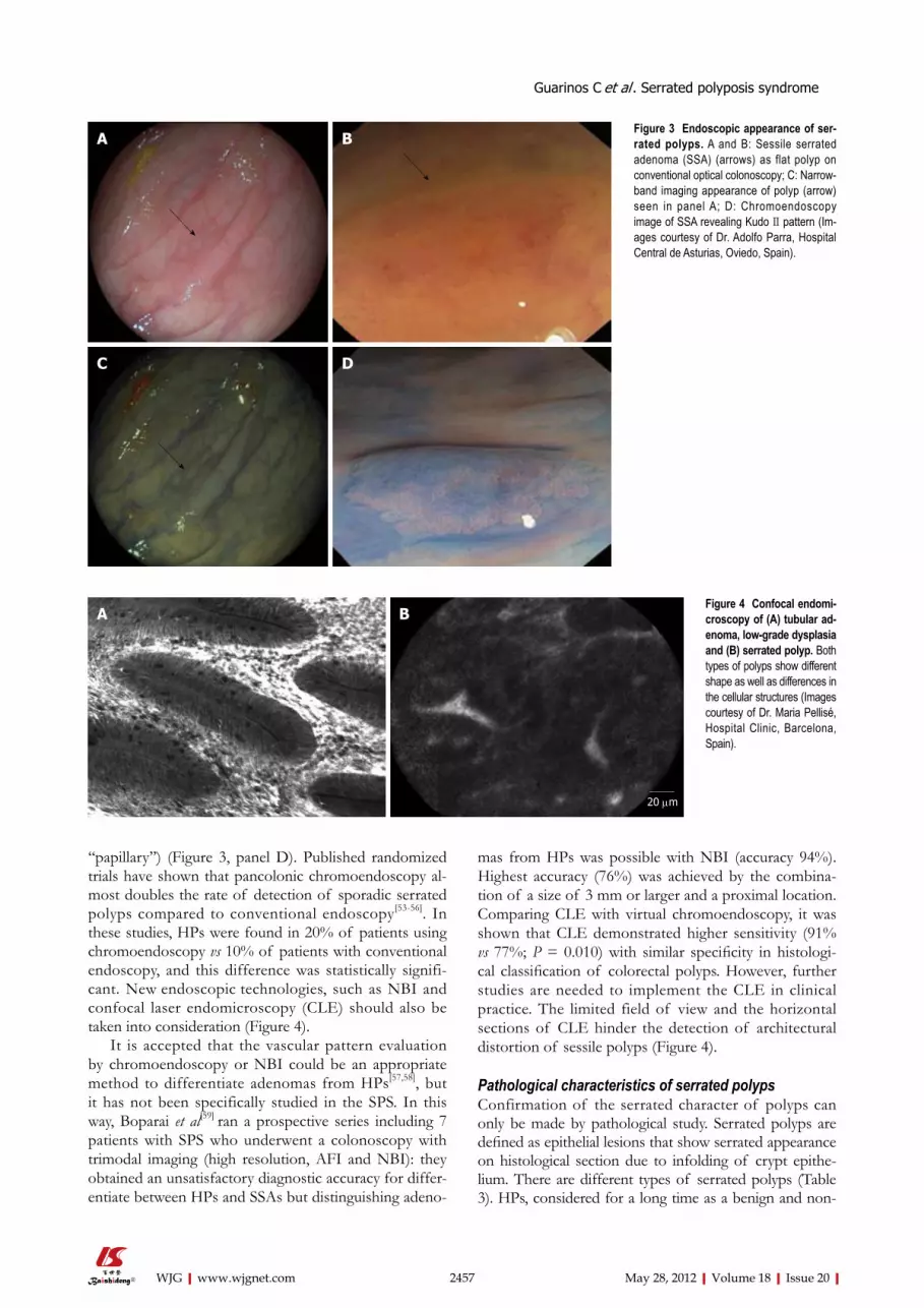
‘‘papillary’’) (Figure 3, panel D). Published randomized trials have shown that pancolonic chromoendoscopy al-most doubles the rate of detection of sporadic serrated polyps compared to conventional endoscopy[53-56]. In these studies, HPs were found in 20% of patients using chromoendoscopy vs 10% of patients with conventional endoscopy, and this difference was statistically signifi-cant. New endoscopic technologies, such as NBI and confocal laser endomicroscopy (CLE) should also be taken into consideration (Figure 4).
It is accepted that the vascular pattern evaluation by chromoendoscopy or NBI could be an appropriate method to differentiate adenomas from HPs[57,58], but it has not been specifically studied in the SPS. In this way, Boparai et al[59] ran a prospective series including 7 patients with SPS who underwent a colonoscopy with trimodal imaging (high resolution, AFI and NBI): they obtained an unsatisfactory diagnostic accuracy for differ-entiate between HPs and SSAs but distinguishing adeno-
mas from HPs was possible with NBI (accuracy 94%). Highest accuracy (76%) was achieved by the combina-tion of a size of 3 mm or larger and a proximal location. Comparing CLE with virtual chromoendoscopy, it was shown that CLE demonstrated higher sensitivity (91% vs 77%; P = 0.010) with similar specificity in histologi-cal classification of colorectal polyps. However, further studies are needed to implement the CLE in clinical practice. The limited field of view and the horizontal sections of CLE hinder the detection of architectural distortion of sessile polyps (Figure 4).
Pathological characteristics of serrated polypsConfirmation of the serrated character of polyps can only be made by pathological study. Serrated polyps are defined as epithelial lesions that show serrated appearance on histological section due to infolding of crypt epithe-lium. There are different types of serrated polyps (Table 3). HPs, considered for a long time as a benign and non-
2457 May 28, 2012|Volume 18|Issue 20|WJG|www.wjgnet.com
A B
C D
Figure 3 Endoscopic appearance of ser-rated polyps. A and B: Sessile serrated adenoma (SSA) (arrows) as flat polyp on conventional optical colonoscopy; C: Narrow-band imaging appearance of polyp (arrow) seen in panel A; D: Chromoendoscopy image of SSA revealing Kudo Ⅱ pattern (Im-ages courtesy of Dr. Adolfo Parra, Hospital Central de Asturias, Oviedo, Spain).
20 μm
Figure 4 Confocal endomi-croscopy of (A) tubular ad-enoma, low-grade dysplasia and (B) serrated polyp. Both types of polyps show different shape as well as differences in the cellular structures (Images courtesy of Dr. Maria Pellisé, Hospital Clinic, Barcelona, Spain).
A B
Guarinos C et al . Serrated polyposis syndrome

premalignant colorectal lesion, SSA, mixed polyps (MP), and TSA are included in this group[4,48,60,61] (Figure 5).
HPs are the most common colorectal polyps. Sporad-ic HPs are usually small (2-5 mm)[10], multiple and mainly distributed in the rectum and sigmoid colon[10,47]. HPs have been divided into two main histological subtypes: microvesicular serrated polyps (MVSPs) (Figure 5, panel A), in which columnar cells have mucin-filled vesicles within atypical cytoplasm, and goblet cell serrated polyps (GCSPs) with conspicuous goblet cells that are predomi-nantly found in the distal colon[4,23,47]. MVSPs seem to be the precursor lesion of SSA, especially when located in the right colon. In fact, both have the same molecular
genetic abnormalities such as mutations in BRAF and CIMP. MVSPs show large and regular stellate pit open-ings. However, the large GCSPs are likely to have KRAS mutation, which is infrequently found in SSA. There is some evidence that large GCSPs are potential precursors of dysplastic serrated polyps which show KRAS muta-tions[47,61,62] . A third type of HP has been added, mucin poor type, but its frequency and importance is lower than the two main HP types[6].
SSA is an atypical HP variant described by Torlakovic and Snover in 1996[63]. SSAs are larger than (usually great-er than 1 cm) HPs and more frequently located in the right colon[10]. Histologically, SSAs are distinguished from
2458 May 28, 2012|Volume 18|Issue 20|WJG|www.wjgnet.com
Polyp name Alternative terminology Morphology and significance Predominant location Molecular features
Hyperplastic polyp, goblet type
Type 1 hyperplastic polyp Subtype of hyperplastic polyp with conspicuous goblet cells and showing the least morphologic deviation from normal; Described as goblet-cell rich type
Distal colon: Sigmoid and rectum
Frequent KRAS mutation (54%)
Hyperplastic polyp, microvesicular type
Type 2 hyperplastic polyp Variant of hyperplastic polyp in which columnar cells have mucin-filled vesicles within the apical cytoplasm and goblet cells are relatively inconspicuous
Right colon and distal colon
Frequent BRAF mutation (76%) and CIMP (68%)
Sesile serrated adenoma
Sessile serrated polyp; Serrated polyp with atypical proliferation
Advanced type of serrated polyp with abnormalities of architecture and proliferation but lacking the classic features of epithelial dysplasia (intraepithelial neoplasia)
Right colon Frequent BRAF mutation (75%-82%) and CIMP (92%)
Sessile serrated adenoma with cytological dysplasia
Mixed polyp Rare serrated polyp that includes two separate components: Nondysplastic (usually SSA) and either traditional adenoma or serrated adenoma
Right and left colon Frequent BRAF mutation, (89%)
Serrated adenoma Mixed hyperplastic adenomatous polyp;Atypical hyperplastic polyp;TSA
Relatively rare neoplastic polyp having a serrated architecture reminiscent of hyperplastic polyp but with unequivocal traditional adenomatous dysplasia; Comprises < 5% of serrated polyps
Left colon Marked molecular heterogeneity;May have either KRAS or BRAF mutation
Table 3 Characteristics of serrated polyps
SSA: Sessile serrated adenoma; TSA: Traditional serrated adenoma; CIMP: CpG island methylator phenotype.
Figure 5 Pathological types of serrated polyps. A: Mi-crovesicular serrated polyp; B: Sessile serrated adeno-mas; C: Traditional serrated adenoma; D: Mixed polyp.
A B
C D
Guarinos C et al . Serrated polyposis syndrome

typical HPs by the presence of inverted T- or L-shaped crypt bases that reflect disordered proliferation (Figure 5, panel B). Other features include dilated crypts and ser-ration extending into the lower third of the crypt. Focal nuclear stratification, mild nuclear atypia, or dystrophic goblet cells may be seen in the crypt bases[47,60,61]. More-over, SSAs show increased mucin production, absence of enteroendocrine cells, and absence of a thickened base-ment membrane under the surface[43]. Other less common features include small foci of pseudostratification and eo-sinophilic change (identical to that seen in TSAs) of the surface epithelium. Small prominent nucleoli, open chro-matin, and irregular nuclear contours also might be pres-ent, along with mitoses in the upper third of the crypts or on the surface itself[61]. SSAs are thought to represent approximately 2% of all colonoscopically removed pol-yps, over 8% of all polyps that were previously diagnosed as HPs and around 18% of all serrated polyps[60].
MP, also called SSA with cytological dysplasia include two separate hyperplastic and adenomatous components (Figure 5, panel C)[21,23]. One component is usually SSA (nondysplastic) whereas the second dysplastic compo-nent is either adenoma or TSA.
TSAs, usually present on distal location, are dysplastic serrated polyps which lack SSA patterns and more closely resemble conventional adenoma with tubulovillous archi-tecture (Figure 5, panel D)[4,24,47,60]. Ectopic crypt forma-tion, defined by the presence of crypts with bases not seated adjacent to the muscularis mucosae, is a feature that makes it possible to distinguish between TSAs and SSAs[4]. Columnar cells from the epithelium show eosino-philic cytoplasm, centrally placed elongated nuclei that are hyperchromatic and display pseudostratification[46].
There is no strong morphological evidence suggest-ing that SSAs are the precursor of TSAs, otherwise there are some histological and epidemiologic differences for keeping these lesions apart in different categories[4,61]. SSAs have been associated with proximal CRCs, high level of CIMP, BRAF mutations and MSI-high[47,48]. TSAs have been associated with distal location and MSS, CIMP-low CRCs with KRAS mutations[48]. SSAs, TSAs and MPs are described as “advanced serrated polyps” and represent approximately 5%-15% of all serrated polyps found in colonoscopy patients[23].
FUTURE DIRECTIONSAdvances in the knowledge about the serrated pathway of carcinogenesis are making it possible to differentiate a new type of CRC with different natural history, prog-nosis and response to chemotherapy treatment. For this reason it is important to be able to easily identify this kind of colorectal tumor and its precursors. Identifica-tion of molecular markers in both polyps and cancers that follow this pathway will provide the opportunity of a better understanding of how these tumours grow and how we could explain differences in clinical presenta-tion, evolution and symptoms in different types of CRC. These molecular markers will also allow improvement
in the identification of patients with serrated polyposis, moving forward the currently used clinical criteria, and will give us better rationale for appropriate management and surveillance intervals for patients and their relatives.
REFERENCES1 Ferlay J, Shin HR, Bray F, Forman D, Mathers C, Parkin
DM. Estimates of worldwide burden of cancer in 2008: GLOBOCAN 2008. Int J Cancer 2010; 127: 2893-2917
2 Fearon ER, Vogelstein B. A genetic model for colorectal tu-morigenesis. Cell 1990; 61: 759-767
3 Jass JR. Classification of colorectal cancer based on correla-tion of clinical, morphological and molecular features. His-topathology 2007; 50: 113-130
4 Leggett B, Whitehall V. Role of the serrated pathway in colorectal cancer pathogenesis. Gastroenterology 2010; 138: 2088-2100
5 Torlakovic E, Skovlund E, Snover DC, Torlakovic G, Nesland JM. Morphologic reappraisal of serrated colorectal polyps. Am J Surg Pathol 2003; 27: 65-81
6 Snover DC, Ahnen DJ, Burt RW, Odze RD. Serrated polyps of the colon and rectum and serrated (“hyperplastic”) pol-yposis. In: Bosman ST, Carneiro F, Hruban RH, Theise ND. WHO Classification of tumours of the digestive system. Berlin: Springer-Verlag, 2010
7 Boparai KS, Mathus-Vliegen EM, Koornstra JJ, Nagengast FM, van Leerdam M, van Noesel CJ, Houben M, Cats A, van Hest LP, Fockens P, Dekker E. Increased colorectal cancer risk during follow-up in patients with hyperplastic polyposis syndrome: a multicentre cohort study. Gut 2010; 59: 1094-1100
8 Yeoman A, Young J, Arnold J, Jass J, Parry S. Hyperplastic polyposis in the New Zealand population: a condition as-sociated with increased colorectal cancer risk and European ancestry. N Z Med J 2007; 120: U2827
9 Chow E, Lipton L, Lynch E, D’Souza R, Aragona C, Hodg-kin L, Brown G, Winship I, Barker M, Buchanan D, Cowie S, Nasioulas S, du Sart D, Young J, Leggett B, Jass J, Macrae F. Hyperplastic polyposis syndrome: phenotypic presentations and the role of MBD4 and MYH. Gastroenterology 2006; 131: 30-39
10 Ferrández A, Samowitz W, DiSario JA, Burt RW. Pheno-typic characteristics and risk of cancer development in hy-perplastic polyposis: case series and literature review. Am J Gastroenterol 2004; 99: 2012-2018
11 Kalady MF, Jarrar A, Leach B, LaGuardia L, O’Malley M, Eng C, Church JM. Defining phenotypes and cancer risk in hyperplastic polyposis syndrome. Dis Colon Rectum 2011; 54: 164-170
12 Lage P, Cravo M, Sousa R, Chaves P, Salazar M, Fonseca R, Claro I, Suspiro A, Rodrigues P, Raposo H, Fidalgo P, Nobre-Leitão C. Management of Portuguese patients with hyperplastic polyposis and screening of at-risk first-degree relatives: a contribution for future guidelines based on a clinical study. Am J Gastroenterol 2004; 99: 1779-1784
13 Rubio CA, Stemme S, Jaramillo E, Lindblom A. Hyperplas-tic polyposis coli syndrome and colorectal carcinoma. En-doscopy 2006; 38: 266-270
14 Boparai KS, Reitsma JB, Lemmens V, van Os TA, Mathus-Vliegen EM, Koornstra JJ, Nagengast FM, van Hest LP, Keller JJ, Dekker E. Increased colorectal cancer risk in first-degree relatives of patients with hyperplastic polyposis syn-drome. Gut 2010; 59: 1222-1225
15 Samowitz WS, Albertsen H, Sweeney C, Herrick J, Caan BJ, Anderson KE, Wolff RK, Slattery ML. Association of smoking, CpG island methylator phenotype, and V600E BRAF mutations in colon cancer. J Natl Cancer Inst 2006; 98: 1731-1738
2459 May 28, 2012|Volume 18|Issue 20|WJG|www.wjgnet.com
Guarinos C et al . Serrated polyposis syndrome

16 Walker RG, Landmann JK, Hewett DG, Worthley DL, But-tenshaw RL, Knight N, Webb PM, Whiteman DC, Whitehall VL, Leggett BA. Hyperplastic polyposis syndrome is associ-ated with cigarette smoking, which may be a modifiable risk factor. Am J Gastroenterol 2010; 105: 1642-1647
17 Wallace K, Grau MV, Ahnen D, Snover DC, Robertson DJ, Mahnke D, Gui J, Barry EL, Summers RW, McKeown-Eyssen G, Haile RW, Baron JA. The association of lifestyle and dietary factors with the risk for serrated polyps of the colorectum. Cancer Epidemiol Biomarkers Prev 2009; 18: 2310-2317
18 Wynter CV, Walsh MD, Higuchi T, Leggett BA, Young J, Jass JR. Methylation patterns define two types of hyperplas-tic polyp associated with colorectal cancer. Gut 2004; 53: 573-580
19 Carvajal-Carmona LG, Howarth KM, Lockett M, Polanco-Echeverry GM, Volikos E, Gorman M, Barclay E, Martin L, Jones AM, Saunders B, Guenther T, Donaldson A, Paterson J, Frayling I, Novelli MR, Phillips R, Thomas HJ, Silver A, Atkin W, Tomlinson IP. Molecular classification and genetic pathways in hyperplastic polyposis syndrome. J Pathol 2007; 212: 378-385
20 Chan TL, Zhao W, Leung SY, Yuen ST. BRAF and KRAS mutations in colorectal hyperplastic polyps and serrated adenomas. Cancer Res 2003; 63: 4878-4881
21 Kambara T, Simms LA, Whitehall VL, Spring KJ, Wynter CV, Walsh MD, Barker MA, Arnold S, McGivern A, Mat-subara N, Tanaka N, Higuchi T, Young J, Jass JR, Leggett BA. BRAF mutation is associated with DNA methylation in serrated polyps and cancers of the colorectum. Gut 2004; 53: 1137-1144
22 Beach R, Chan AO, Wu TT, White JA, Morris JS, Lunago-mez S, Broaddus RR, Issa JP, Hamilton SR, Rashid A. BRAF mutations in aberrant crypt foci and hyperplastic polyposis. Am J Pathol 2005; 166: 1069-1075
23 Young J, Jass JR. The case for a genetic predisposition to serrated neoplasia in the colorectum: hypothesis and review of the literature. Cancer Epidemiol Biomarkers Prev 2006; 15: 1778-1784
24 Lazarus R, Junttila OE, Karttunen TJ, Mäkinen MJ. The risk of metachronous neoplasia in patients with serrated adeno-ma. Am J Clin Pathol 2005; 123: 349-359
25 Lindor NM. Hereditary colorectal cancer: MYH-associated polyposis and other newly identified disorders. Best Pract Res Clin Gastroenterol 2009; 23: 75-87
26 Jasperson KW, Tuohy TM, Neklason DW, Burt RW. Heredi-tary and familial colon cancer. Gastroenterology 2010; 138: 2044-2058
27 Young J, Barker MA, Simms LA, Walsh MD, Biden KG, Bu-chanan D, Buttenshaw R, Whitehall VL, Arnold S, Jackson L, Kambara T, Spring KJ, Jenkins MA, Walker GJ, Hopper JL, Leggett BA, Jass JR. Evidence for BRAF mutation and variable levels of microsatellite instability in a syndrome of familial colorectal cancer. Clin Gastroenterol Hepatol 2005; 3: 254-263
28 Frazier ML, Xi L, Zong J, Viscofsky N, Rashid A, Wu EF, Lynch PM, Amos CI, Issa JP. Association of the CpG island methylator phenotype with family history of cancer in pa-tients with colorectal cancer. Cancer Res 2003; 63: 4805-4808
29 Chan AO, Issa JP, Morris JS, Hamilton SR, Rashid A. Con-cordant CpG island methylation in hyperplastic polyposis. Am J Pathol 2002; 160: 529-536
30 Minoo P, Baker K, Goswami R, Chong G, Foulkes WD, Ruszkiewicz AR, Barker M, Buchanan D, Young J, Jass JR. Extensive DNA methylation in normal colorectal mucosa in hyperplastic polyposis. Gut 2006; 55: 1467-1474
31 Jarrar AM, Church JM, Fay S, Kalady MF. Is the phenotype mixed or mistaken? Hereditary nonpolyposis colorectal can-cer and hyperplastic polyposis syndrome. Dis Colon Rectum 2009; 52: 1949-1955
32 Boparai KS, Dekker E, Van Eeden S, Polak MM, Bartelsman JF, Mathus-Vliegen EM, Keller JJ, van Noesel CJ. Hyperplas-tic polyps and sessile serrated adenomas as a phenotypic expression of MYH-associated polyposis. Gastroenterology 2008; 135: 2014-2018
33 Sweet K, Willis J, Zhou XP, Gallione C, Sawada T, Alho-puro P, Khoo SK, Patocs A, Martin C, Bridgeman S, Heinz J, Pilarski R, Lehtonen R, Prior TW, Frebourg T, Teh BT, Marchuk DA, Aaltonen LA, Eng C. Molecular classification of patients with unexplained hamartomatous and hyper-plastic polyposis. JAMA 2005; 294: 2465-2473
34 Roberts A, Nancarrow D, Clendenning M, Buchanan DD, Jenkins MA, Duggan D, Taverna D, McKeone D, Walters R, Walsh MD, Young BW, Jass JR, Rosty C, Gattas M, Pelzer E, Hopper JL, Goldblatt J, George J, Suthers GK, Phillips K, Parry S, Woodall S, Arnold J, Tucker K, Muir A, Drini M, Macrae F, Newcomb P, Potter JD, Pavluk E, Lindblom A, Young JP. Linkage to chromosome 2q32.2-q33.3 in familial serrated neoplasia (Jass syndrome). Fam Cancer 2011; 10: 245-254
35 Brooker JC, Saunders BP, Shah SG, Thapar CJ, Suzuki N, Williams CB. Treatment with argon plasma coagulation reduces recurrence after piecemeal resection of large sessile colonic polyps: a randomized trial and recommendations. Gastrointest Endosc 2002; 55: 371-375
36 Iino H, Jass JR, Simms LA, Young J, Leggett B, Ajioka Y, Watanabe H. DNA microsatellite instability in hyperplastic polyps, serrated adenomas, and mixed polyps: a mild muta-tor pathway for colorectal cancer? J Clin Pathol 1999; 52: 5-9
37 Goel A, Nagasaka T, Arnold CN, Inoue T, Hamilton C, Niedzwiecki D, Compton C, Mayer RJ, Goldberg R, Bertag-nolli MM, Boland CR. The CpG island methylator pheno-type and chromosomal instability are inversely correlated in sporadic colorectal cancer. Gastroenterology 2007; 132: 127-138
38 Hawkins NJ, Ward RL. Sporadic colorectal cancers with mi-crosatellite instability and their possible origin in hyperplas-tic polyps and serrated adenomas. J Natl Cancer Inst 2001; 93: 1307-1313
39 Lee S, Cho NY, Choi M, Yoo EJ, Kim JH, Kang GH. Clinico-pathological features of CpG island methylator phenotype-positive colorectal cancer and its adverse prognosis in rela-tion to KRAS/BRAF mutation. Pathol Int 2008; 58: 104-113
40 Tanaka H, Deng G, Matsuzaki K, Kakar S, Kim GE, Miura S, Sleisenger MH, Kim YS. BRAF mutation, CpG island methylator phenotype and microsatellite instability occur more frequently and concordantly in mucinous than non-mucinous colorectal cancer. Int J Cancer 2006; 118: 2765-2771
41 Velho S, Moutinho C, Cirnes L, Albuquerque C, Hamelin R, Schmitt F, Carneiro F, Oliveira C, Seruca R. BRAF, KRAS and PIK3CA mutations in colorectal serrated polyps and cancer: primary or secondary genetic events in colorectal carcinogenesis? BMC Cancer 2008; 8: 255
42 Jass JR. Colorectal polyposes: from phenotype to diagnosis. Pathol Res Pract 2008; 204: 431-447
43 Jass JR. Hyperplastic polyps and colorectal cancer: is there a link? Clin Gastroenterol Hepatol 2004; 2: 1-8
44 Jover R, Nguyen TP, Pérez-Carbonell L, Zapater P, Payá A, Alenda C, Rojas E, Cubiella J, Balaguer F, Morillas JD, Clofent J, Bujanda L, Reñé JM, Bessa X, Xicola RM, Nicolás-Pérez D, Castells A, Andreu M, Llor X, Boland CR, Goel A. 5-Fluorouracil adjuvant chemotherapy does not increase survival in patients with CpG island methylator phenotype colorectal cancer. Gastroenterology 2011; 140: 1174-1181
45 Minoo P, Moyer MP, Jass JR. Role of BRAF-V600E in the serrated pathway of colorectal tumourigenesis. J Pathol 2007; 212: 124-133
46 Rosenberg DW, Yang S, Pleau DC, Greenspan EJ, Stevens RG, Rajan TV, Heinen CD, Levine J, Zhou Y, O’Brien MJ. Mutations in BRAF and KRAS differentially distinguish ser-
2460 May 28, 2012|Volume 18|Issue 20|WJG|www.wjgnet.com
Guarinos C et al . Serrated polyposis syndrome

rated versus non-serrated hyperplastic aberrant crypt foci in humans. Cancer Res 2007; 67: 3551-3554
47 Huang CS, Farraye FA, Yang S, O’Brien MJ. The clinical significance of serrated polyps. Am J Gastroenterol 2011; 106: 229-240; quiz 241
48 East JE, Saunders BP, Jass JR. Sporadic and syndromic hy-perplastic polyps and serrated adenomas of the colon: clas-sification, molecular genetics, natural history, and clinical management. Gastroenterol Clin North Am 2008; 37: 25-46, v
49 Whitehall VL, Walsh MD, Young J, Leggett BA, Jass JR. Methylation of O-6-methylguanine DNA methyltransferase characterizes a subset of colorectal cancer with low-level DNA microsatellite instability. Cancer Res 2001; 61: 827-830
50 Hesson LB, Hitchins MP, Ward RL. Epimutations and can-cer predisposition: importance and mechanisms. Curr Opin Genet Dev 2010; 20: 290-298
51 Worthley DL, Whitehall VL, Buttenshaw RL, Irahara N, Greco SA, Ramsnes I, Mallitt KA, Le Leu RK, Winter J, Hu Y, Ogino S, Young GP, Leggett BA. DNA methylation within the normal colorectal mucosa is associated with pathway-specific predisposition to cancer. Oncogene 2010; 29: 1653-1662
52 Kudo S, Tamura S, Nakajima T, Yamano H, Kusaka H, Watanabe H. Diagnosis of colorectal tumorous lesions by magnifying endoscopy. Gastrointest Endosc 1996; 44: 8-14
53 Brooker JC, Saunders BP, Shah SG, Thapar CJ, Thomas HJ, Atkin WS, Cardwell CR, Williams CB. Total colonic dye-spray increases the detection of diminutive adenomas during routine colonoscopy: a randomized controlled trial. Gastrointest Endosc 2002; 56: 333-338
54 Hurlstone DP, Cross SS, Slater R, Sanders DS, Brown S. Detecting diminutive colorectal lesions at colonoscopy: a randomised controlled trial of pan-colonic versus targeted chromoscopy. Gut 2004; 53: 376-380
55 Lapalus MG, Helbert T, Napoleon B, Rey JF, Houcke P, Ponchon T. Does chromoendoscopy with structure enhance-
ment improve the colonoscopic adenoma detection rate? Endoscopy 2006; 38: 444-448
56 Le Rhun M, Coron E, Parlier D, Nguyen JM, Canard JM, Alamdari A, Sautereau D, Chaussade S, Galmiche JP. High resolution colonoscopy with chromoscopy versus standard colonoscopy for the detection of colonic neoplasia: a ran-domized study. Clin Gastroenterol Hepatol 2006; 4: 349-354
57 Rastogi A, Bansal A, Wani S, Callahan P, McGregor DH, Cherian R, Sharma P. Narrow-band imaging colonoscopy--a pilot feasibility study for the detection of polyps and cor-relation of surface patterns with polyp histologic diagnosis. Gastrointest Endosc 2008; 67: 280-286
58 Tischendorf JJ, Wasmuth HE, Koch A, Hecker H, Traut-wein C, Winograd R. Value of magnifying chromoendosco-py and narrow band imaging (NBI) in classifying colorectal polyps: a prospective controlled study. Endoscopy 2007; 39: 1092-1096
59 Boparai KS, van den Broek FJ, van Eeden S, Fockens P, Dekker E. Hyperplastic polyposis syndrome: a pilot study for the differentiation of polyps by using high-resolution endoscopy, autofluorescence imaging, and narrow-band imaging. Gastrointest Endosc 2009; 70: 947-955
60 Liang JJ, Alrawi S, Tan D. Nomenclature, molecular genet-ics and clinical significance of the precursor lesions in the serrated polyp pathway of colorectal carcinoma. Int J Clin Exp Pathol 2008; 1: 317-324
61 Sandmeier D, Benhattar J, Martin P, Bouzourene H. Ser-rated polyps of the large intestine: a molecular study com-paring sessile serrated adenomas and hyperplastic polyps. Histopathology 2009; 55: 206-213
62 Hiraoka S, Kato J, Fujiki S, Kaji E, Morikawa T, Murakami T, Nawa T, Kuriyama M, Uraoka T, Ohara N, Yamamoto K. The presence of large serrated polyps increases risk for colorectal cancer. Gastroenterology 2010; 139: 1503-1510
63 Torlakovic E, Snover DC. Serrated adenomatous polyposis in humans. Gastroenterology 1996; 110: 748-755
S- Editor Gou SX L- Editor O’Neill M E- Editor Li JY
2461 May 28, 2012|Volume 18|Issue 20|WJG|www.wjgnet.com
Guarinos C et al . Serrated polyposis syndrome

REVIEW
Globus pharyngeus: A review of its etiology, diagnosis and treatment
Bong Eun Lee, Gwang Ha Kim
Bong Eun Lee, Gwang Ha Kim, Department of Internal Medi-cine, Pusan National University School of Medicine and Bio-medical Research Institute, Pusan National University Hospital, Busan 602-739, South KoreaAuthor contributions: Lee BE and Kim GH contributed equal-ly to this manuscript; Kim GH proposed the main idea of the study and contributed to the study design; Lee BE contributed to the study design, performed the literature search, and wrote the manuscript.Supported by A Pusan National University Hospital Clini-cal Research Grant (2011); a grant from the National R and D Program for Cancer Control, Ministry for Health, Welfare and Family Affairs, Republic of Korea, No. 0920050Correspondence to: Gwang Ha Kim, MD, Professor, Depart-ment of Internal Medicine, Pusan National University School of Medicine and Biomedical Research Institute, Pusan National University Hospital, 1-10 Ami-dong, Seo-gu, Busan 602-739, South Korea. [email protected]: +82-51-2407869 Fax: +82-51-2408180Received: December 21, 2011 Revised: January 30, 2012 Accepted: March 9, 2012 Published online: May 28, 2012
AbstractGlobus is a persistent or intermittent non-painful sen-sation of a lump or foreign body in the throat. It is a commonly encountered clinical condition that is usu-ally long-lasting, difficult to treat, and has a tendency to recur. Furthermore, due to the uncertain etiology of globus, it remains difficult to establish standard inves-tigation and treatment strategies for affected patients. As a first step for managing globus, careful history taking and nasolaryngoscopy are essential. Given the benign nature of the condition and the recent notion that gastroesophageal reflux disease is a major cause of globus, empirical therapy with a high dose of proton pump inhibitors is reasonable for patients with typical globus. If patients are nonresponsive to this therapy, definitive assessments such as endoscopy, multichan-nel intraluminal impedance/pH monitoring, and ma-
nometry should be considered. Speech and language therapy, anti-depressants, and cognitive-behavioral therapy can be helpful in patients whose symptoms persist despite negative investigations.
© 2012 Baishideng. All rights reserved.
Key words: Diagnosis; Gastroesophageal reflux disease; Globus; Proton pump inhibitor; Treatment
Peer reviewers: Justin Che Yuen Wu, Professor, Department of Medicine and Therapeutics, The Chinese University of Hong Kong, 9/F Clinical Science Building, Prince of Wales Hospital, Shatin, Hong Kong, China; Diego Garcia-Compean, Faculty of Medicine, Autonomous University of Nuevo Leon, Monterrey 64700, Mexico; Tomohiko Shimatani, Professor, Lab of Adult Nursing and Internal Medicine, Hiroshima International University, 5-1-1 Hirokoshingai, Kure 7370112, Japan
Lee BE, Kim GH. Globus pharyngeus: A review of its etiol-ogy, diagnosis and treatment. World J Gastroenterol 2012; 18(20): 2462-2471 Available from: URL: http://www.wjgnet.com/1007-9327/full/v18/i20/2462.htm DOI: http://dx.doi.org/10.3748/wjg.v18.i20.2462
INTRODUCTIONGlobus, a persistent or intermittent non-painful sensa-tion of a lump or foreign body in the throat, is a well-defined clinical symptom that is usually long-lasting, difficult to treat, and has a tendency to recur. This symp-tom frequently improves with eating and is generally unaccompanied by dysphagia or odynophagia[1]. It is a common condition that accounts for approximately 4% of new referrals to ear, nose and throat (ENT) clinics, and it is reported by up to 46% of apparently healthy individuals, with a peak incidence in middle age[2,3]. This condition is equally prevalent in men and women, though the latter are more likely to seek health care for
World J Gastroenterol 2012 May 28; 18(20): 2462-2471 ISSN 1007-9327 (print) ISSN 2219-2840 (online)
© 2012 Baishideng. All rights reserved.
Online Submissions: http://www.wjgnet.com/[email protected]:10.3748/wjg.v18.i20.2462
2462 May 28, 2012|Volume 18|Issue 20|WJG|www.wjgnet.com

Lee BE et al . Globus pharyngeus: An updated review
this symptom[4].Hippocrates first noted globus pharyngeus approxi-
mately 2500 years ago[5]. In 1707, Purcell[6] was the first to accurately describe the condition; he believed that globus resulted from pressure on the thyroid cartilage due to contraction of the strap muscles of the neck. In the past, globus was described as “globus hystericus” because of its frequent association with menopause or psychogenic factors. However, Malcomson[7] coined the more accurate term “globus pharyngeus” in 1968 after discovering that most patients experiencing globus did not have a hysterical personality. The etiology of globus is still unknown but appears to be multifactorial (Table 1). Although data are limited, recent studies have focused on gastroesophageal reflux disease (GERD), abnormali-ties of the upper esophageal sphincter (UES), psycho-logical and psychiatric disorders, and stress as major factors contributing to the globus sensation. The variety of potential etiologies has made it difficult to establish standard investigation and treatment strategies for af-fected patients.
The aim of this review is to present the current lit-erature on globus and to discuss its natural history and potential causes, current trends in its diagnosis, and methods for its treatment.
NATURAL HISTORYSince few long-term follow-up studies have been con-ducted on patients with globus, the natural history of this condition has not been fully elucidated. In one study that followed 74 globus patients for an average of 7 years and 7 mo, 45% of the patients had persistent symptoms during the follow-up period[8]. An in-depth analysis of the features at clinical presentation failed to reveal any reliable prognostic indicators. In another long-term follow-up study, 60% of the patients had improved or resolved symptoms over a 5-year period, and the male patients with a history of globus less than 3 mo and who did not complain of any associated throat symptoms were reported to have the greatest chance of becoming asymptomatic or symptomatically improved[9].
POTENTIAL CAUSES OF GLOBUSGastroesophageal reflux diseaseAlthough there is still considerable debate about the causative role of GERD in patients with globus, gas-troesophageal reflux (GER) has been suggested to be a major etiology of this symptom, potentially accounting for 23%-68% of globus patients[10-18].
Malcomson[7] was the first to link GERD to the globus sensation through the use of barium swallow to uncover the presence of reflux in over 60% of patients with globus. Moreover, Koufman[16] found that 58% of patients with globus had abnormal pH results, and Cherry et al[19] demonstrated that 10 out of 12 subjects complained of globus when acid was infused into the
distal esophagus. In a study that performed 24-h double-probe pH monitoring on 25 patients with globus and hoarseness, 72% of the patients exhibited pathologic reflux[20], and the globus symptom score was significantly higher in patients with GERD than in those without[21]. Additionally, globus sensation improved after 8 wk of proton pump inhibitor (PPI) therapy[22]. Several popula-tion-based surveys have supported such a potential link between GERD and the globus symptom by demon-strating an increased risk of globus among patients with GERD symptoms[23-25]. In a study by Dore et al[26], 38.7% of patients with GERD had the globus symptom, and the globus sensation was more prevalent in the non-ero-sive reflux disease group. Discordant data have also been reported[4,27-31]. However, it is clear that many patients with globus have concomitant GERD and that there is a true association between GERD and globus.
Two basic mechanisms have been proposed to explain the association between GERD and the globus sensa-tion[14,32,33]: (1) Direct irritation and inflammation of the laryngopharynx by retrograde flow of gastric contents, also known as laryngopharyngeal reflux (LPR)[15,16,34]; (2) Vagovagal reflex hypertonicity of the UES triggered by acidification or distention of the distal esophagus[18].
Abnormal upper esophageal sphincter functionAbnormal UES function has also been suggested to be a cause of globus sensation[20,35-38]. Elevated UES pressure has been found to be much more frequent in patients with globus sensation than in controls (28% vs 3%), sug-gesting that hypertensive UES is a background factor for globus[30]. Additionally, injection of botulinum toxin into the cricopharyngeal muscle in a patient with both globus and extremely high UES pressure led to a resolution of the globus symptom and a decrease in UES pressure[35]. In a study of high-resolution manometry in patients with globus sensation, normal controls, and GERD patients without globus, hyperdynamic respiratory UES pres-sure changes were most prevalent in patients reporting globus[38]. However, other studies have reported contrary results[39-41].
Esophageal motor disordersThe prevalence of esophageal motor disorders has been
2463 May 28, 2012|Volume 18|Issue 20|WJG|www.wjgnet.com
Gastroesophageal reflux disease Abnormal upper esophageal sphincter function Esophageal motor disorders Pharyngeal inflammatory causes including: pharyngitis, tonsillitis and chronic sinusitis Upper aerodigestive malignancy Hypertrophy of the base of the tongue Retroverted epiglottis Thyroid diseases Cervical heterotopic gastric mucosa Rare laryngopharyngeal tumors Psychological factors and stress
Table 1 Potential cause of globus

reported to be 6%-90% in patients with globus, sug-gesting that esophageal motor disorders are a possible cause of, or a contributing factor in the development of globus[27,28,31,42]. Esophageal manometry has revealed ab-normalities in as many as 67% of globus patients, with nonspecific esophageal motility disorder being the most frequent finding[31]. Moser et al[43] noted that esophageal motor disorders might, before giving rise to dysphagia, be sensed more vaguely and induce the globus sensa-tion. However, to infer an etiological significance of this disorder in globus, it must be shown that the sensation resolves after treatment for the motor disorder.
Pharyngeal inflammatory causesMany conditions that cause irritation and inflammation of the pharynx, such as pharyngitis, tonsillitis, and chron-ic sinusitis with postnasal drip, can be the cause of globus sensation by producing increased local sensitivity[28,44].
Upper aerodigestive tract malignancyThe presence of pharyngolaryngeal or upper esophageal malignancy must be excluded in patients with globus sensation, particularly in cases with “high risk” symp-toms, such as weight loss, dysphagia, throat pain, and lateralization of pathology[5,45].
Hypertrophy of the tongue baseGlobus can be induced by severe hypertrophy of the tongue base, probably due to the follicles touching the posterior wall of the pharynx. Mamede et al[46] demon-strated that hypertrophied follicles were frequent in patients with signs and symptoms of GER and that the symptoms of hypertrophy of the tongue base could be confused with those of GER. Retroverted epiglottisThrough contact with the tongue base or the posterior pharyngeal wall, retroverted epiglottis may cause globus sensation. Symptom relief has been observed after par-tial epiglottectomy[47,48].
Thyroid diseasesImpalpable, ultrasound-detectable abnormalities in the thyroid are known to be more common in patients with globus sensation than in controls[32]. Burns et al[49] noted that as many as one-third of patients with a thyroid mass complained of globus-like symptoms. Post-thyroidecto-my patients may also complain of globus pattern symp-toms, but these frequently diminish with time. Although the exact mechanism of the association between globus and thyroid diseases is poorly understood, some reports have concluded that a thyroidectomy could improve the globus symptom[49-51].
Cervical heterotopic gastric mucosaGlobus sensation has also been linked to the presence of cervical heterotopic gastric mucosa (CHGM)[52-54],
and acid secretion from CHGM appears to cause symp-toms similar to those of GERD, including globus sensa-tion. Patients with CHGM who complained of globus sensation and/or sore throat experienced a significant decrease in their symptoms after argon plasma ablation of CHGM[55,56]. Recently, it has been suggested that the globus symptom may be related to Helicobacter pylori in-fection of the CHGM[57].
Rare tumorsSmooth muscle tumors of the pharynx and post cricoid lymphangioma, as well as oropharyngeal metastasis of Merkel cell carcinoma, have been reported in patients complaining of globus[58-60]. These cases illustrate that patients with persistent globus should be further investi-gated to exclude rare lesions[32].
Psychological factors and stressPsychogenic problems have often been thought to cause or trigger the globus sensation. Personality studies have found higher levels of alexithymia, neuroticism, and psychological distress (including anxiety, low mood, and somatic concerns) and lower levels of extraversion in patients presenting with globus[61,62]. In addition, several studies have reported increased numbers of stressful life events preceding symptom onset, suggesting that life stress might be a cofactor in symptom genesis and in exacerbation. Indeed, up to 96% of patients with globus report symptom exacerbation during periods of high emotional intensity[63,64]. However, some reports have found no differences in the psychological states of pa-tients with globus compared to normal controls[4,10,65]. In actuality, psychiatric diagnoses are prevalent in subjects seeking health care for globus, but an explanation dis-tinct from ascertainment bias has not been established, causing the etiological significance of these psychologi-cal characteristics to remain uncertain[1,65]. Two recent studies reported that psychological status might be dif-ferent between LPR-positive and LPR-negative patients with globus[15,66]. Globus patients with LPR exhibited weaker psychological symptoms than non-LPR globus patients[15], and globus patients who did not respond to PPI had significantly higher anxiety scores[66].
OthersThere have been numerous isolated case reports that have suggested an association of globus with cervical osteophytes[67], temporomandibular joint disorders[68], hy-perviscosity of the nasopharyngeal mucosa[69], Eagle’s syn-drome[70], excessive laryngeal and pharyngeal tension[61], and salivary hypofunction[33].
DIAGNOSISThere has been no consensus regarding how best to diag-nose and manage globus. A study of United Kingdom-based ENT specialists found that 14% performed no
2464 May 28, 2012|Volume 18|Issue 20|WJG|www.wjgnet.com
Lee BE et al . Globus pharyngeus: An updated review

tests on globus patients but rather simply prescribed ant-acid medication if clinically indicated[71]. The remaining 86% investigated globus symptoms in a variety of ways, including rigid endoscopy (61%), barium swallow (56%), or a combination of these methods (17.5%).
Since globus is essentially a benign disorder, investi-gation is primarily aimed at identifying those few cases with upper aerodigestive malignancy. Thus, the first step of an investigation of globus symptoms should be to take a detailed patient history, paying particular attention to the presence of “high risk” symptoms, associated re-flux symptoms, and psychological problems. Additional-ly, physicians should perform a physical examination of the neck followed by nasolaryngoscopic examination of the laryngopharynx, although the routine use of nasolar-yngoscopy in patients with typical globus symptoms re-mains controversial[1]. Patients with typical globus symp-toms usually require no further investigation beyond an outpatient nasolaryngoscopy[5]. However, patients with “alarm signs”, such as dysphagia, odynophagia, throat pain, weight loss, hoarseness, and lateralization of pa-thology, should undergo more extensive evaluation[1].
Reflux symptom index and reflux finding score The symptoms and physical findings of LPR are nonspe-cific and can be confused with other laryngeal conditions caused by smoking, allergies, infections, vocal abuse, postnasal discharge, or neurogenic mechanisms as well as non-pathological variations[72]. Belafsky et al[73,74] proposed a useful self-administered tool, the reflux symptom index (RSI), for assessing the degree of LPR symptoms and developed the reflux finding score (RFS) based on 8 en-dolaryngeal signs for documenting the physical findings and severity of LPR. However, Park et al[15] demonstrated that RFS and RSI have low specificity in globus patients, suggesting that these may not be valid diagnostic tools for LPR in patients with globus.
Barium swallowBarium swallow studies have been reported to iden-tify benign lesions in up to one-third of patients with globus, and the most common findings include hiatal hernia and/or reflux (8%-18%), cervical osteophytes (0.4%-23%), and cricopharyngeal spasm (2.2%)[27,29,75,76]. However, given the prevalence of these findings in the general population, it is difficult to link these disorders to globus[45]. Two studies demonstrated that barium swal-low did not identify any malignancy in typical globus pa-tients[5,29]. Additionally, no pharyngeal or esophageal ma-lignancy was found in a study that reviewed 1145 barium swallows in patients presenting with globus, prompting the authors to conclude that barium swallow should not be systematically requested for the exclusion of malig-nancies in patients with globus[77]. Thus, this test seems to have limited diagnostic value in the investigation of patients with globus.
VideofluoroscopyOf 23 globus patients who received videofluoroscopy, 8 patients showed abnormal results; 5 had laryngeal aspira-tion, 2 had barium stasis in the vallecula and pyriform si-nuses, and 4 had poor pharyngeal elevation[78]. Although it is unlikely that this indicates a causal relationship, vid-eofluoroscopy may help to identify pharyngeal dysfunc-tion in a substantial proportion of globus patients.
24-h dual-probe ambulatory pH monitoringWhereas dual-probe ambulatory pH monitoring has been widely used in the clinical assessment of supra-esophageal GERD, this technology is not yet standard-ized, and its usefulness in the definition of a clinically relevant association with GERD is under debate. This technique has been used to show abnormal esophageal acid exposure in some globus patients[4,11]. However, reflux symptoms such as acid regurgitation and/or heartburn were also noted in these study populations. In a study of globus patients without reflux-like symp-toms, all 24 focal individuals had normal dual-probe pH results[78]. Therefore, ambulatory pH monitoring seems to be less helpful for the evaluation of globus without reflux-like symptoms.
24-h multichannel intraluminal impedance monitoringThe results from several trials indicate that the best way to detect GER in patients with extraesophageal manifes-tations of GERD is to conduct multichannel intralumi-nal impedance (MII)/pH monitoring. In patients expe-riencing persistent globus during PPI therapy, MII/pH monitoring increased the diagnostic yield of standard pH testing in the identification of positive symptom indices through the detection of nonacid reflux; furthermore, proximal reflux was a significant predictor of the globus symptom[79]. In studies investigating the utility of MII/pH monitoring in patients displaying atypical symptoms while “off PPI”, MII/pH monitoring increased the diag-nostic yield for objective detection of atypical manifesta-tions of GERD[80-82]. Thus, this technique appears to be a more promising method of obtaining reliable data for the detection of LPR than 24-h dual probe monitoring, as it can monitor acid as well as nonacid reflux events and can distinguish between liquid and gaseous events. Therefore, MII/pH monitoring appears to be useful for ruling out GERD and for redirecting management of patients with suspected extraesophageal manifestations of GERD.
Flexible esophagogastroscopyEndoscopy has been shown to be superior to barium swallow as a principal means of diagnosing upper aerodigestive tract malignancy[83]. Excellent views of the pyriform fossa and the postcricoid area can be achieved by insufflating air via flexible esophagogastroscopy[75]. Moreover, this procedure enables full esophageal evalu-
2465 May 28, 2012|Volume 18|Issue 20|WJG|www.wjgnet.com
Lee BE et al . Globus pharyngeus: An updated review

ation and diagnosis of reflux esophagitis and/or upper esophageal malignancy as a cause of globus. However, in general, endoscopy is known to have low sensitivity and to be of limited value for the diagnosis of extra-esophageal GERD. A study of 58 patients with pH-documented LPR found that only 19% had esophagitis or Barrett’s metaplasia[84]. In another study of patients with suspected LPR symptoms, esophagitis was generally prevalent[85] but occurred least in patients with globus and throat symptoms. Due to the association between globus and CGHM, it is necessary to carefully evaluate the cervical esophagus[54].
ManometryIf abnormal UES function and esophageal motor dis-order are suspected to be the potential cause of globus, manometry is a useful tool for assessing UES and lower esophageal sphincter pressure, esophageal body contrac-tion amplitude, and peristaltic sequence. However, the etiological significance of such a disorder is difficult to define.
TREATMENTA suggested algorithm for the management of globus patients based on available evidence is shown in Figure 1. Since there is a paucity of controlled studies on the treatment of globus, evidence-based treatment concepts are currently not available, and a review of the litera-
ture reveals that there is no single effective treatment. Given the benign nature of the condition, the likelihood of long-term symptom persistence, and the absence of highly effective pharmacotherapy, the mainstays of treatment are explanation and reassurance[1]. Other es-tablished treatment options include anti-reflux therapy, speech and language therapy, anti-depressants, and cog-nitive-behavioral therapy[86].
Anti-reflux treatmentSince GER has been suggested to be a major cause of globus[10-18], it seems practical that anti-reflux treatment should be the first attempted method for managing patients with globus. As diagnostic tests for GERD are somewhat invasive and costly and because a negative study result does not definitively rule out GER, it seems reasonable to use empirical PPI therapy as a combined method of diagnosis and treatment[87-92]. Although there are no controlled trials looking at the role of PPIs spe-cifically for the treatment of globus, there is a variety of literature addressing the role of PPIs in LPR manage-ment[34,91,93-95]. Current evidence shows that the clinical response to PPIs in LPRD is variable[96,97] and that LPR symptoms improve more slowly than esophageal symp-toms following acid-suppression therapy[98]. It is now widely accepted that extraesophageal GERD requires more aggressive and more prolonged therapy than typi-cal GERD[32]. Empirical twice-daily therapy with PPIs for at least 3 mo is recommended; this can be extended
2466 May 28, 2012|Volume 18|Issue 20|WJG|www.wjgnet.com
Globus
Careful history-taking and neck examination/nasolaryngoscopy
“High risk” patients or abnormal nasolaryngoscopic findings
Further investigations for ruling out malignancy or treatment of abnormalities
Clinical suspicion of GERD
Empirical high-dose PPI therapy
3-mo follow-up
Symptom resolved Symptom partially improved Symptom unresponsive
Titrate PPI therapy Increase PPI dose Definitive assessment- MII/pH monitoring- Endoscopy- Manometry- Barium swallow- Videofluoroscopy
Pathological reflux confirmed Negative investigations
Laparoscopic fundoplicationin selected cases
- Speech therapy/relaxation techniques- Antidepressants/cognitive-behavioral therapy- Consider other experimental therapy
Figure 1 Algorithm for management of globus. GERD: Gastroesophageal reflux disease; PPI: Proton pump inhibitor; MII: Multichannel intraluminal impedance.
Lee BE et al . Globus pharyngeus: An updated review

for a maximum period of 6 mo[87-92,99-102]. A PPI should be taken 30-60 min before meals so that it has reached its highest concentration by the time food intake stimu-lates the proton pumps. After 3-6 mo, responders can be weaned, whereas non-responders should undergo a definitive assessment, such as endoscopy, pH monitor-ing, or MII/pH monitoring. If available, MII/pH moni-toring is preferable to simple pH monitoring because it facilitates the detection of nonacid reflux. Nocturnal acid breakthrough (NAB) may cause incomplete treat-ment response[103,104]. The addition of h.s. histamine-2 receptor antagonists to twice-daily PPI therapy has been suggested to control NAB[103], but it is currently unclear whether this method offers any additional benefit to the long-term control of LPRD[105,106]. Prokinetics are utilized when it is necessary to speed up esophageal and gastric emptying; they can be useful when the clinical response to PPIs is unsatisfactory[16]. Diet and behavioral modification can also decrease the amount of reflux. Recommended dietary modifications include a reduction in the intake of chocolate, fats, citrus fruits, carbonated beverages, spicy tomato-based products, red wines, caf-feine, and late-night meals. Additionally, patients should make more general behavioral modifications, including exercising regularly, avoiding smoking and alcohol, el-evating the head of the bed (10-15 cm), avoiding tight clothes around the waist, and losing weight. Sleeping on one’s left-hand side also helps to decrease reflux. Steward et al[107] demonstrated that lifestyle modification was an independently significant variable in determining the response to pharmacological therapy. An alternative therapeutic strategy is anti-reflux surgery, with some au-thors having reported good improvement rates of LPR symptoms after laparoscopic nissen fundoplication[108-114]. To achieve a better patient outcome, a surgical approach must be taken into consideration in a carefully selected patient population, especially for patients who respond to treatment but are unable to tolerate PPIs due to side effects, those with confirmed pathological GER who do not respond to maximal medical treatment, and those in whom nonacid reflux has been demonstrated by a MII study[115]. However, if symptoms do not improve in the 4 mo following aggressive acid suppression, laparoscopic fundoplication may be unlikely to yield additional bene-fits[116]. Previous clinical responses to pharmacological acid suppression and abnormal pharyngeal pH results are pre-operative predictors of relief from atypical symptoms[117].
Speech and language therapy/relaxation techniquesSpeech therapy/relaxation techniques, including neck and shoulder exercises, general relaxation techniques, voice exercises, and voice hygiene to relieve vocal tract discomfort and tension, have successfully been used to treat patients with persistent globus symptoms[61]. In one uncontrolled study using these techniques on 25 globus patients, 92% experienced improvement following treat-ment. Khalil et al[118] randomly allocated 36 globus pa-
tients to either a speech therapy group or a reassurance group. Those in the speech therapy group used a number of exercises to relieve pharyngolaryngeal tension, includ-ing yawning, adopting a “giggle posture” (which helps retract the false vocal cords), and a “wet swallow” (as opposed to a “dry” or “check swallow,” which patients often perform habitually and which tends to aggravate the globus symptom). Patients also attempted to elimi-nate throat clearing and promote adequate hydration by avoiding smoking, excess tea, and coffee. At the end of 3 mo, patients in the speech therapy group demonstrated significantly better globus symptom scores compared with those recorded prior to the intervention. Individu-als in the speech therapy group also experienced signifi-cant improvements in globus symptoms when compared with controls. However, further research is needed to distinguish whether speech therapy has a specific effect or whether patients simply benefit from general atten-tion and reassurance[119].
Cognitive-behavioral therapy/antidepressantsGlobus is the fourth most common symptom of so-matization disorder after vomiting, aphonia, and pain in the extremities[120]. Cognitive-behavioral therapy has emerged as the best treatment for a variety of somato-form disorders and medically unexplained symptoms[121]. Although there has not yet been a substantial trial of cognitive-behavioral therapy in globus patients, it is likely to be a promising treatment for repeat attenders whose symptoms remain refractory[76].
A small series of anti-depressants have been found to be beneficial for some globus patients with concomi-tant psychiatric disorders, such as panic, somatization, major depression, and agoraphobia[122,123].
Other treatment strategiesThyroidectomy in patients with thyroid disorder[49-51] or partial epiglottectomy in selected cases whose retroverted epiglottis made contact with the tongue base[47,48], were both reported to significantly relieve the globus symptom. In addition, ablation of CHGM by argon plasma coagu-lation has shown some promise in improving chronic globus symptoms[55,56]. Although additional research of these techniques is needed, these approaches would pro-vide some benefit to patients with unexplained chronic globus who are refractory to any medical treatments.
CONCLUSIONAlthough globus is a common clinical condition, its etiol-ogy remains uncertain, and there is no standard protocol for its diagnosis and management. The results of recent studies have strongly suggested that GERD is a major cause of globus, though this remains under considerable debate. Numerous other disorders, such as abnormal UES function, esophageal motility disorders, structural head and neck diseases, and psychological factors, have
2467 May 28, 2012|Volume 18|Issue 20|WJG|www.wjgnet.com
Lee BE et al . Globus pharyngeus: An updated review

been suggested as potential causes of globus. However, it has been rather difficult to establish a causal relation-ship between globus and these disorders because most of the reported studies were uncontrolled, had a small sample size, or were case reports. Currently, careful his-tory taking and nasolaryngoscopy are essential as a first step in managing globus. Given the benign nature of the condition, patients with typical globus do not appear to need further investigation; rather, a 3-mo treatment with high-dose PPIs seems to be a reliable treatment op-tion. If patients are nonresponsive to PPI therapy, they should undergo a definitive assessment, such as endos-copy, pH monitoring, or MII/pH monitoring; MII/pH monitoring in particular may increase the diagnostic yield of GER in globus patients. In cases with negative clinical investigations and consistent globus symptom, other treatment strategies, including speech therapy, anti-depressants, and cognitive-behavioral therapy, should be considered. In the future, well-designed, randomized controlled studies are needed to definitively determine the effect of PPI treatment on globus. In addition, it is necessary to ascertain the etiology of globus via large-scale studies.
REFERENCES1 Galmiche JP, Clouse RE, Bálint A, Cook IJ, Kahrilas PJ,
Paterson WG, Smout AJ. Functional esophageal disorders. Gastroenterology 2006; 130: 1459-1465
2 Moloy PJ, Charter R. The globus symptom. Incidence, therapeutic response, and age and sex relationships. Arch Otolaryngol 1982; 108: 740-744
3 Drossman DA, Li Z, Andruzzi E, Temple RD, Talley NJ, Thompson WG, Whitehead WE, Janssens J, Funch-Jensen P, Corazziari E. U.S. householder survey of functional gas-trointestinal disorders. Prevalence, sociodemography, and health impact. Dig Dis Sci 1993; 38: 1569-1580
4 Batch AJ. Globus pharyngeus (Part I). J Laryngol Otol 1988; 102: 152-158
5 Harar RP, Kumar S, Saeed MA, Gatland DJ. Management of globus pharyngeus: review of 699 cases. J Laryngol Otol 2004; 118: 522-527
6 Purcell J. A treatise of vapours or hysteric fits. 2nd ed. Lon-don: Edward Place, 1707: 72-74
7 Malcomson KG. Globus hystericus vel pharyngis (a recom-maissance of proximal vagal modalities). J Laryngol Otol 1968; 82: 219-230
8 Rowley H, O’Dwyer TP, Jones AS, Timon CI. The natu-ral history of globus pharyngeus. Laryngoscope 1995; 105: 1118-1121
9 Timon C, O’Dwyer T, Cagney D, Walsh M. Globus pharyn-geus: long-term follow-up and prognostic factors. Ann Otol Rhinol Laryngol 1991; 100: 351-354
10 Hill J, Stuart RC, Fung HK, Ng EK, Cheung FM, Chung CS, van Hasselt CA. Gastroesophageal reflux, motility dis-orders, and psychological profiles in the etiology of globus pharyngis. Laryngoscope 1997; 107: 1373-1377
11 Chevalier JM, Brossard E, Monnier P. Globus sensation and gastroesophageal reflux. Eur Arch Otorhinolaryngol 2003; 260: 273-276
12 Wilson JA, Pryde A, Piris J, Allan PL, Macintyre CC, Ma-ran AG, Heading RC. Pharyngoesophageal dysmotility in globus sensation. Arch Otolaryngol Head Neck Surg 1989; 115: 1086-1090
13 Koufman JA, Amin MR, Panetti M. Prevalence of reflux in 113 consecutive patients with laryngeal and voice disorders. Otolaryngol Head Neck Surg 2000; 123: 385-388
14 Oridate N, Nishizawa N, Fukuda S. The diagnosis and management of globus: a perspective from Japan. Curr Opin Otolaryngol Head Neck Surg 2008; 16: 498-502
15 Park KH, Choi SM, Kwon SU, Yoon SW, Kim SU. Diagnosis of laryngopharyngeal reflux among globus patients. Otolar-yngol Head Neck Surg 2006; 134: 81-85
16 Koufman JA. The otolaryngologic manifestations of gastro-esophageal reflux disease (GERD): a clinical investigation of 225 patients using ambulatory 24-hour pH monitoring and an experimental investigation of the role of acid and pepsin in the development of laryngeal injury. Laryngoscope 1991; 101: 1-78
17 Koufman J, Sataloff RT, Toohill R. Laryngopharyngeal re-flux: consensus conference report. J Voice 1996; 10: 215-216
18 Tokashiki R, Funato N, Suzuki M. Globus sensation and increased upper esophageal sphincter pressure with distal esophageal acid perfusion. Eur Arch Otorhinolaryngol 2010; 267: 737-741
19 Cherry J, Siegel CI, Margulies SI, Donner M. Pharyngeal lo-calization of symptoms of gastroesophageal reflux. Ann Otol Rhinol Laryngol 1970; 79: 912-914
20 Smit CF, van Leeuwen JA, Mathus-Vliegen LM, Devriese PP, Semin A, Tan J, Schouwenburg PF. Gastropharyngeal and gastroesophageal reflux in globus and hoarseness. Arch Otolaryngol Head Neck Surg 2000; 126: 827-830
21 Sinn DH, Kim JH, Kim S, Son HJ, Kim JJ, Rhee JC, Rhee PL. Response rate and predictors of response in a short-term empirical trial of high-dose rabeprazole in patients with glo-bus. Aliment Pharmacol Ther 2008; 27: 1275-1281
22 Tokashiki R, Yamaguchi H, Nakamura K, Suzuki M. Glo-bus sensation caused by gastroesophageal reflux disease. Auris Nasus Larynx 2002; 29: 347-351
23 Locke GR, Talley NJ, Fett SL, Zinsmeister AR, Melton LJ. Prevalence and clinical spectrum of gastroesophageal reflux: a population-based study in Olmsted County, Minnesota. Gastroenterology 1997; 112: 1448-1456
24 Rey E, Elola-Olaso CM, Rodríguez-Artalejo F, Locke GR, Díaz-Rubio M. Prevalence of atypical symptoms and their association with typical symptoms of gastroesophageal re-flux in Spain. Eur J Gastroenterol Hepatol 2006; 18: 969-975
25 Cho YS, Choi MG, Jeong JJ, Chung WC, Lee IS, Kim SW, Han SW, Choi KY, Chung IS. Prevalence and clinical spec-trum of gastroesophageal reflux: a population-based study in Asan-si, Korea. Am J Gastroenterol 2005; 100: 747-753
26 Dore MP, Pedroni A, Pes GM, Maragkoudakis E, Tadeu V, Pirina P, Realdi G, Delitala G, Malaty HM. Effect of antise-cretory therapy on atypical symptoms in gastroesophageal reflux disease. Dig Dis Sci 2007; 52: 463-468
27 Wilson JA, Heading RC, Maran AG, Pryde A, Piris J, Allan PL. Globus sensation is not due to gastro-oesophageal re-flux. Clin Otolaryngol Allied Sci 1987; 12: 271-275
28 Batch AJ. Globus pharyngeus: (Part II), Discussion. J Laryn-gol Otol 1988; 102: 227-230
29 Back GW, Leong P, Kumar R, Corbridge R. Value of barium swallow in investigation of globus pharyngeus. J Laryngol Otol 2000; 114: 951-954
30 Corso MJ, Pursnani KG, Mohiuddin MA, Gideon RM, Cas-tell JA, Katzka DA, Katz PO, Castell DO. Globus sensation is associated with hypertensive upper esophageal sphincter but not with gastroesophageal reflux. Dig Dis Sci 1998; 43: 1513-1517
31 Färkkilä MA, Ertama L, Katila H, Kuusi K, Paavolainen M, Varis K. Globus pharyngis, commonly associated with esophageal motility disorders. Am J Gastroenterol 1994; 89: 503-508
32 Remacle M. The diagnosis and management of globus: a
2468 May 28, 2012|Volume 18|Issue 20|WJG|www.wjgnet.com
Lee BE et al . Globus pharyngeus: An updated review

perspective from Belgium. Curr Opin Otolaryngol Head Neck Surg 2008; 16: 511-515
33 Baek CH, Chung MK, Choi JY, So YK, Son YI, Jeong HS. Role of salivary function in patients with globus pharyn-geus. Head Neck 2010; 32: 244-252
34 Ford CN. Evaluation and management of laryngopharyn-geal reflux. JAMA 2005; 294: 1534-1540
35 Halum SL, Butler SG, Koufman JA, Postma GN. Treatment of globus by upper esophageal sphincter injection with botulinum A toxin. Ear Nose Throat J 2005; 84: 74
36 Watson WC, Sullivan SN. Hypertonicity of the cricopharyn-geal sphincter: A cause of globus sensation. Lancet 1974; 2: 1417-1419
37 Hunt PS, Connell AM, Smiley TB. The cricopharyngeal sphincter in gastric reflux. Gut 1970; 11: 303-306
38 Kwiatek MA, Mirza F, Kahrilas PJ, Pandolfino JE. Hyperdy-namic upper esophageal sphincter pressure: a manometric observation in patients reporting globus sensation. Am J Gastroenterol 2009; 104: 289-298
39 Caldarelli DD, Andrews AH, Derbyshire AJ. Esophageal motility studies in globus sensation. Ann Otol Rhinol Laryn-gol 1970; 79: 1098-1100
40 Cook IJ, Dent J, Collins SM. Upper esophageal sphincter tone and reactivity to stress in patients with a history of glo-bus sensation. Dig Dis Sci 1989; 34: 672-676
41 Sun J, Xu B, Yuan YZ, Xu JY. Study on the function of phar-ynx upper esophageal sphincter in globus hystericus. World J Gastroenterol 2002; 8: 952-955
42 Knight RE, Wells JR, Parrish RS. Esophageal dysmotility as an important co-factor in extraesophageal manifestations of gastroesophageal reflux. Laryngoscope 2000; 110: 1462-1466
43 Moser G, Vacariu-Granser GV, Schneider C, Abatzi TA, Pokieser P, Stacher-Janotta G, Gaupmann G, Weber U, Wenzel T, Roden M. High incidence of esophageal motor disorders in consecutive patients with globus sensation. Gastroenterology 1991; 101: 1512-1521
44 Lee JW, Song CW, Kang CD, Hur BW, Jeen YT, Jeon HJ, Lee HS, Lee SW, Um SH, Choi JH, Kim CD, Ryu HS, Hyun JH. Pharyngoesophageal motility in patients with globus sensa-tion. Korean J Gastroenterol 2000; 36: 1-9
45 Cathcart R, Wilson JA. Lump in the throat. Clin Otolaryngol 2007; 32: 108-110
46 Mamede RC, De Mello-Filho FV, Dantas RO. Severe hyper-trophy of the base of the tongue in adults. Otolaryngol Head Neck Surg 2004; 131: 378-382
47 Agada FO, Coatesworth AP, Grace AR. Retroverted epiglot-tis presenting as a variant of globus pharyngeus. J Laryngol Otol 2007; 121: 390-392
48 Quesada JL, Lorente J, Quesada P. Partial epiglottectomy as a possible treatment for globus pharyngeus? Eur Arch Oto-rhinolaryngol 2000; 257: 386-388
49 Burns P, Timon C. Thyroid pathology and the globus symp-tom: are they related? A two year prospective trial. J Laryn-gol Otol 2007; 121: 242-245
50 Marshall JN, McGann G, Cook JA, Taub N. A prospective controlled study of high-resolution thyroid ultrasound in patients with globus pharyngeus. Clin Otolaryngol Allied Sci 1996; 21: 228-231
51 Maung KH, Hayworth D, Nix PA, Atkin SL, England RJ. Thyroidectomy does not cause globus pattern symptoms. J Laryngol Otol 2005; 119: 973-975
52 von Rahden BH, Stein HJ, Becker K, Liebermann-Meffert D, Siewert JR. Heterotopic gastric mucosa of the esophagus: literature-review and proposal of a clinicopathologic clas-sification. Am J Gastroenterol 2004; 99: 543-551
53 Lancaster JL, Gosh S, Sethi R, Tripathi S. Can heterotopic gastric mucosa present as globus pharyngeus? J Laryngol Otol 2006; 120: 575-578
54 Alaani A, Jassar P, Warfield AT, Gouldesbrough DR, Smith I.
Heterotopic gastric mucosa in the cervical oesophagus (inlet patch) and globus pharyngeus--an under-recognised asso-ciation. J Laryngol Otol 2007; 121: 885-888
55 Meining A, Bajbouj M, Preeg M, Reichenberger J, Kassem AM, Huber W, Brockmeyer SJ, Hannig C, Höfler H, Prinz C, Schmid RM. Argon plasma ablation of gastric inlet patches in the cervical esophagus may alleviate globus sensation: a pilot trial. Endoscopy 2006; 38: 566-570
56 Bajbouj M, Becker V, Eckel F, Miehlke S, Pech O, Prinz C, Schmid RM, Meining A. Argon plasma coagulation of cervi-cal heterotopic gastric mucosa as an alternative treatment for globus sensations. Gastroenterology 2009; 137: 440-444
57 Alagozlu H, Simsek Z, Unal S, Cindoruk M, Dumlu S, Dur-sun A. Is there an association between Helicobacter pylori in the inlet patch and globus sensation? World J Gastroenterol 2010; 16: 42-47
58 Husamaldin Z, Aung W, McFerran DJ. Smooth muscle tu-mour of the pharynx: a rare tumour presenting with globus pharyngeus symptoms. J Laryngol Otol 2004; 118: 885-887
59 Smith NM, Stafford FW. Post cricoid lymphangioma. J Lar-yngol Otol 1991; 105: 220-221
60 Reichel OA, Mayr D, Issing WJ. Oropharyngeal metastasis of a Merkel cell carcinoma of the skin. Eur Arch Otorhinolar-yngol 2003; 260: 258-260
61 Wareing M, Elias A, Mitchell D. Management of globus sen-sation by the speech therapist. Logoped Phoniatr Vocol 1997; 22: 39-42
62 Deary IJ, Wilson JA, Kelly SW. Globus pharyngis, personal-ity, and psychological distress in the general population. Psychosomatics 1995; 36: 570-577
63 Harris MB, Deary IJ, Wilson JA. Life events and difficulties in relation to the onset of globus pharyngis. J Psychosom Res 1996; 40: 603-615
64 Thompson WG, Heaton KW. Heartburn and globus in ap-parently healthy people. Can Med Assoc J 1982; 126: 46-48
65 Moser G, Wenzel-Abatzi TA, Stelzeneder M, Wenzel T, Weber U, Wiesnagrotzki S, Schneider C, Schima W, Stacher-Janotta G, Vacariu-Granser GV, Pokieser P, Bergmann H, Stacher G. Globus sensation: pharyngoesophageal function, psychometric and psychiatric findings, and follow-up in 88 patients. Arch Intern Med 1998; 158: 1365-1373
66 Siupsinskiene N, Adamonis K, Toohill RJ, Sereika R. Pre-dictors of response to short-term proton pump inhibitor treatment in laryngopharyngeal reflux patients. J Laryngol Otol 2008; 122: 1206-1212
67 Maran A, Jacobson I. Cervical osteophytes presenting with pharyngeal symptoms. Laryngoscope 1971; 81: 412-417
68 Kirveskari P, Puhakka H. Effect of occlusal adjustment on globus symptom. J Prosthet Dent 1985; 54: 832-835
69 Shiomi Y, Shiomi Y, Oda N, Hosoda S. Hyperviscoelasticity of epipharyngeal mucus may induce globus pharyngis. Ann Otol Rhinol Laryngol 2002; 111: 1116-1119
70 Beder E, Ozgursoy OB, Karatayli Ozgursoy S, Anadolu Y. Three-dimensional computed tomography and surgical treatment for Eagle’s syndrome. Ear Nose Throat J 2006; 85: 443-445
71 Webb CJ, Makura ZG, Fenton JE, Jackson SR, McCormick MS, Jones AS. Globus pharyngeus: a postal questionnaire survey of UK ENT consultants. Clin Otolaryngol Allied Sci 2000; 25: 566-569
72 Vaezi MF, Hicks DM, Abelson TI, Richter JE. Laryngeal signs and symptoms and gastroesophageal reflux disease (GERD): a critical assessment of cause and effect association. Clin Gastroenterol Hepatol 2003; 1: 333-344
73 Belafsky PC, Postma GN, Koufman JA. Validity and reli-ability of the reflux symptom index (RSI). J Voice 2002; 16: 274-277
74 Belafsky PC, Postma GN, Koufman JA. The validity and re-liability of the reflux finding score (RFS). Laryngoscope 2001;
2469 May 28, 2012|Volume 18|Issue 20|WJG|www.wjgnet.com
Lee BE et al . Globus pharyngeus: An updated review
111: 1313-131775 Takwoingi YM, Kale US, Morgan DW. Rigid endoscopy in
globus pharyngeus: how valuable is it? J Laryngol Otol 2006; 120: 42-46
76 Burns P, O’Neill JP. The diagnosis and management of glo-bus: a perspective from Ireland. Curr Opin Otolaryngol Head Neck Surg 2008; 16: 503-506
77 Alaani A, Vengala S, Johnston MN. The role of barium swallow in the management of the globus pharyngeus. Eur Arch Otorhinolaryngol 2007; 264: 1095-1097
78 Chen CL, Tsai CC, Chou AS, Chiou JH. Utility of ambula-tory pH monitoring and videofluoroscopy for the evalua-tion of patients with globus pharyngeus. Dysphagia 2007; 22: 16-19
79 Anandasabapathy S, Jaffin BW. Multichannel intraluminal impedance in the evaluation of patients with persistent globus on proton pump inhibitor therapy. Ann Otol Rhinol Laryngol 2006; 115: 563-570
80 Bajbouj M, Becker V, Neuber M, Schmid RM, Meining A. Combined pH-metry/impedance monitoring increases the diagnostic yield in patients with atypical gastroesophageal reflux symptoms. Digestion 2007; 76: 223-228
81 Lee BE, Kim GH, Ryu DY, Kim DU, Cheong JH, Lee DG, Song GA. Combined Dual Channel Impedance/pH-metry in Patients With Suspected Laryngopharyngeal Reflux. J Neurogastroenterol Motil 2010; 16: 157-165
82 Malhotra A, Freston JW, Aziz K. Use of pH-impedance testing to evaluate patients with suspected extraesophageal manifestations of gastroesophageal reflux disease. J Clin Gastroenterol 2008; 42: 271-278
83 Levine B, Nielsen EW. The justifications and controver-sies of panendoscopy--a review. Ear Nose Throat J 1992; 71: 335-340, 343
84 Koufman JA, Belafsky PC, Bach KK, Daniel E, Postma GN. Prevalence of esophagitis in patients with pH-documented laryngopharyngeal reflux. Laryngoscope 2002; 112: 1606-1609
85 Poelmans J, Feenstra L, Demedts I, Rutgeerts P, Tack J. The yield of upper gastrointestinal endoscopy in patients with suspected reflux-related chronic ear, nose, and throat symp-toms. Am J Gastroenterol 2004; 99: 1419-1426
86 Karkos PD, Wilson JA. The diagnosis and management of globus pharyngeus: our perspective from the United King-dom. Curr Opin Otolaryngol Head Neck Surg 2008; 16: 521-524
87 Divi V, Benninger MS. Diagnosis and management of la-ryngopharyngeal reflux disease. Curr Opin Otolaryngol Head Neck Surg 2006; 14: 124-127
88 Celik M, Ercan I. Diagnosis and management of laryngo-pharyngeal reflux disease. Curr Opin Otolaryngol Head Neck Surg 2006; 14: 150-155
89 Remacle M, Lawson G. Diagnosis and management of la-ryngopharyngeal reflux disease. Curr Opin Otolaryngol Head Neck Surg 2006; 14: 143-149
90 Pontes P, Tiago R. Diagnosis and management of laryngo-pharyngeal reflux disease. Curr Opin Otolaryngol Head Neck Surg 2006; 14: 138-142
91 Mahieu HF. Review article: The laryngological manifesta-tions of reflux disease; why the scepticism? Aliment Pharma-col Ther 2007; 26 Suppl 2: 17-24
92 Bove MJ, Rosen C. Diagnosis and management of laryngo-pharyngeal reflux disease. Curr Opin Otolaryngol Head Neck Surg 2006; 14: 116-123
93 El-Serag HB, Lee P, Buchner A, Inadomi JM, Gavin M, McCarthy DM. Lansoprazole treatment of patients with chronic idiopathic laryngitis: a placebo-controlled trial. Am J Gastroenterol 2001; 96: 979-983
94 Noordzij JP, Khidr A, Evans BA, Desper E, Mittal RK, Reibel JF, Levine PA. Evaluation of omeprazole in the treat-ment of reflux laryngitis: a prospective, placebo-controlled, randomized, double-blind study. Laryngoscope 2001; 111:
2147-215195 Issing WJ, Karkos PD, Perreas K, Folwaczny C, Reichel O.
Dual-probe 24-hour ambulatory pH monitoring for diag-nosis of laryngopharyngeal reflux. J Laryngol Otol 2004; 118: 845-848
96 Katz PO, Castell DO. Medical therapy of supraesophageal gastroesophageal reflux disease. Am J Med 2000; 108 Suppl 4a: 170S-177S
97 Vaezi MF. Extraesophageal manifestations of gastroesopha-geal reflux disease. Clin Cornerstone 2003; 5: 32-38; discus-sion 39-40
98 Oridate N, Takeda H, Asaka M, Nishizawa N, Mesuda Y, Mori M, Furuta Y, Fukuda S. Acid-suppression therapy of-fers varied laryngopharyngeal and esophageal symptom relief in laryngopharyngeal reflux patients. Dig Dis Sci 2008; 53: 2033-2038
99 Park W, Hicks DM, Khandwala F, Richter JE, Abelson TI, Milstein C, Vaezi MF. Laryngopharyngeal reflux: prospec-tive cohort study evaluating optimal dose of proton-pump inhibitor therapy and pretherapy predictors of response. Laryngoscope 2005; 115: 1230-1238
100 Williams RB, Szczesniak MM, Maclean JC, Brake HM, Cole IE, Cook IJ. Predictors of outcome in an open label, thera-peutic trial of high-dose omeprazole in laryngitis. Am J Gas-troenterol 2004; 99: 777-785
101 Amin MR, Postma GN, Johnson P, Digges N, Koufman JA. Proton pump inhibitor resistance in the treatment of laryn-gopharyngeal reflux. Otolaryngol Head Neck Surg 2001; 125: 374-378
102 Belafsky PC, Postma GN, Koufman JA. Laryngopharyngeal reflux symptoms improve before changes in physical find-ings. Laryngoscope 2001; 111: 979-981
103 Peghini PL, Katz PO, Bracy NA, Castell DO. Nocturnal re-covery of gastric acid secretion with twice-daily dosing of proton pump inhibitors. Am J Gastroenterol 1998; 93: 763-767
104 Peghini PL, Katz PO, Castell DO. Ranitidine controls nocturnal gastric acid breakthrough on omeprazole: a con-trolled study in normal subjects. Gastroenterology 1998; 115: 1335-1339
105 Fackler WK, Ours TM, Vaezi MF, Richter JE. Long-term effect of H2RA therapy on nocturnal gastric acid break-through. Gastroenterology 2002; 122: 625-632
106 Ours TM, Fackler WK, Richter JE, Vaezi MF. Nocturnal acid breakthrough: clinical significance and correlation with esophageal acid exposure. Am J Gastroenterol 2003; 98: 545-550
107 Steward DL, Wilson KM, Kelly DH, Patil MS, Schwartzbau-er HR, Long JD, Welge JA. Proton pump inhibitor therapy for chronic laryngo-pharyngitis: a randomized placebo-control trial. Otolaryngol Head Neck Surg 2004; 131: 342-350
108 Lindstrom DR, Wallace J, Loehrl TA, Merati AL, Toohill RJ. Nissen fundoplication surgery for extraesophageal manifes-tations of gastroesophageal reflux (EER). Laryngoscope 2002; 112: 1762-1765
109 Oelschlager BK, Eubanks TR, Oleynikov D, Pope C, Pel-legrini CA. Symptomatic and physiologic outcomes after operative treatment for extraesophageal reflux. Surg Endosc 2002; 16: 1032-1036
110 Fernando HC, El-Sherif A, Landreneau RJ, Gilbert S, Chris-tie NA, Buenaventura PO, Close JM, Luketich JD. Efficacy of laparoscopic fundoplication in controlling pulmonary symptoms associated with gastroesophageal reflux disease. Surgery 2005; 138: 612-616; discussion 612-616
111 Westcott CJ, Hopkins MB, Bach K, Postma GN, Belafsky PC, Koufman JA. Fundoplication for laryngopharyngeal re-flux disease. J Am Coll Surg 2004; 199: 23-30
112 Catania RA, Kavic SM, Roth JS, Lee TH, Meyer T, Fantry GT, Castellanos PF, Park A. Laparoscopic Nissen fundopli-cation effectively relieves symptoms in patients with laryn-
2470 May 28, 2012|Volume 18|Issue 20|WJG|www.wjgnet.com
Lee BE et al . Globus pharyngeus: An updated review

gopharyngeal reflux. J Gastrointest Surg 2007; 11: 1579-1587; discussion 1579-1587
113 Sala E, Salminen P, Simberg S, Koskenvuo J, Ovaska J. La-ryngopharyngeal reflux disease treated with laparoscopic fundoplication. Dig Dis Sci 2008; 53: 2397-2404
114 Antoniou SA, Delivorias P, Antoniou GA, Natsiopoulos I, Kalambakas A, Dalenbäck J, Makridis C. Symptom-focused results after laparoscopic fundoplication for refractory gas-troesophageal reflux disease--a prospective study. Langen-becks Arch Surg 2008; 393: 979-984
115 Hassall E. Decisions in diagnosing and managing chronic gastroesophageal reflux disease in children. J Pediatr 2005; 146: S3-12
116 Qadeer MA, Swoger J, Milstein C, Hicks DM, Ponsky J, Richter JE, Abelson TI, Vaezi MF. Correlation between symptoms and laryngeal signs in laryngopharyngeal reflux. Laryngoscope 2005; 115: 1947-1952
117 So JB, Zeitels SM, Rattner DW. Outcomes of atypical symp-toms attributed to gastroesophageal reflux treated by lapa-
roscopic fundoplication. Surgery 1998; 124: 28-32118 Khalil HS, Bridger MW, Hilton-Pierce M, Vincent J. The use
of speech therapy in the treatment of globus pharyngeus patients. A randomised controlled trial. Rev Laryngol Otol Rhinol (Bord) 2003; 124: 187-190
119 Millichap F, Lee M, Pring T. A lump in the throat: Should speech and language therapists treat globus pharyngeus? Disabil Rehabil 2005; 27: 124-130
120 Othmer E, DeSouza C. A screening test for somatization disorder (hysteria). Am J Psychiatry 1985; 142: 1146-1149
121 Kroenke K. Efficacy of treatment for somatoform disorders: a review of randomized controlled trials. Psychosom Med 2007; 69: 881-888
122 Cybulska EM. Globus hystericus--a somatic symptom of depression? The role of electroconvulsive therapy and anti-depressants. Psychosom Med 1997; 59: 67-69
123 Brown SR, Schwartz JM, Summergrad P, Jenike MA. Glo-bus hystericus syndrome responsive to antidepressants. Am J Psychiatry 1986; 143: 917-918
S- Editor Gou SX L- Editor Rutherford A E- Editor Li JY
2471 May 28, 2012|Volume 18|Issue 20|WJG|www.wjgnet.com
Lee BE et al . Globus pharyngeus: An updated review

ORIGINAL ARTICLE
Lactobacillus plantarum B7 inhibits Helicobacter pylori growth and attenuates gastric inflammation
Chompoonut Sunanliganon, Duangporn Thong-Ngam, Somying Tumwasorn, Naruemon Klaikeaw
Chompoonut Sunanliganon, Duangporn Thong-Ngam, Department of Physiology, Faculty of Medicine, Chulalongkorn University, Bangkok 10330, ThailandSomying Tumwasorn, Department of Microbiology, Faculty of Medicine, Chulalongkorn University, Bangkok 10330, ThailandNaruemon Klaikeaw, Department of Pathology, Faculty of Medicine, Chulalongkorn University, Bangkok 10330, ThailandAuthor contributions: Sunanliganon C performed the experi-ments, collected the data and wrote the manuscript; Thong-Ngam D designed the study, performed the experiments, analyzed the data and edited the manuscript; Tumwasorn S provided Helicobacter pylori and Lactobacillus plantarum B7; Klaikeaw N co-ordinated the pathological examination.Supported by The 90th Anniversary of Chulalongkorn Univer-sity Fund (Ratchada phiseksomphot Endowment Fund); and the grant of Ratchada phiseksomphot, Faculty of Medicine, Chul-alongkorn University, Bangkok, ThailandCorrespondence to: Duangporn Thong-Ngam, MD, Associ-ate Professor, Department of Physiology, Faculty of Medicine, Chulalongkorn University, Bangkok 10330, Thailand. [email protected]: +662-256-42672050 Fax: +662-256-42672823Received: December 21, 2011 Revised: February 3, 2012Accepted: March 9, 2012Published online: May 28, 2012
AbstractAIM: To determine the anti-Helicobacter property of Lactobacillus plantarum B7 (L. plantarum) B7 superna-tants in vitro and the protective effects of L. plantarum B7 on serum tumor necrosis factor-alpha (TNF-α), gas-tric malondialdehyde (MDA) level, apoptosis, and his-topathology in Helicobacter pylori (H. pylori )-induced gastric inflammation in rats.
METHODS: In vitro , the inhibition of H. pylori growth was examined using L. plantarum B7 supernatants at pH 4 and pH 7 and at the concentration of 1×, 5× and 10× on plates inoculated with H. pylori . The inhibi-tory effect of H. pylori was interpreted by the size of the inhibition zone. In vitro , male Sprague-Dawley rats
were randomly divided into four groups including group 1 (control group), group 2 (H. pylori infected group), group 3 (H. pylori infected with L. plantarum B7 106 CFUs/mL treated group) and group 4 (H. pylori infected with L. plantarum B7 1010 CFUs/mL treated group). One week after H. pylori inoculation, L. plantarum B7 106 CFUs/mL or 1010 CFUs/mL were fed once daily to group 3 and group 4, respectively, for one week. Blood and gastric samples were collected at the end of the study.
RESULTS: In vitro , at intact pH 4, mean inhibitory zone diameters of 8.5 mm and 13 mm were noted at concentrations of 5× and 10× of L. plantarum B7 supernatant disks, respectively. At adjusted pH 7, L. plantarum B7 supernatants at concentrations of 5× and 10× yielded mean inhibitory zone diameters of 6.5 mm and 11 mm, respectively. In the in vitro study, in group 2, stomach histopathology revealed mild to moderate H. pylori colonization and inflammation. The level of gastric MDA and epithelial cell apoptosis were significantly increased compared with group 1. The se-rum TNF-α level was significant decreased in group 3 compared with group 2 (P < 0.05). In addition, L. plan-tarum B7 treatments resulted in a significant improve-ment in stomach pathology, and decreased gastric MDA level and apoptotic epithelial cells.
CONCLUSION: L. plantarum B7 supernatant inhibits H. pylori growth. This inhibition was dose-dependent and greater at pH 4. Moreover, L. plantarum B7 at-tenuated H. pylori -induced gastric inflammation.
© 2012 Baishideng. All rights reserved.
Key words: Apoptosis; Gastric inflammation; Helico-bacter pylori ; Lactobacillus plantarum B7; Lipid peroxi-dation
Peer reviewer: Francesco Luzza, Professor, Dipartimento di Medicina Sperimentale e Clini, Università di Catanzaro, Via Tommaso Campanella 115, 88100 Catanzaro, Italy
World J Gastroenterol 2012 May 28; 18(20): 2472-2480 ISSN 1007-9327 (print) ISSN 2219-2840 (online)
© 2012 Baishideng. All rights reserved.
Online Submissions: http://www.wjgnet.com/[email protected]:10.3748/wjg.v18.i20.2472
2472 May 28, 2012|Volume 18|Issue 20|WJG|www.wjgnet.com

Sunanliganon C et al . L. plantarum B7 attenuated H. pylori -induced gastric inflammation
Sunanliganon C, Thong-Ngam D, Tumwasorn S, Klaikeaw N. Lactobacillus plantarum B7 inhibits Helicobacter pylori growth and attenuates gastric inflammation. World J Gastroenterol 2012; 18(20): 2472-2480 Available from: URL: http://www.wjgnet.com/1007-9327/full/v18/i20/2472.htm DOI: http://dx.doi.org/10.3748/wjg.v18.i20.2472
INTRODUCTIONHelicobacter pylori (H. pylori) is a gram-negative, spiral shaped bacterium that has the unique ability of being able to colonize the human gastric mucosa and infects more than half of the world’s population. H. pylori causes chronic gastritis, plays an etiologic role in peptic ulcer disease and is considered a risk factor in the de-velopment of gastric cancer and gastric lymphoma[1]. In 1994, H. pylori was classified as a type Ⅰ carcinogen by the World Health Organization[2].
H. pylori infection is characterized by enhanced pro-duction of proinflammatory mediators such as tumor necrosis factor-alpha (TNF-α), interleukin (IL)-1β, IL-2, IL-6 and IL-8 and the infiltration of lamina propria with inflammatory cells. H. pylori lipopolysaccharides (LPS) and released surface proteins stimulate lamina propria mononuclear cells and macrophages to produce proin-flammatory cytokines such as TNF-α, IL-1β, and the generation of reactive oxygen species (ROS)[3]. TNF-α and IL-1β are potent inducers of IL-8 expression in many cell types. Furthermore, H. pylori is capable of interacting with epithelial cell surfaces to produce IL-8. The release of these inflammatory mediators results in the expression of CD11b/CD18 on leukocytes and in-tercellular adhesion molecule-1 on endothelial cells, the migration of leukocytes to a site of inflammation, and finally the generation of ROS[4].
ROS can react with the double bonds of polyunsatu-rated fatty acids (PUFAs), present in the membranes of phospholipids, resulting in lipid peroxidation. One of the major secondary oxidation products of peroxidized PUFAs is malondialdehyde (MDA)[5]. H. pylori also in-duces gastric epithelial cell apoptosis both in vitro[6] and in vivo[7]. Studies have shown that the H. pylori colonized stomach contains more apoptotic epithelial cells than normal epithelial cells. Moreover, the increased numbers of apoptotic epithelial cells decrease to normal after eradication of H. pylori[7].
H. pylori eradication is suboptimal because current treatment regimens result in adverse side effects, poor compliance, and an increasing prevalence of antibiotic re-sistance[8]. Therefore, alternative treatments are of interest.
Lactobacilli are probiotics which, when administered in adequate amounts, may confer a benefit to the host[9]. The most commonly used organisms in probiotic products are Lactobacillus sp. and Bifidobacterium sp.[10]. L. plantarumis commonly found in the human gastrointestinal tract (GI-tract). It is important in the production of a variety of fer-mented foods such as sauerkraut, Korean kimchi, cheese,
sausages and stockfish, and is also used as a probiotic. Moreover, there is increasing evidence that L. plantarum has anti-Helicobacter activity and shows modulatory effects on the immune system[11,12]. Importantly, L. plantarum is acid and bile tolerant, survives passage through the GI-tract, and is safe in humans and animals.
The aim of this study was to examine the in vitro anti-Helicobacter activity of L. plantarum B7 supernatants using the disk diffusion method and the effects of L. plantarum B7 on gastric histopathology, serum TNF-α, gastric MDA level, and cell apoptosis in H. pylori infection in vivo.
MATERIALS AND METHODSIn vitro studyThe disk diffusion method was used to assess the anti-H. pylori activity of L. plantarum B7 supernatants at intact and neutralized pH and various concentrations of 1×, 5× and 10× against H. pylori.
Bacterial strains and culture conditions: H. pylori ATCC 43504 was grown on Columbia agar (Oxoid, Bas-ingstoke, United Kingdom) containing 7% sheep blood and 7% horse serum. Plates were incubated at 37 ℃ under microaerophilic conditions (10% CO2, 5% O2 and 85% N2) produced by a gas generating system, Anaero-Pack (MGC, Japan), for 72 h in an anaerobic jar (Oxoid, Basingstoke, United Kingdom).
L. plantarum B7, isolated from Thai dyspeptic pa-tients, was stored in de Man-Rogosa-Sharpe (MRS) broth (Oxoid, Basingstoke, United Kingdom) with 20% glycerol at -80 ℃. This strain was recovered from frozen stock and cultivated twice on MRS agar anaerobically (10% CO2, 10% H2 and 80% N2) at 37 ℃ in an anaero-bic jar for 48 h. A single colony of L. plantarum B7 was then inoculated into 10 mL of MRS broth and grown at 37 ℃ under anaerobic conditions for 24 h in a 15 mL conical centrifuge tube (Corning, New York, United States). The OD600 of the culture was determined us-ing a spectrophotometer (Bio-Rad Smart SpecTM Plus), adjusted to OD600 with 0.1 in 10 mL of MRS broth and incubated for 48 h. After incubation, the culture supernatant was collected by centrifugation at 1000 × g for 10 min at 4 ℃ and then filtered using a 0.22 µm pore size filter unit (Minisart, Germany). The supernatant of Lactobacillus without the cell pellet was called the Lactoba-cillus condition media (LCM). The concentration and pH of LCM were adjusted to 1×, 5× and 10× by speed-vac-uum drying (speed-vacuum, Savant Instruments, United States) and resuspending in an appropriate volume of intact pH 4 and adjusted pH 7 MRS broth. Sterile 6 mm-membrane disks (Whatman, Maidstone, United King-dom) were then dipped into the resuspended LCM for at least 1 h at room temperature.
Disk diffusion method: The various concentrations of L. plantarum B7 supernatants were evaluated at two pH values, intact pH 4, and adjusted pH 7 with NaOH.
2473 May 28, 2012|Volume 18|Issue 20|WJG|www.wjgnet.com

H. pylori was spread on Columbia blood agar plates, and L. plantarum B7 (LCM) disks were placed directly on the surface of the agar. The plates were incubated under microaerophilic conditions at 37 ℃ for 72 h, after which the diameters of the inhibition zones were measured in millimeters. In this study, the MRS broth was used as a negative control. The experiments were carried out in duplicate and mean values of the growth inhibition zones were measured.
In vivo studyBacteria preparation: H. pylori was subcultured twice on Columbia blood agar. Plates were incubated at 37 ℃ under microaerophilic conditions for 72 h. L. plantarum B7 was originally obtained from Thai dyspeptic patients who visited King Chulalongkorn Memorial Hospital. This strain was cultivated twice on MRS agar anaerobi-cally at 37 ℃ for 48 h.
Animal preparation: Thirty-two male Sprague-Dawley rats (Salaya Research Animal Center, Mahidol University, Bangkok, Thailand), weighing about 150-250 g at the be-ginning of the experiment, were used. The experimental protocol was approved by the Ethical Committee of Medicine Faculty, Chulalongkorn University, Thailand. The animals were housed in Macrolon cages (5 animals per cage), given food and tap water ad libitum at room temperature (18 ℃-22 ℃), humidity 55%, and a 12/12 h-light/dark cycle.
Experimental protocol: The rats were randomly divid-ed into four experimental groups (eight rats each group) as follows. Group 1: Rats were fed phosphate buffered saline (PBS) 1 mL/rat by gavage twice a day at an in-terval of four hours for three consecutive days. Then, they were housed with free access to water and standard food for 1 wk. After that, the animals were treated with PBS 1 mL/rat by gavage once daily for 1 wk. Group 2: Rats were inoculated with H. pylori using the method of Thong-Ngam et al[13]. Briefly, the rats were pre-treated with streptomycin suspended in tap water (5 mg/mL) for three days before H. pylori inoculation. The H. pylori suspension (5 × 10¹⁰ CFUs/mL) in PBS was adminis-tered (1 mL/rat) by gavage twice daily at an interval of four hours for three consecutive days. One week after the inoculation, the animals were treated with PBS (1 mL/rat) by gavage once daily for one week. Group 3: One week after H. pylori inoculation, the rats were treat-ed by gavage with L. plantarum B7 106 CFUs/mL sus-pended in PBS once daily for 1 wk. Group 4: One week after H. pylori inoculation, the rats were treated by gavage with L. plantarum B7 1010 CFUs/mL suspended in PBS once daily for 1 wk.
At the end of the experiment, animals were sacri-ficedby an overdose of intraperitoneal thiopental so-dium. Blood samples were then collected for TNF-α determination using enzyme-linked immunosorbent assay (ELISA). The stomach was removed. One-half of
the stomach was frozen in liquid nitrogen, and stored at -80 ℃ for MDA analysis. The remainder of the stomach was fixed in 4% paraformaldehyde in phosphate buffer solution to determine histopathology and epithelial cell apoptosis.
Determination of serum cytokine levels: Blood samples were taken by cardiac puncture, allowed to clot for two hours at room temperature before centrifuging for 20 min at approximately 1000 × g. Then, the serum was removed and stored at -80 ℃ for determination of TNF-α level using an ELISA kit (R and D Systems, United States).
Assessment of H. pylori infection and examination of histopathology: The presence of H. pylori infection in the rats was determined by the urease test and histo-pathological examination by a blinded pathologist. After completing the experiment, the rats were sacrificed. The stomach was removed and 2 mm2 of gastric mucosa from the antrum was immediately dissected and placed in the urease tube to examine urease activity.
The remaining tissue from the gastric antrum biopsy was fixed in 4% paraformaldehyde in phosphate buffer solution at pH 7.4 and room temperature. The tissue was processed and stained with hematoxylin-eosin. The slides were observed by light microscopy and the presence of H. pylori was detected by Warthin-Starry staining in un-clear cases. The level of bacterial colonization was evalu-ated using a grading system as follows. Score 0: No bac-teria detected; Score 1: Mild colonization in some gastric crypts; Score 2: Mild colonization in most gastric crypts; Score 3: Moderate colonization in all gastric crypts. The results are presented as the bacterial colonization scores for each group. In addition to H. pylori colonization, the gastric inflammation level was estimated and scored fol-lowing the updated Sydney System[14]. The infiltration of polymorphonuclear leukocytes in the gastric mucosa, defining the inflammatory scores, was recorded. Scores from 0 to 3 represented normal, mild, moderate and marked histopathological changes, respectively.
Determination of gastric malondialdehyde: Gastric MDA level was measured using the thiobarbituric acid (TBA) reactive substances assay kit (Cayman, United States). The principle is that the reaction of one mol-ecule of MDA and two molecules of TBA form a red MDA-TBA complex under high temperature (90 ℃-100 ℃) and acidic conditions, which can be quantitated using a spectrophotometer at 532 nm. The assay pro-cedures were performed as described. The content of MDA was expressed in terms of nmol/mg protein.
Determination of gastric epithelial cell apoptosis: Apoptosis was measured by the identification of apop-totic nuclei in sections of stomachusing fragment end labeling of DNA (Apoptosis detectionkit, Chemicon, United States). In brief, the DNA fragments were al-
2474 May 28, 2012|Volume 18|Issue 20|WJG|www.wjgnet.com
Sunanliganon C et al . L. plantarum B7 attenuated H. pylori -induced gastric inflammation

lowed to bind an antidigoxigenin antibody that was conjugated to a peroxidase. Diaminobenzidine was ap-plied to develop a dark brown color and the slides were counterstained with hematoxylin. The positive stained cells showed dark brown nuclei under light microscopy. To verify the incidence of apoptosis, the dark brown-stained cells were counted. One thousand gastric epithe-lial cells were counted for each rat. The data were shown as a percentage (%) of apoptotic cells calculated as: the percentage of apoptotic cells (%) = (numbers of posi-tive stained cells × 100)/1000.
Statistical analysisAll data are presented as mean ± SD. The means were compared by one-way analysis of variance followed by least significant different post hoc test. All statistical tests were performed using SPSS for Windows version 13.0 (SPSS Inc, Chicago, IL, United States). Differences were considered statistically significant at P < 0.05.
RESULTSIn vitro studyDisk diffusion method: At intact pH 4, mean inhibitory zone diameters of 8.5 ± 0.7 mm and 13 ± 0 mm were noted at the concentrations of 5× and 10× of L. planta-rum B7 supernatant disks, respectively. At adjusted pH 7, mean inhibitory zone diameters of 6.5 ± 0.7 mm and 11 ± 1.4 mm were noted at the concentrations of 5× and 10× of L. plantarum B7 supernatant disks, respec-
tively (Table 1). Both intact pH 4 and adjusted pH 7 of L. plantarum B7 supernatants showed dose-dependent anti-H. pylori activity. The supernatant of pH 4 L. planta-rum B7 at the concentration of 10× showed the clearest inhibition (Figure 1).
In vivo studyChanges in TNF-α level: The serum TNF-α level was not significantly different between the control group and H. pylori infected group. However, in the L. plantarum B7 106 CFUs/mL treated group, a significant decrease in se-rum TNF-α level was noted compared with the H. pylori infected group (P = 0.019). The average concentrations of serum TNF-α were 17.22 ± 0.63 pg/mL, 18.05 ± 1.94 pg/mL, and 16.52 ± 0.84 pg/mL in the control, H. pylori infected, and in the L. plantarum B7 106 CFUs/mL treated group, respectively. The average serum TNF-α levels in all groups are shown in Figure 2.
Histopathological examination: H. pylori infection in rats was determined by the urease test and histopathol-ogy. Histopathology in the control group was normal, while in the H. pylori infected group there was moderate H. pylori colonization and inflammation. The L. planta-rum B7 106 CFUs/mL treated and L. plantarum B7 1010 CFUs/mL treated groups showed reduced H. pylori colo-nization and improved stomach inflammation (Figures 3 and 4). The histology scores for H. pylori colonization and gastric inflammation are summarized in Table 2.
Determination of gastric malondialdehyde: The level of gastric MDA increased significantly in the H. pylori infected compared with the control group (3.46 ± 1.25 nmol/mg vs 1.05 ± 0.41 nmol/mg protein, P = 0.000, respectively). After one week of 106 CFUs/mL or 1010 CFUs/mL of L. plantarum B7 suspension, there was a significant decrease in elevated gastric MDA level in both L. plantarum B7 treated groups compared with the H. pylori infected group (1.28 ± 0.69, 1.37 ± 0.66 nmol/mg vs 3.46 ± 1.25 nmol/mg protein, P = 0.000, respectively) (Figure 5).
Determination of gastric epithelial cell apoptosis: The percentage of apoptotic cells was significantly in-
2475 May 28, 2012|Volume 18|Issue 20|WJG|www.wjgnet.com
Concentration of L. plantarum Diameters of inhibition zone (mm)
B7 supernatant Intact pH 4 Adjusted pH 7
MRS (negative control) 0 0 1× 0 0 5× 8.5 ± 0.7 6.5 ± 0.7 10× 13 ± 0 11 ± 1.4
Table 1 Inhibition zone diameters (mm) of all Lactobacillus plantarum B7 supernatant concentrations at intact pH 4 and adjusted pH 7 (mean ± SD) (n = 2)
L. plantarum: Lactobacillus plantarum; MRS: Man-Rogosa-Sharpe.
Adjusted pH = 7Intact pH = 4
MRS 1× 5× 10×Concentration of L. plantarum B7 supernatant
14
12
10
8
6
4
2
0Dia
met
er o
f inh
ibiti
on z
one
(mm
)
Figure 1 A bar graph shows the mean ± SD of inhibitory zone diameters (mm) of all Lactobacillus plantarum B7 supernatant concentrations at intact pH 4 and adjusted pH 7 (n = 2). L. plantarum: Lactobacillus plantarum; MRS: Man-Rogosa-Sharpe.
CON HP HP + L (L) HP + L (H)
22
20
18
16
14
12
10
Seru
m T
NF-
α (
pg/m
L)
Figure 2 A bar graph shows the mean ± SD of serum tumor necrosis factor-alpha level (pg/mL) in all groups. CON: Control group; HP: Helico-bacter pylori (H. pylori) infected group; HP + L (L): Lactobacillus plantarum (L. plantarum) B7 106 CFUs/mL treated group; HP + L (H): L. plantarum B7 1010 CFUs/mL treated group. Each group is represented by the mean of 8 rats. aP < 0.05 vs H. pylori infected group. TNF-α: Tumor necrosis factor-alpha.
a
Sunanliganon C et al . L. plantarum B7 attenuated H. pylori -induced gastric inflammation

2476 May 28, 2012|Volume 18|Issue 20|WJG|www.wjgnet.com
GE
GEGE
GE
A B
C D
Figure 3 Hematoxylin-eosin stained gastric sections (×40). A: Control group showed no Helicobacter pylori (H. pylori); B: H. pylori infected group showed colo-nization (arrows) of H. pylori; C and D: Lactobacillus plantarum (L. plantarum) B7 106 CFUs/mL treated and L. plantarum B7 1010 CFUs/mL treated groups showed decreased H. pylori colonization. GE: Gastric epithelium.
GE
MM
SMLP
GE
MM
SMLPML
A B
C DGE
MM
SM
LP
GE
SM
MM
LP
Figure 4 Hematoxylin-eosin stained gastric sections (×20). A: Control group showed normal gastric histopathology; B: Helicobacter pylori infected group showed infiltration of inflammatory cells (arrows); C and D: Lactobacillus plantarum (L. plantarum) B7 106 CFUs/mL treated and L. plantarum B7 1010 CFUs/mL treated groups showed improvements in gastric inflammation. GE: Gastric epithelium; LP: Lamina propria; MM: Muscularis mucosae; SM: Submucosa; ML: Muscularis.
Sunanliganon C et al . L. plantarum B7 attenuated H. pylori -induced gastric inflammation

creased in the H. pylori infected group when compared with the control group (7.44 ± 2.65 vs 0.58 ± 0.13, P = 0.0001, respectively). After treatment with 106 CFUs/mL or 1010 CFUs/mL of L. plantarum B7 suspension, the per-centage of apoptotic cells was significantly decreased at 106 CFUs/mL (P = 0.027) and 1010 CFUs/mL (P = 0.038) compared with the H. pylori infected group. The average percentages of apoptotic cells in all the groups are shown in Figure 6. Figure 7 shows gastric sections processed for apoptosis by the terminal deoxynucleotidyl transferase-mediated dUTP nick-end labeling (TUNEL) reaction.
DISCUSSIONThe in vitro study with intact pH 4 and adjusted pH 7 of L. plantarum B7 supernatants showed concentration-dependent anti-H. pylori activity, however, the culture supernatants of intact pH 4 L. plantarum B7 supernatant showed higher inhibition. This implied that low pH val-ues are important for anti-H. pylori activity. In a study by Boyanova et al[15], the anti-Helicobacter activity of L. del-brueckii subsp. bulgaricus cultures was strain-dependent and better at their native pH.
It is known that Lactobacillus secretes metabolic products such as lactic acid which exerts activity against H. pylori[16]. Lactic acid inhibits the urease activity and viability of H. pylori. Several studies have reported that bacteriocin, peroxide, proteinase, exopolysaccharide and cell wall components, called Lactobacillus-inhibitory fac-tors, have antibacterial effects[17,18]. In addition, Cocon-nier et al[19] showed that a heat-stable antimicrobial sub-stance secreted by L. acidophilus LB was active against H. pylori infection.
In summary, our in vitro study found that L. plantarum B7 supernatant inhibited H. pylori growth in a dose-de-pendent manner and was better at intact pH 4 indicating that the amount of antimicrobial substance released by
L. plantarum B7 correlated with the intensity of the inhibi-tory effect against H. pylori. Furthermore, the anti-H. pylori activity of this substance was supported by low pH values.
The present in vivo study showed that the gastric his-topathology in the H. pylori infected group revealed mild to moderate H. pylori colonization and inflammation as well as increased gastric MDA and gastric epithelial cell apoptosis.
H. pylori induces a host inflammatory response in-cluding production of cytokines, resulting in mucosal damage. The produced cytokines lead to infiltration of inflammatory cells, namely polymorphonuclear neutro-phils (PMNs), lymphocytes and macrophages, at the site of infection. These inflammatory cells then release large amounts of ROS, causing tissue injury. Wilson et al[20] showed that the gastric mucosal levels of proinflamma-tory cytokines, such as TNF-α, IL-1β, IL-6 and IL-8, were significantly higher in H. pylori positive patients than in H. pylori negative patients. Crabtree et al[21] showed that increased gastric mucosal production of TNF-α and IL-6 was associated with H. pylori gastritis. Moreover, they im-plied that inflammatory cytokines generated locally within the gastric mucosa can be relevant to the gastric physiol-ogy of H. pylori infection.
As mentioned above, infection with H. pylori in the
2477 May 28, 2012|Volume 18|Issue 20|WJG|www.wjgnet.com
Group NumberLevel of H. pylori
colonization1Gastric
inflammation2
0 1 2 3 0 1 2 3 Control group 8 8 - - - 8 - - - H. pylori infected group 8 1 5 2 - - 3 5 - L. plantarum B7 106 CFUs/mL treated group
8 4 4 - - - 8 - -
L. plantarum B7 1010 CFUs/mL treated group
8 3 5 - - - 8 - -
Table 2 Summary of the scores for bacterial colonization lev-els and gastric inflammation in all groups
1The stomach samples were evaluated for Helicobacter pylori (H. pylori) colonization by the pathologist using the following scoring system. Score 0: No bacteria detected; Score 1: Mild colonization in some gastric crypts; Score 2: Mild colonization in most gastric crypts; Score 3: Moderate coloni-zation in all gastric crypts. 2The gastric inflammation level was estimated and scored by the pathologist following the updated Sydney System[16]. The infiltration of polymorphonuclear leucocytes in the gastric mucosa defining the inflammatory scores was recorded. Scores from 0 to 3 rep-resented normal, mild, moderate and marked histopathology changes, respectively. L. plantarum: Lactobacillus plantarum.
CON HP HP + L (L) HP + L (H)
5.04.54.03.53.02.52.01.51.00.50.0G
astr
ic M
DA
(nm
ol/m
g pr
otei
n)
Figure 5 A bar graph shows the mean ± SD of gastric malondialdehyde levels (nmol/mg protein) in all groups. CON: Control group; HP: Helicobacter pylori (H. pylori) infected group; HP + L (L): Lactobacillus plantarum (L. planta-rum) B7 106 CFUs/mL treated group; HP + L (H): L. plantarum B7 1010 CFUs/mL treated group. Each group is represented by the mean of 8 rats. aP < 0.05 vs control group; cP < 0.05 vs H. pylori infected group. MDA: Malondialdehyde.
a
c c
CON HP HP + L (L) HP + L (H)
12
10
8
6
4
2
0
Apop
totic
cel
ls (
%)
a
a,c a,c
Figure 6 A bar graph shows the mean ± SD of apoptotic cells (%) in all groups. CON: Control group; HP: Helicobacter pylori (H. pylori) infected group; HP + L (L): Lactobacillus plantarum (L. plantarum) B7 106 CFUs/mL treated group; HP + L (H): L. plantarum B7 1010 CFUs/mL treated group. Each group is represented by the mean of 8 rats. aP < 0.05 vs control group; cP < 0.05 vs H. pylori infected group.
Sunanliganon C et al . L. plantarum B7 attenuated H. pylori -induced gastric inflammation

gastric mucosa is known to activate the production of many proinflammatory cytokines including TNF-α, IL-1β, IL-6 and IL-8. The production of these proinflam-matory cytokines is not limited to the local site of infec-tion, as these cytokines are produced in numbers and contribute to the systemic circulation. In 2006, Prabjone et al[22] investigated the effects of chronic H. pylori infec-tion on serum TNF-α level in rats. They found a signifi-cant increase in serum TNF-α in the H. pylori infected groups compared with the control groups. In the present study, no significant increase in serum TNF-α level was observed in the H. pylori infected group.
Several studies have shown that H. pylori strains with the cagA+/vacAs1 genotype are more virulent than strains with other genotypes[23]. Similarly, Azuma et al[24] reported that H. pylori cagA+ strains were involved in more intense tissue responses than cagA- strains. Moreover, epidemio-logical studies have shown that colonization with cagA+ H. pylori is associated with an increased risk for the devel-opment of both peptic ulcer disease and gastric cancer. In an in vitro study, Zhang et al[25] demonstrated that H. pylori cagA+ strains induced an increased oxidative burst in PMNs with higher ROS production. Recently, studies have shown that ROS production in gastric mucosa is enhanced by infection with cagA+ H. pylori species with an extensive accumulation of neutrophils in both pa-tients with chronic gastritis and gastric ulcer[4,5]. In this study, rats infected with H. pylori cagA+, vacA+ strains were found to have significantly increased gastric MDA levels,
suggesting that oxidative stress may be associated with the cagA+ status of H. pylori.
Furthermore, we demonstrated that H. pylori cagA+, vacA+ strains can induce epithelial cell apoptosis in rats. The cagA gene or expression of VacA might be involved in gastroduodenal diseases by affecting apoptosis. The cagA gene is a marker of the presence of the pathogenic-ity island that encodes disease-associated virulence fac-tors and is associated with the expression of VacA[26]. In 2006, Cabral et al[27] showed that the expression of pro-apoptotic proteins such as Bax and Bak was higher than anti-apoptotic proteins including Bcl-2 and Bcl-XL in most gastric biopsies from patients with H. pylori gastritis and was significantly higher in patients infected by cagA+ strains than in those infected by cagA-. Moreover, they found that Bak expression was higher at the lesser curva-ture (antrum and incisura) than in the other regions and was correlated with atrophy. These results suggest that in addition to cagA, vacA plays a crucial role in the in-duction of apoptosis. In the present study, our data also showed that infection with H. pylori cagA+, vacA+ strains leads to elevated gastric MDA levels, as previously men-tioned. MDA, a major product of lipid peroxidation, can react with DNA to form MDA-DNA adducts, resulting in DNA damage.
Several previous investigations have shown the anti-inflammatory properties of Lactobacillus. A study by Johnson-Henry et al[28] found that the probiotic combi-nation containing L. rhamnosus R0011 and L. acidophilus
2478 May 28, 2012|Volume 18|Issue 20|WJG|www.wjgnet.com
A B
C D
Figure 7 Representative gastric sections processed for the apoptosis assay by terminal deoxynucleotidyl transferase-mediated dUTP nick-end labeling reaction (×20). A: Control group; B: Helicobacter pylori infected group; C and D: Lactobacillus plantarum (L. plantarum) B7 106 CFUs/mL treated and L. plantarum B7 1010 CFUs/mL treated groups showed a decrease in gastric epithelium apoptosis. The arrows indicate terminal deoxynucleotidyl transferase-mediated dUTP nick-end labeling-positive gastric epithelial cell apoptosis.
Sunanliganon C et al . L. plantarum B7 attenuated H. pylori -induced gastric inflammation

R0052 decreased the effects of H. pylori infection in a C57BL/6 mouse model of infection by reducing H. py-lori colonization and alleviating H. pylori-induced gastric mucosa inflammation. In 2003, Peña et al[29] showed that L. rhamnosus GG was able to antagonize H. pylori LPS-in-duced TNF-α production in murine macrophages in vitro by a contact-independent mechanism. Ko et al[12] report-ed that L. plantarum was capable of inhibiting epithelial barrier dysfunction and reducing IL-8 secretion induced by TNF-α. In addition to anti-inflammatory activity, sev-eral studies have shown that Lactobacillus also has effec-tive antioxidative and anti-apoptotic properties. Truusalu et al[30] found that L. fermentum ME-3 suppressed exces-sive oxidative stress-associated inflammation induced by S. typhimurium infection in a mouse model. Using the same experimental typhoid fever model, they also showed that treatment with L. fermentum ME-3 alone or in combination with an antimicrobial quinolone (ofloxa-cin) leads to a significant decrease in lipid peroxidation and the glutathione redox ratio (GSSG/GSH). In 2010, Zhang et al[31] reported that oral L. plantarum treatment in rats with obstructive jaundice increased GSH levels in the liver and stimulated GSH biosynthesis, resulting in attenuated oxidative damage. Using the TUNEL assay, they also showed that treatment with L. plantarum signifi-cantly decreased hepatic apoptosis. In addition, Lam et al[32] showed that pre-treatment of rats with L. rhamnosus GG markedly reduced ethanol-induced mucosal lesion area and gastric cell apoptosis.
Interestingly, all of these studies were concordant with our results. In the current study, we found that L. plantarum B7 treatment resulted in improved stomach pathology, and decreased serum TNF-α level, gastric MDA level, and apoptotic epithelial cells. However, the mechanisms of action are unclear and require further investigation.
In conclusion, the present study showed that H. pylori in-fection induced gastric injury by increasing levels of H. pylori colonization and inflammation, gastric MDA and epithe-lial cell apoptosis. L. plantarum B7 may have anti-H. pylori activity in vitro and anti-inflammatory effects on H. pylori infection by improving stomach histopathology, and re-ducing serum TNF-α levels, gastric MDA and epithelial cell apoptosis.
COMMENTSBackgroundHelicobacter pylori (H. pylori) infection induces the production of proinflamma-tory mediators such as tumor necrosis factor-alpha (TNF-α), interleukin (IL)-1β, IL-2, IL-6 and IL-8, and infiltration of the lamina propria with inflammatory cells as well as the generation of reactive oxygen species (ROS). However, these H. pylori-induced inflammatory responses do not appear to confer protective im-munity, and may lead to the excess production of ROS, oxidative bursts caused by phagocytic cells, and gastric tissue damage. Lactobacillus plantarum (L. plantarum) B7 has anti-H. pylori activity in vitro and anti-inflammatory properties resulting in the alleviation of gastric injury in H. pylori-induced gastritis in rats.Research frontiersL. plantarum is a non-pathogenic gram-positive bacterium that exerts anti-H.
pylori activity and immunomodulatory effects. H. pylori infection can cause gas-tric mucosal damage by increasing H. pylori colonization and inflammation lev-els, gastric malondialdehyde (MDA) and epithelial cell apoptosis. The hallmark of this study was the interesting results which showed an inhibitory effect of L. plantarum B7 supernatant on H. pylori growth in vitro, and an improvement in stomach pathology, reduction in serum TNF-α level, gastric MDA and epithelial cell apoptosis following treatment with L. plantarum B7.Innovations and breakthroughsA previous study showed that L. plantarum B7 has anti-inflammatory proper-ties in vitro. However, it is not clear whether L. plantarum B7 has in vivo effects on H. pylori-induced gastric inflammation. Therefore, in this study, the authors examined the anti-inflammatory effect of L. plantarum B7 in rats and found that L. plantarum B7 ameliorated H. pylori-induced gastritis by improving stomach pathology, and decreasing TNF-α production, gastric MDA level and epithelial cell apoptosis. Moreover, supernatants of L. plantarum B7 showed anti-H. pylori activity in vitro.ApplicationsL. plantarum B7 may be beneficial in clinical application, and can be used as an adjunct to antibiotics to decrease H. pylori-induced gastric inflammation and reduce side effects of triple therapy.Peer reviewThis is an experimental study on the effect of H. pylori infection in gastric in-flammation. This study shows the efficacy of L. plantarum B7 in treatment of H. pylori-induced gastritis reflects in attenuated levels of H. pylori colonization, gastric inflammation, cytokine production, gastric MDA, and apoptotic cells. Also, the results from in vitro study demonstrates the inhibitory effect of L. plan-tarum B7 supernatants on H. pylori growth.
REFERENCES1 Blaser MJ. Helicobacter pylori and gastric diseases. BMJ
1998; 316: 1507-15102 Parsonnet J, Friedman GD, Vandersteen DP, Chang Y, Vo-
gelman JH, Orentreich N, Sibley RK. Helicobacter pylori in-fection and the risk of gastric carcinoma. N Engl J Med 1991; 325: 1127-1131
3 McGee DJ, Mobley HL. Mechanisms of Helicobacter pylori infection: bacterial factors. Curr Top Microbiol Immunol 1999; 241: 155-180
4 Naito Y, Yoshikawa T. Molecular and cellular mechanisms involved in Helicobacter pylori-induced inflammation and oxidative stress. Free Radic Biol Med 2002; 33: 323-336
5 Augusto AC, Miguel F, Mendonça S, Pedrazzoli J, Gurguei-ra SA. Oxidative stress expression status associated to He-licobacter pylori virulence in gastric diseases. Clin Biochem 2007; 40: 615-622
6 Wagner S, Beil W, Westermann J, Logan RP, Bock CT, Trautwein C, Bleck JS, Manns MP. Regulation of gastric epithelial cell growth by Helicobacter pylori: offdence for a major role of apoptosis. Gastroenterology 1997; 113: 1836-1847
7 Moss SF, Calam J, Agarwal B, Wang S, Holt PR. Induction of gastric epithelial apoptosis by Helicobacter pylori. Gut 1996; 38: 498-501
8 Meyer JM, Silliman NP, Wang W, Siepman NY, Sugg JE, Morris D, Zhang J, Bhattacharyya H, King EC, Hopkins RJ. Risk factors for Helicobacter pylori resistance in the United States: the surveillance of H. pylori antimicrobial resistance partnership (SHARP) study, 1993-1999. Ann Intern Med 2002; 136: 13-24
9 Fuller R. Probiotics in man and animals. J Appl Bacteriol 1989; 66: 365-378
10 Saxelin M, Tynkkynen S, Mattila-Sandholm T, de Vos WM. Probiotic and other functional microbes: from markets to mechanisms. Curr Opin Biotechnol 2005; 16: 204-211
11 Rokka S, Pihlanto A, Korhonen H, Joutsjoki V. In vitro growth inhibition of Helicobacter pylori by lactobacilli belonging to the Lactobacillus plantarum group. Lett Appl Microbiol 2006; 43: 508-513
2479 May 28, 2012|Volume 18|Issue 20|WJG|www.wjgnet.com
COMMENTS
Sunanliganon C et al . L. plantarum B7 attenuated H. pylori -induced gastric inflammation

12 Ko JS, Yang HR, Chang JY, Seo JK. Lactobacillus plantarum inhibits epithelial barrier dysfunction and interleukin-8 secretion induced by tumor necrosis factor-alpha. World J Gastroenterol 2007; 13: 1962-1965
13 Thong-Ngam D, Prabjone R, Visedopas N, Chatsuwan T. A simple rat model of chronic Helicobacter pylori infection for research study. Thai J Gastroenterol 2005; 6: 3-7
14 Dixon MF, Genta RM, Yardley JH, Correa P. Classification and grading of gastritis. The updated Sydney System. In-ternational Workshop on the Histopathology of Gastritis, Houston 1994. Am J Surg Pathol 1996; 20: 1161-1181
15 Boyanova L, Stephanova-Kondratenko M, Mitov I. Anti-He-licobacter pylori activity of Lactobacillus delbrueckii subsp. bulgaricus strains: preliminary report. Lett Appl Microbiol 2009; 48: 579-584
16 Midolo PD, Lambert JR, Hull R, Luo F, Grayson ML. In vitro inhibition of Helicobacter pylori NCTC 11637 by or-ganic acids and lactic acid bacteria. J Appl Bacteriol 1995; 79: 475-479
17 Silva M, Jacobus NV, Deneke C, Gorbach SL. Antimicrobial substance from a human Lactobacillus strain. Antimicrob Agents Chemother 1987; 31: 1231-1233
18 Pritchard GG, Coolbear T. The physiology and biochem-istry of the proteolytic system in lactic acid bacteria. FEMS Microbiol Rev 1993; 12: 179-206
19 Coconnier MH, Lievin V, Hemery E, Servin AL. Antago-nistic activity against Helicobacter infection in vitro and in vivo by the human Lactobacillus acidophilus strain LB. Appl Environ Microbiol 1998; 64: 4573-4580
20 Wilson M, Seymour R, Henderson B. Bacterial perturbation of cytokine networks. Infect Immun 1998; 66: 2401-2409
21 Crabtree JE, Lindley IJ. Mucosal interleukin-8 and Helico-bacter pylori-associated gastroduodenal disease. Eur J Gas-troenterol Hepatol 1994; 6 Suppl 1: S33-S38
22 Prabjone R, Thong-Ngam D, Wisedopas N, Chatsuwan T, Patumraj S. Anti-inflammatory effects of Aloe vera on leukocyte-endothelium interaction in the gastric microcir-culation of Helicobacter pylori-infected rats. Clin Hemorheol Microcirc 2006; 35: 359-366
23 Suerbaum S, Michetti P. Helicobacter pylori infection. N Engl J Med 2002; 347: 1175-1186
24 Azuma T, Ohtani M, Yamazaki Y, Higashi H, Hatakeyama M. Meta-analysis of the relationship between CagA sero-positivity and gastric cancer. Gastroenterology 2004; 126: 1926-1927; author reply 1926-1927
25 Zhang Q, Dawodu JB, Etolhi G, Husain A, Gemmell CG, Russell RI. Relationship between the mucosal production of reactive oxygen radicals and density of Helicobacter pylori in patients with duodenal ulcer. Eur J Gastroenterol Hepatol 1997; 9: 261-265
26 Cover TL, Krishna US, Israel DA, Peek RM. Induction of gastric epithelial cell apoptosis by Helicobacter pylori vacu-olating cytotoxin. Cancer Res 2003; 63: 951-957
27 Cabral MM, Mendes CM, Castro LP, Cartelle CT, Guerra J, Queiroz DM, Nogueira AM. Apoptosis in Helicobacter py-lori gastritis is related to cagA status. Helicobacter 2006; 11: 469-476
28 Johnson-Henry KC, Mitchell DJ, Avitzur Y, Galindo-Mata E, Jones NL, Sherman PM. Probiotics reduce bacterial coloni-zation and gastric inflammation in H. pylori-infected mice. Dig Dis Sci 2004; 49: 1095-1102
29 Peña JA, Versalovic J. Lactobacillus rhamnosus GG decreas-es TNF-alpha production in lipopolysaccharide-activated murine macrophages by a contact-independent mechanism. Cell Microbiol 2003; 5: 277-285
30 Truusalu K, Mikelsaar RH, Naaber P, Karki T, Kullisaar T, Zilmer M, Mikelsaar M. Eradication of Salmonella Ty-phimurium infection in a murine model of typhoid fever with the combination of probiotic Lactobacillus fermentum ME-3 and ofloxacin. BMC Microbiol 2008; 8: 132
31 Zhang L, Li N, Caicedo R, Neu J. Alive and dead Lactoba-cillus rhamnosus GG decrease tumor necrosis factor-alpha-induced interleukin-8 production in Caco-2 cells. J Nutr 2005; 135: 1752-1756
32 Lam EK, Yu L, Wong HP, Wu WK, Shin VY, Tai EK, So WH, Woo PC, Cho CH. Probiotic Lactobacillus rhamnosus GG enhances gastric ulcer healing in rats. Eur J Pharmacol 2007; 565: 171-179
S- Editor Gou SX L- Editor Webster JR E- Editor Li JY
2480 May 28, 2012|Volume 18|Issue 20|WJG|www.wjgnet.com
Sunanliganon C et al . L. plantarum B7 attenuated H. pylori -induced gastric inflammation

ORIGINAL ARTICLE
Key factors in developing the trinitrobenzene sulfonic acid-induced post-inflammatory irritable bowel syndrome model in rats
Hong-Yan Qin, Hai-Tao Xiao, Justin CY Wu, Brian M Berman, Joseph JY Sung, Zhao-Xiang Bian
Hong-Yan Qin, Hai-Tao Xiao, Zhao-Xiang Bian, School of Chinese Medicine, Hong Kong Baptist University, Hong Kong 999077, ChinaJustin CY Wu, Joseph JY Sung, Faculty of Medicine, The Chinese University of Hong Kong, Hong Kong 999077, ChinaBrian M Berman, Center for Integrative Medicine, University of Maryland, School of Medicine, Baltimore, MD 21201, Unit-ed StatesAuthor contributions: Qin HY performed the majority of ex-periments and data analysis, and wrote the manuscript; Xiao HT participated in the animal experiments; Wu JCY participated in data analysis; Berman BM participated in experimental design and reviewing of the manuscript; Sung JJY participated in the critical review of the manuscript; Bian ZX participated in the conception, experimental design and critical review and writing of the manuscript. Supported by Hong Kong Jockey Club Institute of Chinese Medicine, No. JCICM-4-07Correspondence to: Zhao-Xiang Bian, Professor, School of Chinese Medicine, Hong Kong Baptist University, Hong Kong 999077, China. [email protected] Telephone: +852-34112905 Fax: +852-34112929 Received: October 22, 2011 Revised: December 15, 2011Accepted: December 22, 2011Published online: May 28, 2012
AbstractAIM: To investigate the key factors in developing the trinitrobenzene sulfonic acid (TNBS)-induced post-inflammatory irritable bowel syndrome (PI-IBS) model in rats.
METHODS: TNBS was administered to rats at the fol-lowing conditions: (1) with different doses (20, 10, 5 mg/0.8 mL per rat); (2) with same dose in different concentrations (20 mg/rat, 25, 50 mg/mL); (3) in dif-ferent ethanol percentage (25%, 50%); and (4) at depth either 4 cm or 8 cm from anus. At 5 d and 4 wk after TNBS administration, inflammation severity and
inflammation resolution were evaluated. At 4 and 8 wk after TNBS application, visceral hyperalgesia and enterochromaffin (EC) cell hyperplasia were assayed by abdominal withdrawal reflex test, silver staining and capillary electrophoresis.
RESULTS: Our results showed that: (1) TNBS induced dose-dependent acute inflammation and inflamma-tion resolution. At 5 d post TNBS, the pathological score and myeloperoxidase (MPO) activity in all TNBS treated rats were significantly elevated compared to that of the control (9.48 ± 1.86, 8.18 ± 0.67, 5.78 ± 0.77 vs 0, and 3.55 ± 1.11, 1.80 ± 0.82, 0.97 ± 0.08 unit/mg vs 0.14 ± 0.01 unit/mg, P < 0.05). At 4 wk post TNBS, the pathological score in high and median dose TNBS-treated rats were still significantly higher than that of the control (1.52 ± 0.38 and 0.80 ± 0.35 vs 0, P < 0.05); (2) Intracolonic TNBS administration position affected the persistence of visceral hyperalge-sia. At 4 wk post TNBS, abdominal withdrawal reflex (AWR) threshold pressure in all TNBS-treated groups were decreased compared to that of the control (21.52 ± 1.73 and 27.10 ± 1.94 mmHg vs 34.44 ± 1.89 mmHg, P < 0.05). At 8 wk post TNBS, AWR threshold pressure in 8 cm administration group was still signifi-cantly decreased (23.33 ± 1.33 mmHg vs 36.79 ± 2.29 mmHg, P < 0.05); (3) Ethanol percentage affected the TNBS-induced inflammation severity and visceral hyperalgesia. In TNBS-25% ethanol-treated group, the pathological score and MPO activity were significantly lowered compared to that of the TNBS-50% ethanol-treated group, while AWR threshold pressure were sig-nificantly elevated (36.33 ± 0.61 mmHg vs 23.33 ± 1.33 mmHg, P < 0.05); and (4) TNBS (5 mg/0.8 mL per rat, in 50% ethanol, 8 cm from anus)-treated rats recov-ered completely from the inflammation with acquired visceral hyperalgesia and EC cell hyperplasia at 4 wk after TNBS administration.
CONCLUSION: TNBS dosage, concentration, intraco-
World J Gastroenterol 2012 May 28; 18(20): 2481-2492 ISSN 1007-9327 (print) ISSN 2219-2840 (online)
© 2012 Baishideng. All rights reserved.
Online Submissions: http://www.wjgnet.com/[email protected]:10.3748/wjg.v18.i20.2481
2481 May 28, 2012|Volume 18|Issue 20|WJG|www.wjgnet.com

Qin HY et al . TNBS-induced PI-IBS rat model
lonic administration position, and ethanol percentage play important roles in developing visceral hyperalgesia and EC cell hyperplasia of TNBS-induced PI-IBS rats.
© 2012 Baishideng. All rights reserved.
Key words: Post-inflammatory; Irritable bowel syn-drome; Rat model; Trinitrobenzene sulfonic acid; Key factors
Peer reviewer: Kwang Jae Lee, Professor, Department of Gastroenterology, Ajou University Hospital, San5, Woncheondong, Yeongtongku, Suwon 443-721, South Korea
Qin HY, Xiao HT, Wu JCY, Berman BM, Sung JJY, Bian ZX. Key factors in developing the trinitrobenzene sulfonic acid-induced post-inflammatory irritable bowel syndrome model in rats. World J Gastroenterol 2012; 18(20): 2481-2492 Available from: URL: http://www.wjgnet.com/1007-9327/full/v18/i20/2481.htm DOI: http://dx.doi.org/10.3748/wjg.v18.i20.2481
INTRODUCTIONPost-infectious irritable bowel syndrome (PI-IBS) is a subgroup of IBS in which the IBS patients developed their symptoms after recovery from an acute gastroin-testinal infection[1,2]. The features of PI-IBS, such as ur-gency, loose stool, and abdominal pain, are very similar to those of the diarrhea-predominant IBS[3,4]. Prospec-tive studies indicate that 3%-36% of enteric infections may lead to the generation of new IBS symptoms; the precise incidence mainly depends on the infecting organ-ism and the host responses[1]. Though the term “PI-IBS” was first coined by Chaudhary and Truelove in 1962[5], little attention has been paid to it until recently[1]. The causes and/or underlying mechanisms of PI-IBS are still not fully understood, although it is believed that altered gut flora, changed intestinal permeability, activated gut immunity, and functional or structural changes in enteric nervous system are important factors[1,6].
Validated animal models, which aim to mimic one or more features of human disease, play important roles in the studies of mechanisms and potential therapeutic agents for diseases. Based on the current understanding of the clinical features and underlying mechanisms of PI-IBS, the ideal model for PI-IBS should bear the char-acter with one or more features of IBS, such as visceral hypersensitivity, motility dysfunction, alterations in per-meability or secretion, and enterochromaffin (EC) cell hyperplasia, and complete recovery from initial infec-tion/inflammation[7]. The chemical agent-induced post-inflammatory IBS animal models have been widely used in mechanistic studies of PI-IBS because the chemical agent-induced inflammatory response and host immune activation can, to some extent, mimic the characteristics of PI-IBS[8,9], and these alterations are consistent with the findings from clinical studies[10,11]. Our previous sys-tematic review showed that the trinitrobenzene sulfonic
acid (TNBS)-induced model is the most commonly used PI-IBS model[7], and the major features of IBS (i.e., vis-ceral hypersensitivity, motility dysfunction, and alteration in permeability or secretion), can be developed in this model. However, there are large variations in the proto-col of model development, such as TNBS dosage and concentration, position of intracolonic TNBS adminis-tration, ethanol percentage, and the optimal time after intracolonic TNBS administration for model application, etc.[7]. These variations make comparison between studies to be very difficult, if not impossible.
As a chemical hapten, TNBS is capable of binding to tissue proteins and stimulating T helper 1 cell medi-ated immunity; thus, it has been widely used to induce acute colitis by intracolonic administration[12]. It has been reported that TNBS-induced acute inflammation and damage becomes maximal from 3 d to 1 wk after instilla-tion[9]. Previous studies have shown that TNBS had dose-dependent effects on mucosal inflammation[13,14], sug-gesting dosage is an important factor in TNBS-induced inflammation severity and colonic recovery from such inflammation. However, in previous studies on the TN-BS-induced PI-IBS model, the dosage of TNBS varied from 20 mg per rat to 30 mg per rat[7]. Further, there is no consensus on how long it will take for TNBS-induced inflammation recovery after TNBS administration. Some studies, using TNBS (30 mg/rat), took 4 or 6 wk for inflammation recovery after intracolonic TNBS adminis-tration[15,16], while another study took at least 8 wk[17]. In addition, TNBS is generally administered into the colon lumen at either 4 cm or 8 cm from the anus[7]. Given the anatomical and physiological differences between these two sites (descending colon vs transverse colon), it is pos-sible - but not known - whether the position of TNBS administration influences the results. Furthermore, etha-nol is routinely used as a breaker to the mucosal barrier in TNBS-induced PI-IBS studies, and previous studies have shown that 30%-50% ethanol alone induced acute inflammation and hyperemia[18,19]. The commonly used ethanol percentage in developing TNBS-induced PI-IBS model are 25% and 50%[14,20]; and whether the ethanol percentage influences the development of TNBS-induced PI-IBS rat model is still not clear.
Concerning the above variations in developing proto-col of the TNBS-induced PI-IBS rat model, the present study aimed to investigate the effects of some impact factors (i.e., TNBS dosage and concentration, position of TNBS administration, and ethanol percentage) on TNBS-induced acute inflammation, inflammation reso-lution, and later acquired features of IBS (i.e., visceral hyperalgesia and EC cell hyperplasia).
MATERIALS AND METHODSMaterialsTNBS (2,4,6-trinitrobenzenesulfonic acid solution), hex-adecyltrimethylammonium bromide, o-dianisidine dihy-drochloride, sodium hyposulfite, and silver nitrate were
2482 May 28, 2012|Volume 18|Issue 20|WJG|www.wjgnet.com

all purchased from Sigma-Aldrich (St. Louis, MO, United States). Chloral hydrate was purchased from Kou Hing Hong Scientific Supplies (Hong Kong, China).
AnimalsMale Sprague-Dawley rats (aged 6 wk with body weight around 220 g) were obtained from the Laboratory Ani-mal Services Centre, The Chinese University of Hong Kong. Rats were housed 5 per cage and maintained at 25 ℃ under 12 h-12 h alternating light-dark cycle with free access to food and water. Rats were maintained in laboratory conditions for 1 wk to adapt to the environ-ment before each experiment. All animal studies were carried out in accordance with the guidelines of the Committee on Use of Human and Animal Subjects in Teaching and Research, Hong Kong Baptist University.
Experimental designThe rats were randomly divided into 7 groups. TNBS (20 mg/0.4 mL per rat) was given to rats in group 1 (n = 10). The rats in group 2 (n = 10), group 3 (n = 10), and group 4 (n = 14) were intracolonically administered with TNBS at a dose of 20, 10 and 5 mg per rat in 0.8 mL ethanol, respectively. In the above 4 groups, TNBS in 50% ethanol saline solution was administered at a depth of 8 cm from the anus. Rats in group 5 (n = 14) were given TNBS (5 mg/0.8 mL per rat in 50% ethanol) at a depth of 4 cm from the anus, while rats in group 6 (n = 14) were given TNBS (5 mg/0.8 mL per rat in 25% etha-nol) at 8 cm from the anus. Group 7 (n = 14) was set as a control; rats in this group were given saline 0.8 mL at a depth of 8 cm from the anus (Table 1).
Five days after TNBS or saline administration, 4 rats in each group were selected randomly, and the colon tissues from these rats were harvested for inflammation evaluation. The remaining rats were allowed to recover until the pain threshold pressure was measured. At 4 wk after TNBS administration, five rats in each group were selected randomly for pain threshold pressure evalua-tion; then the colon tissues were collected for inflamma-tion recovery examination. At 8 wk after TNBS admin-istration, the rest of the rats in each group went through pain threshold pressure evaluation to test the stability of visceral hyperalgesia. In order to further investigate
if the TNBS-induced PI-IBS model had EC cell hyper-plasia in the colon tissue, a part of proximal and distal colon tissues were also collected for EC cell number counting and serotonin [5-hydroxytryptamine (5-HT)] content determination.
Induction of colitisColitis was induced according to previous reports, with little modification[16]. Briefly, rats were fasted for 24 h before experiments, and then deeply anesthetized with chloral hydrate (350 mg/kg, i.p.). A fine plastic catheter (external diameter = 0.96 mm) was gently inserted into the descending colon at a depth of 4 cm or 8 cm from anus. The rats were kept in a head-down vertical position, and then TNBS was instilled slowly into the colon lumen within 1 min. After TNBS instillation, the catheter was left in place for 1 min and then slowly removed. The TN-BS-treated rats were left on a warm mound of bedding in head-down position to prevent drug leakage until they regained consciousness. The control rats were similarly administered with 0.8 mL saline instead of TNBS.
Tissue preparationRats were deeply anesthetized with chloral hydrate (350 mg/kg, i.p.); then an approximately 6 cm long piece of colon tissue with drug administration position in the middle was removed. After the photos were taken, 3 cm of the distal part (proximal to the anus) was fixed in 4% paraformaldehyde for histological evaluation., the remaining 3 cm (distal to the anus) was placed in liquid nitrogen and stored in a freezer at -80 ℃ for myeloper-oxidase activity assay. In addition, approximate 3 cm of proximal colon (1-2 cm from cecum) and 3 cm of distal colon (1-2 cm from anus) tissue were also harvested from the PI-IBS model rats; the proximal part (about 1 cm) was fixed in 4% paraformaldehyde for EC cell num-ber counting; the rest was placed in liquid nitrogen, and stored at -80 ℃ for 5-HT content determination.
Histological evaluation The colon sections (5 μm thick) collected at 5 d and 4 wk after TNBS administration were all stained with hema-toxylin and eosin (H and E). Masson trichrome staining[21] was performed on sections collected at 4 wk after TNBS administration for fibrosis evaluation. All sections were examined under a Nikon light microscope (Nikon Inc., Japan). The severity of the acute inflammation and the degree of inflammation resolution was graded using the macroscopic and histological scoring criteria (Tables 2 and 3), which were modified based on previous reports[22,23] ac-cording to the pathologist’s suggestion. Five random fields were selected in each slide; images were captured with 100× magnifications and analyzed using Image J NIH soft-ware. For fibroplasia evaluation, the area stained blue by Masson trichrome staining was measured and adjusted to reflect the total area of the colon tissue[21].
Myeloperoxidase activity assayMyeloperoxidase (MPO) is an enzyme released by neu-
2483 May 28, 2012|Volume 18|Issue 20|WJG|www.wjgnet.com
Groups Treatment (mg/mL per rat, i.col.)
Ethanol (%)
Position (cm to anus)
Time points for evaluation
5 d 4 wk 8 wk
1 TNBS, 20/0.4 per rat 50 8 IE IE - 2 TNBS, 20/0.8 per rat 50 8 IE IE - 3 TNBS, 10/0.8 per rat 50 8 IE IE - 4 TNBS, 5/0.8 per rat 50 8 IE IE/PE PE/EE 5 TNBS, 5/0.8 per rat 50 4 IE IE/PE PE 6 TNBS, 5/0.8 per rat 25 8 IE IE/PE PE 7 Saline, 0.8/rat - 8 IE IE/PE PE/EE
Table 1 Study design and the treatments
TNBS: Trinitrobenzene sulfonic acid; IE: Inflammation evaluation; PE: Pain evaluation; EE: Enterochromaffin cell evaluation.
Qin HY et al . TNBS-induced PI-IBS rat model

trophils in tissue under inflammatory conditions, and the level of MPO activity correlates directly with severity of inflammation[17]. In this study, MPO activity was mea-sured by the modified method described by Krawisz et al[24] and Diop et al[18]. Briefly, the colon tissues were cut into small pieces and homogenized in 0.5% hexadecyltri-methylammonium bromide 1 mL per 100 mg of colon tissue. The homogenates were centrifuged at 19 000 g at 4 ℃ for 15 min. Aliquots of 80 mL supernatant were mixed with 120 μL potassium phosphate buffer (50 mmol, pH 6.0) with 0.0005% o-dianisidine dihydrochlo-ride and 0.1% hydrogen peroxide. MPO activity was calculated from the rate of absorbance change during 1 min at 460 nm; one unit of MPO activity is equal to 1.13 × 10⁻² changes in absorbance at 25 ℃. The results were normalized to the wet weight of colon tissue and expressed as MPO units/mg tissue.
Abdominal withdrawal reflexAbdominal withdrawal reflex (AWR) test was performed as previously described to detect the pain threshold pres-sure[25]. Briefly, rats were lightly anesthetized with ether in order to place a 6 cm long flexible latex balloon into the descending colon and rectum through the anus. The end of the balloon was secured at least 1 cm proximal to the anal verge. Rats were then allowed to recover for at least 30 min. The tube of the balloon was connected via a Y-connector to a sphygmomanometer and colorectal distension was applied in increments of 5 mmHg until a visible contraction of the abdominal wall was ob-served by an investigator blinded to the treatment. The pain threshold pressure was defined as the intensity of colorectal distension that elicited an observable AWR, i.e., a sudden and persistent abdominal muscle contrac-tion with abdomen lift off the platform (Score 3). The pain threshold pressure of all groups was recorded and repeated five times with intervals of at least 5 min for recovery.
Enterochromaffin cell counting Tissue sections (5 μm thick) were deparaffinized in xy-lene, and rehydrated with graded ethanol for silver stain-ing according to a method previously described, with little modification[26]. Sections were incubated with 5% ammoniacal silver solution for 4 h at room tempera-ture, then 2 h in 56 ℃ and subsequently 12 h at room temperature in a dark humidified chamber. After rinsing
with water, 5% sodium hyposulfite was added and sec-tions were incubated for 5 min at room temperature. The brown to black silver precipitate in the cytoplasm of EC cells was considered as a positive reaction. Five random fields at 200× magnifications for each section were captured and saved in the same size and resolution by a researcher blinded to treatment. After calibration, the mucosal fields were circled, and the areas of mucosa were calculated using Image J NIH software. EC cell density was calculated and expressed as the number of EC cells per mm2 of mucosal area.
Serotonin content assessment 5-HT content in the colonic tissue was assayed following the previously reported procedure[27]. Briefly, the colon segment was homogenized in 15% iced trichloroacetic acid; the supernatant of each sample was filtered using 0.22 μm filters and extracted with diethyl ether, then the prepared samples were added to derivatization solu-tion and analyzed by capillary electrophoresis with laser-induced fluorescence detection.
Statistical analysisData are presented as mean ± SE. Differences between two groups were analyzed by Student t test. When mul-tiple groups were compared, data were analyzed using one-way analysis of variance followed by the Student-Newman-Keuls test. Differences were considered sig-nificant when P < 0.05.
RESULTSEffect of trinitrobenzene sulfonic acid concentration on body weight and mortality rateBased on the findings from our previous systematic re-view, TNBS (20 mg/rat, in 50% ethanol) was selected and given to rats at 8 cm from anus in 0.4 or 0.8 mL volume. As shown in Table 4, compared to the control group, the body weight of TNBS-treated rats all decreased mark-edly with loose and bloody stools at 5 d post-TNBS (P < 0.01). Moreover, the body weight in high-concentration TNBS (50 mg/mL)-treated rats was significantly lowered when compared to that treated with low-concentration TNBS (25 mg/mL) (P < 0.05), and 3 rats in the high-concentration TNBS (50 mg/mL)-treated group were found dead. At 4 wk after TNBS administration, the av-erage body weight of high-concentration TNBS-treated rats was still significantly decreased compared to that of rats treated with low-concentration TNBS and saline (P < 0.05); no significant difference was found in the body weight between low-concentration TNBS-treated rats and the saline-treated ones.
Effect of trinitrobenzene sulfonic acid dosage on the severity of acute inflammation and inflammation resolutionFive days after TNBS administration, the results from MPO activity assay and histopathological evaluation showed that, TNBS, when administered at the doses of
2484 May 28, 2012|Volume 18|Issue 20|WJG|www.wjgnet.com
Score Appearance
0 Normal appearance 1 Ulceration with inflammation at 1 or 2 sites 2 More sites of ulceration and inflammation 3 Major sites of damage extending > 1.5 cm along length of colon 4 Major sites of damage extending > 3 cm along length of colon
Table 2 Criteria for macroscopic scoring of colonic ulcer-ation and inflammation
Qin HY et al . TNBS-induced PI-IBS rat model

20, 10, and 5 mg/0.8 mL per rat in 50% ethanol, dose-dependently induced acute inflammation and damage in the colon tissue of rats (Figure 1). The results indicate that the higher the dose TNBS used, the more severe the inflammation and damage induced. As shown in macro-scopic appearance and H and E sections (Figure 1A and B), high dose TNBS (20 mg/rat) induced multiple larger and deeper ulcerations, and dense inflammatory cell infiltra-tion when compared to that treated with low dose TNBS.
As shown in Figure 2, four weeks after TNBS (5-20 mg/rat) administration, there was no significant differ-ence in MPO activity between TNBS-treated rats and the control groups, suggesting no acute inflammation was left at this period. However, results from pathologi-cal evaluation showed that the histological scores in high dose (20 mg/rat) and median dose (10 mg/rat) TNBS-treated groups were still significantly higher compared to that of the control (P < 0.05), but no significant differ-ence was found between low dose TNBS-treated rats and the control, suggesting low dose TNBS-treated rats had completely recovered from the initial inflammation at 4 wk post TNBS. As shown in Figure 2A and B, the colons from high and medium dose TNBS treated rats lost their normal appearance, and more collagen was found in the submucosa and smooth muscle in Masson staining sec-tions, suggesting the occurrence of fibroplasia.
Effect of trinitrobenzene sulfonic acid administration position, and ethanol percentage on the severity of acute inflammation and inflammation resolutionKnowing that low dose TNBS (5 mg/0.8 mL per rat in 50% ethanol)-treated rats can completely recover from initial inflammation at 4 wk after TNBS administration, further studies were based on this dosage selection. As shown in Figure 3, compared to the control, MPO activ-
ity assay and histopathological evaluation all demonstrat-ed that low dose TNBS (5 mg/0.8 mL per rat, in 50% ethanol), when given to rats at 8 cm or 4 cm from anus, induced marked acute inflammation and ulcers at 5 d post TNBS administration (P < 0.01), no difference was found between these two groups. However, the same dose TNBS (5 mg/0.8 mL per rat, 8 cm depth) in 25% ethanol induced significant mild non-ulcer inflammation compared to that treated with TNBS in 50% ethanol (P < 0.01). Four weeks after TNBS administration, there were no significant differences in MPO activity and histopathological score among all TNBS-treated groups and the control, suggesting that the rats treated with low dose TNBS (5 mg/0.8 mL per rat) had all completely recovered at 4 wk after TNBS administration.
Effect of trinitrobenzene sulfonic acid administration position, and ethanol percentage on the visceral hyperalgesiaTo investigate whether the low dose TNBS (5 mg/0.8 mL per rat)-treated rats acquired long-lasting visceral hyperalgesia after inflammation resolution, AWR test was applied to all these TNBS-treated rats at 4 and 8 wk after TNBS administration. As shown in Figure 4, at 4 wk after TNBS administration, the pain threshold pres-sure in all TNBS-treated rats was decreased significantly compared to the control (P < 0.05), but no significant difference was found among these TNBS-treated rats. At 8 wk after TNBS administration, the pain threshold pressure in rats treated with TNBS at the depth of 8 cm was still significantly decreased compared to that of the controls (P < 0.05), but no significant difference was found in rats treated with TNBS at 4 cm from anus or in 25% ethanol.
Enterochromaffin cell hyperplasia in trinitrobenzene sulfonic acid-induced post-infectious irritable bowel syndrome rat modelTo investigate whether the rats treated with low dose TNBS (5 mg/0.8 mL per rat, in 50% ethanol) at 8 cm from anus also acquired other features of PI-IBS, the EC cell number and 5-HT content in the proximal and distal colon tissues were further tested at 4 and 8 wk after TNBS administration. As shown in Figure 5, com-pared to the control, 5-HT content in the proximal co-lon, but not the distal colon, were significantly increased, with 32% (P < 0.01) and 23% (P < 0.05) increased at 4 and 8 wk after TNBS administration, respectively. A
2485 May 28, 2012|Volume 18|Issue 20|WJG|www.wjgnet.com
VariablesSeverity and scoring
0 1 2 3
Ulceration No ulcer Ulcerations not exceeding lamina muscularis mucosae
Ulcerations not exceeding submucosa Ulcerations exceeding submucosa
Edema Normal thickness Submucosal expansion < 30% Submucosal expansion 30%-100% Submucosal expansion > 100% Inflammatory cells No infiltration Few scattered cells Distributed but not dense Dense Fibroplasia Normal collagen Increase < 30% Increase 30%-50% Increase > 50%
Table 3 Criteria of histological scoring in colon tissue
TreatmentTNBS Body weight (g) Mortality rate
concentration 5 d 4 wk (%)
Saline - 249.6 ± 26.7 343.0 ± 28.9 0 TNBS 20 mg/0.8 mL per rat
25 mg/mL 214.5 ± 16.8b 324.5 ± 24.8 0
TNBS 20 mg/0.4 mL per rat
50 mg/mL 179.2 ± 21.4d 291.8 ± 42.6a 33
Table 4 Effects of trinitrobenzene sulfonic acid concentra-tion on body weight and mortality rate
aP < 0.05, bP < 0.01 vs saline-treated group; dP < 0.01 vs TNBS (20 mg/0.8 mL)-treated group. TNBS: Trinitrobenzene sulfonic acid.
Qin HY et al . TNBS-induced PI-IBS rat model
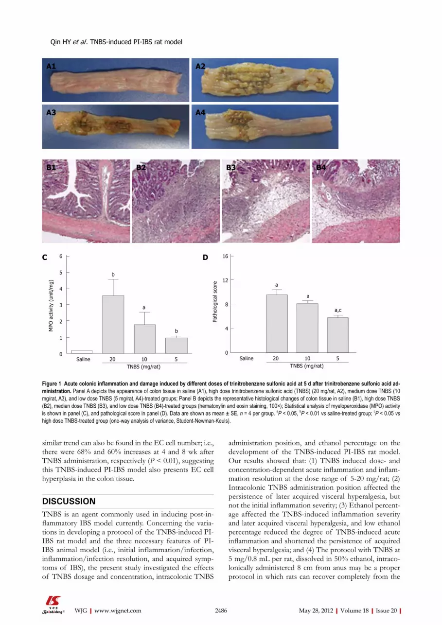
similar trend can also be found in the EC cell number; i.e., there were 68% and 60% increases at 4 and 8 wk after TNBS administration, respectively (P < 0.01), suggesting this TNBS-induced PI-IBS model also presents EC cell hyperplasia in the colon tissue.
DISCUSSIONTNBS is an agent commonly used in inducing post-in-flammatory IBS model currently. Concerning the varia-tions in developing a protocol of the TNBS-induced PI-IBS rat model and the three necessary features of PI-IBS animal model (i.e., initial inflammation/infection, inflammation/infection resolution, and acquired symp-toms of IBS), the present study investigated the effects of TNBS dosage and concentration, intracolonic TNBS
administration position, and ethanol percentage on the development of the TNBS-induced PI-IBS rat model. Our results showed that: (1) TNBS induced dose- and concentration-dependent acute inflammation and inflam-mation resolution at the dose range of 5-20 mg/rat; (2) Intracolonic TNBS administration position affected the persistence of later acquired visceral hyperalgesia, but not the initial inflammation severity; (3) Ethanol percent-age affected the TNBS-induced inflammation severity and later acquired visceral hyperalgesia, and low ethanol percentage reduced the degree of TNBS-induced acute inflammation and shortened the persistence of acquired visceral hyperalgesia; and (4) The protocol with TNBS at 5 mg/0.8 mL per rat, dissolved in 50% ethanol, intraco-lonically administered 8 cm from anus may be a proper protocol in which rats can recover completely from the
2486 May 28, 2012|Volume 18|Issue 20|WJG|www.wjgnet.com
Saline 20 10 5 TNBS (mg/rat)
b
b
a
6
5
4
3
2
1
0
MPO
act
ivity
(un
it/m
g)
Saline 20 10 5 TNBS (mg/rat)
a
a,c
a
16
12
8
4
0
Path
olog
ical
sco
re
A1 A2
A3 A4
B1 B2 B3 B4
C D
Figure 1 Acute colonic inflammation and damage induced by different doses of trinitrobenzene sulfonic acid at 5 d after trinitrobenzene sulfonic acid ad-ministration. Panel A depicts the appearance of colon tissue in saline (A1), high dose trinitrobenzene sulfonic acid (TNBS) (20 mg/rat, A2), medium dose TNBS (10 mg/rat, A3), and low dose TNBS (5 mg/rat, A4)-treated groups; Panel B depicts the representative histological changes of colon tissue in saline (B1), high dose TNBS (B2), median dose TNBS (B3), and low dose TNBS (B4)-treated groups (hematoxylin and eosin staining, 100×); Statistical analysis of myeloperoxidase (MPO) activity is shown in panel (C), and pathological score in panel (D). Data are shown as mean ± SE, n = 4 per group. aP < 0.05, bP < 0.01 vs saline-treated group; cP < 0.05 vs high dose TNBS-treated group (one-way analysis of variance, Student-Newman-Keuls).
Qin HY et al . TNBS-induced PI-IBS rat model

initial inflammation but acquire persistent visceral hy-peralgesia and EC cell hyperplasia at 4 wk after TNBS
administration.As shown in our results, in the dose range of 5-20
2487 May 28, 2012|Volume 18|Issue 20|WJG|www.wjgnet.com
Saline 20 10 5 TNBS (mg/rat)
b
b
2.5
2.0
1.5
1.0
0.5
0.0
Path
olog
ical
sco
re
D
Saline 20 10 5 TNBS (mg/rat)
1.0
0.8
0.6
0.4
0.2
0.0
MPO
act
ivity
(un
it/m
g)
C
A1 A2
A3 A4
B1 B2
B3 B4
Figure 2 Effect of trinitrobenzene sulfonic acid dosage on severity of acute inflammation and inflammation resolution. The colon tissue was collected at 4 wk after trinitrobenzene sulfonic acid (TNBS) administration. Panel A depicts the appearance of colon tissue in saline (A1), high dose TNBS (20 mg/rat, A2), medium dose TNBS (10 mg/rat, A3), and low dose TNBS (5 mg/rat, A4)-treated rats; Panel B depicts the representative histological changes of colon tissue in saline (B1), high dose TNBS (B2), medium dose TNBS (B3), and low dose TNBS (B4)-treated rats (Masson trichrome staining, 100×); Statistical analysis of myeloperoxidase (MPO) activ-ity is shown in panel (C), and pathological score in panel (D). Data are shown as mean ± SE, n = 4 per group. bP < 0.01 vs saline-treated group (one-way analysis of variance, Student-Newman-Keuls).
Qin HY et al . TNBS-induced PI-IBS rat model

mg per rat, the higher the dose of TNBS, the more se-vere the acute inflammation and mucosa damage. This finding is consistent with previous reports that the se-verity and duration in TNBS-induced inflammation is
dose-dependent[13,14,17]. It is notable that the same dose of TNBS (20 mg/rat) when given to rats in different vehicle volumes induced different degrees of damage; a higher TNBS concentration (50 mg/mL) induced
2488 May 28, 2012|Volume 18|Issue 20|WJG|www.wjgnet.com
8 cm-50% EtOH
4 cm-50% EtOH
8 cm-25% EtOH
5 d 4 wk
Saline-8 cmTNBS-8 cm-50% EtOHTNBS-4 cm-50% EtOHTNBS-8 cm-25% EtOH
aa
a,c
5 d 4 wk
2.5
2.0
1.5
1.0
0.5
0.0
Path
olog
ical
sco
re
5 d 4 wk
a,c
a a
2.0
1.5
1.0
0.5
0.0
MPO
act
ivity
(U
/mg)
Saline-8 cmTNBS-8 cm-50% EtOHTNBS-4 cm-50% EtOHTNBS-8 cm-25% EtOH
A
B C
Figure 3 Effects of trinitrobenzene sulfonic acid administration position and ethanol percentage on severity of acute inflammation and inflammation reso-lution. Panel A depicts the appearance of colon tissue in rats treated with trinitrobenzene sulfonic acid (TNBS) at 4 cm or 8 cm from anus or in 25% ethanol. The co-lon tissues were collected at 5 d or 4 wk after TNBS administration, respectively; Statistical analysis of pathological score is shown in panel (B), and myeloperoxidase (MPO) activity in panel (C). Data are shown as mean ± SE, n = 4-5 per group. aP < 0.05 vs saline-treated rats, cP < 0.05 vs rats treated with TNBS at 8 cm from the anus in 50% ethanol (one-way analysis of variance, Student-Newman-Keuls).
Saline-8 cmTNBS-8 cm-50% EtOHTNBS-4 cm-50% EtOHTNBS-8 cm-25% EtOH
4 wk
b
60
50
40
30
20
10
0
AWR t
hres
hold
pre
ssur
e (m
mH
g)
b b
A Saline-8 cmTNBS-8 cm-50% EtOHTNBS-4 cm-50% EtOHTNBS-8 cm-25% EtOH
8 wk
b
60
50
40
30
20
10
0
AWR t
hres
hold
pre
ssur
e (m
mH
g)
B
Figure 4 Effects of low dose trinitrobenzene sulfonic acid administration position and ethanol percentage on later acquired visceral hyperalgesia. Statisti-cal analysis of pain threshold pressure at 4 wk and 8 wk after trinitrobenzene sulfonic acid (TNBS) administration are shown in panel (A) and (B), respectively. Data are shown as mean ± SE, n = 5 per group. bP < 0.01 vs saline-treated rats (one-way analysis of variance, Student-Newman-Keuls). AWR: Abdominal withdrawal reflex.
Qin HY et al . TNBS-induced PI-IBS rat model

more severe damage, even causing animal death in the acute phase. Our pathological results also showed that high-concentration TNBS-treated rats presented local-ized more severe inflammation and deeper ulcerations around the position of TNBS administration, which may be responsible for the occurrence of colitis com-plications, i.e., intestinal obstruction, intra-abdominal infection, and animal death. Concerning the fact that inflammation severity and duration have close correla-tion with TNBS dosage, the effects of TNBS dosage on inflammation resolution was assessed at 4 wk after TNBS administration. According to the pathologist’s suggestion, fibroplasia evaluation index was added into the pathological evaluation criteria for the first time, so as to exactly evaluate inflammation resolution. Our re-sults showed that the animals receiving low dose TNBS (5 mg/rat) recovered completely at 4 wk after TNBS administration, but those receiving high (20 mg/rat) and medium (10 mg/rat) dose were not recovered. This result seems to somewhat conflict with the finding from a previous study in which the rats receiving TNBS (5-20 mg/rat, in 25% ethanol) all recovered after 4 wk[14]. We speculate that this discrepancy may come from the protocol differences between these two studies, such as ethanol percentage, animal strain, or evaluation criteria. Consistent with our findings, the previous study showed that colonic inflammation induced by TNBS (30 mg/rat, in 50% ethanol) lasted for at least 8 wk[17], suggesting the
inflammation resolution is also dose-dependent. Based on previous findings and our results, TNBS dosage plays a critical role in TNBS-induced inflammation severity and resolution, and low dose TNBS (5 mg/rat)-induced inflammation can recover completely at 4 wk after TNBS administration.
Currently, 25% and 50% ethanol are both used as the carrier of TNBS in establishing TNBS-induced PI-IBS model and TNBS is commonly given to rats at the depth of either 4 cm or 8 cm from the anus. Based on low dose TNBS (5 mg/0.8 mL per rat) selection, we fur-ther investigated the effects of ethanol percentage and intracolonic administration position on the severity and resolution of TNBS-induced inflammation. Our results showed that rats treated with low dose TNBS at a depth of 4 cm or 8 cm presented similar inflammation sever-ity and resolution, while the rats treated with TNBS in 25% ethanol developed milder non-ulcer inflammation compared to those treated with TNBS in 50% ethanol. As the carrier of TNBS, ethanol is known as a breaker to the mucosal barrier, and 30%-50% ethanol alone has been proved to induce acute inflammation and hyper-emia[18,28]. Moreover, previous study have shown that 30% ethanol alone induced small areas of hyperemia, while TNBS combined with 30% ethanol induced larger area of necrosis and hyperemia in the colon[18]. These findings indicate that ethanol alone can induce concen-tration-dependent inflammation, and can be considered
2489 May 28, 2012|Volume 18|Issue 20|WJG|www.wjgnet.com
SalineTNBS
ba
Proximal Distal Proximal Distal 4 wk 8 wk
10
8
6
4
2
0
5-H
T co
nten
t of
tis
sue
(ng/
mg)
SalineTNBSb
b
Proximal Distal Proximal Distal 4 wk 8 wk
250
200
150
100
50
0
EC c
ell n
umbe
r (/
mm
2 )
A1 B
CA2
Figure 5 Persistent increases of enterochromaffin cell number and serotonin content in the colon tissue of trinitrobenzene sulfonic acid-induced post-infectious irritable bowel syndrome rat model. The colon tissue was collected at 4 wk and 8 wk after trinitrobenzene sulfonic acid (TNBS) administration. Panel A depicts the representative enterochromaffin (EC) cell staining in colon mucosa, the samples were from rats treated with saline (A1), or TNBS (A2) at 4 wk post TNBS (Scale bar, 100 μm). Statistical analysis of serotonin content is shown in panel (B), and EC cell number in panel (C). Data are shown as mean ± SE, n = 5 per group. aP < 0.05, bP < 0.01 vs saline-treated rats. 5-HT: 5-hydroxytryptamine.
Qin HY et al . TNBS-induced PI-IBS rat model

as an invasive helper in enhancing the effects of TNBS. Therefore, it seems reasonable that milder inflammation was induced in rats treated with equal dose of TNBS (5 mg/rat) but in lower ethanol percentage (25%). These results can also explain why high dose or high concentra-tion TNBS have also been used by some researchers in establishing a PI-IBS model: low concentration ethanol may ameliorate the effects of TNBS[13,14].
Visceral hyperalgesia, the major feature of IBS, was further tested in low dose TNBS (5 mg/0.8 mL per rat) treated rats. From 4 wk to 8 wk post TNBS intracolonic administration at a depth of 8 cm, significant and per-sistent decrease in visceral pain threshold pressure was found, while the intracolonic TNBS administration at a depth of 4 cm or in 25% ethanol presented short-term acquired visceral hyperalgesia. Nowadays, the duration and severity of inflammation are considered as impor-tant risk factors in the development of PI-IBS[29], and severe inflammation which results in deep impairment of the underlining nerve fibers has been proposed to play an important role in the pathogenesis of IBS[30]. Based on the above evidence, we believe the unstable visceral hyperalgesia (i.e., lasting for less than 4 wk) in rats treated with TNBS in 25% ethanol may have close correlation with the mild non-ulcer inflammation ob-served at the acute phase of inflammation. These results also provide us with the information that mild inflam-mation, even without ulceration, can induce later short-term visceral hyperalgesia. However, it seems interesting that the same dose of TNBS when given at the different positions of colon (transverse vs descending) induced different features of acquired visceral hyperalgesia (per-sistent vs short-term), even though the TNBS-induced acute inflammation and damage was similar at first. Con-sidering the unclear and complex pathogenesis of vis-ceral hypersensitivity in PI-IBS, it is not easy to provide a clear explanation for the current findings. However,
during the acute phase of TNBS-induced inflammation, obvious colon dilation and a large amount of retained feces were found in the inflamed transverse colon, but not in the inflamed distal colon. We speculate that the persistent colonic inflammation and dilation may play important roles in enteric nerve system plasticity, and thus influence the feature of acquired visceral hyper-sensitivity. Currently, peripheral and central neuroplas-tic changes are considered to be associated with post-inflammatory persistent visceral hypersensitivity[1,31]. It is also well known that transient colorectal distension in the neonatal period can result in chronic visceral hy-persensitivity and motility dysfunction in adulthood[25,32], and the underlying mechanism is thought to be associ-ated with alterations in peripheral and central nerve systems[33]. Moreover, considering the different structure and functions of the transverse and descending colons, the regional differences in gut flora[34], mast cells[35] and EC cells[36] may also contribute to the different feature of acquired visceral hypersensitivity observed in our study. Further studies are needed to clarify these issues.
EC cell hyperplasia has been found in the colonic specimens of IBS and PI-IBS patients[37-39], and EC cell hyperplasia has been reported to play important role in the development of visceral hypersensitivity in IBS patients[40,41]. To identify whether visceral hyperalgesia is also accompanied with EC cell hyperplasia in the TNBS-induced PI-IBS rat model, EC cell number and 5-HT content were further investigated in the proximal and distal colon of PI-IBS rats. As shown in our study, the EC cell number and 5-HT content in the proximal co-lon, but not the distal colon, were significantly and per-sistently increased in this TNBS model, suggesting the occurrence of EC cell hyperplasia. However, this result differed from the finding from IBS patients, as EC cell hyperplasia in rectal mucosa was commonly reported[3]. This may be explained by the different distribution of
2490 May 28, 2012|Volume 18|Issue 20|WJG|www.wjgnet.com
Qin HY et al . TNBS-induced PI-IBS rat model
Procedures Cautions
1. 7-wk-old male Sprague-Dawley rats, fast 24 h before TNBS administration 2. Mix 1 volume of 12.5 mg/mL TNBS-saline solution with 1 volume of absolute ethanol
Fresh preparation
3. Weight and anesthetized rats with chloral hydrate (350 mg/kg, i.p.) Anesthetization may last for 3-4 h 4. Insert a catheter into the colon at 8 cm from anus Proceed carefully to avoid damage the colon wall 5. Keep the rat with head-down vertical position, instill 0.8 mL TNBS solution slowly into the colon lumen within 1 min
Handle slowly to avoid TNBS leakage
6. Keep the rat in head-down vertical position for 1 min before gently removing the catheter 7. Put the rats in a mound of bedding in head-down position until consciousness recovery
Ensure TNBS solution remains completely in the colon;Keep the rats warm
8. On day 3, 7, 14, 21 and 28, observe and weight the rats On day 3, body weight decreased by 10%-20% with unformed bloody stool;On day 7, body weight regained and reached the original level, the stool become formed but still soft;From day 7 to day 28, body weight increased smoothly, and finally reached the control level
Table 5 Detailed protocols in developing trinitrobenzene sulfonic acid induced post-inflammatory irritable bowel syndrome model in Sprague-Dawley rats
TNBS: Trinitrobenzene sulfonic acid.

EC cells in the gut mucosa between human beings and rats: the vast majority of EC cells in humans mainly re-sides in the small intestine and rectum[42] while the major source of EC cells in rats largely locates in the cecum with a decline trend from the proximal to the distal colon[36]. Consistent with our findings, previous studies also showed that 5-HT content in the rat proximal colon tissue was significantly higher (about 5 fold) than that in the distal colon[43,44]. Therefore, it seems that EC cell hyperplasia in the proximal colon can be regarded as one of the features of the TNBS-induced PI-IBS rat model.
This present study, for the first time, showed that TNBS dosage and concentration, position of intraco-lonic TNBS administration, and ethanol percentage play important roles in developing the TNBS-induced PI-IBS model in Sprague-Dawley rats, suggesting more atten-tion should be paid to these factors when developing a PI-IBS model with TNBS. Concerning the gene dif-ferences among animal strains, the protocol set up here may need some modulation when other rat strains ex-cept Sprague-Dawley rats, are used. Though this TNBS model presented persistent visceral hyperalgesia and EC cell hyperplasia for as long as 4 wk, more studies are still needed to observe the long-lasting visceral hyperalgesia and identify other features of PI-IBS in this model.
In the present study, we found out that the protocol with TNBS at 5 mg/0.8 mL per rat, dissolved in 50% ethanol, intracolonically administered at 8 cm from the anus resulted in persistent visceral hyperalgesia and co-lonic EC cell hyperplasia after complete recovery from the initial inflammation in rats. With this protocol, low-dose TNBS-induced mild mucosa damage and colonic inflammation is reproducible without any animal loss; visceral hyperalgesia and EC cell hyperplasia which last for at least 4 wk occur as early as 4 wk after TNBS ad-ministration. The details of this protocol are presented in Table 5. Ensuring the results from different labora-tories would be comparable, we believe that widespread adoption of a recommended protocol will save a great quantity of time and resources.
ACKNOWLEDGMENTSWe are very grateful to Dr. Zhi Li for his kind sugges-tion in setting up pathological criteria.
COMMENTSBackgroundPost-infectious irritable bowel syndrome (PI-IBS) is a subgroup of IBS in which the patients developed their symptoms after recovery from an acute gastroin-testinal infection. Previous systematic review showed that the trinitrobenzene sulfonic acid (TNBS)-induced model is the most commonly used PI-IBS model, but there are large variations in the protocol of model development. These vari-ations make comparison between studies to be very difficult, if not impossible.Research frontiersThis study aimed to investigate the effects of TNBS dosage, concentration, intracolonic administration position, and ethanol percentage on TNBS-induced acute inflammation, inflammation resolution, and later acquired features of IBS
(i.e., visceral hyperalgesia and enterochromaffin cell hyperplasia).Innovations and breakthroughsRecent studies have highlighted the important status of PI-IBS in functional diseases, as the clear onset and well defined pathophysiological changes of PI-IBS will help people understand not only the PI-IBS but also other subtypes of IBS. Moreover, the validated animal models, which aim to mimic one or more features of human disease, play important roles in the studies of mechanisms and potential therapeutic agents for diseases. This is the first study aimed to in-vestigate the effects of key factors on developing TNBS-induced PI-IBS model in rats. The results and recommended protocol presented in this study will save a great deal of time and resources and ensure the results from different labora-tories be comparable.ApplicationsThis study provides direct information about the effects of key factors on TNBS-induced acute inflammation, inflammation resolution, and later acquired fea-tures of IBS. More attention should be paid to these key factors when develop-ing PI-IBS rat model with TNBS. Peer reviewThis is a good descriptive study in which authors investigate the effects of TNBS dosage and concentration, intracolonic administration position, and etha-nol percentage on the developing TNBS-induced PI-IBS rat model. The topic is interesting and useful for the study of pathophysiology of PI-IBS and the devel-opment of therapeutic agents.
REFERENCES1 Barbara G, Cremon C, Pallotti F, De Giorgio R, Stanghellini
V, Corinaldesi R. Postinfectious irritable bowel syndrome. J Pediatr Gastroenterol Nutr 2009; 48 Suppl 2: S95-S97
2 Neal KR, Barker L, Spiller RC. Prognosis in post-infective irritable bowel syndrome: a six year follow up study. Gut 2002; 51: 410-413
3 Dunlop SP, Jenkins D, Neal KR, Spiller RC. Relative im-portance of enterochromaffin cell hyperplasia, anxiety, and depression in postinfectious IBS. Gastroenterology 2003; 125: 1651-1659
4 Gwee KA, Leong YL, Graham C, McKendrick MW, Col-lins SM, Walters SJ, Underwood JE, Read NW. The role of psychological and biological factors in postinfective gut dysfunction. Gut 1999; 44: 400-406
5 ChaudharyY NA, Truelove SC. The irritable colon syn-drome. A study of the clinical features, predisposing causes, and prognosis in 130 cases. Q J Med 1962; 31: 307-322
6 Thabane M, Marshall JK. Post-infectious irritable bowel syndrome. World J Gastroenterol 2009; 15: 3591-3596
7 Qin HY, Wu JC, Tong XD, Sung JJ, Xu HX, Bian ZX. Sys-tematic review of animal models of post-infectious/post-inflammatory irritable bowel syndrome. J Gastroenterol 2011; 46: 164-174
8 Collins SM, Piche T, Rampal P. The putative role of in-flammation in the irritable bowel syndrome. Gut 2001; 49: 743-745
9 Lamb K, Zhong F, Gebhart GF, Bielefeldt K. Experimental colitis in mice and sensitization of converging visceral and somatic afferent pathways. Am J Physiol Gastrointest Liver Physiol 2006; 290: G451-G457
10 Bergin AJ, Donnelly TC, McKendrick MW, Read NW. Changes in anorectal function in persistent bowel distur-bance following salmonella gastroenteritis. Eur J Gastroen-terol Hepatol 1993; 5: 617-620
11 Barbara G, Cremon C, Gargano L, Giorgio RD, Cogliandro R, Stanghellini V, Corinaldesi R. T1394 Mesalazine Treatment for Intestinal Immune Activation in Patient with Irritable Bowel Syndrome: A Randomized Controlled Pilot Trial. Gastroenterology 2008; 134: A-546
12 Kim HS, Berstad A. Experimental colitis in animal models. Scand J Gastroenterol 1992; 27: 529-537
13 Menozzi A, Pozzoli C, Poli E, Lazzaretti M, Grandi D,
2491 May 28, 2012|Volume 18|Issue 20|WJG|www.wjgnet.com
COMMENTS
Qin HY et al . TNBS-induced PI-IBS rat model

Coruzzi G. Long-term study of TNBS-induced colitis in rats: focus on mast cells. Inflamm Res 2006; 55: 416-422
14 Adam B, Liebregts T, Gschossmann JM, Krippner C, Scholl F, Ruwe M, Holtmann G. Severity of mucosal inflammation as a predictor for alterations of visceral sensory function in a rat model. Pain 2006; 123: 179-186
15 Winchester WJ, Johnson A, Hicks GA, Gebhart GF, Green-wood-van Meerveld B, McLean PG. Inhibition of endothe-lial cell adhesion molecule expression improves colonic hyperalgaesia. Neurogastroenterol Motil 2009; 21: 189-196
16 Collins SM, McHugh K, Jacobson K, Khan I, Riddell R, Murase K, Weingarten HP. Previous inflammation alters the response of the rat colon to stress. Gastroenterology 1996; 111: 1509-1515
17 Morris GP, Beck PL, Herridge MS, Depew WT, Szewczuk MR, Wallace JL. Hapten-induced model of chronic inflam-mation and ulceration in the rat colon. Gastroenterology 1989; 96: 795-803
18 Diop L, Raymond F, Fargeau H, Petoux F, Chovet M, Doherty AM. Pregabalin (CI-1008) inhibits the trinitroben-zene sulfonic acid-induced chronic colonic allodynia in the rat. J Pharmacol Exp Ther 2002; 302: 1013-1022
19 Asfaha S, MacNaughton WK, Appleyard CB, Chadee K, Wallace JL. Persistent epithelial dysfunction and bacterial translocation after resolution of intestinal inflammation. Am J Physiol Gastrointest Liver Physiol 2001; 281: G635-G644
20 Greenwood-Van Meerveld B, Johnson AC, Foreman RD, Linderoth B. Spinal cord stimulation attenuates visceromo-tor reflexes in a rat model of post-inflammatory colonic hy-persensitivity. Auton Neurosci 2005; 122: 69-76
21 Ferrini MG, Kovanecz I, Sanchez S, Umeh C, Rajfer J, Gonzalez-Cadavid NF. Fibrosis and loss of smooth muscle in the corpora cavernosa precede corporal veno-occlusive dysfunction (CVOD) induced by experimental cavernosal nerve damage in the rat. J Sex Med 2009; 6: 415-428
22 Gué M, Bonbonne C, Fioramonti J, Moré J, Del Rio-Lachèze C, Coméra C, Buéno L. Stress-induced enhancement of coli-tis in rats: CRF and arginine vasopressin are not involved. Am J Physiol 1997; 272: G84-G91
23 Fabia R, Ar’Rajab A, Johansson ML, Willén R, Andersson R, Molin G, Bengmark S. The effect of exogenous administra-tion of Lactobacillus reuteri R2LC and oat fiber on acetic acid-induced colitis in the rat. Scand J Gastroenterol 1993; 28: 155-162
24 Krawisz JE, Sharon P, Stenson WF. Quantitative assay for acute intestinal inflammation based on myeloperoxidase activity. Assessment of inflammation in rat and hamster models. Gastroenterology 1984; 87: 1344-1350
25 Al-Chaer ED, Kawasaki M, Pasricha PJ. A new model of chronic visceral hypersensitivity in adult rats induced by colon irritation during postnatal development. Gastroenterol-ogy 2000; 119: 1276-1285
26 Grimelius L. Silver stains demonstrating neuroendocrine cells. Biotech Histochem 2004; 79: 37-44
27 Qi SD, Tian SL, Xu HX, Sung JJ, Bian ZX. Quantification of luminally released serotonin in rat proximal colon by capil-lary electrophoresis with laser-induced fluorescence detec-tion. Anal Bioanal Chem 2009; 393: 2059-2066
28 Asfaha S, Bell CJ, Wallace JL, MacNaughton WK. Pro-longed colonic epithelial hyporesponsiveness after colitis: role of inducible nitric oxide synthase. Am J Physiol 1999; 276: G703-G710
29 Neal KR, Hebden J, Spiller R. Prevalence of gastrointestinal symptoms six months after bacterial gastroenteritis and risk factors for development of the irritable bowel syndrome: postal survey of patients. BMJ 1997; 314: 779-782
30 Wang LH, Fang XC, Pan GZ. Bacillary dysentery as a caus-ative factor of irritable bowel syndrome and its pathogen-esis. Gut 2004; 53: 1096-1101
31 Hughes PA, Brierley SM, Blackshaw LA. Post-inflammatory modification of colonic afferent mechanosensitivity. Clin Exp Pharmacol Physiol 2009; 36: 1034-1040
32 Chaloner A , Rao A, Al-Chaer ED, Greenwood-Van Meerveld B. Importance of neural mechanisms in colonic mucosal and muscular dysfunction in adult rats following neonatal colonic irritation. Int J Dev Neurosci 2010; 28: 99-103
33 Lin C, Al-Chaer ED. Long-term sensitization of primary af-ferents in adult rats exposed to neonatal colon pain. Brain Res 2003; 971: 73-82
34 Meslin JC, Fontaine N, Andrieux C. Variation of mucin distribution in the rat intestine, caecum and colon: effect of the bacterial flora. Comp Biochem Physiol A Mol Integr Physiol 1999; 123: 235-239
35 Furgał A, Litwin JA. Distribution of mast cells along and across successive segments of the rat digestive tract: a quan-titative study. Folia Histochem Cytobiol 1998; 36: 19-27
36 Portela-Gomes GM, Grimelius L, Petersson R, Bergström R. Enterochromaffin cells in the rat gastrointestinal tract. Aspects of factors influencing quantification. Ups J Med Sci 1984; 89: 189-203
37 Lee KJ, Kim YB, Kim JH, Kwon HC, Kim DK, Cho SW. The alteration of enterochromaffin cell, mast cell, and lamina propria T lymphocyte numbers in irritable bowel syndrome and its relationship with psychological factors. J Gastroen-terol Hepatol 2008; 23: 1689-1694
38 Dunlop SP, Jenkins D, Neal KR, Spiller RC. Relative im-portance of enterochromaffin cell hyperplasia, anxiety, and depression in postinfectious IBS. Gastroenterology 2003; 125: 1651-1659
39 Spiller RC, Jenkins D, Thornley JP, Hebden JM, Wright T, Skinner M, Neal KR. Increased rectal mucosal enteroendo-crine cells, T lymphocytes, and increased gut permeability following acute Campylobacter enteritis and in post-dysen-teric irritable bowel syndrome. Gut 2000; 47: 804-811
40 Mawe GM, Coates MD, Moses PL. Review article: intestinal serotonin signalling in irritable bowel syndrome. Aliment Pharmacol Ther 2006; 23: 1067-1076
41 Park JH, Rhee PL, Kim G, Lee JH, Kim YH, Kim JJ, Rhee JC, Song SY. Enteroendocrine cell counts correlate with visceral hypersensitivity in patients with diarrhoea-predominant irritable bowel syndrome. Neurogastroenterol Motil 2006; 18: 539-546
42 Sjölund K, Sandén G, Håkanson R, Sundler F. Endocrine cells in human intestine: an immunocytochemical study. Gastroenterology 1983; 85: 1120-1130
43 Tsukamoto K, Ariga H, Mantyh C, Pappas TN, Yanagi H, Yamamura T, Takahashi T. Luminally released serotonin stimulates colonic motility and accelerates colonic transit in rats. Am J Physiol Regul Integr Comp Physiol 2007; 293: R64-R69
44 Bian ZX, Qin HY, Tian SL, Qi SD. Combined effect of early life stress and acute stress on colonic sensory and motor responses through serotonin pathways: differences between proximal and distal colon in rats. Stress 2011; 14: 448-458
S- Editor Gou SX L- Editor O’Neill M E- Editor Li JY
2492 May 28, 2012|Volume 18|Issue 20|WJG|www.wjgnet.com
Qin HY et al . TNBS-induced PI-IBS rat model

Cost-benefit analysis of esophageal cancer endoscopic screening in high-risk areas of China
Juan Yang, Wen-Qiang Wei, Jin Niu, Zhi-Cai Liu, Chun-Xia Yang, You-Lin Qiao
Juan Yang, Jin Niu, Chun-Xia Yang, Department of Epidemiol-ogy, West China School of Public Health, Sichuan University, Chengdu 610041, Sichuan Province, ChinaWen-Qiang Wei, You-Lin Qiao, Department of Epidemiology, Cancer Institute and Hospital, Chinese Academy of Medical Sci-ences and Peking Union Medical College, Beijing 100021, China Zhi-Cai Liu, Department of Thoracic Surgery, Linzhou Cancer Institute, Linzhou 456550, Henan Province, ChinaAuthor contributions: Wei WQ, Yang CX and Qiao YL de-signed the research and revised the manuscript; Yang J, Wei WQ, Niu J and Liu ZC performed the research; Yang J reviewed the literature, analyzed the data and prepared the paper. Supported by The National Science and Technology Pillar Program of the 11th National Five-Year Plan of China, No. 2006BAI02A15Correspondence to: Dr. Chun-Xia Yang, MD, PhD, Depart-ment of Epidemiology, West China School of Public Health, Sich-uan University, Renming Nanlu 3-17, Chengdu 610041, Sichuan Province, China. [email protected]: +86-28-85501604 Fax: +86-28-85501295Received: June 21, 2011 Revised: December 2, 2011Accepted: April 28, 2012Published online: May 28, 2012
AbstractAIM: To estimate the cost-benefit of endoscopic screen-ing strategies of esophageal cancer (EC) in high-risk areas of China.
METHODS: Markov model-based analyses were con-ducted to compare the net present values (NPVs) and the benefit-cost ratios (BCRs) of 12 EC endoscopic screening strategies. Strategies varied according to the targeted screening age, screening frequencies, and follow-up intervals. Model parameters were collected from population-based studies in China, published lit-eratures, and surveillance data.
RESULTS: Compared with non-screening outcomes, all strategies with hypothetical 100 000 subjects saved life
years. Among five dominant strategies determined by the incremental cost-effectiveness analysis, screening once at age 50 years incurred the lowest NPV (inter-national dollar-I$55 million) and BCR (2.52). Screening six times between 40-70 years at a 5-year interval [i.e., six times(40)f-strategy] yielded the highest NPV (I$99 million) and BCR (3.06). Compared with six times(40)f-strategy, screening thrice between 40-70 years at a 10-year interval resulted in relatively lower NPV, but the same BCR.
CONCLUSION: EC endoscopic screening is cost-ben-eficial in high-risk areas of China. Policy-makers should consider the cost-benefit, population acceptance, and local economic status when choosing suitable screening strategies.
© 2012 Baishideng. All rights reserved.
Key words: Cost-benefit analysis; Esophageal cancer; Endoscopy; Screening; High-risk area
Peer reviewer: Julian Abrams, MD, MS, Assistant Professor of Clinical Medicine, Division of Digestive and Liver Diseases, Columbia University Medical Center, 622 W 168th Street, PH 20-303, New York, NY 10032, United States
Yang J, Wei WQ, Niu J, Liu ZC, Yang CX, Qiao YL. Cost-benefit analysis of esophageal cancer endoscopic screening in high-risk areas of China. World J Gastroenterol 2012; 18(20): 2493-2501 Available from: URL: http://www.wjgnet.com/1007-9327/full/v18/i20/2493.htm DOI: http://dx.doi.org/10.3748/wjg.v18.i20.2493
INTRODUCTIONEsophageal cancer (EC) is the eighth most common cancer and the sixth most common cause of cancer death worldwide[1]. Although the mortality of EC has
ORIGINAL ARTICLE
World J Gastroenterol 2012 May 28; 18(20): 2493-2501ISSN 1007-9327 (print) ISSN 2219-2840 (online)
© 2012 Baishideng. All rights reserved.
Online Submissions: http://www.wjgnet.com/[email protected]:10.3748/wjg.v18.i20.2493
2493 May 28, 2012|Volume 18|Issue 20|WJG|www.wjgnet.com

Yang J et al . Cost-benefit of esophageal cancer screening
sharply reduced over the last three decades, EC remains the fourth leading cause of cancer death in China with a mortality of 15.21/100 000[2]. According to the “Third National Retrospective Sampling Survey of Death Causes Report in 2004-2005 of China”, EC continues to be the major public health burden in some high-risk areas, where the mortality of EC was three times higher than the average of the country. EC is a fatal disease, with a 5-year survival rate of less than 20% even in developed countries[3,4].
To explore suitable control measures in high-risk areas of China, a great number of EC screening studies using endoscopic examinations (i.e., endoscopy with mucosal io-dine staining and index biopsy as a screening technology, combined with pathological examination for confirming and staging the disease) have been conducted for several decades[5-10]. Through early detection and subsequent treatment, the 5-year survival rate of EC increased to 86%[10]. Furthermore, obvious reductions in incidence and mortality rates of EC were observed under endoscopic screening[11].
A national screening program for EC in high-risk areas has become available in 73 sites of 27 provinces of China based on evidence from previous studies. Never-theless, due to lacking comprehensive health economic evaluations on such programs, two key public health questions remain to be answered: is the endoscopic screening cost-beneficial in the long run? Should we use the same screening strategy in both developed and devel-oping high-risk areas of China?
The objective of this paper is to explore appropriate screening strategies for EC in high-risk areas of China from the health economic perspective by comparing the long-term cost-benefits of 12 endoscopic screening alter-natives. It will provide valuable data for policy makers to make decisions on the current screening program.
MATERIALS AND METHODSDecision analysis modelA Markov model was constructed to evaluate the cost-benefit of different screening strategies for EC. In each strategy simulation, a hypothetical cohort with 100 000 participants entered the model at age 40 years and were fol-lowed up until the age of 70 years. Costs and benefits were all discounted at an annual rate of 3%[12]. TreeAge Pro 2009 Suite by TreeAge Software Inc. was used for all analysis.
The natural history of EC was categorized as the fol-lowing health status: normal, mild dysplasia (mD), mod-erate dysplasia (MD), severe dysplasia/carcinoma in situ (SD/CIS), intramucosal carcinoma (IC), submucosal carcinoma (T1N0M0) (SC), invasive carcinoma (INC), and death. Figure 1 depicts the detailed transition processes of EC in the Markov model. Each rectangle represents a health state. During a Markov cycle (1 year), one could transit from his/her current health state to another (in-dicated by arrows between different states) or remain in the same state (indicated by half-circle arrows on the rectangles). Prior to the development of IC, the condi-
tion could spontaneously regress. Once IC developed, no regression could occur. We used the model to evaluate all screening alternatives and non-screening outcomes.
Screening strategiesIn the context of lacking guidelines for EC screening worldwide, we explored 12 screening strategies using en-doscopic examinations. These strategies were performed at varying starting age for screening (40, 45 or 50 years), screening frequency (once, twice, thrice, or six times in the lifetime), and intervals of follow-up for mD and MD cases (e.g., 5 or 3 years). The strategies were listed as “t(y)nf/t(y)f ”, where t denotes the screening frequency, y represents the starting age of screening, nf means we do not follow up the mD and MD cases diagnosed by screening, and f means the mD and MD cases are fol-lowed up every 5 years and 3 years, respectively. For twice and thrice screening strategies in the lifetime, the screen-ing intervals were 10 years; for six times screening strat-egy, the screening intervals were 5 years.
Screening, diagnosis, and treatment procedures for the strategies were all based on the current practice manuals. The participants were screened using endoscopy with iodine staining. If endoscopy revealed a suspected lesion (mD or worse), index biopsy combined with pathological examination were performed consecutively. The detailed procedures of endoscopy were the same with those in the literature[13]. For SD/CIS and IC cases detected by screening, Endoscopic Mucosal Resection and/or Argon Plasma Coagulation served as the standard treatment. For detected patients with SC or worse, therapies included esophagectomy, radiotherapy, and other routine treat-ments. Subjects who were not screened would be diag-nosed and treated if they presented with symptomatic EC. All patients were followed up once by endoscopy in the first year after treatment.
Model parameters The data used in the model were compiled from a vari-ety of sources: (1) the results of our prospective cohort study based on the EC chemoprevention trial of sele-nomethionine and celecoxib in “early detection of EC” (EDEC) program; (2) the results of our population-based
2494 May 28, 2012|Volume 18|Issue 20|WJG|www.wjgnet.com
Normal Mild dysplasia Moderate dysplasia
Invasivecarcinoma
DeathSevere dysplasia/carcinoma in situ
Submucosal carcinoma (T1N0M0) Intramucosal carcinoma
Figure 1 Bubble diagram representing the health states of esophageal cancer and transitions in natural-history Markov model.

screening project “Early Detection and Early Treatment of EC in Demonstration Centers in China” (EDETEC); (3) surveillance data; (4) published literatures; and (5) un-published data.
In the chemoprevention trial of EDEC program, 2213 asymptomatic adults from Linzhou County, Henan Province of China, underwent an endoscopic screen-ing in 1999[14]. Among them, 2189 participants who had histological diagnoses at the baseline evaluations were surveilled until 2007. The primary end-point was the oc-currence of EC, confirmed by village doctors through checking the histological diagnoses in medical records. The project EDETEC covering 11 high-risk areas of EC was launched by the Chinese central government in 2005. The purpose was to increase the early detection and treat-ment rate as well as the 5-year survival rate of EC, and to improve the screening, early detection and treatment program and so forth[15].
Probabilities At the initial model cycle, a hypothetical cohort of 100 000 participants was distributed among various health states based on the proportion of each pathologic stage of EC in the 40-44 years age group. The proportions were calcu-lated in terms of the screening results of Linzhou County in the project EDETEC between 2005 and 2008. Among the 8267 asymptomatic participants aged 40-69 years, 8.2% cases were identified as mD in the 40-44 years age group. Full details are presented in Tables 1 and 2.
In each cycle of a Markov process, the transitions among health states occurred with annual transition prob-
abilities. They were estimated in terms of: (1) published literatures[9,16-21]; and (2) the results of both EDEC and EDETEC projects in Linzhou County (Tables 1 and 2).
It is believed that persons with SD/CIS or lesser ab-normality may not die from EC and that patients with IC or SC may die from all causes including EC. In patients with INC, we assumed that they may mainly die from EC. Therefore, in our model, the corresponding death probabilities for three different populations above were converted from non-esophageal-cancer mortality, all-cause mortality, and case fatality rate of EC, respectively. All age-specific mortality rates were obtained from the death registry reports of Linzhou County between 2004 and 2006. And they were converted to probabilities by the formula: Probability = 1 - Exp (-rt), where “r” repre-sents the rate and ‘t’ denotes the time (Tables 1 and 2).
Screening compliance The compliance of EC screening in different settings var-ied from 33.4% to 77.1%[22]. In the EDETEC program, the screening compliance of EC in Linzhou County dur-ing 2005-2008 was 67% (8267/12 294), which was used as a baseline in this analysis (Table 1).
Screening and treatment costIn our model, both screening and treatment costs included direct and indirect costs, which were calculated from a so-cietal perspective. Direct costs referred to those associated with drugs, disposable supplies, equipment and facilities, staff, etc. In this study, we used costs rather than charges. And they were collected using Micro-costing methods in
2495 May 28, 2012|Volume 18|Issue 20|WJG|www.wjgnet.com
Table 1 Non-age-specific parameters
Parameters Value Parameters Value Parameters Value1
Initial probability2 Transition probability-continued Compliance of screening2 67% (30%-100%) Normal 0.8895 SD/CIS see Table 2 Sensitivity of endoscopy[6] 96% (92%-99%) mD 0.0820 IC see Table 2 Specificity of endoscopy[6] 90% (59%-100%) MD 0.0180 INC Screening cost (I$ per capita)2
SD/CIS 0.0090 Recovering to post-INC 0.7696 Direct cost 61.50 (37.00-119.00) IC 0.0008 Relapsing to INC after treatment 0.2304 Indirect cost 8.31 (8.09-8.53) SC 0.0005 After treatment4 Treatment cost (I$ per capita)[24]2
INC 0.0002 Post-SD/CIS Direct costTransition probability[9,16-21]2,3 Recovering to normal 0.9950 SD/CIS 1292 (1114-1565) Normal Relapsing to SD/CIS 0.0050 IC 1292 (1114-1565) Remaining normal 0.9760 Post-IC SC 1818 (1519-2799) Progression to mD 0.0240 Remaining post-IC 0.9450 INC-screening group 2767 (2332-4031) mD Relapsing to IC 0.0500 INC-control group 4888 (4333-6396) Regression to normal 0.0500 Relapsing and progression to SC 0.0050 Indirect cost Remaining mD 0.9000 Post-SC SD/CIS 1654 (1341-1968) Progression to MD 0.0500 Remaining post-SC 0.8500 IC 1654 (1341-1968) MD Relapsing to SC 0.0500 SC 3369 (2872-3866) Regression to mD 0.0800 Relapsing and progression to INC 0.1000 INC-screening group 3369 (2872-3866) Remaining MD 0.8000 Post-INC (Same with “INC”) INC-control group 5526 (4584-6466) Progression to SD/CIS 0.1200 Death probability see Table 2 Discount rate[12]5 3% (0%-6%)
mD: Mild dysplasia; MD: Moderate dysplasia; SD/CIS: Severe dysplasia/carcinoma in situ; IC: Intramucosal carcinoma; SC: Submucosal carcinoma (T1N0M0); INC: Invasive carcinoma; I$: International dollar. 1For compliance, sensitivity and specificity of endoscopy, screening and treatment costs, and discount rate, the numerals before and in the parenthesis denote the base case and ranges used in sensitivity analysis; 2Data were calculated in terms of the project called “Early Detection and Early Treatment of Esophageal Cancer in Demonstration Centers in China”; 3Data were collected from the death registry report of Linzhou County during 2004-2006; 4Post-SD/CIS, post-IC, post-SC and post-INC represent the “health” condition after treatment of SD/CIS, IC, SC and INC; 5Costs and life years were discounted during the Markov cycles.
Yang J et al . Cost-benefit of esophageal cancer screening

the EDETEC program[23]. Indirect cost was also estimat-ed from our EDETEC program, including those related to transportation, accommodation, and the productivity losses of both patients and their caregivers[24,25]. Consider-ing differences in purchasing power, costs were presented in 2008 international dollars (I$).
Screening cost per capita using endoscopic examina-tion was I$69.81. In screening group, the treatment costs for patients with SD/CIS or worse ranged from I$2964 to I$6136. In control group, the treatment cost for INC cases was I$10 414, much higher than that in screening group (Table 1).
Other variables and assumptionsAccording to a previous study in Linzhou County, the sensitivity and specificity of endoscopic examination were 96% and 90%, respectively[6] (Table 1). For individu-als diagnosed as having precancerous lesions or EC (i.e., SD/CIS or worse), we assumed that they would complete the entire treatment procedures.
Health economic evaluationThe basic outcomes of the model were total costs (in-cluding screening costs and treatment costs) and expect-ed life years. Then the net present values (NPVs) and the benefit-cost ratios (BCRs) were calculated under each of the strategies (for a hypothetical cohort of 100 000 sub-jects followed up from 40 years to 70 years of age).
For each screening cohort, the benefit consisted of the treatment cost averted and productivity gains from screening programs[25], and counted by the formula: ben-efit = GDP per capita of Linzhou in 2008 (I$6542) × (life years of screening cohort - life years of “non-screening” group) + treatment cost of “non-screening” group. The NPV was the benefit minus the total cost of the screen-ing group; the BCR equaled to the benefit divided by the
total cost. The strategies with a NPV > 0 and a BCR > 1 were considered cost-beneficial.
In addition, the screening alternatives were compared using an incremental cost-effectiveness analysis. The strategies that were more expensive and gained fewer life years (dominated), or less costly and less cost-effective (extended dominated) than an alternative were excluded.
Sensitivity analysisGiven the uncertainty about some parameters, univari-ate sensitivity analyses were used to assess the robustness of the model results by varying the values of screening compliance, discount rate, screening cost, treatment cost, sensitivity and specificity of endoscopy within reasonable ranges (Table 1).
Model validationBased on the established natural-history model, the valid-ity of the Markov model was assessed by comparing the model-predicted age-specific incidence and the age-spe-cific proportion of each stage of EC with the observed data in real-world conditions.
RESULTSBaseline resultsCompared with non-screening outcomes, the screening strategies could save life years of 2539-15 384 for a hypo-thetical population of 100 000, with NPVs of I$24 mil-lion-I$99 million and BCRs of 1.61-3.06. Strategies with higher screening frequencies were more cost-beneficial than those with lower screening frequencies (Table 3).
When compared with each other, it indicated that the once(50)f-, twice(40)f-, twice(45)f-, thrice(40)f-, and six times(40)f-strategies were cost-effective, dominating or extended dominating others. In other words, other
2496 May 28, 2012|Volume 18|Issue 20|WJG|www.wjgnet.com
Table 2 Age-specific parameters
Parameters Value
40-yr 45-yr 50-yr 55-yr 60-yr 65-69-yr
Transition probability SD/CIS Regression to MD 0.17 0.15 0.14 0.12 0.11 0.09 Remaining SD/CIS 0.75 0.75 0.74 0.74 0.73 0.73 Progression to IC 0.08 0.10 0.12 0.14 0.16 0.18 IC Remaining IC 0.60 0.50 0.40 0.20 0.15 0.13 Progression to SC 0.40 0.50 0.60 0.80 0.85 0.87 SC Remaining SC 0.80 0.70 0.55 0.20 0.17 0.15 Progression to INC 0.20 0.30 0.45 0.80 0.83 0.85Death probability Non-esophageal-cancer mortality 0.002270 0.003073 0.007054 0.017061 0.019744 0.024105 All-cause mortality 0.002438 0.003383 0.007967 0.019559 0.021985 0.027370 Case fatality rate of esophageal cancer 0.581700 0.581700 0.581700 0.581700 0.581700 0.581700
MD: Moderate dysplasia; SD/CIS: Severe dysplasia/carcinoma in situ; IC: Intramucosal carcinoma; SC: Submucosal carcinoma (T1N0M0); INC: Invasive carcinoma.
Yang J et al . Cost-benefit of esophageal cancer screening

strategies cost more and saved fewer lives, and were ex-cluded. Among the cost-effective screening alternatives, the once(50)f-strategy saved the lowest life years of 6841, and resulted in the fewest NPV of I$55 million and BCR of 2.52. The highest life years saved were observed in the six times(40)f-strategy, with the maximum NPV of I$99 million and BCR of 3.06. Compared with six times(40)f-strategy, the thrice(40)f-strategy saved fewer life years and yielded lower NPV, but had the same BCR.
Sensitivity analysisWhen the sensitivity and specificity of endoscopy, screen-ing and treatment costs, discount rate, and screening compliance were changed once at a time (Table 1), once(50)f-, twice(40)f-, twice(45)f-, thrice(40)f-, and six times(40)f-strategies kept dominant. Uncertainty in those parameters had little effect on the choice of cost-effective strategies.
NPVs and BCRs changed obviously with screening cost, compliance, and discount rate under all cost-effec-tive strategies. Both NPVs and BCRs were relatively less affected by the treatment cost, sensitivity and specificity of endoscopic examination. No matter how these pa-rameters varied within the ranges, the results showed that screening was cost-beneficial with positive NPVs and BCRs > 1. In general, our results were robust (Figure 2).
Model validationComparison of incidence: The cancer registry re-port in Linzhou County during 2004-2006 showed that the age-specific incidence rates of EC were 47.44 per 100 000, 247.77 per 100 000, and 398.00 per 100 000 for the age groups of 40-49, 50-59, and 60-69 years, respec-tively. The corresponding model-predicted rates were
46.19/100 000, 248.14/100 000, and 424.78/100 000, re-spectively. The modeled estimates were about 94%-103% of the observed rates.
Comparison of proportions: First and most important, in any age group, the proportion of each histological grade of EC predicted by model was quite close to the screen-ing results of the EDETEC program in Linzhou County during 2005-2008. And the estimated proportions were within the 99% confidence intervals of the observed data. Secondly, in each age group, the proportions decreased with the severity of the disease. And the proportions of mD ranked first. Last but not the least, for each pathologic grade of EC, the proportions increased with age, and reached the top in the 65-69 year-old group. Such tendency fit the characteristics of natural history of EC, and was also in agreement with the previous reports in other high-risk areas of China[26,27]. In summary, the validity of the model was satisfactory (Figure 3).
DISCUSSIONIt was the first comprehensive cost-benefit assessment for the EC screening using endoscopic examination in China. Compared with no screening, all 12 screening strategies covering a hypothetical population of 100 000 resulted in substantial NPVs and high BCRs. However, when com-pared with each other, only five strategies were cost-effec-tive based on the incremental cost-effectiveness analysis. Among all cost-effective strategies, screening once at age 50 yielded the lowest NPV (I$55 million) and BCR (2.52). Screening six times for those between 40-70 years of age at a 5-year interval yielded the highest NPV (I$99 mil-lion) and BCR (3.06). Compared with the six times(40)f-
2497 May 28, 2012|Volume 18|Issue 20|WJG|www.wjgnet.com
Table 3 Estimated epidemiological and economic effects for each strategy with 100 000 people under baseline assumptions
Screening strategies t(y)nf/ t(y)f1 Life years Life years saved (LYS) Costs (I$) ICER (I$/LYS) Benefit (I$) NPV (I$ ) BCR
Non-screening 1 811 125 - 46 354 958 D - - -Once(40)nf 1 813 664 2539 39 133 890 D 62 964 854 23 830 964 1.61Once(45)nf 1 814 180 3055 38 213 022 D 66 340 477 28 127 455 1.74Once(50)nf 1 814 634 3509 36 989 316 D 69 310 502 32 321 186 1.87Once(40)f 1 817 922 6797 38 007 700 D 90 820 285 52 812 585 2.39Once(45)f 1 818 783 7658 36 792 906 ED 96 452 865 59 659 959 2.62Once(50)f 1 817 966 6841 36 117 125 / 91 108 128 54 991 003 2.52Twice(40)f 1 822 516 11 391 39 532 080 940 120 873 795 81 341 715 3.06Twice(45)f 1 821 595 10 470 38 665 956 702 114 848 701 76 182 745 2.97Twice(50)f 1 819 124 7999 38 261 433 ED 98 683 654 60 422 221 2.58Thrice(40)f 1 823 528 12 403 41 665 346 2108 127 494 203 85 828 857 3.06Thrice(45)f 1 821 827 10 702 40 775 616 D 116 366 423 75 590 807 2.85Six times(40)f 1 826 509 15 384 48 042 566 2139 146 995 621 98 953 055 3.06
I$: International dollar. ICER means incremental cost-effectiveness ratio, which is defined as the additional cost of a specific strategy divided by its additional life years, as compared with the next-less-expensive strategy. D means dominated, e.g., the screening strategy is more expensive and less effective than another strategy. ED means extended dominated, e.g., if a screening strategy has a higher ICER than the next more costly, more effective strategy, it is extendedly dominated by that more cost-effective strategy. NPV: Net present value; BCR: Benefit-cost ratio. 1t(y)nf/t(y)f: t denotes the frequencies of screening, y represents the starting age of screening, nf means we do not follow up the mild dysplasia and moderate dysplasia cases diagnosed by screening, and f means the mild dysplasia and moderate dysplasia cases diagnosed by screening are followed up every five and three years, respectively. For twice and thrice screening strategies in the lifetime, the screening intervals were 10 years; for the six times screening strategy in the lifetime, the screening intervals were five years. “-”: Life years saved, benefits, NPVs and BCRs of screening strategies were all calculated by comparing with non-screening group, accordingly, those are null for non-screening group; “/”: As the cheapest strategy, the ICER is null for the once(50)f-strategy.
Yang J et al . Cost-benefit of esophageal cancer screening

2498 May 28, 2012|Volume 18|Issue 20|WJG|www.wjgnet.com
A 2.97 3.20B 3.03 3.07C 3.02 3.08D 2.85 3.26E 2.24 3.63F 3.00 3.08G 2.96 3.13H 2.74 3.89I 2.36 3.88
Six times(40)f-BCR
2.00 2.50 3.00 3.50 4.00BCR
A 93.09 108.42B 98.47 99.15C 98.33 99.23D 95.37 101.88E 81.36 106.43F 98.48 99.09G 94.72 102.10H 70.71 104.28I 46.44 208.86
Six times(40)f-NPV
20 70 120 170 220NPV (IS, million)
A 80.70 94.10B 85.35 86.02C 85.34 86.03D 82.91 88.20E 74.61 90.58F 85.67 85.88G 80.69 89.78H 50.76 106.52I 41.23 178.64
Thrice(40)f-NPV
20 40 60 80 100 120 140 160 180NPV (IS, million)
A 3.01 3.13B 3.03 3.08C 3.03 3.08D 2.86 3.25E 2.41 3.46F 2.94 3.10G 2.93 3.16H 2.34 3.34 I 2.42 3.77
Thrice(40)f-BCR
2.00 2.50 3.00 3.50 4.00BCR
A 2.95 3.00B 2.93 2.99C 2.93 2.99D 2.79 3.13E 2.47 3.25F 2.87 3.00G 2.85 3.06H 2.20 3.38I 2.46 3.54
Twice(45)f-BCR
2.00 2.50 3.00 3.50 4.00BCR
A 71.49 83.75B 75.69 76.39C 75.65 76.41D 73.72 78.18 E 68.27 79.52F 75.59 76.31G 71.74 79.60H 44.03 97.51I 37.40 157.82
Twice(45)f-NPV
20 40 60 80 100 120 140 160NPV (IS, million)
A 3.05 3.07B 3.03 3.07C 3.03 3.07D 2.89 3.22E 2.49 3.39F 2.96 3.08G 2.90 3.18H 2.26 3.46 I 2.42 3.74
Twice(40)f-BCR
2.00 2.50 3.00 3.50 4.00BCR
A 76.76 88.74B 80.92 81.51C 80.98 81.48D 78.97 83.26E 72.22 85.19F 81.09 82.05G 75.75 85.72H 46.64 103.93I 39.47 167.35
Twice(40)f-NPV
20 40 60 80 100 120 140 160 180NPV (IS, million)
A 2.50 2.54B 2.49 2.54C 2.50 2.54D 2.42 2.62E 2.25 2.66F 2.47 2.53G 2.42 2.60H 1.86 3.10 I 2.21 2.87
Once(50)f-BCR
1.50 2.00 2.50 3.00 3.50BCR
A 51.19 61.12B 54.43 55.23C 54.57 55.16D 53.37 56.28E 50.65 56.79F 54.77 54.99G 51.51 57.74H 30.82 76.55I 27.39 113.42
Once(50)f-NPV
20 40 60 80 100 120NPV (IS, million)
Figure 2 One-way sensitivity analyses for each cost-effective screening strategy. Strategies are expressed as t(y)f: t denotes the frequencies of screening; y denotes the starting age of screening; f means the mild dysplasia and moderate dysplasia diagnosed by screening were followed up every five and three years. For twice and thrice screening strategies in the lifetime, the screening intervals were 10 years; for the six times screening strategy in the lifetime, the screening intervals were 5 years. A: Treatment costs for invasive carcinoma of “non-screening” group; B: Treatment costs for invasive carcinoma of screening group; C: Treatment costs for submucosal carcinoma (T1N0M0); D: Treatment costs for severe dysplasia/carcinoma in situ/intramucosal carcinoma; E: Screening costs; F: Specificity; G: Sensitiv-ity; H: Screening compliance; I: Discount rate. Solid vertical lines represent the base cases of net present value (NPV)s/benefit-cost ratio (BCR)s. For B, C, D, E and I, the left of each bar, the lowest bound of NPVs/BCRs range, was counted on the basis of the maximum values of related parameters; and the right of each bar, the highest bound of NPVs/BCRs range, was counted according to the minimum values of related parameters. For other parameters, the left/right of each bar was calcu-lated based on their minimum/maximum values.
Yang J et al . Cost-benefit of esophageal cancer screening

2499 May 28, 2012|Volume 18|Issue 20|WJG|www.wjgnet.com
strategy, screening thrice between 40-70 years of age at a 10-year interval saved fewer life years and produced lower NPV, but had the same BCR. Under these strategies, the mD and MD cases diagnosed by screening were followed up every 5 years and 3 years, respectively; all patients with SD/CIS or worse found by screening were treated, and followed up once by endoscopy in the first year after treatment. The validation assessment and the sensitivity analysis showed that our results were reliable.
Previously, two similar investigations presented BCRs of 4 and 4-12 for EC screening in China, which were higher than those in our analysis[25,28]. Explanations of the discrepancy from our estimates were: (1) Liu et al[28] and Wei et al[25] investigated 40-69 year old asymptomatic per-sons using cross sectional analyses, while we conducted a hypothetic birth cohort analysis, and followed up the co-hort from 40 years to 70 years of age. Previous studies did not consider that some “normal/mild/moderate” cases defined by screening would progress and suffer from EC in the following life years[9,19-20,29]. A prospective study found that 23.7% mD and 50% MD cases developed EC during the 13-year follow-up[19]. The treatment costs may be very high for these EC patients. Neglecting them would overestimate BCRs; (2) Compared with non-screening, most of the EC patients in the screening group were di-agnosed at earlier stages (87% vs 8%)[24]. As a result, the treatment cost per capita for EC patients in the screening group was lower than that in “non-screening” group. Ac-cording to the formula of BCR, we found that BCR was positively associated with the difference of treatment cost per person between screening and control groups. Unlike Wei et al[25], we estimated the costs from the perspective of resource expenditure other than hospital charges. The difference of treatment costs between the groups in our study was much smaller than that in prior studies. That could account for the difference of BCRs to some extent;
and (3) The costs and benefits were not discounted in previous studies[25,28]. Our sensitivity analysis showed that the discount rates were inversely associated with BCRs. And the BCRs of almost all strategies increased to nearly 4 when the discount rate declined to zero.
As the most widely used summary measures in health economic evaluations, the NPV and BCR are used to de-termine the return on any investment. Our study demon-strated that an investment of I$ 36 117 125 would result in a return of I$ 54 991 003 under once(50)f-strategy. These economic benefits resulted from a reduction in the incidence and mortality of EC, and the productivity gains of the prolonged life years through early detection and subsequent treatment. Our results revealed that the return increased with the screening frequency, and the six times 40)f-strategy resulted in the highest NPV. Although thrice(40)f-strategy yielded lower benefits, it was much less costly than the six times(40)f-strategy. It means that the thrice(40)f-strategy was a suitable alternative for the six times(40)f-strategy if there was an emphasis on capital constraints.
In addition to cost-benefit outcomes, some other factors should be considered when choosing reasonable screening strategy in different settings. First of all, en-doscopy is an invasive examination. Concerns related to the high frequency of screening (e.g., six times in the life-time) can lead to the great deduction of the compliance if it is not appropriately addressed, especially in the areas with low compliance at the time of initial screening, such as some villages of Ci County (33.7%)[22]. Moreover, the total costs of the screening strategy, life years saved, lo-cal economic level, and health resource status should also be weighed and balanced by policy makers. In summary, we recommended that once(50)f-strategy which was the cheapest would be suitable in underdeveloped settings with inadequate health resources, and that thrice(40)f-
30
25
20
15
10
5
0
Age-
spec
ific
prop
ortio
n of
eac
h st
age
of E
C (%
)
mD MD
SD/C
IS
IC/S
C/IN
CmD MD
SD/C
IS
IC/S
C/IN
CmD MD
SD/C
IS
IC/S
C/IN
CmD MD
SD/C
IS
IC/S
C/IN
CmD MD
SD/C
IS
IC/S
C/IN
CmD MD
SD/C
IS
IC/S
C/IN
CmD MD
SD/C
IS
IC/S
C/IN
C
TotalAge 65-69 yrAge 60-64 yrAge 55-59 yrAge 50-54 yrAge 45-49 yrAge 40-44 yr
Pathologic stages of EC
Screening results (SR)99% upper confidence limit of SR99% lower confidence limit of SRModeled results
Figure 3 Comparison of modeled age-specific proportion of each pathologic stage of esophageal cancer with screening results. EC: Esophageal cancer; mD: Mild dysplasia; MD: Moderate dysplasia; SD/CIS: Severe dysplasia/carcinoma in situ; IC: Intramucosal carcinoma; SC: Submucosal carcinoma (T1N0M0); INC: Invasive carcinoma.
Yang J et al . Cost-benefit of esophageal cancer screening

2500 May 28, 2012|Volume 18|Issue 20|WJG|www.wjgnet.com
strategy which could save more life years would be preferable in developed settings with adequate health re-sources.
One issue needed to be emphasized in our analysis was that most data used in our model were calculated from specific epidemiological data of Linzhou, the high-est incidence area of EC worldwide. A great number of endoscopic screenings in this area have been performed since the 1980s, and systematic cancer incidence and death registration have been established. Therefore, the related data from Linzhou County were available and reli-able. Our sensitivity analyses displayed that variation in some important parameters within wide ranges did not have a significant effect on our results. This further con-firmed that our evaluation results mainly based on data from Linzhou were objective and applicable for other similar high-risk areas in China.
It is known that the cost-benefit of screening for EC (or any other cancer) is highly dependent on the inci-dence (and subsequent mortality) of that particular can-cer. Based on our model prediction and area-specific inci-dence of EC in Cancer Registry Annual Report of China in 2004, we preliminarily and roughly estimated the cost-benefit of screening program in moderate-(around the national average level of EC incidence, 15.22/100 000), and low-risk areas (less than half of the national aver-age level for EC incidence, 7.61/100 000). In moderate-risk areas, the BCRs ranged from 1.09-1.59, and screen-ing once at age 50 incurred the highest BCR. In low-risk areas, only the strategy of screening once at age 50 remained cost-beneficial (with a highest BCR of 1.09). The results revealed that in moderate- or low-risk areas, screening program was not so cost-beneficial as that in high-risk areas. The screening once at age 50 was relative-ly preferable. Therefore, our results should be prudential to be used in moderate- or low-risk areas. However, more researches are needed in the future.
Our analysis had several limitations. First, the screen-ing and treatment costs did not include program costs, which might account for a large part of the total costs[12]. The underestimation of costs may result in overrating the benefits of screening strategies, whereas the one-way sensitivity analysis of costs found that even when the costs were increased by over 20%, the screening was still considered as cost-beneficial. Second, in this study, the transition probabilities of all health states should change with age. However, those of normal, mD and MD states were fixed due to the unavailability of the data, which could affect the models’ results to some extent. Hence, further studies on the natural history of EC appear war-ranted. Finally, although we performed one-way sensitiv-ity analyses to evaluate the impact of each uncertainty on the results, we could not quantify the total impact of combinations of the parameter values. We did not con-duct a multivariate probabilistic sensitivity analysis, since data on the probability distributions of variables were un-available. This may more or less influence the outcomes of the sensitivity analysis.
In conclusion, EC endoscopic screening is cost-bene-ficial in high-risk areas of China. The strategy with once screening at age 50 years in the lifetime is the cheapest but saves fewer life years. If decision makers wish to save more life years and get more benefit, the strategy of thrice screening from 40 years of age at an interval of 10 years would be preferable. In different high-risk areas of EC, policy makers should consider the cost-benefit of screening, acceptability in the population, local health resources and economic level when choosing appropriate screening strategies.
ACKNOWLEDGMENTSThe authors thank Professor Guo-Qing Wang for pro-viding suggestions for the transition probabilities and the natural history of EC.
COMMENTSBackgroundEsophageal cancer (EC) remains the fourth-leading cause of cancer death in China, and continues to be the major public health burden in some high-risk areas. Previous studies found that EC screening program using endoscopic examination (i.e., endoscopy with mucosal iodine staining and index biopsy as a screening technology, combined with pathological examination for confirming and staging the disease) could increase the 5-year survival rate, decrease the incidence and mortality of EC. A national screening program for EC in high-risk areas has become available in 73 sites of 27 provinces of China. Nevertheless, the health economic effects in the long run on such programs remain unknown. And whether screening strategy is suitable in regions with different health re-sources and economic level is not clear.Research frontiersTo assess the cost-benefit of screening program in the long run, large-population-based perspective studies are difficult and expensive to conduct, and results would be obtained in decades. Instead, in the area of health economic evaluation for secondary prevention of cancer, the research hotspot is to use Markov model to explore suitable strategies which are cost-effective and cost-beneficial.Innovations and breakthroughsPrevious researches with regard to cost-benefit analyses of EC screening program in China were cross-sectional studies without follow-up, and only evaluated the health economic effects of one screening strategy which is used currently. The authors conducted a hypothetic birth cohort analysis and followed up the cohort from 40 years to 70 years of age on the basis of Markov model, and compared 12 hypothetic screening strategies (different at starting age of screening, screening intervals, etc.) so as to explore preferable screening strat-egies in different areas.Applications The study results suggest that EC endoscopic screening is cost-beneficial in high-risk areas of China. The strategy, screening once at age 50 years in the lifetime, is the cheapest but saves fewer life years. If decision makers wish to save more life years and get more benefit, the strategy, screening thrice from 40 years of age at an interval of 10 years, would be preferable. The results will provide policy makers important information on updating such screening program in high-risk areas.TerminologyMarkov model: Markov model is considered as a powerful tool for simulating the development process of chronic diseases. In Markov models, health states passed through by patients are defined separately; and then through modeling on the basis of a system of transitional probability among states within a cycle (usually 1 year), the development of diseases and the medical resources used in population could be estimated; Cost-benefit analysis: Cost-benefit analysis is a systematic process for calculating and comparing benefits and costs of a project to see whether the benefits outweigh the costs for two purposes: (1) to determine if it is a sound investment; and (2) to see how it compares with alter-nate projects.
COMMENTS
Yang J et al . Cost-benefit of esophageal cancer screening

2501 May 28, 2012|Volume 18|Issue 20|WJG|www.wjgnet.com
Peer reviewThe authors present the results of a decision analysis of endoscopic screening for esophageal squamous cell cancer for a high-risk region in China. They con-clude that endoscopic screening, compared to no screening, is cost-effective, with several different screening schedules that could be used. Overall, this is a nicely done study and is well-written.
REFERENCES1 World Health Organization. GLOBOCAN 2008-Oesopha-
geal Cancer Incidence and Mortality Worldwide in 2008 summary. Available from: URL: http://globocan.iarc.fr/factsheets/cancers/oesophagus.asp
2 Wei WQ, Yang J, Zhang SW, Chen WQ, Qiao YL. [Analysis of the esophageal cancer mortality in 2004 - 2005 and its trends during last 30 years in China]. Zhonghua Yufang Yixue Zazhi 2010; 44: 398-402
3 Jemal A, Siegel R, Ward E, Hao Y, Xu J, Murray T, Thun MJ. Cancer statistics, 2008. CA Cancer J Clin 2008; 58: 71-96
4 Sant M, Aareleid T, Berrino F, Bielska Lasota M, Carli PM, Faivre J, Grosclaude P, Hédelin G, Matsuda T, Møller H, Möller T, Verdecchia A, Capocaccia R, Gatta G, Micheli A, Santaquilani M, Roazzi P, Lisi D. EUROCARE-3: survival of cancer patients diagnosed 1990-94--results and commentary. Ann Oncol 2003; 14 Suppl 5: v61-118
5 Guanrei Y, Songliang Q. Endoscopic surveys in high-risk and low-risk populations for esophageal cancer in China with special reference to precursors of esophageal cancer. Endoscopy 1987; 19: 91-95
6 Dawsey SM, Fleischer DE, Wang GQ, Zhou B, Kidwell JA, Lu N, Lewin KJ, Roth MJ, Tio TL, Taylor PR. Mucosal iodine staining improves endoscopic visualization of squamous dysplasia and squamous cell carcinoma of the esophagus in Linxian, China. Cancer 1998; 83: 220-231
7 Wang GQ. [30-year experiences on early detection and treatment of esophageal cancer in high risk areas]. Zhongguo Yixue Kexueyuan Xuebao 2001; 23: 69-72
8 Lu XJ, Chen ZF, Guo CL, Li SS, Bai WL, Jin GL, Wang YX, Meng FS, Gao F, Hou J. Endoscopic survey of esophageal cancer in a high-risk area of China. World J Gastroenterol 2004; 10: 2931-2935
9 Wang SJ, Zhang LW, Wen DG, Li YS, Yu WF, Wang XL, Wang JH, Li SP, Ma CF, Li YW, Wang SP, Er LM. [Analysis of endoscopic screening for the natural history of precancer-ous lesions in esophagus and cardia]. Zhongguo Zhongliu Linchuang 2007; 34: 370-373
10 Wang GQ, Jiao GG, Chang FB, Fang WH, Song JX, Lu N, Lin DM, Xie YQ, Yang L. Long-term results of operation for 420 patients with early squamous cell esophageal car-cinoma discovered by screening. Ann Thorac Surg 2004; 77: 1740-1744
11 Qiao YL, Hou J, Yang L, He YT, Liu YY, Li LD, Li SS, Lian SY, Dong ZW. [The trends and preventive strategies of esophageal cancer in high-risk areas of Taihang Mountains, China]. Zhongguo Yixue Kexueyuan Xuebao 2001; 23: 10-14
12 World Health Organization. WHO-CHOICE. Available from: URL: http://www.who.int/choice/publications/p_2003_generalised_cea.pdf
13 Roth MJ, Liu SF, Dawsey SM, Zhou B, Copeland C, Wang GQ, Solomon D, Baker SG, Giffen CA, Taylor PR. Cytologic detection of esophageal squamous cell carcinoma and pre-cursor lesions using balloon and sponge samplers in asymp-tomatic adults in Linxian, China. Cancer 1997; 80: 2047-2059
14 Limburg PJ, Wei W, Ahnen DJ, Qiao Y, Hawk ET, Wang G, Giffen CA, Wang G, Roth MJ, Lu N, Korn EL, Ma Y, Caldwell KL, Dong Z, Taylor PR, Dawsey SM. Random-ized, placebo-controlled, esophageal squamous cell cancer chemoprevention trial of selenomethionine and celecoxib. Gastroenterology 2005; 129: 863-873
15 Wei WQ, Wang JB, Yang J, Qiao YL. Esophageal cancer. In: Tuncer AM, Moore M, Qiao YL, Yoo KY, Tajima K, Ozgul N, Gultekin M, editors. Asian Pacific Organization for Can-cer Prevention. Cancer report 2010: The 5th International APOCP Conference; 2010 Apr 3-7; Istanbul, Turkey. Ankara: MN Medical and Nobel Publishing Company, 2010: 209-214
16 Wen DG, Zhang LW, Wang SJ, Li YS, Yu WF, Wang XL, Wang JH, Li SP, Li YW, Wang SP, Er LM, Cong QW, Ma CF. [Sojourn time observation of esophageal and cardia pre-cancerous lesions by periodic endoscopic screening of 301 subjects in Shexian]. Zhengzhou Daxue Xuebao 2007; 42: 62-66
17 Jacob P, Kahrilas PJ, Desai T, Hidvegi D, Walloch J, Yokoo H, Gurley AM, Ostrow JD. Natural history and significance of esophageal squamous cell dysplasia. Cancer 1990; 65: 2731-2739
18 Benedetti G, Sablich R, Vitalba A, Lacchin T, Guido E, Del Bianco T. Intraepithelial carcinoma of the oesophagus: re-port of 25 cases from north-east Italy. Eur J Surg 2000; 166: 622-627
19 Wang GQ, Abnet CC, Shen Q, Lewin KJ, Sun XD, Roth MJ, Qiao YL, Mark SD, Dong ZW, Taylor PR, Dawsey SM. Histological precursors of oesophageal squamous cell carci-noma: results from a 13 year prospective follow up study in a high risk population. Gut 2005; 54: 187-192
20 Dawsey SM, Lewin KJ, Wang GQ, Liu FS, Nieberg RK, Yu Y, Li JY, Blot WJ, Li B, Taylor PR. Squamous esophageal histol-ogy and subsequent risk of squamous cell carcinoma of the esophagus. A prospective follow-up study from Linxian, China. Cancer 1994; 74: 1686-1692
21 Wang LD, Yang HH, Fan ZM, Lü XD, Wang JK, Liu XL, Sun Z, Jiang YN, He X, Zhou Q. Cytological screening and 15 years’ follow-up (1986-2001) for early esophageal squa-mous cell carcinoma and precancerous lesions in a high-risk population in Anyang County, Henan Province, Northern China. Cancer Detect Prev 2005; 29: 317-322
22 Song GH, Meng FS, Chen C, Chen ZF. [Analysis on the factors influencing the compliance to endoscopic screen-ing for early diagnosis and treatment in high-risk area of esophageal cancer]. Zhonghua Liuxingbingxue Zazhi 2009; 30: 977-978
23 Yang J, Wei WQ, Niu J, He YT, Liu ZC, Song GH, Zhao de L, Qiao YL, Yang CX. Estimating the costs of esophageal cancer screening, early diagnosis and treatment in three high risk areas in China. Asian Pac J Cancer Prev 2011; 12: 1245-1250
24 Lv SH, Li BY, Wei WQ, Shao Y, Niu J, Yang J, Lian SY, Qiao YL, Yang CX. [Cost analysis on esophageal cancer treatment among screening residents in Linzhou of Henan province]. Xiandai Yufang Yixue 2010; 19: 3667-3669
25 Wei WQ, Yang CX, Lu SH, Yang J, Li BY, Lian SY, Qiao YL. Cost-benefit analysis of screening for esophageal and gastric cardiac cancer. Chin J Cancer 2011; 30: 213-218
26 Wang GQ, Wei WQ, Lu N, Hao CQ, Lin DM, Zhang HT, Sun YT, Qiao YL, Wang GQ, Dong ZW. [Significance of screening by iodine staining of endoscopic examination in the area of high incidence of esophageal carcinoma]. Ai Zheng 2003; 22: 175-177
27 He Z, Zhao Y, Guo C, Liu Y, Sun M, Liu F, Wang X, Guo F, Chen K, Gao L, Ning T, Pan Y, Li Y, Zhang S, Lu C, Wang Z, Cai H, Ke Y. Prevalence and risk factors for esophageal squa-mous cell cancer and precursor lesions in Anyang, China: a population-based endoscopic survey. Br J Cancer 2010; 103: 1085-1088
28 Liu ZR, Wei WQ, Huang YQ, Qiao YL, Wu M, Dong ZW. [Economic evaluation of “early detection and treatment of esophageal cancer”]. Ai Zheng 2006; 25: 200-203
29 Chen ZF, Wang GQ, Hou J, Zhang JH, Song GH, Qiao CY, Li SS, Chen C. [Follow-up results of esophageal squamous severe dysplasia in 158 cases]. Zhongguo Zhongliu Linchuang 2004; 31: 306-308
S- Editor Shi ZF L- Editor Ma JY E- Editor Zheng XM
Yang J et al . Cost-benefit of esophageal cancer screening

BRIEF ARTICLE
Cervical inlet patch-optical coherence tomography imaging and clinical significance
Chao Zhou, Tejas Kirtane, Tsung-Han Tsai, Hsiang-Chieh Lee, Desmond C Adler, Joseph M Schmitt, Qin Huang, James G Fujimoto, Hiroshi Mashimo
Chao Zhou, Tsung-Han Tsai, Hsiang-Chieh Lee, James G Fujimoto, Department of Electrical Engineering and Computer Science, Research Laboratory of Electronics, Massachusetts Institute of Technology, 77 Massachusetts Avenue, Cambridge, MA 02139-4307, United StatesTejas Kirtane, Qin Huang, Hiroshi Mashimo, Gastroenterol-ogy Section, VA Boston Healthcare System/Harvard School of Medicine, 150 S. Huntington Avenue, Boston, MA 02131, United StatesDesmond C Adler, Joseph M Schmitt, Light Lab Imaging-St Jude Medical, Inc., Westford, MN 55117-9983, United StatesAuthor contributions: Zhou C, Kirtane T and Mashimo H designed the study; Zhou C, Tsai TH, Lee HC, Adler DC and Schmitt JM developed the optical coherence tomography imag-ing system; Zhou C, Tsai TH, Lee HC and Mashimo H collected the data; Zhou C, Kirtane T, Tsai TH, Huang Q and Mashimo H analyzed the data; Fujimoto JG and Mashimo H obtained fund-ing for the study; Zhou C, Kirtane T, Fujimoto JG and Mashimo H wrote the manuscript; Fujimoto JG and Mashimo H are prin-cipal investigators for this study.Supported by The VA Boston Healthcare System and NIH grants R01-CA75289-14, to Fujimoto JG and Mashimo H; and K99-EB010071-01A1, to Zhou C; Air Force Office of Scientific Research contract FA9550-10-1-0063, to Fujimoto JG; Medical Free Electron Laser Program contract FA9550-10-1-0551, to Fujimoto JG; and the MIT/CIMIT Medical Engineering Fellow-ship, to Tsai THCorrespondence to: Hiroshi Mashimo, MD, PhD, Gastroen-terology Section, VA Boston Healthcare System/Harvard School of Medicine, 150 S. Huntington Avenue, Boston, MA 02131, United States. [email protected]: +1-857-2035640 Fax: +1-857-2035666Received: June 24, 2011 Revised: January 9, 2012 Accepted: February 8, 2012Published online: May 28, 2012
Abstract AIM: To demonstrate the feasibility of optical coher-ence tomography (OCT) imaging in differentiating cer-vical inlet patch (CIP) from normal esophagus, Barrett’s
esophagus (BE), normal stomach and duodenum.
METHODS: This study was conducted at the Veterans Affairs Boston Healthcare System (VABHS). Patients undergoing standard esophagogastroduodenoscopy at VABHS, including one patient with CIP, one representa-tive patient with BE and three representative normal subjects were included. White light video endoscopy was performed and endoscopic 3D-OCT images were obtained in each patient using a prototype OCT sys-tem. The OCT imaging probe passes through the work-ing channel of the endoscope to enable simultaneous video endoscopy and 3D-OCT examination of the hu-man gastrointestinal (GI) tract. Standard hematoxylin and eosin (H and E) histology was performed on biop-sy or endoscopic mucosal resection specimens in order to compare and validate the 3D-OCT data.
RESULTS: CIP was observed from a 68-year old male with gastroesophageal reflux disease. The CIP region appeared as a pink circular lesion in the upper esopha-gus under white light endoscopy. OCT imaging over the CIP region showed columnar epithelium structure, which clearly contrasted the squamous epithelium structure from adjacent normal esophagus. 3D-OCT images ob-tained from other representative patients demonstrated distinctive patterns of the normal esophagus, BE, nor-mal stomach, and normal duodenum bulb. Microstruc-tures, such as squamous epithelium, lamina propria, muscularis mucosa, muscularis propria, esophageal glands, Barrett’s glands, gastric mucosa, gastric glands, and intestinal mucosal villi were clearly observed with OCT and matched with H and E histology. These results demonstrated the feasibility of using OCT to evaluate GI tissue morphology in situ and in real-time.
CONCLUSION: We demonstrate in situ evaluation of CIP microstructures using 3D-OCT, which may be a useful tool for future diagnosis and follow-up of pa-tients with CIP.
World J Gastroenterol 2012 May 28; 18(20): 2502-2510 ISSN 1007-9327 (print) ISSN 2219-2840 (online)
© 2012 Baishideng. All rights reserved.
Online Submissions: http://www.wjgnet.com/[email protected]:10.3748/wjg.v18.i20.2502
2502 May 28, 2012|Volume 18|Issue 20|WJG|www.wjgnet.com

Zhou C et al . OCT imaging of cervical inlet patch
© 2012 Baishideng. All rights reserved.
Key words: Cervical inlet patch; Heterotopic gastric mucosa; Optical coherence tomography; Optical bi-opsy; Barrett’s esophagus
Peer reviewers: Kevin M Reavis, The Oregon Clinic, 1040 NW 22nd Ave, Suite 560, Portland, OR 97210, United States; Jeff Butterworth, Shrewsbury and Telford Hospitals NHS Trust, 102 The Mount, Shrewsbury SY3 8PH, United Kingdom; John B Marshall, MD, Professor, Division of Gastroenterology, University of Missouri School of Medicine, One Hospital Drive, Columbia, MO 65212, United States
Zhou C, Kirtane T, Tsai TH, Lee HC, Adler DC, Schmitt JM, Huang Q, Fujimoto JG, Mashimo H. Cervical inlet patch-optical coherence tomography imaging and clinical significance. World J Gastroenterol 2012; 18(20): 2502-2510 Available from: URL: http://www.wjgnet.com/1007-9327/full/v18/i20/2502.htm DOI: http://dx.doi.org/10.3748/wjg.v18.i20.2502
INTRODUCTIONCervical inlet patch (CIP) is characterized by the pres-ence of heterotopic columnar gastric mucosa in the upper esophagus, most commonly located just below the upper esophageal sphincter (UES). Other sites for heterotopic gastric mucosa have been reported in the duodenum, jejunum, cystic duct, ampulla of vater, gall-bladder, rectum and the anus[1-7], but their etiology and pathological significance remain unclear. The incidence of CIP has been reported from as low as 1%, to as much as 10% of endoscopic cases in different adult studies[8,9]. A large autopsy series of 1000 children demonstrated a prevalence of 4.5%[10]. During esophagogastroduode-noscopy (EGD), the region just below the UES is often quickly traversed after overcoming the initial resistance. CIP is usually best seen at the end of an EGD exam while withdrawing back through the esophagus and specifically looking for the condition. One study found almost a 6-8 fold increase in the incidence, from 0.3% to 2.3%, depending upon the endoscopist’s awareness of this entity and thoroughness of examination[11]. Al-though generally asymptomatic, CIP can present with dysphagia[12], stricture[13], ulcers[14], bleeding[15] or fistula[16]. It is unclear whether CIP is congenital or acquired. One postulate is that CIP originates from incomplete embry-onic replacement of the stratified epithelium, which nor-mally starts at the 4th month of gestation. The greater incidence of CIP seen in pediatric populations and in the upper esophageal pouch of children with tracheo-esophageal fistula supports this hypothesis[10,17,18]. Also, immunohistochemical studies suggest an embryologic origin for CIP on account of differences in endocrine markers such as serotonin, glucagon, pancreatic poly-peptide, somatostatin and neurotensin in histologic specimens of CIP and Barrett’s esophagus (BE)[19].
A second postulate is that CIP, especially as noted in
adults, is an acquired metaplastic change occurring in the squamous mucosa of the esophagus and is associated with predisposing factors for gastroesophageal reflux disease (GERD), such as sliding hiatal hernia[20]. Its inci-dence is up to four-fold higher in patients having BE[21] and CIP was found in almost a third of patients having dysplastic BE or adenocarcinoma[22]. Thus, long-standing acid reflux is thought to lead to columnar metaplasia in the upper esophagus, similar to BE. Several reports sug-gest that CIP may progress to adenocarcinoma[23-26].
In this study, we evaluate whether optical coherence tomography (OCT) can assess epithelial differences in CIP compared to normal esophagus, BE, normal stom-ach and duodenum. OCT is an emerging medical imag-ing technology that enables micron-scale, cross-sectional, and three-dimensional (3D) imaging of biological tissues in situ and in real-time[27,28]. OCT is similar to ultrasound B-mode imaging, except that echoes of light, instead of sound, are used to achieve micron-scale image resolu-tions. In vivo endoscopic OCT imaging was first dem-onstrated in rabbit gastrointestinal (GI) and respiratory tracts in 1997[29], and was quickly adopted by multiple groups for investigations in the human GI tract[30-36]. A prospective study involving 121 patients demonstrated 97% sensitivity and 92% specificity for diagnosing BE[33]. Our group has developed a portable, catheter-based prototype OCT system, where the OCT probe can be passed through the accessory channel of a standard en-doscope, and achieves imaging speeds of up to 100 000 axial scans per second with axial resolutions of 5 µm to 7 µm in tissues[37]. Real-time cross-sectional OCT image display and 3D capture capabilities were demonstrated in animals[37], and humans[36,38,39]. 3D-OCT volumetric imaging enables the synthesis of en face views (similar to magnification endoscopy images), the generation of virtual cross-sectional images with arbitrary orientation, the average of multiple frames to reduce speckle and im-prove contrast, and quantitative measurements of tissue morphology. To our knowledge, this is the first descrip-tion of in vivo CIP microstructure using OCT.
MATERIALS AND METHODSImaging protocolThis study was conducted at the Veterans Affairs Bos-ton Healthcare System (VABHS), in compliance with an approved protocol by the institutional review boards at VABHS, Harvard Medical School and Massachusetts In-stitute of Technology. Five male Caucasian patients un-dergoing regular EGD at VABHS from August 2009 to April 2011 were enrolled in this study. This includes one patient with CIP, one representative patient with BE and three representative normal subjects. The representative patient with BE and normal subjects were selected from a large cohort of subjects who were imaged using OCT for another study. White light video endoscopy was per-formed using the Evis Extra Ⅲ high definition system (Olympus America, Center Valley, PA), and endoscopic
2503 May 28, 2012|Volume 18|Issue 20|WJG|www.wjgnet.com

3D-OCT images were obtained using the system de-scribed below.
Optical coherence tomography systemThe 3D-OCT endomicroscopy system was developed in collaboration with LightLab Imaging - St Jude Medical, Inc. and is similar to the system previously described by our group[37]. A Fourier Domain Mode Locking swept laser with a center wavelength of 1310 nm and average output power of 42 mW at a sweep repetition rate of 59 kHz was used as the light source. The full-width-half-maximum bandwidth of the laser sweep was about 120 nm, which supports about 5 µm axial resolution in tissue. The system sensitivity was 103 dB with 13 mW of inci-dent power. The imaging probe, with an outer diameter of 2.5 mm, was introduced through a standard work-ing channel of a high-definition endoscope (Olympus GIF-Q180) to enable simultaneous video endoscopy and 3D-OCT imaging examination. The output beam from the probe was focused to a 15 µm spot and was emit-ted at an angle of about 80 ℃ from the probe axis by a prism. The internal optics in the probe was rotated rap-idly for radial scanning at 60 (or 70) frames per second (fps). Each image frame had about 512 × 1000 pixels at 60 fps (or about 512 × 900 pixels at 70 fps). To acquire a spirally scanned, volumetric OCT data set of the GI tract, the probe was pulled back at 1.0 mm/s along the sheath, which corresponds to a frame-to-frame spacing of 14-17 µm. At this image acquisition speed, a 20 mm × 8 mm × 2 mm 3D-OCT data set was acquired in 20 s.
Individual 2D-OCT frames were displayed on screen for real-time preview. The volumetric data sets were acquired and streamed to a hard drive. During post-processing, each 2D radial frame was unwrapped to cre-ate a rectangular frame. A custom program was written to detect the surface of the plastic probe sheath in each frame, which is used to flatten the image. The flattened 3D-OCT data sets were then loaded into Amira (Resol-veRT, Mercury Computer Systems) for 3D rendering and visualization in different orthogonal imaging planes.
Histology analysisStandard hematoxylin and eosin (H and E) histology was
performed by the pathology service at VABHS on biopsy or endoscopic mucosal resection (EMR) specimens in order to compare and validate the 3D-OCT data. Pho-tomicrographs of the H and E slides were taken under a standard Olympus BX40 microscope using a 4× objective.
RESULTSThe endoscopic view of a CIP in a 68-year old patient referred for endoscopic treatment for long-segment BE is shown in Figure 1. During retraction of the endo-scope, a pink circular lesion was observed under white light endoscopy in the upper esophagus (about 20 cm from the tooth). The histology of the biopsies taken from the lesion later confirmed the finding of CIP (Fig-ure 2C). Endoscopic OCT imaging was performed over the CIP under direct simultaneous visualization with a white light endoscope. From cross-sectional OCT im-ages shown in Figure 2A and B, regions with CIP and the adjacent squamous epithelium can be identified. In addition, the CIP region clearly shows shallower light penetration compared with the adjacent normal esopha-gus. This is similar to typical images from normal gastric mucosa of representative other subjects. Zoomed views shown in Figure 2D and E clearly demonstrate columnar and squamous epithelium in the CIP and the adjacent normal esophagus, respectively. The columnar features observed in the CIP are consistent with the correspond-ing H and E histology shown in Figure 2C.
For comparison, OCT images of a normal gastro-esophageal junction (GEJ) obtained from a representa-tive patient with chronic heartburn symptom (Figure 3). The en face OCT projection image at 350 μm underneath the tissue surface clearly shows the GEJ. The OCT imag-ing probe scans a large field (20 mm × 8 mm) on the tis-sue, which is about 100× larger compared to the region sampled by a standard biopsy (1-2 mm2). Regions with gastric glandular mucosa (left) and esophageal squamous mucosa (right) exhibit clearly different patterns. Cross-sectional OCT images in Figure 3B-D show the GEJ and esophageal squamous epithelium along the probe pullback and rotation directions, respectively. The GEJ, squamous epithelium, lamina propria/muscularis mu-cosa, and esophageal glands underneath the squamous epithelium are clearly observed. Features observed in OCT images also match the representative histology of a normal GEJ shown in Figure 3E.
3D-OCT images from a representative patient with a long segment BE confirmed with histology (Figure 4). The en face projection OCT image at 200 µm underneath the tissue surface shows a similar angulated pattern com-pared with the en face image shown in the gastric mucosa (Figure 4A). Cross-sectional OCT images (Figure 4B and D) clearly show layered structures, where the original squamous mucosa in the esophagus is replaced by the columnar BE mucosa. Two hyper-scattering layers are observed underneath the BE mucosa, where the top lay-er corresponds to the newly formed muscularis mucosa layer which replaces the lamina propria, and the bottom
2504 May 28, 2012|Volume 18|Issue 20|WJG|www.wjgnet.com
CIP
Figure 1 Endoscopic view of cervical inlet patch.
Zhou C et al . OCT imaging of cervical inlet patch

layer corresponds to the muscularis propria. These OCT features are confirmed with corresponding histology of an EMR specimen obtained at the imaging area in the
same patient. Representative OCT images of normal stomach from
a patient with chronic heart burn symptom (Figure 5).
2505 May 28, 2012|Volume 18|Issue 20|WJG|www.wjgnet.com
CIP
CIP
SE
SE
A
B
C
CIP
3×
DSE
3×
E
Figure 2 Endoscopic optical coherence tomography imaging of cervical inlet patch. A: Cross-sectional optical coherence tomography images of cervical inlet patch (CIP); B: Adjacent squamous epithelium, respectively; C: Corresponding hematoxylin and eosin histology obtained from a biopsy at the CIP site; D: 3× magnifi-cation of the CIP; E: Squamous epithelium (SE) region marked in (A). Scale bars: 1 mm.
SE
SE
GEJ
GEJ
G
G
EG
SE
LP/MM
G
Depth: 350 µm ProximalDistal
SE
EG EG
LP/MM
SE GEJ G
A
B
C
D
E
Figure 3 Three-dimensional-optical coherence tomography images of a normal gastro-esophageal junction. A: En face projection optical coherence tomogra-phy (OCT) image at a depth of 350 µm; B: Regions with gastric mucosa and squamous mucosa show distinct features; Cross-sectional OCT image along the probe pullback direction showing the gastro-esophageal junction (GEJ) and normal squamous epithelium (SE) clearly; C, D: Cross-sectional images of the GEJ and SE, cor-responding to the green and blue dashed lines marked in (A), respectively. Structures, such as SE, lamina propria (LP)/muscularis mucosa (MM), esophageal glands (arrows) (EG), and gastric mucosa, can be clearly identified; E: Representative histology at the GEJ. Scale bars: 1 mm.
Zhou C et al . OCT imaging of cervical inlet patch

The en face projection image (Figure 5A) at a depth of 250 µm under the tissue surface represents the typical an-gulated gastric glandular mucosa pattern. Cross-sectional OCT images (Figure 5B and C) clearly show the gastric glandular mucosa. Gastric pits and gastric glands can be observed from cross-sectional OCT images and the im-age features match the representative histology of gastric mucosa shown in Figure 5D. Light penetration in normal gastric tissues is also shallower compared with normal esophagus and BE.
Furthermore, 3D-OCT images of normal duodenum from a patient with chronic heart burn symptom are shown in Figure 6. Distinctive features of the intestinal mucosal villi are observed in the en face OCT projection image (Figure 6A), as well as in the cross-sectional OCT images (Figure 6B and C). The length of individual villi, measured to be around 300-600 µm, matches the cor-responding histology shown in Figure 6E. These results demonstrate the feasibility of using OCT to evaluate GI tissue morphology in situ and in real-time.
DISCUSSIONCIP is an under-appreciated entity in general gastroen-terologist’s practice. In this study, we present imaging re-sults from OCT, a relatively new imaging technology, to describe the gastric type of epithelial patterns in CIP, as
clearly distinct from normal esophageal squamous epi-thelium, BE, or from normal duodenum. Under OCT, CIP exhibits similar columnar structures compared with normal gastric mucosa, and the imaging depth in both CIP and gastric tissues are low. In practice, obtaining biopsies from CIP in patients with troublesome supra-esophageal or laryngeal symptoms may be difficult ow-ing to poor view just below the UES. OCT may allow “optical biopsy” of the CIP epithelium without the need for obtaining tissue specimens, and may be used to as-sess changes suspicious for malignancy in the future. Given its small diameter (2.5 mm) and flexibility, the OCT probes may be introduced orally or nasally without an endoscope, and with better tolerance and potentially less motion artifacts. This may further negate the need for sedation, nursing, or use of the endoscopy unit which has implications beyond endoscopy costs.
There are a number of case reports of adenocar-cinoma arising from heterotopic gastric mucosa in the upper esophagus[23-26,40]. To our knowledge, 31 cases have been reported in the literature where esophageal adeno-carcinoma was found arising from an inlet patch[41-46] and two cases where laryngeal squamous cell carcinoma was found associated with or bordering inlet patches[47]. The pathogenetic link between BE and CIP raises concerns of dysplastic transformation in CIP. Using immunohis-tochemical markers to address potential cellular origins,
2506 May 28, 2012|Volume 18|Issue 20|WJG|www.wjgnet.com
Depth: 200 µm ProximalDistal
BE
BE
BEBE BE
MM
MM
MP
MM
MP
MP
A
B
C
D
E
Figure 4 Three-dimensional-optical coherence tomography images of a long segment Barrett's esophagus. A: En face projection optical coherence tomog-raphy (OCT) image at a depth of 200 µm; B: Cross-sectional OCT images of the long segment Barrett's esophagus (BE) along the probe pullback direction; C, D: Cross-sectional OCT images, corresponding to the green and blue dashed lines marked in (A). BE glands, the muscularis mucosa (MM), and the muscularis propria (MP) layers are clearly seen; D: Histology of an endoscopic mucosal resection specimen obtained from the same subject shows corresponding features observed in the OCT images. Scale bars: 1 mm.
Zhou C et al . OCT imaging of cervical inlet patch
BE BE

Lauwers et al[48] demonstrated similar mechanisms of pathogenesis for CIP and BE on the basis of similarity between immunohistochemical staining patterns be-tween the two entities. Furthermore, the similarity in the expression pattern for cytokeratins 7 and 20 and MUC6 mucin protein were not influenced by the presence or absence of GERD in these CIP patients[48-50]. Based on
these findings, Lauwers et al[48] suggested that CIP may arise as a metaplastic change occurring in the esophageal epithelium. In light of these findings and the pathoge-netic similarity between CIP and BE based on the cyto-keratin expression study by Lauwers, the suspicion that the CIP, at least in adults, arises from local stem cells within the esophagus at or near this “heterotopic” patch
2507 May 28, 2012|Volume 18|Issue 20|WJG|www.wjgnet.com
Depth: 250 µm ProximalDistal
G
GP GP
GG
GPGP
GG
A
B
C D
Figure 5 Three-dimensional-optical coherence tomography images of a normal stomach. A: En face projection optical coherence tomography (OCT) image at a depth of 250 µm; B: Cross-sectional OCT image along the probe pullback direction, corresponding to the red dashed line marked in (A); C: Cross-sectional images of the gastric mucosa, corresponding to the blue dashed line marked in (A). Gastric pits (GP) and gastric glands (arrows) (GG) can be identified; D: Representative histology of a gastric mucosa. Scale bars: 1 mm.
Depth: 350 µm ProximalDistalA
D
B
C
Vi Vi
Vi
ViVi
ViVi
Figure 6 Three-dimensional-optical coherence tomography images of a normal duodenum. A: En face projection optical coherence tomography (OCT) image at a depth of 350 µm; B: Cross-sectional OCT images of the duodenum along the probe pullback direction; C: Cross-sectional OCT image, corresponding to the blue dashed line marked in (A); Mucosal villous structures (Vi) in the duodenum are clearly seen; D: Corresponding histology of the duodenum showing the villi. Scale bars:
Zhou C et al . OCT imaging of cervical inlet patch

is strong. However, unlike BE, a consensus guideline for surveillance of CIP has not been established on account of its relatively low incidence and lack of information on its natural history for dysplastic changes. In this study, we present an alternative approach to evaluate CIP based on OCT imaging. Recently, balloon based OCT probes have been developed in order to allow imaging over the entire esophageal lumen for screening purposes[35,51-53]. The balloon design also helps stabilize the imaging probe to minimize motion artifacts. The advantages of OCT, such as real-time imaging, large area of coverage, and depth resolved imaging, etc., suggest that it may be a useful tool for detection of various GI diseases, includ-ing CIP and Barrett’s esophagus.
In addition, OCT enables visualization of the deeper esophageal glands underneath squamous mucosa, which may not be accessible with standard or jumbo biopsy forceps. As suggested by Lauwers et al[48] CIP may arise from submucosal esophageal mucous glands. If these glands are the origin of dysplasia or malignant transfor-mation, 3D-OCT may be uniquely suited for identifying these early dysplastic changes and following them up. Recently, novel methods have been developed to per-form OCT imaging with contrast agents, such as gold nanoparticles[54-57], and therefore enable molecular tar-geted imaging for early cancer detection. In the future, OCT may also be combined with biomarkers, e.g., the superficial expression of Lgr5 in BE. Localization with depth resolution of such markers may help with directed biopsies or targeted ablation.
Presently, there are no commercially available OCT systems for endoscopic applications. The OCT probe used in this study passes through the working channel of a standard white light endoscope, and the entire OCT system is portable and could fit in a standard endoscopy suite to provide complementary real-time information on tissue microstructures during endoscopy.
One limitation of the current study is the small sample size. Multiple normal subjects and patients with BE were imaged and representative results were shown from a large cohort, but only one subject with CIP was available. The objective of this pilot study is to demon-strate the feasibility of in situ imaging of CIP using 3D-OCT and identify characteristic imaging features of CIP compared to other organs in human upper GI tract. One set of representative OCT images from each organ was demonstrated. However, it is not possible to reach any statistical conclusion from this feasibility study.
In conclusion, we demonstrate in situ evaluation of CIP microstructure using endoscopic 3D-OCT. OCT imaging visualized columnar epithelial structures within the CIP region, which clearly contrasted with the squa-mous epithelium from adjacent normal esophagus, gas-tric mucosa in the stomach and villous structure in the duodenum. The microstructural features observed with OCT also matched those from H and E histological sections. These results demonstrated the feasibility of using OCT to evaluate GI tissue morphology in situ and
in real-time. Since OCT imaging can be performed with small diameter probes introduced orally or nasally, this emerging technology might be used to screen patients with troublesome upper esophageal symptoms for CIP, BE and other changes in the epithelium, even without endoscopy or the need for conscious sedation.
ACKNOWLEDGMENTSThe authors acknowledge the facility support from VA Boston Healthcare System and administrative assistance of Marisa Figueiredo.
COMMENTSBackgroundCervical inlet patch (CIP) is an under-appreciated entity encountered by gas-troenterologists in general practice. Although rare, several reports suggest that CIP may progress to adenocarcinoma. Optical coherence tomography (OCT) is an emerging medical imaging technology that enables micron-scale, cross-sectional, and 3D imaging of biological tissues in situ and in real-time.Research frontiersThis study evaluates whether OCT can optically assess epithelial differences in upper human gastrointestinal (GI) tract in situ. Innovations and breakthroughsOCT imaging over the CIP region showed columnar epithelium structure, which clearly contrasted the squamous epithelium structure from adjacent normal esophagus. OCT images obtained from other patients demonstrated distinc-tive patterns of the normal esophagus, Barrett’s esophagus, normal stomach, and normal duodenum. Microstructures, such as squamous epithelium, lamina propria, muscularis mucosa, muscularis propria, esophageal glands, Barrett’s glands, gastric mucosa, gastric glands, and intestinal mucosal villi were clearly observed with OCT and matched with hematoxylin and eosin histology. OCT may allow real-time “optical biopsy” of the CIP epithelium without the need for obtaining tissue specimens, and may be used to assess suspicious changes of malignancy in the future. Applications Given its small diameter and flexibility, the OCT probe may be introduced orally or nasally without an endoscope or need for moderate sedation, and with bet-ter tolerance and potentially less motion artifacts compared to endoscopy. In addition, the OCT imaging probe scans an approximately 100× larger field com-pared to biopsy, and therefore, might be useful in the future to screen for CIP.Peer reviewThe paper is a very interesting piece of work exploring the utility of OCT primar-ily in characterizing cervical inlet patch but also in other esophageal disorders. It provides a reader with the future directions of and technological advances in upper GI endoscopy in an era when a number of non-white light endoscopic contrast techniques are competing for prime time use to more accurately delin-eate and diagnose pathology.
REFERENCES1 Rifat Mannan AA, Kahvic M, Bharadwaj S, Grover VK.
Gastric heterotopia of the anus: report of two rare cases and review of the literature. Indian J Pathol Microbiol 2008; 51: 240-241
2 Orizio P, Villanacci V, Bassotti G, Falchetti D, Torri F, Ekema G. Heterotopic gastric mucosa in the cystic duct. Int J Surg Pathol 2011; 19: 364-365
3 Mann NS, Mann SK, Rachut E. Heterotopic gastric tissue in the duodenal bulb. J Clin Gastroenterol 2000; 30: 303-306
4 Jarry J, Rault A, Sa Cuhna A, Collet D, Masson B. Acute recurrent pancreatitis by heterotopic fundic mucosa at the ampulla of vater. Pancreas 2009; 38: 351-353
5 Debas HT, Chaun H, Thomson FB, Soon-Shiong P. Func-
2508 May 28, 2012|Volume 18|Issue 20|WJG|www.wjgnet.com
COMMENTS
Zhou C et al . OCT imaging of cervical inlet patch

tioning heterotopic oxyntic mucosa in the rectum. Gastroen-terology 1980; 79: 1300-1302
6 Boybeyi O, Karnak I, Güçer S, Orhan D, Senocak ME. Com-mon characteristics of jejunal heterotopic gastric tissue in children: a case report with review of the literature. J Pediatr Surg 2008; 43: e19-e22
7 Xeropotamos N, Skopelitou AS, Batsis C, Kappas AM. Het-erotopic gastric mucosa together with intestinal metaplasia and moderate dysplasia in the gall bladder: report of two clinically unusual cases with literature review. Gut 2001; 48: 719-723
8 Tang P, McKinley MJ, Sporrer M, Kahn E. Inlet patch: prev-alence, histologic type, and association with esophagitis, Barrett esophagus, and antritis. Arch Pathol Lab Med 2004; 128: 444-447
9 Borhan-Manesh F, Farnum JB. Incidence of heterotopic gas-tric mucosa in the upper oesophagus. Gut 1991; 32: 968-972
10 Rector LE, Connerly ML. Abberant mucosa in the esopha-gus in infants and in children. Arch Pathol 1941; 31: 285-294
11 Azar C, Jamali F, Tamim H, Abdul-Baki H, Soweid A. Prevalence of endoscopically identified heterotopic gastric mucosa in the proximal esophagus: endoscopist dependent? J Clin Gastroenterol 2007; 41: 468-471
12 Akbayir N, Alkim C, Erdem L, Sökmen HM, Sungun A, Başak T, Turgut S, Mungan Z. Heterotopic gastric mucosa in the cervical esophagus (inlet patch): endoscopic preva-lence, histological and clinical characteristics. J Gastroenterol Hepatol 2004; 19: 891-896
13 Yarborough CS, McLane RC. Stricture related to an inlet patch of the esophagus. Am J Gastroenterol 1993; 88: 275-276
14 Byrne M, Sheehan K, Kay E, Patchett S. Symptomatic ul-ceration of an acid-producing oesophageal inlet patch colo-nized by helicobacter pylori. Endoscopy 2002; 34: 514
15 Bataller R, Bordas JM, Ordi J, Llach J, Elizalde JI, Mondelo F. Upper gastrointestinal bleeding: a complication of “inlet patch mucosa” in the upper esophagus. Endoscopy 1995; 27: 282
16 Kohler B, Köhler G, Riemann JF. Spontaneous esophago-tracheal fistula resulting from ulcer in heterotopic gastric mucosa. Gastroenterology 1988; 95: 828-830
17 Skandalakis JE, Gray SW, Ricketts R. The Esophagus. Embryology for Surgeons The Embryological Basis for the Treatment of Congenital Defects. Baltimore: Williams and Wilkins, 1972: 63-90
18 Liebermann-Meffert D, Duranceau A, Stein HJ. Anatomy and Embryology. In: Orringer MB, Heitliller R, editors. Shackelford’s Surgery of the Alimentary Tract. 5th ed. Phila-delphia: WB Saunders, 2002: 3-29
19 Feurle GE, Helmstaedter V, Buehring A, Bettendorf U, Eckardt VF. Distinct immunohistochemical findings in co-lumnar epithelium of esophageal inlet patch and of Barrett’s esophagus. Dig Dis Sci 1990; 35: 86-92
20 Yüksel I, Usküdar O, Köklü S, Başar O, Gültuna S, Unverdi S, Oztürk ZA, Sengül D, Arikök AT, Yüksel O, Coban S. Inlet patch: associations with endoscopic findings in the up-per gastrointestinal system. Scand J Gastroenterol 2008; 43: 910-914
21 Avidan B, Sonnenberg A, Chejfec G, Schnell TG, Sontag SJ. Is there a link between cervical inlet patch and Barrett’s esophagus? Gastrointest Endosc 2001; 53: 717-721
22 Malhi-Chowla N, Ringley RK, Wolfsen HC. Gastric meta-plasia of the proximal esophagus associated with esopha-geal adenocarcinoma and Barrett’s esophagus: what is the connection? Inlet patch revisited. Dig Dis 2000; 18: 183-185
23 Sperling RM, Grendell JH. Adenocarcinoma arising in an inlet patch of the esophagus. Am J Gastroenterol 1995; 90: 150-152
24 Mion F, Lambert R, Partensky C, Cherkaoui M, Berger F. High-grade dysplasia in an adenoma of the upper esopha-gus developing on heterotopic gastric mucosa. Endoscopy
1996; 28: 633-63525 Klaase JM, Lemaire LC, Rauws EA, Offerhaus GJ, van Lan-
schot JJ. Heterotopic gastric mucosa of the cervical esopha-gus: a case of high-grade dysplasia treated with argon plas-ma coagulation and a case of adenocarcinoma. Gastrointest Endosc 2001; 53: 101-104
26 Berkelhammer C, Bhagavan M, Templeton A, Raines R, Walloch J. Gastric inlet patch containing submucosally infil-trating adenocarcinoma. J Clin Gastroenterol 1997; 25: 678-681
27 Huang D, Swanson EA, Lin CP, Schuman JS, Stinson WG, Chang W, Hee MR, Flotte T, Gregory K, Puliafito CA. Opti-cal coherence tomography. Science 1991; 254: 1178-1181
28 Fujimoto JG, Pitris C, Boppart SA, Brezinski ME. Optical coherence tomography: an emerging technology for bio-medical imaging and optical biopsy. Neoplasia 2000; 2: 9-25
29 Tearney GJ, Brezinski ME, Bouma BE, Boppart SA, Pitris C, Southern JF, Fujimoto JG. In vivo endoscopic optical bi-opsy with optical coherence tomography. Science 1997; 276: 2037-2039
30 Bouma BE, Tearney GJ, Compton CC, Nishioka NS. High-resolution imaging of the human esophagus and stomach in vivo using optical coherence tomography. Gastrointest Endosc 2000; 51: 467-474
31 Pitris C, Jesser C, Boppart SA, Stamper D, Brezinski ME, Fujimoto JG. Feasibility of optical coherence tomography for high-resolution imaging of human gastrointestinal tract malignancies. J Gastroenterol 2000; 35: 87-92
32 Sivak MV, Kobayashi K, Izatt JA, Rollins AM, Ung-Run-yawee R, Chak A, Wong RC, Isenberg GA, Willis J. High-resolution endoscopic imaging of the GI tract using optical coherence tomography. Gastrointest Endosc 2000; 51: 474-479
33 Poneros JM, Brand S, Bouma BE, Tearney GJ, Compton CC, Nishioka NS. Diagnosis of specialized intestinal metaplasia by optical coherence tomography. Gastroenterology 2001; 120: 7-12
34 Yang VX, Tang SJ, Gordon ML, Qi B, Gardiner G, Cirocco M, Kortan P, Haber GB, Kandel G, Vitkin IA, Wilson BC, Mar-con NE. Endoscopic Doppler optical coherence tomography in the human GI tract: initial experience. Gastrointest Endosc 2005; 61: 879-890
35 Suter MJ, Vakoc BJ, Yachimski PS, Shishkov M, Lauwers GY, Mino-Kenudson M, Bouma BE, Nishioka NS, Tearney GJ. Comprehensive microscopy of the esophagus in human patients with optical frequency domain imaging. Gastroin-test Endosc 2008; 68: 745-753
36 Adler DC, Zhou C, Tsai TH, Lee HC, Becker L, Schmitt JM, Huang Q, Fujimoto JG, Mashimo H. Three-dimensional optical coherence tomography of Barrett’s esophagus and buried glands beneath neosquamous epithelium following radiofrequency ablation. Endoscopy 2009; 41: 773-776
37 Adler DC, Chen Y, Huber R, Schmitt J, Connolly J, Fujimoto JG. Three-dimensional endomicroscopy using optical coher-ence tomography. Nature Photonics 2007; 1: 709-716
38 Adler DC, Zhou C, Tsai TH, Schmitt J, Huang Q, Mashimo H, Fujimoto JG. Three-dimensional endomicroscopy of the hu-man colon using optical coherence tomography. Opt Express 2009; 17: 784-796
39 Zhou C, Adler DC, Becker L, Chen Y, Tsai TH, Figueiredo M, Schmitt JM, Fujimoto JG, Mashimo H. Effective treat-ment of chronic radiation proctitis using radiofrequency ablation. Therap Adv Gastroenterol 2009; 2: 149-156
40 Chennat J, Ross AS, Konda VJ, Lin S, Noffsinger A, Hart J, Waxman I. Advanced pathology under squamous epi-thelium on initial EMR specimens in patients with Barrett’s esophagus and high-grade dysplasia or intramucosal carcinoma: implications for surveillance and endotherapy management. Gastrointest Endosc 2009; 70: 417-421
41 von Rahden BH, Stein HJ, Becker K, Liebermann-Meffert D, Siewert JR. Heterotopic gastric mucosa of the esophagus: literature-review and proposal of a clinicopathologic clas-
2509 May 28, 2012|Volume 18|Issue 20|WJG|www.wjgnet.com
Zhou C et al . OCT imaging of cervical inlet patch

sification. Am J Gastroenterol 2004; 99: 543-551 42 von Rahden BH, Stein HJ, Becker K, Siewert RJ. Esophageal
adenocarcinomas in heterotopic gastric mucosa: review and report of a case with complete response to neoadjuvant ra-diochemotherapy. Dig Surg 2005; 22: 107-112
43 Balon JM, Mariette C, Fabre S, Tiret E, Triboulet JP. [Primary adenocarcinoma of the cervical esophagus arising from heterotopic gastric mucosa]. Gastroenterol Clin Biol 2003; 27: 836-838
44 Alrawi SJ, Winston J, Tan D, Gibbs J, Loree TR, Hicks W, Rigual N, Lorè JM. Primary adenocarcinoma of cervical esophagus. J Exp Clin Cancer Res 2005; 24: 325-330
45 Alagozlu H, Ergun M, Cindoruk M, Unal S, Dumlu S, Poyr-az A, Dursun A. The rare presentations of a large polyp and an esophageal carcinoma in heterotropic gastric mucosa: a case series. J Med Case Reports 2007; 1: 127
46 Abe T, Hosokawa M, Kusumi T, Kusano M, Hokari K, Ka-gaya H, Watanabe A, Fujita M, Sasaki S. Adenocarcinoma arising from ectopic gastric mucosa in the cervical esopha-gus. Am J Clin Oncol 2004; 27: 644-645
47 Satoh S, Nakashima T, Watanabe K, Toda S, Kuratomi Y, Sugihara H, Inokuchi A. Hypopharyngeal squamous cell carcinoma bordering ectopic gastric mucosa “inlet patch” of the cervical esophagus. Auris Nasus Larynx 2007; 34: 135-139
48 Lauwers GY, Mino M, Ban S, Forcione D, Eatherton DE, Shimizu M, Sevestre H. Cytokeratins 7 and 20 and mucin core protein expression in esophageal cervical inlet patch. Am J Surg Pathol 2005; 29: 437-442
49 Chatelain D, de Lajarte-Thirouard AS, Tiret E, Flejou JF. Adenocarcinoma of the upper esophagus arising in hetero-topic gastric mucosa: common pathogenesis with Barrett’s
adenocarcinoma? Virchows Arch 2002; 441: 406-41150 Bogomoletz WV, Geboes K, Feydy P, Nasca S, Ectors N,
Rigaud C. Mucin histochemistry of heterotopic gastric mu-cosa of the upper esophagus in adults: possible pathogenic implications. Hum Pathol 1988; 19: 1301-1306
51 Li XD, Boppart SA, Van Dam J, Mashimo H, Mutinga M, Drexler W, Klein M, Pitris C, Krinsky ML, Brezinski ME, Fujimoto JG. Optical coherence tomography: advanced tech-nology for the endoscopic imaging of Barrett’s esophagus. Endoscopy 2000; 32: 921-930
52 Xi J, Huo L, Wu Y, Cobb MJ, Hwang JH, Li X. High-resolu-tion OCT balloon imaging catheter with astigmatism correc-tion. Opt Lett 2009; 34: 1943-1945
53 Kang W, Wang H, Pan Y, Jenkins MW, Isenberg GA, Chak A, Atkinson M, Agrawal D, Hu Z, Rollins AM. Endoscopically guided spectral-domain OCT with double-balloon catheters. Opt Express 2010; 18: 17364-17372
54 Loo C, Lin A, Hirsch L, Lee MH, Barton J, Halas N, West J, Drezek R. Nanoshell-enabled photonics-based imaging and therapy of cancer. Technol Cancer Res Treat 2004; 3: 33-40
55 Adler DC, Huang SW, Huber R, Fujimoto JG. Photothermal detection of gold nanoparticles using phase-sensitive optical coherence tomography. Opt Express 2008; 16: 4376-4393
56 Skala MC, Crow MJ, Wax A, Izatt JA. Photothermal optical coherence tomography of epidermal growth factor receptor in live cells using immunotargeted gold nanospheres. Nano Lett 2008; 8: 3461-3467
57 Zhou C, Tsai TH, Adler DC, Lee HC, Cohen DW, Mondelb-latt A, Wang Y, Connolly JL, Fujimoto JG. Photothermal op-tical coherence tomography in ex vivo human breast tissues using gold nanoshells. Opt Lett 2010; 35: 700-702
S- Editor Gou SX L- Editor O’Neill M E- Editor Li JY
2510 May 28, 2012|Volume 18|Issue 20|WJG|www.wjgnet.com
Zhou C et al . OCT imaging of cervical inlet patch

BRIEF ARTICLE
Endomysial antibodies predict celiac disease irrespective of the titers or clinical presentation
Kalle Kurppa, Tiia Räsänen, Pekka Collin, Sari Iltanen, Heini Huhtala, Merja Ashorn, Päivi Saavalainen, Katri Haimila, Jukka Partanen, Markku Mäki, Katri Kaukinen
Kalle Kurppa, Tiia Räsänen, Sari Iltanen, Merja Ashorn, Markku Mäki, Pediatric Research Centre, University of Tam-pere and Tampere University Hospital, Fin-33014, Tampere, FinlandPekka Collin, Katri Kaukinen, Department of Gastroenterol-ogy and Alimentary Tract Surgery, Tampere University Hospital and School of Medicine, University of Tampere, Fin-33014, Tampere, FinlandHeini Huhtala, Tampere School of Public Health, University of Tampere, Fin-33014, Tampere, FinlandPäivi Saavalainen, Department of Medical Genetics and Re-search Program for Molecular Medicine, University of Helsinki, Fin-00014, Helsinki, FinlandKatri Haimila, Jukka Partanen, Research and Development, Finnish Red Cross Blood Service, Fin-00310, Helsinki, FinlandAuthor contributions: Kurppa K, Räsänen T, Collin P, Iltanen S, Huhtala H, Ashorn M, Saavalainen P, Haimila K, Partanen J, Mäki M and Kaukinen K designed the research, contributed to the acquisition of the data and performed critical revision of the manuscript; Kurppa K, Huhtala H and Kaukinen K analyzed the data; Kurppa K and Kaukinen K wrote the paper. Supported by The Academy of Finland Research Council for Health; the Competitive Research Funding of the Pirkanmaa Hospital District; the Sigrid Juselius Foundation; the Founda-tion for Paediatric Research; the National Graduate School of Clinical Investigation; the Ehrnrooth Foundation; the Finnish Gastroenterology Society; the Finnish Pediatric Society and the Finnish Celiac SocietyCorrespondence to: Katri Kaukinen, MD, PhD, Department of Gastroenterology and Alimentary Tract Surgery, Tampere University Hospital and School of Medicine, University of Tampere, Fin-33014, Tampere, Finland. [email protected]: +358-3-35518403 Fax: +358-3-35518402Received: July 28, 2011 Revised: November 2, 2011 Accepted: March 10, 2012 Published online: May 28, 2012
Abstract AIM: To investigate the association between serum antibody levels and a subsequent celiac disease diag-
nosis in a large series of children and adults.
METHODS: Besides subjects with classical gastroin-testinal presentation of celiac disease, the study cohort included a substantial number of individuals with ex-traintestinal symptoms and those found by screening in at-risk groups. Altogether 405 patients underwent clinical, serological and histological evaluations. After collection of data, the antibody values were further graded as low [endomysial (EmA) 1:5-200, transgluta-minase 2 antibodies (TG2-ab) 5.0-30.0 U/L] and high (EmA 1: ≥ 500, TG2-ab ≥ 30.0 U/L), and the sero-logical results were compared with the small intestinal mucosal histology and clinical presentation.
RESULTS: In total, 79% of the subjects with low and 94% of those with high serum EmA titers showed small-bowel mucosal villous atrophy. Furthermore, 96% of the 47 EmA positive subjects who had normal mucosal villi and remained on follow-up either subse-quently developed mucosal atrophy while on a gluten-containing diet, or responded positively to a gluten-free diet.
CONCLUSION: Irrespective of the initial serum titers or clinical presentation, EmA positivity as such is a very strong predictor of a subsequent celiac disease diagnosis.
© 2012 Baishideng. All rights reserved.
Key words: Celiac disease; Diagnosis; Endomysial anti-bodies; Transglutaminase 2 antibodies; Clinical presen-tations
Peer reviewers: Khaled Jadallah, MD, Assistant Professor of Medicine, Gastroenterologist and Hepatologist, Department of Internal Medicine, King Abdullah University Hospital, Jordan University of Science and Technology, Irbid 22110, Jordan; Alyssa M Krasinskas, MD, Assistant Professor, Department of Pathology, University of Pittsburgh Medical Center, Presbyterian
World J Gastroenterol 2012 May 28; 18(20): 2511-2516 ISSN 1007-9327 (print) ISSN 2219-2840 (online)
© 2012 Baishideng. All rights reserved.
Online Submissions: http://www.wjgnet.com/[email protected]:10.3748/wjg.v18.i20.2511
2511 May 28, 2012|Volume 18|Issue 20|WJG|www.wjgnet.com

Kurppa K et al . Endomysial antibodies and celiac disease diagnosis
Hospital, A610, 200 Lothrop Street, Pittsburgh, PA 15213-2546, United States
Kurppa K, Räsänen T, Collin P, Iltanen S, Huhtala H, Ashorn M, Saavalainen P, Haimila K, Partanen J, Mäki M, Kaukinen K. Endomysial antibodies predict celiac disease irrespective of the titers or clinical presentation. World J Gastroenterol 2012; 18(20): 2511-2516 Available from: URL: http://www.wjgnet.com/1007-9327/full/v18/i20/2511.htm DOI: http://dx.doi.org/10.3748/wjg.v18.i20.2511
INTRODUCTIONRecent serological screening studies have revealed that up to 1%-2% of the Western population might be af-fected by celiac disease[1,2]. However, due to its hetero-geneous clinical picture the disease remains markedly underdiagnosed. Sensitive serum endomysial (EmA) and transglutaminase 2 antibodies (TG2-ab) are widely used as a method to select subjects for further investi-gations, but the diagnosis is based on the presence of small-bowel mucosal villous atrophy and crypt hyperpla-sia[3,4]. Unfortunately, the histological definition of the disease involves several problems. First, invasive studies are needed to acquire the mucosal specimens. In addi-tion, biopsy samples may be of poor quality or wrongly orientated, increasing the risk of false positive or nega-tive results[5]. The mucosal damage may be patchy and missed even if several samples are taken[6,7]. Finally, the histological lesion develops gradually and interpretation of borderline cases can be challenging. Since particularly EmA and high values of TG2-ab seem to predict celiac disease with a high specificity, it has been advocated that in seropositive subjects endoscopic studies might not al-ways be needed to establish the diagnosis[8-15]. However, most studies so far have been carried out in tertiary cen-ters with high-risk patients, and the results might not be applicable in everyday clinical practice.
In our local health-care district active celiac disease case-finding has been carried out since the 1980s. As a result, a substantial part of the patients are detected because of atypical symptoms or by active risk-group screening, and currently about 0.7% of the popula-tion have a biopsy-proven diagnosis[16]. Hence, we now sought to establish whether the serum antibodies could predict subsequent celiac disease also in subjects with mild or atypical clinical presentation. Because of the high specificity, EmA has traditionally been considered the gold standard for celiac disease serology, and was thus chosen as the primary inclusion criterion[17,18]. In addition, the results were compared to the widely used serum TG2-ab.
MATERIALS AND METHODSThe study cohort comprised consecutive EmA positive children and adults investigated at the Departments of Pediatrics and Gastroenterology and Alimentary Tract
Surgery, Tampere University Hospital. Primary care phy-sicians were encouraged to refer individuals with celiac disease suspicion for further investigations applying a low index of suspicion. In addition, subjects who partici-pated in population-based research studies were accept-ed. In the hospital demographic data, a family history of celiac disease and symptoms leading to the disease suspi-cion were recorded, and all subjects underwent extensive clinical, serological and histological evaluations. There-after, voluntary EmA positive children and adults con-tinued in the trial. Participants who showed small-bowel mucosal villous atrophy and crypt hyperplasia (Marsh Ⅲ) received a celiac disease diagnosis and were placed on a gluten-free diet. Subjects who had normal villi continued on a gluten-containing diet and were placed on regular serological and histological follow-up. In addition, the possibility to start an experimental trial with a gluten-free diet was offered to EmA positive individuals with nor-mal villous structure (Marsh 0-Ⅱ). Those who consented were re-evaluated after one year, and if a positive clinical, serological and histological response was observed, celiac disease diagnosis was established. Finally, serum TG2-ab were used for comparison in all from whom they were available.
Serum immunoglobulin A (IgA)-class EmA were measured by an indirect immunofluorescence method using human umbilical cord as antigen[19]. A dilution of 1:5 was considered positive, and positive sera were further diluted 1:50, 1:100, 1:200, 1:500, 1:1000, 1:2000 and 1:4000. The antibody titers were further graded as low (1:5-1:200) and high (1:500-1:4000). Serum IgA-class TG2-ab were measured by enzyme-linked immunosor-bent assay (ELISA) (Celikey, Phadia GmbH, Freiburg, Germany) according to the manufacturers’ instructions. Serum TG2-ab values ≥ 5.0 U were considered positive, and the values were further graded as low (5.0-29.9 U/L) and high (≥ 30 U/L)[13]. Total IgA values were tested in all subjects negative to the IgA class serological tests. In case of IgA deficiency the corresponding antibodies were measured in immunoglobulin G (IgG) class.
Upon upper gastrointestinal endoscopy a minimum of three forceps specimens were taken from the distal duodenum, and small-bowel mucosal morphology was determined from several well-oriented biopsy sections as previously described[17]. The degree of mucosal damage was further graded according to the Marsh-Oberhuber classification, where Marsh 0 represents normal mucosa, MarshⅠ-Ⅱ represents increased intraepithelial lympho-cytosis without (Ⅰ) or with (Ⅱ) hyperplastic crypts and Marsh Ⅲ partial (a), subtotal (b) or total (c) villous atro-phy[20,21]. A patchy mucosal lesion was graded according to the most severe histological damage.
Genotyping of the participants for celiac disease-associated human leukocyte antigen (HLA)-DQB1*02 and DQB1*0302 alleles (DQ2 and DQ8) was performed using the DELFIA® Celiac Disease Hybridization Assay (PerkinElmer Life and Analytical Sciences, Wallac Oy, Turku, Finland) or the SSPTM DQB1 low resolution kit (Olerup SSP AB, Saltsjöbaden, Sweden) according to the
2512 May 28, 2012|Volume 18|Issue 20|WJG|www.wjgnet.com

manufacturer’s instructions.χ 2 with cross-tabulation was used for statistical analy-
sis. A P value less than 0.05 was considered statistically significant.
The study protocol was approved by the Ethics Committee of Tampere University Hospital. All subjects or their parents gave written informed consent.
RESULTSIn total, 405 EmA positive children and adults par-ticipated in the study. In 10 subjects the quality of the small-bowel biopsies was insufficient, in 14 EmA was determined as positive (1:5) without further dilution and in three subjects the clinical data were ambiguous. These cases were excluded from further statistical analyses. One patient had selective IgA deficiency and the correspond-ing antibodies were measured in the IgG class. Gastroin-testinal symptoms remained the primary reason for celiac disease suspicion, but almost half of the patients were detected on the basis of extraintestinal symptoms or by screening of at-risk groups and the population (Table 1).
By definition, all participants were positive for EmA.
Serum TG2-ab were measured in 316 EmA positive subjects and proved positive in 286 (91%) of them. Altogether 41% of the participants had high EmA and 54% high TG2-ab value defined at baseline. There was a significant association between serum TG2-ab level and clinical presentation, low antibody values being more common in the screen- than symptom-detected subjects (Table 2). A similar trend was observed with EmA, but the results were not statistically significant (P = 0.061).
Small-bowel mucosal villous atrophy and crypt hy-perplasia (Marsh Ⅲ) were found in altogether 85% of the EmA-positive subjects. There was a significant as-sociation between high antibody values and more severe small-bowel mucosal deterioration; in total 94% of those with high EmA titer evinced villous atrophy (Table 3). There was in this respect no significant difference be-tween children and adults. The percentage of subjects evincing severe small-bowel mucosal damage increased progressively with higher EmA titers, but only the high-est titer 1:4000 was 100% predictive of subsequent vil-lous atrophy and crypt hyperplasia (Table 4).
In total, 40 patients had low and 17 high serum anti-body values without simultaneous villous atrophy (Table 5). Irrespective of the baseline titers, 45 (79%) of these subjects (96% of those who remained on follow-up) either subsequently developed villous atrophy while on a gluten-containing diet, or experienced a positive clinical and serological response and disappearance of early mu-cosal changes on a gluten-free diet (Table 5). The pres-
2513 May 28, 2012|Volume 18|Issue 20|WJG|www.wjgnet.com
Female 265 (67) Age below 18 yr 92 (23) Age (yr), median (range) 40 (1-79) Main reason for disease suspicion Gastrointestinal symptoms1 166 (43) Anemia or malabsorption 38 (10) Extraintestinal symptoms2 50 (13) Screening in at-risk groups3 97 (24) Screening in the population4 39 (10) Unknown 5 (1)
Table 1 Demographic data on the study participants and pri-mary reason for celiac disease suspicion n (%)
1Diarrhea, abdominal pain, flatulence, constipation, dyspepsia, and heart-burn; 2Osteoporosis, infertility, aphthous stomatitis, short stature, delayed puberty, arthralgia, ataxia, epilepsy, fatigue, and alopecia; 3Family history of celiac disease, type 1 diabetes, thyroid disorders, Sjögren´s syndrome, and immunoglobulin A nephropathy; 4Population-based research studies that included serological screening.
EmA, titer TG2-ab, U/L
Low 1:5-200 High 1:≥ 500 Low 5.0-29.9 High ≥ 30
n = 224, % n = 154, % n = 116, % n = 166, %
Abdominal symptoms
45 40 45 45
Anemia or malabsorption
8 12 5 13
Extraintestinal symptoms
9 16 5 13
Screen-detected subjects
38 31 45 28
= 0.061 P = 0.002
Table 2 Serum endomysial and transglutaminase 2 antibody values, divided according to the clinical presentation
EmA: Endomysial; TG2-ab: Transglutaminase 2 antibody.
EmA (L) TG2-ab(U/L)
Low 1:5-200 High 1:≥ 500 Low 5.0-29.9 High ≥ 30
n = 227, % n = 156, % n = 146, % n = 169, %
Marsh 0 5 1 4 1 MarshⅠ-Ⅱ 16 5 16 5 Marsh Ⅲa 22 13 24 12 Marsh Ⅲb 28 29 31 28 Marsh Ⅲc 29 52 25 53
P < 0.001 P < 0.001
Table 3 Association between high and low serum endomysial and transglutaminase 2 antibody values and small-bowel mu-cosal morphology
EmA: Endomysial; TG2-ab: Transglutaminase 2 antibody.
EmA titer Subjects Marsh 0-Ⅱ, n (%) Marsh Ⅲ, n (%)
1: ≥ 5 372 57 (15) 315 (85) 1: ≥ 50 323 39 (12) 284 (88) 1: ≥ 100 282 26 (9) 256 (91) 1: ≥ 200 243 22 (9) 221 (91) 1: ≥ 500 155 9 (6) 146 (94) 1: ≥ 1000 96 3 (3) 93 (97) 1: ≥ 2000 48 2 (4) 46 (96) 1:4000 20 0 (0) 20 (100)
Table 4 Association between endomysial antibody titers and small-bowel mucosal damage
EmA: Endomysial.
Kurppa K et al . Endomysial antibodies and celiac disease diagnosis

ence of the celiac disease-associated HLA-DQ2 or DQ8 genotype was assessed in 299 EmA positive subjects and was found in all of them.
DISCUSSIONIn our large series consisting of both children and adults, approximately half of the participants evinced high serum EmA levels, which was indicative of subsequent small-bowel mucosal villous damage in up to 94% of them. The results showed a high antibody titer to be an excellent predictor of villous atrophy and celiac disease also in high disease prevalence areas and in subjects with subtle or atypical symptoms. In the past few decades it has been observed that besides the classical gastroin-testinal presentation, celiac disease patients may have a wide range of different extraintestinal symptoms. The patients may suffer for example from arthralgia or ar-thritis, osteoporosis, infertility and different neurological symptoms. In addition, screen-detected celiac patients may show only minor laboratory abnormalities or have no symptoms at all[3]. It was essential to investigate the performance of the celiac autoantibodies also in these atypical patients, as they are frequently seen in clinical practice, and may in fact represent the most common clinical presentation of celiac disease[16].
In patients with classical gastrointestinal celiac disease, Valdimarsson et al[8] observed a 100% positive predictive value of EmA for celiac disease in 19 adults, and sug-gested that histological confirmation might not be nec-essary in all such seropositive patients. Recently, similar results have been obtained with high values of TG2-ab. In a study by Barker et al[10], 48 out of 49 children having
TG2-ab more than five times the upper limit of normal (ULN) had diagnostic small bowel mucosal damage. Like-wise, Donaldson et al[11] observed a 100% positive pre-dictive value for celiac disease by using the same cut-off level. In adults, Hill et al[13] suggested that TG2-ab levels more than ten times ULN would be exclusively indicative for celiac disease, and some other authors have presented comparable results[12].
Our findings thus largely accord with those in earlier studies carried out in specialized centers with high-risk patients having classical gastrointestinal presentation of celiac disease. Nevertheless, in the present study there was still a subpopulation of individuals in whom the current histological criteria were not fulfilled. Approxi-mately 6 % of the participants with high and up to 21% of those with low antibody values had normal small-bowel mucosal villous structure, and in total this was seen in 15% of the EmA-positive subjects. It could thus be argued that EmA are not sufficiently specific for a definite diagnosis of celiac disease as such. Interestingly, however, there is an increasing body of data showing that EmA positivity is a very strong predictor of forth-coming celiac disease also in subjects with initially nor-mal villi[17,22-26]. In line with this conception, almost all of our EmA positive patients who had no structural villous damage either evinced a positive serological, clinical and histological response to a gluten-free diet, or subsequent-ly developed villous atrophy while on a normal diet. The existence of a celiac-type disorder in these individuals was further supported by the presence of the relevant HLA type in all in whom it was measured. There is thus strong evidence that, irrespective of the initially normal villous morphology, these EmA positive subjects are truly suffering from celiac disease.
Our study is subject to some limitations. First, al-though a high percentage of the participants had mild or atypical clinical presentation, the number of those found by population-based serological screening was rather low. Consequently, the results cannot be generalized to this patient group, and further studies are needed[27]. Secondly, the mucosal biopsies were taken from the dis-tal duodenum as previously recommended[28]. Judging from recent evidence, however, villous atrophy can oc-casionally be detected only in the bulb area of the small intestine, and in theory celiac disease might in such cases already have been confirmed at the time of the first bi-opsy[7]. Nevertheless, interpretation of bulb specimens may be biased on Brunner glands or peptic inflamma-tion, and their role in the diagnostics remains controver-sial. In addition, a patchy small-bowel mucosal lesion is always possible in celiac disease, which further highlights the importance of serology in the diagnosis.
Although EmA shows excellent specificity for an un-treated celiac disease, it has certain limitations. The im-munofluerescence method is laborious, time-consuming and always somewhat subjective. Since TG2-abs can be easily measured using a practical ELISA method, it would be tempting to use it instead of EmA. Neverthe-
2514 May 28, 2012|Volume 18|Issue 20|WJG|www.wjgnet.com
Low1 EmA and TG2-ab, n = 40
High EmA or TG2-ab, n = 17
Baseline Age, median (range), yr 39 (5-68) 39 (6-70) Females, n (%) 30 (75) 11 (65) Age below 18 yr 7 (18) 7 (41) Gastrointestinal symptoms 28 (70) 12 (71) Extraintestinal symptoms 2 (5) 4 (23) Screen-detected subjects 10 (25) 1 (6) EmA, median (range), titer 1:50 (1:5-1:200) 1:500 (1:5-1:2000) TG2-ab, median (range), U/L 6.3 (0-24.8) 45.5 (13.9-> 100) HLA DQ2 or DQ8, n (%) 33/33 (100) 16/16 (100) Marsh 0 9 (23) 3 (18) MarshⅠ-Ⅱ 31 (77) 14 (82) Follow-up Celiac disease diagnosis 29 (73) 16 (94) Villous atrophy later2 12 (30) 8 (47) Positive response to GFD 17 (43) 8 (47) Gluten, no villous atrophy 2 (5) 0 Lost to follow-up 9 (22) 1 (6)
Table 5 Baseline and follow-up data on subjects with positive endomysial antibodies but normal small-bowel mucosal villous structure
1EmA titer 1: < 500, TG2-ab value < 30.0 U/L; 2Up to 10 yr of follow-up. EmA: Endomysial; TG2-ab: Transglutaminase 2 antibody; HLA: Human leukocyte antigen; GFD: Gluten-free diet.
Kurppa K et al . Endomysial antibodies and celiac disease diagnosis

less, TG2-ab are measured by commercial tests which use different epitopes of TG2 as antigen, and thus the specificity figures for the method have been somewhat inconsistent. Consequently, the positive predictive value of TG2-ab has sometimes been rather low, particularly in low-risk populations[29]. TG2-ab can also be positive in some conditions such as in liver diseases[30]. For these reasons, we decided to use the more laborious and time-consuming but celiac disease-specific EmA as the pri-mary inclusion criterion in our series. As a consequence, the results should not be applied to TG2-ab positive EmA negative subjects. Finally, since antibody-negative subjects were not included in our study, the overall sensi-tivity of the serological tests could not be obtained.
To conclude, EmA positivity as such is a very strong predictor of a subsequent celiac disease diagnosis also in patients with low serum antibody titers and subtle or atypical clinical presentation. Judging from the findings here, invasive endoscopic studies might not be obliga-tory in all such seropositive patients.
COMMENTSBackgroundThe diagnosis of celiac disease is based on the presence of small-bowel muco-sal villous atrophy and crypt hyperplasia, but this histological definition involves several problems. Since particularly endomysial (EmA) and high values of transglutaminase 2 antibodies (TG2-ab) seem to predict celiac disease with high specificity, it has been advocated that in seropositive subjects with gastro-intestinal symptoms endoscopic studies might not be obligatory to establish the diagnosis.Research frontiersNew diagnostic criteria of celiac disease based to serological tests might make the burdensome and invasive endoscopic investigations unnecessary. The au-thors aimed to investigate the association between serum antibody levels and a subsequent celiac disease diagnosis in children and adults with a heteroge-neous clinical presentation.Innovations and breakthroughsThe results showed that high positive values of serological tests are excellent predictors of subsequent villous atrophy and celiac disease also in subjects with atypical or subtle clinical presentation. In addition, positivity of EmA is a very strong indicator of celiac disease diagnosis irrespective of the baseline titers.ApplicationsThese results indicate that gastrointestinal endoscopy might be omitted and celiac disease diagnosis established without further histological confirmation in children and adults with positive EmA.TerminologyCeliac disease is a chronic autoimmune-based triggered by ingested gluten in genetically susceptible individuals. EmA and TG2-ab are serological test with high accuracy for an untreated celiac disease.Peer reviewThis is a nice study that highlights the significance of positive EmA results in a large series of children and adults. The paper is well-written and adds pertinent information to the literature.
REFERENCES1 Fasano A, Berti I, Gerarduzzi T, Not T, Colletti RB, Drago
S, Elitsur Y, Green PH, Guandalini S, Hill ID, Pietzak M, Ventura A, Thorpe M, Kryszak D, Fornaroli F, Wasserman SS, Murray JA, Horvath K. Prevalence of celiac disease in at-risk and not-at-risk groups in the United States: a large multicenter study. Arch Intern Med 2003; 163: 286-292
2 Lohi S, Mustalahti K, Kaukinen K, Laurila K, Collin P, Ris-
sanen H, Lohi O, Bravi E, Gasparin M, Reunanen A, Mäki M. Increasing prevalence of coeliac disease over time. Aliment Pharmacol Ther 2007; 26: 1217-1225
3 Green PH, Cellier C. Celiac disease. N Engl J Med 2007; 357: 1731-1743
4 Revised criteria for diagnosis of coeliac disease. Report of Working Group of European Society of Paediatric Gastroen-terology and Nutrition. Arch Dis Child 1990; 65: 909-911
5 Collin P, Kaukinen K, Vogelsang H, Korponay-Szabó I, Sommer R, Schreier E, Volta U, Granito A, Veronesi L, Mas-cart F, Ocmant A, Ivarsson A, Lagerqvist C, Bürgin-Wolff A, Hadziselimovic F, Furlano RI, Sidler MA, Mulder CJ, Goerres MS, Mearin ML, Ninaber MK, Gudmand-Høyer E, Fabiani E, Catassi C, Tidlund H, Alainentalo L, Mäki M. Antiendomysial and antihuman recombinant tissue trans-glutaminase antibodies in the diagnosis of coeliac disease: a biopsy-proven European multicentre study. Eur J Gastroen-terol Hepatol 2005; 17: 85-91
6 Scott BB, Losowsky MS. Patchiness and duodenal-jejunal variation of the mucosal abnormality in coeliac disease and dermatitis herpetiformis. Gut 1976; 17: 984-992
7 Gonzalez S, Gupta A, Cheng J, Tennyson C, Lewis SK, Bha-gat G, Green PH. Prospective study of the role of duodenal bulb biopsies in the diagnosis of celiac disease. Gastrointest Endosc 2010; 72: 758-765
8 Valdimarsson T, Franzen L, Grodzinsky E, Skogh T, Ström M. Is small bowel biopsy necessary in adults with suspected celiac disease and IgA anti-endomysium antibodies? 100% positive predictive value for celiac disease in adults. Dig Dis Sci 1996; 41: 83-87
9 Scoglio R, Di Pasquale G, Pagano G, Lucanto MC, Magazzù G, Sferlazzas C. Is intestinal biopsy always needed for diag-nosis of celiac disease? Am J Gastroenterol 2003; 98: 1325-1331
10 Barker CC, Mitton C, Jevon G, Mock T. Can tissue transglu-taminase antibody titers replace small-bowel biopsy to diag-nose celiac disease in select pediatric populations? Pediatrics 2005; 115: 1341-1346
11 Donaldson MR, Firth SD, Wimpee H, Leiferman KM, Zone JJ, Horsley W, O’Gorman MA, Jackson WD, Neuhausen SL, Hull CM, Book LS. Correlation of duodenal histology with tissue transglutaminase and endomysial antibody levels in pediatric celiac disease. Clin Gastroenterol Hepatol 2007; 5: 567-573
12 Donaldson MR, Book LS, Leiferman KM, Zone JJ, Neuhau-sen SL. Strongly positive tissue transglutaminase antibodies are associated with Marsh 3 histopathology in adult and pediatric celiac disease. J Clin Gastroenterol 2008; 42: 256-260
13 Hill PG, Holmes GK. Coeliac disease: a biopsy is not always necessary for diagnosis. Aliment Pharmacol Ther 2008; 27: 572-577
14 Vivas S, Ruiz de Morales JG, Riestra S, Arias L, Fuentes D, Alvarez N, Calleja S, Hernando M, Herrero B, Casqueiro J, Rodrigo L. Duodenal biopsy may be avoided when high transglutaminase antibody titers are present. World J Gastro-enterol 2009; 15: 4775-4780
15 Sugai E, Moreno ML, Hwang HJ, Cabanne A, Crivelli A, Nachman F, Vázquez H, Niveloni S, Argonz J, Mazure R, La Motta G, Caniggia ME, Smecuol E, Chopita N, Gómez JC, Mauriño E, Bai JC. Celiac disease serology in patients with different pretest probabilities: is biopsy avoidable? World J Gastroenterol 2010; 16: 3144-3152
16 Collin P, Huhtala H, Virta L, Kekkonen L, Reunala T. Diag-nosis of celiac disease in clinical practice: physician’s alert-ness to the condition essential. J Clin Gastroenterol 2007; 41: 152-156
17 Kurppa K, Collin P, Viljamaa M, Haimila K, Saavalainen P, Partanen J, Laurila K, Huhtala H, Paasikivi K, Mäki M, Kaukinen K. Diagnosing mild enteropathy celiac disease: a randomized, controlled clinical study. Gastroenterology 2009; 136: 816-823
2515 May 28, 2012|Volume 18|Issue 20|WJG|www.wjgnet.com
COMMENTS
Kurppa K et al . Endomysial antibodies and celiac disease diagnosis

18 Tosco A, Salvati VM, Auricchio R, Maglio M, Borrelli M, Coruzzo A, Paparo F, Boffardi M, Esposito A, D’Adamo G, Malamisura B, Greco L, Troncone R. Natural history of potential celiac disease in children. Clin Gastroenterol Hepatol 2011; 9: 320-335; quiz e336
19 Ladinser B, Rossipal E, Pittschieler K. Endomysium anti-bodies in coeliac disease: an improved method. Gut 1994; 35: 776-778
20 Marsh MN. Gluten, major histocompatibility complex, and the small intestine. A molecular and immunobiologic ap-proach to the spectrum of gluten sensitivity (“celiac sprue”). Gastroenterology 1992; 102: 330-354
21 Oberhuber G, Granditsch G, Vogelsang H. The histopa-thology of coeliac disease: time for a standardized report scheme for pathologists. Eur J Gastroenterol Hepatol 1999; 11: 1185-1194
22 Mäki M, Holm K, Koskimies S, Hällström O, Visakorpi JK. Normal small bowel biopsy followed by coeliac disease. Arch Dis Child 1990; 65: 1137-1141
23 Troncone R. Latent coeliac disease in Italy. The SIGEP Working Group on Latent Coeliac Disease. Italian Society for Paediatric Gastroenterology and Hepatology. Acta Paedi-atr 1995; 84: 1252-1257
24 Kaukinen K, Mäki M, Partanen J, Sievänen H, Collin P. Celiac disease without villous atrophy: revision of criteria called for. Dig Dis Sci 2001; 46: 879-887
25 Paparo F, Petrone E, Tosco A, Maglio M, Borrelli M, Salvati VM, Miele E, Greco L, Auricchio S, Troncone R. Clinical,
HLA, and small bowel immunohistochemical features of children with positive serum antiendomysium antibodies and architecturally normal small intestinal mucosa. Am J Gastroenterol 2005; 100: 2294-2298
26 Kurppa K, Ashorn M, Iltanen S, Koskinen LL, Saavalainen P, Koskinen O, Mäki M, Kaukinen K. Celiac disease without villous atrophy in children: a prospective study. J Pediatr 2010; 157: 373-380, 380.e1
27 van Koppen EJ, Schweizer JJ, Csizmadia CG, Krom Y, Hylkema HB, van Geel AM, Koopman HM, Verloove-Vanhorick SP, Mearin ML. Long-term health and quality-of-life consequences of mass screening for childhood celiac disease: a 10-year follow-up study. Pediatrics 2009; 123: e582-e588
28 Hill ID, Dirks MH, Liptak GS, Colletti RB, Fasano A, Guan-dalini S, Hoffenberg EJ, Horvath K, Murray JA, Pivor M, Seidman EG. Guideline for the diagnosis and treatment of celiac disease in children: recommendations of the North American Society for Pediatric Gastroenterology, Hepatol-ogy and Nutrition. J Pediatr Gastroenterol Nutr 2005; 40: 1-19
29 Hopper AD, Cross SS, Hurlstone DP, McAlindon ME, Lobo AJ, Hadjivassiliou M, Sloan ME, Dixon S, Sanders DS. Pre-endoscopy serological testing for coeliac disease: evaluation of a clinical decision tool. BMJ 2007; 334: 729
30 Villalta D, Crovatto M, Stella S, Tonutti E, Tozzoli R, Biz-zaro N. False positive reactions for IgA and IgG anti-tissue transglutaminase antibodies in liver cirrhosis are common and method-dependent. Clin Chim Acta 2005; 356: 102-109
S- Editor Shi ZF L- Editor A E- Editor Li JY
2516 May 28, 2012|Volume 18|Issue 20|WJG|www.wjgnet.com
Kurppa K et al . Endomysial antibodies and celiac disease diagnosis

Efficacy of mosapride citrate with polyethylene glycol solution for colonoscopy preparation
Masahiro Tajika, Yasumasa Niwa, Vikram Bhatia, Hiroki Kawai, Shinya Kondo, Akira Sawaki, Nobumasa Mizuno, Kazuo Hara, Susumu Hijioka, Kazuya Matsumoto, Yuji Kobayashi, Akira Saeki, Asana Akabane, Koji Komori, Kenji Yamao
Masahiro Tajika, Yasumasa Niwa, Hiroki Kawai, Shinya Kondo, Department of Endoscopy, Aichi Cancer Center Hospital, Nagoya 464-8681, JapanVikram Bhatia, Department of Medical Hepatology, Institute of Liver and Biliary Sciences, New Delhi 110070, IndiaAkira Sawaki, Nobumasa Mizuno, Kazuo Hara, Susumu Hijioka, Kazuya Matsumoto, Yuji Kobayashi, Akira Saeki, Asana Akabane, Kenji Yamao, Department of Gastroenterol-ogy, Aichi Cancer Center Hospital, Nagoya 464-8681, JapanKoji Komori, Department of Gastroenterological Surgery, Aichi Cancer Center Hospital, Nagoya 464-8681, JapanAuthor contributions: Tajika M contributed to study conception and design, analysis, and interpretation of the data, and drafting of the article; Niwa Y and Yamao K provided critical revision of the article for important intellectual content and final approval of the article; Bhatia V drafted the article and provided critical revi-sion; Tajika M, Kawai H, Kondo S, Sawaki A, Mizuno N, Hara K, Hijioka S, Matsumoto K, Kobayashi Y, Saeki A and Akabane A performed the research and collected data; Komori K performed the data analysis.Correspondence to: Masahiro Tajika, MD, PhD, Department of Endoscopy, Aichi Cancer Center Hospital, 1-1 Kanokoden, Chikusa-ku, Nagoya 464-8681, Japan. [email protected]: +81-52-7626111 Fax: +81-52-7635233Received: July 8, 2011 Revised: September 22, 2011Accepted: February 27, 2012Published online: May 28, 2012
AbstractAIM: To evaluate the efficacy and safety of adjunc-tive mosapride citrate for bowel preparation before colonoscopy.
METHODS: We conducted a randomized, double-blind, placebo-controlled study with mosapride in addition to polyethylene glycol (PEG)-electrolyte solution. Of 250 patients undergoing colonoscopy, 124 were random-ized to receive 2 L PEG plus 15 mg of mosapride citrate (mosapride group), and 126 received 2 L PEG plus
placebo (placebo group). Patients completed a ques-tionnaire reporting the acceptability and tolerability of the bowel preparation process. The efficacy of bowel preparation was assessed by colonoscopists using a 5-point scale based on Aronchick’s criteria. The primary end point was optimal bowel preparation rates (scores of excellent/good/fair vs poor/inadequate).
RESULTS: A total of 249 patients were included in the analysis. In the mosapride group, optimal bowel preparation rates were significantly higher in the left colon compared with the placebo group (78.2% vs 65.6%, P < 0.05), but not in the right colon (76.5% vs 66.4%, P = 0.08). After excluding patients with severe constipation, there was a significant difference in bowel preparation in both the left and right colon (82.4% vs 66.7%, 80.8% vs 67.5%, P < 0.05, P < 0.01). The in-cidence of adverse events was similar in both groups. Among the subgroup who had previous colonoscopy experience, a significantly higher number of patients in the mosapride group felt that the current prepara-tion was easier compared with patients in the placebo group (34/72 patients vs 24/74 patients, P < 0.05).
CONCLUSION: Mosapride citrate may be an effective and safe adjunct to PEG-electrolyte solution that leads to improved quality of bowel preparation, especially in patients without severe constipation.
© 2012 Baishideng. All rights reserved.
Key words: Mosapride citrate; Bowel preparation; Polyethylene glycol-electrolyte solution; Colonoscopy; Prokinetics
Peer reviewers: Vito Annese, MD, Associate Professor, Depart-ment of Surgical and Medical Sciences, Unit of Gastroenterol-ogy, University Hospital Careggi, Pad. S. Luca Nuovo 16c, Largo Brambilla, 3, 50134 Florence, Italy; Luis Bujanda, PhD, Profes-
BRIEF ARTICLE
World J Gastroenterol 2012 May 28; 18(20): 2517-2525ISSN 1007-9327 (print) ISSN 2219-2840 (online)
© 2012 Baishideng. All rights reserved.
Online Submissions: http://www.wjgnet.com/[email protected]:10.3748/wjg.v18.i20.2517
2517 May 28, 2012|Volume 18|Issue 20|WJG|www.wjgnet.com

Tajika M et al . Mosapride citrate for colonoscopy preparation
sor, Departament of Gastroenterology, CIBEREHD, University of Country Basque, Donostia Hospital, Paseo Dr. Beguiristain s/n, 20014 San Sebastián, Spain; Dr. Mitsuhiro Fujishiro, Department of Gastroenterology, Faculty of Medicine, University of Tokyo, 7-3-1 Hongo, Bunkyo-ku, Tokyo 113-8655, Japan
Tajika M, Niwa Y, Bhatia V, Kawai H, Kondo S, Sawaki A, Mizu-no N, Hara K, Hijioka S, Matsumoto K, Kobayashi Y, Saeki A, Akabane A, Komori K, Yamao K. Efficacy of mosapride citrate with polyethylene glycol solution for colonoscopy preparation. World J Gastroenterol 2012; 18(20): 2517-2525 Available from: URL: http://www.wjgnet.com/1007-9327/full/v18/i20/2517.htm DOI: http://dx.doi.org/10.3748/wjg.v18.i20.2517
INTRODUCTIONPolyethylene glycol (PEG)-electrolyte solution is used worldwide for bowel cleansing. Approximately 2 L of this oral solution, along with a laxative, is usually required for adequate bowel preparation in Japan[1]. However, the need to drink such large volumes of liquid with an un-palatable taste has a negative impact on patient compli-ance[2]. A thorough bowel preparation is required for safe and effective colonoscopy, and inadequate preparation not only decreases the sensitivity, but also increases the difficulty of the procedure[3-5]. Therefore, more effective bowel preparation regimens for colonoscopy are required to improve the acceptability and tolerability of the proce-dure. Prokinetics such as domperidone, metoclopramide, and cisapride have been used in combination with PEG-electrolyte solution to improve the quality of bowel preparation[6-12]. However, the addition of prokinetic agents to PEG-electrolyte solution has not been proven to improve patient tolerance or colonic cleansing[10-12] and is sometimes associated with serious adverse effects. For example, domperidone and metoclopramide may cause extrapyramidal symptoms with long-term use[13]. Cis-apride was withdrawn from the market because of severe cardiac side effects, including QT-interval prolongation and ventricular arrhythmias[14]. Thus, safer and more ef-fective prokinetic agents are needed.
Mosapride citrate (mosapride) is a selective 5-hydroxy-tryptamine-4 (5-HT4) receptor agonist. Mosapride enhanc-es gastric emptying and motility by facilitating acetylcho-line release from the enteric cholinergic neurons, without blocking dopaminergic D2 receptors[15]. It is known to be effective in gastroesophageal reflex disease[16], functional gastrointestinal disorders, such as functional dyspepsia[17], chronic gastritis with delayed gastric emptying, and dia-betic gastroparesis[18]. As 5-HT4 receptors are also locat-ed in the human colon and rectum[19,20], mosapride is also expected to have a prokinetic effect on the colo-rectum. A few clinical studies have reported that mosapride in combination with PEG may enhance bowel cleansing and improve patient acceptability and tolerability[21,22]. How-ever, the efficacy and tolerability of a PEG-electrolyte solution with or without mosapride has not been studied in a double-blind, randomized trial.
We conducted this study to evaluate the efficacy, ac-ceptability, and tolerability of mosapride as an adjuvant to PEG-electrolyte solution for colonoscopy preparation.
MATERIALS AND METHODSThis was a prospective, double-blind, randomized, con-trolled study that included patients who underwent colo-noscopy at Aichi Cancer Center Hospital (ACCH), Na-goya, from January 2009 to October 2009. This study was reviewed and approved by the ethics committee of ACCH.
Study populationAll consecutive outpatients aged 20-80 years who were scheduled for colonoscopy at ACCH were evaluated for study inclusion. Patients with the following clinical fea-tures were excluded: presence of significant cardiac, renal, hepatic, or metabolic comorbidities; presence of ascites or bowel obstruction; known allergy to PEG-electrolyte solution; history of gastric stapling or bypass procedure; or a history of prior colonic or rectal surgery. A gastroen-terologist assessed patient eligibility, and written informed consent was obtained from each patient prior to inclusion.
Randomization and blindingPatients were randomly allocated to receive one of two bowel preparation regimens using a computer-generated random-number list. Patients were randomized in block sizes of two, with serially numbered, sealed, opaque enve-lopes. Concealed allocation was accomplished through non-research personnel who were not involved in this study. Comparisons between subjects who received 2 L PEG plus mosapride (mosapride group) and 2 L PEG plus placebo (placebo group) were made in a double-blind fashion.
Bowel preparation methodsThe colonoscopy preparation steps used in this study are shown in Figure 1. The day before colonoscopy, all pa-tients were instructed to eat a pre-packaged, low-residue diet (Enimaclin CS; Horii Pharmaceutical Ind., Ltd., Osaka, Japan) that consisted of a lunch, snack, and din-ner, and were asked to drink more than 2 L of clear
2518 May 28, 2012|Volume 18|Issue 20|WJG|www.wjgnet.com
PEG, 2 L(250 mL/15 min)
Low residue dietMosapride
Colono-scopy
Mosapride group
9:30 10:00 12:00 13:30 ~
PEG, 2 L(250 mL/15 min)
Low residue dietPlacebo
Colono-scopy
Placebo group
9:30 10:00 12:00 13:30 ~
Day before colonoscopy Day of colonoscopy
Figure 1 Steps of preparation for colonoscopy.

liquid. On the day of the colonoscopy, all participants reported to the endoscopy room at 9:00 am, and received in-hospital bowel preparation. In-hospital preparation is important to ensure the uniformity of procedures within the study, and to remove any confounding caused by poor patient adherence. More than 10 toilet facilities were made available in the endoscopy unit for patient comfort. Six mosapride tablets (15 mg) (Gasmotin; Dai-nippon Sumitomo Pharma Co., Ltd., Osaka, Japan) or six identical-looking placebo tablets were administered orally with water at 09:30. The timing of administration of the mosapride tablets was based on its pharmacokinetics[23]. After 30 min, both groups were instructed to drink 0.25 L of PEG-electrolyte solution (Niflec; Ajinomoto Pharma-ceticals Co., Ltd., Tokyo, Japan) every 15 min.
Evaluation of bowel preparationThe efficacy of bowel preparation was assessed using Aronchick’s criteria[24], as follows: (1) Excellent (small volume of clear liquid, or greater than 95% of colonic surface seen); (2) Good (large volume of clear liquid cov-ering 5% to 25% of colonic surface, but greater than 90% of surface seen); (3) Fair (some semisolid stool that could be suctioned or washed away, but greater than 90% of surface seen); (4) Poor (some semisolid stool that could not be suctioned or washed away, and less than 90% of surface seen); and (5) Inadequate (repeat preparation and colonoscopy needed). Participating endoscopists were trained to use Aronchick’s criteria to achieve a good level of agreement. Investigators performed calibration exer-cises involving more than 20 colonoscopies prior to study commencement, based on their interpretation of scale anchors, to ensure that their findings agreed. The final as-sessment of bowel preparation was divided into two cate-gories: optimal and non-optimal. Bowel preparations rated as fair, good, or excellent, based on Aronchick’s criteria, were considered optimal; poor or inadequate ratings were considered non-optimal. After colonoscopy, two observ-ers, including the endoscopist performing the procedure, determined the score by mutual agreement. They scored the quality of the preparation in the right colon (proximal to the splenic flexure), and the left colon (distal to the splenic flexure) and rectum separately. If the decision was discordant, a third expert reviewer graded and scored the recorded images, and this evaluation was used in the final analysis.
During or immediately following the colonoscopy, the investigator completed a physician questionnaire regard-ing the assessment of bowel preparation, amount of ir-rigation fluid used, time to reach the cecum, and ease of insertion into the cecum and visualization of the colonic lumen regardless of peristalses.
Patient tolerance and other measurementsThe nursing staff recorded the time required to drink the indicated volume of lavage solution. They also recorded the time and number of motions from start of ingestion to the appearance of clear excretions. The nursing staff
checked excretions until 1 h after patients finished the PEG + mosapride solution. If there was a solid stool with muddy excretions or no excretion at that time, we gave the patient an additional preparation, such as addi-tional PEG or enema. A warm water enema of 500 mL volume was given until the excretions were clear. Patients who received an additional preparation were defined by Aronchick’s criteria as inadequate. The patient question-naire, which was administered before bowel cleansing, consisted of 20 questions pertaining to patient character-istics, tolerability, and acceptability of study medication. It also included questions about the following: age; height; body weight; average number of bowel movements per week for the last year; number of previous colonosco-pies; compliance with ingestion of PEG-electrolyte solu-tion; willingness to repeat the same preparation regimen again, if required; ease/difficulty of taking the prepara-tion compared with previous experiences; and presence of subjective symptoms while drinking PEG-electrolyte solution, such as nausea, vomiting, fullness, abdominal pain, and circulatory reactions such as palpitations or chest discomfort. We defined patients who suffered from constipation (defined as < 2 bowel movements per week) for > 1 year as having severe constipation. Patients com-pleted the questionnaire before undergoing the colonos-copy and submitted the form to the nursing staff.
EndpointsThe primary endpoint was the difference in optimal rate of colon cleansing in the mosapride and placebo groups. Secondary endpoints included differences in patients’ ac-ceptability and tolerance of solutions, time to first defeca-tion, frequency of defecation, complete time for colonic preparation, time needed to reach the cecum, amount of irrigation fluid used, and subjective difficulty in colonos-copy insertion to the cecum and in observing the lumen of the colo-rectum because of peristalses.
Statistical analysisThe study was designed to detect an inter-group differ-ence of 11% in the percentage of patients with optimal bowel preparation, with an α error of 5% and a power of 80%. This difference was based on a previous study[21]. The number of patients needed to demonstrate an 11% difference was 125 per treatment group, assuming a drop-out rate of 10%.
The primary efficacy analysis was based on an intent-to-treat analysis and included patients who were random-ized and received any treatment. The preparation of patients in this group was considered optimal or non-optimal based on the colonoscopist’s score regarding cleansing. Patients who did not undergo colonoscopy because of preparation-related adverse events, or prepa-ration failure, or in whom the right colon could not be reached because of bowel obstruction or for technical reasons were excluded. The rates of optimal preparation were compared between the groups by the Chi-square test or Fisher’s exact test for categorical variables.
2519 May 28, 2012|Volume 18|Issue 20|WJG|www.wjgnet.com
Tajika M et al . Mosapride citrate for colonoscopy preparation

For secondary endpoints, the Mann-Whitney U test was used for comparison between continuous variables. Categorical variables were analyzed using the corrected χ 2 or two-sided Fisher’s exact tests, where appropriate. The criterion for statistical significance was P < 0.05.
All statistical analyses were performed using Statistical Analysis Software (SPSS, version 12.0J for the PC, SPSS Japan, Inc., Tokyo, Japan).
RESULTSPatient characteristicsA total of 250 patients were randomized into two groups (Figure 2). Of those randomized to treatment, only one
patient did not receive any treatment or undergo colo-noscopy because he felt chilled and had a fever before treatment. Although 249 patients were analyzed, inser-tion of the colonoscope into the right colon failed in five (4%) patients in the mosapride group (advanced stenos-ing cancer in three and patient refusal in two), because of pain on colonoscopic advancement to the proximal co-lon. These five patients were excluded from the efficacy analysis of the right colon. Baseline characteristics of the patients are shown in Table 1. Differences in age, gender, body mass index, and the number of previous colonos-copies between the mosapride and placebo groups were not significant. However, significantly more patients suf-fering from severe constipation (defined as < 2 bowel
2520 May 28, 2012|Volume 18|Issue 20|WJG|www.wjgnet.com
Assessed for eligibility (n = 266)
Randomized (n = 250)
PEG 2 L plus mosapride (n = 124) PEG 2 L plus placebo (n = 126)
Excluded (n = 11)Reasons Exclusion criterion (n = 6) Withdrew consent (n = 5)
No drug administrated and colonoscopy (n = 1)Reasons Fever
Completed study; safety analysis (n = 125)Completed study; safety analysis (n = 124)
Non-completers (n = 5)Reasons Difficult to reach right side colon
Efficacy analysis of right side colon (n = 119)
Efficacy analysis ofleft side colon (n = 124)
Efficacy analysis (n = 125)
Figure 2 Patient disposition flow chart. PEG: Polyethylene glycol.
Table 1 Baseline characteristics
Variable Overall Excluding patients with severe constipation P value
A (mosapride) B (placebo) C (mosapride) D (placebo) A vs B C vs D
No. of patients 124 125 108 120Age (yr, mean ± SD) 67.3 ± 8.6 67.8 ± 10.1 67.3 ± 8.5 67.5 ± 10.2 NS NS < 60 21 21 18 21
NS NS 60-69 42 44 37 42 ≥ 70 61 60 53 57Male 69 83 64 80
NS NSFemale 55 42 44 40Body mass index (kg/m2, mean ± SD) 22.5 ± 2.9 22.6 ± 2.7 22.7 ± 2.9 22.7 ± 2.6 NS NSBowel movements per week < 2 16 5 0 0
< 0.05 NS ≥ 2 108 120 108 120Previous colonoscopy None (first time) 38 27 32 26
NS NS ≥ 2 86 98 76 94
P value by the χ 2 test. NS: Not significant.
Tajika M et al . Mosapride citrate for colonoscopy preparation

movements per week for > 1 year) were included in the mosapride group compared with the placebo group (P < 0.05). Therefore, we compared the efficacy, acceptability, and tolerability of the bowel preparation solution in sub-groups of patients with or without severe constipation.
Bowel cleansing efficacyAs shown in Table 2, time to first defecation was signifi-cantly shorter in the mosapride group compared with the placebo group (P < 0.001). After excluding patients with severe constipation, the completion time for bowel prep-aration was significantly shorter in the mosapride group compared with the placebo group (P < 0.05). There were no differences in frequency of defecation, the elapsed time from last fluid intake to colonoscopy, time needed to reach the cecum, amount of irrigation fluid used, subjec-tive difficulties in insertion to the cecum, and in observ-ing the lumen of the colo-rectum between groups, or in the frequency of positive endoscopic findings.
The efficacy of bowel preparation is shown in Table 3.
Twenty-five (20.2%) patients required additional prepara-tion in the mosapride group (mean 0.6 L additional PEG in 22 patients, 0.5 L enema in one patient, and both in two patients). Thirty-eight (30.4%) patients required ad-ditional preparation in the placebo group (mean 0.75 L PEG in 35 patients, 0.5 L enema in one patient, and both in two patients).
In the right colon, the number of bowel prepara-tions rated as excellent was significantly higher in the mosapride group than in the placebo group (P < 0.05). However, the rate of optimal preparations did not differ significantly between groups (P = 0.08) (Table 3). After excluding patients with severe constipation, there were significant differences in the number of bowel prepara-tions rated as excellent and the rate of optimal prepara-tion in the mosapride group (P < 0.01 and P < 0.05 in the right colon and P < 0.05 and P < 0.01 in the left co-lon, respectively) (Table 4). In the left colon and rectum, the number of bowel preparations rated as excellent and the rate of optimal preparation were significantly higher
2521 May 28, 2012|Volume 18|Issue 20|WJG|www.wjgnet.com
Table 2 Results of the preparation and endoscopic findings
Variable Overall Excluding patients with severe constipation
P value
A (mosapride) B (placebo) C (mosapride) D (placebo) A vs B C vs D
No. of patients 124 125 108 120Time to first defecation (min, mean ± SD) 55.4 ± 27.3 71.2 ± 28.6 52.9 ± 26.2 70.4 ± 28.9 < 0.001 < 0.001Frequency of defecation (times, median, quartile) 8.3 (4-18) 8.6 (4-18) 8.3 (4-18) 8.0 (4-18) NS NSTime of bowel preparation (min, mean ± SD) 185.1 ± 63.8 198.0 ± 76.5 178.6 ± 58.2 198.0 ± 76.6 0.11 < 0.05Elapsed time from last fluid intake to colonoscopy (min, mean ± SD)
154.6 ± 48.9 154.1 ± 48.2 157.1 ± 63.2 155.5 ± 62.1 NS NS
Cecal intubation rate, n (%) 119 (96.0) 124 (99.2) 108 (100) 120 (100) NS NSInsertion time1 (min, median, quartiles) 7.8 (2-55) 8.5 (2-38) 7.4 (2-55) 8.5 (2-38) NS NSFeel of peristalsis, n (%) 20 (16.1) 22 (17.6) 14 (13.0) 22 (18.3) NS NSAmount of irrigation fluid None 67 65 57 62
NS NS < 50 mL 40 47 33 46 50-100 mL 15 11 12 11 > 100 mL 2 1 2 1Endoscopic findings Cancer 5 5 3 5 Polyps 70 82 61 79 NS NS Diverticulosis 32 39 29 38
P value by the Mann-Whitney U test. 1Insertion time was based on patients in whom the cecal portion of the colon examined. NS: Not significant.
Table 3 Efficacy of overall colon-cleansing
Variable Right colon Left colon and rectum P value
Mosapride Placebo Mosapride Placebo Right Left
No. of patients 119 125 124 125Overall score Excellent 39 24 48 33 < 0.05 < 0.05 Good 34 38 37 39 NS NS Fair 18 21 12 10 NS NS Poor 3 3 2 4 NS NS Inadequate 25 39 25 39 0.07 0.06Optimal ratings, n (%) 91 (76.5) 83 (66.4) 97 (78.2) 82 (65.6) 0.08 < 0.05
P value by the χ 2 test. NS: Not significant.
Tajika M et al . Mosapride citrate for colonoscopy preparation

in the mosapride group than in the placebo group (P < 0.05 and P < 0.05, respectively). These significant differ-ences were maintained even after excluding patients with severe constipation (P < 0.05 and P < 0.05, respectively).
Patient tolerability and safetyThere were no significant differences in compliance, as defined by > 80% and 100% intake of the PEG solu-tion between the two groups (Table 5). Frequencies of symptoms such as nausea, vomiting, distention, abdomi-nal pain, and circulatory reactions were similar in both groups. The proportion of patients who were willing to repeat the same preparation regimen was also similar in the two groups. However, among the subgroup of pa-tients who had undergone a colonoscopy more than twice in the past, a significantly higher number of patients in the mosapride group felt that the current preparation was easier compared with patients in the placebo group (P < 0.05). This significant difference was maintained even af-ter excluding patients with severe constipation (P < 0.05).
DISCUSSIONThis is the first prospective, randomized, double-blind, placebo-controlled study to evaluate the efficacy, accept-
ability, and tolerance of mosapride as an adjuvant to PEG-electrolyte solution for colonoscopy preparation. Mosapride is a benzisoxazole derivative prokinetic drug that is used for the treatment of gastrointestinal symp-toms associated with chronic gastritis and functional dys-pepsia[16,17,25-28]. It facilitates acetylcholine release from the enteric cholinergic neurons by its selective 5-HT4 recep-tor agonistic action[26]. It is also active through its main metabolite M1, which is a 5-HT3 agonist. The action of mosapride resembles that of a previously used 5-HT4 ag-onist, cisapride, which had been reported to be useful for bowel preparation[29,30]. Cisapride had additional effects of blocking K channels and D2 dopaminergic receptors and was withdrawn after its K channel blocking proper-ties led to reports of QT interval prolongation and car-diac arrhythmias. In contrast to cisapride, mosapride does not block K channels or D2 dopaminergic receptors and is believed to have less cardiac toxicity[31].
5-HT4 receptors are present in the myenteric plexus and the muscle of stomach and colon, and mosapride has high affinity for these receptors[20,29,32]. In human studies, mosapride has been found effective for slow transit con-stipation, outlet obstruction-type constipation, constipa-tion in Parkinson’s disease, and constipation associated with irritable bowel syndrome[33-35]. Recently, in guinea
2522 May 28, 2012|Volume 18|Issue 20|WJG|www.wjgnet.com
Table 4 Results of colon-cleansing efficacy excluding patients with severe constipation
Variable Right colon Left colon and rectum P value
Mosapride Placebo Mosapride Placebo Right Left
No. of patients 104 120 108 120Overall score Excellent 37 24 46 33 < 0.01 < 0.05 Good 31 37 33 38 NS NS Fair 16 20 10 9 NS NS Poor 2 3 1 4 NS NS Inadequate 18 36 18 36 < 0.05 < 0.05Optimal ratings, n (%) 84 (80.8) 81 (67.5) 89 (82.4) 80 (66.7) < 0.05 < 0.01
P value by the χ 2 test. NS: Not significant.
Table 5 Results of patient questionnaire n (%)
Variable Overall Excluding patients with severe constipation
P value
A (mosapride) B (placebo) C (mosapride) D (placebo) A vs B C vs D
No. of patients 124 125 108 120Compliance > 80% 120 (96.8) 119 (95.2) 105 (97.2) 114 (95.0) NS NS100% intake 112 (90.3) 115 (92.0) 97 (89.8) 110 (91.7) NS NSAny symptom Nausea 5 (4.0) 6 (4.8) 3 (2.7) 4 (3.3) NS NS Vomiting 0 1 (0.8) 0 0 NS NS Distension 40 (32.3) 31 (24.8) 31 (28.7) 28 (23.3) NS NS Abdominal pain 4 (3.2) 2 (1.6) 3 (2.8) 2 (1.7) NS NS Circulatory reactions 0 0 0 0 NS NSWillingness to repeat the same regimen 77/115 (66.9) 82/112 (73.2) 67/100 (67.0) 79/108 (73.1) NS NSHow easy/difficult to take preparation compared with previous one (easy/invariable/difficult)
34/42/10 24/67/7 32/37/8 23/66/5 < 0.05 < 0.05
P value by the χ 2 test. NS: Not significant.
Tajika M et al . Mosapride citrate for colonoscopy preparation
2523 May 28, 2012|Volume 18|Issue 20|WJG|www.wjgnet.com
pigs, it was reported that mosapride enhanced the colon cleansing action of PEG via an increase in colonic transit, reducing not only fecal residue but also excessive fluid in the colonic lumen[36]. However, it is unclear whether mo-sapride would have additive beneficial effects on bowel cleansing before colonoscopy in humans.
We found that the rate of optimal preparation was significantly higher in the mosapride group compared with the placebo group in the left colon and rectum, but not in the right colon. The number of patients with bow-el preparations rated as excellent was significantly higher for the mosapride group compared with the placebo group, especially for the right colon. Kim et al[37] have also described this differential efficacy of mosapride between the right and left colon in guinea pigs, and ascribed this finding to the differential distribution of colonic 5-HT4 receptors. Although the rates of optimal preparation were not significantly different in the right colon, the number of bowel preparations rated as excellent was significantly higher with the use of mosapride. Further-more, after excluding patients with severe constipation, the rate of optimal preparation was significantly higher in the mosapride group compared with the placebo group. These findings support the efficacy of mosapride for bowel preparation.
In this study, many patients required additional bowel preparation. One possible reason may be that the nursing staff checked excretions 1 h after finishing the prepara-tion, which may have been too short an interval for the PEG solution to adequately cleanse the colon. However, the rate of inadequate cleansing was significantly lower in the mosapride group compared with the placebo group. Furthermore, the time to first defecation was also sig-nificantly shorter in the mosapride group. The beneficial effect of mosapride on gastric emptying was expected to ameliorate nausea, vomiting, and fullness of the abdo-men during bowel preparation. Mishima et al[22] showed that administration of mosapride prior to PEG solution significantly decreased the incidence of uncomfortable abdominal symptoms. However, there were no significant differences in the frequencies of these symptoms be-tween the mosapride group and the placebo group in this study, and more patients were willing to repeat the same preparation regimen in the placebo group. This finding may be due to more abdominal distension and pain in the mosapride group due to its prokinetic effects. According to a postmarketing surveillance study, the most common adverse events associated with mosapride are abdominal pain and loose stools (both 0.35%)[38]. On the other hand, a larger proportion of patients in the mosapride group than in the placebo group felt that the preparation was easier to complete. These findings may support the ef-ficacy of mosapride in terms of patient tolerance and acceptability. It is possible that 2 L of PEG solution is so large that these symptoms are unavoidable; in addition, it is also possible that the dose of mosapride was not suf-ficient to alleviate these symptoms.
Co-administration of laxatives such as sennoside and bisacodyl with lavage solution has been shown to im-
prove colonic cleansing during colonoscopy. Addition of these adjunctive therapies has also allowed for lower vol-ume PEG solutions to be administered with equivalent or increased efficacy. However, the adjunctive therapies have to be taken the day before the procedure. This may lead to sleep disturbances and inconvenience due to frequent defecation. If it is possible to begin the bowel prepara-tion using mosapride on the same day as the colonos-copy, patient tolerability may improve.
Recently, it has been suggested that co-administration of mosapride and PEG-electrolyte solution is useful in preparing the colon for barium enema examination as it allows good evacuation of remaining feces[39]. As a re-sult, mosapride is now approved in Japan for preparation for a barium enema examination, and a total dose of 40 mg mosapride is used. Major side effects have not been reported in Japan with this dose. In the present study, we administered 15 mg of mosapride for colonoscopy preparation, which is the recommended usual daily dos-age of mosapride for adult patients with chronic gastritis. However because the effects of mosapride are reported to be dose-dependent[35,40], additional studies that address optimal dosage and timing of administration are required to clarify the best regimen for colonoscopy.
One of the limitations of this study was that there was a significant difference in the number of patients with severe constipation between the two groups. How-ever, even with the inclusion of patients with severe con-stipation, there was a non-significant trend for improved preparation in the mosapride group, and this difference became significant after the exclusion of this subgroup. It is possible that for patients with severe constipation, the dose of 15 mg mosapride may be insufficient. The second limitation of this study was its single center loca-tion. Finally, we did not evaluate laboratory abnormalities, as co-administration of mosapride and PEG-electrolyte solution is already common in Japan for preparation for barium enema examination[39]. No serious laboratory abnormalities have been reported in Japan with a 40-mg dose of mosapride.
In conclusion, we demonstrated that co-adminis-tration of mosapride with PEG-electrolyte solution improves the quality of bowel preparation for colonos-copy in the left colon. Mosapride may be an effective and safe adjunct to PEG that leads to improved quality of bowel preparation, especially in patients without se-vere constipation.
COMMENTSBackgroundAlthough prokinetics have been used in combination with polyethylene glycol (PEG)-electrolyte solution to improve patient acceptability and tolerance, as well as improve bowel cleansing, the efficacy and safety of these agents remain unproven. Prokinetics such as domperidone, metoclopramide, and cisapride have been used in combination with PEG-electrolyte solution to improve the quality of bowel preparation. However, the addition of prokinetic agents to PEG-electrolyte solution has not been proven to improve patient tolerance or colonic cleansing and can be sometimes associated with serious adverse effects. Thus, safer and more effective prokinetic agents are needed.
COMMENTS
Tajika M et al . Mosapride citrate for colonoscopy preparation

2524 May 28, 2012|Volume 18|Issue 20|WJG|www.wjgnet.com
Research frontiersMosapride citrate (mosapride) is a selective 5-hydroxytryptamine-4 (5-HT4) receptor agonist. Mosapride enhances gastric emptying and motility by facilitat-ing acetylcholine release from the enteric cholinergic neurons, without blocking dopaminergic D2 receptors. It is known to be effective in gastroesophageal re-flex disease, functional gastrointestinal disorders, such as functional dyspepsia, chronic gastritis with delayed gastric emptying, and diabetic gastroparesis. As 5-HT4 receptors are also located in the human colon and rectum, mosapride is also expected to have a prokinetic effect on the colo-rectum. A few clinical stud-ies have reported that mosapride in combination with PEG may enhance bowel cleansing and improve patient acceptability and tolerability.Innovations and breakthroughsThis is the first prospective, randomized, double-blind, placebo-controlled study to evaluate the efficacy, acceptability, and tolerance of mosapride as an adjuvant in PEG-electrolyte solution for colonoscopy preparation. This study demonstrated that co-administration of mosapride with PEG-electrolyte solution improves the quality of bowel preparation for colonoscopy in the left colon.ApplicationsThe study results suggest that mosapride may be an effective and safe adjunct to PEG, leading to an improved quality of bowel preparation, especially in pa-tients without severe constipation. Additional studies that address optimal dos-age and timing of administration are required to clarify the best bowel prepara-tion method for colonoscopy.TerminologyPEG-electrolyte solution: PEG-electrolyte solution is used worldwide for bowel cleansing. Approximately 2 L of this oral solution with some laxatives is usually required for adequate bowel preparation in Japan. However, the need to drink such large volumes of liquid with an unpalatable taste has a negative impact on patient compliance.Peer reviewThis is an interesting and well written study. The methodology and evaluation of data is correct. The conclusion sounds good and useful for the general practice.
REFERENCES1 Iida Y, Miura S, Asada Y, Fukuoka K, Toya D, Tanaka N,
Fujisawa M. Bowel preparation for the total colonoscopy by 2,000 ml of balanced lavage solution (Golytely) and senno-side. Gastroenterol Jpn 1992; 27: 728-733
2 Harewood GC, Wiersema MJ, Melton LJ. A prospective, controlled assessment of factors influencing acceptance of screening colonoscopy. Am J Gastroenterol 2002; 97: 3186-3194
3 Regula J, Rupinski M, Kraszewska E, Polkowski M, Pachlewski J, Orlowska J, Nowacki MP, Butruk E. Colonos-copy in colorectal-cancer screening for detection of advanced neoplasia. N Engl J Med 2006; 355: 1863-1872
4 Wexner SD, Beck DE, Baron TH, Fanelli RD, Hyman N, Shen B, Wasco KE. A consensus document on bowel prepa-ration before colonoscopy: prepared by a task force from the American Society of Colon and Rectal Surgeons (ASCRS), the American Society for Gastrointestinal Endoscopy (ASGE), and the Society of American Gastrointestinal and Endoscop-ic Surgeons (SAGES). Gastrointest Endosc 2006; 63: 894-909
5 Ness RM, Manam R, Hoen H, Chalasani N. Predictors of inadequate bowel preparation for colonoscopy. Am J Gastro-enterol 2001; 96: 1797-1802
6 Ziegenhagen DJ, Zehnter E, Tacke W, Gheorghiu T, Kruis W. Senna vs. bisacodyl in addition to Golytely lavage for colonoscopy preparation--a prospective randomized trial. Z Gastroenterol 1992; 30: 17-19
7 Lazarczyk DA, Stein AD, Courval JM, Desai D. Controlled study of cisapride-assisted lavage preparatory to colonos-copy. Gastrointest Endosc 1998; 48: 44-48
8 Reiser JR, Rosman AS, Rajendran SK, Berner JS, Korsten MA. The effects of cisapride on the quality and tolerance of colonic lavage: a double-blind randomized study. Gastroin-test Endosc 1995; 41: 481-484
9 Katsinelos P, Pilpilidis I, Paroutoglou G, Xiarchos P, Tsolkas
P, Papagiannis A, Giouleme O, Kapelidis P, Papageorgiou A, Dimiropoulos S, Eugenidis N. The administration of cis-apride as an adjuvant to PEG-electrolyte solution for colonic cleansing: a double-blind randomized study. Hepatogastroen-terology 2005; 52: 441-443
10 Martínek J, Hess J, Delarive J, Jornod P, Blum A, Pantofli-ckova D, Fischer M, Dorta G. Cisapride does not improve precolonoscopy bowel preparation with either sodium phos-phate or polyethylene glycol electrolyte lavage. Gastrointest Endosc 2001; 54: 180-185
11 Brady CE, DiPalma JA, Pierson WP. Golytely lavage--is metoclopramide necessary? Am J Gastroenterol 1985; 80: 180-184
12 Rhodes JB, Engstrom J, Stone KF. Metoclopramide reduces the distress associated with colon cleansing by an oral elec-trolyte overload. Gastrointest Endosc 1978; 24: 162-163
13 Brogden RN, Carmine AA, Heel RC, Speight TM, Avery GS. Domperidone. A review of its pharmacological activity, pharmacokinetics and therapeutic efficacy in the symptom-atic treatment of chronic dyspepsia and as an antiemetic. Drugs 1982; 24: 360-400
14 Tonini M, De Ponti F, Di Nucci A, Crema F. Review article: cardiac adverse effects of gastrointestinal prokinetics. Ali-ment Pharmacol Ther 1999; 13: 1585-1591
15 Yoshida N, Omoya H, Oka M, Furukawa K, Ito T, Karasawa T. AS-4370, a novel gastrokinetic agent free of dopamine D2 receptor antagonist properties. Arch Int Pharmacodyn Ther 1989; 300: 51-67
16 Ruth M, Finizia C, Cange L, Lundell L. The effect of mo-sapride on oesophageal motor function and acid reflux in patients with gastro-oesophageal reflux disease. Eur J Gastro-enterol Hepatol 2003; 15: 1115-1121
17 Mizuta Y, Shikuwa S, Isomoto H, Mishima R, Akazawa Y, Masuda J, Omagari K, Takeshima F, Kohno S. Recent in-sights into digestive motility in functional dyspepsia. J Gas-troenterol 2006; 41: 1025-1040
18 Asakawa H, Hayashi I, Fukui T, Tokunaga K. Effect of mo-sapride on glycemic control and gastric emptying in type 2 diabetes mellitus patients with gastropathy. Diabetes Res Clin Pract 2003; 61: 175-182
19 McLean PG, Coupar IM. Stimulation of cyclic AMP forma-tion in the circular smooth muscle of human colon by activa-tion of 5-HT4-like receptors. Br J Pharmacol 1996; 117: 238-239
20 Sakurai-Yamashita Y, Yamashita K, Kanematsu T, Taniya-ma K. Localization of the 5-HT(4) receptor in the human and the guinea pig colon. Eur J Pharmacol 1999; 383: 281-285
21 Nagashima M, Okamura S, Iizuka H, Ohmae Y, Sagawa T, Kudo T, Masuo T, Kobayashi R, Marubashi K, Ishikawa T, Oshimoto H, Yoshida M, Motegi K, Sakamoto T, Iesaki K, Mori M. Mosapride citrate for colonoscopy preparation with lavage. Kitakanto Med J 2002; 52: 111–115
22 Mishima Y, Amano Y, Okita K, Takahashi Y, Moriyama N, Ishimura N, Furuta K, Ishihara S, Adachi K, Kinoshita Y. Ef-ficacy of prokinetic agents in improving bowel preparation for colonoscopy. Digestion 2008; 77: 166-172
23 Sakashita M, Yamaguchi T, Miyazaki H, Sekine Y, Nomi-yama T, Tanaka S, Miwa T, Harasawa S. Pharmacokinetics of the gastrokinetic agent mosapride citrate after single and multiple oral administrations in healthy subjects. Arzneimit-telforschung 1993; 43: 867-872
24 Aronchick CA, Lipshutz WH, Wright SH, Dufrayne F, Berg-man G. A novel tableted purgative for colonoscopic prepara-tion: efficacy and safety comparisons with Colyte and Fleet Phospho-Soda. Gastrointest Endosc 2000; 52: 346-352
25 Hunt RH, Tougas G. Evolving concepts in functional gastro-intestinal disorders: promising directions for novel pharma-ceutical treatments. Best Pract Res Clin Gastroenterol 2002; 16: 869-883
26 Yoshida N, Kato S, Ito T. Mosapride Citrate. Drugs Future 1993; 18: 513-515
Tajika M et al . Mosapride citrate for colonoscopy preparation

2525 May 28, 2012|Volume 18|Issue 20|WJG|www.wjgnet.com
27 Hongo M. Initial approach and pharmacotherapy for func-tional dyspepsia-a large clinical trial in Japan. Gastroenterol-ogy 2006; 130: A-506
28 Kanaizumi T, Nakano H, Matsui Y, Ishikawa H, Shimizu R, Park S, Kuriya N. Prokinetic effect of AS-4370 on gastric emptying in healthy adults. Eur J Clin Pharmacol 1991; 41: 335-337
29 Inui A, Yoshikawa T, Nagai R, Yoshida N, Ito T. Effects of mosapride citrate, a 5-HT4 receptor agonist, on colonic motil-ity in conscious guinea pigs. Jpn J Pharmacol 2002; 90: 313-320
30 Jost WH, Schimrigk K. Long-term results with cisapride in Parkinson’s disease. Mov Disord 1997; 12: 423-425
31 Kii Y, Nakatsuji K, Nose I, Yabuuchi M, Mizuki Y, Ito T. Ef-fects of 5-HT(4) receptor agonists, cisapride and mosapride citrate on electrocardiogram in anaesthetized rats and guin-ea-pigs and conscious cats. Pharmacol Toxicol 2001; 89: 96-103
32 Taniyama K, Makimoto N, Furuichi A, Sakurai-Yamashita Y, Nagase Y, Kaibara M, Kanematsu T. Functions of peripheral 5-hydroxytryptamine receptors, especially 5-hydroxytryp-tamine4 receptor, in gastrointestinal motility. J Gastroenterol 2000; 35: 575-582
33 Odaka T, Suzuki T, Seza A, Yamaguchi T, Saisho H. [Sero-tonin 5- HT4 receptor agonist (mosapride citrate)]. Nihon Rinsho 2006; 64: 1491-1494
34 Liu Z, Sakakibara R, Odaka T, Uchiyama T, Uchiyama T, Yamamoto T, Ito T, Asahina M, Yamaguchi K, Yamaguchi T, Hattori T. Mosapride citrate, a novel 5-HT4 agonist and partial 5-HT3 antagonist, ameliorates constipation in parkin-
sonian patients. Mov Disord 2005; 20: 680-68635 Shimatani H, Kojima Y, Kadowaki M, Nakagawa T, Fujii H,
Nakajima Y, Takaki M. A 5-HT4 agonist mosapride enhances rectorectal and rectoanal reflexes in guinea pigs. Am J Physiol Gastrointest Liver Physiol 2003; 285: G389-G395
36 Mine Y, Morikage K, Oku S, Yoshikawa T, Shimizu I, Yo-shida N. Effect of mosapride citrate hydrate on the colon cleansing action of polyethylene glycol electrolyte lavage solution (PEG-ELS) in guinea pigs. J Pharmacol Sci 2009; 110: 415-423
37 Kim HS, Choi EJ, Park H. The effect of mosapride citrate on proximal and distal colonic motor function in the guinea-pig in vitro. Neurogastroenterol Motil 2008; 20: 169-176
38 Oikawa T, Takemoto Y, Haramu K. Post-marketing surveil-lance of mosapride citrate (Gasmotin) in patients with non-ulcer dyspepsia on long-term administration [in Japanese]. Rinsho Iyaku 2005; 21: 831-837
39 Futei S, Sugino Y, Kuribayashi S, Imai Y, Ueno F, Hibi T, Mitsushima T. [New preparation method for barium enema: efficacy and administration of oral intestinal lavage solution with gastrointestinal prokinetic agent]. Nihon Igaku Hoshasen Gakkai Zasshi 2004; 64: 22-30
40 Miyoshi A, Miwa T, Harasawa S, Yoshida Y, Masamune O, Kimura K, Niwa H, Mori H, Asakura H, Hayakawa T, Kamata T, Kajiyama G, Hayakawa A, Nakashima M. Dose-finding study of AS-4370 (mosapride citrate) in patients with chronic gastritis [in Japanese]. Rinsho Iyaku 1998; 14: 1069-1090
S- Editor Cheng JX L- Editor Cant MR E- Editor Zheng XM
Tajika M et al . Mosapride citrate for colonoscopy preparation

BRIEF ARTICLE
Endoscopic ultrasound-guided biliary drainage with placement of a fully covered metal stent for malignant biliary obstruction
Tae Hyeon Kim, Seong Hun Kim, Hyo Jeong Oh, Young Woo Sohn, Seung Ok Lee
Tae Hyeon Kim, Department of Internal Medicine, School of Medicine, Wonkwang University, Iksan, Chonbuk 570-749, South KoreaSeong Hun Kim, Seung Ok Lee, Department of Internal Medicine, Chonbuk National University Medical School and Hospital, Jeonju, Chonbuk 561-712, South KoreaHyo Jeong Oh, Young Woo Sohn, Department of Internal Medicine, School of Medicine, Wonkwang University, Sanbon, Gyounggi 570-749, South KoreaAuthor contributions: Kim TH and Kim SH made substan-tial contributions to conception and design, drafting the article revising it critically for important intellectual content; these authors contributed equally to this paper; Oh HJ and Sohn YW made the analysis of data; Kim TH, Kim SH and Lee SO contributed to collect patients, endoscopic procedures; and all authors approved the version to be published. Supported by Institute of Wonkwang Medical Science in 2011Correspondence to: Tae Hyeon Kim, MD, Associate Pro-fessor, Department of Internal Medicine, School of Medicine, Wonkwang University, Iksan, Chonbuk 570-749, South Korea. [email protected]: +82-63-8592564 Fax: +82-63-8502205Received: September 27, 2011 Revised: February 6, 2012 Accepted: February 16, 2012Published online: May 28, 2012
AbstractAIM: To determine the utility of endoscopic ultrasound-guided biliary drainage (EUS-BD) with a fully covered self-expandable metal stent for managing malignant biliary stricture.
METHODS: We collected data from 13 patients who presented with malignant biliary obstruction and underwent EUS-BD with a nitinol fully covered self-expandable metal stent when endoscopic retrograde cholangiopancreatography (ERCP) fails. EUS-guided choledochoduodenostomy (EUS-CD) and EUS-guided hepaticogastrostomy (EUS-HG) was performed in 9 pa-
tients and 4 patients, respectively.
RESULTS: The technical and functional success rate was 92.3% (12/13) and 91.7% (11/12), respectively. Using an intrahepatic approach (EUS-HG, n = 4), there was mild peritonitis (n = 1) and migration of the metal stent to the stomach (n = 1). With an extrahepatic approach (EUS-CD, n = 10), there was pneumoperito-neum (n = 2), migration (n = 2), and mild peritonitis (n = 1). All patients were managed conservatively with antibiotics. During follow-up (range, 1-12 mo), there was re-intervention (4/13 cases, 30.7%) necessitated by stent migration (n = 2) and stent occlusion (n = 2).
CONCLUSION: EUS-BD with a nitinol fully covered self-expandable metal stent may be a feasible and ef-fective treatment option in patients with malignant bili-ary obstruction when ERCP fails.
© 2012 Baishideng. All rights reserved.
Key words: Endoscopic ultrasound-guided; Biliary drain-age; Metal stent; Biliary obstruction
Peer reviewer: Yucel Ustundag, Professor, Department of Gastroenterology, Zongudak Karaelmas University Hospital, Kozlu Zonguldak Turkey, 67600 Zonguldak, Turkey
Kim TH, Kim SH, Oh HJ, Sohn YW, Lee SO. Endoscopic ultrasound-guided biliary drainage with placement of a fully covered metal stent for malignant biliary obstruction. World J Gastroenterol 2012; 18(20): 2526-2532 Available from: URL: http://www.wjgnet.com/1007-9327/full/v18/i20/2526.htm DOI: http://dx.doi.org/10.3748/wjg.v18.i20.2526
INTRODUCTIONEndoscopic retrograde cholangiopancreatography (ERCP)
World J Gastroenterol 2012 May 28; 18(20): 2526-2532 ISSN 1007-9327 (print) ISSN 2219-2840 (online)
© 2012 Baishideng. All rights reserved.
Online Submissions: http://www.wjgnet.com/[email protected]:10.3748/wjg.v18.i20.2526
2526 May 28, 2012|Volume 18|Issue 20|WJG|www.wjgnet.com

Kim TH et al . EUS-guided biliary drainage with metal stent
is a well-established technique for providing biliary de-compression in patients with bile duct obstruction. The success rate for effective biliary decompression with ERCP ranges from 90% to 95%[1]. However, there are patients in whom ERCP fails because of unsuccessful biliary can-nulation, or inaccessible papilla due to duodenal stenosis caused by tumor invasion. In these cases, percutaneous biliary drainage (PTBD) is required. However, PTBD can lead to significant complications, including biliary peritoni-tis, hemobilia, pneumothorax, hematoma, liver abscesses, and patient discomfort related to the catheter[2].
Therefore, endoscopic ultrasound-guided biliary drain-age (EUS-BD) using plastic stents has been introduced as an alternative to PTBD in cases of biliary obstruction when ERCP fails[3-6]. However, plastic stent malfunc-tion due to stent clogging after EUS-BD is not uncom-mon[3,7,8]. Fully covered self-expandable metal stent (FC-SEMS) with a large bore diameter may have advantages over plastic stents. Recently, EUS-BD with a FCSEMS was introduced in a few cases of malignant biliary ob-struction after a failed ERCP[9]. The current study was conducted to determine the feasibility, outcomes, and risks of EUS-guided hepaticogastrostomy (EUS-HG) and EUS-guided choledochoduodenostomy (EUS-CD) with an FCSEMS as a biliary diversion technique in pa-tients with malignant biliary obstruction for whom inter-ventional ERCP was unsuccessful.
MATERIALS AND METHODSStudy populationWe collected data on all patients who presented with ob-structive jaundice and who underwent EUS-BD after a failed ERCP during a 20-mo period from February 2009 to September 2010. Failed ERCP was defined as the in-ability to relieve jaundice or failed biliary cannulation. A total of 2209 ERCPs at Wonkwang University Hospital (n = 780) and Jeonbuk National University Hospital (n = 1429) were performed during the study period, and 366 required biliary decompression. Of the 22 (6%) patients who underwent alternative methods for biliary decompression as a result of failed ERCP, 13 had EUS-BD. Study eligibility was determined as follows: (1) Initial biliary cannulation or bile duct decompression by ERCP failed because of accompanying duodenal obstruction, periampullary tumor infiltration, and difficult cannulation (n = 11); (2) A high-grade left-sided hilar stricture as a result of segmental tumor progression with an occluded biliary metal stent was unable to be crossed by a guide-wire (n = 2); (3) The patient refused PTBD (n = 13).
ERCP was performed by 3 experienced endosco-pists (Kim TH, Kim SH and Lee SO). Each endoscopist performs 400 to 450 ERCPs annually. EUS-BD was performed by 2 experienced endoscopists (Kim TH and Kim SH) who perform more than 250 EUS procedures for pancreaticobiliary diseases annually.
In this study, stent occlusion was classified as pre-dominantly tumor ingrowth (seen on fluoroscopy as a narrowing within the stent), tumor overgrowth (seen
radiographically or endoscopically as a new narrowing at the proximal or distal margin of the stent), or sludge/de-bris (demonstrated as echoendoscopic findings or mul-tiple radiographic filling defects that disappeared after extraction with a biliary balloon along with endoscopic visualization of the extracted sludge)[10]. The Institution-al Review Boards at Wonkwang University Hospital and Jeonbuk National University Hospital, South Korea ap-proved this study. All patients provided written informed consent for participation in this study.
TechniquesAntibiotics were permitted in all cases for 3 d to 5 d before and after the intervention. ERCP was initially attempted in each patient by using a therapeutic duo-denoscope (TJF-240; Olympus Optical Co, Tokyo, Japan). When the ERCP was unsuccessful, an EUS was performed using a GF-UCT 240 linear-array echo-endoscope (Olympus Corp., Tokyo, Japan) in a second session (on the same or next day). These patients under-went an EUS-HG or EUS-CD. Using the transgastric approach, the echoendoscope was placed in the cardia or the lesser curvature of the stomach to view the dilated segment 3 of the liver (Figure 1). Using the transduo-denal approach, the echoendoscope was placed in the duodenal bulb to image the extrahepatic bile duct (Figure 2). Color Doppler ultrasonography was used to iden-tify the regional vasculature, and a bile duct puncture was performed with a 19-gauge needle (EUSN-19-T; Cook Endoscopy, Winston-Salem, NC, United States). To confirm successful biliary access, contrast medium was injected under fluoroscopy to demonstrate biliary opacification. A 0.035-inch guidewire was introduced through the EUS needle and advanced in an antegrade or retrograde fashion. Afterwards, 6F and 7F tapered, biliary, bougie catheters (catheter tip, 4F; Cook Endos-copy) were inserted and removed, over the guidewire to dilate the tract. If there was resistance when advancing the 6F bougie catheter, a needle-knife (Microtome; Bos-ton Scientific, Natick, MA, United States) with a 7F shaft diameter or tapered MTW catheter (WTW, Endoscopie, Wesel, Germany) was inserted over the guidewire to dilate the tract. To accomplish this, the tip of a needle-knife (Microtome) with a pure current was gently insert-ed over the guidewire into the biliary system. The needle was then withdrawn, and the needle-knife was pushed in to dilate the tract. A commercially available, fully silicon-covered metal stent with an 8F deployment system and an olive tip (BONASTENT, a nitinol stent with a 10-mm diameter and 6-cm length; Standard Sci Tech Inc., Seoul, South Korea) was placed under echoendoscopic and fluoroscopic view.
Definition of eventsTechnical success was defined as the deployment of the metal stent across the stomach or duodenum, along with the flow of contrast medium and/or bile through the stent. Functional success was defined as a reduction in bilirubin to less than 50% of the pretreatment value
2527 May 28, 2012|Volume 18|Issue 20|WJG|www.wjgnet.com

within 2 wk[11]. An early complication was defined as any stent-related complication within 30 d, including com-plications of bile leakage, pneumoperitoneum, bleeding, and stent migration. A late complication was defined as any stent-related complication, such as stent migration and stent occlusion, occurring 30 d after stent place-ment. Biliary reintervention was defined as any type of endoscopic, percutaneous, or surgical procedure that was required to improve biliary drainage after placement of the stent. Stent occlusion was defined as the recurrence of jaundice and cholestasis and/or evidence of a dilated biliary system on ultrasound (US) or computed tomog-raphy (CT) with a direct view of the upper endoscope, which in all cases would require biliary intervention. Pro-cedure time was defined as the time between puncture of the biliary tract with a 19-gauge needle and placement
of an FCSEMS.
Follow-upFollow-up continued from stent insertion until the death of the patient or to the end of the study. Biochemical parameters and simple abdominal films were assessed at 2 d, 1 wk and 1 mo after stent placement and every 2 mo thereafter. Patient follow-up was based on outpatient examination findings. Imaging (US or CT and simple ab-dominal films) was routinely checked every 3 mo.
Statistical analysisProcedure time (intrahepatic and extrahepatic approaches) and liver function test before and after the EUS-BD using a metal stent were compared using a 2-sample Wilcoxon signed-rank test. Statistical analysis was carried out using
2528 May 28, 2012|Volume 18|Issue 20|WJG|www.wjgnet.com
A B C
Figure 1 Endoscopic ultrasound-guided hepaticogastrostomy. A: In the intrahepatic approach, the linear array echoendoscope was placed in the lesser curva-ture of the stomach for viewing the left intrahepatic system. A 0.035-inch guidewire was introduced through the endoscopic ultrasonography-needle and advanced in an antegrade manner; B and C: A fully covered metal stent was placed under echoendoscopic and fluoroscopic view.
A B
C D
Figure 2 Endoscopic ultrasound-guided choledo-choduodenostomy. A: The extrahepatic approach was carried out using an echoendoscope in the duodenal bulb, permitting imaging of the choledochus. Bile duct puncture was carried out with a 19-gauge needle; B: A 0.035-inch guidewire was introduced through the endo-scopic ultrasonography-needle and advanced in a retro-grade fashion; C and D: A fully covered metal stent was placed under echoendoscopic and fluoroscopic view.
Kim TH et al . EUS-guided biliary drainage with metal stent

SPSS
12.
0 (S
PSS,
Chi
cago
, IL,
Uni
ted
Stat
es),
and
a 2-
taile
d P
valu
e of
< 0
.05
was
con
sider
ed st
atist
ically
sign
ifica
nt. F
indi
ngs a
re e
xpre
ssed
as t
he m
edian
and
rang
e.
RES
ULT
SPa
tient
cha
ract
erist
ics, p
roce
dura
l dat
a, an
d fo
llow
-up
resu
lts a
re p
rese
nted
in T
able
1. A
tota
l of
13 p
atien
ts (9
men
and
4 w
omen
) with
mali
gnan
t bili
ary
obst
ruct
ion
unde
rwen
t E
US-
BD w
ith a
fully
cov
ered
met
al st
ent a
fter a
faile
d E
RCP.
The
etio
logy
of
bilia
ry o
bstr
uctio
n w
as p
ancr
eatic
can
cer i
n 5
patie
nts,
dist
al co
mm
on b
ile d
uct c
ance
r in
6 pa
tient
s, K
latsk
in’s
tum
or in
1 p
atien
t, an
d pe
riphe
ral c
holan
gioc
arcin
oma
in 1
pat
ient.
Reas
ons f
or fa
iled
ERC
P w
ere
duod
enal
sten
osis
due
to a
pre
viou
s duo
dena
l ulce
r (n
= 1
), tu
mor
obs
truc
tion
of th
e du
oden
um ( n
= 1
), pe
riam
pulla
ry tu
mor
infil
tratio
n ( n
= 5
),
2529 May 28, 2012|Volume 18|Issue 20|WJG|www.wjgnet.com
N
o. o
f pa
tien
tA
ge/s
exD
iagn
osis
Rea
son
for
faile
d ER
CP
Bili
ary
drai
nage
ro
ute
Dev
ice
for
punc
ture
/di
lata
tion
Dia
met
er le
ngth
of
sten
t (m
m/c
m)
Tech
nica
l su
cces
sFu
nction
al
succ
ess
Com
plic
atio
nR
eint
erve
ntio
nD
urat
ion
(mo)
of
1st
st
ent
plac
emen
tSt
atus
160
/MC
BD c
ance
rD
uode
nal s
teno
sis
due
to d
uode
nal u
lcer
Tran
sduo
dena
l19
G F
N/c
ysto
stom
e10
/6Su
cces
sSu
cces
sN
oN
oW
hipp
le’s
ope
ratio
nA
live
283
/FC
BD c
ance
rFa
iled
to a
cces
s th
e bi
le
duct
Tran
sduo
dena
l19
G F
N/c
ysto
stom
e 10
/6Su
cces
sSu
cces
sO
bstr
uctio
nSt
ent r
eins
ertio
n6
Dea
d
376
/FC
BD c
ance
rFa
iled
to a
cces
s th
e bi
le
duct
Tran
sduo
dena
l19
G F
N/c
ysto
stom
e 10
/4Su
cces
sSu
cces
sM
igra
tion
Sten
t rei
nser
tion
2D
ead
468
/FC
BD c
ance
rD
uode
nal o
bstr
uctio
n du
e to
mas
sTr
ansd
uode
nal
19G
FN
/cys
tost
ome
10/6
Succ
ess
Succ
ess
No
No
7A
live
566
/MPa
ncre
atic
ca
ncer
Peri
ampu
llary
tum
or
infil
trat
ion
Tran
sduo
dena
l19
G F
N/t
aper
ed
ERC
P ca
nnul
a an
d cy
stos
tom
e
10/4
Succ
ess
Succ
ess
Pneu
mop
erito
neum
/pe
rito
nitis
No
3D
ead
668
/MPa
ncre
atic
ca
ncer
Peri
ampu
llary
tum
or
infil
trat
ion
Tran
sduo
dena
l19
G F
N/b
ougi
e di
la-
tato
r10
/6Su
cces
sSu
cces
sN
oN
o1
Dea
d
767
/FPa
ncre
atic
ca
ncer
Peri
ampu
llary
tum
or
infil
trat
ion
Tran
sduo
dena
l19
G F
N/N
K a
nd
boug
ie d
ilata
tor
10/6
Succ
ess
Succ
ess
No
No
3A
live
859
/MPa
ncre
atic
ca
ncer
Peri
ampu
llary
tum
or
infil
trat
ion
Tran
sduo
dena
l19
G F
N/t
aper
ed
ERC
P ca
nnul
a an
d cy
stos
tom
e
10/6
Succ
ess
Succ
ess
No
No
4A
live
980
/MPa
ncre
atic
ca
ncer
Peri
ampu
llary
tum
or
infil
trat
ion
Tran
sduo
dena
l19
G F
N/t
aper
ed
ERC
P ca
nnul
a an
d cy
stos
tom
e
10/6
Succ
ess
Succ
ess
No
No
3A
live
10
81/M
Kla
tski
n’s
tum
orSt
rict
ure
coul
d no
t be
cros
sed
(hila
r)Tr
ansg
astr
ic19
G F
N/c
ysto
stom
e10
/6Su
cces
sFa
ilM
igra
tion/
pneu
mo-
peri
tone
um/p
erito
nitis
PTBD
5D
ead
11
55/M
Intr
ahep
atic
ch
olan
gioc
ar-
cino
ma
Stri
ctur
e co
uld
not b
e cr
osse
d (h
ilar)
Tran
sgas
tric
19G
FN
/cys
tost
ome
10/6
Succ
ess
Succ
ess
No
No
3D
ead
12
62/M
Panc
reat
ic
canc
erSt
rict
ure
coul
d no
t be
cros
sed
(dis
tal C
BD)
Tran
sgas
tric
19G
FN
10/6
Faile
dD
ead
13
70/M
CBD
can
cer
Stri
ctur
e co
uld
not b
e cr
osse
d (d
ista
l CBD
)Tr
ansg
astr
ic19
G F
N/b
ougi
e di
la-
tato
r10
/6Su
cces
sSu
cces
sM
igra
tion
Sten
t rei
nser
tion
2A
live
Tabl
e 1 Pa
tien
ts’
clin
ical
cha
ract
eristics
and
res
ults
of
endo
scop
ic u
ltra
soun
d-gu
ided
bili
ary
drai
nage
(n
= 1
3)
F: F
emal
e; M
: Mal
e; C
BD: C
omm
on b
ile d
uct;
G: G
auge
; NK
: Nee
del k
nife
; FN
: Fin
e ne
edle
; PTB
D: P
ercu
tane
ous
bilia
ry d
rain
age;
ERC
P: E
ndos
copi
c re
trog
rade
cho
lang
iopa
ncre
atog
raph
y.
Kim TH et al . EUS-guided biliary drainage with metal stent

failure to access the common bile duct (n = 2), high-grade stricture of the distal common bile duct with an occluded biliary plastic stent (n = 2), and high-grade left-sided hilar stricture from segmental tumor progression with an oc-cluded biliary metal stent (n = 2). Patients with obstruc-tion of the duodenum received a concomitant duodenal stent at time of the procedure.
Technical and functional successTechnical success was 92.3% (12/13); there was 1 failure of guidewire insertion after puncture through the trans-gastric approach, because the diameter of intrahepatic bile duct was so small and had sharp angulation. During EUS-BD, no wire passage for rendezvous was attempted in any of the patients. For biliary access, a cystostome only was used in 6 of the 12 patients (4 extrahepatic ap-proach and 2 intrahepatic approach), a tapered MTW catheter and a cystostome together were used 3 of 12 pa-tients, and just bougie dilatators were used in 3 patients, respectively. Median procedure time was 19.5 min (range, 14-35 min). Nine patients were treated using an extra-hepatic approach (all transduodenal). Four patients were treated using an intrahepatic approach (all transgastric). The median diameter of the left intrahepatic bile duct as determined by EUS was 7.3 mm (range, 5.3-9.4 mm).
Functional success was 91.7% (11/12). After the placement of a EUS-BD, the median bilirubin level de-creased significantly from 11.6 mg/dL to 1.7 mg/dL (P = 0.001). The median alkaline phosphatase level also de-creased significantly from 1629.0 IU/L to 113.0 IU/L (P = 0.001). Migration of a metal stent to stomach occurred in 1 patient treated with the transgastric approach.
Early complications after endoscopic ultrasound-guided biliary drainageWith the intrahepatic approach (n = 4), there was stent migration to the stomach with mild peritonitis (n = 1). With the extrahepatic approach (n = 9), there was pneumo-peritoneum with mild peritonitis (n = 1). All patients with peritonitis were managed conservatively with antibiotics. Bleeding and cholangitis were not observed in any of the enrolled patients after the procedure. In addition, there was no significant pain after the procedure. Two patients with duodenal obstruction also received duodenal metal stents.
Follow-upOne patient with distal common bile duct (CBD) cancer
underwent curative resection (Whipple’s operation), and this procedure did not disturb the operative procedure. None of the patients, except the patients that underwent the operation, were lost during the follow-up period (median, 5 mo; range, 1-12 mo). With regard to re-inter-vention, there was re-intervention in 4 of the 13 cases (30.7%), which was necessitated by stent migration (n = 3) and stent occlusion (n = 1). Stent migration occurred 2 d later (EUS-HG) and 2 mo later (EUS-CD and EUS-HG), respectively. In the patients with stent migration, the stents passed spontaneously without lodgment in the bowel. A second FCSEMS was placed through the previous choledochoduodenostomy site in 1 patient. In 2 patients with stent migration, PTBD was inserted into the obstructed left hepatic duct due to his severe general weakness. In 1 patient with stent occlusion by tumor in-growth into the metal stent (choledochoduodenostomy), an additional FCSEMS was placed through the previous choledochoduodenostomy site.
DISCUSSIONEUS-BD may be an alternative method in some patients when endoscopic biliary drainage is unsuccessful because of failed biliary cannulation or tumor infiltration, which limits the endoscopic approach to the major papilla. At present, there are 2 approaches for EUS-BD, the trans-gastric route and the transduodenal route. A plastic or metal stent can be placed by this method. However, most papers reported EUS-BD with a plastic stent[4,5,7,8,12-14]. We thought that FCMEMS with a large bore diameter may have advantages over the plastic stent. There are a few papers describing EUS-BD with a metal stent[6,9,15] (Table 2), however, there are no reports regarding preoperative drainage of biliary obstruction by EUS-BD. The current study demonstrated that transduodenal EUS-BD using a nitinol FCSEMS may be safe and feasible after a failed ERCP. The reasons for failed ERCP were duodenal ste-nosis by a duodenal ulcer, periampullary tumor invasion, and difficult biliary cannulation. In one patient with distal CBD cancer who had duodenal stenosis due to a previ-ous duodenal ulcer, initially EUS-BD was performed to resolve cholangitis and bile drainage, and Whipple’s opera-tion was performed. But EUS-guided metal insertion in this patient did not disturb the operation procedure.
A significant number of self-expandable metal stent
2530 May 28, 2012|Volume 18|Issue 20|WJG|www.wjgnet.com
Study No. of EUS-BD-metal biliary stents
Technical success %
Clinical success % Type of metal stent Procedure-related complications
(No. cases)
Will et al[6] 6 100 83.3 Partially covered (3), uncovered (3) Cholangitis (1) Artifon et al[27] 1 100 100 Partially covered None Bories et al[3] 11 91 100 Partially covered metal stent (3) Biloma (1), cholangitis (1) Park et al[9] 9 100 100 Fully covered Pneumoperitoneum (2) Park et al[28] 5 100 100 Fully covered None Siddiqui et al[29] 8 100 100 Fully covered Duodenal perforation (1) This study 13 92.3 91.7 Fully covered Mild peritonitis (2)
Table 2 Summary of published studies and current study on endoscopic ultrasound-guided biliary drainage with metal stent
EUS-BD: Endoscopic ultrasound-guided biliary drainage.
Kim TH et al . EUS-guided biliary drainage with metal stent

(SEMS) and plastic stents placed to palliate malignant obstructions will be occluded. Repeat transpapillary stent placement may be unsuccessful for an occluded biliary metal or plastic stent after the placement of a hilar metal stent or combined duodenal and biliary metal stent place-ment. In such circumstances, PTBD may be uncomfort-able for patients and has a 10%-30% complication rate, with complications such as cholangitis, bile leak, peritonitis, and stent occlusion[16]. EUS-HG with a FCSEMS may be recommended in such circumstances. In our 2 patients with a hilar metal stent, dilation of the left main duct caused by segmental tumor progression was shown to be aggravated. In another 2 patients with periampullary can-cer drained by a pigtail plastic stent, this stent was occluded by tumor ingrowth and bile plug. Because these patients had jaundice and cholangitis, EUS-HG with a FCSEMS was performed. Some experts have recommended the in-trahepatic approach because it seems to be safer[7,15]. In the intrahepatic approach, one-step placement of a partially covered wall stent may have been limited in earlier studies because there was 1 bile peritonitis and 1 cholangitis. How-ever, we found the transduodenal or extrahepatic approach to be safer and more effective and it is probably less tech-nically difficult compared with the intrahepatic approach. The advantage is that the duodenum is very close to the extrahepatic bile duct and the duodenal wall is thin without major vascular structures.
There were complications with EUS-BD, including stent migration, pneumoperito-neum, and cholangitis[17,18]. In this study, during follow-up (median, 3 mo), there was re-intervention (4/12, 33.3%) necessitated by stent migra-tion (n = 3) and stent occlusion (n = 1). The 3 stent migra-tions occurred 2 d later (EUS-HG), 2 mo later (EUS-CD), and 2 mo later (EUS-CD). The stent migration rate (3/12, 25.0%) was higher than that reported (7%) in another paper[9]. Although a flare at both ends and minimal short-ening of this newly designed nitinol stent may contribute to the prevention of stent migration[19], a slippery covered metal stent with low axial force may lead to migration. In addition, marked CBD dilatation may have affected FCSEMS floating resulting in the minimization of the an-choring effect of the proximal flared end of the FCSEMS.
Studies with uncovered SEMS reported stent occlu-sion rates ranging between 18% and 46%[20,21] with the main cause of obstruction being tumor ingrowth. Al-though partially or fully covered SEMS were designed to prevent this complication, these stents can not completely protect tumor ingrowth in the biliary metal stent[22]. Oc-casional authors have reported no tumor ingrowth with covered SEMSs. However, these series often include few patients and frequently have a relatively high prevalence of tumor overgrowth or sludge formation as a cause of stent failure[23-25]. Our study shows tumor overgrowth was observed in one case with distal CBD cancer.
During the EUS-BD procedure, the leakage of bile into the peritoneum with peritonitis can occur because the gap between the stent and the fistula is likely to occur. The shortening of metal stent can lead to failure of correct stent placement, and stent migration and dislocation after placement. Experience with partially coverd SEMS is lim-
ited and associated with a higher complication rates due to stent shortening[3,6]. To prevent bile leakage, we placed a FCSEMS that is made of nitinol. This stent, with both ends flared, was designed to prevent distal or proximal migration. The nitinol stent has less shortening compared with a partially covered Wall stent, and a fully covered metal stent may prevent bile leakage[19]. Therefore, due to the characteristics of this stent, it may prevent bile perito-nitis. Because the diameter of the working channel of the linear array echoendoscope is only 3.7 mm, it is possible to quickly perform one-step placement of FCSEMS with an 8F-diameter delivery device. In this study, although bile leakage occurred during the procedure, all patients had only mild peritonitis and were treated with conservative management, such as antibiotics and nothing by mouth.
If the gap between the bile duct and the GI tract grows farther after EUS guided biliary drainage with stent, early migration of stent into the abdominal cavity may occur, particularly in the case of a transgastric approach. A fatal complication due to sepsis after development of large fistula related to stent migration into the abdominal cav-ity has recently been reported[26]. However, bile leakage via the relatively large fistula can make bile peritonitis and the bile duct collapse. Therefore, it may be difficult or impos-sible to carry out EUS-BD in these patients. In such a case, PTBD or emergency surgery must also be considered. To prevent severe complications, an endoscopist must have considerable knowledge about periduodenal and hepato-biliary anatomy in linear array EUS. Also, the procedure must be performed quickly and efficiently with appropri-ately sized stents. When selecting a stent, stent structure, axial force and shortening of stent should be considered.
In conclusion, EUS-guided transgastric or transduo-denal biliary drainage with one-step placement of an FC-SEMS may be a promising alternative method to PTBD. It may be a reasonable, feasible, and promising minimally invasive endoscopic approach in selected patients as in-dicated as above. However, several complications can oc-cur, and special attention of the potential complications such as peritonitis and migration are needed. Therefore, additional experience and the development of new com-fortable accessories for this procedure are needed. Also, a large case series and prospective trials are needed to further assess this technique.
COMMENTSBackgroundFor palliation of patients with malignant biliary obstruction, percutaneous bili-ary drainage (PTBD) is a classic method in some patients when endoscopic biliary drainage is unsuccessful because of failed biliary cannulation or tumor infiltration, which limits the endoscopic approach to the major papilla. Recently endoscopic ultrasonography-guided biliary drainage (EUS-BD) with plastic stent or uncovered self-expandable metal stent (SEMS) has been introduced as an alternative to PTBD in these cases of biliary obstruction. Research frontiersThe authors thought that a nitinol fully covered SEMS (FCSEMS) with a large bore diameter may have advantages over the plastic stent and other types of metal stent.Innovations and breakthroughsTechnical and functional success with FCSEMS for palliation of patients with malignant biliary obstruction was respectively 92.3% (12/13) and 91.7% (11/12). Although there was early mild complication such as one case of immediate
2531 May 28, 2012|Volume 18|Issue 20|WJG|www.wjgnet.com
COMMENTS
Kim TH et al . EUS-guided biliary drainage with metal stent

stent migration and 2 cases of bile peritonitis, all complications improved with conservative treatment. Applications EUS-guided transgastric or transduodenal biliary drainage with one-step place-ment of a nitinol FCSEMS may be a promising alternative method to PTBD. Additional experience and the development of new comfortable accessories for this procedure are needed. Also, a large case series and prospective trials are needed to further assess this technique.TerminologyThere are 2 approaches for EUS-BD, the transgastric route and the transduodenal route. EUS-guided hepaticogastrostomy was performed through the transgastric route, and EUS-guided choledochoduodenostomy through the transduodenal route.Peer reviewThe authors reported high success rate with this challenging procedure. They clearly demonstrated that extrahepatic approach is easier and safer than intra-hepatic approach, Although technical expertise is important, the authors dem-onstrated more than 90% technical success with low major complication rates. This paper motivates talented endoscopists to solve biliary obstruction via EUS guided technics.
REFERENCES1 Fogel EL, Sherman S, Devereaux BM, Lehman GA. Thera-
peutic biliary endoscopy. Endoscopy 2001; 33: 31-382 Yee AC, Ho CS. Complications of percutaneous biliary
drainage: benign vs malignant diseases. AJR Am J Roentgenol 1987; 148: 1207-1209
3 Bories E, Pesenti C, Caillol F, Lopes C, Giovannini M. Trans-gastric endoscopic ultrasonography-guided biliary drain-age: results of a pilot study. Endoscopy 2007; 39: 287-291
4 Burmester E, Niehaus J, Leineweber T, Huetteroth T. EUS-cholangio-drainage of the bile duct: report of 4 cases. Gastro-intest Endosc 2003; 57: 246-251
5 Giovannini M, Pesenti C, Rolland AL, Moutardier V, Delp-ero JR. Endoscopic ultrasound-guided drainage of pancre-atic pseudocysts or pancreatic abscesses using a therapeutic echo endoscope. Endoscopy 2001; 33: 473-477
6 Will U, Thieme A, Fueldner F, Gerlach R, Wanzar I, Meyer F. Treatment of biliary obstruction in selected patients by endoscopic ultrasonography (EUS)-guided transluminal biliary drainage. Endoscopy 2007; 39: 292-295
7 Yamao K, Bhatia V, Mizuno N, Sawaki A, Ishikawa H, Taji-ka M, Hoki N, Shimizu Y, Ashida R, Fukami N. EUS-guided choledochoduodenostomy for palliative biliary drainage in patients with malignant biliary obstruction: results of long-term follow-up. Endoscopy 2008; 40: 340-342
8 Yamao K, Sawaki A, Takahashi K, Imaoka H, Ashida R, Mizuno N. EUS-guided choledochoduodenostomy for palli-ative biliary drainage in case of papillary obstruction: report of 2 cases. Gastrointest Endosc 2006; 64: 663-667
9 Park do H, Koo JE, Oh J, Lee YH, Moon SH, Lee SS, Seo DW, Lee SK, Kim MH. EUS-guided biliary drainage with one-step placement of a fully covered metal stent for malig-nant biliary obstruction: a prospective feasibility study. Am J Gastroenterol 2009; 104: 2168-2174
10 Rogart JN, Boghos A, Rossi F, Al-Hashem H, Siddiqui UD, Jamidar P, Aslanian H. Analysis of endoscopic management of occluded metal biliary stents at a single tertiary care cen-ter. Gastrointest Endosc 2008; 68: 676-682
11 van der Gaag NA, Rauws EA, van Eijck CH, Bruno MJ, van der Harst E, Kubben FJ, Gerritsen JJ, Greve JW, Gerhards MF, de Hingh IH, Klinkenbijl JH, Nio CY, de Castro SM, Busch OR, van Gulik TM, Bossuyt PM, Gouma DJ. Preoper-ative biliary drainage for cancer of the head of the pancreas. N Engl J Med 2010; 362: 129-137
12 Itoi T, Itokawa F, Sofuni A, Kurihara T, Tsuchiya T, Ishii K, Tsuji S, Ikeuchi N, Moriyasu F. Endoscopic ultrasound-guided choledochoduodenostomy in patients with failed endoscopic retrograde cholangiopancreatography. World J Gastroenterol 2008; 14: 6078-6082
13 Püspök A, Lomoschitz F, Dejaco C, Hejna M, Sautner T, Gangl A. Endoscopic ultrasound guided therapy of benign and malignant biliary obstruction: a case series. Am J Gastro-enterol 2005; 100: 1743-1747
14 Tarantino I, Barresi L, Repici A, Traina M. EUS-guided bili-ary drainage: a case series. Endoscopy 2008; 40: 336-339
15 Kahaleh M, Hernandez AJ, Tokar J, Adams RB, Shami VM, Yeaton P. Interventional EUS-guided cholangiography: evaluation of a technique in evolution. Gastrointest Endosc 2006; 64: 52-59
16 Pessa ME, Hawkins IF, Vogel SB. The treatment of acute cholangitis. Percutaneous transhepatic biliary drainage be-fore definitive therapy. Ann Surg 1987; 205: 389-392
17 Savides TJ, Varadarajulu S, Palazzo L. EUS 2008 Working Group document: evaluation of EUS-guided hepaticogas-trostomy. Gastrointest Endosc 2009; 69: S3-S7
18 Itoi T, Yamao K. EUS 2008 Working Group document: evaluation of EUS-guided choledochoduodenostomy (with video). Gastrointest Endosc 2009; 69: S8-12
19 May A, Ell C. A new self-expanding nitinol stent (JoStent SelfX) for palliation of malignant biliary obstruction: a pilot study. Endoscopy 2004; 36: 329-333
20 Davids PH, Groen AK, Rauws EA, Tytgat GN, Huibregtse K. Randomised trial of self-expanding metal stents versus polyethylene stents for distal malignant biliary obstruction. Lancet 1992; 340: 1488-1492
21 Isayama H, Komatsu Y, Tsujino T, Sasahira N, Hirano K, Toda N, Nakai Y, Yamamoto N, Tada M, Yoshida H, Shi-ratori Y, Kawabe T, Omata M. A prospective randomised study of “covered” versus “uncovered” diamond stents for the management of distal malignant biliary obstruction. Gut 2004; 53: 729-734
22 Kullman E, Frozanpor F, Söderlund C, Linder S, Sandström P, Lindhoff-Larsson A, Toth E, Lindell G, Jonas E, Freedman J, Ljungman M, Rudberg C, Ohlin B, Zacharias R, Leijon-marck CE, Teder K, Ringman A, Persson G, Gözen M, Eriks-son O. Covered versus uncovered self-expandable nitinol stents in the palliative treatment of malignant distal biliary obstruction: results from a randomized, multicenter study. Gastrointest Endosc 2010; 72: 915-923
23 Kahaleh M, Tokar J, Conaway MR, Brock A, Le T, Adams RB, Yeaton P. Efficacy and complications of covered Wall-stents in malignant distal biliary obstruction. Gastrointest Endosc 2005; 61: 528-533
24 Yang KY, Ryu JK, Seo JK, Woo SM, Park JK, Kim YT, Yoon YB. A comparison of the Niti-D biliary uncovered stent and the uncovered Wallstent in malignant biliary obstruction. Gastrointest Endosc 2009; 70: 45-51
25 Nakai Y, Isayama H, Komatsu Y, Tsujino T, Toda N, Sasahi-ra N, Yamamoto N, Hirano K, Tada M, Yoshida H, Kawabe T, Omata M. Efficacy and safety of the covered Wallstent in patients with distal malignant biliary obstruction. Gastroin-test Endosc 2005; 62: 742-748
26 Martins FP, Rossini LG, Ferrari AP. Migration of a covered metallic stent following endoscopic ultrasound-guided he-paticogastrostomy: fatal complication. Endoscopy 2010; 42 Suppl 2: E126-E127
27 Artifon EL, Chaves DM, Ishioka S, Souza TF, Matuguma SE, Sakai P. Echoguided hepatico-gastrostomy: a case re-port. Clinics (Sao Paulo) 2007; 62: 799-802
28 Park do H, Song TJ, Eum J, Moon SH, Lee SS, Seo DW, Lee SK, Kim MH. EUS-guided hepaticogastrostomy with a fully covered metal stent as the biliary diversion technique for an occluded biliary metal stent after a failed ERCP (with vid-eos). Gastrointest Endosc 2010; 71: 413-419
29 Siddiqui AA, Sreenarasimhaiah J, Lara LF, Harford W, Lee C, Eloubeidi MA. Endoscopic ultrasound-guided transduo-denal placement of a fully covered metal stent for palliative biliary drainage in patients with malignant biliary obstruc-tion. Surg Endosc 2011; 25: 549-555
S- Editor Gou SX L- Editor A E- Editor Li JY
2532 May 28, 2012|Volume 18|Issue 20|WJG|www.wjgnet.com
Kim TH et al . EUS-guided biliary drainage with metal stent

Concomitant lung metastasis in patients with advanced hepatocellular carcinoma
Tian Yang, Jun-Hua Lu, Chuan Lin, Song Shi, Ting-Hao Chen, Rong-Hua Zhao, Yi Wang, Meng-Chao Wu
Tian Yang, Jun-Hua Lu, Chuan Lin, Song Shi, Yi Wang, Meng-Chao Wu, Department of Hepatic Surgery, Eastern Hepa-tobiliary Surgery Hospital, Second Military Medical University, Shanghai 200438, ChinaTing-Hao Chen, Department of Hepatobiliary Surgery, the First People’s Hospital of Ziyang, Ziyang 641205, Sichuan Province, ChinaRong-Hua Zhao, Department of Surgery, Royal University Hos-pital, College of Medicine University of Saskatchewan, Saska-toon, SK S7N0W8, CanadaAuthor contributions: Yang T, Lu JH, Lin C and Shi S con-tributed equally to this work; Yang T, Lu JH, Lin C and Wu MC designed the research; Yang T, Shi S and Wang Y performed the research; Yang T, Lin C and Chen TH analyzed the data; Yang T, Zhao RH and Wang Y wrote the paper.Supported by Grants from National Natural Science Foundation of China, No. 81000166; Shanghai Science and Technology De-velopment Commission, No. 10411963300; Shanghai Program for Excellent Talents in Health System, No. XYQ2011033; and Shanghai Health Bureau, No. ab8307000-2010-92Correspondence to: Meng-Chao Wu, MD, Professor of Sur-gery, Director of Eastern Hepatobiliary Surgery Hospital, Depart-ment of Hepatic Surgery, Eastern Hepatobiliary Surgery Hospital, Second Military Medical University, Shanghai 200438, China. [email protected]: +86-21-81875293 Fax: +86-21-65566085Received: July 27, 2011 Revised: February 8, 2012Accepted: February 26, 2012Published online: May 28, 2012
AbstractAIM: To investigate the clinical features and prog-nostic factors of advanced hepatocellular carcinoma (HCC) patients presenting with lung metastasis at ini-tial diagnosis.
METHODS: Between 2001 and 2010, we recruited 76 consecutive HCC patients initially presenting with lung metastasis, without co-existing metastasis from other sites. These patients were divided into three groups:
untreated group (n = 22), single treatment group (n = 19), and combined treatment group (n = 35).
RESULTS: Metastasis of bilateral lung lobes was com-mon and noted in 35 patients (46.1%), and most of patients (59/76, 77.6%) presented with multiple lung metastatic nodules. Nineteen patients (25.0%) received single-method treatment, including hepatec-tomy in 4, transcatheter arterial chemoembolization in 6, radiotherapy in 5, and oral sorafenib in 4. Thirty-five patients (46.1%) received combined treatment modali-ties. The overall median survival of the all patients was 8.7 ± 0.6 mo; 4.1 ± 0.3, 6.3 ± 2.5 and 18.6 ± 3.9 mo, respectively in the untreated group, single treatment group and combined treatment group, respectively, with a significant difference (log-rank test, P < 0.001). Multivariate analysis revealed that Child-Pugh score, the absence or presence of portal vein tumor throm-bus, and treatment modality were three independent prognostic factors affecting survival of patients with advanced HCC and concomitant lung metastasis.
CONCLUSION: Combined treatment modalities tend to result in a better survival as compared with the con-servative treatment or single treatment modality for HCC patients initially presenting with lung metastasis.
© 2012 Baishideng. All rights reserved.
Key words: Hepatocellular carcinoma; Lung metastasis; Prognosis; Survival; Prognostic factor
Peer reviewer: Dr. Alessandro Ferrero, Surgery, Ospedale Mauriziano, Largo Turati 62, 10128 Torino, Italy
Yang T, Lu JH, Lin C, Shi S, Chen TH, Zhao RH, Wang Y, Wu MC. Concomitant lung metastasis in patients with ad-vanced hepatocellular carcinoma. World J Gastroenterol 2012; 18(20): 2533-2539 Available from: URL: http://www.wjgnet.com/1007-9327/full/v18/i20/2533.htm DOI: http://dx.doi.org/10.3748/wjg.v18.i20.2533
BRIEF ARTICLE
World J Gastroenterol 2012 May 28; 18(20): 2533-2539ISSN 1007-9327 (print) ISSN 2219-2840 (online)
© 2012 Baishideng. All rights reserved.
Online Submissions: http://www.wjgnet.com/[email protected]:10.3748/wjg.v18.i20.2533
2533 May 28, 2012|Volume 18|Issue 20|WJG|www.wjgnet.com

INTRODUCTIONHepatocellular carcinoma (HCC) is the fifth most com-mon cancer in the world. The number of new cases is estimated to be 500 000-1 000 000 per year[1]. Overall, 80% of HCC are attributed to chronic hepatitis B and C infection. Surgical resection with complete extirpation of tumor gives the best chance of a cure for patients with HCC[2]. However, a majority of HCC patients are considered as advanced or even at end-stage at their first hospital visit, with extensive tumor status, for example, macroscopic vascular invasion, extrahepatic metastasis, etc. Of various metastatic sites, the most common site is lung, followed by lymph node, bone and brain[3].
As one type of advanced HCC, HCC presenting with lung metastasis is not unusual, and its prognosis is very poor[4]. Nowadays, there are various treatment modalities for both intrahepatic tumor and extrahepatic metastatic foci of HCC, including surgical resection, transcatheter arterial chemoembolization (TACE), radiotherapy, che-motherapeutics, and recent molecular targeted therapeu-tic drugs[2]. However, the prognosis and treatment out-comes of advanced HCC presenting with lung metastasis remain poorly evaluated. We investigated the prognosis, treatment outcomes, and independent prognostic factors affecting the survival of a series of HCC patients pre-senting with lung metastasis at initial diagnosis.
MATERIALS AND METHODSPatientsFrom January 2001 to December 2010, 103 consecutive patients were diagnosed as having lung metastasis at the first time of HCC diagnosis in the 5th Department of Hepatobiliary Surgery, Eastern Hepatobiliary Surgery Hospital, Second Military Medical University, Shanghai, China. To evaluate the efficacy of anti-tumor treatment modality for patients with HCC and concomitant isolated lung metastasis, we excluded those patients who showed evidence of severe organ failure (renal, respiratory, or cardiologic problems), poor liver function (Child-Pugh class C) that could affect survival, or co-existing metasta-sis from other sites. Therefore, 76 of the 103 patients (59 males and 17 females) were recruited for the study. The last follow-up date was January 30, 2011. This study pro-tocol was approved by the Institutional Review Board of the Eastern Hepatobiliary Hospital.
We used a prospectively maintained database and conducted a retrospective study among these patients. We focused on the prognosis and prognostic factors af-fecting the survival of HCC patients presenting with lung metastasis. To investigate whether local regional or systemic therapy could affect survival, we stratified these 76 patients into three groups: untreated group (n = 22), single treatment group (n = 19), and combined treatment group (n = 35). All of the patients in the untreated group refused any anti-tumor invasive treatment other than conservative treatment, including the support of liver function, after being informed of the expenses and the
Yang T et al . Advanced HCC and concomitant lung metastasis
possible side effects of any anti-tumor invasive treatment. The single treatment method for these patients included hepatectomy, TACE, radiotherapy, chemotherapeutics, and oral sorafenib, while combined treatment modalities were defined as more than one of the above treatment methods or pulmonary metastasectomy.
Laboratory testsLaboratory blood tests including hepatitis B virus (HBV) markers, anti-hepatitis C virus, serum α-fetoprotein (AFP), carcinoembryonic antigen, platelet, serum albumin, se-rum total bilirubin, alanine transaminase, aspartate ami-notransferase, and prothrombin time were performed.
Diagnosis of HCC and lung metastasisThe diagnosis of HCC was based on concordance be-tween two imaging examinations [ultrasonography, com-puted tomography (CT), or magnetic resonance imaging (MRI)] showing arterial hypervascularity in a focal lesion ≥ 2 cm or with the combined criteria of an imaging examination and a serum AFP level greater than 400 ng/mL, according to the criteria of the Conference of the European Association for Study of the Liver[5]. In the present study, the diagnosis of HCC was also confirmed by pathohistology in 20 patients.
Lung metastases were diagnosed in all the 76 patients using imaging techniques, including chest CT (n = 62), positron emission tomography-CT (n = 11), and chest MRI (n = 3). In addition, one patient was further con-firmed by pathohistology after a biopsy, and four patients after pulmonary metastasectomy.
Statistical analysisContinuous data were expressed as mean ± SD or me-dian (range). Categorical variables were compared by the χ 2 test or Fisher exact test, and continuous variables were compared by the Student t test or one-way analysis of variance. The survival rate was calculated using the Kaplan-Meier method, and the log-rank test was used to compare survival rates among three groups. Cox’s pro-portional hazards model was used for multivariate analy-sis. All statistical analysis in this study were done using software package SPSS11.0 (SPSS Inc., Chicago, IL). A P value < 0.05 was defined to be statistically significant.
RESULTSPatient characteristicsThe baseline characteristics of these 76 patients with advanced HCC initially presenting with lung metastasis are summarized in Table 1. The mean age of patients was 52.4 ± 10.7 years. The most common etiology of the liver disease was HBV infection, which occurred in 63 patients (82.9%). There was no statistical difference in the baseline characteristics among the untreated, single treatment and combined treatment groups, including age, etiology of the liver disease, the presence of cirrhosis or ascites, AFP level, Eastern Cooperative Oncology Group (ECOG) scale, Child-Pugh score, and liver function.
2534 May 28, 2012|Volume 18|Issue 20|WJG|www.wjgnet.com
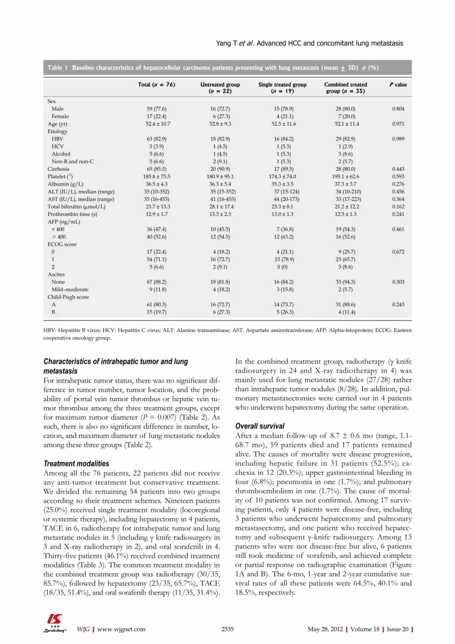
Characteristics of intrahepatic tumor and lung metastasisFor intrahepatic tumor status, there was no significant dif-ference in tumor number, tumor location, and the prob-ability of portal vein tumor thrombus or hepatic vein tu-mor thrombus among the three treatment groups, except for maximum tumor diameter (P = 0.007) (Table 2). As such, there is also no significant difference in number, lo-cation, and maximum diameter of lung metastatic nodules among these three groups (Table 2).
Treatment modalitiesAmong all the 76 patients, 22 patients did not receive any anti-tumor treatment but conservative treatment. We divided the remaining 54 patients into two groups according to their treatment schemes. Nineteen patients (25.0%) received single treatment modality (locoregional or systemic therapy), including hepatectomy in 4 patients, TACE in 6, radiotherapy for intrahepatic tumor and lung metastatic nodules in 5 (including γ knife radiosurgery in 3 and X-ray radiotherapy in 2), and oral sorafenib in 4. Thirty-five patients (46.1%) received combined treatment modalities (Table 3). The common treatment modality in the combined treatment group was radiotherapy (30/35, 85.7%), followed by hepatectomy (23/35, 65.7%), TACE (18/35, 51.4%), and oral sorafenib therapy (11/35, 31.4%).
In the combined treatment group, radiotherapy (γ knife radiosurgery in 24 and X-ray radiotherapy in 4) was mainly used for lung metastatic nodules (27/28) rather than intrahepatic tumor nodules (8/28). In addition, pul-monary metastasectomies were carried out in 4 patients who underwent hepatectomy during the same operation.
Overall survivalAfter a median follow-up of 8.7 ± 0.6 mo (range, 1.1- 68.7 mo), 59 patients died and 17 patients remained alive. The causes of mortality were disease progression, including hepatic failure in 31 patients (52.5%); ca-chexia in 12 (20.3%); upper gastrointestinal bleeding in four (6.8%); pneumonia in one (1.7%); and pulmonary thromboembolism in one (1.7%). The cause of mortal-ity of 10 patients was not confirmed. Among 17 surviv-ing patients, only 4 patients were disease-free, including 3 patients who underwent hepatectomy and pulmonary metastasectomy, and one patient who received hepatec-tomy and subsequent γ-knife radiosurgery. Among 13 patients who were not disease-free but alive, 6 patients still took medicine of sorafenib, and achieved complete or partial response on radiographic examination (Figure 1A and B). The 6-mo, 1-year and 2-year cumulative sur-vival rates of all these patients were 64.5%, 40.1% and 18.5%, respectively.
2535 May 28, 2012|Volume 18|Issue 20|WJG|www.wjgnet.com
Table 1 Baseline characteristics of hepatocellular carcinoma patients presenting with lung metastasis (mean ± SD) n (%)
Total (n = 76) Untreated group (n = 22)
Single treated group (n = 19)
Combined treated group (n = 35)
P value
Sex Male 59 (77.6) 16 (72.7) 15 (78.9) 28 (80.0) 0.804 Female 17 (22.4) 6 (27.3) 4 (21.1) 7 (20.0) Age (yr) 52.4 ± 10.7 52.8 ± 9.3 52.5 ± 11.6 52.1 ± 11.4 0.971Etiology HBV 63 (82.9) 18 (82.9) 16 (84.2) 29 (82.9) 0.989 HCV 3 (3.9) 1 (4.5) 1 (5.3) 1 (2.9) Alcohol 5 (6.6) 1 (4.5) 1 (5.3) 3 (8.6) Non-B and non-C 5 (6.6) 2 (9.1) 1 (5.3) 2 (5.7) Cirrhosis 65 (85.5) 20 (90.9) 17 (89.5) 28 (80.0) 0.443Platelet (-3) 185.8 ± 75.5 180.9 ± 95.1 174.3 ± 74.0 195.1 ± 62.6 0.593Albumin (g/L) 36.5 ± 4.3 36.3 ± 5.4 35.3 ± 3.5 37.3 ± 3.7 0.276ALT (IU/L), median (range) 35 (10-352) 35 (15-352) 37 (15-124) 34 (10-210) 0.456AST (IU/L), median (range) 35 (16-453) 41 (16-453) 44 (20-173) 33 (17-223) 0.364Total bilirubin (μmol/L) 23.7 ± 13.3 28.1 ± 17.4 23.3 ± 8.1 21.2 ± 12.2 0.162Prothrombin time (s) 12.9 ± 1.7 13.3 ± 2.3 13.0 ± 1.3 12.5 ± 1.3 0.241AFP (ng/mL) < 400 36 (47.4) 10 (45.5) 7 (36.8) 19 (54.3) 0.461 ≥ 400 40 (52.6) 12 (54.5) 12 (63.2) 16 (52.6) ECOG score 0 17 (22.4) 4 (18.2) 4 (21.1) 9 (25.7) 0.672 1 54 (71.1) 16 (72.7) 15 (78.9) 23 (65.7) 2 5 (6.6) 2 (9.1) 0 (0) 3 (8.6) Ascites None 67 (88.2) 18 (81.8) 16 (84.2) 33 (94.3) 0.303 Mild–moderate 9 (11.8) 4 (18.2) 3 (15.8) 2 (5.7) Child-Pugh score A 61 (80.3) 16 (72.7) 14 (73.7) 31 (88.6) 0.243 B 15 (19.7) 6 (27.3) 5 (26.3) 4 (11.4)
HBV: Hepatitis B virus; HCV: Hepatitis C virus; ALT: Alanine transaminase; AST: Aspartate aminotransferase; AFP: Alpha-fetoprotein; ECOG: Eastern cooperative oncology group.
Yang T et al . Advanced HCC and concomitant lung metastasis

Comparison of survival among three treatment groupsThe median survival time of combined treatment group was 18.6 ± 3.9 mo, which was longer than that of single treatment group (6.3 ± 2.5 mo) and untreatment group (4.1 ± 0.3 mo), and there was significant statistical dif-ferences among these groups (log-rank test, P < 0.001) (Figure 2). The 6-mo cumulative survival rate of com-bined treatment, single treatment and untreated group was 88.6%, 57.9% and 31.8%, respectively; and the 1-year
survival rate of the three groups was 68.5%, 21.1% and 8.0%, respectively.
2536 May 28, 2012|Volume 18|Issue 20|WJG|www.wjgnet.com
Table 2 Characteristics of intrahepatic tumor and lung metastasis of hepatocellular carcinoma patients presenting with lung metastasis (mean ± SD) n (%)
Total (n = 76) Untreated group (n = 22)
Single treated group (n = 19)
Combined treated group (n = 35)
P value
Intrahepatic tumor Maximum tumor diameter (cm) 9.3 ± 3.0 10.7 ± 3.2 9.7 ± 2.8 8.2 ± 2.5 0.007 Tumor number Solitary 29 (38.2) 9 (40.9) 6 (31.6) 14 (40.0) 0.791 Multiple/diffuse 47 (61.8) 13 (59.1) 13 (68.4) 21 (60.0) Tumor location Right 39 (51.3) 13 (59.1) 8 (42.1) 18 (51.4) 0.834 Left 15 (19.7) 4 (18.2) 4 (21.1) 7 (20.0) Both 22 (28.9) 5 (22.7) 7 (36.8) 10 (28.6) Portal vein tumor thrombus Absence 54 (71.1) 14 (63.6) 12 (63.2) 28 (80.0) 0.283 Presence 22 (28.9) 8 (36.4) 7 (36.8) 7 (20.0) Hepatic vein tumor thrombus Absence 67 (88.2) 19 (86.4) 15 (78.9) 33 (94.2) 0.238 Presence 9 (11.8) 3 (13.6) 4 (21.1) 2 (5.7) Lung metastasis Number of metastasis Solitary 17 (22.4) 4 (18.2) 3 (15.8) 10 (28.6) 0.479 Multiple 59 (77.6) 18 (81.8) 16 (84.2) 25 (71.4) Location of metastasis Right lung lobe 28 (36.8) 6 (27.3) 8 (42.1) 14 (40.0) 0.721 Left lung lobe 13 (17.1) 4 (18.2) 2 (10.5) 7 (20.0) Bialteral lung lobes 35 (46.1) 12 (54.5) 9 (47.4) 14 (40.0) Maximum metastasis diameter (cm) 2.5 ± 0.9 2.6 ± 0.9 2.2 ± 1.2 2.5 ± 0.8 0.445
Table 3 Treatment modalities for intrahepatic tumor and/or metastatic lung nodule(s)
Treatment modalities n (%)
Single treatment modality in treated patients (n = 19) Hepatectomy 4 (21.1) Pulmonary metastasectomy 0 (0) Transcatheter arterial chemoembolization 6 (31.6) Radiotherapy 5 (26.3) Oral sorafenib 4 (21.1)Combined treatment modalities in treated patients (n = 35) Hepatectomy + pulmonary metastasectomy 3 (8.6) Hepatectomy + pulmonary metastasectomy + oral sorafenib 1 (2.9) Hepatectomy + transcatheter arterial chemoembolization + radiotherapy
6 (17.1)
Hepatectomy + radiotherapy 9 (25.7) Hepatectomy + transcatheter arterial chemoembolization + radiotherapy + oral sorafenib
2 (5.7)
Hepatectomy + radiotherapy + oral sorafenib 2 (5.7) Transcatheter arterial chemoembolization + radiotherapy 6 (17.1) Transcatheter arterial chemoembolization + radiotherapy + oral sorafenib
3 (8.6)
Transcatheter arterial chemoembolization + oral sorafenib 1 (2.9) Radiotherapy + oral sorafenib 2 (5.7)
R
10 cm
A
10 cm
B
Figure 1 Overall survival. A: Computed tomography (CT) scan showing multiple metastatic nodules in both lung lobes before anti-tumor treatment in a 54-year-old male patient with lung metastasis due to hepatocellular carcinoma; B: CT scan showing the disappearance of previous lung metastatic nodules in 5 mo after γ-knife radiosurgery and oral sorafenib.
Yang T et al . Advanced HCC and concomitant lung metastasis
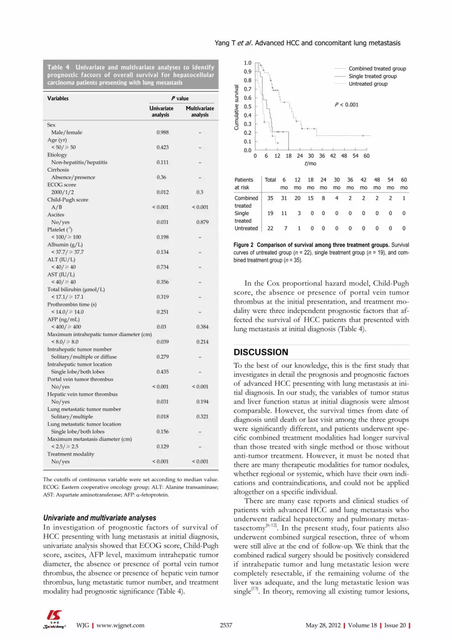
Univariate and multivariate analysesIn investigation of prognostic factors of survival of HCC presenting with lung metastasis at initial diagnosis, univariate analysis showed that ECOG score, Child-Pugh score, ascites, AFP level, maximum intrahepatic tumor diameter, the absence or presence of portal vein tumor thrombus, the absence or presence of hepatic vein tumor thrombus, lung metastatic tumor number, and treatment modality had prognostic significance (Table 4).
In the Cox proportional hazard model, Child-Pugh score, the absence or presence of portal vein tumor thrombus at the initial presentation, and treatment mo-dality were three independent prognostic factors that af-fected the survival of HCC patients that presented with lung metastasis at initial diagnosis (Table 4).
DISCUSSIONTo the best of our knowledge, this is the first study that investigates in detail the prognosis and prognostic factors of advanced HCC presenting with lung metastasis at ini-tial diagnosis. In our study, the variables of tumor status and liver function status at initial diagnosis were almost comparable. However, the survival times from date of diagnosis until death or last visit among the three groups were significantly different, and patients underwent spe-cific combined treatment modalities had longer survival than those treated with single method or those without anti-tumor treatment. However, it must be noted that there are many therapeutic modalities for tumor nodules, whether regional or systemic, which have their own indi-cations and contraindications, and could not be applied altogether on a specific individual.
There are many case reports and clinical studies of patients with advanced HCC and lung metastasis who underwent radical hepatectomy and pulmonary metas-tasectomy[6-12]. In the present study, four patients also underwent combined surgical resection, three of whom were still alive at the end of follow-up. We think that the combined radical surgery should be positively considered if intrahepatic tumor and lung metastatic lesion were completely resectable, if the remaining volume of the liver was adequate, and the lung metastatic lesion was single[13]. In theory, removing all existing tumor lesions,
2537 May 28, 2012|Volume 18|Issue 20|WJG|www.wjgnet.com
Table 4 Univariate and multivariate analyses to identify prognostic factors of overall survival for hepatocellular carcinoma patients presenting with lung metastasis
Variables P value
Univariate analysis
Multivariate analysis
Sex Male/female 0.988 –Age (yr) < 50/≥ 50 0.423 –Etiology Non-hepatitis/hepatitis 0.111 –Cirrhosis Absence/presence 0.36 –ECOG score 2000/1/2 0.012 0.3Child-Pugh score A/B < 0.001 < 0.001Ascites No/yes 0.031 0.879Platelet (-3) < 100/≥ 100 0.198 –Albumin (g/L) < 37.7/≥ 37.7 0.134 –ALT (IU/L) < 40/≥ 40 0.734 –AST (IU/L) < 40/≥ 40 0.356 –Total bilirubin (μmol/L) < 17.1/≥ 17.1 0.319 –Prothrombin time (s) < 14.0/≥ 14.0 0.251 –AFP (ng/mL) < 400/≥ 400 0.03 0.384Maximum intrahepatic tumor diameter (cm) < 8.0/≥ 8.0 0.039 0.214Intrahepatic tumor number Solitary/multiple or diffuse 0.279 –Intrahepatic tumor location Single lobe/both lobes 0.435 –Portal vein tumor thrombus No/yes < 0.001 < 0.001Hepatic vein tumor thrombus No/yes 0.031 0.194Lung metastatic tumor number Solitary/multiple 0.018 0.321Lung metastatic tumor location Single lobe/both lobes 0.156 –Maximum metastasis diameter (cm) < 2.5/≥ 2.5 0.129 –Treatment modality No/yes < 0.001 < 0.001
The cutoffs of continuous variable were set according to median value. ECOG: Eastern cooperative oncology group; ALT: Alanine transaminase; AST: Aspartate aminotransferase; AFP: α-fetoprotein.
1.0
0.9
0.8
0.7
0.6
0.5
0.4
0.3
0.2
0.1
0.0
Cum
ulat
ive
surv
ival
0 6 12 18 24 30 36 42 48 54 60 t /mo
Combined treated groupSingle treated groupUntreated group
P < 0.001
Patients at risk
Total 6 mo
12 mo
18 mo
24 mo
30 mo
36 mo
42 mo
48 mo
54 mo
60 mo
Combined treated
35 31 20 15 8 4 2 2 2 2 1
Single treated
19 11 3 0 0 0 0 0 0 0 0
Untreated 22 7 1 0 0 0 0 0 0 0 0
Figure 2 Comparison of survival among three treatment groups. Survival curves of untreated group (n = 22), single treatment group (n = 19), and com-bined treatment group (n = 35).
Yang T et al . Advanced HCC and concomitant lung metastasis

differ statistically among the three treatment groups, the clinical circumstances at the initial presentation might dif-fer. Therefore, the decision for the treatment mordalities might be biased, and the subsequent patient stratifica-tion into various treatment groups might also be biased. Second, treatment modalities, i.e., radiotherapy, TACE, varied, and the number of patients treated was too small to confirm the effectiveness of each treatment modal-ity. Third, the indications of specific treatment modality varied, and selective modalities were given to selective patients with advanced HCC and lung metastasis. It is very hard to build up a standard therapeutic regime for all patients, regardless of primary and secondary tumor site, size and number, liver functional reserve, and patients’ general condition.
In conclusion, the present study showed that the prognosis of advanced HCC with concomitant lung metastasis at initial diagnosis is very poor, and combined comprehensive treatment modalities tended to signifi-cantly prolong the survival of the patients compared with conservative treatment or single treatment modality. Furthermore, further randomized trials might be required to investigate the optimal treatment modality in the near future.
COMMENTSBackgroundThe prognosis and treatment outcomes of advanced hepatocellular carcinoma (HCC) presenting with lung metastasis remain poorly evaluated.Research frontiersThere are various treatment modalities for advanced HCC, including surgical resection, transcatheter arterial chemoembolization, radiotherapy, chemothera-peutics, and administration of molecular targeted therapeutic drugs. This study investigated the prognosis, treatment outcomes, and independent prognostic factors affecting the survival of a series of HCC patients presenting with lung metastasis at initial diagnosis.Innovations and breakthroughsIn this study, the survival times from date of diagnosis until death or last visit be-tween untreated group, single treatment group, and combined treatment group were significantly different, and patients underwent specific combined treatment modalities had longer survival than those treated with single method or those without anti-tumor treatment. Multivariate analysis revealed that Child-Pugh score, the absence or presence of portal vein tumor thrombus, and treatment modality were three independent prognostic factors affecting survival of patients with advanced HCC and concomitant lung metastasis.ApplicationsAs one type of advanced HCC, HCC presenting with lung metastasis is not unusual, and its prognosis is very poor. The findings in this study may con-tribute to its prognosis, and combined treatment modalities tend to result in a better survival for patients with advanced HCC initially presenting with lung metastasis.Peer reviewThe authors firstly investigated that combined treatment modalities tended to yield better survival prolongation compared with conservative treatment or single treatment modality for HCC patients initially presenting with lung metas-tasis, which may be contribute to its prognosis.
REFERENCES1 El-Serag HB, Rudolph KL. Hepatocellular carcinoma: epide-
miology and molecular carcinogenesis. Gastroenterology 2007; 132: 2557-2576
COMMENTS
including primary and second lesions, is the only possible therapeutic approach till now.
Recently, with development in radiotherapy tech-niques, radiotherapy has been shown to play a potential role in a wide spectrum of HCC, therefore it is necessary to evaluate the effect of radiotherapy[14-16]. In our center, stereotactic radiotherapy, particularly γ knife radiosurgery, has become the major therapeutic mordality for lung and brain metastases of HCC. In the present study, there were 35 patients who underwent radiotherapy for intra-hepatic HCC tumors and/or lung metastatic nodules, accounting for more than 50%, whether or not they are combined with other anti-tumor treatment modalities. Despite advances in radiotherapy delivery, liver toxicity following radiotherapy remains a dose-limiting factor, and investigations to better understand the pathophysiol-ogy of radiotherapy-induced liver toxicity are warranted. There is a particular interest in combining radiotherapy with anti-vascular endothelial growth factor targeting agents for their independent activity in HCC as well as their radiation sensitization properties[17].
Sorafenib is a multikinase inhibitor with effects against tumor proliferation and angiogenesis, and was recently approved for the treatment of advanced HCC[18,19]. Main-tenance sorafenib would probably prevent or delay the intrahepatic and extrahepatic spread of HCC after radio-therapy, which provides the rationale for the combination of these treatment modalities[20]. In the present study, 15 patients received oral sorafenib. Although we did not find its significant efficacy in HCC presenting with lung metastasis due to few cases and less strict design, we be-lieved that sorafenib could be used as one part of com-bined treatment modalities for HCC patients with lung metastasis. However, a large-scale randomized controlled trial is needed to confirm it. Combining surgical resection with sorafenib would be considered as the most optimal treatment modality for HCC patients with lung metasta-sis.. However, it remains to be confirmed by a random-ized controlled clinical trial in the future.
Till now, we have not obtained enough evidences to confirm the role of locoregional hepatectomy in HCC patients with lung metastasis. In our opinion, hepatec-tomy can be performed when primary intrahepatic HCCs are completely resected, the number of lung metastases is less than 3, and the diameter of individual lung metas-tasis is less than 3 cm. In addition, any of treatment mo-dalities, such as radiotherapy or TACE, should be used together for lung metastasis.
In the univariate and subsequent multivariate analyses, Child-Pugh score was an independent prognostic factor for HCC patients with lung metastasis. This indicates that although invasive treatment other than conservative treat-ment can prolong the survival in well-selected patients, one must be careful before applying invasive treatment to all patients.
This study has several limitations. First, it was de-signed retrospectively. Although the patients’ status, including ECOG scale, Child-Pugh score, and liver func-tion, was reviewed in the medical records and did not
2538 May 28, 2012|Volume 18|Issue 20|WJG|www.wjgnet.com
Yang T et al . Advanced HCC and concomitant lung metastasis

2539 May 28, 2012|Volume 18|Issue 20|WJG|www.wjgnet.com
2 Rahbari NN, Mehrabi A, Mollberg NM, Müller SA, Koch M, Büchler MW, Weitz J. Hepatocellular carcinoma: current management and perspectives for the future. Ann Surg 2011; 253: 453-469
3 Natsuizaka M, Omura T, Akaike T, Kuwata Y, Yamazaki K, Sato T, Karino Y, Toyota J, Suga T, Asaka M. Clinical features of hepatocellular carcinoma with extrahepatic metastases. J Gastroenterol Hepatol 2005; 20: 1781-1787
4 Zhang SM, Zeng ZC, Tang ZY, Sun J, Cheng JM, Liu R, Wang P, Zhang BH. Prognostic analysis of pulmonary me-tastases from hepatocellular carcinoma. Hepatol Int 2008; 2: 237-243
5 Bruix J, Sherman M, Llovet JM, Beaugrand M, Lencioni R, Burroughs AK, Christensen E, Pagliaro L, Colombo M, Rodés J. Clinical management of hepatocellular carcinoma. Conclusions of the Barcelona-2000 EASL conference. Euro-pean Association for the Study of the Liver. J Hepatol 2001; 35: 421-430
6 Lam CM, Lo CM, Yuen WK, Liu CL, Fan ST. Prolonged sur-vival in selected patients following surgical resection for pul-monary metastasis from hepatocellular carcinoma. Br J Surg 1998; 85: 1198-1200
7 Nakagawa T, Kamiyama T, Nakanishi K, Yokoo H, Kamachi H, Matsushita M, Todo S. Pulmonary resection for metasta-ses from hepatocellular carcinoma: factors influencing prog-nosis. J Thorac Cardiovasc Surg 2006; 131: 1248-1254
8 Tomimaru Y, Sasaki Y, Yamada T, Eguchi H, Takami K, Ohi-gashi H, Higashiyama M, Ishikawa O, Kodama K, Imaoka S. The significance of surgical resection for pulmonary metasta-sis from hepatocellular carcinoma. Am J Surg 2006; 192: 46-51
9 Kuo SW, Chang YL, Huang PM, Hsu HH, Chen JS, Lee JM, Lee PH, Lee YC. Prognostic factors for pulmonary metasta-sectomy in hepatocellular carcinoma. Ann Surg Oncol 2007; 14: 992-997
10 Chen F, Sato K, Fujinaga T, Sonobe M, Shoji T, Sakai H, Mi-yahara R, Bando T, Okubo K, Hirata T, Date H. Pulmonary resection for metastases from hepatocellular carcinoma. World J Surg 2008; 32: 2213-2217
11 Kawamura M, Nakajima J, Matsuguma H, Horio H, Miyoshi
S, Nakagawa K, Fujisawa T, Kobayashi K. Surgical outcomes for pulmonary metastases from hepatocellular carcinoma. Eur J Cardiothorac Surg 2008; 34: 196-199
12 Kwon JB, Park K, Kim YD, Seo JH, Moon SW, Cho DG, Kim YW, Kim DG, Yoon SK, Lim HW. Clinical outcome after pulmonary metastasectomy from primary hepatocellular car-cinoma: analysis of prognostic factors. World J Gastroenterol 2008; 14: 5717-5722
13 Yang T, Zhang J, Lu JH, Yang LQ, Yang GS, Wu MC, Yu WF. A new staging system for resectable hepatocellular carcino-ma: comparison with six existing staging systems in a large Chinese cohort. J Cancer Res Clin Oncol 2011; 137: 739-750
14 Dawson LA. The evolving role of radiation therapy in hepa-tocellular carcinoma. Cancer Radiother 2008; 12: 96-101
15 Cárdenes HR. Role of stereotactic body radiotherapy in the management of primary hepatocellular carcinoma. Rationale, technique and results. Clin Transl Oncol 2009; 11: 276-283
16 Ma S, Jiao B, Liu X, Yi H, Kong D, Gao L, Zhao G, Yang Y, Liu X. Approach to radiation therapy in hepatocellular carci-noma. Cancer Treat Rev 2010; 36: 157-163
17 Tse RV, Guha C, Dawson LA. Conformal radiotherapy for hepatocellular carcinoma. Crit Rev Oncol Hematol 2008; 67: 113-123
18 Llovet JM, Ricci S, Mazzaferro V, Hilgard P, Gane E, Blanc JF, de Oliveira AC, Santoro A, Raoul JL, Forner A, Schwartz M, Porta C, Zeuzem S, Bolondi L, Greten TF, Galle PR, Seitz JF, Borbath I, Häussinger D, Giannaris T, Shan M, Moscovici M, Voliotis D, Bruix J. Sorafenib in advanced hepatocellular carcinoma. N Engl J Med 2008; 359: 378-390
19 Colombo M. Sorafenib in advanced hepatocellular carci-noma: a further step toward personalized therapy of liver cancer. Gastroenterology 2009; 136: 1832-1835
20 Zhao JD, Liu J, Ren ZG, Gu K, Zhou ZH, Li WT, Chen Z, Xu ZY, Liu LM, Jiang GL. Maintenance of Sorafenib following combined therapy of three-dimensional conformal radiation therapy/intensity-modulated radiation therapy and trans-catheter arterial chemoembolization in patients with locally advanced hepatocellular carcinoma: a phase I/II study. Ra-diat Oncol 2010; 5: 12
‘S- Editor Gou SX L- Editor Ma JY E- Editor Zheng XM
Yang T et al . Advanced HCC and concomitant lung metastasis

Detection of eukaryotic translation initiation factor 4E and its clinical significance in hepatocellular carcinoma
Xiao-Lin Wang, Hong-Pei Cai, Jun-Hui Ge, Xiao-Feng Su
Xiao-Lin Wang, Hong-Pei Cai, Xiao-Feng Su, Gastroenter-ology Department, The Second Military Medical University, Changzheng Hospital, Shanghai 200003, ChinaJun-Hui Ge, Pathology Department, The Second Military Medi-cal University, Changzheng Hospital, Shanghai 200003, ChinaAuthor contributions: Wang XL and Cai HP designed the study; Ge JH performed the immunohistochemical staining and contributed the analytic tools; Su XF collected and analysed data; and Wang XL wrote the manuscript.Correspondence to: Hong-Pei Cai, Professor, Gastroenter-ology Department, The Second Military Medical University, Changzheng Hospital, Shanghai 200003, China. [email protected]: +86-21-81885344 Fax: +86-21-63520020Received: November 6, 2011 Revised: February 20, 2012 Accepted: February 26, 2012Published online: May 28, 2012
AbstractAIM: To study the expression of eukaryotic translation initiation factor 4E (eIF4E), which is closely correlated with malignant tumors, and its relationship to prognosis in hepatocellular carcinoma.
METHODS: Western blotting was performed to quan-tify the elF4E protein expression in the normal human liver cell line L02 and the hepatoma cell lines Hep3B, HepG2, and Huh7. Forty-six hepatocellular carcinoma samples with complete clinical data were obtained from Changzheng Hospital during the period of December 2008 to July 2009. The expression of eIF4E in the tu-mor samples and their adjacent tissues were detected by immunohistochemistry. The relationship between the test results and hepatocellular carcinoma (HCC) prognosis was statistically analysed by using a COX proportional hazard model.
RESULTS: Western blotting analysis showed that there were distinct eIF4E protein bands in all three of the hepatoma cell lines. In particular, the HepG2 cell line
had the highest level of eIF4E protein expression. The L02 cell group had a low eIF4E expression. Immuno-histochemical assay showed that there were 32 cases in which the tumour tissue expression was higher than their adjacent tissues, accounting for 69.57%. There were also 14 cases in which the tumour tissue expres-sion was lower or no significant difference was found, accounting for 30.43%. COX proportional hazards model analysis showed that HCC prognosis was related to the depth of invasion, the overexpression of eIF4E and p53, possibly as independent HCC prognostic predictors.
CONCLUSION: In summary, eIF4E expression is as-sociated with liver cancer, and patients with high eIF4E expression levels have a higher risk of recurrence.
© 2012 Baishideng. All rights reserved.
Key words: Hepatocellular carcinoma; Eukaryotic trans-lation initiation factor 4E; Western blotting; Immunohis-tochemistry; Prognosis
Peer reviewer: Ezio Laconi, Professor, University of Cagliari, Via Porcell 4, 09125 Cagliari, Italy
Wang XL, Cai HP, Ge JH, Su XF. Detection of eukaryotic transla-tion initiation factor 4E and its clinical significance in hepatocel-lular carcinoma. World J Gastroenterol 2012; 18(20): 2540-2544 Available from: URL: http://www.wjgnet.com/1007-9327/full/v18/i20/2540.htm DOI: http://dx.doi.org/10.3748/wjg.v18.i20.2540
INTRODUCTIONEukaryotic translation initiation factor 4E (eIF4E) is a member of the eIF family. It can specifically bind to the cap structure located at the 5’ end of mRNAs named the “m7GpppN cap”, which is necessary for mRNA transla-tion initiation, and affects mRNA metabolism, processing, transportation and translation[1]. It plays an important role
BRIEF ARTICLE
Online Submissions: http://www.wjgnet.com/[email protected]:10.3748/wjg.v18.i20.2540
2540 May 28, 2012|Volume 18|Issue 20|WJG|www.wjgnet.com
World J Gastroenterol 2012 May 28; 18(20): 2540-2544 ISSN 1007-9327 (print) ISSN 2219-2840 (online)
© 2012 Baishideng. All rights reserved.

in regulating the initial stage protein synthesis[2,3]. eIF4E is highly expressed in a variety of human malignancies[4-7], which has been confirmed to be relevant to the occur-rence, invasion and metastasis of carcinomas such as head and neck squamous cell carcinoma[8], laryngeal cancer, non-small cell lung cancer[9-11], breast cancer[12-18], thyroid cancer, oesophageal cancer, stomach cancer, cholangio-carcinoma, colon cancer[19], non-Hodgkin’s lymphoma, acute or chronic myeloid leukaemia[20], and lymphoma[21,22]. Experiments have also confirmed that eIF4E is closely related to the prognosis of many carcinomas. However, eIF4E-related studies in the context of hepatocellular car-cinoma (HCC) are still rare.
In this study, we separately compared the eIF4E expre-ssion levels in normal liver cells with liver cancer cell lines and liver cancer tissues with precancerous tissues. Ad-ditionally, we investigated the influence of eIF4E expres-sion on the prognosis of liver cancer. This research may provide an experimental basis for exploring new ways to treat liver cancer.
MATERIALS AND METHODSStudy objectsWe selected 46 patients with pathological evidence of HCC and complete clinical data from Shanghai Chang-zheng Hospital who had liver surgery from January 2007 to January 2009. In these 46 cases, there were 40 males and 6 females who ranged in age from 31 years to 77 years (median age: 52.26 years). With regards to histologi-cal grade, there were 42 cases of moderately differentiated HCC, and 4 cases were poorly differentiated. A total of 33 patients had a cancer embolus in the intrahepatic bile duct or vein or had an infiltrated peplos, and 13 patients had no cancer tissue in cutting edge and gallbladder and no infiltrated peplos. p53 pathological testing was positive in 39 patients and negative in 7 cases. None of the patients received preoperative radiotherapy or chemotherapy. The follow-up time was 24 mo, and no case was lost.
Major materials and reagentsThe cell lines used for Western blotting were the human liver cancer cell lines Hep3B, HepG2, Huh7, and the normal human liver cell line L02, which was provided by Shanghai Cell Biology Institution of Academia Sinica.
eIF4E (P-2) is a mouse anti-human monoclonal an-tibody raised against full-length eIF4E (Santa Cruz Bio-technology, Inc.). It is recommended for the detection of eIF4E by Western blotting (dilution: 1:200; dilution range: 1:100-1:1000) and immunohistochemistry (includ-ing paraffin-embedded sections; dilution: 1:50; dilution range: 1:50-1:500). The streptavidin-perosidase (SP) kit was provided by Fuzhou Maixin Biotechnology, Inc.
Detection methodsWestern blotting analysis: We tested the eIF4E gene expression level in normal liver cells and different liver cancer cell lines. The four of cell lines (i.e., HepG2,
Hep3B, Huh7, and L02) were incubated with high glu-cose Dulbecco's modified Eagle's medium containing 10% fetal bovine serum at 37.0 ℃ with 5% CO2 until the cell concentration reached 5 × 10⁶ cells/mL. Then, we sequentially performed the protein extraction, bicincho-ninic acid protein quantification, sodium dodecyl sulfate-polyacrylamide gel electrophoresis electrophoresis, pro-tein transfer, membrane closure, antibody incubation, and Bio-Rad chemiluminescence.
Immunohistochemistry: We detected the eIF4E pro-tein expression levels in HCC and their adjacent tissues (SP, a particular type of immunohistochemistry). The tumour and adjacent tissues from the same patient were fixed, dehydrated, sectioned, and made into paraffin biop-sies. We made 46 paraffin sections. The steps of the SP kit included heating on a baking sheet, incubation, wash-ing, sealing, staining, drying, dehydration, and mounting. To analyse the results, we used two scoring methods. The samples were placed in an electron microscope and were scored for staining intensity as follows. 0: No colour; 1: A yellow colour; 2: A claybank colour; and 3: A brown colour. We then graded the samples for the positivity rate as follows. 0: No positive tumour cell staining; 1: ≤ 10% positive cells; 2: 11% to 50% positive cells; 3: 51% to 75% positive cells; and 4: > 75% positive cells. Finally, we added the two scores together, and the sum represented the immunohistochemical score as follows. -: 0; +: 1 to 4; ++: 5 to 8; and + + +: 9 to 12. Each cancer tissue sec-tion was compared with its adjacent tissue.
Follow up: We analysed the number of cases that had HCC recurrence and metastasis during the post-operative 24 mo. The liver cancer recurrence risk was measured using COX proportional hazards model for statistical analysis. The patient age, gender, histological grade, depth of invasion, eIF4E, p53 status and other prognostic indicators were used for the COX proportional hazards model analysis.
Statistical analysisWe used the SPSS 17.0 statistical software for the statisti-cal analysis. A COX proportional hazards model testing level of α = 0.05 and a P < 0.05 was considered statisti-cally significant.
RESULTSeIF4E protein expression in liver cancer cell lines by western blottingWe tested the eIF4E protein expression level in the liver cell line L02 and the liver cancer cell lines Huh7, HepG2, Hep3B. The eIF4E protein bands are shown in Figure 1. The bands were detected by Bio-Rad chemiluminescence to obtain the data shown in Table 1.
The liver cancer cell lines HepG2, Huh7, Hep3B sig-nificantly expressed the eIF4E protein, and in particular, the HepG2 cell line had the highest level of eIF4E pro-tein expression. The normal liver cell L02 also expressed
2541 May 28, 2012|Volume 18|Issue 20|WJG|www.wjgnet.com
Wang XL et al . Detection of eIF4E and its clinical significance

eIF4E; however, its expression level was low. Glyceral-dehyde-3-phosphate dehydrogenase, which was used as the internal reference had bands in each cell line, and no obvious differences were observed with this protein.
eIF4E protein expression in liver cancer and adjacent tissues by immunohistochemistryWe next detected the eIF4E protein expression level in HCC and adjacent tissues. There were 46 pathologi-cal tissue paraffin blocks in which 32 HCC tissue cases had higher eIF4E protein expression than their adjacent tissues, accounting for 69.57%. A total of 14 HCC tis-sue cases had lower expression or no significant differ-ence compared with their adjacent tissues, accounting for 30.43%. The scores were weighted 154:97 = 1.59:1, meaning that, in general, HCC tissues had a higher eIF4E protein expression level than the adjacent tissues.
Figure 2 show that HCC tissues stained significantly stronger than adjacent tissues, indicating that tumour tis-sues had a higher eIF4E protein expression level. The lightly stained central area in Figure 2A represents ne-crotic tissue.
eIF4E may be an independent risk factor for liver cancer prognosisFollow-up statistics showed recurrence in 33 cases and
death in 12 cases. The patient age, gender, histological grade, depth of invasion, eIF4E overexpression, p53 posi-tive status and other prognostic indicators were used for COX proportional hazards model for screening analysis. Ultimately, the depth of invasion, eIF4E, and p53 were in-cluded in the model with a Sig < 0.05 as shown in Table 2. The statistical significance suggests that these three factors are independent risk factors for liver cancer prognosis.
DISCUSSIONIn eukaryotic cells, translational regulation plays an im-portant role in gene expression. eIF4E is involved in the regulation of the mRNA translation process. It can en-hance the translation of some important growth factors and cell growth regulators and affect protein synthesis, the cell cycle, cancer gene activation, and apoptosis; it also plays an important role in malignant transformation and metastasis.
eIF4E regulates the translation of cancer-related mRNAs (i.e., it is involved in the activation of proto-oncogenes, angiogenesis, apoptosis, invasion and metastasis) that are involved in tumour occurrence and development. Nor-mal tissues have a low eIF4E expression level. eIF4E was overexpressed, in a variety of malignant tumours includ-ing head and neck squamous cell carcinoma, laryngeal cancer, lung cancer, breast cancer, thyroid cancer and other cancer tissues[4,23]. Its high expression was correlated with tumour invasion and metastasis. However, studies of eIF4E in liver cancer are rare. At present, there are studies
2542 May 28, 2012|Volume 18|Issue 20|WJG|www.wjgnet.com
Huh7 HepG2 L02 Hep3B
B
A
Figure 1 Eukaryotic translation initiation factor 4E protein and glyceralde-hyde-3-phosphate dehydrogenase protein bands in a normal human liver cell line and three hepatoma carcinoma cell lines. A: Eukaryotic translation initiation factor 4E protein bands; B: Glyceraldehyde-3-phosphate dehydroge-nase protein bands.
Table 1 Eukaryotic translation initiation factor 4E protein expression in the liver cells at different levels
Huh7 HepG2 L02 Hep3B
GAPDH 827.165 884.682 885.437 848.552eIF4E 3161.861 5651.885 775.440 4496.191
GAPDH: Glyceraldehyde-3-phosphate dehydrogenase; eIF4E: Eukaryotic translation initiation factor 4E.
B
A
Figure 2 Photo of hepatocellular carcinoma tissue and adjacent tissue by electron microscopy. A: Hepatocellular carcinoma tissue and adjacent intersti-tial tissue; B: Adjacent tissue. (Hematoxylin and eosin stain, ×100).
Wang XL et al . Detection of eIF4E and its clinical significance

2543 May 28, 2012|Volume 18|Issue 20|WJG|www.wjgnet.com
Innovations and breakthroughsResearch concerning the effects of eIF4E on HCC is limited. The authors tested the expression of the eIF4E protein in liver cancer cell lines and cancer tissues and used COX proportional hazards model analysis to show that eIF4E was an independent risk factor HCC prognosis.ApplicationsThe targeted regulation of the level and activity of eIF4E may inhibit cancer cell growth, which may become a new treatment paradigm in the liver cancer field.TerminologyeIF4E is a member of the eIF family. It can specifically bind to the cap structure located at the 5’ end of mRNAs named the “m7GpppN cap”, which is neces-sary for mRNA translation initiation, and affects mRNA metabolism, process-ing, transportation and translation. It plays an important role in regulating the initial stage protein synthesis. eIF4E is highly expressed in a variety of human malignancies, which has been confirmed to be relevant to the occurrence, invasion and metastasis of carcinomas such as head and neck squamous cell carcinoma, laryngeal cancer, non-small cell lung cancer, breast, thyroid cancer, oesophageal cancer, stomach cancer, cholangiocarcinoma, colon cancer, non-Hodgkin’s lymphoma, acute or chronic myeloid leukaemia, and lymphoma. Experiments have also confirmed that eIF4E is closely related to the prognosis
of many carcinomas.Peer reviewThis paper is interesting and worth being published if authors can satisfactorily address the concerns raised regarding immunohistochemical expression of eIF4E.
REFERENCES1 Rhoads RE. eIF4E: new family members, new binding part-
ners, new roles. J Biol Chem 2009; 284: 16711-16715 2 Sonenberg N. eIF4E, the mRNA cap-binding protein: from
basic discovery to translational research. Biochem Cell Biol 2008; 86: 178-183
3 Xu X, Vatsyayan J, Gao C, Bakkenist CJ, Hu J. Sumoylation of eIF4E activates mRNA translation. EMBO Rep 2010; 11: 299-304
4 Furic L, Rong L, Larsson O, Koumakpayi IH, Yoshida K, Brueschke A, Petroulakis E, Robichaud N, Pollak M, Gaboury LA, Pandolfi PP, Saad F, Sonenberg N. eIF4E phos-phorylation promotes tumorigenesis and is associated with prostate cancer progression. Proc Natl Acad Sci USA 2010; 107: 14134-14139
5 Fan S, Ramalingam SS, Kauh J, Xu Z, Khuri FR, Sun SY. Phosphorylated eukaryotic translation initiation factor 4 (eIF4E) is elevated in human cancer tissues. Cancer Biol Ther 2009; 8: 1463-1469
6 Thumma SC, Kratzke RA. Translational control: a target for cancer therapy. Cancer Lett 2007; 258: 1-8
7 Noske A, Lindenberg JL, Darb-Esfahani S, Weichert W, Buckendahl AC, Röske A, Sehouli J, Dietel M, Denkert C. Activation of mTOR in a subgroup of ovarian carcinomas: correlation with p-eIF-4E and prognosis. Oncol Rep 2008; 20: 1409-1417
that involve the targeting eIF4E in head and neck squa-mous cell carcinoma[24,25], breast cancer[13-18], non-small cell lung cancer[26], blood malignancies[27-29] and other stud-ies[6,30-32]. However, few studies have focused on targeting eIF4E in HCC.
In this study, we tested the expression of eIF4E pro-tein in a normal human liver cell line and three different liver cancer cell lines. eIF4E protein expression was high in the three liver cancer cell lines and higher than in the normal liver cell L02. The HepG2 cell line had an espe-cially high level of eIF4E protein expression. By compar-ing 46 cases of human liver cancer and adjacent tissues, we found that eIF4E protein expression was higher in most of the cancer tissues than in the adjacent tissues. COX proportional hazards model analysis showed that the depth of invasion, eIF4E, and p53 status were inde-pendent risk factors of liver cancer prognosis.
Based on these studies, we believe that eIF4E protein expression may be closely associated with the occurrence of human liver cancer development and prognosis. It has been confirmed in vivo and in vitro that sorafenib treatment can inhibit the RAF/MEK/ERK signal transduction pathway, reduce the eIF4E phosphorylation level, reduce Mcl-l protein, and induce hepatoma cell apoptosis[33,34]. Accordingly, we suggest that lower levels of eIF4E gene expression may inhibit liver cancer. Targeting and adjust-ing the eIF4E level and activity may inhibit cancer cell growth[6,30,31,35], which may become a new paradigm in the field of the biological treatment of liver cancer[36].
COMMENTSBackgroundHepatocellular carcinoma (HCC), which has a poor prognosis and a low five-year survival rate, is the most common malignant tumour in our country. At present, there are no effective therapies including radiotherapy, chemotherapy, and surgery. Eukaryotic translation initiation factor 4E (eIF4E) plays an im-portant role in the translation initiation phase of a eukaryotic cell. It has been confirmed that eIF4E can specifically bind to the 5’ mRNA cap (m7GpppN) and modulate its translation and expression. Its expression is closely associated with the generation, infiltration, and metastasis of many tumours such as head and neck, larynx, lung, mammary gland, thyroid gland, oesophagus, stomach, bile duct, colon.Research frontiersThere are many researchers that are targeting eIF4E
in head and neck squa-
mous cell carcinoma, breast cancer, non-small cell lung cancer, blood ma-lignancies and other carcinomas; however, studies that involve the targeting eIF4E in HCC are rare.
Table 2 COX proportional hazards model analysis
95% Cl for Exp(B)
B SE Wald df Sig Exp(B) Lower Upper
Eukaryotic translation initiation factor 4E 1.971 0.926 4.529 1 0.033 7.179 1.169 44.100Depth of invasion 3.122 1.211 6.650 1 0.010 22.690 2.115 243.423Histological grade 0.410 1.156 0.126 1 0.723 1.506 0.156 14.527Gender 1.671 1.152 2.104 1 0.147 5.319 0.556 50.890Age -0.017 0.028 0.354 1 0.552 0.983 0.930 1.040p53 -3.208 0.825 15.118 1 0.000 0.040 0.008 0.204
B: Coefficient of regression; SE.: Standard error; Wald: The index of regression effect; df: Degrees of freedom; Sig: P value; Exp(B): Odds ratio.
COMMENTS
Wang XL et al . Detection of eIF4E and its clinical significance

2544 May 28, 2012|Volume 18|Issue 20|WJG|www.wjgnet.com
8 Sunavala-Dossabhoy G, Palaniyandi S, Clark C, Nathan CO, Abreo FW, Caldito G. Analysis of eIF4E and 4EBP1 mRNAs in head and neck cancer. Laryngoscope 2011; 121: 2136-2141
9 Khoury T, Alrawi S, Ramnath N, Li Q, Grimm M, Black J, Tan D. Eukaryotic initiation factor-4E and cyclin D1 expres-sion associated with patient survival in lung cancer. Clin Lung Cancer 2009; 10: 58-66
10 Wang R, Geng J, Wang JH, Chu XY, Geng HC, Chen LB. Overexpression of eukaryotic initiation factor 4E (eIF4E) and its clinical significance in lung adenocarcinoma. Lung Cancer 2009; 66: 237-244
11 Yoshizawa A, Fukuoka J, Shimizu S, Shilo K, Franks TJ, Hewitt SM, Fujii T, Cordon-Cardo C, Jen J, Travis WD. Over-expression of phospho-eIF4E is associated with survival through AKT pathway in non-small cell lung cancer. Clin Cancer Res 2010; 16: 240-248
12 McClusky DR, Chu Q, Yu H, Debenedetti A, Johnson LW, Meschonat C, Turnage R, McDonald JC, Abreo F, Li BD. A prospective trial on initiation factor 4E (eIF4E) overexpres-sion and cancer recurrence in node-positive breast cancer. Ann Surg 2005; 242: 584-590; discussion 590-592
13 Holm N, Byrnes K, Johnson L, Abreo F, Sehon K, Alley J, Meschonat C, Md QC, Li BD. A prospective trial on initia-tion factor 4E (eIF4E) overexpression and cancer recurrence in node-negative breast cancer. Ann Surg Oncol 2008; 15: 3207-3215
14 Zhou S, Wang GP, Liu C, Zhou M. Eukaryotic initiation factor 4E (eIF4E) and angiogenesis: prognostic markers for breast cancer. BMC Cancer 2006; 6: 231
15 Wolfort R, de Benedetti A, Nuthalapaty S, Yu H, Chu QD, Li BD. Up-regulation of TLK1B by eIF4E overexpression predicts cancer recurrence in irradiated patients with breast cancer. Surgery 2006; 140: 161-169
16 Flowers A, Chu QD, Panu L, Meschonat C, Caldito G, Low-ery-Nordberg M, Li BD. Eukaryotic initiation factor 4E over-expression in triple-negative breast cancer predicts a worse outcome. Surgery 2009; 146: 220-226
17 Coleman LJ, Peter MB, Teall TJ, Brannan RA, Hanby AM, Honarpisheh H, Shaaban AM, Smith L, Speirs V, Verghese ET, McElwaine JN, Hughes TA. Combined analysis of eIF4E and 4E-binding protein expression predicts breast cancer survival and estimates eIF4E activity. Br J Cancer 2009; 100: 1393-1399
18 Hiller DJ, Chu Q, Meschonat C, Panu L, Burton G, Li BD. Predictive value of eIF4E reduction after neoadjuvant thera-py in breast cancer. J Surg Res 2009; 156: 265-269
19 Rosenwald IB, Chen JJ, Wang S, Savas L, London IM, Pull-man J. Upregulation of protein synthesis initiation factor eIF-4E is an early event during colon carcinogenesis. Oncogene 1999; 18: 2507-2517
20 Topisirovic I, Guzman ML, McConnell MJ, Licht JD, Culjkovic B, Neering SJ, Jordan CT, Borden KL. Aberrant eukaryotic translation initiation factor 4E-dependent mRNA transport impedes hematopoietic differentiation and contrib-utes to leukemogenesis. Mol Cell Biol 2003; 23: 8992-9002
21 Inamdar KV, Romaguera JE, Drakos E, Knoblock RJ, Garcia M, Leventaki V, Medeiros LJ, Rassidakis GZ. Expression of eukaryotic initiation factor 4E predicts clinical outcome in patients with mantle cell lymphoma treated with hyper-CVAD and rituximab, alternating with rituximab, high-dose
methotrexate, and cytarabine. Cancer 2009; 115: 4727-4736 22 Ruggero D, Montanaro L, Ma L, Xu W, Londei P, Cordon-
Cardo C, Pandolfi PP. The translation factor eIF-4E promotes tumor formation and cooperates with c-Myc in lymphoma-genesis. Nat Med 2004; 10: 484-486
23 Hagner PR, Schneider A, Gartenhaus RB. Targeting the translational machinery as a novel treatment strategy for he-matologic malignancies. Blood 2010; 115: 2127-2135
24 Nathan CO, Liu L, Li BD, Abreo FW, Nandy I, De Benedetti A. Detection of the proto-oncogene eIF4E in surgical margins may predict recurrence in head and neck cancer. Oncogene 1997; 15: 579-584
25 Culjkovic B, Borden KL. Understanding and Targeting the Eukaryotic Translation Initiation Factor eIF4E in Head and Neck Cancer. J Oncol 2009; 2009: 981679
26 Zhang B, Zhu C, Chen B, Zhang X, Ye M, Lin A. [Expression and its clinical significance of eIF4E in non-small cell lung cancer]. Zhongguo Feiai Zazhi 2010; 13: 1132-1135
27 Borden KL, Culjkovic-Kraljacic B. Ribavirin as an anti-cancer therapy: acute myeloid leukemia and beyond? Leuk Lym-phoma 2010; 51: 1805-1815
28 Muta D, Makino K, Nakamura H, Yano S, Kudo M, Kuratsu J. Inhibition of eIF4E phosphorylation reduces cell growth and proliferation in primary central nervous system lymphoma cells. J Neurooncol 2011; 101: 33-39
29 Assouline S, Culjkovic B, Cocolakis E, Rousseau C, Beslu N, Amri A, Caplan S, Leber B, Roy DC, Miller WH, Borden KL. Molecular targeting of the oncogene eIF4E in acute myeloid leukemia (AML): a proof-of-principle clinical trial with riba-virin. Blood 2009; 114: 257-260
30 Graff JR, Konicek BW, Carter JH, Marcusson EG. Target-ing the eukaryotic translation initiation factor 4E for cancer therapy. Cancer Res 2008; 68: 631-634
31 Hsieh AC, Ruggero D. Targeting eukaryotic translation ini-tiation factor 4E (eIF4E) in cancer. Clin Cancer Res 2010; 16: 4914-4920
32 Choi CH, Lee JS, Kim SR, Lee YY, Kim CJ, Lee JW, Kim TJ, Lee JH, Kim BG, Bae DS. Direct inhibition of eIF4E reduced cell growth in endometrial adenocarcinoma. J Cancer Res Clin Oncol 2011; 137: 463-469
33 Liu L, Cao Y, Chen C, Zhang X, McNabola A, Wilkie D, Wil-helm S, Lynch M, Carter C. Sorafenib blocks the RAF/MEK/ERK pathway, inhibits tumor angiogenesis, and induces tu-mor cell apoptosis in hepatocellular carcinoma model PLC/PRF/5. Cancer Res 2006; 66: 11851-11858
34 Huynh H, Ngo VC, Koong HN, Poon D, Choo SP, Thng CH, Chow P, Ong HS, Chung A, Soo KC. Sorafenib and rapamy-cin induce growth suppression in mouse models of hepato-cellular carcinoma. J Cell Mol Med 2009; 13: 2673-2683
35 Graff JR, Konicek BW, Vincent TM, Lynch RL, Monteith D, Weir SN, Schwier P, Capen A, Goode RL, Dowless MS, Chen Y, Zhang H, Sissons S, Cox K, McNulty AM, Parsons SH, Wang T, Sams L, Geeganage S, Douglass LE, Neubauer BL, Dean NM, Blanchard K, Shou J, Stancato LF, Carter JH, Mar-cusson EG. Therapeutic suppression of translation initiation factor eIF4E expression reduces tumor growth without toxic-ity. J Clin Invest 2007; 117: 2638-2648
36 Jiang Y, Zhang SH, Han GQ, Qin CY. Interaction of Pdcd4 with eIF4E inhibits the metastatic potential of hepatocellular carcinoma. Biomed Pharmacother 2010; 64: 424-429
S- Editor Gou SX L- Editor A E- Editor Zhang DN
Wang XL et al . Detection of eIF4E and its clinical significance

Association between body mass index and erosive esophagitis: A meta-analysis
Nan Cai, Guo-Zhong Ji, Zhi-Ning Fan, Yan-Feng Wu, Fa-Ming Zhang, Zhi-Fei Zhao, Wei Xu, Zheng Liu
Nan Cai, Guo-Zhong Ji, Zhi-Ning Fan, Fa-Ming Zhang, Zhi-Fei Zhao, Wei Xu, Zheng Liu, Department of Digestive Endos-copy and Medical Center for Digestive Diseases, the Second Af-filiated Hospital of Nanjing Medical University, 121 Jiangjiayuan Road, Nanjing 210011, Jiangsu Province, ChinaYan-Feng Wu, Department of Neurology, the Second Affiliated Hospital of Nanjing Medical University, 121 Jiangjiayuan Road, Nanjing 210011, Jiangsu Province, ChinaAuthor contributions: Cai N designed the study, wrote the paper and reviewed all articles for inclusion with Wu YF; Liu Z performed the final consensus review and the final data analysis; and all authors contribute to this study and approved the final ver-sion to be published.Correspondence to: Zheng Liu, MD, PhD, Department of Digestive Endoscopy and Medical Center for Digestive Diseases, the Second Affiliated Hospital of Nanjing Medical University, 121 Jiangjiayuan Road, Nanjing 210011, Jiangsu Province, China. [email protected]: +86-25-58509883 Fax: +86-25-58509883Received: August 6, 2011 Revised: March 2, 2012Accepted: April 28, 2012Published online: May 28, 2012
AbstractAIM: To conduct a meta-analysis to estimate the deter-minants of the association between erosive esophagitis (EE) and body mass index (BMI).
METHODS: We identified the studies using PubMed. Studies were selected for analysis based on certain in-clusion and exclusion criteria. Data were extracted from each study on the basis of predefined items. Meta-analyses were performed to verify the risk factors, such as obesity and gender.
RESULTS: Twenty-one studies were included in this systematic review. These studies demonstrated an as-sociation between increasing BMI and the presence of EE [95% confidence interval (CI): 1.35-1.88, over-weight, odds ratio (OR) = 1.60, P value homogeneity
= 0.003, 95% CI: 1.65-2.55, obese, OR = 2.05, P < 0.01]. The heterogeneity disappeared by stratifying for gender. No publication bias was observed in this meta-analysis by the Egger method.
CONCLUSION: This analysis demonstrates a positive association between BMI and the presence of EE, espe-cially in males. The risk seems to progressively increase with increasing weight.
© 2012 Baishideng. All rights reserved.
Key words: Erosive esophagitis; Gastroesophageal re-flux disease; Obesity; Body mass index; Meta-analysis
Peer reviewers: Michele Cicala, Professor, MD, Department of Gastroenterology, University Campus Bio Medico, Via Longoni, 83, 00155 Rome, Italy; Wojciech Blonski, MD, PhD, University of Pennsylvania, GI Research-Ground Centrex, 3400 Spruce St, Philadelphia, PA 19104, United States; Kwang Jae Lee, MD, PhD, Professor, Department of Gastroenterology, Ajou University School of Medicine, San5, Woncheondong, Yeongtongku, Suwon 443-721, South Korea
Cai N, Ji GZ, Fan ZN, Wu YF, Zhang FM, Zhao ZF, Xu W, Liu Z. Association between body mass index and erosive esophagitis: A meta-analysis. World J Gastroenterol 2012; 18(20): 2545-2553 Available from: URL: http://www.wjgnet.com/1007-9327/full/v18/i20/2545.htm DOI: http://dx.doi.org/10.3748/wjg.v18.i20.2545
INTRODUCTIONThe symptoms of gastroesophageal reflux disease (GERD) are common health problems in industrialized societies. It is a highly prevalent gastrointestinal disorder encountered in clinical practice[1,2]. Erosive esophagitis (EE) is one of the most common forms of GERD. It occurs when exces-sive reflux of acid and pepsin results in necrosis of surface layers of the esophageal mucosa, thus causing erosions and
BRIEF ARTICLE
World J Gastroenterol 2012 May 28; 18(20): 2545-2553ISSN 1007-9327 (print) ISSN 2219-2840 (online)
© 2012 Baishideng. All rights reserved.
Online Submissions: http://www.wjgnet.com/[email protected]:10.3748/wjg.v18.i20.2545
2545 May 28, 2012|Volume 18|Issue 20|WJG|www.wjgnet.com

Cai N et al . Erosive esophagitis and body mass index
ulcers[3]. The etiology of EE may be multifactorial. Esoph-ageal mucosal resistance, gastroesophageal reflux, volume and composition of the gastric contents, contact time for the refluxed material, the degree of incompetence of the intrinsic lower esophageal sphincter, and the presence of a sliding hiatus hernia are likely important determinants[4]. It is a chronic disease that exhausts socioeconomic and medical resources and its symptoms may lower the quality of life of the patients. Additionally, patients with EE are at increasing risk of developing Barrett’s esophagus and esophageal adenocarcinoma[5].
During the past several decades, obesity has emerged as a major health concern in the Western world[6]. Sev-eral studies have found an increased risk of esophagitis in overweight patients[7-9]. Excess adiposity is a known risk factor for morbidity, including several cancers[10]. Recently, a relationship between obesity and GERD has been reported[11]. One recent population-based case control study reported a strong association between body mass index (BMI) and esophagitis in females, but not in males[12]. Given these associations, it would seem logical that increasing BMI is associated with EE. How-ever, studies on the association between BMI and reflux esophagitis have yielded inconsistent results[13-16], though a few have found a strong relationship between obesity and EE[17,18].
The aim of this study was to investigate the effect of BMI on risk for EE by performing a meta-analysis of all available literature published in PubMed up to April 2011. By performing a meta-analysis of the studies that met our selection criteria, we hoped to better characterize the association between increased BMI and EE.
MATERIALS AND METHODSSearch strategyTwo investigators independently performed a system-atic search of all existing English-language literatures published up to April 2011 using PubMed, an electronic search engine for published manuscripts. Search terms included “obesity”, “BMI”, “overweight” or “BMI”, combined with “reflux or EE”. A total of 268 articles were identified after the preliminary search was reviewed in further details.
Study selectionStudies were included if they met all the following inclu-sion criteria: (1) Cross-sectional, case control, or cohort studies that permitted assessment of a causal relation-ship between BMI and EE; (2) Studies with documented and clearly-defined BMI in kg/m2 for all participants; (3) Studies that reported a relative risk or odds ratio (OR) with confidence intervals or provided sufficient data to permit their calculation; and (4) Studies with EE diag-nosed by upper endoscopy. The inclusion criteria were not otherwise restricted by study size or publication type. The followings were chosen as the exclusion criteria: (1)
Studies not limited to humans or not written in English; (2) Studies that did not report risk estimates or raw data to allow independent calculation of these estimates; and (3) Case reports, case series or studies that lacked a con-trol group.
Data abstractionThe abstracted data included information on the source of the study population, study design (case control, cohort, or cross-sectional), length of the study period, primary aim of the study, exposure definitions (BMI definitions of normal, overweight or obese), exposure measurement method (self-reported vs measured BMI), outcome definitions (diagnosis of EE with endoscopy), total number of subjects with EE, case and control cri-teria, ORs or risk ratios with and without adjustment for potential confounders and potential confounders used for adjustment.
Exposure definitionWe defined body mass categories using the following BMI [weight (in kilograms)/height (in meters)2]: “nor-mal” (BMI between 18.5 and 25 kg/m2), “overweight” (BMI between 25 and 28 kg/m2), and “obese” (BMI ≥ 28 kg/m2). These groupings represented the divisions or quartiles most frequently reported in the literature even though they differed somewhat from BMI categories in common use (overweight, BMI 25-29.9 kg/m2; obese, BMI ≥ 30 kg/m2)[11]. We also created a category that included both overweight and obese (BMI ≥ 25 kg/m2). For each study, we selected the BMI classification that most closely approximated each of these categories. We included more than one estimate from the studies (e.g., if a study reported an OR for persons with a BMI 25-28 kg/m2 and an OR for persons with a BMI ≥ 28 kg/m2, both ORs were included in the summary estimate as BMI ≥ 25 kg/m2)[11]. We then compared the risk of EE among the BMI categories.
We used estimates adjusted for potential confounders whenever they were available; if no adjusted estimates were provided, unadjusted estimates were used or calcu-lated from the data[11].
Outcome definitionAn outcome was defined as EE diagnosed with endosco-py. The severity of EE was graded from A to D accord-ing to the LA classification[19] or modified Savary-Miller classification (grade Ⅰ, single or multiple non-confluent erosions; grade Ⅱ, confluent non-circumferential multi-ple erosion; grade Ⅲ, circumferential erosions; and grade Ⅳ, ulcer and/or stricture)[20].
Statistical analysisThe BMI data were extracted from each study and ana-lyzed with STATA 11.0 (StataCorp, College Station, TX, United States, www.stata.com). Summary OR estimates were calculated using either relative risks (for cohort stud-
2546 May 28, 2012|Volume 18|Issue 20|WJG|www.wjgnet.com

ies) or OR (for case control studies). Summary OR esti-mates were calculated based on the assumption of fixed effects and heterogeneity was tested using the Mantel-Haenszel method[21]. We evaluated heterogeneity by com-paring the results between the fixed effects model and a random effects model[21]. Heterogeneity among the stud-ies was analyzed using χ 2 test and considered present if P ≤ 0.05 or if there was more than a 20% difference in the summary estimates between the two models. To enhance the confidence of the results of the statistics when the number of combined studies was deficient, we used the I2 metric, which describes the proportion of variability across studies that is due to score heterogeneity. If I2 = 0, there is no heterogeneity. I2 > 50% is considered to be indicative of heterogeneity. Larger values indicate greater heterogeneity. If these tests indicated heterogeneity, we explored possible causes[21-24]. Then, to exclude the exces-sive influence of any single study, we assessed whether exclusion of any single study substantially altered the magnitude or heterogeneity of the summary estimate. We also stratified analyses by several factors[25-31]. Funnel plots were produced and Egger’s test[32] was conducted to examine publication bias.
RESULTSWe identified 268 published articles or abstracts (Figure 1). After review of titles and abstracts, 31 articles ap-peared to meet the initial inclusion criteria. The excluded studies were review articles, animal experiments, case series that lacked appropriate control groups and stud-ies that did not report the subject of interest. These 31 studies underwent a complete data abstraction. Ten addi-tional studies were excluded after data abstraction for the following reasons: BMI categories that were inconsistent with the proposed reference ranges[7,33-36], inconsistent outcome definition[37], lack of proper control group[38],
and lack of evaluable risk estimates within the proposed categories[39-41].
The remaining 21 studies[4,8,12,42-59] (i.e., four cross-sec-tional, three cohort, 14 case control studies) were included in the primary analysis (Tables 1 and 2). Twelve studies were conducted for the primary purpose of evaluating the relationship between BMI and EE[4,44,45,49-52,54,55,57-59], eight studies were conducted to identify the variety of risk fac-tors for EE, including BMI[8,12,42,46-48,53,56], and one study de-scribed the clinical characteristics of EE and non-erosive reflux disease, including BMI[43]. In Table 1, controls and normal groups were composed of general population and healthy volunteers. Eighteen studies were included in Table 3 because of their stratification by gender.
The pooled OR of EE related to BMI of 25 kg/m2 or higher was 1.64-fold greater than that of EE related to BMI less than 25 kg/m2 (OR, 1.64, 95% CI: 1.45-1.85, test for homogeneity, P = 0.000, I2 = 65.7%) (Figure 2, Table 3).
Stratification by gender and BMI category showed a homogeneous positive association between increased BMI and EE, and the strength of the association with in-creased BMI (Table 3). The risk for overweight males (OR, 1.40, 95% CI: 1.11-1.75, P = 0.285) increased further for obese males (OR, 1.75, 95% CI: 1.02-2.96, P = 0.099) (Figure 3). The pooled OR in females and males for BMI greater than 25 kg/m2 were 1.45 (95% CI: 1.26-1.66) and 1.52 (95% CI: 1.24-1.87), respectively. Therefore, we con-sidered there was a strong positive association between increasing BMI and EE in males, but not in females.
Evaluation of heterogeneityThe initial summary estimates for EE were heteroge-neous, as described above. Stratification by BMI category did not substantially resolve the heterogeneity; however, additional stratification by gender provided more homo-geneity. Stratification of the entire population by expo-sure measurement (e.g., self-report vs measured), or study design (case control vs cohort) did not substantially influ-ence the initial heterogeneity (Table 3).
Publication biasThe rank correlation test did not suggest the presence of publication bias for the main summary estimates for either the overweight (P = 0.656) or the obese and overweight (P = 0.804). A review of funnel plots did not demonstrate patterns strongly suggestive of publication bias (Figure 4).
DISCUSSIONOur pooled results of observational studies demonstrated a positive association between increased BMI and the risk of EE. The strength of the association increased with increasing BMI and there was a trend towards a stronger association in males than in females. Unlike other non-modifiable risk factors such as age, race and gender, BMI is potentially modifiable. Thus, identifying a relationship
2547 May 28, 2012|Volume 18|Issue 20|WJG|www.wjgnet.com
268 potential relevant articles were indentified and screened for retrieval
237 studies were excluded after reviewing the title and abstract
21 articles met all inclusion and exclusion criteria and were included in the final review
31 articles were reviewed in the detail
10 articles were excluded for the following reason: BMI categories incompatible with standard ranges Lack of evaluable risk estimates Incompatible outcome definition
Figure 1 Flow diagram. BMI: Body mass index.
Cai N et al . Erosive esophagitis and body mass index

between obesity and EE might have significant implica-tions for counseling.
A recent meta-analysis of BMI and GERD complica-tions found heterogeneous results and it was not able to identify strata with homogeneous results[60]. It was pos-
sibly due to their methods of stratification, the utilization of estimates with markedly different measures of BMI association, the absence of studies included in the cur-rent analysis, and the inclusion of studies that did not set up a non-GERD control group. In contrast, in the cur-
2548 May 28, 2012|Volume 18|Issue 20|WJG|www.wjgnet.com
Table 1 Study characteristics
Authors Yr Design Region Population size Case population Reference population
Confounders adjusted for
Ha et al[43] 2010 Case-control South Korea
n = 292 (EE), n = 500 (NERD)
Single hospital Hospital controls G, E, T, J, OD, WHR, TG
Nam et al[44] 2010 Cohort South Korea
n = 495 (EE), n = 3779 (normal)
General population General population WC , WHR , VAT , SAT
Wang et al[46] 2010 Case-control China n = 70 (EE), n = 502 (non-EE)
General population General population A, G, S, B, T, E, C, tea drinking, spicy food consumption, betel
nut useKoo et al[45] 2009 Case-control South
Korean = 42 (EE),
n = 987 (control)General population General population G, T, E, TG ,
Koo et al[45] 2009 Case-control South Korea
n = 42 (EE), n = 1007 (control)
General population General population G, T, E, TG ,
Chua et al[47] 2009 Case-control Taiwan, China
n = 427 (EE), n = 427 (control)
Single hospital Hospital controls TG, Glucose intolerance, HDL-C, SBP
Song et al[48] 2009 Case-control South Korea
n = 639 (EE), n = 5443 (non-EE)
Single hospital Hospital controls A, G, T, E, H, TC, HDL-C, LDL-C, TG, BP, fasting glucose
Lien et al[49] 2009 Case-control Taiwan, China
n = 102 (EE), n = 1942 (non-EE)
Single hospital Hospital controls A, G, J
Lien et al[49] 2009 Case-control Taiwan, China
n = 240 (EE), n = 1662 (non-EE)
Single hospital Hospital controls A, G, J
Nam et al[50] 2009 Cohort South Korea
n = 552 (EE), n = 8019 (non-EE)
General population General population A, WC, E, T
Lee et al[51] 2009 Case-control South Korea
n = 100 (EE), n = 100 (control)
Single hospital Hospital controls WHR, T, J, VAT, SAT, VAT/SAT
Chung et al[52] 2008 Case-control South Korea
n = 3539 (EE), n = 3539 (control )
Single hospital Hospital controls E, T, metabolic syndrome
Zagari et al[53] 2008 Cross-sectional Italy n = 122 (EE), n = 911 (non-EE)
General population General population A, G, E, T, H, J, C, medication use, peptic ulcer
Lee et al[54] 2008 Case-control South Korea
n = 292 (EE), n = 2896 (control)
Medical center Medical center G, TC, TG, WHR, J, T, OD, PBF
Kim et al[42] 2008 Case-control South Korea
n = 1810 (EE), n = 20154 (normal)
Multiple hospital Multiple hospital G, E, J, H, TC, TG, T, medications for liver/heart disease
Moki et al[56] 2007 Case-control Japan n = 191 (EE), n = 4968 (non-EE)
General population General population A, G, BP, TG, FBG
Kim et al[58] 2007 Case-control South Korea
n = 1090 (EE), n = 26229 (non-EE)
Single hospital Hospital controls A, G, E, T
Nocon et al[55] 2007 Cohort Germany n = 5289 (EE), n = 926 (non-EE)
General population General population A, T, E,
Kang et al[57] 2007 Cross-sectional South Korea
n = 161 (EE), n = 2281 (non-EE)
Single hospital Hospital controls A, G, J, T, B, hypertensive drugs, lifestyle choices,
abdominal obesityLabenz et al[8] 2004 Cross-sectional Germany n = 2455 (EE),
n = 2834 (control)Medical center Medical center A, G, R, S, T, E, B, H,
concomitant disease, concomitant medicatons
Nilsson et al[12] 2002 Case-control Sweden n = 179 (EE), n = 179 (control)
Multiple hospital Multiple hospital T, cholecystectomy, I, drugs use
Wilson et al[59] 1999 Case-control United States
n = 189 (EE), n = 1024 (control)
Single hospital Single hospital A, G, J, R
Stene-Larsen et al[4] 1988 Cross-sectional Sweden n = 195 (EE), n = 1029 (control)
Single hospital Single hospital None
A: Age; B: Aspirin or NSAID intake; C: Coffee; D: Meal size; E: Alcohol/ethanol; F: Family history; G: Gender; H: Helicobacter pylori infection; I: Asthma or asthma medication; J: Hiatal hernia; K: Hospital visit or hospitalization; M: Marital status; O: Symptom checklist-90 score; P: Physical activity; Q: Psychosomatic symptoms; R: Race; S: Socioeconomic status, education; T: Tobacco; W: Right handedness; V: Comorbidity; X: Case control status; Y: Birthplace; Z: Hormone replacement therapy; VAT: Visceral adipose tissue; SAT: Subcutaneous adipose tissue; BP: Blood pressure; SBP: Systolic; DBP: Diastolic blood pressure; TC: Total cholesterol; HDL-C: High-density lipoprotein cholesterol; LDL-C: Low-density lipoprotein cholesterol; TG: Triglyceride; HbAlc: Hemoglobin Alc; OD: Obesity degree; WHR: Waist-to-hip ratio; WC: Waist circumference; PBF: Percentage of body fat; FBG: Fasting blood glucose; EE: Erosive esophagitis; NERD: Non-erosive reflux disease; NSAID: Nonsteroidal antiinflammatory drugs.
Cai N et al . Erosive esophagitis and body mass index

2549 May 28, 2012|Volume 18|Issue 20|WJG|www.wjgnet.com
Table 2 Exposure and outcome definitions
Authors Yr Exposure(source)
BMI reference (kg/m2)
Exposure (definitions) Outcome(source)
Outcome(definitions)
BMI overweight (kg/m2)
BMI obese (kg/m2)
BMI overweight + obese (kg/m2)
Ha et al[43] 2010 Measured BMI ≤ 25 ≥ 25 Endoscopy Los Angeles classificationNam et al[44] 2010 Measured BMI < 20 25-29.9 ≥ 30 ≥ 25 Endoscopy Los Angeles classificationWang et al[46] 2010 Measured BMI < 25 25-30 > 30 ≥ 25 Endoscopy Los Angeles classificationKoo et al[45] 2009 Measured BMI < 23 23-24.9 ≥ 25 ≥ 23 Endoscopy Los Angeles classificationKoo et al[45] 2009 Measured BMI < 23 23-24.9 ≥ 25 ≥ 23 Endoscopy Los Angeles classificationChua et al[47] 2009 Self-report < 25 ≥ 25 ≥ 25 Endoscopy Los Angeles classificationSong et al[48] 2009 Measured BMI ≥ 30 Endoscopy Los Angeles classificationLien et al[49] 2009 Self-report < 24 24-26.9 ≥ 27 ≥ 24 Endoscopy Modified Savary-Miller
endoscopic classificationLien et al[49] 2009 Self-report < 24 24-26.9 ≥ 27 ≥ 24 Endoscopy Modified Savary-Miller
endoscopic classificationNam et al[50] 2009 Self-report < 20 25-29.9 ≥ 30 ≥ 25 Endoscopy Los Angeles classificationLee et al[51] 2009 Measured BMI 20-25 25-30 ≥ 30 ≥ 25 Endoscopy Los Angeles classificationChung et al[52] 2008 Measured BMI < 23 23-24.9 ≥ 25 ≥ 23 Endoscopy Los Angeles classificationZagari et al[53] 2008 Self-report 20-24.9 25-29.9 ≥ 30 ≥ 25 Endoscopy Modified Savary-Miller
endoscopic classificationLee et al[54] 2008 Measured BMI < 20 25-30 > 30 ≥ 25 Endoscopy Los Angeles classificationKim et al[42] 2008 Measured BMI < 23 ≥ 25 Endoscopy Los Angeles classificationMoki et al[56] 2007 Measured BMI < 25 ≥ 25 Endoscopy Los Angeles classificationKim et al[58] 2007 Measured BMI 18.9-24.5 25-29.9 ≥ 30 ≥ 25 Endoscopy Los Angeles classificationNocon et al[55] 2007 Measured BMI 25-30 > 30 Endoscopy Los Angeles classificationKang et al[57] 2006 Measured BMI < 25 25-30 > 30 ≥ 25 Endoscopy Los Angeles classificationLabenz et al[8] 2004 Measured BMI < 25 25-30 > 30 Endoscopy Los Angeles classificationNilsson et al[12] 2002 Self-report < 25 25-30 > 30 ≥ 25 Endoscopy Modified Savary-Miller
endoscopic classificationWilson et al[59] 1999 Measured BMI < 20 25-30 > 30 ≥ 25 Endoscopy NAStene-Larsen et al[4] 1988 Measured BMI < 25 25-28 > 28 Endoscopy NA
BMI: Body mass index; NA: Not available.
Study ID
Stene-Larsen et al [4], 1998
Wilson et al [59], 1999
Nilsson et al [12], 2002
Labenz et al [8], 2004
Kang et al [57], 2006
Overall (l 2 = 65.7%, P = 0.000) 1.64 (1.45-1.85) 100.00
0.70 (0.43-1.14) 3.74
1.80 (1.39-2.33) 6.50
1.93 (1.25-2.99) 4.24
1.92 (1.60-2.31) 7.58
1.40 (1.04-1.89) 5.90
RR/OR (95% CI) Weight (%)
0.38 1 4.32
NOTE: Weights are from random effects analysis
Kim et al [58], 2007
Kim et al [42], 2008
Zagari et al [53], 2008
Lee et al [54], 2008
Nam et al [50], 2009 2.80 (1.87-4.19) 4.61
2.54 (1.90-3.40) 6.04
1.12 (0.78-1.61) 5.12
1.30 (1.07-1.58) 7.44
1.65 (1.46-1.86) 8.39
Lien et al [49], (1) 2009
Lee et al [51], 2009
Lien et al [49], (2) 2009 1.47 (1.12-1.93) 6.28
1.95 (1.54-2.47) 6.85
1.85 (1.24-2.76) 4.64
Koo et al [45], (1) 2009
Koo et al [45], (2) 2009
Chua et al [47], 2009
Song et al [48], 2009
Wang et al [46], 2010 2.34 (1.29-4.24) 2.91
2.13 (1.37-3.32) 4.17
1.40 (1.05-1.86) 6.12
1.94 (0.87-4.32) 1.87
1.79 (0.87-3.69) 2.19
Nam et al [44], 2010
Ha et al [43], 2010 0.85 (0.51-1.43) 3.49
1.29 (0.58-2.84) 1.91
Figure 2 Erosive esophagitis and body mass index (overweight and obese) in males and females. The size of the square represents the weight that the cor-responding study exerts in the meta-analysis. RR: Relative risk; OR: Odds ratio.
Cai N et al . Erosive esophagitis and body mass index

rent study, after the creation of more categories of BMI among the studies, stratification by gender demonstrated a homogeneous increase in EE with increasing BMI. A study showed a positive correlation between BMI and EE in females, but not in males[12] and a study of reflux patients showed that obese females, but not obese males, had an increased risk of severe esophagitis[55]. The study by Nilsson[12] also found that the association between obesity and EE was further strengthened by the use of oestrogen replacement medication. The prevalence of GERD symptoms as determined in a study investigat-ing a cohort from North America did not differ between males and females[61]. In contrast, in another study, EE was more common in males than in females from Asia[42]. However, in our study, we found a strong positive asso-ciation between increasing BMI and EE in males, but not in females. This may be because the populations of the included studies were from Asia.
Several hypotheses have been proposed to explain how obesity can cause EE. Abdominal fat may cause reflux through an increase in intrabdominal pressure and subsequent esophageal acid exposure[62,63]. Also, there was a suggestion that hormonal factors related to adiposity are more important than mechanical factors[63]. Obesity is also associated with increased transient lower oesopha-geal sphincter relaxation[64]. Strengths of this analysis include the use of strict criteria for defining our outcome of interest and the consistency of the BMI-EE associa-tion within the males despite different patient popula-tions and different study designs. All the included studies used endoscopy to confirm the diagnosis of EE, which eliminated the possibility of false positive EE cases. Also, we included stratification by study design, location, and source population.
There are potential limitations of this analysis. First, only observational studies were included; study results may be influenced by the presence of measured or un-measured confounding factors, such as physical activity. Second, bias may also exist in the present study because unpublished data were not included, nor were conference abstracts or articles published in a language other than
2550 May 28, 2012|Volume 18|Issue 20|WJG|www.wjgnet.com
1.5
1.0
0.5
0.0
-0.5
Lnrr
0 0.1 0.2 0.3 0.4
SE of: lnrr
Begg’s funnel plot with pseudo 95% confidence limits
Figure 4 Evaluation of publication bias using a funnel plot. No significant funnel asymmetry was observed which could indicate publication bias. The horizontal line in the funnel plot indicates the random effects summary estimate, while the sloping lines indicate the expected 95% CI for a given standard error, assuming no heterogeneity between studies. Each trial is represented by a circle, the area of which represents the trial’s precision. Larger circles represent trials that offer more information.
Study ID
Nilssom et al [12], 2002
Moki et al [56], 2007
Nocon et al [55], 2007
Wang et al [46], 2010
Nam et al [44], 2010
Overall (l 2 = 36.3%, P = 0.179) 1.52 (1.24-1.87) 100.00
1.57 (0.89-2.78) 10.61
2.46 (1.29-4.70) 8.61
1.36 (1.22-1.52) 48.66
1.80 (1.30-2.50) 23.38
1.06 (0.56-2.01) 8.74
RR/OR (95% CI) Weight (%)
0.56 1 4.8
NOTE: Weights are from random effects analysis
Figure 3 Erosive esophagitis and body mass index (overweight and obese) in males. The size of the square represents the weight that the corresponding study exerts in the meta-analysis. RR: Relative risk; OR: Odds ratio.
Table 3 Meta-analysis results in association between body mass index and erosive esophagitis
BMI category OR (95% CI)
P homogeneity I 2 (%) No. of studies
Overall Overweight 1.60
(1.35-1.88)0.003 59.8 12[4,8,12,44,45,50,51,53,54,57-
59]
Obese 2.05 (1.65-2.55)
0.000 74.2 15[4,8,12,44-46,50-54,56-59]
Overweight + obese
1.64 (1.45-1.85)
0.000 65.7 18[4,8,12,43-47,49,50-54,56-59]
Females Overweight 1.47
(1.15-1.88)0.011 7.4 3[12,44,55]
Obese 3.76 (0.92-15.28)
0.340 78.0 3[12,44,55]
Overweight + obese
1.45 (1.26-1.66)
0.579 0.0 4[12,44,55,56]
Males Overweight 1.40
(1.11-1.75)0.285 20.8 4[12,44,46,55]
Obese 1.74 (1.02-2.96)
0.099 52.1 4[12,44,46,55]
Overweight + obese
1.52 (1.24-1.87)
0.179 36.3 5[12,44,46,55,56]
BMI: Body mass index; OR: Odds ratio.
Cai N et al . Erosive esophagitis and body mass index

2551 May 28, 2012|Volume 18|Issue 20|WJG|www.wjgnet.com
English. Third, the exposure definitions (i.e., normal, obese or overweight) differed slightly among the studies. We addressed this, however, by creating more comparable and consistent categories, although few differences still remained. Also, the accuracy of the BMI measurement and its reliability as a measure of adiposity are known to be imperfect.
In summary, based on our extensive review and syn-thesis of the literature, there appears to be a statistically significant association between elevated BMI and EE. Considering the prevalence of obesity and increasing in-cidence rates of EE, it is important to pay more attention to further studies that evaluate the influence of gender, ethnicity or age on EE to examine this association. Several studies have found abdominal visceral obesity to be an independent risk factor for EE[44,57]. Nam et al[44] demon-strated that association between EE and abdominal vis-ceral adipose tissue volume was consistent among males and females, unlike the association between EE and BMI. However, CT or MRI is needed to test abdominal vis-ceral adipose, which are time consuming and costly. So, measuring BMI may be more feasible. It is also important to determine whether weight loss can decrease the inci-dence of EE. Further studies are needed to evaluate the relationship between obesity and EE.
COMMENTSBackgroundBoth obesity and erosive esophagitis (EE) have a high prevalence worldwide. The relationship between them remains controversial.Research frontiersMany studies have been performed to evaluate the body mass index (BMI) for gastroesophageal reflux disease risk. It has been found that there was a posi-tive correlation between BMI and EE in females, but not in males.Innovations and breakthroughs Findings from this meta-analysis suggested the importance of BMI in EE, espe-cially in males.ApplicationsThis study provided the potential measurement indicators to identify high-risk groups for EE in obesity population, especially in males.Terminology BMI: BMI is a heuristic proxy for human body fat based on an individual’s weight and height. It is defined as the individual’s body mass divided by the square of his or her height; EE: EE is a term used to indicate any inflammation, swelling, or irritation of the esophagus. The esophagus becomes inflamed (swollen, ir-ritated and red).Peer reviewThe meta-analysis presents the data on association between obesity and EE. The topic is interesting and the methodology of the meta-analysis is appropriate.
REFERENCES1 Dent J, El-Serag HB, Wallander MA, Johansson S. Epide-
miology of gastro-oesophageal reflux disease: a systematic review. Gut 2005; 54: 710-717
2 Holtmann G. Reflux disease: the disorder of the third mil-lennium. Eur J Gastroenterol Hepatol 2001; 13 Suppl 1: S5-S11
3 Kahrilas PJ. Clinical practice. Gastroesophageal reflux dis-ease. N Engl J Med 2008; 359: 1700-1707
4 Stene-Larsen G, Weberg R, Frøyshov Larsen I, Bjørtuft O,
Hoel B, Berstad A. Relationship of overweight to hiatus her-nia and reflux oesophagitis. Scand J Gastroenterol 1988; 23: 427-432
5 Pisegna J, Holtmann G, Howden CW, Katelaris PH, Sharma P, Spechler S, Triadafilopoulos G, Tytgat G. Review article: oesophageal complications and consequences of persistent gastro-oesophageal reflux disease. Aliment Pharmacol Ther 2004; 20 Suppl 9: 47-56
6 Rennie KL, Jebb SA. Prevalence of obesity in Great Britain. Obes Rev 2005; 6: 11-12
7 Fujiwara Y, Higuchi K, Shiba M, Yamamori K, Watanabe Y, Sasaki E, Tominaga K, Watanabe T, Oshitani N, Arakawa T. Differences in clinical characteristics between patients with endoscopy-negative reflux disease and erosive esophagitis in Japan. Am J Gastroenterol 2005; 100: 754-758
8 Labenz J, Jaspersen D, Kulig M, Leodolter A, Lind T, Mey-er-Sabellek W, Stolte M, Vieth M, Willich S, Malfertheiner P. Risk factors for erosive esophagitis: a multivariate analysis based on the ProGERD study initiative. Am J Gastroenterol 2004; 99: 1652-1656
9 Butany VJ, Singh SH, Lal SK. Study of urinary volume and creatinine excretion in women medical students. Indian J Physiol Pharmacol 1975; 19: 193-198
10 Vainio H, Bianchini F. Evaluation of cancer-preventive agents and strategies a new program at the International Agency for Research on Cancer. Ann N Y Acad Sci 2001; 952: 177-180
11 Corley DA, Kubo A. Body mass index and gastroesopha-geal reflux disease: a systematic review and meta-analysis. Am J Gastroenterol 2006; 101: 2619-2628
12 Nilsson M, Lundegårdh G, Carling L, Ye W, Lagergren J. Body mass and reflux oesophagitis: an oestrogen-dependent association? Scand J Gastroenterol 2002; 37: 626-630
13 Nandurkar S, Locke GR, Fett S, Zinsmeister AR, Cameron AJ, Talley NJ. Relationship between body mass index, diet, exercise and gastro-oesophageal reflux symptoms in a com-munity. Aliment Pharmacol Ther 2004; 20: 497-505
14 Ruigómez A, García Rodríguez LA, Wallander MA, Johans-son S, Graffner H, Dent J. Natural history of gastro-oesoph-ageal reflux disease diagnosed in general practice. Aliment Pharmacol Ther 2004; 20: 751-760
15 Locke GR, Talley NJ, Weaver AL, Zinsmeister AR. A new questionnaire for gastroesophageal reflux disease. Mayo Clin Proc 1994; 69: 539-547
16 Wang JH, Luo JY, Dong L, Gong J, Tong M. Epidemiology of gastroesophageal reflux disease: a general population-based study in Xi’an of Northwest China. World J Gastroen-terol 2004; 10: 1647-1651
17 Lagergren J, Bergström R, Nyrén O. No relation between body mass and gastro-oesophageal reflux symptoms in a Swedish population based study. Gut 2000; 47: 26-29
18 Incarbone R, Bonavina L, Szachnowicz S, Saino G, Perac-chia A. Rising incidence of esophageal adenocarcinoma in Western countries: is it possible to identify a population at risk? Dis Esophagus 2000; 13: 275-278
19 Inamori M, Togawa J, Nagase H, Abe Y, Umezawa T, Na-kajima A, Saito T, Ueno N, Tanaka K, Sekihara H, Kaifu H, Tsuboi H, Kayama H, Tominaga S, Nagura H. Clinical char-acteristics of Japanese reflux esophagitis patients as deter-mined by Los Angeles classification. J Gastroenterol Hepatol 2003; 18: 172-176
20 Savary M, Miller G. The oesophagus Handbook and atlas of endoscopy. Switzerland: Verlag Gassman AG, 1978
21 Petitti DB. Meta-analysis, decision analysis, and cost-effectiveness analysis. 2nd ed. New York: Oxford University Press, 2000
22 Olkin I. Re: “A critical look at some popular meta-analytic methods”. Am J Epidemiol 1994; 140: 297-299; discussion 300-301
COMMENTS
Cai N et al . Erosive esophagitis and body mass index

2552 May 28, 2012|Volume 18|Issue 20|WJG|www.wjgnet.com
23 Poole C, Greenland S. Random-effects meta-analyses are not always conservative. Am J Epidemiol 1999; 150: 469-475
24 DerSimonian R, Laird N. Meta-analysis in clinical trials. Control Clin Trials 1986; 7: 177-188
25 Chalmers TC, Celano P, Sacks HS, Smith H. Bias in treat-ment assignment in controlled clinical trials. N Engl J Med 1983; 309: 1358-1361
26 Jadad AR, Moore RA, Carroll D, Jenkinson C, Reynolds DJ, Gavaghan DJ, McQuay HJ. Assessing the quality of reports of randomized clinical trials: is blinding necessary? Control Clin Trials 1996; 17: 1-12
27 Schulz KF, Chalmers I, Hayes RJ, Altman DG. Empirical evidence of bias. Dimensions of methodological quality as-sociated with estimates of treatment effects in controlled trials. JAMA 1995; 273: 408-412
28 Schulz KF, Chalmers I, Grimes DA, Altman DG. Assessing the quality of randomization from reports of controlled tri-als published in obstetrics and gynecology journals. JAMA 1994; 272: 125-128
29 Imperiale TF. Meta-analysis: when and how. Hepatology 1999; 29: 26S-31S
30 Gerbarg ZB, Horwitz RI. Resolving conflicting clinical tri-als: guidelines for meta-analysis. J Clin Epidemiol 1988; 41: 503-509
31 Kleinbaum DG, Kupper LL, Morgenstern H. Epidemiologic research: Principles and quantitative methods. New York: John Wiley and Sons, 1982
32 Banki F, Demeester SR, Mason RJ, Campos G, Hagen JA, Peters JH, Bremner CG, Demeester TR. Barrett’s esophagus in females: a comparative analysis of risk factors in females and males. Am J Gastroenterol 2005; 100: 560-567
33 Gunji T, Sato H, Iijima K, Fujibayashi K, Okumura M, Sasabe N, Urabe A, Matsuhashi N. Risk factors for erosive esophagitis: a cross-sectional study of a large number of Japanese males. J Gastroenterol 2011; 46: 448-455
34 Patel NR, Ward MJ, Beneck D, Cunningham-Rundles S, Moon A. The Association between Childhood Overweight and Reflux Esophagitis. J Obes 2010; 2010: 136909
35 Tai CM, Lee YC, Tu HP, Huang CK, Wu MT, Chang CY, Lee CT, Wu MS, Lin JT, Wang WM. The relationship be-tween visceral adiposity and the risk of erosive esophagitis in severely obese Chinese patients. Obesity (Silver Spring) 2010; 18: 2165-2169
36 Yasuhara H, Miyake Y, Toyokawa T, Matsumoto K, Taka-hara M, Imada T, Yagi S, Miyatake H, Nakatsu M, Ando M, Hirohata M. Large waist circumference is a risk factor for reflux esophagitis in Japanese males. Digestion 2010; 81: 181-187
37 Elitsur Y, Dementieva Y, Elitsur R, Rewalt M. Obesity is not a risk factor in children with reflux esophagitis: a retrospec-tive analysis of 738 children. Metab Syndr Relat Disord 2009; 7: 211-214
38 Nozu T, Komiyama H. Clinical characteristics of asymp-tomatic esophagitis. J Gastroenterol 2008; 43: 27-31
39 Lee SJ, Song CW, Jeen YT, Chun HJ, Lee HS, Um SH, Lee SW, Choi JH, Kim CD, Ryu HS, Hyun JH. Prevalence of en-doscopic reflux esophagitis among Koreans. J Gastroenterol Hepatol 2001; 16: 373-376
40 Chang CS, Poon SK, Lien HC, Chen GH. The incidence of reflux esophagitis among the Chinese. Am J Gastroenterol 1997; 92: 668-671
41 Furukawa N, Iwakiri R, Koyama T, Okamoto K, Yoshida T, Kashiwagi Y, Ohyama T, Noda T, Sakata H, Fujimoto K. Proportion of reflux esophagitis in 6010 Japanese adults: prospective evaluation by endoscopy. J Gastroenterol 1999; 34: 441-444
42 Kim N, Lee SW, Cho SI, Park CG, Yang CH, Kim HS, Rew JS, Moon JS, Kim S, Park SH, Jung HC, Chung IS. The prevalence of and risk factors for erosive oesophagitis and
non-erosive reflux disease: a nationwide multicentre pro-spective study in Korea. Aliment Pharmacol Ther 2008; 27: 173-185
43 Ha NR, Lee HL, Lee OY, Yoon BC, Choi HS, Hahm JS, Ahn YH, Koh DH. Differences in clinical characteristics between patients with non-erosive reflux disease and erosive esopha-gitis in Korea. J Korean Med Sci 2010; 25: 1318-1322
44 Nam SY, Choi IJ, Ryu KH, Park BJ, Kim HB, Nam BH. Ab-dominal visceral adipose tissue volume is associated with increased risk of erosive esophagitis in men and women. Gastroenterology 2010; 139: 1902-1911.e2
45 Koo JS, Lee SW, Park SM, Jung SW, Yim HJ, Park JJ, Chun HJ, Lee HS, Choi JH, Kim CD, Ryu HS. Abdominal obesity as a risk factor for the development of erosive esophagitis in subjects with a normal esophago-gastric junction. Gut Liver 2009; 3: 276-284
46 Wang FW, Tu MS, Chuang HY, Yu HC, Cheng LC, Hsu PI. Erosive esophagitis in asymptomatic subjects: risk factors. Dig Dis Sci 2010; 55: 1320-1324
47 Chua CS, Lin YM, Yu FC, Hsu YH, Chen JH, Yang KC, Shih CH. Metabolic risk factors associated with erosive esophagi-tis. J Gastroenterol Hepatol 2009; 24: 1375-1379
48 Song HJ, Shim KN, Yoon SJ, Kim SE, Oh HJ, Ryu KH, Ha CY, Yeom HJ, Song JH, Jung SA, Yoo K. The prevalence and clinical characteristics of reflux esophagitis in koreans and its possible relation to metabolic syndrome. J Korean Med Sci 2009; 24: 197-202
49 Lien HC, Chang CS, Yeh HZ, Ko CW, Chang HY, Cheng KF, Sung FC. Increasing prevalence of erosive esophagitis among Taiwanese aged 40 years and above: a comparison between two time periods. J Clin Gastroenterol 2009; 43: 926-932
50 Nam SY, Choi IJ, Nam BH, Park KW, Kim CG. Obesity and weight gain as risk factors for erosive oesophagitis in men. Aliment Pharmacol Ther 2009; 29: 1042-1052
51 Lee HL, Eun CS, Lee OY, Jeon YC, Han DS, Yoon BC, Choi HS, Hahm JS, Ahn YH, Song SY. Association between ero-sive esophagitis and visceral fat accumulation quantified by abdominal CT scan. J Clin Gastroenterol 2009; 43: 240-243
52 Chung SJ, Kim D, Park MJ, Kim YS, Kim JS, Jung HC, Song IS. Metabolic syndrome and visceral obesity as risk factors for reflux oesophagitis: a cross-sectional case-control study of 7078 Koreans undergoing health check-ups. Gut 2008; 57: 1360-1365
53 Zagari RM, Fuccio L, Wallander MA, Johansson S, Fiocca R, Casanova S, Farahmand BY, Winchester CC, Roda E, Bazzo-li F. Gastro-oesophageal reflux symptoms, oesophagitis and Barrett’s oesophagus in the general population: the Loiano-Monghidoro study. Gut 2008; 57: 1354-1359
54 Lee HL, Eun CS, Lee OY, Jeon YC, Sohn JH, Han DS, Yoon BC, Choi HS, Hahm JS, Lee MH, Lee DH. Association be-tween GERD-related erosive esophagitis and obesity. J Clin Gastroenterol 2008; 42: 672-675
55 Nocon M, Labenz J, Jaspersen D, Meyer-Sabellek W, Stolte M, Lind T, Malfertheiner P, Willich SN. Association of body mass index with heartburn, regurgitation and esophagitis: results of the Progression of Gastroesophageal Reflux Dis-ease study. J Gastroenterol Hepatol 2007; 22: 1728-1731
56 Moki F, Kusano M, Mizuide M, Shimoyama Y, Kawamura O, Takagi H, Imai T, Mori M. Association between reflux oesophagitis and features of the metabolic syndrome in Ja-pan. Aliment Pharmacol Ther 2007; 26: 1069-1075
57 Kang MS, Park DI, Oh SY, Yoo TW, Ryu SH, Park JH, Kim HJ, Cho YK, Sohn CI, Jeon WK, Kim BI. Abdominal obesity is an independent risk factor for erosive esophagitis in a Ko-rean population. J Gastroenterol Hepatol 2007; 22: 1656-1661
58 Kim HJ, Yoo TW, Park DI, Park JH, Cho YK, Sohn CI, Jeon WK, Kim BI. Influence of overweight and obesity on upper endoscopic findings. J Gastroenterol Hepatol 2007; 22: 477-481
59 Wilson LJ, Ma W, Hirschowitz BI. Association of obesity
Cai N et al . Erosive esophagitis and body mass index

2553 May 28, 2012|Volume 18|Issue 20|WJG|www.wjgnet.com
with hiatal hernia and esophagitis. Am J Gastroenterol 1999; 94: 2840-2844
60 Hampel H, Abraham NS, El-Serag HB. Meta-analysis: obe-sity and the risk for gastroesophageal reflux disease and its complications. Ann Intern Med 2005; 143: 199-211
61 Locke GR, Talley NJ, Fett SL, Zinsmeister AR, Melton LJ. Prevalence and clinical spectrum of gastroesophageal reflux: a population-based study in Olmsted County, Minnesota. Gastroenterology 1997; 112: 1448-1456
62 El-Serag HB, Ergun GA, Pandolfino J, Fitzgerald S, Tran T, Kramer JR. Obesity increases oesophageal acid exposure. Gut 2007; 56: 749-755
63 El-Serag HB, Tran T, Richardson P, Ergun G. Anthropomet-ric correlates of intragastric pressure. Scand J Gastroenterol 2006; 41: 887-891
64 Wu JC, Mui LM, Cheung CM, Chan Y, Sung JJ. Obesity is associated with increased transient lower esophageal sphincter relaxation. Gastroenterology 2007; 132: 883-889
S- Editor Shi ZF L- Editor Ma JY E- Editor Zheng XM
Cai N et al . Erosive esophagitis and body mass index

BRIEF ARTICLE
Serum inter-cellular adhesion molecule 1 is an early marker of diagnosis and prediction of severe acute pancreatitis
Hai-Hang Zhu, Lin-Lin Jiang
Hai-Hang Zhu, Lin-Lin Jiang, Department of Gastroenterol-ogy, Northern Jiangsu Hospital, College of Clinical Medical Medicine, Yangzhou University, Yangzhou 225001, Jiangsu Province, ChinaAuthor contributions: Zhu HH designed the research, ana-lyzed the data and wrote the paper; Jiang LL performed the research.Correspondence to: Hai-Hang Zhu, Chief, Professor of Medicine, Department of Gastroenterology, Northern Jiangsu Hospital, College of Clinical Medicine, Yangzhou University, 98 West Nantong Road, Yangzhou 225001, Jiangsu Province, China. [email protected]: +86-514-87937475 Fax: +86-514-87937406Received: August 2, 2011 Revised: September 28, 2011 Accepted: February 16, 2012Published online: May 28, 2012
AbstractAIM: To determine if serum inter-cellular adhesion molecule 1 (ICAM-1) is an early marker of the diagno-sis and prediction of severe acute pancreatitis (SAP) within 24 h of onset of pain, and to compare the sen-sitivity, specificity and prognostic value of this test with those of acute physiology and chronic health evalua-tion (APACHE) Ⅱ score and interleukin-6 (IL-6).
METHODS: Patients with acute pancreatitis (AP) were divided into two groups according to the Ranson’s criteria: mild acute pancreatitis (MAP) group and SAP group. Serum ICAM-1, APACHE Ⅱand IL-6 levels were detected in all the patients. The sensitivity, specificity and prognostic value of the ICAM-1, APACHE Ⅱscore and IL-6 were evaluated.
RESULTS: The ICAM-1 level in 36 patients with SAP within 24 h of onset of pain was increased and was significantly higher than that in the 50 patients with MAP and the 15 healthy volunteers (P < 0.01). The ICAM-1 level (25 ng/mL) was chosen as the optimum cutoff to distinguish SAP from MAP, and the sensitivity,
specificity, positive predictive value, negative predic-tive value (NPV), positive likelihood ratio and negative likelihood ratio were 61.11%, 71.42%, 0.6111, 0.7142, 2.1382 and 0.5445, respectively. The area under the curve demonstrated that the prognostic accuracy of ICAM-1 (0.712) was similar to the APACHE-Ⅱscoring system (0.770) and superior to IL-6 (0.508) in distin-guishing SAP from MAP.
CONCLUSION: ICAM-1 test is a simple, rapid and reli-able method in clinical practice. It is an early marker of diagnosis and prediction of SAP within the first 24 h af-ter onset of pain or on admission. As it has a relatively low NPV and does not allow it to be a stand-alone test for the diagnosis of AP, other conventional diagnostic tests are required.
© 2012 Baishideng. All rights reserved.
Key words: Intercellular adhesion molecule-1; Severe acute pancreatitis; Early prediction
Peer reviewer: Toshiyuki Ishiwata, Associate Professor, Department of Pathology, Integrative Oncological Pathology, Nippon Medical School, 1-1-5 Sendagi, Bunkyo-ku, Tokyo 113-8602, Japan
Zhu HH, Jiang LL. Serum inter-cellular adhesion molecule 1 is an early marker of diagnosis and prediction of severe acute pancreatitis. World J Gastroenterol 2012; 18(20): 2554-2560 Available from: URL: http://www.wjgnet.com/1007-9327/full/v18/i20/2554.htm DOI: http://dx.doi.org/10.3748/wjg.v18.i20.2554
INTRODUCTIONMost cases of acute pancreatitis (AP) are mild and self limiting, and recover spontaneously, but approximately 20% of attacks turn to severe acute pancreatitis (SAP) with a life-threatening morbidity and a mortality rate
World J Gastroenterol 2012 May 28; 18(20): 2554-2560 ISSN 1007-9327 (print) ISSN 2219-2840 (online)
© 2012 Baishideng. All rights reserved.
Online Submissions: http://www.wjgnet.com/[email protected]:10.3748/wjg.v18.i20.2554
2554 May 28, 2012|Volume 18|Issue 20|WJG|www.wjgnet.com

Zhu HH et al . ICAM-1 for diagnosis and prediction of SAP
of 20%-30%. Hence, early diagnosis and prediction of the severity of AP are of particular significance[1]. Early prediction of the severity of AP is still difficult in clini-cal practice. Ranson’s score can only be evaluated 48 h after admission. The acute physiology and chronic health evaluation Ⅱ (APACHE Ⅱ) score can be used within a few hours after admission, but its complex and cumber-some performance limits its clinical use[2]. Several labora-tory markers have been developed over the past decade for the early diagnosis and prediction of SAP. Since there is no correlation found between the degree of structural damage to pancreas and clinical manifestation of the disease, no ideal predictive system or biochemi-cal marker has been available. Several reports showed that serum intercellular adhesion molecule-1 (ICAM-1) levels were elevated during the course of AP and cor-related with the severity of the disease and patient out-come. ICAM-1 can be detected as an early marker in the diagnosis of lung injury[3,4]. However, the details of the clinical use of this test for early diagnosis and prediction of SAP remain obscure, especially within the first 24 h in the patients after admission. The aim of this prospec-tive study was to evaluate the use of the ICAM-1 in early diagnosis and prediction of SAP.
MATERIALS AND METHODSStudy populationAll patients with AP were included in the primary analy-sis according to the guidelines of diagnosis and treat-ment of AP established by Branch of Gastroenterology, Chinese Medical Association in 2003[5]. The diagnosis of AP was based on the following features: (1) Prolonged abdominal pain characteristic of AP; (2) Elevated serum amylase and/or lipase levels by at least 3-folds that of normal range; and (3) Characteristic findings of AP on abdominal ultrasonography and/or computed tomog-raphy (CT) scan. Patients who were admitted within the first 24 h of the onset of abdominal pain were not included in the study. Patients with an accompanying disease that might influence the outcome data were ex-cluded, such as postoperative, post-traumatic, post-en-doscopic retrograde cholangiopancreatographic pancre-atitis. Other causes of acute abdominal pain were ruled out. Eighty-six patients with AP included in this pilot study were analyzed in a prospective 1-year investigation performed at a single institution.
Clinical assessmentThe study was approved by the Committee of Research Ethics of our hospital, and informed consent was ob-tained from all the patients and healthy volunteers before enrollment. Demographics (gender, age, occupation, course and characteristics of symptom) and the cause of the pancreatitis (cholelithiasis, alcohol abuse, hyperlipid-emia and others) were recorded. Routine clinical obser-vation, laboratory test and treatment were performed. The APACHE-Ⅱ score was determined within the first
24 h and 48 h of the onset of pain after admission[6]. Ultrasonography was performed every other day in all the patients and/or spiral CT with intravenous contrast was performed in some patients within 48-72 h after admission to assess the extent of inflammation and the degree of pancreas necrosis according to Balthazar’s classification. After the examinations, patients’ data were reviewed to determine the eligibility for inclusion into the study.
Clinical classificationRanson’s score was recorded in the first 24 h and 48 h after admission. Since Ranson et al[7] in 1974 identified 11 prognostic factors, considerable researches have been undertaken to find the ideal predictor(s) that allow rapid and correct assessment of the severity of AP to suit dif-ferent clinical and regional settings. SAP was categorized based on the clinical and laboratory data using Ranson’s score. Cases meeting less than three positive criteria were classified as mild acute pancreatitis (MAP) and those meeting three or more positive criteria were classified as SAP.
Sample collection and enzyme-linked immunosorbent assay for ICAM-1 and interleukin-6 To check the time kinetics of rise in plasma ICAM-1 and interleukin-6 (IL-6) during AP, its levels were quantified in two time-points. The potential of ICAM-1 and IL-6 to predict SAP within the first 24 h and 48 h after the onset of symptoms or on admission was examined. A 5-mL sample of peripheral venous blood was collected twice from the 85 patients with AP. Blood samples ob-tained from the 15 healthy volunteers served as controls. The plasma was separated by centrifugation at 3000 r/min for 10 min at 4 ℃ and stored at -20 ℃ until assayed. Plasma ICAM-1 and IL-6 were quantified with com-mercially available enzyme-linked immunosorbent assay (ELISA) kits (Sen-Xiong Technology Limited Company, Shanghai, China). ELISA plates were read at 450 nm and data was collected. Measurement was performed accord-ing to the instructions of the manufacturer.
Presentation of data and statistical analysis After the examinations, data from each patient were re-viewed to ascertain the eligibility for inclusion into the study. The data of gender, etiology of SAP and MAP, and the categorical variables were compared using χ 2 test. Analysis of variance was performed in the continuous variables of age, ICAM-1, IL-6 levels and other relative laboratory tests for SAP and MAP using Student’s t test. The differences were considered statistically significant if P ≤ 0.05. In order to differentiate SAP from MAP within the first 24 h of the onset of symptoms, the sensi-tivity, specificity, positive predictive value (PPV), negative predictive value (NPV), and positive likelihood ratio (PLR) and negative likelihood ratio (NLR) at different serum levels of ICAM-1 compared with APACHE Ⅱ and IL-6 levels were calculated, respectively. The level of higher
2555 May 28, 2012|Volume 18|Issue 20|WJG|www.wjgnet.com

PLR and lower NLR was chosen as the optimum cutoff point. A receiver operating characteristic (ROC) curves were constructed to determine the reference cutoff point for ICAM-1, APACHE Ⅱ and IL-6 levels that could distinguish between SAP and MAP. An index of the goodness of the test of area under the curve (AUC) was used to measure the ability of distinguishing SAP from MAP. A perfect test had an area of 1.0, while a non-discriminating test had an area of 0.5. All statisti-cal analyses were performed using SPSS v8.00 statistical analysis software (SPSS Inc., Cary, NC).
RESULTSClinical characteristics of AP The prospective study population consisted of 86 con-secutive patients with AP (48 men, 38 women with a mean age of 50.7 years; range, 17-79 years) and 15 healthy volunteers (10 men, 5 women with a mean age of 48.7 years; range, 28-58 years). Forty-nine (56.9%) patients were diagnosed as having MAP and 36 (41.8%) as having SAP according to the Ranson’s criteria. The clinical characteristics of the patients with AP are sum-marized in Table 1. Gallstones were the most common cause of both SAP and MAP (n = 19, 52.7% and n = 20, 40.8%, respectively). There was no difference in the sex, race and etiology of the disease between the SAP and MAP patients. The mean serum amylase and glucose levels were significantly higher in SAP than in MAP (P < 0.05). There was no significant difference in the serum level of total bilirubin between the two groups at the first 24 h after the onset of pain.
Mean scores of APACHE-Ⅱ, serum ICAM-1 and IL-1 levels in patients with SAP and MAPThe mean scores of APACHE-Ⅱ in the SAP patients within the first 24 h after the onset of pain were also significantly higher than in the MAP patients (P < 0.001). The mean serum level of ICAM-1 in the SAP patients
within the first 24 h after the onset of pain was signifi-cantly higher than in the MAP patients (P < 0.001) and the healthy controls (P < 0.001). Significantly higher levels of IL-6 were found in the SAP patients as com-pared with the MAP patients at the 24 h and the healthy controls (P < 0.001). The APACHE-Ⅱ scores within the first 24 h after admission to hospital were significantly higher in SAP than in MAP patients (P < 0.001) (Table 2). The mean serum level of ICAM-1 in SAP patients at 48-72 h after admission was obviously higher than in SAP patients within the first 24 h after admission (P < 0.001). The level of IL-6 declined in the AP patients at 48-72 h after admission, but it was still obviously higher in the SAP patients than in the MAP patients (P < 0.001). The mean scores of APACHE-Ⅱ in SAP and MAP at the 48-72 h after the onset of pain were slightly increased, but the scores in SAP were significantly higher than that in MAP in the first 24 h (Table 3).
Sensitivity, specificity, PPV, NPV, PLR and NLR of ICAM-1 in distinguishing SAP from MAP The sensitivity, specificity, PPV, NPV, PLR and NLR at different serum ICAM-1 levels (10, 15, 25, 30, 35, 40 and 45 ng/mL) were determined respectively. The ICAM-1 level (25 ng/mL) with higher PLR and lower NLR was chosen as the optimum cutoff to distinguish SAP from MAP within first 24 h of the onset of symp-toms, and the sensitivity, specificity, PPV, NPV, PLR and NLR were 61.11%, 71.42%, 0.6111, 0.7142, 2.1382
2556 May 28, 2012|Volume 18|Issue 20|WJG|www.wjgnet.com
SAP, n = 36 MAP, n = 50 P value
Age (mean ± SE, yr) 53.19 ± 14.9 49.4 ± 10.9 > 0.05 Gender (male/female) 18/18 30/20 > 0.05 Length of hospital stay (d) 18.3 ± 1.6 7.3 ± 1.4 < 0.001 Etiology, n (%) Gall stones 19 (52.7) 20 (40.8) < 0.05 Alcohol 4 (11.1) 8 (16.3) Idiopathic 13 (36.1) 21 (42.8) Test at first 24 h Blood amylase (U/L) 456.8 ± 175.7 389 ± 125.7 < 0.05 Total bilirubin (mmol/mL) 39.10 ± 22.42 29.15 ± 25.88 > 0.05 Blood glucose (mmol/mL) 9.61 ± 4.77 6.87 ± 2.63 < 0.05 Blood calculus 2.41 ± 0.33 2.63 ± 2.16 > 0.05 Test at 48-72 h Pancreatic necrosis 16 2 < 0.01 Ranson’s score 5.3 ± 0.5 1.5 ± 0.24 < 0.001
Table 1 Characteristics of 86 patients with acute pancreatitis
SAP: Severe acute pancreatitis; MAP: Mild acute pancreatitis.
SAP, n = 36 MAP, n = 50 Control t P value
APACHE-Ⅱ 14.47 ± 5.81 7.57 ± 1.44 7.9132 < 0.001 ICAM-1 (ng/mL)
29.68 ± 8.04 16.77 ± 4.37 8.12 ± 2.33 9.5028 < 0.001
IL-6 (ng/mL) 68.76 ± 28.62 35.95 ± 11.56 14.46 ± 3.53 7.2700 < 0.001
Table 2 Acute physiology and chronic health evaluation Ⅱ score, serum intercellular adhesion molecule-1 and interleu-kin-6 levels in severe acute pancreatitis and mild acute pan-creatitis patients within the first 24 h after admission
SAP: Severe acute pancreatitis; MAP: Mild acute pancreatitis; APACHE-Ⅱ: Acute physiology and chronic health evaluation Ⅱ; ICAM-1: Intercellular adhesion molecule-1; IL-6: Interleukin-6.
Type < 24 h 48-72 h t P value
APACHE-Ⅱ SAP 14.47 ± 5.81 16.6 ± 2.03 2.0776 < 0.05MAP 7.57 ± 1.44 9.51 ± 1.74 6.0216 < 0.001
ICAM-1 (ng/mL) SAP 29.68 ± 8.04 44.76 ± 12.08 6.2353 < 0.001MAP 16.77 ± 4.37 24.10 ± 5.88 7.0038 < 0.001
IL-6 (ng/mL) SAP 68.76 ± 28.62 42.19 ± 12.77 5.0868 < 0.001MAP 35.95 ± 11.56 21.76 ± 8.65 6.8798 < 0.001
Table 3 Changes of serum intercellular adhesion molecule-1 and interleukin-6 levels in severe acute pancreatitis patients after admission
SAP: Severe acute pancreatitis; MAP: Mild acute pancreatitis; APACHE-Ⅱ: Acute physiology and chronic health evaluation Ⅱ; ICAM-1: Intercellular adhesion molecule-1; IL-6: Interleukin-6.
Zhu HH et al . ICAM-1 for diagnosis and prediction of SAP

and 0.5445, respectively. The APACHE-Ⅱ scores (< 4, 4-8, 9-12, 13-16, 17-20 and > 20) were also calculated. The sensitivity, specificity, PPV, NPV, PLR and NLR at the optimum cutoffs of APACHE-Ⅱ score > 8 were 77.77%, 55.10%, 0.5600, 0.7714, 1.7320 and 0.2223, respectively. The different serum IL-6 levels (20, 30, 40, 50, 60, 70 and 80 ng/mL) were respectively calculated using the same method. The sensitivity, specificity, PPV, NPV, PLR and NLR at the optimum cutoffs of IL-6 50 ng/mL were 36.11%, 63.26%, 0.6169, 0.5740, 1.5869 and 0.9180, respectively (Table 4).
ROC curves and AUC of ICAM-1, APACHE-Ⅱ and IL-6 in distinguishing SAP from MAPThe optimum cutoffs of APACHE-Ⅱ score > 8, serum ICAM-1 (25 ng/mL) and IL-6 (50 ng/mL) which were used to distinguish SAP from MAP within the first 24 h of the onset of symptoms were analyzed by construct-ing a ROC curve. The AUC of ICAM-1, APACHE-II and IL-6 in predicting SAP was 0.712, 0.770 and 0.508, respectively (Figure 1). The AUC demonstrated that the prognostic accuracy of ICAM-1 was similar to the APACHE-Ⅱ scoring system. The AUC of serum ICAM-1 level was obviously superior to that of IL-6. The serum IL-6 level was not capable of distinguishing SAP from MAP within the first 24 h of the onset of symptoms.
DISCUSSIONIn China, the exact incidence of AP is still unknown, but it is increasing gradually in recent years. AP is a life-threatening illness with an annual incidence of 30-50 attacks per 100 000 inhabitants[8]. It is an inflammatory process that presents different severity degrees, rang-ing from a mild self-limiting disease with interstitial edema in the pancreas, to a severe disease with exten-sive necrosis[9]. It progresses to a severe illness with a prolonged course in about 15%-20% of the patients. These severely ill patients may develop organ failure and/or local complications such as pancreatic necro-sis. Approximately, 75% of the cases are mild with a mortality below 1%. Eighty percent of the patients
could recover completely, while 20% had their disease worsened according to the Atlanta criteria. The mortal-ity increases up to 20% if the disease progresses into a severe necrotizing form[10] and the mortality can be as high as 30%-40%[11]. Current management strategies for patients with SAP include early admission to intensive care units, vigorous intravenous resuscitation, and ur-gent endoscopic retrograde cholangiopancreatography when cholangitis or biliary obstruction is present, anti-biotic prophylaxis in patients with pancreatic necrosis, and close patient monitoring.
Thus, the management of AP is still challenging mainly due to the delay in admission to hospital after the onset of symptoms and the difficulty in discriminating MAP from SAP, especially within the first 24 h. There is an urgent need for an early and accurate prediction of SAP to ensure timely interventions in a specialized care setting[12]. The early recognition and diagnosis are an im-portant goal in the optimal management of SAP. Sever-ity assessment is indispensable to the selection of proper initial treatment in the management of AP.
Several prognostic scoring systems are being used in predicting SAP: Ranson’s, Glasgow’s, APACHE-Ⅱ, the bedside index for severity of AP (BISAP), and computed tomography severity index (CTSI). Papachristou et al[13] reported that the AUC for BISAP, Ranson’s, APACHE-Ⅱ and CTSI in predicting SAP are 0.81, 0.94, 0.78 and 0.84, respectively in 185 patients with AP and found that all these scoring systems had a high accuracy in predicting SAP in the first 24 h after admission. The components of the BISAP score are clinically relevant and easy to obtain. Lujano-Nicolás et al[14] evaluated the severity of AP according to the Ranson’s and APACHE-Ⅱ scores on admission in 28 patients with AP and corre-lated these scales with the local pancreatic complications according to the Balthazar classification. They found that
2557 May 28, 2012|Volume 18|Issue 20|WJG|www.wjgnet.com
Cutoffs Sensitivity (%)
Specificity (%) PPV NPV PLR NLR
APACHE-Ⅱ > 8 77.77 55.10 0.5600 0.7714 1.7320 0.2223 ICAM-1 25 ng/mL 61.11 71.42 0.6111 0.7142 2.1382 0.5445 IL-6 50 ng/mL 36.11 63.26 0.6190 0.5740 0.8826 1.1680
Table 4 Sensitivity, specificity, positive predictive value, nega-tive predictive value, positive likelihood ratio and negative likeli-hood ratio of acute physiology and chronic health evaluation Ⅱ, intercellular adhesion molecule-1 and interleukin-6 in distin-guishing severe acute pancreatitis from mild acute pancreatitis
APACHE-Ⅱ: Acute physiology and chronic health evaluation Ⅱ; ICAM-1: Intercellular adhesion molecule-1; IL-6: Interleukin-6; PPV: Positive pre-dictive value; NPV: Negative predictive value; PLR: Positive likelihood ratio; NLR: Negative likelihood ratio.
Figure 1 Cutoffs of acute physiology and chronic health evaluation Ⅱ score > 8, serum intercellular adhesion molecule-1 (25 ng/mL) and interleukin-6 (50 pg/mL) levels that can distinguish severe acute pancreatitis from mild acute pancreatitis within 24 h of the onset of symptoms were analyzed by constructing a receiver operating characteristic curve. The area under the curve for intercellular adhesion molecule-1 (ICAM-1), acute physiology and chronic health evaluation Ⅱ (APACHE-Ⅱ) score > 8 and interleukin-6 (IL-6) in predicting severe acute pancreatitis was 0.712, 0.770 and 0.508, respectively.
0 0.1 0.2 0.3 0.4 0.5 0.6 0.7 0.8 0.9 1.0 1-Speciality
1.0
0.9
0.8
0.7
0.6
0.5
0.4
0.3
0.2
0.1
0.0
Sens
itivi
ty
APACHE-ⅡICAM-1IL-6
Zhu HH et al . ICAM-1 for diagnosis and prediction of SAP

these scales have discrepancies when compared with to-mographic Balthazar and these scales were not correlated well with the tomographic Balthazar degrees. The Ran-son’s prognostic signs and the Glasgow’s score can only be applied 48 h after admission. The APACHE-Ⅱ score has the invaluable advantage of being useful within a few hours after admission, and it can be assessed serially. However, it is cumbersome, which limits its use in clini-cal practice. The current gold standard for staging AP combines the clinical criteria with CT, but because of the high cost, exposure to ionizing radiation, and lack of sensitivity and specificity in the early stage of the disease, it has limited availability.
Numerous efforts have been made in recent years to identify objective markers that can predict the severity of AP on admission. Various biochemical tests, such as C-reactive protein (CRP), tumor necrosis factor, IL-2 and IL 6, have been developed over the past decade for early diagnosis and prediction of severity of AP[15-17]. However, except for CRP, none of them can accurately predict the disease severity within 24 h of onset, and the outcome in triage to the intensive care unit has not been reported[18]. As there is no correlation between the de-gree of structural damage to pancreas and clinical mani-festation of the disease, there has been no ideal predic-tive system or biochemical marker for this disease. Little is known about clinical predictors of early readmission for AP. The ideal predictors of the severity of AP are described as being simple, quick, highly sensitive, highly specific, safe, reproducible, and cheap. An immediate test with a high specificity, AUC and low NLR is required. Unfortunately, this ideal predictor is still not available[19].
Adhesion molecules are involved in the inflammatory response during AP. ICAM-1, a single-chain transmem-brane glycoprotein with a molecular weight of 80-110 kDa, consists of five Ig-like domains, a hydrophobic transmembrane domain and a short cytoplasmic C-termi-nal domain. Its ligand includes lymphocyte function-asso-ciated antigen-1 and macrophage antigen-1. Under physi-ological conditions, ICAM-1 is expressed at a low level in endothelial cells and epithelial cells or constitutively on the surface of alveolar cells, providing the underlying mo-lecular basis for cell recognition, activation, proliferation, differentiation and motility, thereby helping stabilize the internal environment of the body. ICAM-1 also plays a key role in pathological events, such as inflammatory reac-tions, including acute renal failure and acute pancreatitis[20].
One of the most common complications of AP is acute lung injury, during which ICAM-1 plays an impor-tant role by participating in leukocyte adhesion and acti-vation as well as by inducing the “cascade effect” of in-flammatory mediators, pulmonary microcirculation dys-function and even acute respiratory distress syndrome, multiple organ failure or death. The upregulation of ICAM-1 expression in the lung during acute lung injury is one of main pathogeneses; the early detection of the ICAM-1 expression level may contribute to the preven-tion and treatment of acute lung injury[3,4]. Sun et al[20] in-
vestigated the ICAM-1 in mediating the development of AP from a local disease to a systemic illness in rats and found that upregulation of ICAM-1 and subsequent leu-kocyte infiltration appear to be significant events of pan-creatic and pulmonary injuries. Intracapillary leucocyte accumulation represents a novel protective and poten-tially lifesaving mechanism of hemostasis in acute pan-creatitis. This process depends on the expression of lym-phocyte function-associated antigen 1 and ICAM-1 and precedes the classical steps of the leucocyte recruitment cascade[21]. Chooklin et al[22] measured the serum levels of pro-inflammatory and anti-inflammatory cytokines in 51 AP patients who were diagnosed with pancreatitis-associated lung injury with and without the development of organ dysfunction and found that in the pathogenesis of respiratory complications in AP cytokines, chemo-kines and adhesion molecules, in particular ICAM-1, play major roles. High ICAM-1 concentration was found in plasma during AP, which was not reduced by Dx treatment. Dexamethasone down-regulates ICAM-1 ex-pression, but it does not completely prevent leukocyte recruitment during sodium taurocholate-induced AP[23]. Pancreatic ICAM-1 expression was increased in single-nucleotide polymorphism as compared with the con-trols. After calcitonin gene-related peptide application, pancreatic ICAM-1 expression was attenuated[24]. Graft pancreatitis is induced by ischemia/reperfusion injury in which neutrophil infiltration is believed to be a crucial early event. The data suggested that ICAM-1 was already up-regulated during cold ischemia, possibly representing the mechanism of early neutrophil infiltration observed in human pancreatic ischemia/reperfusion injury[25].
Irrespective of the underlying etiology, the immune response is almost identical in severe cases of AP. While the triggering factors of AP are still poorly understood, cytokines are considered as important mediators in the pathophysiology of SAP. Perejaslov et al[26] reported that peak levels of sICAM-1 on admission in AP 87 patients had a subsequent decrease and these mediators are cor-related with the disease severity, development of mul-tiple organ dysfunction syndrome and necrosis and may be used as prognostic markers. Several reports showed that serum ICAM-1 levels are elevated during the course of AP and correlate with the severity of the disease and patient outcome. ICAM-1 can be detected as an early marker for the diagnosis of lung injury[20]. Although serum ICAM-1 level was increased in AP patients and closely related to the development of SAP in many clinical studies, the details of the use of ICAM-1 for early diagnosis and prediction of SAP remain obscure, especially in the first 24 h after the admission. The aim of our prospective study was to evaluate the use of the ICAM-1 in early diagnosis and prediction of SAP, and to compare the sensitivity, specificity and prognostic value of this test with those of APACHE-Ⅱ score and IL-6.
Among the 86 patients with AP in this study, 49 pa-tients were diagnosed as having MAP and 36 as having SAP according to the Ranson’s criteria. The mean serum
2558 May 28, 2012|Volume 18|Issue 20|WJG|www.wjgnet.com
Zhu HH et al . ICAM-1 for diagnosis and prediction of SAP

level of ICAM-1 in the SAP patients within the first 24 h of the onset of pain was significantly higher than in MAP patients and the healthy controls. The mean serum level of ICAM-1 in the SAP patients at 48-72 h after admission was obviously higher than in the SAP group. The result showed that ICAM-1 is a simple, rapid and reliable method for use within the first 24 h after admission. It has a higher specificity and a sensitivity for early diagnosis and prediction of SAP. The AUC value demonstrated that the prognostic accuracy of ICAM-1 is similar to the APACHE-Ⅱ scoring system and obviously superior to IL-6 in distinguishing SAP from MAP. Previ-ous reports showed the usefulness of serum IL-6 level in determining the severity of acute pancreatitis[18], but our data indicated that the serum IL-6 level could not distinguish SAP from MAP within the first 24 h. The ex-act reason for the difference is still unknown. Although the APACHE-Ⅱ scoring system has a high prognostic accuracy, it is too complex and cumbersome, which limits its clinical use. However, ICAM-1 has a relatively low NPV and does not allow it to be a stand-alone tool for diagnosis of acute pancreatitis; and the use of other conventional diagnostic tools remains a requirement.
In conclusion, we found that serum ICAM-1 levels rose within the first 24 h after the onset of AP and that early measurement of serum ICAM-1 levels could dis-tinguish SAP from MAP. It has a higher sensitivity, spec-ificity and NPV. It could be used as a test in screening patients with AP and predicting the outcome in patients with SAP. The predictive accuracy of ICAM-1 is similar to APACHE-Ⅱ scoring system and obviously superior to IL-6. But its low NPV does not allow it to be a stand-alone tool for the diagnosis and prediction of AP. To identify a novel predictor of severity and outcome of AP is needed so as to improve the predictive accuracy.
COMMENTSBackgroundMost cases of acute pancreatitis (AP) are mild and self limiting, and recover spontaneously, but approximately 20% of attacks turn to severe acute pancre-atitis (SAP) with a life-threatening morbidity and a mortality rate of 20%-30%. There is no established biomarker for early diagnosis and prediction of severe pancreatitis. Early diagnosis and prediction of the severity of AP is of particular significance.Research frontiersSerum intercellular adhesion molecule-1 (ICAM-1) levels are elevated during the course of AP and correlate with the severity of the disease. ICAM-1 can be detected as an early marker for the diagnosis of lung injury. The details of the clinical use of ICAM-1 for early diagnosis and prediction of severity in AP remain obscure, especially in the first 24 h after the admission. This study evaluated the use of the ICAM-1 in early diagnosis and prediction of SAP, and compared the sensitivity, specificity and prognostic value of this test with those of acute physi-ology and chronic health evaluation (APACHE) Ⅱ score and interleukin-6 (IL-6). Innovations and breakthroughsSerum ICAM-1 is elevated within the first 24 h after the onset of symptoms. El-evated ICAM-1 levels are associated with the severity of AP with a higher sensi-tivity, specificity and negative predictive value for early diagnosis and prediction of severity in AP. The predictive accuracy of ICAM-1 is similar to the APACHE-Ⅱ scoring system and obviously superior to IL-6. ApplicationsSerum ICAM-1 is an early marker for the diagnosis and prediction of SAP, and
the test is simple, rapid and reliable that can be used in clinic practice. The results obtained are important for both early diagnosis and treatment of pancreatitis.TerminologyICAM-1 is a single-chain transmembrane glycoprotein. Under physiological con-ditions, it is expressed at a low level in endothelial cells providing the underlying molecular basis for cell recognition, activation, proliferation, differentiation and motility. ICAM-1 also plays a key role in pathological events, such as inflamma-tory reactions, including acute renal failure and acute pancreatitis Peer reviewThe manuscript reports that ICAM-1 is a useful marker for early diagnosis and a potential predictor of severe acute pancreatitis. These data are important for early treatment for pancreatitis patients.
REFERENCES1 Kamer E, Unalp HR, Derici H, Tansug T, Onal MA. Early
diagnosis and prediction of severity in acute pancreatitis using the urine trypsinogen-2 dipstick test: a prospective study. World J Gastroenterol 2007; 13: 6208-6212
2 Lempinen M, Stenman UH, Finne P, Puolakkainen P, Haa-piainen R, Kemppainen E. Trypsinogen-2 and trypsinogen activation peptide (TAP) in urine of patients with acute pancreatitis. J Surg Res 2003; 111: 267-273
3 Zhang X, Wu D, Jiang X. Icam-1 and acute pancreatitis com-plicated by acute lung injury. JOP 2009; 10: 8-14
4 Zhao X, Andersson R, Wang X, Dib M, Wang X. Acute pan-creatitis-associated lung injury: pathophysiological mecha-nisms and potential future therapies. Scand J Gastroenterol 2002; 37: 1351-1358
5 Branch of digestive diseases of Chinese Medical Asso-ciation. Guidelines for diagnosis and treatment of acute pancreatitis(draft). Zhonghua Neike Zazhi 2004; 43: 236-238
6 Knaus WA, Draper EA, Wagner DP, Zimmerman JE. APACHE II: a severity of disease classification system. Crit Care Med 1985; 13: 818-829
7 Ranson JH, Rifkind KM, Roses DF, Fink SD, Eng K, Spencer FC. Prognostic signs and the role of operative management in acute pancreatitis. Surg Gynecol Obstet 1974; 139: 69-81
8 McKay CJ, Imrie CW. The continuing challenge of early mortality in acute pancreatitis. Br J Surg 2004; 91: 1243-1244
9 Targarona Modena J, Barreda Cevasco L, Arroyo Basto C, Orellana Vicuña A, Portanova Ramírez M. Total enteral nu-trition as prophylactic therapy for pancreatic necrosis infec-tion in severe acute pancreatitis. Pancreatology 2006; 6: 58-64
10 Slavin J, Ghaneh P, Sutton R, Hartley M, Rowlands P, Gar-vey C, Hughes M, Neoptolemos J. Management of necrotiz-ing pancreatitis. World J Gastroenterol 2001; 7: 476-481
11 Spanier BWM, Bruno MJ, Mathus-Vliegen EMH. Enteral nutrition and acute pancreatitis: a review. Gastroenterol Res Practice 2010; 2011: 1155-1153
12 Bollen TL, Singh VK, Maurer R, Repas K, van Es HW, Banks PA, Mortele KJ. A comparative evaluation of radio-logic and clinical scoring systems in the early prediction of severity in acute pancreatitis. Am J Gastroenterol 2012; 107: 612-619
13 Papachristou GI, Muddana V, Yadav D, O’Connell M, Sand-ers MK, Slivka A, Whitcomb DC. Comparison of BISAP, Ranson’s, APACHE-II, and CTSI scores in predicting organ failure, complications, and mortality in acute pancreatitis. Am J Gastroenterol 2010; 105: 435-441; quiz 442
14 Lujano-Nicolás LA, Pérez-Hernández JL, Durán-Pérez EG, Serralde-Zúñiga AE. Corelation among clinical, biochemical and tomographic criteria in order to evaluate the severity in acute pancreatitis. Rev Esp Enferm Dig 2010; 102: 376-380
15 Schütte K, Malfertheiner P. Markers for predicting sever-ity and progression of acute pancreatitis. Best Pract Res Clin Gastroenterol 2008; 22: 75-90
16 Digalakis MK, Katsoulis IE, Biliri K, Themeli-Digalaki K. Serum profiles of C-reactive protein, interleukin-8, and tu-
2559 May 28, 2012|Volume 18|Issue 20|WJG|www.wjgnet.com
COMMENTS
Zhu HH et al . ICAM-1 for diagnosis and prediction of SAP

mor necrosis factor-alpha in patients with acute pancreatitis. HPB Surg 2009; 2009: 878490
17 Rau B, Steinbach G, Baumgart K, Gansauge F, Grünert A, Beger HG. The clinical value of procalcitonin in the predic-tion of infected necrosis in acute pancreatitis. Intensive Care Med 2000; 26 Suppl 2: S159-S164
18 Nathens AB, Curtis JR, Beale RJ, Cook DJ, Moreno RP, Ro-mand JA, Skerrett SJ, Stapleton RD, Ware LB, Waldmann CS. Management of the critically ill patient with severe acute pancreatitis. Crit Care Med 2004; 32: 2524-2536
19 Brisinda G, Vanella S, Crocco A, Mazzari A, Tomaiuolo P, Santullo F, Grossi U, Crucitti A. Severe acute pancreatitis: advances and insights in assessment of severity and man-agement. Eur J Gastroenterol Hepatol 2011; 23: 541-551
20 Sun W, Watanabe Y, Wang ZQ. Expression and significance of ICAM-1 and its counter receptors LFA-1 and Mac-1 in experimental acute pancreatitis of rats. World J Gastroenterol 2006; 12: 5005-5009
21 Ryschich E, Kerkadze V, Deduchovas O, Salnikova O, Parseliunas A, Märten A, Hartwig W, Sperandio M, Schmidt J. Intracapillary leucocyte accumulation as a novel anti-haemorrhagic mechanism in acute pancreatitis in mice. Gut
2009; 58: 1508-151622 Chooklin S. Pathogenic aspects of pulmonary complica-
tions in acute pancreatitis patients. Hepatobiliary Pancreat Dis Int 2009; 8: 186-192
23 Ramudo L, Yubero S, Manso MA, Sanchez-Recio J, Weru-aga E, De Dios I. Effects of dexamethasone on intercellular adhesion molecule 1 expression and inflammatory response in necrotizing acute pancreatitis in rats. Pancreas 2010; 39: 1057-1063
24 Schneider L, Hartwig W, Flemming T, Hackert T, Fortunato F, Heck M, Gebhard MM, Nawroth PP, Bierhaus A, Buchler MW, Werner J. Protective effects and anti-inflammatory pathways of exogenous calcitonin gene-related peptide in severe necrotizing pancreatitis. Pancreatology 2009; 9: 662-669
25 Wiessner R, Eisold S, Linnebacher M, Bünger C, Nizze H, Wacke R, Benz S, Schareck W, Klar E. Up-regulation of ICAM-1 during cold ischemia triggers early neutrophil infil-tration in human pancreas allograft reperfusion. Transplant Proc 2009; 41: 3622-3627
26 Perejaslov A, Chooklin S, Bihalskyy I. Implication of inter-leukin 18 and intercellular adhesion molecule (ICAM)-1 in acute pancreatitis. Hepatogastroenterology 2008; 55: 1806-1813
S- Editor Gou SX L- Editor Ma JY E- Editor Li JY
2560 May 28, 2012|Volume 18|Issue 20|WJG|www.wjgnet.com
Zhu HH et al . ICAM-1 for diagnosis and prediction of SAP

BRIEF ARTICLE
Prevalence of depressive and anxiety disorders in Chinese gastroenterological outpatients
Xiao-Jing Li, Yan-Ling He, Hong Ma, Zhe-Ning Liu, Fu-Jun Jia, Ling Zhang, Lan Zhang
Xiao-Jing Li, Ling Zhang, Lan Zhang, Mental Health Center of West China Hospital, Sichuan University, Chengdu 610041, Sichuan Province, ChinaYan-Ling He, Shanghai Mental Health Center, Shanghai Jiao Tong University, Shanghai 200030, ChinaHong Ma, Peking University Institute of Mental Health, Bei-jing 100007, China Zhe-Ning Liu, Mental Health Institute of the Second Xiangya Hospital, Central South University, Changsha 410011, Hunan Province, ChinaFu-Jun Jia, Guangdong Mental Health Center, Guangdong Gen-eral Hospital, Guangzhou 510120, Guangdong Province, China Author contributions: Li XJ performed statistical analysis and wrote the manuscript; He YL, Ma H, Liu ZN, Jia FJ and Zhang L carried out the study; Zhang L provided the analytical assis-tance; He YL was responsible for study design; both Zhang L and He YL were involved in editing the manuscript.Supported by The former Wyeth Pharmaceutical Co., Ltd., Madison, NJ, United StatesCorrespondence to: Dr. Lan Zhang, Mental Health Center of West China Hospital, Sichuan University, Chengdu 610041, Sichuan Province, China. [email protected]: +86-28-85422633 Fax: +86-28-85422632Received: November 10, 2011 Revised: February 20, 2012 Accepted: March 9, 2012 Published online: May 28, 2012
AbstractAIM: To investigate the prevalence and physicians’ detection rate of depressive and anxiety disorders in gastrointestinal (GI) outpatients across China.
METHODS: A hospital-based cross-sectional survey was conducted in the GI outpatient departments of 13 general hospitals. A total of 1995 GI outpatients were re-cruited and screened with the Hospital Anxiety and De-pression Scale (HADS). The physicians of the GI depart-ments performed routine clinical diagnosis and manage-ment without knowing the HADS score results. Subjects with HADS scores ≥ 8 were subsequently interviewed by psychiatrists using the Mini International Neuropsy-
chiatric Interview (MINI) to make further diagnoses.
RESULTS: There were 1059 patients with HADS score ≥ 8 and 674 (63.64%) of them undertook the MINI interview by psychiatrists. Based on the criteria of Diagnostic and Statistical Manual of Mental Disorders (4th edition), the adjusted current prevalence for de-pressive disorders, anxiety disorders, and comorbidity of both disorders in the GI outpatients was 14.39%, 9.42% and 4.66%, respectively. Prevalence of depres-sive disorders with suicidal problems [suicide attempt or suicide-related ideation prior or current; module C (suicide) of MINI score ≥ 1] was 5.84% in women and 1.64% in men. The GI physicians’ detection rate of de-pressive and anxiety disorders accounted for 4.14%.
CONCLUSION: While the prevalence of depressive and anxiety disorders is high in Chinese GI outpatients, the detection rate of depressive and anxiety disorders by physicians is low.
© 2012 Baishideng. All rights reserved.
Key words: Depression; Anxiety; Prevalence; Gastroin-testinal outpatients; Mini International Neuropsychiat-ric Interview
Peer reviewer: Maha Maher Shehata, Professor, Department of Internal Medicine, Gastroenterology and Hepatology Unit, Medical Specialized Hospital, Mansoura 35516, Egypt
Li XJ, He YL, Ma H, Liu ZN, Jia FJ, Zhang L, Zhang L. Prevalence of depressive and anxiety disorders in Chinese gastroenterological outpatients. World J Gastroenterol 2012; 18(20): 2561-2568 Available from: URL: http://www.wjgnet.com/1007-9327/full/v18/i20/2561.htm DOI: http://dx.doi.org/10.3748/wjg.v18.i20.2561
INTRODUCTIONGastrointestinal (GI) disease is a serious illness, which
World J Gastroenterol 2012 May 28; 18(20): 2561-2568 ISSN 1007-9327 (print) ISSN 2219-2840 (online)
© 2012 Baishideng. All rights reserved.
Online Submissions: http://www.wjgnet.com/[email protected]:10.3748/wjg.v18.i20.2561
2561 May 28, 2012|Volume 18|Issue 20|WJG|www.wjgnet.com

Li XJ et al . Depression and anxiety in gastroenterological clinics
frequently affects a patient’s physical and emotional wellbeing as well as being heavily affected by stress[1-3]. Meanwhile depression and anxiety have been identified as risk factors for some GI diseases[4-6].
Various studies using a variety of assessment meth-ods have demonstrated that high levels of depression and anxiety exist in patients with GI symptoms[7-9]. It has also been shown that patients with comorbid anxi-ety and depressive disorders tend towards more severe symptoms, longer recovery times, poorer outcomes, and greater use of healthcare resources[10-12]. Despite the like-lihood of GI patients to suffer from emotional distress, it has been reported that physicians in the GI depart-ment often fail to identify most cases of depression and/or anxiety, leading to under-treatment in 40%-90% of patients[13-16].
Patients with depressive and anxiety disorders often have one or more somatic symptoms (e.g., cardiopulmo-nary or gastrointestinal), which may be partly induced by emotional disorders[17-19]. On the other hand, many pa-tients with depression or anxiety visit non-psychiatric de-partments, especially the GI department, for their physi-cal complaints[20-22]. All these facts contribute to the low detection rate of emotional disorders among GI patients.
It is necessary to determine prevalence estimates of emotional disorders in GI patients to facilitate rea-sonable medical resources allocation. These have been assessed in a number of studies throughout America, Europe, and China, including the Hong Kong and Tai-wan regions[15,23-25]. However, the economic status and cultural traditions of mainland China are unique, and likely to make the situation of mainland Chinese GI pa-tients distinctive.
This is the first large-sample, multicenter study based on a mainland Chinese population to estimate the preva-lence of depression and anxiety in adult GI outpatients. This cross-sectional study was carried out with GI out-patients from 13 tertiary general hospitals in Beijing, Shanghai, Guangzhou, Changsha and Chengdu. The purpose of this study was to characterize the prevalence of depressive and/or anxiety disorders among GI out-patients and to determine the non-psychiatric physician identification rate of these disorders in GI outpatients.
MATERIALS AND METHODSEthicsThis survey was approved by the Shanghai Mental Health Center Ethics Committee. All patients provided informed written consent.
SubjectsA multi-center, cross-sectional study was carried out in the outpatient departments of 15 tertiary general hos-pitals in Beijing, Shanghai, Guangzhou, Changsha, and Chengdu to estimate the prevalence of depressive and anxiety disorders in adult outpatients from gastroenter-ology, gynecology, cardiovascular and neurology depart-
ments. However, only 13 hospitals provided complete data for the GI department (one lacked a GI depart-ment, and the other incomplete patient data). Consecu-tive patients visiting outpatient departments were re-cruited for the study. Patients were included if they were over 18 years, consented to study participation, and were able to complete the questionnaires. Exclusion criteria included being previously screened, serious physical or mental condition, language or hearing problem, incom-plete records, or ongoing psychological treatment. About 140 consecutive GI outpatients were investigated in each hospital during 4-5 consecutive working days randomly selected from the 22 normal workdays in a month by us-ing SAS (v9.0) software.
Research instrumentsThe somatic symptoms, as well as depression/anxiety, were assessed with the Patient Health Questionnaire 15-Item (PHQ-15)[26], Hospital Anxiety and Depression Scale (HADS)[27,28], and Mini International Neuropsychi-atric Interview (MINI)[29]. PHQ-15 is a self-report ques-tionnaire used to screen and assess somatic symptoms. It consists of 15 physical symptoms, scaled 0-2 points for each. The higher the score, the more severe the symptom. The 14-item HADS[27,28] questionnaire evaluates severity of anxiety and depression using 7 items for each afflic-tion, and is widely used in general hospitals. Each item’s severity is rated from 0 (none) to 3 (severe). Scores ≥ 8 indicate probable anxiety or depression with great reli-ability and validity[30], and were regarded as positive in the preliminary screening of this study. MINI[29], a structured diagnostic instrument, is used to make diagnoses accord-ing to the Diagnostic and Statistical Manual of Mental Health Disorders (4th edition, DSM-Ⅳ) and the Interna-tional Statistical Classification of Disease-10. The MINI Chinese version has good reliability and validity[31-33].
Study designThis multi-center, cross-sectional study was carried out in five cities: Beijing, Shanghai, Guangzhou, Chengdu, and Changsha (representing north, east, south, west and central China, respectively). In the first stage, outpatients were screened with PHQ-15 and HADS scales, and then visited GI physicians for their original complaints. The coordinators calculated scores of both scales, kept physi-cians blind to results, and recorded physicians’ diagnosis and treatment. In the second stage, subjects with HADS scores ≥ 8 were assessed and diagnosed by psychiatrists with MINI. The study design is shown in Figure 1.
Statistical analysisData analysis and management were performed using the Statistical Package for Social Sciences v17 (SPSS Inc., Chicago, IL, United States). Demographic data were described by frequency and percentage, and the lack of data in one item, such as sex or diagnosis, was treated as a missing value. Subjects who were positive in the pre-liminary screening stage but did not complete the psy-
2562 May 28, 2012|Volume 18|Issue 20|WJG|www.wjgnet.com

chiatrists’ interview were excluded.According to a previous publication[34], prevalence
was described as the percentage of positive subjects among those who completed the trial, and adjusted prevalence was calculated according to the HADS score distribution among all eligible subjects. The 95% CI of adjusted prevalence were computed using the Gaussian approximation to the log-likelihood. Categorical data, such as differences of prevalence in sex and age, or dif-ferences between individuals with and without depres-sive and/or anxiety disorders, were compared using the χ 2 test at the < 0.05 significance level. Consecutive data with normal distribution, such as age, were expressed as mean ± SD and analyzed using the t test.
“Recognized” or “detected” indicated diagnosis of depressive or anxiety disorders according to a physician’s clinical judgment, prescription of antidepressants or antianxiety drugs, or referral to psychiatry or psychology departments.
RESULTS Demographic characteristicsThe study comprised 1995 outpatients, aged 18-89 (45.2 ± 15.6) years and 54.19% were female. The patients’ de-mographic characteristics are presented in the first two columns of Table 1. One thousand and fifty-nine subjects screened positive (HADS score ≥ 8), 580 (54.77%) of whom were female. Among these 1059, 674 completed the second stage screening of psychiatrists’ interview with MINI. Among the missing subjects (n = 385), 173 (44.9%) did not complete the further interview and 212 (55.1%) refused to attend the interview (Figure 1). There were no significant differences between missed and followed-up cases in sex (χ 2 = 0.066, P = 0.797) or age (43.72 ± 15.45 vs 42.89 ± 14.92, t = -0.860, P = 0.390).
Of the completed subjects, 371 (55.0%) were women
and 323 (47.9%) were diagnosed with one or more types of depressive disorders and/or anxiety disorders. Among the 323 confirmed subjects, 194 (60.1%) were women (other characteristics are described in the third column of Table 1). Subjects with depressive and/or anxiety dis-orders were more likely to be female and younger than those without such disorders (Table 1).
Prevalence of depressive and anxiety disorders in gastrointestinal outpatientsThe adjusted prevalence of depressive and anxiety disor-ders are shown in Table 2. One hundred and eighty-one subjects had current depression disorders, 117 had cur-rent anxiety disorders, and 59 had current comorbidity. This indicates that 32.6% (59/181) of individuals with current depressive disorders had at least one type of anxiety disorder, and 50.4% (59/117) of subjects with current anxiety disorders were affected by depressive disorders as well.
The prevalence of all types of depressive disorders and anxiety disorders, according to DSM-Ⅳ criteria, are detailed in Table 2. Among the depressive disorders, de-pressive episode was the most common with an adjusted current prevalence of 11.23%, while substance-induced mood disorder had the lowest adjusted current preva-lence (0.35%). Among 181 outpatients with depressive disorders, 51 (28.2%) had suicidal problems [suicide at-tempt or suicide-related ideation prior or current; mod-ule C (suicide) of MINI, score ≥ 1], indicating that over a quarter of individuals with depressive disorders were at suicide risk.
Sex differences among current prevalence of depressive disorders and/or anxiety disorders The current adjusted prevalence of depressive disorders and/or anxiety disorders was significantly different be-tween male and female outpatients (Table 3). The preva-lence of depressive disorders, anxiety disorders, and either depressive or anxiety disorders was significantly (P < 0.05) higher in female GI outpatients. The adjusted current prevalence of depressive disorders with suicidal problems was statistically significantly higher in women (mean 5.84%; 95% CI: 4.44-7.24) than in men (mean 1.64%; 95% CI: 0.82-2.46) (χ 2 = 23.096, P = 0.00), and the mean relative risk was 3.71 (95% CI: 2.10-6.56, P < 0.01).
Physicians’ detection and treatment of depressive and anxiety disorders in gastrointestinal outpatientsAmong 323 digestive outpatients who were diagnosed with depressive disorders and/or anxiety disorders by MINI, complete information of physicians’ diagnoses and treatments was available for 290 cases (n = 13 miss-ing diagnosis information, and n = 21 missing treatment information).
The detection rate by physicians was 4.14% (12/290). Among the 12 detected subjects, five were treated with psychotropic drugs, including amitriptyline or doxepin (n = 2). Another seven were referred to the psychiatry de-
2563 May 28, 2012|Volume 18|Issue 20|WJG|www.wjgnet.com
Outpatients were screened using PHQ-15 and HADS and finished clinical visits (1995)
Subjects with HADS score ≥ 8 (1059)
Subjects completed the MINI interview (674)
Subjects were diagnosed as depressive disorders and/or anxiety disorders (323)
Subjects refused (212) and unfinished (173) the MINI interview
Subjects with HADS score < 8 (936)
Termination
Missing
End
Screen
Diagnose Subjects without depressive disorders and/or anxiety disorders (351)
Figure 1 Flowchart of the study on prevalence of depressive disorders and/or anxiety disorders among gastrointestinal outpatients from 13 general hos-pitals in China. PHQ-15: Patient Health Questionnaire 15-Item; HADS: Hospital Anxiety and Depression Scale; MINI: Mini International Neuropsychiatric Interview.
Li XJ et al . Depression and anxiety in gastroenterological clinics

partment. Meanwhile, three out of 67 (4.48%) subjects with suicide risk were identified and received psychiatric management, including psychiatry department referral (n = 1) and doxepin treatment (n = 2).
DISCUSSIONThe current study evaluated the prevalence of depres-sive and anxiety disorders among mainland Chinese out-
patients visiting GI clinics, regardless of confirmed GI diagnosis. The adjusted current prevalence of depressive disorders, anxiety disorders, and comorbid disorders was 14.39%, 9.42% and 4.66%, respectively.
It is well recognized that depressive and anxiety disorders impair life quality and cause a heavy disease burden[35-38]. Nevertheless, more than half of patients with depression or anxiety visit non-psychiatric depart-ments, especially the GI department, for somatic symp-
2564 May 28, 2012|Volume 18|Issue 20|WJG|www.wjgnet.com
Screened subjects (n = 1995)
Frequency and percentage of subjects with and without depressive disorders and/or anxiety disorders according to the MINI (n = 674)
Characteristic With depressive disorders and/or anxiety disorders (n = 323)
Without depressive disorders and/or anxiety disorders (n = 351)
Sex χ 2 P value Male 914 (45.81) 29 (39.94) 173 (49.29) 6.116 0.013a
Female 1081 (54.19) 194 (60.06) 178 (50.71) Occupation χ 2 P value Laborer/attendant 282 (14.14) 52 (16.10) 46 (13.11) 5.595 0.588 Office worker 227 (11.38) 38 (11.76) 45 (12.82) Businessman 139 (6.97) 25 (7.74) 25 (7.12) Teacher 110 (5.51) 17 (5.26) 19 (5.41) Manager 244 (12.23) 36 (11.15) 48 (13.68) Farmer 264 (13.23) 43 (13.31) 40 (11.40) Soldier 9 (0.45) 0 (0) 3 (0.85) Other 720 (36.09) 112 (34.67) 124 (35.33) Age groups, yr mean ± SD mean ± SD t P value
43.01 ± 14.80 46.12 ± 15.74 3.330 0.001b
18-29 377 (18.90) 69 (21.36) 85 (24.22) 30-44 659 (33.03) 115 (35.60) 114 (32.48) 45-59 562 (28.17) 93 (28.80) 95 (27.07) 60- 397 (19.90) 46 (14.24) 57 (16.24)
Table 1 Baseline characteristics and comparison of subjects with and without depressive disorders and/or anxiety disorders n (%)
The last four columns present the characteristics and the comparison of the two groups (outpatients with and without depressive disorders and/or anxiety disorders). There are statistically significant differences between the two groups in sex (aP < 0.05 vs with depressive disorders group) and age (bP < 0.01 vs with depressive disorders group) by χ 2 test and t test, respectively. MINI: Mini International Neuropsychiatric Interview.
DiagnosisFrequency, adjusted prevalence (%) and 95% CI (%) based on results of the MINI
Current Lifetime
GI outpatients in 13 general hospitals Depressive disorders 181 14.39 (12.85-15.93) 228 18.35 (16.65-20.05) Anxiety disorders 117 9.42 (8.14-10.70) 122 9.82 (8.51-11.13) Comorbid depressive and anxiety disorders 59 4.66 (3.74-5.58) 69 5.46 (4.46-6.46) Depressive disorders or anxiety disorders 239 19.20 (17.47-20.93) 281 22.71 (20.87-24.55) DSM-Ⅳ by using the MINI interview Depressive episode 141 11.23 (9.84-12.62) 183 14.79 (13.23-16.35) Depressive disorder with suicidal problems 51 3.91 (3.06-4.76) 58 4.46 (3.55-5.37) Mood disorders due to physical disease 26 2.01 (1.39-2.63) 32 2.51 (1.87-3.15) Dysthymia 16 1.25 (0.76-1.74) 34 2.66 (1.95-3.37) Substance-induced mood disorders 4 0.35 (0.10-0.61) 4 0.35 (0.10-0.61) General anxiety disorder 57 4.66 (3.74-5.58) 57 4.66 (3.74-5.58) Specific phobia 20 1.65 (1.09-2.21) 20 1.65 (1.09-2.21) Social phobia (social anxiety disorder) 20 1.60 (1.05-2.15) 20 1.60 (1.05-2.15) Panic disorder 17 1.35 (0.84-1.86) 24 1.95 (1.34-2.56) Agoraphobia 17 1.35 (0.84-1.86) 21 1.75 (1.17-2.33) Obsessive-compulsive disorder 16 1.30 (0.80-1.80) 16 1.30 (0.80-1.80)
Table 2 Adjusted prevalence of depressive and anxiety disorders among gastrointestinal outpatients in 13 general hospitals in mainland China and Diagnostic and Statistical Manual of Mental Health Disorders (4th edition) by using the Mini International Neuropsychiatric Interview
Depressive episode is the most common depressive disorder, while substance-induced mood disorder has the lowest adjusted current prevalence. The most common anxiety disorder is general anxiety disorder, followed by social anxiety disorder, panic disorder, agoraphobia, and obsessive-compulsive disorders. MINI: Mini International Neuropsychiatric Interview; GI: Gastrointestinal; DSM-Ⅳ: Diagnostic and Statistical Manual of Mental Health Disorders (4th edition).
Li XJ et al . Depression and anxiety in gastroenterological clinics

toms[20,21,39,40]. However, most general physicians are not appropriately trained in psychiatry and cannot diagnose or treat depressive and anxiety disorders. Thus, GI physi-cians tend towards a low detection rate[41-43]. It is meaning-ful to investigate overall prevalence of depressive and/or anxiety disorders in GI outpatients to understand the actual patient population involved and the importance of diagnosing such disorders.
According to our knowledge, this is the largest study investigating the prevalence of depressive and anxiety disorders in GI outpatients from tertiary general hos-pitals in mainland China. The reliability of the current prevalence figures was assured by the use of experienced psychiatrists administering a structured diagnostic in-strument. The tertiary general hospitals enrolled in this study were distributed in north (Beijing), east (Shanghai), south (Guangzhou), west (Chengdu) and central (Chang-sha) China, and represent the majority of national ter-tiary general hospitals. In addition, the DSM-Ⅳ-based MINI was used by experienced psychiatrists to produce accurate and consistent diagnoses. Finally, the study was carried out in two stages, preliminary screening and diag-nostic interview.
Prevalence of depressive disorders and/or anxiety disorders in general hospitals or primary careThe overall prevalence figures of depressive disorders and/or anxiety disorders in general medical care have been reported previously[15,44]. The current adjusted prev-alence of depressive disorders in our study was 14.39%. However, this value was 19.5% in a meta-analysis of primary care patients in ten countries[41]. The current adjusted prevalence of anxiety disorders reported in our study was 9.42%, which was lower than the 19.0% prevalence reported among Belgian outpatients in 86 general practices[45] and the 19.5% prevalence reported in 15 United States general medical care centers[15]. These apparent discrepancies may be a result of subjects in the previous studies being from primary care and the Pri-mary Care Evaluation of Mental Disorders being used for diagnosis.
Furthermore, other previous domestic investigations have reported varying prevalence of depressive disorders and anxiety disorders. Qin et al[46] reported prevalence of 11.01% for depressive disorders in internal medical outpatients from 23 general hospitals in Shenyang. The
prevalence of depression was 12.5% in family practices in Taiwan[25], while the prevalence of anxiety disorders was 11.61% in six tertiary general hospitals in Shenyang[42]. Generally speaking, these different results were due to variances in subjects and investigation instruments. The prevalence of depressive disorders and/or anxiety dis-orders in our study and other domestic studies are lower than results from abroad, which may relate to differences in ethnicity or culture[47,48].
The 1.25% current prevalence of dysthymia, the third top depressive disorder in our study, was higher than the 0.6% prevalence in Shanghai subjects reported by the WHO[43] in 1990, but was similar to the 2.1% mean prevalence for all international sites that participat-ed in the research and the 2.8% prevalence of dysthymia in the study of Qin et al[46]. It was lower than the 12.6% prevalence of dysthymia among outpatients from 86 general practices in Belgium[45].
It is well-known that comorbidity of depressive dis-orders and anxiety disorders can exacerbate symptoms, and co-occurrence of anxiety is an independent risk factor of suicide among depressive patients[35,49]. In the current study, anxiety disorders were comorbid in 32.6% of depressive individuals. This comorbid proportion in depressive patients was found to be 68.9% in a study conducted in 15 centers of China[50], and 50.6% in the United States[51]. It is a common phenomenon that de-pressive disorders and anxiety disorders are in comorbid-ity among outpatients in general medical care.
Detection rate by physicians in general hospitals or primary careDetection rate in this study was 4.14%, similar to the 4% reported for Shenyang[42,46]. A United States-based study of outpatients with GI symptoms revealed that 52% of anxious patients and 26% of depressive patients were recognized by gastroenterologists[16]. Family practices surveyed in Taiwan[25] indicated that the recognition rate of depression disorders was 12.5%, and that of general anxiety disorder was 8.0%. Prevalence of depression disorders in internal medicine inpatients was 26.9% and only 40% of these patients received antidepressant treatment[52]. Another MINI-based study of internal medicine inpatients revealed that prevalence of depres-sive disorders was 26%, and 43.8% of them were treated with antidepressants[53]. A meta-analysis conducted by
2565 May 28, 2012|Volume 18|Issue 20|WJG|www.wjgnet.com
Diagnosis based on MINI Frequency, adjusted current prevalence (%) and 95% CI (%) based on results of MINI exam χ 2 P value
Men Women
Depressive disorders 70 12.06 (9.95-14.17) 111 16.40 (14.19-18.61) 7.555 0.006b
Anxiety disorders 46 8.00 (6.24-9.76) 116 10.75 (8.90-12.60) 4.339 0.037a
Depressive disorders and anxiety disorders 24 4.06 (2.78-5.34) 35 5.10 (3.79-6.41) 1.224 0.269 Depressive disorders or anxiety disorders 92 16.01 (13.63-18.39) 147 21.96 (19.49-24.43) 11.285 0.001b
Table 3 Current adjusted prevalence of depressive disorders and/or anxiety disorders and comparison between men and women
There are significant differences between male and female patients in the current prevalence of depressive disorders (bP < 0.01), anxiety disorders (aP < 0.05) and either depressive or anxiety disorders (bP < 0.01), respectively. MINI: Mini International Neuropsychiatric Interview.
Li XJ et al . Depression and anxiety in gastroenterological clinics

Mitchell et al[41] indicated that correct diagnosis rate of clinicians was 47.3%-50.1%. The remarkable difference in detection rate between other investigations and ours suggests the urgent need to improve the diagnosing rates in mainland China.
Meanwhile, comorbid disorders deserve great atten-tion due to their significant correlation to suicide risk. Current prevalence of depressive disorders with suicidal problems was 3.91% in our study, suggesting that over a quarter of patients with depressive disorders were at suicide risk, while only 4.48% of those patients were recognized. Carson et al[54] indicated that morbidity of major depression with suicide ideation was 29.9%, while its recognition rate by physicians was 58%. Moreover, prevalence of depression and/or anxiety disorders in our study was higher in females than in males, which is consistent with results in Qin’s study[42,46], and reminds physicians to pay more attention to female outpatients with mood problems.
Discrepancies of prevalence and detection rate be-tween our study and previous studies likely reflect the limitations of methodology, which require significant ef-fort to be overcome in subsequent research.
These findings confirm the high prevalence of depres-sive and anxiety disorders and disappointing detection and treatment rate in the GI departments, and highlight the particular challenge posed by the contrasts between these two rates. Although all 13 tertiary hospitals represent the top general hospitals in China, low recognition and treat-ment rates raise significant concerns and indicate the need to improve the physician’s abilities to diagnose and iden-tify emotional disorders in GI patients.
Several potential explanations exist for the high prevalence of depressive and anxiety disorders and low detection rate in GI outpatients. Physicians are less spe-cialized than psychiatrists in recognizing mental disor-ders correctly. Furthermore, culture may limit physicians’ abilities in this regard. In the Chinese traditional culture, social and cognitive processes or mental status are close-ly related, which contributes to interpreting emotional distress and anxiety as social or ethical problems rather than mental disorders. Somatic symptoms can also serve as cultural idioms of depressive emotion[55-57]. Depressed or anxious people are inclined to experience physical symptoms, masking the underlying mental disorder[39]. In addition, there is a distorted cognition of mental disor-ders. It is common to consider depressive individuals as having no self-control and weak. Jorm et al[58] reported that around a quarter of Australian adults consider an-tidepressants as harmful to suicidal depressive patients, who are more likely to reject relevant treatments, includ-ing psychotherapy. Finally, the established stigma of mental disorders causes hiding of emotional problems and rationalization to resist therapy. Dramatic reports in the mainstream media of aggressive behavior by mental disorder sufferers prejudice both patients and physicians against the disorder[59-61].
Previous studies have proven that depressive and anxi-
ety disorders influence prognosis of physical diseases, raise medical risk, and increase economic burden[62,63]. However, appropriate treatment does benefit recovery from physical disease and maintenance of social func-tion[64-66]. Therefore, clinicians should improve their ability to diagnose depression and anxiety, especially in patients with complaints of unexplained GI symptoms.
LimitationsSeveral limitations exist in the current study. Firstly, ex-cluding outpatients who could not complete the investi-gation due to severe physical or mental dysfunction may have biased the results since severity of physical symp-toms is positively related to depression, anxiety or other mental problems[67,68]. Secondly, the 385 missing cases (due to busy schedules and denial of mental issues) from the diagnostic interview accounted for 19.3% of the total. There were no statistically significant differences between missing and follow-up cases in sex (χ 2 = 0.066, P = 0.797) or age (t = -0.860, P = 0.390). Although sta-tistical adjustment was performed, representation of the sample in the study may have been impacted.
COMMENTSBackgroundDepressive disorders and anxiety disorders are common in general hospitals and represent significant risks to patients’ quality-of-life. Patients visiting non-psychiatric departments may have at least one somatic symptom which is partly of emotional origin, challenging non-psychiatric physicians to detect emotional disorders. Research frontiersEmotional disorders in gastrointestinal (GI) patients have been assessed in a number of studies in America, Europe and China, including non-mainland regions of Hong Kong and Taiwan. However, the economic status and cultural traditions of mainland China are quite distinctive from foreign countries and even the non-mainland regions of China. It is important to understand the men-tal health situation of mainland Chinese GI patients. Innovations and breakthroughsThe current study determined the prevalence of depressive and/or anxiety disorders and physicians’ detection rates in tertiary care hospitals across main-land China. In particular, this is the first multi-center study from the mainland of China with a large number of patients to report the prevalence of depression and anxiety in adult GI outpatients. Furthermore, the diagnosis of depressive and anxiety disorders was made with the Mini International Neuropsychiatric Interview diagnostic instrument. ApplicationsThe results of this study suggest that clinicians should improve their abilities to detect emotional disorders. Furthermore, they serve to remind the government or medical institutions of the importance of promoting productive interactions between psychiatry and other departments.Peer reviewThis study is well designed including group analysis and statistics. In particular, this is the first multi-center study from the mainland of China with a large num-ber of patients to report the prevalence of depression and anxiety in adult GI outpatients.
REFERENCES1 Levenstein S, Ackerman S, Kiecolt-Glaser JK, Dubois A.
Stress and peptic ulcer disease. JAMA 1999; 281: 10-11 2 Mayer EA. The neurobiology of stress and gastrointestinal
disease. Gut 2000; 47: 861-869
2566 May 28, 2012|Volume 18|Issue 20|WJG|www.wjgnet.com
COMMENTS
Li XJ et al . Depression and anxiety in gastroenterological clinics

3 Bhatia V, Tandon RK. Stress and the gastrointestinal tract. J Gastroenterol Hepatol 2005; 20: 332-339
4 Goodwin RD, Stein MB. Generalized anxiety disorder and peptic ulcer disease among adults in the United States. Psy-chosom Med 2002; 64: 862-866
5 De la Roca-Chiapas JM, Solís-Ortiz S, Fajardo-Araujo M, Sosa M, Córdova-Fraga T, Rosa-Zarate A. Stress profile, coping style, anxiety, depression, and gastric emptying as predictors of functional dyspepsia: a case-control study. J Psychosom Res 2010; 68: 73-81
6 Levy RL, Olden KW, Naliboff BD, Bradley LA, Francisconi C, Drossman DA, Creed F. Psychosocial aspects of the func-tional gastrointestinal disorders. Gastroenterology 2006; 130: 1447-1458
7 Mussell M, Kroenke K, Spitzer RL, Williams JB, Herzog W, Löwe B. Gastrointestinal symptoms in primary care: preva-lence and association with depression and anxiety. J Psycho-som Res 2008; 64: 605-612
8 Addolorato G, Mirijello A, D’Angelo C, Leggio L, Ferrulli A, Abenavoli L, Vonghia L, Cardone S, Leso V, Cossari A, Capristo E, Gasbarrini G. State and trait anxiety and de-pression in patients affected by gastrointestinal diseases: psychometric evaluation of 1641 patients referred to an internal medicine outpatient setting. Int J Clin Pract 2008; 62: 1063-1069
9 Walker EA, Katon WJ, Jemelka RP, Roy-Bryne PP. Comor-bidity of gastrointestinal complaints, depression, and anxi-ety in the Epidemiologic Catchment Area (ECA) Study. Am J Med 1992; 92: 26S-30S
10 Löwe B, Spitzer RL, Williams JB, Mussell M, Schellberg D, Kroenke K. Depression, anxiety and somatization in prima-ry care: syndrome overlap and functional impairment. Gen Hosp Psychiatry 2008; 30: 191-199
11 Haag S, Senf W, Häuser W, Tagay S, Grandt D, Heuft G, Gerken G, Talley NJ, Holtmann G. Impairment of health-related quality of life in functional dyspepsia and chronic liver disease: the influence of depression and anxiety. Ali-ment Pharmacol Ther 2008; 27: 561-571
12 Katon W, Lin EH, Kroenke K. The association of depres-sion and anxiety with medical symptom burden in patients with chronic medical illness. Gen Hosp Psychiatry 2007; 29: 147-155
13 Cepoiu M, McCusker J, Cole MG, Sewitch M, Belzile E, Ci-ampi A. Recognition of depression by non-psychiatric phy-sicians--a systematic literature review and meta-analysis. J Gen Intern Med 2008; 23: 25-36
14 Wittchen HU, Höfler M, Meister W. Prevalence and rec-ognition of depressive syndromes in German primary care settings: poorly recognized and treated? Int Clin Psychophar-macol 2001; 16: 121-135
15 Kroenke K, Spitzer RL, Williams JB, Monahan PO, Löwe B. Anxiety disorders in primary care: prevalence, impairment, comorbidity, and detection. Ann Intern Med 2007; 146: 317-325
16 Keefer L, Sayuk G, Bratten J, Rahimi R, Jones MP. Mul-ticenter study of gastroenterologists’ ability to identify anxiety and depression in a new patient encounter and its impact on diagnosis. J Clin Gastroenterol 2008; 42: 667-671
17 Caballero L, Aragonès E, García-Campayo J, Rodríguez-Artalejo F, Ayuso-Mateos JL, Polavieja P, Gómez-Utrero E, Romera I, Gilaberte I. Prevalence, characteristics, and attri-bution of somatic symptoms in Spanish patients with major depressive disorder seeking primary health care. Psychoso-matics 2008; 49: 520-529
18 Kroenke K, Spitzer RL, Williams JB, Linzer M, Hahn SR, deGruy FV, Brody D. Physical symptoms in primary care. Predictors of psychiatric disorders and functional impair-ment. Arch Fam Med 1994; 3: 774-779
19 Vaccarino AL, Sills TL, Evans KR, Kalali AH. Prevalence and association of somatic symptoms in patients with Major Depressive Disorder. J Affect Disord 2008; 110: 270-276
20 Wang T, Chen YL, Lu YR. A survey of depression patients with physical symptoms as the first symptom attending gastroenterology outpatient clinic of a general hospital: an analysis of 5754 cases. Shijie Huaren Xiaohua Zazhi 2010; 18: 851-853
21 Simon GE, VonKorff M, Piccinelli M, Fullerton C, Ormel J. An international study of the relation between so-matic symptoms and depression. N Engl J Med 1999; 341: 1329-1335
22 Menchetti M, Belvederi Murri M, Bertakis K, Bortolotti B, Berardi D. Recognition and treatment of depression in pri-mary care: effect of patients’ presentation and frequency of consultation. J Psychosom Res 2009; 66: 335-341
23 Mergl R, Seidscheck I, Allgaier AK, Möller HJ, Hegerl U, Henkel V. Depressive, anxiety, and somatoform disorders in primary care: prevalence and recognition. Depress Anxiety 2007; 24: 185-195
24 King M, Nazareth I, Levy G, Walker C, Morris R, Weich S, Bellón-Saameño JA, Moreno B, Svab I, Rotar D, Rifel J, Maaroos HI, Aluoja A, Kalda R, Neeleman J, Geerlings MI, Xavier M, de Almeida MC, Correa B, Torres-Gonzalez F. Prevalence of common mental disorders in general practice attendees across Europe. Br J Psychiatry 2008; 192: 362-367
25 Liu CY, Chen CY, Cheng AT. Mental illness in a general hospital’s family medicine clinic in Taiwan. Psychiatry Clin Neurosci 2004; 58: 544-550
26 Kroenke K, Spitzer RL, Williams JB. The PHQ-15: valid-ity of a new measure for evaluating the severity of somatic symptoms. Psychosom Med 2002; 64: 258-266
27 Bjelland I, Dahl AA, Haug TT, Neckelmann D. The validity of the Hospital Anxiety and Depression Scale. An updated literature review. J Psychosom Res 2002; 52: 69-77
28 Ye WF, Xu JM. Application and evaluation of General Hos-pital Anxiety and Depression Scale in general hospital pa-tients. Zhongguo Xingwei Yixve Zazhi 1993; 2: 17-19
29 Sheehan DV, Lecrubier Y, Sheehan KH, Amorim P, Janavs J, Weiller E, Hergueta T, Baker R, Dunbar GC. The Mini-Inter-national Neuropsychiatric Interview (M.I.N.I.): the develop-ment and validation of a structured diagnostic psychiatric interview for DSM-IV and ICD-10. J Clin Psychiatry 1998; 59 Suppl 20: 22-33; quiz 34-57
30 Herrmann C. International experiences with the Hospital Anxiety and Depression Scale--a review of validation data and clinical results. J Psychosom Res 1997; 42: 17-41
31 Lecrubier Y, Sheehan DV, Weiller E, Amorim P, Bonora I, Sheehan KH, Janavs J, Dunbar GC. The Mini International Neuropsychiatric Interview (MINI). A short diagnostic structured interview: Reliability and validity according to the CIDI. Eur Psychiatry 1997; 12: 224-231
32 Sheehan DV, Lecrubier Y, Sheehan KH, Janavs J, Weiller E, Keskiner A, Schinka J, Knapp E, Sheehan MF, Dunbar GC. The validity of the Mini International Neuropsychiatric Interview (MINI) according to the SCID-P and its reliability. Eur Psychiatry 1997; 12: 232-241
33 Si TM, Shu L, Dang WM, Su YA, Chen JX, Dong WT, Kong QM, Zhang WH. Evaluation of the reliability and validity of chinese version of the Mini-International Neuropsychiatric Interview in patients with mental disorders. Zhongguo Xinli Weisheng Zazhi 2009; 23: 493-497
34 He YL, Ma H, Zhang L, Liu ZN, Jia FJ, Zhang MY. [A cross-sectional survey of the prevalence of depressive-anxiety disorders among general hospital outpatients in five cities in China]. Zhonghua Neike Zazhi 2009; 48: 748-751
35 Sareen J, Cox BJ, Afifi TO, de Graaf R, Asmundson GJ, ten Have M, Stein MB. Anxiety disorders and risk for suicidal ideation and suicide attempts: a population-based longitudi-nal study of adults. Arch Gen Psychiatry 2005; 62: 1249-1257
36 Balázs J, Lecrubier Y, Csiszér N, Koszták J, Bitter I. Preva-lence and comorbidity of affective disorders in persons making suicide attempts in Hungary: importance of the first
2567 May 28, 2012|Volume 18|Issue 20|WJG|www.wjgnet.com
Li XJ et al . Depression and anxiety in gastroenterological clinics

depressive episodes and of bipolar II diagnoses. J Affect Dis-ord 2003; 76: 113-119
37 Harris EC, Barraclough B. Suicide as an outcome for mental disorders. A meta-analysis. Br J Psychiatry 1997; 170: 205-228
38 Murray CJ, Lopez AD, Jamison DT. The global burden of disease in 1990: summary results, sensitivity analysis and future directions. Bull World Health Organ 1994; 72: 495-509
39 Kirmayer LJ, Robbins JM, Dworkind M, Yaffe MJ. Soma-tization and the recognition of depression and anxiety in primary care. Am J Psychiatry 1993; 150: 734-741
40 Han YC, Zong YH, Zhang YH, Wang WH, Hui XiQ, Wang XQ. Clinical analysis of somatic symptoms in 117 cases with major depression. Zhongguo Xinli Weisheng Zazhi 2008; 22: 874-877
41 Mitchell AJ, Vaze A, Rao S. Clinical diagnosis of depression in primary care: a meta-analysis. Lancet 2009; 374: 609-619
42 Qin X, Phillips MR, Wang W, Li Y, Jin Q, Ai L, Wei S, Dong G, Liu L. Prevalence and rates of recognition of anxiety disorders in internal medicine outpatient departments of 23 general hospitals in Shenyang, China. Gen Hosp Psychiatry 2010; 32: 192-200
43 Xiao Z, Yan H, Xiao S. Depressive disorders among outpa-tients in general hospitals. Zhonghua Yixve Zazhi 1999; 79: 329-331
44 Li XY, Zhang YP, Wang ZQ, Yang SJ, Phillips MR. Preva-lence of depressive disorders among patients treated in gen-eral hospitals in Beijing. Zhongguo Shenjing Jingshen Jibing Zazhi 2010; 36: 65-69
45 Ansseau M, Dierick M, Buntinkx F, Cnockaert P, De Smedt J, Van Den Haute M, Vander Mijnsbrugge D. High prevalence of mental disorders in primary care. J Affect Disord 2004; 78: 49-55
46 Qin X, Wang W, Jin Q, Ai L, Li Y, Dong G, Liu L, Phillips MR. Prevalence and rates of recognition of depressive dis-orders in internal medicine outpatient departments of 23 general hospitals in Shenyang, China. J Affect Disord 2008; 110: 46-54
47 Jackson-Triche ME, Greer Sullivan J, Wells KB, Rogers W, Camp P, Mazel R. Depression and health-related quality of life in ethnic minorities seeking care in general medical set-tings. J Affect Disord 2000; 58: 89-97
48 Riolo SA, Nguyen TA, Greden JF, King CA. Prevalence of depression by race/ethnicity: findings from the National Health and Nutrition Examination Survey III. Am J Public Health 2005; 95: 998-1000
49 Moffitt TE, Harrington H, Caspi A, Kim-Cohen J, Goldberg D, Gregory AM, Poulton R. Depression and generalized anxiety disorder: cumulative and sequential comorbidity in a birth cohort followed prospectively to age 32 years. Arch Gen Psychiatry 2007; 64: 651-660
50 Shi SX, Zhang MY, Wu WY, Lu Z, Xin ZT, n ZHi, Liu YL, Zhao JP, Sun XL, Li M, Zhang N, Liu SW, Tao M, Li HC, PingYang Y, Wei J, Ji JL, Zhao BL, Chen SQ, Qu ZW. Multi-center study of the clinical features in depression comorbid-ity with anxiety disorders. Shanghai Jingshen Yixve 2009; 21: 198-202
51 Fava M, Rankin MA, Wright EC, Alpert JE, Nierenberg AA, Pava J, Rosenbaum JF. Anxiety disorders in major depres-sion. Compr Psychiatry 2000; 41: 97-102
52 Rentsch D, Dumont P, Borgacci S, Carballeira Y, deTonnac
N, Archinard M, Andreoli A. Prevalence and treatment of depression in a hospital department of internal medicine. Gen Hosp Psychiatry 2007; 29: 25-31
53 Cigognini MA, Furlanetto LM. Diagnosis and pharmaco-logical treatment of depressive disorders in a general hospi-tal. Rev Bras Psiquiatr 2006; 28: 97-103
54 Carson AJ, Best S, Warlow C, Sharpe M. Suicidal ideation among outpatients at general neurology clinics: prospective study. BMJ 2000; 320: 1311-1312
55 Kirmayer LJ. Cultural variations in the clinical presentation of depression and anxiety: implications for diagnosis and treatment. J Clin Psychiatry 2001; 62 Suppl 13: 22-28; discus-sion 29-30
56 Kirmayer LJ. Cultural variations in the response to psychi-atric disorders and emotional distress. Soc Sci Med 1989; 29: 327-339
57 Lin TY. Psychiatry and Chinese culture. West J Med 1983; 139: 862-867
58 Jorm AF, Christensen H, Griffiths KM. Belief in the harm-fulness of antidepressants: results from a national survey of the Australian public. J Affect Disord 2005; 88: 47-53
59 Prior L, Wood F, Lewis G, Pill R. Stigma revisited, disclo-sure of emotional problems in primary care consultations in Wales. Soc Sci Med 2003; 56: 2191-2200
60 Sirey JA, Bruce ML, Alexopoulos GS, Perlick DA, Friedman SJ, Meyers BS. Stigma as a barrier to recovery: Perceived stigma and patient-rated severity of illness as predictors of antidepressant drug adherence. Psychiatr Serv 2001; 52: 1615-1620
61 Crisp AH, Gelder MG, Rix S, Meltzer HI, Rowlands OJ. Stigmatisation of people with mental illnesses. Br J Psychia-try 2000; 177: 4-7
62 Weissman MM, Neria Y, Gameroff MJ, Pilowsky DJ, Wick-ramaratne P, Lantigua R, Shea S, Olfson M. Positive screens for psychiatric disorders in primary care: a long-term fol-low-up of patients who were not in treatment. Psychiatr Serv 2010; 61: 151-159
63 Tian J, Chen ZC, Hang LF. The effects of psychological sta-tus of the patients with digestive system cancers on progno-sis of the disease. Cancer Nurs 2009; 32: 230-235
64 Küchler T, Bestmann B, Rappat S, Henne-Bruns D, Wood-Dauphinee S. Impact of psychotherapeutic support for patients with gastrointestinal cancer undergoing surgery: 10-year survival results of a randomized trial. J Clin Oncol 2007; 25: 2702-2708
65 Whooley MA, Simon GE. Managing depression in medical outpatients. N Engl J Med 2000; 343: 1942-1950
66 Wells KB, Sherbourne C, Schoenbaum M, Duan N, Mer-edith L, Unützer J, Miranda J, Carney MF, Rubenstein LV. Impact of disseminating quality improvement programs for depression in managed primary care: a randomized con-trolled trial. JAMA 2000; 283: 212-220
67 Richardson LP, Lozano P, Russo J, McCauley E, Bush T, Katon W. Asthma symptom burden: relationship to asthma severity and anxiety and depression symptoms. Pediatrics 2006; 118: 1042-1051
68 Di Marco F, Verga M, Reggente M, Maria Casanova F, San-tus P, Blasi F, Allegra L, Centanni S. Anxiety and depression in COPD patients: The roles of gender and disease severity. Respir Med 2006; 100: 1767-1774
S- Editor Gou SX L- Editor Logan S E- Editor Li JY
2568 May 28, 2012|Volume 18|Issue 20|WJG|www.wjgnet.com
Li XJ et al . Depression and anxiety in gastroenterological clinics

BRIEF ARTICLE
Evaluation of malignancy using Ki-67, p53, EGFR and COX-2 expressions in gastrointestinal stromal tumors
Jing Jiang, Mei-Shan Jin, Jian Suo, Yin-Ping Wang, Liang He, Xue-Yuan Cao
Jing Jiang, Division of Clinical Epidemiology, Jilin University First Hospital, Changchun 130021, Jilin Province, ChinaMei-Shan Jin, Yin-Ping Wang, Division of Pathology, Jilin University First Hospital, Changchun 130021, Jilin Province, ChinaJian Suo, Liang He, Xue-Yuan Cao, Department of Gastro-intestinal Surgery, Jilin University First Hospital, Changchun 130021, Jilin Province, ChinaAuthor contributions: Jiang J and Cao XY designed the study and carried out most of the experiments; Jin MS and Cao XY collected patients’ clinical information; Jin MS and Wang YP analyzed pathological data; Jiang J performed statistical analy-sis and wrote the manuscript; Suo J and He L provided valuable suggestions in the preparation of the manuscript.Correspondence to: Xue-Yuan Cao, MD, PhD, Department of Gastrointestinal Surgery, Jilin University First Hospital, 71 Xinmin Street, Changchun 130021, Jilin Province, China. [email protected]: +86-431-887832737 Fax: +86-431-887832889Received: July 8, 2011 Revised: September 27, 2011Accepted: October 27, 2011 Published online: May 28, 2012
AbstractAIM: To investigate the role of expressions of Ki-67, p53, epidermal growth factor receptor (EGFR) and cyclooxygenase-2 (COX-2) in gastrointestinal stromal tumor (GIST) grading and prognosis.
METHODS: Tumor tissue was collected retrospectively from 96 patients with GIST. Antibodies against Ki-67, p53, EGFR and COX-2 were used for immunohisto-chemical staining. Tumor grading was designated ac-cording to a consensus system and the staining was quantified in 3 categories for each antibody in the sta-tistical analysis.
RESULTS: The Ki-67 expression in GISTs was signifi-cantly associated with the size of the tumors, mitotic rate and the risk of malignancy (χ 2 = 15.51, P = 0.02; χ 2 = 22.27, P < 0.001; χ 2 = 20.05; P < 0.001). The
p53 expression was also significantly correlated with mitotic rate and the risk of malignancy (χ 2 = 9.92, P = 0.04; χ 2 = 9.97; P = 0.04). Over-expression of Ki-67 was strongly correlated with poor survival (χ 2 = 10.44, P = 0.006), but no correlation was found between the expression of p53, EGFR or COX-2 and survival. Multivariate analysis further demonstrated that Ki-67 expression (relative risk = 15.78, 95% CI: 4.25-59.37) could be used as an independent prognostic value for GIST patients. Adjuvant imatinib therapy could im-prove clinical outcomes in the patients with high risk and intermediate risk of recurrence after complete tu-mor resections (median survival time: 52 mo vs 37 mo, χ 2 = 7.618, P = 0.006).
CONCLUSION: Our results indicated that the expres-sion of Ki-67 could be used as an independent prog-nostic factor for GIST patients.
© 2012 Baishideng. All rights reserved.
Key words: Gastrointestinal stromal tumor; Prognosis; Ki-67 alteration; p53; Epidermal growth factor receptor
Peer reviewer: Yasuhiro Matsumura, MD, PhD, Investigative Treatment Division, Research Center for Innovative Oncology, National Cancer Center Hospital East, 6-5-1 Kashiwanoha, Kashiwa, Chiba 277-8577, Japan
Jiang J, Jin MS, Suo J, Wang YP, He L, Cao XY. Evaluation of malignancy using Ki-67, p53, EGFR and COX-2 expressions in gastrointestinal stromal tumors. World J Gastroenterol 2012; 18(20): 2569-2575 Available from: URL: http://www.wjgnet.com/1007-9327/full/v18/i20/2569.htm DOI: http://dx.doi.org/10.3748/wjg.v18.i20.2569
INTRODUCTIONGastrointestinal stromal tumor (GIST) is one of the most frequent mesenchymal neoplasms of the gastroin-
World J Gastroenterol 2012 May 28; 18(20): 2569-2575 ISSN 1007-9327 (print) ISSN 2219-2840 (online)
© 2012 Baishideng. All rights reserved.
Online Submissions: http://www.wjgnet.com/[email protected]:10.3748/wjg.v18.i20.2569
2569 May 28, 2012|Volume 18|Issue 20|WJG|www.wjgnet.com

Jiang J et al . Malignancy of GISTs and Ki-67 expression
testinal tract. In the elderly, micro-GIST (the tumor size smaller than 1 cm) is detected in 20%-30% of individu-als over 60 years old[1,2]. GIST occurs along the gastroin-testinal tract and commonly invades in the stomach and small intestine. The tumors rarely arise from extragastro-intestinal sites, such as omentum or mesentery[3]. Most GISTs express c-kit. Monoclonal antibodies against c-kit, DOG1 and protein kinase C theta have been developed as helpful diagnostic adjuncts in pathology[4-6]
. GISTs have a wide clinical spectrum, ranging from
virtually benign to highly aggressive tumors. Up to 30% of GISTs recur and progress to metastatic disease even after the complete excision of tumors. Despite a remarkable progress in the understanding of GISTs, it is still difficult to make a prognosis due to the vari-ability of disease[7]. According to the National Institutes of Health (NIH) classification system, GISTs are clas-sified into four categories: very low, low, intermediate and high risk[8]. The prognosis of patients is commonly stratified based on tumor size and mitotic counts in the NIH system. Previous studies have demonstrated that nuclear atypia and tumor necrosis all contribute to prognostic outcomes of GIST patients. Further, some studies showed that gastric GISTs had lower risks of recurrence than nongastric tumors with the same size and same mitotic count[9]. The four-point classification only distinguishes GISTs with high-risk from those with low-risk[10]. The system using multiple histopathological parameters for GIST prognosis is subjective and lacks reproducibility[11].
The proliferation marker Ki-67, tumor suppressor gene p53, cyclooxygenase-2 (COX-2) and epidermal growth factor receptor (EGFR) have been identified as prognostic biomarkers in tumors of epithelial origin. However, there has been no study analyzing these mark-ers systematically in a large cohort of mesenchymal tu-mors, especially in GISTs[12,13]. In this study, Ki-67, p53, EGFR and COX-2 expressions were fully investigated in the GIST tumor specimens from 96 patients and the grade of the tumor was established based on the im-munohistochemical staining of each protein. The grades were then compared with patients’ clinical features and roles of prognostic values for GISTs were evaluated. The study indicated that determination of these tumori-genetic and cell proliferative proteins provides alterna-tive measurements for follow-up and prognosis.
MATERIALS AND METHODSPatientsFrom January 2005 through December 2009, 134 pa-tients were initially diagnosed as having mesenchymal gastrointestinal tumors at Jilin University First Hospital. Thirty-three patients were excluded from the study due to recurred tumors or the tumors being partially resected. Thus, 101 patients underwent successful surgical opera-tions for complete resection of tumors. Following the surgery, patients with high-risk and intermediate risk were treated with imatinib (Glivec®, Novartis Pharmaceuticals,
Basel, Switzerland) at a dose of 400 mg/d for 3 years. No imatinib treatment was given before the surgery. Five cas-es were lost to follow-up. Ninety-six patients were retro-spectively evaluated in the study. Informed consents were obtained from all patients and the study was approved by the local human ethical committee of Jilin University First Hospital. Original hematoxylin and eosin-stained sections were reviewed in each case by two pathologists (Jin MS and Wang YP) according to GIST characteristics described by Miettinen[14]. All tumors from 96 patients were confirmed to be GISTs based on a combination of histological evaluations (highly cellular spindles/epi-thelioids/mixed cell tumors), and c-kit, DOG1, CD34 positive staining. The clinical information regarding the patients is summarized in Table 1.
Immunohistochemical analysis Histological sections (4 μm) of 10% formalin-fixed, paraffin-embedded material were used for immunohis-tochemical staining. Prior to a primary antibody staining, the slide was pretreated with citric acid or ethylenedi-aminetetraacetic acid buffer in a pressure cooker for antigen retrieval. Endogenous peroxidase activity was quenched by 3% H2O2 blocking reagent for 10 min. The slide was incubated with a primary antibody at 4 ℃ overnight, and then immunostained with the avidin-biotin peroxidase complex (DAKO, CA). Finally, the slide was stained with diaminobenzidine according to the manufacturer’s protocol (DAKO, CA). The slide was
2570 May 28, 2012|Volume 18|Issue 20|WJG|www.wjgnet.com
n % of 96 GISTs
Age (yr, median age = 55 yr) < 40 12 12.5 40-60 52 54.2 > 60 32 33.3 Sex Male 57 59.4 Female 39 40.6 Site Esophagus 3 3.1 Gastric 45 46.9 Intestine 37 38.5 EGIST 11 11.5 Tumor size (cm, median size = 7.0) ≤ 2 7 7.3 > 2 to ≤ 5 29 30.2 > 5 to ≤ 10 34 35.4 > 10 26 27.1 Mitotic rate (per 50 HPFs) ≤ 5 54 56.3 6 to 10 28 29.2 > 10 14 14.6 Risk of malignancy High risk 45 46.9 Intermediate risk 24 25.0 Low risk 24 25.0 Very low risk 3 3.1
Table 1 Clinicopathological features of 96 patients with gas-trointestinal stromal tumor
GISTs: Gastrointestinal stromal tumors; EGIST: Extra GIST; HPFs: High-power fields.

rinsed three times with phosphate buffered saline after each step of staining. The sections were stained with primary antibodies against c-kit (Clone: YR145, dilution: 1/50, Cell Marque Corporation, CA), CD34 (QBEnd/10, dilution: 1/100, Neomarkers, CA), Ki-67 (MIB-1, dilu-tion: 1/100, DAKO, Carpinteria, CA), DOG1 (SP31, dilution: 1/100, Spring Bioscience, Pleasanton), SMA (IA4, dilution: 1/200, Cell Marque Corporation, CA), p27 (1B4, dilution: 1/20, Novocastra), p53 (SP5, dilu-tion: 1/100, Zymed Laboratories, San Francisco), S-100 (6E6, dilution: 1/100, Neomarkers, CA), Desmin (D33, dilution:1/50, Cell Marque Corporation, CA), EGFR (EGFR.113, dilution: 1/200, Novocastra Laboratories Ltd, Newcastle, United Kingdom), and COX-2 (SP21, dilution: 1/50, Neomarkers, CA, United States), respec-tively. All primary antibodies used in the study were bio-tinylated monoclonal antibodies. The stained slides were evaluated quantitatively or semi-quantitatively by two independent pathologists who were blinded from clinical data. Percentages of positive cells stained with a special antibody observed by two pathologists were consistent and the mean values were determined.
The nuclear staining for Ki-67 and p53, and cyto-plasmic immunostaining for EGFR and COX-2, were considered as positive cells of the reaction. According to previous studies[15,16], the following scoring assessments for Ki-67 and p53 were used. The score 0 was assigned for < 5%, 1 for > 5% and < 10%, 2 for > 10% of Ki-67 staining positive cells. The p53 scoring system was 0 as-signed for < 5%, 1 for > 5% and < 25%, 2 for > 25% of p53 staining positive cells. EGFR scoring system was 0 assigned for < 10%, 1 for > 10% and < 60%, 2 for > 60% of EGFR positive cells based on the systems described by Nakagawa et al[17] and Gumurdulu et al[18]. The COX-2 scoring system was 0 assigned for no posi-tive cells; 1 for < 25% and score 2 for > 25% of COX-2 staining positive cells according to Fux et al[19].
Statistical analysisThe Chi-square test and Fisher’s exact test were used to analyze relationships between clinicopathological fea-tures and expression levels of biomarkers. Kaplan-Meier test was applied with a log-rank test to study associa-tions between categorical variables and the mean values of survival among groups. Cox proportional hazards regression analysis was used to estimate a hazard risk for survival and 95% CI was applied. The SPSS program (version 18.0) was used for statistical analysis. A P value of < 0.05 was considered statistically significant.
RESULTSClinicopathological findings and follow-upClinicopathological features of the patients are sum-marized in Table 1. The median age of 96 patients was 55 years (range, 26-82 years). Histomorphology showed that the neoplastic cells were predominantly spindle-shaped (83/96, 86.5%). Based on the modified NIH risk
consensus system, 45 (46.9%), 24 (25.0%), 24 (25.0%) and 3 (3.1%) cases were classified as high-risk, interme-diate-risk, low risk and very low risk categories, respec-tively. Fifty-three cases (55.2%) had mild nuclear atypia; 32 cases (33.4%) showed severe nuclear atypia, but 11 patients (11.4%) had no nuclear atypia. Tumor necrosis was found in 39 cases of the patients (40.6%).
At the time of study, the mean or the median dura-tion of the follow-up period was 31 mo or 29 mo, re-spectively. Medical charts were available for 96 of 101 patients (95%). Sixty-nine patients (54.2%) received the imatinib treatment at a dose of 400 mg/d for 13 mo to 36 mo (median, 26 mo). Thirty-seven patients (82%) from the high risk group and 15 patients (62.5%) from the intermediate group required the imatinib treatment. Disease specific 1, 2, 3 and 4 year survival probabilities were 0.97, 0.89, 0.79, and 0.77 (0.65-0.87), respectively. Of the 96 cases, 19 patients (19.8%) died from GISTs and 6 patients (6.3%) died from unrelated causes.
Immunohistochemical findingsEighty-eight (91.3%) tumor specimens were stained positive for c-kit. The tumors isolated from 8 patients (8.7%) were negative for c-kit but positive for DOG1 and/or CD34 staining. Reactivity with Desmin was found in 3 (3.1%) cases. Positive SMA and S-100 stain-ing were also noted in 46 (47.9%) and 12 (12.5%) cases, respectively. Based on the Ki-67 index, 53.1% of tumors (n = 51) scored 0; 34.4% (n = 33) scored 1; and 12.5% (n = 12) scored 2. 34.4% of tumor specimens (n = 33) were p53 staining positive in the nuclei of over 25% of the cells. EGFR staining was found in most cases. Forty-two (43.8%) cases scored 2, and 27 (28.1%) cases scored 1 for EGFR staining. COX-2 overexpressed in 36 (37.5%) cases (Table 2 and Figure 1).
Clinicopathological features and GIST grades catego-rized by the staining of Ki-67, p53, EGFR or COX-2 are established in Table 3. The expression of Ki-67 was significantly associated with tumor size (P = 0.02), mitotic rate (P < 0.001) and the risk of malignancy (P < 0.001). The p53 expression was also correlated with mitotic rate (P = 0.04), tumor site (P = 0.02) and the risk of malignancy (P = 0.04). The levels of COX-2 protein were significantly higher in gastric tumors and spindle cell-like tumors (P < 0.001 and P = 0.05, respectively). In contrast, no correlation was found between the EGFR expression and clinicopathological factors or the risk of malignancy.
Survival analysisThe 3-year survival rates for disease specific survival (DSS) were 100%, 89%, 79% and 67% for groups at very low-risk, low-risk, intermediate-risk and high risk by the modified NIH risk categories, respectively. Asso-ciations between DSS and different protein biomarkers were analyzed using a multivariate analysis (Table 3 and Figure 2). The survival rates were strongly associated with tumor size (P = 0.004), mitotic count (P = 0.001),
2571 May 28, 2012|Volume 18|Issue 20|WJG|www.wjgnet.com
Jiang J et al . Malignancy of GISTs and Ki-67 expression

tumor location (P = 0.018), the NIH modified risk cri-teria (P < 0.001, Figure 2A), Ki-67 amplification (P < 0.001, Figure 2B), and adjuvant imatinib therapy (median survival period 52 mo vs 37 mo, χ 2 = 7.618, P = 0.006, Figure 2C). No significance was found when comparing survival rates with the EGFR expression, or the COX-2 expression. In the high-risk group, Ki-67 overexpres-sion was significantly associated with poor survival (χ 2 = 10.44, P = 0.006), but no statistical significance was found between the p53 expression and survival (χ 2 = 4.744, P = 0.089). Using a multivariate analysis, a poor survival was observed in the high risk category [relative risk (RR) = 12.23; 95% CI: 1.61-92.81] graded using the modified NIH risk consensus system or in category 2
scored by the Ki-67 expression (RR = 15.78; 95% CI: 4.25-59.37) (Table 3).
DISCUSSION
In the absence of reliable genetic and immunohisto-chemical biomarkers in GISTs, the tumor size and mitot-ic rate are often used to assess risk probabilities in GIST patients. Large retrospective cohort studies have shown that the NIH classification carries substantial prognos-tic value[20]. Using classical morphological parameters, our results were consistent with previous studies on the prognosis of GIST patients.
Ki-67, a nuclear protein associated with cell prolif-
2572 May 28, 2012|Volume 18|Issue 20|WJG|www.wjgnet.com
VariableKi-67 P53 EGFR COX-2
0+ 1+ 2+ P value 0+ 1+ 2+ P value 0+ 1+ 2+ P value 0+ 1+ 2+ P value Tumor size (cm) ≤ 2 6 1 0 0.02 3 3 1 0.47 1 3 3 0.41 2 2 3 0.07 > 2 to ≤ 5 21 7 1 8 14 7 6 12 11 17 7 5 > 5 to ≤ 10 15 15 4 9 10 15 11 8 15 20 9 5 > 10 9 10 7 5 11 10 9 4 13 21 1 4 Total 51 33 12 25 38 33 27 27 42 60 19 17 Mitotic rate (per 50 HPFs) ≤ 5 39 12 3 < 0.001 17 20 17 0.04 16 16 22 0.77 38 8 8 0.41 > 5 to ≤ 10 10 14 4 8 13 7 7 6 15 14 8 6 > 10 2 7 5 0 5 9 4 5 5 8 3 3 Risk of malignancy Very low + low risk 23 4 0 < 0.001 9 11 7 0.04 6 11 10 0.56 17 6 4 0.50 Intermediate risk 13 9 2 9 11 4 7 5 12 12 5 7 High risk 15 20 10 7 16 22 14 11 20 31 8 6
Table 2 Ki-67, P53, epidermal growth factor receptor, cyclooxygenase-2 expression related to clinicopathological features
EGFR: Epidermal growth factor receptor; COX-2: Cyclooxygenase-2; HPFs: High-power fields.
A B C
D E F
Figure 1 Images of gastrointestinal stromal tumor using hematoxylin-eosin staining and immunohistochemical staining. A: Hematoxylin-eosin stain; B: DOG1 stain; C: Ki-67 stain; D: P53 stain; E: Epidermal growth factor receptor stain; F: Cyclooxygenase-2 stain.
Jiang J et al . Malignancy of GISTs and Ki-67 expression

eration, expresses in all cell cycle phases except for G0. A recent study demonstrated that the automated assess-ments of Ki-67 staining with computing image analysis can be used for prognostic assessments of patients with breast cancer[21]. However, the prognostic value of Ki-67 as a potential biomarker has not been fully investigated in GISTs[22,23]. The present study shows that the expres-sion of Ki-67 or p53 is significantly associated with many clinicopathological features in GISTs; higher score for Ki-67 staining was directly correlated with poor sur-vival; Ki-67 was superior to other protein markers tested in survival assessments, particularly in the high risk group, suggesting that Ki-67 immunostaining is a reliable and independent marker for the prediction of clinical outcomes in patients with GISTs.
The tumor suppressor p53 plays an important role in the regulation of cell cycle, DNA repair and pro-grammed cell death. The functional loss of p53 disrupts these pathways and results in the selection of tumor cells with growth advantage[24]. p53 has been reported as a prognostic marker in a wide variety of carcinomas, as well as in GISTs[25]. A study showed that impaired p53 expression was often found in advanced GISTs and a strong effect of p53 on the progression-free survival was also observed[18]. The accumulation of p53 protein was significantly associated with mitotic rate and the risk of malignancy in the present study.
The activation of EGFR is associated with cell growth and transformation. There are few reports analyzing the EGFR expression in GISTs. A study has suggested that a transforming growth factor alpha (TGF-α)/EGFR autocrine loop is present in GISTs, in which TGF-α pro-motes the proliferation of GIST tumor cells through an interaction of EGFR with HER-1[26]. Co-expressions of EGFR and several EGFR ligands were observed with the upregulation of ADAM17 in GISTs. The authors sug-gested that the EGFR activation was through shedding of EGFR ligands by ADAM17 and consequently resulted in GIST progression and growth[17]. To our knowledge, there has been no study assessing prognostic values of EGFR in a large cohort of GISTs. However, no signifi-cant association was found between the EGFR expres-sion and prognostic analysis of GISTs in our study.
Increased COX-2 expression has been observed in colorectal adenoma and carcinoma[27]. The induction of COX-2 has been shown to promote cell growth, inhibit apoptosis and enhance cell motility and adhe-sion[28]. Over-expression of COX-2 has tumorigenic ef-fects in animal models[29]. Expression levels of COX-2 and vascular endothelial growth factor were found to
2573 May 28, 2012|Volume 18|Issue 20|WJG|www.wjgnet.com
Variable RR (95% CI) P value
Risk of malignancy Very low/low risk 1.00 (reference) < 0.001 Intermediate risk 1.32 (0.81-21.2) High risk 12.23 (1.61-92.81) Ki-67 expression 0+ 1.00 (reference) < 0.001 1+ 3.75 (0.97-14.54) 2+ 15.78 (4.25-59.37) P53 expression 0+ 1.00 (reference) 0.20 1+ 2.11 (0.24-18.37) 2+ 4.49 (0.54-37.11) EGFR expression 0+ 1.00 (reference) 0.50 1+ 1.97 (0.60-6.50) 2+ 1.17 (0.36-3.77) COX-2 expression 0+ 1.00 (reference) 0.19 1+ 0.84 (0.42-4.36) 2+ 1.99 (0.55-7.21)
Table 3 Multivariate analyses for disease-specific survival
EGFR: Epidermal growth factor receptor; COX-2: Cyclooxygenase-2; RR: Relative risk.
Disease specific survival Risk of malignancyLow + very lowIntermediateHighLow + very low-censoredIntermediate-censoredHigh-censored
0 10 20 30 40 50 60 Survival time (mo)
1.0
0.8
0.6
0.4
0.2
0.0
Surv
ival
rat
e
A
B
C
Disease specific survival Ki-67 expression0+1+2+0+-censored1+-censored2+-censored
0 10 20 30 40 50 60 Survival time (mo)
1.0
0.8
0.6
0.4
0.2
0.0
Surv
ival
rat
e
Disease specific survival TreatmentControl groupTreatment groupControl group-censoredTreatment group-censored
0 10 20 30 40 50 60 Survival time (mo)
1.0
0.8
0.6
0.4
0.2
0.0
Surv
ival
rat
e
Figure 2 Kaplan-Meier plots for predicting disease specific survival based on the modified National Institutes of Health consensus system (A), Ki-67 expression (B) and adjuvant imatinib therapy (C).
Jiang J et al . Malignancy of GISTs and Ki-67 expression

be significantly higher in malignant GISTs than those in benign and intermediate GISTs[30]. A study reported a correlation between the COX-2 expression and tumor cell proliferation in GISTs, but no association was found between COX-2 expression and mortality, metastasis, tumor size, the risk of stages, the dose of tyrosine ki-nase inhibitors, or the rate of complete resection[31]. Our results demonstrated that levels of COX-2 expression were significantly different between gastric tumors and nongastric tumors (P < 0.001), but no significant rela-tionship was found between the COX-2 expression and risk factors, or survival.
Imatinib therapy reduces rates of recurrence in GISTs. Nevertheless, it remains unclear how to screen patients who would be more likely to benefit from the adjuvant therapy. In our study, we found that imatinib treatment could significantly improve 3-year DSS rates in the inter-mediate and high risk categories of patients after a com-plete tumor resection.
In conclusion, Ki-67 expression is significantly asso-ciated with GIST malignancy and can be used as a puta-tive prognostic marker in GISTs. p53 and COX-2 also provide additional valuable information in the evaluation of malignancy and types of GISTs.
COMMENTSBackgroundGastrointestinal stromal tumors (GISTs) are known to have a wide variability in malignancy and prognosis. Risk grading based on tumor size, location and mitotic counts has been proposed to predict adverse outcomes of GIST in the literature. Research frontiersRecent molecular studies have found that the deregulations of Ki-67, cyclin A, B1, D1, E, cdc2 and other cell-cycle regulators were significantly associated with a shorter disease-free survival in GISTs.Innovations and breakthroughsIn this study, expressions of Ki-67, p53, epidermal growth factor receptor (EGFR) and COX-2 were investigated in a large cohort of GIST patients and their roles as prognostic values for GISTs were also evaluated. To our knowledge, this is the first assessment of the prognostic value of EGFR in patients with GISTs.Applications The immunohistochemical staining of these tumorigenetic and cell proliferative proteins provides an alternative approach for follow-up and clinical decisions regarding the treatment for GISTs.Peer reviewThis paper describes a retrospective clinicopathological study of GIST.
REFERENCES1 Rubin BP, Heinrich MC, Corless CL. Gastrointestinal stro-
mal tumour. Lancet 2007; 369: 1731-1741 2 Rossi S, Gasparotto D, Toffolatti L, Pastrello C, Gallina G,
Marzotto A, Sartor C, Barbareschi M, Cantaloni C, Mes-serini L, Bearzi I, Arrigoni G, Mazzoleni G, Fletcher JA, Casali PG, Talamini R, Maestro R, Dei Tos AP. Molecular and clinicopathologic characterization of gastrointestinal stromal tumors (GISTs) of small size. Am J Surg Pathol 2010; 34: 1480-1491
3 Nilsson B, Bümming P, Meis-Kindblom JM, Odén A, Dor-tok A, Gustavsson B, Sablinska K, Kindblom LG. Gastroin-testinal stromal tumors: the incidence, prevalence, clinical course, and prognostication in the preimatinib mesylate era-
-a population-based study in western Sweden. Cancer 2005; 103: 821-829
4 Andersson J, Bümming P, Meis-Kindblom JM, Sihto H, Nupponen N, Joensuu H, Odén A, Gustavsson B, Kindblom LG, Nilsson B. Gastrointestinal stromal tumors with KIT exon 11 deletions are associated with poor prognosis. Gas-troenterology 2006; 130: 1573-1581
5 Liegl B, Hornick JL, Corless CL, Fletcher CD. Monoclonal antibody DOG1.1 shows higher sensitivity than KIT in the diagnosis of gastrointestinal stromal tumors, including un-usual subtypes. Am J Surg Pathol 2009; 33: 437-446
6 Miettinen M, Wang ZF, Lasota J. DOG1 antibody in the differential diagnosis of gastrointestinal stromal tumors: a study of 1840 cases. Am J Surg Pathol 2009; 33: 1401-1408
7 Kang YN, Jung HR, Hwang I. Clinicopathological and im-munohistochemical features of gastointestinal stromal tu-mors. Cancer Res Treat 2010; 42: 135-143
8 Nakamura N, Yamamoto H, Yao T, Oda Y, Nishiyama K, Imamura M, Yamada T, Nawata H, Tsuneyoshi M. Prog-nostic significance of expressions of cell-cycle regulatory proteins in gastrointestinal stromal tumor and the relevance of the risk grade. Hum Pathol 2005; 36: 828-837
9 Joensuu H. Risk stratification of patients diagnosed with gas-trointestinal stromal tumor. Hum Pathol 2008; 39: 1411-1419
10 Dorn J, Spatz H, Schmieder M, Barth TF, Blatz A, Henne-Bruns D, Knippschild U, Kramer K. Cyclin H expression is increased in GIST with very-high risk of malignancy. BMC Cancer 2010; 10: 350
11 Blay JY, Bonvalot S, Casali P, Choi H, Debiec-Richter M, Dei Tos AP, Emile JF, Gronchi A, Hogendoorn PC, Joensuu H, Le Cesne A, McClure J, Maurel J, Nupponen N, Ray-Co-quard I, Reichardt P, Sciot R, Stroobants S, van Glabbeke M, van Oosterom A, Demetri GD. Consensus meeting for the management of gastrointestinal stromal tumors. Report of the GIST Consensus Conference of 20-21 March 2004, under the auspices of ESMO. Ann Oncol 2005; 16: 566-578
12 Song Z, Wang JL, Pan YL, Tao DY, Gan MF, Huang KE. Sur-vival and prognostic factors analysis in surgically resected gastrointestinal stromal tumor patients. Hepatogastroenterol-ogy 2009; 56: 149-153
13 Ruiz-Tovar J, Diez-Tabernilla M, Housari G, Martinez-Molina E, Sanjuanbenito A. Gastrointestinal stromal tumors: actin expression, a new prognostic factor? Am Surg 2010; 76: 1244-1250
14 Miettinen M, Sobin LH, Lasota J. Gastrointestinal stromal tumors of the stomach: a clinicopathologic, immunohisto-chemical, and molecular genetic study of 1765 cases with long-term follow-up. Am J Surg Pathol 2005; 29: 52-68
15 Di Vizio D, Demichelis F, Simonetti S, Pettinato G, Terracci-ano L, Tornillo L, Freeman MR, Insabato L. Skp2 expression is associated with high risk and elevated Ki67 expression in gastrointestinal stromal tumours. BMC Cancer 2008; 8: 134
16 Meara RS, Cangiarella J, Simsir A, Horton D, Eltoum I, Chhieng DC. Prediction of aggressiveness of gastrointesti-nal stromal tumours based on immunostaining with bcl-2, Ki-67 and p53. Cytopathology 2007; 18: 283-289
17 Nakagawa M, Nabeshima K, Asano S, Hamasaki M, Uesugi N, Tani H, Yamashita Y, Iwasaki H. Up-regulated expres-sion of ADAM17 in gastrointestinal stromal tumors: coex-pression with EGFR and EGFR ligands. Cancer Sci 2009; 100: 654-662
18 Gumurdulu D, Erdogan S, Kayaselcuk F, Seydaoglu G, Par-sak CK, Demircan O, Tuncer I. Expression of COX-2, PCNA, Ki-67 and p53 in gastrointestinal stromal tumors and its relationship with histopathological parameters. World J Gas-troenterol 2007; 13: 426-431
19 Fux R, Schwab M, Thon KP, Gleiter CH, Fritz P. Cyclooxy-genase-2 expression in human colorectal cancer is unre-lated to overall patient survival. Clin Cancer Res 2005; 11: 4754-4760
20 Huang HY, Huang WW, Lin CN, Eng HL, Li SH, Li CF, Lu
2574 May 28, 2012|Volume 18|Issue 20|WJG|www.wjgnet.com
COMMENTS
Jiang J et al . Malignancy of GISTs and Ki-67 expression

D, Yu SC, Hsiung CY. Immunohistochemical expression of p16INK4A, Ki-67, and Mcm2 proteins in gastrointestinal stromal tumors: prognostic implications and correlations with risk stratification of NIH consensus criteria. Ann Surg Oncol 2006; 13: 1633-1644
21 Konsti J, Lundin M, Joensuu H, Lehtimäki T, Sihto H, Holli K, Turpeenniemi-Hujanen T, Kataja V, Sailas L, Isola J, Lun-din J. Development and evaluation of a virtual microscopy application for automated assessment of Ki-67 expression in breast cancer. BMC Clin Pathol 2011; 11: 3
22 Romeo S, Debiec-Rychter M, Van Glabbeke M, Van Paas-sen H, Comite P, Van Eijk R, Oosting J, Verweij J, Terrier P, Schneider U, Sciot R, Blay JY, Hogendoorn PC. Cell cycle/apoptosis molecule expression correlates with imatinib re-sponse in patients with advanced gastrointestinal stromal tumors. Clin Cancer Res 2009; 15: 4191-4198
23 Gunji Y, Nikaidou T, Okazumi S, Matsubara H, Shimada H, Nabeya Y, Aoki T, Makino H, Miyazaki S, Ochiai T. Evalu-ation of Ki-67 and p53 expression in primary and repeated liver metastases of GISTs. Hepatogastroenterology 2005; 52: 829-832
24 Vousden KH, Prives C. Blinded by the Light: The Growing Complexity of p53. Cell 2009; 137: 413-431
25 Hata Y, Ishigami S, Natsugoe S, Nakajo A, Okumura H, Mi-
yazono F, Matsumoto M, Hokita S, Aikou T. P53 and MIB-1 expression in gastrointestinal stromal tumor (GIST) of the stomach. Hepatogastroenterology 2006; 53: 613-615
26 Cai YC, Jiang Z, Vittimberga F, Xu X, Savas L, Woda B, Cal-lery M, Banner B. Expression of transforming growth factor-alpha and epidermal growth factor receptor in gastrointesti-nal stromal tumours. Virchows Arch 1999; 435: 112-115
27 Chan AT. COX-2 expression in adenoma: an imperfect marker for chemoprevention. Gut 2010; 59: 568-569
28 Wu WK, Sung JJ, Lee CW, Yu J, Cho CH. Cyclooxygenase-2 in tumorigenesis of gastrointestinal cancers: an update on the molecular mechanisms. Cancer Lett 2010; 295: 7-16
29 Spugnini EP, Porrello A, Citro G, Baldi A. COX-2 overex-pression in canine tumors: potential therapeutic targets in oncology. Histol Histopathol 2005; 20: 1309-1312
30 Miao R, Liu N, Wang Y, Li L, Yu X, Jiang Y, Li J. Coexpres-sion of cyclooxygenase-2 and vascular endothelial growth factor in gastrointestinal stromal tumor: possible relations to pathological parameters and clinical behavior. Hepatogas-troenterology 2008; 55: 2012-2015
31 Sevinc A, Camci C, Sari I, Kalender ME, Er O, Soyuer I, Dikilitas M, Yilmaz U, Sagol O, Alacacioglu A. Cyclooxy-genase-2 expression in gastrointestinal stromal tumours. Asian Pac J Cancer Prev 2010; 11: 849-853
S- Editor Tian L L- Editor Logan S E- Editor Li JY
2575 May 28, 2012|Volume 18|Issue 20|WJG|www.wjgnet.com
Jiang J et al . Malignancy of GISTs and Ki-67 expression
Effects of glycine on phagocytosis and secretion by Kupffer cells in vitro
Hui-Wen Wu, Ke-Ming Yun, De-Wu Han, Rui-Ling Xu, Yuan-Chang Zhao
Hui-Wen Wu, Ke-Ming Yun, De-Wu Han, Rui-Ling Xu, Yuan-Chang Zhao, Institute of Hepatology, Shanxi Medical Univer-sity, Taiyuan 030001, Shanxi Province, China Author contributions: Yun KM and Han DW designed the re-search; Wu HW, Yun KM, Xu RL and Zhao YC performed the research; Wu HW and Yun KM wrote the paper.Supported by The Natural Science Foundation of Taiyuan City, China, No. 09121014Correspondence to: Ke-Ming Yun, Professor, Institute of Hepatology, Shanxi Medical University, 56 Xin Jian Nan Road, Taiyuan 030001, Shanxi Province, China. [email protected]: +86-351-4135537 Fax: +86-351-4135197Received: September 26, 2011 Revised: February 10, 2012Accepted: March 10, 2012Published online: May 28, 2012
AbstractAIM: To investigate the effects and mechanisms of ac-tion of glycine on phagocytosis and tumor necrosis fac-tor (TNF)-α secretion by Kupffer cells in vitro .
METHODS: Kupffer cells were isolated from normal rats by collagenase digestion and Percoll density gra-dient differential centrifugation. After culture for 24 h, Kupffer cells were incubated in fresh Dulbecco's Modification of Eagle’s Medium containing glycine (G1: 1 mmol/L, G2: 10 mmol/L, G3: 100 mmol/L and G4: 300 mmol/L) for 3 h, then used to measure phagocytosis by a bead test, TNF-α secretion after lipopolysaccharide stimulation by radioactive immuno-assay, and microfilament and microtubule expression by staining with phalloidin-fluorescein isothiocyanate (FITC) or a monoclonal anti-α tubulin-FITC antibody, respectively, and evaluated under a ultraviolet fluores-cence microscope.
RESULTS: Glycine decreased the phagocytosis of Kupffer cells at both 30 min and 60 min (P < 0.01, P < 0.05). The numbers of beads phagocytosed by Kupffer
cells in 30 min were 16.9 ± 4.0 (control), 9.6 ± 4.1 (G1), 12.1 ± 5.7 (G2), 8.1 ± 3.2 (G3) and 7.5 ± 2.0 (G4), and were 22.5 ± 7.9 (control), 20.1 ± 5.8 (G1), 19.3 ± 4.8 (G2), 13.5 ± 4.7 (G3) and 9.2 ± 3.1 (G4) after 60 min. TNF-α secretion by Kupffer cells in G1 (0.19 ± 0.03), G2 (0.16 ± 0.04), G3 (0.14 ± 0.03) and G4 (0.13 ± 0.05) was significantly less than that in controls (0.26 ± 0.03, P < 0.01), and the decrease in secretion was dose-de-pendent (P < 0.05). Microfilaments of Kupffer cells in G2, G3 and G4 groups were arranged in a disorderly manner. The fluorescence densities of microtubules in G1 (53.4 ± 10.5), G2 (54.1 ± 14.6), G3 (64.9 ± 12.1) and G4 (52.1 ± 14.2) were all lower than those in the controls (102.2 ± 23.7, P < 0.01), but the decrease in microtubule fluo-rescence density was not dose-dependant.
CONCLUSION: Glycine can decrease the phagocytosis and secretion by Kupffer cells in vitro , which may be related to the changes in the expression of microfila-ments and microtubules induced by Kupffer cells.
© 2012 Baishideng. All rights reserved.
Key words: Glycine; Kupffer cell; Phagocytosis; Secretion
Peer reviewer: Yujin Hoshida, MD, PhD, Cancer Program, Broad Institute, 7 Cambridge Center, Cambridge, MA 02142, United States
Wu HW, Yun KM, Han DW, Xu RL, Zhao YC. Effects of glycine on phagocytosis and secretion by Kupffer cells in vitro. World J Gastroenterol 2012; 18(20): 2576-2581 Available from: URL: http://www.wjgnet.com/1007-9327/full/v18/i20/2576.htm DOI: http://dx.doi.org/10.3748/wjg.v18.i20.2576
INTRODUCTIONGlycine has been well characterized in the spinal cord as an inhibitory neurotransmitter which activates expression of the glycine-gated chloride channel (GlyR) in postsyn-
BRIEF ARTICLE
World J Gastroenterol 2012 May 28; 18(20): 2576-2581ISSN 1007-9327 (print) ISSN 2219-2840 (online)
© 2012 Baishideng. All rights reserved.
Online Submissions: http://www.wjgnet.com/[email protected]:10.3748/wjg.v18.i20.2576
2576 May 28, 2012|Volume 18|Issue 20|WJG|www.wjgnet.com

Wu HW et al . Glycine effect on Kupffer cell activity
aptic membranes. Kupffer cells contain a GlyR similar to that described previously in the central nervous sys-tem[1,2]. Many studies have shown that dietary or intrave-nous glycine has a protective effect in rat models against endotoxic shock, hemorrhagic shock, liver ischemia-reperfusion, liver transplantation, and alcohol-induced liver injury and is most likely to exert this effect by inacti-vating the Kupffer cells via this newly identified GlyR[3-10].
Our previous studies also indicated that glycine protected rats from thioacetamide-induced liver injury and intesti-nal endotoxemia[11,12]. The mechanism involved may be related to inhibition of the release of pro-inflammatory cytokines by Kupffer cells induced by glycine. In vivo and in vitro experiments have found that glycine inhibits the secretion of tumor necrosis factor (TNF)-α and inter-leukin (IL)-6 in Kupffer cells[13-15]. However, the impact of glycine on phagocytosis by Kupffer cells has not been reported, and the mechanisms underlying the effect of glycine on TNF-α secretion by Kupffer cells have not been fully understood. Our in vitro study showed that lipopolysaccharide (LPS) probably enhanced or inhib-ited the phagocytosis of Kupffer cells by acting through mechanisms involving microfilaments or microtubules[16]. This study aimed to investigate the effects of glycine on phagocytosis and the mechanisms underlying TNF-α se-cretion by Kupffer cells in vitro.
MATERIALS AND METHODSAnimalsAdult male Wistar rats weighing 300-330 g were obtained from the Experimental Animal Center of Shanxi Medical University (China). All animals were fed with standard laboratory chow and water was available ad libitum. The experimental protocols were approved by the Shanxi Ani-mal Research Ethics Committee.
ReagentsPolystyrene beads (1.1 μm), monoclonal anti-α tubulin-fluorescein isothiocyanate (FITC) conjugate, LPS (Escherichia coli Serotype 0128:B12), collagenase Ⅳ, phalloidin-FITC, hydroxyethyl piperazine ethanesulfonic acid (HEPES) Percoll, and Dulbecco’s Modification of Eagle’s Medium (DMEM) were purchased from Sigma (St. Louis, United States); a radioimmunoassay kit for TNF-α measurement was purchased from the Radio-Immunity Institute of the Chinese Liberation Army Omni-hospital (Beijing, China); glycine, sodium pentobarbital, fetal bovine serum (FBS), penicillin G, streptaquaine, insulin, glutamine, trypan blue, and all other reagents not specifically mentioned elsewhere were prepared by Beijing Chemical Inc. (Beijing, China).
Isolation and culture of Kupffer cellsKupffer cells from Wistar rats were isolated by collage-nase digestion and differential centrifugation, using Per-coll density gradients as described previously with slight modifications[17]. Briefly, the liver was perfused in situ through the portal vein with Ca2+ and Mg2+ free Hanks’
balanced salt solution (HBSS) containing 0.5 mmol/L ethylene glycol-bis (β-aminoethyl ether)-N,N,N,N- tet-raacetic acid (EGTA) at 37 ℃ for 5 min at a flow rate of 26 mL/min. Subsequently, perfusion was performed with HBSS containing 0.05% collagenase Ⅳ at 37 ℃ for 5 min. After the liver was digested, it was excised and cut into small pieces in collagenase buffer. The suspension was filtered through nylon gauze, and the filtrate was centrifuged twice at 50 × g at 4 ℃ for 3 min to remove parenchymal cells. The nonparenchymal cell fraction was washed with buffer and centrifuged on a density cushion of Percoll at 1000 × g at 4 ℃ for 20 min to obtain the Kupffer cell fraction, and the cells obtained were washed with buffer again. The viability of isolated Kupffer cells was determined by trypan blue exclusion and routinely exceeded 90%. Cells were seeded onto 24-well culture plates (Corning, NY) or 25 mm × 25 mm glass coverslips at a density of 1 × 10⁶ or 5 × 10⁵ and cultured in DMEM sup-plemented with 10% FBS, antibiotics (100 U/mL penicillin G and 100 μg/mL streptomycin sulfate), 0.1 U/100 mL insulin and 15 mmol/L glutamine at 37 ℃ with 5% CO2. Non-adherent cells were removed after 1 h by replacing the culture medium. All adherent cells phagocytosed latex beads and stained positive for catalase, confirming that they were Kupffer cells, and cells were cultured for 24 h before experiment.
Effects of glycine on Kupffer cellsCells were seeded onto 24-well plates and 12 mm × 12 mm glass coverslips, and incubated with fresh medium containing glycine (G1: 1 mmol/L, G2: 10 mmol/L, G3: 100 mmol/L and G4: 300 mmol/L) at 37 ℃ with 5% CO2 for 3 h. Phagocytosis and expression of microfila-ments and microtubules by Kupffer cells were measured by the bead phagocytosis test, fluorescence staining and immunofluorescence staining, as described below.
Measurement of phagocytosis by Kupffer cells Phagocytosis by Kupffer cells was evaluated by the Kupffer cell’s ability to ingest polystyrene beads according to the modified method of Hirose et al[18]. Briefly, cells were seeded onto 12 mm × 12 mm glass coverslips or glass plates and incubated with fresh medium containing 0.05% polystyrene beads for 30 min or 60 min at 37 ℃ with 5% CO2. Follow-ing vigorous pipetting to remove non-phagocytosed latex beads, the coverslips or glass plates were washed 3 times with PBS and fixed with 2% formaldehyde or methanol for 5 min. After staining by Giemsa’s method for 15 min at room temperature and washing 3 times with PBS, the coverslips were inverted onto glass slides and observed un-der phase contrast microscope. The mean number of latex beads phagocytosed by each Kupffer cell was counted in at least 20 Kupffer cells per field at magnification of 200 times, 5 fields per coverslip in 6 coverslips.
Measurement of Kupffer cell secretionKupffer cells were seeded into 24-well plates at a den-sity of 1 × 10⁶/well and incubated with fresh DMEM
2577 May 28, 2012|Volume 18|Issue 20|WJG|www.wjgnet.com

containing 100 ng/mL LPS for 60 min at 37 ℃ with 5% CO2. At the end of this period, the medium was col-lected, centrifuged at 1000 × g at 4 ℃ for 10 min, and the supernatant was stored at -80 ℃ until used for TNF-α assay. TNF-α in medium was measured using the radio-immunoassay kit. The levels of TNF-α in the wells rep-resented the secretion of Kupffer cells.
Measurement of microfilament expression by Kupffer cells Kupffer cells were stained with phalloidin-FITC according to the modified method of Wulf et al[19]. Briefly, Kupffer cells were seeded onto 12 mm × 12 mm glass coverslips at a density of 5 × 10⁵ (1 × 10⁴ to 2 × 10⁴ cells/coverslip), fixed with 2% formaldehyde for 20 min and extracted with 0.5% Triton X-100 for 15 min. The fixed cells were then washed 3 times with PBS (10 mmol/L, pH 7.4) and stained with phalloidin-FITC for 45 min at room temperature in the dark. They were then washed for a further 3 times with PBS, the coverslips were inverted onto mounting medium applied to glass slides, and they were observed and photographed under a ultraviolet (UV) fluorescence microscope with a high magnification of 400 times. Mounted preparations could be stored in the dark at 2 ℃-8 ℃.
Measurement of microtubules in Kupffer cells Microtubules in Kupffer cells were stained with a monoclo-nal anti-α tubulin-FITC antibody according to the method recommended by the producer. Briefly, Kupffer cells were seeded onto 12 mm × 12 mm glass coverslips at a density of 5 × 10⁵ (1 × 10⁴ to 2 × 10⁴ cells/coverslip). They were then fixed with cold methanol for 10 min at -20 ℃ and rinsed twice with cold acetone (-20 ℃) for 10 s, then the cell layer was rehydrated in PBS (10 mmol/L, pH 7.4) for at least 30 min and stained with monoclonal anti-α tubulin-FITC (1:25 diluted with PBS containing 1% bovine serum albumin) in a dark-room for 60 min at room temperature. The stained cells were washed 3 times with PBS, the cover-slips were inverted onto mounting medium applied to glass slides and observed and photographed under a UV fluores-cence microscope. Mounted preparations could be stored in
the dark at 2-8 ℃. The fluorescence density was measured in 10 cells using the MIAS-300 picture analysis system from at least 5 fields in each picture at a high magnification of 400 times.
Statistical analysisAll results were expressed as mean ± SD. Statistical differ-ences between means were analyzed by one-way analysis of variance or t test using the SPSS 12.0 statistical pack-age. Statistical significance level was set at P < 0.05.
RESULTSEffects of glycine on phagocytosis by Kupffer cells When incubated in 5% CO2 with fresh medium contain-ing glycine at 37 ℃ for 30 min or 60 min, phagocytosis by Kupffer cells decreased significantly. The number of beads phagocytosed by Kupffer cells in groups G3 and G4 was less than that of group G1 in 60 min. There were no significant differences in the amount of beads phagocytosed by Kupffer cells among the G2, G3 and G4 groups (Table 1 and Figure 1).
Effects of glycine on TNF-α secretion by Kupffer cellsWhen incubated in 5% CO2 with fresh medium contain-ing glycine at 37 ℃ for 3 h, TNF-α secretion by Kupffer cells decreased significantly, and the decrease in secretion was dose dependent. TNF-α concentrations detected in the medium of groups G3 and G4 were significantly lower than in the medium of group G1 (Table 2).
2578 May 28, 2012|Volume 18|Issue 20|WJG|www.wjgnet.com
A B
Figure 1 Effects of glycine on phagocytosis by Kupffer cells in vitro. A: Phagotosis by Kupffer cells in the control group 30 min after the addition of latex beads, 200×; B: Phagotosis by Kupffer cells in group G3 30 min after the addition of latex beads, 200×.
Table 1 Effects of glycine on phagocytosis by Kupffer cells in vitro (mean ± SD)
Groups Beads observed in Kupffer cells (n = 6)
30 min 60 min
Control 16.9 ± 4.0 22.5 ± 7.9G1 9.6 ± 4.1b 20.1 ± 5.8G2 12.1 ± 5.7a 19.3 ± 4.8G3 8.1 ± 3.2b 13.5 ± 4.7b,c
G4 7.5 ± 2.0b 9.2 ± 3.1b,d
aP < 0.05, bP < 0.01 vs control; cP < 0.05, dP < 0.01 vs G1.
Wu HW et al . Glycine effect on Kupffer cell activity

Effects of glycine on microfilaments of Kupffer cells After 3 h incubation in 5% CO2 with fresh medium con-taining glycine at 37 ℃, Kupffer cells stained with FITC-Phalloidin did not demonstrate organized microfilaments in groups G2, G3 or G4. There were no significant differences in the microfilament fluorescence densities among Kupffer cells in control, G1, G2, G3 and G4 groups (Figure 2).
Effects of glycine on microtubules of Kupffer cells Following 3 h incubation in fresh medium containing gly-cine at 37 ℃ with 5% CO2, Kupffer cells were stained with
monoclonal anti-α tubulin-FITC. A significant decrease in the fluorescence density of microtubules was observed in Kupffer cells incubated with glycine as compared with the controls. However, the fluorescence density of the micro-tubules did not show a dose-dependent decrease among G1, G2, G3 and G4 groups (Table 2 and Figure 3).
DISCUSSIONKupffer cells are the main component of the host mono-cyte-macrophage system, and their two main functions are phagocytosis and secretion. There is much evidence indi-cating that activation of Kupffer cells and their production of pro-inflammatory cytokines contribute to the pathogen-esis of different liver injuries, including alcoholic liver dis-ease (ALD), non-alcoholic fatty liver disease (NAFLD) and liver failure among others[20-22]. Tsujimoto et al[23] showed that phagocytic activity of Kupffer cells was decreased in a rat model of nonalcoholic steatohepatitis. Glycine is a non-essential amino acid and an inhibitory neurotransmitter in the central nervous system. Many studies have shown that dietary or intravenous glycine can protect against a variety of liver injuries[3-10]. In this study, we found that glycine decreases the phagocytosis and secretion of Kupffer cells in vitro.
2579 May 28, 2012|Volume 18|Issue 20|WJG|www.wjgnet.com
A B
Figure 2 Effects of glycine on expression of microfilaments by Kupffer cells in vitro. A: The expression of microfilaments by Kupffer cells in the control group, stained with Phalloidin-fluorescein isothiocyanate (FITC), 400×; B: The expression of microfilaments by Kupffer cells in group G3, stained with Phalloidin-FITC, 400×.
A B
Figure 3 Effects of glycine on expression of microtubules by Kupffer cells in vitro. A: The expression of microtubules by Kupffer cells in the control group, stained with monoclonal anti-α tubulin-fluorescein isothiocyanate (FITC) conjugate, 400×; B: The expression of microtubules by Kupffer cells in group G3, stained with monoclonal anti-α tubulin-FITC conjugate, 400×.
Table 2 Effects of glycine on tumor necrosis factor-α secretion and microtubule density of Kupffer cells in vitro (n = 6) (mean ± SD )
Groups TNF-α detected in medium (μg/mL)
Microtubule density of Kupffer cells
Control 0.26 ± 0.03 102.2 ± 23.7G1 0.19 ± 0.03d 53.4 ± 10.5d
G2 0.16 ± 0.04d 54.1 ± 14.6d
G3 0.14 ± 0.03a,d 64.9 ± 12.1d
G4 0.13 ± 0.05a,d 52.1 ± 11.4d
aP < 0.05 vs G1; dP < 0.01 vs control. TNF: Tumor necrosis factor.
Wu HW et al . Glycine effect on Kupffer cell activity

Effects of glycine on phagocytosis by Kupffer cells The mechanisms of Kupffer cell phagocytosis are still not completely understood. The ruffling of the cell membrane and formation of pseudopodia may play an important role and these effects are believed to be ac-complished by the cytoskeleton. In the cytoskeleton, actin-myosin interaction through the calcium-calmodulin system plays a major role in this activity[24]. In this system, intracellular Ca2+ combines with calmodulin to form the active calcium-calmodulin complex, which activates an enzyme, myosin light chain kinase, to phosphorylate the light chain of myosin. Phosphorylated myosin, but not unphosphorylated myosin, can interact with actin to induce activity of the cell membrane and pseudopodia, leading to phagocytosis. This process is reversible, in that a phosphatase can catalyze dephosphorylation of myosin, restoring it to a form that can not be activated by actin.
Previous studies have shown that integrity of the cytoskeletal system is important for phagocytosis of Kupffer cells. Depolymerization of the cytoskeleton de-creased phagocytosis by Kupffer cells[25-27]. However, the effects of glycine on phagocytosis by Kupffer cells have not been reported.
The present experiments show that glycine decreases phagocytosis by Kupffer cells in vitro, causes disordering of the microfilaments in Kupffer cells, and reduces their expression of microtubules. All these results show that glycine can decrease the phagocytosis of Kupffer cells by acting on the microfilaments and microtubules.
Effects of glycine on secretion by Kupffer cells Some studies have shown that both CD14 and non-CD14 mechanisms are involved in the TNF-α secretion of monocytes and Kupffer cells, and that both endocytosis and Ca2+ are required for endotoxin-stimulated TNF-α release by Kupffer cells in rats[28-30]. Previous studies have shown that glycine can protect against many injuries and illnesses in rat models, most likely by inactivating Kupffer cells and decreasing TNF-α secretion[3-15]. An in vitro study has shown that glycine prevents the increases in [Ca2+]i caused to LPS by activating chloride influx-reduced syn-thesis and release of toxic mediators by Kupffer cells[2]. Thus, glycine can activate the chloride influx, prevent the increases in [Ca2+]i and reduce the TNF-α secretion of Kupffer cells.
Other studies have demonstrated the involvement of a microtubule-dependent mechanism in TNF-α secre-tion by monocytes. Taxol, a microtubule-stabilizing an-tineoplastic agent, induced expression of tumor TNF-α in macrophages[31]. Microtubule-disrupting agents such as colchicine had opposite effects on TNF-α produc-tion[32-34]. The present experiments showed that glycine significantly decreased TNF-α secretion and microtubule expression. Some of our results are consistent with pre-vious reports[13-15], leading us to believe that glycine can prevent TNF-α secretion by Kupffer cells through dis-ruption of microtubules.
In summary, glycine decreases both phagocytosis and secretion by Kupffer cells in vitro, which is probably re-
lated to glycine-induced changes in expression of micro-filaments and microtubules in Kupffer cells.
ACKNOWLEDGMENTS The authors thank Chun-Ni Zhao for performing the statistical analysis.
COMMENTSBackgroundActivated Kupffer cells are most likely involved in the pathogenesis of different liver injuries. Glycine generally is considered as a protective agent for liver inju-ries. The mechanism may be related to the fact that glycine inhibits the release of pro-inflammatory cytokines by Kupffer cells. So, it is very important to clarify the impact of glycine on the phagocytosis and secretion by Kupffer cells. Research frontiersIt is believed that cytoskeleton plays a vital physiological role in phagocytosis by Kupffer cells, and depolymerization of cytoskeleton decreases the phagocytosis by Kupffer cells. Glycine protects against liver injuries by preventing the eleva-tion of intracellular Ca2+ and reducing pro-inflammatory cytokines production by Kupffer cells. But the impact of glycine on phagocytosis by Kupffer cells is still unclear, and the mechanisms of glycine on tumor necrosis factor-α secretion by Kupffer cells have not been completely understood.Innovations and breakthroughsThis is the first study to report that glycine decreases the phagocytosis of Kupffer cells by acting on the microfilaments and microtubules in vitro.Applications This study suggests that glycine may be an effective agent which could provide a future strategy for therapeutic intervention in the treatment of liver injuries induced by activated Kupffer cells.Peer reviewIt is an interesting study with appropriate methodology and the results are clear and of great importance.
REFERENCES 1 Froh M, Thurman RG, Wheeler MD. Molecular evidence
for a glycine-gated chloride channel in macrophages and leukocytes. Am J Physiol Gastrointest Liver Physiol 2002; 283: G856-G863
2 Ikejima K, Qu W, Stachlewitz RF, Thurman RG. Kupffer cells contain a glycine-gated chloride channel. Am J Physiol 1997; 272: G1581-G1586
3 Matilla B, Mauriz JL, Culebras JM, González-Gallego J, González P. [Glycine: a cell-protecting anti-oxidant nutri-ent]. Nutr Hosp 2002; 17: 2-9
4 Ikejima K, Iimuro Y, Forman DT, Thurman RG. A diet con-taining glycine improves survival in endotoxin shock in the rat. Am J Physiol 1996; 271: G97-G103
5 Zhong Z, Enomoto N, Connor HD, Moss N, Mason RP, Thurman RG. Glycine improves survival after hemorrhagic shock in the rat. Shock 1999; 12: 54-62
6 Currin RT, Caldwell-Kenkel JC, Lichtman SN, Bachmann S, Takei Y, Kawano S, Thurman RG, Lemasters JJ. Protection by Carolina rinse solution, acidotic pH, and glycine against lethal reperfusion injury to sinusoidal endothelial cells of rat livers stored for transplantation. Transplantation 1996; 62: 1549-1558
7 Thurman RG, Schemmer P, Zhong Z, Bunzendahl H, von Frankenberg M, Lemasters JJ. Kupffer cell-dependent reper-fusion injury in liver transplantation: new clinically relevant use of glycine. Langenbecks Arch Chir Suppl Kongressbd 1998; 115: 185-190
8 Schemmer P, Bradford BU, Rose ML, Bunzendahl H, Ra-leigh JA, Lemasters JJ, Thurman RG. Intravenous glycine
2580 May 28, 2012|Volume 18|Issue 20|WJG|www.wjgnet.com
COMMENTS
Wu HW et al . Glycine effect on Kupffer cell activity

2581 May 28, 2012|Volume 18|Issue 20|WJG|www.wjgnet.com
improves survival in rat liver transplantation. Am J Physiol 1999; 276: G924-G932
9 Iimuro Y, Bradford BU, Forman DT, Thurman RG. Glycine prevents alcohol-induced liver injury by decreasing alcohol in the rat stomach. Gastroenterology 1996; 110: 1536-1542
10 Yin M, Ikejima K, Arteel GE, Seabra V, Bradford BU, Kono H, Rusyn I, Thurman RG. Glycine accelerates recovery from alcohol-induced liver injury. J Pharmacol Exp Ther 1998; 286: 1014-1019
11 Zhang W,Yun KM, Han DW. [Study on the protective mechanism of glycine on rat intestinal endotoxemia]. Shanxi Yike Daxue Xuebao 2003; 34: 97-98
12 Zhang W, Yun KM, Han DW. [Study on protection of gly-cine from thioacetamide induced acute liver injury]. Shanxi Yike Daxue Xuebao 2003; 34: 210-211
13 Alarcon-Aguilar FJ, Almanza-Perez J, Blancas G, Angeles S, Garcia-Macedo R, Roman R, Cruz M. Glycine regulates the production of pro-inflammatory cytokines in lean and monosodium glutamate-obese mice. Eur J Pharmacol 2008; 599: 152-158
14 Wang G, Wang Y, Guan FL, Ren GC, Wang YZ. [The ef-fect of combination of glycine and methylprednisolone on Kupffer cells of liver after hemorrhagic shock in rats]. Zhon-ghua Waike Zazhi 2006; 44: 349-352
15 Garcia-Macedo R, Sanchez-Muñoz F, Almanza-Perez JC, Duran-Reyes G, Alarcon-Aguilar F, Cruz M. Glycine in-creases mRNA adiponectin and diminishes pro-inflamma-tory adipokines expression in 3T3-L1 cells. Eur J Pharmacol 2008; 587: 317-321
16 Yuan KM, Han DW, Xu RL, Zhao YC. Effect of LPS on phagocytosis of rat Kupffer cells in vitro. Chin J Pathophysiol 2003; 19: 795-798
17 Smedsrød B, Pertoft H. Preparation of pure hepatocytes and reticuloendothelial cells in high yield from a single rat liver by means of Percoll centrifugation and selective adher-ence. J Leukoc Biol 1985; 38: 213-230
18 Hirose M, Watanabe S, Ueno T, Kitami N, Sato N. Pertus-sis toxin-induced redistribution of cortical actomyosin and inhibition of phagocytosis in rat Kupffer cells. J Gastroenterol Hepatol 1993; 8: 348-352
19 Wulf E, Deboben A, Bautz FA, Faulstich H, Wieland T. Flu-orescent phallotoxin, a tool for the visualization of cellular actin. Proc Natl Acad Sci USA 1979; 76: 4498-4502
20 Tsukamoto H, Lu SC. Current concepts in the pathogenesis of alcoholic liver injury. FASEB J 2001; 15: 1335-1349
21 Solga SF, Diehl AM. Non-alcoholic fatty liver disease: lumen-liver interactions and possible role for probiotics. J
Hepatol 2003; 38: 681-68722 Jaeschke H, Gores GJ, Cederbaum AI, Hinson JA, Pessayre
D, Lemasters JJ. Mechanisms of hepatotoxicity. Toxicol Sci 2002; 65: 166-176
23 Tsujimoto T, Kawaratani H, Kitazawa T, Hirai T, Ohishi H, Kitade M, Yoshiji H, Uemura M, Fukui H. Decreased phagocytic activity of Kupffer cells in a rat nonalcoholic ste-atohepatitis model. World J Gastroenterol 2008; 14: 6036-6043
24 Bretscher A. Microfilament structure and function in the cortical cytoskeleton. Annu Rev Cell Biol 1991; 7: 337-374
25 Sun WB, Han BL, Peng ZM, Li K, Ji Q, Chen J, Wang HZ, Ma RL. Effect of aging on cytoskeleton system of Kupffer cell and its phagocytic capacity. World J Gastroenterol 1998; 4: 77-79
26 Watanabe S, Hirose M, Ueno T, Kominami E, Namihisa T. Integrity of the cytoskeletal system is important for phago-cytosis by Kupffer cells. Liver 1990; 10: 249-254
27 Dijkstra J, van Galen M, Scherphof G. Effects of (dihydro)-cytochalasin B, colchicine, monensin and trifluoperazine on uptake and processing of liposomes by Kupffer cells in cul-ture. Biochim Biophys Acta 1985; 845: 34-42
28 Su GL. Lipopolysaccharides in liver injury: molecular mechanisms of Kupffer cell activation. Am J Physiol Gastro-intest Liver Physiol 2002; 283: G256-G265
29 Byrd-Leifer CA, Block EF, Takeda K, Akira S, Ding A. The role of MyD88 and TLR4 in the LPS-mimetic activity of Taxol. Eur J Immunol 2001; 31: 2448-2457
30 Lichtman SN, Wang J, Zhang C, Lemasters JJ. Endocyto-sis and Ca2+ are required for endotoxin-stimulated TNF-alpha release by rat Kupffer cells. Am J Physiol 1996; 271: G920-G928
31 Wang J, Kobayashi M, Han M, Choi S, Takano M, Hashino S, Tanaka J, Kondoh T, Kawamura K, Hosokawa M. MyD88 is involved in the signalling pathway for Taxol-induced apop-tosis and TNF-alpha expression in human myelomonocytic cells. Br J Haematol 2002; 118: 638-645
32 Li Z, Davis GS, Mohr C, Nain M, Gemsa D. Suppression of LPS-induced tumor necrosis factor-alpha gene expression by microtubule disrupting agents. Immunobiology 1996; 195: 640-654
33 Li Z, Davis GS, Mohr C, Nain M, Gemsa D. Inhibition of LPS-induced tumor necrosis factor-alpha production by colchicine and other microtubule disrupting drugs. Immuno-biology 1996; 195: 624-639
34 Allen JN, Herzyk DJ, Wewers MD. Colchicine has opposite effects on interleukin-1 beta and tumor necrosis factor-alpha production. Am J Physiol 1991; 261: L315-L321
S- Editor Lv S L- Editor Ma JY E- Editor Zheng XM
Wu HW et al . Glycine effect on Kupffer cell activity

CASE REPORT
Exceptionally rare cause of a total stomach resection
Jadwiga Snarska, Krzysztof Jacyna, Jacek Janiszewski, Danuta Shafie, Katarzyna Iwanowicz, Anna Żurada
Jadwiga Snarska, Krzysztof Jacyna, Katarzyna Iwanowicz, Department of General Surgery, Faculty of Medical Sciences, University of Warmia and Mazury in Olsztyn, 10561 Olsztyn, Poland Jacek Janiszewski, Department of Internal Diseases, Medical Care Centre of the Ministry of the Interior and Administration with the Warmia and Mazury Centre of Oncology in Olsztyn, 10561 Olsztyn, Poland Danuta Shafie, Department of Pathology, Faculty of Medical Sciences, University of Warmia and Mazury in Olsztyn, 10561 Olsztyn, PolandAnna Żurada, Department of Anatomy, Faculty of Medical Sciences, University of Warmia and Mazury in Olsztyn, 10561 Olsztyn, PolandAuthor contributions: Snarska J was responsible for the re-search design and wrote the paper; Snarska J, Janiszewski J and Iwanowicz K performed the surgical research; Jacyna K per-formed internal research; Shafie D performed histopathological research; Snarska J, Jacyna K, Janiszewski J, Shafie D, Iwano-wicz K and Żurada A analyzed and interpreted the data; Snarska J and Żurada A designed the paper, drafted the article, revised it, and obtained final approval. Correspondence to: Jadwiga Snarska, MD, PhD, Depart-ment of General Surgery, Faculty of Medical Sciences, Uni-versity of Warmia and Mazury in Olsztyn, ul. Żołnierska 14C, 10561 Olsztyn, Poland. [email protected] Telephone: +48-89-5277004 Fax: +48-89-5245301Received: November 16, 2011 Revised: January 18, 2012 Accepted: March 9, 2012 Published online: May 28, 2012
Abstract The first-ever case of a 54-year-old woman who over-dosed on non-steroidal anti-inflammatory drugs in an attempt at suicide. Before that incident, she had not been treated for coexisting diseases such as rheuma-toid arthritis or depression. At the time of admission to the General Surgery Department, the patient reported pains in the epigastric region with accompanying nau-sea and vomiting with mucous content as well as the inability to ingest food orally. Despite parenteral and enteral feeding, the patient exhibited a drop in body mass. The histopathologic examination of a sample tak-
en from the stomach during gastroscopy showed some non-specific necrotic and inflammatory masses with granulation. Intraoperatively, a very small, infiltrated stomach with an initial section of duodenum was iden-tified. A total stomach resection together with the re-construction of digestive tract continuity was performed using the Roux-Y method. Histopathologic examination of the stomach revealed a deep, chronic and exacerbat-ed inflammatory condition with an extensive ulceration over the entire length of the stomach, reaching up to the pylorus. Additionally, numerous lymphatic glands with inflammatory reaction changes were observed.
© 2012 Baishideng. All rights reserved.
Keywords:Non-steroidal anti-inflammatory drugs poi-soning; Total stomach resection; Roux-Y anastomosis
Peerreviewer:Yasuhiro Kodera, Professor, Department of Surgery Ⅱ, Nagoya University Graduate School of Medicine, 65 Tsurumai-cho, Showa-ku, Nagoya 4668550, Japan
Snarska J, Jacyna K, Janiszewski J, Shafie D, Iwanowicz K, Żurada A. Exceptionally rare cause of a total stomach resec-tion. World J Gastroenterol 2012; 18(20): 2582-2585 Available from: URL: http://www.wjgnet.com/1007-9327/full/v18/i20/2582.htm DOI: http://dx.doi.org/10.3748/wjg.v18.i20.2582
INTRODUCTIONCurrently, total stomach resection is typically performed in adults with stomach cancer[1-9]. Other causes of total stomach resection include chemical burns of the diges-tive tract caused by the consumption of toxic, most often caustic, substances, which occurs accidentally in children and in adults who have attempted suicide[10]. Most recent studies describe burns in the upper part of the digestive tract occurring after the consumption of concentrated acids, pesticides and bases used as detergents, bleachers or rust-removers[11-13]. Drug poisonings usually cause symp-toms in the central nervous system and the circulatory and
World J Gastroenterol 2012 May 28; 18(20): 2582-2585 ISSN 1007-9327 (print) ISSN 2219-2840 (online)
© 2012 Baishideng. All rights reserved.
Online Submissions: http://www.wjgnet.com/[email protected]:10.3748/wjg.v18.i20.2582
2582 May 28, 2012|Volume 18|Issue 20|WJG|www.wjgnet.com

Snarska J et al . Total stomach resection after NSAID poisoning
respiratory systems as well as bleeding because they most often involve the ingestion of soporific, psychotropic and cardiac medicines. Non-steroidal anti-inflammatory drugs (NSAIDs) are commonly used in treatments globally. In some countries, e.g., in the United States, NSAIDs are used protractedly and in large doses, particularly in elderly patients. The administration of NSAIDs involves the risk of complications occurring in the digestive tract. It has been shown that these medicines may damage mucous membranes in the stomach and induce complications such as ulcerations, hemorrhages, or perforations[14-17]. The aim of the article is to present a female patient who consumed a very large amount of NSAIDs and Tramal at the same time in a suicide attempt.
CASE REPORTA 54-year-old female was admitted to the General Surgery Department of the University Hospital in Olsztyn from the Internal Medicine Department to undergo surgical treatment of a pylorus narrowing that was induced by the consumption of drugs in a suicide attempt. Approximately five months before that, the woman took 60 NSAID pills (including Ibuprom, Ketonal, Diklofenak, Aspirin) and also ingested 300 mg of Tramal. After that episode, she was treated at the intensive care unit and then at the de-partment of internal diseases. During that time, the patient was fed parenterally and, later, also enterally. Prior to that incident, the woman had not been treated for coexisting diseases such as rheumatoid arthritis or depression. At the time of admission to the surgery department, the patient reported epigastric pain with nausea and vomiting with mucous content as well as the inability to consume food orally. Despite parenteral and enteral feeding, the patient exhibited a 16-kg drop in body mass. Upon admission to the surgery department, her weight was 59 kg. The patient also suffered from pain in the epigastric and umbilical regions. However, the belly was soft with no pathologi-cal resistance, and the liver was not enlarged. Biochemical examinations showed minimal anemia (hemoglobin 10.4 g/dL) and increased fibrinogen values (up to 479 mg%) at international normalized ratio 1.10. During gastroscopy, a very small stomach was noticeably covered by scars and numerous fibrin-covered ulcerations. The stomach lacked a mucous membrane and hemorrhaged when touched with the device. The length of the stomach in the upper region was 2-3 cm. The distal part of the stomach was ex-cessively narrowed and inaccessible. The pylorus channel allowed only for the insertion of the Flokar Ch10/130 cm catheter, which was placed properly behind the ligament of Treitz, with resistance. The histopathologic examina-tion of a sample taken from the stomach revealed the ex-istence of necrotic and inflammatory masses with granula-tion. The patient was qualified to undergo surgery opera-tional treatment. A very small, infiltrated stomach with an initial section of duodenum was identified intraoperatively. Those organs exhibited excessive inflammatory infiltra-tion. There were also numerous enlarged lymphatic glands, which were removed. After preparation and total resec-tion of the stomach (Figure 1) with the initial part of the
duodenum, a Roux-Y anastomosis was performed using a 25-mm circular stapler. The esophageal resection line was 10 mm and free from pathological changes; the esopha-gus was observed to have intact epithelium and stroma with dispersed inflammatory infiltration. The initial duo-denum resection line was 12-mm long with a preserved margin of mucous membrane and chronic exacerbated inflammation in the mucous and submucosal membranes. Histopathologic examination of the stomach revealed a deep, chronic and exacerbated inflammatory condition with an extensive ulceration over the entire length, reach-ing up to the pylorus. Microscopic examination revealed numerous lymphatic glands with inflammatory changes (Figures 2 and 3). The post-operative period was compli-cated on the fifth day due to bile leakage from the part of the duodenum closed with a linear stapler. A relaparotomy revealed numerous adhesions, as well as an orifice, 2 mm in diameter, on the top of the closed, bloated duodenum
2583 May 28, 2012|Volume 18|Issue 20|WJG|www.wjgnet.com
A
B1
B2
Figure 1 Pathological changes in atrophic gastritis after treatment. A: Tubular resected stomach as a whole, 9 cm long; B1 and B2: Cross-section of stomach with its 2 cm thick wall and its atrophic mucose membrane.

stump, approximately 10 mm from the stitch line, out of which bile flowed heavily. Through the orifice, a drain of the same diameter was introduced into the duodenum as a duodenostomy. Four days after the surgery, the patient was treated with a respirator. On the following days, apart from some non-edemic and inflammatory changes in the lower segments of the lungs, the course of treatment was satisfactory. The patient was fed parenterally and enterally through a Flocare-type tube. When performing control examinations, the drain was removed from the duodenum stump (14th day). On the 16th day, the patient was admin-istered an oral diet that was extended gradually. On the 21st day, she was discharged from the hospital, in good general condition, with body mass of 61.5 kg.
DISCUSSIONBenzodiazepines and other psychotropic drugs are the most common causes of suicide-related drug poisoning. Few studies have examined suicide attempts that involve NSAID overdoses. The long-term administration of NSAIDs, even in therapeutic doses, results in digestive tract complications in the form of inflammation, ulcer-ation, mucous membrane bleeding and hemorrhage as well as perforations to the stomach and duodenum[14-17]. The consumption of NSAIDs in larger amounts causes damage to the stomach’s mucous membrane, even re-sulting in total necrosis, as observed in the current case. During the six-month-long feeding treatment of the
2584 May 28, 2012|Volume 18|Issue 20|WJG|www.wjgnet.com
Figure 2 Representative photomicrographs of hematoxylin and eosin stained sections from the stomach’s necrosis of submucous membrane. A: The inflammatory infiltration of the submucosal membrane of stomach (A1 magnitude 40× and A2 magnitude 100×); B: The ulceration-granulation in the stomach’s submu-cosal membrane, magnitude 40×.
Figure 3 Representative photomicrographs sections from the stomach (magnitude 40×). A: The large, transmural inflammatory cell infiltration of submucosal membrane CD3 (+); B: Mixed inflammatory cell infiltrations CD3 (+).
A1 B
A2
A B
Snarska J et al . Total stomach resection after NSAID poisoning

patient and her endoscopic observation at the Depart-ment of Internal Medicine, preparations were made for surgery. The patient was in poor psychological condi-tion and did not agree to undergo surgery. Within the borders of the stomach and duodenum, inflammatory infiltrate almost completely closed the pylorus with numerous adhesions to the surrounding tissues. The mucous membrane had slipped down completely, and its place was taken by necrotic and inflammatory tissues. It has already been proven that high doses of acetylsali-cylic acid increase the risk of hemorrhage during the course of peptic ulcer disease[18]. In the current case, there was no information related to the adverse effects of NSAIDs administered to patients with osteoarthritis. When patients overdose for suicidal reasons, the dura-tion of contact between the mucous membrane and the caustic substance is long, as in the case of our patient. The exposure time, type of drug (e.g., hydrophilic prop-erties), concentration in the stomach, contents of the stomach before drug consumption, degree of pylorus contraction and operations performed on the digestive tract that impair its motor activity are of crucial impor-tance with respect to the level of damage and the extent of changes to the stomach. The morphological changes to the stomach upon consumption of large amounts of NSAIDs may be accompanied by damage to the liver, kidneys and heart[17,19]. Nelson et al[20] described a case of liver failure requiring transplantation and simultaneous intestinal necrosis upon consumption of large amounts of acetaminophen and ibuprofen by their schizophrenic patient. The toxic activity of those drugs damages the mucous membrane and stops the production of prosta-glandins in the intestines. A massive hemorrhage from a duodenum or stomach ulcer may often be observed among patients who have consumed NSAIDs even in therapeutic doses[21,22]. Morphological changes in the di-gestive tract are usually described in the literature as liq-uefactive or coagulative necrosis but refer to poisonings with concentrated bases or acids. Regardless of the type of poison consumed orally, chronic ulcerations of the stomach’s mucous membrane, excessive inflammation, clots in the micro-circulation, bacterial colonization and increased fibroblastic activity may develop. The toxic activity of any drug depends not only on the consumed dose but also on other circumstances. Having a meal, drinking alcohol, and the ingestion of other medicines influences the patient’s clinical condition. Most often, oral poisonings cause changes to the upper part of the diges-tive tract and lead to damage to the liver and kidneys. The current literature lacks reports on total resection of the stomach due to poisoning with NSAIDs and Tramal.
REFERENCES1 Shiraishi N, Yasuda K, Kitano S. Laparoscopic gastrectomy
with lymph node dissection for gastric cancer. Gastric Cancer 2006; 9: 167-176
2 Kunisaki C, Akiyama H, Nomura M, Matsuda G, Otsuka Y, Ono H, Nagahori Y, Hosoi H, Takahashi M, Kito F, Shimada
H. Comparison of surgical results of D2 versus D3 gastrec-tomy (para-aortic lymph node dissection) for advanced gas-tric carcinoma: a multi-institutional study. Ann Surg Oncol 2006; 13: 659-667
3 Samarasam I, Chandran BS, Sitaram V, Perakath B, Nair A, Mathew G. Palliative gastrectomy in advanced gastric can-cer: is it worthwhile? ANZ J Surg 2006; 76: 60-63
4 Kunisaki C, Ishino J, Nakajima S, Motohashi H, Akiyama H, Nomura M, Matsuda G, Otsuka Y, Ono HA, Shimada H. Outcomes of mass screening for gastric carcinoma. Ann Surg Oncol 2006; 13: 221-228
5 Dittrich S, Theuring F. [Carcinoma in the gastric stump--a study of autopsy material]. Zentralbl Allg Pathol 1985; 130: 211-216
6 Speicher JE, Thirlby RC, Burggraaf J, Kelly C, Levasseur S. Results of completion gastrectomies in 44 patients with post-surgical gastric atony. J Gastrointest Surg 2009; 13: 874-880
7 Munson JL, O’Mahony R. Radical gastrectomy for cancer of the stomach. Surg Clin North Am 2005; 85: 1021-1032, vii
8 Sierzega M, Kolodziejczyk P, Kulig J. Impact of anastomotic leakage on long-term survival after total gastrectomy for carcinoma of the stomach. Br J Surg 2010; 97: 1035-1042
9 Kulig J, Sierzega M, Kolodziejczyk P, Dadan J, Drews M, Fraczek M, Jeziorski A, Krawczyk M, Starzynska T, Wallner G. Implications of overweight in gastric cancer: A multi-center study in a Western patient population. Eur J Surg Oncol 2010; 36: 969-976
10 Stiff G, Alwafi A, Rees BI, Lari J. Corrosive injuries of the oesophagus and stomach: experience in management at a re-gional paediatric centre. Ann R Coll Surg Engl 1996; 78: 119-123
11 Herrington JL. Stenosis of the gastric antrum and proximal duodenum resulting from the ingestion of a corrosive agent. Am J Surg 1964; 107: 580-585
12 Gago O, Ritter FN, Martel W, Orvald TO, Delavan JW, Diet-erle RV, Kirsh MM, Kahn DR, Sloan H. Aggressive surgical treatment for caustic injury of the esophagus and stomach. Ann Thorac Surg 1972; 13: 243-250
13 Díaz-Sánchez A, Carrión G, Barreiro A, Ortiz C, De Fuen-mayor ML, Gimeno M, Ponferrada A, Martín S, Aldeguer M. Massive gastric necrosis from hydrochloric acid ingestion. Rev Esp Enferm Dig 2009; 101: 568-570
14 Fennerty MB. NSAID-related gastrointestinal injury. Evi-dence-based approach to a preventable complication. Post-grad Med 2001; 110: 87-88, 91-94
15 Wolfe MM, Lichtenstein DR, Singh G. Gastrointestinal tox-icity of nonsteroidal antiinflammatory drugs. N Engl J Med 1999; 340: 1888-1899
16 Singh G, Rosen Ramey D. NSAID induced gastrointestinal complications: the ARAMIS perspective--1997. Arthritis, Rheumatism, and Aging Medical Information System. J Rheumatol Suppl 1998; 51: 8-16
17 Marrone GC, Silen W. Pathogenesis, diagnosis and treat-ment of acute gastric mucosal lesions. Clin Gastroenterol 1984; 13: 635-650
18 Kelly JP, Kaufman DW, Jurgelon JM, Sheehan J, Koff RS, Shapiro S. Risk of aspirin-associated major upper-gastroin-testinal bleeding with enteric-coated or buffered product. Lancet 1996; 348: 1413-1416
19 Mann NS, Mann SK. Gastric mucosal barrier, drug-induced acute erosive gastritis and stress ulcer. South Med J 1977; 70: 1179-1182
20 Nelson H, Katz D, Dunn T, Singh G, Voigt M, Whitaker E, Thomsen D. Rhabdomyolysis and necrotic bowel after acet-aminophen and ibuprofen overdose. Pharmacotherapy 2007; 27: 608-612
21 Evans C, Chalmers-Watson TA, Gearry RB. Medical image. Combination NSAID-codeine preparations and gastrointes-tinal toxicity. N Z Med J 2010; 123: 92-93
22 Merhav H, Rothstein H, Simon D, Pfeffermann R. Duode-nostomy revisited. Int Surg 1988; 73: 254-256
S- Editor Gou SX L- Editor A E- Editor Li JY
2585 May 28, 2012|Volume 18|Issue 20|WJG|www.wjgnet.com
Snarska J et al . Total stomach resection after NSAID poisoning

CASE REPORT
Lamivudine treatment enabling right hepatectomy for hepatocellular carcinoma in decompensated cirrhosis
Koichi Honda, Masataka Seike, Shin-ichiro Maehara, Koichiro Tahara, Hideaki Anai, Akira Moriuchi, Toyokichi Muro
Koichi Honda, Masataka Seike, First Department of Internal Medicine, Oita University, Oita 879-5593, JapanShin-ichiro Maehara, Koichiro Tahara, Hideaki Anai, Depart-ment of Surgery, National Hospital Organization Oita Medical Center, Oita 870-0263, JapanAkira Moriuchi, Department of Pathology, National Hospital Organization Oita Medical Center, Oita 870-0263, JapanToyokichi Muro, Department of Gastroenterology and Hepatol-ogy, National Hospital Organization Oita Medical Center, Oita 870-0263, JapanAuthor contributions: Honda K wrote the manuscript; Honda K, Maehara S, Tahara K, Anai H and Muro T performed the clinical work; Moriuchi A discussed the pathology results; and Seike M reviewed the paper.Correspondence to: Koichi Honda, MD, PhD, First Depart-ment of Internal Medicine, Oita University, 1-1 Hasama-machi, Yufu City, Oita 879-5593, Japan. [email protected]: +81-97-5865793 Fax: +81-97-5494480Received: November 21, 2011 Revised: February 15, 2012 Accepted: March 9, 2012 Published online: May 28, 2012
AbstractA 69-year-old man was admitted to our hospital in Oc-tober 2003, for further examination of two liver tumors. He was diagnosed with hepatocellular carcinoma (HCC) arising from decompensated hepatitis B virus (HBV)-related cirrhosis. Long-term lamivudine administration improved liver function dramatically despite repeated treatment for HCC. His Child-Pugh score was 9 points at start of lamivudine treatment, improving to 5 points after 1 year. His indocyanine green at 15 min after injection test score was 48% before lamivudine treat-ment, improving to 22% after 2 years and to 5% after 4 years. Radiofrequency ablation controlled the HCC foci and maintained his liver function. In April 2009, abdomi-nal computed tomography revealed a tumor thrombus in the right portal vein. Since his indocyanine green test results had improved to less than 10%, we performed a right hepatectomy, which was successful. To our knowledge, there have been no documented reports of
patients undergoing successful right hepatectomy for HCC arising from decompensated cirrhosis. The find-ings observed in our patient indicate the importance of nucleoside analogs for treating HBV-related HCC.
© 2012 Baishideng. All rights reserved.
Key words: Hepatitis B virus; Lamivudine; Hepatocellu-lar carcinoma; Decompensated cirrhosis; Hepatectomy
Peer reviewer: Dimitrios E Giakoustidis, Professor, Department of Transplantation, Aristotle University of Thessaloniki, 49 Konstantinoupoleos Street, 54642 Thessaloniki, Greece
Honda K, Seike M, Maehara S, Tahara K, Anai H, Moriuchi A, Muro T. Lamivudine treatment enabling right hepatectomy for hepatocellular carcinoma in decompensated cirrhosis. World J Gastroenterol 2012; 18(20): 2586-2590 Available from: URL: http://www.wjgnet.com/1007-9327/full/v18/i20/2586.htm DOI: http://dx.doi.org/10.3748/wjg.v18.i20.2586
INTRODUCTIONThe hepatitis B virus (HBV) infects more than 400 mil-lion people worldwide[1] and is an important risk factor for the development of hepatocellular carcinoma (HCC). In Japan, about 1% of individuals in the general popu-lation are HBV carriers, accounting for about 14% of patients with liver cirrhosis[2] and 15%-20% of those with HCC[3,4]. The prognosis of patients with HCC aris-ing from chronic liver disease is dependent not only on tumor factors but on hepatic functional reserve. De-pending on patient age, liver transplantation may be a good therapeutic option in patients with poor functional reserve. Lamivudine treatment is beneficial in patients with HBV-related HCC because it contributes to im-provement of remnant liver function. We describe here a patient with decompensated HBV-related cirrhosis who developed HCC. Lamivudine therapy improved liver function and enabled a right hepatectomy 5 years later.
World J Gastroenterol 2012 May 28; 18(20): 2586-2590 ISSN 1007-9327 (print) ISSN 2219-2840 (online)
© 2012 Baishideng. All rights reserved.
Online Submissions: http://www.wjgnet.com/[email protected]:10.3748/wjg.v18.i20.2586
2586 May 28, 2012|Volume 18|Issue 20|WJG|www.wjgnet.com

Honda K et al . Potentiality of lamivudine in HCC treatment
CASE REPORTA 69-year old man was admitted to our hospital in Oc-tober 2003 for examination of two liver tumors. He had been diagnosed with hepatitis B in 1994 and treated with glycyrrhizin. His liver function deteriorated gradually, with ascites appearing in May 2001. He was first admit-ted to our hospital for treatment of intractable ascites (Figure 1A). Laboratory tests showed that his serum albumin (alb) concentration was 2.7 g/dL, his total bili-rubin (T-Bil) was 2.8 mg/dL, his aspartate aminotrans-ferase (AST) was 54 IU/L, his alanine aminotransferase (ALT) was 43 IU/L, and his prothrombin time (PT) was 42%. Administration of diuretic drugs was not effective, but treatment with a preparation of albumin resulted in the disappearance of ascites 1 mo later. Afterward, the ascites was kept under control by administration of di-uretics. In October 2003, a computed tomographic (CT) scan of the abdomen revealed two HCCs (4.5 and 2.5 cm in diameter) in the right hepatic lobe (Figure 1B, C). Laboratory tests showed alb 2.5 g/dL, T-Bil 2.4 mg/dL, AST 152 IU/L, ALT 98 IU/L, PT 47%, indocyanine green at 15 min after injection (ICGR15) 48%, alpha-fetoprotein 444 ng/mL, and protein induced by vitamine K absence or antagonist Ⅱ <10 mAU/mL. He was posi-tive for HBe antigen, negative for HBe antibody, and had an HBV-DNA viral load of 6.7 log copies/mL. Begin-ning in November 2003, he was treated with 100 mg/d lamivudine. The two HCCs were treated by transcatheter arterial chemoembolization (TACE) and radiofrequency ablation (RFA) (Figure 1D, E). Both tumors were treated successfully and the patient’s liver function recovered gradually after initiation of lamivudine treatment. In September 2005, an abdominal CT scan revealed a re-current HCC, located near one of the previously treated
tumors; this lesion was treated successfully with TACE and RFA. At this time, laboratory tests showed alb 3.7 g/dL, T-Bil 0.7 mg/dL, AST 23 IU/L, ALT 22 IU/L, PT 84% and ICGR15 22%. All HCC treatments were based on clinical practice guidelines in Japan[5], with the patient providing informed consent.
In May 2006, two HCC recurrences were detected in the right liver lobe and treated with TACE and RFA. Laboratory tests showed good liver function, alb 3.9 g/dL, T-Bil 1.1 mg/dL, AST 22 IU/L, ALT 11 IU/L, PT 95% and ICGR15 25%. In June 2007, a recurrent HCC was treated with TACE and RFA. Liver function was also excellent at this time (alb 4.0 g/dL, T-Bil 0.7 mg/dL and PT 100%). In September 2007, his viral load had again increased, with breakthrough hepatitis, and the YMDD mutation was detected. Treatment with adefovir dipivoxil plus lamivudine resulted in a gradual reduction in viral load. In December 2007, abdominal CT revealed five HCCs in the right lobe; these were treated by TACE and RFA. The patient was then treated with low-dose cisplatin and 5-fluorouracil infused through the hepatic artery. Laboratory tests showed alb 4.1 g/dL, T-Bil 1.0 mg/dL, AST 38 IU/L, ALT 30 IU/L, PT 88% and ICGR15 5%. Due to the development of a pseudoaneu-rysm in his hepatic artery, infusion of chemotherapy was discontinued. In March 2009, two HCCs were detected in the right lobe and were treated by RFA. Laboratory tests showed alb 3.7 g/dL, T-Bil 0.7 mg/dL, AST 35 IU/L, ALT 31 IU/L, PT 77% and ICGR15 10%. In April 2009, abdominal CT and CT angiography revealed a tumor thrombus in the right portal vein, but no lesion could be detected in the left lobe (Figure 2). Although he was diagnosed with decompensated cirrhosis, of Child-Pugh C, when first hospitalized, lamivudine treatment improved his liver function sufficiently, with an improve-
2587 May 28, 2012|Volume 18|Issue 20|WJG|www.wjgnet.com
Figure 1 Computed tomography in our patient. It shows a cirrhotic pattern of the liver and massive ascites at first admission (A); Dynamic computed tomography revealed two hepatocellular carcinomas, 4.5 cm (B) and 2.5 cm (C) in diameter, in the right lobe; These two lesions were treated by transcatheter arterial chemoembo-lization and radiofrequency ablation (D, E).
A B C
D E

ment of ICGR15 results to < 10%, to allow the success-ful performance of a right hepatectomy in April 2009 (Figure 3A). After liver resection, his AST rose to 1220 IU/L, his T-Bil to 1.9 mg/dL, and his PT decreased to 54%, followed by gradual recovery of liver function. He recovered well and left the hospital 1 mo after surgery.
Histologic examination of the extracted specimen showed a moderately differentiated HCC with portal tu-mor thrombus (Figure 3B) and multiple intrahepatic me-tastases. Fibrosis of varying extent was observed in the cancer-free area, with some areas showing severe fibrosis with pseudolobules and others showing mild fibrosis (Figure 3C). Abdominal CT in June 2009 suggested the recurrence of HCC in the left lobe (Figure 4), and abdominal angiography revealed multiple HCCs. These tumors were treated by TACE, but this was not effective. The left lobe tumors subsequently enlarged and the pa-tient’s liver function deteriorated gradually. The patient died in December 2009 (Clinical course Figure 5).
DISCUSSIONChronic hepatitis B is a progressive liver disease, leading to cirrhosis and HCC[6]. Before antiviral agents became
established as effective treatments for hepatitis B, the prognosis of patients with end-stage HBV infection was generally poor. The 5-year survival rates of patients with compensated and decompensated cirrhosis have been reported to be 55%-84%[7,8] and 14%[9], respec-tively. Lamivudine, an antiviral drug, is an oral nucleoside analog that inhibits DNA synthesis by terminating the nascent proviral DNA chain. It rapidly reduces both serum HBV-DNA and transaminase concentrations[10]. Prolonged viral suppression can result in histological improvement, including the regression of fibrosis[10-13]. Although a subgroup of individuals with extremely ad-vanced disease require urgent transplantation, lamivudine treatment can achieve significant improvement in liver function and reduce the morbidity of many of patients
2588 May 28, 2012|Volume 18|Issue 20|WJG|www.wjgnet.com
Figure 2 Computed tomography during hepatic arteriography, showing a portal tumor thrombus (arrow) in the right portal vein. Hepatocellular carci-noma was not detected in the left lobe.
Figure 4 Abdominal computed tomography showing the recurrence of hepatocellular carcinoma (arrow) in the left lobe two months after right hepatectomy.
A
B
C
Figure 3 Macroscopic and microscopic appearance of the resected liver. A: A tumor thrombus was detected in the right portal vein (arrow); B: A moder-ately differentiated hepatocellular carcinoma with a trabecular pattern was seen in the right portal vein [hematoxylin and eosin (HE) stain, ×40]; C: Fibrosis of varying extent was observed in the cancer-free area. Some pseudolobules with severe fibrosis were present in the left part of the photograph (HE stain, ×40).
A
Honda K et al . Potentiality of lamivudine in HCC treatment

with decompensated cirrhosis[14-19]. At present entecavir is recommended as the primary oral agent for hepatitis B because of its strong antiviral effects and low resistance rate, as well as being effective in treating decompensated cirrhosis[20]. Long-term lamivudine monotherapy can induce the emergence of resistant viruses with an amino acid substitution in the YMDD motif of the viral DNA polymerase[21]. Particularly in patients with decompensat-ed cirrhosis, breakthrough hepatitis resulting from such a mutation may lead to hepatic failure[22] if other antiviral drugs such as adefovir dipivoxil[23,24] are not administered. Long-term treatment with lamivudine has been reported to reduce the incidence of HCC[25,26]. In addition, lami-vudine has been found to improve liver function[27,28] and survival[29] in patients with HBV-related HCC after initial treatment of HCC. We have described a patient with HCC arising from decompensated HBV-related cirrhosis. Long-term lamivudine treatment improved his remnant liver function dramatically, despite repeated TACE and RFA sessions for HCC. Although his Child-Pugh score at the start of lamivudine treatment was 9 points, it im-proved up to 5 points 1 year later. Moreover, he scored 48% on an ICGR15 test performed before his first treat-ment for HCC, but this score improved to 22% after 2 years and to 5% after 4 years, with the latter considered safe for the performance of a right hepatectomy[30].
Despite repeated RFA, the liver function of this patient was well maintained. Generally, RFA has been regarded as safe and effective for HCC, and has been found to maintain liver function[31-33]. In our patient, lamivudine and RFA were effective in maintaining liver function. A previous case report described a patient with decompensated HBV-related cirrhosis, who, following lamivudine treatment, underwent a hepatectomy for HCC after liver function had improved[34]. That patient, however, underwent a partial hepatectomy for a small HCC. To our knowledge, no prior report has described
a successful right hepatectomy for HCC arising from decompensated HBV-associated liver cirrhosis. The find-ings reported in this patient indicate the importance of nucleoside analogs for treating HBV-related HCC.
In conclusion, we found that lamivudine treatment was beneficial for our patient with decompensated HBV-related cirrhosis and HCC, increasing the likelihood of treatment for HCC.
REFERENCES1 Lai CL, Ratziu V, Yuen MF, Poynard T. Viral hepatitis B.
Lancet 2003; 362: 2089-20942 Michitaka K, Nishiguchi S, Aoyagi Y, Hiasa Y, Tokumoto
Y, Onji M. Etiology of liver cirrhosis in Japan: a nationwide survey. J Gastroenterol 2010; 45: 86-94
3 Edamoto Y, Tani M, Kurata T, Abe K. Hepatitis C and B virus infections in hepatocellular carcinoma. Analysis of di-rect detection of viral genome in paraffin embedded tissues. Cancer 1996; 77: 1787-1791
4 Taura N, Fukushima N, Yastuhashi H, Takami Y, Seike M, Watanabe H, Mizuta T, Sasaki Y, Nagata K, Tabara A, Ko-morizono Y, Taketomi A, Matsumoto S, Tamai T, Muro T, Nakao K, Fukuizumi K, Maeshiro T, Inoue O, Sata M. The incidence of hepatocellular carcinoma associated with hepa-titis C infection decreased in Kyushu area. Med Sci Monit 2011; 17: PH7-P11
5 Makuuchi M, Kokudo N. Clinical practice guidelines for hepatocellular carcinoma: the first evidence based guide-lines from Japan. World J Gastroenterol 2006; 12: 828-829
6 Park BK, Park YN, Ahn SH, Lee KS, Chon CY, Moon YM, Park C, Han KH. Long-term outcome of chronic hepatitis B based on histological grade and stage. J Gastroenterol Hepatol 2007; 22: 383-388
7 Weissberg JI, Andres LL, Smith CI, Weick S, Nichols JE, Garcia G, Robinson WS, Merigan TC, Gregory PB. Survival in chronic hepatitis B. An analysis of 379 patients. Ann In-tern Med 1984; 101: 613-616
8 Realdi G, Fattovich G, Hadziyannis S, Schalm SW, Almasio P, Sanchez-Tapias J, Christensen E, Giustina G, Noventa F. Survival and prognostic factors in 366 patients with com-pensated cirrhosis type B: a multicenter study. The Investi-gators of the European Concerted Action on Viral Hepatitis (EUROHEP). J Hepatol 1994; 21: 656-666
9 de Jongh FE, Janssen HL, de Man RA, Hop WC, Schalm SW, van Blankenstein M. Survival and prognostic indicators in hepatitis B surface antigen-positive cirrhosis of the liver. Gastroenterology 1992; 103: 1630-1635
10 Lai CL, Chien RN, Leung NW, Chang TT, Guan R, Tai DI, Ng KY, Wu PC, Dent JC, Barber J, Stephenson SL, Gray DF. A one-year trial of lamivudine for chronic hepatitis B. Asia Hepatitis Lamivudine Study Group. N Engl J Med 1998; 339: 61-68
11 Honkoop P, de Man RA, Zondervan PE, Schalm SW. His-tological improvement in patients with chronic hepatitis B virus infection treated with lamivudine. Liver 1997; 17: 103-106
12 Suzuki Y, Kumada H, Ikeda K, Chayama K, Arase Y, Saitoh S, Tsubota A, Kobayashi M, Koike M, Ogawa N, Tanikawa K. Histological changes in liver biopsies after one year of lamivudine treatment in patients with chronic hepatitis B infection. J Hepatol 1999; 30: 743-748
13 Dienstag JL, Goldin RD, Heathcote EJ, Hann HW, Woess-ner M, Stephenson SL, Gardner S, Gray DF, Schiff ER. His-tological outcome during long-term lamivudine therapy. Gastroenterology 2003; 124: 105-117
14 Kapoor D, Guptan RC, Wakil SM, Kazim SN, Kaul R, Agar-wal SR, Raisuddin S, Hasnain SE, Sarin SK. Beneficial effects
2589 May 28, 2012|Volume 18|Issue 20|WJG|www.wjgnet.com
Figure 5 Clinical course of the patient. After lamivudine administration, his liver function gradually improved despite repeated treatments for hepatocel-lular carcinoma (HCC), and his indocyanine green at 15 min after injection (ICGR15) test score was 5% 4 years after initiation of lamivudine treatment. He underwent a successful right hepatectomy for HCC 5 years after beginning lamivudine treatment. alb: Albumin; PT: Prothrombin time; TACE: Transcatheter arterial chemoembolization; RFA: Radiofrequency ablation; 5-FU: 5-Fluorouracil; CDDP: Cisplatin.
Honda K et al . Potentiality of lamivudine in HCC treatment
Lamivudine 100 mg/d
Adefovir 10 mg/dalb (g/dL)
Right hepatectomyTACE + RFATACE + RFA
Low-dose 5-FU + CDDP
alb
PT RFA
PT (%)
TACE + RFA TACE + RFA
ICGR15 (%) 48 10 5 25 22
120
100
80
60
40
20
0
4.54.03.53.02.52.01.51.00.50.0
2001-1-1 2002-1-1 2003-1-1 2004-1-1 2005-1-1 2006-1-1 2007-1-1 2008-1-1 2009-1-1 2010-1-1

of lamivudine in hepatitis B virus-related decompensated cirrhosis. J Hepatol 2000; 33: 308-312
15 Villeneuve JP, Condreay LD, Willems B, Pomier-Layr-argues G, Fenyves D, Bilodeau M, Leduc R, Peltekian K, Wong F, Margulies M, Heathcote EJ. Lamivudine treatment for decompensated cirrhosis resulting from chronic hepati-tis B. Hepatology 2000; 31: 207-210
16 Yao FY, Bass NM. Lamivudine treatment in patients with severely decompensated cirrhosis due to replicating hepati-tis B infection. J Hepatol 2000; 33: 301-307
17 Fontana RJ, Hann HW, Perrillo RP, Vierling JM, Wright T, Rakela J, Anschuetz G, Davis R, Gardner SD, Brown NA. Determinants of early mortality in patients with decom-pensated chronic hepatitis B treated with antiviral therapy. Gastroenterology 2002; 123: 719-727
18 Hann HW, Fontana RJ, Wright T, Everson G, Baker A, Schiff ER, Riely C, Anschuetz G, Gardner SD, Brown N, Griffiths D. A United States compassionate use study of lamivudine treatment in nontransplantation candidates with decompen-sated hepatitis B virus-related cirrhosis. Liver Transpl 2003; 9: 49-56
19 Tseng PL, Lu SN, Tung HD, Wang JH, Changchien CS, Lee CM. Determinants of early mortality and benefits of lami-vudine therapy in patients with hepatitis B virus-related de-compensated liver cirrhosis. J Viral Hepat 2005; 12: 386-392
20 Shim JH, Lee HC, Kim KM, Lim YS, Chung YH, Lee YS, Suh DJ. Efficacy of entecavir in treatment-naïve patients with hepatitis B virus-related decompensated cirrhosis. J Hepatol 2010; 52: 176-182
21 Liaw YF, Chien RN, Yeh CT, Tsai SL, Chu CM. Acute exac-erbation and hepatitis B virus clearance after emergence of YMDD motif mutation during lamivudine therapy. Hepatol-ogy 1999; 30: 567-572
22 Nishida T, Kobashi H, Fujioka S, Fujio K, Takaguchi K, Ikeda H, Kawaguchi M, Ando M, Araki Y, Higashi T, Shoji B, Takaki A, Iwasaki Y, Sakaguchi K, Shiratori Y, Yamamoto K. A prospective and comparative cohort study on efficacy and drug resistance during long-term lamivudine treatment for various stages of chronic hepatitis B and cirrhosis. J Gas-troenterol Hepatol 2008; 23: 794-803
23 Perrillo R, Hann HW, Mutimer D, Willems B, Leung N, Lee WM, Moorat A, Gardner S, Woessner M, Bourne E, Brosgart CL, Schiff E. Adefovir dipivoxil added to ongoing lamivu-dine in chronic hepatitis B with YMDD mutant hepatitis B virus. Gastroenterology 2004; 126: 81-90
24 Peters MG, Hann Hw H, Martin P, Heathcote EJ, Buggisch P, Rubin R, Bourliere M, Kowdley K, Trepo C, Gray Df D, Sullivan M, Kleber K, Ebrahimi R, Xiong S, Brosgart CL. Adefovir dipivoxil alone or in combination with lamivudine in patients with lamivudine-resistant chronic hepatitis B.
Gastroenterology 2004; 126: 91-10125 Liaw YF, Sung JJ, Chow WC, Farrell G, Lee CZ, Yuen H,
Tanwandee T, Tao QM, Shue K, Keene ON, Dixon JS, Gray DF, Sabbat J. Lamivudine for patients with chronic hepa-titis B and advanced liver disease. N Engl J Med 2004; 351: 1521-1531
26 Matsumoto A, Tanaka E, Rokuhara A, Kiyosawa K, Kumada H, Omata M, Okita K, Hayashi N, Okanoue T, Iino S, Tani-kawa K. Efficacy of lamivudine for preventing hepatocellular carcinoma in chronic hepatitis B: A multicenter retrospective study of 2795 patients. Hepatol Res 2005; 32: 173-184
27 Kuzuya T, Katano Y, Kumada T, Toyoda H, Nakano I, Hi-rooka Y, Itoh A, Ishigami M, Hayashi K, Honda T, Goto H. Efficacy of antiviral therapy with lamivudine after initial treatment for hepatitis B virus-related hepatocellular carci-noma. J Gastroenterol Hepatol 2007; 22: 1929-1935
28 Kim JH, Park JW, Koh DW, Lee WJ, Kim CM. Efficacy of lamivudine on hepatitis B viral status and liver function in patients with hepatitis B virus-related hepatocellular carci-noma. Liver Int 2009; 29: 203-207
29 Kubo S, Tanaka H, Takemura S, Yamamoto S, Hai S, Ichi-kawa T, Kodai S, Shinkawa H, Sakaguchi H, Tamori A, Habu D, Nishiguchi S. Effects of lamivudine on outcome after liver resection for hepatocellular carcinoma in patients with active replication of hepatitis B virus. Hepatol Res 2007; 37: 94-100
30 Imamura H, Sano K, Sugawara Y, Kokudo N, Makuuchi M. Assessment of hepatic reserve for indication of hepatic re-section: decision tree incorporating indocyanine green test. J Hepatobiliary Pancreat Surg 2005; 12: 16-22
31 Livraghi T, Solbiati L, Meloni MF, Gazelle GS, Halpern EF, Goldberg SN. Treatment of focal liver tumors with percuta-neous radio-frequency ablation: complications encountered in a multicenter study. Radiology 2003; 226: 441-451
32 Lu DS, Yu NC, Raman SS, Lassman C, Tong MJ, Britten C, Durazo F, Saab S, Han S, Finn R, Hiatt JR, Busuttil RW. Percutaneous radiofrequency ablation of hepatocellular car-cinoma as a bridge to liver transplantation. Hepatology 2005; 41: 1130-1137
33 N’Kontchou G, Mahamoudi A, Aout M, Ganne-Carrié N, Grando V, Coderc E, Vicaut E, Trinchet JC, Sellier N, Beau-grand M, Seror O. Radiofrequency ablation of hepatocel-lular carcinoma: long-term results and prognostic factors in 235 Western patients with cirrhosis. Hepatology 2009; 50: 1475-1483
34 Nakanishi S, Michitaka K, Miyake T, Hidaka S, Yoshino I, Konishi I, Iuchi H, Horiike N, Onji M. Decompensated hepatitis B virus-related cirrhosis successfully treated with lamivudine allowing surgery for hepatocellular carcinoma. Intern Med 2003; 42: 416-420
S- Editor Gou SX L- Editor A E- Editor Li JY
2590 May 28, 2012|Volume 18|Issue 20|WJG|www.wjgnet.com
Honda K et al . Potentiality of lamivudine in HCC treatment

World J Gastroenterol 2012 May 28; 18(20): I ISSN 1007-9327 (print) ISSN 2219-2840 (online)
© 2012 Baishideng. All rights reserved.
Online Submissions: http://www.wjgnet.com/[email protected]
ACKNOWLEDGMENTS
Acknowledgments to reviewers of World Journal of Gastroenterology
Many reviewers have contributed their expertise and time to the peer review, a critical process to ensure the quality of World Journal of Gastroenterology. The editors and authors of the articles submitted to the journal are grateful to the following reviewers for evaluating the articles (including those published in this issue and those rejected for this issue) during the last editing time period.
Masahiro Arai, MD, PhD, Department of Gastroenterology, Toshiba General Hospital, 6-3-22 Higashi-ooi, Shinagawa-ku, Tokyo 140-8522, Japan
Mauro Bernardi, Professor, Internal Medicine, Cardioangiology, Hepatology, University of Bologna, Semeiotica Medical, Policlinico S. Orsola, Malpighi, Via Massarenti, 9, 40138 Bologna, Italy
A Mithat Bozdayi, MD, PhD, Hepatology Institute, Department of Gastroenterology, Ankara Medical Faculty, Ankara University, 06100 Cebeci Ankara, Turkey
Filip Braet, Associate Professor, Australian Key Centre for Microscopy and Microanalysis, Madsen Building (F09), The Uni-versity of Sydney, Sydney NSW 2006, Australia
Dr. Justin MM Cates, MD, PhD, Department of Pathology, Vanderbilt University Medical Center, Medical Center North, C-3322, 1161 21st Avenue South, Nashville, TN 37232, United States
Wan-Long Chuang, MD, PhD, MS, Professor, Director, Hepa-tobiliary Division, Department of Internal Medicine, Kaohsiung Medical University, No. 100 Shih-Chuan 1st Road, Kaohsiung 807, Taiwan, China
Peter L Lakatos, MD, PhD, Assistant Professor, 1st Depart-ment of Medicine, Semmelweis University, Koranyi S 2A, H1083 Budapest, Hungary
Teng-Yu Lee, MD, Division of Gastroenterology and Hepato-logy, Department of Internal Medicine, Taichung Veterans General Hospital, 160, Sec. 3, Taichung Harbor Road, Taichung 407, Taiwan, China
Shin Maeda, Professor and Chairman, Department of Gastro-enterology, Yokohama City University Graduate School of Medicine, 3-9 Fukuura, Kanazawa-ku, Yokohama 236-0004, Japan
Nobuyuki Matsuhashi, MD, Department of Gastroenterology, Kanto Medical Center, NTT East, 5-9-22 Higashi-gotanda,
Shinagawa-ku, Tokyo 141-8625, Japan
Giovanni D De Palma, Professor, Department of Surgery and Advanced Technologies, University of Naples Federico Ⅱ, School of Medicine, 80131 Naples, Italy
Dr. Nagarajan Perumal, Compliance Veterinarian, Center for Life Science, Institutional Animal Care and Use Committee Office, National University of Singapore, Singapore 117456, Singapore
Francisco Rodriguez-Frias, PhD, Proteins Hepatitis Molecular Genetics Unitat, Biochemistry Department, Vall d’Hebron Uni-cersitary Hospital, 08035 Barcelona, Spain
Dr. Rene Schmidt, MD, Department of Anesthesiology, Freiburg University Medical Center, Hugstetter Strasse 55, 79106 Freiburg, Germany
Yukihiro Shimizu, MD, PhD, Kyoto Katsura Hospital, 17 Yamada-Hirao, Nishikyo, Kyoto 615-8256, Japan
Yoshihisa Takahashi, MD, Department of Pathology, Teikyo University School of Medicine, 2-11-1 Kaga, Itabashi-ku, Tokyo 173-8605, Japan
Pietro Valdastri, MScEE, PhD, Assistant Professor of Indus-trial BioEngineering, The BioRobotics Institute, Scuola Superiore Sant’Anna, Polo Sant’Anna Valdera, Viale Rinaldo Piaggio, 34, 56025 Pontedera, Italy
Kilian Weigand, MD, Medical Specialist for Internal Medicine, Medizin Ⅳ, Department of Gastroenterology, Infectious Dis-eases and Intoxications, University Hospital Heidelberg, Im Neuenheimer Feld 410, 69120 Heidelberg, Germany
Frank Ulrich Weiss, PhD, Department of Medicine A, Ernst-Moritz-Arndt University, Friedrich-Loeffler-Str. 23a, 17475 Greifswald, Germany
Dr. Thomas Wex, PhD, Clinic of Gastroenterology, Hepatology and Infectious Diseases, Otto-von-Guericke University Magdeburg, Leipziger Str. 44, 39120 Magdeburg, Germany
Satoshi Yamagiwa, MD, PhD, Division of Gastroenterology and Hepatology, Niigata University Graduate School of Medical and Dental Sciences, 757 Asahimachi-dori, Chuo-ku, Niigata 951-8510, Japan
Li Yuyuan, Professor, Department of Gastroenterology, First Municipal People’s Hospital of Guangzhou, 1 Panfu Road, Guangzhou 510180, Guangdong Province, China
I May 28, 2012|Volume 18|Issue 20|WJG|www.wjgnet.com

Events Calendar 2012January 13-15, 2012Asian Pacific Helicobacter pylori Meeting 2012 Kuala Lumpur, Malaysia
January 19-21, 2012American Society of Clinical Oncology 2012 Gastrointestinal Cancers SymposiumSan Francisco, CA 3000, United States
January 19-21, 20122012 Gastrointestinal Cancers SymposiumSan Francisco, CA 94103, United States
January 20-21, 2012American Gastroenterological Association Clinical Congress of Gastroenterology and HepatologyMiami Beach, FL 33141, United States
February 3, 2012The Future of Obesity TreatmentLondon, United Kingdom
February 16-17, 20124th United Kingdom Swallowing Research Group ConferenceLondon, United Kingdom
February 23, 2012Management of Barretts Oesophagus: Everything you need to knowCambridge, United Kingdom
February 24-27, 2012Canadian Digestive Diseases Week 2012Montreal, Canada
March 1-3, 2012International Conference on Nutrition and Growth 2012Paris, France
March 7-10, 2012Society of American Gastrointestinal and Endoscopic Surgeons Annual Meeting San Diego, CA 92121, United States
March 12-14, 2012World Congress on Gastroenterology and UrologyOmaha, NE 68197, United States
March 17-20, 2012Mayo Clinic Gastroenterology and HepatologyOrlando, FL 32808, United States
March 26-27, 201226th Annual New Treatments in Chronic Liver Disease San Diego, CA 92121, United States
March 30-April 2, 2012Mayo Clinic Gastroenterology and Hepatology San Antonio, TX 78249, United States
March 31-April 1, 201227th Annual New Treatments in Chronic Liver Disease San Diego, CA 92121, United States
April 8-10, 20129th International Symposium on Functional GI DisordersMilwaukee, WI 53202, United States
April 13-15, 2012 Asian Oncology Summit 2012 Singapore, Singapore
April 15-17, 2012European Multidisciplinary Colorectal Cancer Congress 2012Prague, Czech
April 18-20, 2012The International Liver Congress 2012 Barcelona, Spain
April 19-21, 2012 Internal Medicine 2012 New Orleans, LA 70166, United States
April 20-22, 2012Diffuse Small Bowel and Liver DiseasesMelbourne, Australia
April 22-24, 2012EUROSON 2012 EFSUMB Annual
MeetingMadrid, Spain
April 28, 2012 Issues in Pediatric OncologyKiev, Ukraine
May 3-5, 20129th Congress of The Jordanian Society of GastroenterologyAmman, Jordan
May 7-10, 2012Digestive Diseases Week Chicago, IL 60601, United States
May 17-21, 20122012 ASCRS Annual Meeting-American Society of Colon and Rectal Surgeons Hollywood, FL 1300, United States
May 18-19, 2012 Pancreas Club Meeting San Diego, CA 92101, United States
May 18-23, 2012SGNA: Society of Gastroenterology Nurses and Associates Annual Course Phoenix, AZ 85001, United States
May 19-22, 20122012-Digestive Disease WeekSan Diego, CA 92121, United States
June 2-6, 2012American Society of Colon and Rectal Surgeons Annual MeetingSan Antonio, TX 78249, United States
June 18-21, 2012Pancreatic Cancer: Progress and Challenges Lake Tahoe, NV 89101, United States
July 25-26, 2012 PancreasFest 2012 Pittsburgh, PA 15260, United States
September 1-4, 2012OESO 11th World Conference Como, Italy
September 6-8, 20122012 Joint International
Neurogastroenterology and Motility MeetingBologna, Italy
September 7-9, 2012The Viral Hepatitis CongressFrankfurt, Germany
September 8-9, 2012New Advances in Inflammatory Bowel Disease La Jolla, CA 92093, United States
September 8-9, 2012Florida Gastroenterologic Society 2012 Annual Meeting Boca Raton, FL 33498, United States
September 15-16, 2012 Current Problems of Gastroenterology and Abdominal SurgeryKiev, Ukraine
September 20-22, 20121st World Congress on Controversies in the Management of Viral Hepatitis Prague, Czech
October 19-24, 2012American College of Gastroenterology 77th Annual Scientific Meeting and Postgraduate Course Las Vegas, NV 89085, United States
November 3-4, 2012Modern Technologies in Diagnosis and Treatment of Gastroenterological PatientsDnepropetrovsk, Ukraine
November 4-8, 2012The Liver MeetingSan Francisco, CA 94101, United States
November 9-13, 2012American Association for the Study of Liver Diseases Boston, MA 02298, United States
December 1-4, 2012Advances in Inflammatory Bowel DiseasesHollywood, FL 33028, United States
World J Gastroenterol 2012 May 28; 18(20): I ISSN 1007-9327 (print) ISSN 2219-2840 (online)
© 2012 Baishideng. All rights reserved.
Online Submissions: http://www.wjgnet.com/[email protected]
I May 28, 2012|Volume 18|Issue 20|WJG|www.wjgnet.com
MEETINGS

GENERAL INFORMATIONWorld Journal of Gastroenterology (World J Gastroenterol, WJG, print ISSN 1007-9327, online ISSN 2219-2840, DOI: 10.3748) is a weekly, open-access (OA), peer-reviewed journal supported by an editorial board of 1352 experts in gastroenterology and hepatol-ogy from 64 countries.
The biggest advantage of the OA model is that it provides free, full-text articles in PDF and other formats for experts and the public without registration, which eliminates the obstacle that traditional journals possess and usually delays the speed of the propagation and communication of scientific research results. The open access model has been proven to be a true ap-proach that may achieve the ultimate goal of the journals, i.e. the maximization of the value to the readers, authors and society.
Maximization of personal benefitsThe role of academic journals is to exhibit the scientific levels of a country, a university, a center, a department, and even a scien-tist, and build an important bridge for communication between scientists and the public. As we all know, the significance of the publication of scientific articles lies not only in disseminating and communicating innovative scientific achievements and academic views, as well as promoting the application of scientific achieve-ments, but also in formally recognizing the “priority” and “copy-right” of innovative achievements published, as well as evaluating research performance and academic levels. So, to realize these desired attributes of WJG and create a well-recognized journal, the following four types of personal benefits should be maximized. The maximization of personal benefits refers to the pursuit of the maximum personal benefits in a well-considered optimal manner without violation of the laws, ethical rules and the benefits of oth-ers. (1) Maximization of the benefits of editorial board members: The primary task of editorial board members is to give a peer re-view of an unpublished scientific article via online office system to evaluate its innovativeness, scientific and practical values and deter-mine whether it should be published or not. During peer review, editorial board members can also obtain cutting-edge information in that field at first hand. As leaders in their field, they have prior-ity to be invited to write articles and publish commentary articles. We will put peer reviewers’ names and affiliations along with the article they reviewed in the journal to acknowledge their contribu-tion; (2) Maximization of the benefits of authors: Since WJG is an open-access journal, readers around the world can immediately download and read, free of charge, high-quality, peer-reviewed articles from WJG official website, thereby realizing the goals and significance of the communication between authors and peers as well as public reading; (3) Maximization of the benefits of readers: Readers can read or use, free of charge, high-quality peer-reviewed articles without any limits, and cite the arguments, viewpoints, concepts, theories, methods, results, conclusion or facts and data of pertinent literature so as to validate the innovativeness, scientific and practical values of their own research achievements, thus en-suring that their articles have novel arguments or viewpoints, solid
evidence and correct conclusion; and (4) Maximization of the ben-efits of employees: It is an iron law that a first-class journal is un-able to exist without first-class editors, and only first-class editors can create a first-class academic journal. We insist on strengthening our team cultivation and construction so that every employee, in an open, fair and transparent environment, could contribute their wisdom to edit and publish high-quality articles, thereby realiz-ing the maximization of the personal benefits of editorial board members, authors and readers, and yielding the greatest social and economic benefits.
Aims and scopeThe major task of WJG is to report rapidly the most recent re-sults in basic and clinical research on esophageal, gastrointestinal, liver, pancreas and biliary tract diseases, Helicobacter pylori, endos-copy and gastrointestinal surgery, including: gastroesophageal reflux disease, gastrointestinal bleeding, infection and tumors; gastric and duodenal disorders; intestinal inflammation, micro-flora and immunity; celiac disease, dyspepsia and nutrition; viral hepatitis, portal hypertension, liver fibrosis, liver cirrhosis, liver transplantation, and metabolic liver disease; molecular and cell biology; geriatric and pediatric gastroenterology; diagnosis and screening, imaging and advanced technology.
ColumnsThe columns in the issues of WJG will include: (1) Editorial: To introduce and comment on major advances and developments in the field; (2) Frontier: To review representative achievements, com-ment on the state of current research, and propose directions for future research; (3) Topic Highlight: This column consists of three formats, including (A) 10 invited review articles on a hot topic, (B) a commentary on common issues of this hot topic, and (C) a com-mentary on the 10 individual articles; (4) Observation: To update the development of old and new questions, highlight unsolved problems, and provide strategies on how to solve the questions; (5) Guidelines for Basic Research: To provide guidelines for basic research; (6) Guidelines for Clinical Practice: To provide guidelines for clinical diagnosis and treatment; (7) Review: To review systemi-cally progress and unresolved problems in the field, comment on the state of current research, and make suggestions for future work; (8) Original Article: To report innovative and original find-ings in gastroenterology; (9) Brief Article: To briefly report the novel and innovative findings in gastroenterology and hepatology; (10) Case Report: To report a rare or typical case; (11) Letters to the Editor: To discuss and make reply to the contributions published in WJG, or to introduce and comment on a controversial issue of general interest; (12) Book Reviews: To introduce and comment on quality monographs of gastroenterology and hepatology; and (13) Guidelines: To introduce consensuses and guidelines reached by international and national academic authorities worldwide on basic research and clinical practice gastroenterology and hepatology.
Name of journalWorld Journal of Gastroenterology
Online Submissions: http://www.wjgnet.com/[email protected]
World J Gastroenterol 2012 May 28; 18(20): I-VIISSN 1007-9327 (print) ISSN 2219-2840 (online)
© 2012 Baishideng. All rights reserved.
I May 28, 2012|Volume 18|Issue 20|WJG|www.wjgnet.com
INSTRUCTIONS TO AUTHORS

ISSN and EISSNISSN 1007-9327 (print)ISSN 2219-2840 (online)
Editor-in-chiefFerruccio Bonino, MD, PhD, Professor of Gastroenterology, Director of Liver and Digestive Disease Division, Department of Internal Medicine, University of Pisa, Director of General Medicine 2 Unit University Hospital of Pisa, Via Roma 67, 56124 Pisa, Italy
Myung-Hwan Kim, MD, PhD, Professor, Head, Department of Gastroenterology, Director, Center for Biliary Diseases, University of Ulsan College of Medicine, Asan Medical Center, 388-1 Pungnap-2dong, Songpa-gu, Seoul 138-736, South Korea
Kjell Öberg, MD, PhD, Professor, Department of Endocrine Oncology, Uppsala University Hospital, SE-751 85 Uppsala, Sweden
Matt D Rutter, MBBS, MD, FRCP, Consultant Gastroenterolo-gist, Senior Lecturer, Director, Tees Bowel Cancer Screening Centre, University Hospital of North Tees, Durham University, Stockton-on-Tees, Cleveland TS19 8PE, United Kingdom
Andrzej S Tarnawski, MD, PhD, DSc (Med), Professor of Medicine, Chief Gastroenterology, VA Long Beach Health Care System, University of California, Irvine, CA, 5901 E. Seventh Str., Long Beach, CA 90822, United States
Editorial office World Journal of GastroenterologyEditorial Department: Room 903, Building D, Ocean International Center,No. 62 Dongsihuan Zhonglu,Chaoyang District, Beijing 100025, ChinaE-mail: [email protected]://www.wjgnet.comTelephone: +86-10-59080039Fax: +86-10-85381893
Indexed and abstracted inCurrent Contents®/Clinical Medicine, Science Citation Index Expanded (also known as SciSearch®), Journal Citation Reports®, Index Medicus, MEDLINE, PubMed, PubMed Central, Digital Object Identifier, and Directory of Open Access Journals. ISI, Thomson Reuters, 2010 Impact Factor: 2.240 (35/71 Gastroen-terology and Hepatology).
Published byBaishideng Publishing Group Co., Limited
SPECIAL STATEMENTAll articles published in this journal represent the viewpoints of the authors except where indicated otherwise.
Biostatistical editingStatistical review is performed after peer review. We invite an ex-pert in Biomedical Statistics from to evaluate the statistical method used in the paper, including t test (group or paired comparisons), chi-squared test, Ridit, probit, logit, regression (linear, curvilinear, or stepwise), correlation, analysis of variance, analysis of covari-ance, etc. The reviewing points include: (1) Statistical methods should be described when they are used to verify the results; (2) Whether the statistical techniques are suitable or correct; (3) Only
homogeneous data can be averaged. Standard deviations are pre-ferred to standard errors. Give the number of observations and subjects (n). Losses in observations, such as drop-outs from the study should be reported; (4) Values such as ED50, LD50, IC50 should have their 95% confidence limits calculated and compared by weighted probit analysis (Bliss and Finney); and (5) The word ‘significantly’ should be replaced by its synonyms (if it indicates extent) or the P value (if it indicates statistical significance).
Conflict-of-interest statementIn the interests of transparency and to help reviewers assess any potential bias, WJG requires authors of all papers to declare any competing commercial, personal, political, intellectual, or religious interests in relation to the submitted work. Referees are also asked to indicate any potential conflict they might have reviewing a particular paper. Before submitting, authors are suggested to read “Uniform Requirements for Manuscripts Submitted to Biomedi-cal Journals: Ethical Considerations in the Conduct and Reporting of Research: Conflicts of Interest” from International Committee of Medical Journal Editors (ICMJE), which is available at: http://www.icmje.org/ethical_4conflicts.html.
Sample wording: [Name of individual] has received fees for serving as a speaker, a consultant and an advisory board member for [names of organizations], and has received research funding from [names of organization]. [Name of individual] is an employ-ee of [name of organization]. [Name of individual] owns stocks and shares in [name of organization]. [Name of individual] owns patent [patent identification and brief description].
Statement of informed consentManuscripts should contain a statement to the effect that all hu-man studies have been reviewed by the appropriate ethics com-mittee or it should be stated clearly in the text that all persons gave their informed consent prior to their inclusion in the study. Details that might disclose the identity of the subjects under study should be omitted. Authors should also draw attention to the Code of Ethics of the World Medical Association (Declara-tion of Helsinki, 1964, as revised in 2004).
Statement of human and animal rightsWhen reporting the results from experiments, authors should follow the highest standards and the trial should conform to Good Clinical Practice (for example, US Food and Drug Admin-istration Good Clinical Practice in FDA-Regulated Clinical Trials; UK Medicines Research Council Guidelines for Good Clinical Practice in Clinical Trials) and/or the World Medical Association Declaration of Helsinki. Generally, we suggest authors follow the lead investigator’s national standard. If doubt exists whether the research was conducted in accordance with the above stan-dards, the authors must explain the rationale for their approach and demonstrate that the institutional review body explicitly ap-proved the doubtful aspects of the study.
Before submitting, authors should make their study ap-proved by the relevant research ethics committee or institutional review board. If human participants were involved, manuscripts must be accompanied by a statement that the experiments were undertaken with the understanding and appropriate informed consent of each. Any personal item or information will not be published without explicit consents from the involved patients. If experimental animals were used, the materials and methods (experimental procedures) section must clearly indicate that ap-propriate measures were taken to minimize pain or discomfort, and details of animal care should be provided.
Instructions to authors
II May 28, 2012|Volume 18|Issue 20|WJG|www.wjgnet.com

SUBMISSION OF MANUSCRIPTSManuscripts should be typed in 1.5 line spacing and 12 pt. Book Antiqua with ample margins. Number all pages consecutively, and start each of the following sections on a new page: Title Page, Abstract, Introduction, Materials and Methods, Results, Discussion, Acknowledgements, References, Tables, Figures, and Figure Legends. Neither the editors nor the publisher are responsible for the opinions expressed by contributors. Manu-scripts formally accepted for publication become the permanent property of Baishideng Publishing Group Co., Limited, and may not be reproduced by any means, in whole or in part, without the written permission of both the authors and the publisher. We reserve the right to copy-edit and put onto our website accepted manuscripts. Authors should follow the relevant guidelines for the care and use of laboratory animals of their institution or national animal welfare committee. For the sake of transparency in regard to the performance and reporting of clinical trials, we endorse the policy of the ICMJE to refuse to publish papers on clinical trial results if the trial was not recorded in a publicly-accessible registry at its outset. The only register now available, to our knowledge, is http://www.clinicaltrials.gov sponsored by the United States National Library of Medicine and we encourage all potential contributors to register with it. However, in the case that other registers become available you will be duly notified. A letter of recommendation from each author’s organization should be provided with the contributed article to ensure the pri-vacy and secrecy of research is protected.
Authors should retain one copy of the text, tables, photo-graphs and illustrations because rejected manuscripts will not be returned to the author(s) and the editors will not be responsible for loss or damage to photographs and illustrations sustained dur-ing mailing.
Online submissionsManuscripts should be submitted through the Online Submission System at: http://www.wjgnet.com/1007-9327office. Authors are highly recommended to consult the ONLINE INSTRUC-TIONS TO AUTHORS (http://www.wjgnet.com/1007-9327/g_info_20100315215714.htm) before attempting to submit on-line. For assistance, authors encountering problems with the On-line Submission System may send an email describing the prob-lem to [email protected], or by telephone: +86-10-5908-0039. If you submit your manuscript online, do not make a postal contri-bution. Repeated online submission for the same manuscript is strictly prohibited.
MANUSCRIPT PREPARATIONAll contributions should be written in English. All articles must be submitted using word-processing software. All submissions must be typed in 1.5 line spacing and 12 pt. Book Antiqua with ample margins. Style should conform to our house format. Required in-formation for each of the manuscript sections is as follows:
Title pageTitle: Title should be less than 12 words.
Running title: A short running title of less than 6 words should be provided.
Authorship: Authorship credit should be in accordance with the standard proposed by ICMJE, based on (1) substantial contribu-tions to conception and design, acquisition of data, or analysis and interpretation of data; (2) drafting the article or revising it critically
for important intellectual content; and (3) final approval of the version to be published. Authors should meet conditions 1, 2, and 3.
Institution: Author names should be given first, then the com-plete name of institution, city, province and postcode. For ex-ample, Xu-Chen Zhang, Li-Xin Mei, Department of Pathology, Chengde Medical College, Chengde 067000, Hebei Province, China. One author may be represented from two institutions, for example, George Sgourakis, Department of General, Viscer-al, and Transplantation Surgery, Essen 45122, Germany; George Sgourakis, 2nd Surgical Department, Korgialenio-Benakio Red Cross Hospital, Athens 15451, Greece.
Author contributions: The format of this section should be: Author contributions: Wang CL and Liang L contributed equally to this work; Wang CL, Liang L, Fu JF, Zou CC, Hong F and Wu XM designed the research; Wang CL, Zou CC, Hong F and Wu XM performed the research; Xue JZ and Lu JR contributed new reagents/analytic tools; Wang CL, Liang L and Fu JF analyzed the data; and Wang CL, Liang L and Fu JF wrote the paper.
Supportive foundations: The complete name and number of supportive foundations should be provided, e.g. Supported by National Natural Science Foundation of China, No. 30224801
Correspondence to: Only one corresponding address should be provided. Author names should be given first, then author title, affiliation, the complete name of institution, city, postcode, prov-ince, country, and email. All the letters in the email should be in lower case. A space interval should be inserted between country name and email address. For example, Montgomery Bissell, MD, Professor of Medicine, Chief, Liver Center, Gastroenterology Division, University of California, Box 0538, San Francisco, CA 94143, United States. [email protected]
Telephone and fax: Telephone and fax should consist of +, country number, district number and telephone or fax number, e.g. Telephone: +86-10-59080039 Fax: +86-10-85381893
Peer reviewers: All articles received are subject to peer review. Normally, three experts are invited for each article. Decision for acceptance is made only when at least two experts recommend an article for publication. Reviewers for accepted manuscripts are acknowledged in each manuscript, and reviewers of articles which were not accepted will be acknowledged at the end of each issue. To ensure the quality of the articles published in WJG, reviewers of accepted manuscripts will be announced by publish-ing the name, title/position and institution of the reviewer in the footnote accompanying the printed article. For example, review-ers: Professor Jing-Yuan Fang, Shanghai Institute of Digestive Disease, Shanghai, Affiliated Renji Hospital, Medical Faculty, Shanghai Jiaotong University, Shanghai, China; Professor Xin-Wei Han, Department of Radiology, The First Affiliated Hospital, Zhengzhou University, Zhengzhou, Henan Province, China; and Professor Anren Kuang, Department of Nuclear Medicine, Huaxi Hospital, Sichuan University, Chengdu, Sichuan Province, China.
AbstractThere are unstructured abstracts (no less than 256 words) and structured abstracts (no less than 480). The specific require-ments for structured abstracts are as follows:
An informative, structured abstracts of no less than 480 words should accompany each manuscript. Abstracts for original contributions should be structured into the following sections.
Instructions to authors
III May 28, 2012|Volume 18|Issue 20|WJG|www.wjgnet.com

AIM (no more than 20 words): Only the purpose should be included. Please write the aim as the form of “To investigate/study/…”; MATERIALS AND METHODS (no less than 140 words); RESULTS (no less than 294 words): You should present P values where appropriate and must provide relevant data to illustrate how they were obtained, e.g. 6.92 ± 3.86 vs 3.61 ± 1.67, P < 0.001; CONCLUSION (no more than 26 words).
Key wordsPlease list 5-10 key words, selected mainly from Index Medicus, which reflect the content of the study.
TextFor articles of these sections, original articles and brief arti-cles, the main text should be structured into the following sec-tions: INTRODUCTION, MATERIALS AND METHODS, RESULTS and DISCUSSION, and should include appropri-ate Figures and Tables. Data should be presented in the main text or in Figures and Tables, but not in both. The main text format of these sections, editorial, topic highlight, case report, letters to the editors, can be found at: http://www.wjgnet.com/1007-9327/g_info_20100315215714.htm.
IllustrationsFigures should be numbered as 1, 2, 3, etc., and mentioned clear-ly in the main text. Provide a brief title for each figure on a sep-arate page. Detailed legends should not be provided under the figures. This part should be added into the text where the figures are applicable. Figures should be either Photoshop or Illustra-tor files (in tiff, eps, jpeg formats) at high-resolution. Examples can be found at: http://www.wjgnet.com/1007-9327/13/4520.pdf; http://www.wjgnet.com/1007-9327/13/4554.pdf; http://www.wjgnet.com/1007-9327/13/4891.pdf; http://www.wjgnet.com/1007-9327/13/4986.pdf; http://www.wjgnet.com/1007-9327/13/4498.pdf. Keeping all elements compiled is necessary in line-art image. Scale bars should be used rather than magnification factors, with the length of the bar defined in the legend rather than on the bar itself. File names should identify the figure and panel. Avoid layer-ing type directly over shaded or textured areas. Please use uniform legends for the same subjects. For example: Figure 1 Pathological changes in atrophic gastritis after treatment. A:...; B:...; C:...; D:...; E:...; F:...; G: …etc. It is our principle to publish high resolution-figures for the printed and E-versions.
TablesThree-line tables should be numbered 1, 2, 3, etc., and mentioned clearly in the main text. Provide a brief title for each table. De-tailed legends should not be included under tables, but rather added into the text where applicable. The information should complement, but not duplicate the text. Use one horizontal line under the title, a second under column heads, and a third below the Table, above any footnotes. Vertical and italic lines should be omitted.
Notes in tables and illustrationsData that are not statistically significant should not be noted. aP < 0.05, bP < 0.01 should be noted (P > 0.05 should not be noted). If there are other series of P values, cP < 0.05 and dP < 0.01 are used. A third series of P values can be expressed as eP < 0.05 and fP < 0.01. Other notes in tables or under illustra-tions should be expressed as 1F, 2F, 3F; or sometimes as other symbols with a superscript (Arabic numerals) in the upper left corner. In a multi-curve illustration, each curve should be la-
beled with ●, ○, ■, □, ▲, △, etc., in a certain sequence.
AcknowledgmentsBrief acknowledgments of persons who have made genuine contributions to the manuscript and who endorse the data and conclusions should be included. Authors are responsible for obtaining written permission to use any copyrighted text and/or illustrations.
REFERENCESCoding systemThe author should number the references in Arabic numerals ac-cording to the citation order in the text. Put reference numbers in square brackets in superscript at the end of citation content or after the cited author’s name. For citation content which is part of the narration, the coding number and square brackets should be typeset normally. For example, “Crohn’s disease (CD) is associated with increased intestinal permeability[1,2]”. If references are cited directly in the text, they should be put together within the text, for example, “From references[19,22-24], we know that...”.
When the authors write the references, please ensure that the order in text is the same as in the references section, and also ensure the spelling accuracy of the first author’s name. Do not list the same citation twice.
PMID and DOIPleased provide PubMed citation numbers to the reference list, e.g. PMID and DOI, which can be found at http://www.ncbi.nlm.nih.gov/sites/entrez?db=pubmed and http://www.cross-ref.org/SimpleTextQuery/, respectively. The numbers will be used in E-version of this journal.
Style for journal referencesAuthors: the name of the first author should be typed in bold-faced letters. The family name of all authors should be typed with the initial letter capitalized, followed by their abbreviated first and middle initials. (For example, Lian-Sheng Ma is ab-breviated as Ma LS, Bo-Rong Pan as Pan BR). The title of the cited article and italicized journal title (journal title should be in its abbreviated form as shown in PubMed), publication date, volume number (in black), start page, and end page [PMID: 11819634 DOI: 10.3748/wjg.13.5396].
Style for book referencesAuthors: the name of the first author should be typed in bold-faced letters. The surname of all authors should be typed with the initial letter capitalized, followed by their abbreviated middle and first initials. (For example, Lian-Sheng Ma is abbreviated as Ma LS, Bo-Rong Pan as Pan BR) Book title. Publication number. Publica-tion place: Publication press, Year: start page and end page.
FormatJournalsEnglish journal article (list all authors and include the PMID where ap-
plicable)1 Jung EM, Clevert DA, Schreyer AG, Schmitt S, Rennert J,
Kubale R, Feuerbach S, Jung F. Evaluation of quantitative contrast harmonic imaging to assess malignancy of liver tumors: A prospective controlled two-center study. World J Gastroenterol 2007; 13: 6356-6364 [PMID: 18081224 DOI: 10.3748/wjg.13.6356]
Chinese journal article (list all authors and include the PMID where ap-plicable)
2 Lin GZ, Wang XZ, Wang P, Lin J, Yang FD. Immunolog-
Instructions to authors
IV May 28, 2012|Volume 18|Issue 20|WJG|www.wjgnet.com

ic effect of Jianpi Yishen decoction in treatment of Pixu-diarrhoea. Shijie Huaren Xiaohua Zazhi 1999; 7: 285-287
In press3 Tian D, Araki H, Stahl E, Bergelson J, Kreitman M.
Signature of balancing selection in Arabidopsis. Proc Natl Acad Sci USA 2006; In press
Organization as author4 Diabetes Prevention Program Research Group. Hyper-
tension, insulin, and proinsulin in participants with impaired glucose tolerance. Hypertension 2002; 40: 679-686 [PMID: 12411462 PMCID:2516377 DOI:10.1161/01.HYP.00000 35706.28494.09]
Both personal authors and an organization as author 5 Vallancien G, Emberton M, Harving N, van Moorse-
laar RJ; Alf-One Study Group. Sexual dysfunction in 1, 274 European men suffering from lower urinary tract symptoms. J Urol 2003; 169: 2257-2261 [PMID: 12771764 DOI:10.1097/01.ju.0000067940.76090.73]
No author given6 21st century heart solution may have a sting in the tail. BMJ
2002; 325: 184 [PMID: 12142303 DOI:10.1136/bmj.325. 7357.184]
Volume with supplement7 Geraud G, Spierings EL, Keywood C. Tolerability and
safety of frovatriptan with short- and long-term use for treatment of migraine and in comparison with sumatrip-tan. Headache 2002; 42 Suppl 2: S93-99 [PMID: 12028325 DOI:10.1046/j.1526-4610.42.s2.7.x]
Issue with no volume8 Banit DM, Kaufer H, Hartford JM. Intraoperative frozen
section analysis in revision total joint arthroplasty. Clin Orthop Relat Res 2002; (401): 230-238 [PMID: 12151900 DOI:10.1097/00003086-200208000-00026]
No volume or issue9 Outreach: Bringing HIV-positive individuals into care.
HRSA Careaction 2002; 1-6 [PMID: 12154804]
BooksPersonal author(s)10 Sherlock S, Dooley J. Diseases of the liver and billiary
system. 9th ed. Oxford: Blackwell Sci Pub, 1993: 258-296Chapter in a book (list all authors)11 Lam SK. Academic investigator’s perspectives of medical
treatment for peptic ulcer. In: Swabb EA, Azabo S. Ulcer disease: investigation and basis for therapy. New York: Marcel Dekker, 1991: 431-450
Author(s) and editor(s)12 Breedlove GK, Schorfheide AM. Adolescent pregnancy.
2nd ed. Wieczorek RR, editor. White Plains (NY): March of Dimes Education Services, 2001: 20-34
Conference proceedings13 Harnden P, Joffe JK, Jones WG, editors. Germ cell tu-
mours V. Proceedings of the 5th Germ cell tumours Con-ference; 2001 Sep 13-15; Leeds, UK. New York: Springer, 2002: 30-56
Conference paper14 Christensen S, Oppacher F. An analysis of Koza’s compu-
tational effort statistic for genetic programming. In: Foster JA, Lutton E, Miller J, Ryan C, Tettamanzi AG, editors. Ge-netic programming. EuroGP 2002: Proceedings of the 5th European Conference on Genetic Programming; 2002 Apr 3-5; Kinsdale, Ireland. Berlin: Springer, 2002: 182-191
Electronic journal (list all authors)15 Morse SS. Factors in the emergence of infectious dis-
eases. Emerg Infect Dis serial online, 1995-01-03, cited 1996-06-05; 1(1): 24 screens. Available from: URL: http://www.cdc.gov/ncidod/eid/index.htm
Patent (list all authors)16 Pagedas AC, inventor; Ancel Surgical R&D Inc., as-
signee. Flexible endoscopic grasping and cutting device and positioning tool assembly. United States patent US 20020103498. 2002 Aug 1
Statistical dataWrite as mean ± SD or mean ± SE.
Statistical expressionExpress t test as t (in italics), F test as F (in italics), chi square test as χ2 (in Greek), related coefficient as r (in italics), degree of free-dom as υ (in Greek), sample number as n (in italics), and probabil-ity as P (in italics).
UnitsUse SI units. For example: body mass, m (B) = 78 kg; blood pres-sure, p (B) = 16.2/12.3 kPa; incubation time, t (incubation) = 96 h, blood glucose concentration, c (glucose) 6.4 ± 2.1 mmol/L; blood CEA mass concentration, p (CEA) = 8.6 24.5 mg/L; CO2 volume fraction, 50 mL/L CO2, not 5% CO2; likewise for 40 g/L formal-dehyde, not 10% formalin; and mass fraction, 8 ng/g, etc. Arabic numerals such as 23, 243, 641 should be read 23 243 641.
The format for how to accurately write common units and quantums can be found at: http://www.wjgnet.com/1007-9327/g_info_20100315223018.htm.
AbbreviationsStandard abbreviations should be defined in the abstract and on first mention in the text. In general, terms should not be ab-breviated unless they are used repeatedly and the abbreviation is helpful to the reader. Permissible abbreviations are listed in Units, Symbols and Abbreviations: A Guide for Biological and Medical Editors and Authors (Ed. Baron DN, 1988) published by The Royal Society of Medicine, London. Certain commonly used abbreviations, such as DNA, RNA, HIV, LD50, PCR, HBV, ECG, WBC, RBC, CT, ESR, CSF, IgG, ELISA, PBS, ATP, EDTA, mAb, can be used directly without further explanation.
ItalicsQuantities: t time or temperature, c concentration, A area, l length, m mass, V volume.Genotypes: gyrA, arg 1, c myc, c fos, etc.Restriction enzymes: EcoRI, HindI, BamHI, Kbo I, Kpn I, etc.Biology: H. pylori, E coli, etc.
Examples for paper writingEditorial: http://www.wjgnet.com/1007-9327/g_info_20100315 220036.htm
Frontier: http://www.wjgnet.com/1007-9327/g_info_20100315 220305.htm
Topic highlight: http://www.wjgnet.com/1007-9327/g_info_20 100315220601.htm
Observation: http://www.wjgnet.com/1007-9327/g_info_201003 12232427.htm
Guidelines for basic research: http://www.wjgnet.com/1007-93 27/g_info_20100315220730.htm
Instructions to authors
V May 28, 2012|Volume 18|Issue 20|WJG|www.wjgnet.com

Guidelines for clinical practice: http://www.wjgnet.com/1007- 9327/g_info_20100315221301.htm
Review: http://www.wjgnet.com/1007-9327/g_info_20100315 221554.htm
Original articles: http://www.wjgnet.com/1007-9327/g_info_20 100315221814.htm
Brief articles: http://www.wjgnet.com/1007-9327/g_info_2010 0312231400.htm
Case report: http://www.wjgnet.com/1007-9327/g_info_2010 0315221946.htm
Letters to the editor: http://www.wjgnet.com/1007-9327/g_info_ 20100315222254.htm
Book reviews: http://www.wjgnet.com/1007-9327/g_info_2010 0312231947.htm
Guidelines: http://www.wjgnet.com/1007-9327/g_info_2010 0312232134.htm
RESUBMISSION OF THE REVISED MANUSCRIPTSPlease revise your article according to the revision policies of WJG. The revised version including manuscript and high-resolu-tion image figures (if any) should be re-submitted online (http://www.wjgnet.com/1007-9327office/). The author should send the copyright transfer letter, responses to the reviewers, English language Grade B certificate (for non-native speakers of English) and final manuscript checklist to [email protected].
Language evaluation The language of a manuscript will be graded before it is sent for revision. (1) Grade A: priority publishing; (2) Grade B: minor language polishing; (3) Grade C: a great deal of language polish-ing needed; and (4) Grade D: rejected. Revised articles should reach Grade A or B.
Copyright assignment formPlease download a Copyright assignment form from http://
www.wjgnet.com/1007-9327/g_info_20100315222818.htm.
Responses to reviewersPlease revise your article according to the comments/sugges-tions provided by the reviewers. The format for responses to the reviewers’ comments can be found at: http://www.wjgnet.com/1007-9327/g_info_20100315222607.htm.
Proof of financial supportFor paper supported by a foundation, authors should provide a copy of the document and serial number of the foundation.
Links to documents related to the manuscript WJG will be initiating a platform to promote dynamic interac-tions between the editors, peer reviewers, readers and authors. After a manuscript is published online, links to the PDF version of the submitted manuscript, the peer-reviewers’ report and the revised manuscript will be put on-line. Readers can make com-ments on the peer reviewer’s report, authors’ responses to peer reviewers, and the revised manuscript. We hope that authors will benefit from this feedback and be able to revise the manuscript accordingly in a timely manner.
Science news releasesAuthors of accepted manuscripts are suggested to write a science news item to promote their articles. The news will be released rap-idly at EurekAlert/AAAS (http://www.eurekalert.org). The title for news items should be less than 90 characters; the summary should be less than 75 words; and main body less than 500 words. Science news items should be lawful, ethical, and strictly based on your original content with an attractive title and interesting pictures.
Publication feeWJG is an international, peer-reviewed, Open-Access, online jour-nal. Articles published by this journal are distributed under the terms of the Creative Commons Attribution Non-commercial License, which permits use, distribution, and reproduction in any medium, provided the original work is properly cited, the use is non commercial and is otherwise in compliance with the license. Authors of accepted articles must pay a publication fee. The related standards are as follows. Publication fee: 1300 USD per article. Editorial, topic highlights, book reviews and letters to the editor are published free of charge.
Instructions to authors
VI May 28, 2012|Volume 18|Issue 20|WJG|www.wjgnet.com
