editorial board - universitatea de medicină şi farmacie
TRANSCRIPT

Medicine in Evolution Volume XVI, No. 2, 2010, Timişoara
1
EDITORIAL BOARD
Founding editor
Associate editors Prof. Borţun Cristina, DMD, PhD Prof. Virgil Păunescu, MD, PhD Assist. Prof. Daniela Jumanca, MD, PhD
Editor in chief Prof. Angela Codruţa Podariu, DMD, PhD
Assistant Editor Assist. Prof. Sava-Roşianu Ruxandra, MD, PhD
National Editorial Board
Prof. Ardelean Lavinia, DMD, PhD Timişoara
Prof. Bratu Dorin, DMD, PhD Timişoara
Prof. Bratu Eisabeta, DMD, PhD Timişoara
Prof. Benghia Viorica, DMD, PhD Timişoara
Prof. Belengeanu Valerica, DMD, PhD Timişoara
Brehar-Cioflec Dana, MD, PhD Timişoara
Assist. Prof. Bucur Adina, MD, PhD, Timişoara
Assist. Prof. Burtică Călin MD, PhD, Timişoara
Assist. Prof. Ciobanu Virgil, MD, PhD Timişoara
Assist. Prof. Cornianu Mărioara, MD, PhD Timişoara
Prof. Carligeriu Virgil, DMD, PhD, Timişoara
Prof. Cristescu Carmen, MD, PhD, Timişoara
Assoc. Prof. Chirileanu Diana Ruxanda, MD, PhD,
Timişoara
Prof. Câmpian Radu, DMD, PhD, Cluj-Napoca
Prof. Dragulescu Ştefan, I. MD, PhD Timişoara
Prof. Dumitraşcu Victor, MD, PhD, Timişoara
Prof. Dănila Ioan, DMD, PhD, Iaşi
Assist. Prof. Dumitrache Adina, MD, PhD, Bucureşti
Prof. Forna Norina Consuela, DMD, PhD, Iaşi
Assist. Prof. Găluşcan Atena, DMD, PhD Timişoara
Prof. Glăvan Florica, DMD, PhD, Timişoara
Assist. Prof. Goţia Laura, MD, PhD, Timişoara
Prof. Hanganu Carmen Stela, DMD, PhD, Iaşi
Assoc. Prof. Ianeş Emilia, DMD, PhD, Timişoara
Prof. Ioniţă Hortensia, MD, PhD , Timişoara
Prof. Iliescu Andrei, DMD, PhD, Bucureşti
Prof. Kurunczi Ludovic, MD, PhD, Timişoara
Prof. Lazăr Fulger, MD, PhD, Timişoara
Prof. Mancaş Silvia, MD, PhD, Timişoara
Prof. Matekovits Gheorghe, MD, PhD, Timişoara
Prof. Mihalaş Gheorghe, MD, PhD, Timişoara
Prof. Mercuţ Veronica, DMD, PhD, Craiova
Prof. Onisei Doina, DMD, PhD, Timişoara
Prof. Ancusa Mircea, MD, PhD

Medicine in Evolution Volume XVI, No. 2, 2010, Timişoara
2
Assist. Prof. Oancea Roxana, DMD, PhD, Timişoara
Prof. Pricop Marius, DMD, PhD Timişoara
Assist. Prof. Popovici Ramona, DMD, PhD Timişoara
Prof. Păcurar Mariana, DMD, PhD Târgu-Mureş
Prof. Pătroi Gabriela, DMD, PhD, Craiova
Prof. Popşor Sorin, DMD, PhD, Târgu Mureş
Popescu Nicolae, MD, PhD Drobeta Turnu Severin
Prof. Romînu Mihai, DMD, PhD, Timişoara
Prof. Urtilă Emil, DMD, PhD, Timişoara
Prof. Urtilă Rodica, DMD, PhD,
Timişoara
Assoc. Prof. Suciu Mircea, DMD, PhD Timişoara
Prof. Székely Melinda, DMD, PhD, Târgu-Mureş
Prof. Vlădescu Cristian, MD, PhD, Bucureşti
Vuia Eliza Elena, MD, PhD, Reşiţa
Assist. Prof. Teodorescu Elina, MD, PhD Bucureşti
Assoc. Prof. Zetu Irina, MD, Phd Iaşi
International Editorial Board Prof. Abdellatif Abid, Tunis
Prof. Baez Martha, USA
Prof. Baez Ramon, USA
Prof. Borutta Annerose, Germany
Prof. Djukanovic Dragoslav, Serbia
Prof. Eaton Kenneth A, U.K.
Prof. Edwards Gwyn, UK
Prof. Feng Chai, France
Prof. Fusun Ozer, Turkey
Prof. Gruner Wolfgang, Germany
Prof. Hartmut F. Hildebrand, France
Prof. Kielbassa Andrej, Germany
Prof. Kotsanos Nikolaos, Greece
Prof. Lange Brian, USA
Prof. Lopes Luis Pires, Portugal
Prof. Lynch Denis P., USA
Prof. Marthaler Thomas, Switzerland
Prof. Nagy Kathalin, Hungary
Prof. Paganelli Corrado, Italy
Prof. Pine Cynthia, U.K
Prof. Plesh Octavia, USA
Prof. Radnai Marta, Hungary
Prof. Lucien Reclaru, Switzerland
Schreiber Ovidiu Michael, Germany
Prof. Sculean Anton, Czech Republic Prof. Soltani Mohamed, Tunis
Prof. Sasic Mirjana, Serbia
Prof. Valea Valin Victor, Germany
Prof. Zimmer Stefan, Germany Prof. Wember Matthes, Germany
Medicine in Evolution Volume XVI, No. 2, 2010, Timişoara
3
CONTENTS
ARTICLES
Nicoleta Bertici, Voicu Tudorache, Marius Butur SYSTEMIC DETERMINATION IN COPD ...................................................................................................................................................... 5 Moise Marcel, Podariu Angela Codruţa, Jumanca Daniela, Romînu Roxana UPPER MOLAR DISTALIZATION IN CLASS II MALOCCLUSIONS USING DISTAL JET APPLIANCE – A CASE REPORT. .....................................................................................................................................................11
Nataša Trtić, Željka Kojić, Dragoslav Djukanović, Olivera Dolić, Nataša Gajić AN INVESTIGATION OF FREQUENCY AND DISTRIBUTION OF FORDYCE‘S SPOTS ....................................................................................................................................................17 Ioana Filimon, Găluşcan Atena, Iftime Carmen, Ovidiu Fodor METHODS TO ASSES ANXIETY TOWARDS DENTAL TREATMENT: PHARMACHOLOGICAL AND PSYCHOLOGICAL TECHNIQUES. ORAL HEALTH INVOLVEMENT. .....................................................................................................................................................25
Dinu Ştefania, Bratu Elisabeta PREVALENCE OF HYPODONTIA IN ORTHODONTIC PATIENTS. .....................................................................................................................................................31
Kinga Tatar, Hortensia Ioniţă THE USE OF QUANTITATIVE REAL-TIME PCR IN MONITORING TREATMENT RESPONSE – A NEW STANDARD OF CARE IN CHRONIC MYELOID LEUKEMIA PATIENTS- .....................................................................................................................................................37
Sonia Tănăsescu, Ioan Popa, Rodica Urtilă, Ruxandra Băcanu METHODS OF DIAGNOSIS IN THE CMV INFECTION ....................................................................................................................................................43
Adriana Socaci, Voicu Tudorache Viorel Serban
PULMONARY TUBERCULOSIS AND DIABETES MELLITUS:A DANGEROUS
ASSOCIATION…………………............................................................................................................47
Medicine in Evolution Volume XVI, No. 2, 2010, Timişoara
4
Bordei Anca Georgiana, Dragoş Stanciu ORTHODONTIC TREATMENT CONSIDERATIONS FOR THE SINGLE-TOOTH IMPLANT WHEN A LATERAL INCISOR IS MISSING ...................................................................................................................................................55 Călin Muntean, Cosmin Câtu, Leonard Mada, Atena Găluşcan INTEGRATED HEALTHCARE INFORMATION SYSTEM IN MEDICAL CARE .....................................................................................................................................................59 Daniela Jumanca, Angela Codruţa Podariu, Atena Găluşcan, Roxana Oancea, Ramona Popovici, Ruxandra Sava Roşianu, Anita Roşu ORAL HYGIENE MONITORING BY PATIENTS WITH FIXED APPLIANCES ....................................................................................................................................................65 Răzvan Mihai Şuşan, Monica Maria Şuşan, Corina Şerban, Constantin Tudor ORAL GLUCOSE-LOWERING AGENTS, INSULIN RESISTANCE AND ENDOTHELIAL DYSFUNCTION .....................................................................................................................................................71 Aurelian Duran, Gheorghe Mihalaş, Atena Găluşcan EXPECTANT WOMAN PERSPECTIVES ON PERSONAL HEALTH RECORDS: AN ASSESSMENT OF NEEDS AND CONCERNS. DESIGN PRINCIPLES FOR PREGNANCY PHR. ....................................................................................................................................................81 Cristina A. Popa, Hortensia Ioniţă, Nicoleta Andreescu, Alina Belengeanu PHILADELPHIA CHROMOSOME IN ACUTE LEUKEMIA ....................................................................................................................................................93 Ana-Maria Popovici-Muţ , Anita Roşu, Ruxandra Sava Roşianu, Angela Codruţa Podariu ORAL HEALTH – THE RESULT OF AN EFFICIENT DOCTOR-PATIENT COMMUNICATION. ....................................................................................................................................................99
Medicine in Evolution Volume XVI, No. 2, 2010, Timişoara
5
PAGES 5 -10
SYSTEMIC DETERMINATION IN COPD
NICOLETA BERTICI1, VOICU TUDORACHE1, MARIUS BUTUR2
1 Department of Pneumology, University of Medicine and Pharmacy “Victor Babeş” Timişoara 2 Department of Mathematics and Biostatistics, University of Medicine and Pharmacy “Victor Babeş” Timişoara
Correspondence to:
Dr. Bertici Nicoleta, Clinica de Pneumologie II, Sp. Cl.‖V. Babes‖ str. Gh. Adam nr.13, Timisoara, Fax: 0256.22000, Telefon: 074027319, E-mail: [email protected] Prof.Marius Butur, Department of Mathematics and Biostatistics, Victor Babes University of Medicine and Pharmacy, Timisoara, Telefon 0730102012, E-mail [email protected]
ABSTRACT
Objective . The aims of this study is to investigate COPD by using new specific tests or questionnaires and evaluation the costs and the predictive value of these new parameters.
Method: randomized study on 30 (M 30) patients with COPD, age 67.7 ± 7 years. Every patient was followed by smoking history, date of commencement, disease diagnosis, correct treatment, comorbidities correct staging (after GOLD). Were evaluated in addition: the degree of dyspnea (MRC/Borg scale), effort tolerance (6 minute walking test), nutritional status (BMI, bioimpedanceometry), degree of depression (Beck DI scale), quality of life (questionnaire ST. Georges).
Results: The patients were predominantly from rural areas (53.33%) with severe (27%) and very severe (63%) stages of COPD. In 29 of the patients were found comorbidities and 70% of them had the nutritional status changed. Tolerance to effort was found low at 57% (223 m above the predicted average of 366.43 m). Depression was found in 54% as moderate and severe form in 5% of patients. Overall, quality of life was affected in 93% of patients.
Conclusions: For a correct evaluation of COPD is necessary the enlargement of the investigation fields, referring both the pulmonary and systemic determinations and complications induced by the disease and in the same time the enlargement of intervention / therapeutic field and improvement of the evolution and prognosis.
Key words: systemic effects, assessment.
INTRODUCTION
If initial COPD was defined1 as a strictly pulmonary affection characterized by progressive and irreversible obstruction and inflammation of the airways, in the last decade more studies 2,3,4,5 proved the apparition / association of some extrapulmonary effects that impose the necessity to approach COPD as a multisystemic affection. The natural course of COPD induces the apparition of this
systemic effects and comorbidities, just that sometimes all this are marked and clinical explicit, and sometimes discrete, faint 5,6,7,8.
Unanimous acceptance this disease requires the need to revise the staging process, treatment and prognosis, so that, in addition to pulmonary effects the systemic determinations and the impact of the disease on patient lives should be quanitified 6,7,8.
Medicine in Evolution Volume XVI, No. 2, 2010, Timişoara
6
MATERIAL AND METHOD
Randomized study on 30 patients with primary diagnosis of COPD at different stages of severity, all male, hospitalized during 2004-2008. Patients received treatment according to GOLD guidelines. After 3 years the occurred exacerbation was and survival were evaluated.
Originally assessed parameters: - Typical: history, spirometry, X-ray
examination / CT and laboratory tests.
- Additional information: the degree of breathlessness, effort tolerance, nutritional and inflammatory status, degree of depression, quality of life.
Parameters evaluated at 3 years:
- Number of exacerbations (readmission to hospital)
- Survival The calculation of prognostic indices
composites respectively BODE 9 and DOSE.
Evaluation of dyspnoea 1. Borg scale of dyspnoea perception -
used by the patient to quantify the degree of dyspnea after effort 10, 11
2. MRC scale (Medical Research Council) - the assessment of dyspnoea at rest 11, 12, 14. Nutritional assessment was done by
calculating BMI (body mass index), measure the diameter of the thighs and by bioimpedanceometry (following changes in body composition) 14. Systemic inflammation was followed by assays of CRP, ESR. Evaluation of tolerance to effort - was performed by 6-minute walk test (6MWT). This is a simple test, reproducible, inexpensive, require minimal equipment. Patients were instructed to keep up pace on a given distance (30 m) on flat ground for 6 minutes, measuring the distance covered. Were followed dyspnoea, TA, pulse oximetry. The distance traveled by each patient was compared with the
distance predicted for a healthy individual the same age, weight and height.
Formulas for calculating the estimated normal set of Enright and Sherril 8, 12 in 1998:
For men: Δ = 7.57 x H (cm) - 5.02 x age (years) - 1.76 x weight (kg) – 309. The lower limit of normal: Δ - Δ 153 or 80% of the distance calculated at 40 years and 70% of that in 80 years.
For women: Δ = 2.11 x H (cm) - 5.78 x age (years) - 2.29 x weight (kg) + 667 .The lower limit of normal: Δ - Δ 139 or 80% of the distance calculated at 40 years and 70% of that in 80 years.
Evaluation of psychological status (depression / anxiety) - was made by questionnaire BDI (Beck Depression Index). Evaluation of quality of life (QL-Quality of Life) was determined using the Saint George's questionnaire for quality of life. (Saint George's Questionary of Quality of Life) 10.
The questionnaire is structured as follows:
Part I - clinical symptoms / accuse topics: coughing, expectoration, dyspnoea, wheezing, frequency, duration and schedule of episodes of dyspnoea, number of days of "quiet" / week.
Part II - 7 sections, following the impact of all disease on daily activities, current status of psycho-social, professional, material and treatment possibilities.
For each answer there is a score by
summing up and reporting the percentage giving the final score. The higher the score, the lower is the quality of life. Maximum score is 100%.
BODE composite index (proposed by Celli and collaborators 9) is already validated as the best predictor of prognosis in COPD than ventilating function measured by VEMS It includes:
- body mass index (BMI) - bronchial obstruction (FEV1) - dyspnea (MRC scale) - effort capacity (6 minute walk test)
Medicine in Evolution Volume XVI, No. 2, 2010, Timişoara
7
Table 1 The calculation of Index BODE
BODE
SCOR 0 1 2 3
FEV1
(%) ≥65 50-64 39-49 ≤ 35
Distance
6MWT ≥ 350 250-349 150-249 ≤ 149
Dispnoea
(MRC) 0-1 2 3 4-5
BMI > 21 ≤ 21
The significance of values: - High values (8-10) indicate a higher
risk of death (80% in the next 28 months)
- Low values (0-3) indicate a favorable prognosis
DOSE composite index (proposed by
R. Jones et colab.in 200713) seems to be a good predictor of prognosis in COPD, yet invalid.
It includes: - the number of exacerbations - bronchial obstruction (FEV1) - dyspnea (MRC sacle) - status of nonsmoker / smoker
Table 2 The calculation of Index DOSE
DOSE SCORE
0 1 2 3
MRC Scale 1 -2 3 4 5
FEV1 >50 39-40 30
Exacerbation/an 0-1 2-3 >3
Smoking history nonsmoker smoker
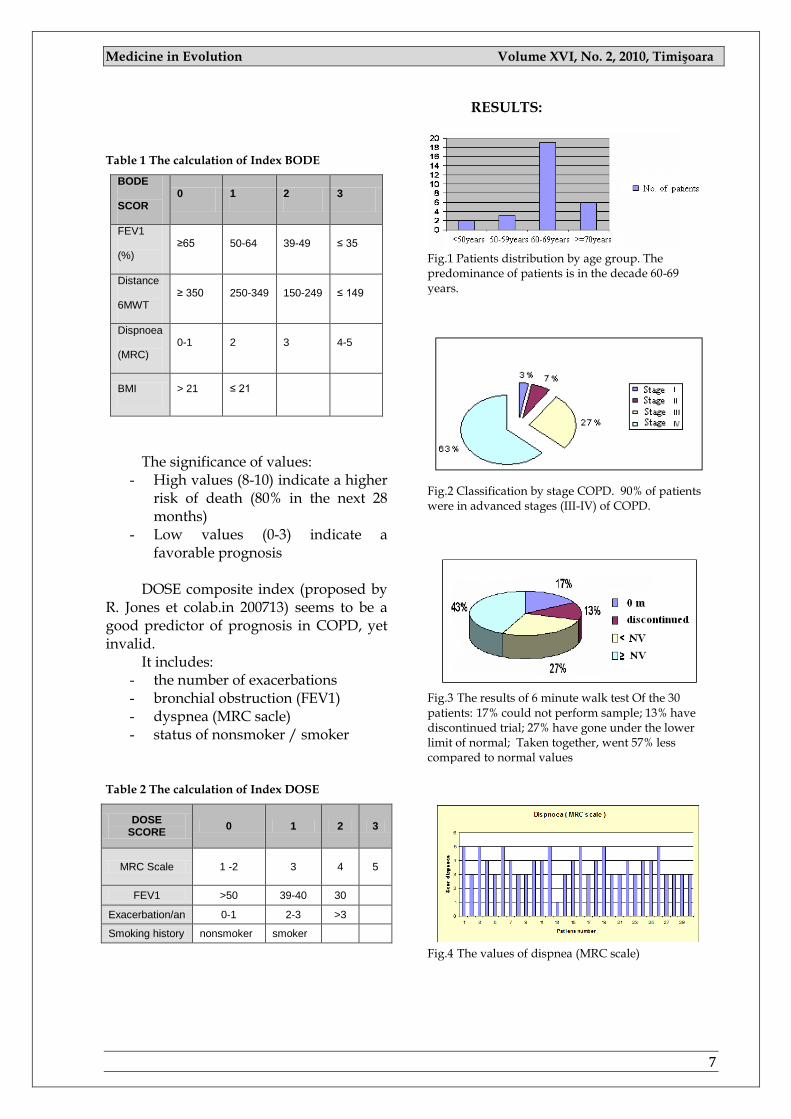
RESULTS:
Fig.1 Patients distribution by age group. The predominance of patients is in the decade 60-69 years.
Fig.2 Classification by stage COPD. 90% of patients were in advanced stages (III-IV) of COPD.
Fig.3 The results of 6 minute walk test Of the 30 patients: 17% could not perform sample; 13% have discontinued trial; 27% have gone under the lower limit of normal; Taken together, went 57% less compared to normal values
Fig.4 The values of dispnea (MRC scale)
Medicine in Evolution Volume XVI, No. 2, 2010, Timişoara
8
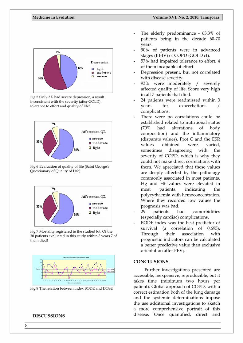
Fig.5 Only 3% had severe depression, a result inconsistent with the severity (after GOLD), tolerance to effort and quality of life!
Fig.6 Evaluation of quality of life (Saint George's Questionary of Quality of Life)
Fig.7 Mortality registered in the studied lot. Of the 30 patients evaluated in this study within 3 years 7 of them died!
The correlation between BODE and DOSE
0
2
4
6
8
10
12
1 2 3 4 5 6 7 8 9 10 11 12 13 14 15 16 17 18 19 20 21 22 23 24 25 26 27 28 29 30
Numbers of patients
Value BODE
DOSE
Fig.8 The relation between index BODE and DOSE DISCUSSIONS
- The elderly predominance - 63.3% of patients being in the decade 60-70 years.
- 90% of patients were in advanced stages (III-IV) of COPD (GOLD cf).
- 57% had impaired tolerance to effort, 4 of them incapable of effort.
- Depression present, but not correlated with disease severity.
- 93% were moderately / severely affected quality of life. Score very high in all 7 patients that died.
- 24 patients were readmissed within 3 years for exacerbations / complications.
- There were no correlations could be established related to nutritional status (70% had alterations of body composition) and the inflammatory (disparate values). Prot C and the ESR values obtained were varied, sometimes disagreeing with the severity of COPD, which is why they could not make direct correlations with them. We apreciated that these values are deeply affected by the pathology commonly associated in most patients. Hg and Ht values were elevated in most patients, indicating the polycythaemia with hemoconcentraion. Where they recorded low values the prognosis was bad.
- 29 patients had comorbidities (especially cardiac) complications.
- BODE index was the best predictor of survival (a correlation of 0,695). Through their association with prognostic indicators can be calculated a better predictive value than exclusive orientation after FEV1.
CONCLUSIONS
Further investigations presented are accessible, inexpensive, reproducible, but it takes time (minimum two hours per patient). Global approach of COPD, with a correct estimation both of the lung damage and the systemic determinations impose the use additional investigations to sketch a more comprehensive portrait of this disease. Once quantified, direct and
Medicine in Evolution Volume XVI, No. 2, 2010, Timişoara
9
indirect physio and morphopathological alterations enlarge the therapeutic area and
improve prognosis in COPD.
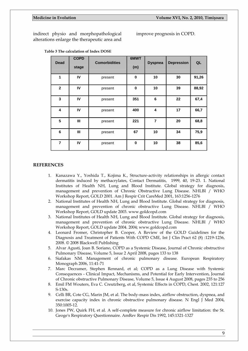
Table 3 The calculation of Index DOSE
Dead COPD
stage Comorbidities
6MWT
(m) Dyspnea Depression QL
1 IV present 0 10 30 91,26
2 IV present 0 10 39 88,92
3 IV present 351 6 22 67,4
4 IV present 400 4 17 66,7
5 III present 221 7 20 68,8
6 III present 67 10 34 75,9
7 IV present 0 10 38 85,6
REFERENCES
1. Kanazawa Y., Yoshida T., Kojima K., Structure-activity relationships in allergic contact
dermatitis induced by methacrylates, Contact Dermatitis, 1999, 40, 19-23. 1. National Institutes of Health NH, Lung and Blood Institute. Global strategy for diagnosis, management and prevention of Chronic Obstructive Lung Disease. NHLBI / WHO Workshop Report, GOLD 2001. Am J Respir Crit CareMed 2001, 163:1256-1276
2. National Institutes of Health NH, Lung and Blood Institute. Global strategy for diagnosis, management and prevention of chronic obstructive Lung Disease. NHLBI / WHO Workshop Report, GOLD update 2003. www.goldcopd.com
3. National Institutes of Health NH, Lung and Blood Institute. Global strategy for diagnosis, management and prevention of chronic obstructive Lung Disease. NHLBI / WHO Workshop Report, GOLD update 2004. 2004; www.goldcopd.com
4. Leonard Fromer, Christopher B. Cooper, A Review of the GOLD Guidelines for the Diagnosis and Treatment of Patients With COPD CME, Int J Clin Pract 62 (8) :1219-1236, 2008. © 2008 Blackwell Publishing
5. Alvar Agusti, Joan B. Soriano, COPD as a Systemic Disease, Journal of Chronic obstructive Pulmonary Disease, Volume 5, Issue 2 April 2008, pages 133 to 138
6. Siafakas NM. Management of chronic pulmonary disease. European Respiratory Monograph 2006, 11:41-71
7. Marc Decramer, Stephen Rennard, et al; COPD as a Lung Disease with Systemic Consequences - Clinical Impact, Mechanisms, and Potential for Early Intervention, Journal of Chronic obstructive Pulmonary Disease, Volume 5, Issue 4 August 2008, pages 235 to 256
8. Emil FM Wouters, Eva C. Creutzberg, et al, Systemic Effects in COPD, Chest. 2002, 121:127 S-130s.
9. Celli BR, Cote CG, Marin JM, et al. The body-mass index, airflow obstruction, dyspnea, and exercise capacity index in chronic obstructive pulmonary disease. N Engl J Med 2004, 350:1005-12.
10. Jones PW, Quirk FH, et al. A self-complete measure for chronic airflow limitation: the St. Geoge's Respiratory Questionnaire. AmRev Respir Dis 1992, 145:1321-1327

Medicine in Evolution Volume XVI, No. 2, 2010, Timişoara
10
11. Mahler DA, Weinberg DH et al. The measurement of dyspnea: contents, interobserver agreement and Physiologie correlates of two new clinical indexes. Chest 1984, 85:751-758
12. Brian Torrey, ATS guidelines for the six-minute walk test - American Thoracic Society Committee on Proficiency Standards for Clinical Pulmonary Function Laboratories. American Family Physician. http://findarticles.com/p/articles/mi_m3225/is_5_66/ai_92233569
13. Jones R, Hyland M, Harding S, et al. The derivation of a new index of severity for COPD patients, the DOSE index: MRC dyspnea scale, airflow obstruction, smoking status and exacerbations. European Respiratory Congress 2007 room K 21, Abstract vol.252s
14. Tschopp JM, Nutrition et insuffisants respiratoires : qualite de vie a domicile – Med et Hzg, 1995,2006, 776 -778.
Medicine in Evolution Volume XVI, No. 2, 2010, Timişoara
11
PAGES 11 -15
UPPER MOLAR DISTALIZATION IN CLASS II MALOCCLUSIONS USING DISTAL JET APPLIANCE – A CASE REPORT.
MOISE MARCEL 1, PODARIU ANGELA CODRUŢA 2, JUMANCA DANIELA 2, ROMÎNU ROXANA 3
1 Institute of Orthognathology and Prosthodontics, Timişoara 2 Department of Preventive, Community Dentistry and Oral Health, Faculty of Dentistry, University of Medicine and Pharmacy "Victor Babeş" Timişoara 3 Department of Dental Materials and Prostheses Technology, Faculty of Dentistry, University of Medicine and Pharmacy "Victor Babeş" Timişoara
Correspondence to:
Dr. Moise Marcel, Intitute of Orthognathology and Prosthodontics, street: Treboniu Laurean No 5, Timişoara, Romania, phone: +40-256 241 639, mobile: +40-724 859 979, E-mail: [email protected]
ABSTRACT
A common strategy for correcting Class II malocclusion without extraction is to distalize the molars. The Distal Jet, described by Carano & Testa (1996), is the most widely used distalizer device in
Orthodontics as it provides good distalization with minimum side effects compared to others (Chiu et al., 2005). The Distal Jet consists of a bilateral piston and tube arrangement, with the tube embedded in an acrylic
Nance button in the palate, supported by attachments on the first or second premolars. A bayonet wire is inserted into the lingual sheath of each first molar band and the free end is inserted into the tubes, much like a piston. A nickeltitanium open-coil spring and an activation collar are placed around each tube. Compressing the coil spring generates a distally directed force. The activation collar is retracted and the mesial setscrew in each collar is locked onto the tube to maintain the force. The active components have to be placed palatally. Ideally, they result in lines of force running close to the center of resistance of the molars. As opposed to the cervical headgear with which molar distalization can be achieved only as a combination of dental crown tipping with subsequent root uprighting, the biomechanics of the appliance should, in theory, allow translatory molar distalization.
Key words: distal jet, class II malocclusion, molar distalization.
INTRODUCTION
The concept of combination class II therapy incorporates mechanics to improve the predictability of traditional Class II treatment while requiring less patient co-operation. This technique combines orthodontic and orthopedic mechanics, performed in a single cohesive phase of fixed appliance therapy Class II
combination therapy begins with maxillary molar distalization using the distal jet followed by fixed functional auxiliaries (in our case a modified Nance button with an inclined plane for mandible advancement).
Class II malocclusions form a heterogeneous group of patients that represents a significant portion of the
Medicine in Evolution Volume XVI, No. 2, 2010, Timişoara
12
patients who typically present for orthodontic treatment. Resolving Class II molar relationships by distalizing maxillary molars may be indicated for patients with maxillary dentoalveolar protrusion or minor skeletal discrepancies (but not for those patients who also exhibit significant dental crowding).5
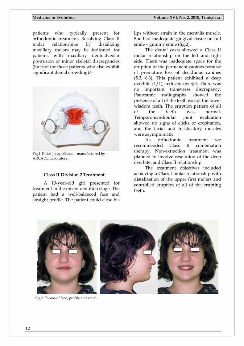
Fig.1 Distal Jet appliance – manufactured by ARCADE Laboratory.
Class II Division 2 Treatment
A 10-year-old girl presented for treatment in the mixed dentition stage. The patient had a well-balanced face and straight profile. The patient could close his
lips without strain in the mentalis muscle. She had inadequate gingival tissue on full smile – gummy smile (fig.2).
The dental casts showed a Class II molar relationship on the left and right side. There was inadequate space for the eruption of the permanent canines because of premature loss of deciduous canines (5.3, 6.3). This patient exhibited a deep overbite (1/1), reduced overjet. There was no important transverse discrepancy. Panoramic radiographs showed the presence of all of the teeth except the lower wisdom teeth. The eruption pattern of all of the teeth was normal. Temporomandibular joint evaluation showed no signs of clicks or crepitation, and the facial and masticatory muscles were asymptomatic.
As orthodontic treatment we recommended Class II combination therapy. Non-extraction treatment was planned to involve resolution of the deep overbite, and Class II relationship.
The treatment objectives included achieving a Class I molar relationship with distalization of the upper first molars and controlled eruption of all of the erupting teeth.
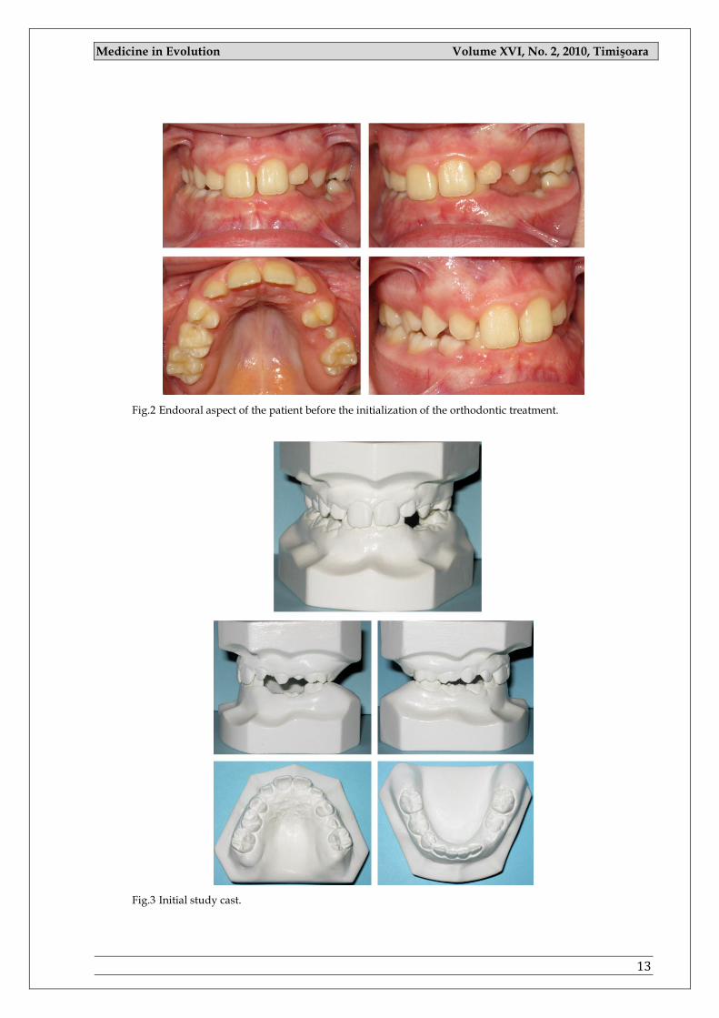
Fig.2 Photos of face, profile and smile.
Medicine in Evolution Volume XVI, No. 2, 2010, Timişoara
13
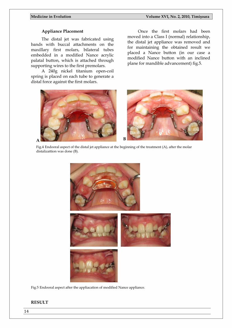
Fig.2 Endooral aspect of the patient before the initialization of the orthodontic treatment.
Fig.3 Initial study cast.
Medicine in Evolution Volume XVI, No. 2, 2010, Timişoara
14
Appliance Placement
The distal jet was fabricated using bands with buccal attachments on the maxillary first molars, bilateral tubes embedded in a modified Nance acrylic palatal button, which is attached through supporting wires to the first premolars.
A 240g nickel titanium open-coil spring is placed on each tube to generate a distal force against the first molars.
Once the first molars had been moved into a Class I (normal) relationship, the distal jet appliance was removed and for maintaining the obtained result we placed a Nance button (in our case a modified Nance button with an inclined plane for mandible advancement) fig.5.
A B
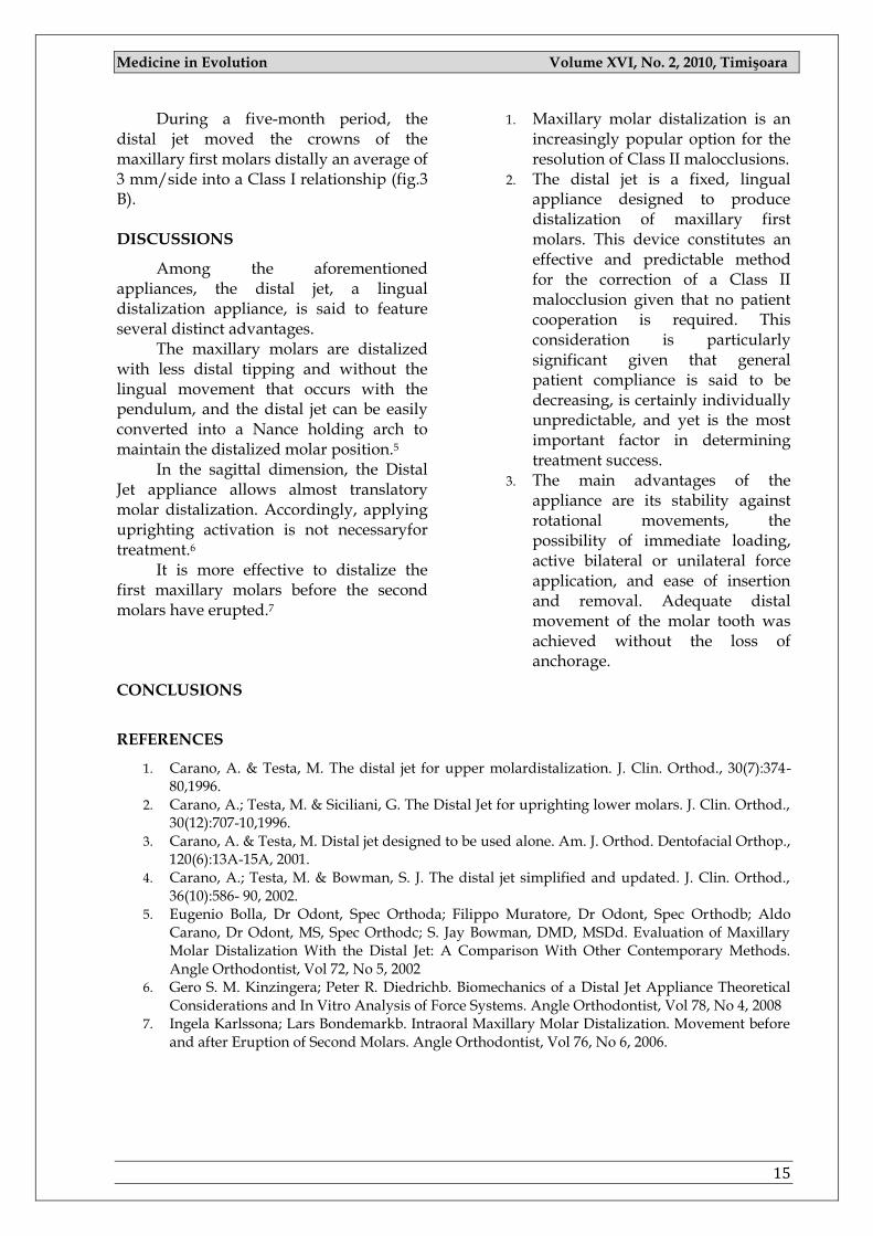
Fig.4 Endooral aspect of the distal jet appliance at the beginning of the treatment (A), after the molar distalizattion was done (B).
Fig.5 Endooral aspect after the appliacation of modified Nance appliance.
RESULT
Medicine in Evolution Volume XVI, No. 2, 2010, Timişoara
15
During a five-month period, the distal jet moved the crowns of the maxillary first molars distally an average of 3 mm/side into a Class I relationship (fig.3 B).
DISCUSSIONS
Among the aforementioned appliances, the distal jet, a lingual distalization appliance, is said to feature several distinct advantages.
The maxillary molars are distalized with less distal tipping and without the lingual movement that occurs with the pendulum, and the distal jet can be easily converted into a Nance holding arch to maintain the distalized molar position.5
In the sagittal dimension, the Distal Jet appliance allows almost translatory molar distalization. Accordingly, applying uprighting activation is not necessaryfor treatment.6
It is more effective to distalize the first maxillary molars before the second molars have erupted.7
CONCLUSIONS
1. Maxillary molar distalization is an increasingly popular option for the resolution of Class II malocclusions.
2. The distal jet is a fixed, lingual appliance designed to produce distalization of maxillary first molars. This device constitutes an effective and predictable method for the correction of a Class II malocclusion given that no patient cooperation is required. This consideration is particularly significant given that general patient compliance is said to be decreasing, is certainly individually unpredictable, and yet is the most important factor in determining treatment success.
3. The main advantages of the appliance are its stability against rotational movements, the possibility of immediate loading, active bilateral or unilateral force application, and ease of insertion and removal. Adequate distal movement of the molar tooth was achieved without the loss of anchorage.
REFERENCES
1. Carano, A. & Testa, M. The distal jet for upper molardistalization. J. Clin. Orthod., 30(7):374-80,1996.
2. Carano, A.; Testa, M. & Siciliani, G. The Distal Jet for uprighting lower molars. J. Clin. Orthod., 30(12):707-10,1996.
3. Carano, A. & Testa, M. Distal jet designed to be used alone. Am. J. Orthod. Dentofacial Orthop., 120(6):13A-15A, 2001.
4. Carano, A.; Testa, M. & Bowman, S. J. The distal jet simplified and updated. J. Clin. Orthod., 36(10):586- 90, 2002.
5. Eugenio Bolla, Dr Odont, Spec Orthoda; Filippo Muratore, Dr Odont, Spec Orthodb; Aldo Carano, Dr Odont, MS, Spec Orthodc; S. Jay Bowman, DMD, MSDd. Evaluation of Maxillary Molar Distalization With the Distal Jet: A Comparison With Other Contemporary Methods. Angle Orthodontist, Vol 72, No 5, 2002
6. Gero S. M. Kinzingera; Peter R. Diedrichb. Biomechanics of a Distal Jet Appliance Theoretical Considerations and In Vitro Analysis of Force Systems. Angle Orthodontist, Vol 78, No 4, 2008
7. Ingela Karlssona; Lars Bondemarkb. Intraoral Maxillary Molar Distalization. Movement before and after Eruption of Second Molars. Angle Orthodontist, Vol 76, No 6, 2006.

Medicine in Evolution Volume XVI, No. 2, 2010, Timişoara
16
Medicine in Evolution Volume XVI, No. 2, 2010, Timişoara
17
PAGES 17 -23
AN INVESTIGATION OF FREQUENCY AND DISTRIBUTION OF FORDYCE’S SPOTS
NATASA TRTIŠ 1, ŽELJKA KOJIŠ 1, DRAGOSLAV DJUKANOVIŠ 1, OLIVERA DOLIŠ 2 , NATAŠA GAJIŠ 3
1 Department of Periodontics and Oral Medicine, Faculty of Medicine, Banja Luka, Republic of Srpska, Bosnia and Herzegovina 2 Department of Preventive and Pediatric Dentistry, Faculty of Medicine, Banja Luka, Republic of Srpska, Bosnia and Herzegovina 3 Department of Endodontics, Faculty of Medicine, Banja Luka, Republic of Srpska, Bosnia and Herzegovina
Correspondence to:
Natasa Trtic, Postal address: Bulevar vojvode Petra Bojovica 1A, Mob phone: 0038765656725, Fax: 0038751348130, E-mail:[email protected]
ABSTRACT
Objective of this research was to reveal the frequency, distribution, location and other characteristics of Fordyce's spots in a group of persons in relation to their sex, age and other characteristics.
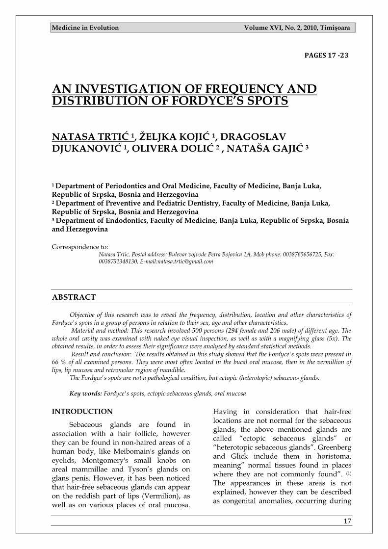
Material and method: This research involved 500 persons (294 female and 206 male) of different age. The whole oral cavity was examined with naked eye visual inspection, as well as with a magnifying glass (5x). The obtained results, in order to assess their significance were analyzed by standard statistical methods.
Result and conclusion: The results obtained in this study showed that the Fordyce's spots were present in 66 % of all examined persons. They were most often located in the bucal oral mucosa, then in the vermillion of lips, lip mucosa and retromolar region of mandible.
The Fordyce's spots are not a pathological condition, but ectopic (heterotopic) sebaceous glands.
Key words: Fordyce's spots, ectopic sebaceous glands, oral mucosa
INTRODUCTION
Sebaceous glands are found in association with a hair follicle, however they can be found in non-haired areas of a human body, like Meibomain's glands on eyelids, Montgomery's small knobs on areal mammillae and Tyson‘s glands on glans penis. However, it has been noticed that hair-free sebaceous glands can appear on the reddish part of lips (Vermilion), as well as on various places of oral mucosa.
Having in consideration that hair-free locations are not normal for the sebaceous glands, the above mentioned glands are called ―ectopic sebaceous glands‖ or ―heterotopic sebaceous glands‖. Greenberg and Glick include them in horistoma, meaning‖ normal tissues found in places where they are not commonly found‖. (1) The appearances in these areas is not explained, however they can be described as congenital anomalies, occurring during
Medicine in Evolution Volume XVI, No. 2, 2010, Timişoara
18
development, or anatomical variations. Some authors, for example: Sewerin indicate the protective, i.e. lubricative function of these glands. [2]
The first description of these glands found in oral cavity was given by J.A. Fordyce's in 1896. At the time, their real nature was unknown; hence the appearance was associated with some systemic diseases, such as lues and named ―Fordyce's disease‖. Later researches have determined their real nature and the name were changed into ―Fordyce‘s granules‖, ―Fordyce's grains‖ or ―Fordyce's spots‖.
The reason to perform this research is that there are very different, even contradictory data on the frequency of appearance, localization, size and other characteristics of these glands in oral mucosa, as well as the fact that the presence of these glands can often be an obstacle in a diagnosis procedure. Therefore, it is necessary to point out the importance of the differential diagnosis of this condition in an oral cavity.
It was observed that if Fordyce‘s spot were not accurately diagnosed, the patients were treated with unnecessary drugs, even recommended to surgical procedures. Additional reason can be found in a fact
that once observed by patient, these changes could cause a cancerophobia. There are a very few papers and data on this problem in contemporary literature.
PURPOSE
The goal of the research is to determine the frequency of Fordyce‘s spot appearance using the sample of larger number of persons, in relation with age and sex. Additional goal is to determine different characteristics of these granules, such as: localization (whether they appear, only on one side of the oral cavity or on both sides), number, clustering, size, shape, color.
MATERIAL AND METHOD
In total, 500 persons were included in the research, of both sexes and various age. (Graph no. 1.). Persons were chosen by random choice method, among patients‘ dental office patients, school children and students. Ethical approval for the study was obtained from The Research Ethics Committee of Faculty of Medicine, University of Banja Luca, Republic of Srpska, Bosnia and Herzegovina.
Graph no. 1. Age and number of examined persons.
The presence of Fordyce's spot was
established in the following way: Patients were told to wash mouth
with water thoroughly, upon which the naked eye examination of the oral cavity
was performed, followed by examination by maginfying glass with 5x magnification. In some cases it was needed to dry the mucus surface with the jet of air or gauze
Medicine in Evolution Volume XVI, No. 2, 2010, Timişoara
19
tampons. The results are recorded in special cards.
RESULTS AND DISCUSSIONS
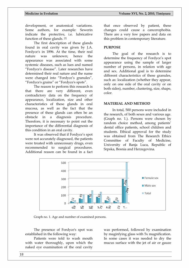
In the research Fordyce‘s spots were found in 330 persons, out of 500 examined persons (294 females and 206 males), giving 66% of persons with Fordyce‘s granule.
χ ² test did not result in statistically significant difference (p<0, 05) in distribution of Fordyce‘s spots in relation to age and sex.
Before presenting data given by other authors, it is necessary to point out that there are very few papers on this appearance in the literature, especially in the contemporary literature; hence the results of older researches will be shown as well. The fact that the data are observed from the older studies is irrelevant as these were epidemic studies valuable for the comparison. Margolies and Weidman found Fordyce‘s spots in 70% of 248 examined persons.(3) Mc Goodwin found 54% persons with Fordyce‘s granule in group of 96 examined students.(4) Sponge recorded the presence of Fordyce‘s grains in 80% of examined adults.(5) Alawi and Siddiqui found Fordyce's granule localized
on oral mucosa in 80% of examined adults. [6] Gorsky and associates found Fordyce's granule in 94,9% in a group of 2 462 examined Israeli Jews. [7] Halperin and associates examined 2478 persons and found Fordyce's granule in 82 to 88%. [8] Reichart and associates report that Fordyce's spots are found in 80 to 90% of adults, and not found in children, the ones examined in men are larger, spread on wider areas and clustered. [9] Sokic and Djukanovic found them in 61% of examined persons, more frequent in men (68%), then in women (57%), and interestingly, they found Fordyce's granules in the group of children up to 9 years old, 26% of examined.(10) Flinck and associates found them in 1% of newborns. [11] Laskaris, Cawson and Odell report the appearance of Fordyce‘s spots in approximately 80% of persons of both sexes, stating that with age they become more rendered. [12, 13] Greenberg and Glick also report they are more frequently found in men, especially after puberty, increasing in size with age. [1]
Our research has revealed the similar, or slightly less percentage of persons with Fordyce‘s spots, as observed by other authors. (Graph no. 2.).
Graph no.2. Percentage of persons with Fordyce's granules, in reference to age and sex.
Medicine in Evolution Volume XVI, No. 2, 2010, Timişoara
20
Characteristics of Fordyce's granules established by clinical examination:
Subjective symptoms: Fordyce's granules are not followed by any subjective difficulties. They are usually detected by chance, or discovered by dentists during routine checkups. Sometimes the presents of these glands causes anxiety in patients, and in some cases develops a cancerophobia. In rare cases, patients could feel stinging or similar difficulties in the area affected by Fordyce's granule. However, those difficulties are not caused by these glands, usually there are other causes (e.g. mechanically induced erosions of mucosa, or pyrosis of cavity mucosa). Size of granule: In the largest number of observed cases, the cell diameter was about 1 mm (in 66%). In 28% it was less than 1 mm, and in 6% bigger than 1 mm, but never over 2 mm. Laskaris reports that most frequently, their size is up to 0,5mm, while, in contrary, Cowson and Odell emphasize that their size is ―up to a few millimeters, especially in olders‖. [12, 13]. This finding is opposite to the opinions of all other authors, including the opinion of
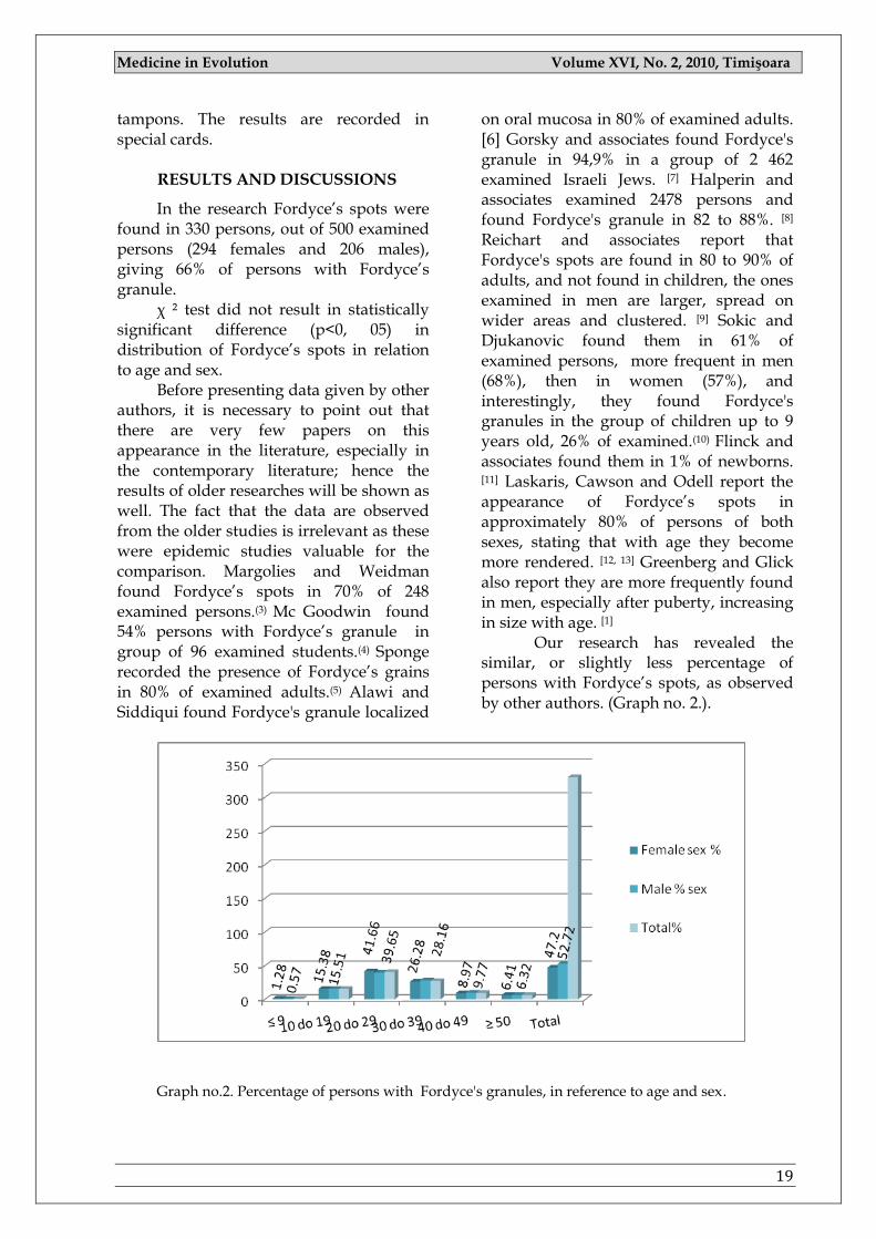
the authors of this study. Lamey and Lewis point out that the number and size of spots increase with age. [14]. Structure of granule: Each granule observed separately, with naked eye, appeared compact. Similar structure was noticed by observing with magnifying glass, however, in some cases it was found that granule was actually made of more parts. This was confirmed by histopathology research. Biopsy of Fordyce‘s spots, under microscope reveals sebaceous glands made of two or three lobules. Color of granule: The color is usually yellowish, however, sometimes is more yellow, and sometimes more whitish. Our finding indicates that the color depends upon the presence of pigmentation (melanogen), thickness of epithelium, which covers them and localization. Many authors report that the color is ―beige-yellowish‖, Jankovic. [15]. Number of granules in one region: The number of grains is very variable, and there are always more of them. Thus, we never found only one isolated grain (Graph no.3).
Graph No. 3. Number of grains in one patient in one observed region and percentage of persons
with that number of granules.
Aspect of granule to the level of
surrounding tissue: Fordyce's granules are most often somewhat above the level of surrounding mucosa or epithelium of the reddish part of lips.
The palpation detects small grains. Localization of granule: Based on our
findings, Fordyce's grains are most
frequently found in buccal oral mucosa, reddish part of lips, lip mucosa and in retro molar region of mandible. Hereby, it should be pointed out that in one person they are often found in more areas. When they are located in the buccal oral mucosa, most frequently are on the level of occlusal line in distally regions, in the area of molars. It is important to emphasize that
Medicine in Evolution Volume XVI, No. 2, 2010, Timişoara
21
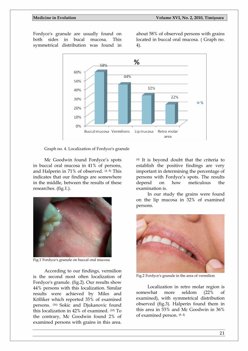
Fordyce's granule are usually found on both sides in bucal mucosa. This symmetrical distribution was found in
about 58% of observed persons with grains located in buccal oral mucosa. ( Graph no. 4).
Graph no. 4. Localization of Fordyce's granule
Mc Goodwin found Fordyce‘s spots
in buccal oral mucosa in 41% of persons, and Halperin in 71% of observed. (4, 8) This indicates that our findings are somewhere in the middle, between the results of these researches. (fig.1.).
Fig.1 Fordyce's granule on buccal oral mucosa
According to our findings, vermilion
is the second most often localization of Fordyce's granule. (fig.2). Our results show 44% persons with this localization. Similar results were achieved by Miles and Kölliker which reported 35% of examined persons. (16) Sokic and Djukanovic found this localization in 42% of examined. (10) To the contrary, Mc Goodwin found 2% of examined persons with grains in this area.
(4) It is beyond doubt that the criteria to establish the positive findings are very important in determining the percentage of persons with Fordyce‘s spots. The results depend on how meticulous the examination is.
In our study the grains were found on the lip mucosa in 32% of examined persons.
Fig.2 Fordyce's granule in the area of vermilion
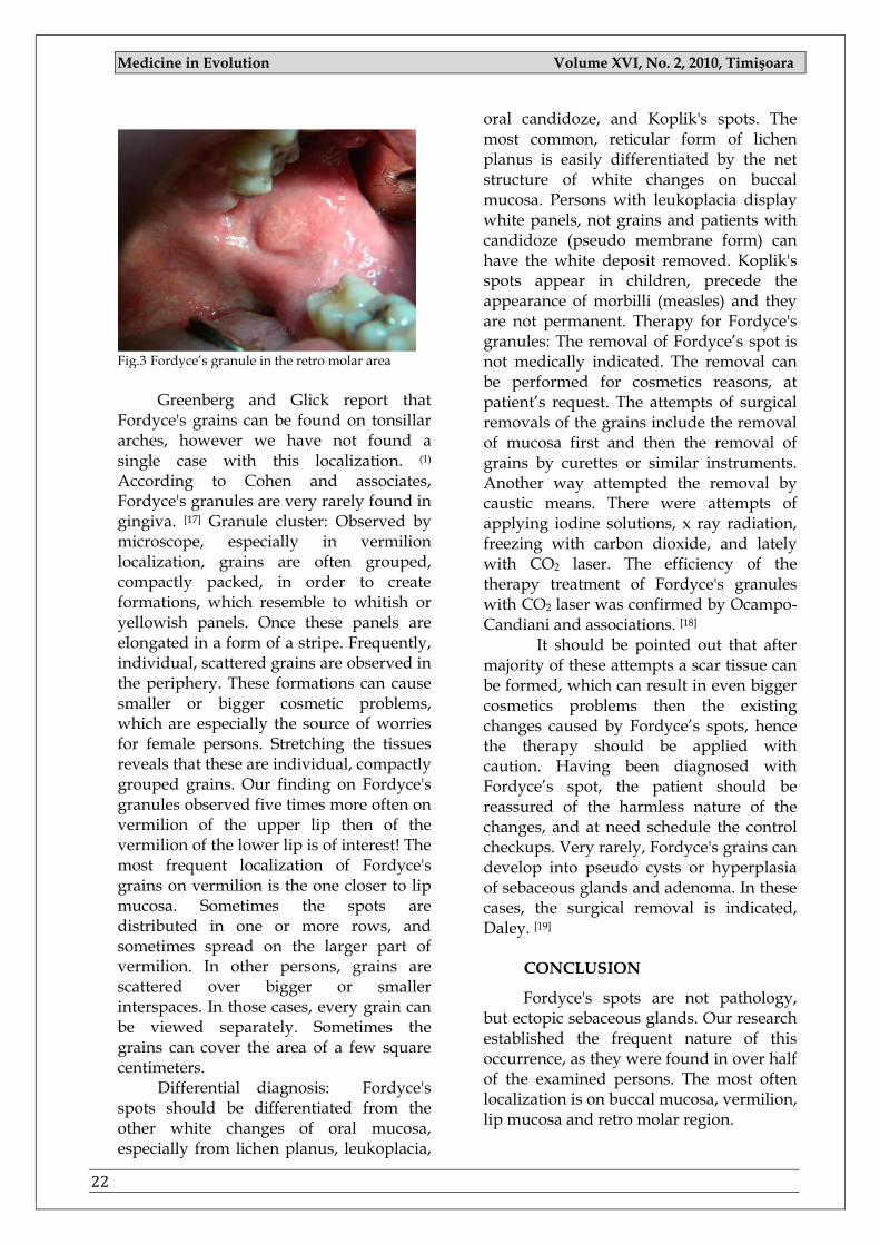
Localization in retro molar region is
somewhat more seldom (22% of examined), with symmetrical distribution observed (fig.3). Halperin found them in this area in 53% and Mc Goodwin in 36% of examined person. (8, 4)
Medicine in Evolution Volume XVI, No. 2, 2010, Timişoara
22
Fig.3 Fordyce‘s granule in the retro molar area
Greenberg and Glick report that
Fordyce's grains can be found on tonsillar arches, however we have not found a single case with this localization. (1)
According to Cohen and associates, Fordyce's granules are very rarely found in gingiva. [17] Granule cluster: Observed by microscope, especially in vermilion localization, grains are often grouped, compactly packed, in order to create formations, which resemble to whitish or yellowish panels. Once these panels are elongated in a form of a stripe. Frequently, individual, scattered grains are observed in the periphery. These formations can cause smaller or bigger cosmetic problems, which are especially the source of worries for female persons. Stretching the tissues reveals that these are individual, compactly grouped grains. Our finding on Fordyce's granules observed five times more often on vermilion of the upper lip then of the vermilion of the lower lip is of interest! The most frequent localization of Fordyce's grains on vermilion is the one closer to lip mucosa. Sometimes the spots are distributed in one or more rows, and sometimes spread on the larger part of vermilion. In other persons, grains are scattered over bigger or smaller interspaces. In those cases, every grain can be viewed separately. Sometimes the grains can cover the area of a few square centimeters.
Differential diagnosis: Fordyce's spots should be differentiated from the other white changes of oral mucosa, especially from lichen planus, leukoplacia,
oral candidoze, and Koplik's spots. The most common, reticular form of lichen planus is easily differentiated by the net structure of white changes on buccal mucosa. Persons with leukoplacia display white panels, not grains and patients with candidoze (pseudo membrane form) can have the white deposit removed. Koplik's spots appear in children, precede the appearance of morbilli (measles) and they are not permanent. Therapy for Fordyce's granules: The removal of Fordyce‘s spot is not medically indicated. The removal can be performed for cosmetics reasons, at patient‘s request. The attempts of surgical removals of the grains include the removal of mucosa first and then the removal of grains by curettes or similar instruments. Another way attempted the removal by caustic means. There were attempts of applying iodine solutions, x ray radiation, freezing with carbon dioxide, and lately with CO2 laser. The efficiency of the therapy treatment of Fordyce's granules with CO2 laser was confirmed by Ocampo-Candiani and associations. [18]
It should be pointed out that after majority of these attempts a scar tissue can be formed, which can result in even bigger cosmetics problems then the existing changes caused by Fordyce‘s spots, hence the therapy should be applied with caution. Having been diagnosed with Fordyce‘s spot, the patient should be reassured of the harmless nature of the changes, and at need schedule the control checkups. Very rarely, Fordyce's grains can develop into pseudo cysts or hyperplasia of sebaceous glands and adenoma. In these cases, the surgical removal is indicated, Daley. [19]
CONCLUSION
Fordyce's spots are not pathology, but ectopic sebaceous glands. Our research established the frequent nature of this occurrence, as they were found in over half of the examined persons. The most often localization is on buccal mucosa, vermilion, lip mucosa and retro molar region.

Medicine in Evolution Volume XVI, No. 2, 2010, Timişoara
23
REFERENCES
1. Greenberg M.S, Glick M. Burket's Oral medicine, 10th ed., BC Decker Inc, Hamilton, 2003; pp 114/115
2. Sewerin I. The sebaceous glands in the vermilion border of the lips and in the oral mucosa of man, Acta Odontol. Scand. 33, Suppl 1975; 33:1
3. Margolies A, Weidman F. Statistical and histologic studies of Fordyce‘s disease. Arch derm syphilol.1921;3(6):723-742
4. Mc Goodwin R.C, Fordyce‘s granules in pigmented oral mucosae. Journal of dental research.1964;43:773
5. Sponge J.D. Sebaceous metaplasia in the oral cavity occurring in association with dentigerous cyst epithelium. Oral surgery, M&Path. 1966;21
6. Alawi F. Siddiqui A.Sebaceous carcinoma of oral mucosa: Case report and review of the literature. Oral surg. Oral med. Oral pathol. Oral radio. and endodontics; 2005; 99:79-84
7. Gorsky M, Buchner A, Fundoianu-Dayan D, Cohen C. Fordyce's granules in the oral mucosa of adult Israeli Jews.Comunity dentistry and oral epidemiology; 1986; 14(4):231-232
8. Halperin V, Kolas S, Jeffers K.R. The occurrence of Fordyce spots, benign migratory glossitis, median rhomboid glossitis and fissured tongue in 2.478 dental patiens. Oral Surg, Oral Med, Oral Path; 1953; 6:1072
9. Reichart P.A, Philipsen H.P.: Oral Patology, Theime, Stuttgart-New York, 2000; pp 64 10. Sokić G, Đukanović D. Učestalost ''Fordajsovog oboljenja'', Zbornik radova 13. Stomatološke
nedelje SRS, Soko Banja, 1978; pp 13/17 11. Flinck A, Paludan A, Matsson L, Holm A.K, Axelsson I.Oral findings in a group of newborn
Swedish children. International journal of pediatric dentistry/the British Pedodontic Society and the International Associationof Dentistry for Children;1994; 4(2):67-73
12. Laskaris G. Color atlas of oral diseases, Thieme, Stuttgart- New York, 1988; pp 4/5 13. Cawson R.A, Odell E.W. Essentials of Oral Pathology and Oral Medicine, Curchill Livingstone,
6th ed., Edinburgh, 1998; 4:209 14. Lamey P.J, Lewis M.A.O. A clinical guide to oral medicine, 2nd ed. British Dental Association,
London, 1990, pp 60/61 15. Janković LJ. Oralna medicina, 2.izd. Zavod za udžbenike i nastavna sredstva, Beograd 2004, pp
158 and 190. 16. Miles A.E.W. Sebaceous glands in the lip and cheek mucosa in man. Brit. Dent. J.1958; 105(7) 17. Cohen R.E, Aguirre A, Drinnan A.J, Neiders M.E, Mastroianni A.J Ectopic gingival sebaceous
glands presenting as localized periodontitis. Journal of periodontology; 1990;61(1):58-60 18. Ocampo- Candiani J, Villarreal-Rodrigues A, Quinones-Fernandez AG, Herz-Ruelas M.E, Ruiz-
Esperza J. Treatment of Fordyce's spots with CO2 laser. Dermatologic Surgery; 2003; 29:869-871
19. Daley T.D. Intraoral Sebaceous hyperplasia. Diagnostic criteria. Oral surg, Oral Med, Oral Pathol. 1993; 75 (3): 343/7

Medicine in Evolution Volume XVI, No. 2, 2010, Timişoara
24
Medicine in Evolution Volume XVI, No. 2, 2010, Timişoara
25
PAGES 25 -30
METHODS TO ASSES ANXIETY TOWARDS DENTAL TREATMENT: PHARMACHOLOGICAL AND PSYCHOLOGICAL TECHNIQUES. ORAL HEALTH INVOLVEMENT.
IOANA FILIMON1, GĂLUŞCAN ATENA2, IFTIME CARMEN3, OVIDIU FODOR3
1 Phd student, University of Oradea 2 Department of Preventive, Community Dentistry and Oral Health, Faculty of Dentistry University of Medicine and Pharmacy “Victor Babeş” Timişoara 3 University of Oradea, Faculty of Medicine and Pharmacy,
Correspondence to: Atena Galuscan, Timisoara, srt. T. Vladimirescu nr.14a, [email protected]
ABSTRACT
Background: Despite late discoveries related to pain control and communication paths, fear and anxiety towards dental treatment are still important factors in dental treatment. Fear of dentists is common and a potentially distressing problem, both for the public and for the dental practitioners. Avoiding the dentist because of fear can have serious ramifications in terms of dental health and overall well-being.
Aim and objectives: The purpose of this study is to investigate the valences of the assessment methods regarding anxiety and fear towards dental treatment.
Methods: A total of 28 patients with high anxiety scores from a private practice were included. They were divided in three groups: the patients from the first group were given premedication before the dental procedure, the second group was administered premedication and a brief introduction about the procedure and the third group was administered premedication and extensive explanations about the procedure as well as psychological intervention .The STAI X1 and STAI X2, DFS and DAS scales were used to evaluate the level of anxiety pre and post intervention.
Results: The levels of general and dental anxiety were considerably lower in group 3 when premedication combined with extensive explanations and psychological intervention was given before the dental treatment. The results indicated that patients with high anxiety level tend to present high trait anxiety, but high trait anxiety seems not to predispose to high dental anxiety.
Conclusions and clinical implications: The incidence of dental fear may be less if clinicians use both psychological and medical methods to control anxiety in dental treatment.
Key words: anxiety towards dental treatment, pain control, oral sedation
INTRODUCTION
Modern dentistry is ultraconservatory and based on early diagnosis and prophylactic treatment.The
succes of each dental treatment resides in good oral hygiene and periodical visits to the specialist. Fear of the dentist has a bad influence on the treatment outcome and can determine the pacient to be late for the
Medicine in Evolution Volume XVI, No. 2, 2010, Timişoara
26
appointment or even to give up the treatment even if it is necessary. Researches established that high levels of anxiety and fear are correlated with a lot of factors like: long periods between two doctor‘s appointments, bad estethics or disfunctions of the oral cavity, high frequency of symptoms (1, 3, 5, 11, 14).
Anxiety towards dental treatment is seen in all social and age groups. Research data reveal that the percent of anxiety towards dental treatment in the general population lies between 38-46%. The fact that more than one third of the general population is affected by this type of anxiety is also reflected in high individual and social costs determined by the avoiding of treatment or solicitating it only in emergency cases when restorations are very expensive.12
Literature in this field shows that a great percent of patients who suffer from anxiety towards dental treatment relate it to a traumatic apisode during childhood (10,
11). Negative experience doesn‘t always reside in physical trauma but can appear as a result of the lack of interpersonal communication between the doctor and the patient. Often patients with high levels of anxiety complain about the doctor‘s behaviour as being impersonal, cold and not caring. Similar, a great percent of patients underline the importance of the doctors‘ personal qualities when they undergo dental treatment. The way patients perceive the practitioner‘s behaviour is highly correlated with the patient‘s satisfaction.7
Reciprocal trust represents a fundamental aspect of each interpersonal relationship especially when it comes to the doctor-patient relationship and the patient is abandoning himself to the practitioner‘s hands without knowing what the procedure will be like.7 Lack of trust provocs uncertainty feelings and increases the stress levels.This trust is build over many treatment sessions when patient and practitioner can observe eachother. Interpersonal comunication skils, non-verbal and verbal messages play a key role: for example, one patient who is afraid of
losing control over the situation has to convince himself that he can control the event by sending a message that will stop the procedure, and another patient who is more sensitive has to feel that the doctor understands his sensitivity and accepts it, treating him with more attention.8
Friedman et al 7 describes the doctor-patient relationship as a pyramid. The base is represented by the initial comunication process when the practitioner colects information about the patient and the patient describes his previous experiences, expectations and needs regarding the treatment. Gradually, as the examination and medical proceedings are succeding, reciprocal trust is build and other complex procedures can be implemented.
MATERIAL AND METHODS:
The general objective of the paper was to describe a pilot study that identifies the positive valences and the limitations of assesment methods of anxiety towards dental treatment and that finds new ways of lowering these disfunctional reactions. This will lead to the increasing eficacity of dental treatment and the elaboration of new preventive strategies centered on the empathic doctor-patient relationship.
SPECIFIC AIMS:
1. Identifying the differences between general anxiety and anxiety/fear towards dental treatment when solicitating it (preintervention).
2. Identifying different levels of general anxiety and anxiety/fear towards treatment in patient who benefit from treatment procedures (postintervention).
Research is organized as a
comparative study between patients with different degrees of general anxiety and/or anxiety toward denal proceeduresin different stages of the therapeutic interaction with the dentist.
Research has been conducted on 28 patients at the privat dental practice, aged between 18-59 years, with a mean age of
Medicine in Evolution Volume XVI, No. 2, 2010, Timişoara
27
32.46. The sample is of total type, meaning that all adult patients who came to the dental practice have been selected for the study during one month and have been then treated for different conditions of the oral cavity.
After the anamnesis which identifies the presence of dental trauma, the subjects have been asked to fill in the scales from the brochures. They were assured that all data is confidential.
The research instruments contained in the brochure were the State-Trait Anxiety Inventory (Stai X1 Şi Stai X2), Dental Fear Evaluation Inventory (DFS), and Dental Anxiety Scale (DAS). After the anamnesis, patients with high levels of general anxiety but also high levels of anxiety towards dental treatment have been included in the study.
They were divided into three groups:
(1) patients without premedication; (2) patients with premedication
associated with a brief explanation about its effects;
(3) patients with premedication associated with explanations, question answering and psichological counciling.
The distribution of patients was
stochastic using the simple lottery method. For premedication a tranquilizer from the benzodiasepin family has been used: diazepam 10 mg administrated half an hour before the intervention.
Research instruments
1. Anamnesis: the anamnesis details the medical history of general and dental conditions, obtaines the informal accept and tries to identify possible early trauma related to dental treatment.
2. STATE-TRAIT ANXIETY INVENTORY, (STAI X1 şi STAI X2): STAI is an instrument that comprises two autoevaluatio scales to measure two distinct concept of anxiety, the anxiety state (A-state) and the anxiety trait (A-trait). The A-trait scale comprises 20 enunciations to which
people express the way they feel in general. Anxiety as a trait refers to individual, relatively stabile differences towards anxiety, regarding the tendency to respond to situations perceved as frightening with growing levels of anxiety. The A-state scale also comprises 20 enunciations but the subject is instructed to indicate the way they feel at a given moment in time. The anxiety state represents an emotional, tranzitional state or a condition of the human body characterized by self-conscious feelings of tension and fear and an increased activity of the vegetative nervous system.the A-state can vary as intensity and fluctuate over time.
3. Dental Anxiety Scale. (DAS).2 it‘s goal is to evaluate anxiety towards dental treatment.This scale contains a questionaire that solicitates the patients to exactly evaluate the way they feel about the dental treatment. The scale compises 4 items that face the subjects to a graded series of situations asociated to dental treatment. The subject can choose from five answers the one that best describes his feelings towards the given situation.
4. Dental Fear Survey (DFS) 4 measures fear towards the dentist. The scale contains 20 items couched to identify specific and unique answers to a variety of stimulations correlated to dental activity and a global stimulation of fear towards the dentist. The scale is based on on the learning theory and is more relevant than other scales what concerns comprehension and treating anxiety and fear towards the dentist. The scale comprises three subscales that identify three factors: the tendency to avoid dental treatment, the physiologic reaction during actual treatment and the fear during actual treatment.
RESULTS
We assumed that the level of general anxiety and of anxiety/fear towards dental
Medicine in Evolution Volume XVI, No. 2, 2010, Timişoara
28
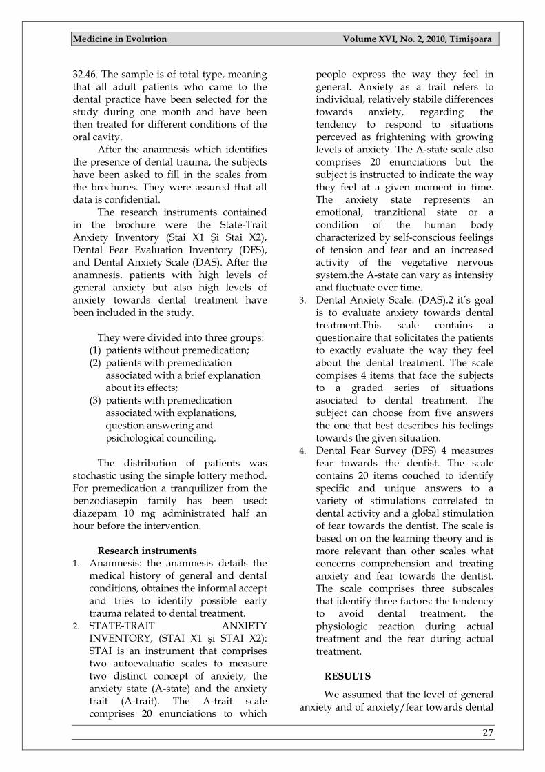
treatment present differences considering past dental trauma, whereas patients with significant trauma have higher levels of general anxiety and anxiety/fear towards dental treatment. The study‘s design is cvasiexperimental and the classifying variable is the type of the analised person: with sgnificant dental trauma, with insignificant dantal trauma, without dental trauma. Subjects who report significant dental trauma – N=12, representing 42, 85% of the sample – record a high level of
anxiety-state (m=55, 09). They have high scores of STAI II which demonstrating a stability in the anxiety trait (m=59, 23). One of the patients with past significant trauma has a high level of the anxiety state (m=54) and a medium level of the anxiety trait (m=39). Subjects who report significant dental trauma record high scores of dental anxiety (m=14, 29) and dental fear (m=4, 33). Medium values of anxiety state, anxiety trait, dental anxiety and dental fear are represented in chart no.1.
Fig.1 Medium values of anxiety state, anxiety trait, dental anxiety and dental fear considering past trauma
We have cosidered that the level of
general anxiety and anxiety/fear towards dental treatment in patients with high preintervention levels varies postintervention based on premedication.
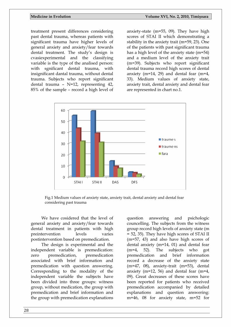
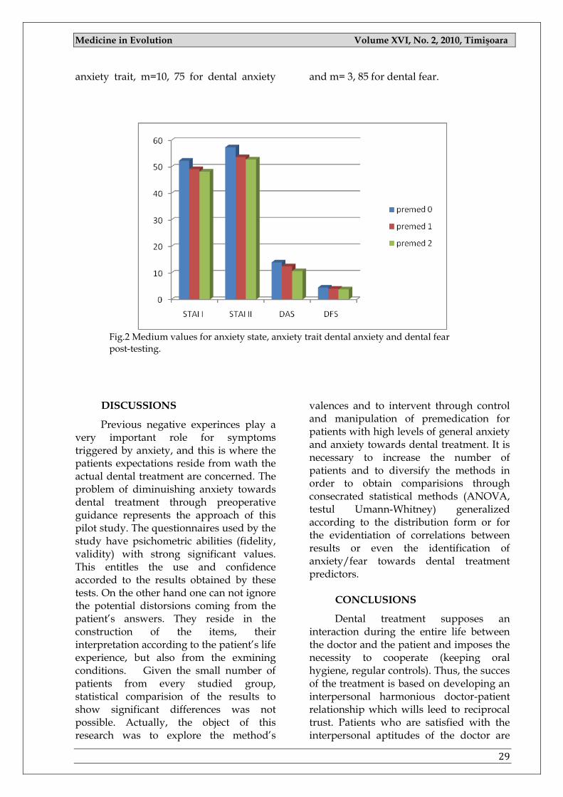
The design is experimental and the independent variable is premedication: zero premedication, premedication associated with brief information and premedication with question answering. Corresponding to the modality of the independent variable the subjects have been divided into three groups: witness group, without medication, the group with premedication and brief information and the group with premedication explanations
question answering and psichologic councelling. The subjects from the witness group record high levels of anxiety state (m = 52, 35). They have high scores of STAI II (m=57, 43) and also have high scores of dental anxiety (m=14, 01) and dental fear (m=4, 52). The subjects who got premedication and brief information record a decrease of the anxiety state (m=47, 08), anxiety-trait (m=53), dental anxiety (m=12, 56) and dental fear (m=4, 09). Great decreases of these scores have been reported for patients who received premedication accompanied by detailed explanations and question answering: m=46, 08 for anxiety state, m=52 for
Medicine in Evolution Volume XVI, No. 2, 2010, Timişoara
29
anxiety trait, m=10, 75 for dental anxiety and m= 3, 85 for dental fear.
Fig.2 Medium values for anxiety state, anxiety trait dental anxiety and dental fear post-testing.
DISCUSSIONS
Previous negative experinces play a very important role for symptoms triggered by anxiety, and this is where the patients expectations reside from wath the actual dental treatment are concerned. The problem of diminuishing anxiety towards dental treatment through preoperative guidance represents the approach of this pilot study. The questionnaires used by the study have psichometric abilities (fidelity, validity) with strong significant values. This entitles the use and confidence accorded to the results obtained by these tests. On the other hand one can not ignore the potential distorsions coming from the patient‘s answers. They reside in the construction of the items, their interpretation according to the patient‘s life experience, but also from the exmining conditions. Given the small number of patients from every studied group, statistical comparision of the results to show significant differences was not possible. Actually, the object of this research was to explore the method‘s
valences and to intervent through control and manipulation of premedication for patients with high levels of general anxiety and anxiety towards dental treatment. It is necessary to increase the number of patients and to diversify the methods in order to obtain comparisions through consecrated statistical methods (ANOVA, testul Umann-Whitney) generalized according to the distribution form or for the evidentiation of correlations between results or even the identification of anxiety/fear towards dental treatment predictors.
CONCLUSIONS
Dental treatment supposes an interaction during the entire life between the doctor and the patient and imposes the necessity to cooperate (keeping oral hygiene, regular controls). Thus, the succes of the treatment is based on developing an interpersonal harmonious doctor-patient relationship which wills leed to reciprocal trust. Patients who are satisfied with the interpersonal aptitudes of the doctor are
Medicine in Evolution Volume XVI, No. 2, 2010, Timişoara
30
more self-conscious and respect the treatment indications, don‘t give up appointments, whereas patients who have a negative image of the medical profession
come seldom to the practice, mostly when pain occurs and don‘t follow preventive treatment.
REFERENCES:
1. Berggren U, Meynert G. Dental fear and avoidance: Causes, symptoms, consequences. J.AmDentAssoc. 1984; 109: 247-251.
2. Corah, N.L.,Gale, E.N. and Illig. S.J. Assesment of a Dental Anxietz Scale, JAmDentAsooc. 1969; 97:816-818
3. Dionne R., Gordon S., McCullagh L., Phero J. Assessing the need for Anaesthesia and Sedation in the General Population. JAmDentAssoc. 1998; 129:167-173.
4. Fischer. J., Corcoran. K. Measures for Clinical Practice and reasearch Oxford University Press, New York, 2007
5. Hagglin C., Berggren U., Hakeberg M., Ahlqwist M. Dental anxiety among middel-aged and elderly women in Sweden. A study of oral state, utilization of dental services and concomitant factors. Gerodontology 1996; 13:25-34.
6. Holtzman J, Berg R, Mann J, Berkey D. The relationship of age and gender to fear and anxiety response to dental care. Spec Care Dentist 1997; 17: 82-87.
7. Ilana Eli. Oral Psychophysiology - Stress, Pain and Behavior in Dental Care CRC Press Inc., Boca Raton, Fl., USA, 1992
8. Law A, Logan H, Baron RS. Desire for control, felt control and stress inoculation training during dental treatment. J Pers Soc Psychol 1994; 10:926-36.
9. Liddell A, Locker D. Gender and age differences in attitudes to dental pain and dental control. Community Dent Oral Epridemiol 1997; 25: 314-8.
10. Locker D, Liddell A. Correlates of dental anxiety among older adults. J Dent Res 1991; 70:198-203.
11. Milgrom P, Fiset L., Melnick S., Weinstein P. The prevalence and practice management cosequences of dental fear in a major US city. J.A.DentAssoc. 1998; 116:641-647.
12. Rotaru A. Implicaţii multidisciplinare în durerea orală şi craniofacială. Cluj Napoca: Clusium 2001
13. Spielberger CD, Gorsuch RL, Lushene RE. STAI, manual for the State-Trait-Anxiety-Inventory. Palo Alto: Consulting Psychologist Press, 1970
14. Ter Horst G., de Witt CA. Review of behavioral research in dentistry 1987-1992:dental anxiety, dentist-patient relationship, compliance and dental attendance. IntDentJ 1993; 43:265-78.
Medicine in Evolution Volume XVI, No. 2, 2010, Timişoara
31
PAGES 31 -36
PREVALENCE OF HYPODONTIA IN ORTHODONTIC PATIENTS.
DINU ŞTEFANIA 1, BRATU ELISABETA 1
1 Department of Pedodontics-Orthodontics, Faculty of Dentistry University of Medicine and Pharmacy “Victor Babeş”Timişoara Correspondence to:
Dinu Stefania, Timisoara, str.P/ta Eftimie Murgu, nr 2 [email protected]
ABSTRACT
Aim and objectives. The aim of this study was to investigate the prevalence of hypodontia in permanent dentition, among patients who were treated in our Department of Pedodontics-Orthodontics of University of Medicine and Pharmacy in Timisoara.
Material and methods. In order to analyze the prevalence of hypodontia, we examined orthodontic files of 1350 patients, which included orthopantomograms, study models and anamnestic data. Patients with cleft lip/palate or having tooth loss due to dental caries, periodontal disease, traumas, and congenitally missing third molars were excluded from this study.
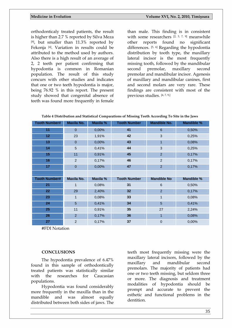
Results. The prevalence of hypodontia was 6.47% excluding third molars.The most frequently missing teeth were the maxillary lateral incisors, followed by the mandibular and maxillary second premolars.
Conclusions. Prevalence of hypodontia found in this sample of orthodontically treated patients was within the wide range reported in the literature for a normal population.
Key words: Hypodontia, congenitally missing teeth, prevalence
INTRODUCTION
Missing teeth (tooth agenesis) is one of the most common developmental problems in children. [4] Congenital lack of a tooth results from disturbances during early stages of tooth development. A tooth is defined to be congenitally missing if it has not erupted in the oral cavity and is not visible on a radiograph. [1] Hypodontia is the term most frequently used when describing the phenomenon of congenitally missing teeth (CMT) in general. Many other terms appear in the literature to describe a reduction in the number of teeth: oligodontia, anodontia, aplasia of teeth, congenitally missing teeth, absence
of teeth, teeth agenesis, and lack of teeth. Hypodontia and oligodontia are classified as isolated or nonsyndromic hypodontia/oligodontia and syndromic hypodontia/oligodontia or hypodontia/ oligodontia associated with syndromes. The term hypodontia is used in a narrow sense when the number of missing teeth is one or a few. [1] The cause of tooth agenesis may be due to environmental factors such as radiation, chemotherapy, or may be hereditary. Congenitally missing teeth may be transmitted as autosomal dominant, autosomal recessive or X –linked genetic condition. Its occurrence may be isolated or non-syndromatic hypodontia and hypodontia associated with syndromes. [1]
Medicine in Evolution Volume XVI, No. 2, 2010, Timişoara
32
Two mutated genes in human, MSX1 and PAX are known to cause agenesis of permanent teeth. [12] There are a lot of studies about the prevalence and distribution of hypodontia in different countries, showing some variation in populations, on continents and among races. The data for hypodontia, excluding the third molars, in both genders combined varies from 2.8% in the Malaysian population [7] to 11.3% in the Irish [8] and 11.3% in Slovenian populations. [4] The different findings could be explained by the variety in the samples examined in terms of age range, ethnicity and type of radiographs used for evaluation.Most studies have found a higher prevalence in girls than in boys and excepted third
molars and the most common affected teeth from hypodontia are second premolars and lateral incisors [9].
AIM AND OBJECTIVES
The purpose of this study was to establish the prevalence and distribution of non-syndromic hypodontia among patients who were treated in our Department of Pedodontics-Orthodontics and to compare present results with the specific findings of other populations. The prevalence was evaluated in relation to gender, specific missing teeth, the location and pattern of distribution in the maxillary and mandibular arches and right and left sides.
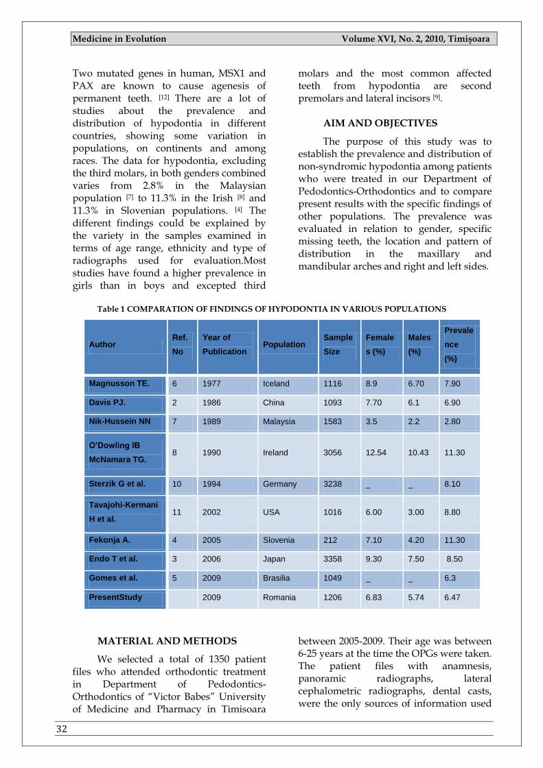
Table 1 COMPARATION OF FINDINGS OF HYPODONTIA IN VARIOUS POPULATIONS
Author Ref.
No
Year of
Publication Population
Sample
Size
Female
s (%)
Males
(%)
Prevale
nce
(%)
Magnusson TE. 6 1977 Iceland 1116 8.9 6.70 7.90
Davis PJ. 2 1986 China 1093 7.70 6.1 6.90
Nik-Hussein NN 7 1989 Malaysia 1583 3.5 2.2 2.80
O’Dowling IB
McNamara TG. 8 1990 Ireland 3056 12.54 10.43 11.30
Sterzik G et al. 10 1994 Germany 3238 _ _ 8.10
Tavajohi-Kermani
H et al. 11 2002 USA 1016 6.00 3.00 8.80
Fekonja A. 4 2005 Slovenia 212 7.10 4.20 11.30
Endo T et al. 3 2006 Japan 3358 9.30 7.50 8.50
Gomes et al. 5 2009 Brasilia 1049 _ _ 6.3
PresentStudy 2009 Romania 1206 6.83 5.74 6.47
MATERIAL AND METHODS
We selected a total of 1350 patient files who attended orthodontic treatment in Department of Pedodontics-Orthodontics of ―Victor Babes‖ University of Medicine and Pharmacy in Timisoara
between 2005-2009. Their age was between 6-25 years at the time the OPGs were taken. The patient files with anamnesis, panoramic radiographs, lateral cephalometric radiographs, dental casts, were the only sources of information used
Medicine in Evolution Volume XVI, No. 2, 2010, Timişoara
33
to diagnose hypodontia. If an accurate diagnosis of hypodontia could not be made, the files were excluded. Radiographs of patients with any syndrome, cleft lip and palate or having tooth loss due to dental caries, periodontal disease, traumas, orthodontic reasons were excluded from the study. From the total amount out the patient files, we only selected 1206 for a sufficient quality.
One author analyzed all radiographs on the dental viewer, using a magnifying glass if needed. A tooth is defined to be congenitally missing (CMT) if it has not erupted in the oral cavity and could not be identified or discerned radiographicallly
based on calcification, and there is no evidence of extraction. [1, 3]. To avoid any false positive results, and because premolars show great variability in the initiation of calcification, they were only considered CMT from age of 7 years. [1, 7] Third molars were not included in this investigation. All data were statistically analyzed using the Windows XP-Excel Statistical Package. To compare the differences between male and female patients, maxillary and mandibular jaw chi-square test was performed (chi-square =0.53, degrees of freedom = 1, p = 0.46). The level of significance tested was P>0.05.
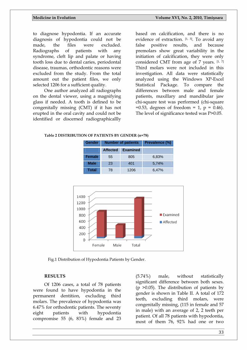
Table 2 DISTRIBUTION OF PATIENTS BY GENDER (n=78)
Gender Number of patients Prevalence (%)
Affected Examined
Female 55 805 6,83%
Male 23 401 5,74%
Total 78 1206 6,47%
Fig.1 Distribution of Hypodontia Patients by Gender.
RESULTS
Of 1206 cases, a total of 78 patients were found to have hypodontia in the permanent dentition, excluding third molars. The prevalence of hypodontia was 6.47% for orthodontic patients. The seventy eight patients with hypodontia compromise 55 (6, 83%) female and 23
(5.74%) male, without statistically significant difference between both sexes. (p >0.05). The distribution of patients by gender is shown in Table II. A total of 172 teeth, excluding third molars, were congenitally missing, (115 in female and 57 in male) with an average of 2, 2 teeth per patient. Of all 78 patients with hypodontia, most of them 76, 92% had one or two
Medicine in Evolution Volume XVI, No. 2, 2010, Timişoara
34
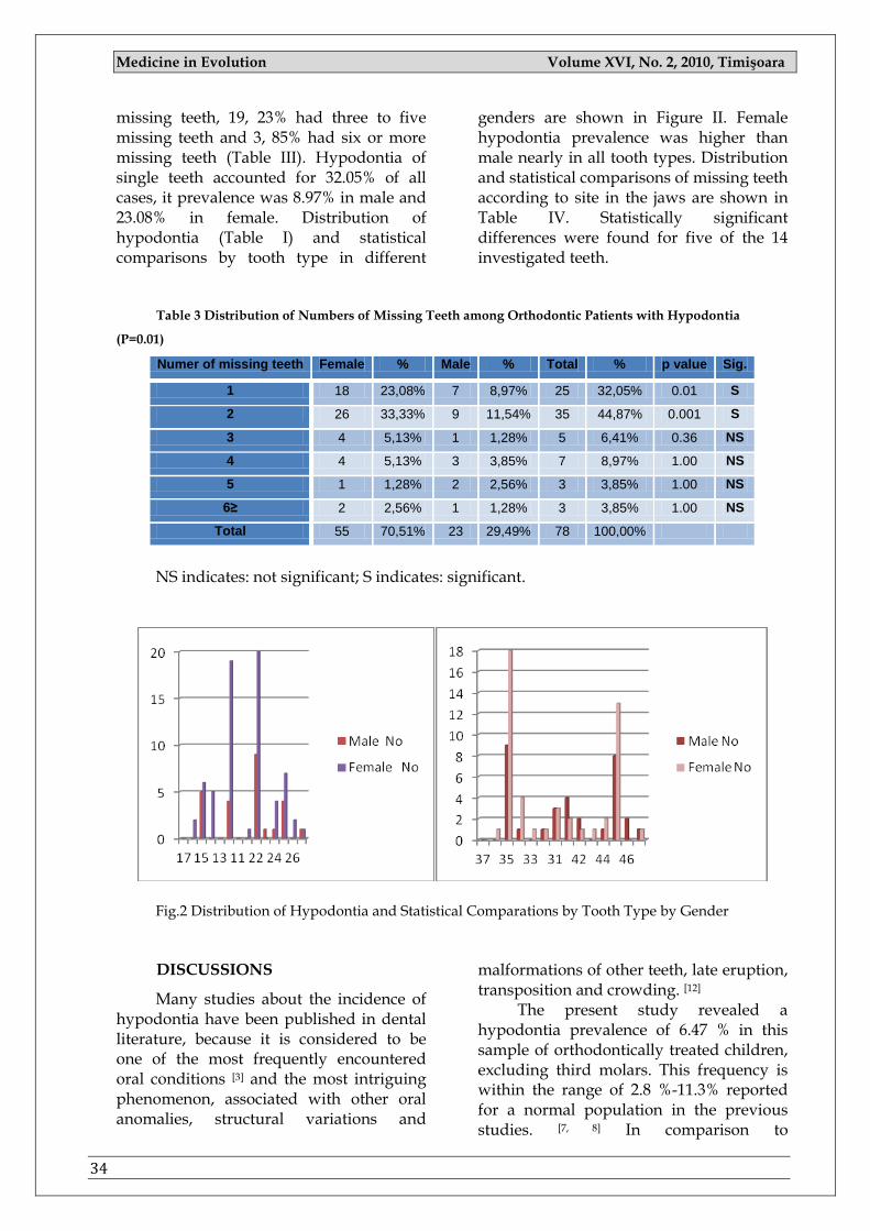
missing teeth, 19, 23% had three to five missing teeth and 3, 85% had six or more missing teeth (Table III). Hypodontia of single teeth accounted for 32.05% of all cases, it prevalence was 8.97% in male and 23.08% in female. Distribution of hypodontia (Table I) and statistical comparisons by tooth type in different
genders are shown in Figure II. Female hypodontia prevalence was higher than male nearly in all tooth types. Distribution and statistical comparisons of missing teeth according to site in the jaws are shown in Table IV. Statistically significant differences were found for five of the 14 investigated teeth.
Table 3 Distribution of Numbers of Missing Teeth among Orthodontic Patients with Hypodontia
(P=0.01)
Numer of missing teeth Female % Male % Total % p value Sig.
1 18 23,08% 7 8,97% 25 32,05% 0.01 S
2 26 33,33% 9 11,54% 35 44,87% 0.001 S
3 4 5,13% 1 1,28% 5 6,41% 0.36 NS
4 4 5,13% 3 3,85% 7 8,97% 1.00 NS
5 1 1,28% 2 2,56% 3 3,85% 1.00 NS
6≥ 2 2,56% 1 1,28% 3 3,85% 1.00 NS
Total 55 70,51% 23 29,49% 78 100,00%
NS indicates: not significant; S indicates: significant.
Fig.2 Distribution of Hypodontia and Statistical Comparations by Tooth Type by Gender
DISCUSSIONS
Many studies about the incidence of hypodontia have been published in dental literature, because it is considered to be one of the most frequently encountered oral conditions [3] and the most intriguing phenomenon, associated with other oral anomalies, structural variations and
malformations of other teeth, late eruption, transposition and crowding. [12]
The present study revealed a hypodontia prevalence of 6.47 % in this sample of orthodontically treated children, excluding third molars. This frequency is within the range of 2.8 %-11.3% reported for a normal population in the previous studies. [7, 8] In comparison to
Medicine in Evolution Volume XVI, No. 2, 2010, Timişoara
35
orthodonticaly treated patients, the result is higher than 2.7 % reported by Silva Meza [9], but smaller than 11.3% reported by Fekonja [4]. Variation in results could be attributed to the method used by authors. Also there is a high result of an average of 2, 2 teeth per patient confirming that hypodontia is common in Romanian population. The result of this study concurs with other studies and indicates that one or two teeth hypodontia is major, being 76.92 % in this report. The present study showed that congenital absence of teeth was found more frequently in female
than male. This finding is in consistent with some researchers [2, 3, 7, 9] meanwhile other reports found no significant differences. [5, 6] Regarding the hypodontia distribution by tooth type, the maxillary lateral incisor is the most frequently missing tooth, followed by the mandibular second premolar, maxillary second premolar and mandibular incisor. Agenesis of maxillary and mandibular canines, first and second molars are very rare. These findings are consistent with most of the previous studies. [4, 7, 9,]
Table 4 Distribution and Statistical Comparations of Missing Teeth According To Site in the Jaws
Tooth Number# Maxila No. Maxila % Tooth Number Mandible No. Mandible %
11 0 0,00% 41 6 0,50%
12 23 1,91% 42 3 0,25%
13 0 0,00% 43 1 0,08%
14 5 0,41% 44 3 0,25%
15 11 0,91% 45 2 0,17%
16 2 0,17% 46 2 0,17%
17 0 0,00% 47 2 0,17%
Tooth Number# Maxila No. Maxila % Tooth Number Mandible No Mandible %
21 1 0,08% 31 6 0,50%
22 29 2,40% 32 2 0,17%
23 1 0,08% 33 1 0,08%
24 5 0,41% 34 5 0,41%
25 11 0,91% 35 27 2,24%
26 2 0,17% 36 1 0,08%
27 2 0,17% 37 0 0,00%
#FDI Notation
CONCLUSIONS
The hypodontia prevalence of 6.47% found in this sample of orthodontically treated patients was statistically similar with the researches for Caucasian populations.
Hypodontia was found considerably more frequently in the maxilla than in the mandible and was almost equally distributed between both sides of jaws. The
teeth most frequently missing were the maxillary lateral incisors, followed by the maxillary and mandibular second premolars. The majority of patients had one or two teeth missing, but seldom three or more. The diagnosis and treatment modalities of hypodontia should be prompt and accurate to prevent the esthetic and functional problems in the dentition.

Medicine in Evolution Volume XVI, No. 2, 2010, Timişoara
36
Early evaluation of the number of missing teeth and consideration of size and number of teeth remaining in both arches
should aid the clinician in planning and managing treatment.
REFERENCES
1. Arte S, Pirinen S. Hypodontia. Orphanet encychlopedia, www.orpha.net/data/GB/UK-hypodontia.pdf. 2007; 1-7.
2. Davis PJ. Hypodontia and hyperdontia of permanent teeth in Hong Kong schoolchildren. Community Dent Oral Epidemiol 1987;15:218-220. 1994;55:61-69.
3. Endo T, Ozoe R, Kubota M, Kubota M, Aktiyama M, ShimookaS. A survey of hypodontia on Japanese orthodontic patients. Am J Orthod Dentofacial Orthop 2006; 129:29-35.
4. Fekonja A. Hypodontia in orthodontically treated children. Eur J Orthod 2005;27:457-460. 5. Gomes RR, da Fonseca JA, Paula LM, Faber J, Acevedo AC Prevalence of hypodontia in
orthodontic patients in Brasilia, Brazil.. Eur J Orthod. 2009 Oct 16. 6. Magnusson TE. An epidemiologic study of dental space anomalies in Icelandic schoolchildren.
Community Dent Oral Epidemiol 1977;5:292-300. 7. Nik-Hussein NN. Hypodontia in the permanent dentition: a study of its prevalence in
Malaysian children. Aust Orthod J 1989;11:93-95. 8. O‘Dowling IB, McNamara TG. Congenital absence of permanent teeth among Irish school-
children. J Irish Dent Assoc 1990;36:136-138. 9. Silva Meza R. Radiographic assessment of congenitally missing teeth in orthodontic patients.
Int J Paediatr Dent 2003;13:112-116. 10. Sterzik G, Steinbicker V, Karl N. The etiology of hypodontia. Fortschr Kieferorthop 1994;55:61-
69. 11. Tavajohi-Kermani H, Kapur R, Sciote JJ. Tooth agenesis and craniofacial morphology in an
orthodontic population. Am J Orthod Dentofacial Orthop 2002;122:39-47. 12. Vastardis H.The genetics of human tooth agenesis: new discoveries for understanding dental
anomalies. Am J Orthod Dentofacial Orthop 2000;117:650-656.
Medicine in Evolution Volume XVI, No. 2, 2010, Timişoara
37
PAGES 37 -42
THE USE OF QUANTITATIVE REAL-TIME PCR IN MONITORING TREATMENT RESPONSE – A NEW STANDARD OF CARE IN CHRONIC MYELOID LEUKEMIA PATIENTS-
KINGA TĂTAR 1, HORTENSIA IONIŢĂ 1
1 Department Hematology, City Hospital Timisoara, University of Medicine and Pharmacy
“Victor Babeş” Timişoara
Correspondence to: Dr.Kinga Tatar, City Hospital Timisoara, Gheorghe Dima Street, No.5email: [email protected]
ABSTRACT
Chronic myeloid leukemia (CML) is a myeloproliferative disorder, characterized by a specific chromosomal aberration, the Philadelphia [Ph] chromosome. The Ph chromosome is the result of a reciprocal translocation between the long arms of chromosomes 9 and 22, t (9; 22) (q34; q11).
The molecular consequence of this translocation is a novel fusion gene, BCR-ABL, which encodes a constitutively active tyrosine kinase, implicated in pathophysiology and development of CML [1, 2]. Imatinib mesylate currently the golden standard for front line treatment of CML is a selective inhibitor of the BCR-ABL tyrosine kinase activity.
Responses to imatinib occur at hematologic, cytogenetic and molecular levels. IM therapy now allows the majority of patients with CML to reach CCyR - a confirmed good prognostic indicator [3]. In different studies, 87% of patients in chronic, 17% of patients in accelerated, and 7% of patients in blast phases reached the important clinical aim of Ph negativity [3-5]. Once a patient has achieved CCyR, monitoring of residual cells is usually performed by estimating the BCR-ABL transcripts on a molecular level using quantitative real-time polymerase chain reaction (QRT-PCR). A 2-log reduction in BCR-ABL transcripts correlates with Ph negativity in CCyR.
Patients achieving a 3-log reduction in BCR-ABL transcripts are defined as having a major molecular response (MMR), a surrogate marker closely correlating with the probability of disease free survival [8]. Patients failing to achieve this 3-log response, at any time during therapy, had significantly shorter progression-free survival [6]. After a 5 to 6 log reduction, BCR-ABL transcripts can no longer be detected by QRT-PCR and patients are designated as having complete molecular response (CMR). But even with the most sensitive QRT-PCR assay, CMR is consistent with the persistence in the patient’s body of up to 106 or 107 leukemic cells [7]. Due to the proliferation of such residual cells a significant fraction of those patients who have responded on a deep molecular level, lose this response and progress to advanced phase disease.
The clinical advantage of the extremely sensitive method of QRT-PCR is to be alerted by rising transcript levels at a very early time point, usually weeks or even months before the onset of clinical symptoms, allowing early therapeutic intervention with a beneficial impact on survival.
Key words: BCR-ABL mRNA, control gene, major molecular response, minimal residual disease.
Medicine in Evolution Volume XVI, No. 2, 2010, Timişoara
38
INTRODUCTION
Initially used for qualitative identification of bcr-abl transcript in patients for which the presence of Philadelphia chromosome could not be demonstrated by classical cytogenetic examination or after STEM cell transplantation, this technique has undergo a significant development, in the past years the techniques of competitive PCR and finally the real-time PCR being developed. (15).
In chronic myeloid leukemia the real-time PCR is combined with reverse transcription PCR in order to quantify the level of BCR-ABL mRNA. Thus the chain of mRNA is first transcribed in a complementary DNA by revers transcription, followed by the amplification of the obtained DNA by the polymerase reaction.
Real-time PCR techniques used in CML
monitoring There are several techniques used in
CML patient monitoring, the best known are using the TaqMan system (Byosistem Foster City, CA), or the Cycler platform (Roche Diagnostics, Indianapolis). The TaqMan Real-time PCR measures accumulation of a product via the fluorophore during the exponential stages of the PCR, rather than at the end point as in conventional PCR [11]. The exponential increase of the product is used to determine the threshold cycle, CT, i.e. the number of PCR cycles at which a significant exponential increase in fluorescence is detected, and which is directly correlated with the number of copies of DNA template present in the reaction. The set up of the reaction is very similar to a conventional PCR, but is carried out in a real-time thermal cycler that allows measurement of fluorescent molecules in the PCR tubes. LightCycler methodology is using a platform called LightCycler-the only technology currently accredited for routines molecular diagnosis; all other technologies are
accredited only for research purposes. The used quantifying method is based on hybridization probes using the Free Resonance Energy Transfer (FRET). The main advantages of this technique are that the reaction is followed during the amplification phase in real time, thus eliminating the necessity of co-amplifying a competitor. As previously described, the existing techniques vary with respect of the type of device, the location of the probes, the type of real-time reaction and the control gene. These differences translate from clinical point of view in few downsides related to large variability of determinations ( up to 0,5 log or 2 – 5 %), false negative results in laboratories using a low sensitivity QRT-PCR technique and difficulties in interpreting results by clinicians due to lack of international standardization. In the attempt of overcoming this downsides, in 2003 took place a collaborative action called Europe against Cancer (EAC), including 26 laboratories from 10 countries [14].
The scope of this program was to establish standardized protocols for fusion gene quantification by TaqMan technique. It was proposed to be used a common forward primer placed on the 13th exon of BCR gene, and a reverse primer located on the exon 2 of gene ABL, for amplification of both transcripts (b2a2, b3a2). [15]
Using standardized protocols, EAC identified the most appropriate control gene to be amplified in parallel with the sample. [14]
The role of this control gene is to correct the qualitative and quantitative variations of the RNA and to appreciate the sensitivity of each determination.
From the initial number of 14 candidate genes, based on the absence of pseudogenes, stability and gene expression there were 3 genes selected: Abelson (Abl), beta-2-microglobulin (B2M) and betaglucuronidase (GUS).
Although all 3 genes had a stable expression in the studied samples, it was established that only ABL gene had similar expressions in samples from both healthy
Medicine in Evolution Volume XVI, No. 2, 2010, Timişoara
39
and leukemic subjects, and it was proposed as control gene.
Expresing and interpreting results. Currently there are different ways of reporting RT-qPCR results. One of this is to report the number of bcr-abl copies per microgram of RNA, but this approach has some disadvantages: the measurement of RNA concentration is not exact, and the quality and efficacy of the reverse transcription is not taken in to account. Another approach is to express the results as a ratio of the number of BCR-ABL copies and the copies of the control gene. According to this approach the equal number of BCR-ABL copies and control gene at diagnostic represents 100%. In the IRIS study it was used for the first time the concept of logarithmic reduction from a standardized baseline of untreated patients. Achievement of a 3 log reduction or more from the standardized baseline was considered as a Major Molecular Response and achieving this was strongly associated with a better progression free survival. The baseline standard was established by measuring the BCR-ABL/ABL ratio in 30 CML patients before treatment initiation. The same 30 samples were processed in 3 different laboratories. This approach has several advantages: makes possible the expression of results on a common international scale, once the laboratory has established the standard baseline and furthermore it is possible to evaluate the level of response for a patient without actually knowing the pretreatment level of BCR-ABL. However this method has the disadvantage that in order to establish a save level for the minimal residual disease it is necessary to exchange a large number of samples between several labs. In order to overcome this disadvantage it was proposed that the Standardized Baseline from IRIS study to be considered as 100% and the 3 log reduction (corresponding to MMR) to be established at 0.1% [16].
In order to express results on an international scale, each laboratory has to establish a conversion factor by using a
series of samples with known value. By using this CF, the BCR-ABL value obtained in a certain laboratory (BCR-ABL L) can be expressed on the International Scale (BCR-ABL IS) using the formula:
BCR-ABL L X CF = BCR-ABL IS The Conversion Factor (CF) is
calculated according to the bellow formula, where MMR IS the value of BCR-ABL corresponding to the MMR in the IRIS study and MMR Eq represents the value of BCR-ABL L corresponding to RMM as defined in the IRIS study.
CF=MMRIS/MMREq However there is a need for
additional efforts for standardizing this technique by using standardized reference samples, an essential step in obtaining comparable results in across different laboratories.
Clinical significance of the results:
Achieving a 2 log reduction in the transcript level at the moment of complete cytogenetic or a 3 log reduction at any moment thereafter represents an independent prognostic factor for the progression free survival [6].
Molecular studies performed in the IRIS trial have demonstrated for the first time that the level of minimal residual disease at 12 months expressed as log reduction from the baseline is significantly associated with the event free survival and risk of progression in newly diagnosed patients who achieved a CCR under imatinib treatment.
This threshold of 3 or more BCR-ABL log reduction identified a subset of patients with an extremely favorable prognostic, with only sporadic loss of responses and very low risk for progression. These results were confirmed also on the longer follow-ups of the IRIS trial.
Another study where cytogenetic examination was scheduled at one year, demonstrated that a rapid fall in the BCR-
Medicine in Evolution Volume XVI, No. 2, 2010, Timişoara
40
ABL mRNA level can predict the cytogenetic response, having an important prognostic value. Based on the increasing evidence regarding the impact of BCR-ABL reduction on the prognostic of imatinib treated patients the European LeukemiaNet introduced Major Molecular Response (MMR) as a parameter in the operational definition of the optimal response to imatinib therapy. Obtaining this resposnse and subsequent loss of it signifies that the chanses for an optimal response are low, this patients being eligiblefor another therapeutic option. Also the absence of MMR at 12 months represents a warning sign for a certain category of patients the disese characteristics might adversely affect the long time prognostic, a closer monitoringof this patients being required.(18)
The presence of MMR at 18 months is considered as optimal response and according to ELN the significance is long term outcome for patients not achieving this response on imatinib treatment might not be favorable [8].
In a more recent study performed at Hammersmith Institution it was performed the outcome analysis for 224 chronic –phase CML patients in an attempt to assess for the extent to which the ELN recommendation can be validated in clinical practice. According to this study patients in CcyR who failed to achieve MMR at 12 or 18 months were more likely to lose their CcyR than patients who achieved MMR: 23.6% vs. 2,6% and 24,6%vs)%, although there was no significant impact on the PFS and OS [19]. One possible explanation found by the authors is that early intervention when patients lose their CcyR may successfully prevent the progression of the leukemia; thus the adverse influence of lack of a MMR could in some cases be reversed by a change to a more effective therapy [19].
Definition of molecular responses
As previously mentioned there were many controversies regarding the definition and expression of major
molecular response, at this moment most working parties recommend to define MMR as a decrease of transcript level to 0.1% from the baseline standard level. The definition of complete molecular response as an undetectable transcript corresponds to a 4-5 log reduction, but is subject to the sensibility variations of the technique used. For this reason whenever a CMR is reported it is recommended that the sensibility of the technique to be also communicated and this result should be confirmed by nested PCR. After the initiation of a TKI treatment it is recommended to monitor the BCR-ABL level once every 3 months, before a complete cytogenetic response is achieved and every 6 months after [18].
It is clinically relevant to characterize the response as: [13] descending transcript, undetectable transcript, stable transcript level (plateau), increasing transcript level. Regarding the definition and significance of the increasing transcript level, there are still ongoing controversies: the Adelaide group found a statistical significance between the 2 fold increase in BCR-ABL transcript level and the emergence of mutations. The technique used in this study was extremely sensitive, so in case of less sensitive settings it is recommended to perform mutational analysis in case of a 5-5 fold increase of the transcript level, confirmed by successive determinations. In practice the BCR-ABL level increase is more frequently associated with kinase domain mutations in patients who never achieved a major molecular response. A more recent study identified a trashold of 2.6 increse intranscript levelas being predicitive for identification of BCR-ABL kinaze domain mutations. (20)
CONCLUSION
The prognostic of CML patients considerably improved over the past years due to highly efficient therapeutic options. In consequence the adequate monitoring of these patients is becoming more and more important. In the past years several studies in CML patients propose as efficacy
Medicine in Evolution Volume XVI, No. 2, 2010, Timişoara
41
endpoint the achievement of major or complete molecular response in patients with complete cytogenetic response.
However the long term prognostic significance of these molecular parameters is still to be determined.
Although the standardization of this technique still needs some fine tunings, the role of molecular monitoring in CML patients is clearly established.
The use of this technique makes possible the definition of responses as
optimal/suboptimal and moreover it identifies a category of patients with extremely favorable prognosis, it can be an early predictor for achieving the CCR, but in the same time for relapse, disease progression or emergence of kinase domain mutation. In the era of tyrosine kinaze inhibitors, this technique should be available to all hematologists in order to provide an optimal standard of care for CML patients.
REFERENCES
1. Daley G. Q., Van Etten R. A., Baltimore D. Induction of chronic myelogenous leukaemia in mice by the P210 bcr/abl gene of the Philadelphia chromosome,. Science, 1990, 274:824-830.
2. Kelliher M., Mc Laughlin J., Witte O., Rosenberg N. Induction of a chronic myelogenous leukaemia-like syndrome in mice with v-abl and bcr/abl, Proc Natl Acad Sci USA,1990;87:6659-6653.
3. Druker B. J., Guilhot F., O‘Brien S. G., Gathmann I., Kantarjian H., Gattermann N. et al. Five-Year Follow-up of Patients Receiving IM for Chronic Myeloid Leukemia, N Engl J Med, 2006; 355: 2408-17.
4. Talpaz M., Silver R. T., Druker B. J., Goldman J. M., Gambacorti-Passerini C., Guilhot F. et al. IM induces durable hematologic and cytogenetic responses in patients with accelerated phase chronic myeloid leukemia: results of a phase 2 study, Blood, 2002; 99: 1928-37.
5. Sawyers C. L., Hochhaus A., Feldman E., Goldman J. M., Miller C. B., Ottmann O. G. et al. IM induces hematologic and cytogenetic responses in patients with chronic myelogenous leukemia in myeloid blast crisis: results of a phase II study, Blood, 2002; 99: 3530-39.
6. Press R. D., Love Z., Tronnes A. A., Yang R., Tran T., Mongoue-Tchokote S. et al. BCR-ABL mRNA levels at and after the time of a complete cytogenetic response (CCR) predict the duration of CCR in Imatinib-treated patients with CML, Blood, 2006,107:4250-4256
7. Kaeda J, Chase A, Goldman JM. Cytogenetic and molecular monitoring of residual disease in chronic myeloid leukaemia, Acta Haematologica, 2002; 107: 64-75.
8. Baccarani M., Saglio G., Goldman JM, Hochhaus A., Simonsson B., Appelbaum F. et al. Evolving concepts in the management of chronic myeloid leukemia: recommendations from an expert panel on behalf of the European Leukemia Net, Blood, 2006; 108: 1809-20
9. Quintas-Cardama A, Cortes J E., Chronic Myeloid Leukemia, Diagnosis and treatment, Mayo Clin Proc./Jul 2006, 81(7): 973-988
10. Goldman J., How I treat chronic myloid leukemia in the imatinib era, Blood, 2007, 110:8 2826-2837; Jul 12, 2007
11. Heid CA, Stevens J, Livak KJ, Williams PM., Real time quantitative PCR, Genome Res,1996 , 6: 986-994.
12. Kutyavin IV, Afonina IA, Mills A, Gorn VV, Lukhtanov EA, Belousov ES, Singer MJ, Walburger DK, Lokhov SG, Gall AA, Dempcy R, Reed MW, Meyer RB, Hedgpeth J , 3'-minor groove binder-DNA probes increase sequence specificity at PCR extension temperatures, Nucleic Acids Res, 2000, 28: 655-661.
13. Martinelli G, Iacobucci I, Soverini S et al, Monitoring minimal residual disease and controlling drug resistance in chronic myeloid leukaemia patients in treatment with imatinib as a guide to clinical management, Hematol Oncol, 2006, 24: 196-204
14. Beillard, N Pallisgaard, VHJ van der Velden, W Bi, et al. Evaluating of candidate control genes for diagnosis and residual disease detection in leukemic patients using ―real-time ‖ quantitative reverse transcriptase polymerase chain reaction (RQ-PCR)-a Europe agains cancer program; Leukemia, 2003, 17: 2474-2486.

Medicine in Evolution Volume XVI, No. 2, 2010, Timişoara
42
15. Gabert, E Beillard1,VHJ van der Velden, W Bi, D Grimwade et al, Standardisation and quality control studies of ―real-time ‖ quantitative reverse transcriptase polymerase chain reaction (RQ-PCR)-a Europe against cancer program, Leukemia 2003; 17: 2318-2357.
16. Hughes TP et al. Monitoring patients CML responding to treatment with tyrosine kinase inhibitors- Review and recommendations for ‗harmonizing‘ current methodology for detecting BCR/ABL transcript and kinase domain mutations for expressing results Blood, 2006; 108:28-37.
17. Baccarani M, Pane F, Saglio G, et al, Monitoring treatment of chronic myeloid leukemia, Haematologica, 2008;93(2): 161
18. Baccarani M, et al Chronic Myeloid Leukemia: An Update of Concepts and 19. Management Recommendations of European LeukemiaNet, Published Ahead of Print on
November 2, 2009 as 10.1200/JCO.2009.25.0779 20. Marin D, D Milojkovic, E Olavarria, J S Khorashad, et. Al. European LeukemiaNet criteria for
failure or sub-optimal response reliably identify patients with CML in early chronic phase treated with imatinib whose eventual outcome is poor,, Blood 2008, prepublished online, Aug 20, doi:10.1182/blood-2008-06-162388
21. Press D et al,Determining the rise in BCR-ABLRNAthat optimally predicts a kinase domain mutation in patients with chronic myeloid leukemia on imatinib, Blood, 24 September 2009 : 114, 2598-2605
Medicine in Evolution Volume XVI, No. 2, 2010, Timişoara
43
PAGES 43 -45
METHODS OF DIAGNOSIS IN THE CMV INFECTION
SONIA TANĂSESCU 1, IOAN POPA1, RODICA URTILĂ1, RUXANDRA BĂCANU 1
1 Clinic II Pediatrics – University of Medicine and Pharmacy “Victor Babeş” Timişoara
Correspondence to: Sonia Tanasescu, Timisoara str. Evlia Celebi nr.3, [email protected]
ABSTRACT
The CMV infection represents the most common congenital viral infection, while fetopathy is the most frequent infection, transmitted from mother to fetus. Most cytomegalovirus infections remain undiagnosed, due to the lack of clinical signs, with tendencies of intermittent reactivation, without relevant symptoms. The infection is diagnosed with virological and serological identification methods. In practice, the serology tests must be correlated with the virology tests, both to establish a predictive diagnosis of CMV infection and to reveal the ways in which the virus is transmitted.
Key words: CMV infection, methods of diagnosis, serology tests.
INTRODUCTION
The CMV infection represents a problem of public health, its prevalence being between 0.2 - 2.4%, with a higher percent in developing countries and in countries with lower social and economical level, as compared to the developed countries.
Also, according to the studies in the specialty literature, the prevalence of the infection in the developed countries is of approximately 40%, as compared to 100% in the developing countries.
Most infection forms are asymptomatic. The asymptomatic forms have a good prognosis, yet 15% of the newborns develop important sequelae. Out of the newborns presenting symptoms, 25-30% dies, while 80-90% develops severe sequelae (8, 9).
Most cytomegalovirus infections remain undiagnosed, due to the lack of clinical signs, as they have the tendency to reactivate intermittently, without relevant symptoms.
The persons carrying a CMV infection develop antibodies against the infection; these antibodies remain in the host organism for its whole life. Numerous laboratory tests can determine the presence of these antibodies (8).
Serological tests:
The tests are performed in order to determine the specific antibodies, IgM and IgG type respectively, which are the serological markers of the CMV infection. Nowadays, there are several techniques for the identification of such antibodies: IM (immunofluorescence), Latex, ELISA
Medicine in Evolution Volume XVI, No. 2, 2010, Timişoara
44
(enzyme-linked immunosorbent assay) and RIA (radio immunoassay).
The most common and accessible technique to determine the anti-CMV antibodies is the ELISA method. (4) The ELISA method uses as antigen the cellular lysate from the CMV infected fibroblasts cultures. The IgM-type antibodies are the first produced by the human body, in response to the CMV infection. They appear at approximately 3-4 weeks after the infection exposure and remain in the organism for 3-4 months.
Due to the fact that IgM-type antibodies can be detected only after 4 weeks from the infection time, the serological diagnosis is not useful in the early detection of the infection. The IgG-type antibodies present an increasing titer during the active phase of the infection, becoming stable after that.
Thus, a person who has been exposed to the CMV infection shall have a stable titer of IgG for the whole life, which means that the virus has become inactive. The virus can be reactivated in certain conditions, such as: gestation, immune deficiencies, etc (2, 4).
A rapid growth of the IgM titer, with a later increase of the IgG titer, indicates a primary infection. A parallel increase of the IgM and IgG indicates, in general, a secondary infection (reactivation, re-infection).
The virological diagnosis supposes the isolation of the virus in urine, saliva or biopsy fragments.
Most frequently used methods are:
a) The cell culture: This is the oldest method of CMV infection diagnosis. It is performed on human fibroblast cell cultures. The cytopathic effect consists in the emergence of cell foci, the typical aspect being of characteristic intracellular inclusions, known as "owl-eyes". The positive cultures can be detected within 1-2 days, yet the negative ones require a period of 3 weeks, for a possible determination of a positive result. This fact is a
disadvantage in the CMV detection and is due either to a small number of viruses in the collected samples, or to the re-infection with another strain (5,
10). b) DEAFE (Detection of early antigen
fluorescent foci). It is a fast diagnostic method of the cytomegalic infection, performed by inoculating the biological product into a cell culture and its later examination, after 24 hours, by immunofluorescence. The test can be qualitative (the presence or absence of the CMV) or quantitative (viral load) (3, 5).
c) Electronic microscopy. The characteristic aspect of large cells, with inclusions known as "owl-eyes" is observed on histological slices, while by immunofluorescence, using monoclonal antibodies, we can trace the virus in the bronchial or broncho-alveolar lavage fluid (5, 7).
d) Determining the viral DNA by PCR (polymerase chain reaction). This is a fast detection method. The high levels of DNA indicate an acute infection, while the progressive decrease of the DNA level indicates a response to the antiviral treatment. The disadvantage of this method is that it is impossible to detect very low DNA levels (1, 6).
e) The antigenemia test. It is based on detecting a specific structural protein (pp65), present on the surface of the polymorphonuclear lymphocytes. The number of infected leucocytes is related to the infection severity. The advantage of this test is that a quantitative evaluation of the viral infection is performed in the blood and the results are obtained very fast, in the same day. Also, especially when associated with HIV, correlations between the value of the antigenemia, symptomatology and therapeutic actions can be made (1, 6). The disadvantage of this test is that,
being very laborious, it is not applied as a routine. In practice, the serology tests must be correlated with the virology tests, both
Medicine in Evolution Volume XVI, No. 2, 2010, Timişoara
45
to establish a predictive diagnosis of CMV infection and to reveal the ways in which the virus is transmitted.
In conclusion:
a certain diagnosis of congenital CMV infection is established by corroborating the virological diagnosis (the isolation of the virus in the urine, the saliva or the faeces, during the first weeks after birth, confirmed by repeating the tests 2-3 weeks later), with the serological diagnosis (the presence of specific IgM).
a significant child-specific titer of high IgG can mean both a congenital infection (if the titer of antibodies increases in dynamics at 1, 3 and 6 months)
and a pre-natal infection (if the antibody titer remains constant).
a person who has been exposed to the CMV infection during childhood, procreation (during pregnancy) and/or presents immunodeficiency, can be diagnosed by the virological and serological methods, used in parallel.
As a conclusion, both paraclinical
methods, used in consensus, can establish the CMV infection diagnosis and can determine the ways in which the virus is transmitted (primary – recurrent; congenital – acquired), as well as the immunological state of the subject (immunocompetent – immunodepressed).
REFERENCES:
1. Blok, M. J, V. J. Goossens, S. J. V. Vanherle, B. Top, N. Tacken, J. M. Middeldorp, M. H. L. Christiaans, J. P. van Hooff, and C. A. Bruggeman. Diagnostic value of monitoring human cytomegalovirus late pp67 mRNA expression in renal-allograft recipients by nucleic acid sequence-based amplification. J. Clin. Microbiol. 1998; 36:1341-1346.
2. Bodeus M, Beulne D, Goubau P. Ability of three IgG-avidity assays to exclude recent cytomegalovirus infection. Eur J Clin Microbiol Infect Dis. 2001; 20(4): 248-252.
3. Casteels, A., A. Naessens, F. Gordts, L. De Catte, A. Bougatef, and W. Foulon. 1999. Neonatal screening for congenital cytomegalovirus infections. J. Perinat. Med. 27:116-121.
4. Dennin RH, Hernan J, Schultz E et al. Serologische Zytomegalievirus Diagnostik mittles ELISA-Technik bei nierentransplantierten Patienten. Dtsch Med Wschr 1984; 109:214-218.
5. Guerra, B., T. Lazzarotto, S. Quarta, M. Lanari, L. Bovicelli, A. Nicolosi, and M. P. Landini. 2000. Prenatal diagnosis of symptomatic congenital cytomegalovirus infection. Am. J. Obstet. Gynecol. 183:476-482.
6. Poddar SK Detection of adenovirus using PCR and molecular beacon. J Virol Methods 1999; 82: 19–26
7. Roizman B., Whitley RJ., Lopez C. – The human herpesviruses Raven Prees New York, 1993. 8. Waldo E. Nelson – Textbook of pediatrics, 16-th ediţion, W.B. Saunder Company, London 2000,
981-983 9. Yamamoto AY, Figueiredo LTM, Mussi-Pinhata MM- Prevalence and clinical aspects of
congenital cytomegalovirus infection. J Pediatr (Rio J) 1999, 75:23-28; 10. Zucca C, Binda S, Borgatti R, Triulzi F, Radice L, Butte C, Barkhaus PE, Barbi M. Retrospective
diagnosis of congenital cytomegalovirus infection and cortical maldevelopment. Robert-Gnansia E. Embryopathie à cytomégalovirus. Encyclopédie Orphanet. Octobre 2003.

Medicine in Evolution Volume XVI, No. 2, 2010, Timişoara
46
Medicine in Evolution Volume XVI, No. 2, 2010, Timişoara
47
PAGES 47 -53
PULMONARY TUBERCULOSIS AND DIABETES MELLITUS: A DANGEROUS ASSOCIATION
ADRIANA SOCACI1, VOICU TUDORACHE 2, VIOREL SERBAN3
1 “Dr. V.Babeş” Clinical Hospital, Pneumophthisiology Ambulatory Center, Timişoara, 2 “Dr. V.Babeş” Clinical Hospital, Pneumophthisiology Department, Timişoara, 3 County Clinical Hospital, Diabetes, Nutrition and Metabolic Disease Clinic, Timişoara
Correspondence to:
SOCACI ADRIANA mobil:0723705832 e-mail: [email protected]
ABSTRACT
Objectives: Pulmonary tuberculosis is one of the most important disorders as far as global mortality is
concerned. Diabetes mellitus represents a major risk factor in pulmonary tuberculosis especially in areas where
endemics maintain uncontrolled values. In its turn, pulmonary tuberculosis may complicate diabetes mellitus, the
two disorders negatively enhancing each other. The present study aims at revising the data on the role of diabetes
mellitus in clinical-radiological manifestations and therapeutic results of pulmonary tuberculosis. Besides, the
study also analyzes epidemiological aspects of the two disorders, the potential mechanisms by which diabetes
mellitus can induce pulmonary tuberculosis, the effects of tuberculosis on the control of diabetes mellitus, as well
as therapeutic and pharmacokinetic problems related to the co-existence of diabetes mellitus and pulmonary
tuberculosis.
Key words: pulmonary tuberculosis, diabetes mellitus, association
INTRODUCTION
The association between pulmonary
tuberculosis (TB) and diabetes mellitus (DM) was first mentioned in the 11th century by Avicenna who maintained that phthisis often complicates DM; in 1928, JH Barach observes a worldwide increase of DM which also affects the increase of TB-DM cases, with important clinical, social and economical implications (3). However, the global importance of DM as a TB-risk factor is still mostly unknown, although several studies have shown that the
prevalence of TB among diabetics is 2-5 times higher than among non-diabetics (14). On the other hand, it seems that there is little information about the strength and the nature of the association between TB and DM (2, 20). The present study aims at an overall revision of the data about the association between TB and DM.
EPIDEMIOLOGICAL ASPECTS: TUBERCULOSIS AND DIABETES MELLITUS
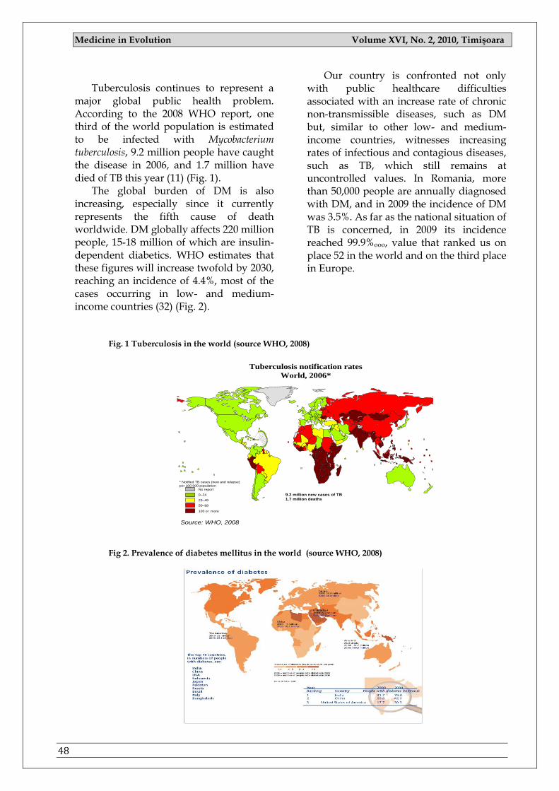
Medicine in Evolution Volume XVI, No. 2, 2010, Timişoara
48
Tuberculosis continues to represent a
major global public health problem. According to the 2008 WHO report, one third of the world population is estimated to be infected with Mycobacterium tuberculosis, 9.2 million people have caught the disease in 2006, and 1.7 million have died of TB this year (11) (Fig. 1).
The global burden of DM is also increasing, especially since it currently represents the fifth cause of death worldwide. DM globally affects 220 million people, 15-18 million of which are insulin-dependent diabetics. WHO estimates that these figures will increase twofold by 2030, reaching an incidence of 4.4%, most of the cases occurring in low- and medium-income countries (32) (Fig. 2).
Our country is confronted not only with public healthcare difficulties associated with an increase rate of chronic non-transmissible diseases, such as DM but, similar to other low- and medium-income countries, witnesses increasing rates of infectious and contagious diseases, such as TB, which still remains at uncontrolled values. In Romania, more than 50,000 people are annually diagnosed with DM, and in 2009 the incidence of DM was 3.5%. As far as the national situation of TB is concerned, in 2009 its incidence reached 99.9%ooo, value that ranked us on place 52 in the world and on the third place in Europe.
Fig. 1 Tuberculosis in the world (source WHO, 2008)
Source: WHO, 2008
* Notified TB cases (new and relapse)
per 100 000 population
Tuberculosis notification rates
World, 2006*
No report
0–24
25–49
50–99
100 or more
9.2 million new cases of TB
1.7 milIion deaths
Fig 2. Prevalence of diabetes mellitus in the world (source WHO, 2008)
Medicine in Evolution Volume XVI, No. 2, 2010, Timişoara
49
DIABETES MELLITUS AS RISK FAKTOR OR PULLMONARY TUBERCULOSIS AS COMPLICATION?
DM is a metabolic disorder
characterized by high blood sugar as a result of insulin deficiency, impaired effectiveness of insulin action or both. Chronic high blood sugar is associated with long-term organ failure and malfunction, affecting mainly the eyes, kidneys, nerves, heart and blood vessels. DM may increase the sensitivity to general infections, especially to TB.
TB-induced morbidity is several times higher in diabetics than in non-diabetics. Even pre-diabetic, sub-clinical disorders of the blood sugar metabolism (latent diabetes) have been associated with a higher risk of morbidity. The predisposition of diabetes for infection, especially for TB, can be explained by the alterations induced by the high blood sugar which decreases immune competence at several levels. Low immune competence in DM would in turn allow the latent TB infection to become clinically manifest, the risk being greater as the DM is more severe (metabolic imbalance). Recent studies have shown that, in the same socio-economical and contagiousness conditions, diabetics (especially insulin-dependent ones) aged between 40-49 years get infected and develop more severe forms of TB than the non-diabetic population. Active TB may develop following the transformation of the infection into a disease in 5 – 10% of the cases, while in most cases (90%) Mycobacterium tuberculosis may reside in the host body for several years without triggering the disease (the so-called latent TB condition). It is estimated that the transformation of latent TB into active TB occurs in 5-10% of the cases, the risk being higher when an immune-regulating factor is also associated (21, 23).
Diabetes mellitus is one of the risk factors responsible for the development of active TB, due to the four metabolic disorders it induces: chronic high blood sugar followed by disorders of the sugar,
lipid and protein metabolism, consequences of the defective insulin secretion and/or action (31).
Certain diseases are associated with TB more often than a random association would indicate (syntrophic relation). In most cases, TB occurs as a complication of the primary disease which facilitates its occurrence and often worsens its development. Conversely, TB may negatively influence the development of the primary disorder. The incidence of these associations varies, according to the incidence of each of the two disorders and the degree of the syntrophic affinity between them. A common and severe syntrophy is represented by the association between TB and DM.
POSSIBLE UNDERLYING CAUSES FOR THE TB-DM ASSOCIATION
A probable cause for the increase of the
incidence of TB in diabetics could be a defective defense of the host body and a diminished immune competence (9, 17) (Table I). Immune imbalance predominantly involves the cell-mediated branch of the immune system. At the same time, the high values of the blood sugar seem to have a distinct influence on platelet bactericidal function, even short-term exposures to blood sugar levels of 200 mg% significantly depressing the activity of these cells in the host respiratory apparatus (18, 22). This aspect has been confirmed by the finding that, in inadequately-controlled DM, with high levels of glycosylated haemoglobin, TB follows a more destructive course and is associated with a higher mortality rate. Several pulmonary physiological anomalies have also been detected in diabetics, contributing to a late clearance and the spreading of the infection in the body (9).
The infection with tuberculous bacilli leads to subsequent alterations of the cytokines, platelet-monocytes and T-cell population, and of the CD4/CD8 ratio (10, 27). T-lymphocyte levels, the CD4 and CD8
Medicine in Evolution Volume XVI, No. 2, 2010, Timişoara
50
subsets, play a central role in the modulation of the host defense response against mycobacteria and greatly influence the relapse rate of TB activity (30).
Diabetes mellitus also induces an increase of pro-inflammatory cytokine levels, such as the tumour necrosis factor (TNF alpha), interleukin (IL) – 1B, IL – 18 and IL – 16. As a consequence to a
diminished insulin resistance, these cytokines promote protein and glucose transportation by increasing lipolysis receptors with secondary effects on the body‘s defense mechanism. The high levels of these cytokines may be reversed by insulin through a better metabolic control of DM (9, 28).
Table 1 Immune defects and impaired pulmonary physiological function in diabetics
Immunologic abnormalities in diabetics
Pulmonary dysfunctions in diabetics
Abnormal chemotaxis, adherence, phagocytosis and bacericidal function of polymorphonuclear
Low bronchial reactivity
Decreased peripheral monocytes with impaired phagocytosis
Reduced elastic recoil and lung volumes
Poor blast transformation of lymphocytes Reduced diffusion capacity
Defective C3 opsonic function Occult mucus plugging of airways
Reduced ventilation response to hypoxemia and
Various studies have shown that an impaired glucose tolerance (IGT) in TB is more severe in manifest DM. Although the values of glucose tolerance bounce back to normal in many cases, following effective chemotherapy, the percentage of those with IGT is a significant one as, according to the International Diabetes Federation, 1% of those with IGT may develop manifest DM.
IGT may be triggered by severe acute stress causes. Fever, extended inactivity and malnutrition stimulate stress hormones such as epinephrine, glucagon, cortisol and growth hormones, which act in synergy, raising glycaemic values to more than 200 mg% (25). Plasma IL-1 and TNF alpha concentrations are also high in severe disorders (just as in TB) and may stimulate anti-insulin hormones (5). Age, coexisting disorders and alcoholism also influence the body‘s response to stress. The pancreatic endocrine function may be severely impaired in TB and, at the same time, patients with DM-TB have a higher incidence of chronic pancreatitis, disorders
that result in a condition of relative insulin impairment (16). CLINICAL AND RADIOLOGICAL ASPECTS IN THE TB-DM ASSOCIATION
The symptoms of one of the disorders
often mimic the symptoms of the other. Weight-loss, appetite loss and fatigue are common symptoms for both disorders. The association TB-DM is more common in persons over the age of 40, and men seem to have a somewhat higher risk than women (12). In most cases (70-80%) DM may precede TB; it is quite common for the two disorders to be diagnosed simultaneously, TB playing the role of ―revealer‖ for an ignored pre-existing DM. In some rare cases, DM may develop on the background of a pre-existing TB. TB patients who develop DM have certain characteristics of the disorders: greater severity at onset, higher level of pulmonary involvement and sequelar alterations. Diabetics who develop TB have higher levels of their blood sugar and are
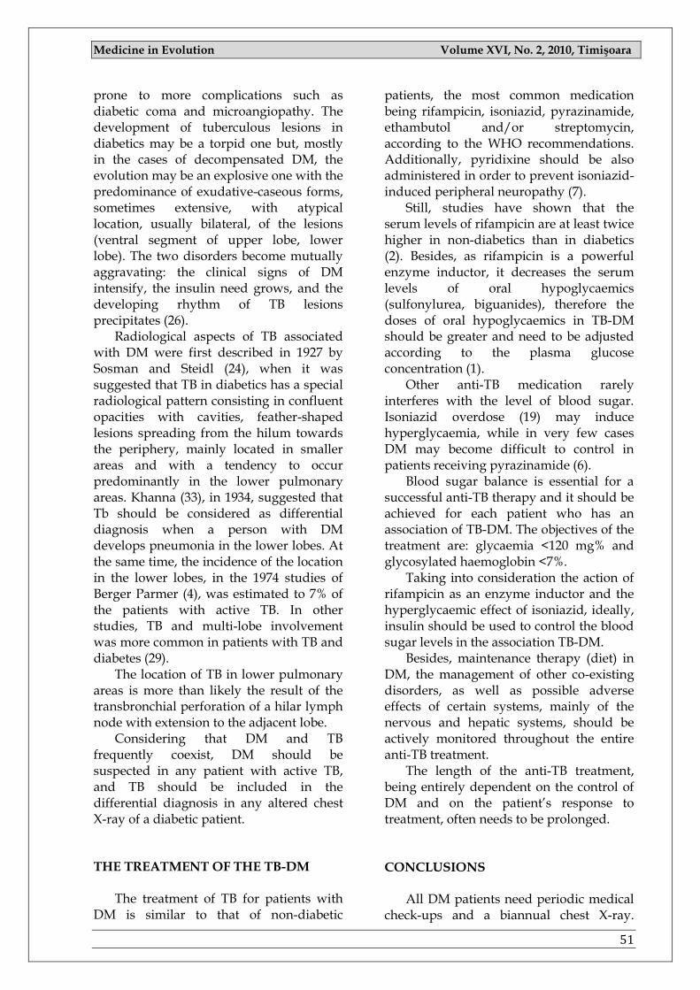
Medicine in Evolution Volume XVI, No. 2, 2010, Timişoara
51
prone to more complications such as diabetic coma and microangiopathy. The development of tuberculous lesions in diabetics may be a torpid one but, mostly in the cases of decompensated DM, the evolution may be an explosive one with the predominance of exudative-caseous forms, sometimes extensive, with atypical location, usually bilateral, of the lesions (ventral segment of upper lobe, lower lobe). The two disorders become mutually aggravating: the clinical signs of DM intensify, the insulin need grows, and the developing rhythm of TB lesions precipitates (26).
Radiological aspects of TB associated with DM were first described in 1927 by Sosman and Steidl (24), when it was suggested that TB in diabetics has a special radiological pattern consisting in confluent opacities with cavities, feather-shaped lesions spreading from the hilum towards the periphery, mainly located in smaller areas and with a tendency to occur predominantly in the lower pulmonary areas. Khanna (33), in 1934, suggested that Tb should be considered as differential diagnosis when a person with DM develops pneumonia in the lower lobes. At the same time, the incidence of the location in the lower lobes, in the 1974 studies of Berger Parmer (4), was estimated to 7% of the patients with active TB. In other studies, TB and multi-lobe involvement was more common in patients with TB and diabetes (29).
The location of TB in lower pulmonary areas is more than likely the result of the transbronchial perforation of a hilar lymph node with extension to the adjacent lobe.
Considering that DM and TB frequently coexist, DM should be suspected in any patient with active TB, and TB should be included in the differential diagnosis in any altered chest X-ray of a diabetic patient.
THE TREATMENT OF THE TB-DM
The treatment of TB for patients with
DM is similar to that of non-diabetic
patients, the most common medication being rifampicin, isoniazid, pyrazinamide, ethambutol and/or streptomycin, according to the WHO recommendations. Additionally, pyridixine should be also administered in order to prevent isoniazid-induced peripheral neuropathy (7).
Still, studies have shown that the serum levels of rifampicin are at least twice higher in non-diabetics than in diabetics (2). Besides, as rifampicin is a powerful enzyme inductor, it decreases the serum levels of oral hypoglycaemics (sulfonylurea, biguanides), therefore the doses of oral hypoglycaemics in TB-DM should be greater and need to be adjusted according to the plasma glucose concentration (1).
Other anti-TB medication rarely interferes with the level of blood sugar. Isoniazid overdose (19) may induce hyperglycaemia, while in very few cases DM may become difficult to control in patients receiving pyrazinamide (6).
Blood sugar balance is essential for a successful anti-TB therapy and it should be achieved for each patient who has an association of TB-DM. The objectives of the treatment are: glycaemia <120 mg% and glycosylated haemoglobin <7%.
Taking into consideration the action of rifampicin as an enzyme inductor and the hyperglycaemic effect of isoniazid, ideally, insulin should be used to control the blood sugar levels in the association TB-DM.
Besides, maintenance therapy (diet) in DM, the management of other co-existing disorders, as well as possible adverse effects of certain systems, mainly of the nervous and hepatic systems, should be actively monitored throughout the entire anti-TB treatment.
The length of the anti-TB treatment, being entirely dependent on the control of DM and on the patient‘s response to treatment, often needs to be prolonged.
CONCLUSIONS
All DM patients need periodic medical
check-ups and a biannual chest X-ray.
Medicine in Evolution Volume XVI, No. 2, 2010, Timişoara
52
These check-ups should be even more thorough for patients over 40 or with a
weigh that is 10% below the ideal body weight.
Any diabetic that suddenly develops a cough or weight loss, whose chest X-ray has alterations, or who needs increased doses of anti-diabetics (oral hypoglicaemics or insulin) to maintain their glycaemic balance, should be investigated for a possible TB.
As the data regarding the association TB-DM are still insufficient, despite all the
research conducted so far, research in the field should be continued because, if the association TB-DM is not recognized and properly treated in due time, it might endanger both the control of TB and the successful treatment of DM, especially since both epidemics represent a global public health problem.
REFERENCES 1. Acocella G, Canti R. Interaction of rifampicin with other drugs. Tubercle 1980, 61, 171 2. Alisjahbana B, Sahiratmadja E, Nelwan EJ, et al. The effect of type 2 diabetes mellitus on the presentation and treatment response of pulmonary tuberculosis. Clin Infect Dis. 2007;45:428-435. 3. Barach JH. Historical facts in Diabetes. Ann Med Hist; 1928, 10,387 4. Berger H. Lower lung field tuberculosis. Chest 1974; 65: 522-526
5. Fong Y, Marano MA, Moldawer LL ct. al. The acute splanchnic and peripheral tissue metabolic response to endotoxm in humans. JClin Invest 1990, 85, 1896
6. Girling DJ. Achersc effects of antitubcrculosis drugs. Dnigi 1982, 23:1, 56 7. Gupta S, Koirala J, Khardori R, Khardori N. Infections in diabetes mellitus and hyperglycemia. Infect Dis Clin North Am. 2007;21:617-638. 8. Kaslow RA. Infections in diabetes. In: Harris MI, Hammon RF, eds. Diabetes in America. Bethesda, MD: National Institutes of Health; 1985:XIXI-18. NIH publication 85-1468.
9. Koziel H, Koziel MJ. Pulmonary complications of diabetes mellitus. Infect Dis Clin North Am 1995, 9, 67
10. Karuchunskn MA, Gcrgcrt Via, lakovlcva OB. Specific features of cellular immunity of pulmonary tuberculosis in patients with diabetes mellitus; Problem Tnberk 1997, 6, 59 11. http://www.who.int/tb/publications/global_report/ 2008/pdf/key_points_es.pdf
12. Holden HM, Hiltz JE. The tuberculous diabetic. Can Med Asso J 1962. 87, 797
13. Ikezoe J.Takeuchi N, Johkoh Tctc. al. CT appearances of pulmonary tuberculosis in diabetic and immunocompromised patients: Comparison with patients who had no underlying disease. A J R 1992. 159, 1175 14. Jeon CY, Murray MB. Diabetes mellitus increases the risk of active tuberculosis: a systematic review of 13 observational studies. PLoS Med 2008;5:e152. 15. Joseph T Morris, Maj MC. Pulmonary Tuberculosis in Diabetics. Chest 1992;102;539-541
16. Mollcntzc WF, Pansegrouw DR, Stcyn AF. Diabetes mellitus, pulmonary tuberculosis and chronic calcific pancreatitis revisited. South Afr Med J 1990, 78 : 235
17. McMahon MM, Bistrian Bruce R. Host defenses and susceptibility to infection in patients with diabetes mellitus. Infect Dis Clin North Am 1995,9,1
18. MacRury Sm, Gcmtnel C G, Patcrson K R et al Changes in phagocytic function with glycacmic control in diabetic patients. J Clin Pathol 1989, 42, 1 143 19. Mandcll GL, Sande MA. Drugs used in the chemotherapy of tuberculosis and leprosy. In Goodman
and Oilman‘s - The pharmacological basis of Therapeutics. Ed. Oilman AG. Goodman LS. Gilman A. 6th
Edition Macmillan Publishing Co. Inc. New York, 1980. p1200 20. Muller LM, Gorter KJ, Hak E, et al. Increased risk of common infections in patients with type 1 and 2 diabetes mellitus. Clin Infect Dis. 2005;41:281-288. 21. Parrish NM, Dick JD, Bishai WR. Mechanisms of latency in Mycobacterium tuberculosis. Trends Microbiol 1998;6:107-112.
22. Repine J, Clawson C, Goetz F. Bactericidal function of neutiophils from patients with acute bacterial infections and from diabetes. J Inject Dis 1980, 142, 869

Medicine in Evolution Volume XVI, No. 2, 2010, Timişoara
53
23. Stewart GR, Robertson BD, Young DB. Tuberculosis: a problem with persistence. Nat Rev Microbiol 2003;1:97-105. 24. Sosman MC, Steidl JH. Diabetic tuberculosis. Am J Roentgenol 1927; 17:625
25. Shamoon M, Hcndlci R, Sherwin R. Synergistic interaction among anti-insulin hormones in the pathogcncsis of stress hyperglyccmia in humans J CIni Endocnnol Metab 1981,52, 1235
26. Smurova TF. Lung tuberculosis with associated diabetes mellitus. Excerpta Medico Chest Dis Thorac Surg Tuberc 1980,37,660
27. Tsukaguchi K, OkamuraH, Ikuno M ct al. The relation between diabetes mellitus and IFN-gamma, IL-12 and IL-IO production by CD4+ T cells and monocytcs in patients with pulmonary tuberculosis. Kekkaku 1997. 72, 617 28. Turina M, Fry DE, Polk HC Jr. Acute hyperglycemia and innate immune system: clinical, cellular, and molecular aspects. Crit Care Med. 2005;33:1624-1633.
29. Umut S, Tosun GA, Yildirin N. Radiographic location of pulmonary tuberculosis in diabetic patients. Chest 1994. 106. 326
30. Yu CT, Wang CH, Huang TJ et, al. Relation of bronchoalveolar lavage T lymphocyte subpopulations to rate of regression of active pulmonary tuberculosis Thorax 1995, 50, 869 31. World Health Organization. Definition, diagnosis and classification of diabetes mellitus and its complications. Part 1: Diagnosis and classification of diabetes mellitus. Geneva: World Health Organization; 1999. 32. Wild S, Roglic G, Green A, Sicree R, King H. Global prevalence of diabetes: estimates for the year 2000 and projections for 2030. Diabetes Care 2004;27:1047-1053. 33. Weaver R. Unusual radiographic presentation of pulmonary tuberculosis in diabetic patients. Am Rev Respiratory Dis. 1974; 109:162-63

Medicine in Evolution Volume XVI, No. 2, 2010, Timişoara
54
Medicine in Evolution Volume XVI, No. 2, 2010, Timişoara
55
PAGES 55 -58
ORTHODONTIC TREATMENT CONSIDERATIONS FOR THE SINGLE-TOOTH IMPLANT WHEN A LATERAL INCISOR IS MISSING
BORDEI ANCA GEORGIANA1, DRAGOŞ STANCIU1
1 Department of Orthodontics, Faculty of Dentistry, University of Medicine and Pharmacy
„Carol Davila” Bucureşti
Correspondence to: Bordei Anca Georgiana, [email protected]
ABSTRACT
Implant restorations have become a primary treatment option for the replacement of missing lateral incisors. The central incisor and canine often erupt in less than optimal positions adjacent to the edentulous lateral incisor space, and therefore preprosthetic orthodontic treatment is frequently required. Derotation of the central incisor and canine, space closure and correction of root proximities may be required to create appropriate space in which to place the implant and achieve an esthetic restoration.
Key words: CMV infection, methods of diagnosis, serology tests.
INTRODUCTION
The successful use of dental implants to replace missing teeth has been one of the most ―exciting and evolving areas of clinical dentistry‖ this decade. At a time when esthetic dentistry has gained prominence, permanent prosthodontic solutions such as implants have become optimal esthetic treatment options. While implants have expanded restorative treatment options, treatment planning has become more complex for the dental practitioner, and an interdisciplinary team approach is recommended.
This interdisciplinary approach may involve preprosthetic orthodontic treatment following consultations with an oral surgeon or periodontist and restorative dentist to ensure orthodontic
alignment will facilitate the surgical, implant and restorative treatment.
Aspects of orthodontic treatment required for implant restoration of missing lateral incisors are discussed. Orthodontic Treatment Planning
Treatment alternatives for restoring edentulous spaces resulting from missing laterals include removable partial dentures, conventional fixed bridges, resin-bonded bridges, orthodontic repositioning of canines to close the edentulous space, and single-tooth implant. Although adjacent teeth may have to be repositioned orthodontically to create adequate space for an implant, implants do not necessitate ―altering‖ or ―removing‖ parts of the natural dentition and are therefore the
Medicine in Evolution Volume XVI, No. 2, 2010, Timişoara
56
most conservative of the prosthodontic options for replacing missing lateral incisors. Implants can also maintain the alveolar ridge, enhance occlusal function and provide optimal esthetics.
Early investigation will give the patient time to explore all possible treatment options including implant restorations. A full set of orthodontic records including radiographs, models and clinical photographs are recommended for the diagnosis of congenitally missing laterals and to plan the preprosthetic orthodontic alignment. A diagnostic wax set-up is also beneficial for planning treatment and esthetics.
Participating clinicians — the orthodontist, periodontist, oral surgeon, restorative dentist, prosthodontist — should determine the patient‘s treatment plan collaboratively and communicate throughout the course of treatment to ensure all aspects of treatment are considered and the overall treatment objectives are achieved. The choice that have to be made is between opening or closing the space of the lateral incisor.If the space is to be opened for an implant, the canines will need to be moved distally, which may result in development of the alveolar ridge in the canine region. In cases where the occlusion and esthetics of the canine in the lateral position are acceptable, closure of the lateral space by the mesially positioned canine may be the simplest alternative treatment option. The benefit of space closure over prosthetic replacement depends on the specific occlusion as well as the morphology and esthetics of the canine.
When planning for the placement of a single-tooth implant, the orthodontist must ensure adequate space between the crowns and roots. Both the quantity and quality of alveolar bone must be assessed before implant placement is considered. To accommodate a standard implant there should be a minimum of 10 mm of inciso-gingival bone and a minimum of 6.0 mm of facial-lingual bone. In cases where there is insufficient alveolar bone for implant placement, ridge augmentation may be
necessary in addition to orthodontic repositioning of adjacent teeth. Adequate space for the implant is also required between the adjacent roots.
The average dental implant fixture is 3.75 mm wide, and 1 to 2 mm of space is necessary between the fixture and the adjacent roots.
Typically, between 6 and 8 mm of bone between the central and canine roots is recommended. Creating adequate space between the roots must be specifically addressed since the central and canine roots may be brought into closer proximity when the teeth are initially aligned orthodontically. To create adequate space for the implant, further orthodontic treatment may be necessary to move the roots further apart. Space for the coronal restoration must also be assessed. The average implant platform, which is 4.0 mm wide, requires a space of 1.0 mm mesially and distally between the platform and the adjacent tooth to facilitate proper healing and the development of a papilla postoperatively.
Thus, a minimum of 6 mm of space for the lateral crown is required.
As discussed above, one goal of orthodontic alignment is to achieve sufficient bone between the roots to place the implant. The roots of the central incisor and canine should be parallel to slightly divergent to avoid complications resulting from root proximity. Usually, the tip of the central incisor is approximately 5 degrees while that of the canine is 13 degrees, which means that the roots are slightly divergent.
There are additional mechanotherapy treatment options that can be used to orthodontically position the roots of the adjacent teeth and create adequate space for the implant. These include ideal placement of brackets to achieve the correct root and crown positions; bending the archwire to accentuate root divergence; or bonding a contralateral bracket on a central incisor (such as placing the maxillary right central incisor bracket on the maxillary left central incisor) to accentuate root divergence in the implant
Medicine in Evolution Volume XVI, No. 2, 2010, Timişoara
57
area. (Placement of the contralateral bracket on the canine is never indicated as this would cause the canine root to move into the edentulous area and compromise implant placement.)
Esthetics as well as occlusion must be considered in the final orthodontic positioning of the teeth adjacent to the edentulous space. To satisfy the ―golden proportion‖ principle of esthetics, the space for the maxillary lateral incisor should be approximately two-thirds of the width of the central incisor. However, if the patient is missing only one maxillary lateral incisor, the space required to achieve symmetrical esthetics and occlusion is primarily dictated by the width of the contralateral incisor. Once the permanent central incisor and canine have been positioned orthodontically to create adequate mesio-distal space between the crowns and the roots of the teeth, orthodontic retention is necessary to maintain this space and the position of the teeth. While the braces are in place, an acrylic denture tooth with a bonded bracket can be ligated to the archwire to further maintain space and improve esthetics. Following removal of the fixed appliances, conventional types of orthodontic retainers (Hawley retainers) can be used to maintain the space until the implant is placed and restored. Removable vacuum-form retainers containing bonded acrylic denture teeth are also acceptable in the interim as they prevent relapse in 3 dimensions.
However, caution should be taken when using vacuum-form retainers with respect to the occlusion especially when only one arch has been treated orthodontically.
Open bites, both anterior and posterior, may be created with a vacuum-form retainer as this type of full-coverage retainer may allow selective teeth to overerupt and thus create more orthodontic problems. When a patient does not plan to have implant treatment immediately, resin-bonded bridges or
transitional all-acrylic partial dentures are also acceptable options that satisfy the goals of retention, function and esthetics. The length of time in retention prior to implant placement will depend on the orthodontic treatment that was completed. For example, if significant rotations were corrected orthodontically, longer or possibly permanent retention may be required.
The optimal time for placement of implants is after growth of the maxilla, mandible and alveolus is complete. If implants are placed before growth is complete, the surrounding alveolar bone may continue to develop vertically and adjacent teeth may continue to erupt.
Thus a discrepancy between the gingival margins of the implant and the natural teeth is created and the implant appears to become submerged.
This creates a functional as well as an esthetic problem.For males, completion of facial growth, which often corresponds to general growth, may not occur until the age of 21 years; in young women, growth may be completed by age 15. If growth is complete, dental implants can be placed as soon as the edentulous space has been created and the tissues have stabilized following orthodontic treatment.
CONCLUSIONS
Dental implants are a treatment of choice for most patients with congenitally missing laterals. An implant will preserve tooth structure and alveolar bone and provide esthetics and function. However, successful restorative treatment involving implants depends on interdisciplinary treatment planning, especially if preprosthetic orthodontic tooth alignment is required.
The roots of the teeth adjacent to the edentulous implant region must be parallel or slightly divergent to create sufficient bone for implant placement, and there must be sufficient space between the crowns to place and restore the implant.

Medicine in Evolution Volume XVI, No. 2, 2010, Timişoara
58
REFERENCES
1. Schweizer CM, Schlegel AK, Rudzki-Janson I. Endosseous dental implants in orthodontic therapy. Int Dent J 1996; 46(2):61-8.
2. Phillips K, Kois J. Aesthetic peri-implant site development. The restorative connection. Dent Clin North Am 1998; 42(1):57-70.
3. Rupp RP, Dillehay JK, Squire CF. Orthodontics, prosthodontics, and periodontics: a multidisciplinary approach. Gen Dent 1997; 45(3):286-9.
4. Cronin RJ, Cagna DR. An update on fixed prosthodontics. J Am Dent Assoc 1997; 128(4):425- 36.
5. Millar BJ, Taylor NG. Lateral thinking: the management of missing upper lateral incisors. Br Dent J 1995; 79(3):99-106.
6. Bishara SE. Impacted maxillary canines: a review. Am J Orthod Dentofacial Orthop 1992; 101(2):159-71.
7. Kokich VG, Spear FM. Guidelines for managing the orthodontic-restorative patient. Semin Orthod 1997; 3(1):3-20.
8. Spear FM, Mathews DM, Kokich VG. Interdisciplinary management of single-tooth implants. Semin Orthod 1997; 3(1):45-72.
9. Shroff B, Siegel SM, Feldman S, Siegel SC. Combined orthodontic and prosthetic therapy. Special considerations. Dent Clin North Am 1996; 40(4):911-43.
10. Kokich V. Esthetics and anterior tooth position: an orthodontic perspective. Part III: Mesiolateral relationships. J Esthetic Dent 1993; 5(5):200-7.
11. Ong MA, Wang HL, Smith FN. Interrelationship between periodontics and adult orthodontics. J Clin Periodontol 1998; 25(4):271-7.
12. Hess D, Buser D, Dietschi D, Grossen G, Schonenberger A, Belzer UC. Esthetic single-tooth
replacement with implants: a team approach. Quintessence Int 1998; 29(2):77-86.
Medicine in Evolution Volume XVI, No. 2, 2010, Timişoara
59
PAGES 59 -64
INTEGRATED HEALTHCARE INFORMATION SYSTEM IN MEDICAL CARE
CĂLIN MUNTEAN1, COSMIN CÂTU1, LEONARD MADA2 ATENA GĂLUŞCAN3
1 Department of Medical Informatics & Biostatistics, “Victor Babeş” University of Medicine & Pharmacy, Timişoara 2 Syonic SRL 3 Department of Preventive Dentistry, Community and Oral Health, “Victor Babeş” University of Medicine & Pharmacy Timişoara
Correspondence to: Calin Muntean, Timisoara str.T. Vladimirescu nr. 14A, [email protected]
ABSTRACT
The healthcare industry faces growing scrutiny and pressure. On the one hand, healthcare organizations are expected to improve the quality of patient care, while on the other, they face demands to decrease the escalation of health care costs. Add to the mix increasing regulatory mandates for patient data security and escalating volumes of electronic information. Due to the evolution to electronic formats of patient data, the amount of information that is being exchanged is exploding. As we’ve all seen, Internet technology has become a dominant factor in business, academia, and healthcare, therefore the software architecture, design patterns and framework has been built for the complexities and challenges of the Web. Our model of IT Infrastructure is an independent integrated medical information system implementing a unique patient healthcare record in the setting of GPs, specialists and hospital healthcare system in Romania, built on a central database with a secure online interface. Setting: over 900 Romanian Healthcare Providers are using our model implemented in ICMed. Population: data was available for over 1000000 patients as of 2009. Requirements for an integrated healthcare IT system were analysed from the perspective of recent trends in IT technology and healthcare, European legislation, pharmaceutical industry, research developments and Romanian e-health policies. Recent trends emphasise two major directions: 1.) Quality management including assisted medical decision, error-proofing mechanisms, pharmacovigilance and adverse events handling and, 2.) Patient-physician shared electronic health records at a national and international level. Requirements for these features include a centralized health record integrating all medical data, methods to safeguard data security and confidentiality and flexible user interfaces to improve acceptance and reduce errors in a complex system. E-source is another development particularly useful for the pharmaceutical industry.
Key words: Web 2.0, Patient Healthcare Record, Quality Management
INTRODUCTION
Virtually every encounter between patient and healthcare provider creates data on health, treatment protocols, insurance eligibility and claims, billing and payment, etc. This in turn triggers a wide
range of healthcare community members beyond the patient and doctor: benefi ts administrators, insurance claims reviewers, payment processors, etc. The structure of modern healthcare systems involves integrating a variety of healthcare providers, starting with GPs and
Medicine in Evolution Volume XVI, No. 2, 2010, Timişoara
60
continuing with specialists in the outpatient setting and hospital inpatient setting as well as laboratory and pharmacy units. Our mission is to remove one obstacle - the high cost and complexity of clinical information software and IT Infrastructure. By leveraging the global resource network of healthcare institutions, universities, vendors and individuals, we design and build a high quality medical informatics system for GPs and Clinics seeking a low cost solution.
Quality management in medical healthcare is a complex task. Medical errors occur frequently and can cause substantial morbidity, mortality and costs [1,2]. Preventing these errors necessitates some challenging activities, insofar as even measuring adverse events is faced with difficulty [3,4]. Incomplete medical records, workload and time limitations, complex medical tasks, poor user interfaces and many other factors contribute to this high rate of medical errors. The gateway to a patient's medical record is centred on the general practitioner. Shortcomings in the healthcare stem therefore often directly or indirectly from limitation in the primary health setting. Previous studies have shown a significant variation in physician performance, poor performance being associated with advanced age [5,6], level of clinical experience [7], availability of continuous medical education and numerous other factors. General strategies have been proposed to improve the quality of medical care [8], including various campaigns by the Institute of Healthcare Improvement [9], The Six Sigma Way [10], Evidence Based Medicine-specific methods and a continuous medical education.
Measuring outcomes is one of the most important aspects during any quality assessment. The NHS compiled a set of outcome measures to guide the reimbursement of General Practitioners in the UK and to foster the development of high quality primary medical care. [11]
Analysing outcomes in the primary healthcare is however hampered by the low mortality in this setting, the long time spans that need to be followed, and the
very heterogeneous population of patients and diseases. As a consequence, a medical informatics system capturing all the relevant information over lengthy periods of time offers a clear advantage over peer reviews and auditing strategies. ICmed is a healthcare IT system that integrates all major medical players using a unique patient record. It was developed initially around primary healthcare providers and was later extended towards outpatient specialty and inpatient hospital care, as well as integration with local medical laboratories and pharmacies. This enables a single, trusted patient record across multiple channels, provider networks, and diverse internal systems that manage patient and service information. It allows a consolidated view of member benefits across multiple member products -such as medical, detail, behavioural health, pharmacy, flexible spending accounts, health savings accounts, and benefit spending cards - and multiple healthcare applications. The objective of this study was to analyse future trends in the development of medical information systems and use this knowledge to improve our model of medical IT framework. Further, we analysed the usage pattern of the model in primary healthcare setting in order to verify the adequacy of this IT system in monitoring physicians‘ performance in the primary healthcare setting.
MATERIALS AND METHODS
Two authors searched the medical literature and the medical legislation in various European countries and the US for recent advances and developments in the medical field. This study was centred on corroborating these requirements with actual medical data collected from healthcare providers using our model of integrated healthcare IT system and with technical challenges posed by these requirements. Initially, the model was centred on primary healthcare, being later extended to cover both outpatient specialty and inpatient care. This framework is
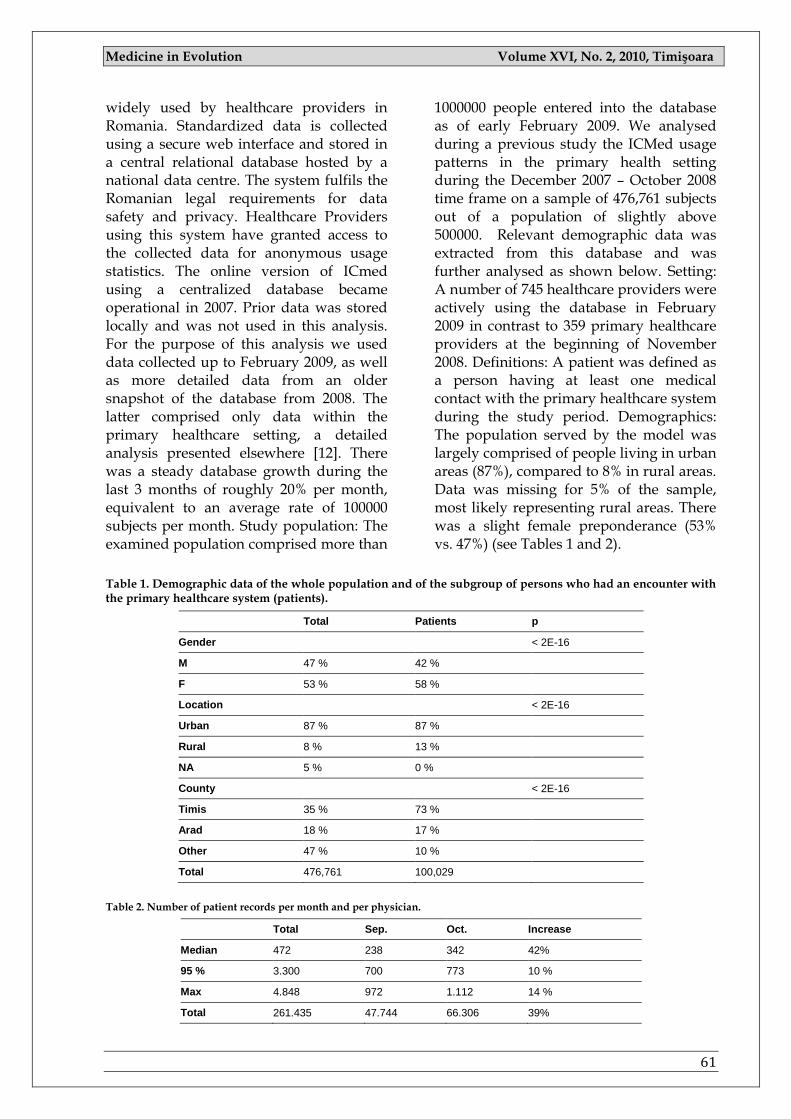
Medicine in Evolution Volume XVI, No. 2, 2010, Timişoara
61
widely used by healthcare providers in Romania. Standardized data is collected using a secure web interface and stored in a central relational database hosted by a national data centre. The system fulfils the Romanian legal requirements for data safety and privacy. Healthcare Providers using this system have granted access to the collected data for anonymous usage statistics. The online version of ICmed using a centralized database became operational in 2007. Prior data was stored locally and was not used in this analysis. For the purpose of this analysis we used data collected up to February 2009, as well as more detailed data from an older snapshot of the database from 2008. The latter comprised only data within the primary healthcare setting, a detailed analysis presented elsewhere [12]. There was a steady database growth during the last 3 months of roughly 20% per month, equivalent to an average rate of 100000 subjects per month. Study population: The examined population comprised more than
1000000 people entered into the database as of early February 2009. We analysed during a previous study the ICMed usage patterns in the primary health setting during the December 2007 – October 2008 time frame on a sample of 476,761 subjects out of a population of slightly above 500000. Relevant demographic data was extracted from this database and was further analysed as shown below. Setting: A number of 745 healthcare providers were actively using the database in February 2009 in contrast to 359 primary healthcare providers at the beginning of November 2008. Definitions: A patient was defined as a person having at least one medical contact with the primary healthcare system during the study period. Demographics: The population served by the model was largely comprised of people living in urban areas (87%), compared to 8% in rural areas. Data was missing for 5% of the sample, most likely representing rural areas. There was a slight female preponderance (53% vs. 47%) (see Tables 1 and 2).
Table 1. Demographic data of the whole population and of the subgroup of persons who had an encounter with the primary healthcare system (patients).
Total Patients p
Gender < 2E-16
M 47 % 42 %
F 53 % 58 %
Location < 2E-16
Urban 87 % 87 %
Rural 8 % 13 %
NA 5 % 0 %
County < 2E-16
Timis 35 % 73 %
Arad 18 % 17 %
Other 47 % 10 %
Total 476,761 100,029
Table 2. Number of patient records per month and per physician.
Total Sep. Oct. Increase
Median 472 238 342 42%
95 % 3.300 700 773 10 %
Max 4.848 972 1.112 14 %
Total 261.435 47.744 66.306 39%
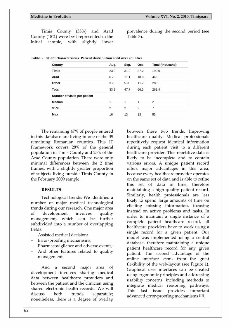
Medicine in Evolution Volume XVI, No. 2, 2010, Timişoara
62
Timis County (35%) and Arad County (18%) were best represented in the initial sample, with slightly lower
prevalence during the second period (see Table 3).
Table 3. Patient characteristics. Patient distribution split over counties.
County Aug. Sep. Oct. Total (thousand)
Timis 23.3 31.0 37.2 190.0
Arad 6.7 11.1 18.0 44.0
Other 3.7 5.9 11.7 28.5
Total 33.8 47.7 66.3 261.4
Number of visits per patient
Median 1 1 1 2
95 % 2 2 2 7
Max 16 13 13 53
The remaining 47% of people entered
in this database are living in one of the 39 remaining Romanian counties. This IT Framework covers 28% of the general population in Timis County and 25% of the Arad County population. There were only minimal differences between the 2 time frames, with a slightly greater proportion of subjects living outside Timis County in the February 2009 sample.
RESULTS
Technological trends: We identified a number of major medical technological trends during our research. One major area of development involves quality management, which can be further subdivided into a number of overlapping fields:
Assisted medical decision;
Error-proofing mechanisms;
Pharmacovigilance and adverse events;
And other features related to quality management.
And a second major area of
development involves sharing medical data between healthcare providers and between the patient and the clinician using shared electronic health records. We will discuss both trends separately; nonetheless, there is a degree of overlap
between these two trends. Improving healthcare quality: Medical professionals repetitively request identical information during each patient visit to a different healthcare provider. This repetitive data is likely to be incomplete and to contain various errors. A unique patient record offers major advantages in this area, because every healthcare provider operates on the same set of data and is able to refine this set of data in time, therefore maintaining a high quality patient record. Similarly, health professionals are less likely to spend large amounts of time on eliciting missing information, focusing instead on active problems and tasks. In order to maintain a single instance of a complete patient healthcare record, all healthcare providers have to work using a single record for a given patient. Our model was implemented using a central database, therefore maintaining a unique patient healthcare record for any given patient. The second advantage of the online interface stems from the great flexibility of the web-layout (see Figure 1). Graphical user interfaces can be created using ergonomic principles and addressing usability concerns, including methods to integrate medical reasoning pathways. This last issue provides important advanced error-proofing mechanisms [12].
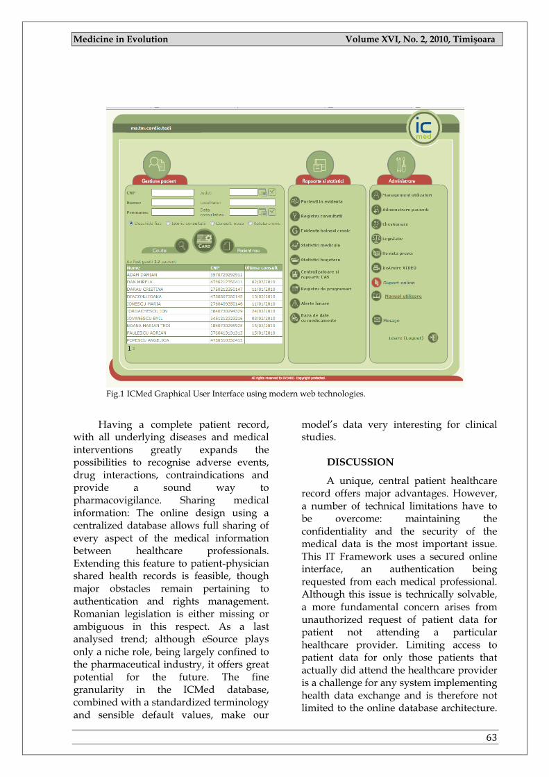
Medicine in Evolution Volume XVI, No. 2, 2010, Timişoara
63
Fig.1 ICMed Graphical User Interface using modern web technologies.
Having a complete patient record,
with all underlying diseases and medical interventions greatly expands the possibilities to recognise adverse events, drug interactions, contraindications and provide a sound way to pharmacovigilance. Sharing medical information: The online design using a centralized database allows full sharing of every aspect of the medical information between healthcare professionals. Extending this feature to patient-physician shared health records is feasible, though major obstacles remain pertaining to authentication and rights management. Romanian legislation is either missing or ambiguous in this respect. As a last analysed trend; although eSource plays only a niche role, being largely confined to the pharmaceutical industry, it offers great potential for the future. The fine granularity in the ICMed database, combined with a standardized terminology and sensible default values, make our
model‘s data very interesting for clinical studies.
DISCUSSION
A unique, central patient healthcare record offers major advantages. However, a number of technical limitations have to be overcome: maintaining the confidentiality and the security of the medical data is the most important issue. This IT Framework uses a secured online interface, an authentication being requested from each medical professional. Although this issue is technically solvable, a more fundamental concern arises from unauthorized request of patient data for patient not attending a particular healthcare provider. Limiting access to patient data for only those patients that actually did attend the healthcare provider is a challenge for any system implementing health data exchange and is therefore not limited to the online database architecture.
Medicine in Evolution Volume XVI, No. 2, 2010, Timişoara
64
The usage of standardized coding systems (ATC, ICD10), allows advanced methods of pharmacovigilance. Although WHO-DRL (MEDRA) and SNOMED add a more detailed description of drugs and medical terms, they are not free to use, and there are no Romanian translations of these systems. Our model already implements a detailed drugs database covering every single drug registered in Romania, putting therefore advanced analysis possibilities into this framework.
CONCLUSIONS
Our model offers a flexible framework to integrate all healthcare providers using a centralized database with online access. This architecture offers completely new opportunities in quality management. Extending this model into additional areas necessitates addressing further issues, including continuous security improvements and advances in the pertinent legislation.
REFERENCES
1. J.M. Aranaz-Andres, C. Aibar-Remon, J. Vitaller-Murillo, P. Ruiz-Lopez, R. Limon-Ramirez, E. Terol-Garcia, the ENEAS work group, Incidence of adverse events related to healthcare in Spain: results of the Spanish National Study of Adverse Events. J. Epidemiol. Community Health 62 (2008), 1022-1029.
2. S.N. Weingart, R.M. Wilson, R.W. Gibberd, B. Harrison, Epidemiology of medical error, BMJ 320 (2000), 774-777.
3. P. Michel, J.L. Quenon, A.M. de Sarasqueta, O. Scemama, Comparison of three methods for estimating rates of adverse events and rates of preventable adverse events in acute care hospitals, BMJ 328 (2004), 199.
4. R.A. Hayward, T.P. Hofer, Estimating hospital deaths due to medical errors: preventability is in the eye of the reviewer, JAMA 286 (2001), 415-420.
5. R. McAuley, W. Paul, G. Morrison, et al., Five-year results of the peer assessment program of the College of Physicians and Surgeons of Ontario, CMAJ 143 (1990), 1193-1199.
6. Charles, H. Britt, L. Valenti, The independent effect of age of general practitioner on clinical practice, MJA 185 (2) (2006), 105-109.
7. N.K. Choudhry, R.H. Fletcher, S.B. Soumerai, Systematic review: the relationship between clinical experience and quality of healthcare, Ann Intern Med 142 (2005), 260-273.
8. T. Bodenheimer, The American Healthcare System. The Movement for Improved Quality in Healthcare, New Eng J Med 340 (1999), 488-492.
9. J.L. Reinertsen, M. Bisognano, M.D. Pugh, Seven Leadership Leverage Points for Organization-Level Improvement in Healthcare (Second Edition), Massachusetts: Institute for Healthcare Improvement, Cambridge, 2008 (Available on www.ihi.org).
10. J.O. Westgard, Six Sigma Quality Design and Control (Second Edition). Westgard QC, Inc, 2006. 11. NHS Employers and the General Practitioners Committee, Quality and Outcomes Framework
guidance for GMS contract 2008/09. Delivering investment in general practice, 2008. 12. N.G. Leveson, System Safety Engineering: Back To The Future, available online from
http://sunnyday.mit.edu/book2.pdf.
Medicine in Evolution Volume XVI, No. 2, 2010, Timişoara
65
PAGES 65 -69
ORAL HYGIENE MONITORING BY PATIENTS WITH FIXED APPLIANCES
DANIELA JUMANCA1, ANGELA CODRUŢA PODARIU1, ATENA GĂLUŞCAN1, ROXANA OANCEA1, RAMONA POPOVICI1, RUXANDRA SAVA ROŞIANU1, ANITA ROŞU1
1 Department of Preventive Dentistry, Community and Oral Health, “Victor Babeş” University of Medicine & Pharmacy Timişoara
Correspondence to: Daniela JumancaTimisoara, str.T.Vladimirescu nr.14A e-mail: [email protected]
ABSTRACT
If we look around, we will see many teenagers and young adults wearing fix appliances. So, braces aren't really as bad as we may think. Today, braces come in a variety of styles and materials, making „life with braces” much easier, more comfortable, and even more stylish than in the past. However, many people feel more comfortable and self-confident with properly aligned, attractive teeth, and orthodontic care can help improve appearance and build self-confidence. But one of the most difficult problems in orthodontic treatment with fixed appliances is the control of enamel demineralization around the brackets and the bands. These elements, together with other adjuvant elements used in orthodontic treatment (such as: arches, springs, elastics, power chains, etc.), make the patient’s dental hygiene more difficult and the accumulation of plaque easier. That’s why, a significant increases in oral bacteria appears during orthodontic treatment, especially to those with fix appliances. White spots, caries, gingivitis (and sometimes periodontitis) are often a side effect of orthodontic treatment with fixed appliances that is caused by inadequate removal of dental plaque around the orthodontic appliances.
Key words: Prevention, oral hygiene, demineralization, white spots, dental plaque.
INTRODUCTION
Oral hygiene is very important during the orthodontic treatment. Cleaning the teeth is one of the simplest and least invasive ways to maintain a beautiful smile and healthy teeth. Regular brushing and flossing help keep teeth and gums healthy, but only a professional cleaning by a dentist or dental hygienist can prevent or delay the onset of white spots, caries and periodontal diseases. Some deposits such as dental plaque and tartar are difficult to remove through brushing alone, so regular professional cleanings help reduce these decay-causing agents. The prevalence of demineralization in orthodontic patients
has been reported between 2% and 96%.1,2,3 It has been generally accepted that the combined application of fluoride regimes, oral hygiene instructions, and dietary control can contribute greatly to the inhibition of demineralization during fixed-appliance treatment.3,4 These methods, however, rely on patient compliance. Other noncompliant methods have been created to deliver fluoride adjacent to orthodontic appliances.5 Demineralisation takes place when specific bacteria are retained for a long time on the enamel surface. The bacteria metabolize fermentable carbohydrates and produce organic acids and these acids dissolve the
Medicine in Evolution Volume XVI, No. 2, 2010, Timişoara
66
calcium phosphate mineral of the enamel and dentin, resulting in demineralization.6 Demineralisation is first observed clinically as white spot lesions. The deminesalized area beneath the dental plaque and the body of the enamel lesion can lose as much as 50% of the original mineral content.7 Enamel decalcification around orthodontic bands and brackets has long been a concern.8 Studies showed that orthodontic appliances increase the accumulation and adherence of plaque in mouth.9 Streptococcus mutans and Lactobacillus concentrations in the oral cavity increase in conjunction with orthodontic treatment and fixed appliances.10 These and other bacteria ferment carbohydrates to produce organic acids. These acids can, over time, lead to the dissolution of calcium and phosphate ions from the enamel surfaces. This process of decalcification may lead to white spot lesions and even capitation in a very short decade: after only 4 weeks.11
Keeping their smile healthy and bright is one of the most important ways people can care for their health and appearance. The suggested frequency of professional teeth cleanings depends on a number of factors. Children and adults with healthy teeth and gums should have their teeth cleaned twice a year, while people who are wearing a fix appliance should have their teeth checked more frequently (once in a month or at least in 6 weeks). Early detection of oral problems gives more options to treat potential complications with less pain and financial hardship.
AIM AND OBJECTIVES
The aim of this study was to evaluate the effects of a bad oral hygiene versus a good oral hygiene in patients wearing fix appliances. This includes: professional cleanings ones in a month (also known as dental prophylaxis) performed by the dentist (or dental hygienist); teeth brushing minimum twice a day (with a manual or electric toothbrush); use of interdental brushes; use of dental floss (minimum once a day) together with fluoride treatment (toothpaste and mouthwash specifically
formulated), to prevent tooth demineralisation and decay, gum disease and to preserve the health of the teeth in general.
MATERIAL AND METHODS
A number of 24 subjects requiring orthodontic treatment with fix appliances (with ages between 16 and 22) were enrolled in the study (during a period of 3 years). All subjects were informed and educated about what an orthodontic treatment means; and about what are they allowed to do or not. The subjects were divided into two groups: (A) - one group received repeated professional tooth cleaning combined with oral hygiene instruction; and the control group (B) - received oral hygiene instruction only at the beginning of the treatment. The smooth vestibular surfaces of the teeth and the proximal areas were scanned with the DIAGNOdent device; and the measured values at the baseline and after the period of orthodontic treatment were compared. There were a significant difference in the DIAGNOdent readings between the first and the final evaluations in the control group (B), but there were no significant difference between the two evaluations in group (A). At the beginning – before the first scanning, the profesional teeth cleaning was performed using the Airflow device. The Airflow device is ideal for professional cleaning and is using a jet formed by a mixture of air, powder and water; it removes all dental plaque, soft deposits and surface stains from pits, fissures, interproximal spaces and smooth surfaces of the teeth. This method is efficient and effective for surface preparation of the tooth before bracket placement; it is efficient and effective for plaque removal around bracketed and banded teeth and is also suitable for removal of adhesive after bracket debonding. During the orthodontic treatment it is necessary to remove the plaque properly. Therefore, there is a higher risk of white spots, caries, gingivitis or even parodontitis, following a treatment
Medicine in Evolution Volume XVI, No. 2, 2010, Timişoara
67
with fixed appliances. A professional teeth cleaning is particularly necessary for patients who are not willing or not able to perform the required oral hygiene. These appliances present a greater challenge in routine polishing due to the fact that brackets, wires and elastics impede access with the rubber cup. Plaque removal with the Airflow is much faster than any conventional cleaning technique because the removal of arch wire ligatures and springs is not necessary. It is to be noted that this does not damage the appliance. Furthermore, orthodontic appliances are not influenced by this method, whilst the rubber cup technique may cause, in some cases, minor damage to brackets or arch wires. After professional cleaning with airflow system, the teeth were scanned with the DIAGNOdent device. This is an instrument used to assess integrity of enamel smooth surface and proximal area in pre- and post-bonded orthodontic patients. Modern management of dental caries has three major components: prevention, control, and treatment, and is based on appropriate diagnosis of the disease and detection of pathological changes (lesion formation in its earliest stages). DIAGNOdent is a useful tool for early detection of initial carious lesion. In the late 90‗, a laser fluorescence device, the DIAGNOdent (DIAGNOdent; KaVo, Biberach, Germany) has been developed for more objective caries diagnosis. Diagnosis using this device is based on the fact that the fluorescence emitted from carious surfaces is greater than that emitted from sound surfaces when they are irradiated with a laser beam with a wavelength of 655 nm.
The DIAGNOdent consists of a probe, a fiber-optic lead and a main unit that generates the laser beam (wavelength: 655 nm). There are two types of tip – a cone-shaped tip and a broad tip – that are placed on the probe of the DIAGNOdent. The cone-shaped tip is suited for diagnosis of fissure caries and the broad tip for diagnosis of smooth surface caries. The DIAGNOdent displays the real time values
and maximum values of the fluorescence in the test site. Changes in the tooth substances associated with progression of the carious process are reflected in an increased amount of fluorescent light. The cause of this increased level of fluorescence was the presence of chromofores associated with bacteria present in the infected tooth structure. A numerical value (0-99) is assigned to the degree of fluorescence as an indicator of the extent of caries. The device helps in recognition of initial carious lesion (pits and fissures, smooth surfaces, proximal surfaces),when there is no possibility of clinical and radiographic detection; and in monitoring of existing lesions.The assessment of a tooth with the laser fluorescence system was carried out as follows: after calibration with a ceramic standard, the fluorescence of a sound spot on the smooth surface of the tooth was measured in order to provide a baseline value for that tooth. This value was then subtracted electronically from the fluorescence of the site to be measured. In order to get the maximum extension of caries or tooth demineralisation, the instrument was tilted around the measuring site. This ensured that the tip picked up fluorescence where the demineralisation has the maximum value. A rising tone, starting at a value of 15, helped the examiner to find the maximum fluorescence value of the site under study. Comparison of at least 2 consecutive measurement of the same site within a few months indicates the level of caries activity.
RESULTS:
After the examination with DIAGNOdent device the following values were obtained:
Values between 2-10: no active lesions
Values between 10-25: initial carious lesion
Values between 25-35: superficial dentinal caries
Values over than 35: deep dentinal caries

Medicine in Evolution Volume XVI, No. 2, 2010, Timişoara
68
Acording to the obtained values (included in the label), in the control group (B) it is a significant difference in the DIAGNOdent readings between the first and the final evaluations. In group (A), there have been no significant differences between the two evaluations. In group (A), it was a problem with the pacient nr.9, especially in the lower jaw, because of week dental hygiene. The values measured after the debonding were up to 18; indicated initial carious lesions. The enamel structure was affected and there
occurred white spots. Those, with a proper teeth brushing and with the topical fluoridation, we obtained a remineralistion. In the control group (B), occurred problems with 7 of the patients. Nr. 4 was the most affected and needed composite restorations at 1.5., 1.4., 4.4 and 4.5. The other demineralisations were in a reversible stage.This means that oral hygiene, especially professional cleaning is of major importance in mentioning tooth health during orthodontic treatment with fix appliances.
REFERENCES:
1. Zabokova-Bilbilova E, Stafilov T, Sotirovska-Ivkovska A, Sokolovska F. Prevention of enamel demineralization during orthodontic treatment: An in vitro study using GC Tooth Mousse. Balk J Stom.2008;12:133–137.
2. Basdra EK, Huber H, Komposch Fluoride released from orthodontic bonding agents alters the enamel surface and inhibits enamel demineralization in vitro. Am J Orthod Dentofac Orthop. 1996;109:466–472.
3. Bloom RH, Brown LR. A study of the effects of orthodontic appliances on the oral microbial flora. Oral Surg. 1964;17:658–667. [PubMed]
4. Mizrahi E. Enamel demineralization following orthodontic treatment. Am J Orthod. 1982;82:62–67.[PubMed]
5. Årtun J, Brobakken BO. Prevalence of carious white spots after orthodontic treatment with multibonded appliances. Eur J Orthod. 1986;8:229–234. [PubMed]
6. Kukleva MP, Shetkova DG, Beev VH. Comparative age study of the risk of demineralization during orthodontic treatment with brackets. Folia Med. 2002;44:56–59.
7. Gorelick L, Geiger AM, Gwinnett AJ. Incidence of white spot formation after bonding and banding. Am J Orthod. 1982;81:93–98. [PubMed]
8. Mitchell L. Decalcification during orthodontic treatment with fixed appliances—an overview. Br J Orthod.1992;19:199–205. [PubMed]
9. Millet DT, Nunn JH, Welbury RR, Gordon PH. Decalcification in relation to brackets bonded with glass ionomer cement or a resin adhesive. Angle Orthod. 1999;69:65–70. [PubMed]
10. Schmit JL, Staley RN, Wefel JS, Kanellis M, Jakobsen JR, Keenan PJ. Effect of fluoride varnish on demineralization adjacent to brackets bonded with RMGI cement. Am J Orthod Dentofacial Orthop.2002;122:125–134. [PubMed]
11. de Bruyn H, Arends J. Fluoride varnishes: a review. J Biol Buccale. 1987;15:71–82. [PubMed] 12. Helfenstein U, Steiner M. Fluoride varnishes (Duraphat): a meta-analysis. Community Dent
Oral Epidemiol. 1994;22:1–5. [PubMed] 13. Arends J, Lodding A, Petersson LG. Fluoride uptake in enamel. In vitro comparison of topical
agents.Caries Res. 1980;14:403–413. [PubMed] 14. Sudjalim TR, Woods MG, Manton DJ. Prevention of white spot lesions in orthodontic practice:
a contemporary review. Aust Dent J. 2006;51:284–289. [PubMed] 15. Vorhies AB, Donly KJ, Staley RN, Wefel JS. Enamel demineralization adjacent to orthodontic
brackets bonded with hybrid glass ionomer cements: an in vitro study. Am J Orthod Dentofacial Orthop.1998;114:668–674. [PubMed]
16. Farrow ML, Newman SM, Oesterle LJ, Shellhart WC. Filled and unfilled restorative materials to reduce enamel decalcification during fixed-appliance orthodontic treatment. Am J Orthod Dentofacial Orthop.2007;132:578, e1–6. [PubMed]
17. Antonucci JM, Skrtic D. Matrix resin effects on selected physicochemical properties of amorphous calcium phosphate composites. J Bioact Comput Polym. 2005;20:29–49.
18. Reich E, Lussi A, Newbrun E. Caries-risk assessment. Int Dent J. 1999;49:15–26. [PubMed]

Medicine in Evolution Volume XVI, No. 2, 2010, Timişoara
69
19. Sudjalim TR, Woods MG, Manton DJ, Reynolds EC. Prevention of demineralization around orthodontic brackets in vitro. Am J Orthod Dentofacial Orthop. 2007;131:705e1–e9. [PubMed]
20. Benson PE, Shah AA, Millett DT, Dyer F, Parkin N, Vine RS. Fluorides, orthodontics and demineralization: a systematic review. J Orthod. 2005;32:102–114. [PubMed]
21. Lussi A, Hellwig E. Performance of a new laser fluorescence device for the detection of occlusal caries in vitro. J Dent. 2006;34:467–471. [PubMed]
22. Gurbuz T, Yilmaz Y, Sengul F. Performance of laser fluorescence for residual caries detection in primary teeth. Eur J Dent. 2008;2:176–184. [PubMed]
23. Bader JD, Shugars DA. A systematic review of the performance of a laser fluorescence device for detecting caries. J Am Dent Assoc. 2004;135:1413–1426. [PubMed]
24. Novaes TF, Matos R, Braga MM, Imparato JC, Raggio DP, Mendes FM. Performance of a Pen-Type Laser Fluorescence Device and Conventional Methods in Detecting Approximal Caries Lesions in Primary Teeth - in vivo Study. Caries Res. 2009;43:36–42. [PubMed]
25. Gorton J, Featherstone JD. In vivo inhibition of demineralization around orthodontic brackets. Am J Orthod Dentofacial Orthop. 2003;123:10–14. [PubMed]

Medicine in Evolution Volume XVI, No. 2, 2010, Timişoara
70
Medicine in Evolution Volume XVI, No. 2, 2010, Timişoara
71
PAGES 71 - 80
ORAL GLUCOSE-LOWERING AGENTS, INSULIN RESISTANCE AND ENDOTHELIAL DYSFUNCTION
RĂZVAN MIHAI ŞUŞAN1, MONICA MARIA ŞUŞAN1, CORINA ŞERBAN1, CONSTANTIN TUDOR1
1 University of Medicine & Pharmacy “Victor Babeş” Timişoara
Correspondence to: Razvan Mihai Susan e-mail: [email protected]
ABSTRACT
Insulin resistance and endothelial dysfunction are frequently comorbid states. Insulin resistance is characterized by the diminished ability of insulin to initiate intracellular signaling. Endothelial cells are a very active substrate, secreting mediators essential in modulating vascular tone, inflammation, haemostasis, cell proliferation and oxidative stress. Endothelial dysfunction is defined as inadequate endothelial-mediated vasodilatation. The 2007 European Society of Cardiology (ESC) and European Association for the Study of Diabetes (EASD) guide on pre-diabetes and cardiovascular disease recommended following drugs for the insulin resistance therapy: biguanides, thiazolidinediones and alpha-glucosidase inhibitors. This article discusses the choice of specific antihyperglycemic agents targeting insulin resistance, not only for their effectiveness in lowering glucose, but also for their extraglycemic effects, especially on endothelial dysfunction in order to reduce cardiovascular complications. The choise of the insulin sensitizing agent with the best cardioprotective profile depends on duration and complications of disease, safety profiles, tolerability, ease of use and expense.
Key words: insulin resistance, insulin sensitizers, endothelial dysfunction, cardiovascular disease.
INTRODUCTION
Insulin resistance (IR) and endothelial dysfunction (ED) are frequently comorbid states. Cellular, physiological, epidemiological and clinical studies strongly support a reciprocal relationship between ED and IR. IR may be a prelude (like prediabet status) to or a part of type 2 diabetes (T2D) features.
Much evidence supports the presence of IR as the fundamental pathophysiological disturbance responsible for the cluster of metabolic and cardiovascular disorders, known as the metabolic syndrome. The presence of IR and ED represents early changes in individuals at high risk for developing
cardiovascular disease. The metabolic syndrome identifies a group of subjects who are at very high risk (>20% for those with diabetes and 10-20% for those with two or more risk factors) to experience a future cardiovascular event. It is very important to determinate the efficacy of the oral glucose-lowering therapy on ED because indication of glucose-lowering drugs based exclusively upon their glycaemic effects is no longer an option and became scientifically unwise. Several late trials emphasise the beneficial role of the therapy with insulin sensitizing agents in treating the early, subclinical stages of cadiovascular disease. In addition to variable effects on glycemia, specific effects
Medicine in Evolution Volume XVI, No. 2, 2010, Timişoara
72
of this therapy on cardiovascular disease (CVD) risk factors were also considered important. This article discusses the choice of specific antihyperglycemic agents targeting IR, not only for their effectiveness in lowering glucose, but also for their extraglycemic effects, especially on ED in order to reduce long-term cardiovascular complications.
Insulin Resistance and Endothelial
Dysfunction
There is a clear association between insulin sensitivity and vascular endothelial function in normal subjects, obese individuals and in patients with T2D. Several studies have demonstrated that insulin has direct physiological effects on the normal endothelium and increases nitric oxide (NO) availability. IR is a generalized phenomenon affecting many tissues. As a consequence, altered insulin signalling in the endothelium may represent a logical candidate for the underlying mechanism for both IR and ED.
Beyond their morphological function, endothelial cells are a very active substrate, secreting mediators essential in modulating vascular tone, inflammation, haemostasis, cell proliferation and oxidative stress.
Endothelium also contributes to mitogenesis, angiogenesis, vascular permeability, and fluid balance. The endothelial cells produce vasodilator substances such as nitric oxide, prostacyclin, endothelium-derived hyperpolarizing factors, C-type natriuretic peptide and vasoconstrictors such as endothelin-1 (ET-1), angiotensin II (AT II), thromboxane A2 and reactive oxygen species (ROS).
Endothelium participates in inflammation by producing inflammatory modulators - NO, intercellular adhesion molecule (ICAM), vascular adhesion molecule (VCAM), E-selectin, nuclear factor kappa-B (NFkB) and in haemostasis by synthesising plasminogen activator, tissue factor inhibitor, von Willebrand factor, NO, prostacyclin, thromboxane A2,
plasminogen-activator inhibitor-1 (PAI-1) and fibrinogen. [1] ED is defined as paradoxical or inadequate endothelial-mediated vasodilatation. ED is the result of an imbalance between the action of vasodilatator and vasoconstrictor factors, which favours vasoconstriction. This imbalance manifests like impaired endothelium-dependent vasodilatation and decreased NO bioavailability, due to decreased NO synthesis or sequestration of NO. ED appears to be the initiating event in atherosclerosis, contributing to ischemic coronary artery disease and may be of great value in predicting the risk of cardiovascular events. When vascular endothelium is intact, L-arginine is converted by the enzyme endothelial nitric oxide synthase (eNOS) on L-arginine aminoacid, producing NO and L-citruline, requiring O2 and Nicotinamide Adenine Dinucleotid Phosphate (NADP) coenzyme. An endogenous competitive inhibitor of eNOS is asymmetric dimethylarginine (ADMA). The enzyme eNOS has three known isoforms: eNOS-I (from neurological tissue) and eNOS-III (from endothelial cells) which are mainly constitutive, both responding to agonist that increases intracellular calcium, and eNOS-II (expressed in macrophage and endothelial cells due to the effect of pro-inflammatory cytokines) which can release several times more NO and for longer periods than the constitutive eNOS. [2] IR is an impairment of insulin-stimulated glucose and/or lipid metabolism, compared to the response in healthy subjects, and it is characterized by the diminished ability of insulin to initiate intracellular signalling.
The normal cellular response to insulin is mediated by two classical pathways involving phosphatidylinositol 3 kinase (PI3K) and mitogen-activated protein kinase (MAPK). IR is characterized by impairment in PI3K-dependent signalling, whereas other insulin-signalling mechanisms, including MAPK-dependent pathway, are not affected. Compensatory increase in insulin levels might stimulate MAPK pathway, leading to an imbalance
Medicine in Evolution Volume XVI, No. 2, 2010, Timişoara
73
between PI3K- and MAPK-dependent functions of insulin. In conclusion, vascular IR is characterised by a selective impairment of the PI3K pathway accompanied by an enhancement of the MAPK pathway. [3]
The effects of PI3K activation are: increased eNOS activity, increased glucose transporter type 4 (GLUT4) translocation, decreased endothelial progenitor cells (EPC) apoptosis and increased EPC migration. Other intermediates known as activators of in vascular endothelium by PI3K dependent signalling pathway are: adiponectin, high-density lipoprotein (HDL), estrogens, glucocorticoids, and dehydroepiandrosterone (DHEA).
The inhibitors of PI3K are AT II, ET-1, free fatty acids, oxidative stress, advanced glycosylation end products and inflammatory cytokines.
MAPK activation promotes inflammation and EPC apoptosis and increases adhesion molecules and ET-1 concentrations. The main effect of NO is vasodilatation: NO crosses the endothelial intima and reaches the smooth muscular tissue of the arterial wall producing its relaxation. Beyond its vasodilatator effect, NO has a multitude of beneficial effects such as antiplatelet, anti-inflammatory, anti-proliferative and anti-oxidant effects, reduces vascular permeability, reduces monocyte and lymphocyte adhesion molecules synthesis (VCAM), reduces cell growth, proliferation and migration, reduces activation of thrombogenic factors increases fibrinolysis, inhibits proatherogenic and pro-inflammatory cytokines expression. NO bioavailability depends on alterations in eNOS expression and eNOS activation by changes in intracellular calcium or phosphorylation status. NO could be also inactivated by these changes. NO deficiency results from decreased synthesis and release, combined with an exaggerated consumption in tissues exposed to an overabundance of ROS and‖reactive nitrogen species‖ (RNS), which accumulate secondary to disturbances in carbohydrate and lipid
metabolism. ROS promote the formation of peroxynitrite, an important mediator in oxidation of LDL, lead to degradation of the eNOS cofactor tetrahydrobiopterin (BH4), with increase in BH2 and activation of reductase function of eNOS, upregulate adhesion molecules (VCAM and ICAM) and chemotactic molecules (MCP-1- macrophage chemoattractant peptide-1) and are involved in apoptosis. Increased formation of ROS is a feature of numerous pathologies, including heart failure, hypertension, and T2D. [1]
Low NO bioavailability upregulates VCAM by decreasing the concentration of nuclear factor kappa-B (NFkB) inhibitor (I-kB) and by inducing MCP-1 expression and may activate matrix metalloproteinases (MMP) which is implicated in weakening the fibrous cap of the atheromatous plaque which plays a major role in initiating myocardial infarction. NO inhibits platelet aggregation and subsequently reduced NO contributes to thrombogenicity and to the severity of the acute event. The expression of adhesion molecules (VCAM, ICAM and E-selectin) plays a role in the initiation of the inflammatory process. ROS, CRP, and lectin-like oxidized LDL receptor-1 (LOX-1) also upregulate endothelial expression of these adhesion molecules. Increased expressions of inflammatory cytokines and adhesion molecules favour the oxidized and glycated LDL formation, initiating the formation of fat-filled macrophages or foam cells. Oxidized LDL uptake by LOX-1 reduces eNOS expression and stimulates adhesion molecules expression. LOX-1 expression can be stimulated by AT II and ET-1. [3]
There is a bidirectional relationship between IR and ED. ED affects the transcapillary passage of insulin to target tissues, contributing to impaired insulin action. Reduced expansion of the capillary network with slowing the microcirculatory blood flow to metabolically active tissues contributes to the impairment of insulinstimulated glucose and lipid metabolism. This creates a vicious negative
Medicine in Evolution Volume XVI, No. 2, 2010, Timişoara
74
feedback cycle in which progressive ED and pathological changes in glucose and lipid metabolism develop as a consequence of the IR. The molecular mechanisms of interconnection between IR and ED are still unclear. Three shared mechanisms between IR and ED are known: glucotoxicity, lipotoxicity and inflamation. Hiperglycemia causes IR by increasing the oxidative stress, the proinflammatory signaling, the synthesis of advanced glycation end products (AGEs) and the flux through the hexosamine biosynthetic pathway. Hyperglycemia leads to AGE, wich induce ROS and promote vascular inflammation by enhanced expression of IL-6, VCAM and MCP-1. Hyperglycemia induces expression of extracellular matrix and procoagulant proteins, decreases endothelial cell proliferation, inhibits fibrinolysis and increases apoptosis, resulting in ED. FFA levels are another link between IR and ED. Like hyperglycemia, elevated FFA levels induce oxidative stress and proinflammatory signaling. Proinflammatory cytokines may contribute to IR by modulating insulin signaling and transcription. The most studied proinflammatory cytokine implicated in IR is TNF-α. TNF-α activates a variety of serine kinases, which increase serine phosphorylation of IRS-1/2, leading to decreased activity of PI-3 kinase and Akt. TNF-α and IL-1β activate JNK and IKKβ, which in turn lead to activation of AP-1 and NFkB. This inhibits insulin-stimulated activation of eNOS and expression of eNOS. NFkB stimulates the expression of adhesion molecules. NO has anti-inflammatory effect in endothelium by inhibiting NFkB activity and reducing the expression of leukocyte adhesion molecules. TNF-α stimulates the expression of inflammatory proteins, such as CRP and IL6. CRP is an important marker of vascular inflammation and its plasma levels are correlated with cardiovascular risk. CRP directly promotes CVD by modulating the expression of proinflammatory cytokines in endothelium. CRP decreases eNOS expression, upregulates angiotensin
receptor type 1 expression, increases the secretion of ET-1 in the endothelium and increases the expression of endothelial adhesion molecules (ICAM, VCAM, E-selectin, and MCP-1). [1] It appears to be a clear association between insulin sensitivity and vascular endothelial function in normal subjects, obese individuals and in patients with T2D. Several studies have demonstrated that insulin has direct physiological effects on the normal endothelium and increases NO availability. IR is a generalized phenomenon affecting many tissues. As a consequence, altered insulin signalling in the endothelium may represent a logical candidate for the underlying mechanism for both IR and ED.
Insulin resistance therapy with
hipoglycemic agents
IR can be improved by reducing insulin requirements and increasing sensitivity to insulin action in target tissues. Lifestyle changes play an important role in achieving these goals, having both prophylactic and curative effects. In addition, the insulin senzitizing agents and adjuvant therapy for reducing insulin need are useful. The 2007 European Society of Cardiology (ESC) and European Association for the Study of Diabetes (EASD) guide on pre-diabetes and CVD recommended following drug categories for the treatment of IR: biguanides, thiazolidinediones and alpha-glucosidase inhibitors. [4] An important role in IR may have new therapeutical agents, most of them still tested, like glucagon-like peptide-1 (GLP1) analogues (parenteral administration) and dipeptidyl peptidase-4 (DPP4) inhibitors (oral administration). Antidiabetics stimulating endogenous insulin secretion has not proven effect on IR so far.
METFORMIN
Mechanism of action and glycemic effect. Metformin is the only biguanide available. Its major effect is to decrease hepatic
Medicine in Evolution Volume XVI, No. 2, 2010, Timişoara
75
glucose output and lower fasting glycemia. Metformin monotherapy can lower fasting plasma glucose (FPG) by 60 to 70 mg/dl and lower hemoglobin A1c by 1.0-2.0 percentage points. [5]
Effects on vascular function. Metformin‘s action on the vasculature depends significantly on the inhibition of glucotoxicity and the reduction of circulating levels of advanced glycated end products as well as on the reduction of IR/hyperinsulinemia. At molecular level, these mechanisms synergize to establish a more favorable balance between the MAPK- and the PI3K-dependent pathways. Data obtained in vitro suggest a beneficial effect of metformin on endothelial function that may be related to increased endothelial nitric oxide production. Metformin decreases oxidative stress and reduces lipid peroxidation. Metformin also appears to have direct beneficial effects on endothelium, mediated by AMP kinase activation, independently of glucose lowering and insulin sensitization. As a result, metformin treatment may suppress NFkB, reducing the production of proinflammatory cytokines and adhesion molecules, while it may increase NO release through phosphorylation of eNOS in endothelial cells. Metformin enhances the peroxisome proliferator-activated receptor-gamma coactivator-1alpha (PGC-1α) expression. Both AMP kinase and PGC are major regulators of fatty acid oxidation. Metformin has also favorable effects on some inflammatory markers such as CRP levels. [6] The eNOS competive inhibitor, ADMA, is significantly reduced in plasma after metformin treatment in patients with T2D. No change in plasma levels of TNFα was found in subjects with impaired glucose tolerance after treatment with metformin for 10 weeks. The Biguanides and the Prevention of the Risk of Obesity (BIGPRO) 1 study demonstrated metformin‘s effects in reducing levels of tissue plasminogen activator and von Willebrand factor and thus promoting a profibrinolytic state. In addition,
metformin has also been shown to decrease platelet aggregation, thus reducing procoagulant tendencies. [7]
Metformin has also been shown to exert antiproliferative effects on vascular smooth muscle cells through inhibition of PKC pathway which can also slow the plaque formation. In conclusion metformin may contribute through a variety of mechanisms to the amelioration of endothelial function.
Effects on cardiometabolic risk surogates and events. Several clinical studies have indicated that macrovascular complications of diabetes are reduced by metformin. Metformin has been shown to reduce body weight, serum free fatty acid levels, triglycerides (TG), and total and LDL cholesterol while maintaining or increasing HDL levels. These pleiotropic effects are presumed to be the mechanism by which metformin is able to act on the development and progression of CVD.
Side effects and safety. When used as monotherapy, metformin has not been associated with hypoglycemia and has been used safely, without causing hypoglycemia even in patients with prediabetic hyperglycemia. It is generally well tolerated, with the most common adverse effects being gastrointestinal (20%) including nausea, abdominal pain, bloating, anorexia, metallic taste and diarrhea. These side effects can be minimized by starting with a low dose, titrating slowly, and by taking metformin with food. Metformin interferes with vitamin B12 absorption but is very rarely associated with anemia. Lactic acidosis, an extremely rare but potentially fatal complication, can occur with the administration of metformin (occurrence of 3 cases per 100,000 patient years). Metformin is contraindicated in 1) patients with serum creatinine > 1.5 (males) and > 1.4 (females) – although recent studies have suggested that metformin is safe unless the estimated glomerular filtration rate falls to < 30 ml/min, 2) patients with hepatic dysfunction, 3) patients with congestive heart failure requiring pharmacological treatment, 4) patients
Medicine in Evolution Volume XVI, No. 2, 2010, Timişoara
76
with a history of binge drinking, and 5) patients with acute or chronic lactic acidosis. Renal and hepatic function, as well as routine hematology, should be tested at least annually for patients taking metformin.
Clinical trials. In a substudy of the United Kingdom Prospective Diabetes Study (UKPDS), 753 overweight patients with T2D were randomized to conventional (diet) treatment or intensive glycaemic control with metformin or SU/insulin for an average of 10 years. Compared with the conventional treated group, patients with metformin treatment had a significant 32% risk reduction for any diabetes-related outcome measure, as well as significant risk reductions of 39, 42 and 36% for myocardial infarction, diabetes-related death and all-cause mortality respectively, independent of the achieved level of HbA1c. A recent 10-year followup study of patients who participated in the UKPDS reported continued benefit of metformin therapy. Metformin treatment did not reduce the number of patients with microvascular outcome measures. [8]
In the‖A Diabetes Outcome Progression Trial‖ (ADOPT), 4,360 newly diagnosed T2D patients were treated for 4 years with rosiglitazone, glyburide or metformin. There was no significant difference in the CVD risk between the metformin and rosiglitazone groups, but the trial was not statistically powerful enough to detect differences in CVD risk. In the DIGAMI-2 trial, 1181 patients with T2D treated with insulin, sulfonylureas (SU) or metformin were followed for 2 years after a myocardial infarction. Metformin therapy proved to have a protective effect on new myocardial infarction.
‖Hyperinsulinemia: the Outcome of its Metabolic Effects‖ (HOME) trial included 390 patients with T2D treated with metformin in addition to ongoing insulin therapy. The follow-up period was 4.3 years. The primary outcome was an aggregate of microvascular disease, CVD and mortality.
Secondary outcomes were CVD (fatal and non-fatal) and microvascular disease separately. At the end of the trial there was no significant decrease for the risk of the primary outcome. However, metformin treatment significantly reduced the risk of secondary CVD outcomes (e.g. myocardial infarction, stroke, peripheral arterial reconstruction) by 39%. The reduction observed in the secondary microvascular outcome was non-significant. [8]
The largest and most methodologically rigorous trial was the Diabetes Prevention Program (DPP), which randomized 2,155 individuals with IGT to metformin or placebo. After a mean follow-up period of 2.8 years, the incidence of diabetes was 7.8% in the placebo-treated patients versus 4.8% in those treated with metformin; metformin was also associated with a 2.0 kg weight reduction compared with placebo.
In post hoc subgroup analyses, the benefits of metformin were primarily observed in patients < 60 years of age and patients with a BMI ≥35 kg/m2. After metformin was discontinued at the end of the DPP study, patients were observed for a 1 to 2-week washout period, and the number of new cases of diabetes was ascertained. In the 79% of eligible patients who completed a washout visit, the incidence of diabetes increased from 25.2 to 30.6% in the metformin group and from 33.4 to 36.7% in the placebo group. When results of the washout period were included in the overall analysis, metformin still significantly decreased diabetes incidence. [9]
Glitazones
Mechanism of action and glycemic effect. Rosiglitazone and pioglitazone are the two thiazolidinediones (TZDs or glitazones) approved. Troglitazone was removed from the market due to its hepatotoxicity. TZDs are peroxisome proliferator–activated receptor γ (PPARγ) modulators; they increase the sensitivity of muscle, fat, and liver to endogenous and exogenous insulin. Also, TZDs act by inhibiting
Medicine in Evolution Volume XVI, No. 2, 2010, Timişoara
77
hepatic gluconeogenesis. TZDs decrease FPG by 35 to 40 mg/dl and lower hemoglobin A1c by 0.5 to 1.5%. The TZDs appear to have a durable effect on glycemic control. [8]
Effects on vascular function. Thiazolidinediones and in particular pioglitazone have been shown to improve endothelial function in diabetes. First of all, TDZs improve PI3K signaling pathways and increase expression of eNOS in endothelial cells. TZDs may also increase eNOS activity and NO bioavailability by decreasing ADMA levels in vessels of diabetic patients. [6]
Rosiglitazone reduces NADPH oxidase activity and superoxide ion O2 generation in the vascular tissues obtained from obese, diabetic, leptin receptor-deficient mice. Similarly, pioglitazone has been found to reduce superoxide ion O2− generation in human coronary artery endothelial cells. Among the vascular effects that have been identified as associated with TZDs are the vessel vasodilatation due to the blockade of potassium and calcium channels, the inhibition of vascular smooth muscle proliferation, the decrease in plasminogen activator receptor–1 and CRP, and the inhibition of platelet aggregation. Decreased platelet aggregation in response to TZDs is likely due to restoration of endothelial NO production. In addition, TZDs may help to maintain plaque stability by reducing expression of matrix metalloproteases-9 (MMP-9). [2]
Antiinflammatory effects of both pioglitazone and rosiglitazone have been linked to their ability to decrease plasma concentrations of pro-inflammatory mediators such as TNF-α, leptin, PAI-1 and CRP, to increase circulating levels of protective adiponectin, and to decrease vascular expression of adhesion molecules. The effect of TZDs on lowering CRP was more pronounced as compared to metformin. PPARγ agonists have been shown to reduce endothelial expression of VCAM and ICAM in activated endothelium. In addition, pioglitazone has
also reduces LOX-1 as well as VCAM expression which can potentially prevent plaque formation. [7] TZDs might also facilitate angiogenic EPCs differentiation, increasing the number and function of EPCs in patients with coronary artery disease.
Effects on cardiometabolic risk surogates and events. A randomized controlled trial has demonstrates that four weeks of therapy with pioglitazone improves shear stress induced flow-mediated vasodilatation (FMD) – an endothelial dependent phenomenon – of brachial artery in patients with T2D with no effect on endothelium independent vasodilatation. In contrast, rosiglitazone was not found to be associated with significant improvement in FMD. Several studies have shown that glitazones improve CVD risk markers by lowering the blood pressure and TG and increasing HDL cholesterol. The TZDs have a beneficial (pioglitazone) or neutral (rosiglitazone) effect on atherogenic lipid profiles. Both pioglitazone and rosiglitazone raises HDL cholesterol. However, rosiglitazone may also slightly raise LDL cholesterol with minimal effect on TG levels, while pioglitazone has a minimal effect on LDL cholesterol, but decreases TG levels. There is a possibility of a pro-atherogenic effect by treatment with glitazones due to the increase in LDL cholesterol. However, glitazones also increase the size of LDL particles, which theoretically makes the LDL particles less atherogenic. This effect is more pronounced for pioglitazone than for rosiglitazone. [8]
Pioglitazone was shown to decrease carotid intima-media thickness (IMT) in subjects with T2D. Both glitazones reduce the restenosis rate in stented patients, but in some studies in-stent restenosis rate was markedly reduced by rosiglitazone compared with the control group in the absence of a glycemic difference. Myocardial blood flow measured by positron emission tomography in type 2 diabetic patients treated with rosiglitazone was improved in patients with duration of
Medicine in Evolution Volume XVI, No. 2, 2010, Timişoara
78
diagnosed diabetes less than 8 years, but this effect was not significant in type 2 diabetic patients with longer duration of disease. [7]
Side effects and safety. When used as monotherapy, both rosiglitazone and pioglitazone have not been associated with hypoglycemia. Unfortunately, TZDs are associated with a 1 to 4 kg weight gain. There is an increase in adiposity, largely subcutaneous, with some reduction in visceral fat shown in some studies. Thiazolidinediones may cause increases in plasma volume that result in edema and small decreases in hemoglobin and hematocrit. The most common adverse effects with TZDs are weight gain and fluid retention, with peripheral edema and a twofold increased risk for congestive heart failure. Rosiglitazone and pioglitazone should be used with caution in patients with advanced congestive heart failure (NYHA Class III/IV). Several meta-analyses have suggested a 30-40% relative increase in risk for myocardial infarction with rosiglitazone. The clearance of rosiglitazone and pioglitazone is decreased in patients with moderate to severe liver disease. Rosiglitazone and pioglitazone should not be used if alanine transaminase (ALT) values are greater than 2.5 times the upper limit of normal. Additionally, for both agents, liver function tests should be monitored every 2 months for 1 year, then periodically thereafter.
Clinical trials. The Prospective Pioglitazone Clinical Trial In Macrovascular Events (PRO-active) trial randomized 5,238 patients with T2D and known CVD to add-on placebo or pioglitazone. The primary outcome measure (a composite of CVD events) was insignificantly reduced with pioglitazone intervention, whereas the secondary CVD outcome measure (death, non-fatal myocardial infarction and stroke) was significantly reduced. Pioglitazone was associated with a 16 % reduction in death, myocardial infarction, and stroke. The Rosiglitazone Evaluated for Cardiac Outcomes and Regulation of glycemia in Diabetes (RECORD) trial – mean 5.5-year
follow-up – examined the effect of rosiglitazone in combination with either metformin or insulin secretagogues in 4,447 patients with T2D free of known CVD. Rosiglitazone did not increase overall cardiovascular mortality [HR = 0.84 (0.59-1.18)], but there was a small increased risk of myocardial infarction caused by rosiglitazone when compared with other glucose-lowering agents [HR = 1.14 (0.80-1.63)]. However, the few CVD events suggest that these analyses have low statistical power. Both pioglitazone and rosiglitazone treatment have been associated with an increased risk of congestive heart failure. A controversial meta-analyse has shown a statistically significant increase in risk for myocardial infarction and cardiovascular death (RR=1.43, respectively 1.64) for rosiglitazone and consequently American Diabetes Association and EASD have recommended practitioners not to use rosiglitazone. Food and Drug Administration (FDA) mandated an adequately powered CV outcomes study comparing rosiglitazone and pioglitazone with placebo, known as TIDE (Thiazolidinedione Intervention with Vitamin D Evaluation), scheduled to complete in 2015. In RECORD trial the risk of any bone fracture was increased with rosiglitazone by 57% (p<0.0001) overall and by 82% in women and 23% in men. Fractures of the upper limbs or distal lower limbs predominated.
α-Glucosidase inhibitors
Mechanism of action and glycemic effect. α-Glucosidase inhibitors (acarbose, miglitol) reduce the rate of digestion of polysaccharides in the proximal small intestine, lowering postprandial glucose levels without causing hypoglycemia. As monotherapy, α-glucosidase inhibitors lower FPG by 20 to 30 mg/dl and Hb A1c levels by 0.5-0.8 percentage points. Additionally, acarbose and miglitol decrease post-prandial glucose by 30 to 70 mg/dl. [5]
Medicine in Evolution Volume XVI, No. 2, 2010, Timişoara
79
Effects on cardiometabolic risk surogate and events. Acarbose and miglitol have minimal effect on cholesterol and body weight. A meta-analysis of 7 randomized trials involving exposure to acarbose for 52 weeks showed that glycemic control, TG levels, body weight, and systolic blood pressure improved significantly in the acarbose treatment arm.
Side effects and safety. Gastrointestinal adverse events are common including: abdominal pain (21%), diarrhea (33%), and flatulence (77%). Acarbose may cause elevations in liver function tests; therefore, it is recommended to monitor hepatic enzymes every 3 months for 1 year, then periodically. In contrast, miglitol is excreted primarily by the kidneys, and should be used with caution in moderate to severe renal failure. In clinical trials, 25-45% of participants have discontinued α-glucosidase inhibitor use as a result of this side effect.
Clinical trials. In the Study To Prevent Noninsulin-Dependent Diabetes Mellitus (STOP-NIDDM) trial, the incidence of diabetes was 32% in the acarbose group and 42% in the placebo group during 39 months of observation. Nearly 25% of individuals discontinued therapy early, predominantly due to acarbose induced gastrointestinal toxicity. At study end, 60% of eligible patients were observed for a 3-month washout period, during which 15% of acarbose-treated patients developed diabetes compared with 10.5% of placebo-treated patients. In a secondary analysis, acarbose revealed a significant risk reduction for acute myocardial infarctions and other CVD events from 4.7 to 2.1%. A significant reduction in the progression of IMT was observed in the acarbose group compared with the placebo group. After an average time of 3.9 years, IMT increased by 0.02 ± 0.07 mm in the acarbose group compared with 0.05 ± 0.06 mm in the placebo group. The annual increase of IMT was thus reduced by approximately 50% in the acarbose group compared with the placebo group. [7]
Dipeptidyl peptidase-4 inhibitors Mechanism of action and glycemic effect.
DPP-4 inhibitors (sitagliptin, vildagliptin) reduce the degradation of GLP-1 and GIP, increasing glucose-mediated insulin secretion and suppressing glucagon secretion. DPP-4 inhibitors lower A1c levels by 0.6-0.9 percentage points. [5]
Effects on vascular function. There are relatively few data available in humans. On the basis of their mode of action, it is theoretically expected that these agents would mainly modulate vascular inflammation, oxidative stress, and the formation of glycation end products with regard to effects on CVD risk.
Side effects and safety. DPP-4 inhibitors are relatively well tolerated, they do not cause hypoglycemia when used as monotherapy and are weight neutral. The interference with immune function is of concern, because DPP-4 inhibitors are expressed inclusive in immune cells. An increase in upper respiratory infections has been reported. [7]
CONCLUSIONS
The available data reviewed have shown that the three cathegories of drugs indicated in IR, metformin, the TZDs (pioglitazone at this time), and the α-glucosidase inhibitor acarbose proved to be effective in reducing cardiovascular risk.
Thus, pharmacological therapies targeting IR may be beneficial in the treatment of cardiovascular disorders associated with IR.
It appears that thiazolidinediones and metformin modulate endothelial function and plaque formation through distinctly separate pathways making their combination an attractive possibility in decelerating atherosclerosis.
The choise of the insulin sensitizing agent with the best cardioprotective profile depends on duration and complications of disease, safety profiles, tolerability, ease of use and expense.

Medicine in Evolution Volume XVI, No. 2, 2010, Timişoara
80
REFERENCES
1. Kim JA, Montagnani M, Koh KK, Quon MJ. Reciprocal relationships between insulin resistance and endothelial dysfunction. Circulation 2006; 113:1888-1904.
2. Potenza MA, Gagliardi S, Nacci C et al. Endothelial dysfunction in diabetes: from mechanisms to therapeutic targets. Curr Med Chem. 2009;16(1):94-112.
3. Cersosimo E, DeFronzo RA. Insulin resistance and endothelial dysfunction: the road map to cardiovascular diseases. Diabetes Metab Res Rev 2006; 22: 423-436.
4. Ryden L, Standl E et al. Guidelines on diabetes, pre-diabetes, and cardiovascular diseases: executive summary. European Heart Journal (2007) 28, 88–136
5. Nathan DM, Buse JB, Davidson MB et al. Medical management of hyperglycemia in type 2 diabetes: a consensus algorithm for the initiation and adjustment of therapy: a consensus statement of the American Diabetes Association and the European Association for the Study of Diabetes. Diabetes Care. 2009 Jan;32(1):193-203.
6. Nathanson D, Nyström T. Hypoglycemic pharmacological treatment of type 2 diabetes: targeting the endothelium. Mol Cell Endocrinol. 2009 Jan 15;297(1-2):112-26
7. Uwaifo GI, Ratner RE. Differential effects of oral hypoglycemic agents on glucose control and cardiovascular risk. Am J Cardiol. 2007 Feb 19;99(4A):51B-67B...
8. Hemmingsen B, Lundl S et al. Oral hypoglycaemic agents, insulin resistance and cardiovascular disease in patients with type 2 diabetes. European Journal of Endocrinology. 2009;161:1-9
9. Knowler W, Barrett-Connor E, Fowler S et al. Reduction in the incidence of type 2 diabetes with lifestyle intervention or metformin. N Engl J Med. 2002; 7;346(6):393-403.
10. Home PD, Pocock SJ, Beck-Nielsen H et al. Rosiglitazone evaluated for cardiovascular outcomes in oral agent combination therapy for type 2 diabetes (RECORD): a multicentre, randomised, open-label trial. Lancet. 2009 Jun 20;373(9681):2125-35.
11. Nissen SE. The rise and fall of rosiglitazone. Eur Heart J. 2010 Apr;31(7):773-6.
Medicine in Evolution Volume XVI, No. 2, 2010, Timişoara
81
PAGES 81 - 91
EXPECTANT WOMAN PERSPECTIVES ON PERSONAL HEALTH RECORDS: AN ASSESSMENT OF NEEDS AND CONCERNS. DESIGN PRINCIPLES FOR PREGNANCY PHR.
AURELIAN DURAN 1, GHEORGHE MIHALAŞ 1, ATENA GĂLUŞCAN 1
1 Victor Babes University of Medicine & Pharmacy Timisoara
Correspondence to: Aurelian Duran: Phd student, Dept. of Medical Informatics & Biostatistics, Victor Babes University of Medicine & Pharmacy Timisoara, reluduran@gmail .com
ABSTRACT
Personal health records (PHRs) have the potential to empower people in managing their long term healthiness and in communicating effectively with their health care providers. However, currently we have only anecdotal data concerning how and why someone might use a personal health record.
Many policy and industry leaders now agree that empowerment of consumers - enhanced by convenient access to networked health information services will help drive necessary changes to the health care sector.
The objective is to give users the ability to compile electronic copies of their personal health information, including their own contributions, under a set of fair practices that respect personal preferences for how information may be collected and shared. The term "networked" implies connectivity across entities.
Pregnancy care is undergoing an evolution. The trend toward professional control and technological intervention begun decades ago is under challenge by some women's groups and health professionals, who seek to make the pregnant woman herself the decision centre. In the same period, a developing literature concerning the relation of health to psychological and social factors has demonstrated their importance and raised the issue of the place of social support and social action in health care.
Key words: Personal Health Record, Pregnancy, Design Principles.
INTRODUCTION
Consumer adoption of digitally networked services has transformed the culture of many industries - often in ways unimaginable barely a decade ago. Consider these examples of rapid consumer adoption of web-based technologies:
• Communications: E-mail is now an indispensable tool of communication for hundreds of millions of people worldwide. Instant messaging and Voice over Internet Protocol (VoIP), such as skype.com, are
increasingly accepted alternatives to traditional telephones.
• Search: The indexing of online information places enormous research power in the hands of individuals. People now ―Google‖ without thinking of picking up a phone book or going to a library. Search engines are exposing ever more granular information, such as full-text searches of vast libraries of books.
• E-commerce: Web sites such as Amazon or eBay create ever-expanding communities of buyers and sellers, which

Medicine in Evolution Volume XVI, No. 2, 2010, Timişoara
82
in turn create ever-expanding content, inventory, and transactions. Opening up online access to previously proprietary networks, such as real estate listings and flight schedules, has precipitated dramatic new conveniences for consumers and efficiencies for industry.
Fig.1 PHR consumer access service structure
• Personal finance: Consumers
embrace ATMs, debit cards, personal finance and tax software, and online banking and investment brokerage services. Such online transactions and self management tools replace even mail, phone, and retail encounters with financial institutions.
• Entertainment: The explosive popularity of Apple Computer‘s iPod represents a progression toward individual manipulation and portability of entertainment media and other data. No longer passive consumers of radio program director decisions, individuals increasingly create and share their own ―playlists‖ and ―podcasts.‖
• Content: Perhaps the most interesting techno-social trend is how newly networked consumers generate whole new bodies of content. Bloggers, who use software that makes it easy to self-publish on the web, are directly challenging political and journalistic institutions, among others. People are now pouring their innermost thoughts and images into the worldwide digital stream through online communities, such as MySpace.com and YouTube.com. Wikipedia represents a related and equally powerful trend: online collaborative
publishing that derives its authority through the self-regulating nature of open communities. Wikipedia is now the most frequently visited reference site on the Internet.
Within these successful innovations in other sectors than health care is simply to observe that they share a few basic traits:
1. They are highly useful. All of the examples described above provide rapid utility and convenience by taking available digital data, making it digestible, and providing immediate value to consumers.
2. They are easy to use. Web applications that have diffused broadly typically deliver not only high utility, but also a simple user interface that does not limit or burden the consumer.
3. They are free or inexpensive for consumers to use. Whether supported through advertisements or not-for-profit foundations, dramatic-growth applications generally collect small or no fees from consumers.
4. They rapidly proliferate due to the power of networks. Consumers connect to various networks via their credit cards, cell phones, e-mail accounts, affinity club memberships, and so on. Search engines point to information residing across a vast number of sources, all tied together by the Internet (which itself is a network of networks). Point-to-point communication tools like e-mail and cell phones work because they can slice across competing networks. Credit cards work across competing banks because there are worldwide networks that tie them together. People trust strangers on eBay because there is a trusted payment network, PayPal, as well as a network of buyers and sellers who provide accountability by collectively and publicly rating each other. Sites like Wikipedia and MySpace have created arrays of communities of people with similar interests. A key ingredient to the successes cited above is a fresh openness toward consumer access to, and contribution of, information. By contrast, the health care industry has moved more slowly toward providing consumers with online access to
Medicine in Evolution Volume XVI, No. 2, 2010, Timişoara
83
health data and interactive services. Personal health information is different - often more complex, scattered, sensitive, and less structured - than the other types of information cited above. However, electronic personal health records (PHRs) represent an emerging vehicle to increase consumer participation in the health sector. PHRs encompass a wide variety of applications that enable people to collect, view, manage, or share copies of their health information or transactions electronically. Many PHR applications in existence today facilitate the viewing of health information. A new generation of PHRs promotes the development of multiple and diverse applications that act on personal health information to help users with specific tasks. Although there are many variants, PHRs are based on the fundamental concept of facilitating an individual's access to and creation of personal health information in a usable computer application that the individual (or a designee) controls. We do not envision PHRs as a substitute for the professional and legal obligation for recordkeeping by health care professionals and entities. However, they do portend a beneficial trend toward greater engagement of consumers in their own health and health care. Today‘s PHRs are generally ―un-networked.‖ They typically require the consumer to enter data manually or get a view of information from a single entity such as one health plan, one Pharmacy, or perhaps one health care provider‘s electronic health record (EHR). Yet most people have relationships with many different doctors and health care entities and must coordinate their care across several providers and entities.
If the PHR is limited to one particular relationship, it may not meet the long-term needs of many whose information is dispersed across organizations. Some people in a stable relationship with one integrated delivery system may today have their information adequately accessible through an application from that institution. However, for most people, over
time, PHRs would be much more useful if they were networked to aggregate the consumer‘s health information across multiple sources (e.g., the consumer‘s insurance eligibility and claims, her records from all of her doctors, her lab results, her pharmacy services, her diagnostic imaging, etc.).
As we move toward the creation of a networked health information environment, the potential of privacy intrusions increases, with potentially devastating impact on quality and access to healthcare. A set of principles must to be followed to protect privacy in an Networked Health Information Environment. Taken together, these principles form a comprehensive approach to privacy, the hallmark for which is that personal information be handled according to the individual‘s understanding and consent.
• Openness and Transparency: Consumers should be able to know what information has been collected about them, the purpose of its use, who can access and use it, and where it resides. They should also be informed about how they may obtain access to information collected about them and how they may control who has access to it.
• Purpose specification: The purposes for which personal data are collected should be specified at the time of collection, and the subsequent use should be limited to those purposes, or others that are specified on each occasion of change of purpose.
• Collection limitation and data minimization: Personal health information should only be collected for specified purposes and should be obtained by lawful and fair means. The collection and storage of personal health data should be limited to that information necessary to carry out the specified purpose. Where possible, consumers should have the knowledge of or provide consent for collection of their personal health information
• Use limitation: Personal data should not be disclosed, made available, or
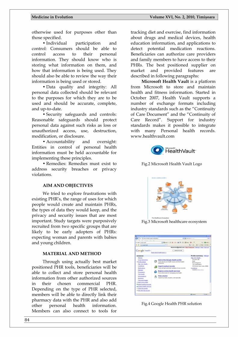
Medicine in Evolution Volume XVI, No. 2, 2010, Timişoara
84
otherwise used for purposes other than those specified.
• Individual participation and control: Consumers should be able to control access to their personal information. They should know who is storing what information on them, and how that information is being used. They should also be able to review the way their information is being used or stored.
• Data quality and integrity: All personal data collected should be relevant to the purposes for which they are to be used and should be accurate, complete, and up-to-date.
• Security safeguards and controls: Reasonable safeguards should protect personal data against such risks as loss or unauthorized access, use, destruction, modification, or disclosure.
• Accountability and oversight: Entities in control of personal health information must be held accountable for implementing these principles.
• Remedies: Remedies must exist to address security breaches or privacy violations.
AIM AND OBJECTIVES
We tried to explore frustrations with existing PHR‘s, the range of uses for which people would create and maintain PHRs, the types of data they would keep, and the privacy and security issues that are most important. Study targets were purposively recruited from two specific groups that are likely to be early adopters of PHRs: expecting woman and parents with babies and young children.
MATERIAL AND METHOD
Through using actually best market positioned PHR tools, beneficiaries will be able to collect and store personal health information from other authorized sources in their chosen commercial PHR. Depending on the type of PHR selected, members will be able to directly link their pharmacy data with the PHR and also add other personal health information. Members can also connect to tools for
tracking diet and exercise, find information about drugs and medical devices, health education information, and applications to detect potential medication reactions. Beneficiaries can authorize care providers and family members to have access to their PHRs. The best positioned supplier on market and provided features are described in following paragraphs:
Microsoft Health Vault is a platform from Microsoft to store and maintain health and fitness information. Started in October 2007, Health Vault supports a number of exchange formats including industry standards such as the ―Continuity of Care Document‖ and the ―Continuity of Care Record‖. Support for industry standards makes it possible to integrate with many Personal health records. www.healthvault.com
Fig.2 Microsoft Health Vault Logo
Fig.3 Microsoft healthcare ecosystem
Fig.4 Google Health PHR solution
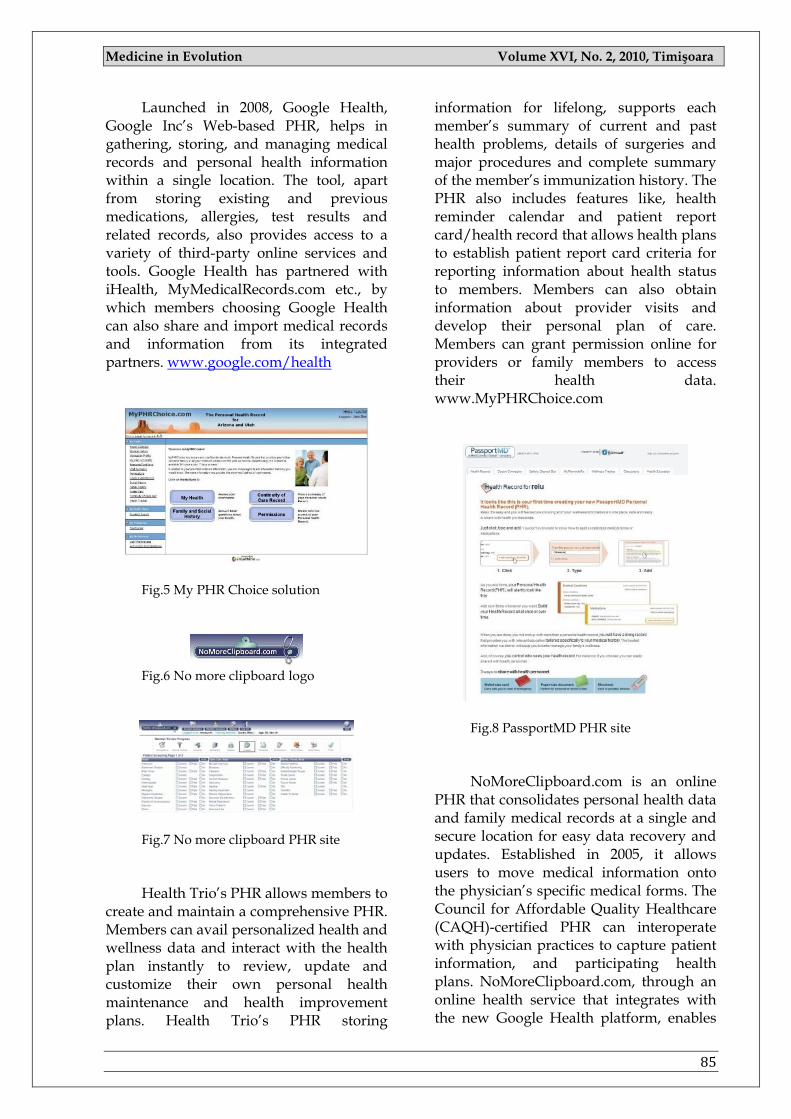
Medicine in Evolution Volume XVI, No. 2, 2010, Timişoara
85
Launched in 2008, Google Health, Google Inc‘s Web-based PHR, helps in gathering, storing, and managing medical records and personal health information within a single location. The tool, apart from storing existing and previous medications, allergies, test results and related records, also provides access to a variety of third-party online services and tools. Google Health has partnered with iHealth, MyMedicalRecords.com etc., by which members choosing Google Health can also share and import medical records and information from its integrated partners. www.google.com/health
Fig.5 My PHR Choice solution
Fig.6 No more clipboard logo
Fig.7 No more clipboard PHR site
Health Trio‘s PHR allows members to
create and maintain a comprehensive PHR. Members can avail personalized health and wellness data and interact with the health plan instantly to review, update and customize their own personal health maintenance and health improvement plans. Health Trio‘s PHR storing
information for lifelong, supports each member‘s summary of current and past health problems, details of surgeries and major procedures and complete summary of the member‘s immunization history. The PHR also includes features like, health reminder calendar and patient report card/health record that allows health plans to establish patient report card criteria for reporting information about health status to members. Members can also obtain information about provider visits and develop their personal plan of care. Members can grant permission online for providers or family members to access their health data. www.MyPHRChoice.com
Fig.8 PassportMD PHR site
NoMoreClipboard.com is an online
PHR that consolidates personal health data and family medical records at a single and secure location for easy data recovery and updates. Established in 2005, it allows users to move medical information onto the physician‘s specific medical forms. The Council for Affordable Quality Healthcare (CAQH)-certified PHR can interoperate with physician practices to capture patient information, and participating health plans. NoMoreClipboard.com, through an online health service that integrates with the new Google Health platform, enables
Medicine in Evolution Volume XVI, No. 2, 2010, Timişoara
86
Google Health users to deliver health information and medical records to treating physicians who may not have an electronic medical record system. www.NoMoreClipboard.com
PassportMD, Inc., the private consumer directed healthcare company, through its propriety tools and software, allows members to mange Web-based PHR, gain access to monthly versions of Harvard Health Letter and Harvard Mental Health Letter, create customized and personalized call reminders, share and exchange data with physicians through HIPAA compliant encrypted emails and letters, as well as store other vital data. www.passportmd.com/medicare
Although very large and powerful corporations have invested many resources to develop Personal Healths Records for a wide range of users, for the expecting women isn‘t yet developed an system to meet her specific needs during this life period.
RESULTS
If some general features are always covered from existing commercial PHR, like:
A calendar to keep track of medical appointments,
Reminders to refill prescriptions Family plans for tracking several
people, some important features, which we
consider that a Pregnancy oriented PHR must be necessarily contain, are not included in none of market leaders commercial PHR. These very specialized features must to interact active with expectant women and give her guidelines for daily life. These information‘s must be simple, consciously and clearly and must be a orientation guide through daily overcame information‘s bushy.
Suggestions for activity and nutrition. Nutritional principles through pregnancy.
Information‘s and warnings about medications and drug interactions during
pregnancy because especially of teratogens factors or conditions
Preparation for labor and delivery. Signs of Approaching Labor. False labor symptoms.
Because almost all pregnant women
need to increase their intake of protein, certain vitamins and minerals such as folic acid and iron, and calories (for energy) and limitation of junk food, since it offers little more than empty calories.
Through our study we propose to introduce in the Pregnancy PHR following principles for a pregnant proper nutrition:
a. Good Nutrition. Good nutrition
during pregnancy is essential for: (1) The well-being of the
mother and the developing fetus. (2) Development of effective
uterine musculature. (3) Development of breast
tissue. (4) Development of an
adequate functioning placenta. Poorly nourished mothers have placentas with fewer and smaller cells. Also, poorly developed placentas have a reduced ability to synthesize substances needed by the fetus, to facilitate the flow of needed nutrients, and to inhibit passage of potentially harmful substances.
(5) Development of infant's weight, length, bones, and brain. A nutritionally deprived fetus may have decreased development of brain cells. If optimum nutrition is provided after birth, the effects on the brain may be reversible.
(6) Continued development of the infant after birth.
b. Chronic Malnutrition. This has been
shown to be related to reproduction problems (this includes difficulties during pregnancy, labor, and delivery), increased perinatal mortality, low birth weight, and other problems with the newborn.
c. Nutritional Risk Factors in
Pregnancy that Require Observation.
Medicine in Evolution Volume XVI, No. 2, 2010, Timişoara
87
(1) Risk factors at the onset of pregnancy.
(a) Adolescence. Many adolescents are nutritionally at risk due to a variety of complex and interrelated emotions and social and economic factors that may adversely affect dietary intake. Their nutritional needs are greater and pose much concern from nurses and physicians.
(b) Frequent pregnancies. These pregnancies may have depleted nutrient stores. This situation can compromise maternal and fetal health and well-being.
(c) Poor reproductive history. Previous poor weight gain, pregnancy-induced hypertension (PIH), previous stillbirth or small for gestational age (SGA) baby, premature delivery, and prenatal infection are all common in women who are or have been poorly nourished in the past. These women may need more than the usual nutrition guidance.
(d) Economic deprivation. This refers to the pregnant woman who is not able to afford proper food. There are several programs that help with the purchase of food or that offer supplements.
(e) Bizarre food patterns. This includes faddish diets. A woman may enter pregnancy either having or continuing to be on a faddish or otherwise nutritionally inadequate diet.
(f) Vegetarian diets. This diet may not contain any or enough protein or vitamins for a developing fetus. Intense nutritional counseling will be required to work out a diet pattern during the prenatal period.
(g) Smoking, drug addiction, and alcoholism. Physiologic problems may have been present. Pregnant women who indulge in this category may have major physiologic problems. There is the possibility that the expecting woman may not consume sufficient quantities of nutritious foods and, in addition, can cause major problems to the fetus.
(h) Chronic systemic disease. There may have been medical problems, which may have interfered with ingestion,
absorption, or utilization of nutrients. Drugs used to treat these conditions may also affect nutrition by similar interference. Counseling should include general nutrition guidelines for prenatal care and diet therapy.
(i) Pre-pregnant weight. This may be at risk if the patient is fifteen percent or more below or twenty percent or more above the standard weight for health.
(2) Risk factors identified during pregnancy.
(a) Anemia of pregnancy. Many pregnant women have a lack of iron stores large enough to meet the needs of pregnancy.
(b) Pregnancy-induced hypertension (PIH). This may be seen in more patients with poor diets. However, there is no definite documentation of PIH's relationship to the diet.
(c) Inadequate weight gain. This may be an indication of maternal and fetal malnutrition (intrauterine growth retardation (IUGR)). It is important to document the pattern of weight gain in pregnancy as well as the total amount of weight gained.
(d) Excessive weight gain. This may be due to fluid retention. However, the pregnant woman should be carefully assessed for PIH.
d. Caloric Requirements of
Pregnancy. (1) Daily caloric requirements
for a pregnant woman are about 300 more than their normal requirements of 2300 to 2700 calories. The exact requirements are dependent on the woman's age, multiple births, and the woman's activity. Calories should be selected for quality rather than quantity. "Empty calories" do not count.
(2) Pregnancy is not the time to correct weight problems. Maintenance of a minimum of 1500 calories a day is essential for fetal development throughout the pregnancy. Patients who gain extra weight the first seven months then decide to cut

Medicine in Evolution Volume XVI, No. 2, 2010, Timişoara
88
back so as not to go overweight deprive the fetus of:
(a) Nutrients necessary when the fetal brain cells are growing the fastest.
(b) Nutrients necessary when the protective layer of fat is being developed.
(3) Foods rich in protein, iron,
and essential nutrients are recommended to be eaten on a daily basis. During the first two trimesters of pregnancy, iron is transferred to the fetus in moderate amounts, but during the last trimester when the fetus builds its reserve, the amount transferred is accelerated ten times.
(4) Recommended weight gain for a normal pregnancy is 10 to 14 kilograms.
e. Menu planning. A diet consisting of
a variety of foods can supply needed nutrients. The increased quantities of essential nutrients needed during the pregnancy may be met by skillful planning around the basic four food groups.
The recommended daily intake from
the basic four food groups are as follows: (1) Milk group- 1000 ml per
day. (2) Meat group-4 servings per
day to include: (a) Beef, veal, pork, poultry, or
fish. (b) Eggs each day. (c) Liver once a week. (3) Vegetable and fruit group. (a) 2 servings daily of dark green or
yellow vegetables. (b) 2 servings daily of fruit. (4) Bread and cereal group-4
servings per day. Information about cravings during
pregnancy a. Craving. This is a strong desire for
a certain type of food, usually carbohydrates.
b. Pica. This is an intense craving for and ingestion of nonnutritive substances such as clay, laundry starch, raw flour, and rice. This type of craving is characteristic of but is not limited to lower socioeconomic groups, ethnic groups, and regional areas, which prefer certain substances. Even though the cause is unknown, it interferes with good nutrition. Pica appears to be related to iron deficiency anemia as either a cause or an effect.
c. Treatment or Counseling. (1) Anything that depresses
good nutritional intake should be evaluated. This type of depression may be caused by nausea or vomiting, food fads or lack of finances, smoking or alcoholism, or personal or social problems. If a problem is identified, it should be reported to the charge nurse or physician for appropriate referral to the correct people who can relieve or eliminate the problem.
(2) Total dietary intake on a daily basis may need to be assessed.
(3) Dietary needs of pregnancy should be reinforced at every visit to the doctor.
Information about obesity during and
after pregnancy a. Obesity is common and frequently
a serious problem among modern society. The expectant woman is considered overweight if she is 10 percent over her desirable weight for their height and age group. If the patient is 20 percent over her desirable weight at the beginning of the pregnancy, she is considered at risk.
b. These women require close observation and additional education. The most frequently prescribed diet is 1500 to 1800 calories per day. The expecting woman must be advised that this in not the time to diet to lose weight. Encouragement is greatly needed during the pregnancy.
Environmental teratogenic factors (e.g. alcohol) are preventable. Environmental teratogens cannot induce congenital abnormalities in the first month of gestation. In addition, teratogens usually
Medicine in Evolution Volume XVI, No. 2, 2010, Timişoara
89
cause specific congenital abnormalities or syndromes. Finally, the importance of chemical structures, administrative routes and reasons for treatment at the evaluation of medicinal products must to be considered. On the other hand, in the so-called case-control epidemiological studies in general recall bias was not limited. These biases explain that the teratogenic risk of drugs is exaggerated, while the benefit of medicine use during pregnancy is underestimated. This overwhelming information‘s which arrives to expecting woman induce a fear feeling. Thus, a better balance is needed between the risk and benefit of drug treatments during pregnancy. Of course, we have to do our best to reduce the risk of teratogenic drugs as much as possible, however, it is worth stressing the preventive effect of drugs for maternal diseases (e.g. diabetes mellitus and hyperthermia) related congenital abnormalities. Therefore in our proposal for Pregnancy PHR information‘s about teratogens factors is a must chapter. This part must be developed with correct and confident informations, with the goal to spoil pregnant woman fears.
a. A teratogen is an agent or factor that causes the production of physical defects in the developing fetus.
b. Many drugs are known to have teratogenic effects on the fetus if taken during pregnancy. Drugs are the most widely recognized cause of structural defects in the developing fetus. Expecting womans need to be cautioned about taking any medication without a physician's approval.
Over-the-counter medicines such as
nose drops, cold remedies, and sleep medications may cause problems.
(1) Examples of known effects: (a) Physical abnormalities - no
arms or legs. (b) Hemorrhage or jaundice. (c) Neurologic symptoms. (d) Abnormal dental
pigmentation. (e) Addiction.
(f) Vaginal malignancy or altered sperm causing infertility.
(2) The effects of many drugs may not be known until later years during the growth and development of the child.
c. Teratogenic drug examples. (1) Thalidomide-used in
England in the 1950's and 1960's as a sedative.
(2) Phenytoin (Dilantin)®-used for seizures.
(3) Methotrexate®-used to treat neoplastic diseases.
(4) Diethylstilbestrol®-used for vasomotor symptoms during menopause.
(5) Accutane®--used to treat cystic acne.
d. Teratogenic viruses and
parasites. (1) Herpes simplex. (2) Rubella (German measles). (3) Toxoplasmosis. This is
transmitted by cat feces and raw meat. (4) Influenza or viral infections
in the early weeks of pregnancy. e. Other teratogenic conditions. (1) Hyperthermia. (2) Maternal disease (diabetes). (3) Maternal malnutrition. (4) X-rays should be avoided.
Radiation from the x-rays can cause deformity of the fetuses if exposed in the first trimester.
(5) Environmental pollutants. (6) Lead. (7) Increase in maternal age. (8) Tobacco and alcohol. f. Expectant woman need to be
reminded of the potential dangers of the things they may do or take. The worst damage to the fetus is done in the early weeks of the pregnancy before she even knows she is pregnant.
Childbirth isn't exactly a test, but an
expecting woman does need to study. Being prepared and educated about what
Medicine in Evolution Volume XVI, No. 2, 2010, Timişoara
90
to expect in the delivery room will help reduce your anxiety and make everything easier. Through our study we propose to introduce in the Pregnancy PHR some information about Labor and delivery with the goal to assist expectant women in the learning process.
Preparation for Labor and delivery a. Relaxation and Psychological
Control of Pain. Several methods of relaxation and psychological control of pain during labor are listed below:
(1) Lamaze method (Psychoprophylactic method-PPM). This method is the most widely taught. It deals with combating the fears associated with pregnancy by teaching relaxation and breathing techniques.
(a) The pregnant is taught to replace responses of restlessness and loss of control with more useful activity.
(b) The pregnant is taught to respond to pain with respiratory activity and relaxation of uninvolved muscles.
(c) The pregnant is taught controlled breathing and mind-focusing techniques.
(d) The partner is taught to help the pregnant stay in control.
(2) Bradley method (husband-coached childbirth). This is similar to the Lamaze method. Emphasis is placed on slow, deep breathing along with complete relaxation. Women using this practice often appear to be asleep during labor. However, they are not asleep, but are simply in a state of deep mental relaxation.
(3) Hypnosis. This is an induced state of extreme suggestibility in which the patient is insensible to outside impressions except the suggestion of her attendant.
b. Signs of Approaching Labor. These signs of approaching labor are taught to all pregnant women. When the expecting woman notices them, she is aware that labor will be forthcoming. The signs are:
(1) Lightening. This is the descent of the fetus into the brim of the pelvis (dropping). Lightening occurs in the
last 10 to 14 days of pregnancy in a primigravida. It may not occur until actual onset of labor in multigravidas. The pregnant woman identifies it as being able to breathe easier.
(2) False labor (Braxton-Hicks Contractions). This is intermittent uterine contractions occurring at irregular intervals, which serve to tone the uterus.
(3) "Show." This is when the blood-tinged mucoid vaginal discharge becomes more pronounced and red as cervical dilatation increases during labor.
(4) "Burst of energy." This is an increase in energy level. It occurs approximately 24 hours before onset of labor. The pregnant woman should be advised to relax during this time since labor will be starting soon.
(5) Rupture of membranes. This occasionally may be the first sign. Due to the risk of the prolapse cord, the pregnant woman needs to be aware that she should come to the hospital immediately even if she is not having contractions. If the membranes rupture prematurely, it then becomes a complication.
(6) Frequent urination. This, again, becomes a problem in the last stages of pregnancy. Pressure on the bladder is due to the enlarging uterus and the head settling back into the pelvis.
DISCUSSIONS & CONCLUSIONS
Most of the consumers currently keep health records of some kind – primarily their health-related financial records. However, they have little confidence in their ability to find and use particular records when needed, and difficulties in sharing records between health provides often resulted in repetition of tests or medical procedures.
As expected, most consumers would like their ―ideal‖ PHR to include lab test results (tracked over time), medications (names and dosages, the doctors who prescribed the medicines, prescription numbers, refills, etc.), and appointments
Medicine in Evolution Volume XVI, No. 2, 2010, Timişoara
91
with health care providers and the outcomes of those appointments.
Pregnant woman would like to include also tips and guidelines, information about They would most like to view their PHR to prepare for an upcoming physician visit and to monitor and investigate trends over time; in addition, they would like emergency health care providers to have access to their PHR.
While most users were comfortable with their care provider viewing and editing their PHR, they were very wary of other people, not directly involved in their health care, having access to their PHR (e.g., insurance companies or government agencies).
While the participants were not universally comfortable with any external entity holding their records, most were uncomfortable with taking on the responsibility of holding their records for themselves.
The results of this study have identified a number of features that need to be included in a Pregnancy PHR to ensure its acceptance by a broad range of potential expecting women and her families. While there is good consensus about the content of a PHR and the ways in which it might be used, there was little agreement about who should hold and maintain the records of an individual. If PHR systems are to be sustainable over the long term, we must also understand the level and type of commitment needed to maintain them.
REFERENCES
1. Connectiong for Health - Markle Foundation. www.connectingforhealth.org June 2008 2. Markle Foundation, The Personal Health Working Group.(2003). Connecting for health: a
public-private collaboration. Final Report. Retrieved December 3, 2005 from http://www.markle.org/ downloadable_assets/final_phwg_report1.pdf
3. Denton, I.C.(2001) Will patients use electronic personal health records? Responses from a real-life experience. Journal of Health Information Management, 15 (3), 251-259.
4. Gibbins, J. & Thomson, A.M. (2001). Women's expectations and experiences of childbirth. Midwifery, 17, 302-313.
5. Nichols, M., Roux, G., & Harris, N. (2007). Primigravid and multigravid women: Prenatal Perspectives.Journal of Perinatal Education, 16(2), 21-32.
6. Risica, P. M. & Phipps, M. G. (2006). Educational preferences in a prenatal clinic population. International Journal of Childbirth Education, 21(4), 4-7.
7. Rowe RE, Garcia J, Macfarlane AJ, Davidson LL. (2002) Improving communication between professionals and women in maternity care: a structured review. Health Expectations. 5(1): 63-83, 2002
8. Carrarino, J. E. & Moos, M. K. (2004). Local health departments: An under recognized source for women at high risk for preterm birth. American Journal of Maternal Child Nursing, 29(6), 366-372.
9. Vonderheid, S. C., Norr, K. F., & Handler, A. S. (2007). Prenatal health promotion content and health behaviors. Western Journal of Nursing Research, 29(3), 258-276.
10. Ueckert, F., Goerz, M., Ataian, M., Tessmann, S., & Prokosch, H.U. (2003). Empowerment of patients and communication with health care professionals through an electronic health record. International Journal of Medical Informatics, 70, 99-108.
11. Pregnancy: your guide to pregnancy. (2004). Retrieved December 16, 2005. from WebMD website: http://www.webmd.com/content/article/88/99782.htm
12. Carrarino, J. E. & Moos, M. K. (2004). Local health departments: An under recognized source for women at high risk for preterm birth. American Journal of Maternal Child Nursing, 29(6), 366-372.
13. Vonderheid, S. C., Norr, K. F., & Handler, A. S. (2007). Prenatal health promotion content and health behaviors. Western Journal of Nursing Research, 29(3), 258-276.
14. Ueckert, F., Goerz, M., Ataian, M., Tessmann, S., & Prokosch, H.U. (2003). Empowerment of patients and communication with health care professionals through an electronic health record. International Journal of Medical Informatics, 70, 99-108.

Medicine in Evolution Volume XVI, No. 2, 2010, Timişoara
92
15. Wald, J.S., Middleton, B., Bloom, A., Walmsley, D., Gleason, M., Nelson, E., et al. (2004). A patient-controlled journal for an electronic medical record: issues and challenges. Medinfo, 11, 1166-1170.
16. Taylor H, Leitman R, editors. Patient/physician online communication: many patients want it, would pay for it, and it would influence their choice doctors and health plans. Harris Interactive Health Care News. 2002;2(8):1-3. Available at: http://www.harrisinteractive.com/news/newsletters/healthnews/HI_HealthCareNews2002Vol2_Iss08.pdf Retrieved July 31, 2009
17. Augusto, J.C. Temporal reasoning for decision support in medicine. (2005). Artificial Intelligence in Medicine, 33: 1—24 Medline:15617978
18. Plaisant C, Mushlin R, Snyder A, Li J, Heller D, Shneiderman B. Lifelines: using visualization to enhance navigation and analysis of patient records. AMIA Symposium 1998:76-80. Medline:9929185
19. Shahar Y, Goren-Bar D, Boaz D, Tahan F. Distributed, intelligent, interactive visualization and exploration of time-oriented clinical data and their abstractions. Artificial Intelligence in Medicine 2005. Medline:16343873
20. Rasmussen D, Perez A. 40 Weeks +: The Essential Pregnancy Organizer. Mango Media; 2005.
Medicine in Evolution Volume XVI, No. 2, 2010, Timişoara
93
PAGES 93 -97
PHILADELPHIA CHROMOSOME IN ACUTE LEUKEMIA
CRISTINA A. POPA 1, HORTENSIA IONIŢĂ 2, NICOLETA ANDREESCU 1, ALINA BELENGEANU 3
1 University of Medicine and Pharmacology„Victor Babeş” Timişoara – Department of Genetics 2 Timişoara City Universitary Emergency Hospital – Department of Hematology 3 University of Medicine and Pharmacology „Victor Babeş” Timişoara – Department of Cellular biology
Correspondence to: Cristina A. Popa, Teiului No.14, Ap 4, 300681, Timisoara, Timis, Romania, Tel: 0728020060, E-mail: [email protected], Institution adress: Eftimie Murgu Square, No. 2, Timisoara, Romania
ABSTRACT
Objective: The current study including patients with acute leukemia tries to identify the presence of Philadelphia chromosome and its implications in evolution, prognosis and therapeutic approach of the disease.
Material and Methods: 52 cytogenetic analyses of patients with leukemia with ages between 2-73 years have been performed in the Cytogenetic laboratory of the University of Medicine and Pharmacy Victor Babes Timisoara during 2004-2009.
Results: In this study lot Ph chromosome was identified in 3 cases of ALL with B cells and in 5 cases of AML. Two of the patients with AML presented Ph chromosome as singular anomaly. Another 3 patients had associated Ph chromosome with other chromosomal anomalies: one case had del11q23, the second one had trisomy 8 and the third had monosomy 5 and isochromosome 17q.
Conclusions: Cytogenetic analysis has been shown to be an important prognostic factor, capable of predicting remission duration and the therapeutic management.
Key words: philadelphia chromosome, cytogenetic analysis, acute leukemia
INTRODUCTION
Acute leukemia is a clonal disturbance due to malign transformation of a myeloid or lymphoid progenitor cell, which allows the classification of leukemia in acute lymphoblastic leukemia and acute myeloblastic leukemia. For both types of leukemia the evolution is rapid to death. AML is probably the most extensively analyzed human neoplastic disease. Cytogenetic studies of AML have contributed substantially to our understanding of the mechanisms of
leukemogenesis. Acquired cytogenetic abnormalities have been shown to represent tumor markers of diagnostic and prognostic importance. Using morphological, enzymatic and imunological criteria AML was classified in 8 subtypes. Acute lymphoblastic leukemia (ALL) originates in a single B or T lymphocyte progenitor: ALL pre-B, ALL with B cells, ALL with T cells. According FAB classification there the following ALL subtypes: L1, L2, L3.
Ph chromosome was the first cytogenetic abnormality described in

Medicine in Evolution Volume XVI, No. 2, 2010, Timişoara
94
cancer. Philadelphia chromosome is a shorter 22 chromosome which results from a trasnslocation of ABL gene (chromosome 9) on the 5‘ end of BCR gene (chromosome 22) (1). It will appear a hybride gene BCR-ABL which will be transcripted in ARN-m. Children lymphoblastic leukemias with Ph+ have the split point in m-bcr region (minor split point) and the transcript will be p190. In myeloblastic leukemia there is a different transcript of Philadelphia chromsome, in which the BCR gene split point in μ-bcr region (micro split point)
resulting a 2e19 transcript encoding a p230 protein. At a molecular level, the rearrangement of the BCR and ABL genes is encoding a fusion protein with increased and deregulated tyrosine kinase activity (3). Patients with lymphoblastic acut leukemia Ph+, usually are old and present leococytosis, an eleveted number of periferical blasts, limphadenopatia and splenomegaly (2).Most of the cases of lymphoblastic acut leukemia have a abnormal karyotype, they may present numerical or structural anomalies.
Fig.1 Karyotype of a patient showing Ph Chromosome.
Fig.2 Metaphase of a patient showing Ph Chromosome.

Medicine in Evolution Volume XVI, No. 2, 2010, Timişoara
95
Fig.3 Interphase FISH showing Philadelphia chromosome, which appears as dual fusion (yellow) signal. The genes rearranged are: BCR (green on chromosome 22)/ABL (red chromosome 9).
Fig.4. Karyotype of a patient showing Ph Chromosome and trisomy 8.
Fig.5. Karyotype of a patient showing Ph Chromosome and monosomy 5.
Medicine in Evolution Volume XVI, No. 2, 2010, Timişoara
96
MATERIAL AND METHOD
52 cytogenetic analyses of patients with leukemia have been performed in the Cytogenetic laboratory of the University of Medicine and Pharmacy Victor Babes Timisoara during 2004-2009. The patients were referred to the cytogenetic laboratory from the Hematological Clinic of the Municipal Hospital Timisoara. In this study were included 22 males and 30 females, ages between 2-73 years. From the total of 52 patients, 24 were diagnosed with ALL and 28 with AML. Cytogenetic analyses were done using bone marrow, it have been initiated cell culture of 24, 48 and 72 hours. Additionally direct method was used.
Interphase in situ hybridization with flourochromes was done for confirmation. The specimen was obtained by marrow aspiration, 1-2 ml of marrow are aspirated aseptically into a syringe coated with sodium heparin and transferred to a sterile 15 ml centrifuge tube containing 5 ml culture medium (RPMI 1640, 100 units sodium heparin). To prepare metaphase cells, the sample was exposed to mitotic inhibitors and afterwards to hypotonic solution KCl (0.075M), and fixative (absolute methanol: glacial acetic acid, 3:1). Slides are prepared by dropping the cell suspension onto precleaned glass microscope slides, and the slides were air dried.
The slides were prccesed using G-banding technique. For confirmation was used FISH technique. Slides were immersed in +37 ºC 2xSSC for 30 min and then dehydrate in alcohol 70% and 100% 2 min each. Slides were denaturated at +73ºC (in a pre-warmed water bath) for 2 min and rinsed in ice-cold series of alcohol 70-80-100% 2 min each. Slides were put on a slide warmer (40ºC) and applied 10ul probe (7μl hybridization buffer, 1μl probe, 2μl distillate water), overnight were left at 37 ºC.
The slides were analyzed through a fluorescent microscope. The best images were captured using the camera mounted
on the microscope attached to a computer with karyotyping and FISH software.
RESULTS
In this study lot Ph chromosome was identified in 3 cases of ALL with B cells and in 5 cases of AML (subtype M1, M2, M7). Two of the patients with AML presented Ph chromosome as singular anomaly. Another 3 patients had associated Ph chromosome with other chromosomal anomalies: one case had del11q23, the second one had trisomy 8 and the third had monosomy 5 and isochromosome 17q.
DISCUSSIONS
We have set out to identify in the studied cases the possible correlations between the presences of Ph11 chromosome as unique chromosomal anomaly or associated to other chromosomal anomalies and the model of disease progression. Ph1 chromosome is the typical chromosomal anomaly for LMC but there are other types of leukemia showing the same chromosomal aberration. This chromosomal anomaly is associated with poor prognosis and rapid evolution to death. The remission period of the patients showing Ph1 chromosome is shorter than in the group with normal karyotype (4).
The 2 patients with myeloblastic acute leukemia having Ph1 chromosome as unique cytogenetic anomaly have presented the specific characteristics in evolution. One of the cases with myeloblastic acute leukemia associated Ph1 chromosome and another chromosomal modification: del11q23.On the long arm of chromosome 11 there are several genes that may be involved in tumorigenesis: MLL1 (located at 11q23 frequently rearranged in acute leukemia) and LOH11CR2A (a potential tumor suppressor gene located at 11q23). These genes probably are involved in cancer progression rather than initiation, because this deletion was previously reported in late stages and in cases with complex karyotypes (4).
Medicine in Evolution Volume XVI, No. 2, 2010, Timişoara
97
The response to specific therapy, the evolution and prognosis were correlated for this case with the association of Ph1 chromosome and del11.The patient with trisomy 8 and Ph1 chromosome had a better evolution after treatment and the remission was long. According to other studies, trisomy 8, relatively specific for myeloid disorders, is rare in the lymphatic leukemias, at least as a solitary anomaly. Patients with trisomy 8 usually present a myelodysplastic preleukemic phase before developing AML (4).
Loss of material from chromosome 5 or monosomy 5 in bone marrow cells is common in myelodysplastic syndromes (MDS) and acute myelocytic leukemia (AML).The patient with monosomy 5 and Ph1 chromosome is included in the same category with poor prognosis. The explication is the fact that the commonly deleted region (CDR) or critical region has been defined as 5q31~q33 and contains multiple genes involved in cellular growth, hematopoiesis, cell cycle control, cell adhesion, and tumor suppression (4).
The isochromosome of the long arm of chromosome 17 is a common secondary change in chronic myeloid leukemia but it was also reported in acute leukemia. The presence of a solitary I (17q) identifies a subgroup of patients with rapid progression to AML, poor response to chemotherapy, and short survival after transformation (4).
The association I (17q) with Ph1 chromosome in our patient karyotype was not related with a good prognosis.
CONCLUSIONS
The diagnostic cytogenetic analysis has been shown to be an important prognostic factor, capable of predicting remission duration and the therapeutic management.
All the investigated cases diagnosed with acute leukemia which presented Ph1 chromosome as unique chromosomal anomaly or associated to other chromosomal aberrations had a rapid evolution to death, poor prognosis and the response to specific therapy was reduced and unpredictable.
Further molecular and functional studies are needed to elucidate the role of loss of chromosomal material, especially of chromosome 5, in the pathogenesis of AML with a complex aberrant karyotype. The analysis of gene expression may allow specifying which genes show a reduced expression.
For the confirmation of previous data, CGH may provide useful information regarding the nature of genomic aberrations that take place in cases with complex karyotypes (5).
The high resolution and the use of cases with complex karyotype can provide data about candidate genes involved in leukemias.
REFERENCES:
1. Costa D., Carrio A., Madrigal I., Arias A., Valera A., Colomer D., Aguilar J.L., Teixido M., Camos M., Cervantes F., Campo E. Studies of complex Ph translocations in cases with chronic myelogenous leukemia and one with acute lymphoblastic leukemia. Cancer Genet Cytogenet. 2006 Apr 1; 166(1):89-93.
2. Stefan Faderl, Hagop Kantarjian, Mosche Talpaz, Zeev Estrov, Clinical significance of Cytogenetic abnormalities in adult acute lymphoblastic leukaemia, Blood, iunie 1998, vol. 91, no. 11, 3995-4019.
3. Kurzrock R., Gutterman J., Talpaz M. The molecular genetics of Philadelphia chromosome-positive leukemias. The New England Journal of Med 1988 vol 319, 990-998.
4. Alvarez S, Cigudosa JC: Gains, losses and complex karyotypes in myeloid disorders: A light at the end of the tunnel. Hematol Oncol 23: 18–25 (2005).
5. Paulsson K, Heidenblad M, Strombeck B, Staaf J, Jonsson G, et al: High-resolution genome-wide array-based comparative genome hybridization reveals cryptic chromosome changes in AML and MDS cases with trisomy 8 as the sole cytogenetic aberration. Leukemia 20: 840–846 (2006).

Medicine in Evolution Volume XVI, No. 2, 2010, Timişoara
98
Medicine in Evolution Volume XVI, No. 2, 2010, Timişoara
99
PAGES 99 -101
ORAL HEALTH – THE RESULT OF AN EFFICIENT DOCTOR-PATIENT COMMUNICATION.
ANA-MARIA POPOVICI-MUŢ 1, ANITA ROŞU2, RUXANDRA SAVA ROŞIANU2, ANGELA PODARIU2
1 Faculty of Medicine and Pharmacy, University of Oradea, Dental Medicine II 2 University of Medicine and Pharmacy, “Victor Babeş”Timişoara, Faculty of Dentistry, Department of Preventive Dentistry Correspondence to:
Ana Maria Mut, [email protected]
ABSTRACT
Oral health represents an essential component of health maintenance because of the negative impact that different ore-dental conditions have on the general health status and their consequences. Doctor-patient communication plays a very important role in maintaining the oral health status, the practitioner being able to offer attention and moral support for the patient in order to obtain a satisfactory therapeutical result. Good communication increases the frequency of visits to the dentist, diminuishes anxiety and the practitioner has to understand human behaviuor in order to modelate it for oral health promotion and to develop a succesful practice. Once a patient is educated and is giving oral health the proper attention one will observe the benefic effects on social grounds gathered in the general well-being and the increase of life quality.
Communication in medical practice means assuming a high communicative competence that represents a priority for proffessionals in the medical field, especially for doctors. Thus, it means recognizing and decoding messages transmitted by the patient on one hand and using adecquate ways of communication in order to transmit informations to the patient on the other hand.
Key words: oral health, doctor-patient communication, education, feedback.
INTRODUCTION
Health refers to the absence of disease or disability, incapacity and handicap being regarded as a positive aspect of wellbeing. As an indespensible component of social development, health represents a necessity, but also a human right, a process that contributes to a good balance of the whole organism. The human body represents an inexhaustible source for medical science and the oral cavity is the gateway, so we can say that dentistry plays a key role for general health. The necesity of health education
has to be permanent for all age groups, regardless of the environment and has to be professionally acomplished by every practitioner. Doctor-patient communication has benefic effects on the phisiological and psychological of the patient during the therapeutical process but also regarding the compliance of medical indications or satisfaying the patients needs.
MATERIAL AND METHODS
The most elementary way of communication presumes a transmitter,
Medicine in Evolution Volume XVI, No. 2, 2010, Timişoara
100
who, using a certain manner of speaking, encodes a message, a communication channel which is represented by the message and a receiver, who gets the message, decodes it and identifies it‘s meaning. As a matter of fact these are the components of the information process. In the information process the evaluation of the feedback loop in communicating the message is very important. This is when a role alternation occurs: the message receiver becomes the transmitter. The transmitter (the source) is represented by the dentist who transmits his health knowledge to the receiver, represented by the patient, who is accepting the message. The doctor is the one who generates and provides informations building a message and initiating the communication. The source has to fulfill two conditions: credibility (authenticity and credibility) and attractiveness (language, looks, and clothing). Through talking about the medical procedures, explaining the maneuvers, even if this creates a certain discomfort for the patient, the doctor can appeal to the understanding of the patient, educating, informing him in order to achieve maximum succes. Therefore the doctor has to adapt his language to the patient‘s degree of education and age, otherwise he is risking transmitting a message the patient isn‘t capable to understand. In order to cope with this situation a doctor has to posess a series of psichological knowledges to help him avoid pointless and damaging conflicts for each part. The doctor-patient relationship needs sensitivity, availability to listen to the patients needs, to understand and help him, the practitioner having to use his medical psichological knowledge from the first session in order to win the patients trust.
The patient himself can help the doctor recounting informations he knows from direct investigations, this is why the anamnesis is very important in the process of relating to the patient. Many times a patient can have a prepostrous behaviour determined by the psychic discomfort abducted from the pain. When it is
prolongued, an untrusting feeling of depression can appear towards the practitioner and the possibilities of the medical science.
The message is the fundamental unit of the communication process, composed of words, images, signs and sound which derive from the codification process in order to be easily recognized by the receptor through decoding. It has to be interesting, accesible, convincing, directional and acceptable.
The doctor-patient communication is well attained under the following conditions:
The distance between the interlocutors has two be less than 1 meter in order to obtain an intimate climate;
Standing face to face is the best position;
A simple and concise language has to be used;
The questions have to be simple, direct;
The doctor has to show that he understands the patient;
The doctor must not contradict the patient brutally, but has to descover his errors and achieve an attitude change;
The patient has to be always asked if he understands what he was told;
The circumambient has to correspund (comfortable chairs, adequate light).
Also, motivation is one of the key
requirements which anyone who works with patients has to take into consideration. The dentist is obliged to include in his actions the awakening of motivation in order to obtain feedback from the patient. Lack of health education is a major problem. In his educational work, especially with children, the practitioner has to know and to turn to account the whole potential of motivation. This work has to arrouse the patient‘s wish to have the best oral health status.
Medicine in Evolution Volume XVI, No. 2, 2010, Timişoara
101
RESULTS
Through his attitude towards the patint, the well balanced optimism, the dentist will gain the patient‘s trust, restoring the optimism he needs during the healing process, so between the two parts grows collaboration, a harmonious relationship based on trust wreathed by succes. This is how dentists can become an active instrument of conserving structural, functional and psicho-social integrity, involving the patient in combating problems like smoking, bad hygiene or disfunctions of the digestive system.
CONCLUSIONS
Oral health means more than having a pleasent smile. Untreated dental lesions can have severe consequences not only for
oral health but also for the whole body. Thus the oral health status indicates the general health status, many general conditions being signalised in the oral cavity, meaning that the doctor also has to drive attention on general health problems. According to the principle „preventing is easier than treating‖, one can observe that good communication between the dentist and the patient can induce consciousness regarding the importance of oral hygiene contirbuting to the maintaining of good oral health status and general health status. Educational actions are an efficient messure to improve the knowledge level regarding the sanogen conduct, but also to attenuate disparities determined by ethnic criteria, socio-economic status, and educational level.
REFERENCES
1. Ashoka L. – Public Health and Preventive Denstri. 2. Angela Podariu, Daniela Jumanca, A.Găluşcan, Roxana Văcaru, Ramona Munteanu - „
Stomatologie comunitară „ Timişoara Waldpress 2002. 3. Adina Dumitrache, Ruxandra Moraru, Loredana Dumitraşcu – „Ştiinţe comportamentale‖,
Bucureşti, Editura Cermaprint 2006. 4. Andre de Peretti, Jean Marie Legand, Jean Boniface – „ Tehnici de comunicare‖ Editura Polirom
Iasi, 2001. 5. Dinu Mihai – „ Comunicarea‖ – Editura Algos, București, 2000. 6. Ovidiu Grivu, Angela Podariu, Anca Baila – „ Prevenţia în stomatologie „ Editura Mirton,
Timişoara 1995. 7. Podariu Angela Codruţa, Jumanca D., Găluşcan A., Văcaru R., Munteanu R., - „ Tratat de prevenţie
oro-dentară „, Timişoara, Editura Waldpress 2003. 8. Ramona Amina Popovici, Angela Codruţa Podariu, Daniela Jumanca, Atena Găluşcan, Roxana
Oancea, Ruxandra Sava Roşianu, 2007. – „ Educaţia pentru sănătatea oro-dentară. Managementul proiectelor educaţionale „, Timişoara, Editura Mirton.
9. Vasile Sebastian Dancu – ‖Comunicarea în managementul instituțional‖ – Editura Gewalt, Cluj-
Napoca, 2000.

Medicine in Evolution Volume XVI, No. 2, 2010, Timişoara
102

Medicine in Evolution Volume XVI, No. 2, 2010, Timişoara
103
INSTRUCTIONS FOR AUTHORS
The journal publishes general reviews, studies and clinical, epidemiological, experimental and laboratory research, clinical case presentations, papers from the history of medicine, reviews, scientific and technical state-of-the-art articles, medical informations and opinions. Only papers which have not been published or sent for publishing in other journals are accepted. The authors are responsable for the opinions expressed in the papers. The paper must be edited both in Romanian and in English; the English version will be supervised by our collaborator Dana Brehar-Cioflec, MD, PhD; typed on white A4 paper (fonts - Times New Roman 12, Romanian characters, line spacing 1.5, upper and lower margins 2cm, left border 3cm, right border 2cm) and on CD, DVD or Memory Stick.
Manuscripts will not exceed: general reviews: 6-8 pages studies and researches: 5-7 pages case presentations: 2-4 pages reviews, scientific and technical state-of-the-art articles, medical
informations and opinions: 1-2 pages. The paper will be edited according to international editing rules for manuscripts.
The title will be written in capital characters and it will be followed by the name and surname of the author (authors), followed by their place of work (place where the paper has been elaborated). Studies and researches will be followed by a brief abstract, followed by 3-4 key-words.
The body of the paper will be structured on the following chapters: introduction, aim, objectives, material and method, results and discussions, conclusions. The references will be presented alphabetically and in conformity to the Vancouver Convention, including:
for articles: name of the authors and surname initials, title of the article in the original language, title of the journal according to the international abreviation system, year of issue, volume, number, pages;
for books: name of the authors and surname initials, volume, publisher (editors), city of publishing, year of issue.
Citation of references inside the body of the paper will be put between brackets,
Harward style (author, year) or Vancouver style (number in square brackets or superscript). Cited reference titles will be selected, maximum 6 for studies and case presentations and 12 for general reviews.
Acceptance, rejection or the need of alterations in sent materials, or in inconography, will be comunicated to the authors in due time. For this, the authors will indicate the person and address for corespondence (phone number, e-mail address). Given the less pleasant experience of the editorial board with some articles being rejected because they did not meet publishing criteria, we decided to support those who intend to publish in this journal by detailing the way such a paper should be elaborated, as well as our requirements.

Medicine in Evolution Volume XVI, No. 2, 2010, Timişoara
104
Except some particular aspects concerning this journal, the following details are general requirements asked or imposed by other journals as well. Conditions to be met in order to propose a paper for publishing. The main author has the responsability to make sure the article has been approved by all the other authors. The journal will have copyright for papers accepted for publishing. The editorial board reservs the right to change the style and dimensions of an article (major changes will be discussed with the main author) and to decide the date of issue.
2. FIRST PUBLICATION
The editorial board will not consider a paper already reported in a published general review or described in a paper proposed to or accepted by another journal. This does not exclude papers which have been rejected by other journals. Also, papers which have been presented at a scientific meeting will be accepted for discussion if they have not been entirely or partially published in a similar publication.
„Multiple‖ publishing of the same study is seldom justified. One of the possible justifications is publishing in a second language but only if the following conditions are met:
Editors of both journals involved are fully informed; Priority of the initial publication will be respected by a minimum
publishing interval of two weeks; For the second publication, a shortened version will suffice; The second version strictly reflects data and interpretations in the first; A footnote may state: „This article is based upon a study initially published
in [title of the journal]‖.
3. PATERNITY
Paternity must reflect the common decision of the coauthors. Each author must have participated enough to take public responsability for the content.
A paper with collective paternity must have a key person responsable for the article.
4. COPYRIGHT
In order to reproduce materials from other sources, written agreement from the copyright owner must be obtained:
photographer – for unpublished photographs; hospital where the photographer (physician) is employed – for unpublished
photographs performed during the employment period; initial publisher – for a table, picture or text which have previously been
published elsewhere.
5. ETHICAL ASPECTS
Do not use name of patients, initials or hospital observation charts numbers. If a photograph of a body part which could allow direct or deductive recognition of the

Medicine in Evolution Volume XVI, No. 2, 2010, Timişoara
105
patient needs publishing, then the paper must be accompanied by the written consent of the patient and clinician, as well.
6. PRESENTING THE MANUSCRIPT
For the journal „Medicine in evolution”, the manuscript must be typed double spaced, on white A4 paper – 210 x 297mm, on one side (2.5cm upper and lower borders, 3cm left and 2cm right border, respectively), in clear characters, no further corrections or addings. It is advisable that articles are presented on CD or other data transfer methods, in Word format, 12 Times New Roman fonts - using Romanian characters – respecting the same page order, accompanied by a printed version. Graphs – black and white or coloured – may be generated in MS Excel or MS Graph, inserted in the body of the paper or presented in a different file. Infected materials will not be used. 6.1. FIRST PAGE (TITLE PAGE)
Together with the title and names of the authors, the first page must include the affiliation, professional and university degree (if applicable), marked by asterisc for every author; it is advisable to give at least a phone and/or fax number or e-mail address of the first author who may be contacted by the editors for additional recommendations or explanations.
6.2. ABSTARCT OF THE PAPER
6.2.1 Recommendations for original studies Original studies must include a structured abstarct of maximum 150 words,
containing the following titles and informations: Aim and objectives; Material and methods; Results; Conclusions; Key words: give 3-5 key words; The abstract will be translated into an international circulation language.
6.3 CONTENT OF THE PAPER
6.3.1 For original articles The text will usually be divided into sections:
Introduction – presentation of general aspects, in the context of the approached theme
Aim and objectives – Define the aim of the article. Briefly expose the rationale of the presented study or observation. Make strictly pertinent referals and do not exhaustively review the subject. Do not include data or conclusions from the paper.
Material and methods – Describe the selection of observations or subjects for the experiment (including controls). Identify methods, equipments (with the name and address of the manufacturer in brackets) and give sufficient details on procedures. Give references for the selected methods,
Medicine in Evolution Volume XVI, No. 2, 2010, Timişoara
106
including statistical methods; offer details and brief descriptions for previously published methods which are not well known; describe new or substantially modified methods, justify their use and assess their limitations. Precisely identify all used drugs and chemicals, including generic names, dosage and administration ways. Describe statistical methods with sufficient details for reported results to be verified. Whenever possible, quantify discovered aspects and present them with appropriate measurement indicators for the uncertainty or error of measurement (such as confidence intervals).
Results – Present results in a logical succession as text, tables and illustrations. Emphasize or briefly describe only important observations.
Discussions – Underline new, important aspects of the study. Do not repeat in detail data which have been presented in previous sections. Include implications of revealed aspects and their limitations, including implications for future studies. Connect your observations to other relevant studies. Relate the results to the aim proposed for the study.
Conclusions – organize conclusions which emerge from the study. In the end state: a) contributions to be acknowledged but which do not justify paternity right; b) thanks for technical support; c) thanks for financial or material support.
6.3.2 Indications for case reports
Themes may be selected from all medical fields. Manuscripts which offer a special gain for daily activity will have priority. The title must be clearly, precisely stated. It may be completed by a subtitle. It is advisable to include in the key words of the title the main message, the special element which may be observed from the case evolution. The content of a case report must be divided into three parts:
Introduction – It must include a maximum of 15 typed rows (half page). Here, the main medical problem is summarized in order to place the case in a specific domain.
Case report – It contains essential specific information on the case. In order to make a logical, chronological and didactical case report the
following 5 chapters are needed: I. Anamnesis; II. Clinical examination data; III. Laboratory data; IV. Additional paraclinical investigations; V. Treatment and evolution.
Discussions – The reason for the case report must be stated. The report must be patient-centered. Occasional deviations from typical (characteristic) evolutions, nosologically important facts must be presented in such a manner to expose the clinical picture as completely as possible. The case report must not appear as an appendix of a general review. Dimensions of a case report: maximum 6-8 typed pages, 30 rows of 60 characters/page.

Medicine in Evolution Volume XVI, No. 2, 2010, Timişoara
107
6.4. MEASUREMENT UNITS, SYMBOLS, ABREVIATIONS
All measurements must be expressed in International System (IS) units. Abreviations must be fully explained when first used.
6.5. TABLES
Tables are noted with Roman figures and they will have a brief and concise title, concordant with their content.
6.6. ILLUSTRATIONS
Number all illustrations in Arabic figures in a single succession. Apply a label on the back side of every illustration, containing its number and an arrow indicating the upper side. Coloured illustrations may be accepted but it is the choice of the editors, according to particular technical abilities of each journal issue, or it may involve a fee in special cases.
6.7. EXPLANATIONS FOR DRAWINGS AND GRAPHS
Explanation for drawings and graphs must be clear and in readable dimensions, considering the necessary publishing shrinkage.
6.8. PHOTOGRAPHS
Offer glossy, good quality photographs. Any annotation, inscription, etc. must contrast with the ground. Microphotographs must include a scale marker.
6.9. ILLUSTRATION LEGENDS
Include explanations for each used symbol, etc. Identify the printing method for microphotographs.
6.10. REFERENCES
A numbered list of references must be provided at the end of the paper. The list should be arranged in the order of citation in the text of the publication, assignment or essay, not in alphabetical order(according to the Vancouver rules). List only one reference per reference number. It is very important that you use the correct punctuation and that the order of details in the references is also correct.
Books - Standard format - #. Author of Part, AA. Title of chapter or part. In: Editor A, Editor B, editors. Title: subtitle of Book. Edition(if not the first). Place of publication: Publisher; Year. p. page numbers.
Journal Articles - Standard format - #. Author of article AA, Author of article BB, Author of article CC. Title of article. Abbreviated Title of Journal. year; vol(issue):page number(s).
E-Books - Standard format - #. Author A, Author B. Title of e-book [format]. Place: Publisher; Date of original publication [cited year abbreviated month day]. Available from : Source. URL.
Medicine in Evolution Volume XVI, No. 2, 2010, Timişoara
108
E-Journals - Standard format - #. Author A, Author B. Title of article. Abbreviated Title of Journal [format]. year [cited year abbreviated month day];vol(no):page numbers[estimated if necessary]. Available from: Database Name (if appropriate). URL.
Internet Documents - Standard format - #. Author A, Author B. Document title. Webpage name [format]. Source/production information; Date of internet publication [cited year month day]. Available from: URL.
7. COPIES FOR PUBLISHING
In order to accelerate publishing, the main author will send a set of printed sheets presenting the final version of the paper, as it will appear in the journal. It is really helpful that texts to be also sent on electronic support, diacritic characters mandatory.
8. REJECTION OF PAPERS
If a paper does not meet publishing conditions, whatever these may be, the editors will notify the first author on this fact, without the obligation of returning the material. Original photographs or the whole material will be returned only if the author comes to the editor and takes them.
Papers submitted for publishing will be addressed to:
Prof. Angela Codruta Podariu, DMD, PhD Journal Medicine in evolution Department of Preventive, Community Dental Medicine and Oral Health Splaiul Tudor Vladimirescu no. 14 A 300041, Timişoara Phone: 0256-204950 Email: [email protected]
Dana Brehar-Cioflec, MD, PhD Institute of Public Health “Prof. Dr. Leonida Georgescu” Timişoara Bd. Victor Babeş no. 16 300226, Timişoara Phone: 0256-492101 Email: [email protected]

Medicine in Evolution Volume XVI, No. 2, 2010, Timişoara
109

Medicine in Evolution Volume XVI, No. 2, 2010, Timişoara
110

Medicine in Evolution Volume XVI, No. 2, 2010, Timişoara
111

Medicine in Evolution Volume XVI, No. 2, 2010, Timişoara
112

Medicine in Evolution Volume XVI, No. 2, 2010, Timişoara
113
Order form
□ Rate 38 Euro (Outside Romania)
□ Rate 160 Ron(Inside Romania)
The money will be pay in the account: RO80 ABNA 3600 264 1000 89 288 opened at RBS BANK, Timisoara
Name____________________________________________
Speciality ________________________________________
Adress __________________________________________
Phone ________Fax ___________Email ________________ …………………………………………………………………………………………………
Order form
□ Rate 38 Euro (Outside Romania)
□ Rate 160 Ron(Inside Romania)
The money will be pay in the account: RO80 ABNA 3600 264 1000 89 288 opened at RBS BANK, Timisoara
Name____________________________________________
Speciality ________________________________________
Adress __________________________________________
Phone ________Fax ___________Email ________________