edge article - rsc publishing
TRANSCRIPT
ChemicalScience
EDGE ARTICLE
Ope
n A
cces
s A
rtic
le. P
ublis
hed
on 0
3 Se
ptem
ber
2020
. Dow
nloa
ded
on 3
/13/
2022
10:
35:4
8 PM
. T
his
artic
le is
lice
nsed
und
er a
Cre
ativ
e C
omm
ons
Attr
ibut
ion
3.0
Unp
orte
d L
icen
ce.
View Article OnlineView Journal | View Issue
Glycan–glycan in
aDepartment of Physics and Astronomy, The U
UKbInsight SFI Research Centre for Data Analy
IrelandcDepartment of Chemistry, Durham UniversdDepartment of Chemistry, The University oeFaculty of Infectious and Tropical Diseases,
School of Hygiene and Tropical Medicine, Lo
[email protected] of Infection, Immunity & Cardi
The University of Sheffield, Sheffield TeachigStrathclyde Institute of Pharmacy and
Strathclyde, Glasgow G4 0RE, UKhSchool of Engineering, Newcastle University
† Electronic supplementary information (EForce spectroscopy data of L. mexicana
CA7AE LPG antibody- and glucose-mretraction curves and force histogramscontaining peak adhesive force output frfrom the multi-peak analysis of Leishma
force event data from the multi-peak anaIllustrative force maps of L. mexica
Cite this: Chem. Sci., 2020, 11, 10973
All publication charges for this articlehave been paid for by the Royal Societyof Chemistry
Received 13th June 2020Accepted 2nd September 2020
DOI: 10.1039/d0sc03298k
rsc.li/chemical-science
This journal is © The Royal Society o
teractions determine Leishmaniaattachment to the midgut of permissive sand flyvectors†
Amy R. Hall, ‡ab Jamie T. Blakeman, a Ahmed M. Eissa, §c Paul Chapman,ad
Ana L. Morales-Garcıa, {a Laura Stennett,ke Oihane Martin,**e Emilie Giraud, ††e
David H. Dockrell, ‡‡f Neil R. Cameron, §§c Martin Wiese, g Laith Yakob, e
Matthew E. Rogers *e and Mark Geoghegan {{ah
Direct glycan–glycan interactions are increasingly implicated in survival and pathogenicity of bacteria. Here,
we show that they can be exploited by protozoan parasites in their insect hosts. Force spectroscopy
revealed that Leishmania promastigotes display a high-affinity biomolecular interaction between their
lipophosphoglycan glycocalyx and mimics of N-acetyl-D-galactosamine, commonly expressed on the
midguts of a wide range of sand fly vector species. This enabled gut-adhesive nectomonad
promastigotes of Leishmania mexicana to efficiently bind to membrane-bound mucin-like, O-linked
glycoproteins of the sand fly Lutzomyia longipalpis, an event crucial for parasite survival, and accounts
for a permissive mode of binding. Thus, direct interaction between parasite and sand fly midgut glycans
are key to permitting vector competence for all forms of leishmaniasis worldwide. In addition, these
studies demonstrate the feasibility of interfering with these interactions as transmission-blocking vaccines.
Introduction
Leishmaniasis is a vector-borne disease caused by protozoabelonging to the genus Leishmania, transmitted by the bite ofa female phlebotomine sand y.1–3 There are few effective drugsagainst leishmaniasis, many of which are toxic, and over-reliance on them has led to a worldwide rise in drug
niversity of Sheffield, Sheffield S3 7RH,
tics, Dublin City University, Glasnevin,
ity, Durham DH1 3LE, UK
f Sheffield, Sheffield S3 7HF, UK
Department of Disease Control, London
ndon WC1E 7HT, UK. E-mail: matthew.
ovascular Disease, The Medical School,
ng Hospitals, Sheffield S10 2RX, UK
Biomedical Sciences, University of
, Newcastle NE1 7RU, UK
SI) available: Experimental methods.nectomonads and metacyclics withodied AFM tips (approach and). LPG Western blot analysis. Dataom the force maps; force event datania mexicana WT nectomonads; andlysis of L. mexicana WT metacyclics.na highlighting large peak force
f Chemistry 2020
resistance.4 Consequently, there is an urgent need to under-stand the biology and life cycle strategies of these parasites inorder to identify new targets for drugs and vaccines.5
Of particular interest is the reliance of Leishmania on itssurface glycoconjugates to anchor themselves to the midgutepithelium of the sand y.6 Midgut attachment and directionalmovement is crucial to Leishmania development as it allows the
interactions ($0.1 nN) for the interaction with galactose andglucose-modied AFM tips. See DOI: 10.1039/d0sc03298k
‡ Present address: Insight SFI Research Centre for Data Analytics, Dublin CityUniversity, Ireland.
§ Present address: Department of Chemistry, University of Warwick, Coventry,UK; Department of Polymers, Chemical Industries Research Division,National Research Centre, Cairo, Egypt
{ Present address: Procter and Gamble Newcastle Innovation Centre, Newcastleupon Tyne, UK.
k Present address: Department of Imaging Chemistry and Biology, King'sCollege London, London, UK.
** Present address: Servicio de Microbiologıa, Hospital Universitario Ramon yCajal, Madrid, Spain.
†† Present address: Institut Pasteur, Paris, France.
‡‡ Present address: Centre for Inammation Research, University ofEdinburgh, Edinburgh, UK.
§§ Department of Materials Science and Engineering, Monash University,Clayton, Australia.
{{ Present address: School of Engineering, Newcastle University,Newcastle-Upon-Tyne, UK.
Chem. Sci., 2020, 11, 10973–10983 | 10973
Chemical Science Edge Article
Ope
n A
cces
s A
rtic
le. P
ublis
hed
on 0
3 Se
ptem
ber
2020
. Dow
nloa
ded
on 3
/13/
2022
10:
35:4
8 PM
. T
his
artic
le is
lice
nsed
und
er a
Cre
ativ
e C
omm
ons
Attr
ibut
ion
3.0
Unp
orte
d L
icen
ce.
View Article Online
parasites to resist expulsion from the sand y when it defecates,and therefore to persist beyond the initial blood meal phase ofinfection.7 Attachment to the sand y gut is the weakest point inthe parasite life cycle; a signicant proportion of parasites arelost at this stage and their survival is dependent on a smallpopulation of Leishmania that anchor themselves success-fully.6,8Disruption of this phase of parasite development has thepotential to form the basis of a transmission-blockingintervention.
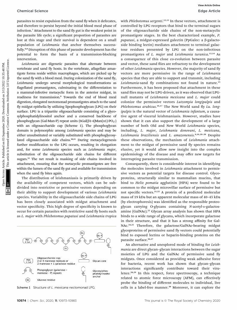
Leishmania are digenetic parasites that alternate betweenvertebrate and sand y hosts. In the vertebrate, aagellate amas-tigote forms reside within macrophages, which are picked up bythe sand y with a bloodmeal. During colonization of the sand y,Leishmania undergo several morphological transformations asagellated promastigotes, culminating in the differentiation toa mammal-infective metacyclic form in the anterior midgut, inreadiness for transmission.9 Towards the end of blood mealdigestion, elongated nectomonad promastigotes attach to the sandy midgut epithelia by utilizing lipophosphoglycan (LPG) on theirsurface. LPG is a tripartite oligosaccharide consisting of a glyco-sylphosphatidylinositol anchor and a conserved backbone ofphosphoglycan (Gal-Man-P) repeat units (6Gal(b1-4)Man(a1)-PO4-)with an oligosaccharide cap (Scheme 1). The phosphoglycandomain is polymorphic among Leishmania species and may beeither unsubstituted or variably substituted with phosphoglycosy-lated oligosaccharide side chains.10,11 During metacyclogenesisfurther modication to the LPG occurs, resulting in elongationand, for some Leishmania species such as Leishmania major,substitution of the oligosaccharide side chains for differentsugars.11 The net result is masking of side chains involved inattachment, ensuring that the metacyclic promastigotes are freewithin the lumen of the sand y gut and available for transmissionwhen the sand y bites again.
The distribution of leishmaniasis is primarily driven bythe availability of competent vectors, which can be sub-divided into restrictive or permissive vectors depending ontheir ability to support development of various Leishmaniaspecies. Variability in the oligosaccharide side chains of LPGhas been closely associated with midgut attachment andvector specicity. This high degree of specicity is known tooccur for certain parasites with restrictive sand y hosts suchas L. major with Phlebotomus papatasi and Leishmania tropica
Scheme 1 Structure of L. mexicana nectomonad LPG.
10974 | Chem. Sci., 2020, 11, 10973–10983
with Phlebotomus sergenti.12,13 In these vectors, attachment iscontrolled by LPG receptors that bind to the terminal sugarsof the oligosaccharide side chains of the non-metacyclicpromastigote stages. In the best characterized example, P.papatasi, a midgut-expressed galectin (PpGalec: a b-galacto-side binding lectin) mediates attachment to terminal galac-tose residues presented by LPG on the non-infectiouspromastigotes of L. major and Leishmania turanica.13,14 Asa consequence of this close co-evolution between parasiteand vector, these sand ies are refractory to the developmentof other Leishmania species. However, the majority of sand yvectors are more permissive in the range of Leishmaniaspecies that they are able to support and transmit, includingLeishmania-sand y combinations not found in nature.15
Furthermore, it has been proposed that attachment in thesesand ies may not be LPG-driven, as it was observed that LPG-null mutants of Leishmania mexicana and L. major couldcolonize the permissive vectors Lutzomyia longipalpis andPhlebotomus arabicus.16,17 The New World sand y Lu. long-ipalpis is the natural vector of Leishmania infantum, a causa-tive agent of visceral leishmaniasis. However, studies haveshown that it can also support the development of a largenumber of both Old and New World Leishmania species,including, L. major, Leishmania donovani, L. mexicana,Leishmania braziliensis and L. amazonensis.9,16,18–20 Despitethese observations, the mechanism of Leishmania attach-ment to the midgut of permissive sand y species remainselusive, yet it would allow new insight into the complexepidemiology of the disease and may offer new targets forinterrupting parasite transmission.
Consequently, there is considerable interest in identifyingthe molecules involved in Leishmania attachment to permis-sive vectors as potential targets for disease control. Glyco-proteins, structurally similar to mammalian mucins, thatbind to Helix pomatia agglutinin (HPA) were found to becommon to the midgut microvillar surface of permissive butnot specic vectors.21,22 A protein of a predicted molecularmass of 19 kDa but an apparent molecular mass of 40–45 kDa(by electrophoresis) was identied as the responsible proteo-glycan carrying O-glycans containing N-acetyl-D-galactos-amine (GalNAc).23 Glycan array analysis has shown that HPAbinds to a wide range of glycans, which incorporate galactosein their structure, and that it has a strong affinity for Gal-NAc.24,25 Therefore, the galactose/GalNAc-bearing midgutglycoproteins of permissive sand y vectors could potentiallybind to exposed lectins or heparin-binding proteins on theparasite surface.26,27
An alternative and unexplored mode of binding for Leish-mania are direct glycan–glycan interactions between the sugarmoieties of LPG and the GalNAc of permissive sand ymidguts. Once considered as providing weak adhesive forcefor bacteria, recent work has shown that glycan–glycaninteractions signicantly contribute toward their viru-lence.28,29 In this respect, force spectroscopy, a techniquerelated to atomic force microscopy (AFM), can effectivelyprobe the binding of different molecules to individual, livecells in a label-free manner.30 Moreover, it can explore the
This journal is © The Royal Society of Chemistry 2020
Edge Article Chemical Science
Ope
n A
cces
s A
rtic
le. P
ublis
hed
on 0
3 Se
ptem
ber
2020
. Dow
nloa
ded
on 3
/13/
2022
10:
35:4
8 PM
. T
his
artic
le is
lice
nsed
und
er a
Cre
ativ
e C
omm
ons
Attr
ibut
ion
3.0
Unp
orte
d L
icen
ce.
View Article Online
distribution of particular molecules on the cell surface withnanometre resolution.30–32
To test the direct glycan–glycan interactions hypothesis, forcespectroscopy was used to directly measure the strength of adhe-sion between LPG and a GalNAc-mimicking glycopolymer and toobtain nanoscale information on the localization and distribu-tion of GalNAc-binding molecules on the surface of L. mexicanametacyclic and nectomonad promastigotes. To achieve this, AFMtips interacting with the parasite were coated with a galactose(Gal)-bearing synthetic glycopolymer, which acted to mimicmultivalent GalNAc glycoproteins. (Lectins generally showsignicant binding to both Gal and GalNAc.33,34)
The growth of polymers (brushes) from AFM tips tointerrogate physical35,36 or biological37 systems has rarelybeen performed, and the work presented here shows howthis approach can be used to understand the molecular rolesinvolved in hosting parasitic pathogens. The results showthat the LPG on the surface of gut-adhesive nectomonadpromastigotes display high affinity for GalNAc mimics, in theorder of adhesion displayed by bacterial pili–protein–mucininteractions. This mode of adhesion was stage-specic anddependent on the presence of LPG on the parasite surface.Moreover, interference with its binding in sand ies reveal itas a potent target for transmission blockade. These force
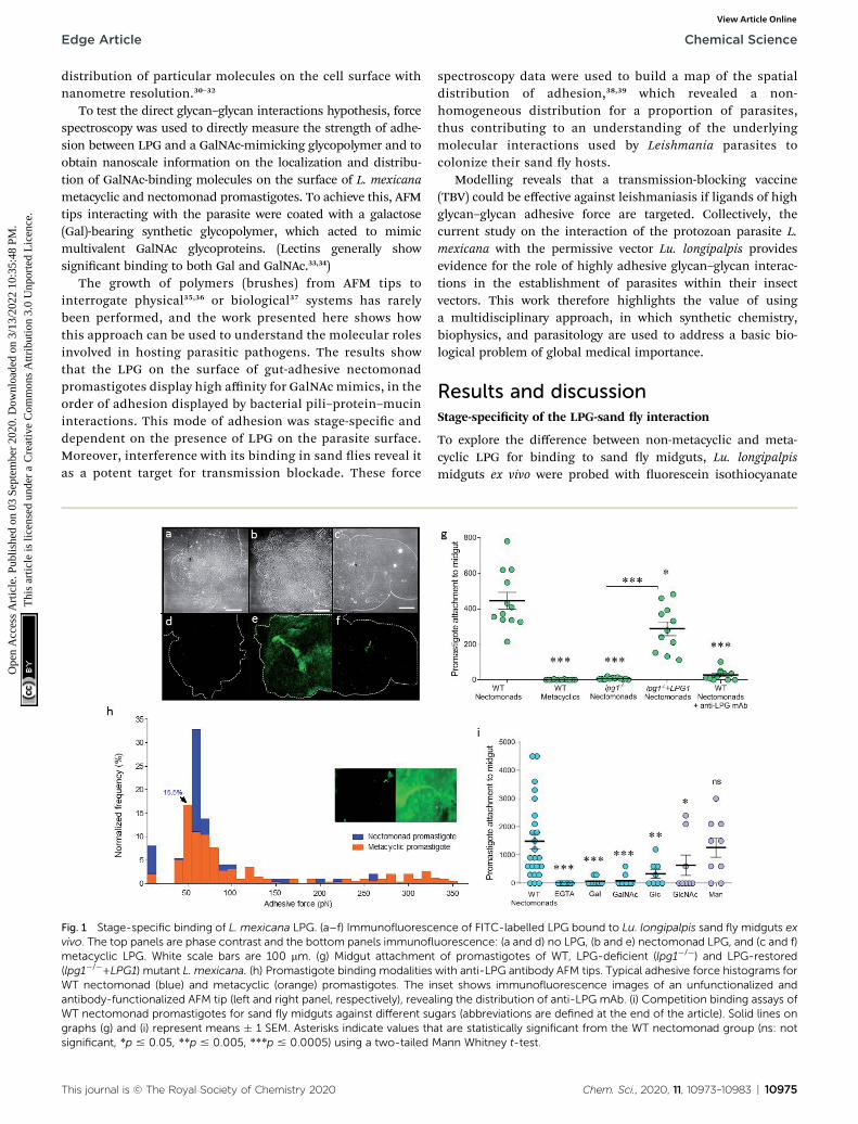
Fig. 1 Stage-specific binding of L. mexicana LPG. (a–f) Immunofluorescvivo. The top panels are phase contrast and the bottom panels immunoflmetacyclic LPG. White scale bars are 100 mm. (g) Midgut attachment(lpg1�/�+LPG1) mutant L. mexicana. (h) Promastigote binding modalitiesWT nectomonad (blue) and metacyclic (orange) promastigotes. The inantibody-functionalized AFM tip (left and right panel, respectively), reveaWT nectomonad promastigotes for sand fly midguts against different sugraphs (g) and (i) represent means � 1 SEM. Asterisks indicate values thsignificant, *p # 0.05, **p # 0.005, ***p # 0.0005) using a two-tailed M
This journal is © The Royal Society of Chemistry 2020
spectroscopy data were used to build a map of the spatialdistribution of adhesion,38,39 which revealed a non-homogeneous distribution for a proportion of parasites,thus contributing to an understanding of the underlyingmolecular interactions used by Leishmania parasites tocolonize their sand y hosts.
Modelling reveals that a transmission-blocking vaccine(TBV) could be effective against leishmaniasis if ligands of highglycan–glycan adhesive force are targeted. Collectively, thecurrent study on the interaction of the protozoan parasite L.mexicana with the permissive vector Lu. longipalpis providesevidence for the role of highly adhesive glycan–glycan interac-tions in the establishment of parasites within their insectvectors. This work therefore highlights the value of usinga multidisciplinary approach, in which synthetic chemistry,biophysics, and parasitology are used to address a basic bio-logical problem of global medical importance.
Results and discussionStage-specicity of the LPG-sand y interaction
To explore the difference between non-metacyclic and meta-cyclic LPG for binding to sand y midguts, Lu. longipalpismidguts ex vivo were probed with uorescein isothiocyanate
ence of FITC-labelled LPG bound to Lu. longipalpis sand fly midguts exuorescence: (a and d) no LPG, (b and e) nectomonad LPG, and (c and f)of promastigotes of WT, LPG-deficient (lpg1�/�) and LPG-restoredwith anti-LPG antibody AFM tips. Typical adhesive force histograms forset shows immunofluorescence images of an unfunctionalized andling the distribution of anti-LPG mAb. (i) Competition binding assays ofgars (abbreviations are defined at the end of the article). Solid lines onat are statistically significant from the WT nectomonad group (ns: notann Whitney t-test.
Chem. Sci., 2020, 11, 10973–10983 | 10975
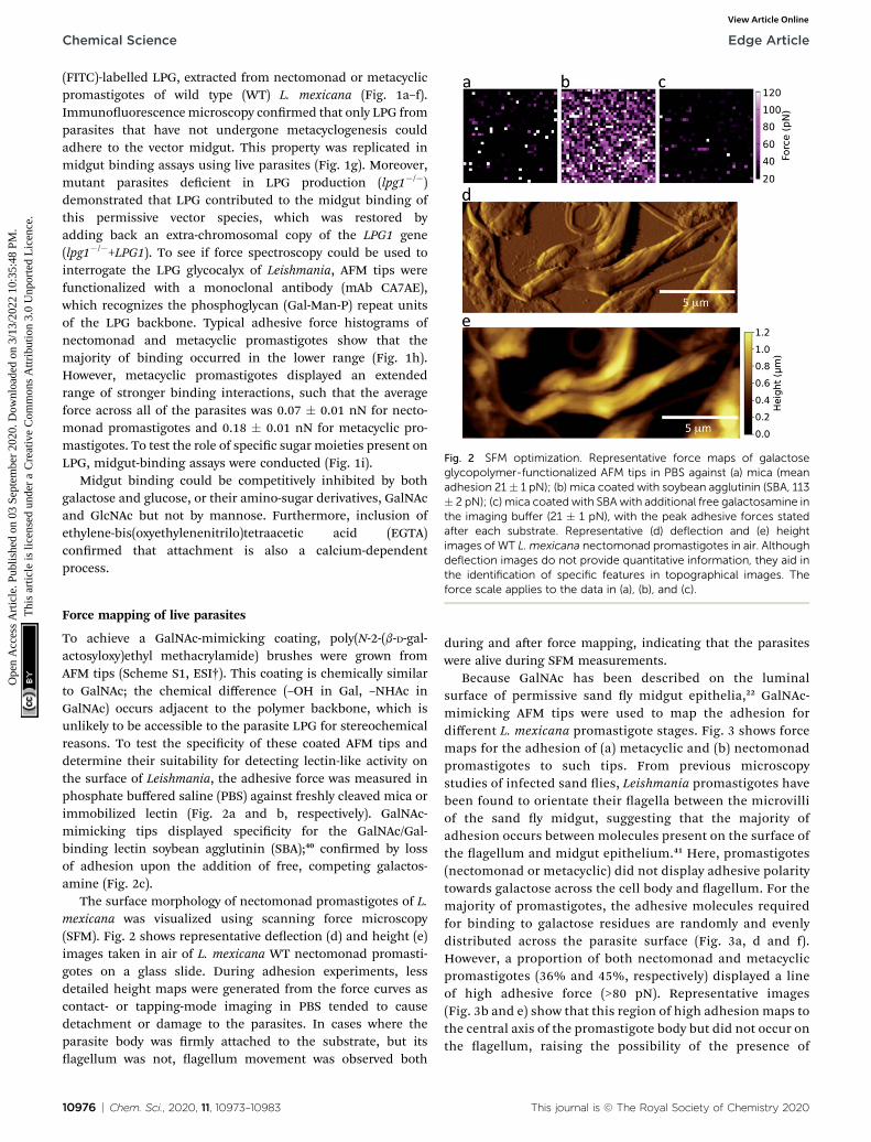
Fig. 2 SFM optimization. Representative force maps of galactoseglycopolymer-functionalized AFM tips in PBS against (a) mica (meanadhesion 21 � 1 pN); (b) mica coated with soybean agglutinin (SBA, 113� 2 pN); (c) mica coated with SBA with additional free galactosamine inthe imaging buffer (21 � 1 pN), with the peak adhesive forces statedafter each substrate. Representative (d) deflection and (e) heightimages of WT L. mexicana nectomonad promastigotes in air. Althoughdeflection images do not provide quantitative information, they aid inthe identification of specific features in topographical images. The
Chemical Science Edge Article
Ope
n A
cces
s A
rtic
le. P
ublis
hed
on 0
3 Se
ptem
ber
2020
. Dow
nloa
ded
on 3
/13/
2022
10:
35:4
8 PM
. T
his
artic
le is
lice
nsed
und
er a
Cre
ativ
e C
omm
ons
Attr
ibut
ion
3.0
Unp
orte
d L
icen
ce.
View Article Online
(FITC)-labelled LPG, extracted from nectomonad or metacyclicpromastigotes of wild type (WT) L. mexicana (Fig. 1a–f).Immunouorescencemicroscopy conrmed that only LPG fromparasites that have not undergone metacyclogenesis couldadhere to the vector midgut. This property was replicated inmidgut binding assays using live parasites (Fig. 1g). Moreover,mutant parasites decient in LPG production (lpg1�/�)demonstrated that LPG contributed to the midgut binding ofthis permissive vector species, which was restored byadding back an extra-chromosomal copy of the LPG1 gene(lpg1�/�+LPG1). To see if force spectroscopy could be used tointerrogate the LPG glycocalyx of Leishmania, AFM tips werefunctionalized with a monoclonal antibody (mAb CA7AE),which recognizes the phosphoglycan (Gal-Man-P) repeat unitsof the LPG backbone. Typical adhesive force histograms ofnectomonad and metacyclic promastigotes show that themajority of binding occurred in the lower range (Fig. 1h).However, metacyclic promastigotes displayed an extendedrange of stronger binding interactions, such that the averageforce across all of the parasites was 0.07 � 0.01 nN for necto-monad promastigotes and 0.18 � 0.01 nN for metacyclic pro-mastigotes. To test the role of specic sugar moieties present onLPG, midgut-binding assays were conducted (Fig. 1i).
Midgut binding could be competitively inhibited by bothgalactose and glucose, or their amino-sugar derivatives, GalNAcand GlcNAc but not by mannose. Furthermore, inclusion ofethylene-bis(oxyethylenenitrilo)tetraacetic acid (EGTA)conrmed that attachment is also a calcium-dependentprocess.
force scale applies to the data in (a), (b), and (c).
Force mapping of live parasites
To achieve a GalNAc-mimicking coating, poly(N-2-(b-D-gal-actosyloxy)ethyl methacrylamide) brushes were grown fromAFM tips (Scheme S1, ESI†). This coating is chemically similarto GalNAc; the chemical difference (–OH in Gal, –NHAc inGalNAc) occurs adjacent to the polymer backbone, which isunlikely to be accessible to the parasite LPG for stereochemicalreasons. To test the specicity of these coated AFM tips anddetermine their suitability for detecting lectin-like activity onthe surface of Leishmania, the adhesive force was measured inphosphate buffered saline (PBS) against freshly cleaved mica orimmobilized lectin (Fig. 2a and b, respectively). GalNAc-mimicking tips displayed specicity for the GalNAc/Gal-binding lectin soybean agglutinin (SBA);40 conrmed by lossof adhesion upon the addition of free, competing galactos-amine (Fig. 2c).
The surface morphology of nectomonad promastigotes of L.mexicana was visualized using scanning force microscopy(SFM). Fig. 2 shows representative deection (d) and height (e)images taken in air of L. mexicana WT nectomonad promasti-gotes on a glass slide. During adhesion experiments, lessdetailed height maps were generated from the force curves ascontact- or tapping-mode imaging in PBS tended to causedetachment or damage to the parasites. In cases where theparasite body was rmly attached to the substrate, but itsagellum was not, agellum movement was observed both
10976 | Chem. Sci., 2020, 11, 10973–10983
during and aer force mapping, indicating that the parasiteswere alive during SFM measurements.
Because GalNAc has been described on the luminalsurface of permissive sand y midgut epithelia,22 GalNAc-mimicking AFM tips were used to map the adhesion fordifferent L. mexicana promastigote stages. Fig. 3 shows forcemaps for the adhesion of (a) metacyclic and (b) nectomonadpromastigotes to such tips. From previous microscopystudies of infected sand ies, Leishmania promastigotes havebeen found to orientate their agella between the microvilliof the sand y midgut, suggesting that the majority ofadhesion occurs between molecules present on the surface ofthe agellum and midgut epithelium.41 Here, promastigotes(nectomonad or metacyclic) did not display adhesive polaritytowards galactose across the cell body and agellum. For themajority of promastigotes, the adhesive molecules requiredfor binding to galactose residues are randomly and evenlydistributed across the parasite surface (Fig. 3a, d and f).However, a proportion of both nectomonad and metacyclicpromastigotes (36% and 45%, respectively) displayed a lineof high adhesive force (>80 pN). Representative images(Fig. 3b and e) show that this region of high adhesion maps tothe central axis of the promastigote body but did not occur onthe agellum, raising the possibility of the presence of
This journal is © The Royal Society of Chemistry 2020
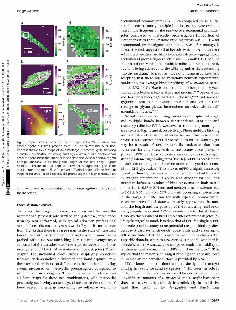
Fig. 3 Representative adhesive force maps of live WT L. mexicanapromastigote surfaces probed with GalNAc-mimicking AFM tips.Representative force maps of (a) a metacyclic promastigote showinga random distribution of strong binding events and (b) a nectomonadpromastigote from the subpopulation that displayed a central regionof high adhesive force along the length of the cell body. Higherresolution images of (a) and (b) are shown in the right-hand panels (d)and (e), focusing on a 1.5–2.0 mm2 area. Typical height (c) and force (f)maps of the anterior of a metacyclic promastigote in higher resolution.
Edge Article Chemical Science
Ope
n A
cces
s A
rtic
le. P
ublis
hed
on 0
3 Se
ptem
ber
2020
. Dow
nloa
ded
on 3
/13/
2022
10:
35:4
8 PM
. T
his
artic
le is
lice
nsed
und
er a
Cre
ativ
e C
omm
ons
Attr
ibut
ion
3.0
Unp
orte
d L
icen
ce.
View Article Online
a more adhesive subpopulation of promastigotes during sandy infection.
Force–distance curves
To assess the range of interactions measured between thenectomonad promastigote surface and galactose, force spec-troscopy was performed, with typical adhesion proles andsample force–distance curves shown in Fig. 4. It can be seenfrom Fig. 4a that there is a large range in the scale of measuredforces for both nectomonad and metacyclic promastigotesprobed with a GalNAc-mimicking AFM tip (the average forceacross all of the parasites was 64 � 5 pN for nectomonad pro-mastigotes and 42 � 3 pN for metacyclic promastigotes). This isdespite the individual force curves displaying conservedfeatures, such as molecule extension and bond rupture. Fromthese results there is a clear difference in the number of bindingevents measured on metacyclic promastigotes compared tonectomonad promastigotes. This difference is reected acrossall force maps for these two combinations, with metacyclicpromastigotes having, on average, almost twice the number offorce curves in a map containing no adhesive events as
This journal is © The Royal Society of Chemistry 2020
nectomonad promastigotes (75 � 5% compared to 39 � 5%,Fig. 4b). Furthermore, multiple binding events were over tentimes more frequent on the surface of nectomonad promasti-gotes compared to metacyclic promastigotes (proportion offorce maps with three or more binding events was 5 � 1% fornectomonad promastigotes and 0.3 � 0.2% for metacyclicpromastigotes), suggesting that ligands, which have multivalentadhesion properties, are likely to bemore densely aggregated onnectomonad promastigotes.42 (The anti-LPG mAb CA7AE on theother hand rarely exhibited multiple adhesion events, possiblydue to it being adsorbed to the AFM tip rather than extendinginto the medium.) To put this mode of binding in context, andaccepting that there will be variations between experimentalconditions, the average binding affinity of L. mexicana necto-monad LPG for GalNAc is comparable to other protein–glycaninteractions between bacterial pili and mucins;43,44 bacterial piliand host pneumocytes;45 bacterial adhesins;46–48 and soybeanagglutinin and porcine gastric mucin,49 and greater thana range of glycan–glycan interactions recorded within self-assembling mucins.50–52
Sample force curves showing extension and rupture of singleand multiple bonds between functionalized AFM tips anda strongly adhesive WT L. mexicana nectomonad promastigoteare shown in Fig. 4c and d, respectively. These multiple bindingevents illustrate that strong adhesion between the nectomonadpromastigote surface and GalNAc residues on the sand y gutmay be a result of LPG or LPG-like molecules that bearnumerous binding sites, such as membrane proteophospho-glycan (mPPG), or dense concentrations of ligands with single,strongly interacting binding sites (Fig. 4e). mPPG is predicted tobe 300–400 nm long and therefore to extend beyond the dense15 nm LPG glycocalyx.53 This makes mPPG a highly accessibleligand for binding partners and potentially important for sandy midgut attachment. It could also account for the longextension before a number of binding events on both necto-monad (up to 0.83� 0.04 mm) andmetacyclic promastigotes (upto 0.64 � 0.05 mm), with 50% of events occurring at extensionsin the range 100–260 nm for both types of promastigote.Measured extension distances are only approximate becauseboth the length and the position of the interacting residue onthe glycopolymer-coated AFM tip contribute to this distance.Although the number of mPPG molecules on promastigotes (alllife cycle stages) is much less than that of LPG,53 a single mPPGmolecule provides many more potential receptor-binding sites,because it displays leucine-rich repeat units and carries up to800 serine-linked LPG-like phosphoglycan chains clustered ina specic domain, whereas LPG carries just one.53 Despite this,LPG-decient L. mexicana promastigotes retain their ability tosynthesize and incorporate mPPG on their surface.54 Thisargues that the majority of midgut binding and adhesive forceto GalNAc on the parasite surface is provided by LPG.
LPG is known to be the dominant parasite ligand for midgutbinding in restrictive sand y species.55,56 However, its role inmidgut attachment in permissive sand ies is less well dened.LPG-decient mutants of L. mexicana and L. major have beenshown to survive, albeit slightly less efficiently, in permissivesand ies such as Lu. longipalpis and Phlebotomus
Chem. Sci., 2020, 11, 10973–10983 | 10977
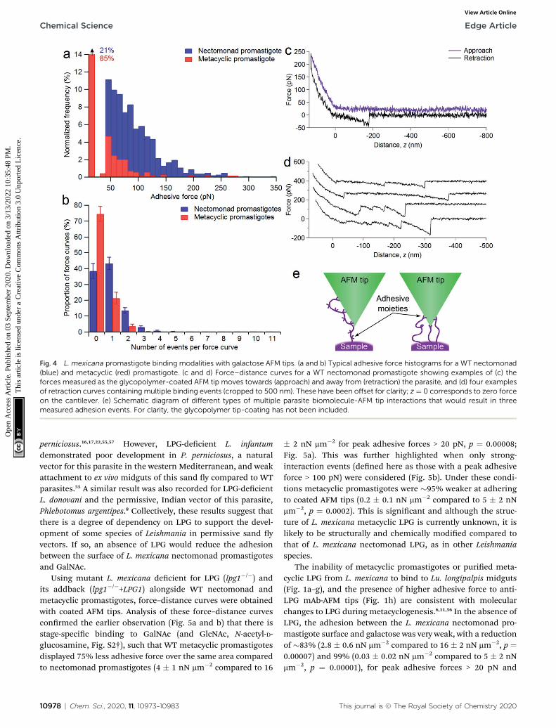
Fig. 4 L. mexicana promastigote binding modalities with galactose AFM tips. (a and b) Typical adhesive force histograms for a WT nectomonad(blue) and metacyclic (red) promastigote. (c and d) Force–distance curves for a WT nectomonad promastigote showing examples of (c) theforces measured as the glycopolymer-coated AFM tip moves towards (approach) and away from (retraction) the parasite, and (d) four examplesof retraction curves containing multiple binding events (cropped to 500 nm). These have been offset for clarity; z ¼ 0 corresponds to zero forceon the cantilever. (e) Schematic diagram of different types of multiple parasite biomolecule-AFM tip interactions that would result in threemeasured adhesion events. For clarity, the glycopolymer tip-coating has not been included.
Chemical Science Edge Article
Ope
n A
cces
s A
rtic
le. P
ublis
hed
on 0
3 Se
ptem
ber
2020
. Dow
nloa
ded
on 3
/13/
2022
10:
35:4
8 PM
. T
his
artic
le is
lice
nsed
und
er a
Cre
ativ
e C
omm
ons
Attr
ibut
ion
3.0
Unp
orte
d L
icen
ce.
View Article Online
perniciosus.16,17,22,55,57 However, LPG-decient L. infantumdemonstrated poor development in P. perniciosus, a naturalvector for this parasite in the western Mediterranean, and weakattachment to ex vivo midguts of this sand y compared to WTparasites.55 A similar result was also recorded for LPG-decientL. donovani and the permissive, Indian vector of this parasite,Phlebotomus argentipes.8 Collectively, these results suggest thatthere is a degree of dependency on LPG to support the devel-opment of some species of Leishmania in permissive sand yvectors. If so, an absence of LPG would reduce the adhesionbetween the surface of L. mexicana nectomonad promastigotesand GalNAc.
Using mutant L. mexicana decient for LPG (lpg1�/�) andits addback (lpg1�/�+LPG1) alongside WT nectomonad andmetacyclic promastigotes, force–distance curves were obtainedwith coated AFM tips. Analysis of these force–distance curvesconrmed the earlier observation (Fig. 5a and b) that there isstage-specic binding to GalNAc (and GlcNAc, N-acetyl-D-glucosamine, Fig. S2†), such that WT metacyclic promastigotesdisplayed 75% less adhesive force over the same area comparedto nectomonad promastigotes (4 � 1 nN mm�2 compared to 16
10978 | Chem. Sci., 2020, 11, 10973–10983
� 2 nN mm�2 for peak adhesive forces > 20 pN, p ¼ 0.00008;Fig. 5a). This was further highlighted when only strong-interaction events (dened here as those with a peak adhesiveforce > 100 pN) were considered (Fig. 5b). Under these condi-tions metacyclic promastigotes were �95% weaker at adheringto coated AFM tips (0.2 � 0.1 nN mm�2 compared to 5 � 2 nNmm�2, p ¼ 0.0002). This is signicant and although the struc-ture of L. mexicana metacyclic LPG is currently unknown, it islikely to be structurally and chemically modied compared tothat of L. mexicana nectomonad LPG, as in other Leishmaniaspecies.
The inability of metacyclic promastigotes or puried meta-cyclic LPG from L. mexicana to bind to Lu. longipalpis midguts(Fig. 1a–g), and the presence of higher adhesive force to anti-LPG mAb-AFM tips (Fig. 1h) are consistent with molecularchanges to LPG during metacyclogenesis.6,11,56 In the absence ofLPG, the adhesion between the L. mexicana nectomonad pro-mastigote surface and galactose was very weak, with a reductionof �83% (2.8 � 0.6 nN mm�2 compared to 16 � 2 nN mm�2, p ¼0.00007) and 99% (0.03 � 0.02 nN mm�2 compared to 5 � 2 nNmm�2, p ¼ 0.00001), for peak adhesive forces > 20 pN and
This journal is © The Royal Society of Chemistry 2020
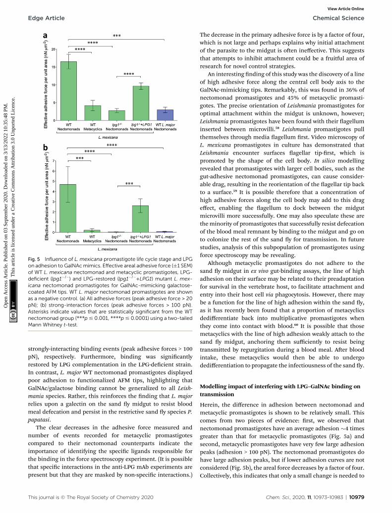
Fig. 5 Influence of L. mexicana promastigote life cycle stage and LPGon adhesion to GalNAcmimics. Effective areal adhesive force (�1 SEM)of WT L. mexicana nectomonad and metacyclic promastigotes, LPG-deficient (lpg1�/�) and LPG-restored (lpg1�/�+LPG1) mutant L. mex-icana nectomonad promastigotes for GalNAc-mimicking galactose-coated AFM tips. WT L. major nectomonad promastigotes are shownas a negative control. (a) All adhesive forces (peak adhesive force > 20pN); (b) strong-interaction forces (peak adhesive forces > 100 pN).Asterisks indicate values that are statistically significant from the WTnectomonad group (***p# 0.001, ****p# 0.0001) using a two-tailedMann Whitney t-test.
Edge Article Chemical Science
Ope
n A
cces
s A
rtic
le. P
ublis
hed
on 0
3 Se
ptem
ber
2020
. Dow
nloa
ded
on 3
/13/
2022
10:
35:4
8 PM
. T
his
artic
le is
lice
nsed
und
er a
Cre
ativ
e C
omm
ons
Attr
ibut
ion
3.0
Unp
orte
d L
icen
ce.
View Article Online
strongly-interacting binding events (peak adhesive forces > 100pN), respectively. Furthermore, binding was signicantlyrestored by LPG complementation in the LPG-decient strain.In contrast, L. major WT nectomonad promastigotes displayedpoor adhesion to functionalized AFM tips, highlighting thatGalNAc/galactose binding cannot be generalized to all Leish-mania species. Rather, this reinforces the nding that L. majorrelies upon a galectin on the sand y midgut to resist bloodmeal defecation and persist in the restrictive sand y species P.papatasi.
The clear decreases in the adhesive force measured andnumber of events recorded for metacyclic promastigotescompared to their nectomonad counterparts indicate theimportance of identifying the specic ligands responsible forthe binding in the force spectroscopy experiment. (It is possiblethat specic interactions in the anti-LPG mAb experiments arepresent but that they are masked by non-specic interactions.)
This journal is © The Royal Society of Chemistry 2020
The decrease in the primary adhesive force is by a factor of four,which is not large and perhaps explains why initial attachmentof the parasite to the midgut is oen ineffective. This suggeststhat attempts to inhibit attachment could be a fruitful area ofresearch for novel control strategies.
An interesting nding of this study was the discovery of a lineof high adhesive force along the central cell body axis to theGalNAc-mimicking tips. Remarkably, this was found in 36% ofnectomonad promastigotes and 45% of metacyclic promasti-gotes. The precise orientation of Leishmania promastigotes foroptimal attachment within the midgut is unknown, however;Leishmania promastigotes have been found with their agelluminserted between microvilli.58 Leishmania promastigotes pullthemselves through media agellum rst. Video microscopy ofL. mexicana promastigotes in culture has demonstrated thatLeishmania encounter surfaces agellar tip-rst, which ispromoted by the shape of the cell body. In silico modellingrevealed that promastigotes with larger cell bodies, such as thegut-adhesive nectomonad promastigotes, can cause consider-able drag, resulting in the reorientation of the agellar tip backto a surface.59 It is possible therefore that a concentration ofhigh adhesive forces along the cell body may add to this drageffect, enabling the agellum to dock between the midgutmicrovilli more successfully. One may also speculate these arethe minority of promastigotes that successfully resist defecationof the blood meal remnant by binding to the midgut and go onto colonize the rest of the sand y for transmission. In futurestudies, analysis of this subpopulation of promastigotes usingforce spectroscopy may be revealing.
Although metacyclic promastigotes do not adhere to thesand y midgut in ex vivo gut-binding assays, the line of highadhesion on their surface may be related to their preadaptationfor survival in the vertebrate host, to facilitate attachment andentry into their host cell via phagocytosis. However, there maybe a function for the line of high adhesion within the sand y,as it has recently been found that a proportion of metacyclicsdedifferentiate back into multiplicative promastigotes whenthey come into contact with blood.60 It is possible that thosemetacyclics with the line of high adhesion weakly attach to thesand y midgut, anchoring them sufficiently to resist beingtransmitted by regurgitation during a blood meal. Aer bloodintake, these metacyclics would then be able to undergodedifferentiation to propagate the infectiousness of the sand y.
Modelling impact of interfering with LPG–GalNAc binding ontransmission
Herein, the difference in adhesion between nectomonad andmetacyclic promastigotes is shown to be relatively small. Thiscomes from two pieces of evidence: rst, we observed thatnectomonad promastigotes have an average adhesion �4 timesgreater than that for metacyclic promastigotes (Fig. 5a) andsecond, metacyclic promastigotes have very few large adhesionpeaks (adhesion > 100 pN). The nectomonad promastigotes dohave large adhesion peaks, but if lower adhesion curves are notconsidered (Fig. 5b), the areal force decreases by a factor of four.Collectively, this indicates that only a small change is needed to
Chem. Sci., 2020, 11, 10973–10983 | 10979
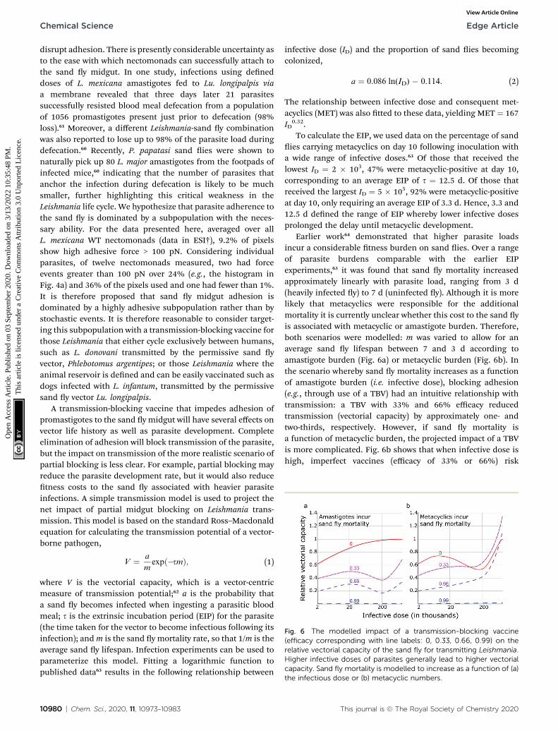
Fig. 6 The modelled impact of a transmission-blocking vaccine(efficacy corresponding with line labels: 0, 0.33, 0.66, 0.99) on therelative vectorial capacity of the sand fly for transmitting Leishmania.Higher infective doses of parasites generally lead to higher vectorialcapacity. Sand fly mortality is modelled to increase as a function of (a)
Chemical Science Edge Article
Ope
n A
cces
s A
rtic
le. P
ublis
hed
on 0
3 Se
ptem
ber
2020
. Dow
nloa
ded
on 3
/13/
2022
10:
35:4
8 PM
. T
his
artic
le is
lice
nsed
und
er a
Cre
ativ
e C
omm
ons
Attr
ibut
ion
3.0
Unp
orte
d L
icen
ce.
View Article Online
disrupt adhesion. There is presently considerable uncertainty asto the ease with which nectomonads can successfully attach tothe sand y midgut. In one study, infections using deneddoses of L. mexicana amastigotes fed to Lu. longipalpis viaa membrane revealed that three days later 21 parasitessuccessfully resisted blood meal defecation from a populationof 1056 promastigotes present just prior to defecation (98%loss).61 Moreover, a different Leishmania-sand y combinationwas also reported to lose up to 98% of the parasite load duringdefecation.60 Recently, P. papatasi sand ies were shown tonaturally pick up 80 L. major amastigotes from the footpads ofinfected mice,60 indicating that the number of parasites thatanchor the infection during defecation is likely to be muchsmaller, further highlighting this critical weakness in theLeishmania life cycle. We hypothesize that parasite adherence tothe sand y is dominated by a subpopulation with the neces-sary ability. For the data presented here, averaged over allL. mexicana WT nectomonads (data in ESI†), 9.2% of pixelsshow high adhesive force > 100 pN. Considering individualparasites, of twelve nectomonads measured, two had forceevents greater than 100 pN over 24% (e.g., the histogram inFig. 4a) and 36% of the pixels used and one had fewer than 1%.It is therefore proposed that sand y midgut adhesion isdominated by a highly adhesive subpopulation rather than bystochastic events. It is therefore reasonable to consider target-ing this subpopulation with a transmission-blocking vaccine forthose Leishmania that either cycle exclusively between humans,such as L. donovani transmitted by the permissive sand yvector, Phlebotomus argentipes; or those Leishmania where theanimal reservoir is dened and can be easily vaccinated such asdogs infected with L. infantum, transmitted by the permissivesand y vector Lu. longipalpis.
A transmission-blocking vaccine that impedes adhesion ofpromastigotes to the sand y midgut will have several effects onvector life history as well as parasite development. Completeelimination of adhesion will block transmission of the parasite,but the impact on transmission of the more realistic scenario ofpartial blocking is less clear. For example, partial blocking mayreduce the parasite development rate, but it would also reducetness costs to the sand y associated with heavier parasiteinfections. A simple transmission model is used to project thenet impact of partial midgut blocking on Leishmania trans-mission. This model is based on the standard Ross–Macdonaldequation for calculating the transmission potential of a vector-borne pathogen,
V ¼ a
mexpð�smÞ; (1)
where V is the vectorial capacity, which is a vector-centricmeasure of transmission potential;62 a is the probability thata sand y becomes infected when ingesting a parasitic bloodmeal; s is the extrinsic incubation period (EIP) for the parasite(the time taken for the vector to become infectious following itsinfection); andm is the sand y mortality rate, so that 1/m is theaverage sand y lifespan. Infection experiments can be used toparameterize this model. Fitting a logarithmic function topublished data63 results in the following relationship between
10980 | Chem. Sci., 2020, 11, 10973–10983
infective dose (ID) and the proportion of sand ies becomingcolonized,
a ¼ 0.086 ln(ID) � 0.114. (2)
The relationship between infective dose and consequent met-acyclics (MET) was also tted to these data, yielding MET ¼ 167ID
0.32.To calculate the EIP, we used data on the percentage of sand
ies carrying metacyclics on day 10 following inoculation witha wide range of infective doses.63 Of those that received thelowest ID ¼ 2 � 103, 47% were metacyclic-positive at day 10,corresponding to an average EIP of s ¼ 12.5 d. Of those thatreceived the largest ID ¼ 5 � 105, 92% were metacyclic-positiveat day 10, only requiring an average EIP of 3.3 d. Hence, 3.3 and12.5 d dened the range of EIP whereby lower infective dosesprolonged the delay until metacyclic development.
Earlier work64 demonstrated that higher parasite loadsincur a considerable tness burden on sand ies. Over a rangeof parasite burdens comparable with the earlier EIPexperiments,63 it was found that sand y mortality increasedapproximately linearly with parasite load, ranging from 3 d(heavily infected y) to 7 d (uninfected y). Although it is morelikely that metacyclics were responsible for the additionalmortality it is currently unclear whether this cost to the sand yis associated with metacyclic or amastigote burden. Therefore,both scenarios were modelled: m was varied to allow for anaverage sand y lifespan between 7 and 3 d according toamastigote burden (Fig. 6a) or metacyclic burden (Fig. 6b). Inthe scenario whereby sand y mortality increases as a functionof amastigote burden (i.e. infective dose), blocking adhesion(e.g., through use of a TBV) had an intuitive relationship withtransmission: a TBV with 33% and 66% efficacy reducedtransmission (vectorial capacity) by approximately one- andtwo-thirds, respectively. However, if sand y mortality isa function of metacyclic burden, the projected impact of a TBVis more complicated. Fig. 6b shows that when infective dose ishigh, imperfect vaccines (efficacy of 33% or 66%) risk
the infectious dose or (b) metacyclic numbers.
This journal is © The Royal Society of Chemistry 2020
Edge Article Chemical Science
Ope
n A
cces
s A
rtic
le. P
ublis
hed
on 0
3 Se
ptem
ber
2020
. Dow
nloa
ded
on 3
/13/
2022
10:
35:4
8 PM
. T
his
artic
le is
lice
nsed
und
er a
Cre
ativ
e C
omm
ons
Attr
ibut
ion
3.0
Unp
orte
d L
icen
ce.
View Article Online
exacerbating vectorial capacity. This is because, even thoughparasite development from amastigote to metacyclic form ispartially blocked, the consequently reduced metacyclic-associated sand y mortality more than offsets this blockade.Meaning, in transmission settings where the average infectivedose is high, a TBV that has less than approximately 66% effi-cacy could pose a health risk.
Conclusions
LPG has been shown to be responsible for stage-specic adhe-sion of L. mexicana nectomonad promastigotes to GalNAc; anepitope found in a mucin-like glycoprotein, coating permissivesand y midguts. These results indicate that LPG-dependentand independent mechanisms operate side-by-side in permis-sive vectors and that LPG adhesion is mediated by relatively fewhigh-force glycan–glycan interactions. In our model of permis-sive sand y attachment, the presence of free GalNAc resulted ina signicant decrease in promastigote binding. This is strongevidence that GalNAc provides an essential substrate for midgutattachment, mediated through glycan–glycan interactions.Modelling indicates that targeting such midgut attachmentmechanisms is a viable transmission-blocking strategy againstleishmaniasis. Furthermore, the work has demonstrated thatthe growth of polymer brushes decorated with specic moietiesas a means of interrogating microorganisms is a viable strategyfor understanding specic interactions. This technology isversatile and is likely to be applicable in the study of parasite–vector interactions beyond that of Leishmania with its sand yhost. Furthermore, there is scope for development in otherareas, for example by tethering antiparasitics or antibiotics tothe brush.
Author contributions
MER, ARH, MG, and NRC conceived and designed the experi-ments; ARH, JTB, MER, AME, PC, LS, OM, and EG performedthe experiments; ARH, ALMG, JTB, and MG analysed the forcespectroscopy data; LY performed the modelling; MW providedLeishmania mutant cell lines; MG, MER, and DHD supervisedthe work; ARH, LY, MER, and MG wrote the paper, to which allauthors contributed and which all authors have approved.
Conflicts of interest
There are no conicts of interest to declare.
Abbreviations
AFM
This journal is
Atomic force microscopy
EGTA Ethylene-bis(oxyethylenenitrilo)tetraacetic acid EIP Extrinsic incubation period FITC Fluorescein isothiocyanate Gal Galactose GalNAc N-acetyl-D-galactosamine Glc Glucose© The Royal Society of Chemistry 2020
GlcNAc
N-acetyl-D-glucosamine HPA Helix pomatia agglutinin LPG Lipophosphoglycan mAb Monoclonal antibody Man Mannose mPPG Membrane proteophosphoglycan PBS Phosphate buffered saline SEM Standard error of the mean SFM Scanning force microscopy SBA Soybean agglutinin TBV Transmission-blocking vaccine WT Wild-typeAcknowledgements
MG thanks Prof. Michel Bergeron (Universite Laval) for thesuggestion of Leishmania. MG andMER acknowledge Prof. SteveBrocchini (UCL: University College London) for the introduc-tion. ARH acknowledges Prof. David Alsteens (UCL: UniversiteCatholique de Louvain) for useful discussions. MER and EGwere supported by the BBSRC (David Phillips Fellowship awar-ded to MER, BB/H022406/1) and MER also acknowledges theBBSRC for support of LS through a London InterdisciplinaryDoctoral Programme rotation project; LY and MER acknowl-edge funding from the MRC through the VALIDATE programme(MR/R005850/1); OM was supported by the Basque Government(Fellowship BF109.183); AME was supported by the LeverhulmeTrust (F/00128/BO); PC and ALMG were supported by the EPSRC(EP/I012060/1) and ALMG also acknowledges CONACyT fora doctoral studentship; and ARH and JTB by the EPSRC throughrespectively a doctoral training grant and iCASE award associ-ated with the doctoral training centre in polymers, so matter,and colloids. Nuria Melisa Morales-Garcıa is gratefullyacknowledged for using her skill in BioRender to providea table-of-contents graphic.
Notes and references
1 J. Alvar, I. D. Velez, C. Bern, M. Herrero, P. Desjeux,J. Cano, J. Jannin, M. den Boer, D. Argaw,S. Bhattacharya, M. Ejov, A. N. Elkhouri, J. A. Ruiz-Postigo and J. Serrano, PLoS One, 2012, 7, e35671.
2 P. A. Bates, Int. J. Parasitol., 2007, 37, 1097–1106.3 World Health Organization, Control of the Leishmaniases,WHO, Geneva, 2010.
4 A. S. Nagle, S. Khare, A. B. Kumar, F. Supek, A. Buchynskyy,C. J. N. Mathison, N. K. Chennamaneni, N. Pendem,F. S. Buckner, M. H. Gelb and V. Molteni, Chem. Rev.,2014, 114, 11305–11347.
5 S. L. Cro and P. Olliaro, Clin. Microbiol. Infect., 2011, 17,1478–1483.
6 D. Sacks and S. Kamhawi, Annu. Rev. Microbiol., 2001, 55,453–483.
7 T. Beneke, F. Demay, E. Hookway, N. Ashman, H. Jeffery,J. Smith, J. Valli, T. Becvar, J. Myskova, T. Lestinova,
Chem. Sci., 2020, 11, 10973–10983 | 10981
Chemical Science Edge Article
Ope
n A
cces
s A
rtic
le. P
ublis
hed
on 0
3 Se
ptem
ber
2020
. Dow
nloa
ded
on 3
/13/
2022
10:
35:4
8 PM
. T
his
artic
le is
lice
nsed
und
er a
Cre
ativ
e C
omm
ons
Attr
ibut
ion
3.0
Unp
orte
d L
icen
ce.
View Article Online
S. Shaq, J. Sadlova, P. Volf, R. J. Wheeler and E. Gluenz,PLoS Pathog., 2019, e1007828.
8 D. L. Sacks, G. Modi, E. Rowton, G. Spath, L. Epstein,S. J. Turco and S. M. Beverley, Proc. Natl. Acad. Sci. U. S.A., 2000, 97, 406–411.
9 M. E. Rogers, M. L. Chance and P. A. Bates, Parasitology,2002, 124, 495–507.
10 R. R. de Assis, I. C. Ibraim, P. M. Nogueira, R. P. Soares andS. J. Turco, Biochim. Biophys. Acta, 2012, 1820, 1354–1365.
11 C.-L. Forestier, Q. Gao and G.-J. Boons, Front. Cell. Infect.Microbiol., 2014, 4, 193.
12 S. Kamhawi, G. B. Modi, P. F. P. Pimenta, E. Rowton andD. L. Sacks, Parasitology, 2000, 121, 25–33.
13 S. Kamhawi, M. Ramalho-Ortigao, V. M. Pham, S. Kumar,P. G. Lawyer, S. J. Turco, C. Barillas-Mury, D. L. Sacks andJ. G. Valenzuela, Cell, 2004, 119, 329–341.
14 P. Volf, P. M. Nogueira, J. Myskova, S. J. Turco andR. P. Soares, Parasitol. Int., 2014, 63, 683–686.
15 P. Volf and J. Myskova, Trends Parasitol., 2007, 23, 91–92.16 M. E. Rogers, T. Ilg, A. V. Nikolaev, M. A. J. Ferguson and
P. A. Bates, Nature, 2004, 430, 463–467.17 A. Svarovska, T. H. Ant, V. Seblova, L. Jecna, S. M. Beverley
and P. Volf, PLoS Neglected Trop. Dis., 2010, 4, e580.18 P. Cecılio, A. C. A. M. Pires, J. G. Valenzuela,
P. F. P. Pimenta, A. Cordeiro-da-Silva, N. F. C. Secundinoand F. Oliveira, J. Infect. Dis., 2020, 222, 1199–1203.
19 J. S. P. Doehl, Z. Bright, S. Dey, H. Davies, J. Magson,N. Brown, A. Romano, J. Dalton, A. I. Pinto,J. W. Pitchford and P. M. Kaye, Nat. Commun., 2017, 8, 57.
20 P. M. Nogueira, A. C. Guimaraes, R. R. Assis, J. Sadlova,J. Myskova, K. Pruzinova, J. Hlavackova, S. J. Turco,A. C. Torrecilhas, P. Volf and R. P. Soares, Parasites Vectors,2017, 10, 608.
21 L. G. Evangelista and A. C. R. Leite, J. Med. Entomol., 2002,39, 432–439.
22 J. Myskova, M. Svobodova, S. M. Beverley and P. Volf,Microbes Infect., 2007, 9, 317–324.
23 J. Myskova, A. Dostalova, L. Penickova, P. Halada,P. A. Bates and P. Volf, Parasites Vectors, 2016, 9, 413.
24 M. A. Haseeb, C. Thors, E. Linder and L. K. Eveland, Exp.Parasitol., 2008, 119, 67–73.
25 A. Markiv, D. Peiris, G. P. Curley, M. Odell andM. V. Dwek,J. Biol. Chem., 2011, 286, 20260–20266.
26 E. de Almeida Marques da Silva and T. V. Fialho Martins, J.Glycobiol., 2016, 5, 1000120.
27 L. M. de Castro Cortes, M. C. de Souza Pereira, F. S. da Silva,B. A. Pereira, F. O. de Oliveira Junior, R. O. de AraujoSoares, R. P. Brazil, L. Toma, C. M. Vicente, H. B. Nader,M. de Fatima Madeira, F. J. Bello and C. R. Alves,Parasites Vectors, 2012, 5, 142.
28 C. J. Day, E. N. Tran, E. A. Semchenko, G. Tram,L. E. Hartley-Tassell, P. S. K. Ng, R. M. King,R. Ulanovsky, S. McAtamney, M. A. Apicella,J. Tiralongo, R. Morona, V. Korolik and M. P. Jennings,Proc. Natl. Acad. Sci. U. S. A., 2015, 112, E7266–E7275.
10982 | Chem. Sci., 2020, 11, 10973–10983
29 C. Formosa-Dague, M. Castelain, H. Martin-Yken,K. Dunker, E. Dague and M. Sletmoen, Microorganisms,2018, 6, 39.
30 Y. F. Dufrene, mBio, 2014, 5, e01363.31 D. Alsteens, M. C. Garcia, P. N. Lipke and Y. F. Dufrene,
Proc. Natl. Acad. Sci. U. S. A., 2010, 107, 20744–20749.32 L. Arnal, G. Longo, P. Stupar, M. F. Castez, N. Cattelan,
R. C. Salvarezza, O. M. Yantorno, S. Kasas andM. E. Vela, Nanoscale, 2015, 7, 17563–17572.
33 V. S. R. Rao, K. Lam and P. K. Qasba, J. Biomol. Struct. Dyn.,1998, 15, 853–860.
34 V. Sharma, V. R. Srinivas, P. Adhikari, M. Vijayan andA. Surolia, Glycobiology, 1998, 8, 1007–1012.
35 M. Raari, Z. J. Zhang, S. R. Carter, G. J. Leggett andM. Geoghegan, Macromolecules, 2015, 48, 6272–6279.
36 M. Raari, Z. J. Zhang, S. R. Carter, G. J. Leggett andM. Geoghegan, Tribol. Lett., 2018, 66, 11.
37 P. Schon, E. Kutnyanszky, B. ten Donkelaar,M. G. Santonicola, T. Tecim, N. Aldred, A. S. Clare andG. J. Vancso, Colloids Surf., B, 2013, 102, 923–930.
38 V. Dupres, F. D. Menozzi, C. Locht, B. H. Clare,N. L. Abbott, S. Cuenot, C. Bompard, D. Raze andY. F. Dufrene, Nat. Methods, 2005, 2, 515–520.
39 A. R. Hall and M. Geoghegan, Rep. Prog. Phys., 2018, 81,036601.
40 H. Lis, B.-A. Sela, L. Sachs and N. Sharon, Biochim. Biophys.Acta, 1970, 211, 582–585.
41 L. L. Walters, G. B. Modi, R. B. Tesh and T. Burrage, Am. J.Trop. Med. Hyg., 1987, 36, 294–314.
42 H. Handa, S. Gurczynski, M. P. Jackson and G. Mao,Langmuir, 2010, 26, 12095–12103.
43 A. P. Gunning, D. Kavanaugh, E. Thursby, S. Etzold,D. A. MacKenzie and N. Juge, Int. J. Mol. Sci., 2016, 17, 1854.
44 P. Tripathi, A. Beaussart, D. Alsteens, V. Dupres, I. Claes,I. von Ossowski, W. de Vos, A. Palva, S. Lebeer,J. Vanderleyden and Y. F. Dufrene, ACS Nano, 2013, 7,3685–3697.
45 A. Beaussart, A. E. Baker, S. L. Kuchma, S. El-Kirat-Chatel,G. A. O'Toole and Y. F. Dufrene, ACS Nano, 2014, 8, 10723–10733.
46 O. Bjornham, J. Bugaytsova, T. Boren and S. Schedin,Biophys. Chem., 2009, 143, 102–105.
47 O. Bjornham, E. Fallman, O. Axner, J. Ohlsson,U. J. Nilsson, T. Boren and S. Schedin, J. Biomed. Opt.,2005, 10, 044024.
48 K. H. Simpson, G. Bowden, M. Hook and B. Anvari, J.Bacteriol., 2003, 185, 2031–2035.
49 M. Sletmoen, T. K. Dam, T. A. Gerken, B. T. Stokke andC. F. Brewer, Biopolymers, 2009, 91, 719–728.
50 K. E. Haugstad, T. A. Gerken, B. T. Stokke, T. K. Dam,C. F. Brewer and M. Sletmoen, Biomacromolecules, 2012,13, 1400–1409.
51 K. E. Haugstad, S. Hadjialirezaei, B. T. Stokke, C. F. Brewer,T. A. Gerken, J. Burchell, G. Picco and M. Sletmoen,Glycobiology, 2016, 26, 1338–1350.
52 C. Tromas, J. Rojo, J. de la Fuente, A. Barrientos, R. Garcıaand S. Penades, Angew. Chem., Int. Ed., 2001, 40, 3052–3055.
This journal is © The Royal Society of Chemistry 2020
Edge Article Chemical Science
Ope
n A
cces
s A
rtic
le. P
ublis
hed
on 0
3 Se
ptem
ber
2020
. Dow
nloa
ded
on 3
/13/
2022
10:
35:4
8 PM
. T
his
artic
le is
lice
nsed
und
er a
Cre
ativ
e C
omm
ons
Attr
ibut
ion
3.0
Unp
orte
d L
icen
ce.
View Article Online
53 T. Ilg, Parasitol. Today, 2000, 16, 489–497.54 T. Ilg, EMBO J., 2000, 19, 1953–1962.55 L. Jecna, A. Dostalova, R. Wilson, V. Seblova, K.-P. Chang,
P. A. Bates and P. Volf, Parasitology, 2013, 140, 1026–1032.56 D. L. Sacks, Cell. Microbiol., 2001, 3, 189–196.57 N. Secundino, N. Kimblin, N. C. Peters, P. Lawyer,
A. A. Capul, S. M. Beverley, S. J. Turco and D. Sacks,Cell. Microbiol., 2010, 12, 906–918.
58 R. Wilson, M. D. Bates, A. Dostalova, L. Jecna, R. J. Dillon,P. Volf and P. A. Bates, PLoS Neglected Trop. Dis., 2010, 4,e816.
59 B. J. Walker, R. J. Wheeler, K. Ishimoto and E. A. Gaffney, J.Theor. Biol., 2019, 462, 311–320.
This journal is © The Royal Society of Chemistry 2020
60 T. D. Seram, I. V. Coutinho-Abreu, F. Oliveira,C. Meneses, S. Kamhawi and J. G. Valenzuela, Nat.Microbiol., 2018, 3, 548–555.
61 M. E. Rogers, M. Hajmova, M. B. Joshi, J. Sadlova,D. M. Dwyer, P. Volf and P. A. Bates, Cell. Microbiol.,2008, 10, 1363–1372.
62 C. Garrett-Jones, Nature, 1964, 204, 1173–1175.63 V. Seblova, V. Volfova, V. Dvorak, K. Pruzinova, J. Votypka,
A. Kassahun, T. Gebre-Michael, A. Hailu, A. Warburg andP. Volf, PLoS Neglected Trop. Dis., 2013, 7, e2187.
64 M. E. Rogers and P. A. Bates, PLoS Pathog., 2007, 3, e91.
Chem. Sci., 2020, 11, 10973–10983 | 10983