e-cadherin regulates neural stem cell self-renewal
TRANSCRIPT
Development/Plasticity/Repair
E-Cadherin Regulates Neural Stem Cell Self-Renewal
Phillip Karpowicz,* Sandrine Willaime-Morawek,* Laurent Balenci, Brian DeVeale, Tomoyuki Inoue, andDerek van der KooyDepartment of Molecular Genetics, University of Toronto, Toronto, Ontario, Canada M5S 1A8
E-Cadherin, a cell adhesion protein, has been shown to take part in the compartmentalization, proliferation, survival, and differentiationof cells. E-Cadherin is expressed in the adult and embryonic forebrain germinal zones in vivo, and in clonal colonies of cells derived fromthese regions and grown in vitro. Mice carrying E-Cadherin floxed genes crossed to mice expressing Cre under the Nestin promoterdemonstrate defects in the self-renewal of neural stem cells both in vivo and in vitro. The functional role of E-Cadherin is furtherdemonstrated using adhesion-blocking antibodies in vitro, which specifically target cadherin extracellular adhesive domains. Adultneural stem cell colonies decrease in the presence of E-Cadherin antibodies in a dosage-dependent manner, in contrast to P-Cadherinantibody. On overexpression of normal E-Cadherin and a mutated E-Cadherin, containing no intracellular binding domain, an increasednumber of clonal adult neural stem cell colonies are observed. These data suggest it is specifically E-Cadherin adhesion that is responsiblefor these self-renewal effects. These data show the importance of E-Cadherin in the neural stem cell niche and suggest E-Cadherinregulates the number of these cells.
IntroductionStem cell (SC) behavior is of interest for its biological insights aswell as its possible therapeutic utility. Neural stem cells (NSCs)(Alvarez-Buylla et al., 2001) are localized within the subependy-mal zone of the forebrain (Morshead and van der Kooy, 2001), aregion that can be thought of as the NSC niche. The notion of aniche that influences SC maintenance has emerged as a compel-ling theory that explains certain SC characteristics (Alvarez-Buylla and Lim, 2004; Ohlstein et al., 2004). Molecular factorsthat operate within and that comprise the SC niche are thought todetermine cell behavior within that localized environment (Kaiand Spradling, 2003), as well as to account for both the persis-tence of the SC in that environment throughout the lifetime ofthe animal.
Short-range factors in the niche might comprise a restrictedmicroenvironment in which the division of SCs would producedaughter cells in drastically different contexts. Thus, by virtue ofits slightly different position, the daughter retained in the nichemight resume the same SC behavior as its parent, whereas theother would assume a different fate. Examples of such short-range signals are gap junctions, which have been shown to influ-ence the proliferation of neural progenitors (Cheng et al., 2004),and the Notch juxtacrine signaling pathway, which has generaleffects on neural development and specific effects on the mainte-
nance of NSCs (Hitoshi et al., 2002; Campos et al., 2006; Louviand Artavanis-Tsakonas, 2006).
One important aspect of the niche is the mechanism by whichNSCs remain in the germinal zone. Candidates for anchoringthese cells are cadherins, cell adhesion proteins that are thoughtto play a role in diverse tissues (Takeichi, 1995). It is currentlyunderstood that cadherins of the same type bind homophilicallyand drive cells to sort together to self-assemble into aggregatesthat maximize such homotypic adhesion events (Foty and Stein-berg, 2005).Vertebrate cadherins are thought to be involved inthe compartmentalization of different neural regions during de-velopment (Redies, 2000).
E-Cadherin is transiently expressed in the developing dien-cephalons and mesencephalon of mouse embryos (Shimamuraand Takeichi, 1992) in which it is believed to participate in thesegmentation of the developing brain (Matsunami and Takeichi,1995). Although it is downregulated in most of the brain duringembryogenesis, the expression of E-Cadherin is seen in the ven-tricles of the developing (Rasin et al., 2007) and adult brain (Kuoet al., 2006), regions in which NSCs reside and/or contact.
We examined the loss and gain of E-Cadherin in NSCs of theadult mouse brain. NSCs arise during development and arethought to contribute to neurogenesis in the embryo and theadult (Tropepe et al., 1999). Such NSCs can be characterized invivo or in vitro using a clonal cell culture system in which singlecells dissected from the adult or embryonic neural regions dem-onstrate both self-renewal and multipotentiality (Reynolds et al.,1992; Morshead et al., 1994; Tropepe et al., 1999). Our resultsshow that E-Cadherin is expressed by NSCs and regulates NSCself-renewal in the murine forebrain.
Materials and MethodsAnimal dissection and cell culture. E-Cadherinfloxed/� (Boussadia et al.,2002) and Nestin-Cre [B6.Cg-Tg(Nes-cre)1Kln/J; The Jackson Labora-tory] mice were crossed to obtain a E-Cadherinfloxed/�; Nestin-Cre strain
Received Jan. 5, 2009; revised Feb. 13, 2009; accepted Feb. 16, 2009.This work was supported by the Natural Sciences and Engineering Research Council, Stem Cell Network/Juvenile
Diabetes Research Foundation (to S.W.-M.), and Canadian Institutes of Health Research. We thank Drs. Elaine Fuchsand Rolf Kemler for sharing the E-Cadherin flox mouse strain, Dr. Rudiger Blindt for the N-Cadherin cDNA, and Dr.Seiji Hitoshi for the pMXIE and E-Cad–pMXIE retroviral constructs. As well, members of the van der Kooy Laboratoryare thanked for their insightful comments.
*P.K. and S.W.-M. contributed equally to this work.Correspondence should be addressed to Phillip Karpowicz at the above address. E-mail:
[email protected]:10.1523/JNEUROSCI.0037-09.2009
Copyright © 2009 Society for Neuroscience 0270-6474/09/293885-12$15.00/0
The Journal of Neuroscience, March 25, 2009 • 29(12):3885–3896 • 3885

that expresses Cre specifically in the nervous system. This was then back-crossed with E-Cadherinfloxed/foxed to obtain the E-Cadherinfloxed/floxed;Nestin-Cre strain (Ecad �/�). E-Cadherin�/�; Nestin-Cre offspring fromthese crosses were used as wild-type controls (Ecad Wt/Wt). CD1 and B57wild-type mice, Ecad Wt/Wt, and Ecad �/� mice were dissected, and theirNSCs were cultured as previously described (1) for embryonic day 9.5(E9.5) and E13.5 embryonic forebrain ventricles (Tropepe et al., 1999) or(2) for adult mouse forebrain ventricles (Reynolds et al., 1992; Morsheadet al., 1994). Cells were cultured at 10 cells/�l after dissection and pas-saged thereafter at 5 cells/�l in all experiments. Neurospheres arisingfrom Ecad �/� were passaged exactly as wild types, and both were countedafter 7 d in vitro. The number of spheres was �100 –50 per well forwild-type animals and one-half of this for the Ecad �/� animals in thelong-term passaging experiments. Antibodies against E-Cadherin(ECCD-1 and ECCD-2; Zymed), N-Cadherin (Sigma-Aldrich), orP-Cadherin (Zymed) were dissolved in water and added to media justbefore application, at the concentrations indicated in the text.
Plasmid construction and retroviral infection. The pMXIE retroviralconstruct has been described previously (Hitoshi et al., 2002). To gener-ate the pMXIE–E-Cadherin construct, human E-cadherin cDNA wasamplified by PCR in 50 �l volume consisting of 1 �M sense (5�-CCCTCGCTCGAGGTCCCCGGCCCAG-3�) and antisense (5�-CCTCTCTCGAGATCTCTAGTCGTCCTCG-3�) primers, 2.5 mM
Mg 2�, 0.3 mM dNTP, 1 �l of Takara LA Taq polymerase (Takara), andpLKpac1 human E-Cadherin plasmid (a gift from Dr. A. B. Reynolds,Vanderbilt University, Nashville, TN) as a template. PCR parametersincluded the following: denaturation at 95°C for 30 s, annealing at 60°Cfor 60 s, and extension at 72°C for 180 s for 20 cycles. The amplified DNAfragments were digested with XhoI and BglII and ligated to the XhoI–BamHI site of the pMXIE retroviral vector plasmid. To generate thepMXIE–N-Cadherin construct, the plasmid was first cut using XhoI andBamHI. An oligo containing the sites SalI–BamHI–XhoI–BglII, in thatorder, was digested with SalI and BglII. The cut products were ligated tomodify the multiple cloning site of pMXIE to contain BamHI, XhoI in thecorrect sequence, allowing for the insertion of the human N-CadherincDNA (a gift from Dr. R. Blindt, University Hospital Aachen, Aachen,Germany) after its excision from the pCMX plasmid using BamHI andXhoI. A total of 100,000 cells was exposed to virus at a ratio of 10 virusparticles to 1 cell in the presence of 5 ng/�l hexadimethrine bromide(Sigma-Aldrich). Cells were incubated with retrovirus in 250 �l of cellculture media [containing EGF (epidermal growth factor) and FGF] for90 min while being centrifuged at 1000 rpm at room temperature. Cellswere then resuspended, recounted, and plated as described above. Beforeuse, colonies were examined to confirm retrovirus integration and trans-gene expression by fluorescence microscopy.
Immunocytochemistry/immunohistochemistry. Adult mice were anes-thetized and perfused using 4% paraformaldehyde (Sigma-Aldrich) dis-solved in cold Stockholm’s PBS, pH 7.3. After perfusion, brains weredissected from cranium and fixed overnight at 4°C in 4% paraformalde-hyde. Brains were then washed with Stockholm’s and equilibrated inStockholm’s containing 30% w/v sucrose (Sigma-Aldrich) at 4°C. Sam-ples were then embedded in cryoprotectant (Thermo Fisher Scientific)and sectioned on a Jencon’s OTF5000 cryostat at 15 or 20 �m thickness.NSC colonies were coated with Matrigel for 30 min at 37°C. Cell attach-ment was assessed by gently tapping plates under the microscope. Cellswere fixed using 4% paraformaldehyde (Sigma-Aldrich) dissolved incold Stockholm’s PBS, pH 7.3, for 15 min. Sections or colonies werewashed three times for 10 min each time with Stockholm’s PBS plus 0.3%Triton X-100 detergent (Sigma-Aldrich). To detect bromodeoxyuridine(BrdU), cells were exposed to 4N HCl for 30 min. Samples were thenblocked using 1% bovine serum albumin (Sigma-Aldrich) plus 10% nor-mal goat serum (Sigma-Aldrich) in Stockholm’s, pH 7.3, 0.3% TritonX-100 (Sigma-Aldrich) for 60 min at room temperature. Primary anti-bodies were applied overnight in Stockholm’s, 1.0% NGS, 0.3% TritonX-100. �-E-Cadherin (Millipore; 1:100), �-N-Cadherin (GC-4; Sigma-Aldrich; 1:100), �-BrdU Bu1/75 (Abcam; 1:500), �-Nestin (MilliporeBioscience Research Reagents; 1:1000), �-�-tubulin isotype III (Sigma-Aldrich; 1:500), �-pan-histone (Millipore Bioscience Research Reagents;1:500), �-CD-133 (eBioscience; 1:50), �-polysialylated neural cell adhe-
sion molecule (PSA-NCam) (Millipore Bioscience Research Reagents;1:200), �-Sox2 (Millipore Bioscience Research Reagents; 1:200), and�-glial fibrillary acidic protein (Sigma-Aldrich; 1:400) were used. Sam-ples were washed three times with Stockholm’s and blocked again usingthe same conditions above. Secondary antibodies were applied at 37°Cfor 50 min (for colony immunocytochemistry) or 2 h (for section immu-nohistochemistry) in Stockholm’s PBS, 1.0% normal goat serum. TRITC(tetramethylrhodamine isothiocyanate)-, FITC-, or HRP-conjugatedantibodies (Jackson ImmunoResearch Laboratories; 1:250) or 488, 568,633 nm Alexa Fluor antibodies (Invitrogen; 1:300) were used as appro-priate. Cadherin staining was enhanced by tyramide signal amplification(TSA Plus Cyanine 3/Fluorescein kit; PerkinElmer). Nuclei were coun-terstained with 10 �g/ml Hoechst 33258 (Sigma-Aldrich) or Topro-3(Invitrogen). Sections were mounted and coverslipped using Gel Mount(Biomeda).
Microscopy. Photographs were taken under 40�/0.55 (dry lens) objec-tive using a 40�/0.60 Olympus IX81 inverted microscope with theOlympus Microsuite, version 3.2, Analysis imaging system software (SoftImaging Systems). Confocal micrographs were visualized and photo-graphed using a Plan-Apochromat 60�/1.40 oil-immersion lens objec-tive on a LSM510 (Carl Zeiss). Confocal sections of �0.5 �m thicknesswere taken. Detection settings were kept constant when comparingEcad Wt/Wt versus Ecad �/� samples; otherwise, these were adjusted asneeded. All photos were processed using Adobe Photoshop CS2 software;images of Ecad Wt/Wt versus Ecad �/� were prepared identically.
Transmission electron microscopy. Nine-month-old wild-type andEcadh �/� mice were anesthetized and perfused using 2.5% glutaralde-hyde (Electron Microscopy Sciences) dissolved in Sorensen’s phosphatebuffer. Brains were then dissected out and fixed for 24 h at 4°C in thefixative solution mentioned above. Tissues were sectioned on a vi-bratome at 200 �m and kept in fresh fixative solution for 3 more hours at4°C and postfixed in 1% osmium tetroxide for 2 h. Brain slices were thenwashed in Sorensen’s phosphate buffer and dehydrated using a gradedseries of ethanol, 50, 70, and 95%, twice each for 15 min followed by threebaths of 100% EtOH, and samples were finally washed in propylene oxide(twice 15 min each). Thick sections were placed into polyethylene BEEMcapsules, embedded in Epon Araldite resin, and kept at 60°C for 48 h.After identification of the lateral ventricle and SEZ areas, samples werecut again, placed on resin blocks and then sectioned on a ReichertUltracut E microtome to 90 nm thickness and collected on mesh coppergrids (Electron Microscopy Sciences). Wild-type and conditional knock-out sections were cut in parallel. Ultrathin sections were counterstainedusing saturated uranyl acetate for 15–20 min and electron-opaquestained in Reynold’s lead citrate. EM pictures were captured with aHitachi H7000 transmission electron microscope. A minimum of sixsections was examined from each animal, and a total of 20 sections pergenotype.
Fluorescence-activated cell sorting. Cells were sorted on FACS DiVa (BDBiosciences) system. Cells were sorted at �9000 events per second, andfractions were kept on ice until plated. At the outset of each experiment,CD1 [green fluorescent protein (GFP)-negative] adult neurosphere cellsand GFP transgenic adult neurosphere cells served as negative and posi-tive controls, respectively, to set the gates for cell sorting. Fluorescencemicroscopy confirmed the positivity of retrovirus-infected cells just be-fore use. Cells were dissociated mechanically and blocked for 30 min at37°C, in Dulbecco’s PBS, pH 7.3, plus 10% normal goat serum (Sigma-Aldrich). Cells were then exposed to 2 �g/ml primary anti-E-Cadherinantibody ECCD2 (Zymed; 1:500), in 1 ml of Dulbecco’s plus 3% goatserum for 1 h at 37°C. Cells were then washed two times in 10 ml ofDulbecco’s and exposed to goat anti-mouse 633 nm Alexa Fluor antibody(Invitrogen; 1:300) for an additional hour in the same conditions as theprimary. Cells were then washed two times in 10 ml of Dulbecco’s andsorted. Embryonic stem cells served as positive controls to confirm theefficacy of the E-Cadherin antibody.
Cell death detection. Terminal deoxynucleotidyl transferase-mediatedbiotinylated UTP nick end labeling (TUNEL) was performed using aFluorescein In Situ Cell Death Detection kit (Roche Applied Science).
PCR and reverse transcription-PCR. Tail clip DNA and mRNA wereextracted using the DNeasy Tissue kit (QIAGEN) and RNeasy Mini/
3886 • J. Neurosci., March 25, 2009 • 29(12):3885–3896 Karpowicz et al. • E-Cadherin Regulates Neural Stem Cells
Micro kits (QIAGEN), respectively. Genotyping was performed using thefollowing primer sequences: Cdh1 forward, GAATTCTGAACATCAT-TATCAGTATTTA; reverse, TGACACATGCCTTTACTTTAGT; Creforward, GCGGTCTGGCAGTAAAAACTATC; reverse, GTGAAACAG-CATTGCTGTCACTT; IL2 forward, CTAGGCCACAGAATTGAAA-GATCT; reverse, GTAGGTGGAAATTCTAGCATCATCC. Transcriptdetection was performed using one-step reverse transcription (RT) reac-tions in the RNeasy kits (QIAGEN), either on bulk cultures or single NSCcolonies, with the following sequences: Cdh1/E-Cadherin, forward,CGTGATGAAGGTCTCAGCC; reverse, GATGGGGGCTTCATTC-ACG; Cdh2/N-Cadherin, forward, CCTGGAATGCGGCATAC;reverse, GAAGATCAAACGCGAACG; �-Catenin, forward, CATGTTC-CCTGAGACGCTAGA; reverse, CAGAGTCCCAGCAGTACAACG;�-Catenin, forward, TTTATCGCATCTGAAAATTGTCG; reverse, CT-TGGTCATCTTGTCAATCGC; p120, forward, CACCATCAAC-GAAGTTATCGC; reverse, GCAGGTAGAGTGGCGCTAAA; Actin,forward, GAAGTGTGATGTGGATATCCGC; reverse, AGAAGCAT-TTGCGGTGGAC. Nested PCR on the Cdh1/E-Cadherin RT productwas performed with the following sequences: forward, CAGACGAT-GACGTCAACAC; reverse, CCTCATTCTCAGGCACTTG.
Statistics. Analysis was performed using GraphPad Prism 4.0. Com-parisons between samples were performed by unpaired t tests; compari-sons between multiple groups were performed by ANOVA with Dun-nett’s post test as required.
ResultsE-Cadherin is present in the adult and embryonic brain andin clonal stem cell coloniesFirst, E-Cadherin transcript was probed in the adult forebraingerminal zones. We found E-Cadherin RNA, and that of its bind-ing partners �-Catenin, �-Catenin, and p120, present by RT-PCR both in vivo and in clonal colonies in vitro derived fromadult ventricle tissues (supplemental Table 1, available at www.jneurosci.org as supplemental material). Forebrain germinalzone cells from E9.5 and E13.5 embryos, as well as their respectivecolonies, also were found to express these same transcripts.N-Cadherin expression, which is known to be present in neuraltissues, was similarly confirmed in adult ventricular tissue andadult-derived colonies (supplemental Table 1, available at www.jneurosci.org as supplemental material).
Second, adult forebrain ventricles were assayed by immuno-histochemistry to confirm the presence of E-Cadherin protein inthe NSC niche. Consistent with published results by Kuo et al.(2006), E-Cadherin presence was confirmed in the ependymalcells of the ventricles as well as at low levels in the subependymalzone (Fig. 1A). N-Cadherin was shown to occupy a similar dis-tribution in these very same regions (Fig. 1B). Dissection of adultventricular zones followed by cellular dissociation, antibodystaining, and flow cytometry confirmed that 4.2 � 0.8% of dis-sected adult forebrain germinal zone cells expressed theE-Cadherin protein (data not shown).
Antibody was next applied to the clonal colonies grown fromNSCs dissected from germinal zones in the embryo or the adult.Immunocytochemistry showed the presence of E-Cad in clonalE9.5, E13.5, and adult neurosphere colonies (data not shown).Because Cadherins have been proposed to mediate cell sorting,we examined 7 d NSC neurospheres to determine whetherE-Cad-positive cells demonstrated any localization within these(Fig. 1Ci). Although E-Cadherin-positive cells clustered togetherin these colonies, no complete separation between E-Cadherin-positive and -negative cells was observed in any of the coloniesexamined. This may be attributable to the presence ofN-Cadherin in these cells, which, when examined, showed ex-pression throughout all cells in these colonies (Fig. 1Cii).
Together, these data show that RNA transcripts of E-Cadherin
and its associated binding partners are found both in vivo and inthe in vitro colonies derived from NSCs. The E-Cadherin proteinis present in the adult brain and in SC-initiated colonies derivedfrom different developmental time points. However, E-Cadherinalone does not appear to mediate complete sorting out (Steinbergand Takeichi, 1994) between E-Cadherin-positive and -negativeneural cells arising from NSCs in vitro.
Disruption of E-Cadherin in vivo reduces NSC self-renewalbut increases neural progenitor proliferationPrevious studies have shown that neurogenesis is impaired inmutations of the adherens junction protein �-E-Catenin (Lien etal., 2006). In contrast, neurogenesis appears normal in mutantsof the protein aPKC� (atypical protein kinase C�), which isthought to regulate the formation of adherens junctions (Imai etal., 2006). These studies presumably affected adherens junctionsthroughout the nervous system. We attempted to focus on mu-tations of E-Cadherin in areas in which NSCs specifically reside,because E-Cadherin expression seems localized to ventricular re-gions of the adult forebrain (Kuo et al., 2006; Rasin et al., 2007).E-Cadherin-null embryos are not viable (Larue et al., 1994), andthus the role of this protein cannot be examined in the adult brainusing conventional mutants. E-Cadherin conditional knock-outswere assayed by crossing mice carrying LoxP sites in introns 5 and10 of the E-Cadherin gene (Boussadia et al., 2002) with miceexpressing the Cre-recombinase enzyme under the Nestin pro-moter (Tronche et al., 1999). Given that all of these tissues derivefrom Nestin-expressing precursors, this cross should result in thedeletion of functional E-Cadherin protein in all CNS tissues. Theefficient excision of genomic E-Cadherin was confirmed by PCR(Fig. 1D) and by immunohistochemistry (Fig. 1A). It was notedthat N-Cadherin expression and localization was normal on de-letion of E-Cadherin (Fig. 1B).
Conditional E-Cadherin mutant mice (Ecad�/� mice) ob-tained from these crosses did not seem abnormal in appearanceor behavior. Indeed, no phenotypes were readily observed inadult Ecad�/� brains, suggesting that any transient effects ofE-Cadherin expression in the embryonic and adult brain are neg-ligible or compensated for during development. We examinedcell proliferation in the forebrain ventricles of 2-month-old miceby BrdU uptake 1 h after administration (Fig. 2A). Ecad�/� micewere noted to have an increased number of cells in S-phase com-pared with their littermate wild-type (Ecad Wt/Wt) controls duringthis time of early adulthood. However, because NSCs are a mi-nority of the cells in this region, it is most likely that this differ-ence is attributable to the progenitors residing in these tissues.NSCs themselves are thought to divide relatively slowly and theirpresence can be distinguished from that of fast-dividing adjacentneural progenitor cells by assaying long-term BrdU retentionamong cells in these regions (Morshead et al., 1998). The long-term retention of BrdU was examined to resolve between NSCsand neural progenitors in the forebrain ventricles. We did notobserve any differences in the numbers of BrdU-retaining cellsbetween 2-month-old Ecad�/� and Ecad Wt/Wt mice, 1 monthafter five BrdU injections administered every 3 h over 15 h total(Fig. 2A). These data suggested that neural progenitor prolifera-tion was accelerated in the absence of E-Cadherin, but NSC num-bers were normal with or without the protein.
We sought to determine whether an age-dependent effect ofE-Cadherin became apparent during neurogenesis at later timepoints during adulthood. Animals were aged to 9 month timepoint and neural cell proliferation was assessed in the Ecad�/�
after the same short-term, 1 h BrdU pulse. Aged SVZ cells from
Karpowicz et al. • E-Cadherin Regulates Neural Stem Cells J. Neurosci., March 25, 2009 • 29(12):3885–3896 • 3887
Ecad�/� mice also showed increased cell proliferation comparedwith wild-type Ecad Wt/Wt controls showing that the effect ofE-Cad loss is a lifelong increase in cell division of cells in theforebrain germinal zone (Fig. 2B–D). However, in contrast to
young 2-month-old mice, aged 9-month-old Ecad�/� mice had asignificant decrease in BrdU-retaining cells in the subependymalzone (Fig. 2D). These results suggest that E-Cadherin functionsto restrict neural progenitor divisions in the neurogenic adult
Figure 1. A, E-Cadherin is expressed in the adult murine ventricles. Image shows confocal micrographs of E-Cadherin in the adult forebrain lateral ventricular germinal zone (green). E-Cadherinstains primarily in ependymal cells and to a lesser extent in subependymal cells in wild-type animals (i, ii) versus conditional knock-outs (iii, iv), whose E-Cadherin staining is almost completelyabsent. Nuclei are shown in merge in red. B, N-Cadherin is expressed in the adult murine ventricles. The image shows confocal micrographs of N-Cadherin in the forebrain lateral ventricular germinalzone (green). Similar to E-Cadherin, N-Cadherin staining is seen particularly in the ependymal cells rather than subependymal cells. Both E-Cadherin wild type (i) and conditional knock-outs (ii)display N-Cadherin in these regions. Nuclei are shown in merge in red. C, E-Cadherin and N-Cadherin are expressed at day 7 in vitro in adult NSC colonies. Merged images show sections of large NSCcolonies stained for E-Cadherin (green; i) and N-Cadherin (green; ii). DAPI (4�,6-diamidino-2-phenylindole) counterstain is blue. N-Cadherin staining was noted to be stronger and more widelyexpressed than E-Cadherin in these colonies. D, E-Cadherin PCR of colonies derived from NSCs dissected from adult forebrain ventricles. Image shows complete loss of E-Cadherin PCR product inwild-type (n 5 animals; rows 1–5) versus conditional knock-out animals (n 5 animals; rows 6 –10). DNA was extracted from passage 2 NSC colonies to avoid contamination from endothelialcells, also present in the SVZ, which express E-Cadherin protein. Because primers span intron 10 of the E-Cadherin gene, the larger band indicates alleles in which a loxP site is present. The lower bandsshow wild-type allele. Row 11 is negative (water) control.
3888 • J. Neurosci., March 25, 2009 • 29(12):3885–3896 Karpowicz et al. • E-Cadherin Regulates Neural Stem Cells
subependymal zone. The decrease in NSC numbers in aged ani-mals may reflect a decrease in self-renewal of NSCs, or a senes-cence or increased cell death in this population.
Nine-month-old E-Cadherin conditional knock-outs werefurther examined to assess the demography of neural cells in theforebrain germinal zone. Ecad Wt/Wt and Ecad�/� mice were
found to contain cells expressing GFAP, Nestin, PSA-NCam (Fig.2E,F), as well as Sox2 and CD-133 (data not shown) in approx-imately equivalent proportions between genotypes. However,when assessing specifically BrdU-retaining cells, Ecad�/� werefound to have less GFAP- and Nestin-expressing cells, whereascells expressing PSA-NCam were in equivalent proportions be-
Figure 2. A, BrdU uptake is increased but BrdU retention is equivalent, in 2 month E-Cadherin conditional knock-out forebrain ventricular subependyma. Left, The graph shows an increase inBrdU � progenitors in Ecad �/� mice (n 3) compared with their Ecad Wt/Wt littermate controls (n 3) at 2 months of age. The asterisk indicates that the difference is significant (t 4.529; df 58; p � 0.05). Right, Same graph shows numbers of BrdU � nuclei in 2-month-old BrdU � retaining progenitors, 1 month after BrdU injection, in Ecad �/� mice (n 3) compared with theirEcad Wt/Wt littermate controls (n 3). There are no significant differences. B, BrdU uptake in 9 month wild-type forebrain ventricles. The sections show BrdU-positive Ecad Wt/Wt neural precursors(arrow) after 1 h BrdU exposure in vivo. i shows brightfield. BrdU is green (ii, iii) and nuclei are counterstained in red by a pan-histone antibody (iii). C, BrdU uptake in 9 month E-Cadherin conditionalknock-out forebrain ventricles. Sections show BrdU Ecad �/�-positive neural precursors (arrows) after 1 h BrdU exposure in vivo. i shows brightfield. BrdU is green (ii, iii) and nuclei are red (iii). Notethe increase in Ecad �/� cells entering S-phase during short-term BrdU administration. D, BrdU uptake is increased but BrdU retention is decreased in 9 month E-Cadherin conditional knock-outforebrain ventricular subependyma. Left, The graph shows increase in BrdU � progenitors in Ecad �/� mice (n 4) compared with their Ecad Wt/Wt littermate controls (n 5) at 9 months of age.The asterisk indicates that the difference in BrdU � nuclei after 1 h BrdU exposure is significant (t 6.013; df 88; p � 0.05). Right, Same graph shows decrease in 9-month-old BrdU � retainingprogenitors, 1 month after BrdU administration, in Ecad �/� mice (n 5) compared with their Ecad Wt/Wt littermate controls (n 5). The asterisk indicates this reduction is significant (t 3.899;df 118; p � 0.05) in contrast to that observed in 2 month animals. E, GFAP is present in both wild-type and conditional knock-out forebrain ventricles. Confocal micrographs show GFAP positivityin both Ecad Wt/Wt (i, iii) as well as Ecad �/� (ii, iv) SVZ cells. E-Cadherin is green, GFAP is blue, and nuclei are counterstained red. The boxed area in i is shown magnified in iii, and ii in iv. F, E-Cadherinconditional knock-out forebrain shows normal cell demographics. The graph shows numbers of GFAP, PSA-NCam, and Nestin-positive cells present in the 9-month-old forebrain of Ecad Wt/Wt andEcad �/� animals. No significant differences are noted in the presence of any of these cell types (n 3 animals of each genotype sampled). G, E-Cadherin loss leads to reduction in GFAP and NestinBrdU-retaining cells in 9-month-old animals. The graph shows numbers of BrdU-retaining cells that colabel for GFAP, PSA-NCam, or Nestin. The asterisks show that there are significant differencesbetween Ecad Wt/Wt and Ecad �/� GFAP-expressing cells (t 5.758; df 4; p � 0.05) as well as Nestin-expressing cells (t 4.341; df 4; p � 0.05). These results contrast with the generallyequivalent labeling of GFAP and Nestin in the aged forebrain and suggest the decreases in these numbers are specific to the BrdU-retaining cell population. Error bars indicate SEM.
Karpowicz et al. • E-Cadherin Regulates Neural Stem Cells J. Neurosci., March 25, 2009 • 29(12):3885–3896 • 3889
tween genotypes (Fig. 2G). Because NSCsare thought to express GFAP (Imura et al.,2003; Morshead et al., 2003), this sug-gested that the population of NSCs wasstill present, although reduced, in theEcad�/� animals.
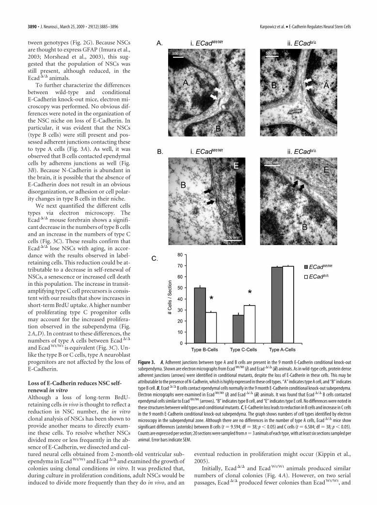
To further characterize the differencesbetween wild-type and conditionalE-Cadherin knock-out mice, electron mi-croscopy was performed. No obvious dif-ferences were noted in the organization ofthe NSC niche on loss of E-Cadherin. Inparticular, it was evident that the NSCs(type B cells) were still present and pos-sessed adherent junctions contacting theseto type A cells (Fig. 3A). As well, it wasobserved that B cells contacted ependymalcells by adherens junctions as well (Fig.3B). Because N-Cadherin is abundant inthe brain, it is possible that the absence ofE-Cadherin does not result in an obviousdisorganization, or adhesion or cell polar-ity changes in type B cells in their niche.
We next quantified the different cellstypes via electron microscopy. TheEcad�/� mouse forebrain shows a signifi-cant decrease in the numbers of type B cellsand an increase in the numbers of type Ccells (Fig. 3C). These results confirm thatEcad�/� lose NSCs with aging, in accor-dance with the results observed in label-retaining cells. This reduction could be at-tributable to a decrease in self-renewal ofNSCs, a senescence or increased cell deathin this population. The increase in transit-amplifying type C cell precursors is consis-tent with our results that show increases inshort-term BrdU uptake. A higher numberof proliferating type C progenitor cellsmay account for the increased prolifera-tion observed in the subependyma (Fig.2A,D). In contrast to these differences, thenumbers of type A cells between Ecad�/�
and Ecad Wt/Wt is equivalent (Fig. 3C). Un-like the type B or C cells, type A neuroblastprogenitors are not affected by the loss ofE-Cadherin.
Loss of E-Cadherin reduces NSC self-renewal in vitroAlthough a loss of long-term BrdU-retaining cells in vivo is thought to reflect areduction in NSC number, the in vitroclonal analysis of NSCs has been shown toprovide another means to directly exam-ine these cells. To resolve whether NSCsdivided more or less frequently in the ab-sence of E-Cadherin, we dissected and cul-tured neural cells obtained from 2-month-old ventricular sub-ependyma in Ecad Wt/Wt and Ecad�/� and examined the growth ofcolonies using clonal conditions in vitro. It was predicted that,during culture in proliferation conditions, adult NSCs would beinduced to divide more frequently than they do in vivo, and an
eventual reduction in proliferation might occur (Kippin et al.,2005).
Initially, Ecad�/� and Ecad Wt/Wt animals produced similarnumbers of clonal colonies (Fig. 4A). However, on two serialpassages, Ecad�/� produced fewer colonies than Ecad Wt/Wt, and
Figure 3. A, Adherent junctions between type A and B cells are present in the 9 month E-Cadherin conditional knock-outsubependyma. Shown are electron micrographs from Ecad Wt/Wt (i) and Ecad �/� (ii) animals. As in wild-type cells, protein denseadherent junctions (arrows) were identified in conditional mutants, despite the loss of E-Cadherin in these cells. This may beattributable to the presence of N-Cadherin, which is highly expressed in these cell types. “A” indicates type A cell, and “B” indicatestype B cell. B, Ecad �/� B cells contact ependymal cells normally in the 9 month E-Cadherin conditional knock-out subependyma.Electron micrographs were examined in Ecad Wt/Wt (i) and Ecad �/� (ii) animals. It was found that Ecad �/� B cells contactedependymal cells similar to Ecad Wt/Wt (arrows). “B” indicates type B cell, and “E” indicates type E cell. No differences were noted inthese structures between wild types and conditional mutants. C, E-Cadherin loss leads to reduction in B cells and increase in C cellsin the 9 month E-Cadherin conditional knock-out subependyma. The graph shows numbers of cell types identified by electronmicroscopy in the subependymal zone. Although there are no differences in the number of type A cells, Ecad �/� mice showsignificant differences (asterisks) between B cells (t 9.594; df 38; p � 0.05) and C cells (t 6.584; df 38; p � 0.05).Counts are expressed per section; 20 sections were sampled from n 3 animals of each type, with at least six sections sampled peranimal. Error bars indicate SEM.
3890 • J. Neurosci., March 25, 2009 • 29(12):3885–3896 Karpowicz et al. • E-Cadherin Regulates Neural Stem Cells
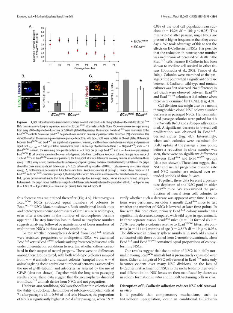
this decrease was maintained thereafter (Fig. 4A). HeterozygousEcad�/Wt NSCs produced equal numbers of colonies toEcad Wt/Wt NSCs (data not shown). Both conditional knock-outsand heterozygous neurospheres were of similar size as wild types,even after a decrease in the number of neurospheres becameapparent. The step function loss in clonal neurosphere numbersuggests a halving, followed by maintenance of lower numbers, ofmultipotent NSCs in these in vitro conditions.
To test whether neurospheres derived from Ecad�/� animalswere restricted progenitors or multipotent NSCs, we examinedEcad�/� versus EcadWt/Wt colonies arising from newly dissected cellsunder differentiation conditions to ascertain whether differences ex-isted in their output of neural progeny. There were no differencesamong these groups tested, with both wild-type (colonies sampledfrom n 6 animals) and mutant colonies (sampled from n 6animals) giving rise to equivalent numbers of neurons, as assessed bythe use of �-III-tubulin, and astrocytes, as assessed by the use ofGFAP (data not shown). Together with the long-term passagingresults above, these data suggest that the neurospheres dissectedfrom Ecad�/� animals derive from NSCs and not progenitors.
Under in vitro conditions, NSCs are the cells within colonies withthe ability to subclone. The number of subclone-competent cells at7 d after passage is 1.3 � 0.5% of total cells. However, the proportionof NSCs is significantly higher at 2–3 d after passaging, when 3.9 �
0.8% of the total cell population can sub-clone (t 19.26; df 101; p � 0.05). Thismeans 2–3 d after passage, single NSCs arepresent at higher frequencies than they are atday 7. We took advantage of this to test theeffects on E-Cadherin in NSCs. It is possiblethat the reduction in neurosphere numberwas an outcome of increased cell death in theEcad�/� cells because E-Cadherin has beenshown to mediate cell survival in other tis-sues (Boussadia et al., 2002; Tinkle et al.,2004). Colonies were examined at the pas-sage 3 time point when a significant decreasebetween E-Cadherin wild-type and mutantcultures was first observed. No differences incell death were observed between Ecad�/�
and EcadWt/Wt colonies at 3 d culture whenthese were examined by TUNEL (Fig. 4B).
Cell division rate might also be a meansthrough which clonal NSC colony numberdecreases in passaged NSCs. Hence similarthird-passage colonies were pulsed for 4 hin vitro with BrdU and subsequently exam-ined. A significant decrease in overall cellproliferation was observed in Ecad�/�-derived clones (Fig. 4C). Interestingly,when such colonies were examined forBrdU uptake at the passage 2 time point,before a reduction in clone number wasseen, there were no significant differencesbetween Ecad�/� and Ecad Wt/Wt groups(data not shown). These data suggest thatNSC and neural progenitor division rateand NSC number are reduced over ex-tended periods of time in vitro.
Together, these data forecast a prema-ture depletion of the NSC pool in olderEcad�/� mice. We reexamined the pro-duction of neural stem cells colonies to
verify whether such a decrease was apparent over time. Dissec-tions were performed on older 9 month Ecad�/� mice to testwhether the number of NSCs is lowered at later time points. Inline with this prediction, primary Ecad�/� colony number wassignificantly decreased compared with wild types in aged animals.In three separate assays, Ecad�/� mice (n 10) formed 63.0 �7.0% neurosphere colonies relative to Ecad Wt/Wt littermate con-trols (n 11) at 9 months of age (t 2.867; df 19; p � 0.05).The difference in primary sphere numbers in such old animalscontrasted with those obtained from 2-month-old animals, whenEcad�/� and Ecad Wt/Wt contained equal proportions of colony-forming NSCs.
Our results suggest that the number of NSCs is initially nor-mal in young Ecad�/� animals but is prematurely exhausted overtime. Either an impaired NSC self-renewal in Ecad�/� mice onlybecomes evident over many NSC divisions, or the loss ofE-Cadherin attachment of NSCs in the niche leads to their even-tual differentiation. NSC losses are then manifested by decreasesin colony formation in vitro and in BrdU-retaining cells in vivo.
Disruption of E-Cadherin adhesion reduces NSC self-renewalin vitroIt is possible that compensatory mechanisms, such asN-Cadherin upregulation, occur in conditional E-Cadherin
Figure 4. A, NSC colony formation is reduced in E-Cadherin conditional knock-outs. The graph shows the inability of Ecad �/�
NSCs to maintain over long-term passage, in contrast to Ecad Wt/Wt littermate controls. Clonal NSC colonies were averaged arisingfrom every 5000 cells plated on dissection, or 2500 cells plated after passage. The averages from Ecad �/� were normalized to theEcad Wt/Wt controls. Colonies of Ecad �/� begin to show a deficit in number at passage 3 after dissection (P3) and maintain thisdeficit thereafter. The remaining colonies were passaged exactly as wild types; both were replated in 24-well plates. Differencesbetween Ecad Wt/Wt and Ecad �/� are significant at passages 3 onward, and the interaction between genotype and passage issignificant (F(15,109) 1.946; p � 0.05). Primary time point is an average of cells dissected from n 10 (Ecad �/�) and n 11(Ecad Wt/Wt) animals; the remaining time points contain n 7 mice per passage Ecad �/� and n 4 – 6 mice per passageEcad Wt/Wt. B, Cell death is equivalent between wild-type and E-Cadherin conditional knock-out colonies. Images show merge of3 d Ecad �/� and Ecad Wt/Wt colonies at passage 3, the time point at which differences in colony number arise between thesegroups. TUNEL assay (arrow) reveals cell nuclei undergoing apoptosis (green); nuclei are counterstained by DAPI (blue). The graphshows that there are no significant differences ( p 0.05) between the proportion of TUNEL � cells per colony (n 3 animals pergroup). C, Proliferation is decreased in E-Cadherin conditional knock-out colonies at passage 3. Images show merge of 3 dEcad �/� and Ecad Wt/Wt colonies at passage 3, the time point at which differences in colony number arise between these groups.BrdU uptake (arrow) reveals nuclei that have entered S-phase (yellow in merged image). Nuclei are counterstained using pan-histone (red). The graph shows that there are significant differences (asterisk) between the proportion of BrdU � cells per colony(t 4.043; df 4; p � 0.05; n 3 animals per group). Error bars indicate SEM.
Karpowicz et al. • E-Cadherin Regulates Neural Stem Cells J. Neurosci., March 25, 2009 • 29(12):3885–3896 • 3891

knock-outs and mask effects mediated by E-Cadherin. The func-tional role of E-Cadherin in vitro was directly investigated inwild-type mice, using adhesion-blocking antibodies that specifi-cally target cadherin extracellular adhesive domains. HomophilicE-Cadherin binding between adjacent cells has been shown toelicit signal transduction simply by engaging the extracellulardomains of cadherins (Liu et al., 2006; Perrais et al., 2007). Ad-ditionally, as with the Ecad�/� mutants, blocking of Cadherinbinding might also decrease cell to cell contact in wild-type NSCs(Bendel-Stenzel et al., 2000).
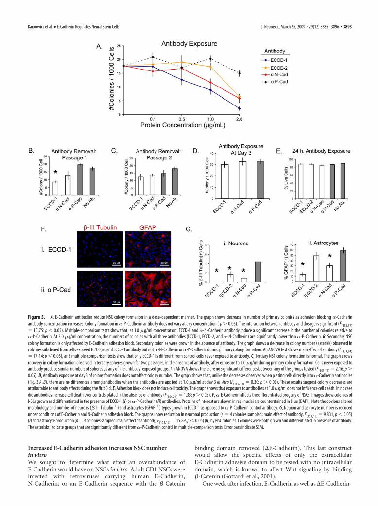
Adult CD1 wild-type cells dissected from the adult forebrainventricular subependyma were grown, passaged, and raised in thepresence of such antibodies for 7 d. Clonal adult neurospherenumber decreased in the presence of E-Cadherin andN-Cadherin antibodies in a dosage-dependent manner in con-trast to P-Cadherin antibody, which had no effect on clonal col-ony formation (Fig. 5A). The colonies observed in theseadhesion-blocking conditions appeared equal in size as their un-treated counterparts (data not shown). Notably, bothE-Cadherin blocking antibodies demonstrated negative effectson colony number at high concentrations. This suggests that spe-cifically blocking either E-Cadherin or N-Cadherin adhesion it-self in neurosphere cells leads to a decrease in NSC self-renewal.
NSC cultures were grown in the presence of cadherin blockingantibodies at 1.0 �g/ml, a concentration at which a reduction incolony number is observed using the ECCD1 �-E-Cadherin an-tibody (Fig. 5A). Although a reduction in clonal colony numberwas observed for both ECCD1 and �-N-Cadherin, the coloniessuccessfully raised in the presence of these antibodies appearednormal. These colonies then were passaged a second time to seewhether any effects of �-E-Cad or �-N-Cadherin were apparentin the cells in these colonies. In the absence of E-Cadherin anti-body, the number of secondary colonies arising from primaryones grown in E-Cadherin and N-Cadherin blocking antibodieswere also reduced (Fig. 5B) showing that E- and N-Cadherinengagement also reduced NSC self-renewal in the primary colo-nies. However, by the third passage, colonies derived fromE-Cadherin-exposed cells produced similar numbers of NSCprogeny (Fig. 5C) as ones grown from cells never exposed toantibody block. These data show that the decrease in NSC num-ber under conditions of lessened E- and N-Cadherin adhesion isreversible in wild-type cells.
Neural cells begin to divide after 1–2 d on passage under clonalproliferation conditions (P. Karpowicz and D. van der Kooy,unpublished observations). From each 7 d colony, a small subsetof the progeny [of �3500 total cells (Karpowicz et al., 2005)] havethe competence to subclone. We applied Cadherin antibodies tocolonies at day 3, a time point at which �10 cells are present ineach clonal colony. Intriguingly, no reduction in colony numberis observed when antibodies are applied at this point (Fig. 5D). Itis possible that these antibodies take effect on the niche whenNSCs are present at higher frequency in the first few days afterplating, than when colonies are fully formed.
Cell death was next examined in cells initially exposed toantibody to see whether this might explain a reduction in NSCnumber before cells begin to divide. Trypan blue exclusionshowed that, in the first 24 h that single cells were exposed toantibody, no differences in cell death were apparent betweencells (Fig. 5E). These data show that the effects of adhesionblocking antibodies are not attributable to toxicity before cellsdivide.
Disruption of E-Cadherin adhesion reduces NSC numberduring the first few divisions of these cells in vitro. Because these
effects also take place on primary clonal colonies as well as onpassaged colonies, these data are consistent with the notion that aNSC divides to produce niche support cells. Such cells maintainthe NSC in an undifferentiated state, but when this adhesion isblocked a deficiency in NSC number ensues.
The effects of blocking E-Cadherin adhesion on differentia-tion were next assayed. Although we previously noted no effectson the differentiation between Ecad�/� and Ecad Wt/Wt NSCs, thepossibility that E-Cadherin adhesion would affect NSC differen-tiation was examined in wild-type cells that carry a functionalE-Cadherin gene. First, colonies raised in the presence of anti-body and differentiated in the presence of antibody were exam-ined. In these conditions, curious phenotypes were observed inneuronal and glial cells exposed to E-Cadherin blocking antibod-ies, in particular ECCD-1 (Fig. 5Fi). In contrast, P-Cadherinblocking antibodies (Fig. 5Fii) demonstrated no altered pheno-type. Not only did E-Cadherin antibody abolish neuronal pro-duction (Fig. 5Gi), but in these conditions GFAP� astrocyteswere reduced in number (Fig. 5Gii) and the diameter of the pro-cesses of those that remained was decreased, when either E- andN-Cadherin were blocked. The most significant effects were ob-served with the ECCD-1 and �-N-Cadherin antibodies. The nor-mal differentiation noted in NSCs grown in ECCD-2 rather thanECCD-1 is likely a result of the concentrations of this antibody,which is only observed to produce proliferation deficits at higherconcentrations than 1.0 �g/ml (Fig. 5A).
Second, colonies grown in normal (nonantibody) prolifer-ation conditions, but differentiated in the presence of E-, N-,or P-Cadherin blocking antibodies, were assayed for alter-ations in their output of differentiated progeny. We found nodifferences in either �-III-tubulin � neurons or GFAP � astro-cytes in NSC colonies differentiated in any of the blockingantibodies (data not shown). Finally, colonies raised in thepresence of cadherin function blocking antibodies, but differ-entiated in the absence of the antibodies, were examined.Again, there were no differences in the proportion GFAP �
cells; however, neuron production was reduced whenN-Cadherin or E-Cadherin antibody is present during prolif-eration but not differentiation (data not shown).
These data show that blocking of endogenous E-Cadherin de-creases neuronal differentiation in vitro. Because these effects donot occur during differentiation conditions, E-Cadherin depen-dency is not a feature of neurons themselves but, rather, the pro-liferation of a neuronal progenitor when E-Cadherin adhesion isblocked during colony formation. This results in a loss of neuro-nal cells produced from the neuronal progenitors. As well, a re-duction in E- and N-Cadherin adherence reduces astrocyte pro-duction. However, this effect only takes place if both proliferationand differentiation are undertaken in the presence of blockingantibodies, suggesting that cellular adhesion may be critical dur-ing the transition of an undifferentiated precursor into a differ-entiated astrocyte. Such an effect is not seen in the Ecad�/� mu-tant, suggesting that a compensatory mechanism overcomes thisdeficiency in the mutant.
Together, these experiments indicate that inhibitingE-Cadherin adhesion reduces NSC number. Such phenomenaonly take place during the first divisions of NSC and/or neuralprogenitors in vitro, are reversible, and do not take effect throughthe death of NSCs. E-Cadherin also appears to affect the differ-entiation of astrocytic cells, because these transition from an un-differentiated to a differentiated state.
3892 • J. Neurosci., March 25, 2009 • 29(12):3885–3896 Karpowicz et al. • E-Cadherin Regulates Neural Stem Cells
Increased E-Cadherin adhesion increases NSC numberin vitroWe sought to determine what effect an overabundance ofE-Cadherin would have on NSCs in vitro. Adult CD1 NSCs wereinfected with retroviruses carrying human E-Cadherin,N-Cadherin, or an E-Cadherin sequence with the �-Catenin
binding domain removed (�E-Cadherin). This last constructwould allow the specific effects of only the extracellularE-Cadherin adhesive domain to be tested with no intracellulardomain, which is known to affect Wnt signaling by binding�-Catenin (Gottardi et al., 2001).
One week after infection, E-Cadherin as well as �E-Cadherin-
Figure 5. A, E-Cadherin antibodies reduce NSC colony formation in a dose-dependent manner. The graph shows decrease in number of primary colonies as adhesion blocking �-Cadherinantibody concentration increases. Colony formation in �-P-Cadherin antibody does not vary at any concentration ( p 0.05). The interaction between antibody and dosage is significant (F(153,57)
15.75; p � 0.05). Multiple-comparison tests show that, at 1.0 �g/ml concentration, ECCD-1 and �-N-Cadherin antibody induce a significant decrease in the number of colonies relative to�-P-Cadherin. At 2.0 �g/ml concentration, the numbers of colonies with all three antibodies (ECCD-1, ECCD-2, and �-N-Cadherin) are significantly lower than �-P-Cadherin. B, Secondary NSCcolony formation is only affected by E-Cadherin adhesion block. Secondary colonies were grown in the absence of antibody. The graph shows a decrease in colony number (asterisk) observed incolonies subcloned from cells exposed to 1.0 �g/ml ECCD-1 antibody but not �-N-Cadherin or �-P-Cadherin during primary colony formation. An ANOVA test shows main effect of antibody (F(153,84)
17.14; p � 0.05), and multiple-comparison tests show that only ECCD-1 is different from control cells never exposed to antibody. C, Tertiary NSC colony formation is normal. The graph showsrecovery in colony formation observed in tertiary spheres grown for two passages, in the absence of antibody, after exposure to 1.0 �g/ml during primary colony formation. Cells never exposed toantibody produce similar numbers of spheres as any of the antibody-exposed groups. An ANOVA shows there are no significant differences between any of the groups tested (F(153,72) 2.16; p 0.05). D, Antibody exposure at day 3 of colony formation does not affect colony number. The graph shows that, unlike the decreases observed when plating cells directly into �-Cadherin antibodies(Fig. 3 A, B), there are no differences among antibodies when the antibodies are applied at 1.0 �g/ml at day 3 in vitro (F(152,14) 0.30; p 0.05). These results suggest colony decreases areattributable to antibody effects during the first 3 d. E, Adhesion block does not induce cell toxicity. The graph shows that exposure to antibodies at 1.0 �g/ml does not influence cell death. In no casedid antibodies increase cell death over controls plated in the absence of antibody (F(154,24) 1.33; p 0.05). F, �-E-Cadherin affects the differentiated progeny of NSCs. Images show colonies ofNSCs grown and differentiated in the presence of ECCD-1 (i) or �-P-Cadherin (ii) antibodies. Proteins of interest are shown in red; nuclei are counterstained in blue (DAPI). Note the obvious alteredmorphology and number of neurons (�-III Tubulin �) and astrocytes (GFAP �) types grown in ECCD-1 as opposed to �-P-Cadherin control antibody. G, Neuron and astrocyte number is reducedunder conditions of E-Cadherin and N-Cadherin adhesion block. The graphs show reduction in neuronal production (n 4 colonies sampled; main effect of antibody, F(153,13) 9.831, p � 0.05)(i) and astrocyte production (n 4 colonies sampled; main effect of antibody, F(153,15) 15.89, p � 0.05) (ii) by NSC colonies. Colonies were both grown and differentiated in presence of antibody.The asterisks indicate groups that are significantly different from �-P-Cadherin control in multiple-comparison tests. Error bars indicate SEM.
Karpowicz et al. • E-Cadherin Regulates Neural Stem Cells J. Neurosci., March 25, 2009 • 29(12):3885–3896 • 3893
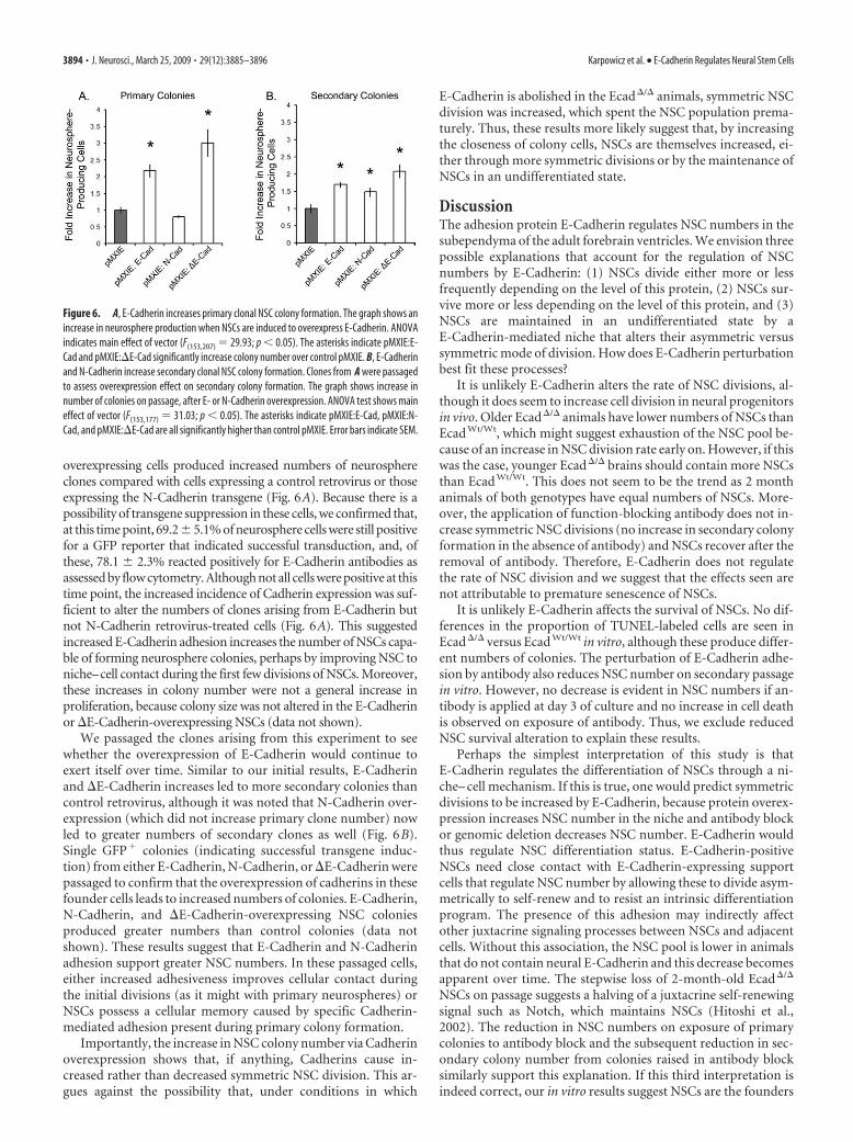
overexpressing cells produced increased numbers of neurosphereclones compared with cells expressing a control retrovirus or thoseexpressing the N-Cadherin transgene (Fig. 6A). Because there is apossibility of transgene suppression in these cells, we confirmed that,at this time point, 69.2 � 5.1% of neurosphere cells were still positivefor a GFP reporter that indicated successful transduction, and, ofthese, 78.1 � 2.3% reacted positively for E-Cadherin antibodies asassessed by flow cytometry. Although not all cells were positive at thistime point, the increased incidence of Cadherin expression was suf-ficient to alter the numbers of clones arising from E-Cadherin butnot N-Cadherin retrovirus-treated cells (Fig. 6A). This suggestedincreased E-Cadherin adhesion increases the number of NSCs capa-ble of forming neurosphere colonies, perhaps by improving NSC toniche–cell contact during the first few divisions of NSCs. Moreover,these increases in colony number were not a general increase inproliferation, because colony size was not altered in the E-Cadherinor �E-Cadherin-overexpressing NSCs (data not shown).
We passaged the clones arising from this experiment to seewhether the overexpression of E-Cadherin would continue toexert itself over time. Similar to our initial results, E-Cadherinand �E-Cadherin increases led to more secondary colonies thancontrol retrovirus, although it was noted that N-Cadherin over-expression (which did not increase primary clone number) nowled to greater numbers of secondary clones as well (Fig. 6B).Single GFP� colonies (indicating successful transgene induc-tion) from either E-Cadherin, N-Cadherin, or �E-Cadherin werepassaged to confirm that the overexpression of cadherins in thesefounder cells leads to increased numbers of colonies. E-Cadherin,N-Cadherin, and �E-Cadherin-overexpressing NSC coloniesproduced greater numbers than control colonies (data notshown). These results suggest that E-Cadherin and N-Cadherinadhesion support greater NSC numbers. In these passaged cells,either increased adhesiveness improves cellular contact duringthe initial divisions (as it might with primary neurospheres) orNSCs possess a cellular memory caused by specific Cadherin-mediated adhesion present during primary colony formation.
Importantly, the increase in NSC colony number via Cadherinoverexpression shows that, if anything, Cadherins cause in-creased rather than decreased symmetric NSC division. This ar-gues against the possibility that, under conditions in which
E-Cadherin is abolished in the Ecad�/� animals, symmetric NSCdivision was increased, which spent the NSC population prema-turely. Thus, these results more likely suggest that, by increasingthe closeness of colony cells, NSCs are themselves increased, ei-ther through more symmetric divisions or by the maintenance ofNSCs in an undifferentiated state.
DiscussionThe adhesion protein E-Cadherin regulates NSC numbers in thesubependyma of the adult forebrain ventricles. We envision threepossible explanations that account for the regulation of NSCnumbers by E-Cadherin: (1) NSCs divide either more or lessfrequently depending on the level of this protein, (2) NSCs sur-vive more or less depending on the level of this protein, and (3)NSCs are maintained in an undifferentiated state by aE-Cadherin-mediated niche that alters their asymmetric versussymmetric mode of division. How does E-Cadherin perturbationbest fit these processes?
It is unlikely E-Cadherin alters the rate of NSC divisions, al-though it does seem to increase cell division in neural progenitorsin vivo. Older Ecad�/� animals have lower numbers of NSCs thanEcad Wt/Wt, which might suggest exhaustion of the NSC pool be-cause of an increase in NSC division rate early on. However, if thiswas the case, younger Ecad�/� brains should contain more NSCsthan Ecad Wt/Wt. This does not seem to be the trend as 2 monthanimals of both genotypes have equal numbers of NSCs. More-over, the application of function-blocking antibody does not in-crease symmetric NSC divisions (no increase in secondary colonyformation in the absence of antibody) and NSCs recover after theremoval of antibody. Therefore, E-Cadherin does not regulatethe rate of NSC division and we suggest that the effects seen arenot attributable to premature senescence of NSCs.
It is unlikely E-Cadherin affects the survival of NSCs. No dif-ferences in the proportion of TUNEL-labeled cells are seen inEcad�/� versus Ecad Wt/Wt in vitro, although these produce differ-ent numbers of colonies. The perturbation of E-Cadherin adhe-sion by antibody also reduces NSC number on secondary passagein vitro. However, no decrease is evident in NSC numbers if an-tibody is applied at day 3 of culture and no increase in cell deathis observed on exposure of antibody. Thus, we exclude reducedNSC survival alteration to explain these results.
Perhaps the simplest interpretation of this study is thatE-Cadherin regulates the differentiation of NSCs through a ni-che– cell mechanism. If this is true, one would predict symmetricdivisions to be increased by E-Cadherin, because protein overex-pression increases NSC number in the niche and antibody blockor genomic deletion decreases NSC number. E-Cadherin wouldthus regulate NSC differentiation status. E-Cadherin-positiveNSCs need close contact with E-Cadherin-expressing supportcells that regulate NSC number by allowing these to divide asym-metrically to self-renew and to resist an intrinsic differentiationprogram. The presence of this adhesion may indirectly affectother juxtacrine signaling processes between NSCs and adjacentcells. Without this association, the NSC pool is lower in animalsthat do not contain neural E-Cadherin and this decrease becomesapparent over time. The stepwise loss of 2-month-old Ecad�/�
NSCs on passage suggests a halving of a juxtacrine self-renewingsignal such as Notch, which maintains NSCs (Hitoshi et al.,2002). The reduction in NSC numbers on exposure of primarycolonies to antibody block and the subsequent reduction in sec-ondary colony number from colonies raised in antibody blocksimilarly support this explanation. If this third interpretation isindeed correct, our in vitro results suggest NSCs are the founders
Figure 6. A, E-Cadherin increases primary clonal NSC colony formation. The graph shows anincrease in neurosphere production when NSCs are induced to overexpress E-Cadherin. ANOVAindicates main effect of vector (F(153,207) 29.93; p � 0.05). The asterisks indicate pMXIE:E-Cad and pMXIE:�E-Cad significantly increase colony number over control pMXIE. B, E-Cadherinand N-Cadherin increase secondary clonal NSC colony formation. Clones from A were passagedto assess overexpression effect on secondary colony formation. The graph shows increase innumber of colonies on passage, after E- or N-Cadherin overexpression. ANOVA test shows maineffect of vector (F(153,177) 31.03; p � 0.05). The asterisks indicate pMXIE:E-Cad, pMXIE:N-Cad, and pMXIE:�E-Cad are all significantly higher than control pMXIE. Error bars indicate SEM.
3894 • J. Neurosci., March 25, 2009 • 29(12):3885–3896 Karpowicz et al. • E-Cadherin Regulates Neural Stem Cells
of niche cells (because E-Cadherin effects are seen in clonal NSCcolonies in this system). The identity of these support cells isunclear; these may be any neural cell types including NSCs them-selves. This idea that stem cells form their own niches in culture isnot without precedence. It has for instance been observed thatembryonic stem cells can form their own niches, cell autono-mously, by producing differentiated progeny that support theundifferentiated founder cells themselves (Bendall et al., 2007).
E-Cadherin regulation of NSC division symmetry might actdirectly through a parallel signaling pathway that is affected byadhesion. In conditions of increased Cadherin presence, thenumber of SCs contacting support cells would affect the signalsSCs received. Contacting cells might thus determine the func-tional symmetry of NSC division by physical closeness; thoseNSCs that do not contact support cells in the niche would becomeprogenitors (Lin, 1997). A related idea is that contact to a locusactually polarizes cells, allowing them to divide asymmetrically(Betschinger and Knoblich, 2004). Studies have demonstrated apolarization of cadherins in precursors in a variety of systems(Song et al., 2002; Rasin et al., 2007). Indeed, it appears that theproteins Numb and Numblike function to polarize E-Cadherinin the processes connecting radial glia to the ventricles (Rasin etal., 2007). Our interpretations are consistent with these studies,although we did not find clear evidence of altered polarizationwhen E-Cadherin is absent. However, we find that E-Cadherinpromotes the NSC self-renewal symmetric mode of division,keeping the NSCs in an undifferentiated state.
In the fly gonad, Drosophila E-Cadherin has been shown toassociate germline SCs with a group of cells that signal to the SCsand that, in part, define the SC niche (Song et al., 2002; Yamashitaet al., 2003; Ohlstein et al., 2004). Germline SCs that cannot dif-ferentiate, upregulate cadherins that enable them to outcompetewild-type germline SCs for occupancy of the SC niche (Jin et al.,2008). Mouse E-Cadherin has been shown to induce Rac1 activ-ity, which increases epithelial cell proliferation in vitro (Liu et al.,2006). E-Cadherin may also decrease cellular proliferation in avariety of cell lines by a �-Catenin-independent, noncanonicalWnt signaling pathway (Perrais et al., 2007). Moreover,E-Cadherin has been also found to have effects on the differenti-ation of murine germ cells (Okamura et al., 2003) and epithelia(Larue et al., 1996). These characteristics of maintenance andproliferation also support cadherins as regulators of SC behavior.
An alternative explanation to a niche-mediated self-renewalmechanism is that E-Cadherin directly signals to NSCs. Cad-herins can participate in signaling processes through their se-questration of �-Catenin. Our results, which show that NSCsexposed to function-blocking antibodies differentiate differentlythan Ecad�/� NSCs (Fig. 5F,G), seem to support a direct role ofE-Cadherin rather than a niche-mediated role. That wild-typecells show altered differentiation under E-Cadherin block mightargue for a direct signaling mechanism. However, our results alsoshow that neurosphere number is halved in the presence of anti-body (Fig. 5A), which is similar to the halving observed inEcad�/� NSCs (Fig. 4A). These discrepancies are difficult to ex-plain with any certainty. At this time, we are not able resolvebetween the direct role of E-Cadherin in NSC self-renewal versusa niche-mediated role.
The loss of colony number by E-Cadherin, but notP-Cadherin antibody-exposed NSCs, suggests that E-Cadherin isspecifically needed by adult NSCs. However, the differences ob-served between mutants and wild types, antibody-raised NSCsand controls, and Cadherin overexpressing NSCs and controlsare no greater than twofold. In the Ecad�/�, this is perhaps attrib-
utable to compensatory adhesion performed by N-Cadherin,which is known to be coexpressed with E-Cadherin in the ven-tricular zones (Rasin et al., 2007) and which we find in abundancein the Ecad�/� forebrain germinal zones. In recent studies, bothproteins were observed to polarize radial glia in this region, whichmeans that the conditional ablation of one might be compen-sated for by the other. This accounts for our observation thatN-Cadherin retrovirus and blocking antibody phenocopy thesame E-Cadherin perturbations, and it is possible conditionalN-Cadherin ablation in these regions would produce an equiva-lent phenotype as seen in the Ecad�/�. Differences in adhesionblock by antibodies are most likely lessened because of insuffi-cient concentrations of �-E-Cadherin and �-N-Cadherin, whichproduce more pronounced effects at higher doses.
Indeed, if E-Cadherin regulates NSC differentiation by aniche-type mechanism, this may account for the increase in BrdUuptake in progenitors after 1 h exposure in vivo. If E-Cadherinonly affects NSCs, then these non-NSC neural progenitor pheno-types are a downstream outcome. Differentiating NSCs leavetheir relatively quiescent niche and enter a progenitor pool whereproliferation is more rapid. This is then manifested as an increasein short-term BrdU uptake. At present, we cannot concludewhether E-Cadherin affects NSCs and non-NSCs differently. Thespecific ablation and overexpression in NSCs themselves wouldhave to be performed to resolve this issue.
The regulation of SC number by support cells suggests aniche-dependent mechanism of sensing physiological changemay exist in the brain. The upregulation of a particular Cadherinwithin precursors could increase SC binding to support cellgroups, which increases the number of SCs through the dediffer-entiation of progenitors into SCs (Yamashita et al., 2003; Brawleyand Matunis, 2004; Kai and Spradling, 2004). Indeed, the obser-vation that germline SCs with higher cadherin levels are morecompetitive in the niche of Drosophila (Jin et al., 2008) offers atempting explanation for the increase in NSC colonies underconditions of cadherin overexpression (Fig. 6A). It is tempting tospeculate that both NSCs and support cells have the ability toregulate NSC number, the former by intrinsically regulating itsown expansion and the latter by limiting NSC expansion via reg-ulation of E-Cadherin expression. In line with this reasoning,there is some evidence that suggests both Notch signaling-dependent niche cells and Dpp signaling-dependent SCs partici-pate in bidirectional signaling processes that simultaneously reg-ulate the SC and niche cell pool (Ward et al., 2006; Song et al.,2007).
E-Cadherin is lost in nearly all forms of epithelial cancers(Christofori and Semb, 1999), and its loss generally increasesrather than decreases cell proliferation (Tinkle et al., 2004; Liu etal., 2006; Perrais et al., 2007). Here, we show that this same pro-tein underlies an opposite role in SCs. Such differences shed lighton distinctions between stem and cancer cells that are of funda-mental importance in understanding tumorigenesis (Reya et al.,2001).
ReferencesAlvarez-Buylla A, Lim DA (2004) For the long run: maintaining germinal
niches in the adult brain. Neuron 41:683– 686.Alvarez-Buylla A, García-Verdugo JM, Tramontin AD (2001) A unified hy-
pothesis on the lineage of neural stem cells. Nat Rev Neurosci 2:287–293.Bendall SC, Stewart MH, Menendez P, George D, Vijayaragavan K,
Werbowetski-Ogilvie T, Ramos-Mejia V, Rouleau A, Yang J, Bosse M,Lajoie G, Bhatia M (2007) IGF and FGF cooperatively establish the reg-ulatory stem cell niche of pluripotent human cells in vitro. Nature448:1015–1021.
Karpowicz et al. • E-Cadherin Regulates Neural Stem Cells J. Neurosci., March 25, 2009 • 29(12):3885–3896 • 3895

Bendel-Stenzel MR, Gomperts M, Anderson R, Heasman J, Wylie C (2000)The role of cadherins during primordial germ cell migration and earlygonad formation in the mouse. Mech Dev 91:143–152.
Betschinger J, Knoblich JA (2004) Dare to be different: asymmetric cell di-vision in Drosophila, C. elegans and vertebrates. Curr Biol 14:R674 –R685.
Boussadia O, Kutsch S, Hierholzer A, Delmas V, Kemler R (2002)E-cadherin is a survival factor for the lactating mouse mammary gland.Mech Dev 115:53– 62.
Brawley C, Matunis E (2004) Regeneration of male germline stem cells byspermatogonial dedifferentiation in vivo. Science 304:1331–1334.
Campos LS, Decker L, Taylor V, Skarnes W (2006) Notch, epidermalgrowth factor receptor, and beta1-integrin pathways are coordinated inneural stem cells. J Biol Chem 281:5300 –5309.
Cheng A, Tang H, Cai J, Zhu M, Zhang X, Rao M, Mattson MP (2004) Gapjunctional communication is required to maintain mouse cortical neuralprogenitor cells in a proliferative state. Dev Biol 272:203–216.
Christofori G, Semb H (1999) The role of the cell-adhesion moleculeE-cadherin as a tumour-suppressor gene. Trends Biochem Sci 24:73–76.
Foty RA, Steinberg MS (2005) The differential adhesion hypothesis: a directevaluation. Dev Biol 278:255–263.
Gottardi CJ, Wong E, Gumbiner BM (2001) E-cadherin suppresses cellulartransformation by inhibiting beta-catenin signaling in an adhesion-independent manner. J Cell Biol 153:1049 –1060.
Hitoshi S, Alexson T, Tropepe V, Donoviel D, Elia AJ, Nye JS, Conlon RA,Mak TW, Bernstein A, van der Kooy D (2002) Notch pathway moleculesare essential for the maintenance, but not the generation, of mammalianneural stem cells. Genes Dev 16:846 – 858.
Imai F, Hirai S, Akimoto K, Koyama H, Miyata T, Ogawa M, Noguchi S,Sasaoka T, Noda T, Ohno S (2006) Inactivation of aPKClambda resultsin the loss of adherens junctions in neuroepithelial cells without affectingneurogenesis in mouse neocortex. Development 133:1735–1744.
Imura T, Kornblum HI, Sofroniew MV (2003) The predominant neuralstem cell isolated from postnatal and adult forebrain but not early embry-onic forebrain expresses GFAP. J Neurosci 23:2824 –2832.
Jin Z, Kirilly D, Weng C, Kawase E, Song X, Smith S, Schwartz J, Xie T (2008)Differentiation-defective stem cells outcompete normal stem cells forniche occupancy in the Drosophila ovary. Cell Stem Cell 2:39 – 49.
Kai T, Spradling A (2003) An empty Drosophila stem cell niche reactivatesthe proliferation of ectopic cells. Proc Natl Acad Sci U S A100:4633– 4638.
Kai T, Spradling A (2004) Differentiating germ cells can revert into func-tional stem cells in Drosophila melanogaster ovaries. Nature 428:564 –569.
Karpowicz P, Morshead C, Kam A, Jervis E, Ramunas J, Ramuns J, Cheng V,van der Kooy D (2005) Support for the immortal strand hypothesis:neural stem cells partition DNA asymmetrically in vitro. J Cell Biol170:721–732.
Kippin TE, Martens DJ, van der Kooy D (2005) p21 loss compromises therelative quiescence of forebrain stem cell proliferation leading to exhaus-tion of their proliferation capacity. Genes Dev 19:756 –767.
Kuo CT, Mirzadeh Z, Soriano-Navarro M, Rasin M, Wang D, Shen J, SestanN, Garcia-Verdugo J, Alvarez-Buylla A, Jan LY, Jan YN (2006) Postnataldeletion of Numb/Numblike reveals repair and remodeling capacity inthe subventricular neurogenic niche. Cell 127:1253–1264.
Larue L, Ohsugi M, Hirchenhain J, Kemler R (1994) E-cadherin null mutantembryos fail to form a trophectoderm epithelium. Proc Natl Acad SciU S A 91:8263– 8267.
Larue L, Antos C, Butz S, Huber O, Delmas V, Dominis M, Kemler R (1996)A role for cadherins in tissue formation. Development 122:3185–3194.
Lien WH, Klezovitch O, Fernandez TE, Delrow J, Vasioukhin V (2006)alphaE-catenin controls cerebral cortical size by regulating the hedgehogsignaling pathway. Science 311:1609 –1612.
Lin H (1997) The tao of stem cells in the germline. Annu Rev Genet31:455– 491.
Liu WF, Nelson CM, Pirone DM, Chen CS (2006) E-cadherin engagementstimulates proliferation via Rac1. J Cell Biol 173:431– 441.
Louvi A, Artavanis-Tsakonas S (2006) Notch signalling in vertebrate neuraldevelopment. Nat Rev Neurosci 7:93–102.
Matsunami H, Takeichi M (1995) Fetal brain subdivisions defined by R-and E-cadherin expressions: evidence for the role of cadherin activity inregion-specific, cell-cell adhesion. Dev Biol 172:466 – 478.
Morshead CM, van der Kooy D (2001) A new “spin” on neural stem cells?Curr Opin Neurobiol 11:59 – 65.
Morshead CM, Reynolds BA, Craig CG, McBurney MW, Staines WA, Moras-sutti D, Weiss S, van der Kooy D (1994) Neural stem cells in the adultmammalian forebrain: a relatively quiescent subpopulation of sub-ependymal cells. Neuron 13:1071–1082.
Morshead CM, Craig CG, van der Kooy D (1998) In vivo clonal analysesreveal the properties of endogenous neural stem cell proliferation in theadult mammalian forebrain. Development 125:2251–2261.
Morshead CM, Garcia AD, Sofroniew MV, van der Kooy D (2003) Theablation of glial fibrillary acidic protein-positive cells from the adult cen-tral nervous system results in the loss of forebrain neural stem cells but notretinal stem cells. Eur J Neurosci 18:76 – 84.
Ohlstein B, Kai T, Decotto E, Spradling A (2004) The stem cell niche: themeand variations. Curr Opin Cell Biol 16:693– 699.
Okamura D, Kimura T, Nakano T, Matsui Y (2003) Cadherin-mediated cellinteraction regulates germ cell determination in mice. Development130:6423– 6430.
Perrais M, Chen X, Perez-Moreno M, Gumbiner BM (2007) E-cadherinhomophilic ligation inhibits cell growth and epidermal growth factorreceptor signaling independently of other cell interactions. Mol Biol Cell18:2013–2025.
Rasin MR, Gazula VR, Breunig JJ, Kwan KY, Johnson MB, Liu-Chen S, Li HS,Jan LY, Jan YN, Rakic P, Sestan N (2007) Numb and Numbl are requiredfor maintenance of cadherin-based adhesion and polarity of neural pro-genitors. Nat Neurosci 10:819 – 827.
Redies C (2000) Cadherins in the central nervous system. Prog Neurobiol61:611– 648.
Reya T, Morrison SJ, Clarke MF, Weissman IL (2001) Stem cells, cancer,and cancer stem cells. Nature 414:105–111.
Reynolds BA, Tetzlaff W, Weiss S (1992) A multipotent EGF-responsivestriatal embryonic progenitor cell produces neurons and astrocytes.J Neurosci 12:4565– 4574.
Shimamura K, Takeichi M (1992) Local and transient expression ofE-cadherin involved in mouse embryonic brain morphogenesis. Devel-opment 116:1011–1019.
Song X, Zhu CH, Doan C, Xie T (2002) Germline stem cells anchored byadherens junctions in the Drosophila ovary niches. Science296:1855–1857.
Song X, Call GB, Kirilly D, Xie T (2007) Notch signaling controls germlinestem cell niche formation in the Drosophila ovary. Development134:1071–1080.
Steinberg MS, Takeichi M (1994) Experimental specification of cell sorting,tissue spreading, and specific spatial patterning by quantitative differ-ences in cadherin expression. Proc Natl Acad Sci U S A 91:206 –209.
Takeichi M (1995) Morphogenetic roles of classic cadherins. Curr Opin CellBiol 7:619 – 627.
Tinkle CL, Lechler T, Pasolli HA, Fuchs E (2004) Conditional targeting ofE-cadherin in skin: insights into hyperproliferative and degenerative re-sponses. Proc Natl Acad Sci U S A 101:552–557.
Tronche F, Kellendonk C, Kretz O, Gass P, Anlag K, Orban PC, Bock R, KleinR, Schutz G (1999) Disruption of the glucocorticoid receptor gene in thenervous system results in reduced anxiety. Nat Genet 23:99 –103.
Tropepe V, Sibilia M, Ciruna BG, Rossant J, Wagner EF, van der Kooy D(1999) Distinct neural stem cells proliferate in response to EGF and FGFin the developing mouse telencephalon. Dev Biol 208:166 –188.
Ward EJ, Shcherbata HR, Reynolds SH, Fischer KA, Hatfield SD, Ruohola-Baker H (2006) Stem cells signal to the niche through the Notch path-way in the Drosophila ovary. Curr Biol 16:2352–2358.
Yamashita YM, Jones DL, Fuller MT (2003) Orientation of asymmetricstem cell division by the APC tumor suppressor and centrosome. Science301:1547–1550.
3896 • J. Neurosci., March 25, 2009 • 29(12):3885–3896 Karpowicz et al. • E-Cadherin Regulates Neural Stem Cells