dyslipidemia in children treated with a braf inhibitor ... - mdpi
TRANSCRIPT
Cancers 2022, 14, 2693. https://doi.org/10.3390/cancers14112693 www.mdpi.com/journal/cancers
Article
Dyslipidemia in Children Treated with a BRAF Inhibitor for
Low‐Grade Gliomas: A New Side Effect?
Marco Crocco 1,2,*, Antonio Verrico 1, Claudia Milanaccio 1, Gianluca Piccolo 1,2, Patrizia De Marco 3,
Gabriele Gaggero 4, Valentina Iurilli 5, Sonia Di Profio 6, Federica Malerba 2, Marta Panciroli 2, Paolo Giordano 2,
Maria Grazia Calevo 7, Emilio Casalini 2,8, Natascia Di Iorgi 2,8 and Maria Luisa Garrè 1
1 Neuroncology Unit, IRCCS Istituto Giannina Gaslini, 16147 Genova, Italy;
[email protected] (A.V.); [email protected] (C.M.); [email protected] (G.P.);
[email protected] (M.L.G.) 2 Department of Neuroscience, Rehabilitation, Ophthalmology, Genetics, Maternal and Child Health,
University of Genova, 16132 Genova, Italy; federica‐[email protected] (F.M.);
[email protected] (M.P.); [email protected] (P.G.); [email protected] (E.C.);
[email protected] (N.D.I.) 3 UOC Genetica Medica, IRCCS Istituto Giannina Gaslini, 16147 Genoa, Italy; [email protected] 4 Department of Clinical Pathology, IRCCS Ospedale Policlinico San Martino, 16132 Genoa, Italy;
[email protected] 5 Pharmacy Unit, IRCCS Istituto Giannina Gaslini, 16147 Genova, Italy; [email protected] 6 Clinical Psychology, IRCCS Istituto Giannina Gaslini, 16147 Genoa, Italy; [email protected] 7 Epidemiology and Biostatistics Unit, IRCCS Istituto Giannina Gaslini, 16147 Genova, Italy;
[email protected] 8 Department of Pediatrics, IRCCS Istituto Giannina Gaslini, 16147 Genova, Italy
* Correspondence: [email protected]
Simple Summary: The use of targeted therapies is revolutionizing the prognosis of many cancers;
however, there is still limited knowledge of their side effects. Dyslipidemia is often present in cancer
patients due to mechanisms that are directly or indirectly related to cancer or therapies. The aim of
our study is to investigate the effects of vemurafenib on lipid metabolism in a cohort of pediatric
patients treated for brain tumors. For the first time, we describe dyslipidemia as a possible side
effect of the BRAF inhibitors. A better understanding of the pathways that are involved in
dyslipidemia could also help with a better understanding of the drug‐resistance mechanisms in
cancer cells.
Abstract: BRAF inhibitors, in recent years, have played a central role in the disease control of
unresectable BRAF‐mutated pediatric low‐grade gliomas (LGGs). The aim of the study was to
investigate the acute and long‐term effects of vemurafenib on the lipid metabolism in children
treated for an LGG. In our cohort, children treated with vemurafenib (n = 6) exhibited alterations in
lipid metabolism a few weeks after starting, as was demonstrated after 1 month (n = 4) by the high
plasma levels of the total cholesterol (TC = 221.5 ± 42.1 mg/dL), triglycerides (TG = 107.8 ± 44.4
mg/dL), and low‐density lipoprotein (LDL = 139.5 ± 51.5 mg/dL). Despite dietary recommendations,
the dyslipidemia persisted over time. The mean lipid levels of the TC (222.3 ± 34.7 mg/dL), TG (134.8
± 83.6 mg/dL), and LDL (139.8 ± 46.9 mg/dL) were confirmed abnormal at the last follow‐up (45 ±
27 months, n = 6). Vemurafenib could be associated with an increased risk of dyslipidemia. An
accurate screening strategy in new clinical trials, and a multidisciplinary team, are required for the
optimal management of unexpected adverse events, including dyslipidemia.
Keywords: BRAF; vemurafenib; lipid metabolism; cholesterol; triglycerides; dyslipidemia; brain
tumor; low grade glioma
Citation: Crocco, M.; Verrico, A.;
Milanaccio, C.; Piccolo, G.;
De Marco, P.; Gaggero, G.; Iurilli, V.;
Di Profio, S.; Malerba, F.;
Panciroli, M.; et al. Dyslipidemia in
Children Treated with a BRAF
Inhibitor for Low‐Grade Gliomas: A
New Side Effect? Cancers 2022, 14,
2693. https://doi.org/10.3390/
cancers14112693
Academic Editor: David Wong
Received: 22 April 2022
Accepted: 27 May 2022
Published: 29 May 2022
Publisher’s Note: MDPI stays
neutral with regard to jurisdictional
claims in published maps and
institutional affiliations.
Copyright: © 2022 by the authors.
Licensee MDPI, Basel, Switzerland.
This article is an open access article
distributed under the terms and
conditions of the Creative Commons
Attribution (CC BY) license
(https://creativecommons.org/license
s/by/4.0/).
Cancers 2022, 14, 2693 2 of 14
1. Introduction
Low‐grade gliomas are the most common central‐nervous‐system (CNS) tumors
among children [1]. The prognosis for these tumors is generally excellent, with the 10‐year
overall survival (OS) between 85 and 95% [2]. In pediatric low‐grade gliomas (pLGGs),
several factors, such as grading, location, age at diagnosis, the extent of surgery, and
biological characteristics, influence the OS. A complete surgical resection, whenever
feasible, tends to be curative, and a wait‐and‐see approach can be adopted. pLGGs that
cannot be resected without the risk of permanently damaging important functions, or that
progress after resection, require additional therapy.
Chemotherapy can control tumors by up to 50% of pLGGs, and over 90% of patients
are still alive 20 years after radiotherapy [3]. Considering the excellent OS, the goal of the
medical therapy should be to ensure the stability of the disease and to minimize the early
and late effects of therapy. In the last decade, biological drugs have filled an important
void in the therapeutic armamentarium against pLGGs when they are not resectable and
are refractory or recurrent following standard chemotherapy regimens. In these cases,
treatment with biological/targeted therapy has the potential to prevent morbidities and
sequelae that are typically associated with chemo‐radiotherapy, such as neurocognitive
impairment, ototoxicity, nephrotoxicity, peripheral neuropathy, and hypothalamic–
pituitary disfunction [4–6].
The understanding of the biological pathways that lead to pLGGs has allowed the
development of new biological therapies that directly interrupt these pathways. pLGGs
most frequently have somatic driver genetic alterations that converge on the activation of
the Rapidly Accelerated Fibrosarcoma/Mytogenic‐Activated Protein Kinase (RAS/MAPK)
pathway [7]. Rearrangements that afflict the genes v‐Raf murine sarcoma viral oncogene
homolog B1 (BRAF) and KIAA1549–BRAF are the most frequent somatic driver alterations
across all pLGGs [8,9]. The BRAF V600E mutation is reported in 20–35% of pLGGs [10,11],
and, in particular, it is found in 9% of pilocytic astrocytomas, 50% of gangliogliomas, and
66% of pleomorphic xanthoastrocytomas [8,10]. The BRAF V600E mutation confers a
worse prognosis because of the insensitivity to traditional chemotherapy, and a higher
propensity toward malignant transformation in combination with CDKN2A deletion [12].
Hence, inhibiting MAPK signaling by using small‐molecule inhibitors, such as BRAF
inhibitors (BRAFi), may be a promising strategy in pLGGs [13–16]. It is also known that
the various combinations of MEKi and BRAFi can delay or prevent the occurrence of
resistance to a single therapy and may reduce side effects (SEs). [17–20]. These therapies
may lead to a rapid and prolonged response of the tumors [6,19,21,22]. As observed in
other gliomas, such as subependymal giant cell astrocytoma (SEGA), in which stopping
the mammalian target of rapamycin mTOR inhibitors (mTORi) results in inevitable tumor
regrowth, after the discontinuation of BRAFi, up to 75% of patients experienced rapid
progression in a few weeks. However, upon rechallenge with BRAFi, 90% achieved an
objective response [23].
Unfortunately, BRAFi are not free from the development of toxicity and adverse
events (AE), and most of them have been reported in adult populations with melanoma,
in which these new therapies are most commonly used, while few studies are available on
pLGGs. Vemurafenib (a second‐generation BRAFi) has already been studied in the
treatment of BRAF V600E e V600K mutation‐positive melanoma in the BRIM‐3 Trial of
337 adult patients. In this trial, a total of 334 patients (99%) reported at least one AE in the
vemurafenib arm. The most common AEs (occurring in >20% of patients) in the
vemurafenib arm were rash, arthralgia, alopecia, fatigue, photosensitivity reaction,
nausea, diarrhea, headache, hyperkeratosis, pruritus, dry skin, skin papilloma, decreased
appetite, pain in extremity, pyrexia, vomiting, and squamous‐cell carcinoma of the skin
[24]. Preliminary data regarding BRAFi toxicities are also emerging in pediatric‐age
patients, and the most observed toxicities are pyrexia, hematological, dermatological,
cardiac, and ophthalmic diseases [25]. The association of MEKi and BRAFi may mitigate
some of the toxicities induced by the “paradoxical activation” of the MAPK pathway
Cancers 2022, 14, 2693 3 of 14
when a BRAFi is used as a single agent in BRAF wild‐type cells [26,27]. Despite numerous
studies that compare the different BRAFi molecules, there are no head‐to‐head clinical
trials that compare the different agent combinations, and most of the safety data originate
from the confirmatory phase III trials or pharmacovigilance studies in adult patients
[24,28]. It is very important to study the SEs and the AEs in pediatric populations,
considering the necessity of the long‐term use of BRAFi (as is commonly carried out with
mTORi in patients with tuberous sclerosis and SEGA). Moreover, the novel agents
uncovered unexpected and unexplored AEs, and represent an important medical
challenge. A better understanding of the SEs of these therapies is imperative.
The aim of our study is to expand the knowledge of the long‐term AEs of BRAF
inhibitors in pediatric populations by analyzing, retrospectively, the serum lipid
concentrations in a cohort of pediatric patients treated with vemurafenib for LGG at our
institute. We also discuss the possible role of lipid metabolism in resistance to BRAFi.
2. Materials and Methods
We collected and retrospectively reviewed clinical, laboratory, and instrumental data
of all patients treated with the BRAF inhibitor vemurafenib for LGG at the Giannina
Gaslini Children’s Hospital in Genoa, Italy, between 1 May 2015 and 31 December 2021.
Patients treated with vemurafenib, with an age up to 18 years of age at the time of
diagnosis, and with follow up of at least 6 months and 2 lipid‐level samples (after starting
treatment), were eligible.
The histology diagnosis was confirmed by the national reviewer, for all patients. All
patients had a BRAFV600E mutation confirmed by sequencing performed using the
polymerase chain reaction (kit Easy Braf real‐time PCR, Diatech Pharmacogenetics, Jesi,
Italy).
Before and during the BRAFi treatment (at 1, 3, 6, 12 months, and every 6 months
thereafter), each patient underwent a detailed clinical and laboratory investigation to rule
out possible organ disorders that contraindicated a BRAFi treatment (including a
complete blood count, biochemical liver‐ and kidney‐function tests, auxological and
endocrinological assessments, ECG, and echocardiography with a cardiological
examination). After the incidental finding of dyslipidemia in the second treated patient,
fasting blood samples for lipid panel test were collected within 1 month before and at 1,
3, 6, 12 months after the initiation of treatment with vemurafenib (and every 12 months
thereafter).
The auxological data were obtained from the auxo‐endocrinological assessments to
which the patients were routinely subjected. Height was measured by a Harpenden
Stadiometer, with an accuracy of ±1 mm. The weight was measured on a digital scale, with
an accuracy of ±0.1 kg. BMI was calculated as weight (kg) divided by height (m) squared
and transformed to standard‐deviation scores using the WHO reference values [29].
All the families receive a dietary recommendation based on the Mediterranean diet
[30] at the first interview with the oncologist pediatrician. Patients with alterations in the
lipid or glucose profiles are referred for nutritional counseling.
Fasting blood samples were obtained from 6 patients with LGG. Venous blood
samples were collected by venipuncture or central venous catheter between 8 a.m. and 12
p.m., after an overnight fast. The serum and plasma were immediately separated, the lipid
panel (triglycerides, total cholesterol, high‐density lipoprotein cholesterol (HDL)) was
quantified on the same day. An enzymatic colorimetric assay was used to determine total
cholesterol, triglyceride, and direct HDL levels. Fasting plasma low‐density lipoprotein
cholesterol (LDL‐C) was calculated using the Friedewald formula [31]. Fasting glycemia
was determined using an enzymatic hexokinase assay.
Descriptive statistics were generated for the whole cohort, and data were expressed
as mean and standard deviation for continuous variables. Median value and range were
calculated and reported, as were absolute or relative frequencies for categorical variables.
We analyzed the data available for all patients, before and after the incidental finding of
Cancers 2022, 14, 2693 4 of 14
dyslipidemia in the second patient (after which the systematic and scheduled analysis of
the lipid profile was started). The box plots were used to show distributions of numeric
variable values at 1 month before treatment, and at 1 month, 3 months, 6 months, 12
months, and at last follow‐up after initiating treatment with vemurafenib. Box plots
visually show the minimum value, the first quartile, the median, the third quartile, and
the maximum value.
All data were analyzed with SPSS software for Windows (IBM SPSS Statistics for
Windows, version 26. IBM Corp., Armonk, NY, USA).
This study was conducted in accordance with the Declaration of Helsinki. Informed
consent was obtained from all the families.
3. Results
3.1. Study Population
We enrolled six patients (three males, three females) treated with the BRAFi
vemurafenib, which was the first‐choice BRAF target therapy in our hospital until
December 2018. The demographic and clinical features of the six patients are reported in
Table 1 and in Supplementary Table S1.
Table 1. Demographic and clinical features of the patients treated with vemurafenib.
Characteristic Vemurafenib Group (n = 6)
Sex number:
male/female 3/3
Age at diagnosis:
mean years ± SD 5.5 ± 5.6
median 3.4 (0.3; 13.8)
Age at the start of vemurafenib:
mean years ± SD 8.4 ± 6.1
median 7.1 (2.8; 18.8)
Tumor site:
hypothalamic/chiasmatic 4
basal ganglia 1
spinal cord 1
Tumor histology:
ganglioglioma 4
pilocytic astrocytoma 2
Previous treatment:
surgery or biopsy only 1
one line of chemotherapy 2
two lines of chemotherapy 1
≥three lines of chemotherapy 1
≥three lines of chemotherapy + RT 1
In the absence of pharmacokinetic and pharmacodynamic data for vemurafenib in
pLGGs, we decided to start the treatment with a low dose of 370 mg/m2 (twice a day).
Subsequent increases in the dosage were made every 2 weeks until the target range of
960–1100 mg/m2/day was reached after 1 month. Dose adjustments were made on a case‐
by‐case basis during follow‐up, depending on the patient’s drug tolerance. A patient with
ganglioglioma reached the target dose after 30 months of starting the treatment, which
was due to a resurgence of skin toxicity during dose‐escalation attempts.
A patient with ganglioglioma was switched from the BRAFi vemurafenib to the
BRAFi dabrafenib and the MEKi trametinib after 21 months of treatment because of severe
skin toxicity. A patient with a pilocytic astrocytoma discontinued therapy with
vemurafenib after 13 months because of tumor progression (neuroradiologically
confirmed), which required antiedema therapy with dexamethasone, followed by
Cancers 2022, 14, 2693 5 of 14
chemotherapy and radiotherapy. In all the other patients, the treatment with BRAFi is still
ongoing at the closing date of the database. The most frequent locations of tumors were
the optic pathway/hypothalamic region (n = 4), followed by the basal ganglia (n = 1) and
the spinal cord (n = 1). At the start of the target therapy, a tumor from one patient was
disseminated.
The mean age at the start of targeted therapy was 8.4 ± 6.1 years (range: 3.5–18.8). The
mean time from the last follow‐up during treatment to the start of vemurafenib was 44.6
± 26.5 months (range: 14.7–77.2). Before the vemurafenib, five out of six patients were
treated with a neurosurgery partial resection, and five out of six with chemotherapy (one
of these patients was also treated with radiotherapy) (Supplementary Table S1).
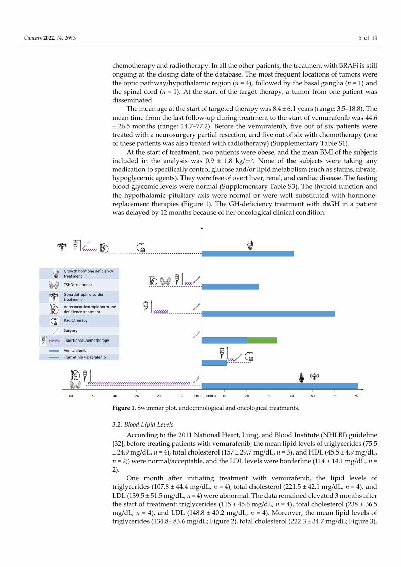
At the start of treatment, two patients were obese, and the mean BMI of the subjects
included in the analysis was 0.9 ± 1.8 kg/m2. None of the subjects were taking any
medication to specifically control glucose and/or lipid metabolism (such as statins, fibrate,
hypoglycemic agents). They were free of overt liver, renal, and cardiac disease. The fasting
blood glycemic levels were normal (Supplementary Table S3). The thyroid function and
the hypothalamic–pituitary axis were normal or were well substituted with hormone‐
replacement therapies (Figure 1). The GH‐deficiency treatment with rhGH in a patient
was delayed by 12 months because of her oncological clinical condition.
Figure 1. Swimmer plot, endocrinological and oncological treatments.
3.2. Blood Lipid Levels
According to the 2011 National Heart, Lung, and Blood Institute (NHLBI) guideline
[32], before treating patients with vemurafenib, the mean lipid levels of triglycerides (75.5
± 24.9 mg/dL, n = 4), total cholesterol (157 ± 29.7 mg/dL, n = 3), and HDL (45.5 ± 4.9 mg/dL,
n = 2;) were normal/acceptable, and the LDL levels were borderline (114 ± 14.1 mg/dL, n =
2).
One month after initiating treatment with vemurafenib, the lipid levels of
triglycerides (107.8 ± 44.4 mg/dL, n = 4), total cholesterol (221.5 ± 42.1 mg/dL, n = 4), and
LDL (139.5 ± 51.5 mg/dL, n = 4) were abnormal. The data remained elevated 3 months after
the start of treatment: triglycerides (115 ± 45.6 mg/dL, n = 4), total cholesterol (238 ± 36.5
mg/dL, n = 4), and LDL (148.8 ± 40.2 mg/dL, n = 4). Moreover, the mean lipid levels of
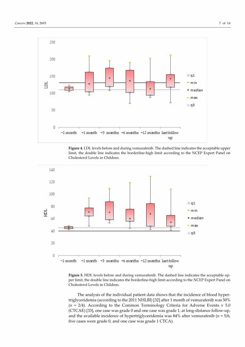
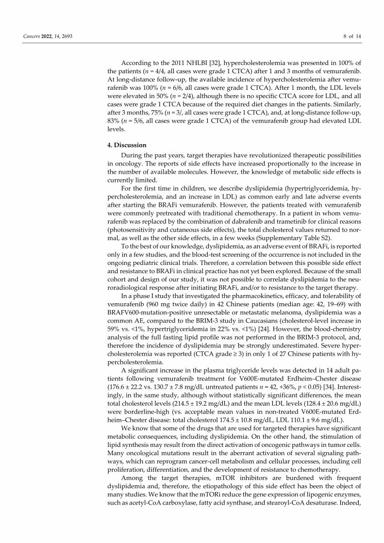
triglycerides (134.8± 83.6 mg/dL; Figure 2), total cholesterol (222.3 ± 34.7 mg/dL; Figure 3),
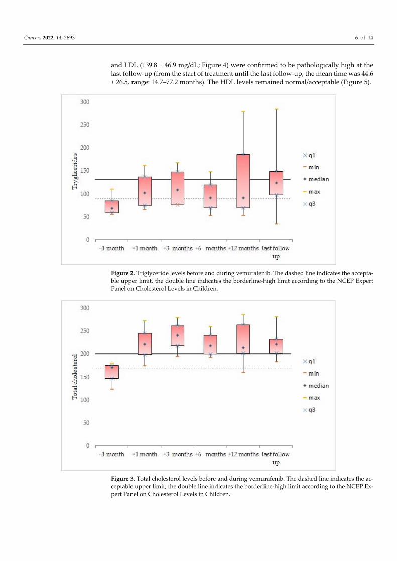
Cancers 2022, 14, 2693 6 of 14
and LDL (139.8 ± 46.9 mg/dL; Figure 4) were confirmed to be pathologically high at the
last follow‐up (from the start of treatment until the last follow‐up, the mean time was 44.6
± 26.5, range: 14.7–77.2 months). The HDL levels remained normal/acceptable (Figure 5).
Figure 2. Triglyceride levels before and during vemurafenib. The dashed line indicates the accepta‐
ble upper limit, the double line indicates the borderline‐high limit according to the NCEP Expert
Panel on Cholesterol Levels in Children.
Figure 3. Total cholesterol levels before and during vemurafenib. The dashed line indicates the ac‐
ceptable upper limit, the double line indicates the borderline‐high limit according to the NCEP Ex‐
pert Panel on Cholesterol Levels in Children.
Cancers 2022, 14, 2693 7 of 14
Figure 4. LDL levels before and during vemurafenib. The dashed line indicates the acceptable upper
limit, the double line indicates the borderline‐high limit according to the NCEP Expert Panel on
Cholesterol Levels in Children.
Figure 5. HDL levels before and during vemurafenib. The dashed line indicates the acceptable up‐
per limit, the double line indicates the borderline‐high limit according to the NCEP Expert Panel on
Cholesterol Levels in Children.
The analysis of the individual patient data shows that the incidence of blood hyper‐
triglyceridemia (according to the 2011 NHLBI) [32] after 1 month of vemurafenib was 50%
(n = 2/4). According to the Common Terminology Criteria for Adverse Events v 5.0
(CTCAE) [33], one case was grade 0 and one case was grade 1, at long‐distance follow‐up,
and the available incidence of hypertriglyceridemia was 84% after vemurafenib (n = 5/6,
five cases were grade 0, and one case was grade 1 CTCA).
Cancers 2022, 14, 2693 8 of 14
According to the 2011 NHLBI [32], hypercholesterolemia was presented in 100% of
the patients (n = 4/4, all cases were grade 1 CTCA) after 1 and 3 months of vemurafenib.
At long‐distance follow‐up, the available incidence of hypercholesterolemia after vemu‐
rafenib was 100% (n = 6/6, all cases were grade 1 CTCA). After 1 month, the LDL levels
were elevated in 50% (n = 2/4), although there is no specific CTCA score for LDL, and all
cases were grade 1 CTCA because of the required diet changes in the patients. Similarly,
after 3 months, 75% (n = 3/, all cases were grade 1 CTCA), and, at long‐distance follow‐up,
83% (n = 5/6, all cases were grade 1 CTCA) of the vemurafenib group had elevated LDL
levels.
4. Discussion
During the past years, target therapies have revolutionized therapeutic possibilities
in oncology. The reports of side effects have increased proportionally to the increase in
the number of available molecules. However, the knowledge of metabolic side effects is
currently limited.
For the first time in children, we describe dyslipidemia (hypertriglyceridemia, hy‐
percholesterolemia, and an increase in LDL) as common early and late adverse events
after starting the BRAFi vemurafenib. However, the patients treated with vemurafenib
were commonly pretreated with traditional chemotherapy. In a patient in whom vemu‐
rafenib was replaced by the combination of dabrafenib and trametinib for clinical reasons
(photosensitivity and cutaneous side effects), the total cholesterol values returned to nor‐
mal, as well as the other side effects, in a few weeks (Supplementary Table S2).
To the best of our knowledge, dyslipidemia, as an adverse event of BRAFi, is reported
only in a few studies, and the blood‐test screening of the occurrence is not included in the
ongoing pediatric clinical trials. Therefore, a correlation between this possible side effect
and resistance to BRAFi in clinical practice has not yet been explored. Because of the small
cohort and design of our study, it was not possible to correlate dyslipidemia to the neu‐
roradiological response after initiating BRAFi, and/or to resistance to the target therapy.
In a phase I study that investigated the pharmacokinetics, efficacy, and tolerability of
vemurafenib (960 mg twice daily) in 42 Chinese patients (median age: 42, 19–69) with
BRAFV600‐mutation‐positive unresectable or metastatic melanoma, dyslipidemia was a
common AE, compared to the BRIM‐3 study in Caucasians (cholesterol‐level increase in
59% vs. <1%, hypertriglyceridemia in 22% vs. <1%) [24]. However, the blood‐chemistry
analysis of the full fasting lipid profile was not performed in the BRIM‐3 protocol, and,
therefore the incidence of dyslipidemia may be strongly underestimated. Severe hyper‐
cholesterolemia was reported (CTCA grade ≥ 3) in only 1 of 27 Chinese patients with hy‐
percholesterolemia.
A significant increase in the plasma triglyceride levels was detected in 14 adult pa‐
tients following vemurafenib treatment for V600E‐mutated Erdheim–Chester disease
(176.6 ± 22.2 vs. 130.7 ± 7.8 mg/dL untreated patients n = 42, +36%, p < 0.05) [34]. Interest‐
ingly, in the same study, although without statistically significant differences, the mean
total cholesterol levels (214.5 ± 19.2 mg/dL) and the mean LDL levels (128.4 ± 20.6 mg/dL)
were borderline‐high (vs. acceptable mean values in non‐treated V600E‐mutated Erd‐
heim–Chester disease: total cholesterol 174.5 ± 10.8 mg/dL, LDL 110.1 ± 9.6 mg/dL).
We know that some of the drugs that are used for targeted therapies have significant
metabolic consequences, including dyslipidemia. On the other hand, the stimulation of
lipid synthesis may result from the direct activation of oncogenic pathways in tumor cells.
Many oncological mutations result in the aberrant activation of several signaling path‐
ways, which can reprogram cancer‐cell metabolism and cellular processes, including cell
proliferation, differentiation, and the development of resistance to chemotherapy.
Among the target therapies, mTOR inhibitors are burdened with frequent
dyslipidemia and, therefore, the etiopathology of this side effect has been the object of
many studies. We know that the mTORi reduce the gene expression of lipogenic enzymes,
such as acetyl‐CoA carboxylase, fatty acid synthase, and stearoyl‐CoA desaturase. Indeed,
Cancers 2022, 14, 2693 9 of 14
the mTORi are responsible for an increase in the total cholesterol and/or triglycerides by
interfering with the protein kinase of the mTOR pathway [35].
New evidence supports that lipid metabolism is implicated in driving the tumor mi‐
croenvironment and the cancer‐cell phenotype, which contributes to the development and
survival of cancer cells [36]. Changes in lipid metabolism can affect numerous cellular
processes, including cell proliferation, differentiation, and motility [36]. In the tumor cells,
the lipids can be used to store energy, synthesize the basic elements that are necessary for
the cellular growth and proliferation (such as membranes), and participate in cell signal‐
ing [36,37]. Cancer cells compete for oxygen and nutrients with the host cells, and they
maintain their malignant potential by modifying the lipid metabolism. The oxidative ca‐
tabolism of lipids provides ATP and NADH, both of which are essential to controlling
environmental stress and promoting survival [37,38].
Therefore, lipid‐metabolism reprogramming is an essential link between the tumor
and the host metabolism, with implications in sensitivity to chemotherapies [37], includ‐
ing target therapies [39].
A potential link between BRAFV600E and lipid‐metabolism regulation in cancer cells
is suggested by some cell and mouse model studies [37,40,41]. In 2015, Kang et al. [42]
demonstrated the interaction between oncogenic BRAF V600E and the enzyme 3‐hy‐
droxy‐3‐methylglutaryl‐CoA lyase (HMGCL), which is involved in lipid metabolism by
producing ketone bodies. HMGCL expression is upregulated in BRAF V600E melanoma
and hairy‐cell leukemia. BRAF upregulates HMGCL through an octamer transcription
factor, Oct‐1, which leads to increased intracellular levels of the HMGCL product, aceto‐
acetate, which selectively enhances the binding of the BRAF V600E, but not the BRAF
wild‐type to MEK1 in V600E‐positive cancer cells, to promote the activation of MEK–ERK
signaling and, therefore, tumor growth. In 2017, Xia et al. [43] showed that a high‐fat ke‐
togenic diet increased the serum levels of acetoacetate, which led to the potential tumor
growth of BRAF V600E‐expressing human melanoma cells in xenograft mice. The high‐
fat diets resulted in increased growth rates, masses, and sizes of tumors, without affecting
the body weight in these mice. In contrast, a high‐fat diet did not affect the tumor growth
rates, masses, sizes, or the body weight in mice with tumor xenografts expressing an active
NRAS Q61R mutation. The increased tumor growth in xenograft mice (BRAF mutated)
fed with a high‐fat diet was not due to differences in the quantity of the food intake. In
both mice models, the consumption of a high‐fat diet did not significantly affect the serum
levels of D‐b‐hydroxybutyrate (3HB), but significantly increased the serum cholesterol
levels compared to control mice fed with a normal diet. Treatment with hypolipidemic
agents or an inhibitory homolog of acetoacetate attenuated the BRAF V600E tumor growth
[43].
Valvo et al., in 2021[41], showed that, in BRAFV600E papillary thyroid carcinoma,
the de novo lipid synthesis significantly increased (1.58‐ and 1.34‐fold changes in hetero‐
zygous and homozygous BRAFV600E ‐derived cell lines, respectively) within 6h in vemu‐
rafenib‐treated cancer cells. The xenograft mouse data further showed that human
BRAFV600E tumor cells became less responsive to vemurafenib within two weeks, and
ultimately exhibited increased tumor growth when the Acetyl‐CoA Carboxylase 2 gene
(ACC2) was knocked down. This suggests that silencing the ACC2 (a rate‐limiting enzyme
for de novo lipid synthesis and the inhibition of fatty acid oxidation) may contribute to
BRAFV600E‐inhibitor (e.g., vemurafenib) resistance and increased tumor growth.
BRAFV600E inhibition increased the de novo lipid‐synthesis rates, decreased fatty acid
oxidation due to the oxygen‐consumption rate, and increased the intracellular reactive‐
oxygen‐species (ROS) production, which can trigger tumor‐cell proliferation or death [41].
The modulation of numerous genes, including multiple oncogenes, growth factors,
and tumor suppressors, are activated by reactive oxygen species (ROS) and the modifica‐
tion of the level of the AMP/ATP ratio that is due to cancer‐cell metabolic plasticity (both
possible effects of cancer and anticancer therapy, including BRAFi and MEKi) [44]. The
HIF‐1 and AMP‐activated protein kinase (AMPK), which operate as energy biosensors of
Cancers 2022, 14, 2693 10 of 14
oxidative stress and master regulators of cellular metabolism, play a crucial role in this
phenomenon. [45,46]. The AMPK regulates the ATP level through the switch from ana‐
bolic to catabolic metabolism via the stimulation of glucose uptake, aerobic glycolysis, and
mitochondrial oxidative metabolism, which is mainly due to the β‐oxidation of fatty acids
[46]. These pathways interplay with HIF‐1, and, therefore, a variety of oncogenes, such as
Ras, c‐Myc, and p53, and the Akt/PKB, PI3K, and mTOR signaling pathways, sustain can‐
cer‐cell proliferation and survival [47–49]. The gene KRAS is also directly implicated in
ROS generation by NADPH oxidases [50]. Cancer‐cell survival and metastasis can be sus‐
tained by lipid biosynthesis that is promoted by a shift in the glutamine metabolism from
oxidation to reductive carboxylation [49].
In BRAF V600E melanoma cells, altered lipid metabolism could contribute to tar‐
geted therapy resistance through the modification of the activation of several lipogenesis
pathways [51–53]. New evidence shows that the SREBP‐1‐dependent activation of lipo‐
genesis is required for tumor growth and for cell survival in multiple cancer models, in‐
cluding high‐grade glioma [54,55]. In BRAF‐mutant melanomas, therapy resistance to
vemurafenib is supported by the Sterol Regulatory Element‐Binding Protein (SREBP1) ac‐
tivation [56] and the upregulation of the S1 P‐dependent signaling pathway
[37,40,51,52,57]. In sensitive BRAF‐mutant models, vemurafenib caused the decrease in
lipogenesis and the activation of SREBP‐1. All showed high levels of lipogenesis, even in
the presence of the inhibitor. However, this was not seen in therapy‐resistant models, in
which BRAFi only induced a moderate decrease in the SREBP‐1 levels and did not signif‐
icantly affect lipogenesis [56]. Probably this is due to the activation of the alternative ERK
pathway that is linked to therapy resistance and that is a known regulator of SREBP
[58,59], as is shown by the decreased levels of SREBP‐1 in therapy‐resistant cells treated
with the MEK inhibitor trametinib [56]. The expressions of well‐established mSREBP‐1
downstream targets, such as ACLY, ACACA, and FASN, were also consistently reduced.
These findings indicate that the reactivation of the ERK pathway contributes to sustained
SREBP‐1 activity in therapy‐resistant melanoma cells [56]. Moreover, the expression of
key lipogenic enzymes—SREBP‐1 downstream targets—such as fatty acid synthase
(FASN) acetyl‐CoA carboxylase‐1, were found to be inversely associated with drug re‐
sistance in BRAF‐mutant cell lines [56].
In the current study, most of these laboratory AEs met the criteria as AEs because the
events were medically significant and required diet modification. All these events were
grade 1 CTCA, asymptomatic, and did not require a change in treatment or dose modifi‐
cation. However, because of the possible need for long‐term use, these observed results
may affect the overall benefit/risk assessment of vemurafenib in patients with high cardi‐
ovascular risk.
5. Conclusions
The targeted therapies for brain tumors are innovative and promising oncological
treatments, and as a result, their use has expanded widely. The effectiveness of BRAFi,
and its use in combination with other new target therapies, is increasing, and therefore
the spectrum of side effects needs to be further explored.
The toxicities that are related to these new agents are generally not life threatening;
however, the long‐term effects are unknown, and they could potentially be a limiting fac‐
tor in chronic life‐long use. An accurate screening strategy in new clinical trials, and a
multidisciplinary team, are required for the optimal management of unexpected adverse
events.
We describe, for the first time, the possible side effects of BRAFi in a case series of
children treated for LGG. In our study, children treated with vemurafenib showed a wors‐
ening in their lipid profiles, with a significant increase in triglycerides, LDL, and total
cholesterol over time. New prospective and multicentric clinical trials of larger study
groups are needed to confirm our observation; therefore, the evaluation of the serum lipid
Cancers 2022, 14, 2693 11 of 14
balance should be implemented in future experimental protocols, including BRAFi and/or
MEKi.
Because of the large amount of data that show the possible role of lipid metabolism
in the mechanisms of resistance and response to biological therapies, new future studies
should explore this hypothesis.
Supplementary Materials: The following supporting information can be downloaded at:
https://www.mdpi.com/article/10.3390/cancers14112693/s1, Supplementary Table S1: Detailed de‐
mographic and clinical features of the patients treated with vemurafenib; Supplementary Table S2:
Lipid levels before and after switch of treatment from vemurafenib to dabrafenib and trametinib;
Supplementary Table S3: Fasting blood glucose, biochemical liver, and kidney‐function tests before
and during vemurafenib.
Author Contributions: Writing—original draft: M.C.; writing—review and editing: M.L.G. and
N.D.I.; conceptualization, methodology, and data curation: A.V.; data curation: F.M. and P.G.; for‐
mal analysis: M.G.C.; investigation: C.M., G.P., E.C., P.D.M., G.G., V.I., and S.D.P.; visualization:
M.P. All authors have read and agreed to the published version of the manuscript.
Funding: This research received no external funding.
Institutional Review Board Statement: The study was conducted in accordance with the Declara‐
tion of Helsinki, and approved by Ethics Committee of Regione Liguria (protocol code 334/2019).
Informed Consent Statement: Informed consent was obtained from all subjects involved in the
study.
Data Availability Statement: The data presented in this study are available on request from the
corresponding author.
Conflicts of Interest: The authors declare no conflict of interest.
References
1. Johnson, K.J.; Cullen, J.; Barnholtz‐Sloan, J.S.; Ostrom, Q.T.; Langer, C.E.; Turner, M.C.; McKean‐Cowdin, R.; Fisher, J.L.; Lupo,
P.J.; Partap, S.; et al. Childhood brain tumor epidemiology: A brain tumor epidemiology consortium review. Cancer Epidemiol.
Biomark. Prev. 2014, 23, 2716–2736. https://doi.org/10.1158/1055‐9965.EPI‐14‐0207.
2. de Blank, P.; Bandopadhayay, P.; Haas‐Kogan, D.; Fouladi, M.; Fangusaro, J. Management of pediatric low‐grade glioma. Curr.
Opin. Pediatr. 2019, 31, 21–27. https://doi.org/10.1097/MOP.0000000000000717.
3. Merchant, T.E.; Conklin, H.M.; Wu, S.; Lustig, R.H.; Xiong, X. Late effects of conformal radiation therapy for pediatric patients
with low‐grade glioma: Prospective evaluation of cognitive, endocrine, and hearing deficits. J. Clin. Oncol. 2009, 27, 3691–3697.
https://doi.org/10.1200/JCO.2008.21.2738.
4. Armstrong, G.T.; Liu, Q.; Yasui, Y.; Huang, S.; Ness, K.K.; Leisenring, W.; Hudson, M.M.; Donaldson, S.S.; King, A.A.; Stovall,
M.; et al. Long‐term outcomes among adult survivors of childhood central nervous system malignancies in the Childhood Can‐
cer Survivor Study. J. Natl. Cancer Inst. 2009, 101, 946–958. https://doi.org/10.1093/jnci/djp148.
5. Aloi, D.; Belgioia, L.; Barra, S.; Giannelli, F.; Cavagnetto, F.; Gallo, F.; Milanaccio, C.; Garrè, M.; Di Profio, S.; Di Iorgi, N.; et al.
Neuroendocrine late effects after tailored photon radiotherapy for children with low grade gliomas: Long term correlation with
tumour and treatment parameters. Radiother. Oncol. 2017, 125, 241–247. https://doi.org/10.1016/j.radonc.2017.09.034.
6. Lassaletta, A.; Zapotocky, M.; Mistry, M.; Ramaswamy, V.; Honnorat, M.; Krishnatry, R.; Stucklin, A.G.; Zhukova, N.; Arnoldo,
A.; Ryall, S.; et al. Therapeutic and Prognostic Implications of BRAF V600E in Pediatric Low‐Grade Gliomas. J. Clin. Oncol. 2017,
35, 2934–2941. https://doi.org/10.1200/JCO.2016.71.8726.
7. Zhang, J.; Wu, G.; Miller, C.P.; Tatevossian, R.G.; Dalton, J.D.; Tang, B.; Orisme, W.; Punchihewa, C.; Parker, M.; Qaddoumi, I.;
et al. St. Jude Children’s Research Hospital–Washington University Pediatric Cancer Genome Project. Whole‐genome sequenc‐
ing identifies genetic alterations in pediatric low‐grade gliomas. Nat. Genet. 2013, 45, 602–612. https://doi.org/10.1038/ng.2611.
8. Behling, F.; Barrantes‐Freer, A.; Skardelly, M.; Nieser, M.; Christians, A.; Stockhammer, F.; Rohde, V.; Tatagiba, M.; Hartmann,
C.; Stadelmann, C.; et al. Frequency of BRAF V600E mutations in 969 central nervous system neoplasms. Diagn. Pathol. 2016, 11,
55. https://doi.org/10.1186/s13000‐016‐0506‐2.
9. Bar, E.E.; Lin, A.; Tihan, T.; Burger, P.C.; Eberhart, C.G. Frequent gains at chromosome 7q34 involving BRAF in pilocytic astro‐
cytoma. J. Neuropathol. Exp. Neurol. 2008, 67, 878–887. https://doi.org/10.1097/NEN.0b013e3181845622.
10. Schindler, G.; Capper, D.; Meyer, J.; Janzarik, W.; Omran, H.; Herold‐Mende, C.; Schmieder, K.; Wesseling, P.; Mawrin, C.;
Hasselblatt, M.; et al. Analysis of BRAF V600E mutation in 1,320 nervous system tumors reveals high mutation frequencies in
pleomorphic xanthoastrocytoma, ganglioglioma and extra‐cerebellar pilocytic astrocytoma. Acta Neuropathol. 2011, 121, 397–
405. https://doi.org/10.1007/s00401‐011‐0802‐6.
Cancers 2022, 14, 2693 12 of 14
11. Ho, C.Y.; Mobley, B.C.; Gordish‐Dressman, H.; Vandenbussche, C.J.; Mason, G.E.; Bornhorst, M.; Esbenshade, A.J.; Tehrani, M.;
Orr, B.A.; LaFrance, D.R.; et al. A clinicopathologic study of diencephalic pediatric low‐grade gliomas with BRAF V600 muta‐
tion. Acta Neuropathol. 2015, 130, 575–585. https://doi.org/10.1007/s00401‐015‐1467‐3.
12. Mistry, M.; Zhukova, N.; Merico, D.; Rakopoulos, P.; Krishnatry, R.; Shago, M.; Stavropoulos, J.; Alon, N.; Pole, J.D.; Ray, P.N.;
et al. BRAF mutation and CDKN2A deletion define a clinically distinct subgroup ofchildhood secondary high‐grade glioma. J.
Clin. Oncol. 2015, 33, 1015–1022. https://doi.org/10.1200/JCO.2014.58.3922.
13. Selt, F.; van Tilburg, C.M.; Bison, B.; Sievers, P.; Harting, I.; Ecker, J.; Pajtler, K.W.; Sahm, F.; Bahr, A.; Simon, M.; et al. Response
to trametinib treatment in progressive pediatric low‐grade glioma patients. J. Neurooncol. 2020, 149, 499–510.
https://doi.org/10.1007/s11060‐020‐03640‐3.
14. Fangusaro, J.; Onar‐Thomas, A.; Young Poussaint, T.; Wu, S.; Ligon, A.H.; Lindeman, N.; Banerjee, A.; Packer, R.J.; Kilburn,
L.B.; Goldman, S.; et al. Selumetinib in paediatric patients with BRAF‐aberrant or neurofibromatosis type 1‐associated recurrent,
refractory, or progressive low‐grade glioma: A multicentre, phase 2 trial. Lancet Oncol. 2019, 20, 1011–1022.
https://doi.org/10.1016/S1470‐2045(19)30277‐3.
15. Nicolaides, T.; Nazemi, K.J.; Crawford, J.; Kilburn, L.; Minturn, J.; Gajjar, A.; Gauvain, K.; Leary, S.; Dhall, G.; Aboian, M.; et al.
Phase I study of vemurafenib in children with recurrent or progressive BRAFV600E mutant brain tumors: Pacific Pediatric
Neuro‐Oncology Consortium study (PNOC‐002). Oncotarget 2020, 11, 1942–1952. https://doi.org/10.18632/oncotarget.27600.
16. Hargrave, D.R.; Bouffet, E.; Tabori, U.; Broniscer, A.; Cohen, K.J.; Hansford, J.R.; Geoerger, B.; Hingorani, P.; Dunkel, I.J.; Russo,
M.W.; et al. Efficacy and Safety of Dabrafenib in Pediatric Patients with BRAF V600 Mutation‐Positive Relapsed or Refractory
Low‐Grade Glioma: Results from a Phase I/IIa Study. Clin. Cancer Res. 2019, 25, 7303–7311. https://doi.org/10.1158/1078‐
0432.CCR‐19‐2177.
17. Marks, A.M.; Bindra, R.S.; DiLuna, M.L.; Huttner, A.; Jairam, V.; Kahle, K.T.; Kieran, M.W. Response to the BRAF/MEK inhibi‐
tors dabrafenib/trametinib in an adolescent with a BRAF V600E mutated anaplastic ganglioglioma intolerant to vemurafenib.
Pediatr. Blood Cancer. 2018, 65, 26969. https://doi.org/10.1002/pbc.26969.
18. Touat, M.; Gratieux, J.; Condette Auliac, S.; Sejean, K.; Aldea, S.; Savatovsky, J.; Perkins, G.; Blons, H.; Ligon, K.L.; Idbaih, A.; et
al. Vemurafenib and cobimetinib overcome resistance to vemurafenib in BRAF‐mutant ganglioglioma. Neurology 2018, 91, 523–
525. https://doi.org/10.1212/WNL.0000000000006171.
19. Robert, C.; Grob, J.J.; Stroyakovskiy, D.; Karaszewska, B.; Hauschild, A.; Levchenko, E.; Sileni, V.C.; Schachter, J.; Garbe, C.;
Bondarenko, I.; et al. Five‐Year Outcomes with Dabrafenib plus Trametinib in Metastatic Melanoma. N. Engl. J. Med. 2019, 381,
626–636. https://doi.org/10.1056/NEJMoa1904059.
20. Schreck, K.C.; Grossman, S.A.; Pratilas, C.A. BRAF Mutations and the Utility of RAF and MEK Inhibitors in Primary Brain
Tumors. Cancers 2019, 11, 1262. https://doi.org/10.3390/cancers11091262.
21. Garnier, L.; Ducray, F.; Verlut, C.; Mihai, M.I.; Cattin, F.; Petit, A.; Curtit, E. Prolonged Response Induced by Single Agent
Vemurafenib in a BRAF V600E Spinal Ganglioglioma: A Case Report and Review of the Literature. Front. Oncol. 2019, 9, 177.
https://doi.org/10.3389/fonc.2019.00177.
22. Del Bufalo, F.; Ceglie, G.; Cacchione, A.; Alessi, I.; Colafati, G.S.; Carai, A.; Diomedi‐Camassei, F.; De Billy, E.; Agolini, E.;
Mastronuzzi, A.; et al. BRAF V600E Inhibitor (Vemurafenib) for BRAF V600E Mutated Low Grade Gliomas. Front. Oncol. 2018,
8, 526. https://doi.org/10.3389/fonc.2018.00526.
23. Nobre, L.; Zapotocky, M.; Ramaswamy, V.; Ryall, S.; Bennett, J.; Alderete, D.; Guill, J.B.; Baroni, L.; Bartels, U.; Bavle, A.; et al.
Outcomes of BRAF V600E Pediatric Gliomas Treated with Targeted BRAF Inhibition. JCO Precis. Oncol. 2020, 4, 561–571.
https://doi.org/10.1200/PO.19.00298.
24. Chapman, P.B.; Robert, C.; Larkin, J.; Haanen, J.B.; Ribas, A.; Hogg, D.; Hamid, O.; Ascierto, P.A.; Testori, A.; Lorigan, P.C.; et
al. Vemurafenib in patients with BRAFV600 mutation‐positive metastatic melanoma: Final overall survival results of the ran‐
domized BRIM‐3 study. Ann. Oncol. 2017, 28, 2581–2587. https://doi.org/10.1093/annonc/mdx339.
25. Rizzo, D.; Ruggiero, A.; Amato, M.; Maurizi, P.; Riccardi, R. BRAF and MEK inhibitors in pediatric glioma: New therapeutic
strategies, new toxicities. Expert Opin. Drug Metab. Toxicol. 2016, 12, 1397–1405. https://doi.org/10.1080/17425255.2016.1214710.
26. Larkin, J.; Ascierto, P.A. Combined vemurafenib and cobimetinib in BRAF‐mutated melanoma. N. Engl. J. Med. 2014, 371, 1867–
1876. https://doi.org/10.1056/NEJMoa1408868.
27. Larkin, J.; Ascierto, P.A.; Dréno, B.; Atkinson, V.; Liszkay, G.; Maio, M.; Mandalà, M.; Demidov, L.; Stroyakovskiy, D.; Thomas,
L.; et al. Safety of BRAF+MEK Inhibitor Combinations: Severe Adverse Event Evaluation. Cancers 2020, 12, 1650.
https://doi.org/10.3390/cancers12061650.
28. McArthur, G.A.; Chapman, P.B.; Robert, C.; Larkin, J.; Haanen, J.B.; Dummer, R.; Ribas, A.; Hogg, D.; Hamid, O.; Ascierto, P.A.;
et al. Safety and efficacy of vemurafenib in BRAF(V600E) and BRAF(V600K) mutation‐positive melanoma (BRIM‐3): Extended
follow‐up of a phase 3, randomised, open‐label study. Lancet Oncol. 2014, 15, 323–332. https://doi.org/10.1016/S1470‐
2045(14)70012‐9.
29. WHO Multicentre Growth Reference Study Group. WHO Child Growth Standards based on length/height, weight and age.
Acta Paediatr. Suppl. 2006, 450, 76–85. https://doi.org/10.1111/j.1651‐2227.2006.tb02378.x.
30. Willett, W.C.; Sacks, F.; Trichopoulou, A.; Drescher, G.; Ferro‐Luzzi, A.; Helsing, E.; Trichopoulos, D. Mediterranean diet pyra‐
mid: A cultural model for healthy eating. Am. J. Clin. Nutr. 1995, 61, 1402–1406.
31. Friedewald, W.T.; Levy, R.I.; Fredrickson, D.S. Estimation of the concentration of low‐density lipoprotein cholesterol in plasma,
without use of the preparative ultracentrifuge. Clin. Chem. 1972, 18, 499–502.
Cancers 2022, 14, 2693 13 of 14
32. Expert Panel on Integrated Guidelines for Cardiovascular Health and Risk Reduction in Children and Adolescents; National
Heart, Lung, and Blood Institute. Expert panel on integrated guidelines for cardiovascular health and risk reduction in children
and adolescents: Summary report. Pediatrics 2011, 128, 213–256. https://doi.org/10.1542/peds.2009‐2107C.
33. NCI. National Cancer Institute Common Terminology Criteria for Adverse Events (CTCAE) v.5.0. Available online:
https://ctep.cancer.gov/protocoldevelopment/electronic_applications/docs/CTCAE_v5_Quick_Reference_5x7.pdf (accessed on
1 April 2022).
34. Cohen‐Aubart, F.; Guerin, M.; Poupel, L.; Cluzel, P.; Saint‐Charles, F.; Charlotte, F.; Arsafi, Y.; Emile, J.F.; Frisdal, E.; Le Goff,
C.; et al. Hypoalphalipoproteinemia and BRAFV600E Mutation Are Major Predictors of Aortic Infiltration in the Erdheim‐Ches‐
ter Disease. Arter. Thromb. Vasc. Biol. 2018, 38, 1913–1925. https://doi.org/10.1161/ATVBAHA.118.310803.
35. Vergès, B.; Walter, T.; Cariou, B. Endocrine side effects of anti‐cancer drugs: Effects of anti‐cancer targeted therapies on lipid
and glucose metabolism. Eur. J. Endocrinol. 2014, 170, 43–55. https://doi.org/10.1530/EJE‐13‐0586.
36. Wang, W.; Bai, L.; Li, W. The Lipid Metabolic Landscape of Cancers and New Therapeutic Perspectives. Front. Oncol. 2020, 8,
605154. https://doi.org/10.3389/fonc.2020.605154.
37. Stamatakos, S.; Beretta, G.L.; Vergani, E.; Dugo, M.; Corno, C.; Corna, E.; Tinelli, S.; Frigerio, S.; Ciusani, E.; Rodolfo, M.; et al.
Deregulated FASN Expression in BRAF Inhibitor‐Resistant Melanoma Cells Unveils New Targets for Drug Combinations. Can‐
cers 2021, 13, 2284. https://doi.org/10.3390/cancers13092284.
38. Carracedo, A.; Cantley, L.C.; Pandolfi, P.P. Cancer metabolism: Fatty acid oxidation in the limelight. Nat. Rev. Cancer 2013, 13,
227–232. https://doi.org/10.1038/nrc3483.
39. Germain, N.; Dhayer, M.; Boileau, M.; Fovez, Q.; Kluza, J.; Marchetti, P. Lipid Metabolism and Resistance to Anticancer Treat‐
ment. Biology 2020, 9, 474. https://doi.org/10.3390/biology9120474.
40. Hong, X.; Roh, W.; Sullivan, R.J.; Wong, K.H.K.; Wittner, B.S.; Guo, H.; Dubash, T.D.; Sade‐Feldman, M.; Wesley, B.; Horwitz,
E.; et al. The Lipogenic Regulator SREBP2 Induces Transferrin in Circulating Melanoma Cells and Suppresses Ferroptosis. Can‐
cer Discov. 2021, 11, 678–695. https://doi.org/10.1158/2159‐8290.CD‐19‐1500.
41. Valvo, V.; Iesato, A.; Kavanagh, T.R.; Priolo, C.; Zsengeller, Z.; Pontecorvi, A.; Stillman, I.E.; Burke, S.D.; Liu, X.; Nucera, C.
Fine‐Tuning Lipid Metabolism by Targeting Mitochondria‐Associated Acetyl‐CoA‐Carboxylase 2 in BRAFV600E Papillary Thy‐
roid Carcinoma. Thyroid 2021, 31, 1335–1358. https://doi.org/10.1089/thy.2020.0311.
42. Kang, H.B.; Fan, J.; Lin, R.; Elf, S.; Ji, Q.; Zhao, L.; Jin, L.; Seo, J.H.; Shan, C.; Arbiser, J.L.; et al. Metabolic Rewiring by Oncogenic
BRAF V600E Links Ketogenesis Pathway to BRAF‐MEK1 Signaling. Mol. Cell 2015, 6, 345–358. https://doi.org/10.1016/j.mol‐
cel.2015.05.037.
43. Xia, S.; Lin, R.; Jin, L.; Zhao, L.; Kang, H.B.; Pan, Y.; Liu, S.; Qian, G.; Qian, Z.; Konstantakou, E.; et al. Prevention of Dietary‐Fat‐
Fueled Ketogenesis Attenuates BRAF V600E Tumor Growth. Cell Metab. 2017, 25, 358–373.
https://doi.org/10.1016/j.cmet.2016.12.010.
44. Yuan, L.; Mishra, R.; Patel, H.; Alanazi, S.; Wei, X.; Ma, Z.; Garrett, J.T. BRAF Mutant Melanoma Adjusts to BRAF/MEK Inhibi‐
tors via Dependence on Increased Antioxidant SOD2 and Increased Reactive Oxygen Species Levels. Cancers 2020, 12, 1661.
https://doi.org/10.3390/cancers12061661.
45. Mihaylova, M.M.; Shaw, R.J. The AMPK signalling pathway coordinates cell growth, autophagy and metabolism. Nat. Cell Biol.
2011, 13, 1016–1023. https://doi.org/10.1038/ncb2329.
46. Jia, D.; Lu, M.; Jung, K.H.; Park, J.H.; Yu, L.; Onuchic, J.N.; Kaipparettu, B.A.; Levine, H. Elucidating cancer metabolic plasticity
by coupling gene regulation with metabolic pathways. Proc. Natl. Acad. Sci. USA 2019, 116, 3909–3918.
https://doi.org/10.1073/pnas.1816391116.
47. Semenza, G.L. Oxygen sensing, hypoxia‐inducible factors, and disease pathophysiology. Annu. Rev. Pathol. 2014, 9, 47–71.
https://doi.org/10.1146/annurev‐pathol‐012513‐104720.
48. Shaw, R.J.; Cantley, L.C. Ras, PI(3)K and mTOR signalling controls tumour cell growth. Nature 2006, 441, 424–430.
https://doi.org/10.1038/nature04869.
49. Moldogazieva, N.T.; Lutsenko, S.V.; Terentiev, A.A. Reactive Oxygen and Nitrogen Species‐Induced Protein Modifications:
Implication in Carcinogenesis and Anticancer Therapy. Cancer Res. 2018, 78, 6040–6047. https://doi.org/10.1158/0008‐5472.CAN‐
18‐0980.
50. Ogrunc, M.; Di Micco, R.; Liontos, M.; Bombardelli, L.; Mione, M.; Fumagalli, M.; Gorgoulis, V.G.; dʹAdda di Fagagna, F. On‐
cogene‐induced reactive oxygen species fuel hyperproliferation and DNA damage response activation. Cell Death Differ. 2014,
21, 998–1012. https://doi.org/10.1038/cdd.2014.16.
51. Innocenzi, D.; Alò, P.L.; Balzani, A.; Sebastiani, V.; Silipo, V.; La Torre, G.; Ricciardi, G.; Bosman, C.; Calvieri, S. Fatty acid
synthase expression in melanoma. J. Cutan. Pathol. 2003, 30, 23–28. https://doi.org/10.1034/j.1600‐0560.2003.300104.x.
52. Kapur, P.; Rakheja, D.; Roy, L.C.; Hoang, M.P. Fatty acid synthase expression in cutaneous melanocytic neoplasms. Mod. Pathol.
2005, 18, 1107–1012. https://doi.org/10.1038/modpathol.3800395.
53. Butler, L.M.; Perone, Y.; Dehairs, J.; Lupien, L.E.; de Laat, V.; Talebi, A.; Loda, M.; Kinlaw, W.B.; Swinnen, J.V. Lipids and cancer:
Emerging roles in pathogenesis, diagnosis and therapeutic intervention. Adv. Drug Deliv. Rev. 2020, 159, 245–293.
https://doi.org/10.1016/j.addr.2020.07.013.
54. Griffiths, B.; Lewis, C.A.; Bensaad, K.; Ros, S.; Zhang, Q.; Ferber, E.C.; Konisti, S.; Peck, B.; Miess, H.; East, P. Sterol regulatory
element binding protein‐dependent regulation of lipid synthesis supports cell survival and tumor growth. Cancer Metab. 2013,
1, 3. https://doi.org/10.1186/2049‐3002‐1‐3.
Cancers 2022, 14, 2693 14 of 14
55. Lewis, C.A.; Brault, C.; Peck, B.; Bensaad, K.; Griffiths, B.; Mitter, R.; Chakravarty, P.; East, P.; Dankworth, B.; Alibhai, D.; et al.
SREBP maintains lipid biosynthesis and viability of cancer cells under lipid‐ and oxygen‐deprived conditions and defines a
gene signature associated with poor survival in glioblastoma multiforme. Oncogene 2015, 34, 5128–5140.
https://doi.org/10.1038/onc.2014.439.
56. Talebi, A.; Dehairs, J.; Rambow, F.; Rogiers, A.; Nittner, D.; Derua, R.; Vanderhoydonc, F.; Duarte, J.A.G.; Bosisio, F.; Van den
Eynde, K.; et al. Sustained SREBP‐1‐dependent lipogenesis as a key mediator of resistance to BRAF‐targeted therapy. Nat. Com‐
mun. 2018, 9, 2500. https://doi.org/10.1038/s41467‐018‐04664‐0.
57. Garandeau, D.; Noujarède, J.; Leclerc, J.; Imbert, C.; Garcia, V.; Bats, M.L.; Rambow, F.; Gilhodes, J.; Filleron, T.; Meyer, N.; et
al. Targeting the Sphingosine 1‐Phosphate Axis Exerts Potent Antitumor Activity in BRAFi‐Resistant Melanomas. Mol. Cancer
Ther. 2019, 18, 289–300. https://doi.org/10.1158/1535‐7163.MCT‐17‐1141.
58. Kotzka, J.; Müller‐Wieland, D.; Koponen, A.; Njamen, D.; Kremer, L.; Roth, G.; Munck, M.; Knebel, B.; Krone, W. ADD1/SREBP‐
1c mediates insulin‐induced gene expression linked to the MAP kinase pathway. Biochem. Biophys. Res. Commun. 1998, 249, 375–
359. https://doi.org/10.1006/bbrc.1998.9161.
59. Fleischmann, M.; Iynedjian, P.B. Regulation of sterol regulatory‐element binding protein 1 gene expression in liver: Role of
insulin and protein kinase B/cAkt. Biochem. J. 2000, 349, 13–17. https://doi.org/10.1042/0264‐6021:3490013.