drugs in development and dietary approach for duchenne muscular dystrophy
TRANSCRIPT
AUTHOR
PROOF
COPY
Not for
publication
© 2015 Angelini and Tasca. This work is published by Dove Medical Press Limited, and licensed under Creative Commons Attribution – Non Commercial (unported, v3.0) License. The full terms of the License are available at http://creativecommons.org/licenses/by-nc/3.0/. Non-commercial uses of the work are permitted without any further
permission from Dove Medical Press Limited, provided the work is properly attributed. Permissions beyond the scope of the License are administered by Dove Medical Press Limited. Information on how to request permission may be found at: http://www.dovepress.com/permissions.php
Orphan Drugs: Research and Reviews 2015:5 1–11
Orphan Drugs: Research and Reviews Dovepress
submit your manuscript | www.dovepress.com
Dovepress 1
R e v i e w
open access to scientific and medical research
Open Access Full Text Article
55677
Drugs in development and dietary approach for Duchenne muscular dystrophy
Corrado Angelinielisabetta TascaNeuromuscular Laboratory, Fondazione San Camillo Hospital iRCCS, venice, italy
Correspondence: Corrado Angelini Neuromuscular Laboratory, Fondazione San Camillo Hospital iRCCS, via Alberoni 70, 30124 Lido venice, italy Tel +39 049 821 6155 Fax +39 049 821 6163 email [email protected]
Abstract: Therapeutic trials studying Duchenne muscular dystrophy (DMD) in Europe and
the USA have been done using a protocol that includes manual muscle testing and functional
testing, and have shown the efficacy of steroid drugs in various doses and regimens. Further,
drisapersen and eteplirsen (exon skipping drugs) and ataluren (a drug to overcome stop codon
mutations) have achieved some clinical improvement. Cardioprotective drugs are efficacious in
DMD, and eplerenone, an aldosterone inhibitor and diuretic, is now being used treat the disease.
The dietary approach should be used in wheelchair-bound DMD children in combination with
respiratory assistance. The importance of some of the treatments proposed is that they might
also be useful in other genetic disorders where stop codon mutations are present; moreover, it
is possible that these new treatments will improve quality of life for many patients.
Keywords: Duchenne muscular dystrophy, steroids, ataluren, drisapersen, eplerenone,
eteplirsen
IntroductionDuchenne muscular dystrophy (DMD) is a relatively common muscle disorder
caused by dystrophin deficiency. Over the past 25 years, several clinical trials have
been carried out with agents directed to reducing the increase in connective tis-
sue and with agents designed to induce production of dystrophin by frameshifting
deletions of one or more exons abutting exon 51 or by stop codon read-through.1,2
Secondary approaches have been tried to compensate for the lack of dystrophin,
such as reduction of the fibrosis or inflammation caused by nitric oxide synthase.
Cardiac insufficiency is common in the advanced stages of DMD, and seems to
benefit from beta-blockers, angiotensin-converting enzyme (ACE) inhibitors, and
diuretics.3,4
It is important in DMD drug studies that patients are carefully selected, using clinical
and molecular criteria so that no atypical cases are included. A positive X-linked family
history or dystrophin gene and protein analysis (when available) is required. Although
a medication might theoretically be most helpful in the preclinical stage of the disease
(detected by elevated creatine kinase in at-risk individuals), it is difficult to assess
the efficacy of treatment in DMD boys younger than 3–5 years of age. Most studies
have involved DMD patients 5 years of age or older who have at least some detectable
weakness but are not so advanced that a drug may no longer be able to stop or reverse
the dystrophic process.5–8
For inclusion in most trials, DMD patients must be able to walk and present
at least two of the following criteria: a genetic diagnosis of sporadic DMD with

Orphan Drugs: Research and Reviews 2015:5submit your manuscript | www.dovepress.com
Dovepress
Dovepress
2
Angelini and Tasca
mutation type (stop codon, deletion, missense); an X-linked
family history with a proven genetic diagnosis; and muscle
biopsy changes consistent with DMD (ie, absence of
dystrophin).
Patients should be excluded if they have facial muscle
weakness, skin rash, or sensory abnormalities, and when
affected male relatives are still ambulating at the age of 16
years. Such patients could be included in a separate treat-
ment group corresponding to Becker’s dystrophy. To include
sporadic cases of DMD when X-linked inheritance cannot
be demonstrated, a molecular diagnosis (either on a protein
or genetic basis) is required.
Patients who have received systemic medications other
than antibiotics or drugs to treat a specific illness should
also be excluded. Elective surgery (ie, release of con-
tractures or Achilles tenotomy) should not be performed
during the drug trial. Physical therapy must be similar
for all patients at a single center, but no specific therapy
program is recommended, and it is expected that range of
motion of the extremities will be recorded periodically.
However, the range of exercise of extremities should not
be formally assessed since this is quite variable and dif-
ficult to analyze.
Natural historyIn the natural history of DMD, a constant improvement
of strength and functional test performance is recorded up
to 7 years of age, when muscle strength and motor ability
reaches a plateau. After this age, there is an increase in con-
nective tissue in skeletal muscles and loss of muscle fibers,
which causes progressive worsening of muscle function
(Figure 1). The inability to walk at least 10 meters without
aid is defined as “loss of ambulation”.
An important issue is that walking ability is lost around
10.5 years of age, with some variation depending mainly on
the amount of residual dystrophin.9 Prolongation of indepen-
dent walking ability is therefore an important primary target
in long-term clinical trials, although this might be influenced
by external factors, such as surgical interventions, steroid
therapy, excessive weight gain, and traumatic bone fractures.
Once the child is wheelchair-bound, progressive loss of
strength in the respiratory and trunk muscles determines the
degree of ventilatory insufficiency and kyphoscoliosis.
Manual muscle testingThe strength of most of the major muscles of the limbs
and trunk can be readily recorded and remeasured using
Figure 1Notes: Muscle features seen on hematoxylin-eosin staining in untreated DMD patients include increased connective tissue and a variable amount of degenerating (A) and regenerating (B) fibers. Dystrophin immunolabelling in control (C) and DMD muscles showing complete absence of dystrophin (D) or isolated (E) or clustered dystrophin-positive revertant fibers (F and G). Neuronal nitric oxide synthase immunolabelling is correctly localized at the sarcolemma in control muscle (H) and absent in DMD muscle (I). Abbreviation: DMD, Duchenne muscular dystrophy.

Orphan Drugs: Research and Reviews 2015:5 submit your manuscript | www.dovepress.com
Dovepress
Dovepress
3
Drugs and dietary treatment for DMD
manual testing. Several studies have demonstrated that the
degree and rate of muscle weakness and rate of deterioration
of individual muscles varies in a predictable fashion in DMD,
depending on the muscle tested.5,7,8 Therefore, muscle testing
can be simplified by omitting muscles that are either too weak
to be accurately tested or are not expected to deteriorate rap-
idly enough to detect any drug effect. Accordingly, muscles
in the upper extremities (eg, deltoid and triceps brachii)
and lower extremities (eg, quadriceps and iliopsoas) should
be scored for muscle strength using the Medical Research
Council (MRC) recommendations. All muscles are tested
with the limb held at 90°. Recently, a more simple revised
MRC scale with only four grades has been proposed;10 its
usefulness should be evaluated in future trials.
Functional muscle testingA fast and efficient functional test that has already been
validated, known as the GSGC (Gait, Stairs, Gowers,
Chair) score, consists of several functional maneuvers that
can be graded according to disease severity.11,12 These tests
are adapted from a standardized series of protocols and are
given considerable emphasis because they do not require
the same level of cooperation as manual muscle strength
testing. Although functional tests are easy to perform, even
in younger children who cannot cooperate with manual
muscle testing, a major disadvantage of these tests is that
their results may abruptly worsen at 9–10 years of age.
In ambulatory patients, the function of the upper extremities
usually remains quite good, while a progressive deterioration
can be expected in the function of the lower extremities. For
this reason, these functional tests place greater emphasis on
the lower extremities.
Standard 9 inch stairs with four steps seems to be a reliable
functional test for DMD. A series of chairs is used to evaluate
the function of “arising from a chair” to ensure that the child’s
legs are at 90° with the feet on the floor. It is emphasized that
all tests should be performed in a uniform manner, with each
child using the same equipment for each examination.
Six-minute walking testMany studies conducted in the last 10 years, especially those
involving the new molecular drugs, have used this functional
test as an outcome,12 but in DMD children there is extreme
variability in its performance because of their variable
motivation or early fatigability, and these factors can affect
the reliability of the test. Therefore, the results of drug trials
using only this parameter as a functional outcome should be
interpreted with caution.
Drugs in developmentSeveral categories of drugs are in use, and act on different
pathogenetic mechanisms in DMD (Table 1).
CorticosteroidsCorticosteroids continue to be the gold standard for the treat-
ment of DMD. However, the choice of steroid and mode of
administration is still a matter of debate. Use of corticoster-
oids should be discussed with parents and not started before
3 years of age; however, the steroid is usually stopped when
boys become wheelchair-bound.
In two trials, we established that deflazacort (Lepetit
Company, Milan, Italy; Teofarma, Pavia, Italy; EG Labnet,
Edison, NJ, USA) at 2 mg/kg on alternate days or 0.9 mg/kg
stabilized muscle strength in DMD when compared with a
placebo group.5,13 Further, deflazacort appeared to cause less
weight gain than prednisone, and bone sparing was more
evident with deflazacort than with prednisone.10
Deflazacort has demonstrated good efficacy in slow-
ing progression of DMD and delaying loss of independent
ambulation.5,14 Many corticosteroid trials have been con-
ducted in DMD, and hundreds of patients have been treated
in many double-blind, randomized, controlled trials.5–8,13,15
Table 1 Properties of drugs in development for DMD
Drug Deflazacort Eplerenone Ataluren Drisapersen Eteplirsen
Compound Oxazolidine glucocorticoid derivative
Aldosterone inhibitor
PTC124 2′-O-methyl phosphorothioate
Phosphorodiamidate morpholino oligomers
Route of administration
Oral Oral Oral Subcutaneous injection intravenous infusion
Dose 0.9 mg/kg 25 mg/day 40 mg/kg/day 6 mg/kg 30–50 mg/kg/weekAction Anti-inflammatory Diuretic Bypass stop codon
mutationAnti-sense oligonucleotide to skip exon 51
Anti-sense oligonucleotide to skip exon 51
Company Marathon Pharmaceuticals
Pfizer Inc PTC Therapeutics inc.
GSK and Prosensa Sarepta Therapeutics inc.
Abbreviation: DMD, Duchenne muscular dystrophy.

Orphan Drugs: Research and Reviews 2015:5submit your manuscript | www.dovepress.com
Dovepress
Dovepress
4
Angelini and Tasca
These trials have shown the benefit of steroids, with a
marked initial improvement in the first 3–6 months and a
subsequent slowing of disease progression for over 3 years.
The mechanism of action of steroids is unknown, but
they may act in a number of ways, probably by inhibiting
connective-fibrous substitution of the muscle or by increasing
the synthesis of dystrophin and of other synergistic sarcolem-
mal proteins, such as utrophin.
Different steroids with different regimens (daily, alter-
nate day, pulsed) have been used with the aim of obtaining
maximal efficacy with fewer side effects. Many uncertain-
ties remain about when to begin therapy, but earlier (after
3–5 years of age) is thought to be better. An open-label trial
on DMD and bone mineral density16 found that prednisone
0.35 mg has the same positive effects on muscle strength
and function as higher doses. Another trial8 compared
two dosages of prednisone, ie, 0.3 mg/kg day and 0.75 mg/kg
day, and patients on 0.75 mg/kg day improved more than
patients on 0.3 mg/kg day.
Two trials reported that deflazacort had fewer side effects
than prednisone.11 This oxazolidine derivative has been
shown to prevent bone loss in children with other diseases.17
We conducted a controlled, randomized, double-blind trial
to compare deflazacort 0.9 mg/kg/day with prednisone
0.75 mg/kg/day in 18 boys with DMD.13 The effects of the
steroids were examined after 12 months by comparing the
status of the treated patients with that of historical controls.
The two steroids were equally effective in improving motor
function and functional performance. The average increase
in weight at 9 months with respect to baseline was 5%
(2 kg) in the deflazacort group and 18% in the prednisone
group (P,0.005), and the change remained significant
after 12 months (P,0.05). After 2 years, the increase in
body weight was 19% in the deflazacort group and 41%
in the prednisone group. Mild cataracts were observed in
three patients in the deflazacort group and in one patient in
the prednisone group.
At a European Neuromuscular Center workshop,18 Reit-
ter reported a large multicenter trial comparing deflazacort
and prednisone in 100 boys with DMD in Germany, in
which 14 patients dropped out prematurely due to intoler-
able weight gain; the majority of these patients were treated
with prednisone. Among the 80 patients who completed
the study, the weight gain was significantly higher in
those receiving prednisone. However, more boys in the
deflazacort group developed cataract. Both studies used
manual muscle testing and timed motor functions (eg, run-
ning, climbing stairs), and the outcomes were similar.
Growth was impaired in all boys regardless of the type of
steroid administered. The conclusion was that deflazacort
appeared to cause less severe side effects than prednisone,
particularly with regard to weight gain, which could be
important to maximize motor performance. Both drugs
were equally effective in preserving muscle performance,
and in order to fit the disorder it is important to taper them
according to the side effects.
The worldwide experience with steroids in DMD has
been summarized in a chapter.19 The ability of long-term
corticosteroids to significantly improve muscle strength and
to prolong the ability to walking independently beyond the
age of 10 years was confirmed by a Cochrane systematic
review of corticosteroids in DMD.20
Deflazacort is available in Europe and Canada, but is not
available in the USA, although Marathon Pharmaceuticals
(Northbrook, IL, USA) has submitted a proposal to the National
Institutes of Health for a study in children with DMD.
OxandroloneOxandrolone (Pfizer Inc., Walton Oaks, UK) is an anabolic
(androgenic) steroid widely used to promote growth in chil-
dren with Turner syndrome and in those with constitutional
delay of growth and puberty. Oxandrolone has been dem-
onstrated to increase protein content in muscle and fat-free
mass. A 3-month open-label trial of oxandrolone 0.1 mg/kg
day was performed in ten DMD boys,21 and showed an aver-
age muscle score improvement of 0.315 against the estimated
loss of 0.1 in the known natural history of DMD. In this trial,
muscle strength was significantly improved after 1 month.
A subsequent study by Fenichel et al22 did not confirm the
long-term effect of oxandrolone, which is currently not rec-
ommended for long-term treatment in DMD.
GivinostatGivinostat (ITF2357; Italfarmaco, Rome, Italy) is a histone
deacetylase inhibitor with a potential anti-inflammatory
effect. This drug had a positive effect in dystrophin-deficient
Mdx mice, with increased muscle fiber size and decreased
amounts of connective tissue noted.23 Givinostat has received
an orphan drug designation and is undergoing a multicenter
trial in seven centers in Italy.
GentamicinGentamicin was the first drug identified to be able to read
through stop codon dystrophin.24 Its administration had a
positive effect, but the possibility of side effects includ-
ing renal toxicity or deafness have limited its use. In cell
Orphan Drugs: Research and Reviews 2015:5 submit your manuscript | www.dovepress.com
Dovepress
Dovepress
5
Drugs and dietary treatment for DMD
cultures, gentamicin interacts with the ribosomal subunit in
the transcription of RNA, suppressing the termination codon
and inserting in its place another amino acid. In a study of
12 DMD patients performed over a 6-month period, expres-
sion of dystrophin was observed in six patients, but without
any clinical effect.25
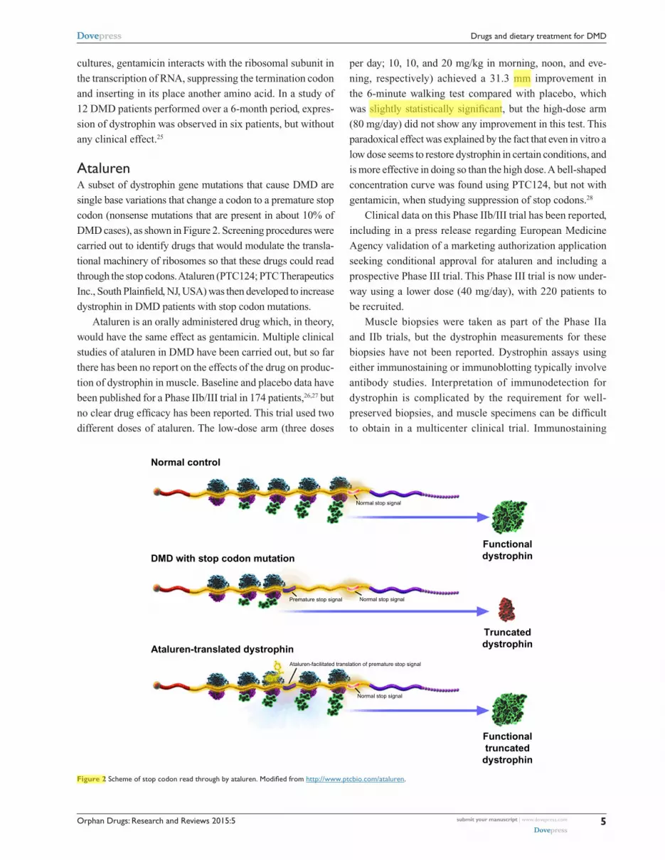
AtalurenA subset of dystrophin gene mutations that cause DMD are
single base variations that change a codon to a premature stop
codon (nonsense mutations that are present in about 10% of
DMD cases), as shown in Figure 2. Screening procedures were
carried out to identify drugs that would modulate the transla-
tional machinery of ribosomes so that these drugs could read
through the stop codons. Ataluren (PTC124; PTC Therapeutics
Inc., South Plainfield, NJ, USA) was then developed to increase
dystrophin in DMD patients with stop codon mutations.
Ataluren is an orally administered drug which, in theory,
would have the same effect as gentamicin. Multiple clinical
studies of ataluren in DMD have been carried out, but so far
there has been no report on the effects of the drug on produc-
tion of dystrophin in muscle. Baseline and placebo data have
been published for a Phase IIb/III trial in 174 patients,26,27 but
no clear drug efficacy has been reported. This trial used two
different doses of ataluren. The low-dose arm (three doses
per day; 10, 10, and 20 mg/kg in morning, noon, and eve-
ning, respectively) achieved a 31.3 mm improvement in
the 6-minute walking test compared with placebo, which
was slightly statistically significant, but the high-dose arm
(80 mg/day) did not show any improvement in this test. This
paradoxical effect was explained by the fact that even in vitro a
low dose seems to restore dystrophin in certain conditions, and
is more effective in doing so than the high dose. A bell-shaped
concentration curve was found using PTC124, but not with
gentamicin, when studying suppression of stop codons.28
Clinical data on this Phase IIb/III trial has been reported,
including in a press release regarding European Medicine
Agency validation of a marketing authorization application
seeking conditional approval for ataluren and including a
prospective Phase III trial. This Phase III trial is now under-
way using a lower dose (40 mg/day), with 220 patients to
be recruited.
Muscle biopsies were taken as part of the Phase IIa
and IIb trials, but the dystrophin measurements for these
biopsies have not been reported. Dystrophin assays using
either immunostaining or immunoblotting typically involve
antibody studies. Interpretation of immunodetection for
dystrophin is complicated by the requirement for well-
preserved biopsies, and muscle specimens can be difficult
to obtain in a multicenter clinical trial. Immunostaining
Normal control
Functionaldystrophin
Normal stop signal
Normal stop signal
Normal stop signal
Ataluren-facilitated translation of premature stop signal
Premature stop signal
Truncateddystrophin
Functionaltruncated
dystrophin
DMD with stop codon mutation
Ataluren-translated dystrophin
Figure 2 Scheme of stop codon read through by ataluren. Modified from http://www.ptcbio.com/ataluren.

Orphan Drugs: Research and Reviews 2015:5submit your manuscript | www.dovepress.com
Dovepress
Dovepress
6
Angelini and Tasca
signals are also challenging to quantify because of the pres-
ence of dystrophin-positive revertant fibers in about 40%
of DMD boys, the numbers of which increase with age.9
Dystrophin immunoblotting is technically challenging for
the large (427 kDa) and relatively low abundance of dys-
trophin protein. Difficulties in measuring dystrophin have
been suggested as a motivation to embark on multicenter
Phase III trials rather than seeking accelerated approval based
on dystrophin expression as a surrogate biomarker. An open
question is whether ataluren has sufficient activity to read
through stop codons and rescue biochemical defects.29–31
DrisapersenAn alternative approach to increasing production of dystro-
phin in muscle is exon skipping.32 This approach involves
anti-sense oligonucleotide (AON) drugs that are able to bind
specifically to dystrophin RNA while it is being transcribed
and processed/spliced (see Figure 3). Exon skipping has
emerged as a promising therapeutic approach for DMD.
The general strategy is to introduce an AON molecule that
pairs with a target sequence in the pre-mRNA for dystrophin.
This pairing causes the nuclear splicing machinery to skip
over the target sequence, thereby excluding one or more
mutation-bearing exons that would prevent production of
the dystrophin protein. The end result is a shorter but still
functional dystrophin molecule.
AON drugs are designed to bind to an exon neighboring
a patient’s deletion mutation, modulating splicing of the
RNA to exclude an additional exon. In this manner, an
out-of-frame mRNA transcript, which is unable to make
dystrophin protein, is converted to an in-frame transcript
capable of producing dystrophin. A shortened dystrophin will
be produced, similar to what happens in dystrophin-positive
revertant fibers or in Becker dystrophin.
The initial target for development for exon skipping
was exon 51, since about 13% of boys with DMD have a
frameshift mutation that can be treated by inducing a skip
of exon 51 (although there is some disagreement about the
actual percentage, with some estimates being as high as
17%). The skipping of exons other than 51 (eg, 44 and 53)
is under consideration.
Two different AON molecules have been developed by
the pharmaceutical industry and taken to clinical trials.
Drisapersen (PRO051, GSK2402968) was developed by
a partnership between Prosensa Therapeutics (Leiden, the
Netherlands) and GSK (Brentford, UK), and was constructed
using 2′-O-methyl phosphorothioate chemistry. This com-
pound has the advantage of extensive clinical experience in
drug programs for other genetic disorders (eg, cystic fibrosis).
Prosensa Therapeutics was the early developer of the exon
skipping approach in DMD. Phase II trials were carried out,
and these included functional tests such as the 6-minute
walking test, as well as investigation of muscle biopsies to
monitor production of dystrophin as the primary endpoint.33,34
The investigators observed that 64%–97% of muscle fibers
expressed the dystrophin protein in amounts ranging from
17% to 35%. While there was evidence of de novo dystrophin,
its levels appeared low and variable, keeping in mind the pres-
ence of dystrophin produced by revertant fibers. However,
the biochemical assay (Western blotting or semiquantitative
immunohistochemistry) used to quantify dystrophin remains
difficult to evaluate.
A subset of patients in Phase II extension trials showed
stabilization of disease progression on drisapersen,34 but
only marginal improvement in the 6-minute walking test,
which was used as a secondary functional outcome measure.
A Phase III trial in 186 patients that used the 6-minute walk-
ing test as the primary outcome measure found no statistically
significant improvement on drisapersen. In fact, in a trial
using 6 mg/kg by subcutaneous injection, there was an
improvement in the 6-minute walking test of 31 meters,35 and
GSK terminated the trial in September 2013. An extension
A
45 46 47 48 51
51
52
52
53
5345 46 47 48
B Eteplirsen
Figure 3 Mechanism of exon 51 skipping in the dystrophin gene.Notes: (A) example of deletion of exon 49–50 that results in an out-of-frame deletion in mRNA and truncated dystrophin protein. (B) example of eteplirsen action consisting in skipping of exon 51: in-frame mRNA transcription is restored, enabling production of a functional dystrophin protein.

Orphan Drugs: Research and Reviews 2015:5 submit your manuscript | www.dovepress.com
Dovepress
Dovepress
7
Drugs and dietary treatment for DMD
study was allowed only recently. The implications of these
results for exon 51 and other exon-skipping drugs are under
discussion.
eteplirsenAnother molecule for exon 51 skipping presently in clinical
trials is a phosphorodiamidate morpholino oligomer (PMO)
compound36,37 developed by Sarepta Therapeutics Inc.
(Cambridge, MA, USA). This AON drug appears to have
the advantage of lower toxicity and a considerably broader
therapeutic window.32,38 However, there is still little clinical
experience with this drug. Preclinical studies in mouse and
dog models have shown dystrophin expression in muscle,
and the extent of dystrophin expression appears to be dose-
dependent.39–42 A single-blind, placebo-controlled study in
humans gave proof-of-concept to the drug.43
Two Phase II studies have been published for an exon
51 morpholino drug known as eteplirsen (AVI-4658). Both
Phase II trials of eteplirsen used de novo production of dys-
trophin in muscle as the primary outcome measure. In the
studies by Cirak et al,44,45 increasing doses of PMO were
injected into cohorts of DMD boys aged 5–15 years; from
the third cohort onwards, while receiving doses of 2 mg/kg
and above, dystrophin-positive fibers increased and 8%–18%
dystrophin expression was seen on Western blotting. Biopsy
samples showed a reduction in cytotoxic T-cells, which
appears consistent with the fact that most patients received
concomitant steroids.2
Mendell et al46 tried two different doses (30 mg/kg/
week and 50 mg/kg/week by endovenous administration) in
12 DMD boys, and was able to meet the primary outcome
and to obtain dystrophin-positive fibers. Moreover, in the
6-minute walking test, while the control group declined,
there was stabilization of walking ability with a difference of
about 67.3 meters. It is clear that 12 children are few, while
Sarepta is pursuing accelerated approval for eteplirsen and
conducted additional studies.
The amount of de novo production of dystrophin in DMD
muscle was variable in the AON studies, but the amount
of expression appeared greater than with the 2′-O-methyl
phosphorothioate compound in the study by Cirak et al.44
Sarepta issued a press release in 2013 concerning a new
drug application for eteplirsen in the treatment of DMD,
providing information on its approach and interactions
with the US Food and Drug Administration. In this press
release, Sarepta stated that it intended to seek accelerated
approval based on results for Phase IIb de novo dystrophin
expression in the muscles of DMD patients, but the US Food
and Drug Administration requested additional information
related to the methodology and verification of dystrophin
quantification. This is the first attempt of a company to seek
accelerated drug development approval from regulatory
authorities based on biomarker data in DMD muscle, such
as measurement of dystrophin.
The results of AON trials are hindered by several main
factors:
• Poor penetrance of AON in tissues. Delivery of AON to
cardiac muscle remains problematic. A recent new AON
drug (tricyclo-DNA oligomers), on systemic delivery in
the Mdx mouse, seems to be able to promote a higher
degree of rescued dystrophin in skeletal muscle, the heart,
and to a less extent, the brain.47
• Most AON drugs have to be delivered either by intra-
muscular or intravenous administration.
• Dystrophin in tissues such as skeletal muscle in the limbs
and diaphragm is a structural protein of the sarcolemma
and ameliorates the disease only when the dystrophin
complex is restored.
eplerenoneDMD boys develop progressive weakness and lose the ability
to walk by 10 years of age, and then develop dilated cardio-
myopathy, a leading cause of death. In this context, Raman
et al48 presented a positive placebo-controlled randomized
trial in 42 DMD boys from three US centers treated with
eplerenone (Pfizer Inc.) or placebo in association with ste-
roids and on a background of ACE inhibitors or angiotensin
receptor blockers. Eplerenone works by blocking the hormone
aldosterone (and the aldosterone receptor), and, because it
lowers the amount of sodium and water retained in the body,
it is used as a diuretic in many cardiac conditions.
Forty-two DMD boys (aged 11–19 years) were admin-
istered eplerenone 25 mg or placebo in addition to other
cardioprotective drugs and were followed for 12 months.
Significantly decreased left ventricular circumferential
strain as a primary outcome was found on cardiac magnetic
resonance imaging.48 A longer follow-up would be necessary
to determine if the combination of eplerenone and other
cardioprotective treatments results in prolonged survival.
Another potential limitation of this trial is the relative small
cohort of patients, but in a disorder like DMD it is difficult
to recruit adequate numbers of patients and perform blinded
cardiac magnetic resonance imaging studies.
Prolonged survival due to improvement in respiratory
clinical care, such as non-invasive positive pressure ventila-
tion, has led to an increased incidence of cardiomyopathy
Orphan Drugs: Research and Reviews 2015:5submit your manuscript | www.dovepress.com
Dovepress
Dovepress
8
Angelini and Tasca
being seen in DMD patients.49 Two patterns of cardiac
involvement are recognized: left ventricular wall motion
abnormality, which is frequent in the early stage of the
disease; and dilated cardiomyopathy,4 which is more frequent
in the second decade of life, such as in the patients recruited
for the study reported by Raman et al.48
In a retrospective analysis, Markham et al50 demonstrated
that steroid-naïve patients (aged 10 years or younger) were
four times more likely to have decreased cardiac function
than patients in the same age group who had received ste-
roids, while patients older than 10 years who had not received
steroids were 15 times more likely to have decreased cardiac
function than patients in the same age group who had taken
steroids. Prophylactic use of cardioprotective drugs is also
worth pursuing. In a small randomized trial in France, early
ACE inhibitor (ie, perindopril) monotherapy in DMD patients
aged 9.5–13.0 years delayed the onset and progression of left
ventricular dysfunction, monitored by decline in ejection
fraction.51
The beneficial effect of a combination of a beta-blocker
and ACE inhibitor was established in 56 wheelchair-bound
patients (mean age 19.5 years), with 5-year and 7-year sur-
vival rates of 93% and 84%, respectively.52
One possible limitation of the study by Raman et al48 is
that their DMD patients were taking several different drugs
in addition to eplerenone or placebo. In fact, duration of ACE
inhibitors, angiotensin receptor blockers, and beta-blocker
assumption was about 1.6 years. Further, most of the patients
included (35/42, 83%) were on steroids (either prednisone 30
mg or deflazacort 25 mg) in addition to eplerenone or placebo
for an average of 5.5 years. It is conceivable that in steroid-
treated cases, an aldosterone inhibitor could be more benefi-
cial due to the secondary mineralocorticoid effect of steroids.
On the other hand, treatment with corticosteroids might
directly influence the development of cardiomyopathy,14,49,50
and various types of steroids, eg, deflazacort or prednisone,
might have different effects on the heart. Of note, a body
weight increase might negatively impact cardiac function, and
a differential weight increase has been found with deflazacort
versus prednisone.11
Eplerenone appears to be a promising drug for use in
DMD, and a future trial could assess the effects of eplerenone
on left ventricular function in younger patients and evaluate
their motor outcomes. It is important to realize that medica-
tions used for adult heart failure do not necessarily benefit the
heart in children. The dystrophic process in heart and muscle
is different in time frame and location; in the heart, necrosis
starts from the posterobasal region of the left ventricle, after
which remodeling occurs, with ventricular dilatation and
reduction of ejection fraction.49
The lack of important side effects suggests that epler-
enone could be used to improve and prolong the life of
patients with DMD. The prognosis for this disease, once
considered to be fatal by 20 years of age, has now shifted,
with many patients surviving up to their fourth decade with
various clinical care combinations. Future trials should
investigate whether treating cardiomyopathy with eplerenone
may benefit not only patients with DMD, but also those with
other muscular dystrophies and severe cardiomyopathy,
such as Becker muscular dystrophy, sarcoglycanopathies,
and laminopathies.
Eplerenone has been used in a pilot study in a 22-year-old
female DMD carrier, in whom muscle edema was assessed
by magnetic resonance imaging. Treatment with eplerenone
in this carrier resulted in a reduction of leg circumferences
attributed to decreased edema, with the leg muscles becoming
stronger with a lower water content and no progression of
dystrophy.53 Although eplerenone has not yet been tested for
its antifibrotic effect in skeletal muscle, the positive results
on cardiac and smooth muscle support a putative beneficial
effect.
SildenafilNitric oxide stimulates soluble guanylate cyclase (such)
to produce cyclic guanosine monophosphate (cGMP),
and in the absence of dystrophin, the nitric oxide-soluble
guanylate cyclase-cGMP pathway is disrupted, impairing the
microcirculation in the heart and muscle. Nucleotide phos-
phodiesterases (PDEs) hydrolyze cGMP and regulate their
downstream signaling. PDE5 expression in cardiomyocytes
is low at baseline and increases in response to ischemia or
pressure overload from heart failure. Impaired blood flow in
muscle and heart tissue in dystrophin-deficient Mdx and nitric
oxide synthase-deficient Mdx mice was rescued by inhibi-
tion of PDE5.54 Nitric oxide inhibitors to treat cardiac and
skeletal muscle are undergoing trials in dystrophinopathies.
Unfortunately, in patients with DMD or Becker muscular
dystrophy, a clinical trial with sildenafil (Pfizer Inc.), a PDE5
inhibitor, did not improve cardiomyopathy, with 30% of
patients progressing to ventricular dilatation.55
Dietary approachAs far as the impact of nutritional status and energy require-
ments in children with DMD is concerned, micronutrients are
needed and vitamin D and calcium should be supplemented,
especially in patients receiving steroids.

Orphan Drugs: Research and Reviews 2015:5 submit your manuscript | www.dovepress.com
Dovepress
Dovepress
9
Drugs and dietary treatment for DMD
Whole grains, vegetables, fruits, legumes, sea vegetables,
saturated fats and essential fats, vitamin E, and coenzyme
Q10 (400 mg/day) may possibly promote bone and general
health. L-carnitine and N-acetyl-cysteine may help DMD
children to maintain their energy.56
A small group of DMD patients were evaluated over
a 6-month period while receiving a standardized diet rich
in fructose, since this sugar seems to be better utilized by
dystrophic muscle.57,58 According to Ellis,57 there is more
incorporation of triglycerides in muscle using glucose-
labeled 14C then fructose, therefore this sugar should not
increase deposition of triglyceride droplets. Carrol et al59
demonstrated that palmitic acid, a component of triglycerides,
is poorly oxidized in DMD muscle. Fourteen DMD patients
with a fructose diet in their natural history were compared
with 132 DMD patients with a normocaloric diet in their natu-
ral history. Patients on the fructose diet had better functional
test results, which were significant for Gowers maneuver
(P,0.001) after 2 months of the diet, and the percent MRC
index of muscle strength also improved.
Another diet trial was conducted using branched-chain
amino acids (a mixture of leucine, valine, and isoleucine;
0.4 g/kg/day) for one year in 18 DMD patients. These
branched-chain amino acids are indeed directed to reducing
degradation of muscle protein. No significant difference in
ability to perform the Gowers maneuver was found between
the branched-chain amino acid group and the placebo group.
The only noticeable adverse event was the difficulty in getting
children to ingest this mixture of amino acids, which can be
unpalatable for children.58
Long-term dietary supplementation of L-arginine (a
nitric oxide synthase substrate) was also not a viable therapy
for dystrophinopathy,60 but administering antioxidants that
attenuate attack by superoxides and restore the bioactive
nitric oxide level might be a useful approach for the treatment
of these disorders.
PerspectivesResearch on muscular dystrophy is characterized by difficulty
in therapeutic efforts, but several drugs are now clinically
available for various stages of the disease, ie, corticosteroids,
cardioprotective drugs, and possibly also eplerenone. Much
attention has been given to molecular therapies, ie, AONs
to skip specific deletion mutations and ataluren to bypass
stop codon mutations.1 Their benefit remains unclear, given
that molecular drugs bring about only a modest increase in
the 6-minute walking test; however, it is also still unknown
if they increase independent walking ability and survival.
These drugs await open validation in a larger cohort of
patients. An appropriate number of patients is required to
evaluate new drugs using the various methodologies proposed.
The number of patients required to study the effect of different
steroid regimens in the ongoing FOR-DMD trial is about 300,
to be recruited using functional outcomes. Further, molecular
therapies are restricted to selected patients, ie, ataluren would
be of benefit to only 13% of DMD patients1 and AONs are
tailored for individual mutations.
Although molecular drugs like AONs and ataluren
are now in development, the available data suggest that
the most useful treatment for DMD is steroids, which are
recommended until patients lose the ability to walk. The side
effect profile of deflazacort appears to be better than that of
other steroids, and should be used in ambulant DMD patients.
For prevention of cardiac insufficiency, a beta-blocker and
the combination of ACE inhibitors and eplerenone has to be
explored. A controlled diet, low in calories and glucose and
rich in fructose and calcium could be useful to control body
weight. The beneficial effects of cardioprotective drugs in
DMD patients need further study using clinical endpoints
such as death or hospitalization for heart failure.
ConclusionIn sum, we have overviewed the possibility of using the above-
mentioned drugs to prolong independent ambulation in DMD
patients of 3–4 years. According to the published standard of
care,61 this approach improves respiratory function, and when
associated with ventilatory night support and cardioprotection,
extends the life expectancy in DMD patients of two decades.
Molecular medicine could be applied to obtain exon skipping
and bypass stop codon mutations in other genetic and neuro-
muscular disorders, leading to benefit in many patients. While
no single approach provides a cure for patients with DMD, all
approaches have the potential to limit the disease.
DisclosureThe authors report no conflicts of interest in this work.
References1. Finkel RS, Flanigan KM, Wong B, et al. Phase 2a study of ataluren-
mediated dystrophin production in patients with nonsense mutation Duchenne muscular dystrophy. PLoS One. 2013;8:e81302.
2. Angelini C. Neuromuscular diseases: advances in therapy and diagnosis. Lancet Neurol. 2012;11:15–17.
3. Bushby K, Finkel R, Birnkrant DJ, et al. Diagnosis and management of Duchenne muscular dystrophy, part 1: diagnosis, and pharmacological and psychosocial management. Lancet Neurol. 2010;9:77–93.
4. Melacini P. Cardiac problems in DMD. In: Angelini C, editor. Muscular Dystrophy: Causes and Management. New York, NY, USA: Nova Science Publishing; 2013.

Orphan Drugs: Research and Reviews 2015:5submit your manuscript | www.dovepress.com
Dovepress
Dovepress
10
Angelini and Tasca
5. Angelini C, Pegoraro E, Turella E, Intino MT, Pini A, Costa C. Deflazacort in Duchenne dystrophy: study of long-term effect. Muscle Nerve. 1994;17:386–391.
6. Mesa L, Dubrovsky AL, Corderi J, Marco P, Flores D. Steroids in Duchenne muscular dystrophy: deflazacort trial. Neuromuscul Disord. 1991;1:261–266.
7. Mendell JR, Moxley RT, Griggs RG, et al. Randomized, double-blind six-month trial of prednisone in Duchenne’s muscular dystrophy. N Engl J Med. 1989;320:1592–1597.
8. Griggs RC, Moxley RT, Mendell JR, et al. Prednisone in Duchenne dystrophy: a randomized, controlled trial defining the time course and dose response. Arch Neurol. 1991;48:383–388.
9. Fanin M, Danieli GA, Cadaldini M, Miorin M, Vitiello L, Angelini C. Dystrophin-positive f ibers in Duchenne dystrophy: origin and correlation to clinical course. Muscle Nerve. 1995;18:1115–1120.
10. Vanhoutte EK, Faber CG, van Nes SI, et al. Modifying the Medical Research Council grading system through Rasch analyses. Brain. 2012;135:1639–1649.
11. Angelini C. The role of corticosteroids in muscular dystrophy: a critical appraisal. Muscle Nerve. 2007;36:424–435.
12. Semplicini C, Angelini C. Clinical scales for the evaluation of neuromus-cular patients. In: Angelini C, editor. Muscular Dystrophy: Causes and Management. New York, NY, USA: Nova Science Publishing; 2013.
13. Bonifati DM, Ruzza G, Bonometto P, et al. A multicenter, double-blind, randomized trial of deflazacort versus prednisone in Duchenne muscular dystrophy. Muscle Nerve. 2000;23:1344–1347.
14. Biggar WD, Harris VA, Eliasoph L, Alman B. Long-term benefits of deflazacort treatment for boys with Duchenne muscular dystrophy in their second decade. Neuromuscul Disord. 2006;16:249–255.
15. Fenichel GM, Mendell JR, Moxley RT, et al. A comparison of daily and alternate-day prednisone therapy in the treatment of Duchenne muscular dystrophy. Arch Neurol. 1991;48:575–579.
16. Backman E, Henriksson KG. Low-dose prednisolone treatment in Duchenne and Becker muscular dystrophy. Neuromuscul Disord. 1995;5:233–241.
17. Loftus J, Allen R, Hesp R, et al. Randomized double-blind trial of deflaza-cort versus prednisone in juvenile chronic (or rheumatoid) arthritis: a rela-tively bone sparing effect of deflazacort. Pediatrics. 1991;88:428–436.
18. Dubowitz V. 75th European Neuromuscular Center International Workshop: second workshop on the treatment of muscular dystrophy, December 10–12, 1999, Naarden, The Netherlands. Neuromuscul Disord. 2000;10:313–320.
19. Dubrovsky AL, Angelini C. Treatment with steroids of DMD. In: Angelini C, editor. Muscular Dystrophy: Causes and Management. New York, NY, USA: Nova Science Publishing; 2013.
20. Manzur AY, Kuntzer T, Pike M, Swan A. Glucocorticoid corticoster-oids for Duchenne muscular dystrophy. Cochrane Database Syst Rev. 2008;1:CD003725.
21. Fenichel G, Pestronk A, Florence J, Robison V, Hemelt V. A beneficial effect of oxandrolone in the treatment of Duchenne muscular dystrophy: a pilot study. Neurology. 1997;48:1225–1226.
22. Fenichel GM, Griggs RC, Kissel J, et al. A randomized efficacy and safety trial of oxandrolone in the treatment of Duchenne dystrophy. Neurology. 2001;56:1075–1079.
23. Consalvi S, Mozzetta C, Bettica P, et al. Preclinical studies in the Mdx mouse model of Duchenne muscular dystrophy with the histone deacetylase inhibitor givinostat. Mol Med. 2013;19:79–87.
24. Politano L, Nigro G, Nigro V, et al. Gentamicin administration in Duchenne patients with premature stop codon. Preliminary results. Acta Myol. 2003;22:15–21.
25. Wagner KR, Hamed S, Hadley DW, et al. Gentamicin treatment of Duchenne and Becker muscular dystrophy due to nonsense mutations. Ann Neurol. 2001;49:706–711.
26. McDonald CM, Henricson EK, Abresch RT, et al. The 6-minute walk test and other endpoints in Duchenne muscular dystrophy: longitudinal natural history observations over 48 weeks from a multicenter study. Muscle Nerve. 2013;48:343–356.
27. McDonald CM, Henricson EK, Abresch RT, et al. The 6-minute walk test and other clinical endpoints in Duchenne muscular dystrophy: reliability, concurrent validity, and minimal clinically important differ-ences from a multicenter study. Muscle Nerve. 2013;48:357–368.
28. Welch EM, Barton ER, Zhuo J, et al. PTC124 targets genetic disorders caused by nonsense mutations. Nature. 2007;447:87–91.
29. Auld DS, Thorne N, Maguire WF, Inglese J. Mechanism of PTC124 activity in cell-based luciferase assays of nonsense codon suppression. Proc Natl Acad Sci U S A. 2009;106:3585–3590.
30. Auld DS, Lovell S, Thorne N, et al. Molecular basis for the high-affinity binding and stabilization of firefly luciferase by PTC124. Proc Natl Acad Sci U S A. 2010;107:4878–4883.
31. McElroy SP, Nomura T, Torrie LS, et al. A lack of premature termina-tion codon read-through efficacy of PTC124 (Ataluren) in a diverse array of reporter assays. PLoS Biol. 2013;11:e1001593.
32. Hoffman EP, Bronson A, Levin AA, et al. Restoring dystrophin expression in Duchenne muscular dystrophy muscle progress in exon skipping and stop codon read through. Am J Pathol. 2011;179:12–22.
33. Van Deutekom JC, Janson AA, Ginjaar IB, et al. Local dystrophin restoration with antisense oligonucleotide PRO051. N Engl J Med. 2007;357:2677–2686.
34. Goemans NM, Tulinius M, van den Akker JT, et al. Systemic administration of PRO051 in Duchenne’s muscular dystrophy. N Engl J Med. 2011;364:1513–1522.
35. Voit T, Topaloglu H, Straub V, et al. Safety and efficacy of drisapersen for the treatment of Duchenne muscular dystrophy (DEMAND II): an exploratory, randomised, placebo-controlled phase 2 study. Lancet Neurol. 2014;13:987–996.
36. Fletcher S, Honeyman K, Fall AM, Harding PL, Johnsen RD, Wilton SD. Dystrophin expression in the Mdx mouse after localised and systemic administration of a morpholino antisense oligonucleotide. J Gene Med. 2006;8:207–216.
37. Alter J, Lou F, Rabinowitz A, et al. Systemic delivery of morpholino oligonucleotide restores dystrophin expression bodywide and improves dystrophic pathology. Nat Med. 2006;12:175–177.
38. Sazani P, Ness KP, Weller DL, Poage D, Nelson K, Shrewsbury AS. Chemical and mechanistic toxicology evaluation of exon skipping phosphorodiamidate morpholino oligomers in Mdx mice. Int J Toxicol. 2011;30:322–333.
39. Malerba A, Thorogood FC, Dickson G, Graham IR. Dosing regimen has a significant impact on the efficiency of morpholino oligomer-induced exon skipping in mdx mice. Hum Gene Ther. 2009;20:955–965.
40. Yokota T, Lu QL, Partridge T, et al. Efficacy of systemic morpholino exon-skipping in Duchenne dystrophy dogs. Ann Neurol. 2009;65:667–676.
41. Yokota T, Nakamura A, Nagata T, et al. Extensive and prolonged restoration of dystrophin expression with vivo-morpholino-mediated multiple exon skipping in dystrophic dogs. Nucleic Acid Ther. 2012;22: 306–315.
42. Wu B, Lu P, Benrashid E, et al. Dose-dependent restoration of dystrophin expression in cardiac muscle of dystrophic mice by systemically delivered morpholino. Gene Ther. 2010;17:132–140.
43. Kinali M, Arechavala-Gomeza V, Feng L, et al. Local restora-tion of dystrophin expression with the morpholino oligomer AVI-4658 in Duchenne muscular dystrophy: a single-blind, placebo- controlled, dose-escalation, proof-of-concept study. Lancet Neurol. 2009;8:918–928.
44. Cirak S, Arechavala-Gomeza V, Guglieri M, et al. Exon skipping and dystrophin restoration in patients with Duchenne muscular dystrophy after systemic phosphorodiamidate morpholino oligomer treatment: an open-label, phase 2, dose-escalation study. Lancet. 2011;378: 595–605.
45. Cirak S, Feng L, Anthony K, et al. Restoration of the dystrophin-associated glycoprotein complex after exon skipping therapy in Duchenne muscular dystrophy. Mol Ther. 2012;20:462–467.
46. Mendell JR, Rodino-Klapac LR, Sahenk Z, et al. Eteplirsen for the treatment of Duchenne muscular dystrophy. Ann Neurol. 2013;74: 637–647.

Orphan Drugs: Research and Reviews
Publish your work in this journal
Submit your manuscript here: http://www.dovepress.com/orphan-drugs-research-and-reviews-journal
Orphan Drugs: Research and Reviews is an international, peer-reviewed, open access journal publishing original research, reports, reviews and commentaries on all areas of the design and development of orphan drugs for the treatment of rare diseases through to clinical applications. Clinical outcomes, patient safety, and programs for the development and
effective, safe, and sustained use of medicines will be a feature of the journal. The manuscript management system is completely online and includes a very quick and fair peer-review system, which is all easy to use. Visit http://www.dovepress.com/testimonials.php to read real quotes from published authors.
Orphan Drugs: Research and Reviews 2015:5 submit your manuscript | www.dovepress.com
Dovepress
Dovepress
Dovepress
11
Drugs and dietary treatment for DMD
47. Goyenvalle A, Griffith G, Babbs A, et al. Functional correction in mouse models of muscular dystrophy using exon-skipping tricycle-DNA oligomers. Nat Med. 2015;21:270–275.
48. Raman SV, Hor KN, Mazur W, et al. Eplerenone for early cardiomyo-pathy in Duchenne muscular dystrophy: a randomised, double-blind, placebo-controlled trial. Lancet Neurol. 2015;14:153–161.
49. Melacini P, Vianello A, Villanova C, et al. Cardiac and respira-tory involvement in advanced stage Duchenne muscular dystrophy. Neuromuscul Disord. 1996;6:367–376.
50. Markham LW, Spicer RL, Khoury PR, Wong BL, Mathews KD, Cripe LH. Steroid therapy and cardiac function in Duchenne muscular dystrophy. Pediatr Cardiol. 2005;26:768–771.
51. Duboc D, Meune C, Lerebours G, Devaux JY, Vaksmann G, Becane HM. Effect of perindopril on the onset and progression of left ventricular dysfunction in Duchenne muscular dystrophy. J Am Coll Cardiol. 2005;45:855–857.
52. Ogata H, Ishikawa Y, Ishikawa Y, Minami R. Beneficial effects of beta-blockers and angiotensin-converting enzyme inhibitors in Duchenne muscular dystrophy. J Cardiol. 2009;53:72–78.
53. Lehman-Horn F, Weber MA, Nagel AM, et al. Rationale for treating oedema in Duchenne muscular dystrophy with eplerenone. Acta Myol. 2012;31:31–39.
54. Percival JM, Whitehead NP, Adams ME, et al. Sildenafil reduces respiratory muscle weakness and fibrosis in the Mdx mouse model of Duchenne muscular dystrophy. J Pathol. 2012;228:77–87.
55. Leung DG, Herzka DA, Thompson WR, et al. Sildenafil does not improve cardiomyopathy in Duchenne/Becker muscular dystrophy. Ann Neurol. 2014;76:541–549.
56. Chen JY, Yen MH, Chen HS, et al. Health support: health promotion for families with a DMD child. In: Angelini C, editor. Muscular Dystrophy: Causes and Management. New York, NY, USA: Nova Science Publishing; 2013.
57. Ellis DA. Intermediary metabolism of muscle in Duchenne muscular dystrophy. Br Med Bull. 1980;36:165–171.
58. Angelini C, Tasca E. Management and clinical trials in Duchenne dystrophy. In: Honorio S, editor. Duchenne Muscular Dystrophy: Symptoms, Management and Prognosis. New York, NY, USA: Nova Science Publishing; 2015.
59. Carrol JE, Norris BJ, Brooke MJ. Defective (U-C14) palmitic oxidation in Duchenne muscular dystrophy. Neurology. 1985;35:96–98.
60. Wehling-Henricks M, Jordan MC, Gotoh T, Grody WW, Roos KP, Tidball JG. Arginine metabolism by macrophages promotes car-diac and muscle fibrosis in mdx muscular dystrophy. PLoS One. 2010;5:e10763.
61. Sejerson T, Bushby K, TREAT-NMD EU Network of Excellence. Standards of care for Duchenne muscular dystrophy: brief TREAT- NMD recommendations. Adv Exp Med Biol. 2009;652:13–21.