dr. c. senthil kumar, m.pharm., ph.d., associated professor
TRANSCRIPT
EVALUVATION OF ANTI-OXIDANT AND HEPATOPROTECTIVE ACTIVITY OF
IXOREA COCCINEA LEAF EXTRACTS BY USING INVITRO AND INVIVO
MODELS
A Dissertation submitted to THE TAMIL NADU Dr. M.G.R. MEDICAL UNIVERSITY,
CHENNAI– 600 032
In partial fulfillment of the requirements for the award of the Degree of
MASTER OF PHARMACY IN
BRANCH –VI – PHARMACOLOGY
Submitted by
Mr. MUHAMMED SHANAVAS V.K
REGISTRATION No.261526153
Under the guidance of
Dr. C. SENTHIL KUMAR, M.Pharm., Ph.D.,
Associated Professor
Department of Pharmacology
DEPARTMENT OF PHARMACOLOGY KARPAGAM COLLEGE OF PHARMACY
COIMBATORE-641 032
MAY – 2017
CERTIFICATES
CERTIFICATE
This is to certify that this dissertation entitled by EVALUVATION
OF ANTI-OXIDANT AND HEPATOPROTECTIVE ACTIVITY OF IXOREA
COCCINEA LEAF EXTRACTS BY USING INVITRO AND INVIVO
MODELSsubmitted by Mr MUHAMMED SHANAVAS V K to The Tamil
Nadu Dr.M.G.R Medical University Chennai in partial fulfillment for the
degree of MASTER OF PHARMACY IN PHARMACOLOGY is a bonafied
work carried out by candidate under the guidance and supervision of
Dr.C.Senthil Kumar Associated professor in the Department of
Pharmacology , Karpagam college of Pharmacy Coimbatore-32
.
I have fully satisfied with his performance and work. I have forwarded
this dissertation work for evaluation.
Station : Dr .S.MOHAN M.Pharm,Ph.D
Date: Principal
CERTIFICATE
This is to certify that this dissertation entitled by EVALUVATION OF
ANTI-OXIDANT AND HEPATOPROTECTIVE ACTIVITY OF IXOREA
COCCINEA LEAF EXTRACTS BY USING INVITRO AND INVIVO
MODELS submitted by Mr MUHAMMED SHANAVAS V K to The Tamil
Nadu Dr.M.G.R Medical University , Chennai in partial fulfillment for the
degree of MASTER OF PHARMACY IN PHARMACOLOGY is a bonafied
work carried out by candidate under my guidance and supervision of
Department of Pharmacology ,Karpagam College of Pharmacy
Coimbatore-32.
I have fully satisfied with his performance and work. I have forward this
dissertation work for evaluation.
Station : Dr.C. SENTHIL KUMAR,MPhram,PhD
Date : Associated Professor
Department of Pharmacology
DECLARATION
I hereby declare that this dissertation EVALUVATION OF ANTI-
OXIDANT AND HEPATOPROTECTIVE ACTIVITY OF IXOREA
COCCINEA LEAF EXTRACTS BY USING INVITRO AND INVIVO
MODELS submitted by me , in partial fulfillment of requirements for the
degree of MASTER OF PHARMACY IN PHARMACOLOGY to The Tamil
Nadu Dr.M.G.R Medical University , Chennai is the result of my original
and independent research work carried out under the guidance of
Dr.C.Senthil Kumar.,M.Pharm Associated Professor , Department of
Pharmacology ,Karpagam College of Pharmacy , Coimbatore -32
Station : MUHAMMED SHANAVAS VK
Date : Reg . No . 261526153
EVALUATION CERTIFICATE
his is to certify that disseration worl entitled EVALUVATION OF ANTI-
OXIDANT AND HEPATOPROTECTIVE ACTIVITY OF IXOREA
COCCINEA LEAF EXTRACTS BY USING INVITRO AND INVIVO
MODELS submitted by Mr.MUHAMMED SHANAVAS VK bearing Reg.
No : 261526153 to the The Tamil Nadu Dr.M.G.R Medical University ,
Chennai in the partial fulfillment for the degree of MASTER OF
PHARMACY IN PHARMACOLOGY is a bonafied work carried out during
the academic year 2016-2017 by the candidate at Department of
Pharmacology , Karpagam College of Pharmacy , Coimbatore and
evaluated by us.
Examination centre :
Date :
Internal Examiner Convenor of Examination
External examiner
DEDICATED TO MY BELOVED PARENTS
SIBLINGS ,TEACHERS ,FRIENDS AND
ALMIGHTY
ACKNOWLEDGEMENT
ACKNOWLEDGEMENT
First of all I would like to thank God for his blessings to do this
research work successfully . With immense pleasure and pride i would like
to take his oppurtunity in expressing my deep sense of gratitude to my
beloved guide Prof. G. Nagaraja Perumal M. Pharm Professor and Head ,
department of Pharmacology , Karpagam College of Pharmacy under
whose active guidance , innovative ideas , Constant inspiration na
encouragement of the work entitled “EVALUVATION OF ANTI-OXIDANT
AND HEPATOPROTECTIVE ACTIVITY OF IXOREA COCCINEA LEAF
EXTRACTS BY USING INVITRO AND INVIVO MODELS” has been
carried out.
I wish to express my deep sense of gratitude to Dr.R.Vasanthakumar ,
Chairman of Karpagam Group of instituitions for the facilities provided me
in this instituition.
My sincere thanks to our respected and beloved Principal Dr.S.Mohan, M
Pharm ,Ph.D, Karpagam College of Pharmacy for his encouragement and
also providing all facilities in this instituition to the fullest possible extent
extent enabling me to complete this work successfully.
It is my pleasure to express my honourable thanks to Mr.NAGARAJA
PERUMAL Professor & Head , Department of Pharmaceutics, helped me
to proceed my work
My whole hearted thanks to to Mr.D. Ranjith kumar , M Pharm,Asst.
Professor,Department of Pharmaceutical Analysis for his kind advice.
I convey my gratitude to Mr.C. Balakumar, M. Pharm ,Asst. Professor
,Department of Pharmaceutical chemistry helped me to proceed useful
ideas.
My sincere thnanks to Mr : Muthukumar , Department of Pharmacology ,
for her indispensible support which enable me to complete this work
successfully.
I am also conveying my thanks to Dr. M. Karpagavalli , M. Pharm, PhD
Associate Professor, Department of Pharmaceutical chemistry, for
encouragement and valuable suggestion during this work.
I take this opportunity with pride and immense pleasure expressing my
deep sense of gratitude to my co gude Dr.Hashim,K.M, Director of U WIN
LIFE SCIENCES, whose innovative ideas,guidance, inspiration,
tremendous encouragement, help and continuous supervision has made
the dissertation a grand and glaring success to complete.
My glorious acknowledgement to Mr.N. Shafi and Mujeeb Lab Assistant of
U WIN LIFE SCIENCES for encouraging us in a kind and generous
manner to complete his work.
I express my sincere thanks to Mr. K. Nahas , Lab assistants of U WIN
LIFE SCIENCES , for his kind support.
I convey my gratitude to Mr. S. Asker , Lab Assistant , Department of
Pathology for his kind support.
I am duly bound to all my non teaching staffs of Karpagam collge of
Pharmacy for their valuable advices and co-operation.
Above all , I am remain indebted to my senior and my class mates
(Anoopa, Bhavan,Shanavas,Amritha,Habeeb,Sijad,Ubaid), to my beloved
parents who inspired and guided me and also for being tha back bone for
all my successfull endeavours in my life.
MUHAMMED SHANAVAS V K
(261526153)
CONTENTS
SL NO.
CONTENT
PAGE NO.
1
INTRODUCTION
1
2
LITERATURE REVIEW
31
3
AIM AND OBJECTIVE
34
4
PLAN OF WORK
35
5
PLANT PROFILE
36
6
MATERIALS AND METHODS
39
7
RESULTS AND DISCUSSION
53
8
SUMMARY AND CONCLUSION
73
9
BIBLIOGRAPHY
75
FIGURE INDEX
SL NO CONTENT P.NO
1 Location of liver in Human Digestive System4 2
2 Liver showing right and left lobes separated by Falciform
Ligament5
4
3 Hepatic blood flow path: source of blood, passing through the
liver and return back to the heart3
7
4 Histology of Hepatic lobule7 11
5 Schematic Representation of Metabolic activation of
Acetaminophen Toxicity51
24
6 Schematic Representation Depicting Role Of Oxidative Stress
In Acetaminophen Toxicity51
25
7 Schematic Representation effect Of Mitochondrial Permeability
Transition In Acetaminophen Toxicity51.
26
8 Mechanism of ethanol induced hepatotoxicity59. 27
9 Ixora coccinea plant 36
10 Superoxide radical reducing activity of ICLE and vitamin C 37
11 DPPH radical reducing activity of ICLE and vitamin C. 59
12 Effect of ICLE on SGPT concentration in paracetamol induced
model
61
13 Effect of ICLE on SGOT concentration in paracetamol induced
model
62
14 Effect of ICLE on ALP concentration in paracetamol induced
model
64
15 Effect of ICLE on Total Protein in paracetamol induced model 66
16 Effect of ICLE on Total Bilurubin in paracetamol induced
model
69
17 Body weight change for Paracetamol induced model 70
18 Histopathology study of ICLE 72
TABLE INDEX
S.NO CONTENT P. NO
1 Types of Hepatotoxic Agents42 22
2 Taxonomical classification of Ixora coccinea 36
3 Vernacular names of Ixora coccinea103 37
4 experimental designs for acute toxicity studies 46
5 Experimental design of paracetamol induced liver toxicity 48
6 Extraction of Ixora coccinea leaf Extract (ICLE) 53
7 Qualitative phyto chemical screening of Ixora coccinea leaf
extract (ICLE)
55
8 Effect of ICLE on superoxide in vitro radical scavenging
activity
56
9 Study of in vitro DPPH Radical Scavenging Activity of ICLE 58
10 Effect of icle on SGPT concentration in paracetamol induced
model
60
11 Effect of ICLE on SGOT concentration in paracetamol
induced model
62
12 Effect of ICLE on ALP concentration in paracetamol induced
model
63
13 Effect of ICLE on Total Proteins in paracetamol induced
model
65
14 Effect of ICLE on Total Bilurubin in paracetamol induced
model
67
15 Effect of ICLE on Body weight for paracetamol induced model 69

Page 1
CHAPTER I
1. INTRODUCTION
1.1.Anatomy of liver
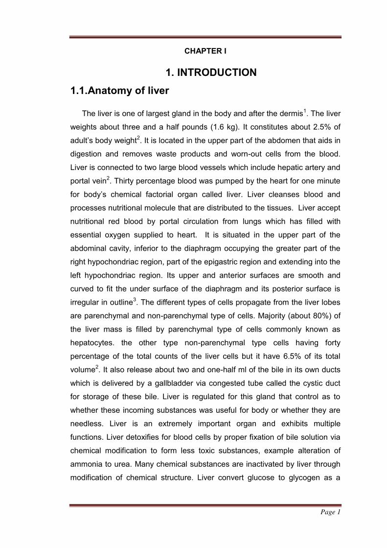
The liver is one of largest gland in the body and after the dermis1. The liver
weights about three and a half pounds (1.6 kg). It constitutes about 2.5% of
adult’s body weight2. It is located in the upper part of the abdomen that aids in
digestion and removes waste products and worn-out cells from the blood.
Liver is connected to two large blood vessels which include hepatic artery and
portal vein2. Thirty percentage blood was pumped by the heart for one minute
for body’s chemical factorial organ called liver. Liver cleanses blood and
processes nutritional molecule that are distributed to the tissues. Liver accept
nutritional red blood by portal circulation from lungs which has filled with
essential oxygen supplied to heart. It is situated in the upper part of the
abdominal cavity, inferior to the diaphragm occupying the greater part of the
right hypochondriac region, part of the epigastric region and extending into the
left hypochondriac region. Its upper and anterior surfaces are smooth and
curved to fit the under surface of the diaphragm and its posterior surface is
irregular in outline3. The different types of cells propagate from the liver lobes
are parenchymal and non-parenchymal type of cells. Majority (about 80%) of
the liver mass is filled by parenchymal type of cells commonly known as
hepatocytes. the other type non-parenchymal type cells having forty
percentage of the total counts of the liver cells but it have 6.5% of its total
volume2. It also release about two and one-half ml of the bile in its own ducts
which is delivered by a gallbladder via congested tube called the cystic duct
for storage of these bile. Liver is regulated for this gland that control as to
whether these incoming substances was useful for body or whether they are
needless. Liver is an extremely important organ and exhibits multiple
functions. Liver detoxifies for blood cells by proper fixation of bile solution via
chemical modification to form less toxic substances, example alteration of
ammonia to urea. Many chemical substances are inactivated by liver through
modification of chemical structure. Liver convert glucose to glycogen as a
Page 2
storage form of energy and it produces glucose from disaccharides and
polysaccharides such as sugars, starches and protein molecules
Figure. No: 1: Location of liver in Human Digestive System4
Page 3
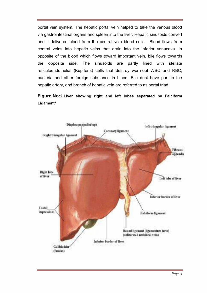
The liver is situated below the diaphragm which occupy right side of hypochondriac
and region in the abdominopelvic cavity. The liver is completely covered by a
dense irregular connective tissue layer that lies deep to the peritoneum. It is
divided into two principal lobes-a large right lobe and a smaller, wedge-
shaped, left lobe separated by the falciform ligament. Right lobe is described
by many anatomists to have an inferior quadrate lobes and a posterior
caudate type lobes. The falciform ligament extends from undersurface of the
diaphragm from the upper surface of the liver part, between two significant
lobes of the liver, helped to suspend the liver. Liver is composed of several
components:
Hepatocytes are the functional cells of the liver which are arranged in pairs of
columns radiating from a central vein. A wide range of metabolic, secretory
and endocrine functions are performed by hepatocytes. These are specialized
epithelial cells with 5-12 sides that make-up about 80% of the volume of the
liver. Hepatocytes form complex three dimensional arrangements called
hepatic laminae and they are the sheet of hepatocytes one cell thick lined to
either side by the endothelial-lining spaces called hepatic sinusoids. Grooves
inside the cell surface between neighboring hepatic cells which provide gaps
for the canaliculi of hepatocytes that secrete bile. Bile is a yellow, brown, or
olive-green colur type liquid which secreted from hepatic cells, which provide
an excretory product and a digestive enzyme secretor.
Bile canaliculi are narrow intercellular canals that collect bile secreted by
hepatocytes. From bile canaliculi, bile passes into small bile ducts. These
small ducts combined by form the higher right and left hepatic void that
commonly connect and exit the liver via common hepatic duct. This common
hepatic duct joins the cystic duct from the gallbladder to form the common bile
duct. Bile enters the cystic duct and temporarily stored in the gallbladder. After
a meal, various stimuli cause contraction of the gallbladder, which releases
stored bile into the common bile duct.
Hepatic sinusoids are freely permeable capillaries about sheets of liver cells
that get oxygenated blood via different branches of hepatic artery and higher
amount of nutrient rich de-oxygenated blood from the branches of the hepatic
Page 4
portal vein system. The hepatic portal vein helped to take the venous blood
via gastrointestinal organs and spleen into the liver. Hepatic sinusoids convert
and it delivered blood from the central vein blood cells. Blood flows from
central veins into hepatic veins that drain into the inferior venacava. In
opposite of the blood which flows toward important vein, bile flows towards
the opposite side. The sinusoids are partly lined with stellate
reticuloendothelial (Kupffer’s) cells that destroy worn-out WBC and RBC,
bacteria and other foreign substance in blood. Bile duct have part in the
hepatic artery, and branch of hepatic vein are referred to as portal triad.
Figure.No:2:Liver showing right and left lobes separated by Falciform
Ligament5
Page 5
The hepatocytes, bile duct contents and hepatic sinusoids are organized into
an anatomical and functional units by three different types:
Hepatic lobule: For many years, anatomy fellow are suggest that the
hepatic lobule are liver’s functional units. They describes the each hepatic
lobule are shaped as that of a hexagonal. At its center is central vein
emerging out by they form a rows of hepatocytes and hepatic sinusoidal cells.
This model is suggested by detailed autopsy of the liver of the adult pigs.
From the study of human liver have not predicted the hepatic lobules anatomy
having a thick layers of connective tissues type.
Portal lobule: This model emphasized the endocrine function of the liver,
i.e., bile release. According to the bile ducts in a portal triad is consider as the
middle of the portal lobules. These portal lobule’s are triangular shaped and is
having a 3 imaginary straight lines which connect three central veins which
are near by the portal triad. These model which are not gained wide spread
acceptance.
Hepatic acinus: In past years, the accepted structural and functional unit of
the liver known as the hepatic acinus. These are approximately oval shape
mass which includes portions of two neighboring hepatic lobules. Small axis
of hepatic acinus is described by the branches of the portal triad- branches
contain a hepatic artery, hepatic vein and bile ducts. Long axis of the acinus
inter connected closest to short axis. Hepatocytes in the hepatic acinus are
arranged in three zones from its short axis, with no sharp among them. Cells
inside the zone 1 are closer to branches of portal triad and these cells are first
to accept oxygen, essential food material and toxins from the receiving blood
cells. These cells are gain-up glucose and save it by converting glycogen after
food ingestion, so during fasting period get the energy via glycogen to
glucose. These are first shows the characteristic morphological changes.
Zone 1 cells are last ones to die when circulation is impaired and the first
ones to regenerate. Cells in zone three are much farther from branches of
portal triad and are the last to show the effects of bile blockage or presence to
Page 6
toxins, the first ones effect is the abnormal circulation, retarded o rate of
regeneration and evidence of fat accumulation. The cells in zone 2 show
structural and functional characteristics intermediate between cells in zone 1
to 3.
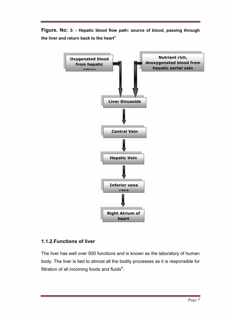
1.1.1.Blood supply of liver
The liver receives blood from two sources, hepatic artery and hepatic portal
vein. From hepatic artery it obtains oxygenated blood and from hepatic portal
vein it receives deoxygenated blood that contains newly absorbs essential
nutrients, therapeutic molecules, and possibly nonpathogenic microorganism
and may receive toxins from the gastrointestinal tract. Branches of both
hepatic artery and portal vein carry blood into liver sinusoids where
hepatocytes extracts oxygen most of the nutrients and certain poisons.
Nutrients needed by other cells and products manufactured by the
hepatocytes are secreted back into the blood After blood passed inside
central vein and eventually passes via hepatic vein. Branches of hepatic
portal vein, artery and bile duct typically accompany each other in their
distribution through liver. Collectively these three structures are called portal
triad1.
Page 7
Figure. No: 3: - Hepatic blood flow path: source of blood, passing through
the liver and return back to the heart3
1.1.2.Functions of liver
The liver has well over 500 functions and is known as the laboratory of human
body. The liver is tied to almost all the bodily processes as it is responsible for
filtration of all incoming foods and fluids6.
Nutrient rich,
deoxygenated blood from
hepatic portal vein
Oxygenated blood
from hepatic
artery
Liver Sinusoids
Central Vein
Hepatic Vein
Inferior vena
cava
Right Atrium of
heart
Page 8
1.1.2.1. Carbohydrate metabolism
Liver is important in maintaining normal blood glucose level. Liver can
break down glycogen to glucose (glycogenolysis) and release glucose into the
bloodstream, when blood glucose level decreases. Liver helped the
conversion of certain amino acid by glucose through lactic acid
(gluconeogenesis) and convert other sugar molecules such as fructose and
galactose reduced to glucose. Liver converts glucose to glycogen
(glycogenesis) and also undergoes the covertion of triglycerides (lipogenisis).
1.1.2.2. Lipid metabolism
Liver stores some triglycerides from fatty acids through acetyl
coenzyme A known as beta oxidation. It possibly converts excess acetyl
coenzyme A to ketone bodies (ketogenesis). It synthetize lipoproteins, that
transportation of fatty acids, triglycerides (TG) and cholesterol from the body
cells. Cholesterol is synthesized by hepatocytes and cholesterol involves the
formation of bile salts.
1.1.2.3. Protein metabolism
Most of the plasma proteins, such as α and β globulins, glycol proteins
(albumin and fibrinogen) are synthesized from liver cells. Also, liver enzymes
can perform transamination. Liver deaminates amino acids so that they are
used in the ATP synthesis or conversion from carbohydrates or fats. It
converts resulting toxic ammonia into much less toxic urea for excretion in
urine.
1.1.2.4. Removal of drugs and hormones
Liver can detoxify substances such as alcohol or excrete drugs like penicillin,
erythromycin, and so on into bile. It is also trigger or chemically alter thyroid
hormones and steroid hormones (estrogens and aldosterone).
Page 9
1.1.2.5. Excretion of bilirubin
Bilirubin released from heme of red blood cells is absorbed in the liver through
the blood and release to bile. Most of bilirubin in bile is metabolized in the
intestine by bacteria and eliminated in feces.
1.1.2.6. Synthesis of bile salts
These are helped in small intestine functioning as an emulsification process
and absorption of lipid molecules, cholesterol, phospholipids, and lipoproteins.
1.1.2.7. Storage
In addition to glycogen, liver stores water soluble and fat-soluble vitamins (A,
B12, D, E, and K) and essential minerals (iron and copper). Hepatocytes
contain a protein called apoferritin that combines with iron to form ferritin, the
form in which iron is stored in liver. The iron is secreated from the liver is
essential requirement of the body.
1.1.2.8. Phagocytosis
The stellate reticuloendothelial (Kupffer’s) cells of the liver phagocytize worn-
out red and white blood cells and some type of the bacteria.
1.1.2.9. Activation of vitamin D
The cutaneous layer of skin, liver and kidneys essential for activation
vitaminD1.
1.1.2.10. Secretion and excretion of Bile
Bile is an incomplete synthetic substance and partially act as digestive
secretion. Each day hepatic cells secrete 800-1000ml of bile. It has a PH of
7.6-8.6. Bile mainly consist water, bile salts, cholesterol and phospholipid
known as lecithin, bile pigments and several ions. Principle bile pigment is
bilirubin6.
Page 10
1.1.2.11. Synthesis of vitamin A from carotene
Carotene is the pro-vitamin found in some plants for example carrots and
green leaves of vegetables.
1.1.2.12. Production of heat
Liver able to create amount of energy which has a high metabolic rate and
produces considerable amount of heat. It is an important heat producing
organ of body3.
1.1.3.Pathology of liver7
All forms of injury to the liver such as microbiologic, toxic, circulatory or
traumatic result in necrosis in liver. The extent of involvement of hepatic
lobules necrosis varies. Accordingly, liver cell necrosis are divided into 3
types: diffuse (submissive to massive), zonal and focal.
1.1.3.1.Diffuse (Submassive to massive).
When there is extensive and diffuse necrosis of the liver involving all the cells
in groups of lobules, it is termed diffuse, or submissive to massive necrosis. It
is most commonly caused by viral hepatitis or drug toxicity.
1.1.3. 2. Zonal necrosis.
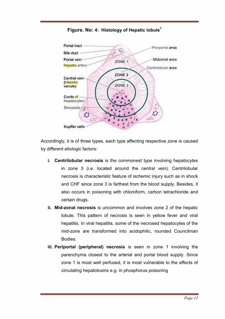
Zonal necrosis is necrosis of hepatocytes in three different zones of the
hepatic lobule as shown in the figure below.
Page 11
Figure. No: 4: Histology of Hepatic lobule7
Accordingly, it is of three types, each type affecting respective zone is caused
by different etiologic factors:
i. Centrilobular necrosis is the commonest type involving hepatocytes
in zone 3 (i.e. located around the central vein). Centrilobular
necrosis is characteristic feature of ischemic injury such as in shock
and CHF since zone 3 is farthest from the blood supply. Besides, it
also occurs in poisoning with chloroform, carbon tetrachloride and
certain drugs.
ii. Mid-zonal necrosis is uncommon and involves zone 2 of the hepatic
lobule. This pattern of necrosis is seen in yellow fever and viral
hepatitis. In viral hepatitis, some of the necrosed hepatocytes of the
mid-zone are transformed into acidophilic, rounded Councilman
Bodies.
iii. Periportal (peripheral) necrosis is seen in zone 1 involving the
parenchyma closest to the arterial and portal blood supply. Since
zone 1 is most well perfused, it is most vulnerable to the effects of
circulating hepatotoxins e.g. in phosphorus poisoning
Page 12
1.1.3.3. Focal Necrosis
This form of necrosis involves small groups of hepatocytes irregularly
distributed in the hepatic lobule. Focal necrosis is most often caused by
microbiological infections. These include viral hepatitis, military tuberculosis,
typhoid fever and various other forms of bacteria, viral and fungal infections.
Focal necrosis may also occur in drug-induced hepatitis.
1.1.4.Enzymes involved with liver
1.1.4.1.Alanine transaminase
Alanine transaminase or ALT is a transaminase, serum glutamic–
pyruvic transaminase (SGPT or also known as alanine aminotransferase
(ALAT)) commonly observed in many tissues and body fluids principally in
liver. ALT is released into serum as a result of tissue injury8
i.Function
ALT catalyzes the reversible transfer of an amino groups in the L-alanine
enzyme to α-ketoglutarate proteins forms such as pyruvate and L-glutamate.
L-Glutamate + Pyruvate ↔ α-Ketoglutarate + L-
Alanine
ii.Clinical Significance
It is commonly estimated clinically as a parameter of diagnostic
evaluation of hepatocellular injury in order to determine liver health. ALT has
actually measured by international units/liter (IU/L) 9&10 when used in
diagnosis. 10-40 IU/L are the standard reported range of experimental
studies11.
iii.Elevated levels
Significantly abnormal range of Alanine transaminase (ALT) often
suggest the abnormality of conditions including viral hepatitis, diabetes
mellitus induced cell necrosis, heart failure, liver injury, infectious
Page 13
mononucleosis, bile duct problems and myopathy. Because of these reason,
ALT is one of the important parameter used for screening of liver diseases.
Dietary choline deficiency shows marked elevation in ALT levels. These
enzyme variation levels of ALT do not have significance of that medical
problem is present. Fluctuation of ALT level is normal during course of day
and ALT levels can increase in response to strenuous physical excercise11.
When elevated ALT levels are found in blood concentration subsequently
narrowed down by measuring other enzyme concentration (example liver- cell
damage usually distinguished from biliary duct problems by measuring
increased ALP). Myopathy-related ALT levels can be found out by measuring
the creatine kinase enzymes. Several drugs elevate ALT levels, for example,
Zileuton. For years, American Red Cross society used for ALT testing as part
of the key enzyme of tests to ensure the safety of its blood pumping by
deferring donors with elevated ALT levels. Main reason was to specify donors
have an infection with Hepatitis C because there is a no specific test available
for these12.
1.1.4.2.Aspartate transaminase
Aspartate Transaminase (AST) also called Aspartate Aminotransferase
(ASAT/AAT/AspAT) or Serum Glutamic Oxaloacetic Transaminase (SGOT), is
a transaminase enzyme containing pyridoxal phosphate. AST catalyses
reversible transfer of α-amino group between aspartate and glutamate .it is a
key enzyme required for amino acid metabolism in human. It commonly
present in liver, heart, skeletal muscle, kidneys, brain and red blood cells and
AST is commonly measured clinically as a marker for liver health. It is also
associated with liver parenchymal cell metabolism. The ratio of AST/ALT is
may be useful for differentiation between etiology of liver damage 13, 14.
Reference range is 6-40IU/L15.
i.Function
AST catalyzes the interconversion of aspartate and α-ketoglutarate to
oxaloacetate and glutamate.
Page 14
Aspartate (Asp) + α- Ketoglutarate ↔ Oxaloacetate + Glutamate (Glu)
As prototypical transaminase AST relies on PLP as a cofactor to transfer
amino group from aspartate or glutamate to corresponding ketoacid16.
iii.Isoenzymes
Two isoenzymes are present in wide variety of eukaryotes. In humans,
i. GOT1 / c AST, the cytosolic iso enzyme synthesized mainly from red
blood cells and heart.
ii. GOT2 / m AST, the mitochondrial isoenzyme present predominantly in
liver.
These isoenzymes are considered to be evolved from a common
gene duplication and subsequent synthesi17.
iv.Clinical significance:
It is raised in liver inflammation. It is also elevated in diseases such as
myocardial infaraction, acute pancreatitis, nephrotoxicity, hemotoxicity,
musculoskeletal diseases and trauma. AST was used initially biochemical
marker for diagnosis of acute myocardial infarction but now redundant and
has been superseded by the cardiac troponins18. AST is commonly measured
clinically as a part of diagnostic liver function test inorder to determine liver
health.
Reference range-
Male 8-40IU/L
Female 6-34IU/L
1.1.4.3.Alkaline phosphatase
Alkaline phosphatase (ALP) have functioning towords removing phosphate
group containing molecules such as nucleotides, proteins, and alkaloids.
Process of removing phosphate group is called de-phosphorylation. By the
maintaince of effective and also alkaline environment. It is sometimes used
Page 15
similarly as basic phosphatase19. ALP is mainly present in cells lining of the
biliary ducts.
i.Elevated levels
ALP levels in plasma increase due to large bile duct constriction,
intrahepatic cholestasis. ALP is found also in bone and placental tissue and
hence higher in growing children and elderly patients haveing Paget’s
disease. In third trimester of pregnancy ALP is two to three times higher.
ii.Reference range: 30-120 IU/L20.Liver (ALP): Cholestasis, cholecystitis,
cholangitis, cirrhosis, hepatitis, fatty liver, sarcoidosis, liver tumor, liver
metastases, drug intoxication21. Placental ALP is elevated in seminomas22
and active form of rickets as well as in following diseases23.
Biliary construction.
Bone conditions.
Osteoblastic bone cancer.
Osteomalacia.
Liver disorder/ hepatitis.
Leukemia.
Lymphoma.
Paget’s disease.
Sarcoidosis.
Hyperparathyroidism.
iii.Lowered levels
Following diseases may lead to decreased levels of alkaline phosphatase-
Hypophosphatasia (autosomal recessive disease).
Postmenopausal women undertaking estrogen therapy due to
osteoporosis.
Hypothyroidism or severe anemia.
Children affected with achondroplasia and cretinism.
Children who are victims of severe episode of enteritis.
Page 16
Pernicious and aplastic anemia.
myelogenous leukemia.
Wilson’s disease.
Apart from these, the following chemicals are clinically studied to reduce
alkaline phosphatase: Oral contraceptives24.
1.1.4.4.Total protein
Total protein includes total amount of two classes of proteins present in fluid
portion of blood. These include albumin and globulin. Total protein tests
measures amount of albumin and globulin which are major groups of protein
in blood. A low total protein level due to liver disorder, kidney disorder or
disorder which protein is not digested or absorbed properly25.
i.Normal Range: 6.0 - 8.3gm/dl
ii.Higher -than –normal levels may be due to:
Chronic inflammation or infection (HIV, Hepatitis B or C).
Bone marrow disorders (Multiple myeloma, Waldenstroms disease).
iii.Lower-than-normal levels may be due to:
Bleeding (Hemorrhage).
Burns (extensive).
Liver disease.
Glomerulonephritis and nephritic syndrome.
Malabsorption.
Malnutrition26.
1.1.4.5.Total bilirubin
Total bilirubin (TBIL) test checks levels of bilirubin in blood. Bilirubin
(orange-yellow pigment) is the waste product of normal break down of red
blood cells. Bilirubin passes through liver and eventually passes out of body
as feces and small amount in urine. Before reaching liver bilirubin is called
Page 17
unconjugated. Inside liver it combines with certain sugars to create water
soluble form called conjugated bilirubin27.
i.Normal Range: 0.3 -1.9mg/dl
ii.Clinical significance
Total bilirubin is usually measured to screen for or to monitor liver or
gallbladder diseases. Presence of high amount of bilirubin in body leads to
jaundice.
iii.Higher Levels:
Drug toxicity.
Liver diseases such as hepatitis.
Biliary stricture.
Cancer of gallbladder or pancreas.
Gallstones28.
Erythroblastosis fetalis.
Physiological jaundice.
Sickle cell anemia29.
HIV Infection.
Bacterial infection inside blood.
1.1.5.Hepatotoxicity
Liver diseases are the major medical problems faced by the people all
over the world30. About 20,000 deaths occur every year due to liver
disorders31. In Africa and in Asia, the main causes of liver diseases are
viruses and parasitic infections, whereas in Europe and in North America, a
major cause is alcohol abuse30. Liver diseases are mainly caused by toxic
chemicals, excessive intake of alcohol, infections and autoimmune
disorders32. Hepatotoxicity due to drug appears to be a common contributing
factor. Liver is expected not only to carryout physiological functions but also
to protect against the hazardous of harmful drugs and chemicals33. Drug
induced chemical injury is responsible for 5% of all hospital admissions and

Page 18
50% of all acute liver failures. More than 75% of cases of immunological
reaction of drugs leading to liver transplantation or death34.
Hepatotoxicity mainly implies chemical-driven liver damage. Certain
drugs when taken in overdose and sometimes even when administered within
therapeutic ranges may injure many organs. Some chemical agents including
those that are used in laboratories (Ccl4, paracetamol) and industries (Lead,
arsenic) and natural chemicals (microcystine, aflatoxins) and herbal remedies
(cascara sagrada, ephedra) can also cause hepatotoxicity. Chemicals which
cause liver injury are collectively known as hepatotoxins34.
NSAIDS35 (Acetaminophen36, Aspirin, Ibuprofen)
Glucocorticoids.
Anti-Tubercular drug (Isoniazid) 37.
Industrial toxins (arsenic, carbon tetrachloride, vinyl chloride).
Herbal remedies (Ackee fruit, camphor, cycasin, kava leaves, valerian,
comfrey) 38.
1.1.5.1.Pathophysiological mechanisms
Pathophysiological mechanisms of hepatotoxicity are still being
identified and which include both hepatocellular and extracellular effects.
Following are some mechanisms:
Disruption of hepatocyte: Drugs can bind to intracellular proteins by
covalent binding which result in a lower in ATP levels subsequent disruption.
Splitting of actin these fibrils at the surface of the hepatocyte causes rupture
of the membrane of liver.
Disruption of transport protein: Bile flow may be interrupted by drugs that
affect transport proteins at canalicular membrane. Loss of villous due to the
interruption of transport pumps leading to multidrug resistance- associated
protein 3 prevent excretion of bilirubin resulting in cholestasis.
Cytolytic T-cell activation: Covalent binding of drug to P-450 enzyme acts
as an immunogenic activation of T-cells and cytokines leading to immune
reaction.
Page 19
Apoptosis of hepatocytes: Stimulation of these pathway may leading
for programmed necrosis of hepatocytes.
Mitochondrial disruption: Some drugs inhibit mitochondrial function
by dual effect on both beta- oxidation energy productions by inhibiting
the release of the e dinucleotide, subsequently reduce the ATP
production.
Bile duct injury: free radicle produced metabolites excreted in the bile
may leading to necrosis of bile duct epithelium.
1.1.5.2.Drug toxicity mechanisms
Classic division of drug reactions is of at least 2 major groups which include:
(1) Drugs which directly affect liver.
(2) Drugs which mediate an immune response.
Intrinsic / predictable drug reactions: molecules that fall into this
drug category lead to reproducible injuries in mammals and injury is
related to dose. Injury can be due to drug itself or to metabolite.
Acetaminophen is a suitable example of well-known predictable
hepatotoxin at higher therapeutic doses. Another example is carbon
tetrachloride.
Idiosyncratic / unpredictable drug reactions: These drug reactions
can be subdivided into those that are classified as hypersensitivity or
immunoallergic and those that are metabolic-idiosyncratic. It occurs
without obvious dose-dependency and in an unpredictable fashion.39
1.1.5.3.Symptoms
List of signs and symptoms depicted in various causes for Hepatotoxicity
include 15 symptoms as listed below:
Nausea
Vomiting
Abdominal pain
Loss of appetite
Page 20
Diarrhea
Tiredness
Weakness
Jaundice
Yellow eyes
Yellow skin
Enlarged liver
Abnormal liver function test results
Swelling in feet
Weight gain due to water retention
Prolonged bleeding time.
1.1.5.4. Treatment
These are various sources for hepatotoxicity treatment mode these
selected by consultation by physician about the treatment or change in
treatment regimen. Treatment of hepatotoxicity has dependent upon
causative agent, degree of liver dysfunction and age and general health of
patient. Treatments for hepatotoxicity include:
Withdrawal of causative medication or removal from exposure to
causative agent.
Regular monitoring of patient and review of liver function – where liver
dysfunction is mild to moderate and liver function is improving.
Complete avoidance of alcohol and medication that may contribute to
further liver damage.
N-Acetylcysteine is used for paracetamol toxicity.
Management of symptoms of liver damage.
Nutrition – with vitamin supplementation as required
Regular exercise inorder to maintain muscle mass.
Ursodeoxycholic acid.
Page 21
Management of pruritus
Cholestyramine
Antihistamines.
Management of ascites
Low sodium diet.
Diuretics – furosemide, spironolactone.
Removal of fluid via a needle in the abdomen –
Paracentesis.
Portosytemic shunting.
Management of portal hypertension
Beta - blockers
Oesophageal variceal banding
Portocaval shunt
Management of acute liver failure due to hepatotoxicity
Supportive care always in intensive care unit – airway
protection, fluid and electrolyte management.
Management of complications such as bleeding
problems and hepatic encephalopathy.
Liver transplantation – for acute fulminant liver failure or end stage
cirrhosis.
Page 22
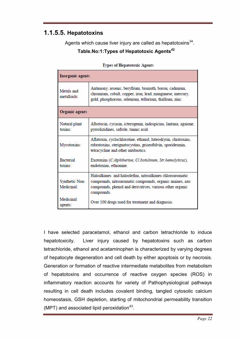
1.1.5.5. Hepatotoxins
Agents which cause liver injury are called as hepatotoxins34.
Table.No:1:Types of Hepatotoxic Agents42
I have selected paracetamol, ethanol and carbon tetrachloride to induce
hepatotoxicity. Liver injury caused by hepatotoxins such as carbon
tetrachloride, ethanol and acetaminophen is characterized by varying degrees
of hepatocyte degeneration and cell death by either apoptosis or by necrosis.
Generation or formation of reactive intermediate metabolites from metabolism
of hepatotoxins and occurrence of reactive oxygen species (ROS) in
inflammatory reaction accounts for variety of Pathophysiological pathways
resulting in cell death includes covalent binding, tangled cytosolic calcium
homeostasis, GSH depletion, starting of mitochondrial permeability transition
(MPT) and associated lipid peroxidation43.
Page 23
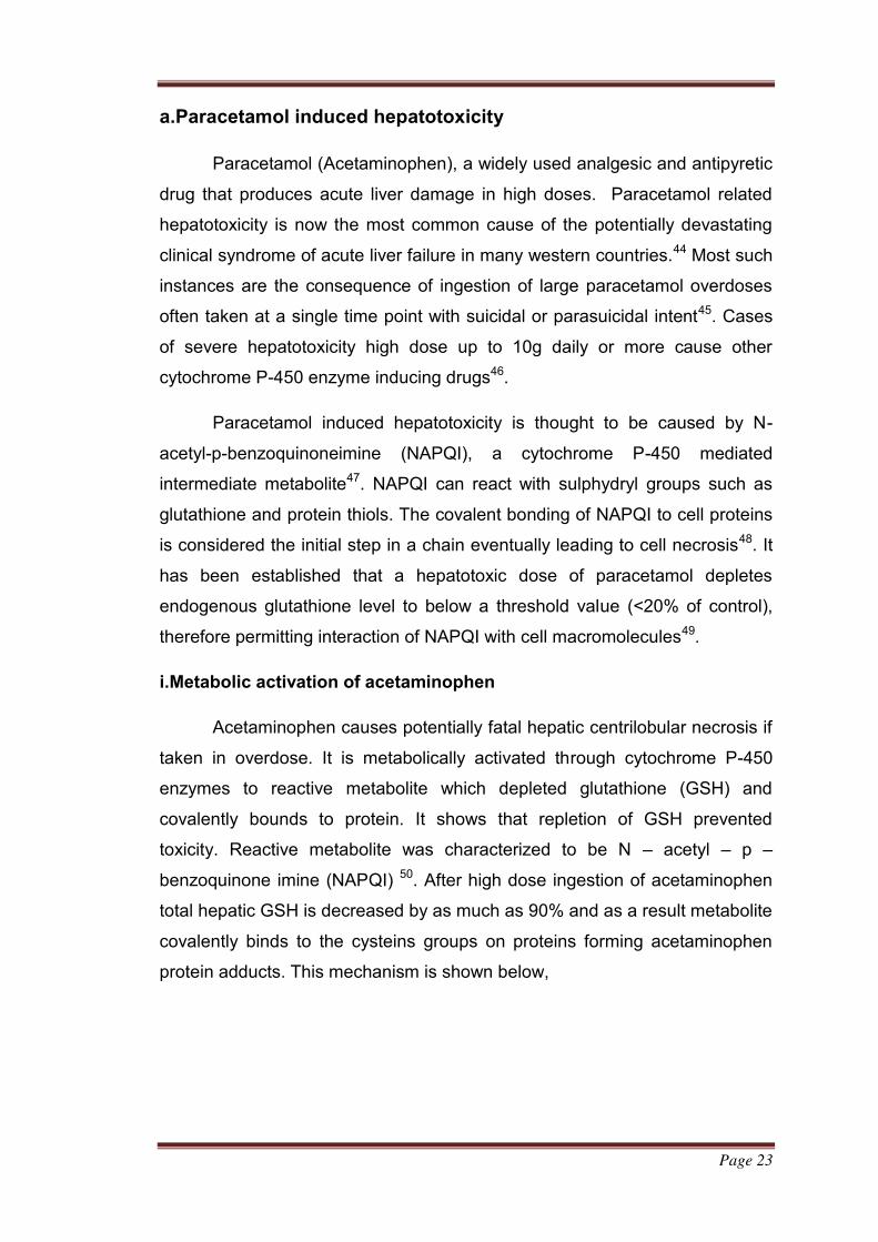
a.Paracetamol induced hepatotoxicity
Paracetamol (Acetaminophen), a widely used analgesic and antipyretic
drug that produces acute liver damage in high doses. Paracetamol related
hepatotoxicity is now the most common cause of the potentially devastating
clinical syndrome of acute liver failure in many western countries.44 Most such
instances are the consequence of ingestion of large paracetamol overdoses
often taken at a single time point with suicidal or parasuicidal intent45. Cases
of severe hepatotoxicity high dose up to 10g daily or more cause other
cytochrome P-450 enzyme inducing drugs46.
Paracetamol induced hepatotoxicity is thought to be caused by N-
acetyl-p-benzoquinoneimine (NAPQI), a cytochrome P-450 mediated
intermediate metabolite47. NAPQI can react with sulphydryl groups such as
glutathione and protein thiols. The covalent bonding of NAPQI to cell proteins
is considered the initial step in a chain eventually leading to cell necrosis48. It
has been established that a hepatotoxic dose of paracetamol depletes
endogenous glutathione level to below a threshold value (<20% of control),
therefore permitting interaction of NAPQI with cell macromolecules49.
i.Metabolic activation of acetaminophen
Acetaminophen causes potentially fatal hepatic centrilobular necrosis if
taken in overdose. It is metabolically activated through cytochrome P-450
enzymes to reactive metabolite which depleted glutathione (GSH) and
covalently bounds to protein. It shows that repletion of GSH prevented
toxicity. Reactive metabolite was characterized to be N – acetyl – p –
benzoquinone imine (NAPQI) 50. After high dose ingestion of acetaminophen
total hepatic GSH is decreased by as much as 90% and as a result metabolite
covalently binds to the cysteins groups on proteins forming acetaminophen
protein adducts. This mechanism is shown below,
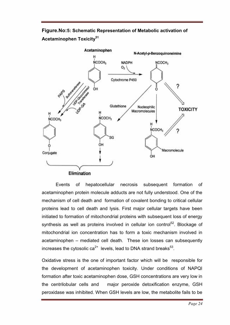
Page 24
Figure.No:5: Schematic Representation of Metabolic activation of
Acetaminophen Toxicity51
Events of hepatocellular necrosis subsequent formation of
acetaminophen protein molecule adducts are not fully understood. One of the
mechanism of cell death and formation of covalent bonding to critical cellular
proteins lead to cell death and lysis. First major cellular targets have been
initiated to formation of mitochondrial proteins with subsequent loss of energy
synthesis as well as proteins involved in cellular ion control52. Blockage of
mitochondrial ion concentration has to form a toxic mechanism involved in
acetaminophen – mediated cell death. These ion losses can subsequently
increases the cytosolic ca2+ levels, lead to DNA strand breaks53.
Oxidative stress is the one of important factor which will be responsible for
the development of acetaminophen toxicity. Under conditions of NAPQI
formation after toxic acetaminophen dose, GSH concentrations are very low in
the centrilobular cells and major peroxide detoxification enzyme, GSH
peroxidase was inhibited. When GSH levels are low, the metabolite fails to be
Page 25
detoxified by conjugation; it accumulates and causes liver injury54. Lipid
peroxidation resulting from oxidative stress contributes to the initiation and
recently reported data suggest that acetaminophen hepatotoxicity is mediated
by an initial metabolic oxidation, covalent bonding and subsequent activation
of macrophages to form reactive oxygen and nitrogen species55. Protection
against oxidation is provided by glutathione and by system of soluble and
enzymatic defenses.
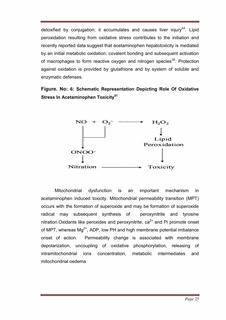
Figure. No: 6: Schematic Representation Depicting Role Of Oxidative
Stress In Acetaminophen Toxicity51
Mitochondrial dysfunction is an important mechanism in
acetaminophen induced toxicity. Mitochondrial permeability transition (MPT)
occurs with the formation of superoxide and may be formation of superoxide
radical may subsequent synthesis of peroxynitrite and tyrosine
nitration.Oxidants like peroxides and peroxynitrite, ca2+ and Pi promote onset
of MPT, whereas Mg2+, ADP, low PH and high membrane potential imbalance
onset of action. Permeability change is associated with membrane
depolarization, uncoupling of oxidative phosphorylation, releasing of
intramitochondrial ions concentration, metabolic intermediates and
mitochondrial oedema
Page 26
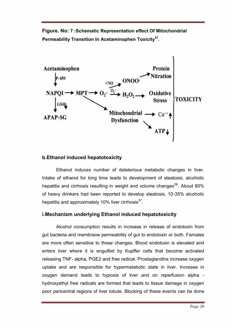
Figure. No: 7 :Schematic Representation effect Of Mitochondrial
Permeability Transition In Acetaminophen Toxicity51.
b.Ethanol induced hepatotoxicity
Ethanol induces number of deleterious metabolic changes in liver.
Intake of ethanol for long time leads to development of steatosis, alcoholic
hepatitis and cirrhosis resulting in weight and volume changes56. About 80%
of heavy drinkers had been reported to develop steatosis, 10-35% alcoholic
hepatitis and approximately 10% liver cirrhosis57.
i.Mechanism underlying Ethanol induced hepatotoxicity
Alcohol consumption results in increase in release of endotoxin from
gut bacteria and membrane permeability of gut to endotoxin or both. Females
are more often sensitive to these changes. Blood endotoxin is elevated and
enters liver where it is engulfed by Kupffer cells that become activated
releasing TNF- alpha, PGE2 and free radical. Prostaglandins increase oxygen
uptake and are responsible for hypermetabolic state in liver. Increase in
oxygen demand leads to hypoxia of liver and on reperfusion alpha -
hydroxyethyl free radicals are formed that leads to tissue damage in oxygen
poor pericentral regions of liver lobule. Blocking of these events can be done
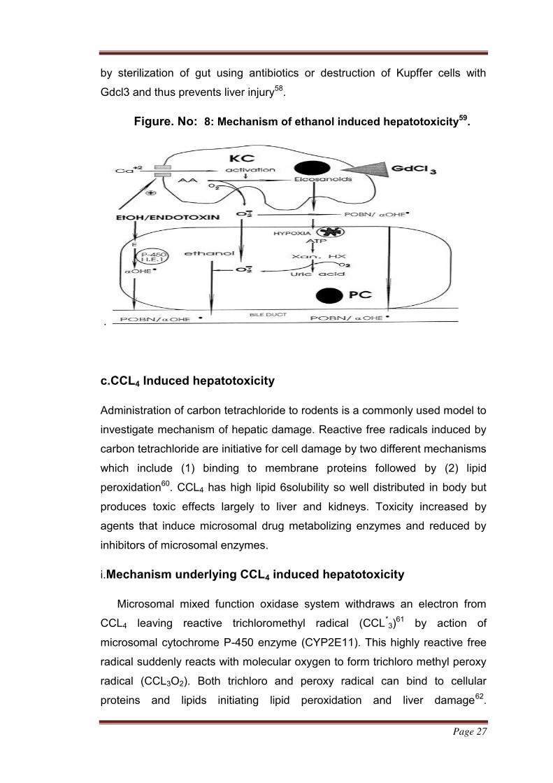
Page 27
by sterilization of gut using antibiotics or destruction of Kupffer cells with
Gdcl3 and thus prevents liver injury58.
Figure. No: 8: Mechanism of ethanol induced hepatotoxicity59.
.
c.CCL4 Induced hepatotoxicity
Administration of carbon tetrachloride to rodents is a commonly used model to
investigate mechanism of hepatic damage. Reactive free radicals induced by
carbon tetrachloride are initiative for cell damage by two different mechanisms
which include (1) binding to membrane proteins followed by (2) lipid
peroxidation60. CCL4 has high lipid 6solubility so well distributed in body but
produces toxic effects largely to liver and kidneys. Toxicity increased by
agents that induce microsomal drug metabolizing enzymes and reduced by
inhibitors of microsomal enzymes.
i.Mechanism underlying CCL4 induced hepatotoxicity
Microsomal mixed function oxidase system withdraws an electron from
CCL4 leaving reactive trichloromethyl radical (CCL*3)
61 by action of
microsomal cytochrome P-450 enzyme (CYP2E11). This highly reactive free
radical suddenly reacts with molecular oxygen to form trichloro methyl peroxy
radical (CCL3O2). Both trichloro and peroxy radical can bind to cellular
proteins and lipids initiating lipid peroxidation and liver damage62.
Page 28
Trichloromethyl free radical has life time of only about 100 microseconds and
so has time to diffuse for only short distance within liver cell before
undergoing secondary reactions. Secondary reactions are responsible for
biochemical damage may be of different kinds which include:
a) Oxidation of thiols to form disulphide bonds.
b) Saturation of double bonds in lipids, nucleotides, proteins which results
in covalent attachment of free radical group of those sites.
c) Lipid peroxidation reaction where polyunsaturated membrane lipids are
converted to peroxide derivative and finally to aldehydes and other
products leading to further cascade of reaction resulting in irreversible
membrane damage.
Prolonged administration of ccl4 leads to cirrhosis and hepatic
carcinoma61.
1.1.6. Modern medicines for treatment of liver diseases
Liver diseases can be treated using allopathic as well as by using herbal
drugs.
1.1.6.1. Hepatoprotective allopathic treatment
Few modern medicines are available for treating liver diseases that includes:
1) Ursodeoxycholic acid (Ursodiol)
Ursodiol decreases intestinal absorption and suppresses hepatic
synthesis and storage of cholesterol. It is mainly used in management
of chronic hepatic diseases in humans.
2) Penicillamine
Penicillamine chelates several metals like copper, iron, lead and
mercury forming stable water soluble complexes which are renally
excreted.
Other drugs:
Antiviral medication such as alpha interferon, ribavirin, steroids, antibiotics
etc. are also used in liver diseases63.Drugs like tricholinecitrate,
Page 29
trithioparamethoxy phenyl propane, essential phospholipids, combination of
drugs such as L-ornithine, L-aspartate and pancreatin, silymarin and
Ursodeoxycholic acid are usually prescribed for hepatitis, cirrhosis and other
liver diseases64. N-acetylcysteine is used in early phases of acetaminophen
toxicity. L-carnitine is potentially valuable during valproate toxicity.
Cholestyramine can be used to alleviate pruritus39.
i.Disadvantages of allopathic drugs
Side effects of many modern medicines are mostly alarming. Interactions,
contra-interactions, side effects and toxicity of synthetic medicine vary from
mild to severe that includes insomnia, vomiting, fatigue, dry mouth, diarrhea,
constipation, dizziness, suicidal thought, depression, seizures, anemia, hair
loss, high blood sugar, swelling, impotency, confusion, fainting and finally
death65.Antibiotics usually cause stomach upset or allergic reactions.
Interferon shows side effects as flu-like illness with fever and body aches63.
1.1.6.2.Herbal hepatoprotective drug treatment
A number of polyherbal preparations have been used in treating various liver
disorders since ages. Some herbal formulations include:
a) Liv-52: It is a non-toxic hepatoprotective drug from Himalaya Drug Co.
Liv-52 can improve clinical parameters in patients having liver damage
mainly in alcoholic liver damage.
b) LIMARIN®: It has potent hepatoprotective and free radical scavenging
(antioxidant) activity. It is derived from active extract of fruit of silybum
marianum63.
Some of the polyherbal formulations have been verified for
hepatoprotective activity against chemical driven liver damage in
experimental animals which include Liv52, Liv42, Jigrine, Koflet66,
Cirrhitin, Livex and Hepatomed etc.
1.1.6.2.1.Limitations of herbal preparations
Herbal- based preparations for treating liver disorders has been in use
in India for long time and has been popularized worldwide by leading
Page 30
pharmaceuticals. Despite of popularity of herbal medicines for liver diseases
in particular, are still unacceptable treatment modalities for liver diseases.
Limiting factors include:
Lack of standardization procedures of herbal preparations.
Lack of identification of active components and principles.
Lack of randomized controlled clinical trials (RCTs).
Lack of toxicological evaluation67.
Poor solubility.
Poor bioavailability.
Poor hepatic cell regeneration.
i..Hepatoprotective activity of silymarin
Mechanism of action of Silybin is complex and highly beneficial in protecting
hepatocytes. It blocks penetration of various toxins into hepatocytes and thus
prevents cell death. It protects liver from oxidative intracellular free radicals by
increasing activity of enzyme superoxide dismutase and peroxidase as well as
by increasing concentration of glutathione and activity of peroxidase. Silybin
strengthens and stabilizes cell membranes, inhibits synthesis of
prostaglandins associated with lipid peroxidation and promotes regeneration
of liver through stimulation of protein synthesis and thus effects on production
of new hepatocytes88.Silybin acts in four different ways:
Antioxidant, scavenger and regulator of intracellular content of
glutathione.
Cell membrane stabilizer and permeability regulator that prevent
hepatotoxic agents from entering hepatocytes.
Promoters of ribosomal RNA production, stimulating liver
regulation.
Inhibitors of transformation of stellate hepatocytes into
myofibroblasts- process which is responsible for deposition of collagen
fibers leading to cirrhosis8
Page 31
CHAPTER II
2.LITERATURE REVIEW
Afiwa Missebukpo,et,al (2010) 93 was investigated the hydro-alcoholic
extract of Ixora coccinea (ICE) exhibit the anti-asthmatic activity in an
ovalbumin (OVA) induced asthmatic rat model. These facts led us to examine
their antioxidant activities. The free radical 1,1-diphenyl-2-picrylhydrazyl
(DPPH) scavenging activity and the intracellularly antioxidant activity of ICE
were determined. The protective effect of ICE against 2,2′ azobis (2-
amidinopropane) hydrochloride (AAPH)-induced red blood cell lysis was also
evaluated. It was found that ICE could scavenge DPPH with an IC50 of 283.3
μg/ml and protected red blood cell against AAPH-induced hemolysis with an
IC50 of 72.92 versus 52.08 μg/ml for ascorbic acid. Erythrocytes obtained
from the ICE-administrated rats showed an enhanced resistance to
hemolysis. In OVA-induced asthma, rats were sensitized and challenged with
ovalbumin. The effect of ICE at 1500 mg/kg per os on malondialdehyde
(MDA) production and lung catalase activity were determined. ICE
significantly reduced the lipid peroxidation and enhanced catalase activity in
lung (p < 0.05). In conclusion, the hydro-alcoholic extract of I. coccinea
possesses an antioxidant activity and protective effect against free-radical-
induced hemolysis
Prabu,et,al(2010) 94 reported the anti-diarrhoeal activity of aqueous extract
of the leaves of Ixora coccinea against a castor oil induced diarrhoea model in
rats. The gastrointestinal transit rate was expressed as the percentage of the
longest distance which was traversed by the charcoal, divided by the total
length of the small intestine. The weight and the volume of the intestinal
content induced by castor oil were studied by the enteropooling method.
Loperamide was used as a positive control. The plant-extract showed
significant (P<0.001) inhibitor activity against castor oil induced diarrhoea and
castor oil induced enteropooling in rats at the dose of 400 mg/kg. There was
significant reduction in gastrointestinal motility by the charcoal meal test in
rats.
Page 32
Moni Rani,et,al (2008) 95 reported the anti-oxidant activity of the methanol
extract of Ixora coccinea L by DPPH free radical scavenging assay, reducing
power and total antioxidant capacity using phosphor molybdenum method.
Preliminary phytochemical screening revealed that the extract of the leaf of
Ixora coccinea possesses flavonoids, steroids and tannin materials. The
methanolic extract showed significant activities in all antioxidant assays
compared to the standard antioxidant in a dose dependent manner and
remarkable activities to scavenge reactive oxygen species (ROS) may be
attributed to the high amount of hydrophilic phenolics. In DPPH radical
scavenging assay the IC50 value of the extract was found to be 100.53 μg/mL
while ascorbic acid had the IC50 value 58.92 μg/mL. Thus Ixora coccinea
extract showed strong reducing power and total antioxidant capacity.
Latha,et,al (2010)96 reported the hepatoprotective activity in ethanolic
extracts of three different plants Ixora coccinea (IC), Rhinacanthus nasuta
(RN), Spilanthes ciliata (SC) on the aflatoxin B1 (AFB1) –intoxicated livers of
albino male Wistar rats. Biochemical parameters, including serum hepatic
enzymes (glutamate oxaloacetate transaminase, glutamate pyruvate
transaminase and alkaline phosphatase), were studied. Pre-treatment of the
rats with oral administration of these plant ethanolic extracts, prior to AFB1
was found to provide significant protection against toxin induced liver damage,
determined 72 hours after the AFB1 challenge (1.5 mg/kg, intraperitoneally)
was evidenced by a significant lowering of the activity of the serum enzymes
and enhanced hepatic reduced GSH status. Pathological examination of the
liver tissues supported the biochemical findings. The three plant extracts, IC,
RN and SC, showed significant anti-lipid peroxidant effects in vitro.
Nagaraj,et,al (2011) 97 reported thesynthesis of gold nanoparticles in
aqueous medium using leaf extracts of Ixora coccinea as reducing and
stabilizing agent. On treating chloroauric acid solution with extract, rapid
reduction of chloroaurate ions is observed leading to the formation of the
highly stable gold nanoparticles in solution. The synthesized nanoparticles are
confirmed by colour changes and it has been characterized by UV-visible
pectroscopy. Presence of this strong broad plasmon peak has been well
documented for variousMe- NPs, with sizes ranging all the way from 2 to 100
Page 33
nm. The morphology and size of the biologically synthesized gold
nanoparticles were determined using TEM. The images clearly showed that
the average size of the nanotriangles is about 200 nm, while, the spherical
like particles show very small size about 5-10 nm. This study also showed that
gold nanoparticles with antibiotic show more inhibitory zones than compared
to the standard antibiotics.
Panikar,et,al(1998)98 reported theantitumour activity of Ixora coccinea
L. (Rubiaceae) leafs was studied in comparison to intraperitoneally
transplanted Dalton's lymphoma (ascitic and solid tumours) and Ehrlich
ascites carcinoma (EAC) tumours in mice. Intraperitoneal administration of
200 mg/kg of the active fraction (AF) of the I. coccinea leaf increased the life-
span of DLA and EAC ascitic tumour-bearing mice by 113 and 68%,
respectively. The AF showed less activity against solid tumours (DLA) as
compared to ascitic tumours. The same active fraction showed 50%
cytotoxicity to DLA, EAC and Sarcoma-180 (S- 180) cells in vitro at
concentrations of 18, 60 and 25 μg/ml, respectively. It was not toxic to normal
lymphocytes, whereas it was toxic to transformed lymphocytes from
leukaemic patients, acute lymphoblastic leukaemia (ALL) and chronic
myelogenous leukaemia (CML) and K-562 suspension cell cultures. The AF
inhibited tritiated thymidine incorporation in cellular DNA. Thus the Thus the
anti-tumor activity of Ixora coccinea plant was proved.
Yasmeen,et,al(2011)99 reported the hypoglycaemic and the
hypolipidaemic activity of the aqueous extract of the leaves of Ixora Coccinea
Linn in alloxan induced diabetic albino rats. The aqueous extract of leaves of
Ixora Coccinea showed significant reduction (p<0.01) in the blood glucose
levels and the serum lipid profile levels, with 400 mg/kg of body weight in the
alloxan induced diabetic rats as compared to the controls.
Elumalai,et,al(2012)100 was studied the phytochemical and ethano
pharmacological profile of Ixora coccinea and he reported it have anti-
oxidant,anti-inflamatory and antidiabetic activity.
Page 34
CHAPTER III
3. AIM AND OBJECTIVES
Plants that cure liver diseases so considerable interest has developed
in the examination of these numerous plants remedies which are useful in
liver diseases. So it is necessary to find new drugs of importance in hepato
protective activity with fewer side effects. Moreover it is necessary to produce
scientific validation to drugs of herbal origin in common use under Ayurvedic
Siddha Unani systems of medicine. Why I have to select this particular
disease is few effective drugs available for modern therapy, it produce side
effect during the treatment is worse than the condition of liver damage
Phytochemical investigation will be a useful tool for the identification
and authentication of the plant for industrial and further research purpose.
Total phenol content of a tested material is related to the antioxidant activity.
Antioxidants, which can scavenge free radicals, have an important role in
pharmacological systems. Antioxidants are emerging as prophylactic and
therapeutic agents. Hence, antioxidant was also evaluated for the potent
extract.
And now I have undertaken the study of evaluation of anti-oxidant and
hepatoprotective activity of ixorea coccinea leaf extracts by various
hepatotoxin induced albino rat models
To select plant based on their ethno medical uses and preparation
of their extracts.
To screen phytochemical profile.
To screen the selected extract for antioxidant using various in vitro
methods
To screen the potent plant extract for their in vivo hepatoprotective
activities
Page 35
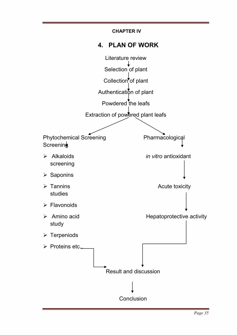
CHAPTER IV
4. PLAN OF WORK
Literature review
Selection of plant
Collection of plant
Authentication of plant
Powdered the leafs
Extraction of powered plant leafs
Phytochemical Screening Pharmacological
Screening
Alkaloids in vitro antioxidant
screening
Saponins
Tannins Acute toxicity
studies
Flavonoids
Amino acid Hepatoprotective activity
study
Terpeniods
Proteins etc.,
Result and discussion
Conclusion
Page 36
CHAPTER V
5.PLANT PROFILE
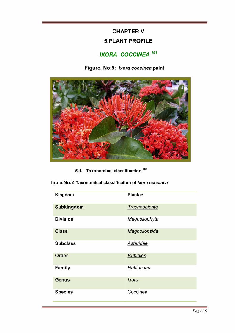
IXORA COCCINEA 101
Figure. No:9: ixora coccinea palnt
5.1. Taxonomical classification 102
Table.No:2:Taxonomical classification of Ixora coccinea
Kingdom Plantae
Subkingdom Tracheobionta
Division Magnoliophyta
Class Magnoliopsida
Subclass Asteridae
Order Rubiales
Family Rubiaceae
Genus Ixora
Species Coccinea
Page 37
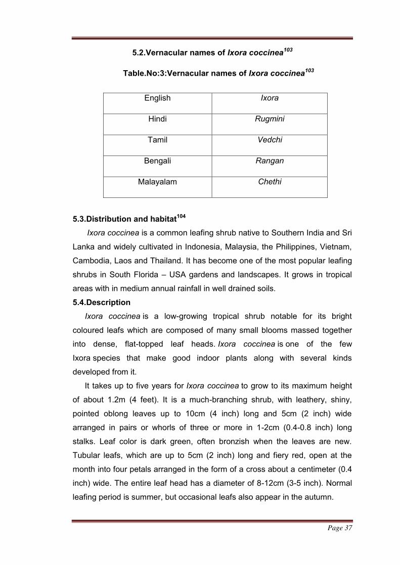
5.2.Vernacular names of Ixora coccinea103
Table.No:3:Vernacular names of Ixora coccinea103
5.3.Distribution and habitat104
Ixora coccinea is a common leafing shrub native to Southern India and Sri
Lanka and widely cultivated in Indonesia, Malaysia, the Philippines, Vietnam,
Cambodia, Laos and Thailand. It has become one of the most popular leafing
shrubs in South Florida – USA gardens and landscapes. It grows in tropical
areas with in medium annual rainfall in well drained soils.
5.4.Description
Ixora coccinea is a low-growing tropical shrub notable for its bright
coloured leafs which are composed of many small blooms massed together
into dense, flat-topped leaf heads. Ixora coccinea is one of the few
Ixora species that make good indoor plants along with several kinds
developed from it.
It takes up to five years for Ixora coccinea to grow to its maximum height
of about 1.2m (4 feet). It is a much-branching shrub, with leathery, shiny,
pointed oblong leaves up to 10cm (4 inch) long and 5cm (2 inch) wide
arranged in pairs or whorls of three or more in 1-2cm (0.4-0.8 inch) long
stalks. Leaf color is dark green, often bronzish when the leaves are new.
Tubular leafs, which are up to 5cm (2 inch) long and fiery red, open at the
month into four petals arranged in the form of a cross about a centimeter (0.4
inch) wide. The entire leaf head has a diameter of 8-12cm (3-5 inch). Normal
leafing period is summer, but occasional leafs also appear in the autumn.
English Ixora
Hindi Rugmini
Tamil Vedchi
Bengali Rangan
Malayalam Chethi
Page 38
Various kind of Ixora which have Ixora coccinea as a parent produce
differently coloured blooms, chiefly in shade of orange, yellow and pink, as
well as red.
5.5.Propagation
Propagate Ixora coccinea from stem cuttings 5-8cm (2-3 inch) long taken
in spring. Trim each cutting immediately below a leaf, remove that leaf and dip
the cut end in hormone rooting powder. Plant the cutting in a 5-8cm (2-3 inch)
pot containing a moistened equal-parts mixture of peat moss and coarse sand
or perlite. Enclose the whole in a plastic bag or propagating case and stand it
in bright filtered light at a temperature of 21-27°C (70-81°F). When the cutting
has rooted – probably in four to six weeks – uncover it gradually over a two or
three week’s period in order to acclimatize the new plant to the less humid
atmosphere of the room. When the new plant is fully uncovered, begin to
water moderately (allowing a couple of centimeters (0.4-0.8 inch) or so of the
potting mixture to dry out between watering again) and apply standard liquid
fertilizer once every two weeks. About three months after the start of the
propagation move the new plant into a slightly bigger pot of the recommended
potting mixture for adult plants and treat it as mature.
5.6.Uses105
It is primarily ornamental plant. Cut specimens are long lasting and are often
used in floral arrangements. It is sometimes used as a hedge. In tropical Asia
the leafs, bark and leaves of this plant are used in traditional medicine.
Page 39
CHAPTER VI
6. MATERIALS AND METHODS
6.1.Collection and authentication of plant
Ixora coccinea leaf was procured from the Botany Central council for
Research in Ayurvedia and Siddha Govt of India.(Certificate
No:UWAU/350/16) The dried leafs are authenticated by Chelladurai.V
research officer Botany Central council for Research in Ayurved and Siddha
Govt of India.
6.2.Extraction Procedure:
i.Preparation of Ixora coccinea leaf extract106
The leaf were initially separated from the main plants body and rinsed
with distilled water and shade dried and then homogenized into fine powder
and stored in air tight bottles. A total of 10 g of leaf air dried powder was
weighed and was placed in 100 mL of organic solvents (methanol and
ethanol) in a conical flask and then kept in a rotary shaker at 190-220 rpm for
24 h. And then it was filtered with the help of muslin cloth and centrifuged at
10 000 rpm for 5 min. The supernatant was collected and the solvent was
evaporated by solvent distillation apparatus to make the final volume of one-
fourth of the original volume, giving a concentration of 40 mg/mL. It was
stored at 40 °C in air tight bottles for further studies.
6.3.Phyto chemical screening107-108
The plant may be containing the following compound such as
carbohydrate, protein, and lipids. That is utilized as food by man. It also
contains the compound like. Tannins, glycosides, alkaloids. Volatiles oils. The
compound that is responsible for lots of medicinal properties
Page 40
6.3.1.Test for carbohydrates
6.3.1.1..Molisch test
The sample powdered was added with 1 ml of alpha napthol
solution along with conc Sulphuric acid solution in the test tube reddish
colour was produced at the junction between 2 liquid
6.3.1.2..Fehling test.
To the sample powder was added with both Fehling A and
Fehling B solution and placed in the water bath for a sufficient time.
This shows the brick red colour.
6.3.1.3..Benedicts test.
To the sample powder add 8 drops of benedict’s reagents and
Boil the sample vigorously for 5 min it shows the red ppt.
6.3.2.Test for alkaloids
To the small of stored powder (sample) was taken and add few drops
of hydrochloric acid and filtered. The filtered was tested with various
alkaloid agents.
6.3.2.1.Mayer’s reagents
To a small of above filter add small quantity of Mayer’s reagent
to form cream precipitate.
6.3.2.2..Dragendorff’s reagents
From the above filter add small amount of Dragendorffs
reagents it forms a orange brown precipitate.
6.3.3.Test for flavonoids
To the filter of the plant extract add 5 ml of dilute ammonia
solution and followed by the addition of concentrated sulphuric acid. It
forms a yellow colour.
Page 41
6.3.4.Test for steroids.
6.3.4.1.Salkowski test
Few amount of plant extract was mixed with chloroform and the
same volume of sulphuric acid is added on it. Cherry red colour was
obtained in the chloroform layer.
6.3.4.2.Libbbermann burchatd test:
The extract is dissolved in 2 ml of chloroform 10 drops of acetic
acid and conc. Sulphuric acid were added. Now the solution becomes
reddish colour then it turns to bluish green colour.
6.3.5.Test for tannins.
From few amount of plant extract is treated with vanillin hydrochloric
acid reagent. It forms, pink or red colour due to the formation of
phloroglucinol,
6.3.6.Test for protein.
6.3.6.1..Millon’s reagents.
Millon’s reagents (mercuric nitrate in nitric acid containing a
trace of nitrous acid) usually yields a white precipitate on addition to a
protein solution which turns red on heating.
6.3.6.2.Ninhydrin Test.
From the sample solution add 2 drops a freshly prepared
0.2% ninhydrine reagent was added to the extract and heating.
Development of blue colour may indicate the presence of peptide,
amino acid (PROTEIN).
6.3.7.Test for glycosides
6.3.7.1.Keller- killani test.
From the small quantity of small powder acetic acid was
dissolved and adds few drops of ferric chloride and transferred to the
Page 42
surface of conc Sulphuric acid. At the junction, reddish brown colour
was formed,
6.3.8.Test for saponins.
6.3.8.1.Foam test
1 ml of extract solution is diluted separately with distilled water to 20 ml
and shaken in a graduated cylinder for 15 minutes. A 1 cm layer of foam
indicates the presence of Saponins.
6.3.9.Test for Terpenoids:
About 0.5 g of plant extract in separate test tube was taken with 2 ml of
chloroform; 5 ml of concentrated sulphuric acid was carefully added to form a
layer and observed for presence of reddish brown color interface to show
positive results for the presence of terpenoid.
6.4. In vitro antioxidant activities
6.4.1. Superoxide radical scavenging activity109
Principle:
The superoxide anion radical scavenging activity was
determined by nitro blue tetrazolium (NBT) reduction method of Mc Cord and
Fridovich (1969). The assay is based on the ability of drug to inhibit the
reduction of nitro blue tetrazolium (NBT) by Superoxide, which is generated
by the reaction of photo reduction of riboflavin within the system. The
superoxide radical thus generated reduce the NBT to a blue colored complex.
i.Reagents
Nitro blue tetrazolium (NBT) - 1.5nm (12.3mg/10ml)
Riboflavin - 0.12µm (4.5mg/100ml)
NaCN/EDTA - 0.0015% NaCN in 0.1M EDTA
Phosphate buffer - 0.06M ( pH 7.8 )
Page 43
ii.Procedure:
The reaction mixture contained EDTA (0.1 M), 0.3mM NaCN, Riboflavin
(0.12mM), NBT (1.5 n moles), Phosphate buffer (67mM, pH 7.8) and various
concentrations of the seed oil extract in a final volume of 3ml. The tubes were
illuminated under incandescent lamp for 15min. The optical density at 560 nm
was measured before and after illumination. The inhibition of superoxide
radical generation was determined by comparing the absorbance values of
the control with that of seed oil extract and fraction-IV. Vitamin C was used as
positive control. The concentration of fraction-IV required to scavenge 50%
superoxide anion (IC50 value) was then calculated.
iii.calculation
% inhibition = × 100
6.4.2. DPPH radical reducing activity 110:
i.Principle
It is a rapid and simple method to measure antioxidant capacity. It
involves the use of free radical, DPPH (2, 2- Diphenyl - 1- picryl hydrazyl)
(Aquino et al, 2001). The odd electron in the DPPH free radical gives a strong
absorption maximum at 517nm and is purple in color. The color turns from
purple to yellow when the odd electron of DPPH radical becomes paired with
hydrogen from a free radical scavenging antioxidant to form the reduced
DPPH-H. The resulting decolourisation is stochiometric with respect to the
number of electrons captured.
i.reagent
DPPH - 3mg in 25ml methanol (stored in dark bottle)
Methanol
ii.Procedure
Freshly prepared DPPH (187 µl) was taken in different test tubes
protected from sunlight. To this solution added different concentrations (0, 25,
Page 44
50, 75,100,150,200µg/ml) of seed oil extract and fraction-IV. The volume was
made up to 1ml with methanol. Keep the tubes in dark and after 20 min
absorbance was measured at 515nm. Methanol was used as blank and
vitamin C was used as positive control. The concentration of test materials to
scavenge 50% DPPH radical (IC50 value) was calculated from the graph
plotted with % inhibition against Concentration.
iii.Calculation
% inhibition = × 100
Page 45
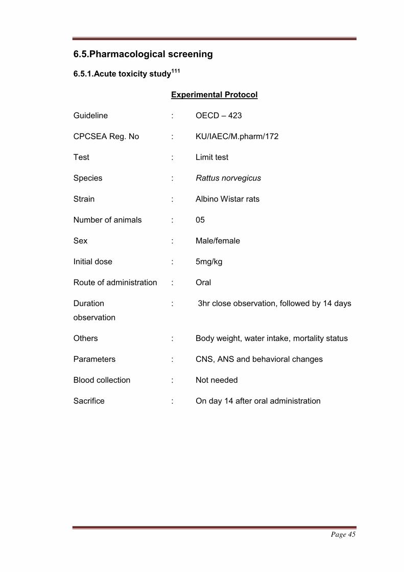
6.5.Pharmacological screening
6.5.1.Acute toxicity study111
Experimental Protocol
Guideline : OECD – 423
CPCSEA Reg. No : KU/IAEC/M.pharm/172
Test : Limit test
Species : Rattus norvegicus
Strain : Albino Wistar rats
Number of animals : 05
Sex : Male/female
Initial dose : 5mg/kg
Route of administration : Oral
Duration : 3hr close observation, followed by 14 days
observation
Others : Body weight, water intake, mortality status
Parameters : CNS, ANS and behavioral changes
Blood collection : Not needed
Sacrifice : On day 14 after oral administration
Page 46
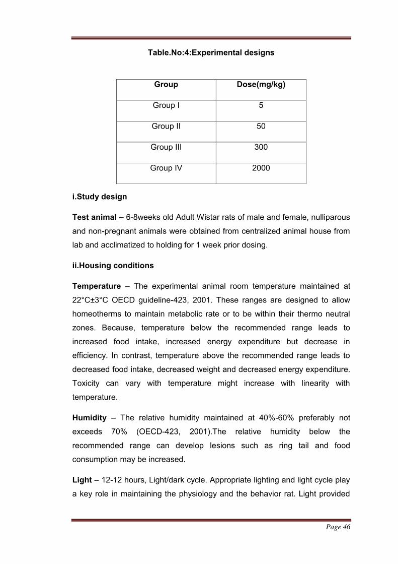
Table.No:4:Experimental designs
i.Study design
Test animal – 6-8weeks old Adult Wistar rats of male and female, nulliparous
and non-pregnant animals were obtained from centralized animal house from
lab and acclimatized to holding for 1 week prior dosing.
ii.Housing conditions
Temperature – The experimental animal room temperature maintained at
22°C±3°C OECD guideline-423, 2001. These ranges are designed to allow
homeotherms to maintain metabolic rate or to be within their thermo neutral
zones. Because, temperature below the recommended range leads to
increased food intake, increased energy expenditure but decrease in
efficiency. In contrast, temperature above the recommended range leads to
decreased food intake, decreased weight and decreased energy expenditure.
Toxicity can vary with temperature might increase with linearity with
temperature.
Humidity – The relative humidity maintained at 40%-60% preferably not
exceeds 70% (OECD-423, 2001).The relative humidity below the
recommended range can develop lesions such as ring tail and food
consumption may be increased.
Light – 12-12 hours, Light/dark cycle. Appropriate lighting and light cycle play
a key role in maintaining the physiology and the behavior rat. Light provided
Group Dose(mg/kg)
Group I 5
Group II 50
Group III 300
Group IV 2000
Page 47
for adequate vision and for neuroendocrine regulation of diurnal and circadian
cycles (CPCSEA guidelines for laboratory animal facility 2003).
Light intensity – The light intensity maintained at 325 lux approximately 1m
above the floor. Consideration of variations in light intensity, for the
arrangement of animals on cage rack for toxicology study is necessary.
Caging – Polypropylene cages with solid bottom and walls. Lids made up of
stainless steel grill capable of holding of both feed and water.
ii.Feeding condition – Sterile laboratory feed (ad libitum) and RO water
bottles daily.
iii.Feed – Brown colored chow diet
iv.Drug administration – Animals were fasted for 12hour prior to dosing on
day 0. Treatment rats were dosed by oral gavages, using a curved and ball
tipped stainless steel feeding needle, with 20% gum acacia solution.
v.Clinical observations – All rats were monitored continuously for 4 hour
after dosing for signs of toxicity. For the remainder of the 14 days study
period, animals were monitored and any additional behavioral or clinical signs
of toxicity. Animal’s body weight was measured prior to dosing and on days 7
and 14.On all animals were killed and at the end of the study LD50 value was
established. Clinical observations and gross pathological examination was
carried out.
6.5.2. Evaluvation of hepatoprotective activity
6.5.2.1.Paracetamol induced liver toxicity
i.Animals
Healthy adult male Albino rats (120-200 gram body weight) were
procured and randomly assigned to 5 groups, each containing 6 animals in
polypropylene cages layered with husk and maintained in a controlled room at
a temperature (22±3°C) and light (12 hours light/dark cycle). Animals were
allowed free access to water and standard pellet diet. Animals were cared in
accordance with the “Guide for the care and use of laboratory animals” and
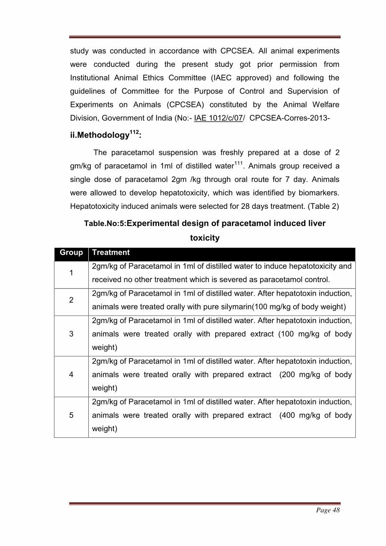
Page 48
study was conducted in accordance with CPCSEA. All animal experiments
were conducted during the present study got prior permission from
Institutional Animal Ethics Committee (IAEC approved) and following the
guidelines of Committee for the Purpose of Control and Supervision of
Experiments on Animals (CPCSEA) constituted by the Animal Welfare
Division, Government of India (No:- IAE 1012/c/07/ CPCSEA-Corres-2013-
ii.Methodology112:
The paracetamol suspension was freshly prepared at a dose of 2
gm/kg of paracetamol in 1ml of distilled water111. Animals group received a
single dose of paracetamol 2gm /kg through oral route for 7 day. Animals
were allowed to develop hepatotoxicity, which was identified by biomarkers.
Hepatotoxicity induced animals were selected for 28 days treatment. (Table 2)
Table.No:5:Experimental design of paracetamol induced liver
toxicity
Group Treatment
1 2gm/kg of Paracetamol in 1ml of distilled water to induce hepatotoxicity and
received no other treatment which is severed as paracetamol control.
2 2gm/kg of Paracetamol in 1ml of distilled water. After hepatotoxin induction,
animals were treated orally with pure silymarin(100 mg/kg of body weight)
3
2gm/kg of Paracetamol in 1ml of distilled water. After hepatotoxin induction,
animals were treated orally with prepared extract (100 mg/kg of body
weight)
4
2gm/kg of Paracetamol in 1ml of distilled water. After hepatotoxin induction,
animals were treated orally with prepared extract (200 mg/kg of body
weight)
5
2gm/kg of Paracetamol in 1ml of distilled water. After hepatotoxin induction,
animals were treated orally with prepared extract (400 mg/kg of body
weight)
Page 49
6.5.2.2.Evaluation of hepatoprotective efficacy:
Efficacy of the extract was assessed using biochemical marker
enzymes such as,
SGPT.
SGOT.
ALP (Alkaline Phosphatase).
Total Protein.
Total Bilirubin.
Change in body weight.
6.5.2.3.Sample collection and biochemical assay:
The blood samples obtained were collected into plain sample
tubes and centrifuged at 2000 rpm for 5 minutes to separate serum. Serum
was carefully collected and kept in eppendrof tubes for the determination of
the biochemical parameters.
6.5.2.4.Assessment of serum marker enzymes:
Serum level of SGPT and SGOT: These are the potential
biomarkers of hepatic injury and were estimated using a commercial kit
(Medsource Ozone Biomedicals Pvt. Ltd) (Reitman’s and Frankel, 1957).
Serum total protein (T. Protein was determined using the method Lowery et al
1951). Serum total bilirubin (T. BIL) was determined using the method (Mallay
et al 1937). Alkaline phosphatase (ALP) was determined using the method
(king et al 1954), were assayed using standard Diagnostic kits at Benghazi
medical center.
6.5.2.5.Change in Body Weight:
Animal body weight was measured weekly once from the starting day
to end of the study in paracetamol, ccl4 and ethanol induced models.
Page 50
6.5.2.6.Serum enzyme parameters
a.Alanine transaminase (ALT)113
i.Principle of Procedure
ALT catalyzes the transamination of L-Alanine to α-Ketoglutarate (α-
KG), forming L-Glutamate and Pyruvate. The Pyruvate formed is reduced to
Lactate by Lactate Dehydrogenase (LDH) with simultaneous oxidation of
reduced Nicotinamide-Adenine Dinucleotide (NADH). The change in
absorbance is measured using a dichromatic (340, 700 nm) rate technique.
L-Alanine + α-KG L-Glutamate + Pyruvate
P5P, Tris, pH 7.4
LDH
Pyruvate + NADH + H+ Lactate + NAD+
ii.Summary
The ALT method is an adaptation of the recommended procedure of
the IFCC as described by Bergmeyer118. The procedure is based on the
principles outlined by Wroblewski and LaDue but is modified to contain
pyridoxal-5-phosphate (P5P) as an activator and to replace phosphate buffer
with tris (hydroxymethyl) amino methane. Quantitation of ALT is a useful
parameter in evaluating liver function. Significant elevations of ALT are found
in diseases of the liver, such as hepatitis, necrosis, jaundice and cirrhosis.
ALT levels can be elevated even before clinical jaundice appears.
b.Aspartate transaminase (AST)
i.Principle of Procedure114
AST catalyzes the transamination from L-Aspartate to α-
Ketoglutarate, forming L-Glutamate and Oxaloacetate. The Oxaloacetate
formed is reduced to malate by Malate Dehydrogenase (MDH) with
simultaneous oxidation of reduced Nicotinamide Adenine Dinucleotide
(NADH). The change in absorbance with time due to the conversion of NADH
Page 51
to NAD is directly proportional to the AST activity and is measured using a bi
chromatic (340, 700 nm) rate technique.
AST
L-Aspartate + α-Ketoglutarate L- Glutamate + Oxaloacetate
MDH
Oxaloacetate + NADH Malate + NAD
ii.Summary
The AST method is an adaptation of the methodology
recommended by the International Federation of Clinical Chemistry (IFCC).
The method uses the coenzyme Pyridoxal-5-Phosphate (P5P) to activate the
apoenzyme and lactic acid Dehydrogenase (LDH) to eliminate pyruvate
interference. AST levels are 10-200 fold elevated in patients with acute
hepatic necrosis, viral hepatitis and drug induced poisoning. SGOT levels are
also elevated 10 fold in patients with post hepatic jaundice, intra hepatic
cholestasis and less than 10 fold in alcoholic and hepatic steatosis.
c.Alkaline phosphatase (ALP)
i.Principle of Procedure115
ALP catalyzes the transphosphorylation of p-
nitrophenylphosphate (p-NPP) to p-nitro phenol (p-NP) in the presence of the
transphosphorylating buffer, 2amino-2-methyl-1-propanol (AMP). The reaction
is enhanced through the use of magnesium and zinc ions. The change in
absorbance at 405 nm due to the formation of p-NP is directly proportional to
the ALP activity, since other reactants are present in non-rate limiting
quantities and is measured using a bichromatic (405, 510 nm) rate technique.
ALP
P-NPP + AMP p-NP + AMP + PO4
Ph 10.35 Mg/Zn
Page 52
ii.Summary
Measurements of ALP are used in the investigation of
hepatobiliary and bone disease119. The ALP method is based on a procedure
published by Bowers and McComb and more recently reviewed by Rej. This
method responds to all ALP isoenzymes in human serum. Marked increase of
ALP is seen in obstructive jaundice and biliary cirrhosis.
6.5.8.Histopathology Study116
Rats from all the treatment groups and control groups were euthanized on
the day 7. After gross observation, liver was collected and fixed in 10%
Neutral Buffer Formalin.
Trimming: Tissues were trimmed from all the lobes of liver.
Processing: Processing is done with the help of Automated Tissue
Processor (ATP) (Leica ASP 300) for 16 hours.
Embedding: Processed tissues were embedded in paraffin with the help
of paraffin embedding station (Leica EG 1150 H).
Sectioning: Initially blocks were trimmed at 25 microns and then
sectioned at 4 microns with the help of semi-automatic Microtome (Leica
RM 2245).
Staining: Slides were stained by H&E stain at Multistainer (Leica ST
5020).
All the H&E stained slides were observed for pathological findings.
6.5.9.Statistical analysis:
N = 6, Values are Mean±SEM calculated using One way ANOVA
followed by Tukey’s multiple comparison test, *P<0.05 and **P<0.001
compared with Toxic treated group; **P<0.05 compared with vehicle treated
group; # P<0.01.
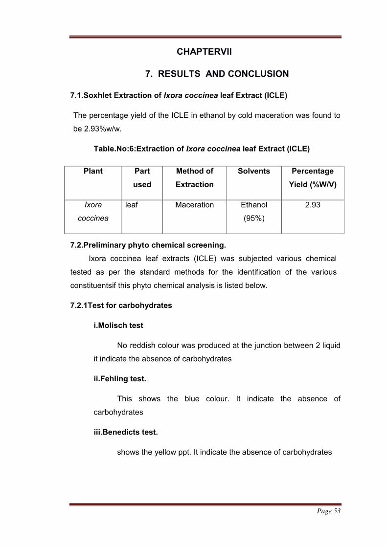
Page 53
CHAPTERVII
7. RESULTS AND CONCLUSION
7.1.Soxhlet Extraction of Ixora coccinea leaf Extract (ICLE)
The percentage yield of the ICLE in ethanol by cold maceration was found to
be 2.93%w/w.
Table.No:6:Extraction of Ixora coccinea leaf Extract (ICLE)
7.2.Preliminary phyto chemical screening.
Ixora coccinea leaf extracts (ICLE) was subjected various chemical
tested as per the standard methods for the identification of the various
constituentsif this phyto chemical analysis is listed below.
7.2.1Test for carbohydrates
i.Molisch test
No reddish colour was produced at the junction between 2 liquid
it indicate the absence of carbohydrates
ii.Fehling test.
This shows the blue colour. It indicate the absence of
carbohydrates
iii.Benedicts test.
shows the yellow ppt. It indicate the absence of carbohydrates
Plant Part
used
Method of
Extraction
Solvents Percentage
Yield (%W/V)
Ixora
coccinea
leaf Maceration Ethanol
(95%)
2.93
Page 54
7.2.2.Test for alkaloids
i..Mayer’s reagents
It form cream precipitate. It indicate the absence of alkaloid
ii.Dragendorff’s reagents
It forms a orange brown precipitate. It indicate the absence of
alkaloid
7.2..3.Test for flavonoids
It forms a yellow colour. It indicates the presence of flavanoid
7.2.4.Test for steroids.
i.Salkowski test
. Cherry red colour was obtained in the chloroform layer. It
indicate the absence of steroid
ii.Libbbermann burchatd test:
solution becomes reddish colour then it turns to bluish green
colour. It indicate the absence of steroid
7.2.5.Test for tannins.
It forms, pink or red colour due to the formation of phloroglucinol, It
indicate the absence of tannins
7.2.6.Test for protein.
I.Millon’s reagents.
solution which turns red on heating. It indicates the presence of
protiens
II.Ninhydrin Test.
Development of blue colour may indicate the presence of
peptide, amino acid (PROTEIN).
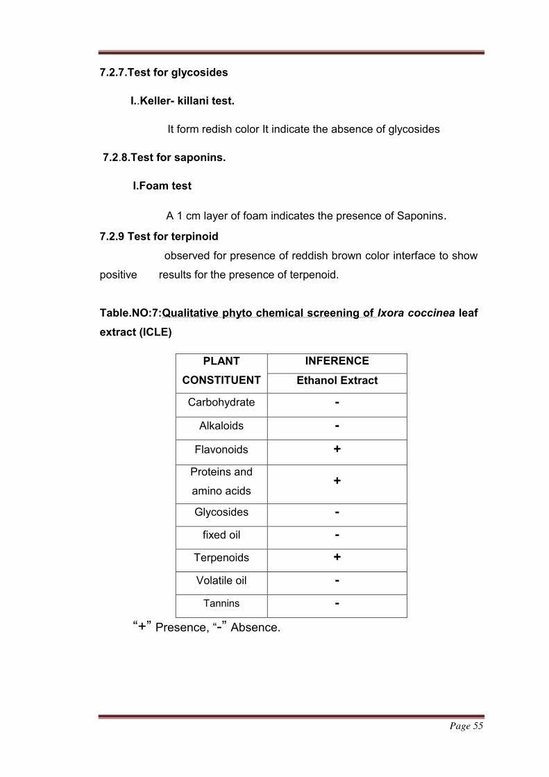
Page 55
7.2.7.Test for glycosides
I..Keller- killani test.
It form redish color It indicate the absence of glycosides
7.2.8.Test for saponins.
I.Foam test
A 1 cm layer of foam indicates the presence of Saponins.
7.2.9 Test for terpinoid
observed for presence of reddish brown color interface to show
positive results for the presence of terpenoid.
Table.NO:7:Qualitative phyto chemical screening of Ixora coccinea leaf
extract (ICLE)
PLANT
CONSTITUENT
INFERENCE
Ethanol Extract
Carbohydrate -
Alkaloids -
Flavonoids +
Proteins and
amino acids +
Glycosides -
fixed oil -
Terpenoids +
Volatile oil -
Tannins -
“+” Presence, “-” Absence.
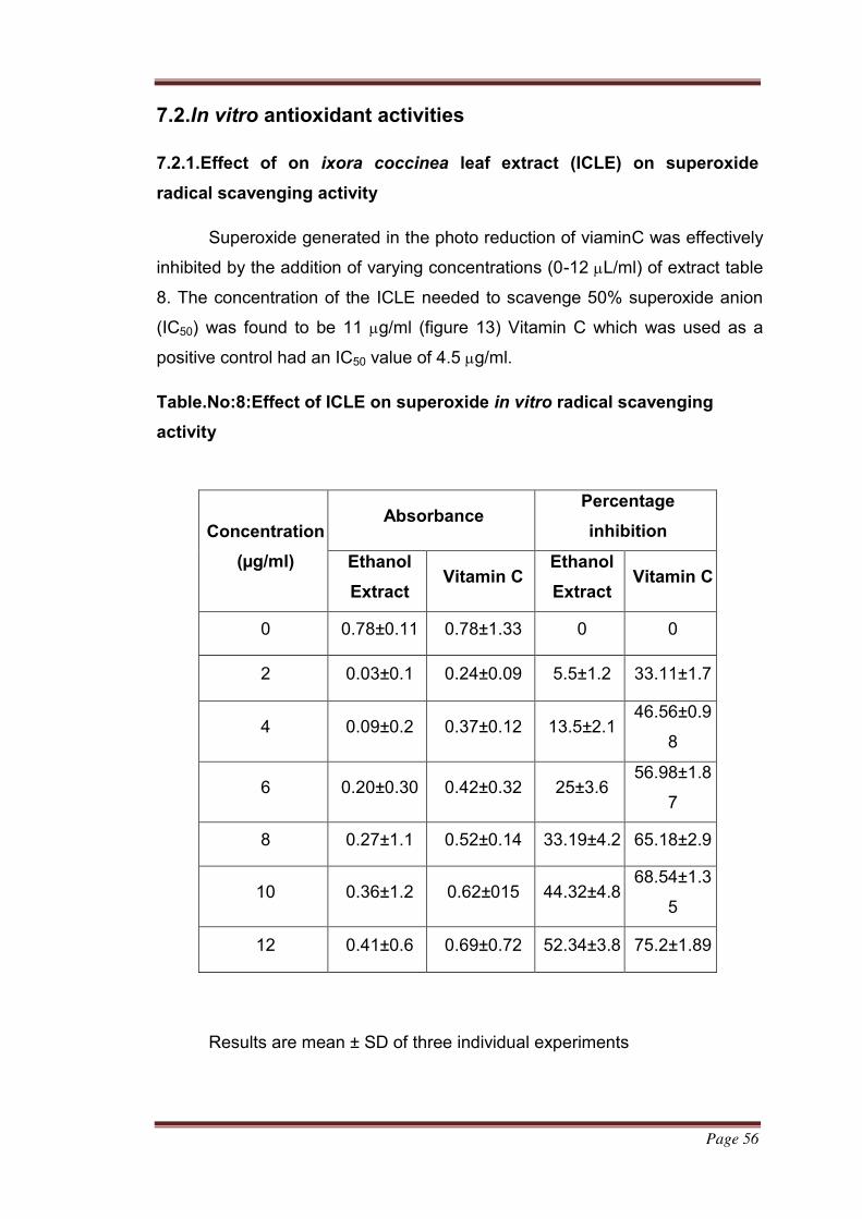
Page 56
7.2.In vitro antioxidant activities
7.2.1.Effect of on ixora coccinea leaf extract (ICLE) on superoxide
radical scavenging activity
Superoxide generated in the photo reduction of viaminC was effectively
inhibited by the addition of varying concentrations (0-12 L/ml) of extract table
8. The concentration of the ICLE needed to scavenge 50% superoxide anion
(IC50) was found to be 11 g/ml (figure 13) Vitamin C which was used as a
positive control had an IC50 value of 4.5 g/ml.
Table.No:8:Effect of ICLE on superoxide in vitro radical scavenging
activity
Results are mean ± SD of three individual experiments
Concentration
(µg/ml)
Absorbance Percentage
inhibition
Ethanol
Extract Vitamin C
Ethanol
Extract Vitamin C
0 0.78±0.11 0.78±1.33 0 0
2 0.03±0.1 0.24±0.09 5.5±1.2 33.11±1.7
4 0.09±0.2 0.37±0.12 13.5±2.1 46.56±0.9
8
6 0.20±0.30 0.42±0.32 25±3.6 56.98±1.8
7
8 0.27±1.1 0.52±0.14 33.19±4.2 65.18±2.9
10 0.36±1.2 0.62±015 44.32±4.8 68.54±1.3
5
12 0.41±0.6 0.69±0.72 52.34±3.8 75.2±1.89
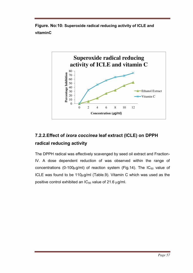
Page 57
Figure. No:10: Superoxide radical reducing activity of ICLE and
vitaminC
0
10
20
30
40
50
60
70
80
0 2 4 6 8 10 12
Per
cen
tag
e In
hib
itio
n
Concentration (µg/ml)
Superoxide radical reducing
activity of ICLE and vitamin C
Ethanol Extract
Vitamin C
7.2.2.Effect of ixora coccinea leaf extract (ICLE) on DPPH
radical reducing activity
The DPPH radical was effectively scavenged by seed oil extract and Fraction-
IV. A dose dependent reduction of was observed within the range of
concentrations (0-100g/ml) of reaction system (Fig.14). The IC50 value of
ICLE was found to be 110g/ml (Table.9). Vitamin C which was used as the
positive control exhibited an IC50 value of 21.6 g/ml.
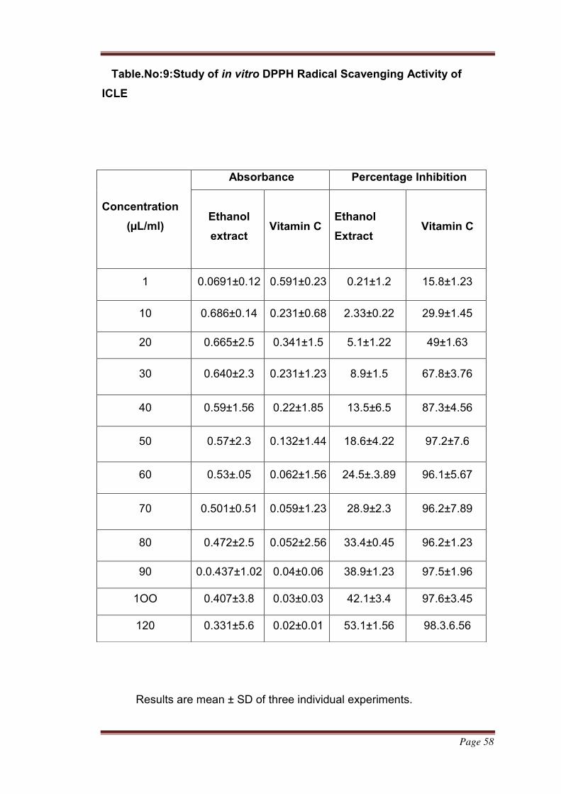
Page 58
Table.No:9:Study of in vitro DPPH Radical Scavenging Activity of
ICLE
Results are mean ± SD of three individual experiments.
Concentration
(µL/ml)
Absorbance Percentage Inhibition
Ethanol
extract
Vitamin C Ethanol
Extract Vitamin C
1 0.0691±0.12 0.591±0.23 0.21±1.2 15.8±1.23
10 0.686±0.14 0.231±0.68 2.33±0.22 29.9±1.45
20 0.665±2.5 0.341±1.5 5.1±1.22 49±1.63
30 0.640±2.3 0.231±1.23 8.9±1.5 67.8±3.76
40 0.59±1.56 0.22±1.85 13.5±6.5 87.3±4.56
50 0.57±2.3 0.132±1.44 18.6±4.22 97.2±7.6
60 0.53±.05 0.062±1.56 24.5±.3.89 96.1±5.67
70 0.501±0.51 0.059±1.23 28.9±2.3 96.2±7.89
80 0.472±2.5 0.052±2.56 33.4±0.45 96.2±1.23
90 0.0.437±1.02 0.04±0.06 38.9±1.23 97.5±1.96
1OO 0.407±3.8 0.03±0.03 42.1±3.4 97.6±3.45
120 0.331±5.6 0.02±0.01 53.1±1.56 98.3.6.56
Page 59
Figure. No:11: DPPH radical reducing activity of ICLE and
vitaminC.
0200
tag
e I
nh
ibit
ion
Axis Title
DPPH radical reducing
activity of ICLE and …Ethanol
Extract
Vitamin C
7.3 Pharmacological study
7.3.1.Acute toxicity studies
Acute toxicity studies on the sd rats show no mortality at adose of
2000mg/kg of ICLE during a time period of 14 days.during the study , no
noticeable were seen in the rats . this help to predict that it does not contain
any type of toxicity and it is full safe . so 200mg/kg between (1/10th dose) of
ethanol extract were selected of that dose for the further study
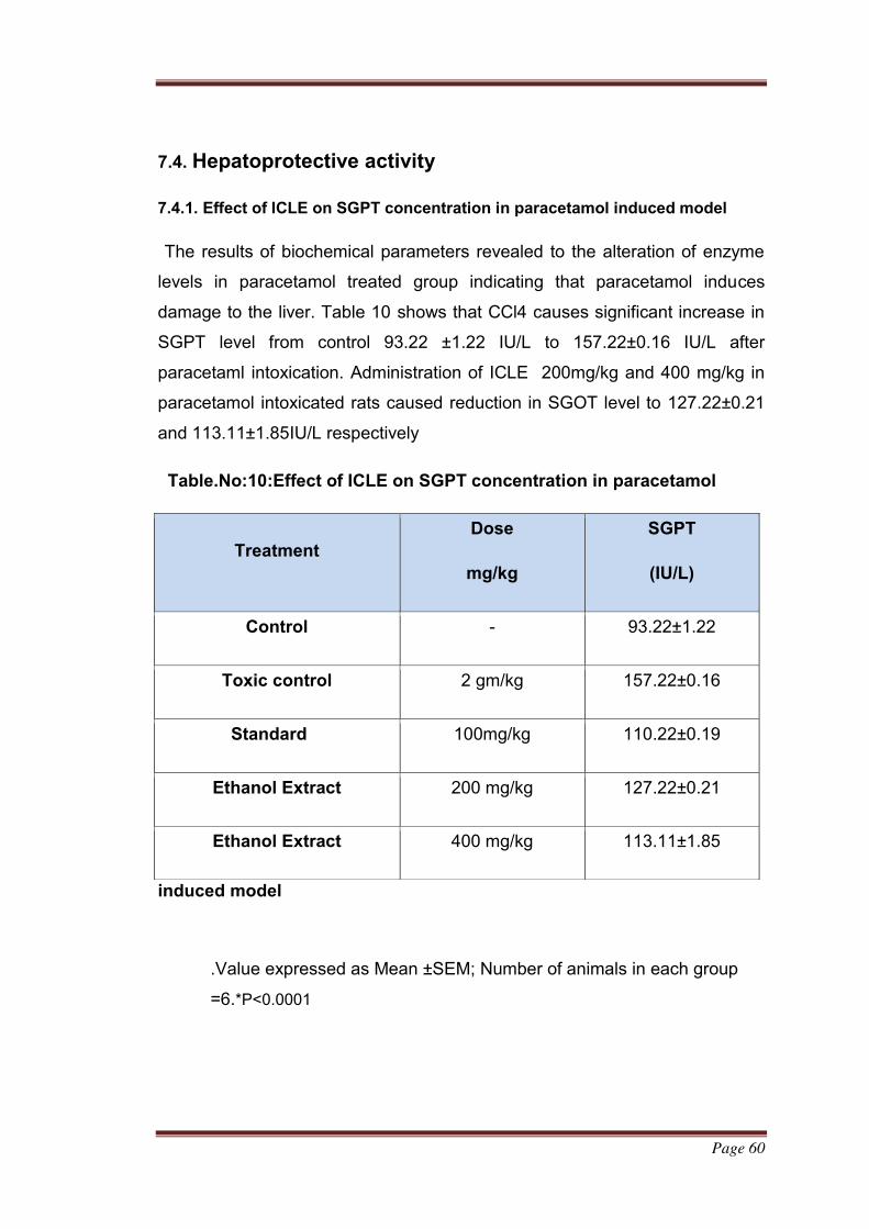
Page 60
7.4. Hepatoprotective activity
7.4.1. Effect of ICLE on SGPT concentration in paracetamol induced model
The results of biochemical parameters revealed to the alteration of enzyme
levels in paracetamol treated group indicating that paracetamol induces
damage to the liver. Table 10 shows that CCl4 causes significant increase in
SGPT level from control 93.22 ±1.22 IU/L to 157.22±0.16 IU/L after
paracetaml intoxication. Administration of ICLE 200mg/kg and 400 mg/kg in
paracetamol intoxicated rats caused reduction in SGOT level to 127.22±0.21
and 113.11±1.85IU/L respectively
Table.No:10:Effect of ICLE on SGPT concentration in paracetamol
induced model
.Value expressed as Mean ±SEM; Number of animals in each group
=6.*P˂0.0001
Treatment
Dose
mg/kg
SGPT
(IU/L)
Control - 93.22±1.22
Toxic control 2 gm/kg 157.22±0.16
Standard 100mg/kg 110.22±0.19
Ethanol Extract 200 mg/kg 127.22±0.21
Ethanol Extract 400 mg/kg 113.11±1.85
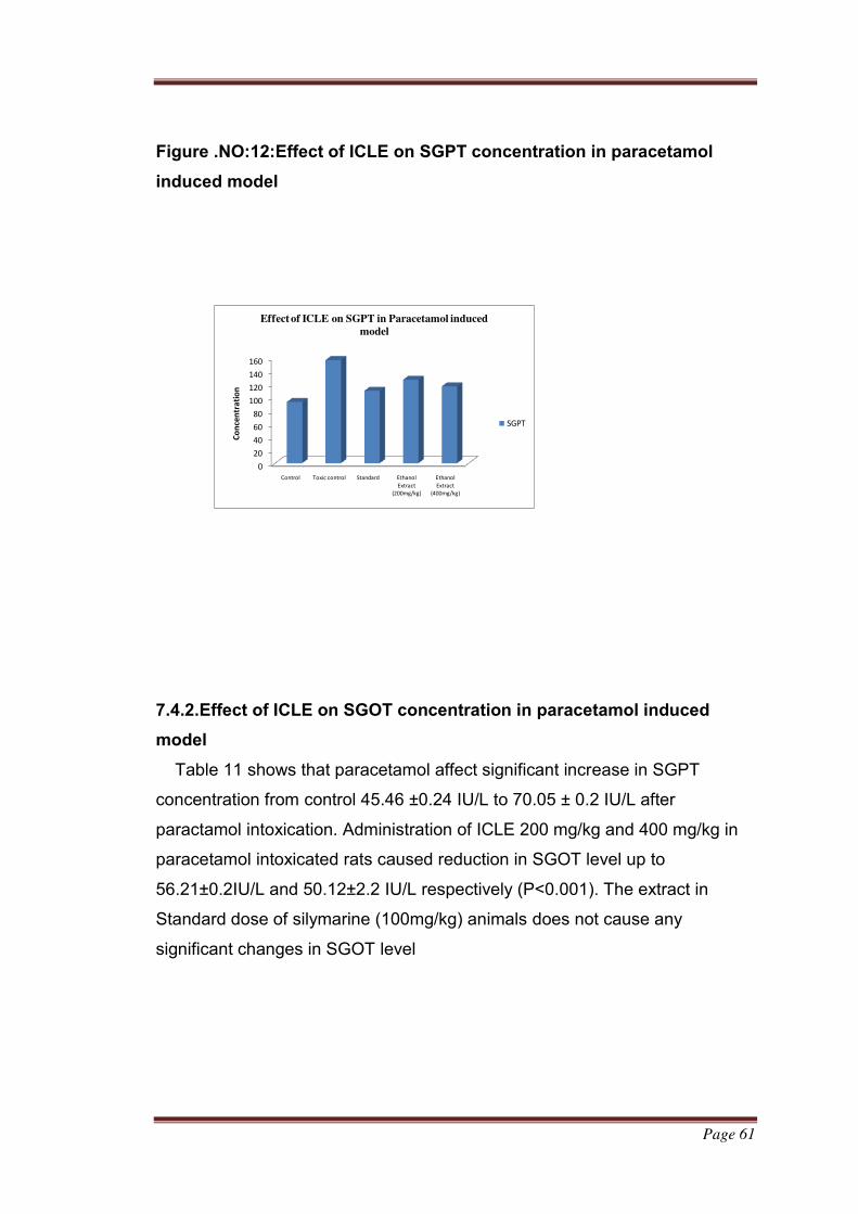
Page 61
Figure .NO:12:Effect of ICLE on SGPT concentration in paracetamol
induced model
0
20
40
60
80
100
120
140
160
Control Toxic control Standard Ethanol
Extract
(200mg/kg)
Ethanol
Extract
(400mg/kg)
Co
nce
ntr
ati
on
Effect of ICLE on SGPT in Paracetamol induced
model
SGPT
7.4.2.Effect of ICLE on SGOT concentration in paracetamol induced
model
Table 11 shows that paracetamol affect significant increase in SGPT
concentration from control 45.46 ±0.24 IU/L to 70.05 ± 0.2 IU/L after
paractamol intoxication. Administration of ICLE 200 mg/kg and 400 mg/kg in
paracetamol intoxicated rats caused reduction in SGOT level up to
56.21±0.2IU/L and 50.12±2.2 IU/L respectively (P<0.001). The extract in
Standard dose of silymarine (100mg/kg) animals does not cause any
significant changes in SGOT level
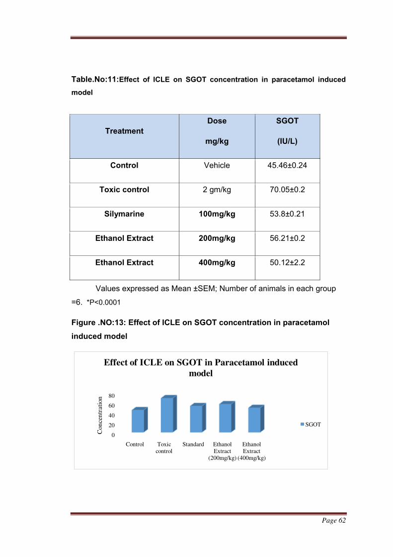
Page 62
Table.No:11:Effect of ICLE on SGOT concentration in paracetamol induced
model
Values expressed as Mean ±SEM; Number of animals in each group
=6. *P˂0.0001
Figure .NO:13: Effect of ICLE on SGOT concentration in paracetamol
induced model
0
20
40
60
80
Control Toxic
control
Standard Ethanol
Extract (200mg/kg)
Ethanol
Extract (400mg/kg)
Con
cen
trat
ion
Effect of ICLE on SGOT in Paracetamol induced
model
SGOT
Treatment
Dose
mg/kg
SGOT
(IU/L)
Control Vehicle 45.46±0.24
Toxic control 2 gm/kg 70.05±0.2
Silymarine 100mg/kg 53.8±0.21
Ethanol Extract 200mg/kg 56.21±0.2
Ethanol Extract 400mg/kg 50.12±2.2
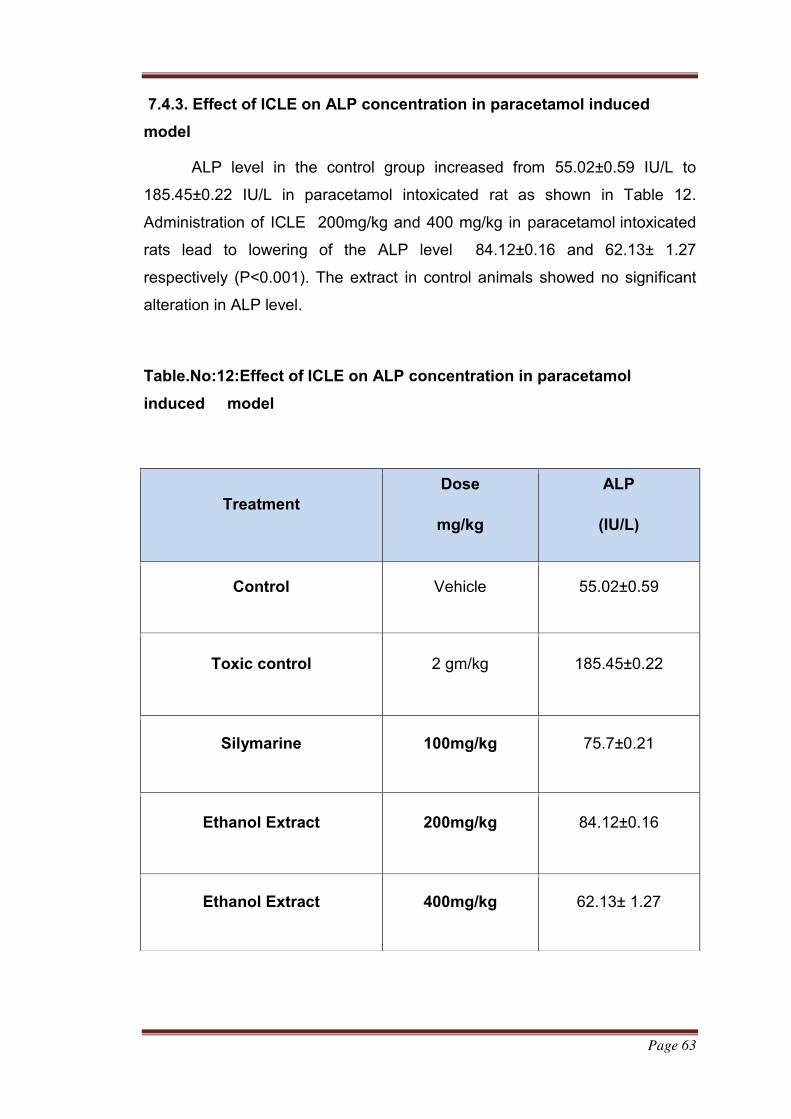
Page 63
7.4.3. Effect of ICLE on ALP concentration in paracetamol induced
model
ALP level in the control group increased from 55.02±0.59 IU/L to
185.45±0.22 IU/L in paracetamol intoxicated rat as shown in Table 12.
Administration of ICLE 200mg/kg and 400 mg/kg in paracetamol intoxicated
rats lead to lowering of the ALP level 84.12±0.16 and 62.13± 1.27
respectively (P<0.001). The extract in control animals showed no significant
alteration in ALP level.
Table.No:12:Effect of ICLE on ALP concentration in paracetamol
induced model
Treatment
Dose
mg/kg
ALP
(IU/L)
Control Vehicle 55.02±0.59
Toxic control 2 gm/kg 185.45±0.22
Silymarine 100mg/kg 75.7±0.21
Ethanol Extract 200mg/kg 84.12±0.16
Ethanol Extract 400mg/kg 62.13± 1.27
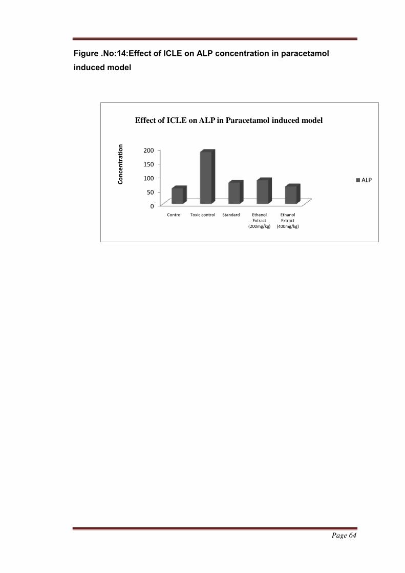
Page 64
Figure .No:14:Effect of ICLE on ALP concentration in paracetamol
induced model
0
50
100
150
200
Control Toxic control Standard Ethanol
Extract
(200mg/kg)
Ethanol
Extract
(400mg/kg)
Co
nce
ntr
ati
on
Effect of ICLE on ALP in Paracetamol induced model
ALP
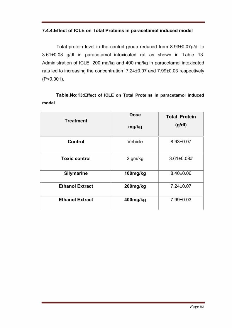
Page 65
7.4.4.Effect of ICLE on Total Proteins in paracetamol induced model
Total protein level in the control group reduced from 8.93±0.07g/dl to
3.61±0.08 g/dl in paracetamol intoxicated rat as shown in Table 13.
Administration of ICLE 200 mg/kg and 400 mg/kg in paracetamol intoxicated
rats led to increasing the concentration 7.24±0.07 and 7.99±0.03 respectively
(P<0.001).
Table.No:13:Effect of ICLE on Total Proteins in paracetamol induced
model
Treatment
Dose
mg/kg
Total Protein
(g/dl)
Control Vehicle 8.93±0.07
Toxic control 2 gm/kg 3.61±0.08#
Silymarine 100mg/kg 8.40±0.06
Ethanol Extract 200mg/kg 7.24±0.07
Ethanol Extract 400mg/kg 7.99±0.03
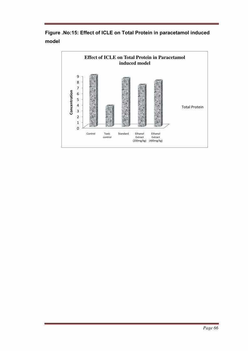
Page 66
Figure .No:15: Effect of ICLE on Total Protein in paracetamol induced
model
0
1
2
3
4
5
6
7
8
9
Control Toxic
control
Standard Ethanol
Extract
(200mg/kg)
Ethanol
Extract
(400mg/kg)
Co
nce
ntr
ati
on
Effect of ICLE on Total Protein in Paracetamol
induced model
Total Protein
Page 67
7.4.5. Effect of ICLE on Total Bilurubin in paracetamol induced model
Destruction of hemoglobin yields bilirubin which is conjugated in the
liver to diglucoroxide and excreted in the bile. Bilirubin accumulates in plasma
when liver insufficiency exists or biliary obstruction is present or rate of
hemolysis increases. The total bilirubin level increased from 3.12±0.02 mg/dL
in the control group to 12.21±0.04mg/dL after paracetamol intoxication as
shown in Table 14 Administration of ICLE 200 mg/kg and 400 mg/kg in
paracetamol intoxicated rats reduced the total bilirubin to 7.60±0.09mg/dL
and 5.13±0.56mg/dL (P<0.001). However, the extracts in control rats showed
no such significant alteration in the serum bilirubin level.
Table.No:14:Effect of ICLE on Total Bilurubin in paracetamol induced
model
Treatment
Dose
mg/kg
Total
bilirubin (mg/dl) Control Vehicle 3.12±0.02
Toxic control 2 gm/kg 12.21±0.04#
Standard 100mg/kg 4.86±0.21
Ethanol Extract 200mg/kg 7.60±0.09
Ethanol Extract 400mg/kg 5.13±0.56
Page 68
Figure .No:16: Effect of ICLE on Total Bilurubin in paracetamol
induced model
0
2
4
6
8
10
12
14
Control Toxic control Standard Ethanol Extract
(200mg/kg)
Ethanol Extract
(400mg/kg)
Co
nce
ntr
ati
on
Effect of ICLE on Total Bilurubin in Paracetamol induced
model
Total Bilurubin
7.4.6. Effect of ICLE on Body Weight for paracetamol induced
model
Table 15represents the body weight ofdifferent groups of rat
treatment. The body weight of Paracetamol induced group II rats has been re
duced comparing with group I. The body weight of Paracetamol induced grou
p II rats has been reduced comparing with group I.After 28days administration
of silymarin and ixora coccinea on group III and group VI rats has gained
the body weight almost to near normal level. The gain in body weight of grou
p IV is compared with group II and it may be due to the regeneration of liver c
ells activity to near normal after the herbal treatment.
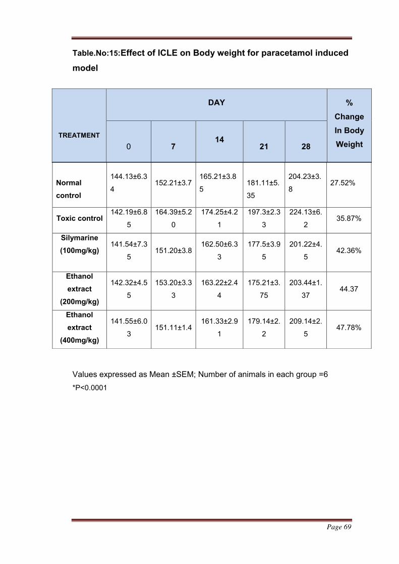
Page 69
Table.No:15:Effect of ICLE on Body weight for paracetamol induced
model
Values expressed as Mean ±SEM; Number of animals in each group =6
*P˂0.0001
TREATMENT
DAY %
Change
In Body
Weight
0
7
14
21
28
Normal
control
144.13±6.3
4 152.21±3.7
165.21±3.8
5
181.11±5.
35
204.23±3.
8 27.52%
Toxic control 142.19±6.8
5
164.39±5.2
0
174.25±4.2
1
197.3±2.3
3
224.13±6.
2 35.87%
Silymarine
(100mg/kg)
141.54±7.3
5 151.20±3.8
162.50±6.3
3
177.5±3.9
5
201.22±4.
5 42.36%
Ethanol
extract
(200mg/kg)
142.32±4.5
5
153.20±3.3
3
163.22±2.4
4
175.21±3.
75
203.44±1.
37 44.37
Ethanol
extract
(400mg/kg)
141.55±6.0
3 151.11±1.4
161.33±2.9
1
179.14±2.
2
209.14±2.
5 47.78%
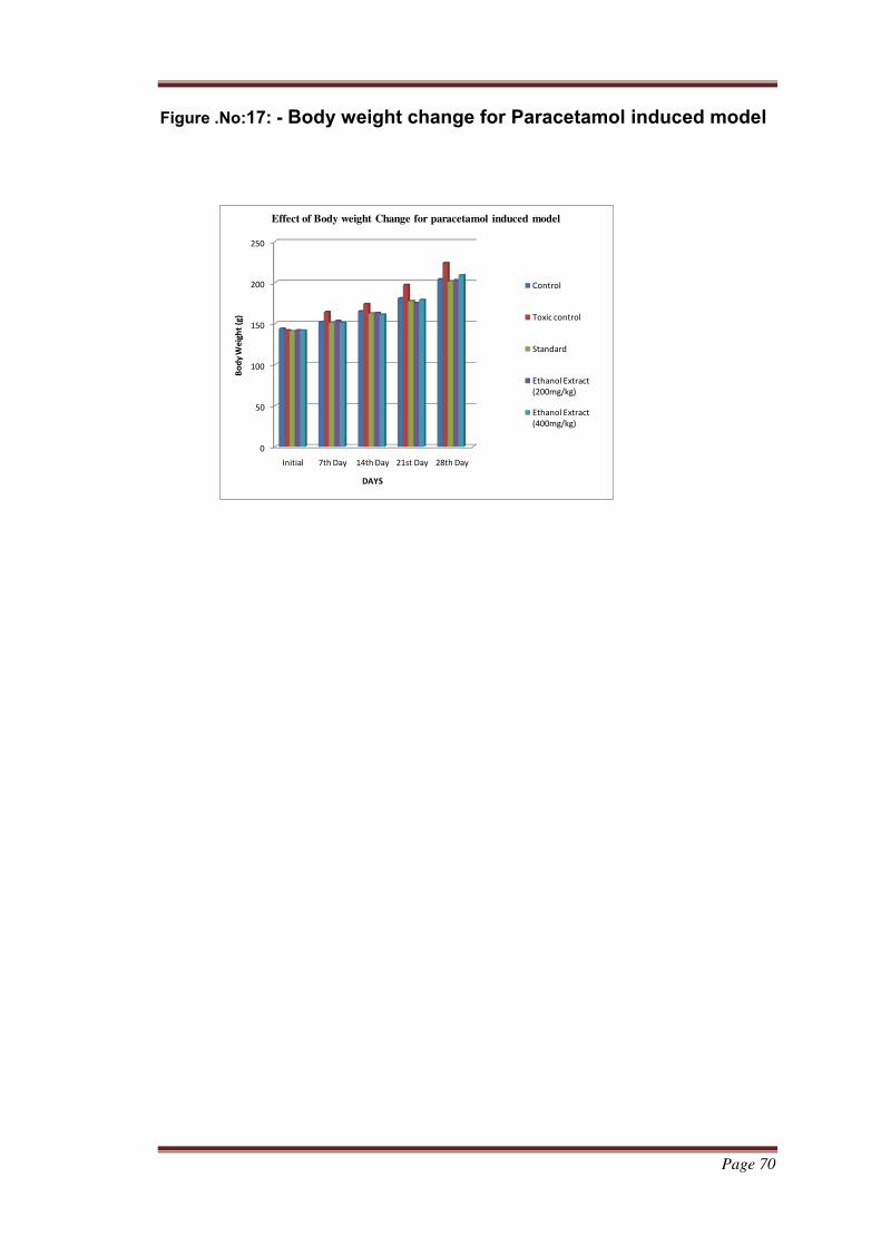
Page 70
Figure .No:17: - Body weight change for Paracetamol induced model
0
50
100
150
200
250
Initial 7th Day 14th Day 21st Day 28th Day
Bo
dy
We
igh
t (g
)
DAYS
Effect of Body weight Change for paracetamol induced model
Control
Toxic control
Standard
Ethanol Extract
(200mg/kg)
Ethanol Extract
(400mg/kg)
Page 71
7.5.HISTOPATHOLOGY
Histology is the study of the microscopic anatomy of cells and tissues
of plants and animals. Histology is an essential tool of biology and medicine.
Histopathology, the microscopic study of diseased tissue, is an important tool
in anatomical pathology, since accurate diagnosis of liver diseases usually
requires histopathological examination of samples.
The hepatoprotective effect of ICLE was confirmed by histological
examination. The liver sections of normal control animals showed hepatic
cells with well-preserved cytoplasm, prominent nucleus and central vein (Fig.
9 Group 1). The normal architecture of liver was completely lost in rats treated
with Paracetamol (Fig. 9 – Group 2) with the appearance of vacuolated
hepatocytes and degenerated nuclei. Vacuolization, fatty metamorphosis in
the adjacent hepatocytes, cell infiltration of lymphocytes and Kupffer cells and
necrosis of hepatocytes were severe in the centrilobular region and these
changes were also observed in areas other than the centrilobular regions. The
livers of rats treated with ICLE 100mg/kg,200 mg/kg,400mg/kg (Fig. 9 group
IV-VI), and silymarin 100 mg/kg (Fig. 9 group III) showed a significant
attenuation from paracetamol induced liver damage as evident from normal
hepatocytes with well-defined nuclei.
Histological features
Normal control shows normal cellular architecture with distinct hepatic
cells, sinusoidal space and a central vein.
Toxic control exhibited severe hepatocyte degeneration, fatty changes
and necrosis.
Silymarine shoes shows mild hepatocyte degeneration.
b ethanol extract shows normal architecture with mild hepatocyte
degeneration through dose dependent
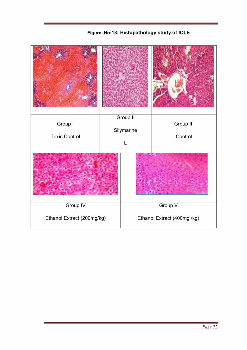
Page 72
Figure .No:18: Histopathology study of ICLE
Group I
Toxic Control
Group II
Silymarine
L
Group III
Control
Group IV
Ethanol Extract (200mg/kg)
Group V
Ethanol Extract (400mg /kg)
Page 73
CHAPTER VIII
8.DISSCSSION
The plant ixora coccinea is widely distributed in suth asia . The s flower
part of plant have been studied its antibacterial activity but the
hepatoprotective effect have been never studied. Hence the objective of the
study is determining this effect from the dried leaf extract of ixora coccinea
The preliminary phytochemical screening of whole plant extracts
indicate in presence of flavanoid, terpenoids , protien and amino acid . may
accounts antioxidant and hepatoprotective activity.
The antioxidant screening shows that that it showed reducing power to
DPPH radicals. But the efficiency showed that far below from Vitamin C. The
concentration of the ICLE needed to scavenge 50% superoxide anion (IC50)
equal to that of standard hence the leaf juice have the significant antioxidant
activity
Liver is one of the important organs of body hence damage to liver
leads to severe pathological problems or death. Liver diseases are mainly
caused by toxic chemicals, excessive intake of alcohol, infections and
autoimmune disorders. Liver injury caused by hepatotoxins such as carbon
tetrachloride, ethanol and acetaminophen is characterized by varying degrees
of hepatocyte degeneration and cell death by either apoptosis or by necrosis.
SGOT, SGPT, ALP, Total Protein and Total Bilirubin levels are largely used
as most common biochemical markers to evaluate liver injury.
Paracetamol (Acetaminophen), a widely used analgesic and antipyretic
drug that produces acute liver damage in high doses. Paracetamol induced
hepatotoxicity is thought to be caused by N-acetyl-p-benzoquinoneimine
(NAPQI), a cytochrome P-450 mediated intermediate metabolite. NAPQI can
react with sulphydryl groups such as glutathione and protein thiols. The
covalent binding of NAPQI to cell proteins is considered the initial step in a
chain eventually leading to cell necrosis. In paracetamol (PCM) treated acute
hepatic injury a significant difference in biochemical markers was observed
between normal and PCM control groups. Results of present study shows that
Page 74
the levels of SGOT, SGPT, ALP and Total Bilirubin were significantly
increased in paracetamol treated groups when compared with normal control
group.
While total protein is decreased in paracetamol treated groups due to
damage produced in endoplasmic reticulum. Comparative analysis on the
effect of SGOT, SGPT, ALP and Total Bilirubin revealed that extracts shows
marked decrease in these enzymes when compared with toxic control group.
While silymarine, and extract of ixora coccinea. Significantly restored
total protein activities to normal level. Moreover, histopathological analysis
showed that normal liver architecture was disturbed by paracetamol treated
rats oral feeding with extracts shows normal architecture with mild hepatocyte
degeneration compared with normal control group.
The preliminary phytochemical screening of whole plant extracts
indicate in presence of flavanoid, terpenoids , protien and amino acid .
The antioxidant studies particularly showed that ICLE have slight antioxidant
potential but that not inferior than standard vitamin C .
Hepatoprotective study results shows that the levels of SGOT, SGPT, ALP
and Total Bilirubin were significantly improvement may accounts
hepatoprotective activity
All these observation imply that the ICLE could be regarded as a favorable
antioxidant and hepatoprotective agents.
As the results indicated that the extract possess significant hepatoprotective
activity, after carrying out a thorough study of clinical trials, the plant can be
considered as a low cost, potent, herbal medicine for liver disorders.
Page 75
CHAPTER IX
9.BIBLIOGRAPHY
1. Gerald J Tortora Bryan Derrickson, Principles of anatomy and
physiology, 13th edition, page 990-995.
2. Kmiec′Z (2001) .‟Cooperation of liver cells in health and disease”. Adv
Anat Embryol Cell Biol. 161:3-13, 1-151.
3. Anne Waugh, Allison Grant, Ross and Wilson, Anatomy and
Physiology in health and illness, 9th edition, page307-309.
4. http://diagramreview.com/wp-content/uploads/simple-Human-Liver-
Diagram.jpg
5. WebMD, Image collection: Human Anatomy.
6. Friedman Scott E, Grendell James h, McQuaid, Kenneth R. Current
diagnosis and treatment in gastroenterology. New York: Lang medical
book/mcgraw-hill.2008; Pp664-679.
7. Essential Pathology, 3rd edition, Harsh Mohan, 2007, page 361.
8. Miller-Keane Encyclopedia and Dictionary of Medicine, Nursing and
Allied Health, Seventh Edition 2003 by Saunders, an imprint of
Elsevier.
9. Wang CS; Chang Ting-Tsung; Yao Wei-Jen; Wang Shan-Tair; Chou
Pesus (2012). ‟Impact of increasing alanine amino transferase levels
within normal range on incident diabetes”. J Formos Med Assoc.
3(4):201-8.
10. Ghouri N; Preiss David; Sattar Naveed (2010). ‟Liver enzymes,
nonalcoholic fatty liver disease and incident cardiovascular disease: a
narrative review and clinical perspective of prospective data”.
Hepatology 52(3): 1156-61.
11. Paul T Giboney M D. Mildly Elevated Liver Transaminase Levels in the
Asymptomatic Patient.
12. Red Cross Donor Requirements.
13. Nyblom H, Berggren U, Balldin J, Olsson R (2004). High AST/ALT ratio
may indicate advanced alcoholic liver disease rather than heavy
drinking”: Alcohol 39(4): 336-339.
Page 76
14. Nyblom H, Bjomsson E, Simren M, Aldenborg F, Almer S, Olsson R
(September 2006). The AST/ALT ratio as an indicator of cirrhosis in
patients with PBC”-Liver Int. 26 (7): 840-845.
15. ‟http:∕∕www.gpnotebook.CO.UK/simplepage.cfm?ID=3222240579
16. Kirsch JF, Eichele G, Vincent MG, Jansonius JN, Gehring H et
al.(1984). ‟Mechanism of action of aspartate aminotransferase
proposed on the basis of its spatial structure” J Mol Biol 174(3):497-
525.
17. Hayashi H, Yoshimura T, Esaki N, Sodak (1990). ‟Recent topics in
pyridoxal 5'-phosphate enzyme studies”. Annu Rev Biochem 59:87-
110.
18. Gaze D C (2007). ‟The role of existing and novel cardiac biomarkers
for cardio protection”. Current Opinion in Investigational Drugs 8(9):
711-7.
19. Taḿas L, Huttová J, Mistrk I, Kogan G (2002). Effect of carboxymethyl
chitin –Glucan on the activity of some Hydrolytic Enzymes in Maize
plants”. Chem. Pap. 56(5):326-329.
20. Medline plus Encyclopedia Liver function tests.
21. Li-Fem H, Rajasoorya C (February 1999).‟The elevated serum alkaline
phosphatase-the chase that led to two endocrinopathies and one
possible unifying diagnosis”. Eur. J. Endocrinol 140(2): 143-7.
22. Lang P H, Millan J L, Stigbrand T, Vessella R L, Ruoslahti E, Fishman
W H (August 1982). ‟Placental alkaline phosphatase as a tumor
marker for seminoma”.Cancer Res. 42(8):3244-7.
23. Dugdale, David C. ‟ALP-blood test Medline plus Medical
Encyclopedia”. Medline Plus Retrieved 2014-02-26.
24. Schiele F, Vincent-Viry M, Foumier B, Starck M, Siest
G(November1998).‟Biological effects of eleven combined oral
contraceptives on serum triglycerides, gamma-glutamyltransferase,
alkaline phosphatase, bilirubin and other biochemical variables”. Clin.
Chem. Lab. Med. 36(11):871-8.
25. Lab Test Online, American Association for Clinical Chemistry, 2013.
Page 77
26. Landry D W, Basari H. Approach to the patient with kidney disease. In:
Goldman L, Schafer A L, eds. Cecil Medicine. 24th ed.Philadelphia,
Pa: Saunders Elsevier, 2011: chap 116.
27. www.myoclinic.com
28. Health line, Christine Case Lo; 2005-2014.
29. Berk PD, Korenblat KM. Approach to patient with jaundice or abnormal
liver test results. In: Goldman L, Ausiello D. eds. Cecil Medicine 23rd
ed. Philadelphia, Pa: Saunders Elsevier, 2007, chap.150.
30.
31. Stricker B, Spoelstra P, In: Drug-Induced hepatic injury. Elsevier,
Amsterdam 1985 pp-41.
32. Sharma Bhavan, Sharma Upendrakumar. Hepatoprotective activity of
some indigenous plants. International journal of pharma tech
research 2009; 1(4): 1330-34.
33. Karunakar hedge, Arun B Joshi. Hepatoprotective and antioxidant
effect of Carissa Spinarum root extract against ccl4 and paracetamol
induced hepatic damage in rats. Bangladesh J Pharmacol 2010;
5:73-76.
34. Vipul Gujrati, Nileshpatel, Venkat N Rao et al. Hepatoprotective activity
of alcoholic and aqueous extracts of leaves of tylophoraindica in rats.
Indian journal of pharmacology 2007; 39(1): 43-47.
35. Ostapowicz G, Fontana R J, Schiodt F V, Larson A, Davrson J T,
Steven H B, Timothy M and Reish J. Results of a prospective study of
acute liver failure at 17 tertiary care centers in the United States. Ann
Intern Med.2002; 137:947-954.
36. Manov I, Motanis H, Frumin I, Lancu T C (2006). Hepatotoxicity of anti-
inflammatory and analgesic drugs: ultrastructural aspects”. Acta
Pharmacol Sin. 27(3):259-72.
37. Keeffe Emmet B; Friedman Lawrence M. (2004). Handbook of liver
diseases. Edinburgh: Churchill Livingstone pp. 104-123.
38. Sarich T C, Adams S P, Petricca G, Wright J M (1999). Inhibition of
Isoniazid induced hepatotoxicity in rabbits by pretreatment with an
amidase inhibitor”. J. Pharmacol. Exp. Ther. 289(2):695-702.
Page 78
39. Pak E, Esrason K T, Wu V H (2004). “Hepatotoxicity of herbal
remedies: an emerging dilemma”. Progress in transplantation (Aliso
Viejo, Calif.) 14(2): 91-6.
40. Nilesh Mehta, Michael R Pinsky; Drug-Induced Hepatotoxicity.
Medscape; Jun 27, 2012.
41. http://www.rightdiagnosis.com/h/hepatotoxicity/symptoms.htm.
42. http://www.rightdiagnosis.;com/h/hepatotoxicity/treatments.htm.
43. Sowjanya Guduguntia, Hepatoprotective activity a review; Pharma
Tutor ART-1701; Pharmacy Infopedia.
44. Jian Wu, Ake Danielsson, Mark A Z; Toxicity of hepatotoxins: new
insights into mechanisms and therapy 8(5): 585-607(1999).
45. Schiodt F V, Atillasoy E, Shakil A O et al. Etiology and outcome for 295
patients with acute liver failure in United states Liver Transplant Surg
1999; 5: 29-34.
46. Makin AJ, Williams R; Paracetamol hepatotoxicity and alcohol
consumption in deliberate and accidental overdose. Q J Med 2000;
93:341-9.
47. Lee W M. Acute liver failure N Engl J Med 1993; 329: 1862-72.
48. Raucy J L, Laskar J M, Lieber C S, Black M. Acetaminophen activation
by human liver cytochrome P450 2EI and P450IA2. Arch Biochem
Biophys 1989; 271:270-283.
49. Tirmenstein M A, Nelson S D. Acetaminophen induced oxidation of
protein thiols: Contribution of impaired thiol metabolizing enzymes
and the breakdown of adenine nucleotide. J Biol chem. 1990;
265:3059-3065.
50. Potter D W, Himson J A, Reactions of N-acetyl-p-benzoquinoneimine
with reduced glutathione, acetaminophen and NADPH. Mol.
Pharmacol 1986; 309: 33-41
51. Mitchell J R, Jollow D J, Potter W Z, Gillette J R and Brodic BB (1973)
Acetaminophen induced hepatic necrosis IV. Protective role of
glutathione J. Pharmacol. Ther. 187: 211-217.
52. Laura P.James, Philip R. Mayeux and Jack A. Hinson; Acetaminophen-
Induced Hepatotoxicity; Drug Metabolism and Disposition. 2003,
vol.31, no.12, 1499-1506.
Page 79
53. Nelson S D. Molecular mechanisms of the hepatotoxicity caused by
acetaminophen. Semin Liver Dis; 1990, 10: 267-278.
54. Salas V M, Corcoran G B. Calcium dependent DNA damage and
adenosine 3′,5′-cyclic monophosphate independent glycogen
phosphorylase activation in an in vitro model of acetaminophen
induced liver injury. Hepatology; 1997, 25:1432-1438.
55. Murtry R J, Snodgrass W R, Mitchell J R. Renal necrosis, glutathione
depletion and covalent binding after acetaminophen Toxicol. Appl.
Pharmacol. 1978, 120: 87-100.
56. Michael S L, Pumford N R, Mayex P R. Pretreatment of mice with
macrophage in activators decreases acetaminophen hepatotoxicity
and the formation of reactive oxygen species and nitrogen species.
Hepatology, 1990, 30, 1 86-195.
57. Kumar V, Cotran R, Robbins S. Robbins basic pathology; 7thedition;
Philadelphia: W B Saunders; 2003.
58. Saravanan N, Nalini N. Antioxidant effect of Hemidesmus indicus on
ethanol induced hepatotoxicity in rats, J Med FOOD 2007; 10:675-82.
59. R G Thurman, B U Bradford, Y Iimuro, M V Frankenberg, K T Knecht;
Mechanisms of alcohol induced hepatotoxicity: studies in rats.
Frontiers in Bioscience, 4, e42-46, July 15, 1999.
60. JN the Journal of Nutrition; Role of Kupffer cells, Endotoxin and Free
radicals in Hepatotoxicity due to Prolonged Alcohol Consumption:
Studies in male rats; 1997, vol. 127. No. 5, 9035-9065.
61. B Dheeba, E Vaishnavi, P Sampathkumar, M Kannan; Biosci.
Biotechnol. Res. Asia, 9, 309 (2012).
62. http://shodhganga.inflibnet.ac.in/bitstream/10603/8127/12/12-
chapter%204.pdf.
63. Recknagal R O, EA Glenda, Jr J A Dolak, R L Walter 1989.Mechanism
of ccl4 toxicity. Pharmacol. Ther.43, 139-154.
64. C:\users/user/pictures/my projects hepatoprotective activity.htm.
65. J B Harborne. Text book of phytochemical methods 3rd edition,
Springer Publishers 2007.
66. Tripathi K D; Essentials of medical pharmacology; 6thedition; Jaypee
brother’s medical publishers; 2008.
Page 80
67. Doreswany R, Sharma D; Plant drugs for liver disorders management.
Indian Drugs 32: 139-44 (1995).
68. Saumendu Deb Roy, Sumit Das, Dibyendu Shil, Koushik Nandan
Dutta; Herbal hepatoprotective agents: A review: World Journal of
Pharmaceutical research Vol 1, Issue 2, 87-99.
69. Bimlesh Kumar et al: A Review of phytochemistry and pharmacology of
flavonoids. Internationale Pharmaceutica Sciencia Jan-Mar 2011 Vol
1, Issue 1.
70. http://www.healthline.com/natstandardcontent/rutin#H3.
71. Pensak Jantrawut et al; Anticancer activity of rutin encapsulated in low
methoxy pectin beads. International journal of pharmacy and
pharmaceutical sciences Vol 6, Issue 3, 2014.
72. Erlund I, Kosonen T, Alfthan G, Maenpaa J, P erttunen K, Kenraali J.
Pharmacokinetics of quercetin from quercetin aglycone and rutin in
healthy volunteers. Eur J Clin Pharmacol 2000; 56:545-53.
73. Khalid H Janbaz: Protective effect of rutin on paracetamol and ccl4
induced hepatotoxicity in rodents. Fitoterapia 73 (2002) 557-563.
74. Robert Domitrovic, Hrvoje Jakovac. Differential hepatoprotective
mechanisms of rutin and quercetin in ccl4- intoxicated BALB/cN mice.
Acta pharmacologica sinica (2012)33: 1260-1270.
75. Lin j p, Yang J S, Lu CC, Chiang J H, Wu C L et al; Rutin exhibits
proliferation of murine leukemia WEHI-3 cells in vivo and promotes
immune response in vivo. Leukemia research 2009; 33: 823-828.
76. Deschner EE, Ruperto J, Wong G, Newmark H L: Quercetin and rutin
as inhibitors of azoxymethanol-induced colonic neoplasia.
Carcinogenesis 1991; 12:1193-1196.
77. Undeger U, Aydin S, Basaran AA and Basaran N: The modulating
effects of quercetin and rutin on Mitomycin C induced DNA damage.
Toxicol Letters 2004; 151: 143-149.
78. David C, Rodriguez G, Bona S, Meurer L et al (2011) Role of quercetin
in preventing thioacetamide induced liver injury in rats. Toxicol
Pathol. 39: 949-957.
79. Parul Lakhanpal, Deepak Kumar Rai. Quercetin: A versatile flavonoid.
Internet Journal of Medical Update 2007, 2(2): 22-37.
Page 81
80. http://o.tqn.com/d/chemistry/1/7/t/s/1-/Quercetin.ipg.
81. Middleton E Jr. (1998); Effect of plant flavonoids on immune and
inflammatory cell function. Adv. Exp. Med. Biol. 439, 175-82.
82. Choi J S, Piao Y J, Kang K W. Effects of quercetin on bioavailability of
doxorubicin in rats: role of CYP3A4 and P-gp inhibition by quercetin,
Arch. Pharm. Res. 2011; 34(4): 607-13.
83. Havsteen B H. The biochemical and medical significance of flavonoids.
Pharmacology and therapeutics 2002; 96: 67-202.
84. Yeon Jeong Kin, Yong Chan Bae et al. Quercetin flavonoid inhibits
proliferation and increases osteogenic differentiation in human
adipose stromal cells. Biochemical pharmacology 2006, 72: 1268-
1278.
85. Haisheng Zhang et al. 2012; Antitumor activities of quercetin and
quercetin-50,8-disulfonate in human colon and breast cancer cell
lines. Food and chemical toxicology, 2012, 50: 1589-1599.
86. Manach et al, 1998. Quercetin is recovered in human plasma as
conjugated derivatives which retain antioxidant properties FEBS Lett,
426:331-336.
87. Anestics Karkanis et al; 2011. Cultivation of milk thistle medicinal
weed. Industrial Crops and Products 2011; 34: 825-830.
88. http://www.medscape.com.
89. Kostek H, Szponar J: Silibinin and its hepatoprotective action from
perspective of a toxicologist. Przeql Lek. 2012; 69(8): 541-3.
90. F Franschini, G Demartini, D Esposti. Pharmacology of silymarin Clin
Drug Invest. 2002; 22(1).
91. Sharma G et al; Silibinin induces growth inhibition and apoptotic cell
death in human lung carcinoma cells. Anticancer Res. 2003 May-Jun;
23(3B): 2649-55.
92. Singh R P, Agarwal R; Prostate cancer prevention by silibinin; Curr.
Cancer Drug Targets 2004 Feb; 4(1): 1-11.
93. S Javed, K Kohli, M Ali.‟Reassessing bioavailability of silymarin”,
Alternative Medicine Review,Vol. 16, no. 3,pp. 239-249, 2011.
94. Afiwa Missebukpo, Kossi Metowogo, Abdoulatif Diallo, Povi Lawson-
Evi, Kwashi Eklu-Gadegbeku, Kodjo A. Aklikokou and Gbeassor
Page 82
Messanvi.Antioxidant effects of Ixora coccinea Linn. in a rat model of
ovalbumin-induced asthma. African Journal of Pharmacy and
Pharmacology. Vol. 7(42), pp. 2794-2800
95. Prabhu B, Yasmeen M, Agashikar N V. (2010).Evaluation of the Anti-
diarrhoeal Activity of the Leaves of Ixora coccinea Linn in rats,
Journal of Clinical and Diagnostic Research, 4: 3298-3303.
96. Moni Rani Saha, Md. Ashraful Alam, Raushanara Akter and Rumana
Jahangir. (2008). In-vitro free radical scavenging activity of Ixora
coccinea L. Bangladesh J Pharmacol, 3: 90-96.
97. Shyamal S, Latha P G, Suja S R, Shine V J, Anuja G I, Sini S, Pradeep
S, Shikha P, Rajasekharan S. (2010). Hepatoprotective effect of three
herbal extracts on aflatoxin B1- intoxicated rat liver, Singapore Med J,
51(4): 326-331.
98. Nagaraj B, Krishnamurthy N, Liny P, Divya T and Dinesh R. (2011).
Biosynthesis of Gold Nanoparticles of Ixora Coccinea leaf extract &
their Antimicrobial activities, 2(4): 557-565.
99. Latha P.G, Panikkar K.R. (1998). Cytotoxic and antitumour principles
from Ixora coccinea leafs, Cancer Letters, 130(1-2): 197-202.
100. Yasmeen Maniyar and Prabu Bhixavatimath. (2011). Evaluation of the
Hypoglycaemic and Hypolipidaemic Activities of the Aqueous
Extract of the leaves of Ixora coccinea Linn in Diabetic Rats. Journal
of Clinical and Diagnostic.Research. 5(7): 1381-1384.
101. A.Elumalai, Chinna Eswaraiah, Yetcharla Venkatesh, Burle Shiva
kumar and Chava Narendar. Phytochemical and pharmacological
profile Of Ixora coccinea Linn. International Journal Of
Pharmacy & Life Sciences 3(3): Mar., 2012] Int. J. of Pharm. &
Life Sci. (IJPLS), Vol. 3, Issue 3: March: 2012, 1563-1567 1563
102. http://plants.usda.gov/java/ClassificationServlet?source=display&classi
d=IXORA#
103. http://www.leafsofindia.net/catalog/slides/Ixora%20red.html
104. http://www.plantsrescue.com/ixora-coccinea/
105. Kokate CK., Purohit AP., Gokhale SB.,Pharmacognosy, Nirali
Prakashan, Pune, 2006, 35, p 106-107.
Page 83
106. Adeniyi SA., Orjiekwe CL., Ehiagbonare JE., Arimah BD., Preliminary
phytochemical analysis and insecticidal activity of ethanolic extract of
four tropical plants (Vernonia amygdalina, Sidaacuta, Ocimum
gratissimum and Telfaria occidentalis) against beans weevil
(Acanthscelides obtectus), Int J Physical Sci, .02010, 5(6), p 753-762
107. Kirtikar KR, Basu BD. Indian Medicinal Plants, Allahabad, Lalit Mohan
Basu Publications, 1998, pp 1590-1592
108. Trease GE, Evans WC. Pharmacognosy (13th ed.). London:
BailliereTindall Ltd., 1989 sidduraju P,becker k(2007).
109. Trease GE, Evans WC. Pharmacognosy (13th ed.). London:
BailliereTindall Ltd., 1989
110. Sidduraju P,becker k(2007).Antioxidant and free radical scavengering
activityes of processed cow pig(cassia histula) seeds. Food
Chemistry 101,10-19
111. Brands Williams W,Cuvellie ME(1995),Uses of free radical method to
evaluate antioxidant activity . Lebensmittel wissenchant and
technologies-28, 25-30
112. OECD guidelines for testing of chemicals. 420.acute oral toxicity –
fixed dose procedure.
113. Smitha Shenoy, Hemnath kumar, Thashma. Hepatoprotective activity
of Plectanthus amboinicus against paracetamol induced
hepatotoxicity in rats.int j Pharmacol and clin sci 2012; 1:32-8.
114. Manokaran S, Jaswanth A,Senottuvelu S, Nandhakumar
J.Hepatoprotective activity of Aerva lanata linn. Against paracetamol
induced hepatotoxicity in rats. Reserch J Phaarm and Tech 2008; 1;
398-400.
115. Mallay HT and Evelyn KA. Estimation of serum bilirubin level with the
photoelectric colorimeter. J Biol Chem. 1937; 119: 481–484.
116. Muthu Gounder Palanivel, Balasubramanian RAJKAPOOR.
Hepatoprotective and Antioxidant Effect of Pisonia aculeata L.
against CCl4- Induced Hepatic Damage in Rats. Sci Pharm. 2008;
76: 203–215
117. Hiral Modi, Vishnu Patel, Komal Patel. Hepatoprotective activity of
Aegle marrmelos against ethanol induced hepatotoxicity in rats. Asian
Page 84
Journal of Pharmaceutical and Clinical Research, volume 5,suppl 4,
2012.