does white matter matter? spatio-temporal dynamics of task switching in aging
TRANSCRIPT
Does White Matter Matter? Spatio-temporal Dynamics of TaskSwitching in Aging
Gabriele Gratton, Emily Wee, Elena I. Rykhlevskaia, Echo E. Leaver, and Monica FabianiUniversity of Illinois at Urbana-Champaign
AbstractOlder adults often encounter difficulties in switching between tasks, perhaps because of age-related decreases in executive function. Executive function may largely depend on connectionsbetween brain areas—connections that may become structurally and functionally weaker in aging.Here we investigated functional and structural age-related changes in switching between a spatialand a verbal task. These tasks were chosen because they are expected to differentially use the twohemispheres. Brain measures included anatomical information about anterior corpus callosum size(CC; the major commissure linking the left and right hemisphere), and the event-related opticalsignal (EROS). Behavioral results indicated that older adults had greater task-switchingdifficulties, which, however, were largely restricted to switching to the spatial task and toindividuals with smaller anterior CCs. The EROS data showed both general switching-relatedactivity in the left middle frontal gyrus (with approximately 300-msec latency) and task-specificactivity in the inferior frontal gyrus, lateralized to the left for the switch-to-verbal condition and tothe right for the switch-to-spatial condition. This lateralization was most evident in younger adults.In older adults, activity in the switch-to-spatial condition was lateralized to the right hemisphere inindividuals with large CC, and to the left in individuals with small CC. These data suggest that (a)task switching may involve both task-general and task-specific processes; and (b) white matterchanges may underlie some of the age-related problems in switching. These effects are discussedin terms of the hypothesis that aging involves some degree of cortical disconnection, bothfunctional and anatomical.
INTRODUCTIONSwitching between tasks is required in many everyday situations, but it becomesprogressively more difficult as we age (Kray & Lindenberger, 2000; Kramer, Hahn, &Gopher, 1999). Task switching can be considered a prototypical executive function, that is, afunction required when there is a need for controlling attention and redirecting theinformation processing system (Bunge & Wallis, 2008). This may involve the dynamicactivation of a set of processes and the suppression of others (Hasher, Lustig, & Zacks,2008). Aging is typically accompanied by diminished efficiency of executive functionswhich, in turn, may be associated with both functional and structural changes in the relevantbrain regions (Raz, 2000). Most neuroscientific studies of executive function have shownthat the prefrontal cortex plays an important role (for a review, see Miller & Cohen, 2001;see also Bunge & Wallis, 2008). In this article, we investigate the spatio-temporal dynamicsof the prefrontal cortex activity during task switching, and their changes with aging. Wewere also interested in the relationship between age-related changes in the structuralconnections between cortical areas and their ability to predict both specific patterns of brain
Reprint requests should be sent to Monica Fabiani, University of Illinois, 2151 Beckman Institute MC-251, 405 N. Mathews Ave.,Urbana, IL 61801, or via [email protected].
NIH Public AccessAuthor ManuscriptJ Cogn Neurosci. Author manuscript; available in PMC 2010 August 9.
Published in final edited form as:J Cogn Neurosci. 2009 July ; 21(7): 1380–1395. doi:10.1162/jocn.2009.21093.
NIH
-PA Author Manuscript
NIH
-PA Author Manuscript
NIH
-PA Author Manuscript
activation and individual differences in behavioral performance. Results from this studysuggest that some of the task-switching costs found in aging may be restricted to individualsexhibiting a decline in structural connectivity. As these older adults also showed alteredfunctional connectivity, the data suggest that task switching and executive function may relyon the interaction between these two forms of connectivity (Rykhlevskaia, Fabiani, &Gratton, 2008).
The neurophysiological bases of executive function have become the focus of extensiveinvestigation (e.g., Bunge & Wallis, 2008; Miller & Cohen, 2001). The task-switchingparadigm has been used extensively in this research because it challenges executive functionto a high degree. In this paradigm, subjects are confronted with stimuli that can be classifiedaccording to a variety of rules, and the rule to be used on a particular trial is frequentlyswitched. There are, in fact, many versions of this task, in which the switching parameters(at every trial, on predictable occasions, etc.) and/or the number and types of alternativetasks are varied (e.g., Kray & Lindenberger, 2000). Data obtained from normal youngeradults show that task switching may be associated with a significant cost, in terms of bothresponse time (RT) and accuracy, even when subjects are given abundant time to prepare forthe task (Monsell, 2003). This suggests that a significant proportion of the problemsoccurring in task switching are due to difficulties in inhibiting a frequently-used (butcurrently inappropriate) classification rule.
Lesion data have shown clear decrements in task-switching performance in people withdamage to pre-frontal regions, in particular, those involving the left middle frontal gyrus(MFG, Aron, Monsell, Sahakian, & Robbins, 2004). The importance of the left MFG in taskswitching is also supported by brain imaging studies, which show that activity in this area isobserved in conditions requiring switching between dimensions (e.g., Agran, Low,Ryklevskaia, Fabiani, & Gratton, submitted; Braver, Reynolds, & Donaldson, 2003;DiGirolamo et al., 2001; Dove, Pollmann, Schubert, Wiggins, & von Cramon, 2000). Inaddition, under these conditions, activations of other cortical areas have also been frequentlyobserved, such as the inferior parietal lobule as well as superior and medial regions of thefrontal cortex (Sohn, Ursu, Anderson, Stenger, & Carter, 2000). These data indicate that aset of brain areas, sometimes labeled the ‘‘fronto-parietal network’’ (FPN; Gilbert &Shallice, 2002; Mesulam, 1990; Posner & Petersen, 1990), may be involved in taskswitching and executive function in general. As these areas appear to be activated across avariety of tasks and conditions, we will label them ‘‘domain- or task-general’’ areas (e.g.,Gratton, Low, & Fabiani, 2008; Gray, Chabris, & Braver, 2003; see also Köhler,Moscovitch, Winocur, Houle, & McIntosh, 1998 for a similar concept in the context ofepisodic memory).
In contrast, other regions of the brain (labeled domain-or task-specific) appear to be activewhen particular stimulus or response dimensions are used. For instance, in a memory task,Kelley et al. (1998) observed functional specialization in the middle/inferior frontal gyrus(MFG/IFG), with the left hemisphere more specialized for verbal stimuli, and the righthemisphere more specialized for face recognition (object recognition activated both the leftand right hemispheres). It has also been frequently proposed that the left hemisphere may bemore specialized for verbal material and the right hemisphere for spatial material (Smith,Jonides, & Koeppe, 1996), and this may be particularly true for inferior frontal regions(Reuter-Lorenz et al., 2000).
Within this framework, one of the roles of executive function during task switching is tocoordinate task-general and task-specific processes. This may occur through a hierarchical,top–down mechanism, in which task-general control areas are activated whenever a switchis needed, leading to the subsequent activation of task-specific areas (so that the appropriate
Gratton et al. Page 2
J Cogn Neurosci. Author manuscript; available in PMC 2010 August 9.
NIH
-PA Author Manuscript
NIH
-PA Author Manuscript
NIH
-PA Author Manuscript
dimension can be used for stimulus classification). Lesion data appear to support this idea,as lesions of the left MFG (and other structures) may lead to a generalized weakening oftask-switching abilities across a variety of tasks (Aron et al., 2004). However, even in thiscase, performance is not typically completely abolished.
Another possibility is that task switching can be accomplished via interactions among task-specific regions, even in the absence of direct top–down influence from task-generalstructures (see Agran et al., submitted; Gratton et al., 2008). For instance, the occurrence ofa particular cue indicating the need for task switching may activate a task-specific regiondirectly which, in turn, may inhibit other task-specific regions and bias the system towardusing a particular dimension to process information. This may account for the ability ofsubjects to achieve some minimum level of task-switching behavior despite having lesionsin the MFG and/or of other structures within the FPN. For the purpose of the current article,we will label this the ‘‘distributed hypothesis’’ to indicate the lack of dependence on a top–down control system. Evidence for the existence of this alternative system would be theobservation of activity in task-specific areas in the absence or in advance of the activity intask-general areas.
A third possibility is that both systems may coexist, and that subjects may use bothdistributed and top–down mechanisms for achieving task-switching behavior. This findingmay be consistent with the observation that lesions in the left MFG lead to reducedefficiency, but not complete failure of task-switching behavior. An outcome consistent withthis logic would be activation of task-specific areas both before and after activation of task-general areas.
Note that the predictions we have made about brain activity during task switching refer notonly to the activation of specific regions but also to their relative order of activation. Acomparative analysis of these hypotheses requires a technology for studying brain activationthat possesses both spatial and temporal resolution. In fact, the technology needs to be ablenot only to determine whether a particular region (such as the left MFG) is activated beforeanother relatively close but more ventral area (such as the left or right IFG) but also whetherthe latter is activated only once or twice within a short interval. In the current study, we usedthe event-related optical signal (EROS), which provides a combination of subcentimeterspatial resolution and millisecond-level temporal resolution (Gratton & Fabiani, 2001, 2007;Gratton, Corballis, Cho, Fabiani, & Hood, 1995). Compared to more traditional measureswith high temporal resolution, such as event-related brain potentials (ERPs), EROSpossesses a greater spatial resolution and localization, does not require assumptions aboutthe number of active areas, but has a more reduced penetration (it is sensitive only to activityin the most superficial 2 to 3 cm of the cortex) and a smaller signal-to-noise ratio.
In the current experiment, we were interested in evaluating some of the neurophysiologicalbases of impaired executive function in aging within a task-switching paradigm. Severaltasks that are highly loaded on executive function (such as the Wisconsin Card Sorting Test;Milner, 1963) typically show a mild degree of impairment with aging. This effect, however,does not appear to be general but is restricted to a subset of older adults (Fabiani &Friedman, 1995). One possibility is that some older adults may suffer from some degree ofstructural brain impairment that does not necessarily reach clinical levels. In fact, a numberof recent studies using advanced imaging analysis methods (such as voxel-based mor-phometry; Ashburner & Friston, 2000, 2001) have revealed consistent age-relatedanatomical changes in the brain (Gordon et al., 2008; Davatzikos & Resnick, 2002; Raz,Gunning-Dixon, Head, Dupuis, & Acker, 1998). Specifically, among the areas showing thelargest changes are those within the FPN. In addition, the anterior white matter, and inparticular, the anterior portion of the corpus callosum (CC), also show clear volumetric
Gratton et al. Page 3
J Cogn Neurosci. Author manuscript; available in PMC 2010 August 9.
NIH
-PA Author Manuscript
NIH
-PA Author Manuscript
NIH
-PA Author Manuscript
decrements with advanced age (Gordon et al., 2008; Sullivan & Pfefferbaum, 2006;Gunning-Dixon & Raz, 2003; O’Sullivan et al., 2001). The anterior CC is responsible forcarrying a large proportion of the fibers connecting the two frontal cortices. It is thereforepossible that executive function impairments in aging may be associated with reductions inthe size of both FPN cortical structures and the white matter tracts connecting them (for adiscussion, see Reuter-Lorenz & Stanczak, 2000). In this article, we directly investigated thesecond of these possibilities by measuring the size of the anterior CC on anatomical MRimages obtained for each subject. However, it is important to note that volumetric changesin the prefrontal cortex may also play a significant role, and that the changes in the CC may,in fact, be secondary to changes in cortical structures.
The role of white matter structures in the decline of executive function in aging underscoresthe importance of another concept—that executive function involves the coordination ofdifferent neurocognitive processes. As these processes may involve different brain regions,it appears quite plausible that a reduction in the efficiency of connections between areas maycontribute to the age-related decline in executive function. As we mentioned, lesion andbrain imaging studies suggest that the left MFG may be critical for executive function ingeneral, and task switching in particular. We have also considered evidence that verbal tasksmay involve preferentially left-hemisphere structures, whereas spatial tasks may involvepreferentially right-hemisphere structures. These premises suggest the hypothesis that, forolder adults with reduced CC, task switching should be more difficult when it involvesswitching to spatial tasks (right-dominant) than when it involves switching to verbal tasks(left-dominant), as control information from the left MFG needs to cross the CC in theformer but not in the latter case.1 Indeed, there is some evidence that spatial tasks may bemore affected by aging than verbal tasks, an observation which led to the formulation of the‘‘hemiaging’’ hypothesis (Meudell & Greenhalgh, 1987; for a recent discussion, seeDaselaar & Cabeza, 2005).
In the current study, we directly investigated several of these issues. We ran both youngerand older adults in a spatial Stroop task (e.g., DeSoto, Fabiani, Geary, & Gratton, 2001). Inthis task, the words ‘‘above’’ or ‘‘below’’ were presented above or below a central fixationcross. A cue presented in advance of the word indicated whether the subject had to respondon the basis of the word meaning (verbal task) or its position (spatial task). In addition toaccuracy and RT measures, we recorded EROS from a montage covering most of the frontalcortex. We focused on the brain activity associated with the cue, and in particular, when thecue signaled that a different dimension should be processed. In addition, for each subject, weobtained a structural MR scan. These data were obtained both for coregister-ing the opticaldata with the corresponding brain anatomy and for deriving anterior CC estimates for eachindividual (corrected for overall intracranial volume).
METHODSSubjects
Participants were 16 younger (age = 18–30 years, 8 women) and 16 older adults (age = 65–82 years, 8 women). All signed informed consent, according to procedures established bythe Campus IRB of the University of Illinois. Subjects were right-handed, native Englishspeakers, with normal or corrected-to-normal vision, in good physical and mental health,and free from medications known to affect the central nervous system. All participants wereadministered the following screening tests: (1) the Modified Mini-Mental Status Exam
1This assumes the hierarchical, top–down model only. From a distributed processing perspective, subjects with small CCs should haveproblems switching to either dimension as inhibitory projections may be compromised bidirectionally. Of course, if both models wereoperating simultaneously, then one would still expect some level of disadvantage for the spatial switch.
Gratton et al. Page 4
J Cogn Neurosci. Author manuscript; available in PMC 2010 August 9.
NIH
-PA Author Manuscript
NIH
-PA Author Manuscript
NIH
-PA Author Manuscript
(Mayeux, Stern, Rosen, & Leventhal, 1981) to screen for early signs of dementia; (2) theBeck’s Depression Inventory (Beck, Steer, & Brown, 1996) to screen for depression; and (3)the Vocabulary subtest of the Wechsler Adult Intelligence Scale—Revised (Wechsler, 1981)to obtain an estimate of the subject’s verbal ability. Only subjects who had a score of 51 orabove on the Modified Mini-Mental Status Exam (max: 57), who had a score of 12 or belowon the Beck’s Depression Inventory, and who had an average or above score for their agegroup on the Vocabulary subtest of the Wechsler Adult Intelligence Scale—Revised wereincluded in this study. Table 1 shows that the two groups were closely matched invocabulary skills and years of education.
Stimuli and TasksParticipants came to the Cognitive Neuroimaging Laboratory on three separate occasions. Inthe first session, they were administered neuropsychological tests after which theyunderwent practice on a spatial Stroop task in which the words ‘‘above’’ and ‘‘below’’ werepresented above and below fixation. EROS data2 were recorded on the second and thirdsessions. Participants sat in a comfortable chair, one meter away from the computer display,with the response box held in their hands. Two conditions were used: (1) a ‘‘meaning’’condition, where participants responded based on the meaning of the word, and (2) a‘‘position’’ condition, where participants responded based on the position of the word. Thetwo conditions were randomly intermixed within each block of trials with equal probability.Participants were instructed to fixate on a cross at the center of the screen. A cue consistingof two letter Ps (denoting position) or Ms (denoting meaning) flanking the fixation crossappeared for 500 msec, indicating which of the two dimensions was to be used to classifythe upcoming imperative stimulus. Two seconds after the onset of the cue, the imperativestimulus was presented for 200 msec, approximately 2°above or below the fixation cross. Oncongruent (i.e., nonconflict) trials, meaning and position matched (e.g., the word‘‘ABOVE’’ presented above the fixation cross); on incongruent (i.e., conflict) trials, the twodimensions did not match (e.g., the word ‘‘ABOVE’’ presented below the fixation cross).Participants were instructed to respond to the dimension of the imperative stimulus indicatedby the cue. They were also instructed to make rapid and accurate judgments by pressing oneof two buttons, one representing ‘‘above,’’ and the other ‘‘below.’’ Hand assignment wasconsistent across conditions for each participant and counterbalanced across participants.They were also trained by verbal feedback to respond at a speed that produced error ratesranging between 0.10 and 0.20, and were given a maximum of 1200 msec to respond. Therewere 16 conditions based on the combination of hand used to respond (left vs. right), cuedstimulus dimension (meaning vs. position), congruency (congruent vs. incongruent), andtask switching (switch vs. nonswitch). There were 64 trials for each of the 16 conditions, fora total of 1024 trials per participant.
Structural MR Recording and CC AnalysisA T1 structural MR scan was obtained for each subject in either a 3-T Siemens Allegra MRScanner (Siemens, Munich, Germany) or a 1.5-T GE Signa Echo Speed MR Scanner(General Electric, Milwaukee, WI). On the Siemens scanner, MP-RAGE images wereacquired with the following parameters: phase encoding direction = anterior to posterior;field of view = 240 × 240 × 172.8 mm; voxel size = 1.3 × 0.9 × 1.2 mm; matrix = 192 × 256× 144; repeat time = 1800 msec; echo time = 4.38 msec; averages = 2; flip angle = 8°; timeof inversion = 900 msec; bandwidth = 33.3 kHz. On the GE scanner, 3-D SPGR imageswere acquired with the following parameters: phase encoding direction = anterior to
2ERP data were also recorded concurrently with the EROS data. The ERP results were, by and large, consistent with the EROS datareported here, but lacked the spatial specificity provided by EROS and were therefore omitted for reasons of space. They will be madeavailable in summary form upon request.
Gratton et al. Page 5
J Cogn Neurosci. Author manuscript; available in PMC 2010 August 9.
NIH
-PA Author Manuscript
NIH
-PA Author Manuscript
NIH
-PA Author Manuscript
posterior; field of view = 240 × 240 × 161.2 mm; voxel size = 0.94 × 0.94 × 1.3 mm; matrix= 256 × 256 × 124; repeat time = 30 msec; echo time = 1 msec; averages = 1; flip angle =45°; bandwidth = 15.63 kHz.
The size of the CC was derived from the sagittal slices of structural MR maps aligned on theanterior–posterior commissure axis. With the aid of FSL software (FMRIB, Oxford, UK,freeware), the cross-sectional area of the anterior third of the CC was automaticallyestimated. This procedure clearly demarcated the CC, which was then isolated from the restof the brain. The area of its anterior third was calculated by averaging across five mid-sagittal slices. Further, a total brain volume estimate was obtained for each subject, and theratio between the square root of the anterior CC area and the cubic root of the brain volume(henceforth referred to as adjusted CC size) was used for all the analyses. Most of theanalyses were repeated using absolute anterior CC size measures, producing results thatwere qualitatively similar to those reported here, but these results will not be included forspace reasons. Note that the type of scanner used for the measurement was not correlatedwith either CC size or adjusted CC size. For some of the analyses, subjects were assigned tosmall and large CC groups on the basis of a median split applied separately for each agegroup. Note that the correlation between volume-adjusted anterior CC size and gender wasnot significant (r = .05), and thus, gender was not included as a factor in any of the analyses.
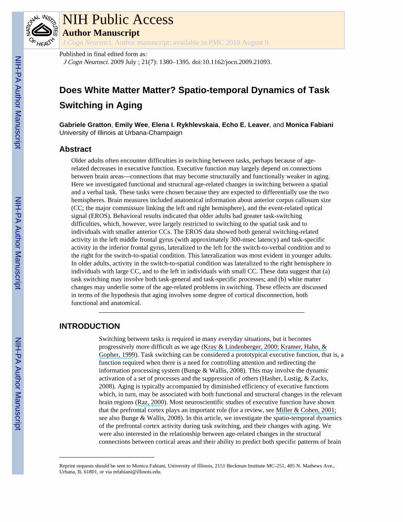
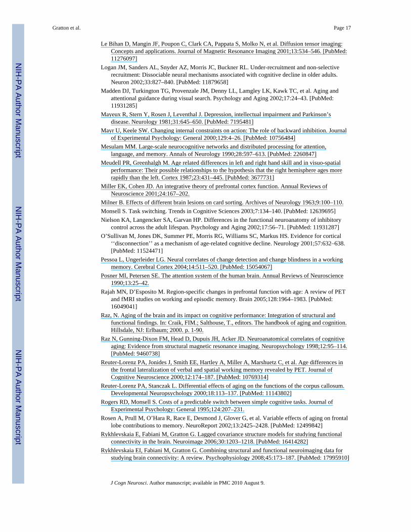
EROS Recording and AnalysisThe data reported here are all time-locked to the onset of the cue. Therefore, they explorepreparatory activities in the absence of response-related activities. Frequency–domain fastoptical data were recorded using an eight-detector Imagent System (ISS, Champaign, IL)concurrently with ERP data. Each detector received light from 10 sources, which were time-multiplexed and arranged such that, during any given time division, only one source waswithin 6 cm of any given detector to avoid cross talk. The detectors (photomultiplier tubes)and sources (laser diodes emitting 690 nm light; average power × 1 mW) were connected tothe subject’s head using optic fibers, which were held in place using a modified motorcyclehelmet. Interdigitized montages were used in the two sessions to achieve a total of 160channels (80 per session). Figure 1 shows an example of the combined montages,coregistered with the structural MR of a representative subject. The yellow and red dots inthe figure indicate the locations of the detectors and sources, respectively. The rows markedby numbers and letters in the left panel indicate the montage for a given session (e.g., row 3and row 3′ were used during different sessions). The order of montages was counterbalancedacross subjects. The montages were chosen to jointly cover most of the frontal lobes, thusincluding the prefrontal regions of interest (ROI) in this study (i.e., superior and middlefrontal gyri and the posterior portion of the IFG).
The light sources were modulated at 220 MHz, and the PMTs were fed a 220.00625-MHzfrequency. This created a 6.25-kHz heterodyning frequency, which was used to computephase delays (the optical-dependent variable used in the study to measure EROS). Opticaldata were obtained continuously over each block at a sampling frequency of 62.5 Hz.
Analysis of the optical data included the following steps: (a) phase range correction (toeliminate phase wrapping around the 360°mark); (b) pulse correction (using a proceduredescribed in Gratton & Corballis, 1995); (c) filtering (using a 0–6 Hz bandpass); (d)segmentation into epochs time-locked to the cue presentation; (e) averaging separately foreach subject, condition, and channel (only data related to the interval between cue andimperative stimulus are presented here); (f ) coregistration with MR data using three fiducialmarkers (based on digitization of the recording locations with a Polhemus 3-D digitizer); (g)transformation into Talairach space (Talairach & Tournoux, 1988); and (h) back projectionof optical data onto the brain surface. Channels crossing the same voxel were averaged
Gratton et al. Page 6
J Cogn Neurosci. Author manuscript; available in PMC 2010 August 9.
NIH
-PA Author Manuscript
NIH
-PA Author Manuscript
NIH
-PA Author Manuscript
together (see Wolf et al., 2000, for a similar approach). Only channels with phase standarddeviation <8°and source–detector distances between 15 and 50 mm were used for theanalysis (see Gratton et al., 2006); (i) computation of individual subject averages; (j)computation of statistical maps across subjects, using an ROI approach. The boundaries ofthe ROI were left: x = −55; right: x = 55; front: y = 40; back: y = 15, which included a broadsection of the prefrontal cortex (BA 6, BA 8, BA 9, and BA 46 on both hemispheres). Datawere corrected for multiple comparisons based on random field theory (see Kiebel, Poline,Friston, Holmes, & Worsley, 1999). (For additional information about analysis of EROSdata, see Gratton & Fabiani, 2007 and Gratton et al., 2006.)
RESULTSCorpus Callosum Size
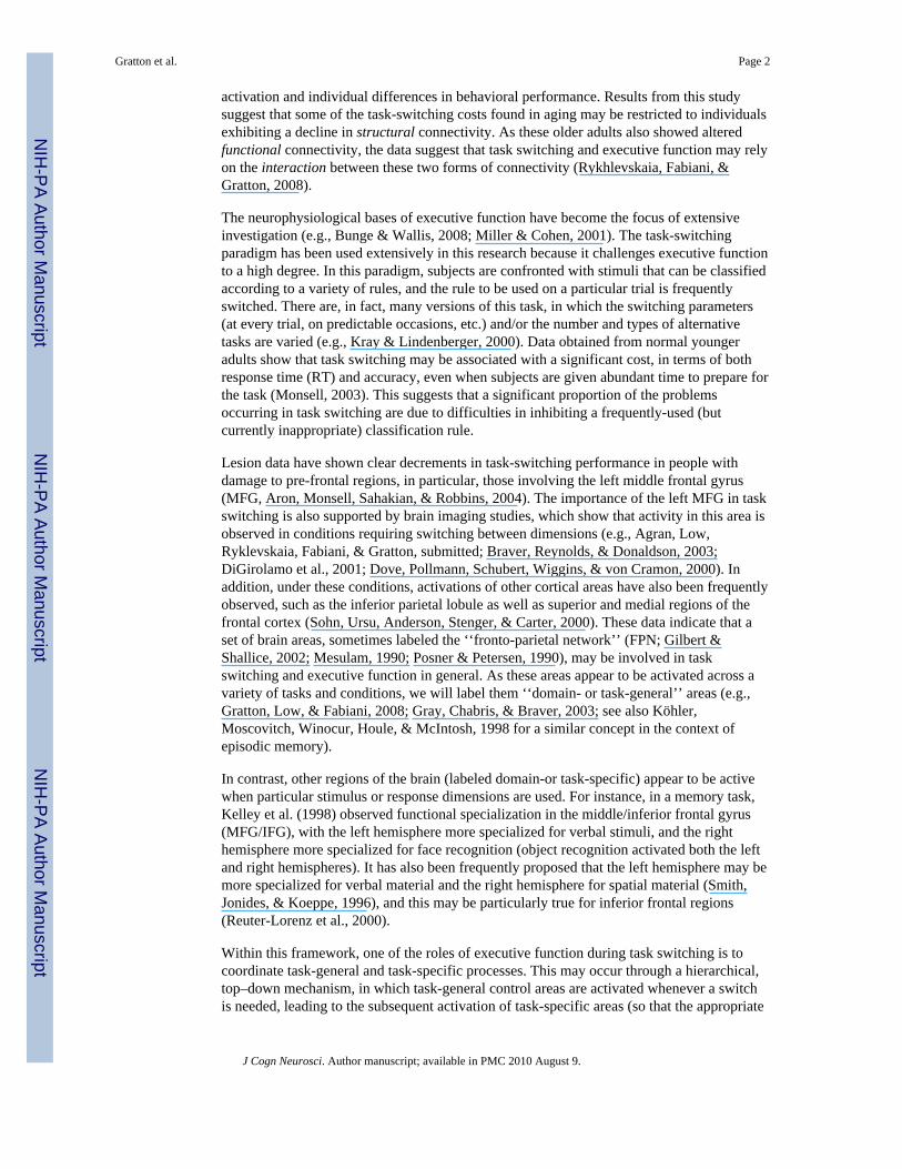
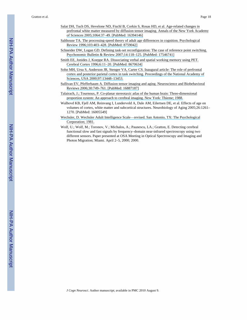
Figure 2 reports a scatterplot of the size of the anterior third of the CC (adjusted by overallbrain volume) and age. On average, the older adults had smaller anterior CCs than theyounger adults [t(30) = 2.13, p < .05]. The data indicate that a substantial proportion of theolder adults had a small CC (i.e., anterior CC size < .0025 of total brain size; see Figure 2)when compared to the younger adults.
BehaviorThe average RT and accuracy for each group (younger and older), switch condition (switchvs. no-switch), and task (meaning vs. position) are presented in Table 2, together with theirstandard deviations. The data are collapsed across congruency levels, for reasons of clarityand because this variable is not critical to the current study. Switch costs, expressed as aproportional RT increase on switch compared to no-switch trials, are presented in Figure 3.
Both RT and accuracy data were submitted to four-way ANOVAs, with one between-subjectfactor (group) and three within-subject factors (task, congruency, and switch condition). ForRT, the ANOVA revealed main effects of group [F(1, 30) = 23.59, p < .0001], task [F(1, 30)= 40.12, p < .0001], congruency [F(1, 30) = 63.94, p < .0001], and switch condition [F(1,30) = 26.15, p < .0001]. It also revealed that age group interacted with switch condition[F(1, 30) = 8.36, p < .05] and congruency [F(1, 30) = 7.10, p < .05]. Congruency and taskalso interacted [F(1, 30) = 14.03, p < .001], and a three-way interaction between group, task,and congruency was also significant [F(1, 30) = 6.07, p < .05]. Thus, all the independentvariables produced an effect on RTs. This included faster RTs for the position taskcompared to the meaning task. The effect of congruency was larger in the older than in theyounger adults. The congruency effect was clearly attenuated in the younger adults in theposition task, but this was not observed in the older adults.
The data presented above indicate that, as predicted, the switch costs for RTs were muchgreater in the older (39 ± 9 msec) than in the younger subjects (14 ± 4 msec). This differenceheld even when the proportional cost of task switching was computed [older adults: meancost = 0.059 ± 0.014; younger adults: mean cost = 0.024 ± 0.008; t(30) = 2.13, p < .05]. Thisindicates that the cost of switching is disproportionate with respect to other RT costsobserved in aging and cannot be simply attributed to generalized slowing (Salthouse, 1996).
To eliminate ceiling effects on the accuracy data, the data for each participant and conditionwere subjected to a probit transform before entering them in the ANOVA. The ANOVArevealed main effects of group [F(1, 30) = 22.89, p < .0001], task [F(1, 30) = 11.35, p < .01], congruency [F(1, 30) = 87.38, p < .0001], and switch condition [F(1, 30) = 10.34, p < .01]. It also revealed a number of two-way interactions, including Age group by Task [F(1,30) = 9.02, p < .05], Task by Congruency [F(1, 30) = 20.61, p < .001], Task by Switchcondition [F(1, 30) = 16.08, p < .001], and Switch condition by Congruency [F(1, 30) =
Gratton et al. Page 7
J Cogn Neurosci. Author manuscript; available in PMC 2010 August 9.
NIH
-PA Author Manuscript
NIH
-PA Author Manuscript
NIH
-PA Author Manuscript
4.64, p < .05]. Further, significant three-way interactions included Group by Task by Switchcondition [F(1, 30) = 6.08, p < .05], and Group by Congruency by Switch condition [F(1,30) = 7.89, p < .01].
Although the pattern of accuracy results was quite complex, the data indicated that olderadults were more accurate than younger adults, suggesting an overall speed–accuracytradeoff between the two groups. However, this phenomenon had little or no influence onthe various interactions of age with the other variables. For instance, the effects of taskswitching on accuracy were virtually identical in the older and younger adults, indicatingthat the greater RT cost for the older adults was not compensated by increased accuracy. Thesame was true for the congruency effect. Most important for the purpose of this article, thedata revealed that younger adults were significantly more accurate in the position than in themeaning task; however, this difference was not apparent in the older adults. This leads totwo observations: in general, the position task was easier (i.e., led to faster and moreaccurate responses than the meaning task); the advantage for the position task was greatlyreduced or lost in the older adults. This finding, per se, is consistent with theories predictingimpaired spatial processing or right hemisphere function in aging (such as the ‘‘righthemiaging’’ hypothesis). Interestingly, this age-related disadvantage in the spatial task onlyoccurred for switch trials. This suggests that older adults may have particular problemsredirecting their processing toward spatial information from verbal information.
One of the main purposes of this study was to determine the extent to which age effects aremediated by anatomical changes, in particular, those related to structural connectivity. Ourhypothesis was that the size of the anterior CC may be important in mediating switch costs,in particular, when the switch involves right-hemisphere tasks (in our case, the positiontask). We therefore hypothesized that switching-to-position costs may be inversely related tothe size of the anterior CC (adjusted by brain volume). The relationship between CC sizeand the RT cost of switching to the meaning and position tasks is presented in Figure 4.These data indicate a clear relationship in the position task (r = −.446, p < .05), but not inthe meaning task (r = .013, ns). As a reminder, the position task, and in particular,switching-to-position, showed the largest age-related deficit. To determine whether thesedeficits are, in fact, mediated by the reduced CC size, we performed a stepwise multipleregression procedure, in which adjusted CC size and age (in years) were entered aspredictors of the switching-to-position costs. The raw correlation of age with switch-to-position costs was r = .388 ( p < .05). However, the results of the multiple regressionindicated that only CC size provided a significant prediction of switch cost (beta = −.446, p< .05), and that once the effects of this variable were removed, age was not significantlycorrelated with switch costs (partial r = .24, ns). This indicates that structural impairment (asmeasured by CC size) may play a significant mediating role in certain behavioral switchcosts associated with aging.
Overall, the behavioral data indicate that, as predicted, older adults found it more difficult toswitch between tasks than younger adults. Interestingly, in particular for the older adults,switching to the spatial task was more difficult (in terms of RT cost) than switching to theverbal task, even though the verbal task was overall easier. To understand this apparentlysurprising finding, it may be useful to consider that task switching requires not onlyactivating a new rule but also inhibiting a previously used rule (see Mayr & Keele, 2000, fora discussion of this issue). Further, on successive trials, the rule that had been previouslyinhibited may need to be reactivated. This may be particularly difficult for the most‘‘dominant’’ or most ‘‘automatic’’ rules—in the current study, those associated with the(easier) spatial task. Mayr and his colleagues have argued that this is because the moreautomatic rule must be strongly inhibited. When this rule has been inhibited, switching backto it is particularly difficult (‘‘backward inhibition hypothesis’’).
Gratton et al. Page 8
J Cogn Neurosci. Author manuscript; available in PMC 2010 August 9.
NIH
-PA Author Manuscript
NIH
-PA Author Manuscript
NIH
-PA Author Manuscript
This hypothesis leads to the prediction that the greater the performance advantage for theposition task relative to the meaning task, the more difficult it will be for subjects to switchback to the position task. To test this hypothesis, we computed an index of ‘‘position-taskadvantage’’ by dividing the RT for the meaning task by the RT for the position task for eachsubject. We then examined the correlation between this index and the switch cost (separatelyfor the switch-to-position and switch-to-meaning conditions). For the younger adults, aspredicted by the backward inhibition hypothesis, the greater the advantage for the positiontask, the greater the switch-to-position cost (r = .39, p < .10 in a directional test) and thesmaller the switch-to-meaning cost (r = −.67, p < .01). Interestingly, the ‘‘position-taskadvantage’’ was also negatively correlated with CC size (r = −.58, p < .01). Thesecorrelations where much smaller for the older adults (r = .05, r = −.50, and r = −.21,respectively). Thus, the data provide some support for the backward inhibition hypothesis.Further, the observations that the switch-to-position costs, as well as the relative advantagefor the position task, are more pronounced in subjects with small CC provide some supportfor a ‘‘cortical disconnection hypothesis,’’ with respect to other theories such as the righthemiaging hypothesis, and suggest that cross-inhibition of homologous frontal areas maycontribute to the observed effects.
One final analysis was performed to test the extent to which practice effect was present,given the fact that data were recorded from many trials over two sessions (see Rogers &Monsell, 1995). This was due to the necessity of collecting data from a denser montage thanallowed by the hardware within one recording session, and to obtain a sufficient signal-to-noise ratio for EROS analyses. We compared the RT and accuracy from the first and secondsessions. Session produced a main effect on RT [F(1, 30) = 6.45, p < .05], which was,however, only present in the older adults [Session × Age interaction: F(1, 30) = 6.55, p < .05]. However, the difference between age groups was still present in the second session.Session also interacted with the switch condition [F(1, 30) = 4.68, p < .05], but the effect ofswitch was present in both sessions (30 msec in Session 1 and 20 msec in Session 2). Foraccuracy, there was no main effect of session, but only a three-way interaction between agegroup, session, and congruency [F(1, 30) = 5.36, p < .05], indicating a reduced effect ofcongruency in older adults in Session 2. Overall, the effects of session were of marginallyreducing (but never eliminating) some of the major effects found on behavior. Qualitatively,the same results were obtained in the second session as in the first session.
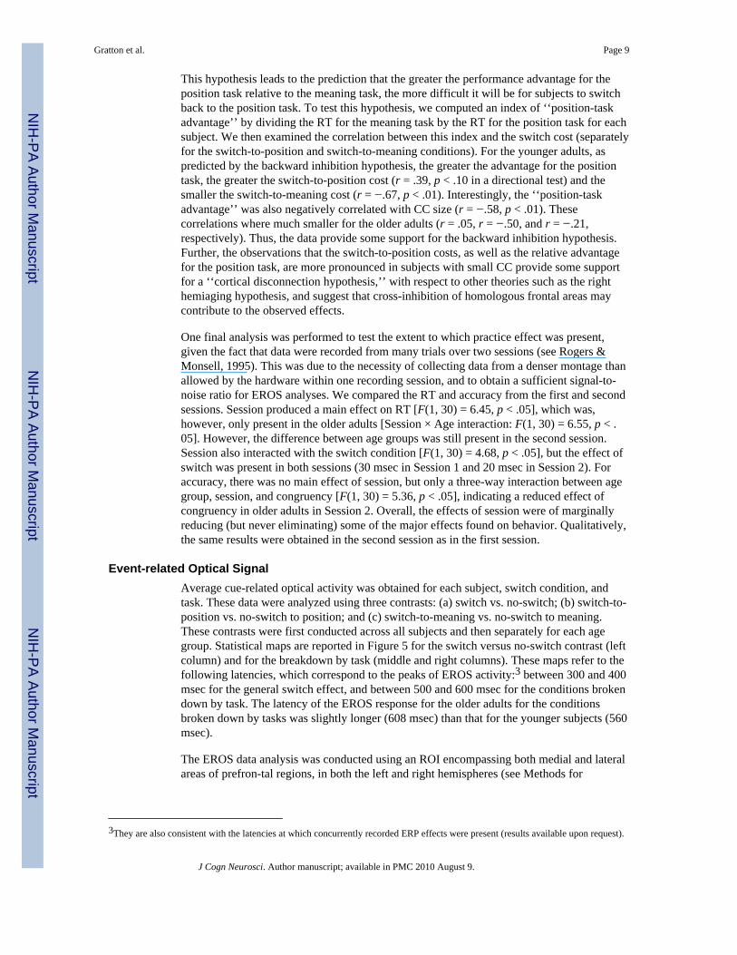
Event-related Optical SignalAverage cue-related optical activity was obtained for each subject, switch condition, andtask. These data were analyzed using three contrasts: (a) switch vs. no-switch; (b) switch-to-position vs. no-switch to position; and (c) switch-to-meaning vs. no-switch to meaning.These contrasts were first conducted across all subjects and then separately for each agegroup. Statistical maps are reported in Figure 5 for the switch versus no-switch contrast (leftcolumn) and for the breakdown by task (middle and right columns). These maps refer to thefollowing latencies, which correspond to the peaks of EROS activity:3 between 300 and 400msec for the general switch effect, and between 500 and 600 msec for the conditions brokendown by task. The latency of the EROS response for the older adults for the conditionsbroken down by tasks was slightly longer (608 msec) than that for the younger subjects (560msec).
The EROS data analysis was conducted using an ROI encompassing both medial and lateralareas of prefron-tal regions, in both the left and right hemispheres (see Methods for
3They are also consistent with the latencies at which concurrently recorded ERP effects were present (results available upon request).
Gratton et al. Page 9
J Cogn Neurosci. Author manuscript; available in PMC 2010 August 9.
NIH
-PA Author Manuscript
NIH
-PA Author Manuscript
NIH
-PA Author Manuscript
boundaries, and green box in Figure 5 for a pictorial representation). The maps are based onaverage Z scores (computed across subjects) for each voxel.
The results indicate the presence of significantly greater brain activation at a latency of 384msec in the left anterior MFG for the switch compared to the no-switch condition (peak Z =3.13, criterion Z = 2.99, peak Talairach coordinates: x = −32, y = 27, z = 38, at the borderbetween BA 8 and BA 9). This response was significant for an interval ranging from 368 to400 msec after the cue, and was present for both the younger and older adults, and for theswitch-to-meaning and switch-to-position tasks. The localization of this responsecorresponds closely to that observed in previous fMRI studies of task switching (Braver etal., 2003; DiGirolamo et al., 2001; Dove et al., 2000). The location of this optical activityand its peak latency also correspond closely to those we observed in a variety of otherpreparatory conditions (see Agran et al., submitted; Gratton et al., 2008). All these studiesindicate that this left MFG activity is, in fact, one in a series of activities within the FPNduring task preparation and attention control. We take the left MFG activity as evidence of a‘‘task-general’’ response—which is operationally defined here as a response that is presentfor both the switch-to-meaning and switch-to-position conditions. The data suggest that this‘‘task-general’’ activity is substantially similar for younger and older adults.
The EROS results also showed a second activity at longer latency (560–608 msec), whichappeared in the left IFG for the switch-to-meaning condition (peak Z = 3.06, critical Z =3.00, peak Talairach coordinates: x = −55, y = 19, z = 25, BA 9) and in the right MFG/IFGfor the switch-to-position condition (peak Z = 2.98, critical Z = 2.98, peak Talairachcoordinates: x = 32, y = 22, z = 45, BA 8, MFG, extending laterally and inferiorly to theIFG). We take this effect as evidence of ‘‘task-specific’’ activity—which is operationallydefined as activity that is systematically lateralized as a function of task condition, and welabel ‘‘canonical’’ the hemisphere that is expected to be more active in a given task (i.e., lefthemisphere for the verbal/meaning task and right hemisphere for the spatial/position task)and ‘‘noncanonical’’ the opposite hemisphere (i.e., left hemisphere for the spatial/positiontask and right hemisphere for the verbal/meaning task). Differently from the left MFGactivity, this more ventral activity (which from now on will be labeled MFG/IFG) showeddifferent patterns in the younger and older subjects. Both groups showed clear left IFGactivity in the switch-to-meaning compared to the no-switch-to-meaning contrast, but onlythe younger subjects showed clear right MFG/IFG activity in the switch-to-position versusno-switch-to-position contrast (see Figure 5).
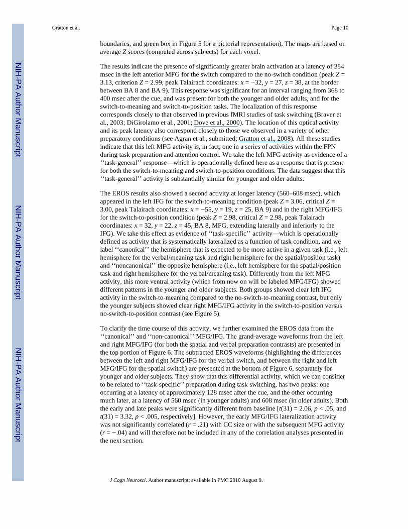
To clarify the time course of this activity, we further examined the EROS data from the‘‘canonical’’ and ‘‘non-canonical’’ MFG/IFG. The grand-average waveforms from the leftand right MFG/IFG (for both the spatial and verbal preparation contrasts) are presented inthe top portion of Figure 6. The subtracted EROS waveforms (highlighting the differencesbetween the left and right MFG/IFG for the verbal switch, and between the right and leftMFG/IFG for the spatial switch) are presented at the bottom of Figure 6, separately foryounger and older subjects. They show that this differential activity, which we can considerto be related to ‘‘task-specific’’ preparation during task switching, has two peaks: oneoccurring at a latency of approximately 128 msec after the cue, and the other occurringmuch later, at a latency of 560 msec (in younger adults) and 608 msec (in older adults). Boththe early and late peaks were significantly different from baseline [t(31) = 2.06, p < .05, andt(31) = 3.32, p < .005, respectively]. However, the early MFG/IFG lateralization activitywas not significantly correlated (r = .21) with CC size or with the subsequent MFG activity(r = −.04) and will therefore not be included in any of the correlation analyses presented inthe next section.
Gratton et al. Page 10
J Cogn Neurosci. Author manuscript; available in PMC 2010 August 9.
NIH
-PA Author Manuscript
NIH
-PA Author Manuscript
NIH
-PA Author Manuscript
Correlations between MeasuresThe EROS data presented above indicate that older adults do not appear to activate the rightMFG/IFG when preparing to switch to a spatial task (Figure 5)—a type of activity that isinstead found in the younger adults. As shown above, older subjects also exhibited specificbehavioral deficits when they had to switch to the spatial task (see Figure 3). In addition,they had an overall reduced anterior CC size, even when adjusted by total brain volume.Finally, reduced anterior CC size was correlated with increased behavioral costs for thespatial task (see Figure 4). In this section, we test whether these phenomena are connected.Specifically, is the impaired right MFG/IFG activity restricted to (or more prominent in)subjects with small anterior CC size? To verify this hypothesis, anterior CC size (adjustedfor total brain volume) was correlated with the amplitude of peak activity in the right MFG/IFG minus that in the left MFG/IFG at a latency of 560 msec (for the younger adults) and608 msec (for the older adults) for the switch-to-spatial condition. The data are presented inthe left portion of Figure 7. This figure indicates that the right MFG/IFG activity for thespatial switch was correlated with CC size (r = .39, p < .05);4 however (central panel ofFigure 7), CC size was not as good of a predictor of left MFG/IFG activity for the verbalswitch (r = −.32, p < .07). In fact, the presence of a negative, albeit not significant,correlation suggests that subjects with small CCs may use the left hemisphere to prepare forthe meaning task at least equally, or perhaps even more, than subjects with large CCs(perhaps due to diminished cross-inhibitory control; Rykhlevskaia, Fabiani, & Gratton,2006; see also Mayr & Keele, 2000). This was also supported by the lack of correlationbetween CC size and RT costs in the switch-to-meaning condition (Figure 4, right). Finally,the right portion of Figure 7 shows that only subjects with small CC exhibited left MFG/IFGactivity in the switch-to-position condition (r = −.52, p < .01). All these CC-size effectswere evident for both younger and older adults, although, overall, there were fewer youngeradults with small CC sizes. The significance of the (non-canonical) left-hemisphere MFG/IFG activity in switching to the position task can also be corroborated by testing itscorrelation with the RT switch cost. In fact, for the switch-to-position condition, the amountof left MFG/IFG activity elicited by the cue was a good predictor of the RT switch cost inresponse to the upcoming imperative stimulus (r = .60, p < .001), suggesting that thisactivity has negative behavioral consequences.
The data presented in Figure 7 suggest that the size of the anterior CC (or some otheranatomical change of which it may be a proxy) may be important in mediating the activity intask-specific areas (MFG/IFG), but only when these areas are located in the righthemisphere. As the left MFG is active at an earlier time during task switching, it is possiblethat the anterior CC may play an important role in relaying information to the righthemisphere when switching-to-spatial task is required. If this is the case, then how dosubjects with small CC perform the switch-to-position operation? One possibility is that theymay perform it by activating left-hemisphere structures instead.
To verify this hypothesis, we separately analyzed the left and right MFG/IFG activity(EROS measures at a latency of 560 msec for the younger adults and 608 msec for the olderadults) in subjects with large and small anterior CC. These results are presented in Figure 8,and indicate that (a) the large CC subjects showed predominantly left-hemisphere activityfor the switch-to-verbal condition and predominantly right-hemisphere activity for theswitch-to-spatial condition; conversely, (b) the subjects with small CC showed left-lateralized activity in both cases. In fact, the left MFG/IFG was significantly more active inthe switch-to-spatial task for the small CC group than for the large CC group [t(24) = 2.26, p
4Note that age was not significantly correlated with this lateralized activity in preparation for a switch to the spatial task (r = −.21).This suggests that CC size rather than age is the critical factor.
Gratton et al. Page 11
J Cogn Neurosci. Author manuscript; available in PMC 2010 August 9.
NIH
-PA Author Manuscript
NIH
-PA Author Manuscript
NIH
-PA Author Manuscript
< .05].5 Further, in the small CC group, there was no significant difference between left andright MFG/IFG activity for the spatial switch [t(12) = 0.14], whereas a significant differencewas observed in the large CC group [t(11) = 2.26, p < .05]. Finally, as presented above, theamount of left MFG/IFG activity during spatial switching was inversely correlated withadjusted CC size (right portion of Figure 7).
DISCUSSIONThe EROS data indicate that the left6 MFG was activated when subjects were required toswitch between tasks in preparation for processing an upcoming imperative stimulus. Thelatency of this activity was between 350 and 400 msec, and was evident for both youngerand older subjects, and for both types of switch (switch-to-meaning and switch-to-position),suggesting that it may contribute to task-general preparatory processes (i.e., processes thatdo not depend on specific task dimensions). In contrast, more ventral prefrontal areas in theleft and right MFG/IFG were each active only for one type of switch (at least in youngersubjects): the left for the switch-to-meaning condition and the right for the switch-to-position condition. In fact, the older adults exhibited, on average, weaker activity in the rightMFG/ IFG for the switch-to-position task. Analyses of the relationship between this activityand anterior CC size revealed that subjects with small CC (irrespective of age; see Figure 7,right) actually showed activity in the left MFG/IFG, rather than in the right MFG/IFG, aftercues signaling that a switch-to-position was needed. These subjects also appeared to havedisproportionate RT costs in the switch-to-position condition. Finally, the amplitude of theMFG/IFG activity was predictive of subsequent switch costs, thus suggesting that it reflectsfunctionally important brain events. These results suggest that some of the problems thatolder adults exhibit in switching between tasks may be attributed to basic structural deficits,which in the current study were linked with CC size. It is important to note, however, thatsimilar phenomena were also apparent in younger adults, albeit to a lesser degree due to thesmaller number of subjects with small CCs in this age group.
Two hypotheses can be entertained to explain the connection between the structural andfunctional data reported here. One possibility is that reduced CC size may lead to reducedright MFG/IFG activation (as the earlier task-general MFG activity was lateralized to theleft hemisphere, thus requiring callosal transfer when right MFG/ IFG activation wasrequired)—a condition that might be labeled a ‘‘partial disconnection’’ syndrome (for asimilar suggestion, see Reuter-Lorenz & Stanczak, 2000). Alternatively, both the reducedCC size and the absence of functional activation in the right MFG/IFG may be explained bya structural/functional right prefrontal deficit, consistent with the ‘‘right hemiaging’’hypothesis (Dase-laar & Cabeza, 2005; Meudell & Greenhalgh, 1987; see also Rajah &D’Esposito, 2005). The latter hypothesis, however, fails to predict the existence of anegative correlation between the performance advantage in the position task and CC size.
Overall, these data highlight a network involved in task switching, which includes both task-general and task-specific structures, as well as variations in the operation of these structuresas a function of aging and anatomical differences, with related behavioral costs. Somecaveats, however, should be considered. To begin with, due to the recording system used inthis study, we were limited to an examination of only the anterior portion of the FPN, andtherefore, the role of parietal and other regions was not investigated. Second, efficient taskswitching should be considered an outcome to which much of the information processingsystem is likely to contribute. As such, it may involve a number of elementary operations
5The reduced n for this analysis is due to the fact that, due to different head shapes and sizes, not all subjects had coverage in this area.6Although it is not possible, on the basis of these data alone, to determine why the cue elicits activity preferentially in the left (ratherthe right) MFG, one can hypothesize that verbal cues (letters) may bias processing to the left hemisphere.
Gratton et al. Page 12
J Cogn Neurosci. Author manuscript; available in PMC 2010 August 9.
NIH
-PA Author Manuscript
NIH
-PA Author Manuscript
NIH
-PA Author Manuscript
(Crone, Wendelen, Donohue, & Bunge, 2006). The left MFG activity we observed isunlikely to represent all of the operations involved in task switching, and possibly not evenall of those involved in its task-general phase (Gratton et al., 2008). In fact, the resultspresented here are consistent with various interpretations of left MFG activity, including thatit may manifest change detection (e.g., Pessoa & Ungerleider, 2004) and/or the redirectingof attention (e.g., Gazzaley et al., 2007; Miller & Cohen, 2001). In fact, all of theseprocesses may contribute to task switching (see Schneider & Logan, 2007). However, it isalso possible that activation of the left MFG may not even be necessary for task switching tooccur. Because task switching is an operation defined at the level of the entire system, itmay be achieved in different ways under different conditions. Thus, although the left MFGmay be important for task switching (as suggested by lesion studies, e.g., Aron et al., 2004),in some cases, task switching could be achieved before, or even in the absence of, left MFGactivation. In fact, some of the data in our study are suggestive that this may occur, as MFG/IFG activity separating task-specific switch from no-switch trials was observed even beforethe 350-msec latency landmark for peak MFG activation (Figure 6, bottom; see also Agranet al., submitted). The possibility that ‘‘task-specific’’ activation may precede ‘‘task-general’’ activation clearly indicates that task switching may occur not only through a top–down, hierarchically organized process but also through a distributed process. A possibleinterpretation of this finding is that, with time on task, participants may develop a ‘‘direct’’link between the cue percept and task-specific areas, perhaps profiting from the consistentstimulus mapping over the large number of trials used in this study. If this were the case,then the early activation of the MFG/IFG should be observed only after extended training.Future research will investigate this issue,7 but in any case, this finding highlights theimportance of a spatio-temporal analysis of brain activity. In summary, it is likely that taskswitching may be achieved with different strategies, including a hierarchical, centralizedprocessing mode in which the left MFG exerts an important role, and a more distributed andautomatic processing mode, which may depend instead on more ventral areas (MFG/IFG).
An interesting aspect of this study was the change in the overall pattern of brain activity as afunction of aging, a finding commonly reported in neuroimaging aging research (forreviews, see Kramer, Fabiani, & Colcombe, 2006; Cabeza, 2002). For instance, a number ofinvestigators using fMRI or PET have reported bilateral brain activations in aging duringtasks that elicit largely unilateral activations in younger subjects (e.g., Colcombe, Kramer,Erickson, & Scalf, 2005; Cabeza, Anderson, Locantore, & McIntosh, 2002; Logan, Sanders,Snyder, Morris, & Buckner, 2002; Madden et al., 2002; Nielson, Langencker, & Garvan,2002; Rosen et al., 2002; Grady, McIntosh, Horwitz, & Rapoport, 2000; Reuter-Lorenz etal., 2000; see also Fabiani & Friedman, 1995). In the current study, the change in brainactivation pattern (which included bilateral activation of the MFG/IFG; see Figure 8) wasrestricted to individuals with specific structural characteristics (small anterior CC size), and,in fact, CC size was more strongly correlated with bilateral activation than age. Thisbilateral activation in subjects with small CC may be associated with reduced cross-hemispheric inhibition (Rykhlevskaia et al., 2006; see also Hasher et al., 2008).
The data reported here are consistent with the interpretation that individuals (and olderadults in particular) adapt to the constraints imposed by the structural changes/characteristicsof their brains, and ‘‘configure’’ their strategies (and patterns of brain activity) in order toperform the task. In the current study, subjects with small CC activated the left MFG/IFG,instead of the right, when a ‘‘switch-to-position’’ was required. It should be noted that,although these subjects also showed greater switch costs, their performance was still wellabove chance. Thus, whatever their strategies, it is clear that they were at least partially
7Such analysis is difficult in the current study due to the fact that different montages were used across sessions, with the montageorder counterbalanced across subjects.
Gratton et al. Page 13
J Cogn Neurosci. Author manuscript; available in PMC 2010 August 9.
NIH
-PA Author Manuscript
NIH
-PA Author Manuscript
NIH
-PA Author Manuscript
successful. Of course, because these data are only correlational, it is not possible to stateconclusively that the functional reconfiguration we observed was caused by reduced CC size(or a structural deficit linked to it), and that, in turn, this caused the drop in performance.
Another hypothesis that could be proposed is that a small CC size is merely a proxy for anoverall impaired brain, which would be challenged by more difficult tasks. According to thishypothesis, older adults with small CC might show a deficit in spatial switching simplybecause this is more difficult than verbal switching, following the ‘‘backward inhibitionhypothesis’’ (Mayr & Keele, 2000). Two results are, in fact, not consistent with theinterpretation that subjects with small CC have an overall less efficient brain. First, the RTadvantage for the position task was even greater in the subjects with small CC than in thesubject with large CC, suggesting that their drop in performance is not generalized. Second,this hypothesis does not explain why the subjects with smaller CC activated the left MFG/IFG while performing the spatial task (a finding that is instead predictable on the basis of thepartial disconnection hypothesis).
The data reported here also present some limitations, which may be addressed by futurestudies. First, due to the design of the optical system used in this study, EROS data wereonly recorded from prefrontal areas. Some of the areas important in task switching andattentional control, such as deep thalamic structures, may be beyond the reach of opticalmethods, but other more superficial areas such as the parietal cortex are accessible withoptical imaging. We have now developed a more extended EROS montage, which will allowus to cover the majority of the cortical surface. Second, only one type of task switching(between spatial and verbal dimensions) was studied. Other conditions need to be exploredto demonstrate the generalizability of the results. We are currently exploring task-switchingparadigms involving other dimensions (Gratton et al., 2008). Third, the task-switchingconditions used here were confounded with other variables (such as cue change vs. cuerepetition; see Schneider & Logan, 2007). This makes the interpretation of some of thefindings ambiguous. For example, we cannot determine whether the left MFG activity wasrelated to the detection of cue changes or to a change in the weight given to different rules.Using additional conditions (such as one in which multiple cues may signal the samedimension) may help resolve this ambiguity. Fourth, although the behavioral and functionalcorrelations with CC size clearly indicated the role of anatomical and structural changes inmediating the effects of task switching, the interpretation of these changes is alsoambiguous. It is possible that CC size (even when corrected by overall brain volume) maystill be a proxy for a more diffused structural change, or for structural changes localized inspecific cortical regions (such as the right MFG/IFG). Indeed, several recent studies indicatethat aging is associated with both a reduction in anterior medial white matter (i.e., anteriorCC size) and a reduction in gray matter in the dorsolat-eral prefrontal cortex as well in otherbrain regions (e.g., Gordon et al., 2008; Salat et al., 2005; Walhovd et al., 2005; Head et al.,2004; Davatzikos & Resnick, 2002; Raz et al., 1998). More refined anatomical measures,beyond the capabilities of the current study, may be needed to evaluate these alternativehypotheses. Coupling diffusion tensor imaging (for a review, see Le Bihan et al., 2001) and/or advanced morphological measures of cortical structures (e.g., voxel-based morphometry;Ashburner & Friston, 2000, 2001) with the types of measurements presented here may helpus determine whether the changes in CC size are related to specific or diffuse changes incortical anatomy and trophism. Fifth, a pure-run condition (i.e., one in which some blocksare entirely made up by switch or no-switch trials) was not used in the current study. Thismakes it difficult to evaluate the global costs of task switching (Kray & Lindenberger,2000).
Notwithstanding these limitations, the data presented here do provide clear indications that acombined approach based on structural and time-resolved functional analysis of changes
Gratton et al. Page 14
J Cogn Neurosci. Author manuscript; available in PMC 2010 August 9.
NIH
-PA Author Manuscript
NIH
-PA Author Manuscript
NIH
-PA Author Manuscript
occurring with age can be useful in isolating components of cognitive aging. Specifically,we provided indications that some of the problems experienced by older adults in taskswitching may be associated with functional reprogramming, perhaps as a consequence oflocalized impairment in anatomical connectivity.
AcknowledgmentsThis work was funded by DARPA (via NSF EIA 00-79800 AFK) to G. Gratton and M. Fabiani, and by NIA grantAG 21887 to M. Fabiani. We thank Brian Gordon, Art Kramer, and Kathy Low for their comments on an earlierversion of this manuscript.
ReferencesAgran J, Low KA, Ryklevskaia EI, Fabiani M, Gratton G. When the rules keep changing: The timing
of activation of task-general and task-specific brain regions involved in preparation. submitted.Aron AR, Monsell S, Sahakian BJ, Robbins TW. A componential analysis of task-switching deficits
associated with lesions of left and right frontal cortex. Brain 2004;127:1561–1573. [PubMed:15090477]
Ashburner J, Friston KJ. Voxel-based morphometry: The methods. Neuroimage 2000;11:805–821.[PubMed: 10860804]
Ashburner J, Friston KJ. Why voxel-based morphometry should be used. Neuroimage 2001;14:1454–1462. [PubMed: 11707101]
Beck, AT.; Steer, RA.; Brown, GK. Manual for the Beck depression inventory. 2. San Antonio, TX:The Psychological Corporation; 1996.
Braver T, Reynolds J, Donaldson D. Neural mechanisms of transient and sustained cognitive controlduring task switching. Neuron 2003;39:713–726. [PubMed: 12925284]
Bunge, SA.; Wallis, JD., editors. Perspectives on rule-guided behavior. New York: Oxford UniversityPress; 2008.
Cabeza R. Hemispheric asymmetry reduction in older adults: The HAROLD model. Psychology andAging 2002;17:85–100. [PubMed: 11931290]
Cabeza R, Anderson ND, Locantore JK, McIntosh AR. Aging gracefully: Compensatory brain activityin high-performing older adults. Neuroimage 2002;17:1394–1402. [PubMed: 12414279]
Colcombe SJ, Kramer AF, Erickson KI, Scalf P. The implications of cortical recruitment and brainmorphology for individual differences in cognitive performance in aging humans. Psychology andAging 2005;20:363–375. [PubMed: 16248697]
Crone EA, Wendelen C, Donohue SE, Bunge SA. Neural evidence for dissociable components of task-switching. Cerebral Cortex 2006;16:475–486. [PubMed: 16000652]
Daselaar, S.; Cabeza, R. Age-related changes in hemispheric organization. In: Cabeza, R.; Nyberg, L.;Park, D., editors. Cognitive neuroscience of aging: Linking cognitive and cerebral aging. NewYork: Oxford University Press; 2005. p. 325-353.
Davatzikos C, Resnick SM. Degenerative age changes in white matter connectivity visualized in vivousing magnetic resonance imaging. Cerebral Cortex 2002;12:767–771. [PubMed: 12050088]
DeSoto MC, Fabiani M, Geary DC, Gratton G. When in doubt, do it both ways: Brain evidence of thesimultaneous activation of conflicting responses in a spatial Stroop task. Journal of CognitiveNeuroscience 2001;13:523–536. [PubMed: 11388924]
DiGirolamo GJ, Kramer AF, Barad V, Cepeda NJ, Weissman DH, Milham MP, et al. General andtask-specific frontal lobe recruitment in older adults during executive processes: A fMRIinvestigation of task-switching. NeuroReport 2001;12:2065–2071. [PubMed: 11435947]
Dove A, Pollmann S, Schubert T, Wiggins CJ, von Cramon DY. Prefrontal cortex activation in taskswitching: An event-related fMRI Study. Cognitive Brain Research 2000;9:103–109. [PubMed:10666562]
Fabiani M, Friedman D. Changes in brain activity patterns in aging: The novelty oddball.Psychophysiology 1995;32:579–594. [PubMed: 8524992]
Gratton et al. Page 15
J Cogn Neurosci. Author manuscript; available in PMC 2010 August 9.
NIH
-PA Author Manuscript
NIH
-PA Author Manuscript
NIH
-PA Author Manuscript
Gazzaley A, Rissman J, Cooney J, Rutman A, Seibert T, Clapp W, et al. Functional interactionsbetween prefrontal and visual association cortex contribute to top–down modulation of visualprocessing. Cerebral Cortex 2007;17:125–135.
Gilbert SJ, Shallice T. Task switching: A PDP model. Cognitive Psychology 2002;44:297–337.[PubMed: 11971634]
Gordon B, Rykhlevskaia E, Brumback CR, Lee Y, Elavsky S, Konopack JF, et al. Anatomicalcorrelates of aging, cardiopulmonary fitness level, and education. Psychophysiology 2008;45:825–838. [PubMed: 18627534]
Grady CL, McIntosh AR, Horwitz B, Rapoport SI. Age-related changes in the neural correlates ofdegreaded and nondegraded face processing. Cognitive Neuropsychology 2000;217:165–186.
Gratton G, Brumback CR, Gordon BA, Pearson MA, Low KA, Fabiani M. Effects of measurementmethod, wavelength, and source–detector distance on the fast optical signal. Neuroimage2006;32:1576–1590. [PubMed: 16872842]
Gratton G, Corballis PM. Removing the heart from the brain: Compensation for the pulse artifact inthe photon migration signal. Psychophysiology 1995;32:292–299. [PubMed: 7784538]
Gratton G, Corballis PM, Cho E, Fabiani M, Hood D. Shades of gray matter: Noninvasive opticalimages of human brain responses during visual stimulation. Psychophysiology 1995;32:505–509.[PubMed: 7568645]
Gratton G, Fabiani M. Shedding light on brain function: The event-related optical signal. Trends inCognitive Sciences 2001;5:357–363. [PubMed: 11477005]
Gratton, G.; Fabiani, M. Optical imaging. In: Parasuraman, R.; Rizzo, M., editors. Neuroergonomics:The brain at work. Cambridge: Oxford University Press; 2007. p. 65-81.
Gratton, G.; Low, KA.; Fabiani, M. Time course of executive processes: Data from the event-relatedoptical signal (EROS). In: Bunge, SA.; Wallis, JD., editors. Perspectives on rule-guided behavior.New York: Oxford University Press; 2008. p. 197-223.
Gray CF, Chabris JR, Braver T. Neural mechanisms of general fluid intelligence. Nature Neuroscience2003;6:316–322.
Gunning-Dixon FM, Raz N. Neuroanatomical correlates of selected executive function in middle-agedand older adults: A prospective MRI study. Neuropsychologia 2003;41:1929–1941. [PubMed:14572526]
Hasher, L.; Lustig, C.; Zacks, R. Inhibitory mechanisms and the control of attention. In: Conway, A.;Jarrold, C.; Kane, M.; Miyake, A.; Towse, J., editors. Variation in working memory. New York:Oxford University Press; 2008. p. 227-249.
Head D, Buckner RL, Shimony JS, Williams LE, Akbudak E, Conturo TE, et al. Differentialvulnerability of anterior white matter in nondemented aging with minimal acceleration in dementiaof the Alzheimer type: Evidence from diffusion tensor imaging. Cerebral Cortex 2004;14:410–423. [PubMed: 15028645]
Kelley WM, Miezin FM, McDermott KB, Buckner RL, Raichle ME, Cohen NJ, et al. Dorsal frontalcortex and medial temporal lobe for verbal and nonverbal memory encoding. Neuron1998;20:927–936. [PubMed: 9620697]
Kiebel SJ, Poline JB, Friston KJ, Holmes AP, Worsley KJ. Robust smoothness estimation in statisticalparametric maps using standardized residuals from the general linear model. Neuroimage1999;10:756–766. [PubMed: 10600421]
Köhler S, Moscovitch M, Winocur G, Houle S, McIntosh AR. Networks of domain-specific andgeneral regions involved in episodic memory for spatial location and object identity.Neuropsychologia 1998;36:129–142. [PubMed: 9539233]
Kramer, AF.; Fabiani, M.; Colcombe, S. Contributions of cognitive neuroscience to the understandingof behavior and aging. In: Birren, JE.; Schaie, KW., editors. Handbook of the psychology of aging.6. New York: Academic Press; 2006. p. 57-83.
Kramer AF, Hahn S, Gopher D. Task coordination and aging: Explorations of executive controlprocesses in the task switching paradigm. Acta Psychologica 1999;101:339–378. [PubMed:10344190]
Kray J, Lindenberger U. Adult age differences in task switching. Psychology and Aging 2000;15:126–147. [PubMed: 10755295]
Gratton et al. Page 16
J Cogn Neurosci. Author manuscript; available in PMC 2010 August 9.
NIH
-PA Author Manuscript
NIH
-PA Author Manuscript
NIH
-PA Author Manuscript
Le Bihan D, Mangin JF, Poupon C, Clark CA, Pappata S, Molko N, et al. Diffusion tensor imaging:Concepts and applications. Journal of Magnetic Resonance Imaging 2001;13:534–546. [PubMed:11276097]
Logan JM, Sanders AL, Snyder AZ, Morris JC, Buckner RL. Under-recruitment and non-selectiverecruitment: Dissociable neural mechanisms associated with cognitive decline in older adults.Neuron 2002;33:827–840. [PubMed: 11879658]
Madden DJ, Turkington TG, Provenzale JM, Denny LL, Lamgley LK, Kawk TC, et al. Aging andattentional guidance during visual search. Psychology and Aging 2002;17:24–43. [PubMed:11931285]
Mayeux R, Stern Y, Rosen J, Leventhal J. Depression, intellectual impairment and Parkinson’sdisease. Neurology 1981;31:645–650. [PubMed: 7195481]
Mayr U, Keele SW. Changing internal constraints on action: The role of backward inhibition. Journalof Experimental Psychology: General 2000;129:4–26. [PubMed: 10756484]
Mesulam MM. Large-scale neurocognitive networks and distributed processing for attention,language, and memory. Annals of Neurology 1990;28:597–613. [PubMed: 2260847]
Meudell PR, Greenhalgh M. Age related differences in left and right hand skill and in visuo-spatialperformance: Their possible relationships to the hypothesis that the right hemisphere ages morerapidly than the left. Cortex 1987;23:431–445. [PubMed: 3677731]
Miller EK, Cohen JD. An integrative theory of prefrontal cortex function. Annual Reviews ofNeuroscience 2001;24:167–202.
Milner B. Effects of different brain lesions on card sorting. Archives of Neurology 1963;9:100–110.Monsell S. Task switching. Trends in Cognitive Sciences 2003;7:134–140. [PubMed: 12639695]Nielson KA, Langencker SA, Garvan HP. Differences in the functional neuroanatomy of inhibitory
control across the adult lifespan. Psychology and Aging 2002;17:56–71. [PubMed: 11931287]O’Sullivan M, Jones DK, Summer PE, Morris RG, Williams SC, Markus HS. Evidence for cortical
‘‘disconnection’’ as a mechanism of age-related cognitive decline. Neurology 2001;57:632–638.[PubMed: 11524471]
Pessoa L, Ungerleider LG. Neural correlates of change detection and change blindness in a workingmemory. Cerebral Cortex 2004;14:511–520. [PubMed: 15054067]
Posner MI, Petersen SE. The attention system of the human brain. Annual Reviews of Neuroscience1990;13:25–42.
Rajah MN, D’Esposito M. Region-specific changes in prefrontal function with age: A review of PETand fMRI studies on working and episodic memory. Brain 2005;128:1964–1983. [PubMed:16049041]
Raz, N. Aging of the brain and its impact on cognitive performance: Integration of structural andfunctional findings. In: Craik, FIM.; Salthouse, T., editors. The handbook of aging and cognition.Hillsdale, NJ: Erlbaum; 2000. p. 1-90.
Raz N, Gunning-Dixon FM, Head D, Dupuis JH, Acker JD. Neuroanatomical correlates of cognitiveaging: Evidence from structural magnetic resonance imaging. Neuropsychology 1998;12:95–114.[PubMed: 9460738]
Reuter-Lorenz PA, Jonides J, Smith EE, Hartley A, Miller A, Marshuetz C, et al. Age differences inthe frontal lateralization of verbal and spatial working memory revealed by PET. Journal ofCognitive Neuroscience 2000;12:174–187. [PubMed: 10769314]
Reuter-Lorenz PA, Stanczak L. Differential effects of aging on the functions of the corpus callosum.Developmental Neuropsychology 2000;18:113–137. [PubMed: 11143802]
Rogers RD, Monsell S. Costs of a predictable switch between simple cognitive tasks. Journal ofExperimental Psychology: General 1995;124:207–231.
Rosen A, Prull M, O’Hara R, Race E, Desmond J, Glover G, et al. Variable effects of aging on frontallobe contributions to memory. NeuroReport 2002;13:2425–2428. [PubMed: 12499842]
Rykhlevskaia E, Fabiani M, Gratton G. Lagged covariance structure models for studying functionalconnectivity in the brain. Neuroimage 2006;30:1203–1218. [PubMed: 16414282]
Rykhlevskaia EI, Fabiani M, Gratton G. Combining structural and functional neuroimaging data forstudying brain connectivity: A review. Psychophysiology 2008;45:173–187. [PubMed: 17995910]
Gratton et al. Page 17
J Cogn Neurosci. Author manuscript; available in PMC 2010 August 9.
NIH
-PA Author Manuscript
NIH
-PA Author Manuscript
NIH
-PA Author Manuscript
Salat DH, Tuch DS, Hevelone ND, Fischl B, Corkin S, Rosas HD, et al. Age-related changes inprefrontal white matter measured by diffusion tensor imaging. Annals of the New York Academyof Sciences 2005;1064:37–49. [PubMed: 16394146]
Salthouse TA. The processing-speed theory of adult age differences in cognition. PsychologicalReview 1996;103:403–428. [PubMed: 8759042]
Schneider DW, Logan GD. Defining task-set reconfiguration: The case of reference point switching.Psychonomic Bulletin & Review 2007;14:118–125. [PubMed: 17546741]
Smith EE, Jonides J, Koeppe RA. Dissociating verbal and spatial working memory using PET.Cerebral Cortex 1996;6:11–20. [PubMed: 8670634]
Sohn MH, Ursu S, Anderson JR, Stenger VA, Carter CS. Inaugural article: The role of prefrontalcortex and posterior parietal cortex in task switching. Proceedings of the National Academy ofSciences, USA 2000;97:13448–13453.
Sullivan EV, Pfefferbaum A. Diffusion tensor imaging and aging. Neuroscience and BiobehavioralReviews 2006;30:749–761. [PubMed: 16887187]
Talairach, J.; Tournoux, P. Co-planar stereotaxic atlas of the human brain: Three-dimensionalproportion system: An approach to cerebral imaging. New York: Thieme; 1988.
Walhovd KB, Fjell AM, Reinvang I, Lundervold A, Dale AM, Eilertsen DE, et al. Effects of age onvolumes of cortex, white matter and subcortical structures. Neurobiology of Aging 2005;26:1261–1270. [PubMed: 16005549]
Wechsler, D. Wechsler Adult Intelligence Scale—revised. San Antonio, TX: The PsychologicalCorporation; 1981.
Wolf, U.; Wolf, M.; Toronov, V.; Michalos, A.; Paunescu, LA.; Gratton, E. Detecting cerebralfunctional slow and fast signals by frequency–domain near-infrared spectroscopy using twodifferent sensors. Paper presented at OSA Meeting in Optical Spectroscopy and Imaging andPhoton Migration; Miami. April 2–5, 2000; 2000.
Gratton et al. Page 18
J Cogn Neurosci. Author manuscript; available in PMC 2010 August 9.
NIH
-PA Author Manuscript
NIH
-PA Author Manuscript
NIH
-PA Author Manuscript
Figure 1.The digitized locations of sources (red) and detectors (yellow) coregistered with a structuralMRI viewed from the axial (left panel, nose at top) and coronal (middle panel) planes, andprojected onto the brain (right panel). The numbered and lettered rows in the left panelindicate the montage for a given session (e.g., row B and row 3 would be used for onemontage and row B′ and row 3′ would be used for the second montage, with the order ofmontages counterbalanced across subjects).
Gratton et al. Page 19
J Cogn Neurosci. Author manuscript; available in PMC 2010 August 9.
NIH
-PA Author Manuscript
NIH
-PA Author Manuscript
NIH
-PA Author Manuscript
Figure 2.Scatterplot of volume-adjusted anterior corpus callosum (CC) size by age. CC size isexpressed as a proportion of the total brain volume (multiplied by 1000).
Gratton et al. Page 20
J Cogn Neurosci. Author manuscript; available in PMC 2010 August 9.
NIH
-PA Author Manuscript
NIH
-PA Author Manuscript
NIH
-PA Author Manuscript
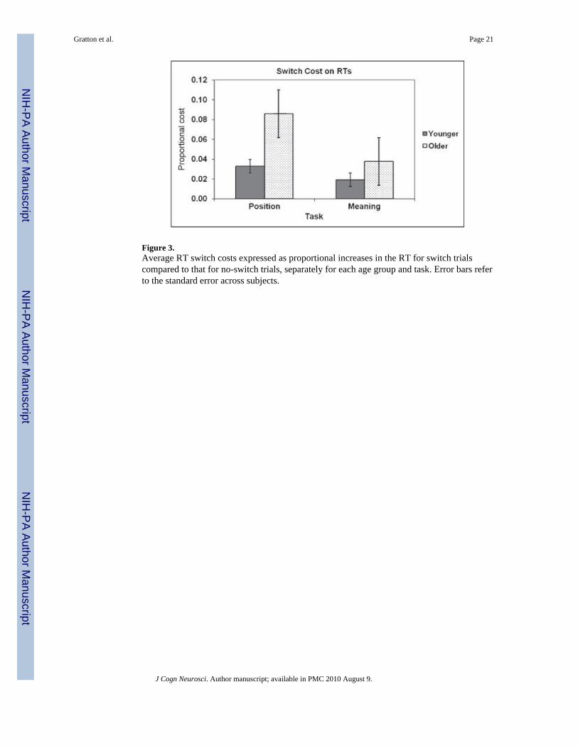
Figure 3.Average RT switch costs expressed as proportional increases in the RT for switch trialscompared to that for no-switch trials, separately for each age group and task. Error bars referto the standard error across subjects.
Gratton et al. Page 21
J Cogn Neurosci. Author manuscript; available in PMC 2010 August 9.
NIH
-PA Author Manuscript
NIH
-PA Author Manuscript
NIH
-PA Author Manuscript
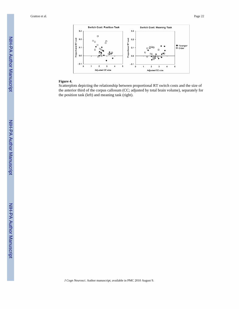
Figure 4.Scatterplots depicting the relationship between proportional RT switch costs and the size ofthe anterior third of the corpus callosum (CC; adjusted by total brain volume), separately forthe position task (left) and meaning task (right).
Gratton et al. Page 22
J Cogn Neurosci. Author manuscript; available in PMC 2010 August 9.
NIH
-PA Author Manuscript
NIH
-PA Author Manuscript
NIH
-PA Author Manuscript
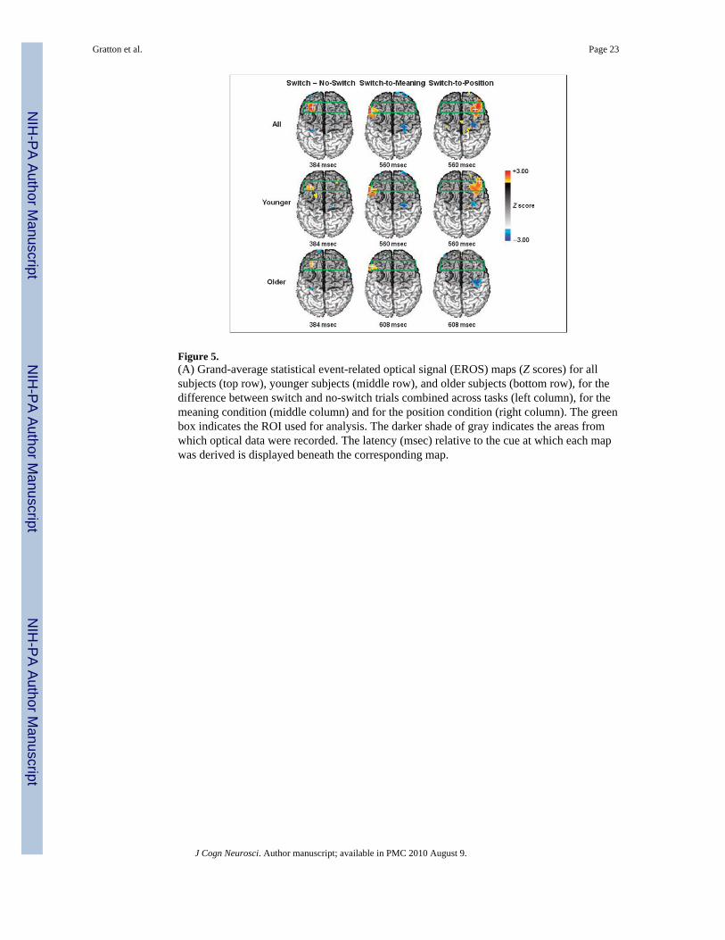
Figure 5.(A) Grand-average statistical event-related optical signal (EROS) maps (Z scores) for allsubjects (top row), younger subjects (middle row), and older subjects (bottom row), for thedifference between switch and no-switch trials combined across tasks (left column), for themeaning condition (middle column) and for the position condition (right column). The greenbox indicates the ROI used for analysis. The darker shade of gray indicates the areas fromwhich optical data were recorded. The latency (msec) relative to the cue at which each mapwas derived is displayed beneath the corresponding map.
Gratton et al. Page 23
J Cogn Neurosci. Author manuscript; available in PMC 2010 August 9.
NIH
-PA Author Manuscript
NIH
-PA Author Manuscript
NIH
-PA Author Manuscript
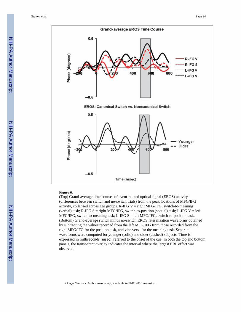
Figure 6.(Top) Grand-average time courses of event-related optical signal (EROS) activity(differences between switch and no-switch trials) from the peak locations of MFG/IFGactivity, collapsed across age groups. R-IFG V = right MFG/IFG, switch-to-meaning(verbal) task; R-IFG S = right MFG/IFG, switch-to-position (spatial) task; L-IFG V = leftMFG/IFG, switch-to-meaning task; L-IFG S = left MFG/IFG, switch-to-position task.(Bottom) Grand-average switch minus no-switch EROS lateralization waveforms obtainedby subtracting the values recorded from the left MFG/IFG from those recorded from theright MFG/IFG for the position task, and vice versa for the meaning task. Separatewaveforms were computed for younger (solid) and older (dashed) subjects. Time isexpressed in milliseconds (msec), referred to the onset of the cue. In both the top and bottompanels, the transparent overlay indicates the interval where the largest ERP effect wasobserved.
Gratton et al. Page 24
J Cogn Neurosci. Author manuscript; available in PMC 2010 August 9.
NIH
-PA Author Manuscript
NIH
-PA Author Manuscript
NIH
-PA Author Manuscript
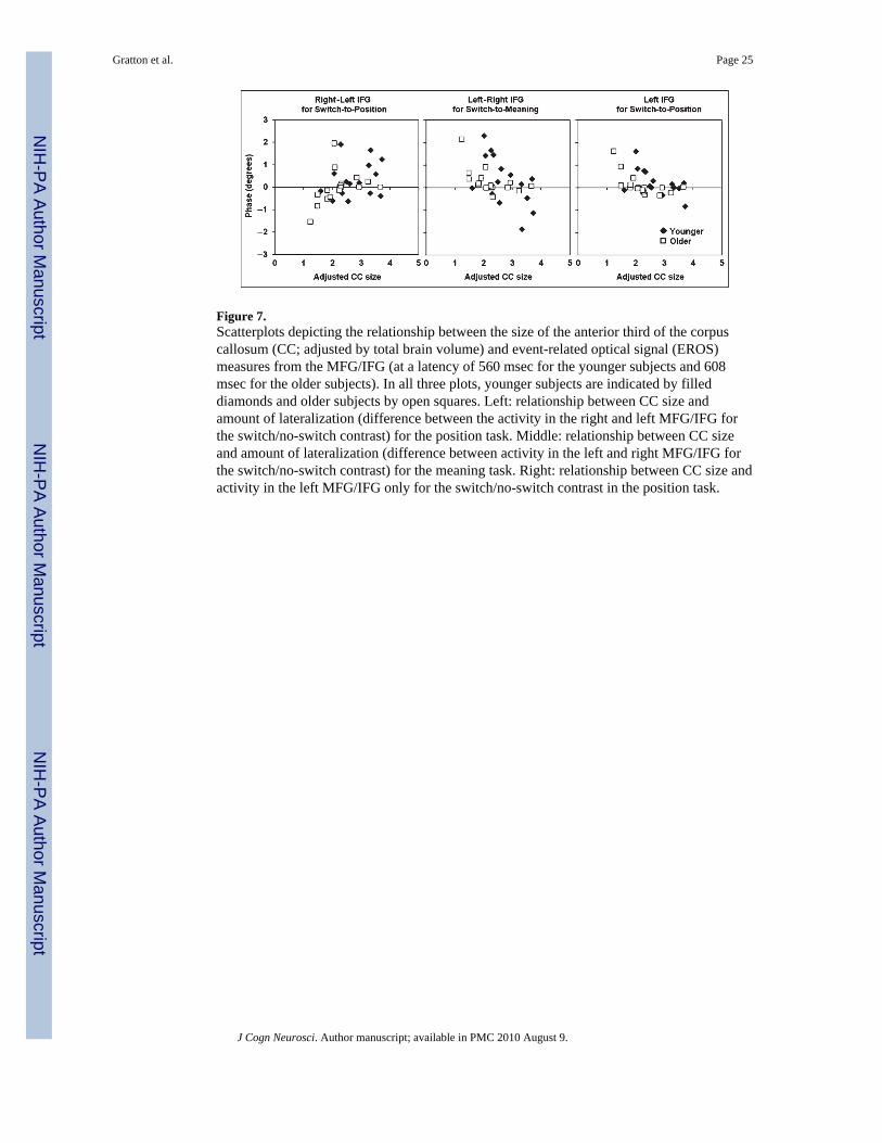
Figure 7.Scatterplots depicting the relationship between the size of the anterior third of the corpuscallosum (CC; adjusted by total brain volume) and event-related optical signal (EROS)measures from the MFG/IFG (at a latency of 560 msec for the younger subjects and 608msec for the older subjects). In all three plots, younger subjects are indicated by filleddiamonds and older subjects by open squares. Left: relationship between CC size andamount of lateralization (difference between the activity in the right and left MFG/IFG forthe switch/no-switch contrast) for the position task. Middle: relationship between CC sizeand amount of lateralization (difference between activity in the left and right MFG/IFG forthe switch/no-switch contrast) for the meaning task. Right: relationship between CC size andactivity in the left MFG/IFG only for the switch/no-switch contrast in the position task.
Gratton et al. Page 25
J Cogn Neurosci. Author manuscript; available in PMC 2010 August 9.
NIH
-PA Author Manuscript
NIH
-PA Author Manuscript
NIH
-PA Author Manuscript
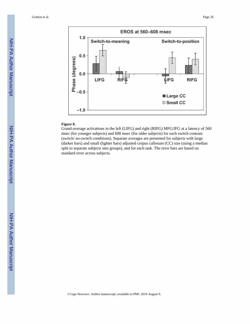
Figure 8.Grand-average activations in the left (LIFG) and right (RIFG) MFG/IFG at a latency of 560msec (for younger subjects) and 608 msec (for older subjects) for each switch contrast(switch/ no-switch conditions). Separate averages are presented for subjects with large(darker bars) and small (lighter bars) adjusted corpus callosum (CC) size (using a mediansplit to separate subjects into groups), and for each task. The error bars are based onstandard error across subjects.
Gratton et al. Page 26
J Cogn Neurosci. Author manuscript; available in PMC 2010 August 9.
NIH
-PA Author Manuscript
NIH
-PA Author Manuscript
NIH
-PA Author Manuscript

NIH
-PA Author Manuscript
NIH
-PA Author Manuscript
NIH
-PA Author Manuscript
Gratton et al. Page 27
Table 1
Demographic and Neuropsychological Characteristics of the Younger and Older Adult Groups (StandardDeviations in Parentheses)
Younger (n = 16) Older (n = 16)
Measure Mean (SD) Mean (SD)
Age (years) 23.11 (3.57) 76.07 (5.23)
Education (years) 15.94 (2.54) 15.94 (3.21)
Modified MMS* 56.69 (0.60) 54.63 (1.50)
Vocabulary Scaled Score 13.19 (2.01) 12.13 (2.09)
*One-tailed t test, p < .01.
J Cogn Neurosci. Author manuscript; available in PMC 2010 August 9.
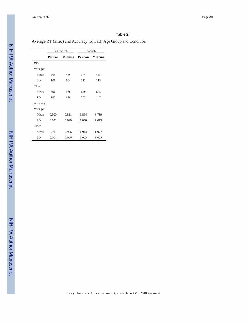
NIH
-PA Author Manuscript
NIH
-PA Author Manuscript
NIH
-PA Author Manuscript
Gratton et al. Page 28
Table 2
Average RT (msec) and Accuracy for Each Age Group and Condition
No Switch Switch
Position Meaning Position Meaning
RTs
Younger
Mean 366 446 378 455
SD 108 104 112 113
Older
Mean 590 666 640 695
SD 192 120 203 147
Accuracy
Younger
Mean 0.920 0.811 0.894 0.789
SD 0.051 0.090 0.060 0.083
Older
Mean 0.941 0.926 0.914 0.927
SD 0.054 0.056 0.053 0.055
J Cogn Neurosci. Author manuscript; available in PMC 2010 August 9.