does sumoylation control k2p1/twik1 background k+ channels?
TRANSCRIPT
Matters Arising
Does Sumoylation ControlK2P1/TWIK1 Background K+ Channels?Sylvain Feliciangeli,1,4 Saıd Bendahhou,1,4 Guillaume Sandoz,1,4 Pierre Gounon,2 Markus Reichold,3
Richard Warth,3 Michel Lazdunski,1 Jacques Barhanin,1 and Florian Lesage1,*1 Institut de Pharmacologie Moleculaire et Cellulaire, CNRS UMR6097, Institut Paul Hamel, 660, route des lucioles, 06560 Valbonne,
France2Centre Commun de Microscopie Appliquee, Universite de Nice, 06108 Nice Cedex, France3 Institute of Physiology, Universitatsstrasse 31, 93053 Regensburg, Germany4These authors contributed equally to this work.
*Correspondence: [email protected]
DOI 10.1016/j.cell.2007.06.012
SUMMARY
A novel model for the regulation of cell excitabil-ity has recently been proposed. It originatesfrom the observation that the background K+
channel K2P1 (TWIK1) may be silenced bysumoylation in Xenopus oocytes and that inac-tivation of the putative sumoylation site (muta-tion K274E) gives rise to robust current expres-sion in transfected COS-7 cells. Here, we showthat only the mutation K274E, and not K274R, isassociated with an increase of K2P1 currentdensity, suggesting a charge effect of K274E.Furthermore, we failed to observe any bandshift by western blot analysis that would con-firm an eventual sumoylation of K2P1 in COS-7cells and oocytes.
INTRODUCTION
K2P1 (or TWIK1) has been cloned from a human kidney
cDNA library (Lesage et al., 1996a). Sequence analysis
predicted a unique membrane topology with four trans-
membrane segments and two pore-forming domains
(Lesage et al., 1996a). Following the identification of
K2P1, homologous K2P proteins were rapidly isolated
from Drosophila and mammals (Goldstein et al., 2005;
Kim, 2005; Lesage and Lazdunski, 2000; Patel and Hon-
ore, 2001; Talley et al., 2003). When compared to other
K2P channels, K2P1 displays a couple of unique features.
Like its closest homolog TWIK2 (K2P6) (Patel et al., 2000),
K2P1 produces currents with a rapidly inactivating com-
ponent. Because of this inactivation, their steady-state
current voltage relationships are much more similar to
that of the weak inwardly rectifying ROMK1 current (Les-
age et al., 1996a) than those of the other K2P currents that
follow the Goldman Hodgkin Katz equation (Duprat et al.,
1997; Fink et al., 1996). Another unique feature of K2P1 is
the difficulty to record currents from transfected cells and
the fact that no native currents corresponding to K2P1
have yet been reported. However, mice deficient for K2P1
have impaired regulation of phosphate transport in the
proximal tubule and of water transport in the medullary
collecting duct, strongly suggesting that K2P1 is func-
tional and contributes to membrane trafficking/expression
of transport molecules in the kidney (Nie et al., 2005). We
have shown that K2P1 is mainly localized in the recycling
endosomal compartment located at the apical side of
transfected kidney cells and native proximal tubule cells
(Decressac et al., 2004). In a variety of nonpolarized cells,
K2P1 immunoreactivity was detected almost exclusively
in the pericentriolar recycling compartment (Decressac
et al., 2004). The mechanism that controls surface expres-
sion/retrieving of K2P1 is not yet characterized but may be
under the dependency of the small G protein ARF6 and its
nucleotide exchange factor EFA6 that interacts with K2P1
(Decressac et al., 2004).
Recently, it has been suggested that K2P1 is addressed
to the cell surface when expressed in Xenopus oocytes
and that addition of a small ubiquitin modifier (SUMO)
peptide to lysine 274 (K274) is responsible for a block of
channel activity (Rajan et al., 2005). From these results,
the authors of the study proposed that K2P1 is a plasma
membrane channel and that its silencing by sumoylation
is the major mechanism explaining the loss of active chan-
nel expression in transfected and native cells. This work
has gained considerable interest not only because it iden-
tifies a novel mechanism of ion channel regulation but also
for its general implication in cell biology (Wilson and
Rosas-Acosta, 2005). Sumoylation is the posttranscrip-
tional modification of lysine residues in target proteins
by covalent attachment of a SUMO peptide moiety at
the consensus site cKxE/D (where c is a hydrophobic res-
idue and x is any amino acid) (reviewed in Dohmen, 2004).
It is primarily a nucleocytoplasmic phenomenon that me-
diates protein-protein interactions, nucleocytoplasmic
trafficking, and activity of transcription factors, but su-
moylation is also known to take place in the cytoplasm.
Regulation of membrane excitability by sumoylation of
background K+ channels active at rest would provide a
novel level of crosstalk between membrane signaling and
some nuclear events through coordination of sumoylated
Cell 130, 563–569, August 10, 2007 ª2007 Elsevier Inc. 563
states. However, several aspects of K2P1 sumoylation
are highly intriguing. First, the K274 residue in K2P1 does
not belong to a classical consensus site for sumoylation
(LK274KF). Second, if K2P1 is a silenced plasma mem-
brane channel, then it must be kept almost exclusively
sumoylated in many different cell types that do not exhibit
a robust current following K2P1 expression. This situation
has no other example to our knowledge.
Here, we demonstrate that K2P1 is not quantitatively
sumoylated in COS-7 cells and Xenopus oocytes. In the
same expression conditions, the K274E mutation is asso-
ciated with an increase of K2P1 current density as previ-
ously described (Rajan et al., 2005). However, this increase
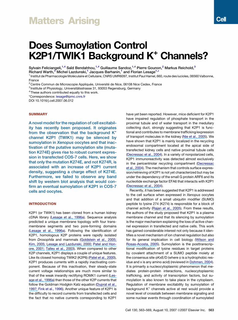
Figure 1. Electrophysiological and Biochemical Character-
ization of K2P1 and Mutant K2P1K274R and K2P1K274E
Channels in COS-7 Cells
(A) Whole-cell current traces from COS-7 cells expressing K2P1,
K2P1K274R, and K2P1K274E channels fused to HcRed. Currents
were elicited by voltage pulses ranging from �120 mV to 40 mV in
20 mV steps, from a holding potential of �80 mV.
(B) Current density was determined at all test potentials from the
steady-state current and whole-cell capacitance. Each value repre-
sents the mean ± SEM, n cells for K2P1 (n = 17), K2P1K274R (n =
17), and K2P1K274E (n = 19).
(C) Current density variation for K2P1K274R and K2P1K274E was nor-
malized to the K2P1 value obtained at +40 mV test potential. Values
are mean ± SEM.
(D) Western blot analysis of K2P1, K2P1K274R, and K2P1K274E.
(E) Western blot analysis of HcRedK2P1 and HcRedK2P1K274E using
anti-K2P1 antibody (as in D) or anti-HcRed antibody. Analysis was car-
ried out as described in Rajan et al. (2005) (lysis buffer containing NEM,
an inhibitor of SUMO isopeptidases).
564 Cell 130, 563–569, August 10, 2007 ª2007 Elsevier Inc.
is not observed with the conservative K274R mutation.
Taken together these data demonstrate that the increase
of current associated with K274E, and absent in K274R,
can probably be attributed to a charge effect and that
SUMO modification at lysine 274 is not the proposed
mechanism for K2P1 silencing.
RESULTS
Expression of K2P1 in COS-7 Cells
In COS-7 cells, K2P1 does not produce macroscopic cur-
rents in the majority of the tested cells (not shown). It has
been reported that alteration of lysine 274 to glutamate
(K274E) leads to functional expression of K2P1 (Rajan
et al., 2005). However, we failed to observe any significant
current upon expression of K2P1K274E in our batch of
COS-7 cells (not shown). We have previously shown that
the fusion of the Heteractis crispa red (HcRed) fluorescent
protein to the amino terminus of K2P1 is associated with
a partial expression of the resulting fusion protein at the
cell surface of proliferating MDCK cells. In these particular
conditions, it was possible to measure K2P1 currents at
the plasma membrane (Decressac et al., 2004). The same
effect was seen in COS-7 cells where HcRedK2P1 and
HcRedK2P1K274E reach the cell surface (not shown)
and produce macroscopic currents (Figure 1A). An
HcRedK2P1K274E fusion protein produced 2.9 times
more current than HcRedK2P1 (Figures 1B and 1C). The
current displayed a very fast inactivation component that
was previously observed for K2P1 expression in oocytes
(Lesage et al., 1996a) and thoroughly characterized for
the closely related channel TWIK2 (Patel et al., 2000).
The kinetics of K2P1 inactivation was extremely fast and
overlapped the membrane capacitive discharge associ-
ated with the voltage pulse. However, the fast inactivating
peak current was clearly not a stimulation artifact and con-
stitutes a hallmark of the TWIK currents.
We tested another mutant of K2P1, HcRedK2P1K274R,
in which lysine 274 is replaced by an arginine residue.
This mutation is more conservative than K274E because
the positive charge at position 274 is conserved and
not substituted by a negative charge. Surprisingly,
HcRedK2P1K274R produced almost the same level of
current as HcRedK2P1 (Figures 1A–1C). The mutation
K274R, unlike K274E, was not associated with an increase
of the HcRedK2P1 current. Even though both substitu-
tions are expected to equally prevent the sumoylation of
K2P1, these results demonstrate that altering lysine
274 by glutamate or arginine does not have the same
effect. Figure 1D shows that K2P1, K2P1K274E, and
K2P1K274R transiently expressed in COS-7 cells ran at
the same apparent molecular weight (MW) when analyzed
by western blot. The 20 kDa shift expected for a protein
being covalently bound to a SUMO moiety is not observed
for K2P1 (SUMO is only 11 kDa, but it migrates aberrantly
at around 20 kDa, even in free form). The apparent MW of
the upper band (37–38 kDa) corresponds to the calculated
MW of K2P1 and fits the MW of K2P1 previously
characterized in transfected cells and in native tissues
(37–40 kDa) (Lesage et al., 1996b, 1997). The band of
lower MW probably corresponds to an immature or de-
graded form of K2P1 produced by the high levels of over-
expression achieved in COS-7 cells. To rule out the pos-
sibility that anti-K2P1 antibodies might not bind to a
sumoylated form of K2P1, the HcRed-tagged K2P1 and
K2P1K274E were detected with either anti-K2P1 or anti-
HcRed antibodies. Clearly, no additional band was re-
vealed by anti-HcRed antibodies (Figure 1E). As for the
nontagged K2P1 (Figure 1D), two bands were detected
for the HcRed-fused proteins that were identical no matter
what antibody was used. Their MWs of around 60–64 kDa
are compatible with the addition of the 26 kDa HcRed
polypeptide to K2P1.
In these experiments, analyzed proteins were first sol-
ubilized in a buffer containing a detergent and a SUMO
isopeptidase inhibitor (NEM) as described in Rajan et al.
(2005). However, and because SUMO isopeptidases are
difficult to inhibit (even in the presence of NEM), we con-
ducted a control experiment based on straight SDS lysis
of total proteins, immediately followed by western blot
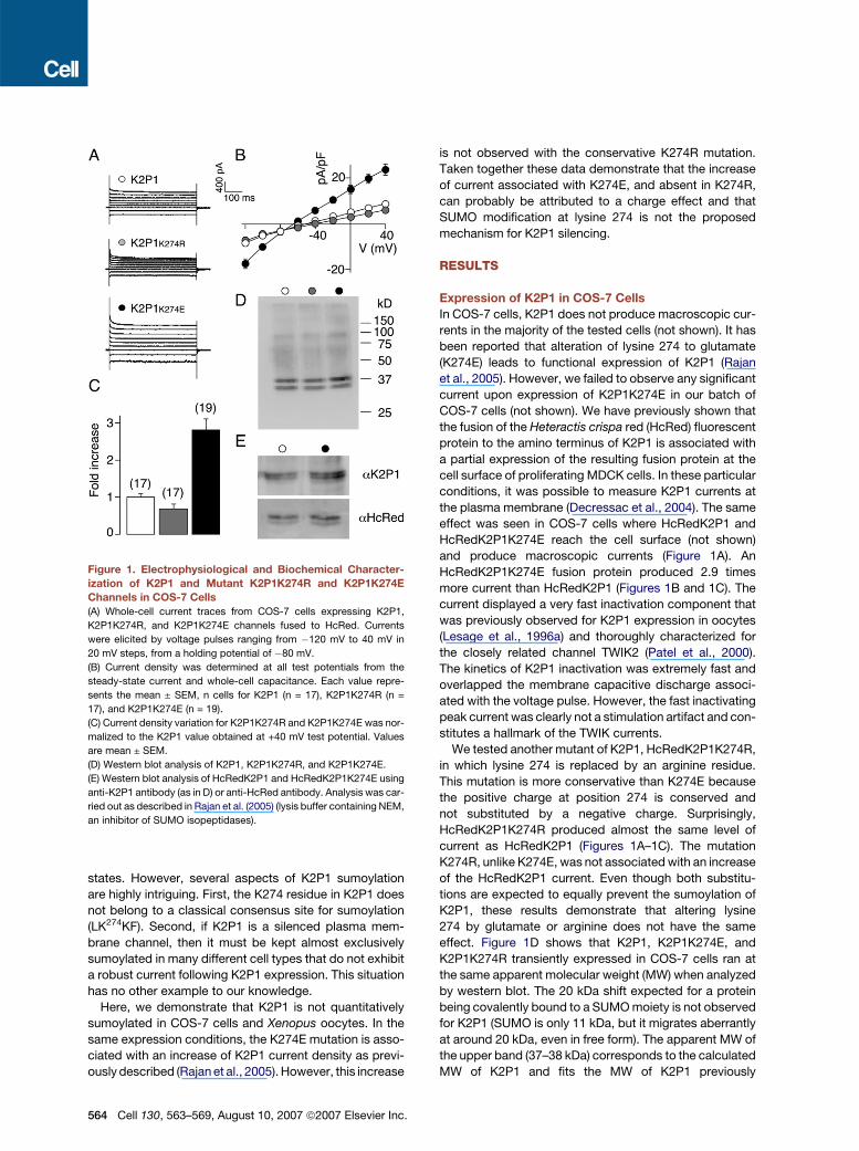
analysis. COS-7 cells were transfected with K2P1 and
the control Ran-GTPase-activating protein RanGAP1
(Matunis et al., 1996), with or without Myc-SUMO1 and
the SUMO ligase Ubc9. In the presence of Ubc9 and
myc-SUMO1, a myc-positive band corresponding to
the sumoylated form of RanGAP1 was clearly detected
as well as other endogenous proteins (Figure 2, left
panel). In exactly the same experimental conditions, nei-
ther myc-labeling (Figure 2, left panel) nor band shift (Fig-
ure 2, right panel) were observed for K2P1, confirming the
absence of quantitative K2P1 sumoylation in COS-7
cells.
Figure 2. RanGAP1, K2P1, Myc-SUMO1, and Ubc9 Coexpres-
sion in COS-7 Cells
Cells were transiently transfected with the indicated plasmids. After
straight cell lysis in a denaturating SDS buffer, total proteins were im-
mediately separated by SDS-PAGE and analyzed by western blot.
Blots were probed with anti-Myc or anti-K2P1 antibodies as indicated.
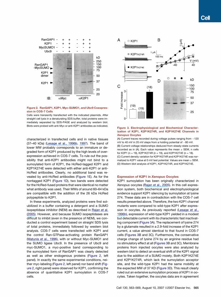
Expression of K2P1 in Xenopus Oocytes
K2P1 sumoylation has been originally characterized in
Xenopus oocytes (Rajan et al., 2005). In this cell expres-
sion system, both biochemical and electrophysiological
evidence support K2P1 silencing by sumoylation at lysine
274. These data are in contradiction with the COS-7 cell
results presented above. Therefore, the two K2P1 channel
mutants were compared to wild-type K2P1 after expres-
sion in oocytes. As previously reported (Lesage et al.,
1996b), expression of wild-type K2P1 yielded in a modest
but detectable current with its characteristic fast inactivat-
ing component (Figure 3A). The replacement of lysine 274
by a glutamate resulted in a 2.9-fold increase of the K2P1
current, a value almost identical to that found in COS-7
cells (Figures 3B and 3C). Furthermore, the conservative
charge change of lysine 274 by an arginine residue had
no stimulatory effect at all (Figures 3B and 3C). Membrane
proteins from injected oocytes were also analyzed by
western blot to detect an eventual shift of the K2P1 signal
due to the addition of a SUMO moiety. Both K2P1K274E
and K2P1K274R, which lack the sumoylation acceptor
site, and the wild-type K2P1 had the same mobility at
the expected MW of 37 KD (Figure 3D). This result clearly
ruled out an extensive sumoylation process of K2P1 in oo-
cytes. Taken together, the oocytes data are in agreement
Figure 3. Electrophysiological and Biochemical Character-
ization of K2P1, K2P1K274R, and K2P1K274E Channels in
Xenopus Oocytes(A) Current traces recorded during voltage pulses ranging from �120
mV to 40 mV in 20 mV steps from a holding potential of �80 mV.
(B) Current-voltage relationships deduced from steady-state currents
recorded as in (A). Each value represents the mean ± SEM, n cells
for K2P1 (n = 19), K2P1K274R (n = 19), and K2P1K274E (n = 18).
(C) Current density variation for K2P1K274R and K2P1K274E was nor-
malized to K2P1 value at 0 mV test potential. Values are mean ± SEM.
(D) Western blot analysis of K2P1, K2P1K274R, and K2P1K274E.
Cell 130, 563–569, August 10, 2007 ª2007 Elsevier Inc. 565
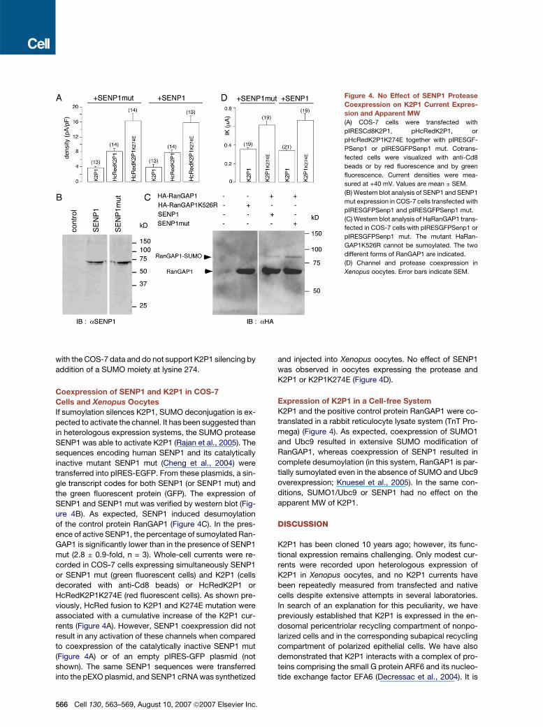
Figure 4. No Effect of SENP1 Protease
Coexpression on K2P1 Current Expres-
sion and Apparent MW
(A) COS-7 cells were transfected with
pIRESCd8K2P1, pHcRedK2P1, or
pHcRedK2P1K274E together with pIRESGF-
PSenp1 or pIRESGFPSenp1 mut. Cotrans-
fected cells were visualized with anti-Cd8
beads or by red fluorescence and by green
fluorescence. Current densities were mea-
sured at +40 mV. Values are mean ± SEM.
(B) Western blot analysis of SENP1 and SENP1
mut expression in COS-7 cells transfected with
pIRESGFPSenp1 and pIRESGFPSenp1 mut.
(C) Western blot analysis of HaRanGAP1 trans-
fected in COS-7 cells with pIRESGFPSenp1 or
pIRESGFPSenp1 mut. The mutant HaRan-
GAP1K526R cannot be sumoylated. The two
different forms of RanGAP1 are indicated.
(D) Channel and protease coexpression in
Xenopus oocytes. Error bars indicate SEM.
with the COS-7 data and do not support K2P1 silencing by
addition of a SUMO moiety at lysine 274.
Coexpression of SENP1 and K2P1 in COS-7
Cells and Xenopus Oocytes
If sumoylation silences K2P1, SUMO deconjugation is ex-
pected to activate the channel. It has been suggested than
in heterologous expression systems, the SUMO protease
SENP1 was able to activate K2P1 (Rajan et al., 2005). The
sequences encoding human SENP1 and its catalytically
inactive mutant SENP1 mut (Cheng et al., 2004) were
transferred into pIRES-EGFP. From these plasmids, a sin-
gle transcript codes for both SENP1 (or SENP1 mut) and
the green fluorescent protein (GFP). The expression of
SENP1 and SENP1 mut was verified by western blot (Fig-
ure 4B). As expected, SENP1 induced desumoylation
of the control protein RanGAP1 (Figure 4C). In the pres-
ence of active SENP1, the percentage of sumoylated Ran-
GAP1 is significantly lower than in the presence of SENP1
mut (2.8 ± 0.9-fold, n = 3). Whole-cell currents were re-
corded in COS-7 cells expressing simultaneously SENP1
or SENP1 mut (green fluorescent cells) and K2P1 (cells
decorated with anti-Cd8 beads) or HcRedK2P1 or
HcRedK2P1K274E (red fluorescent cells). As shown pre-
viously, HcRed fusion to K2P1 and K274E mutation were
associated with a cumulative increase of the K2P1 cur-
rents (Figure 4A). However, SENP1 coexpression did not
result in any activation of these channels when compared
to coexpression of the catalytically inactive SENP1 mut
(Figure 4A) or of an empty pIRES-GFP plasmid (not
shown). The same SENP1 sequences were transferred
into the pEXO plasmid, and SENP1 cRNA was synthetized
566 Cell 130, 563–569, August 10, 2007 ª2007 Elsevier Inc.
and injected into Xenopus oocytes. No effect of SENP1
was observed in oocytes expressing the protease and
K2P1 or K2P1K274E (Figure 4D).
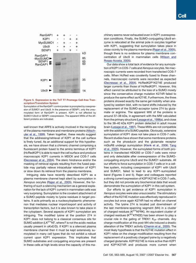
Expression of K2P1 in a Cell-free System
K2P1 and the positive control protein RanGAP1 were co-
translated in a rabbit reticulocyte lysate system (TnT Pro-
mega) (Figure 4). As expected, coexpression of SUMO1
and Ubc9 resulted in extensive SUMO modification of
RanGAP1, whereas coexpression of SENP1 resulted in
complete desumoylation (in this system, RanGAP1 is par-
tially sumoylated even in the absence of SUMO and Ubc9
overexpression; Knuesel et al., 2005). In the same con-
ditions, SUMO1/Ubc9 or SENP1 had no effect on the
apparent MW of K2P1.
DISCUSSION
K2P1 has been cloned 10 years ago; however, its func-
tional expression remains challenging. Only modest cur-
rents were recorded upon heterologous expression of
K2P1 in Xenopus oocytes, and no K2P1 currents have
been repeatedly measured from transfected and native
cells despite extensive attempts in several laboratories.
In search of an explanation for this peculiarity, we have
previously established that K2P1 is expressed in the en-
dosomal pericentriolar recycling compartment of nonpo-
larized cells and in the corresponding subapical recycling
compartment of polarized epithelial cells. We have also
demonstrated that K2P1 interacts with a complex of pro-
teins comprising the small G protein ARF6 and its nucleo-
tide exchange factor EFA6 (Decressac et al., 2004). It is
well known that ARF6 is actively involved in the recycling
of the plasma membrane and membrane proteins (Altsch-
uler et al., 1999). Taken together, these results suggest
that the addressing/retrieval of K2P1 at the cell surface
is finely tuned. As an additional support for this hypothe-
sis, we have shown that a chimeric channel comprising a
fluorescent protein fused to the amino terminus of K2P1
(HcRedK2P1) is able to reach the cell surface and produce
macroscopic K2P1 currents in MDCK and COS-7 cells
(Decressac et al., 2004). The steric hindrance and/or the
masking of retrieval signals resulting from the fused pep-
tide may partially relieve intracellular retention of K2P1
or slow down its retrieval from the plasma membrane.
Intriguing data have recently described K2P1 as a
plasma membrane channel kept silent by sumoylation in
Xenopus oocytes (Rajan et al., 2005). However, the fur-
thering of such a silencing mechanism as a general expla-
nation for the lack of K2P1 current in mammalian cells was
very surprising. Sumoylation is the covalent attachment of
a SUMO peptide moiety to lysine residues in target pro-
teins. It acts primarily as a nucleocytoplasmic phenome-
non that mediates nuclear import/export and activity of
transcription factors, but it is also known to take place in
the cytoplasm. Several aspects of K2P1 sumoylation are
intriguing. The modified lysine at the position 274 in
K2P1 does not belong to a classical consensus site for
SUMO addition (LK274KF, where F is not the acidic residue
usually present in cKxE/D). If K2P1 is a silenced plasma
membrane channel then it must be kept extensively su-
moylated in many cell types that do not exhibit a robust
current upon K2P1 expression. This implicates that
SUMO substrates and conjugating enzymes are present
in these cells at high levels since the capacity of this ma-
Figure 5. Expression in the TnT T7 Promega Cell-free Tran-
scription/Translation System
Sumoylation of the RanGAP1 control protein is prompted by coexpres-
sion of SUMO1 and Ubc9. In the presence of SENP1, only the unsu-
moylated form of RanGAP1 is present. K2P1 is not affected by
SUMO1/Ubc9 or SENP1 coexpression. The apparent MWs of the dif-
ferent proteins are indicated.
chinery seems never exhausted even in K2P1 overexpres-
sion conditions. Finally, the SUMO-conjugating Ubc9 en-
zyme is relocated at the animal pole in oocytes together
with K2P1, suggesting that sumoylation takes place in
close vicinity to the plasma membrane (Rajan et al., 2005),
though there is no evidence for plasma membrane con-
centration of Ubc9 in mammalian cells (Wilson and
Rosas-Acosta, 2005).
Our data show a total lack of evidence for any sumoyla-
tion of K2P1 in COS-7 cells and Xenopus oocytes. No mac-
roscopic currents were recorded from transfected COS-7
cells. When HcRed was covalently fused to these chan-
nels, macroscopic currents were recorded as expected
(Decressac et al., 2004). HcRedK2P1K274E produced
larger currents than those of HcRedK2P1. However, this
effect cannot be attributed to the loss of a SUMO moiety
since the conservative charge mutation K274R failed to
produce the same effect as K274E. Furthermore, the three
proteins showed exactly the same gel mobility when ana-
lyzed by western blot, with no band shifts induced by the
replacement of the SUMO-acceptor lysine 274 by gluta-
mate or arginine. The apparent MW of the proteins is
around 37–38 kDa, in agreement with the MW calculated
from the primary structure (Lesage et al., 1996a), and close
to the 38–40 kDa K2P1 protein detected in adult mouse
brain (Lesage et al., 1997). These MWs are not compatible
with the addition of a SUMO peptide. Obviously, extensive
sumoylation of K2P1 does not take place in COS-7 cells.
Recent studies have shown that some membrane proteins
such as the phosducin and the glutamate receptor
mGluR8 undergo sumoylation (Klenk et al., 2006; Tang
et al., 2005). However, the sumoylated forms of both pro-
teins in transfected HEK293 or COS-7 cells were minor
and only detectable after overexpression of the SUMO-
conjugating enzyme Ubc9 and the SUMO1 substrate. All
our efforts to force sumoylation in COS-7 cells or in a cell-
free system, including coexpression of K2P1 with Ubc9
and SUMO1, failed to lead to any K2P1-sumoylated
band (Figures 3 and 5). Rajan and colleagues reported
a strong current expression of K2P1K274E in COS-7 cells,
but they did not provide any biochemical data that would
demonstrate the sumoylation of K2P1 in this cell system.
Our efforts to get evidence of K2P1 sumoylation in
Xenopus oocytes were also unsuccessful. The stimulatory
effect of the K274E mutation was effectively reproduced in
oocytes but once again K274R had no effect on channel
activity. The lysine 274 is located just downstream of
the last membrane-spanning segment (M4) in a cluster
of charged residues (K274KFRK). An equivalent cluster of
charged residues (K301KTKEE) has been shown to play a
crucial role in the gating of TREK1 K2P channels. Any
charge modification at this post-M4 site induced a drastic
alteration of the TREK1 activity (Chemin et al., 2005). The
most likely hypothesis is that the K274E mutation effect in
K2P1 relies on the charge modification resulting from the
replacement of a positively charged lysine by a negatively
charged glutamate. K2P1K274E is more active than K2P1
and K2P1K274R and produces more current when
Cell 130, 563–569, August 10, 2007 ª2007 Elsevier Inc. 567
expressed at the cell surface in oocytes and in COS-7 cells
(as a HcRed fusion protein in the latter). This hypothesis
is supported by the fact that the current level variations
are quantitatively the same in COS-7 and oocytes. When
analyzed by western blot, K2P1, K2P1K274E, and
K2P1K274R displayed the same apparent MW, again
ruling out any SUMO modification of K2P1.
In conclusion, despite intensive efforts we were unable
to find any in vivo or in vitro evidence supporting SUMO
modification of the background K+ channel K2P1. We
have shown here that K2P1 by itself does produce cur-
rents when present at the cell surface. The current in-
crease associated with the K274E modification is likely
to be a charge effect unrelated to sumoylation. In the ab-
sence of in vivo evidence, sumoylation cannot be consid-
ered as a general mechanism of covalent and reversible
control of background K+ channel function.
EXPERIMENTAL PROCEDURES
Molecular Biology
Human K2P1, K2P1K274E, and K2P1K274R were generated by PCR
and subcloned into pEXO (Lesage et al., 1996a), pIRESCd8 (Fink
et al., 1996), and pHcRed-C1 (Clontech, CA, USA). From pIRESCd8
constructs, a single mRNA coding successively for K2P1 or K2P1K274E
and the cell-surface protein Cd8 protein was produced. In pHcRed-
C1, channel sequences were fused in frame with the fluorescent pro-
tein HcRed. The cDNAs coding for human SENP1 and its catalytically
inactive mutant SENP1R360L/K631M were provided by Dr. Yeh
(Cheng et al., 2004). The open reading frames were transferred in
pIRES-EGFP (BD Biosciences Clontech, CA, USA) and pEXO vectors.
All the constructs were verified by sequencing.
Cell Culture and Electrophysiology
COS-7 cells were maintained in Dulbecco’s modified Eagle media sup-
plemented with 10 % fetal bovine serum and 100 U/ml streptomycine,
100 U/ml penicillin at 37�C in a humidified 5 % CO2 atmosphere. Cells
were transiently transfected by DEAE-Dextran method using 1 mg
DNA of pHcRedC1-K2P1, pHcRedC1-K2P1K274R, and pHcRedC1-
K2P1K274E per 35 mm culture dish. Currents were recorded 48 hr af-
ter transfection. Recordings were conducted in the whole-cell config-
uration at room temperature (�22�C) with an EPC 10 amplifier (HEKA
Electronic, Germany). The pipette solution contained (in mM) 150 KCl,
0.5 MgCl2, 5 EGTA, and 10 HEPES (pH 7.3). The bathing media was (in
mM) 150 NaCl, 5 KCl, 2 CaCl2, 1 MgCl2, and 10 HEPES (pH 7.3). Pi-
pette resistance was 1.5–4 MU. Membrane currents were elicited by
a 500 ms depolarization ranging from �120 mV to +40 mV in a 20
mV increment, from a holding potential of �80 mV. Only cells with se-
ries resistance less than 5 MU were used for analysis. Data acquisition
and analysis were performed using Patchmaster and Pulsefit (HEKA
Electronic, Germany) and IgorPro (WaveMetrics Inc., OR, USA) soft-
wares. pEXO-K2P1, pEXO-K2P1K274R, and pEXO- and pEXO-
K2P1K274E were linearized by BamHI enzyme and capped cRNAs
were synthetized using the T7 RNA polymerase. Defolliculated Xeno-
pus oocytes were injected with cRNAs (15 ng/oocyte) then used for
electrophysiological studies 2 to 4 days following injection. In a 0.3
ml perfusion chamber, a single oocyte was impaled with two standard
microelectrodes (1–2.5 MU resistance) filled with 3 M KCl and main-
tained under voltage clamp using a Dagan TEV 200 amplifier in stan-
dard ND96 solution (96 mM NaCl, 2 mM KCl, 1.8 mM CaCl2, 2 mM
MgCl2, 5 mM HEPES, pH 7.4 with NaOH). Stimulation of the prepara-
tion, data acquisition, and analysis were performed using pClamp soft-
ware (Axon Instruments, CA, USA).
568 Cell 130, 563–569, August 10, 2007 ª2007 Elsevier Inc.
Biochemistry
COS-7 cells were transfected with lipofectamine (Invitrogen). After 48
hr, cells were washed and immediately lysed in the SDS-containing
Laemmli’s buffer or harvested, then resuspended in a buffer containing
(in mM) 100 NaCl, 40 KCl, 20 NEM, 1 EDTA, 20 HEPES-KOH (pH 7.4),
10% glycerol, 1% Triton X-100, and complete protease inhibitor
tablets (Roche) at 4�C. After centrifugation, solubilized proteins were
separated on 10% SDS-PAGE and subjected to western blot analysis
using anti-TWIK1 antibodies (1:1000) (Lesage et al., 1996b) or com-
mercial anti-HcRed (1:200, SC-32188, Santa Cruz biotechnology) or
anti-SENP1 antibodies (1:200, SC-46634, Santa Cruz biotechnology).
cRNA-injected oocytes were ground in lysis buffer without Triton
X-100. After three rounds of low-speed centrifugation (1500 rpm,
10 min, 4�C), the supernatant was submitted to high-speed centrifuga-
tion to collect membrane proteins. The proteins were resuspended in
Laemmli’s loading buffer and analyzed by western blot as described
above. For in vitro transcription/translation, 35S-labeled proteins were
produced by using 35S-methionine and the TnT expression system as
specified by the manufacturer (Promega, WI, USA).
ACKNOWLEDGMENTS
We thank E.T.H. Yeh for SENP1 and SENP1 mut, H. Hibino for
MycSUMO1, F. Melchior for HaRanGAP1 and HaRanGAP1K526R,
and M.J. Lohse for Ubc9. We are grateful to M. Larroque and M. Jodar
for excellent technical assistance with oocyte preparation, cell
cultures, and molecular biology. This work was supported by ‘‘Ligue
National Contre le Cancer’’ (Equipe labelisee to F.L.), by Japan-France
Integrated Action Program SAKURA (06980UF to F.L.), and by ‘‘Deut-
sche Forschungsgemeinschaft’’ (SFB699 to R.W.). S.F. was supported
by a fellowship from the ‘‘Association de Recherche contre le Cancer’’
and F.L. was a recipient of a Contrat d’Interface Clinique, Service de
Neurologie, CHU de Nice.
Received: October 10, 2006
Revised: March 9, 2007
Accepted: June 4, 2007
Published: August 9, 2007
REFERENCES
Altschuler, Y., Liu, S., Katz, L., Tang, K., Hardy, S., Brodsky, F., Apo-
daca, G., and Mostov, K. (1999). ADP-ribosylation factor 6 and endo-
cytosis at the apical surface of Madin-Darby canine kidney cells. J. Cell
Biol. 147, 7–12.
Chemin, J., Patel, A.J., Duprat, F., Lauritzen, I., Lazdunski, M., and
Honore, E. (2005). A phospholipid sensor controls mechanogating of
the K+ channel TREK-1. EMBO J. 24, 44–53.
Cheng, J., Wang, D., Wang, Z., and Yeh, E.T. (2004). SENP1 enhances
androgen receptor-dependent transcription through desumoylation of
histone deacetylase 1. Mol. Cell. Biol. 24, 6021–6028.
Decressac, S., Franco, M., Bendahhou, S., Warth, R., Knauer, S.,
Barhanin, J., Lazdunski, M., and Lesage, F. (2004). ARF6-dependent
interaction of the TWIK1 K+ channel with EFA6, a GDP/GTP exchange
factor for ARF6. EMBO Rep. 5, 1171–1175.
Dohmen, R.J. (2004). SUMO protein modification. Biochim. Biophys.
Acta 1695, 113–131.
Duprat, F., Lesage, F., Fink, M., Reyes, R., Heurteaux, C., and
Lazdunski, M. (1997). TASK, a human background K+ channel to sense
external pH variations near physiological pH. EMBO J. 16, 5464–5471.
Fink, M., Duprat, F., Lesage, F., Reyes, R., Romey, G., Heurteaux, C.,
and Lazdunski, M. (1996). Cloning, functional expression and brain
localization of a novel unconventional outward rectifier K+ channel.
EMBO J. 15, 6854–6862.
Goldstein, S.A., Bayliss, D.A., Kim, D., Lesage, F., Plant, L.D., and
Rajan, S. (2005). International Union of Pharmacology. LV. Nomencla-
ture and molecular relationships of two-P potassium channels. Phar-
macol. Rev. 57, 527–540.
Kim, D. (2005). Physiology and pharmacology of two-pore domain
potassium channels. Curr. Pharm. Des. 11, 2717–2736.
Klenk, C., Humrich, J., Quitterer, U., and Lohse, M.J. (2006). SUMO-1
controls the protein stability and the biological function of phosducin.
J. Biol. Chem. 281, 8357–8364.
Knuesel, M., Cheung, H.T., Hamady, M., Barthel, K.K., and Liu, X.
(2005). A method of mapping protein sumoylation sites by mass spec-
trometry using a modified small ubiquitin-like modifier 1 (SUMO-1) and
a computational program. Mol. Cell. Proteomics 4, 1626–1636.
Lesage, F., and Lazdunski, M. (2000). Molecular and functional prop-
erties of two-pore-domain potassium channels. Am. J. Physiol. Renal
Physiol. 279, F793–F801.
Lesage, F., Guillemare, E., Fink, M., Duprat, F., Lazdunski, M., Romey,
G., and Barhanin, J. (1996a). TWIK-1, a ubiquitous human weakly
inward rectifying K+ channel with a novel structure. EMBO J. 15,
1004–1011.
Lesage, F., Reyes, R., Fink, M., Duprat, F., Guillemare, E., and Lazdun-
ski, M. (1996b). Dimerization of TWIK-1 K+ channel subunits via a disul-
fide bridge. EMBO J. 15, 6400–6407.
Lesage, F., Lauritzen, I., Duprat, F., Reyes, R., Fink, M., Heurteaux, C.,
and Lazdunski, M. (1997). The structure, function and distribution of
the mouse TWIK-1 K+ channel. FEBS Lett. 402, 28–32.
Matunis, M.J., Coutavas, E., and Blobel, G. (1996). A novel ubiquitin-
like modification modulates the partitioning of the Ran-GTPase-acti-
vating protein RanGAP1 between the cytosol and the nuclear pore
complex. J. Cell Biol. 135, 1457–1470.
Nie, X., Arrighi, I., Kaissling, B., Pfaff, I., Mann, J., Barhanin, J., and
Vallon, V. (2005). Expression and insights on function of potassium
channel TWIK-1 in mouse kidney. Pflugers Arch. 451, 479–488.
Patel, A.J., and Honore, E. (2001). Properties and modulation of mam-
malian 2P domain K+ channels. Trends Neurosci. 24, 339–346.
Patel, A.J., Maingret, F., Magnone, V., Fosset, M., Lazdunski, M., and
Honore, E. (2000). TWIK-2, an inactivating 2P domain K+ channel. J.
Biol. Chem. 275, 28722–28730.
Rajan, S., Plant, L.D., Rabin, M.L., Butler, M.H., and Goldstein, S.A.
(2005). Sumoylation silences the plasma membrane leak K+ channel
K2P1. Cell 121, 37–47.
Talley, E.M., Sirois, J.E., Lei, Q., and Bayliss, D.A. (2003). Two-pore-
domain (KCNK) potassium channels: dynamic roles in neuronal func-
tion. Neuroscientist 9, 46–56.
Tang, Z., El Far, O., Betz, H., and Scheschonka, A. (2005). Pias1 inter-
action and sumoylation of metabotropic glutamate receptor 8. J. Biol.
Chem. 280, 38153–38159.
Wilson, V.G., and Rosas-Acosta, G. (2005). Wrestling with SUMO in
a new arena. Sci. STKE 2005, pe32.
Cell 130, 563–569, August 10, 2007 ª2007 Elsevier Inc. 569