dna extraction
TRANSCRIPT

CASE REPORT
DNA Extraction
An Anthropologic Aspect of Bone Remains From Sixth- toSeventh-Century AD Bone Remains
Nunzio Di Nunno, MD, PhD,* Sandro Sublimi Saponetti, BSc,‡ Vito Scattarella, BSc,‡
Patrizia Emanuel, BSc,‡ Stefania Lonero Baldassarra, BSc,† Giuliano Volpe, BSc,§
and Cosimo Di Nunno, MD†
Abstract: In the archeological site of the early Christian Episcopal
complex of Saint Peter, in Canosa di Puglia (Bari, Italy), during the
operations of archaeological excavations, tombs were discovered.
They were dated between the sixth and seventh centuries AD with
carbon 14 methodology. Five skeletons were found in the 5 tombs:
28A: male individual, 43 years old. The height was 170 cm; the
biomass was 65.7 kg. The analysis of the bones indicated several
noteworthy pathologies, such as a number of hypoplasia lines of the
enamel, the presence of Schmorl hernias on the first 2 lumbar
vertebrae, and the outcome of subacromial impingement syndrome.
28E was a male individual, with a biologic age of death of between
44 and 60 years. The height was 177 cm. He had a posttraumatic
fracture callus of the medial third of the clavicle, with an oblique
fracture rima. 29B was a female individual, 44–49 years old. The
height was 158.8 cm; the biomass was 64.8 kg. There was Wells
bursitis on the ischial tuberosity on both sides. 29E was a male
individual, 45–50 years old. The height was 169.47 cm; the biomass
was 70.8 kg. The third and the fourth vertebrae showed Baastrup
syndrome (compression of the vertebral spine). There were radio-
logic signs of deformity on the higher edge of the acetabula and
results of frequent sprains of the ankles. 31A was a male individual,
47–54 years old. The height was 178.65 cm; the biomass was 81 kg.
The vertebral index showed a heavy overloading in the thoracic
lumbar region. There were bony formations under the periosteum on
both on the higher and medium facets of the first metatarsus and on
the higher and lateral facets of the fifth metatarsus on both sides.
As the topography indicates, these small ossifications coincided with
the contact points between the back of the foot and parts of the upper
shoe. From the osseous remains, in particular from the teeth (central
incisors), the DNA was extracted and typed to identify potential
family ties among all the subjects. The extraction technique used
came from the DNA Promega technique, partially modified by the
authors. Stay times of the sample in the extraction buffer were
increased and were increased the polymerase chain reaction (PCR)
cycles.
Key Words: ancient bone remains, DNA extraction, DNA
fingerprint, anthropology
(Am J Forensic Med Pathol 2007;28: 333–341)
As of 2001, systematic archeological digs have been car-ried out in the area of the hill at San Pietro in Canosa, in
the suburbs to the southeast of the town.1–5 These archeolog-ical investigations are being conducted jointly by the Univer-sities of Foggia and Bari and by the Soprintendenza per i Beniarcheologici della Puglia (Apulian Superintendency of Ar-cheological Heritage). During the digs, many tombs havecome to light, containing bone remains. These have recentlystarted to be analyzed from the anthropological standpoint,performing DNA typing. First, a brief geographic and histor-ical outline of the town of Canosa: It is situated in the southof Italy, in the Apulian region, approximately 400 km to thesoutheast of Rome, and is one of the most important arche-ological centers in Apulia and in south Italy in general. Thetown was already important in pre-Roman times, whenCanosa was one of the richest and most powerful Dauniansettlements, and it emerged even more during the Roman era.In the late part of the era, the town became the seat of thegovernors of the provincia Apulia et Calabria; thus, theregional capital. It very early assumed a leading functionfrom the religious point of view, too, thanks to the presenceof a large Christian community guided by powerful bishops,involved in important councils and diplomatic activities. Thegreatest moment for the Canosa church was in the sixthcentury AD, when it was presided over by the famous bishopSabino, who is traditionally held to have governed the dio-cese for over 50 years (AD 514-566) and who set in motion theconstruction of a great number of holy buildings.
Manuscript received December 19, 2005; accepted January 5, 2006.From the *Dipartimento di Scienze Pedagogiche, Psicologiche e Didattiche,
Universita degli Studi del Salento, Lecce, Italy; †Sezione di MedicinaLegale, Di.M.I.M.P., ‡Sezione di Antropologia, Dipartimento di Zoologia,Universita degli Studi di Bari, Bari, Italy; and the §Dipartimento di ScienzeUmane, Universita degli Studi di Foggia, Foggia, Italy.
Reprints: Nunzio Di Nunno, MD, PhD, Dipartimento di Scienze Peda-gogiche, Psicologiche e Didattiche, Universita degli Studi del Salento,Via Mario Stampacchia 45, 73100 Lecce, Italy. E-mail: [email protected].
Copyright © 2007 by Lippincott Williams & WilkinsISSN: 0195-7910/07/2804-0333DOI: 10.1097/PAF.0b013e3181405f35
The American Journal of Forensic Medicine and Pathology • Volume 28, Number 4, December 2007 333
MATERIALS AND METHODS
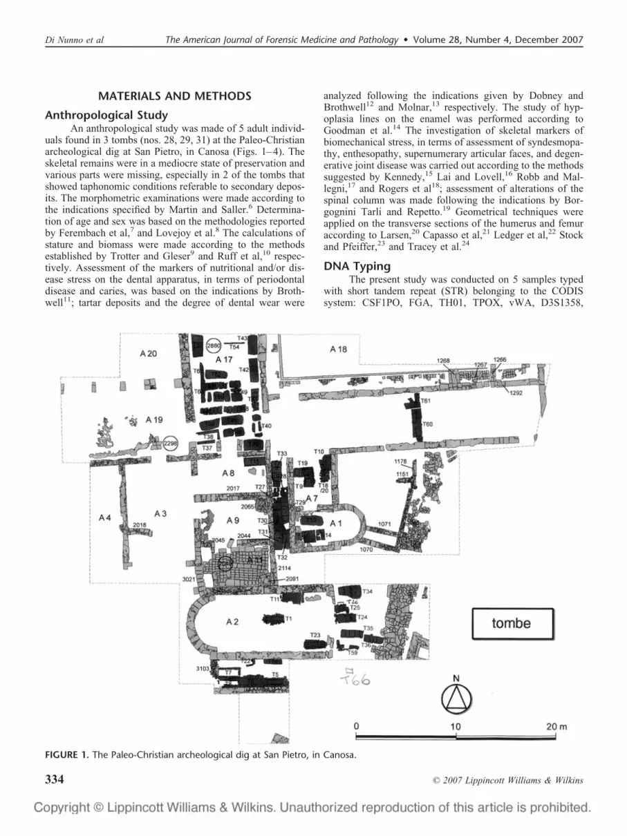
Anthropological StudyAn anthropological study was made of 5 adult individ-
uals found in 3 tombs (nos. 28, 29, 31) at the Paleo-Christianarcheological dig at San Pietro, in Canosa (Figs. 1–4). Theskeletal remains were in a mediocre state of preservation andvarious parts were missing, especially in 2 of the tombs thatshowed taphonomic conditions referable to secondary depos-its. The morphometric examinations were made according tothe indications specified by Martin and Saller.6 Determina-tion of age and sex was based on the methodologies reportedby Ferembach et al,7 and Lovejoy et al.8 The calculations ofstature and biomass were made according to the methodsestablished by Trotter and Gleser9 and Ruff et al,10 respec-tively. Assessment of the markers of nutritional and/or dis-ease stress on the dental apparatus, in terms of periodontaldisease and caries, was based on the indications by Broth-well11; tartar deposits and the degree of dental wear were
analyzed following the indications given by Dobney andBrothwell12 and Molnar,13 respectively. The study of hyp-oplasia lines on the enamel was performed according toGoodman et al.14 The investigation of skeletal markers ofbiomechanical stress, in terms of assessment of syndesmopa-thy, enthesopathy, supernumerary articular faces, and degen-erative joint disease was carried out according to the methodssuggested by Kennedy,15 Lai and Lovell,16 Robb and Mal-legni,17 and Rogers et al18; assessment of alterations of thespinal column was made following the indications by Bor-gognini Tarli and Repetto.19 Geometrical techniques wereapplied on the transverse sections of the humerus and femuraccording to Larsen,20 Capasso et al,21 Ledger et al,22 Stockand Pfeiffer,23 and Tracey et al.24
DNA TypingThe present study was conducted on 5 samples typed
with short tandem repeat (STR) belonging to the CODISsystem: CSF1PO, FGA, TH01, TPOX, vWA, D3S1358,
FIGURE 1. The Paleo-Christian archeological dig at San Pietro, in Canosa.
Di Nunno et al The American Journal of Forensic Medicine and Pathology • Volume 28, Number 4, December 2007
© 2007 Lippincott Williams & Wilkins334

D5S818, D7S820, D8S1179, D13S317, D16S539, D18S51,and D21S11. For samples with teeth, the teeth were treated asfollows. They were first extracted from the dental arch andthen washed first in water and then in a 10% ethanol solution
to eliminate any bacterial contamination. Subsequently, alongitudinal incision was made of each tooth, using thelaboratory drill KAVO EWL 3–36 V � 25.000/min 50 W,equipped with a diamond-point mill disk Mesinger 94 off HP220. The hemidental elements, with the root canal exposed,presented brownish residues of dental pulp; these were re-moved from the root canals using an odontic curette, ASAStainless 1807-12, and were collected in 1.5-mL Eppendorftest tubes. As a preliminary step in the skeletal investigations,the femur shafts were cleaned with a brush and detergent, toremove fragments of earth and any tree roots adhering to thebone. After abundant washing to remove any residual deter-gent, the femur shafts were left to dry at room temperature.Later, electromechanical scraping of their surfaces was per-formed, using a Dremel. The bone structure was drilled anda window was made, from which a piece of compact bone8 � 3 cm in size was taken. The piece of bone was firstbroken into smaller pieces about 1 � 2 cm, using the Dremel.These pieces were placed in a Falcon test tube and repeatedlywashed in deionized water until the washing water wasperfectly clear. They were then washed in ethanol 90°C andthen in ethanol 95°C. After completely drying the samples,they were reduced to powder. DNA extraction from thedental pulp samples was performed following a modifiedprotocol of the Promega Kit “SV Total RNA Isolation Sys-tem,” suitable for DNA extraction from samples containingonly a small number of nucleated cells to begin with. Thesame method was used for the bone samples. The protocolwas partially modified by lengthening the incubation time ofthe cell lysis step: each sample of dental pulp was placed,overnight, at room temperature, in a single microtube con-taining 350 L of SV RNA Lysis Buffer.
DNA extraction from the bone powder was performedusing the commercial kit Geneclean for Ancient DNAQ-Biogene, following a protocol that we modified only asregards the first step: a quantity of 100–500 mg of bonepowder is added to a 1.5-mL Eppendorf test tube containing1 mL of DeHybernation Solution. The microtube is left toincubate at a temperature of 45°C–60°C overnight and agi-tated occasionally. This step may sometimes need to beprotracted for 24 hours. Amplification was obtained using athermal cycler-DNA Gene Amp polymerase chain reaction(PCR) system 9700 (Applied Biosystems, Foster City, CA) andthe AmpFlSTR Identifiler PCR Amplification Kit (Applied Bio-systems).25,26 Analysis of the amplified allele fragments wasperformed by capillary electrophoresis using the ABI Prism 310Genetic Analyzer (Applied Biosystems) and the 310 GeneticAnalyzer performance optimized polymer 4 (POP-4; AppliedBiosystems), 10� Genetic Analyzer Buffer with EDTA, alleleladders, and GeneScan-500 (internal standard).27–30
Data Analysis of the DNA TypingThe results obtained for the 5 samples were used to
make a comparison with the haplotypes of present-day sub-jects belonging to the Apulian population, to assess genotyp-ical and allelic homologies and differences. For this purpose,the genotypes of all the systems analyzed were inserted in thecomputer program S.H.S. Hardware software systems that
FIGURE 2. Particulars of the dig.
FIGURE 3. The Paleo-Christian archeological dig at San Pi-etro, in Canosa, the tombs.
The American Journal of Forensic Medicine and Pathology • Volume 28, Number 4, December 2007 Ancient Bone Remains
© 2007 Lippincott Williams & Wilkins 335

allows estimation of the frequency of the haplotype (genotypecombinations) of each sample from the Apulian population(reference population). Many cases of alleles no longerpresent in the reference population, at least in the samplessubmitted to statistical analysis, were found (Table 1); thecalculation was therefore based only on the frequency of thealleles present in the Apulian population. For all the samples,the differences between the haplotypes of the 5 individualsand of the present-day Apulian population were statisticallysignificant (Table 2).
RESULTS
Tomb 28This is a multiple tomb in which 7 adults were found:
6 male adults and 1 male child. The identification of thesingle individuals was made by exploiting morphometric,
articular, sex, and bone robustness compatibilities, as well aspathologic signs.
Individual 28AThis male subject was aged 40–43 years. He appears to
have had a robust constitution, with a well-developed mus-cular frame. The estimated stature is 170 cm and the bodybiomass, 66 kg. Examination of the teeth showed severeperiodontosis, moderate dental wear, and absence of cariesbut 2 odontogenic abscesses. There was a condition of weightoverload of the cervical vertebrae, as well as Schmorl herniasof the lumbar vertebrae. The femurs showed Poirier facetsand flattening of the subtrochanteric shaft sections, and thetibias showed lateral accessory facets. There was moderateosteoarthritic degeneration in the form of pitting. From thepathologic standpoint, phlogosis of the os zygomaticum (Fig.
FIGURE 4. A, Tomb T28. B, Tomb T29.C, Tomb T31.
Di Nunno et al The American Journal of Forensic Medicine and Pathology • Volume 28, Number 4, December 2007
© 2007 Lippincott Williams & Wilkins336

5), loss of substance in a half-moon shape was evident on theclavicula, corresponding to the area of insertion of the conoidligament, together with a bone alteration of the acromial jointfacet. These lesions are compatible with a past acromion-clavicular dislocation. The person had tibial periostitis (Fig. 6).
Individual 28EThis subject was judged to be a male aged between 44
and 60 years. His stature was calculated as 177 cm. Therewere many lines of enamel hypoplasia, which had formedbetween the ages of 1 and 6 years (Fig. 7). Assessment of thepostcranial indexes revealed a condition of eurybrachia ofboth humeri and of mesocnemia of both tibias; the bonerobustness index was average in both humeri. The vertebralload index showed weight overload in the lumbar and cervi-cal tracts and mild overload in the thoracic tract. Vertebralosteolysis (Fig. 8). There was disk herniation of the 12th and10th thoracic vertebrae and the second lumbar vertebra. The
few musculoskeletal markers available indicate intense mus-cular activity. Osteoarthrosis is present in the form of mod-erate lipping and pitting. The patellas show the incisura andthe fossa of the lateral vastus, both tibias have a kneelingfacet. The left astragalus presents the os trigonum. On the leftclavicula (Fig. 9), a bone callous due to fracture of the thirdclavicular median along an oblique line was observed, withshortening of the segment due to raising of the sternal stumpover the acromial stump. The fracture was not compound andcould have been due to falling on the palm of the hand or onthe shoulder, with the arm abducted.
TABLE 1. Data Analysis of the DNA Typing
D8S1179 D21S11 D7S820 CSF1PO D19S433 vWA TPOX FGA
Bone Canosa T28A 8–9 26–31 15–15 11–13 – 13–17 8–10 –
Bone Canosa T28E 13–18 31.2–33 11–11 12–13 10–10 14–19 6–7 27–27
Bone Canosa T29B 12–13 26–26 7–7 �6–�6 – – 6–9 –
Bone Canosa T29E 12–13 26–26 7–7 �6–�6 – – – –
Bone Canosa T31A 9–10 33–36 8–9 6–7 16–17 12–15 7–7 –
Lab. anthropology personnel 15–15 28–28 8–10 11–13 14–14.2 16–19 8–10 20–22
Lab anthropology personnel 13–14 29–32.2 9–9 10–11 14–14 16–20 8–8 23.2–24.2
Lab forensic genetics personnel 12–14 29–30 8–12 10–10 13–14 17–18 8–11 21–22
Lab forensic genetics personnel 13–14 29–29 12–12 10–12 13–15 17–19 8–11 21–22
Lab forensic genetics personnel 11–13 29–32.2 10–11 10–12 13–14 17–17 8–9 20–25
Lab forensic genetics personnel 11–13 28–30 8–10 11–12 12–15 14–16 8–8 21–24
D18S51 D3S1358 TH01 D13S317 D16S539 D2S1338 D5S818 Amel
Bone Canosa T28A 15–20 16–16 6–6 14–15 9–9 – 7–8 X-Y
Bone Canosa T28E – 20–20 4–8 9–9 5–8 16–17 10–10 X-Y
Bone Canosa T29B 15–17 – 6–9.3 8–12 9–12 19–21 9–12 X-Y
Bone Canosa T29E 15–17 – 6–9 �8–10 �8–9 16–18 9–12 X-Y
Bone Canosa T31A – 13–17 9.3–9.3 8–13 10–15 24–24 13–16 X-Y
Lab anthropology personnel 14–16 15–17 6–7 12–12 11–11 17–19 9–12 X-X
Lab anthropology personnel 13–16 15–16 8–9.3 9–12 11–12 17–18 11–12 X-Y
Lab forensic genetics personnel 15–16 15–17 8–9.3 8–11 9–11 20–24 11–12 X-Y
Lab forensic genetics personnel 15–16 15–17 6–9.3 8–12 9–12 20–23 9–12 X-Y
Lab forensic genetics personnel 13–18 16–17 6–10 8–11 11–13 24–25 12–12 X-X
Lab forensic genetics personnel 16–16 15–17 6–9 9–12 9–11 17–19 12–12 X-Y
TABLE 2. Frequency of Haplotypes With Respect to theApulian Population
Sample Haplotype Frequency
T28A 3.89/1022
T28E 3.65/1023
T29B 8.8/1019
T29E 2.87/1016
T31A 3.54/1018
FIGURE 5. Phlogosis of the os zygomaticum.
The American Journal of Forensic Medicine and Pathology • Volume 28, Number 4, December 2007 Ancient Bone Remains
© 2007 Lippincott Williams & Wilkins 337

Tomb 29This is a multiple tomb in which 5 individuals were
found: 3 adults, 1 male, and 2 females, as well as 1 youngfemale and 1 child. The identification of the single individ-uals was made by exploiting morphometric, articular, sex,and bone robustness compatibilities.
Individual 29BThis is a female subject aged between 50 and 65 years.
This was the only case in which diagnosis of the sex from thebone remains was discordant with the DNA result. There wascomplete fusion between the manubrium and the body of thesternum. The stature was calculated to be 159 cm, and thebody biomass, 65 kg. Assessment of the postcranial indexesshowed flattening of the diaphyses of the right humerus, leftulna, and both femurs. The index of robustness of the bonesshowed a moderate mean score. The lumbar and thoracicvertebrae showed a load within normal limits, while it wasnot possible to estimate the cervical load; herniation of thefirst, third, and fourth lumbar vertebrae was observed (Fig.
10). Analysis of the degree of enthesopathies showed mod-erate muscular activity. There was osteoarthritis in the formof mild lipping of the upper limbs, moderate lipping of thelower limbs, and mild pitting. Osteochondritis dissecans(O.D.) was observed at the level of the hip joint in bothacetabular cavities, while the ischiatic tuberosities presentedtraces of Wells bursitis. Humeral osteolysis (Fig. 11).
Individual 29EThis individual was diagnosed as being of male sex and
aged approximately 45 years. His stature was calculated to be169 cm, and body biomass, 71 kg. Dental examination re-vealed mild dental wear, the presence of 2 caries, an odon-togenic abscess, and a large amount of tartar. Outcome ofsinusitis (Fig. 12). The medial and lateral incisors were spadeshaped. The axial skeleton showed moderate vertebral load inthe cervical tract and the presence of herniation of the lastpart of the thoracic tract. The third and fourth lumbar verte-brae presented Baastrup syndrome (Fig. 13). The sacrum
FIGURE 8. Verteblal osteolysis.
FIGURE 9. On the left clavicula, a bone callous due to frac-ture of the third clavicular median along an oblique line.
FIGURE 6. Tibial periostitis.
FIGURE 7. Enamel hypoplasia.
Di Nunno et al The American Journal of Forensic Medicine and Pathology • Volume 28, Number 4, December 2007
© 2007 Lippincott Williams & Wilkins338

presents a bridge vertebra. Examination of the musculoskel-etal markers of the postcranial skeleton reveals strong tendonimpression on all the segments examined. There were signsof moderate osteoarthrosis, in terms of moderate lipping, mildpitting, but no eburnation. Poirier facet, Charles facet, andAllen fossa were present on the left femur; the right femurshowed only Allen fossa and Poirier facet; both tibias pre-sented accessory lateral and medial facets. Accessory facetswere also present on both astragals. The right patella pre-sented the fossa of the lateral vastus muscle. There wasevident syndesmopathy in virtually all the ligament insertionareas. The iliac bone showed a rough surface bilaterally in theright region above the acetabulum. These roughened, wrinkledsurfaces were more extensive and evident on the right, with thepresence of bone neoformations of syndesmopathic type in thehip joint capsule insertion zone. The superior part of the anteriorsacroiliac ligament was completely ossified. The posterior faceof the ischiatic tuberosity presented clear signs of Wells bursitis.The acetabulum had a marginal bone formation bilaterally, withextension of the superior joint surface and the presence of O.D.cavitation.
Tomb 31This tomb contained 2 individuals of the male sex, 1
adult and 1 adolescent.
Individual 31AThis male subject was aged approximately 54 years.
His stature was 179 cm, and body biomass, 81 kg. Dentalexamination showed the presence of 2 caries and an abscessextending to the left maxilla in the malar zone; there was alsosevere periodontitis, moderate tooth wear, and lines of hyp-oplasia attributable to formation between the ages of 2 yearsand 6 and a half years. Assessment of the postcranial indexes
FIGURE 10. Herniation of the first, third, and fourth lumbarvertebrae was observed.
FIGURE 11. Humeral osteolysis.
FIGURE 12. Outcome of sinusitis.
The American Journal of Forensic Medicine and Pathology • Volume 28, Number 4, December 2007 Ancient Bone Remains
© 2007 Lippincott Williams & Wilkins 339

demonstrated a condition of eurybrachia of the humeri, pla-tolenia of the ulnas, eurymeria of the left femur, and euryc-nemia of the tibias; a moderate robustness index of bothradiuses, the right femur, and both tibias; and high robustnessof the left clavicle. The pillar index was zero in the rightfemur and weak in the left. The vertebral load index showed
a moderate load in the cervical region and overload in thethoracolumbar region. There were bone neoformations of thefeet and subperiosteal formations of the superior medial faceof the first metatarsal and the superior lateral face of the fifthmetatarsal (Fig. 14). These small bone formations, due tocontinual, mild irritant stimuli, were located in correspon-dence with the points of contact between the back of the footand parts of the soles of the shoes worn at the time, such assandals. The observation of enthesopathic traces demon-strates intense muscular activity, as also evinced from thesyndesmopathic traces and osteoarticular degenerations. Bothfemurs presented an extensive Poirier facet and Charles facet;both tibias had a kneeling facet. Accessory facets werepresent on the astragals. The hip bones showed the calcar ofthe iliac crest.
Concluding Pathology NotesThe paleopathological investigation of the 5 individuals
revealed a whole series of skeletal alterations, involving theskull, ribs, vertebral column, and long bones, attributable toinflammatory foci (Table 3), whose distribution and morphol-ogy could likely indicate a specific nature, probably of tuber-cular origin. This finding is of particular interest because itcan be generalized to other individuals found in the samecemetery area.
CONCLUSIONSGenetic and anthropologic investigations have yielded
a series of new data on the bone remains found in the areaaround San Pietro in Canosa di Puglia. From the geneticstandpoint, we can state that significant differences wereobserved between the STR sequences analyzed in the present-day inhabitants of the Apulian region and their medievalancestors. These findings may be justified by the fact that,during the Middle Ages, there were strong gene flows fromthe Germanic and Asian peoples (Goths, Lombards, Avars)that may have conditioned the genome of the native popula-tions. In the subsequent centuries, these gene pools may havebeen diluted, thus strongly altering the gene status of theItalian population. From the anthropologic standpoint, it mustbe borne in mind that part of the cemetery in the archeolog-ical site at San Pietro shows selective conditions, in terms ofalterations indicating inflammatory disease, likely of a spe-cific nature and probably of tubercular origin.
FIGURE 13. The third and fourth lumbar vertebrae pre-sented Baastrup syndrome.
FIGURE 14. There are bone neoformations of the feet andsubperiosteal formations of the superior medial face of thefirst metatarsal.
TABLE 3. Distribution of Skeletal Alterations in the Study Sample
Tomb Sex Age
Endocr.
Alterations
Vertebr.
Ipervasc
Vertebr.
Cistic
General
Ipervasc
Long
Bones
Periostitis
Ribs
Phlogosis/Alterations Hyperostosis
Periartitis
Humerus
Alterat.
Sternum
Alterations
Turbinate
Bones
Phlogosis
e28 A M 40–43 x x x x x x n.r.
28 E M 44–60 n.r. x x x x x n.r. x n.r. n.r.
29 B F 44–65 x x x x n.r. x x
29 E M 45–49 x x x x x x
31 A M 47–54 x x x x x x x x n.r.
Di Nunno et al The American Journal of Forensic Medicine and Pathology • Volume 28, Number 4, December 2007
© 2007 Lippincott Williams & Wilkins340

ACKNOWLEDGMENTSThe authors would like to thank M. V. C. Pragnell, BA,
for help with translation of the text.
REFERENCES1. Volpe G, Annese C, Ciminale M, et al. Il complesso episcopale paleoc-
ristiano di san Pietro a Canosa: prima relazione preliminare (campagnadi scavi 2001). Vetera Christianorum. 2002;39:133–190.
2. Volpe G, Annese C, Corrente M, et al. Il complesso episcopale paleoc-ristiano di san Pietro a Canosa : seconda relazione preliminare (cam-pagna di scavi 2002). Archeologia Medievale. 2003;30:107–164.
3. Volpe G. Il complesso episcopale di San Pietro nel quadro di Canosapaleocristiana, in Atti del Convegno Canosa. Ricerche Storiche 2003(Canosa 14. 12. 2002), a cura di L. Bertoldi Lenoci, Fasano 2003:89–104.
4. Volpe G. Nuovi dati sul complesso episcopale paleocristiano di sanPietro a Canosa, in Atti del Convegno Canosa, Ricerche storiche 2004(Canosa 7. 2.2004), a cura di L. Bertoldi Lenoci, Fasano 2005:15–34.
5. Volpe G, Annese C, Leone D, et al. I mosaici pavimentali del complessopaleocristiano di San Pietro a Canosa (Ba), in Atti del X Colloquiodell’Associazione Italiana per lo Studio e la Conservazione del Mosaico(AISCOM) (Lecce 18–21 febbraio 2004), Roma 2005.
6. Martin R, Saller K. Lehrbuch der Anthropologie. Stuttgart: G. Fischer;1959.
7. Ferembach D, Schwidetzky I, Stloukal M. Raccomandazioni per ladeterminazione dell’eta e del sesso sullo scheletro. Rivista Antropologia.1979;60:5–51.
8. Lovejoy CO, Meindl S, Pryzbeck TR, et al. Chronological metamorphosisof the auricolar surface of the ilium: a new method for the determination ofadult skeletal age at death. Am J Phys Anthropol. 1985;68:15–28.
9. Trotter M, Gleser GC. Corrigenda to estimation of stature from limbbones of American whites and Negroes. Am J Phys Anthropol. 1997;47:355–356.
10. Ruff CR, Trinkaus E, Holliday TW. Body mass and encephalization inPleistocene Homo. Nature. 1997;387:173–176.
11. Brothwell DR. Digging Up Bones. Oxford: University Press; 1981.12. Dobney K, Brothwell DR. A method for evaluating the amont of dental
calculus on teeth from archaeological site. J Archaeol Sci. 1987;14:343–351.
13. Molnar S. Human tooth wear, tooth function and cultural variability.Am J Phys Anthropol. 1971;34:175–189.
14. Goodman AH, Martin DL, Armelagos GJ. Indications of stress frombone and teeth. In: Cohen MN, Armelagos GJ, eds. Paleopathology atthe Origin of the Agriculture. London: Academic Press; 1984:13–49.
15. Kennedy KAR. Skeletal markers of occupational stress. In: Iscan MY,Kennedy KAR, eds. Reconstruction of Life from the Skeleton. NewYork: Liss; 1989:129–160.
16. Lai P, Lovell NC. Skeletal markers of occupational stress in the furtrade: a case study from Hudson’s Bay Company Fur Trade Post. Int J
Osteoarchaeol. 1992;2:221–234.17. Robb J, Mallegni F. Anthropology and paleopathology of Neolithic
human remains from Catignano (Pescara, Italy). Rivista Antropologia.1994;72:197–224.
18. Rogers J, Waldron T, Dieppe P, et al. Arthropathies in paleopathology:the basis of the classification according to most probable cause.J Archaeol Sci. 1987;14:179–193.
19. Borgognini Tarli SM, Repetto E. Skeletal indicators of subsistencepatterns and activity regime in the Mesolithic sample from Grottadell’Uzzo (Trapani, Sicily): a case in study. Hum Evol. 1986;4:331–352.
20. Larsen CS. Bioarchaeology: Interpreting Behaviour from the Human
Skeleton. Cambridge: University Press; 1999.21. Capasso L, Kenneth AR, Kennedy C, et al. Atlas of Occupational
Markers on Human Remains. Teramo: Edigrafital S.p.A.; 1999.22. Ledger M, Holtzhausen L, Constant D, et al. Biomechanical beam
analysis of long bones from a late 18th century slave cemetery in CapeTown, South Africa. Am J Phys Anthropol. 2000;112:207–216.
23. Stock J, Pfeiffer S. Linking structural variability in long bone diaphysesto habitual behaviors: foragers from the southern African Later StoneAge and the Andaman Isles. Am J Phys Anthropol. 2001;115:337–348.
24. Tracey AR, Ruff CB, Plato CC. Hand dominance and bilateral asym-metry in the structure of the second metacarpal. Am J Phys Anthropol.1994;94:203–211.
25. Kimpton CP, Oldroyd NJ, Watson SK, et al. Validation of highlydiscriminating multiplex short tandem repeat amplification systems forindividual identification. Electrophoresis. 1996;17:1283–1293.
26. PE Biosystems. AmpFlSTR Cofiler PCR Amplification Kit User Bulletin.Foster City, CA: PE Biosystems; 1998.
27. PE Biosystems. ABI PRISM 310 Genetic Analyzer User’s Manual: Rev.
1. Foster City, CA: PE Biosystems; 1995.28. PE Biosystems. ABI PRISM 310 Genetic Analyzer User’s Manual.
Foster City, CA: PE Biosystems; 1998.29. PE Biosystems. ABI PRISM GeneScan Analysis Software 2.1 User’s
Manual. Foster City, CA: PE Biosystems; 1996.30. Issaq H, Chan K, Muschik G. The effect of column length, applied
voltage, gel type, and concentration on the capillary electrophoresisseparation of DNA fragments and polymerase chain reaction products.Electrophoresis. 1997;18:1153–1158.
The American Journal of Forensic Medicine and Pathology • Volume 28, Number 4, December 2007 Ancient Bone Remains
© 2007 Lippincott Williams & Wilkins 341