diverticular disease: diagnosis and management - nice
TRANSCRIPT
1
National Institute of Health and Care Excellence
Final
Diverticular disease: diagnosis and management [G] Evidence review for diagnostic tests for acute diverticulitis
NICE guideline NG147
Diagnostic evidence review
November 2019
Final
This evidence review was developed by the National Guideline Centre

Diverticular disease Contents
Diverticular disease
Disclaimer
The recommendations in this guideline represent the view of NICE, arrived at after careful consideration of the evidence available. When exercising their judgement, professionals are expected to take this guideline fully into account, alongside the individual needs, preferences and values of their patients or service users. The recommendations in this guideline are not mandatory and the guideline does not override the responsibility of healthcare professionals to make decisions appropriate to the circumstances of the individual patient, in consultation with the patient and, where appropriate, their carer or guardian.
Local commissioners and providers have a responsibility to enable the guideline to be applied when individual health professionals and their patients or service users wish to use it. They should do so in the context of local and national priorities for funding and developing services, and in light of their duties to have due regard to the need to eliminate unlawful discrimination, to advance equality of opportunity and to reduce health inequalities. Nothing in this guideline should be interpreted in a way that would be inconsistent with compliance with those duties.
NICE guidelines cover health and care in England. Decisions on how they apply in other UK countries are made by ministers in the Welsh Government, Scottish Government, and Northern Ireland Executive. All NICE guidance is subject to regular review and may be updated or withdrawn.
Copyright © NICE 2019. All rights reserved. Subject to Notice of rights.
ISBN: 978-1-4731-3603-8
Diverticular disease Contents
4
Contents 1 Diagnosis of acute diverticulitis .................................................................................. 6
1.1 Review question: For people with suspected acute diverticulitis who are not referred for urgent hospital assessment, which investigations are clinically and cost effective (for example full blood count, C-reactive protein (CRP), endoscopy, CT and MRI) in the diagnosis and assessment of acute diverticulitis during and after the acute episode? ................................................... 6
1.1.1 Introduction ................................................................................................ 6
1.2 Review question: For people with suspected acute diverticulitis who are referred for urgent hospital assessment, which investigations are clinically and cost effective (for example full blood count, C-reactive protein (CRP), endoscopy, CT and MRI) in the diagnosis and assessment of acute diverticulitis during and after the acute episode? ................................................... 6
1.2.1 Introduction ................................................................................................ 6
1.3 PICO table ............................................................................................................. 6
1.4 Clinical evidence ................................................................................................... 8
1.4.1 Included studies ......................................................................................... 8
1.4.2 Excluded studies ........................................................................................ 8
1.4.3 Summary of clinical studies included in the evidence review ...................... 9
1.4.4 Quality assessment of clinical studies included in the evidence review .... 10
1.5 Economic evidence ............................................................................................. 13
1.5.1 Included studies ....................................................................................... 13
1.5.2 Excluded studies ...................................................................................... 13
1.5.3 Health economic modelling ...................................................................... 13
1.5.4 Unit costs ................................................................................................. 14
1.6 Evidence statements ........................................................................................... 15
1.6.1 Clinical evidence statements .................................................................... 15
1.6.2 Health economic evidence statements ..................................................... 15
1.7 The committee’s discussion of the evidence ........................................................ 15
1.7.1 Interpreting the evidence .......................................................................... 15
1.7.2 Cost effectiveness and resource use ....................................................... 16
1.7.3 Other factors the committee took into account ......................................... 18
Appendices ........................................................................................................................ 25
Appendix A: Review protocols ................................................................................... 25
Appendix B: Literature search strategies ................................................................... 30
B.1 Clinical search literature search strategy ...................................................... 30
B.2 Health Economics literature search strategy ................................................. 34
Appendix C: Clinical evidence selection ..................................................................... 40
Appendix D: Clinical evidence tables ......................................................................... 41
Appendix E: Health economic evidence selection ...................................................... 50
Appendix F: Excluded studies.................................................................................... 52

Diverticular disease Contents
5
F.1 Excluded clinical studies ............................................................................... 52
Diverticular disease Diagnosis of acute diverticulitis
© NICE 2019. All rights reserved. Subject to Notice of rights. 6
1 Diagnosis of acute diverticulitis
1.1 Review question: For people with suspected acute diverticulitis who are not referred for urgent hospital assessment, which investigations are clinically and cost effective (for example full blood count, C-reactive protein (CRP), endoscopy, CT and MRI) in the diagnosis and assessment of acute diverticulitis during and after the acute episode?
1.1.1 Introduction
For people presenting with suspected acute diverticulitis the majority can be managed in primary care. For this management strategy to be safe and effective there should be guidance on the investigations that need to be performed to support the diagnosis and assess the severity of the acute diverticulitis.
1.2 Review question: For people with suspected acute diverticulitis who are referred for urgent hospital assessment, which investigations are clinically and cost effective (for example full blood count, C-reactive protein (CRP), endoscopy, CT and MRI) in the diagnosis and assessment of acute diverticulitis during and after the acute episode?
1.2.1 Introduction
It is important to identify people with suspected acute diverticulitis early in order to identify who requires medical treatment such as antibiotics or to identify complications that may require surgical intervention. Complications include purulent peritonitis, uncontrolled sepsis, fistula and obstruction. The early use of diagnostic imaging tests may reduce unnecessary treatments or improve patient outcomes through early appropriate intervention. The purpose of this review is to identify the most clinically and cost effective strategies.
1.3 PICO table
For full details see the review protocol in appendix A.
Table 1: PICO characteristics of diagnostic accuracy review question
Population 3.2 – Adults 18 years and over with suspected acute diverticulitis who are not referred for urgent hospital assessment, during and after the acute episode.
3.3 – Adults 18 years and over with suspected acute diverticulitis who are referred for urgent hospital assessment, during and after the acute episode.
Target condition Acute diverticulitis
Index tests • Full blood count
• C-reactive protein (CRP)
• Endoscopy
Diverticular disease Diagnosis of acute diverticulitis
© NICE 2019. All rights reserved. Subject to Notice of rights. 7
• MRI
• Ultrasound
• CT colonoscopy
• CT
• Combination of above
Reference standard
• CT
• Pathologically/surgically confirmed
Statistical measures
• Sensitivity
• Specificity
• Positive Predictive Value (PPV)
• Negative Predictive Value (NPV)
• Receiver Operating Characteristic (ROC) curve or area under curve
• Relative risk (RR)
Study design Cohort studies
Cross-sectional studies
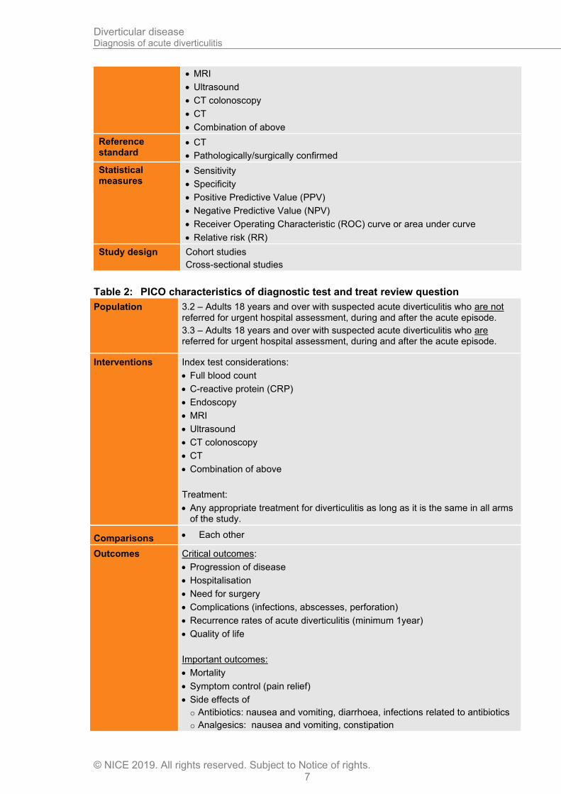
Table 2: PICO characteristics of diagnostic test and treat review question
Population 3.2 – Adults 18 years and over with suspected acute diverticulitis who are not referred for urgent hospital assessment, during and after the acute episode.
3.3 – Adults 18 years and over with suspected acute diverticulitis who are referred for urgent hospital assessment, during and after the acute episode.
Interventions Index test considerations:
• Full blood count
• C-reactive protein (CRP)
• Endoscopy
• MRI
• Ultrasound
• CT colonoscopy
• CT
• Combination of above
Treatment:
• Any appropriate treatment for diverticulitis as long as it is the same in all arms of the study.
Comparisons • Each other
Outcomes Critical outcomes:
• Progression of disease
• Hospitalisation
• Need for surgery
• Complications (infections, abscesses, perforation)
• Recurrence rates of acute diverticulitis (minimum 1year)
• Quality of life
Important outcomes:
• Mortality
• Symptom control (pain relief)
• Side effects of
o Antibiotics: nausea and vomiting, diarrhoea, infections related to antibiotics
o Analgesics: nausea and vomiting, constipation
Diverticular disease Diagnosis of acute diverticulitis
© NICE 2019. All rights reserved. Subject to Notice of rights. 8
1.4 Clinical evidence
1.4.1 Included studies
A search was conducted for prospective and retrospective cohort studies assessing the diagnostic accuracy of tests to identify whether the condition is present (as indicated by the reference standard CT scan) in people under investigation for acute diverticulitis.
Five studies were included in the review on adults with suspected acute diverticulitis who are referred for urgent hospital assessment, during and after the acute episode (3.3)5, 6, 36 59, 74; these are summarised in Table 2 below. Evidence from these studies is summarised in the clinical evidence summary below (Table 3).
No studies were identified for the review on adults with suspected acute diverticulitis who are not referred for urgent hospital assessment, during and after the acute episode (3.2).
No diagnostic RCTs were identified for this review.
See also the study selection flow chart in appendix C and study evidence tables in appendix D.
1.4.2 Excluded studies
See the excluded studies list in appendix H.
Dia
gnosis
of a
cute
div
ertic
ulitis
Div
ertic
ula
r dis
ease
© N
ICE
201
9. A
ll rights
reserv
ed. S
ubje
ct to
No
tice
of rig
hts
.
9
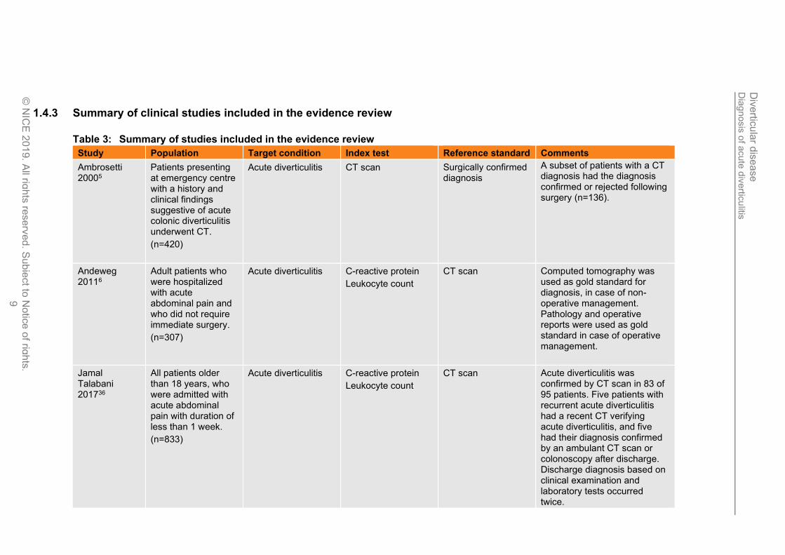
1.4.3 Summary of clinical studies included in the evidence review
Table 3: Summary of studies included in the evidence review
Study Population Target condition Index test Reference standard Comments
Ambrosetti 20005
Patients presenting at emergency centre with a history and clinical findings suggestive of acute colonic diverticulitis underwent CT.
(n=420)
Acute diverticulitis CT scan Surgically confirmed diagnosis
A subset of patients with a CT diagnosis had the diagnosis confirmed or rejected following surgery (n=136).
Andeweg 20116
Adult patients who were hospitalized with acute abdominal pain and who did not require immediate surgery.
(n=307)
Acute diverticulitis C-reactive protein
Leukocyte count
CT scan Computed tomography was used as gold standard for diagnosis, in case of non-operative management. Pathology and operative reports were used as gold standard in case of operative management.
Jamal Talabani 201736
All patients older than 18 years, who were admitted with acute abdominal pain with duration of less than 1 week.
(n=833)
Acute diverticulitis C-reactive protein
Leukocyte count
CT scan Acute diverticulitis was confirmed by CT scan in 83 of 95 patients. Five patients with recurrent acute diverticulitis had a recent CT verifying acute diverticulitis, and five had their diagnosis confirmed by an ambulant CT scan or colonoscopy after discharge. Discharge diagnosis based on clinical examination and laboratory tests occurred twice.
Dia
gnosis
of a
cute
div
ertic
ulitis
Div
ertic
ula
r dis
ease
© N
ICE
201
9. A
ll rights
reserv
ed. S
ubje
ct to
No
tice
of rig
hts
.
10
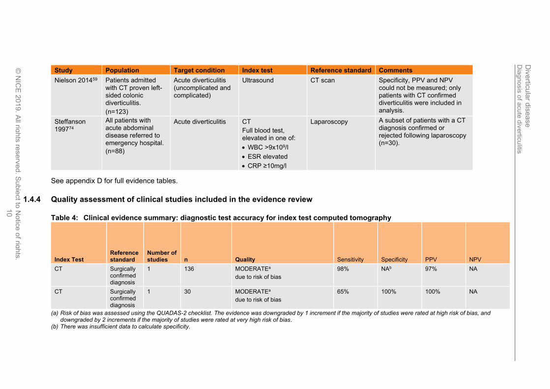
Study Population Target condition Index test Reference standard Comments
Nielson 201459 Patients admitted with CT proven left-sided colonic diverticulitis.
(n=123)
Acute diverticulitis (uncomplicated and complicated)
Ultrasound CT scan Specificity, PPV and NPV could not be measured; only patients with CT confirmed diverticulitis were included in analysis.
Steffanson 199774
All patients with acute abdominal disease referred to emergency hospital.
(n=88)
Acute diverticulitis CT
Full blood test, elevated in one of:
• WBC >9x10⁹/l
• ESR elevated
• CRP ≥10mg/l
Laparoscopy A subset of patients with a CT diagnosis confirmed or rejected following laparoscopy (n=30).
See appendix D for full evidence tables.
1.4.4 Quality assessment of clinical studies included in the evidence review
Table 4: Clinical evidence summary: diagnostic test accuracy for index test computed tomography
Index Test Reference standard
Number of studies n Quality Sensitivity Specificity PPV NPV
CT Surgically confirmed diagnosis
1 136 MODERATEa
due to risk of bias
98% NAb 97% NA
CT Surgically confirmed diagnosis
1 30 MODERATEa
due to risk of bias
65% 100% 100% NA
(a) Risk of bias was assessed using the QUADAS-2 checklist. The evidence was downgraded by 1 increment if the majority of studies were rated at high risk of bias, and downgraded by 2 increments if the majority of studies were rated at very high risk of bias.
(b) There was insufficient data to calculate specificity.
Dia
gnosis
of a
cute
div
ertic
ulitis
Div
ertic
ula
r dis
ease
© N
ICE
201
9. A
ll rights
reserv
ed. S
ubje
ct to
No
tice
of rig
hts
.
11
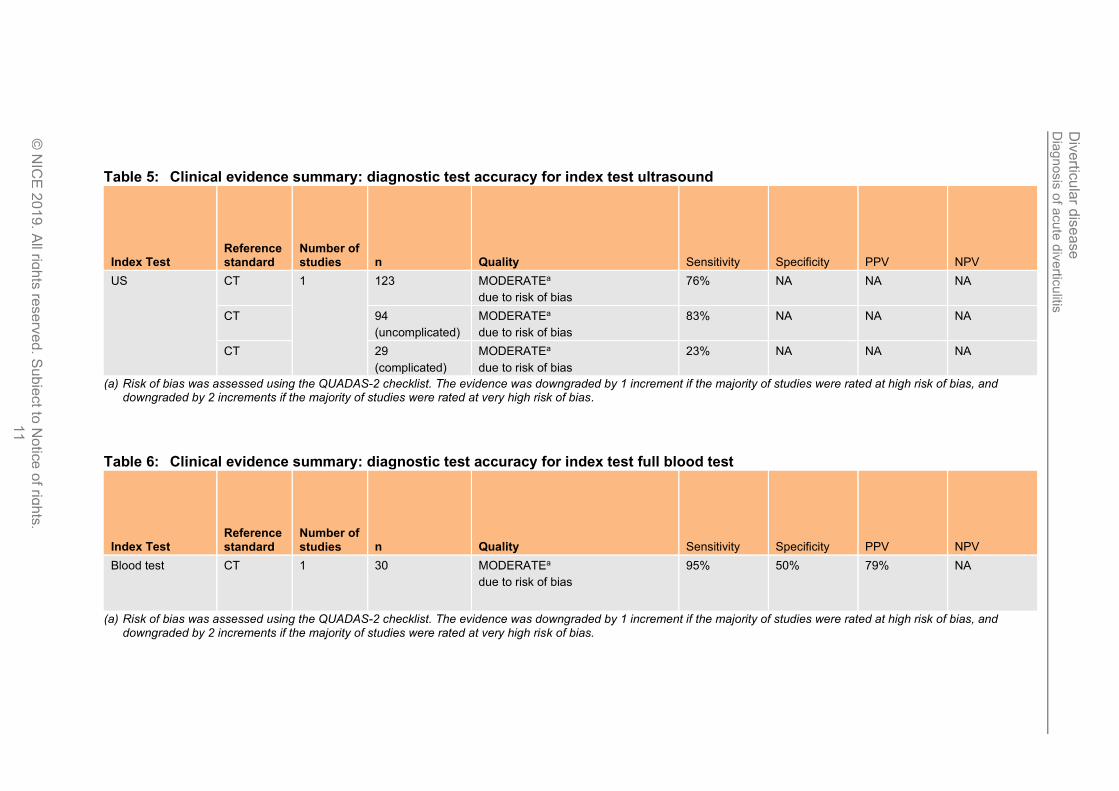
Table 5: Clinical evidence summary: diagnostic test accuracy for index test ultrasound
Index Test Reference standard
Number of studies n Quality Sensitivity Specificity PPV NPV
US CT 1 123 MODERATEa
due to risk of bias
76% NA NA NA
CT 94
(uncomplicated)
MODERATEa
due to risk of bias
83% NA NA NA
CT 29
(complicated)
MODERATEa
due to risk of bias
23% NA NA NA
(a) Risk of bias was assessed using the QUADAS-2 checklist. The evidence was downgraded by 1 increment if the majority of studies were rated at high risk of bias, and downgraded by 2 increments if the majority of studies were rated at very high risk of bias.
Table 6: Clinical evidence summary: diagnostic test accuracy for index test full blood test
Index Test Reference standard
Number of studies n Quality Sensitivity Specificity PPV NPV
Blood test CT 1 30 MODERATEa
due to risk of bias
95% 50% 79% NA
(a) Risk of bias was assessed using the QUADAS-2 checklist. The evidence was downgraded by 1 increment if the majority of studies were rated at high risk of bias, and downgraded by 2 increments if the majority of studies were rated at very high risk of bias.
Dia
gnosis
of a
cute
div
ertic
ulitis
Div
ertic
ula
r dis
ease
© N
ICE
201
9. A
ll rights
reserv
ed. S
ubje
ct to
No
tice
of rig
hts
.
12
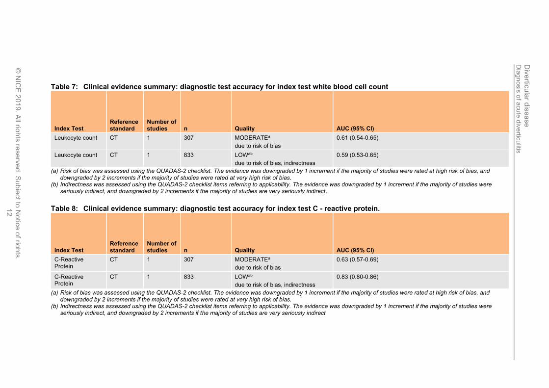
Table 7: Clinical evidence summary: diagnostic test accuracy for index test white blood cell count
Index Test Reference standard
Number of studies n Quality AUC (95% CI)
Leukocyte count CT 1 307 MODERATEa
due to risk of bias
0.61 (0.54-0.65)
Leukocyte count CT 1 833 LOWab
due to risk of bias, indirectness
0.59 (0.53-0.65)
(a) Risk of bias was assessed using the QUADAS-2 checklist. The evidence was downgraded by 1 increment if the majority of studies were rated at high risk of bias, and downgraded by 2 increments if the majority of studies were rated at very high risk of bias.
(b) Indirectness was assessed using the QUADAS-2 checklist items referring to applicability. The evidence was downgraded by 1 increment if the majority of studies were seriously indirect, and downgraded by 2 increments if the majority of studies are very seriously indirect.
Table 8: Clinical evidence summary: diagnostic test accuracy for index test C - reactive protein.
Index Test Reference standard
Number of studies n Quality AUC (95% CI)
C-Reactive Protein
CT 1 307 MODERATEa
due to risk of bias
0.63 (0.57-0.69)
C-Reactive Protein
CT 1 833 LOWab
due to risk of bias, indirectness
0.83 (0.80-0.86)
(a) Risk of bias was assessed using the QUADAS-2 checklist. The evidence was downgraded by 1 increment if the majority of studies were rated at high risk of bias, and downgraded by 2 increments if the majority of studies were rated at very high risk of bias.
(b) Indirectness was assessed using the QUADAS-2 checklist items referring to applicability. The evidence was downgraded by 1 increment if the majority of studies were seriously indirect, and downgraded by 2 increments if the majority of studies are very seriously indirect
Diverticular disease Diagnosis of acute diverticulitis
© NICE 2019. All rights reserved. Subject to Notice of rights. 13
1.5 Economic evidence
1.5.1 Included studies
No relevant health economic studies were identified.
1.5.2 Excluded studies
No health economic studies that were relevant to these questions were excluded due to assessment of limited applicability or methodological limitations.
See also the health economic study selection flow chart in appendix E.
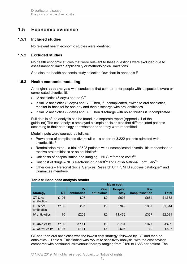
1.5.3 Health economic modelling
An original cost analysis was conducted that compared for people with suspected severe or complicated diverticulitis:
• IV antibiotics (5 days) and no CT
• Initial IV antibiotics (2 days) and CT. Then, if uncomplicated, switch to oral antibiotics, monitor in-hospital for one day and then discharge with oral antibiotics
• Initial IV antibiotics (2 days) and CT. Then discharge with no antibiotics if uncomplicated.
Full details of the analysis can be found in a separate report (Appendix 1 of the guideline).The cost analysis employed a simple decision tree that differentiated patients according to their pathology and whether or not they were readmitted.
Model inputs were sourced as follows:
• Prevalence of complicated diverticulitis – a cohort of 3,222 patients admitted with diverticulitis.9
• Readmission rates – a trial of 528 patients with uncomplicated diverticulitis randomised to receive oral antibiotics or no antibiotics24
• Unit costs of hospitalisation and imaging – NHS reference costs25
• Unit cost of drugs – NHS electronic drug tariff56 and British National Formulary39
• Other costs – Personal Social Services Research Unit22, NHS supplies catalogue57 and Committee members.
Table 9: Base case analysis results
Mean cost
Strategy CT IV
antibiotics Oral
antibiotics Hospital
stay Re-
hospitalisation Total
CT & no antibiotics
£106 £97 £0 £695 £684 £1,582
CT & oral antibiotics
£106 £97 £6 £949 £357 £1,514
IV antibiotics £0 £208 £0 £1,456 £357 £2,021
CT&No vs IV £106 -£111 £0 -£761 £327 -£439
CT&Oral vs IV £106 -£111 £6 -£507 £0 -£507
CT and then oral antibiotics was the lowest cost strategy, followed by ‘CT and then no antibiotics’ - Table 9. This finding was robust to sensitivity analysis, with the cost savings compared with continued intravenous therapy ranging from £150 to £688 per patient. The
Diverticular disease Diagnosis of acute diverticulitis
© NICE 2019. All rights reserved. Subject to Notice of rights. 14
only scenario that ‘CT and then no antibiotics’ was lowest cost was when we used a lower cost of rehospitalisation. The only time that the IV antibiotics strategy was lowest cost was when we used a high estimate of the cost of readmission and made the extreme assumption that there would be no readmissions in the IV antibiotics arm.
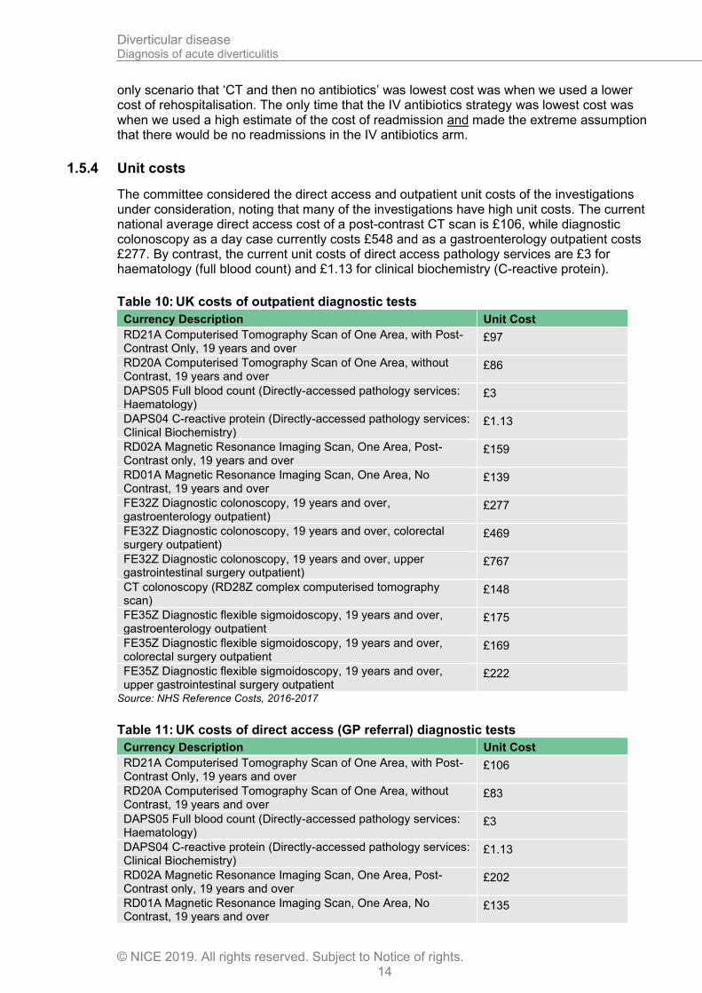
1.5.4 Unit costs
The committee considered the direct access and outpatient unit costs of the investigations under consideration, noting that many of the investigations have high unit costs. The current national average direct access cost of a post-contrast CT scan is £106, while diagnostic colonoscopy as a day case currently costs £548 and as a gastroenterology outpatient costs £277. By contrast, the current unit costs of direct access pathology services are £3 for haematology (full blood count) and £1.13 for clinical biochemistry (C-reactive protein).
Table 10: UK costs of outpatient diagnostic tests
Currency Description Unit Cost
RD21A Computerised Tomography Scan of One Area, with Post-Contrast Only, 19 years and over
£97
RD20A Computerised Tomography Scan of One Area, without Contrast, 19 years and over
£86
DAPS05 Full blood count (Directly-accessed pathology services: Haematology)
£3
DAPS04 C-reactive protein (Directly-accessed pathology services: Clinical Biochemistry)
£1.13
RD02A Magnetic Resonance Imaging Scan, One Area, Post-Contrast only, 19 years and over
£159
RD01A Magnetic Resonance Imaging Scan, One Area, No Contrast, 19 years and over
£139
FE32Z Diagnostic colonoscopy, 19 years and over, gastroenterology outpatient)
£277
FE32Z Diagnostic colonoscopy, 19 years and over, colorectal surgery outpatient)
£469
FE32Z Diagnostic colonoscopy, 19 years and over, upper gastrointestinal surgery outpatient)
£767
CT colonoscopy (RD28Z complex computerised tomography scan)
£148
FE35Z Diagnostic flexible sigmoidoscopy, 19 years and over, gastroenterology outpatient
£175
FE35Z Diagnostic flexible sigmoidoscopy, 19 years and over, colorectal surgery outpatient
£169
FE35Z Diagnostic flexible sigmoidoscopy, 19 years and over, upper gastrointestinal surgery outpatient
£222
Source: NHS Reference Costs, 2016-2017
Table 11: UK costs of direct access (GP referral) diagnostic tests
Currency Description Unit Cost
RD21A Computerised Tomography Scan of One Area, with Post-Contrast Only, 19 years and over
£106
RD20A Computerised Tomography Scan of One Area, without Contrast, 19 years and over
£83
DAPS05 Full blood count (Directly-accessed pathology services: Haematology)
£3
DAPS04 C-reactive protein (Directly-accessed pathology services: Clinical Biochemistry)
£1.13
RD02A Magnetic Resonance Imaging Scan, One Area, Post-Contrast only, 19 years and over
£202
RD01A Magnetic Resonance Imaging Scan, One Area, No Contrast, 19 years and over
£135
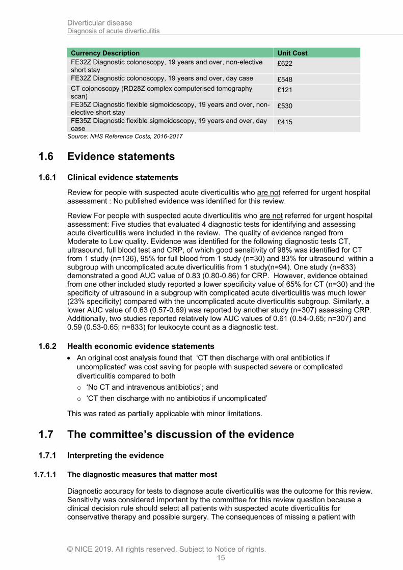
Diverticular disease Diagnosis of acute diverticulitis
© NICE 2019. All rights reserved. Subject to Notice of rights. 15
Currency Description Unit Cost
FE32Z Diagnostic colonoscopy, 19 years and over, non-elective short stay
£622
FE32Z Diagnostic colonoscopy, 19 years and over, day case £548
CT colonoscopy (RD28Z complex computerised tomography scan)
£121
FE35Z Diagnostic flexible sigmoidoscopy, 19 years and over, non-elective short stay
£530
FE35Z Diagnostic flexible sigmoidoscopy, 19 years and over, day case
£415
Source: NHS Reference Costs, 2016-2017
1.6 Evidence statements
1.6.1 Clinical evidence statements
Review for people with suspected acute diverticulitis who are not referred for urgent hospital assessment : No published evidence was identified for this review.
Review For people with suspected acute diverticulitis who are not referred for urgent hospital assessment: Five studies that evaluated 4 diagnostic tests for identifying and assessing acute diverticulitis were included in the review. The quality of evidence ranged from Moderate to Low quality. Evidence was identified for the following diagnostic tests CT, ultrasound, full blood test and CRP, of which good sensitivity of 98% was identified for CT from 1 study (n=136), 95% for full blood from 1 study (n=30) and 83% for ultrasound within a subgroup with uncomplicated acute diverticulitis from 1 study(n=94). One study (n=833) demonstrated a good AUC value of 0.83 (0.80-0.86) for CRP. However, evidence obtained from one other included study reported a lower specificity value of 65% for CT (n=30) and the specificity of ultrasound in a subgroup with complicated acute diverticulitis was much lower (23% specificity) compared with the uncomplicated acute diverticulitis subgroup. Similarly, a lower AUC value of 0.63 (0.57-0.69) was reported by another study (n=307) assessing CRP. Additionally, two studies reported relatively low AUC values of 0.61 (0.54-0.65; n=307) and 0.59 (0.53-0.65; n=833) for leukocyte count as a diagnostic test.
1.6.2 Health economic evidence statements
• An original cost analysis found that ‘CT then discharge with oral antibiotics if
uncomplicated’ was cost saving for people with suspected severe or complicated
diverticulitis compared to both
o ‘No CT and intravenous antibiotics’; and
o ‘CT then discharge with no antibiotics if uncomplicated’
This was rated as partially applicable with minor limitations.
1.7 The committee’s discussion of the evidence
1.7.1 Interpreting the evidence
1.7.1.1 The diagnostic measures that matter most
Diagnostic accuracy for tests to diagnose acute diverticulitis was the outcome for this review. Sensitivity was considered important by the committee for this review question because a clinical decision rule should select all patients with suspected acute diverticulitis for conservative therapy and possible surgery. The consequences of missing a patient with
Diverticular disease Diagnosis of acute diverticulitis
© NICE 2019. All rights reserved. Subject to Notice of rights. 16
acute diverticulitis would have serious health implications, and could result in an increased length of hospital stay during acute episodes.
No evidence was identified for the diagnostic accuracy of endoscopy, MRI, ultrasound, or CT colonoscopy.
1.7.1.2 The quality of the evidence
The quality of evidence ranged from very low to low. This was mostly due to flow and timing bias, resulting in a high risk of bias rating.
Outcomes were downgraded if there was an inappropriate amount of time between the reference test and the index test, such as when a person received a CT diagnosis and then underwent surgery at a later date following secondary complications. Outcomes were also downgraded where they included an indirect population or reported an indirect outcome, including where the reference standard was not consistent across the study population.
1.7.1.3 Benefits and harms
The committee considered the trade-off between using a less costly clinical test such as full blood count and CRP test to inform the decision making and selection of patients for further investigation for acute diverticulitis (and therefore to minimize the impact of a false negative result) and also to reduce radiation risk of imaging patients who do not have any inflammation. Inflammatory markers, commonly the White Blood Cell (WBC) count and C-Reactive Protein (CRP) level, are frequently employed to assist in diagnosing diverticulitis and its complications.
The committee also considered the accuracy and utility of a CT scan to correctly diagnose acute diverticulitis. The committee acknowledged that the one study included in this review assessing the diagnostic accuracy of CT scan showed a high sensitivity and positive predictive value. It was noted that the population from this study were those who were more severely unwell and required surgery, meaning the diagnosis in this population would likely be more clear-cut than would be typical in people with acute diverticulitis.
The committee agreed that CT is recognised as the most effective tool at diagnosing acute diverticulitis, particularly given its capacity to be performed during or shortly after an acute episode. The committee highlighted that endoscopy and CT colonoscopy should not be performed until ~6-8 weeks after an acute episode to prevent risk of perforation of the inflamed tissue and that there was evidence that in the setting of a high quality CT scan this may not be required. CT evaluates the severity and extent of disease and indicates what further treatment is required. Importantly its rules out other causes of the symptoms.
The committee also considered the radiation risks associated with CT scans. Given the condition’s prevalence in older people, the committee felt the increased risk of cancer with radiation exposure was negligible. The committee did agree that pregnant women should not be exposed to the radiation from CT scans, and so should be offered alternative methods of diagnosis such as MRI or ultrasound.
1.7.2 Cost effectiveness and resource use
Diagnostic pathway by setting
The proportion of people requiring emergency surgery for acute diverticulitis is small and the majority of people are managed conservatively with or without antibiotics.
No clinical or economic evidence was identified for investigations in the primary care setting. The committee felt that current practice is to prescribe a course of oral antibiotics to those
Diverticular disease Diagnosis of acute diverticulitis
© NICE 2019. All rights reserved. Subject to Notice of rights. 17
who do not require urgent referral for hospital assessment or sometimes there may be a period of watchful waiting before an antibiotic is prescribed. Where no improvement is seen or the condition deteriorates, the person with suspected acute diverticulitis is reassessed and considered for referral to secondary care.
No health economic evidence was identified for investigations for acute diverticulitis in people who are urgently referred for hospital assessment. In the absence of economic evidence, the low to very low quality clinical evidence for CT, full blood count and C-reactive protein was interpreted alongside the unit costs of the interventions to enable the committee to make qualitative judgements of cost effectiveness.
Imaging
In Chapter H, the committee concluded switching from intravenous to less expensive oral antibiotics and early discharge is safe for people with uncomplicated diverticulitis.
An original cost analysis was conducted that compared for people with suspected severe or complicated diverticulitis
• IV antibiotics and no CT
• Initial IV antibiotics and CT. Then discharge with oral antibiotics if uncomplicated
• Initial IV antibiotics and CT. Then discharge with no antibiotics if uncomplicated
The lowest cost strategy was ‘CT and then discharge with oral antibiotics if uncomplicated’ due to the reduced hospital stay and other cost savings. Discharging with no antibiotics was more costly because of the increased rehospitalisation observed in the clinical review (albeit not statistically significant). These results were robust to sensitivity analysis.
Therefore the committee recommended that patients should receive a CT, as it is diagnostic and likely to be cost saving.
The committee noted that obtaining CT scans during the acute episode might also reduce the number of colonoscopies carried out downstream, which would mean even greater cost savings. The model did not include the cost of antimicrobial resistance but this too would favour the use of CT to step down or cease antibiotics use.
In current practice, the committee believe that about 60% of 15,000 emergency admissions for acute diverticulitis currently receive CT scans. Obtaining CT scans in this population is currently dependent on availability, time of day and severity of the condition. In recommending that CT scans be offered for suspected acute diverticulitis, the committee acknowledged that there might be a significant resource impact, as it anticipates an increase in the number of people requiring scans. However, the cost analysis suggests that this would be more than offset by cost savings from reduced nurse time and hospital bed days.
No clinical or economic evidence was identified for MRI or ultrasound. The committee noted that the use of MRI and ultrasound is current practice only in pregnancy or if contrast CT is contraindicated. Imaging and oral antibiotics was still cost saving when we assumed the cost of an MRI in the analysis instead of CT.
Blood tests
The committee believes that the measurement of electrolytes and a full blood count is current practice and that C-reactive protein is regularly carried out, but is not yet universal. In the hospital setting, the results of the tests can be available after around an hour. No evidence was identified which described the effectiveness and cost effectiveness of white blood cell count and C-reactive protein as risk stratification tools to determine whether CT scans should be carried out. However, the committee felt that the cost of these tests is small and normal results can mean that a CT scan is not needed and therefore it likely that these tests are cost effective.
Diverticular disease Diagnosis of acute diverticulitis
© NICE 2019. All rights reserved. Subject to Notice of rights. 18
1.7.3 Other factors the committee took into account
The committee noted that initial urea and electrolyte tests at admission should be carried out ahead of any anticipated CT to assess renal function and guide CT with relation to user needs. Subsequent non-contrast CT can be carried out if necessary.
Diverticular disease Diagnosis of acute diverticulitis
© NICE 2019. All rights reserved. Subject to Notice of rights. 19
References
1. Abedi N, McKinlay R, Park A. Laparoscopic colectomy for diverticulitis. Current Surgery. 2004; 61(4):366-9
2. Ahn SH, Mayo-Smith WW, Murphy BL, Reinert SE, Cronan JJ. Acute nontraumatic abdominal pain in adult patients: abdominal radiography compared with CT evaluation. Radiology. 2002; 225(1):159-64
3. Alshamari M, Norrman E, Geijer M, Jansson K, Geijer H. Diagnostic accuracy of low-dose CT compared with abdominal radiography in non-traumatic acute abdominal pain: prospective study and systematic review. European Radiology. 2016; 26(6):1766-74
4. Ambrosetti P, Grossholz M, Becker C, Terrier F, Morel P. Computed tomography in acute left colonic diverticulitis. British Journal of Surgery. 1997; 84(4):532-4
5. Ambrosetti P, Jenny A, Becker C, Terrier F, Morel P. Acute left colonic diverticulitis - Compared performance of computed tomography and water-soluble contrast enema: Prospective evaluation of 420 patients. Diseases of the Colon and Rectum. 2000; 43(10):1363-1367
6. Andeweg CS, Knobben L, Hendriks JC, Bleichrodt RP, van Goor H. How to diagnose acute left-sided colonic diverticulitis: proposal for a clinical scoring system. Annals of Surgery. 2011; 253(5):940-6
7. Andeweg CS, Mulder IM, Felt-Bersma RJ, Verbon A, van der Wilt GJ, van Goor H et al. Guidelines of diagnostics and treatment of acute left-sided colonic diverticulitis. Digestive Surgery. 2013; 30(4-6):278-92
8. Andeweg CS, Wegdam JA, Groenewoud J, Wilt GJ, Goor H, Bleichrodt RP. Toward an evidence-based step-up approach in diagnosing diverticulitis. Scandinavian Journal of Gastroenterology. 2014; 49(7):775-784
9. Bharucha AE, Parthasarathy G, Ditah I, Fletcher JG, Ewelukwa O, Pendlimari R et al. Temporal trends in the incidence and natural history of diverticulitis: A population-based study. American Journal of Gastroenterology. 2015; 110(11):1589-96
10. Biondo S, Lopez Borao J, Millan M, Kreisler E, Jaurrieta E. Current status of the treatment of acute colonic diverticulitis: a systematic review. Colorectal Disease. 2012; 14(1):e1-e11
11. Braden B, Ignee A, Hocke M, Palmer RM, Dietrich C. Diagnostic value and clinical utility of contrast enhanced ultrasound in intestinal diseases. Digestive and Liver Disease. 2010; 42(10):667-674
12. Brown DF, Fischer RH, Novelline RA, Kim J, Nagurney JT. The role of abdominal computed tomography scanning in patients with non-traumatic abdominal symptoms. European Journal of Emergency Medicine. 2002; 9(4):330-3
13. Brown SR, Baraza W, Din S, Riley S. Chromoscopy versus conventional endoscopy for the detection of polyps in the colon and rectum. Cochrane Database of Systematic Reviews 2016, Issue 4. Art. No.: CD006439. DOI: 10.1002/14651858.CD006439.pub4.
Diverticular disease Diagnosis of acute diverticulitis
© NICE 2019. All rights reserved. Subject to Notice of rights. 20
14. Buckley O, Geoghegan T, O'Riordain DS, Lyburn ID, Torreggiani WC. Computed tomography in the imaging of colonic diverticulitis. Clinical Radiology. 2004; 59(11):977-983
15. Bugiantella W, Rondelli F, Longaroni M, Mariani E, Sanguinetti A, Avenia N. Left colon acute diverticulitis: an update on diagnosis, treatment and prevention. International Journal of Surgery. 2015; 13:157-64
16. Camera L, Liccardo I, Romano F, Liuzzi R, Rispo A, Imbriaco M et al. Diagnostic efficacy of single-pass abdominal multidetectorrow CT: prospective evaluation of a low dose protocol. British Journal of Radiology. 2017; 90(1070)
17. Caputo P, Rovagnati M, Carzaniga PL. Is it possible to limit the use of CT scanning in acute diverticular disease without compromising outcomes? A preliminary experience. Annali Italiani di Chirurgia. 2015; 86(1):51-5
18. Chabok A, Smedh K, Nilsson S, Stenson M, Pahlman L. CT-colonography in the follow-up of acute diverticulitis: patient acceptance and diagnostic accuracy. Scandinavian Journal of Gastroenterology. 2013; 48(8):979-86
19. Choi JJ, Ogunjemilusi O, Divino CM. Diagnosis and management of diverticula in the jejunum and ileum. American Surgeon. 2013; 79(1):108-10
20. Cobben LP, Groot I, Blickman JG, Puylaert JB. Right colonic diverticulitis: MR appearance. Abdominal Imaging. 2003; 28(6):794-798
21. Coogan S, Klabbatz L, Eisentat M, Chung R. Laparoscopic versus open sigmoid colectomy for diverticular disease: a case controlled study. Surgical Endoscopy. 1997; 11(2):195
22. Curtis L, Burns A. Unit costs of health and social care 2017. Canterbury. Personal Social Services Research Unit University of Kent, 2017. Available from: https://www.pssru.ac.uk/project-pages/unit-costs/unit-costs-2017/
23. Daniels L, Unlu C, de Wijkerslooth TR, Stockmann HB, Kuipers EJ, Boermeester MA et al. Yield of colonoscopy after recent CT-proven uncomplicated acute diverticulitis: a comparative cohort study. Surgical Endoscopy. 2015; 29(9):2605-13
24. Daniels L, Ünlü Ç, Korte N, Dieren S, Stockmann HB, Vrouenraets BC et al. Randomized clinical trial of observational versus antibiotic treatment for a first episode of CT-proven uncomplicated acute diverticulitis. British Journal of Surgery. 2017; 104(1):52-61
25. Department of Health. NHS reference costs 2016-17. 2017. Available from: https://improvement.nhs.uk/resources/reference-costs/ Last accessed: 01/09/2018
26. Dombal FT, Leaper DJ, Staniland JR, McCann AP, Horrocks JC. Computer-aided diagnosis of acute abdominal pain. BMJ. 1972; 2(5804):9-13
27. Domjan J, Blaquiere R, Odurny A. Is minimal preparation computed tomography comparable with barium enema in elderly patients with colonic symptoms? Clinical Radiology. 1998; 53(12):894-898
28. Eisenberg JD, Reisner AT, Binder WD, Zaheer A, Gunn ML, Linnau KF et al. Role of CT in the diagnosis of nonspecific abdominal pain: a multicenter analysis. American Journal of Roentgenology. 2017; 208(3):570-576
29. Etzioni DA, Chiu VY, Cannom RR, Burchette RJ, Haigh PI, Abbas MA. Outpatient treatment of acute diverticulitis: rates and predictors of failure. Diseases of the Colon and Rectum. 2010; 53(6):861-5
Diverticular disease Diagnosis of acute diverticulitis
© NICE 2019. All rights reserved. Subject to Notice of rights. 21
30. Floch CL. Diagnosis and management of acute diverticulitis. Journal of Clinical Gastroenterology. 2006; 40(Suppl 3):S136-44
31. Gallo A, Ianiro G, Montalto M, Cammarota G. The role of biomarkers in diverticular disease. Journal of Clinical Gastroenterology. 2016; 50(Suppl 1):S26-8
32. Gans SL, Atema JJ, Stoker J, Toorenvliet BR, Laurell H, Boermeester MA. C-reactive protein and white blood cell count as triage test between urgent and nonurgent conditions in 2961 patients with acute abdominal pain. Medicine. 2015; 94(9):e569
33. Gong PY, Li JX, Liu FL, Zhang LM, Xie HZ, Sui YB. Retrospective comparison of computed tomography enterography and magnetic resonance enterography in diagnosing small intestine disease. Journal of the Pakistan Medical Association. 2015; 65(7):710-714
34. Halligan S, Saunders B. Imaging diverticular disease. Best Practice & Research in Clinical Gastroenterology. 2002; 16(4):595-610
35. Ince AT, Baysal B, Kayar Y, Arabaci E, Bilgin M, Hamdard J et al. Comparison of tomographic and colonoscopic diagnoses in the presence of colonic wall thickening. International Journal of Clinical and Experimental Medicine. 2014; 7(11):4413-4419
36. Jamal Talabani A, Endreseth BH, Lydersen S, Edna TH. Clinical diagnostic accuracy of acute colonic diverticulitis in patients admitted with acute abdominal pain, a receiver operating characteristic curve analysis. International Journal of Colorectal Disease. 2017; 32(1):41-7
37. Jang T, Chauhan V, Cundiff C, Kaji AH. Assessment of emergency physician-performed ultrasound in evaluating nonspecific abdominal pain. American Journal of Emergency Medicine. 2014; 32(5):457-460
38. Jensen DM, Machicado GA, Jutabha R, Kovacs TO. Urgent colonoscopy for the diagnosis and treatment of severe diverticular hemorrhage. New England Journal of Medicine. 2000; 342(2):78-82
39. Joint Formulary Committee. British National Formulary (BNF) September 2018 update. 2018. Available from: http://www.bnf.org.uk Last accessed:
40. Jung HK, Choung RS, Locke GR, 3rd, Schleck CD, Zinsmeister AR, Talley NJ. Diarrhea-predominant irritable bowel syndrome is associated with diverticular disease: a population-based study. American Journal of Gastroenterology. 2010; 105(3):652-61
41. Juvonen P, Lehtimäki T, Eskelinen M, Ilves I, Vanninen R, Miettinen P et al. The need for surgery in acute abdominal pain: a randomized study of abdominal computed tomography. In Vivo. 2014; 28(3):305-309
42. Kaser SA, Fankhauser G, Glauser PM, Toia D, Maurer CA. Diagnostic value of inflammation markers in predicting perforation in acute sigmoid diverticulitis. World Journal of Surgery. 2010; 34(11):2717-22
43. Kawatkar A, Chu LH, Iyer R, Yen L, Chen W, Erder MH et al. Development and validation of algorithms to identify acute diverticulitis. Pharmacoepidemiology and Drug Safety. 2015; 24(1):27-37
44. Kechagias A, Rautio T, Kechagias G, Makela J. The role of C-reactive protein in the prediction of the clinical severity of acute diverticulitis. American Surgeon. 2014; 80(4):391-5
Diverticular disease Diagnosis of acute diverticulitis
© NICE 2019. All rights reserved. Subject to Notice of rights. 22
45. Kessner R, Barnes S, Halpern P, Makrin V, Blachar A. CT for acute nontraumatic abdominal pain-is oral contrast really required? Academic Radiology. 2017; 24(7):840-845
46. Lameris W, Randen A, Bipat S, Bossuyt PM, Boermeester MA, Stoker J. Graded compression ultrasonography and computed tomography in acute colonic diverticulitis: meta-analysis of test accuracy. European Radiology. 2008; 18(11):2498-2511
47. Lameris W, van Randen A, van Gulik TM, Busch OR, Winkelhagen J, Bossuyt PM et al. A clinical decision rule to establish the diagnosis of acute diverticulitis at the emergency department. Diseases of the Colon and Rectum. 2010; 53(6):896-904
48. Laurell H, Hansson LE, Gunnarsson U. Acute diverticulitis - Clinical presentation and differential diagnostics. Colorectal Disease. 2007; 9(6):496-501
49. Liljegren G, Chabok A, Wickbom M, Smedh K, Nilsson K. Acute colonic diverticulitis: a systematic review of diagnostic accuracy. Colorectal Disease. 2007; 9(6):480-488
50. Lindsay DC, Freeman JG, Cobden I, Record CO. Should colonoscopy be the first investigation for colonic disease? BMJ. 1988; 296(6616):167-169
51. Longstreth GF, Tieu RS. Clinically diagnosed acute diverticulitis in outpatients: Misdiagnosis in patients with irritable bowel syndrome. Digestive Diseases and Sciences. 2016; 61(2):578-88
52. Macconaill K, Downing J, Pai D, Kaur G. Are CT scan based scoring systems of any use in the practical management of acute diverticular disease? United European Gastroenterology Journal. 2014; 2(1 Suppl):A561
53. Millet I, Sebbane M, Molinari N, Pages-Bouic E, Curros-Doyon F, Riou B et al. Systematic unenhanced CT for acute abdominal symptoms in the elderly patients improves both emergency department diagnosis and prompt clinical management. European Radiology. 2017; 27(2):868-877
54. National Institute for Health and Care Excellence. Developing NICE guidelines: the manual. London. National Institute for Health and Care Excellence, 2014. Available from: http://www.nice.org.uk/article/PMG20/chapter/1%20Introduction%20and%20overview
55. Ng CS, Watson CJE, Palmer CR, See TC, Beharry NA, Housden BA et al. Evaluation of early abdominopelvic computed tomography in patients with acute abdominal pain of unknown cause: Prospective randomised study. BMJ. 2002; 325(7377):1387-1389
56. NHS Business Services Authority. NHS electronic drug tariff: September 2018. 2018. Available from: http://www.nhsbsa.nhs.uk/PrescriptionServices/4940.aspx Last accessed: 01/09/2018
57. NHS Supply Chain Catalogue. NHS Supply Chain, 2018. Available from: http://www.supplychain.nhs.uk/
58. Nicholas GG, Miller WT, Fitts WT, Tondreau RL. Diagnosis of diverticulitis of the colon: role of the barium enema in defining pericolic inflammation. Annals of Surgery. 1972; 176(2):205-9
59. Nielsen K, Richir MC, Stolk TT, van der Ploeg T, Moormann GR, Wiarda BM et al. The limited role of ultrasound in the diagnostic process of colonic diverticulitis. World Journal of Surgery. 2014; 38(7):1814-8
Diverticular disease Diagnosis of acute diverticulitis
© NICE 2019. All rights reserved. Subject to Notice of rights. 23
60. Oistamo E, Hjern F, Blomqvist L, Von Heijne A, Abraham-Nordling M. Cancer and diverticulitis of the sigmoid colon. Differentiation with computed tomography versus magnetic resonance imaging: preliminary experiences. Acta Radiologica. 2013; 54(3):237-41
61. Padidar AM, Jeffrey RB, Jr., Mindelzun RE, Dolph JF. Differentiating sigmoid diverticulitis from carcinoma on CT scans: mesenteric inflammation suggests diverticulitis. American Journal of Roentgenology. 1994; 163(1):81-3
62. Porten S, Kielb S. Diagnosis of female diverticula using magnetic resonance imaging. Advances in Urology. 2008; 2008:213516
63. Pradel JA, Adell JF, Taourel P, Djafari M, Monnin-Delhom E, Bruel JM. Acute colonic diverticulitis: Prospective comparative evaluation with US and CT. Radiology. 1997; 205(2):503-512
64. Rampton DS. Diverticular colitis: diagnosis and management. Colorectal Disease. 2001; 3(3):149-53
65. Sala E, Watson CJE, Beadsmoore C, Groot-Wassink T, Fanshawe TR, Smith JC et al. A randomized, controlled trial of routine early abdominal computed tomography in patients presenting with non-specific acute abdominal pain. Clinical Radiology. 2007; 62(10):961-969
66. Sanford MF, Pickhardt PJ. Diagnostic performance of primary 3-dimensional computed tomography colonography in the setting of colonic diverticular disease. Clinical Gastroenterology and Hepatology. 2006; 4(8):1039-47
67. Schnyder P, Moss AA, Thoeni RF, Margulis AR. A double-blind study of radiologic accuracy in diverticulitis, diverticulosis, and carcinoma of the sigmoid colon. Journal of Clinical Gastroenterology. 1979; 1(1):55-66
68. Schreyer AG, Furst A, Agha A, Kikinis R, Scheibl K, Scholmerich J et al. Magnetic resonance imaging based colonography for diagnosis and assessment of diverticulosis and diverticulitis. International Journal of Colorectal Disease. 2004; 19(5):474-80
69. Shen SH, Chen JD, Tiu CM, Chou YH, Chang CY, Yu C. Colonic diverticulitis diagnosed by computed tomography in the ED. American Journal of Emergency Medicine. 2002; 20(6):551-7
70. Shrier D, Skucas J, Weiss S. Diverticulitis: an evaluation by computed tomography and contrast enema. American Journal of Gastroenterology. 1991; 86(10):1466-71
71. Sirany AE, Gaertner WB, Madoff RD, Kwaan MR. Diverticulitis diagnosed in the emergency room: Is it safe to discharge home? Journal of the American College of Surgeons. 2017; 225(1):21-25
72. Snyder MJ. Imaging of colonic diverticular disease. Clinics in Colon and Rectal Surgery. 2004; 17(3):155-162
73. Spinzi G, Belloni G, Martegani A, Sangiovanni A, Del Favero C, Minoli G. Computed tomographic colonography and conventional colonoscopy for colon diseases: A prospective, blinded study. American Journal of Gastroenterology. 2001; 96(2):394-400
74. Stefansson T, Nyman R, Nilsson S, Ekbom A, Pahlman L. Diverticulitis of the sigmoid colon. A comparison of CT, colonic enema and laparoscopy. Acta Radiologica. 1997; 38(2):313-9
Diverticular disease Diagnosis of acute diverticulitis
© NICE 2019. All rights reserved. Subject to Notice of rights. 24
75. Stromberg C, Johansson G, Adolfsson A. Acute abdominal pain: diagnostic impact of immediate CT scanning. World Journal of Surgery. 2007; 31(12):2347-54; discussion 2355-8
76. Thorisson A, Smedh K, Torkzad MR, Påhlman L, Chabok A. CT imaging for prediction of complications and recurrence in acute uncomplicated diverticulitis. International Journal of Colorectal Disease. 2016; 31(2):451-457
77. Toorenvliet BR, Bakker RF, Breslau PJ, Merkus JW, Hamming JF. Colonic diverticulitis: a prospective analysis of diagnostic accuracy and clinical decision-making. Colorectal Disease. 2010; 12(3):179-86
78. Tursi A, Brandimarte G, Di Mario F, Annunziata ML, Bafutto M, Bianco MA et al. Predictive value of the Diverticular Inflammation and Complication Assessment (DICA) endoscopic classification on the outcome of diverticular disease of the colon: An international study. United European Gastroenterology Journal. 2016; 4(4):604-613
79. Turvill J, Aghahoseini A, Sivarajasingham N, Abbas K, Choudhry M, Polyzois K et al. Faecal calprotectin in patients with suspected colorectal cancer: A diagnostic accuracy study. British Journal of General Practice. 2016; 66(648):e499-e506
80. van de Wall BJ, Draaisma WA, van der Kaaij RT, Consten EC, Wiezer MJ, Broeders IA. The value of inflammation markers and body temperature in acute diverticulitis. Colorectal Disease. 2013; 15(5):621-6
81. Vather R, Broad JB, Jaung R, Robertson J, Bissett IP. Demographics and trends in the acute presentation of diverticular disease: a national study. ANZ Journal of Surgery. 2015; 85(10):744-8
82. Wolff JH, Rubin A, Potter JD, Lattimore W, Resnick MB, Murphy BL et al. Clinical significance of colonoscopic findings associated with colonic thickening on computed tomography: is colonoscopy warranted when thickening is detected? Journal of Clinical Gastroenterology. 2008; 42(5):472-5
83. Won Y, Lee HW, Ku YM, Lee SL, Seo KJ, Lee JI et al. Multidetector-row computed tomography (MDCT) features of small bowel obstruction (SBO) caused by Meckel's diverticulum. Diagnostic and Interventional Imaging. 2016; 97(2):227-32
84. Wong CS, Al-Ajami AK, Boshahri M, Naqvi SA. Diagnosis of acute surgical abdomen - The best diagnostic tool to reach a final diagnosiscin. Journal of Acute Disease. 2012; 1(1):23-5
85. Yardimci E, Hasbahceci M, Idiz UO, Atay M, Akbulut H. Is surgery necessary to confirm diagnosis of right-sided diverticulitis in spite of relevant clinical and radiological findings? Turkish Journal of Trauma & Emergency Surgery. 2017; 23(1):61-5
86. Zia N, Hussain T, Salamat A, Mirza S, Hassan F, Waqar A. Diagnostic evaluation of patients presenting with bleeding per rectum by colonoscopy. Journal of Ayub Medical College, Abbottabad. 2008; 20(1):73-6
87. Zielke A, Hasse C, Bandorski T, Sitter H, Wachsmuth P, Grobholz R et al. Diagnostic ultrasound of acute colonic diverticulitis by surgical residents. Surgical Endoscopy. 1997; 11(12):1194-7
Diverticular disease Diagnosis of acute diverticulitis
© NICE 2019. All rights reserved. Subject to Notice of rights. 25
Appendices
Appendix A: Review protocols
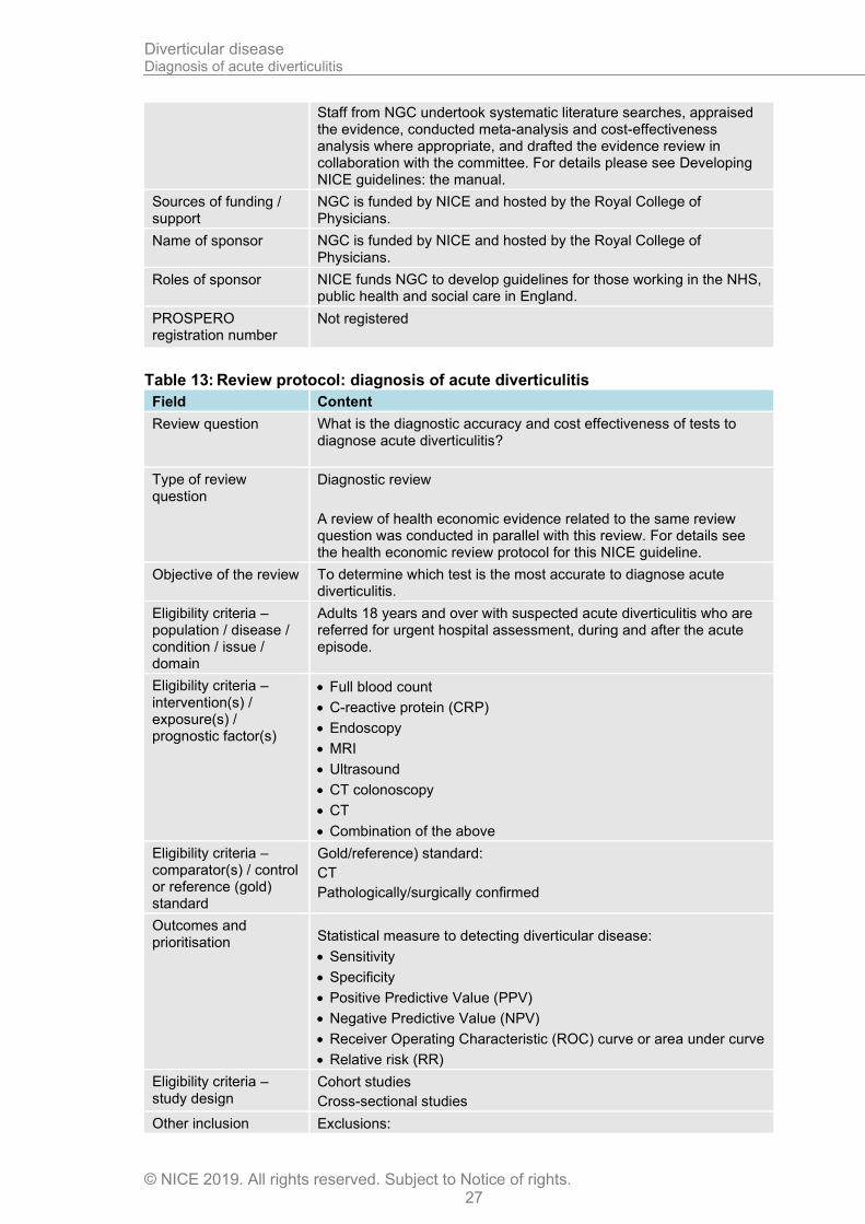
Table 12: Review protocol: diagnosis of acute diverticulitis
Field Content
Review question What is the diagnostic accuracy and cost effectiveness of tests to diagnose acute diverticulitis?
Type of review question
Diagnostic review
A review of health economic evidence related to the same review question was conducted in parallel with this review. For details see the health economic review protocol for this NICE guideline.
Objective of the review To determine which test is the most accurate to diagnose acute diverticulitis.
Eligibility criteria – population / disease / condition / issue / domain
Adults 18 years and over with suspected acute diverticulitis who are not referred for urgent hospital assessment, during and after the acute episode.
Eligibility criteria – intervention(s) / exposure(s) / prognostic factor(s)
• Full blood count
• C-reactive protein (CRP)
• Endoscopy
• MRI
• Ultrasound
• CT colonoscopy
• CT
•
• Combination of the above
Eligibility criteria – comparator(s) / control or reference (gold) standard
Gold/reference) standard:
CT
Pathologically/surgically confirmed
Outcomes and prioritisation Statistical measure to detecting diverticular disease:
• Sensitivity
• Specificity
• Positive Predictive Value (PPV)
• Negative Predictive Value (NPV)
• Receiver Operating Characteristic (ROC) curve or area under curve
• Relative risk (RR)
Eligibility criteria – study design
Cohort studies
Cross-sectional studies
Other inclusion exclusion criteria
Exclusions:
• Children and young people aged 17 years and younger
• Prevention
Proposed sensitivity / subgroup analysis, or meta-regression
Strata:
Subgroups:
• Age: <50 and >50 years
• people of Asian family origin as they are known to develop right-
Diverticular disease Diagnosis of acute diverticulitis
© NICE 2019. All rights reserved. Subject to Notice of rights. 26
sided diverticula
Selection process – duplicate screening / selection / analysis
Studies are sifted by title and abstract. Potentially significant publications obtained in full text are then assessed against the inclusion criteria specified in this protocol.
Data management (software)
• The methodological quality of each study outcome will be assessed using the adjusted QUADAS checklist.
• Pairwise meta-analyses performed using Cochrane Review Manager (RevMan5).
• GRADEpro used to assess the quality of evidence for each outcome
• Bibliographies, citations and study sifting managed using EndNote
• Data extractions performed using EviBase, a platform designed and maintained by the National Guideline Centre (NGC)
Information sources – databases and dates
Medline, Embase, The Cochrane Library
Identify if an update Not applicable
Author contacts https://www.nice.org.uk/guidance/conditions-and-diseases/digestive-tract-conditions/diverticular-disease
Highlight if amendment to previous protocol
For details please see section 4.5 of Developing NICE guidelines: the manual.
Search strategy – for one database
For details please see appendix B
Data collection process – forms / duplicate
A standardised evidence table format will be used, and published as appendix D of the evidence report.
Data items – define all variables to be collected
For details please see evidence tables in Appendix D (clinical evidence tables) or H (health economic evidence tables).
Methods for assessing bias at outcome / study level
Standard study checklists were used to critically appraise individual studies. For details please see section 6.2 of Developing NICE guidelines: the manual
The risk of bias across all available evidence was evaluated for each outcome using an adaptation of the ‘Grading of Recommendations Assessment, Development and Evaluation (GRADE) toolbox’ developed by the international GRADE working group http://www.gradeworkinggroup.org/
Criteria for quantitative synthesis
For details please see section 6.4 of Developing NICE guidelines: the manual.
Methods for quantitative analysis – combining studies and exploring (in)consistency
For details please see the separate Methods report (Chapter R) for this guideline.
Meta-bias assessment – publication bias, selective reporting bias
For details please see section 6.2 of Developing NICE guidelines: the manual.
Confidence in cumulative evidence
For details please see sections 6.4 and 9.1 of Developing NICE guidelines: the manual.
Rationale / context – what is known
For details please see the introduction to the evidence review.
Describe contributions of authors and guarantor
A multidisciplinary committee developed the evidence review. The committee was convened by the National Guideline Centre (NGC) and chaired by James Dalrymple in line with section 3 of Developing NICE guidelines: the manual.
Diverticular disease Diagnosis of acute diverticulitis
© NICE 2019. All rights reserved. Subject to Notice of rights. 27
Staff from NGC undertook systematic literature searches, appraised the evidence, conducted meta-analysis and cost-effectiveness analysis where appropriate, and drafted the evidence review in collaboration with the committee. For details please see Developing NICE guidelines: the manual.
Sources of funding / support
NGC is funded by NICE and hosted by the Royal College of Physicians.
Name of sponsor NGC is funded by NICE and hosted by the Royal College of Physicians.
Roles of sponsor NICE funds NGC to develop guidelines for those working in the NHS, public health and social care in England.
PROSPERO registration number
Not registered
Table 13: Review protocol: diagnosis of acute diverticulitis
Field Content
Review question What is the diagnostic accuracy and cost effectiveness of tests to diagnose acute diverticulitis?
Type of review question
Diagnostic review
A review of health economic evidence related to the same review question was conducted in parallel with this review. For details see the health economic review protocol for this NICE guideline.
Objective of the review To determine which test is the most accurate to diagnose acute diverticulitis.
Eligibility criteria – population / disease / condition / issue / domain
Adults 18 years and over with suspected acute diverticulitis who are referred for urgent hospital assessment, during and after the acute episode.
Eligibility criteria – intervention(s) / exposure(s) / prognostic factor(s)
• Full blood count
• C-reactive protein (CRP)
• Endoscopy
• MRI
• Ultrasound
• CT colonoscopy
• CT
• Combination of the above
Eligibility criteria – comparator(s) / control or reference (gold) standard
Gold/reference) standard:
CT
Pathologically/surgically confirmed
Outcomes and prioritisation Statistical measure to detecting diverticular disease:
• Sensitivity
• Specificity
• Positive Predictive Value (PPV)
• Negative Predictive Value (NPV)
• Receiver Operating Characteristic (ROC) curve or area under curve
• Relative risk (RR)
Eligibility criteria – study design
Cohort studies
Cross-sectional studies
Other inclusion Exclusions:
Diverticular disease Diagnosis of acute diverticulitis
© NICE 2019. All rights reserved. Subject to Notice of rights. 28
exclusion criteria • Children and young people aged 17 years and younger
• Prevention
Proposed sensitivity / subgroup analysis, or meta-regression
Subgroups:
• Age: <50 and >50 years
• people of Asian family origin as they are known to develop right-sided diverticula
Selection process – duplicate screening / selection / analysis
Studies are sifted by title and abstract. Potentially significant publications obtained in full text are then assessed against the inclusion criteria specified in this protocol.
Data management (software)
• The methodological quality of each study outcome will be assessed using the adjusted QUADSAS checklist.
• Pairwise meta-analyses performed using Cochrane Review Manager (RevMan5).
• GRADEpro used to assess the quality of evidence for each outcome
• Bibliographies, citations and study sifting managed using EndNote
• Data extractions performed using EviBase, a platform designed and maintained by the National Guideline Centre (NGC)
Information sources – databases and dates
Medline, Embase, The Cochrane Library
Identify if an update Not applicable
Author contacts https://www.nice.org.uk/guidance/conditions-and-diseases/digestive-tract-conditions/diverticular-disease
Highlight if amendment to previous protocol
For details please see section 4.5 of Developing NICE guidelines: the manual.
Search strategy – for one database
For details please see appendix B
Data collection process – forms / duplicate
A standardised evidence table format will be used, and published as appendix D of the evidence report.
Data items – define all variables to be collected
For details please see evidence tables in Appendix D (clinical evidence tables) or H (health economic evidence tables).
Methods for assessing bias at outcome / study level
Standard study checklists were used to critically appraise individual studies. For details please see section 6.2 of Developing NICE guidelines: the manual
The risk of bias across all available evidence was evaluated for each outcome using an adaptation of the ‘Grading of Recommendations Assessment, Development and Evaluation (GRADE) toolbox’ developed by the international GRADE working group http://www.gradeworkinggroup.org/
Criteria for quantitative synthesis
For details please see section 6.4 of Developing NICE guidelines: the manual.
Methods for quantitative analysis – combining studies and exploring (in)consistency
For details please see the separate Methods report (Chapter R) for this guideline.
Meta-bias assessment – publication bias, selective reporting bias
For details please see section 6.2 of Developing NICE guidelines: the manual.
Confidence in cumulative evidence
For details please see sections 6.4 and 9.1 of Developing NICE guidelines: the manual.
Diverticular disease Diagnosis of acute diverticulitis
© NICE 2019. All rights reserved. Subject to Notice of rights. 29
Rationale / context – what is known
For details please see the introduction to the evidence review.
Describe contributions of authors and guarantor
A multidisciplinary committee developed the evidence review. The committee was convened by the National Guideline Centre (NGC) and chaired by James Dalrymple in line with section 3 of Developing NICE guidelines: the manual.
Staff from NGC undertook systematic literature searches, appraised the evidence, conducted meta-analysis and cost-effectiveness analysis where appropriate, and drafted the evidence review in collaboration with the committee. For details please see Developing NICE guidelines: the manual.
Sources of funding / support
NGC is funded by NICE and hosted by the Royal College of Physicians.
Name of sponsor NGC is funded by NICE and hosted by the Royal College of Physicians.
Roles of sponsor NICE funds NGC to develop guidelines for those working in the NHS, public health and social care in England.
PROSPERO registration number
Not registered
Table 14: Health economic review protocol
Review question
All questions – health economic evidence
Objectives To identify health economic studies relevant to any of the review questions.
Search criteria
• Populations, interventions and comparators must be as specified in the clinical review protocol above.
• Studies must be of a relevant health economic study design (cost–utility analysis, cost-effectiveness analysis, cost–benefit analysis, cost–consequences analysis, comparative cost analysis).
• Studies must not be a letter, editorial or commentary, or a review of health economic evaluations. (Recent reviews will be ordered although not reviewed. The bibliographies will be checked for relevant studies, which will then be ordered.)
• Unpublished reports will not be considered unless submitted as part of a call for evidence.
• Studies must be in English.
Search strategy
A health economic study search will be undertaken using population-specific terms and a health economic study filter – see appendix B below.
Review strategy
Studies not meeting any of the search criteria above will be excluded. Studies published before 2002, abstract-only studies and studies from non-OECD countries or the USA will also be excluded.
Each remaining study will be assessed for applicability and methodological limitations using the NICE economic evaluation checklist which can be found in appendix H of Developing NICE guidelines: the manual (2014).54
Inclusion and exclusion criteria
• If a study is rated as both ‘Directly applicable’ and with ‘Minor limitations’ then it will be included in the guideline. A health economic evidence table will be completed and it will be included in the health economic evidence profile.
• If a study is rated as either ‘Not applicable’ or with ‘Very serious limitations’ then it will usually be excluded from the guideline. If it is excluded then a health economic evidence table will not be completed and it will not be included in the health economic evidence profile.
• If a study is rated as ‘Partially applicable’, with ‘Potentially serious limitations’ or both then there is discretion over whether it should be included.
Diverticular disease Diagnosis of acute diverticulitis
© NICE 2019. All rights reserved. Subject to Notice of rights. 30
Where there is discretion
The health economist will make a decision based on the relative applicability and quality of the available evidence for that question, in discussion with the guideline committee if required. The ultimate aim is to include health economic studies that are helpful for decision-making in the context of the guideline and the current NHS setting. If several studies are considered of sufficiently high applicability and methodological quality that they could all be included, then the health economist, in discussion with the committee if required, may decide to include only the most applicable studies and to selectively exclude the remaining studies. All studies excluded on the basis of applicability or methodological limitations will be listed with explanation in the excluded health economic studies appendix below.
The health economist will be guided by the following hierarchies.
Setting:
• UK NHS (most applicable).
• OECD countries with predominantly public health insurance systems (for example, France, Germany, Sweden).
• OECD countries with predominantly private health insurance systems (for example, Switzerland).
• Studies set in non-OECD countries or in the USA will be excluded before being assessed for applicability and methodological limitations.
Health economic study type:
• Cost–utility analysis (most applicable).
• Other type of full economic evaluation (cost–benefit analysis, cost-effectiveness analysis, cost–consequences analysis).
• Comparative cost analysis.
• Non-comparative cost analyses including cost-of-illness studies will be excluded before being assessed for applicability and methodological limitations.
Year of analysis:
• The more recent the study, the more applicable it will be.
• Studies published in 2002 or later but that depend on unit costs and resource data entirely or predominantly from before 2002 will be rated as ‘Not applicable’.
• Studies published before 2002 will be excluded before being assessed for applicability and methodological limitations.
Quality and relevance of effectiveness data used in the health economic analysis:
• The more closely the clinical effectiveness data used in the health economic analysis match with the outcomes of the studies included in the clinical review the more useful the analysis will be for decision-making in the guideline.
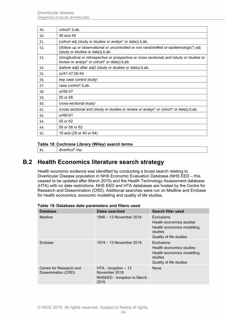
Appendix B: Literature search strategies The literature searches for this review are detailed below and complied with the methodology outlined in Developing NICE guidelines: the manual 2014, updated 2017
For more detailed information, please see the Methodology Review.
B.1 Clinical search literature search strategy
Searches were constructed using a PICO framework where population (P) terms were combined with Intervention (I) and in some cases Comparison (C) terms. Outcomes (O) are rarely used in search strategies for interventions as these concepts may not be well
Diverticular disease Diagnosis of acute diverticulitis
© NICE 2019. All rights reserved. Subject to Notice of rights. 31
described in title, abstract or indexes and therefore difficult to retrieve. Search filters were applied to the search where appropriate.
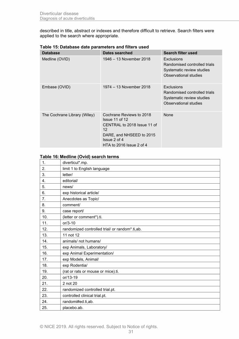
Table 15: Database date parameters and filters used
Database Dates searched Search filter used
Medline (OVID) 1946 – 13 November 2018
Exclusions
Randomised controlled trials
Systematic review studies
Observational studies
Embase (OVID) 1974 – 13 November 2018 Exclusions
Randomised controlled trials
Systematic review studies
Observational studies
The Cochrane Library (Wiley) Cochrane Reviews to 2018 Issue 11 of 12
CENTRAL to 2018 Issue 11 of 12
DARE, and NHSEED to 2015 Issue 2 of 4
HTA to 2016 Issue 2 of 4
None
Table 16: Medline (Ovid) search terms
1. diverticul*.mp.
2. limit 1 to English language
3. letter/
4. editorial/
5. news/
6. exp historical article/
7. Anecdotes as Topic/
8. comment/
9. case report/
10. (letter or comment*).ti.
11. or/3-10
12. randomized controlled trial/ or random*.ti,ab.
13. 11 not 12
14. animals/ not humans/
15. exp Animals, Laboratory/
16. exp Animal Experimentation/
17. exp Models, Animal/
18. exp Rodentia/
19. (rat or rats or mouse or mice).ti.
20. or/13-19
21. 2 not 20
22. randomized controlled trial.pt.
23. controlled clinical trial.pt.
24. randomi#ed.ti,ab.
25. placebo.ab.
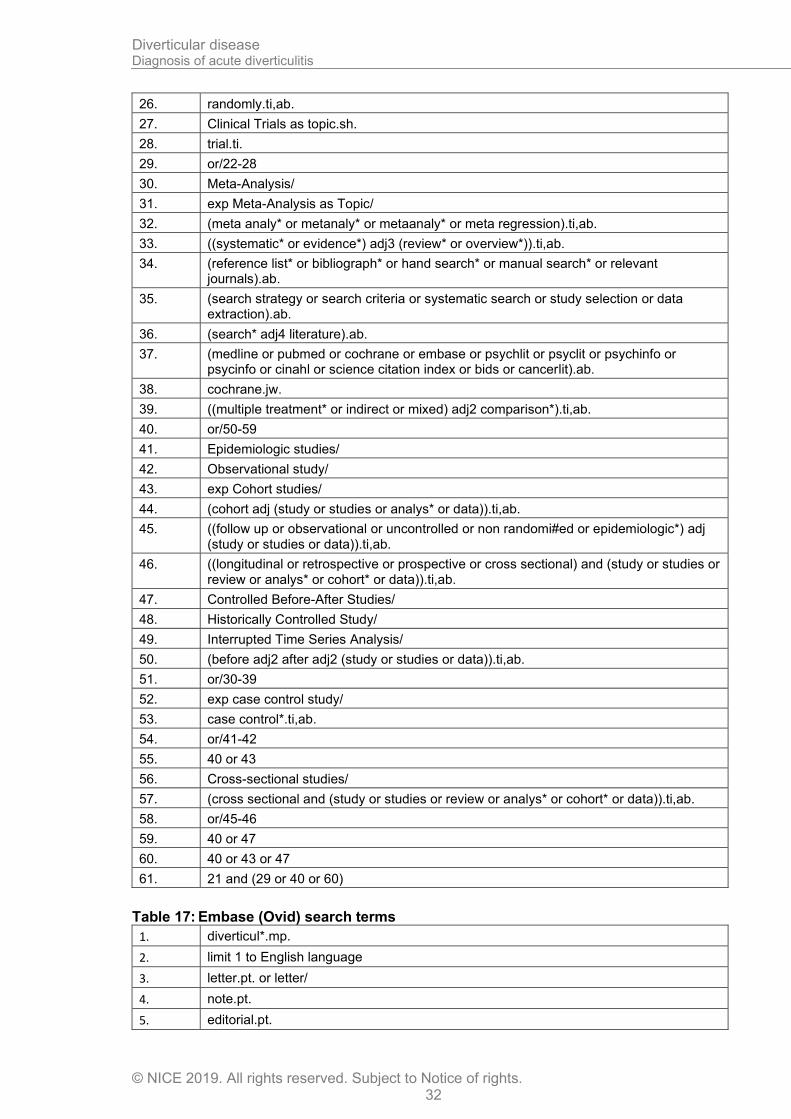
Diverticular disease Diagnosis of acute diverticulitis
© NICE 2019. All rights reserved. Subject to Notice of rights. 32
26. randomly.ti,ab.
27. Clinical Trials as topic.sh.
28. trial.ti.
29. or/22-28
30. Meta-Analysis/
31. exp Meta-Analysis as Topic/
32. (meta analy* or metanaly* or metaanaly* or meta regression).ti,ab.
33. ((systematic* or evidence*) adj3 (review* or overview*)).ti,ab.
34. (reference list* or bibliograph* or hand search* or manual search* or relevant journals).ab.
35. (search strategy or search criteria or systematic search or study selection or data extraction).ab.
36. (search* adj4 literature).ab.
37. (medline or pubmed or cochrane or embase or psychlit or psyclit or psychinfo or psycinfo or cinahl or science citation index or bids or cancerlit).ab.
38. cochrane.jw.
39. ((multiple treatment* or indirect or mixed) adj2 comparison*).ti,ab.
40. or/50-59
41. Epidemiologic studies/
42. Observational study/
43. exp Cohort studies/
44. (cohort adj (study or studies or analys* or data)).ti,ab.
45. ((follow up or observational or uncontrolled or non randomi#ed or epidemiologic*) adj (study or studies or data)).ti,ab.
46. ((longitudinal or retrospective or prospective or cross sectional) and (study or studies or review or analys* or cohort* or data)).ti,ab.
47. Controlled Before-After Studies/
48. Historically Controlled Study/
49. Interrupted Time Series Analysis/
50. (before adj2 after adj2 (study or studies or data)).ti,ab.
51. or/30-39
52. exp case control study/
53. case control*.ti,ab.
54. or/41-42
55. 40 or 43
56. Cross-sectional studies/
57. (cross sectional and (study or studies or review or analys* or cohort* or data)).ti,ab.
58. or/45-46
59. 40 or 47
60. 40 or 43 or 47
61. 21 and (29 or 40 or 60)
Table 17: Embase (Ovid) search terms
1. diverticul*.mp.
2. limit 1 to English language
3. letter.pt. or letter/
4. note.pt.
5. editorial.pt.
Diverticular disease Diagnosis of acute diverticulitis
© NICE 2019. All rights reserved. Subject to Notice of rights. 33
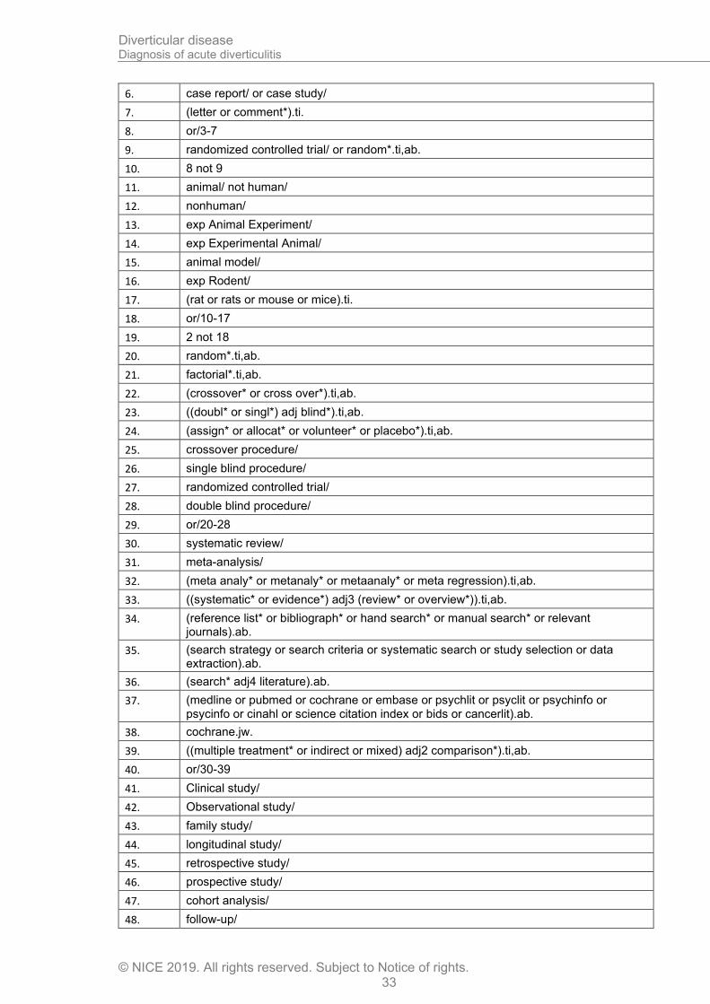
6. case report/ or case study/
7. (letter or comment*).ti.
8. or/3-7
9. randomized controlled trial/ or random*.ti,ab.
10. 8 not 9
11. animal/ not human/
12. nonhuman/
13. exp Animal Experiment/
14. exp Experimental Animal/
15. animal model/
16. exp Rodent/
17. (rat or rats or mouse or mice).ti.
18. or/10-17
19. 2 not 18
20. random*.ti,ab.
21. factorial*.ti,ab.
22. (crossover* or cross over*).ti,ab.
23. ((doubl* or singl*) adj blind*).ti,ab.
24. (assign* or allocat* or volunteer* or placebo*).ti,ab.
25. crossover procedure/
26. single blind procedure/
27. randomized controlled trial/
28. double blind procedure/
29. or/20-28
30. systematic review/
31. meta-analysis/
32. (meta analy* or metanaly* or metaanaly* or meta regression).ti,ab.
33. ((systematic* or evidence*) adj3 (review* or overview*)).ti,ab.
34. (reference list* or bibliograph* or hand search* or manual search* or relevant journals).ab.
35. (search strategy or search criteria or systematic search or study selection or data extraction).ab.
36. (search* adj4 literature).ab.
37. (medline or pubmed or cochrane or embase or psychlit or psyclit or psychinfo or psycinfo or cinahl or science citation index or bids or cancerlit).ab.
38. cochrane.jw.
39. ((multiple treatment* or indirect or mixed) adj2 comparison*).ti,ab.
40. or/30-39
41. Clinical study/
42. Observational study/
43. family study/
44. longitudinal study/
45. retrospective study/
46. prospective study/
47. cohort analysis/
48. follow-up/
Diverticular disease Diagnosis of acute diverticulitis
© NICE 2019. All rights reserved. Subject to Notice of rights. 34
49. cohort*.ti,ab.
50. 48 and 49
51. (cohort adj (study or studies or analys* or data)).ti,ab.
52. ((follow up or observational or uncontrolled or non randomi#ed or epidemiologic*) adj (study or studies or data)).ti,ab.
53. ((longitudinal or retrospective or prospective or cross sectional) and (study or studies or review or analys* or cohort* or data)).ti,ab.
54. (before adj2 after adj2 (study or studies or data)).ti,ab.
55. or/41-47,50-54
56. exp case control study/
57. case control*.ti,ab.
58. or/56-57
59. 55 or 58
60. cross-sectional study/
61. (cross sectional and (study or studies or review or analys* or cohort* or data)).ti,ab.
62. or/60-61
63. 55 or 62
64. 55 or 58 or 62
65. 19 and (29 or 40 or 64)
Table 18: Cochrane Library (Wiley) search terms
#1. diverticul*.mp.
B.2 Health Economics literature search strategy
Health economic evidence was identified by conducting a broad search relating to Diverticular Disease population in NHS Economic Evaluation Database (NHS EED – this ceased to be updated after March 2015) and the Health Technology Assessment database (HTA) with no date restrictions. NHS EED and HTA databases are hosted by the Centre for Research and Dissemination (CRD). Additional searches were run on Medline and Embase for health economics, economic modelling and quality of life studies.
Table 19: Database date parameters and filters used
Database Dates searched Search filter used
Medline 1946 – 13 November 2018
Exclusions
Health economics studies
Health economics modelling studies
Quality of life studies
Embase 1974 – 13 November 2018
Exclusions
Health economics studies
Health economics modelling studies
Quality of life studies
Centre for Research and Dissemination (CRD)
HTA - Inception – 13 November 2018
NHSEED - Inception to March 2015
None
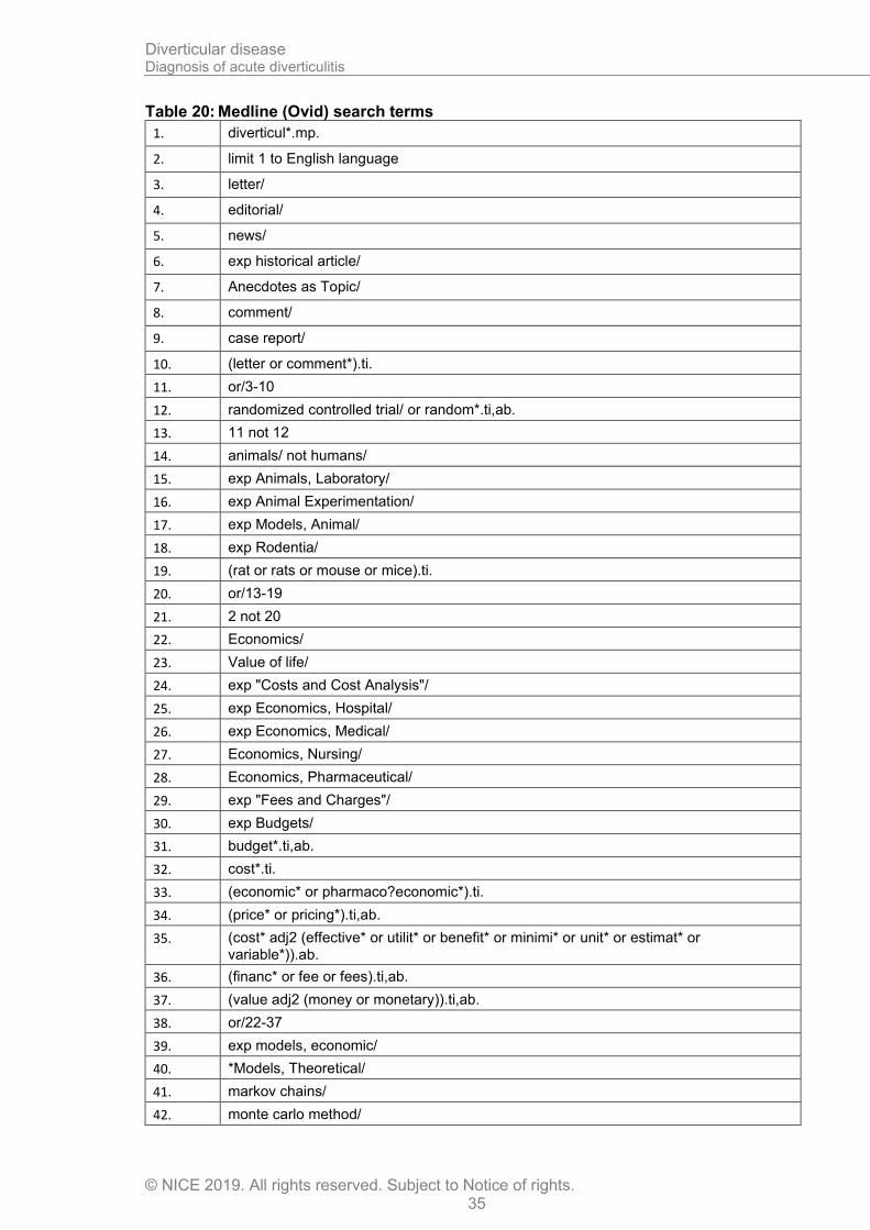
Diverticular disease Diagnosis of acute diverticulitis
© NICE 2019. All rights reserved. Subject to Notice of rights. 35
Table 20: Medline (Ovid) search terms
1. diverticul*.mp.
2. limit 1 to English language
3. letter/
4. editorial/
5. news/
6. exp historical article/
7. Anecdotes as Topic/
8. comment/
9. case report/
10. (letter or comment*).ti.
11. or/3-10
12. randomized controlled trial/ or random*.ti,ab.
13. 11 not 12
14. animals/ not humans/
15. exp Animals, Laboratory/
16. exp Animal Experimentation/
17. exp Models, Animal/
18. exp Rodentia/
19. (rat or rats or mouse or mice).ti.
20. or/13-19
21. 2 not 20
22. Economics/
23. Value of life/
24. exp "Costs and Cost Analysis"/
25. exp Economics, Hospital/
26. exp Economics, Medical/
27. Economics, Nursing/
28. Economics, Pharmaceutical/
29. exp "Fees and Charges"/
30. exp Budgets/
31. budget*.ti,ab.
32. cost*.ti.
33. (economic* or pharmaco?economic*).ti.
34. (price* or pricing*).ti,ab.
35. (cost* adj2 (effective* or utilit* or benefit* or minimi* or unit* or estimat* or variable*)).ab.
36. (financ* or fee or fees).ti,ab.
37. (value adj2 (money or monetary)).ti,ab.
38. or/22-37
39. exp models, economic/
40. *Models, Theoretical/
41. markov chains/
42. monte carlo method/
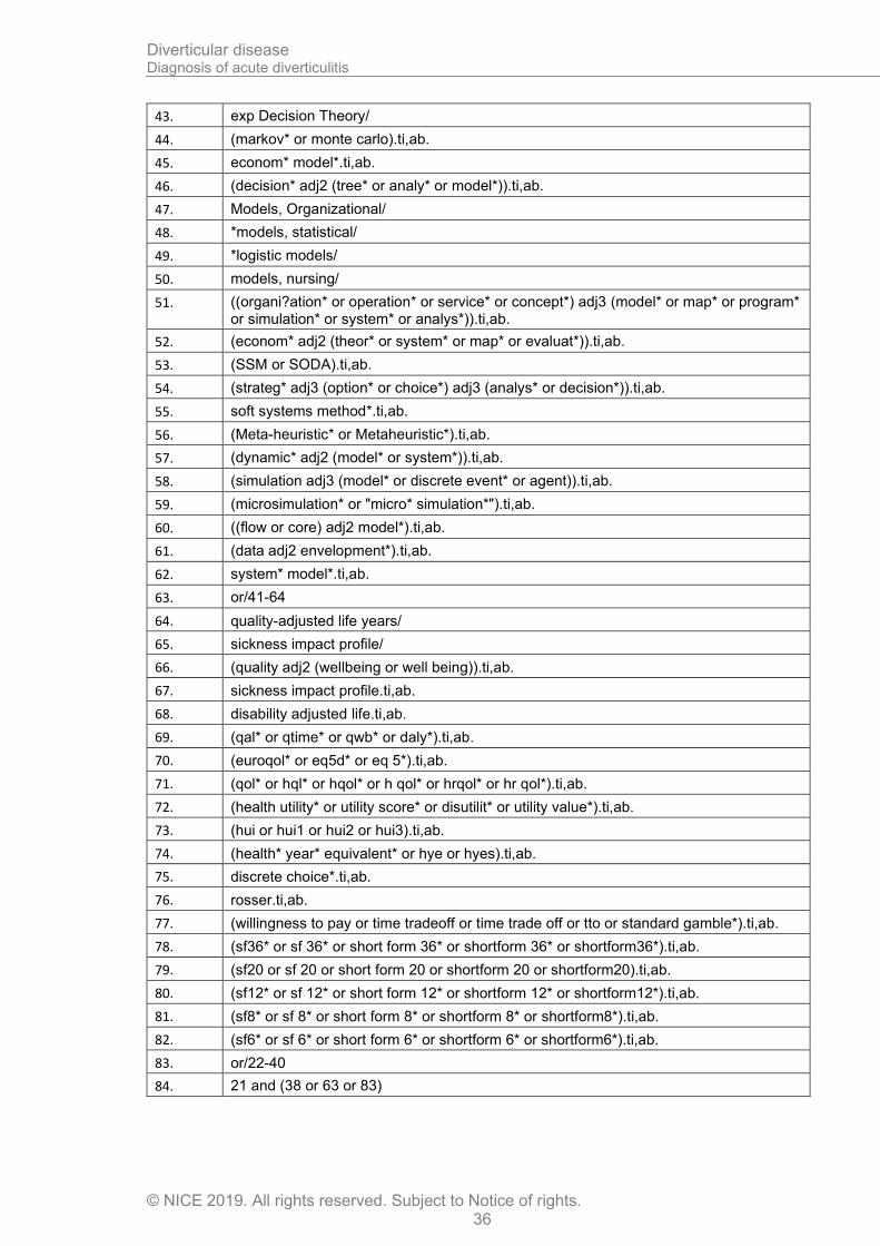
Diverticular disease Diagnosis of acute diverticulitis
© NICE 2019. All rights reserved. Subject to Notice of rights. 36
43. exp Decision Theory/
44. (markov* or monte carlo).ti,ab.
45. econom* model*.ti,ab.
46. (decision* adj2 (tree* or analy* or model*)).ti,ab.
47. Models, Organizational/
48. *models, statistical/
49. *logistic models/
50. models, nursing/
51. ((organi?ation* or operation* or service* or concept*) adj3 (model* or map* or program* or simulation* or system* or analys*)).ti,ab.
52. (econom* adj2 (theor* or system* or map* or evaluat*)).ti,ab.
53. (SSM or SODA).ti,ab.
54. (strateg* adj3 (option* or choice*) adj3 (analys* or decision*)).ti,ab.
55. soft systems method*.ti,ab.
56. (Meta-heuristic* or Metaheuristic*).ti,ab.
57. (dynamic* adj2 (model* or system*)).ti,ab.
58. (simulation adj3 (model* or discrete event* or agent)).ti,ab.
59. (microsimulation* or "micro* simulation*").ti,ab.
60. ((flow or core) adj2 model*).ti,ab.
61. (data adj2 envelopment*).ti,ab.
62. system* model*.ti,ab.
63. or/41-64
64. quality-adjusted life years/
65. sickness impact profile/
66. (quality adj2 (wellbeing or well being)).ti,ab.
67. sickness impact profile.ti,ab.
68. disability adjusted life.ti,ab.
69. (qal* or qtime* or qwb* or daly*).ti,ab.
70. (euroqol* or eq5d* or eq 5*).ti,ab.
71. (qol* or hql* or hqol* or h qol* or hrqol* or hr qol*).ti,ab.
72. (health utility* or utility score* or disutilit* or utility value*).ti,ab.
73. (hui or hui1 or hui2 or hui3).ti,ab.
74. (health* year* equivalent* or hye or hyes).ti,ab.
75. discrete choice*.ti,ab.
76. rosser.ti,ab.
77. (willingness to pay or time tradeoff or time trade off or tto or standard gamble*).ti,ab.
78. (sf36* or sf 36* or short form 36* or shortform 36* or shortform36*).ti,ab.
79. (sf20 or sf 20 or short form 20 or shortform 20 or shortform20).ti,ab.
80. (sf12* or sf 12* or short form 12* or shortform 12* or shortform12*).ti,ab.
81. (sf8* or sf 8* or short form 8* or shortform 8* or shortform8*).ti,ab.
82. (sf6* or sf 6* or short form 6* or shortform 6* or shortform6*).ti,ab.
83. or/22-40
84. 21 and (38 or 63 or 83)
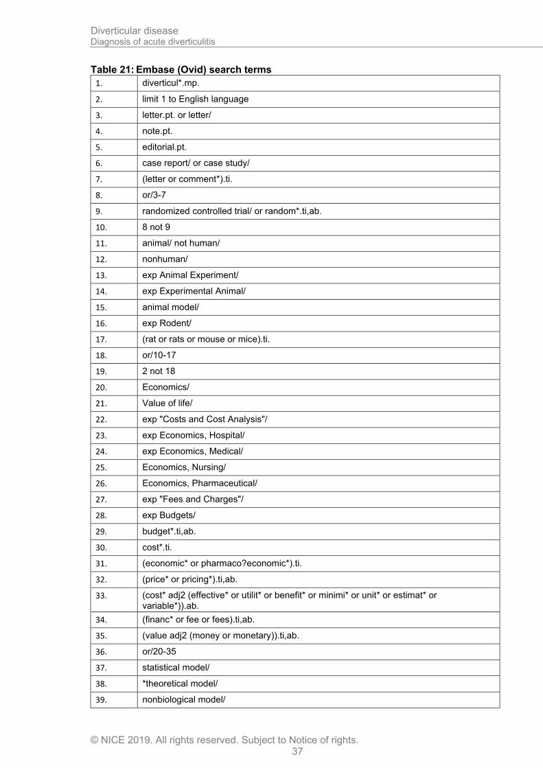
Diverticular disease Diagnosis of acute diverticulitis
© NICE 2019. All rights reserved. Subject to Notice of rights. 37
Table 21: Embase (Ovid) search terms
1. diverticul*.mp.
2. limit 1 to English language
3. letter.pt. or letter/
4. note.pt.
5. editorial.pt.
6. case report/ or case study/
7. (letter or comment*).ti.
8. or/3-7
9. randomized controlled trial/ or random*.ti,ab.
10. 8 not 9
11. animal/ not human/
12. nonhuman/
13. exp Animal Experiment/
14. exp Experimental Animal/
15. animal model/
16. exp Rodent/
17. (rat or rats or mouse or mice).ti.
18. or/10-17
19. 2 not 18
20. Economics/
21. Value of life/
22. exp "Costs and Cost Analysis"/
23. exp Economics, Hospital/
24. exp Economics, Medical/
25. Economics, Nursing/
26. Economics, Pharmaceutical/
27. exp "Fees and Charges"/
28. exp Budgets/
29. budget*.ti,ab.
30. cost*.ti.
31. (economic* or pharmaco?economic*).ti.
32. (price* or pricing*).ti,ab.
33. (cost* adj2 (effective* or utilit* or benefit* or minimi* or unit* or estimat* or variable*)).ab.
34. (financ* or fee or fees).ti,ab.
35. (value adj2 (money or monetary)).ti,ab.
36. or/20-35
37. statistical model/
38. *theoretical model/
39. nonbiological model/
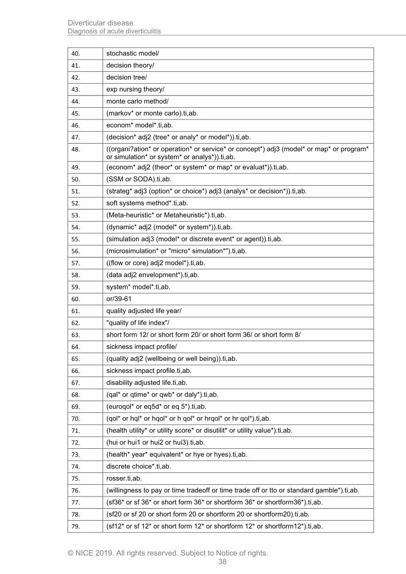
Diverticular disease Diagnosis of acute diverticulitis
© NICE 2019. All rights reserved. Subject to Notice of rights. 38
40. stochastic model/
41. decision theory/
42. decision tree/
43. exp nursing theory/
44. monte carlo method/
45. (markov* or monte carlo).ti,ab.
46. econom* model*.ti,ab.
47. (decision* adj2 (tree* or analy* or model*)).ti,ab.
48. ((organi?ation* or operation* or service* or concept*) adj3 (model* or map* or program* or simulation* or system* or analys*)).ti,ab.
49. (econom* adj2 (theor* or system* or map* or evaluat*)).ti,ab.
50. (SSM or SODA).ti,ab.
51. (strateg* adj3 (option* or choice*) adj3 (analys* or decision*)).ti,ab.
52. soft systems method*.ti,ab.
53. (Meta-heuristic* or Metaheuristic*).ti,ab.
54. (dynamic* adj2 (model* or system*)).ti,ab.
55. (simulation adj3 (model* or discrete event* or agent)).ti,ab.
56. (microsimulation* or "micro* simulation*").ti,ab.
57. ((flow or core) adj2 model*).ti,ab.
58. (data adj2 envelopment*).ti,ab.
59. system* model*.ti,ab.
60. or/39-61
61. quality adjusted life year/
62. "quality of life index"/
63. short form 12/ or short form 20/ or short form 36/ or short form 8/
64. sickness impact profile/
65. (quality adj2 (wellbeing or well being)).ti,ab.
66. sickness impact profile.ti,ab.
67. disability adjusted life.ti,ab.
68. (qal* or qtime* or qwb* or daly*).ti,ab.
69. (euroqol* or eq5d* or eq 5*).ti,ab.
70. (qol* or hql* or hqol* or h qol* or hrqol* or hr qol*).ti,ab.
71. (health utility* or utility score* or disutilit* or utility value*).ti,ab.
72. (hui or hui1 or hui2 or hui3).ti,ab.
73. (health* year* equivalent* or hye or hyes).ti,ab.
74. discrete choice*.ti,ab.
75. rosser.ti,ab.
76. (willingness to pay or time tradeoff or time trade off or tto or standard gamble*).ti,ab.
77. (sf36* or sf 36* or short form 36* or shortform 36* or shortform36*).ti,ab.
78. (sf20 or sf 20 or short form 20 or shortform 20 or shortform20).ti,ab.
79. (sf12* or sf 12* or short form 12* or shortform 12* or shortform12*).ti,ab.
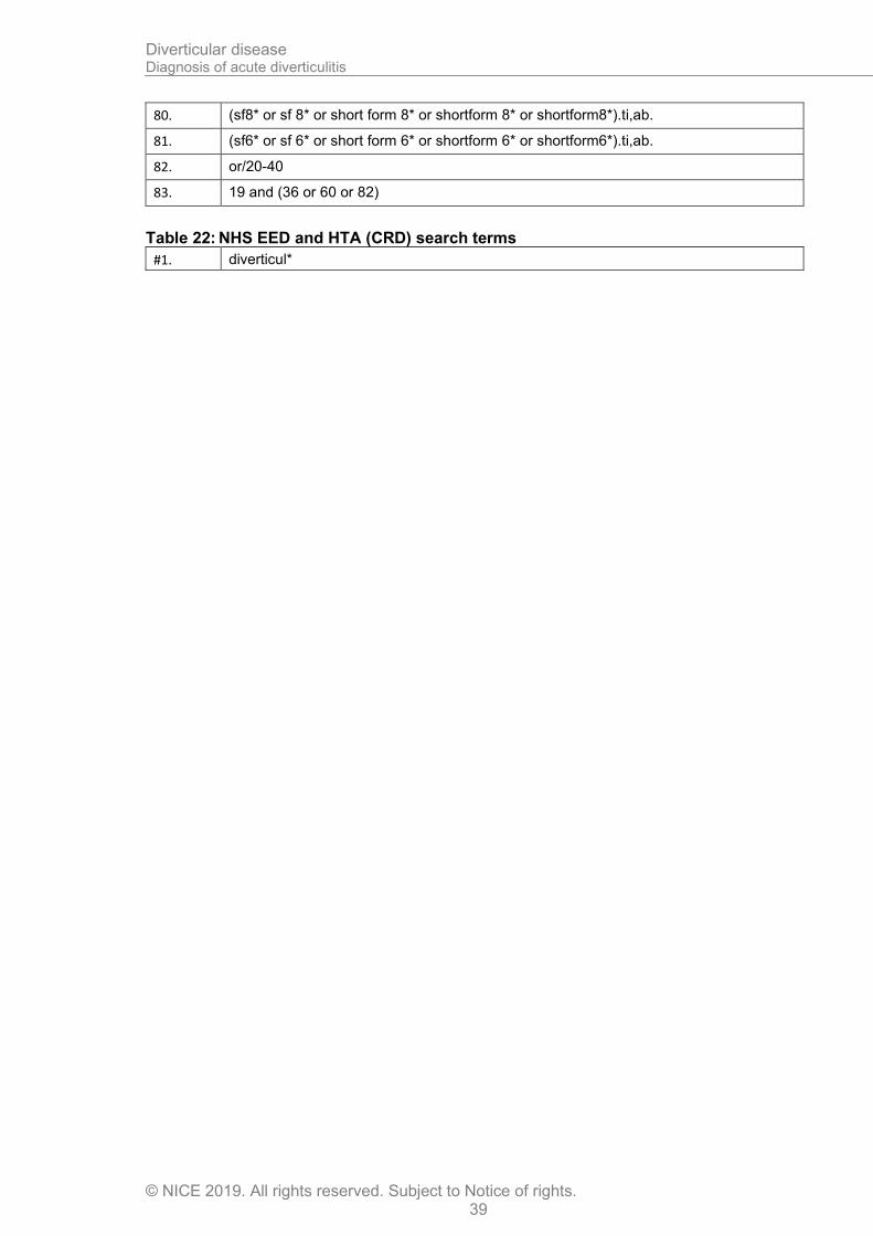
Diverticular disease Diagnosis of acute diverticulitis
© NICE 2019. All rights reserved. Subject to Notice of rights. 39
80. (sf8* or sf 8* or short form 8* or shortform 8* or shortform8*).ti,ab.
81. (sf6* or sf 6* or short form 6* or shortform 6* or shortform6*).ti,ab.
82. or/20-40
83. 19 and (36 or 60 or 82)
Table 22: NHS EED and HTA (CRD) search terms
#1. diverticul*
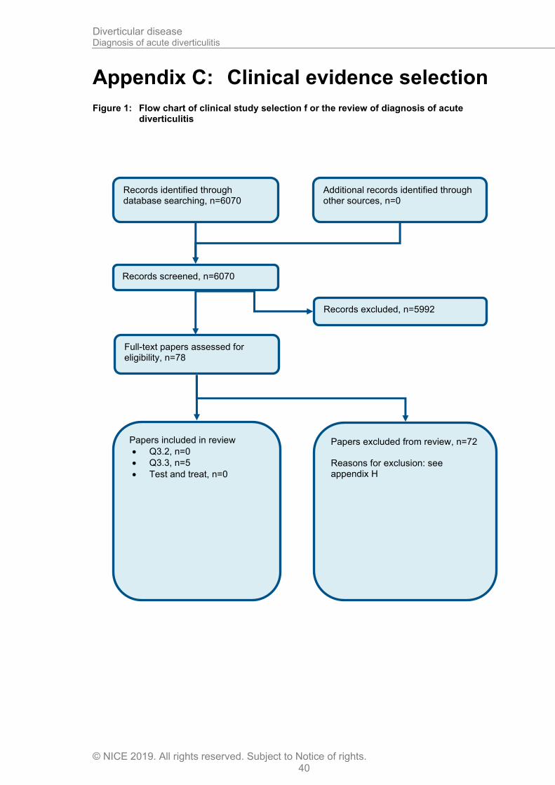
Diverticular disease Diagnosis of acute diverticulitis
© NICE 2019. All rights reserved. Subject to Notice of rights. 40
Appendix C: Clinical evidence selection
Figure 1: Flow chart of clinical study selection f or the review of diagnosis of acute diverticulitis
Records screened, n=6070
Records excluded, n=5992
Papers included in review
• Q3.2, n=0
• Q3.3, n=5
• Test and treat, n=0
Papers excluded from review, n=72 Reasons for exclusion: see appendix H
Records identified through database searching, n=6070
Additional records identified through other sources, n=0
Full-text papers assessed for eligibility, n=78
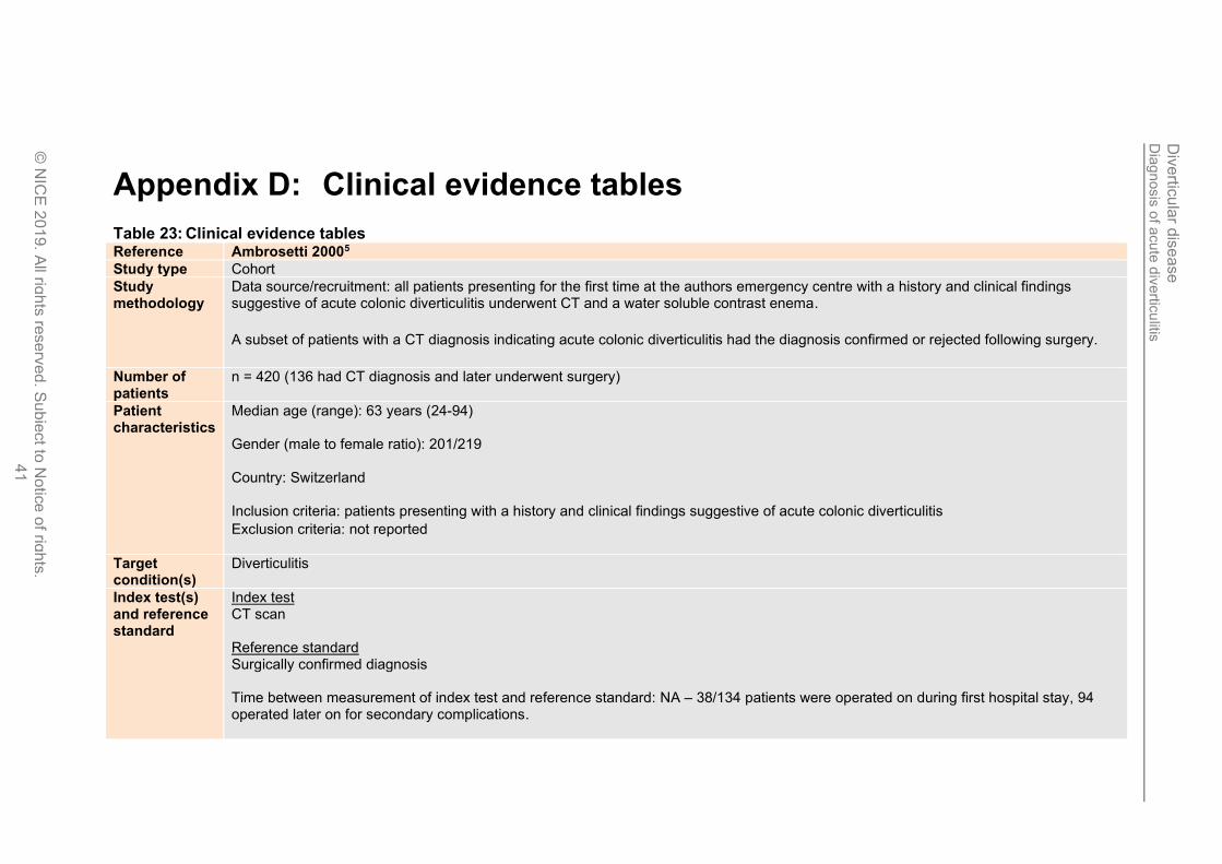
Dia
gnosis
of a
cute
div
ertic
ulitis
Div
ertic
ula
r dis
ease
© N
ICE
201
9. A
ll rights
reserv
ed. S
ubje
ct to
No
tice
of rig
hts
.
41
Appendix D: Clinical evidence tables
Table 23: Clinical evidence tables Reference Ambrosetti 20005
Study type Cohort
Study methodology
Data source/recruitment: all patients presenting for the first time at the authors emergency centre with a history and clinical findings suggestive of acute colonic diverticulitis underwent CT and a water soluble contrast enema.
A subset of patients with a CT diagnosis indicating acute colonic diverticulitis had the diagnosis confirmed or rejected following surgery.
Number of patients
n = 420 (136 had CT diagnosis and later underwent surgery)
Patient characteristics
Median age (range): 63 years (24-94) Gender (male to female ratio): 201/219 Country: Switzerland Inclusion criteria: patients presenting with a history and clinical findings suggestive of acute colonic diverticulitis
Exclusion criteria: not reported
Target condition(s)
Diverticulitis
Index test(s) and reference standard
Index test CT scan Reference standard Surgically confirmed diagnosis Time between measurement of index test and reference standard: NA – 38/134 patients were operated on during first hospital stay, 94 operated later on for secondary complications.
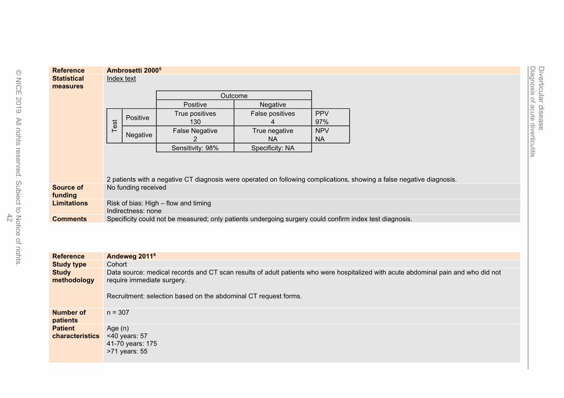
Dia
gnosis
of a
cute
div
ertic
ulitis
Div
ertic
ula
r dis
ease
© N
ICE
201
9. A
ll rights
reserv
ed. S
ubje
ct to
No
tice
of rig
hts
.
42
Reference Ambrosetti 20005
Statistical measures
Index text
Outcome
Positive Negative
Test Positive
True positives False positives PPV
130 4 97%
Negative False Negative True negative NPV
2 NA NA
Sensitivity: 98% Specificity: NA
2 patients with a negative CT diagnosis were operated on following complications, showing a false negative diagnosis.
Source of funding
No funding received
Limitations Risk of bias: High – flow and timing Indirectness: none
Comments Specificity could not be measured; only patients undergoing surgery could confirm index test diagnosis.
Reference Andeweg 20116
Study type Cohort
Study methodology
Data source: medical records and CT scan results of adult patients who were hospitalized with acute abdominal pain and who did not require immediate surgery.
Recruitment: selection based on the abdominal CT request forms.
Number of patients
n = 307
Patient characteristics
Age (n) <40 years: 57 41-70 years: 175 >71 years: 55
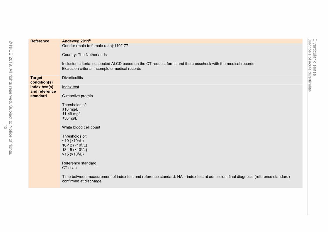
Dia
gnosis
of a
cute
div
ertic
ulitis
Div
ertic
ula
r dis
ease
© N
ICE
20
19
. All rig
hts
reserv
ed. S
ubje
ct to
No
tice
of rig
hts
.
43
Reference Andeweg 20116
Gender (male to female ratio):110/177 Country: The Netherlands Inclusion criteria: suspected ALCD based on the CT request forms and the crosscheck with the medical records
Exclusion criteria: incomplete medical records
Target condition(s)
Diverticulitis
Index test(s) and reference standard
Index test C-reactive protein Thresholds of: ≤10 mg/L 11-49 mg/L ≤50mg/L White blood cell count Thresholds of: <10 (×109/L) 10-12 (×109/L) 13-15 (×109/L) >15 (×109/L) Reference standard CT scan Time between measurement of index test and reference standard: NA – index test at admission, final diagnosis (reference standard) confirmed at discharge
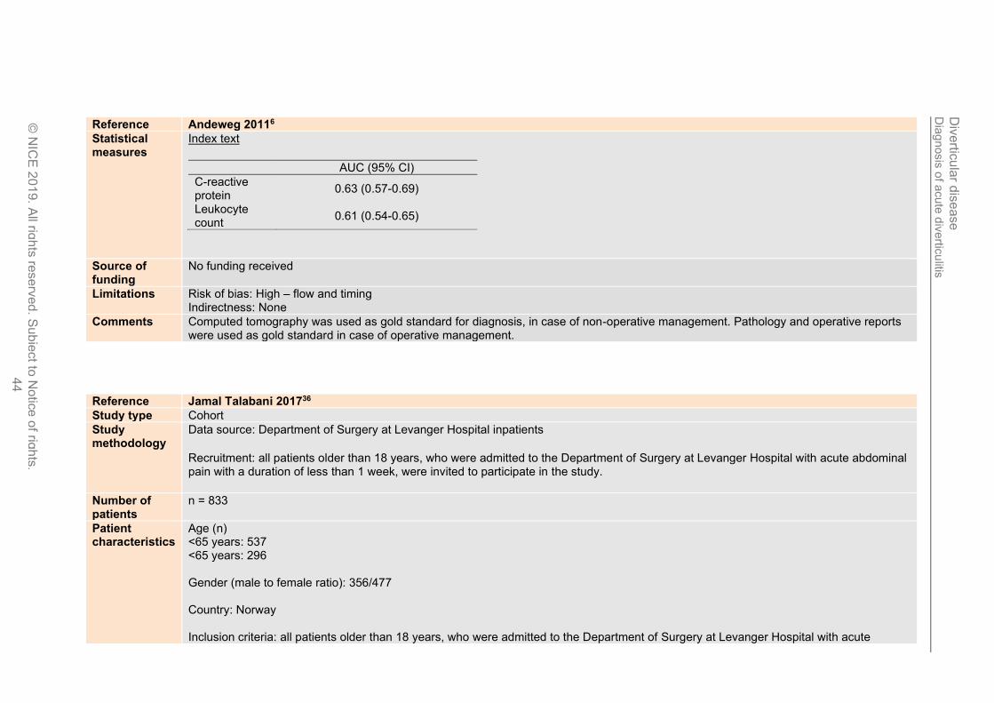
Dia
gnosis
of a
cute
div
ertic
ulitis
Div
ertic
ula
r dis
ease
© N
ICE
201
9. A
ll rights
reserv
ed. S
ubje
ct to
No
tice
of rig
hts
.
44
Reference Andeweg 20116
Statistical measures
Index text
AUC (95% CI)
C-reactive protein
0.63 (0.57-0.69)
Leukocyte count
0.61 (0.54-0.65)
Source of funding
No funding received
Limitations Risk of bias: High – flow and timing Indirectness: None
Comments Computed tomography was used as gold standard for diagnosis, in case of non-operative management. Pathology and operative reports were used as gold standard in case of operative management.
Reference Jamal Talabani 201736
Study type Cohort
Study methodology
Data source: Department of Surgery at Levanger Hospital inpatients
Recruitment: all patients older than 18 years, who were admitted to the Department of Surgery at Levanger Hospital with acute abdominal pain with a duration of less than 1 week, were invited to participate in the study.
Number of patients
n = 833
Patient characteristics
Age (n) <65 years: 537 <65 years: 296 Gender (male to female ratio): 356/477 Country: Norway Inclusion criteria: all patients older than 18 years, who were admitted to the Department of Surgery at Levanger Hospital with acute
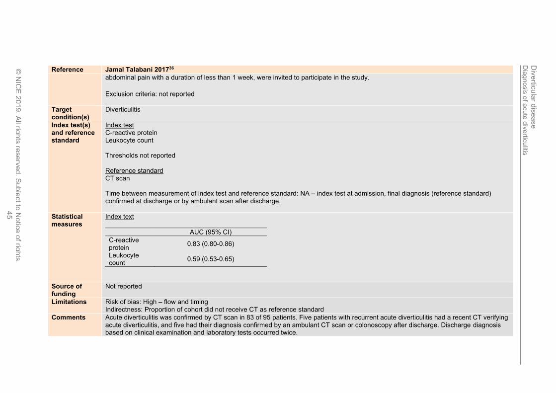
Dia
gnosis
of a
cute
div
ertic
ulitis
Div
ertic
ula
r dis
ease
© N
ICE
201
9. A
ll rights
reserv
ed. S
ubje
ct to
No
tice
of rig
hts
.
45
Reference Jamal Talabani 201736
abdominal pain with a duration of less than 1 week, were invited to participate in the study.
Exclusion criteria: not reported
Target condition(s)
Diverticulitis
Index test(s) and reference standard
Index test C-reactive protein Leukocyte count Thresholds not reported Reference standard CT scan Time between measurement of index test and reference standard: NA – index test at admission, final diagnosis (reference standard) confirmed at discharge or by ambulant scan after discharge.
Statistical measures
Index text
AUC (95% CI)
C-reactive protein
0.83 (0.80-0.86)
Leukocyte count
0.59 (0.53-0.65)
Source of funding
Not reported
Limitations Risk of bias: High – flow and timing Indirectness: Proportion of cohort did not receive CT as reference standard
Comments Acute diverticulitis was confirmed by CT scan in 83 of 95 patients. Five patients with recurrent acute diverticulitis had a recent CT verifying acute diverticulitis, and five had their diagnosis confirmed by an ambulant CT scan or colonoscopy after discharge. Discharge diagnosis based on clinical examination and laboratory tests occurred twice.
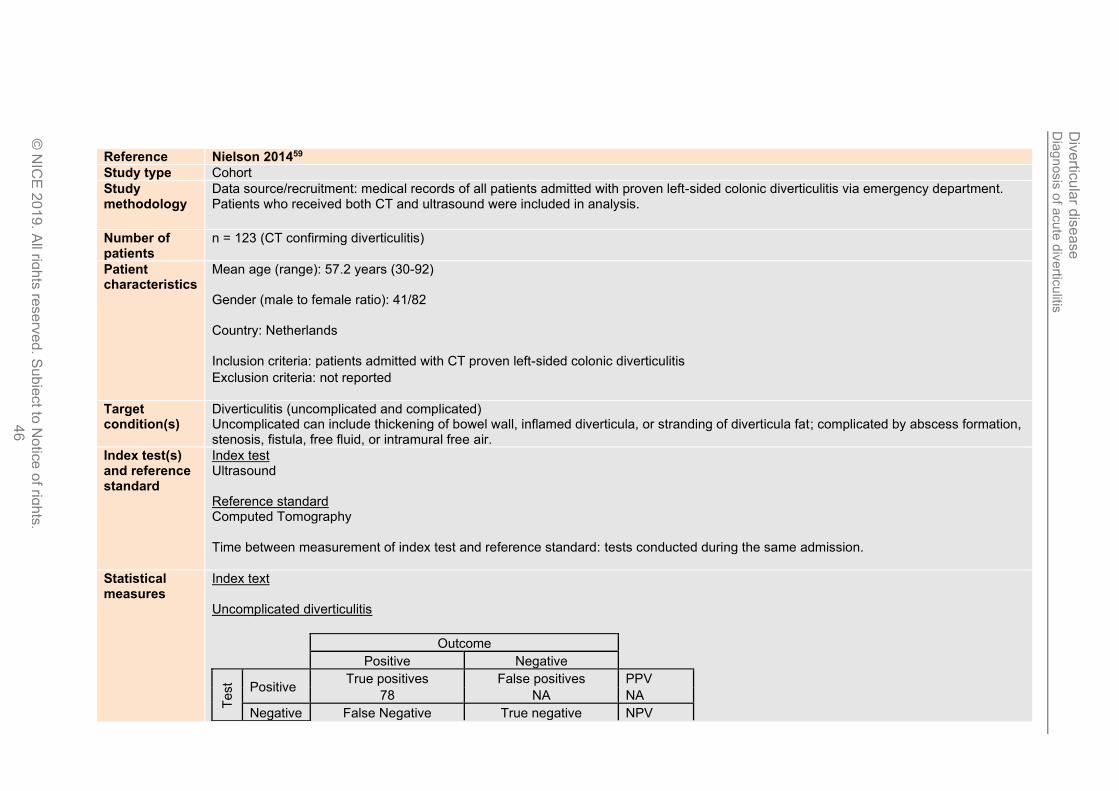
Dia
gnosis
of a
cute
div
ertic
ulitis
Div
ertic
ula
r dis
ea
se
© N
ICE
201
9. A
ll rights
reserv
ed. S
ubje
ct to
No
tice
of rig
hts
.
46
Reference Nielson 201459
Study type Cohort
Study methodology
Data source/recruitment: medical records of all patients admitted with proven left-sided colonic diverticulitis via emergency department. Patients who received both CT and ultrasound were included in analysis.
Number of patients
n = 123 (CT confirming diverticulitis)
Patient characteristics
Mean age (range): 57.2 years (30-92) Gender (male to female ratio): 41/82 Country: Netherlands Inclusion criteria: patients admitted with CT proven left-sided colonic diverticulitis
Exclusion criteria: not reported
Target condition(s)
Diverticulitis (uncomplicated and complicated) Uncomplicated can include thickening of bowel wall, inflamed diverticula, or stranding of diverticula fat; complicated by abscess formation, stenosis, fistula, free fluid, or intramural free air.
Index test(s) and reference standard
Index test Ultrasound Reference standard Computed Tomography Time between measurement of index test and reference standard: tests conducted during the same admission.
Statistical measures
Index text Uncomplicated diverticulitis
Outcome
Positive Negative
Test Positive
True positives False positives PPV
78 NA NA
Negative False Negative True negative NPV
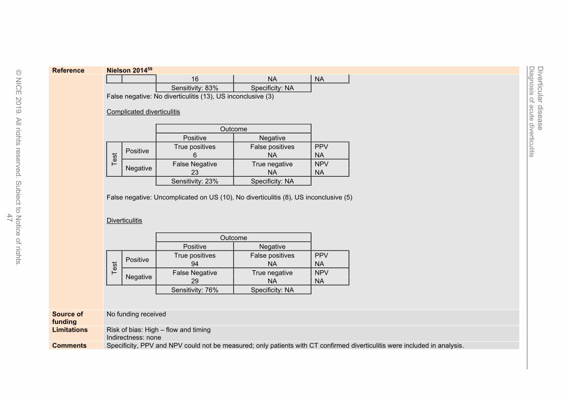
Dia
gnosis
of a
cute
div
ertic
ulitis
Div
ertic
ula
r dis
ease
© N
ICE
201
9. A
ll rights
reserv
ed. S
ubje
ct to
No
tice
of rig
hts
.
47
Reference Nielson 201459
16 NA NA
Sensitivity: 83% Specificity: NA
False negative: No diverticulitis (13), US inconclusive (3) Complicated diverticulitis
Outcome
Positive Negative
Test Positive
True positives False positives PPV
6 NA NA
Negative False Negative True negative NPV
23 NA NA
Sensitivity: 23% Specificity: NA
False negative: Uncomplicated on US (10), No diverticulitis (8), US inconclusive (5) Diverticulitis
Outcome
Positive Negative
Test Positive
True positives False positives PPV
94 NA NA
Negative False Negative True negative NPV
29 NA NA
Sensitivity: 76% Specificity: NA
Source of funding
No funding received
Limitations Risk of bias: High – flow and timing Indirectness: none
Comments Specificity, PPV and NPV could not be measured; only patients with CT confirmed diverticulitis were included in analysis.
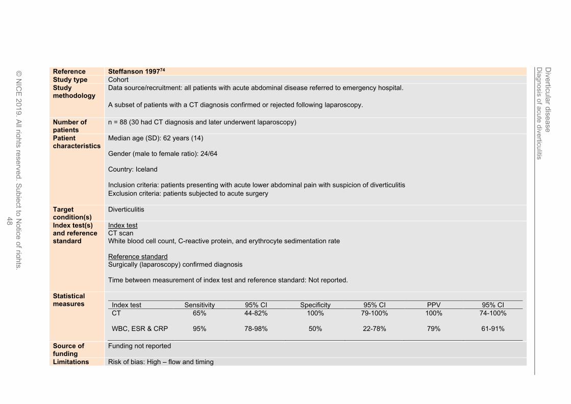
Dia
gnosis
of a
cute
div
ertic
ulitis
Div
ertic
ula
r dis
ease
© N
ICE
201
9. A
ll rights
reserv
ed. S
ubje
ct to
No
tice
of rig
hts
.
48
Reference Steffanson 199774
Study type Cohort
Study methodology
Data source/recruitment: all patients with acute abdominal disease referred to emergency hospital.
A subset of patients with a CT diagnosis confirmed or rejected following laparoscopy.
Number of patients
n = 88 (30 had CT diagnosis and later underwent laparoscopy)
Patient characteristics
Median age (SD): 62 years (14) Gender (male to female ratio): 24/64 Country: Iceland Inclusion criteria: patients presenting with acute lower abdominal pain with suspicion of diverticulitis
Exclusion criteria: patients subjected to acute surgery
Target condition(s)
Diverticulitis
Index test(s) and reference standard
Index test CT scan White blood cell count, C-reactive protein, and erythrocyte sedimentation rate Reference standard Surgically (laparoscopy) confirmed diagnosis Time between measurement of index test and reference standard: Not reported.
Statistical measures
Index test Sensitivity 95% CI Specificity 95% CI PPV 95% CI
CT
65% 44-82% 100% 79-100% 100% 74-100%
WBC, ESR & CRP
95% 78-98% 50% 22-78% 79% 61-91%
Source of funding
Funding not reported
Limitations Risk of bias: High – flow and timing
Dia
gnosis
of a
cute
div
ertic
ulitis
Div
ertic
ula
r dis
ease
© N
ICE
201
9. A
ll rights
reserv
ed. S
ubje
ct to
No
tice
of rig
hts
.
49
Reference Steffanson 199774
Indirectness: none
Comments Negative predictive value could not be measured; only patients undergoing laparoscopy could confirm index test diagnosis.
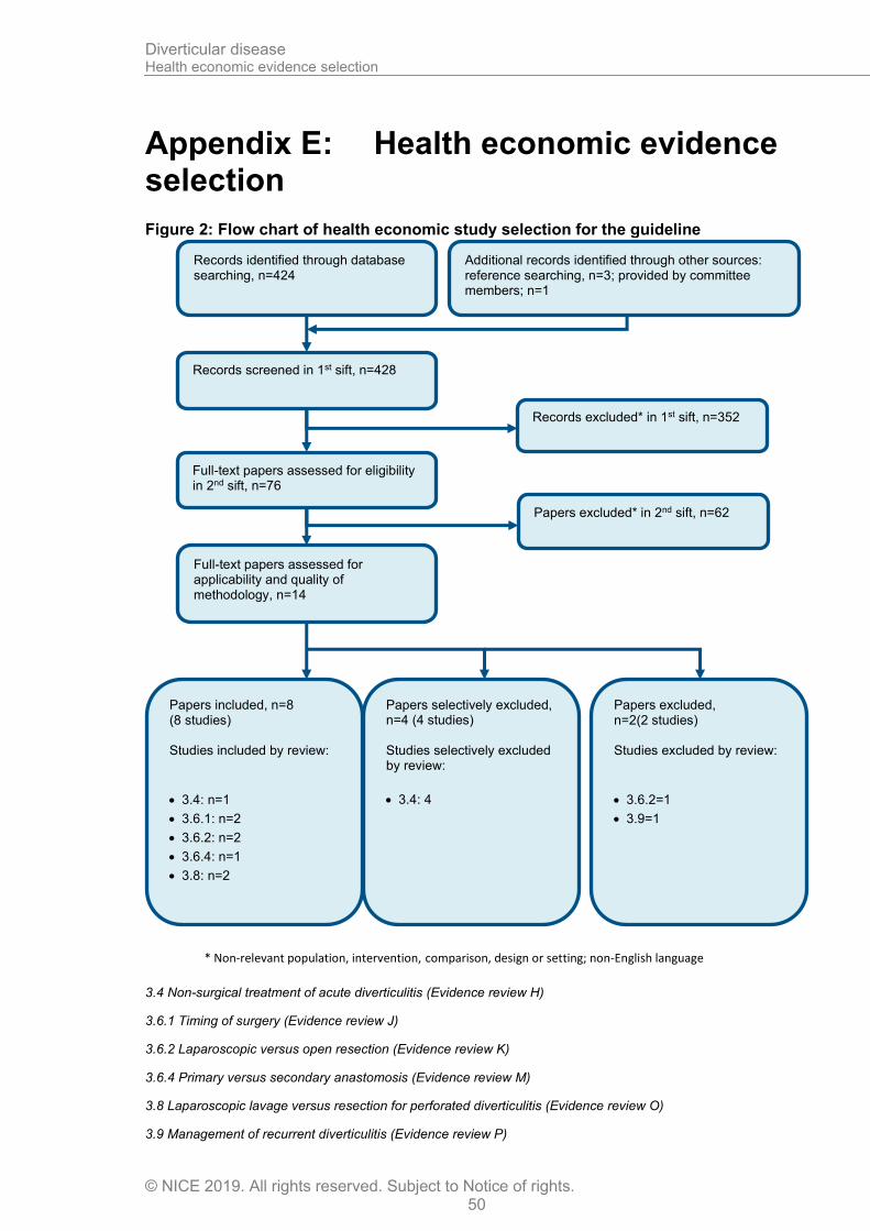
Diverticular disease Health economic evidence selection
© NICE 2019. All rights reserved. Subject to Notice of rights. 50
Appendix E: Health economic evidence selection
Figure 2: Flow chart of health economic study selection for the guideline
3.4 Non-surgical treatment of acute diverticulitis (Evidence review H)
3.6.1 Timing of surgery (Evidence review J)
3.6.2 Laparoscopic versus open resection (Evidence review K)
3.6.4 Primary versus secondary anastomosis (Evidence review M)
3.8 Laparoscopic lavage versus resection for perforated diverticulitis (Evidence review O)
3.9 Management of recurrent diverticulitis (Evidence review P)
Records screened in 1st sift, n=428
Full-text papers assessed for eligibility in 2nd sift, n=76
Records excluded* in 1st sift, n=352
Papers excluded* in 2nd sift, n=62
Papers included, n=8 (8 studies) Studies included by review:
• 3.4: n=1
• 3.6.1: n=2
• 3.6.2: n=2
• 3.6.4: n=1
• 3.8: n=2
Papers selectively excluded, n=4 (4 studies) Studies selectively excluded by review:
• 3.4: 4
Records identified through database searching, n=424
Additional records identified through other sources: reference searching, n=3; provided by committee members; n=1
Full-text papers assessed for applicability and quality of methodology, n=14
Papers excluded, n=2(2 studies) Studies excluded by review:
• 3.6.2=1
• 3.9=1
* Non-relevant population, intervention, comparison, design or setting; non-English language
Diverticular disease Health economic evidence selection
© NICE 2019. All rights reserved. Subject to Notice of rights. 51
Diverticular disease Excluded studies
© NICE 2019. All rights reserved. Subject to Notice of rights. 52
Appendix F: Excluded studies
F.1 Excluded clinical studies
Table 24: Studies excluded from the clinical review
Reference Reason for exclusion
Abedi 20041 Excluded due to inappropriate study design
Ahn 20022 Excluded due to inappropriate comparison
Alshamari 20163 Excluded due to inappropriate study population
Ambrosetti 19974 Excluded due to updated study already being included
Andeweg 20148 Excluded due to inappropriate reference standard
Andeweg 20137 Excluded due to inappropriate outcomes
Biondo 201210 Excluded due to inappropriate outcomes
Braden 201011 Excluded due to inappropriate outcomes
Brown 200212 Excluded due to inappropriate comparison; analysis
Brown 201613 Excluded due to inappropriate study population; incorrect interventions
Buckley 200414 Excluded due to inappropriate study design
Bugiantella 201515 Excluded due to inappropriate outcomes
Camera 201716 Excluded due to inappropriate study population
Caputo 201517 Excluded due to inappropriate comparison
Chabok 201318 Excluded due to inappropriate comparison; outcome
Choi 201319 Excluded due to inappropriate comparison
Cobben 200320 Excluded due to inappropriate study population
Coogan 199721 Excluded due to inappropriate study design
Daniels 201523 Excluded due to inappropriate study population
Dombal 197226 Excluded due to inappropriate diagnostic tests
Domjan 199827 Excluded due to inappropriate outcomes
Eisenberg 201728 Excluded due to inappropriate diagnostic tests
Etzioni 201029 Excluded due to inappropriate comparison
Floch 200630 Excluded due to inappropriate study design
Gallo 201631 Excluded due to inappropriate reference standard
Gans 201532 Excluded due to inappropriate diagnostic tests
Gong 201533 Excluded due to inappropriate study population
Halligan 200234 Excluded due to inappropriate study design
Ince 201435 Excluded due to inappropriate outcomes
Jang 201437 Excluded due to inappropriate study population
Jensen 200038 Excluded due to inappropriate intervention
Jung 201040 Excluded due to inappropriate comparison
Juvonen 201441 Excluded due to inappropriate study population
Kaser 201042 Excluded due to inappropriate outcome
Kawatkar 201543 Excluded due to inappropriate intervention
Kechagias 201444 Excluded due to inappropriate outcome
Kessner 201745 Excluded due to inappropriate comparison
Lameris 200846 Excluded due to inappropriate reference standard
Diverticular disease Excluded studies
© NICE 2019. All rights reserved. Subject to Notice of rights. 53
Reference Reason for exclusion
Lameris 200847 Excluded due to inappropriate reference standard
Laurell 200748 Excluded due to inappropriate comparison
Liljegren 200749 Excluded due to inappropriate diagnostic tests
Lindsay 198850 Excluded due to inappropriate study population
Longstreth 201651 Excluded due to inappropriate comparison
Macconaill 201452 Excluded due to inappropriate study design
Millet 201753 Excluded due to inappropriate diagnostic tests
Ng 200255 Excluded due to inappropriate diagnostic tests
Nicholas 197258 Excluded due to inappropriate comparison
Oistamo 201360 Excluded due to inappropriate outcomes
Padidar 199461 Excluded due to inappropriate study design; comparison; population
Porten 200862 Excluded due to inappropriate study population; outcome
Pradel 199763 Excluded due to inappropriate reference standard
Rampton 200164 Excluded due to inappropriate study design
Sala 200765 Excluded due to inappropriate study design
Sanford 200666 Excluded due to inappropriate comparison
Schnyder 197967 Excluded due to inappropriate outcomes
Schreyer 200468 Excluded due to inappropriate outcome
Shen 200269 Excluded due to inappropriate outcome
Shrier 199170 Excluded due to inappropriate outcome
Sirany 201771 Excluded due to inappropriate intervention; comparison
Snyder 200472 Excluded due to inappropriate study design, no relevant outcomes
Spinzi 200173 Excluded due to inappropriate outcomes
Stromberg 200775 Excluded due to inappropriate comparison
Thorisson 201676 Excluded due to inappropriate diagnostic tests
Toorenvliet 201077 Excluded due to inappropriate comparison
Tursi 201678 Excluded due to inappropriate comparison
Turvill 201679 Excluded due to inappropriate study population; intervention
van de Wall 201380 Excluded due to inappropriate outcome
Wolff 200882 Excluded due to inappropriate outcome
Won 201683 Excluded due to inappropriate comparison
Wong 201284 Excluded due to inappropriate comparison
Yardimci 201785 Excluded due to inappropriate study design; comparison
Zia 200886 Excluded due to inappropriate study population
Zielke 199787 Excluded due to inappropriate comparison