diversity and evolution of the thyroglobulin type1 domain superfamily
TRANSCRIPT
Diversity and Evolution of the Thyroglobulin Type-1 Domain Superfamily
Marko Novinec,* Dusan Kordis,* Vito Turk,* and Brigita Lenarcic*�*Department of Biochemistry and Molecular Biology, Jozef Stefan Institute, Jamova 39, Ljubljana,Slovenia; and �Department of Chemistry and Biochemistry, Faculty of Chemistry and ChemicalTechnology, University of Ljubljana, Askerceva 5, Ljubljana, Slovenia
Multidomain proteins are gaining increasing consideration for their puzzling, flexible utilization in nature. The presence ofthe characteristic thyroglobulin type-1 (Tg1) domain as a protein module in a variety of multicellular organisms suggestspivotal roles for this building block. To gain insight into the evolution of Tg1 domains, we performed searches of protein,expressed sequence tag, and genome databases. Tg1 domains were found to beMetazoa specific, and we retrieved a total of170 Tg1 domain–containing protein sequences. Their architectures revealed a wide taxonomic distribution of proteinscontaining Tg1 domains followed or preceded by secreted protein, acidic, rich in cysteines (SPARC)-type extracellularcalcium-binding domains. Other proteins contained lineage-specific domain combinations of peptidase inhibitory modulesor domains with different biological functions. Phylogenetic analysis showed that Tg1 domains are highly conservedwithin protein structures, whereas insertion into novel proteins is followed by rapid diversification. Seven different basictypes of protein architecture containing the Tg1 domain were identified in vertebrates. We examined the evolution of theseprotein groups by combining Tg1 domain phylogeny with additional analyses based on other characteristic domains. Tes-ticans and secreted modular calcium binding protein (SMOCs) evolved from invertebrate homologs by introduction ofvertebrate-specific domains, nidogen evolved by insertion of a Tg1 domain into a preexisting architecture, and the remain-ing four have unique architectures. Thyroglobulin, Trops, and the major histocompatibility complex class II–associatedinvariant chain are vertebrate specific, while an insulin-like growth factor–binding protein and nidogen were also identifiedin urochordates. Among vertebrates, we observed differences in protein repertoires, which result from gene duplication anddomain duplication. Members of five groups have been characterized at the molecular level. All exhibit subtle differencesin their specificities and function either as peptidase inhibitors (thyropins), substrates, or both. As far as the sequence isconcerned, only a few conserved residues were identified. In combination with structural data, our analysis shows that theTg1 domain fold is highly adaptive and comprises a relatively well-conserved core surrounded by highly variable loopsthat account for its multipurpose function in the animal kingdom.
Introduction
Many proteins in multicellular organisms are madefrom combinations of several autonomously foldingdomains or modules. During evolution, these units havebeen highly mobile and have spread into previously existingproteins or gave birth to new architectures with novel bio-logical functions, principally by mechanisms of exon shuf-fling and duplication (Patthy 2003; Vogel et al. 2004a). Byparticipating in interactions with multiple partners, multido-main proteins contribute greatly to organism complexity(Patthy 2003). Although the repertoire of possible domaincombinations is, in principle, virtually unlimited, only a con-strained number is actually present as a consequence ofstrong evolutionary selection. Some domains have beenshown to be highly versatile, whereas most have only a lim-ited set of neighboring domains (Vogel et al. 2004a). Withthe huge increase in available genomic and expressed se-quence tag (EST) data over the past few years, our knowl-edge of the presence and versatility of many differentmodules has been greatly enlarged.
Thyroglobulin type-1 (Tg1) domains are classified as asuperfamily belonging to the class of small proteins in theStructural Classification of Proteins database (Murzin et al.1995). Theywereoriginally identified as cysteine-richmotifs(Mercken et al. 1985), organized into 10 tandem repeatedunits in the N-terminal third of thyroglobulin (Parma et al.1987). Molina et al. (1996) identified an eleventh repeatand characterized the repeats as being modules found in pro-
teinsfromdifferent families.Basedonthenumberofdisulfidebonds, this group also divided Tg1 domains into two sub-types, subtype 1A (Tg1A) containing three disulfide bondsand subtype 1B (Tg1B) without the second disulfide bond.
Since their original identification in thyroglobulin, Tg1domains have been found as parts of many proteins with dif-ferent origins and functions. Some Tg1 domain–containingproteins inhibit certain peptidases, mostly cysteine cathe-psins. These proteins were named thyropins (Lenarcicand Bevec 1998) and classified as peptidase inhibitor familyI31 (Rawlings, Tolle, and Barrett 2004). For some thyro-pins, it has been shown that their inhibitory properties resideonTg1 domains (Bevec et al. 1996; Lenarcic and Turk 1999;Lenarcic et al. 2000; Meh et al. 2005). The best studied thy-ropin is the p41 splice variant of the major histocompatibil-ity complex class II–associated invariant chain, which isinvolved in antigen processing (Stumptner-Cuvelette andBenaroch 2002). The crystal structure of the p41 fragmentin complexwith cathepsin L (Guncar et al. 1999) is the foun-dation of our understanding of thyropin action. Other well-studied thyropins include equistatin from sea anemoneActinia equina (Lenarcic and Turk 1999; Galesa et al.2003), saxiphilin from the bullfrog Rana catesbeiana(Lenarcic et al. 2000), the chum salmon egg peptidase inhib-itor (Yamashita and Konagaya 1996), and human testican-1(Bocock et al. 2003). The latter is unique among the thyro-pins, being not only an inhibitor but also a substrate forcertain cysteine cathepsins (Meh et al. 2005). The evidencefor Tg1 domain functions other than peptidase inhibitioncomes from the insulin-like growth factor–binding protein(IGFBP)-6. The Tg1 domains of the IGFBPs have long beenproposed to be involved in the binding of insulin-like growthfactors (IGFs), and these interactions have recently been
Key words: thyroglobulin type-1 domain, protein module, evolution,Metazoa.
E-mail: [email protected].
Mol. Biol. Evol. 23(4):744–755. 2006doi:10.1093/molbev/msj082Advance Access publication December 20, 2005
� The Author 2005. Published by Oxford University Press on behalf ofthe Society for Molecular Biology and Evolution. All rights reserved.For permissions, please e-mail: [email protected]
by guest on February 1, 2014http://m
be.oxfordjournals.org/D
ownloaded from
confirmed and characterized on the molecular level(Headey et al. 2004).
Most of the identifiedTg1 domains remain functionallyuncharacterized. However, they are among the most versa-tile domains found in metazoans. In this study, we searchedprotein, EST, and genome databases for proteins containingTg1 domains and examined their domain architectures. Wehave analyzed the phylogenetic relationships between Tg1domains and evaluated the correlation between their phylog-eny and protein architecture. Further, we focused on the ori-gins and evolution of vertebrate Tg1 domain–containingproteins in terms of domain architecture, Tg1 domain phy-logeny, and additionally performed analyses. Finally, welooked into the functional and structural diversity withinthe Tg1 domain superfamily in the light of our study andother available data.
MethodsData Collection
Sequences of Tg1 domain–containing proteins wereobtained by Blast (www.ncbi.nlm.nih.gov/Blast/) searchesof the GenBank nonredundant protein database, EST data-bases, and available genome sequences (see Supplement 1of the Supplementary Material online). Coding sequenceswere extracted from genomic data using GENSCAN(Burge and Karlin 1997). Whenever possible complete pro-tein coding sequences were obtained.
The set of vertebrate proteins includes sequencesobtained from human (Homo sapiens), mouse (Mus muscu-lus), cow (Bos taurus), chicken (Gallus gallus), frog(Xenopus laevis), zebrafish (Danio rerio), and pufferfish(Tetraodon nigroviridis). Invertebrate sequences were ob-tained from available EST databases and the followinggenome databases: sea urchin (Strongylocentrotus purpur-atus), sea squirt (Ciona intestinalis), amphioxus (Bran-chiostoma floridae), Schmidtea mediterranea, nematode(Caenorhabditis elegans), fruit fly (Drosophila mela-nogaster), honeybee (Apis mellifera), flour beetle (Tribo-lium castaneum), starlet sea anemone (Nematostellavectensis), and marine sponge (Reniera sp. JGI-2005).
Determination of Domain Architecture
For characterized proteins, domain architectures weretaken from annotations. For uncharacterized proteins, archi-tectures were determined by querying the ConservedDomain Database v2.02 (www.ncbi.nlm.nih.gov/structure/cdd/) and the InterPro release 8.1 database (www.ebi.ac.uk/interpro/). Additionally, we performed BlastP searches ofthe GenBank nonredundant protein database. Partial proteinsequences were used as queries, and homologous regions ofBlast hits were examined for known structural elements.
Sequence Alignment and Analysis
Amino acid sequences were aligned with ClustalWversion 1.83 (Thompson, Higgins, and Gibson 1994) usingthe BLOSUM series matrices. The created alignments weremanually checked and refined with SEAVIEW (Galtier,Gouy, and Gautier 1996). Residue conservation was deter-mined manually.
Phylogenetic Analysis
Phylogenetic analyses were performed with the PHY-LIP package version 3.63 (Felsenstein 1989). Multiple se-quence alignments were used to create 100 replicates withthe SEQBOOT module. Then, PROTDIST was used to cal-culate protein distances with the Probability Matrix fromBlocks model (Veerassamy, Smith, and Tillier 2003). Fromthe generated distance matrices, trees were created using theNEIGHBOR module with the neighbor-joining (NJ)method (Saitou and Nei 1987). Trees were visualized andmanipulated, and consensus trees were created with theTreeExplorer program of Koichiro Tamura.
Results and DiscussionDomain Architecture and Taxonomic Distributionof Tg1 Domain–Containing Proteins
We retrieved 170 protein sequences containing 333Tg1 domains. Some were already available, but most werereconstructed from EST and genomic data. Complete cod-ing sequences were retrieved for the majority of proteins,while for some only partial sequences were obtained. Do-main architectures were determined for previously uniden-tified proteins, and the complete inventory is available asSupplement 1 of the Supplementary Material online.
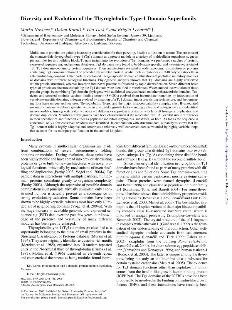
The recognized domain architectures show that Tg1domains are highly versatile modules, which were mobi-lized into proteins with greatly different configurations dur-ing metazoan evolution (fig. 1). Tg1 domain–containingproteins are unequally distributed among diverse metazoanlineages, and distinct domain compositions predominantlyoccur in lineage-specific patterns. The repertoire of domaincombinations is broad and includes peptidase inhibitorymodules of different classes, such as BPTI/Kunitz domains(Laskowski 1986), whey acidic protein (WAP) domains(Hennighausen and Sippel 1982), and trypsin inhibitor–likecysteine-rich domains (Grasberger, Clore, and Gronenborn1994). Of modules with distinct functions frequently foundin other extracellular proteins, von Willebrand factor–likedomains, immunoglobulin-like domains, and epidermalgrowth factor–like domains are most frequent, while othersoccur to a more limited extent. Tg1 domains can also beincorporated into proteins with otherwise nonmodularnature, for example the invariant chain.
In the demosponge Reniera, the representative of themost basal animal lineage, both identified Tg1 domain–containing proteins contain combinations of a Tg1 domainand a WAP domain (fig. 1). This ancient domain combina-tion that is conserved and further expanded by additionaldomains throughout the animal kingdom was however lostduring the evolution of most deuterostome lineages.
In cnidarians, proteins are predominantly composed oftandem Tg1 domain repeats. The number of repeated unitsvaries from 2 to as many as 14 in N. vectensis (fig. 1). Equi-statin, a well-characterized protein from the sea anemone A.equina, comprises three Tg1 domains which exhibit a re-markable degree of inhibitory diversity. The first domaininhibits cysteine cathepsins, the second one the asparticpeptidase cathepsin D, while no activity toward peptidaseshas been identified for the third one (Galesa et al. 2003).
Thyroglobulin Type-1 Domain Evolution 745
by guest on February 1, 2014http://m
be.oxfordjournals.org/D
ownloaded from
746 Novinec et al.
by guest on February 1, 2014http://m
be.oxfordjournals.org/D
ownloaded from
Two proteins containing Tg1 domains and SPARC-typeextracellular calcium-binding (EC) domains were identi-fied in Cnidaria (fig. 1). Similar proteins were identifiedthroughout the animal kingdom, all sharing a Tg1-ECdomain pair. This element resembles a supradomain, asdefined by Vogel et al. (2004b), and may be organizedin two different orientations. In vertebrates, one orientationis found in testicans (domain order EC-Tg1) and the other inSMOCs (domain order Tg1-EC); therefore, we term themtestican-type and SMOC-type supradomain, respectively.Accordingly, proteins containing one of these suprado-mains were termed testican-like or SMOC-like proteins.
Lophotrochozoa are the only metazoan group besidessponges in which none of the supradomains were found, andthe small number of retrieved sequences indicates secondarygene loss of Tg1 domains in this lineage. However, with theavailability of new genome data, further Tg1 domain se-quences might be identified. In Ecdysozoa, Tg1 domains aremore versatile than in cnidarians and proteins comprise uniquearrangements of different types of peptidase-inhibitingmodules, but in general Tg1 domains appear to have beeninfrequently utilized during protostome evolution.
Tg1 domains, however, have been highly mobile dur-ing deuterostome evolution, and each taxonomic grouppossesses a characteristic set of Tg1 domain–containingproteins with some unique domain combinations. The ver-tebrate repertoire consists of seven architecturally distinctgroups. Three of these, the invariant chain, Trops, and thy-roglobulin are found exclusively in vertebrates. Nidogenand IGFBP orthologs were also found in the urochordateC. intestinalis but surprisingly not in the cephalochordateB. floridae. Homologs of the remaining two groups, testi-cans and SMOCs, are present throughout metazoans; how-ever, vertebrate proteins are characterized by addition ofunique vertebrate-specific domains (the testican-uniquedomain in testicans and the SMOC-unique domain inSMOCs).
The Phylogeny of Tg1 Domains in Correlation withProtein Architectures
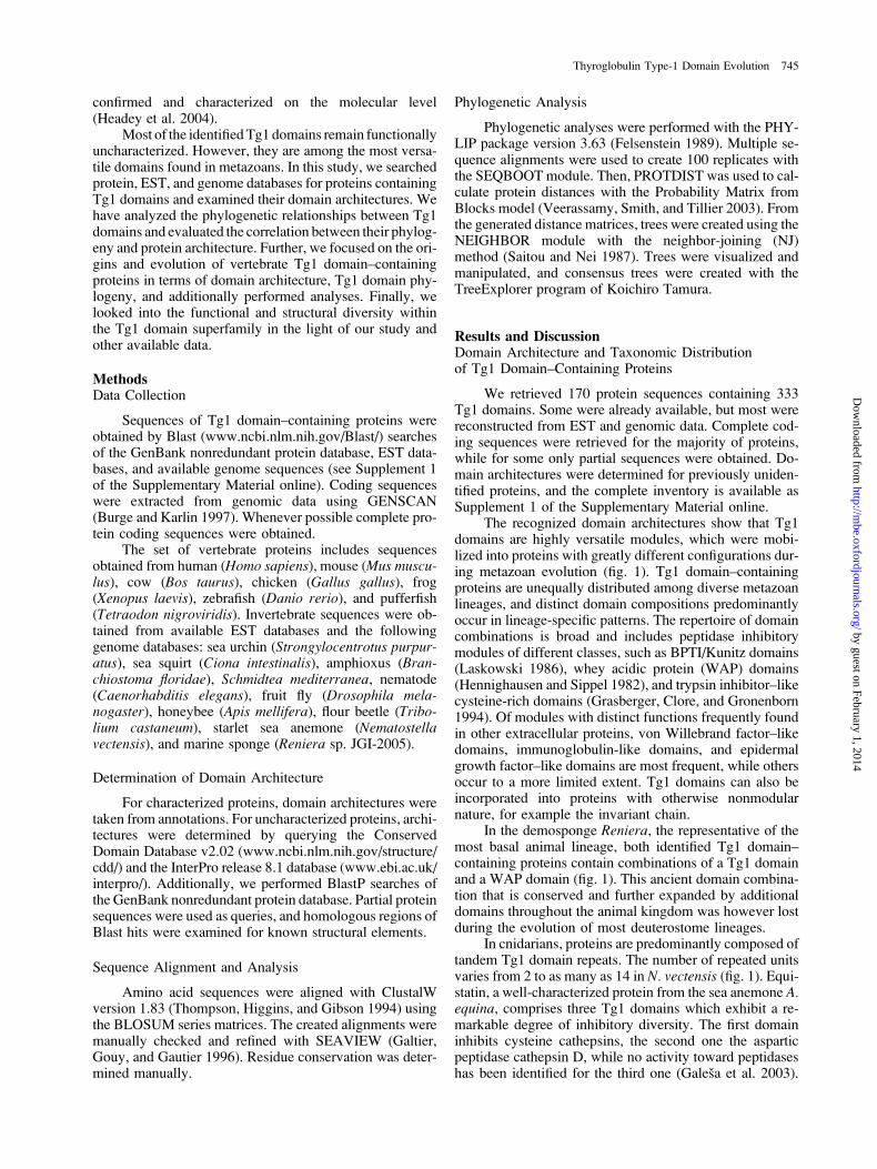
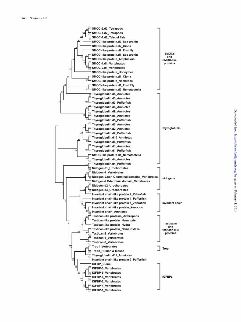
The domain architecture of Tg1 domain–containingproteins differs markedly between diverse metazoans. Toinvestigate a possible correlation between domain architec-ture and Tg1 domain phylogeny, we conducted two sepa-rate phylogenetic analyses, one including all retrieved Tg1domain sequences and the second including only domainsof vertebrate proteins and their invertebrate homologs. Theobtained NJ trees are presented as Supplement 2 of the Sup-
plementary Material online, and a condensed NJ tree of thesecond analysis is shown in figure 2.
Tg1 domains of vertebrates and their invertebratehomologs are divided into seven well-defined groups whichcorrespond to protein architectures identified in vertebrates(fig. 2 and Supplement 2A of the Supplementary Materialonline). Within groups further subdivision is observed.Groups containing multiple paralogs may be subdividedaccording to protein paralogy, as is seen for IGFBPs andtesticans. The IGFBPs are divided into two subgroups, eachcomprising three paralogs, while the C. intestinalis IGFBPhas basal position as the oldest representative. Testican Tg1domains form two groups, one containing domains of inver-tebrate testican-like proteins and vertebrate testican-2 andthe other vertebrate testican-1 and -3 domains. In contrastto the former, proteins with more than one Tg1 domain ex-hibit more complex division patterns. In the SMOC group,Tg1 domains of vertebrate homologs are divided into twosubgroups. One comprises domains near the N-terminusand the other domains near theC-terminus.Within each sub-group, further division corresponding to SMOC paralogy isobserved. The Tg1 domains of invertebrate SMOC-like pro-teins are not clustered, rather they are dispersed throughoutthe group. Vertebrate nidogen Tg1 domains are subdividedinto three branches. Nidogen-2 domains form two sub-groups, one comprising domains nearest the C-terminusin individual proteins and the other comprising the rest,while the third is formed by domains of nidogen-1. Simi-larly, two subgroups are formed by the Tg1 domains of as-cidian nidogens. In the case of thyroglobulin a diffusepattern is observed. The majority form a loosely relatedgroup, whereas domain 11 does not cluster with the rest.
The described seven groups are only partly observed inthe tree constructed from all retrieved sequences (Supple-ment 2B of the SupplementaryMaterial online), as expecteddue to low bootstrap support for the branches. This is mostclearly observed for Tg1 domains of thyroglobulin, whichare separated into four groups, and for those of ascidiannidogens, which do not cluster with their vertebrate paral-ogs. Further differences are observed for individual domainsand for branching patterns within groups, as well as for re-latedness between groups. Nevertheless, the existence ofthese groups shows that, overall, Tg1 domain conservationis tightly linked to the conservation of protein architecture,and this effect is observed over large evolutionary distances.In contrast, introduction of Tg1 domains into novel proteinarchitectures appears to be followed by rapid diversificationbecause clear relationships between Tg1 domains from dif-ferent architectures cannot be determined.
FIG. 1.—Domain architectures of selected Tg1 domain–containing proteins. Proteins are represented as horizontal lines, and rectangles indicateprotein modules. Tg1 domains are colored black, EC domains gray, follistatin-like domains are striped, and the remainder are white. Proteins are identifiedwith protein name or abbreviation. Larger images are not to scale. Asterisks (*) indicate missing protein termini. Domain designations: AC, testican acidicregion; AS, antistasin-like domain; CL, invariant chain class II-associated invariant chain peptide (CLIP) fragment; EGF, epidermal growth factor–likedomain; FN, FOLN domain; FRI, FRI domain; FS, follistatin-like domain; G1, nidogen G1 domain; G2, nidogen G2 domain; IC, invariant chain in-tracellular domain; IG, immunoglobulin-like domain; KU, BPTI/Kunitz domain; LamG, laminin G domain; LC, lipocalin domain; LY, low-density li-poprotein receptor–like YWTD repeat; OLF, olfactomedin-like domain; OP, osteopontin domain; SD, syndecan domain; SEA, SEA domain; SMOC,SMOC-unique domain; Tg2, thyroglobulin type-2 repeat; Tg3, thyroglobulin type-3 repeat; TIL, trypsin inhibitor–like cysteine-rich domain; TM, trans-membrane region; TSP1, thrombospondin type-1 repeat; TST, testican-unique domain; vWA, von Willebrand factor type-A domain; vWC, von Wil-lebrand factor type C domain; and WAP, whey acidic protein–type four-disulfide core domain.
Thyroglobulin Type-1 Domain Evolution 747
by guest on February 1, 2014http://m
be.oxfordjournals.org/D
ownloaded from
748 Novinec et al.
by guest on February 1, 2014http://m
be.oxfordjournals.org/D
ownloaded from
In addition to vertebrate groups, several smallergroups appear in the tree constructed from all availableTg1 domains (Supplement 2 of the Supplementary Materialonline). These mostly comprise domains from closely re-lated species or domains from a single species, which prob-ably evolved by domain duplication or recombination.Interestingly, close relationships are also observed forN. vectensis and B. floridae Tg1 domains, indicating thatseveral proteins of the eumetazoan last common ancestor(LCA) persisted in the deuterostome lineage at least untilthe divergence of the cephalochordates were however mod-ified or lost in protostomes. This observation is in agree-ment with other studies that show a strong resemblancebetween cnidarians and higher deuterostomes (Kortschaket al. 2003; Raible and Arendt 2004).
The generally low bootstrap support for the obtainedrelationships prevents the classification of all Tg1 domainsinto distinct families. Nevertheless, both analyses on Tg1domain phylogeny do provide substantial insight into cer-tain aspects of Tg1 domain evolution. To reinforce thesedata, we performed additional phylogenetic analyses of ver-tebrate Tg1 domain–containing proteins. These analysesare available as Supplement 2C of the SupplementaryMaterial online and were based on modules, which areabundant in individual protein groups. For SMOCs andtesticans the analyses were performed on the respectivesupradomains, for nidogens on their G1 domains, andIGFBPs were examined as whole proteins. Together withTg1 domain phylogeny, we used the data obtained todeduce the evolution of these proteins.
The Birth of the Tg1 Domain
Tg1 domains are Metazoa-specific protein modules,which evolved very early in the evolution of this kingdom.They are predominantly found in extracellular proteins, andtheir occurrence is probably related to the appearance ofmul-ticellular animals,whichwasaccompaniedby rapid evolutionof modules and proteins involved in cell-cell interactionsas well as in interactions of cells with their environment. Acomparison between yeast, fruit fly, and nematode proteinsshows a 10-fold increase in the number of proteins involvedin these interactions in Metazoa (Hazkani-Covo et al. 2004).Based on the broad phyletic distribution of the thyropins(Lenarcic and Bevec 1998; Lenarcic et al. 2000), we canassume that the primordial Tg1 domain originated as a regu-latorofpeptidaseactivityandthatmost, ifnotall,Tg1domainshave retained this function throughout metazoan evolution.
The Evolution of Testicans and SMOCs
The eumetazoan LCA appears to have containeda well-developed set of Tg1 domain–containing proteins,including both testican-like and SMOC-like proteins, asis demonstrated by their presence in Cnidaria (fig. 1 and
Supplement 1 of the Supplementary Material online). TheTg1 domains of both are well conserved in most metazoangroups (fig. 2 and Supplement 2 of the Supplementary Ma-terial online), indicating that they were among the crucialinnovations of the first Eumetazoa. Their existence in basalmetazoans further suggests that two distinct Tg1 domain lin-eages originated early in the evolution of the Tg1 domainsuperfamily and that all members might derive from oneof them.
Unfortunately, the physiological roles of invertebratetestican-like and SMOC-like proteins are unknown. Testi-cans are involved in the regulation of cell attachment (Marrand Edgell 2003; Schnepp et al. 2005), as well as regulationof cysteine cathepsin (Bocock et al. 2003; Meh et al. 2005)and metalloprotease activity (Nakada et al. 2001, 2003).SMOCs, on the other hand, are little known glycoproteins.SMOC-1 was found to be localized predominantly to base-ment membranes (Vannahme et al. 2002), whereas theprecise localization of SMOC-2, which exhibits a somewhatbroader expression pattern (Vannahme et al. 2003), remainsto be determined.
Both SMOC-like and testicans-like proteins arewidely distributed throughout the animal kingdom and havea rich evolutionary history of lineage-specific modifica-tions, which are summarized in figure 3. To evaluate therelationships within each group, we performed phyloge-netic analyses on the respective supradomains (Supplement2C of the Supplementary Material online). In both, supra-domain phylogeny correlates well with species phylogeny,although a few exceptions are seen for SMOC-typesupradomains.
The domain architecture of the N. vectensis SMOC-like protein is remarkably similar to those identified inhigher metazoans (fig. 3). On the other hand, cnidariantestican-like proteins are comprised solely of a testican-typesupradomain. A follistatin-like domain was introduced intothis architecture before the deuterostome-protostome split,as demonstrated by its presence in both vertebrates and fruitfly. Interestingly, both nematode proteins are comprisedsolely of the respective supradomains, probably as a resultof lineage-specific domain losses.
In deuterostomes, SMOC-like proteins are ubiquitous,whereas testican-like proteins appear only in vertebrates. Itis not exactly clear whether the absence of testican-like pro-teins is accounted for by independent secondary gene lossesin these lineages or is simply due to incompleteness of data.
Comparison of vertebrate SMOCs and testicans withinvertebrate homologs shows the presence of additionalvertebrate-specific domains, the testican-unique domainin testicans, and the SMOC-unique domain in SMOCs.These domain architectures were conserved during verte-brate evolution, and further functional diversification wasachieved by gene duplication. Two SMOC paralogs are al-ready present in pufferfish, indicating that the gene dupli-cation producing these paralogs occurred before the teleost
FIG. 2.—Condensed representation of the NJ tree showing Tg1 domains from vertebrate proteins and their invertebrate orthologs. Seven major Tg1domain groups are evident. The tree was constructed using the PHYLIP package with the probability matrix from Blocks (PMB) distance matrix, andbootstrap values were calculated using 100 replicates. The tree was visualized with TreeExplorer.
Thyroglobulin Type-1 Domain Evolution 749
by guest on February 1, 2014http://m
be.oxfordjournals.org/D
ownloaded from
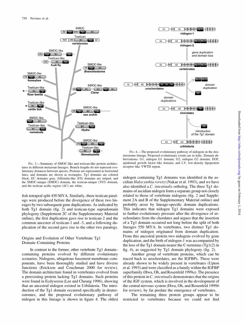
fish-tetrapod split 450MYA. Similarly, three testican paral-ogs were produced before the divergence of these two lin-eages by two subsequent gene duplications. As indicated byboth Tg1 domain (fig. 2) and testican-type supradomainphylogeny (Supplement 2C of the Supplementary Materialonline), the first duplication gave rise to testican-2 and thecommon ancestor of testican-1 and -3, and a following du-plication of the second gave rise to the other two paralogs.
Origins and Evolution of Other Vertebrate Tg1Domain–Containing Proteins
In contrast to the former, other vertebrate Tg1 domain–containing proteins evolved by different evolutionaryscenarios. Nidogens, ubiquitous basement membrane com-ponents, have been thoroughly studied and have diversefunctions (Erickson and Couchman 2000 for review).The domain architecture found in vertebrates evolved froma preexisting protein lacking Tg1 domains. Such proteinswere found in Ecdysozoa (Lee and Cheung 1996), showingthat an ancestral nidogen existed in Urbilateria. The intro-duction of the Tg1 domain occurred specifically in deuter-ostomes, and the proposed evolutionary pathway ofnidogen in this lineage is shown in figure 4. The oldest
nidogen containing Tg1 domains was identified in the as-cidianHalocynthia roretzi (Nakae et al. 1993), and we havealso identified a C. intestinalis ortholog. The three Tg1 do-mains of ascidian nidogen form a separate group not closelyrelated to those of vertebrate nidogens (fig. 2 and Supple-ment 2A and B of the Supplementary Material online) andprobably arose by lineage-specific domain duplications.This indicates that nidogen Tg1 domains were exposedto further evolutionary pressure after the divergence of ur-ochordates from the chordates and argues that the insertionof a Tg1 domain occurred not long before the split of bothlineages 550 MYA. In vertebrates, two distinct Tg1 do-mains of nidogen originated from domain duplication.From this ancestral protein two nidogens evolved by geneduplication, and the birth of nidogen-1 was accompanied bythe loss of the Tg1 domain nearer the C-terminus (Tg1(2) infig. 4), as suggested by Tg1 domain phylogeny (fig. 2).
Another group of vertebrate proteins, which can betraced back to urochordates, are the IGFBPs. These werealready shown to be widely present in vertebrates (Uptonet al. 1993) and were classified as a family within the IGFBPsuperfamily (Hwa, Oh, andRosenfeld 1999a). The presenceof this protein inC. intestinalis demonstrates that the originsof the IGF system, which is involved in the development ofthe central nervous system (Hwa, Oh, and Rosenfeld 1999bfor review), by far predate the emergence of vertebrates.
The remaining three protein groups appear to berestricted to vertebrates because we could not find
FIG. 4.—The proposed evolutionary pathway of nidogens in the deu-terostome lineage. Proposed evolutionary events are in italic. Domain ab-breviations: G1, nidogen G1 domain; G2, nidogen G2 domain; EGF,epidermal growth factor–like domain; and LY, low-density lipoproteinreceptor–like YWTD repeat.
FIG. 3.—Summary of SMOC-like and testican-like protein architec-tures in different metazoan lineages. Branch lengths do not represent evo-lutionary distances between species. Proteins are represented as horizontallines, and domains are shown as rectangles. Tg1 domains are coloredblack, EC domains gray, follistatin-like (FS) domains are striped, andthe SMOC-unique (SMOC) domain, the testican-unique (TST) domain,and the testican acidic region (AC) are white.
750 Novinec et al.
by guest on February 1, 2014http://m
be.oxfordjournals.org/D
ownloaded from
invertebrate homologs. All are highly specialized proteinswith a restricted expression pattern, which appear to havebeen formed during vertebrate evolution. The invariantchain is a critical component of the immune system(Stumptner-Cuvelette and Benaroch 2002 for review),Trops are involved in cell adhesion (Litvinov et al.1997) and epithelia development (Guillemot et al. 2001),while thyroglobulin is involved in hormone signaling.
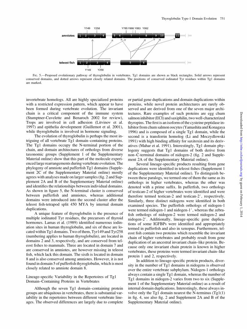
The evolution of thyroglobulin is perhaps the most in-triguing of all vertebrate Tg1 domain–containing proteins.The Tg1 domains occupy the N-terminal portion of thechain, and domain architectures of orthologs from diversetaxonomic groups (Supplement 1 of the SupplementaryMaterial online) show that this part of the molecule experi-enced large rearrangements during vertebrate evolution. Thephylogeny of amniote and pufferfish Tg1 domains (Supple-ment 2C of the Supplementary Material online) mostlyagrees with analysesmade on larger samples (fig. 2 and Sup-plement 2A and B of the Supplementary Material online)and identifies the relationships between individual domains.As shown in figure 5, the N-terminal cluster is conservedbetween pufferfish and amniotes, whereas additionaldomains were introduced into the second cluster after theteleost fish-tetrapod split 450 MYA by internal domainduplications.
A unique feature of thyroglobulin is the presence ofmultiple iodinated Tyr residues, the precursors of thyroidhormones. Lamas et al. (1989) identified numerous iodin-ation sites in human thyroglobulin, and six of these are lo-catedwithin Tg1 domains. Two of them,Tyr149 andTyr258(numbering applies to human thyroglobulin), are located indomains 2 and 3, respectively, and are conserved from tel-eost fishes to mammals. Three are located in domain 7 andare conserved in amniotes, are however missing in teleostfish, which lack this domain. The sixth is located in domain8 and is also conserved among amniotes. However, it is notfound in domain 5 of pufferfish thyroglobulin, which ismostclosely related to amniote domain 8.
Lineage-specific Variability in the Repertoires of Tg1Domain–Containing Proteins in Vertebrates
Although the seven Tg1 domain–containing proteingroups are ubiquitous in vertebrates, there is substantial var-iability in the repertoires between different vertebrate line-ages. The observed differences are largely due to complete
or partial gene duplications and domain duplications withinproteins, while novel protein architectures are rarely ob-served and are derived from one of the seven major archi-tectures. Rare examples of such proteins are egg chumsalmon inhibitor (ECI) and saxiphilin, twowell-characterizedthyropins. The first is an isoformof the cysteine peptidase in-hibitor fromchumsalmonoocytes (Yamashita andKonagaya1996) and is composed of a single Tg1 domain, while thesecond is a transferrin homolog (Li and Moczydlowski1991) with high binding affinity for saxitoxin and its deriv-atives (Mahar et al. 1991). Interestingly, Tg1 domain phy-logeny suggests that Tg1 domains of both derive fromnon–C-terminal domains of nidogen-2 (fig. 2 and Supple-ment 2A of the Supplementary Material online).
Several lineage-specific products resulting from geneduplications were identified in teleost fishes (Supplement 1of the Supplementary Material online). To distinguish be-tween these paralogs, we termed one of them the same as itsorthologs in higher vertebrates, whereas the other wasdenoted with a prime suffix. In pufferfish, two orthologsof testican-2 of higher vertebrates were identified and weretherefore termed testican-2 and testican-2#, respectively.Similarly, three distinct nidogens were identified in bothexamined species. The pufferfish orthologs of nidogen-1were termed nidogen-1 and nidogen-1#, whereas the zebra-fish orthologs of nidogen-2 were termed nidogen-2 andnidogen-2#. Additionally, lineage-specific gene duplica-tions of some IGFBPs were identified and appropriatelytermed in pufferfish and also in xenopus. Furthermore, tel-eost fish contain two proteins which resemble the invariantchain of higher vertebrates and probably result from geneduplication of an ancestral invariant chain–like protein. Be-cause only one invariant chain protein is known in highervertebrates, these proteins were termed invariant chain–likeprotein 1 and 2, respectively.
In addition to lineage-specific protein products, diver-sity in the number of Tg1 domains in nidogens is observedover the entire vertebrate subphylum. Nidogen-1 orthologsalways contain a single Tg1 domain, whereas the number ofTg1 domains in nidogen-2 varies from two to six (Supple-ment 1 of the Supplementary Material online) as a result ofinternal domain duplications. Interestingly, these always in-volve only the Tg1 domain nearest the N-terminus (Tg1(1)in fig. 4, see also fig. 2 and Supplement 2A and B of theSupplementary Material online).
FIG. 5.—Proposed evolutionary pathway of thyroglobulin in vertebrates. Tg1 domains are shown as black rectangles. Solid arrows representconserved domains, and dotted arrows represent closely related domains. The positions of conserved iodinated Tyr residues within Tg1 domainsare marked.
Thyroglobulin Type-1 Domain Evolution 751
by guest on February 1, 2014http://m
be.oxfordjournals.org/D
ownloaded from
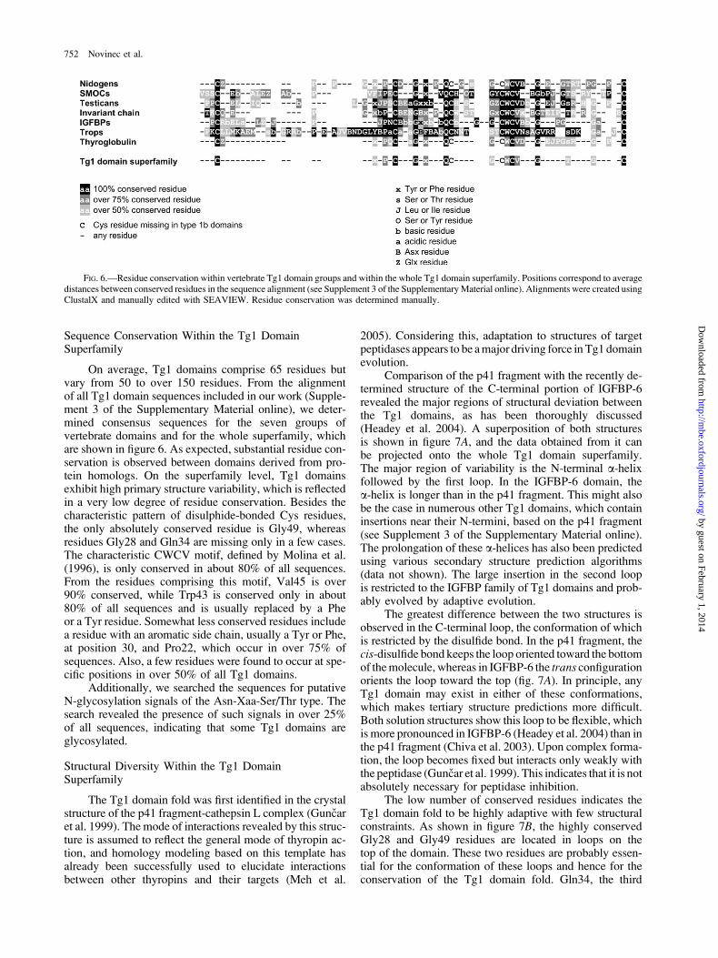
Sequence Conservation Within the Tg1 DomainSuperfamily
On average, Tg1 domains comprise 65 residues butvary from 50 to over 150 residues. From the alignmentof all Tg1 domain sequences included in our work (Supple-ment 3 of the Supplementary Material online), we deter-mined consensus sequences for the seven groups ofvertebrate domains and for the whole superfamily, whichare shown in figure 6. As expected, substantial residue con-servation is observed between domains derived from pro-tein homologs. On the superfamily level, Tg1 domainsexhibit high primary structure variability, which is reflectedin a very low degree of residue conservation. Besides thecharacteristic pattern of disulphide-bonded Cys residues,the only absolutely conserved residue is Gly49, whereasresidues Gly28 and Gln34 are missing only in a few cases.The characteristic CWCV motif, defined by Molina et al.(1996), is only conserved in about 80% of all sequences.From the residues comprising this motif, Val45 is over90% conserved, while Trp43 is conserved only in about80% of all sequences and is usually replaced by a Pheor a Tyr residue. Somewhat less conserved residues includea residue with an aromatic side chain, usually a Tyr or Phe,at position 30, and Pro22, which occur in over 75% ofsequences. Also, a few residues were found to occur at spe-cific positions in over 50% of all Tg1 domains.
Additionally, we searched the sequences for putativeN-glycosylation signals of the Asn-Xaa-Ser/Thr type. Thesearch revealed the presence of such signals in over 25%of all sequences, indicating that some Tg1 domains areglycosylated.
Structural Diversity Within the Tg1 DomainSuperfamily
The Tg1 domain fold was first identified in the crystalstructure of the p41 fragment-cathepsin L complex (Guncaret al. 1999). The mode of interactions revealed by this struc-ture is assumed to reflect the general mode of thyropin ac-tion, and homology modeling based on this template hasalready been successfully used to elucidate interactionsbetween other thyropins and their targets (Meh et al.
2005). Considering this, adaptation to structures of targetpeptidases appears to be amajor driving force in Tg1 domainevolution.
Comparison of the p41 fragment with the recently de-termined structure of the C-terminal portion of IGFBP-6revealed the major regions of structural deviation betweenthe Tg1 domains, as has been thoroughly discussed(Headey et al. 2004). A superposition of both structuresis shown in figure 7A, and the data obtained from it canbe projected onto the whole Tg1 domain superfamily.The major region of variability is the N-terminal a-helixfollowed by the first loop. In the IGFBP-6 domain, thea-helix is longer than in the p41 fragment. This might alsobe the case in numerous other Tg1 domains, which containinsertions near their N-termini, based on the p41 fragment(see Supplement 3 of the Supplementary Material online).The prolongation of these a-helices has also been predictedusing various secondary structure prediction algorithms(data not shown). The large insertion in the second loopis restricted to the IGFBP family of Tg1 domains and prob-ably evolved by adaptive evolution.
The greatest difference between the two structures isobserved in the C-terminal loop, the conformation of whichis restricted by the disulfide bond. In the p41 fragment, thecis-disulfide bond keeps the loop oriented toward the bottomof themolecule, whereas in IGFBP-6 the trans configurationorients the loop toward the top (fig. 7A). In principle, anyTg1 domain may exist in either of these conformations,which makes tertiary structure predictions more difficult.Both solution structures show this loop to be flexible, whichis more pronounced in IGFBP-6 (Headey et al. 2004) than inthe p41 fragment (Chiva et al. 2003). Upon complex forma-tion, the loop becomes fixed but interacts only weakly withthe peptidase (Guncar et al. 1999). This indicates that it is notabsolutely necessary for peptidase inhibition.
The low number of conserved residues indicates theTg1 domain fold to be highly adaptive with few structuralconstraints. As shown in figure 7B, the highly conservedGly28 and Gly49 residues are located in loops on thetop of the domain. These two residues are probably essen-tial for the conformation of these loops and hence for theconservation of the Tg1 domain fold. Gln34, the third
FIG. 6.—Residue conservation within vertebrate Tg1 domain groups and within the whole Tg1 domain superfamily. Positions correspond to averagedistances between conserved residues in the sequence alignment (see Supplement 3 of the SupplementaryMaterial online). Alignments were created usingClustalX and manually edited with SEAVIEW. Residue conservation was determined manually.
752 Novinec et al.
by guest on February 1, 2014http://m
be.oxfordjournals.org/D
ownloaded from
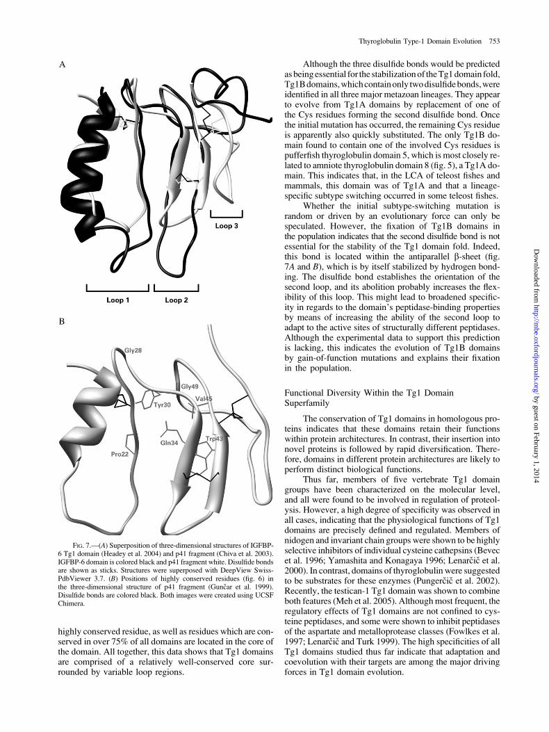
highly conserved residue, as well as residues which are con-served in over 75% of all domains are located in the core ofthe domain. All together, this data shows that Tg1 domainsare comprised of a relatively well-conserved core sur-rounded by variable loop regions.
Although the three disulfide bonds would be predictedasbeingessential for the stabilizationof theTg1domain fold,Tg1Bdomains,whichcontainonly twodisulfidebonds,wereidentified in all three major metazoan lineages. They appearto evolve from Tg1A domains by replacement of one ofthe Cys residues forming the second disulfide bond. Oncethe initial mutation has occurred, the remaining Cys residueis apparently also quickly substituted. The only Tg1B do-main found to contain one of the involved Cys residues ispufferfish thyroglobulin domain 5, which is most closely re-lated to amniote thyroglobulin domain 8 (fig. 5), a Tg1A do-main. This indicates that, in the LCA of teleost fishes andmammals, this domain was of Tg1A and that a lineage-specific subtype switching occurred in some teleost fishes.
Whether the initial subtype-switching mutation israndom or driven by an evolutionary force can only bespeculated. However, the fixation of Tg1B domains inthe population indicates that the second disulfide bond is notessential for the stability of the Tg1 domain fold. Indeed,this bond is located within the antiparallel b-sheet (fig.7A and B), which is by itself stabilized by hydrogen bond-ing. The disulfide bond establishes the orientation of thesecond loop, and its abolition probably increases the flex-ibility of this loop. This might lead to broadened specific-ity in regards to the domain’s peptidase-binding propertiesby means of increasing the ability of the second loop toadapt to the active sites of structurally different peptidases.Although the experimental data to support this predictionis lacking, this indicates the evolution of Tg1B domainsby gain-of-function mutations and explains their fixationin the population.
Functional Diversity Within the Tg1 DomainSuperfamily
The conservation of Tg1 domains in homologous pro-teins indicates that these domains retain their functionswithin protein architectures. In contrast, their insertion intonovel proteins is followed by rapid diversification. There-fore, domains in different protein architectures are likely toperform distinct biological functions.
Thus far, members of five vertebrate Tg1 domaingroups have been characterized on the molecular level,and all were found to be involved in regulation of proteol-ysis. However, a high degree of specificity was observed inall cases, indicating that the physiological functions of Tg1domains are precisely defined and regulated. Members ofnidogen and invariant chain groups were shown to be highlyselective inhibitors of individual cysteine cathepsins (Bevecet al. 1996; Yamashita and Konagaya 1996; Lenarcic et al.2000). In contrast, domains of thyroglobulin were suggestedto be substrates for these enzymes (Pungercic et al. 2002).Recently, the testican-1 Tg1 domain was shown to combineboth features (Meh et al. 2005). Althoughmost frequent, theregulatory effects of Tg1 domains are not confined to cys-teine peptidases, and some were shown to inhibit peptidasesof the aspartate and metalloprotease classes (Fowlkes et al.1997; Lenarcic and Turk 1999). The high specificities of allTg1 domains studied thus far indicate that adaptation andcoevolution with their targets are among the major drivingforces in Tg1 domain evolution.
FIG. 7.—(A) Superposition of three-dimensional structures of IGFBP-6 Tg1 domain (Headey et al. 2004) and p41 fragment (Chiva et al. 2003).IGFBP-6 domain is colored black and p41 fragment white. Disulfide bondsare shown as sticks. Structures were superposed with DeepView Swiss-PdbViewer 3.7. (B) Positions of highly conserved residues (fig. 6) inthe three-dimensional structure of p41 fragment (Guncar et al. 1999).Disulfide bonds are colored black. Both images were created using UCSFChimera.
Thyroglobulin Type-1 Domain Evolution 753
by guest on February 1, 2014http://m
be.oxfordjournals.org/D
ownloaded from
Additionally, some Tg1 domains evolved to performsecondary functions. The Tg1 domains in IGFBPs are in-volved in the binding of IGFs, and these interactions havebeen explained on molecular level for IGFBP-6 (Headeyet al. 2004). In thyroglobulin, several highly conservediodinated Tyr residues were identified within some Tg1domains, indicating direct involvement of these domainsin prohormone storage in the thyroid gland. Most iodinationsites are located within large insertions, which are unique tothese Tg1 domains (see Supplement 3 of the SupplementaryMaterial online), indicating that these additional sequenceswere introduced specifically to expand the functional rangeof Tg1 domains.
Conclusion
Apart from the studies of thyropins and the IGFBPs,Tg1 domains have been largely overlooked, although theyhave been identified in a variety of proteins with differentdomain architectures, functions, and phyletic distributions.The studies on thyropins revealed Tg1 domains to be aunique group of proteolysis regulators with respect to theirhigh specificity as well as their diversity. In this work, wehave shown them to be a superfamily of versatile moduleswith a rich evolutionary history. The first Tg1 domainoriginated early in the evolution of Metazoa, presumablyas a peptidase inhibitor, and has quickly spread and diver-sified. During metazoan evolution, Tg1 domains wereincorporated into proteins with greatly different domainarchitectures, and two supradomain organizations, thetestican-type and the SMOC-type supradomains, that com-prise Tg1 domains and EC domains, occur in most majormetazoan branches. Unfortunately, many identified Tg1domains remain structurally and functionally uncharacter-ized, and future work will certainly reveal more interesting,as yet unknown properties of these domains.
Supplementary Material
Supplementary figures and tables are available atMolecular Biology and Evolution online (http://www.mbe.oxfordjournals.org/).
Acknowledgments
We thank Prof. R. H. Pain for critical reading of themanuscript and Gregor Guncar for technical support. Thiswork was supported by the Slovenian Research Agency byresearch program P1-0140. This work was done entirely atthe Jozef Stefan Institute, Jamova 39, SI-1000 Ljubljana,Slovenia.
Literature Cited
Bevec, T., V. Stoka, G. Pungercic, I. Dolenc, and V. Turk. 1996.Major histocompatibility complex class II-associated p41invariant chain fragment is a strong inhibitor of lysosomalcathepsin L. J. Exp. Med. 183:1331–1338.
Bocock, J. P., C. J. Edgell, H. S. Marr, and A. H. Erickson. 2003.Human proteoglycan testican-1 inhibits the lysosomal cysteineprotease cathepsin L. Eur. J. Biochem. 270:4008–4015.
Burge, C., and S. Karlin. 1997. Prediction of complete gene struc-tures in human genomic DNA. J. Mol. Biol. 268:78–94.
Chiva, C., P. Barthe, A. Codina et al. (14 co-authors). 2003.Synthesis and NMR structure of p41icf, a potent inhibitorof human cathepsin L. J. Am. Chem. Soc. 125:1508–1517.
Erickson, A. C., and J. R. Couchman. 2000. Still more complexityin mammalian basement membranes. J. Histochem. Cytochem.48:1291–1306.
Felsenstein, J. 1989. PHYLIP—phylogeny inference package(version 3.2). Cladistics 5:164–166.
Fowlkes, J. L., K. M. Thrailkill, C. George-Nascimento, C. K.Rosenberg, and D. M. Serra. 1997. Heparin-binding, highlybasic regions within the thyroglobulin type-1 repeat ofinsulin-like growth factor (IGF)-binding proteins (IGFBPs) -3,-5, and -6 inhibit IGFBP-4 degradation. Endocrinology
138:2280–2285.Galesa, K., R. Pain, M. A. Jongsma, V. Turk, and B. Lenarcic.
2003. Structural characterization of thyroglobulin type-1domains of equistatin. FEBS Lett. 539:120–124.
Galtier, N., M. Gouy, and C. Gautier. 1996. SEAVIEW andPHYLO_WIN: two graphic tools for sequence alignmentand molecular phylogeny. Comput. Appl. Biosci. 12:543–548.
Grasberger, B. L., G. M. Clore, and A. M. Gronenborn. 1994.High-resolution structure of Ascaris trypsin inhibitor in solu-tion: direct evidence for a pH-induced conformational transi-tion in the reactive site. Structure 2:669–678.
Guillemot, J. C., M. Naspetti, F. Malergue, P. Montcourrier, F.Galland, and P. Naquet. 2001. Ep-CAM transfection in thymicepithelial cell lines triggers the formation of dynamic actin-richprotrusions involved in the organization of epithelial celllayers. Histochem. Cell Biol. 116:371–378.
Guncar, G., G. Pungercic, I. Klemencic, V. Turk, and D. Turk.1999. Crystal structure of MHC class II-associated p41 Iifragment bound to cathepsin L reveals the structural basisfor differentiation between cathepsins L and S. EMBO J. 18:793–803.
Hazkani-Covo, E., E. Y. Levanon, G. Rotman, D. Graur, and A.Novik. 2004. Evolution of multicellularity in Metazoa: com-parative analysis of the subcellular localization of proteinsin Saccharomyces, Drosophila and Caenorhabditis. Cell Biol.Int. 28:171–178.
Headey, S. J., D. W. Keizer, S. Yao, G. Brasier, P. Kantharidis,L. A. Bach, and R. S. Norton. 2004. C-terminal domain ofinsulin-like growth factor (IGF) binding protein-6: structureand interaction with IGF-II. Mol. Endocrinol. 18:2740–2750.
Hennighausen, L. G., and A. E. Sippel. 1982. Mouse whey acidicprotein is a novel member of the family of �four-disulfide core�proteins. Nucleic Acids Res. 10:2677–2684.
Hwa, V., Y. Oh, and R. G. Rosenfeld. 1999a. Insulin-like growthfactor binding proteins: a proposed superfamily. Acta Paediatr.Suppl. 88:37–45.
———. 1999b. The insulin-like growth factor-binding protein(IGFBP) superfamily. Endocr. Rev. 20:761–787.
Kortschak, R. D., G. Samuel, R. Saint, and D. J. Miller. 2003. ESTanalysis of the cnidarian Acropora millepora reveals extensivegene loss and rapid sequence divergence in the model inver-tebrates. Curr. Biol. 13:2190–2195.
Lamas, L., P. C. Anderson, J. W. Fox, and J. T. Dunn. 1989. Con-sensus sequences for early iodination and hormonogenesis inhuman thyroglobulin. J. Biol. Chem. 264:13541–13545.
Laskowski, M. Jr. 1986. Protein inhibitors of serine proteinases—-mechanism and classification. Adv. Exp. Med. Biol. 199:1–17.
Lee, M., and H. T. Cheung. 1996. Isolation and characterizationof Caenorhabditis elegans extracellular matrix. Biochem.Biophys. Res. Commun. 221:503–509.
Lenarcic, B., and T. Bevec. 1998. Thyropins—new structurallyrelated proteinase inhibitors. Biol. Chem. 379:105–111.
Lenarcic, B., G. Krishnan, R. Borukhovich, B. Ruck, V. Turk, andE.Moczydlowski. 2000. Saxiphilin, a saxitoxin-bindingprotein
754 Novinec et al.
by guest on February 1, 2014http://m
be.oxfordjournals.org/D
ownloaded from
with two thyroglobulin type 1 domains, is an inhibitor ofpapain-like cysteine proteinases. J. Biol. Chem. 275:15572–15577.
Lenarcic, B., and V. Turk. 1999. Thyroglobulin type-1 domainsin equistatin inhibit both papain-like cysteine proteinasesand cathepsin D. J. Biol. Chem. 274:563–566.
Li, Y., and E. Moczydlowski. 1991. Purification and partialsequencing of saxiphilin, a saxitoxin-binding protein fromthe bullfrog, reveals homology to transferrin. J. Biol. Chem.266:15481–15487.
Litvinov, S. V., M. Balzar, M. J. Winter, H. A. Bakker, I. H.Briaire-de Bruijn, F. Prins, G. J. Fleuren, and S. O. Warnaar.1997. Epithelial cell adhesion molecule (Ep-CAM) modulatescell-cell interactions mediated by classic cadherins. J. Cell.Biol. 139:1337–1348.
Mahar, J., G. L. Lukacs, Y. Li, S. Hall, and E. Moczydlowski.1991. Pharmacological and biochemical properties of saxiphi-lin, a soluble saxitoxin-binding protein from the bullfrog (Ranacatesbeiana). Toxicon 29:53–71.
Marr, H. S., and C. J. Edgell. 2003. Testican-1 inhibits attachmentof Neuro-2a cells. Matrix Biol. 22:259–266.
Meh, P., M. Pavsic, V. Turk, A. Baici, and B. Lenarcic. 2005. Dualconcentration-dependent activity of thyroglobulin type-1 do-main of testican: specific inhibitor and substrate of cathepsinL. Biol. Chem. 386:75–83.
Mercken, L.,M. J. Simons, S. Swillens,M.Massaer, andG.Vassart.1985. Primary structure of bovine thyroglobulin deducedfrom the sequence of its 8,431-base complementary DNA.Nature 316:647–651.
Molina, F., M. Bouanani, B. Pau, and C. Granier. 1996. Charac-terization of the type-1 repeat from thyroglobulin, a cysteine-rich module found in proteins from different families. Eur. J.Biochem. 240:125–133.
Murzin, A. G., S. E. Brenner, T. Hubbard, and C. Chothia. 1995.SCOP: a structural classification of proteins database for theinvestigation of sequences and structures. J. Mol. Biol. 247:536–540.
Nakada, M., H. Miyamori, J. Yamashita, and H. Sato. 2003. Tes-tican 2 abrogates inhibition of membrane-type matrix metallo-proteinases by other testican family proteins. Cancer Res.63:3364–3369.
Nakada, M., A. Yamada, T. Takino, H.Miyamori, T. Takahashi, J.Yamashita, and H. Sato. 2001. Suppression of membrane-type1 matrix metalloproteinase (MMP)-mediated MMP-2 activa-tion and tumor invasion by testican 3 and its splicing variantgene product, N-Tes. Cancer Res. 61:8896–8902.
Nakae, H., M. Sugano, Y. Ishimori, T. Endo, and T. Obinata.1993. Ascidian entactin/nidogen. Implication of evolution byshuffling two kinds of cysteine-rich motifs. Eur. J. Biochem.213:11–19.
Parma, J., D. Christophe, V. Pohl, and G. Vassart. 1987. Structuralorganization of the 5# region of the thyroglobulin gene. Evi-dence for intron loss and ‘‘exonization’’ during evolution. J.Mol. Biol. 196:769–779.
Patthy, L. 2003. Modular assembly of genes and the evolution ofnew functions. Genetica 118:217–231.
Pungercic,G., I.Dolenc,M.Dolinar, T.Bevec, S. Jenko,S.Kolaric,and V. Turk. 2002. Individual recombinant thyroglobulintype-1 domains are substrates for lysosomal cysteine protei-nases. Biol. Chem. 383:1809–1812.
Raible, F., and D. Arendt. 2004. Metazoan evolution: some ani-mals are more equal than others. Curr. Biol. 14:R106–R108.
Rawlings, N. D., D. P. Tolle, and A. J. Barrett. 2004. Evolutionaryfamilies of peptidase inhibitors. Biochem. J. 378:705–716.
Saitou, N., andM. Nei. 1987. The neighbor-joining method: a newmethod for reconstructing phylogenetic trees. Mol. Biol. Evol.4:406–425.
Schnepp, A., P. Komp Lindgren, H. Hulsmann, S. Kroger, M.Paulsson, and U. Hartmann. 2005. Mouse testican-2. Expres-sion, glycosylation, and effects on neurite outgrowth. J. Biol.Chem. 280:11274–11280.
Stumptner-Cuvelette, P., and P. Benaroch. 2002. Multiple roles ofthe invariant chain in MHC class II function. Biochim. Bio-phys. Acta 1542:1–13.
Thompson, J.D.,D.G.Higgins, andT. J.Gibson. 1994.CLUSTALW: improving the sensitivity of progressive multiple sequencealignment through sequence weighting, position-specific gappenalties and weight matrix choice. Nucleic Acids Res. 22:4673–4680.
Upton, Z., S. J. Chan, D. F. Steiner, J. C.Wallace, and F. J. Ballard.1993. Evolution of insulin-like growth factor binding pro-teins. Growth Regul. 3:29–32.
Vannahme,C.,S.Gosling,M.Paulsson,P.Maurer,andU.Hartmann.2003. Characterization of SMOC-2, a modular extracellularcalcium-binding protein. Biochem. J. 373:805–814.
Vannahme, C., N. Smyth, N. Miosge, S. Gosling, C. Frie, M.Paulsson, P. Maurer, and U. Hartmann. 2002. Characterizationof SMOC-1, a novel modular calcium-binding protein in base-ment membranes. J. Biol. Chem. 277:37977–37986.
Veerassamy, S., A. Smith, and E. R. Tillier. 2003. A transitionprobability model for amino acid substitutions from blocks.J. Comput. Biol. 10:997–1010.
Vogel, C., M. Bashton, N. D. Kerrison, C. Chothia, and S. A.Teichmann. 2004a. Structure, function and evolution of mul-tidomain proteins. Curr. Opin. Struct. Biol. 14:208–216.
Vogel, C., C.Berzuini,M.Bashton, J.Gough, and S.A. Teichmann.2004b. Supra-domains: evolutionary units larger than singleprotein domains. J. Mol. Biol. 336:809–823.
Yamashita, M., and S. Konagaya. 1996. A novel cysteineprotease inhibitor of the egg of chum salmon, containing acysteine-rich thyroglobulin-like motif. J. Biol. Chem. 271:1282–1284.
Claudia Kappen, Associate Editor
Accepted December 14, 2005
Thyroglobulin Type-1 Domain Evolution 755
by guest on February 1, 2014http://m
be.oxfordjournals.org/D
ownloaded from