distinct, parallel pathways link the medial mammillary bodies to the anterior thalamus in macaque...
TRANSCRIPT
Distinct, parallel pathways link the medial mammillarybodies to the anterior thalamus in macaque monkeys
Seralynne D. Vann,1 Richard C. Saunders2 and John P. Aggleton1
1School of Psychology, Cardiff University, Cardiff, Wales, CF10 3AT, UK2Laboratory of Neuropsychology, National Institute of Mental Health, Bethesda, MD 20892, USA
Keywords: amnesia, anterior thalamic nuclei, hippocampus, mammillary bodies, mammillothalamic tract, memory
Abstract
Mammillary body neurons projecting to the thalamus were identified by injecting retrograde tracers into the medial thalamus ofmacaque monkeys. The source of the thalamic projections from the medial mammillary nucleus showed strikingly different patterns oforganization depending on the site of the injection within the two anterior thalamic nuclei, anterior medialis and anterior ventralis.These data reveal at least two distinct modes by which the primate medial mammillary bodies can regulate anterior thalamic function.Projections to the thalamic nucleus anterior medialis arise mainly from the pars lateralis of the medial mammillary nucleus. Aparticularly dense source is the dorsal cap in the posterior half of the pars lateralis, a subregion that has not previously beendistinguished. In contrast, neurons spread evenly across the medial mammillary nucleus gave rise to projections more laterally in theanterior thalamic nuclei. A third pattern of medial mammillary neurons appeared to provide the source of projections to the rostralmidline thalamic nuclei. In contrast, the labeled cells in the lateral mammillary nucleus were evenly spread across that nucleus,irrespective of injection site. In addition to the established projection to anterior dorsalis, the lateral mammillary nucleus appears toproject lightly to a number of other thalamic nuclei, including lateralis dorsalis, anterior medialis, anterior ventralis, and the rostralmidline nuclei, e.g. nucleus reuniens. These anatomical findings not only reveal novel ways of grouping the neurons within the medialmammillary nucleus, but also indicate that the mammillothalamic connections support cognition in multiple ways.
Introduction
A vital clue to the functions of the mammillary bodies comes from itsprincipal output, the massive, unidirectional projections to the anteriorthalamus. These projections, which mainly arise from the larger, medialmammillary nucleus, comprise the mammillothalamic tract (or bundleof Vicq d’Azyr). This tract is a central link in hippocampal–diencephaliccircuits now widely thought to support memory (Delay & Brion, 1969;Gaffan, 1992; Aggleton & Brown, 1999). Clinical evidence for theimportance of themammillothalamic tract comes from detailed analysesof thalamic strokes, which reveal a very high association between tractdamage and amnesia (Von Cramon et al., 1985; Gentilini et al., 1987;Van derWerf et al., 2000, 2003). Other support comes from descriptionsof mammillary body damage in humans with amnesia (Dusoir et al.,1990; Tanaka et al., 1997; Hildebrandt et al., 2001). The conclusion thatmammillothalamic connections are vital for memory is reinforced bystudies into the effects of selective mammillary body and mammillotha-lamic tract lesions in monkeys and rats (Aggleton & Mishkin, 1985;Parker & Gaffan, 1997; Vann & Aggleton, 2003). Even though theycomprise one of the largest sources of anterior thalamic afferents in theprimate brain (Powell et al., 1957) and seem to be necessary for normalmemory, remarkably little is known about the mammillary bodyprojections to the thalamus in the primate brain.
So far, studies of monkeys have shown that the two mainmammillary nuclei (medial and lateral) have different terminationpatterns within the three anterior thalamic nuclei. The much largermedial mammillary nucleus projects to both nucleus anterior medialisand nucleus anterior ventralis, while the lateral mammillary nucleusprojects to nucleus anterior dorsalis (Veazey et al., 1982b; Xiao &Barbas, 2002b). Both electrophysiological and anatomical evidencesuggest that the two largest anterior thalamic nuclei (anterior medialisand anterior ventralis) have different roles in supporting memory(Vertes et al., 2001; Xiao & Barbas, 2002a, b). If so, then the medialmammillary inputs to these two thalamic nuclei might be expected todiffer, potentially reflecting distinct mnemonic functions. Thisprediction was tested in the present study.Fluorescent retrograde tracers were injected into the anterior thalamic
nuclei and the adjacent nucleus lateralis dorsalis in six rhesus monkeys(Macacca mulatta). Additional information came from six cynomolgusmonkeys (Macaca fascicularis) with injections of horseradish perox-idase (HRP) close to the thalamic midline. The injection placementsmade it possible to address a number of questions. First, whether thereare qualitative differences in the patterns of inputs from the mammillarybodies to nuclei anterior medialis and anterior ventralis. Second,whether the mammillary bodies project to any additional thalamicnuclei. Candidates include the midline thalamic nuclei (Veazey et al.,1982b) and nucleus lateralis dorsalis, the latter sometimes classifiedwithin the anterior thalamic nuclear complex (VanGroen&Wyss, 1992;Bentivoglio et al., 1993). The study also examined the source of inputsto nucleus medialis dorsalis. Although this thalamic nucleus has
Correspondence: Dr R. C. Saunders, as above.E-mail: [email protected]
Received 20 April 2007, revised 21 June 2007, accepted 18 July 2007
European Journal of Neuroscience, Vol. 26, pp. 1575–1586, 2007 doi:10.1111/j.1460-9568.2007.05773.x
ª The Authors (2007). Journal Compilation ª Federation of European Neuroscience Societies and Blackwell Publishing Ltd

repeatedly been implicated in memory processes (Markowitsch, 1982;Aggleton & Mishkin, 1983; Gold & Squire, 2006), its connectionssuggest markedly different functions to those of the mammillary bodiesand anterior thalamic nuclei.
Materials and methods
Data were collected from two different series of tracer injections, oneusing the retrograde transport of fluorescent dyes [fast blue (Fb) anddiamidino yellow (Dy)], the other using the retrograde transport ofHRP. By using two different fluorescent tracers in the same animal it waspossible to inject multiple thalamic sites and make direct comparisonsof retrogradely labeled cells that project to different thalamic targets.In addition, this refinement reduced the number of animals necessary tocomplete the study. Although there are a number of minor variationsin some of the experimental procedures, as well as possible differencesin the sensitivity of the various tracers, both series are described asthey complement each another to provide a more complete picture.Data from these animals also contributed to two earlier studies(Aggleton et al., 1986; Saunders et al., 2005), and the numbering ofthe individual cases corresponds to that previously published. Allexperimental procedures were carried out with the approval of theNIMH, Bethesda, and with strict adherence to the NIH Guide for Careand Use of Laboratory Animals, such that the ‘Principles of laboratoryanimal care’ (NIH Publication no. 86-23, revised 1985) were followed.
Fluorescent tracer injections
Six adult rhesus monkeys received injections of two fluorescenttracers, Dy (Keizer et al., 1983) or Fb (Kuypers et al., 1980), with
different tracers being placed into different thalamic sites in the sameanimal. The animals were tranquilized with ketamine hydrochloride(10–15 mg ⁄ kg, intramuscular injection) and surgical anesthesia wasmaintained with gas (isoflurane 1–4%, to effect). They were placed ina specialized head holder and, following aseptic procedures, dorsalbone and dural flaps were opened to expose the midline. The wall ofthe left hemisphere was then gently retracted, and a 5–10-mm portionof the corpus callosum and the underlying fornix were splitlongitudinally to expose the thalamic midline. An injection of eitherFb (Sigma) or Dy (Sigma) was made through a 5-lL Hamilton syringefitted with a 28-gauge needle. Both tracers were injected as 3%suspensions in distilled water. In three cases (BRh3, BRh4, BRh6), afiber pathway (fornix or extreme capsule) was cut unilaterallyimmediately prior to the injections (Table 1).To minimize the use of animals, some monkeys received two
(BRh2), three (BRh5, BRh6) or even four (BRh3, BRh4) injections ofthe two fluorescent tracers, each in different thalamic sites (Table 1). InBRh2, single injections of the two different tracers (Dy, Fb) weredirected at either the anterior thalamic nuclei (Fb) or nucleus medialisdorsalis (Dy) in opposite hemispheres. In animals BRh3 and BRh4,separate injections were placed in the anterior thalamic nuclei andnucleus medialis dorsalis in the same hemisphere, but using differenttracers (Fb and Dy). This procedure was then repeated in the oppositehemisphere, ensuring that the anterior thalamic nuclei were injectedwith Fb in one hemisphere and Dy in the other hemisphere (Table 1).In case BRh5, bilateral injections (one Fb, the other Dy) were placedin nucleus lateralis dorsalis in opposite hemispheres, while a furtherinjection of Fb was placed in the anterior thalamic nuclei in the samehemisphere as that with the Dy injection. Finally, in BRh6, twoinjections (one Fb, the other Dy) were placed in nucleus lateralisdorsalis in opposite hemispheres, while an injection of Fb was placed
Table 1. List of all cases showing the species (SP) of macaque (Rh, rhesus; Cyn, cynomolgus), injection site and location of label
Case Species Injection site, tracer, surgery
Mammillary body label
LMB MMBb MMBm MMBl MMBlcap
ACy1 Cynomolgus AM, Mid, MD, Reuns (HRP) ++ + + ++ ++++ACy2 Cynomolgus AM, Mid (HRP) ++ 0 + +++ ++++ACy3 Cynomolgus MD, Mid (HRP) (+) 0 0 0 0ACy5 Cynomolgus MD, Mid (HRP) (+) 0 0 0 0ACy7 Cynomolgus MD, Mid (HRP) 0 0 0 0 0ACy26 Cynomolgus Reuns, Mid, AM (HRP) ++ 0 + ++++ +BRh1 Rhesus MD (Fb l) 0 0 0 0 0BRh2 Rhesus AM (Fb r) ++ 0 ++ ++ ++++
MD, Mid (Dy r) 0 0 0 0 0BRh3 Rhesus AV, AM (Dy l, Fx) + (+) ++ ++ +++
MD (Fb l, Fx) – – – – –AM, MTT (Fb r, ECx) ++ ++ +++ +++ +++MD (Dy r, ECx) – – – – –
BRh4 Rhesus AV, AM (Fb r, Fx) + + +++ ++ +++MD (Dy r, Fx) – – – – –AM, Mid (Dy l, ECx) + 0 ++ ++ ++++MD (Fb l, ECx) – – – – –
BRh5 Rhesus AV (Fb l) ++ + +++ ++++ ++LD (Fb r) – – – – –LD (Dy l) + 0 0 0 0
BRh6 Rhesus LD (Dy r, Fx) + 0 0 0 0MD, Mid (Fb, Fx, ECx) (+) – – – –LD (Dy l, ECx) + 0 0 0 0
Abbreviations: AM, anterior medial thalamic nucleus; AV, anterior ventral thalamic nucleus; LD, lateral dorsal thalamic nucleus; LMB, lateral mammillary nucleus;MD, nucleus medialis dorsalis; Mid, midline thalamic nuclei; MMBb, medial mammillary nucleus pars basalis; MMBl, medial mammillary nucleus pars lateralis;MMBlcap, medial mammillary nucleus pars lateralis cap; MMBm, medial mammillary nucleus pars medialis; MTT, mammillothalamic tract; Reuns, nucleusreuniens. Type of tracer (Dy, diamidino yellow; Fb, fast blue; HRP, horseradish peroxidase); hemisphere of injection site (l, left; r, right); and surgery in samehemisphere as injection (ECx, amygdala and extreme capsule cut; Fx, fornix transection). The density of retrograde label (lower) is marked on a scale from zero to++++; the (+) indicates just a couple of cells, and ‘)’ indicates that repeated use of the same tracer in that animal precludes any conclusion.
1576 S. D. Vann et al.
ª The Authors (2007). Journal Compilation ª Federation of European Neuroscience Societies and Blackwell Publishing LtdEuropean Journal of Neuroscience, 26, 1575–1586

in the midline and so involved the most medial portion of medialisdorsalis in both hemispheres (Table 1).Three of the rhesus monkeys (BRh3, BRh4 and BRh6) had
additional surgical procedures immediately before the tracer injec-tions, i.e. as part of the same surgery (Table 1). In all three cases thisinvolved unilateral removal of the fornix and, in the other hemisphere,a unilateral cut in the ventral amygdalofugal pathway ⁄ extremecapsule. Details of these concurrent surgical procedures have beenprovided previously (Aggleton et al., 2005; Saunders et al., 2005).The cut in the ventral amygdalofugal pathway was lateral and dorsal tothe amygdala, and so did not interfere with any structures or tracts inthe present study. Even though the fornix lesions will have removedhippocampal inputs to the anterior thalamic nuclei and the mammillarybodies, the surgery is most unlikely to have affected direct projectionsfrom the mammillary bodies to the anterior thalamus as it wascompleted just prior to tracer injection, i.e. in the same surgery.Furthermore, transection of the fornix in rhesus monkeys does notresult in a long-term loss of neurons within the mammillary bodies,despite evidence of atrophy due to a loss of neuropil (Loftus et al.,2000).After a postoperative period of between 5 and 10 days the animals
were deeply anesthetized with sodium pentobarbital. They were thenperfused intracardially with saline followed by approximately 2 L of4–6% paraformaldehyde in 0.1 m cacodylate buffer (pH 7.4). Thebrains were then removed and placed in a series of cryoprotectantsolutions consisting of first 10% and then 20% glycerol in 0.1 m
cacodylate buffer with 2% dimethylsulfoxide and 4–6% paraformal-dehyde (pH 7.4, 4 �C). Four)6 days after the perfusion the brainswere rapidly frozen by immersion in )75 �C isopentane, and then cutat 40 lm in the coronal plane on a freezing microtome (Rosene et al.,1986). Three, one-in-10 series of sections were mounted immediatelyonto subbed slides, dried and stored in the dark at 4 �C.
HRP injections
A single injection of HRP was made into the medial part of thethalamus in six adult cynomolgus monkeys weighing 3.5–6.8 kg at thetime of surgery. The surgical procedures used to visualize the dorsalthalamus closely matched those for the fluorescent injections. Allinjections were then made under visual guidance. In three cases(ACy1, ACy2, ACy26) the injection tract was in or very close to themidline at the level of the anterior thalamic nuclei. In the remainingthree cases (ACy3, ACy5, ACy7) the HRP injections were targeted atthe magnocellular part of nucleus medialis dorsalis.
Fig. 1. (A) Schematic drawings showing the position of the mammillarybodies in the primate brain. Left: parasagittal view of the medial surface of theadult rhesus monkey brain showing the position of the mammillary bodies(MB) near the base of the brain. Right: detail showing the mammillary bodiesfrom a lateral view, with the fornix (F) entering the rostral part of the structureand the mammillothalamic tract (MTT) exiting dorsally. (B–D) Photomicro-graphs of coronal sections from the rhesus macaque showing the cytoarchi-tectonic and myeloarchitectonic appearance of the MB. (B and D) Nissl-stainedsections showing subdivisions within the mammillary bodies. (C) Myelin-stained (Gallyas) section showing the arrangement of tracts at the mid level ofthe structure. White dashed line indicates the ventral border of the ‘cap’ regionwithin the pars lateralis Abbreviations: AC, anterior commissure; b, medialmammillary nucleus, pars basalis; CC, corpus callosum; H, hippocampus; l,medial mammillary nucleus pars lateralis; lc, ‘cap’ region of the medialmammillary nucleus pars lateralis; LM, lateral nucleus of the mammillarybodies; m, medial mammillary nucleus pars medialis. MMB, medial nucleus ofthe mammillary bodies; T, thalamus. Scale bar: 1 mm.
Mammillothalamic connections in the monkey 1577
ª The Authors (2007). Journal Compilation ª Federation of European Neuroscience Societies and Blackwell Publishing LtdEuropean Journal of Neuroscience, 26, 1575–1586

Four cases received a single injection of 40% HRP (Sigma, type IV)with a 1-lL Hamilton syringe. The volume injected ranged from0.13 lL (ACy2) to 0.15 lL (ACy3, ACy5) to 0.22 lL (ACy1). Inanimal ACy7 a 10% solution of HRP in Tris buffer was deliverediontophoretically through a glass pipette, while in ACy26 the injectionconsisted of 0.09 lL of a 4% solution of HRP (Sigma, type VI)conjugated with wheat germ agglutin.Two days after surgery the monkeys were deeply anesthetized with
Nembutal and perfused intracardially with 0.9% saline followed by asolution of 1% paraformaldehyde and 1.25% glutaraldehyde in 0.1 m
phosphate buffer (pH 7.2). The brains were stored in 30% sucrosebuffer at 4 �C for 3–4 days and then cut in 50-lm coronal sections.A one-in-five series was then treated according to the protocols of
either Hardy & Heimer (1977) or Mesulam (1978). Alternate sectionswere dehydrated, counterstained with thionine and coverslipped, whilethe remaining sections were just dehydrated and coverslipped.
Anatomical designations
The primate mammillary bodies are located at the base of the brain inthe posterior hypothalamus (Fig. 1A) and comprise two main nucleargroups, the medial and lateral mamillary nuclei (Rose, 1939; Veazeyet al., 1982a). By far the larger of the two is the medial mammillarynucleus, which is surrounded by a dense, fiber capsule (Fig. 1B–D).The major afferent tract is the postcommissural fornix, which can be
Fig. 2. Location and extent of injections of retrograde tracers (HRP, Fb, Dy) in the rostral medial thalamus. The extent of the HRP (red horizontal lines, bottom tworows), Dy (yellow) and Fb (blue) injections are shown on coronal sections. The numbers below each section refer to their anterior–posterior level from Olszewski(1952; see also Fig. 1A). Abbreviations: AD, nucleus anterior dorsalis; AM, nucleus anterior medialis; AV, nucleus anterior ventralis; cdc, nucleus centralisdensocellularis; clc, nucleus centralis latocellularis; LD, nucleus lateralis dorsalis; MD, nucleus medialis dorsalis; MTT, mammillothalamic tract; Pcn, nucleusparacentralis; Pf, nucleus parafascicularis; Re, nucleus reuniens; VA, nucleus ventralis anterior.
1578 S. D. Vann et al.
ª The Authors (2007). Journal Compilation ª Federation of European Neuroscience Societies and Blackwell Publishing LtdEuropean Journal of Neuroscience, 26, 1575–1586

traced running into the rostral part of the medial mammillary nucleus.The mammillothalamic tract, the primary efferent tract, exits from thedorsal half of the same nucleus (Fig. 1A). Using the descriptions of thecynomolgus monkey brain by Veazey et al. (1982a), three cytoarchi-tectonic divisions can be observed within the medial mammillarynucleus: pars medialis, pars basalis and pars lateralis (Fig. 1B and 1D,see also Rose, 1939). Pars medialis is distinctive as it contains thelargest neurons within the medial mammillary nucleus, while the cellsin pars basalis are among the smallest in the posterior hypothalamus(Fig. 1B and D). The third component, pars lateralis, is the largestsubdivision and occupies the lateral half of the medial nucleus. Themedial and central parts of pars lateralis are distinguished from parsmedialis by the lower density of cells, mainly as a consequence of thelarge number of fibers in this more lateral part of the medial nucleus.As a consequence there is not a sharp border with pars medialis. In thecaudal half of the medial mammillary nucleus the more dorsal cells inpars lateralis form a distinctive ‘cap’ where they appear more denselyaggregated (Fig. 1). This cap is sometimes more visible in myelinstained sections due to a reduction in fiber density (Fig. 1C).
The other nuclear group, the lateral mammillary nucleus, lies lateraland ventral to the medial mammillary nucleus (Fig. 1B). The lateralnucleus, which is shorter in the anterior–posterior plane than the medialnucleus (Fig. 1A), contains relatively large cells that stain intensely forNissl substance. These cells are uniformly distributed, and so nocytoarchitectonic subdivisions have been described within the primatelateral nucleus. Although a third mammillary nucleus, nucleusintercalatus, has been reported by some authors (Veazey et al.,1982a), there is disagreement over its existence (Crosby & Showers,1969; Nauta & Haymaker, 1969). This region, which lies lateral to pars
lateralis of the medial mammillary nucleus, is indistinct as it isperforated by many fibers. While the study by Veazey et al. (1982a)recognized a separate nucleus intercalatus, it was rarely visible in thepresent study and so is omitted from descriptions of the data.The nomenclature and borders of the thalamic nuclei, including
those of the midline nuclei, are all taken from Olszewski (1952), asthis monograph provides the most complete cytoarchitectonic descrip-tion of all of the thalamic nuclei in the macaque brain. The anteriorthalamic nuclei comprise three major nuclei (anterior medialis, andanterior dorsalis anterior ventralis). Immediately caudal to nucleusanterior dorsalis lies nucleus lateralis dorsalis, which is often groupedwith the anterior thalamic nuclei (Bentivoglio et al., 1993). For thisreason, nucleus lateralis dorsalis also received injections of retrogradetracers.
Results
Although the study used two different species of macaque monkey andthree different retrograde tracers, there was a high degree of internalconsistency across cases. For this reason, the results are grouped byinjection site (Figs 2 and 3) rather than by species or tracer. Indescribing the pattern of results priority is given to those cases inwhich the data are the most straightforward. Key factors are theprecision of the injection site and whether there are injections usingthe same tracer in a different site in the same animal (Table 1). Whenconsidering any potential involvement from injections in the contra-lateral hemisphere it should be noted that while the projections fromthe medial mammillary nucleus to the thalamus are thought to beipsilateral, those from the lateral mammillary nucleus are known to be
Fig. 3. Location and extent of injections of retrograde tracers (HRP, Fb, Dy) into the caudal medial thalamus. The extent of the HRP (horizontal lines), Dy (yellow)and Fb (blue) injections are shown on coronal sections. The numbers refer to the anterior–posterior level of the sections from Olszewski (1952; see also Fig. 1A).Abbreviations: AD, nucleus anterior dorsalis; LD, nucleus lateralis dorsalis; MD, nucleus medialis dorsalis; VPL, nucleus ventralis posterior lateralis.
Mammillothalamic connections in the monkey 1579
ª The Authors (2007). Journal Compilation ª Federation of European Neuroscience Societies and Blackwell Publishing LtdEuropean Journal of Neuroscience, 26, 1575–1586

bilateral (Cruce, 1975; Veazey et al., 1982b). The mammillary bodylabel is depicted on a series of coronal sections, with the anterior–posterior level of these sections matched against Fig. 1.
Injections in nucleus anterior medialis
Very striking evidence of a topographic projection from the medialmammillary nucleus was found in case BRh2 after an injection of Fb
placed in the mid anterior–posterior level of nucleus anterior medialisjust lateral to the midline (Fig. 2). There was an extremely cleardorsoventral gradient of retrograde label within the medial mammil-lary nucleus (Fig. 4). While the dorsal third of the medial mammillarynucleus contained a high density of labeled cells, there was only adiffuse presence throughout the rest of the medial mammillarynucleus. This more dense label was largely confined to pars lateralis,and was especially obvious in the dorsal cap of pars lateralis (Figs 4
Fig. 4. Series of drawings of the mammillary bodies showing the location of retrogradely labeled cells (black dots) in four cases with tracer injections centered inthe medial ATN. The figures down the middle depict the center of each injection site (from Fig. 2). The numbers refer to the anterior–posterior levels from Olszewski(1952; see also Fig. 1A). Abbreviations: AD, nucleus anterior dorsalis; AM, nucleus anterior medialis; AV, nucleus anterior ventralis; clc, nucleus centralislatocellularis; Dy, diamidino yellow; Fb, fast blue; HRP, horseradish peroxidase; MTT, mammillothalamic tract.
1580 S. D. Vann et al.
ª The Authors (2007). Journal Compilation ª Federation of European Neuroscience Societies and Blackwell Publishing LtdEuropean Journal of Neuroscience, 26, 1575–1586

and 5B). Very light Fb label was also present in the dorsal half of themedial mammillary nucleus in the contralateral hemisphere. Finally, afew Fb-labeled cells were spread diffusely across the lateral nucleus ofthe mamillary bodies in both hemispheres.
All cases in which the tracer injection was centered in the nucleusanterior medialis resulted in label in the pars lateralis of the medialmammillary nucleus with a clear dorsoventral gradient in the densityof labeled cells. In case BRh4, the Dy injection (Fig. 2) was centeredin the caudal part of the nucleus anterior medialis, resulting in anumber of labeled cells throughout the medial mammillary nucleus onthe ipsilateral side, with the exception of pars basalis, which contained
few if any cells. As with the more rostral injection there was a cleargradient as the density of label increased appreciably in the dorsal halfof the nucleus (Figs 4 and 5A). The Dy label in the dorsal cap withinpars lateralis was especially dense (Figs 4 and 5). Very small numbersof Dy-labeled cells were present in the lateral nucleus of the mamillarybodies in both hemispheres.Similar patterns of label came from two cases with HRP injections
(ACy1, ACy2) that involved nucleus anterior medialis (Fig. 2). In caseACy2, the needle tract was in the midline portion of rostral nucleusanterior medialis, but also involved nucleus centralis densocellularis.Near mirror image patterns of mammillary body label were present inboth hemispheres, presumably reflecting the spread of HRP intomedial nucleus anterior medialis in both hemispheres (Fig. 2). Thevery large majority of retrograde label in ACy2 was in the parslateralis of the medial mammillary nucleus. In the rostral third of themedial mammillary nucleus this label was grouped in a central regionso that it involved the lateral parts of pars medialis and medial part ofpars lateralis (Fig. 4). At mid anterior–posterior levels pars lateraliscontained a great many retrogradely labeled cells that were presentalmost throughout this subdivision. The dorsal portion of pars medialisalso contained many labeled cells, which contrasted with the rest ofpars medialis and pars basalis as they contained extremely few labeledcells. At slightly more caudal levels the dense retrograde label wasalmost completely confined to the dorsal portion of pars lateralis,completely filling the dorsal cap. Because some sections in case ACy2were counterstained for Nissl, it was possible to confirm that every cellcontained HRP retrograde label in the dorsal cap region of parslateralis. At this more caudal level there were only a handful of labeledcells distributed across pars medialis. The lateral nucleus of themammillary bodies contained a scattering of labeled cells in bothhemispheres. These cells appeared to be evenly distributed across thelateral nucleus, except for a lack of label at the most caudal portion ofthis nucleus.The needle placement in ACy1 was very similar to ACy2 as it
crossed centralis densocellularis and midline anterior thalamic nuclei(Fig. 2). The HRP injection was, however, just a little more caudal anda little deeper. Although a much greater amount of HRP was injectedinto the thalamus, the pattern of retrograde label within the mammil-lary bodies indicated a restricted region of uptake. Apart from the mostrostral level of the mammillary bodies, where labeled cells werescattered evenly across the medial mammillary nucleus, the denseretrograde label was almost entirely confined to pars lateralis (Fig. 4).Here, the label was very heavily concentrated in the lateral and dorsalparts of pars lateralis. This label filled much of the dorsal cap in thecaudal half of the structure, only avoiding its most medial part.Occasional labeled cells were also found in pars basalis and parsmedialis within the medial mammillary nucleus, but these were veryfew in number. The lateral nucleus of the mammillary bodiescontained a scattering of labeled cells, with no obvious topography.While case BRh3 had an injection of Fb centered in the nucleus
anterior medialis, the injection tract clearly severed the mammillotha-lamic tract (Fig. 2). The fibers of the mammillothalamic tract weredensely filled, and in contrast to the localized label within the parslateralis there was dense label throughout the medial mammillarynucleus. The lateral nucleus of the mammillary bodies nucleus also hadnumerous labeled cells throughout (Fig. 6).
Injections in nucleus AV
In case BRh4 an injection of Fb was centered in the mid anterior–posterior level of nucleus anterior ventralis, and is likely to have
Fig. 5. (A–F) Series of photomicrographs showing examples of retrogradelylabeled cells in the mammillary bodies after fluorescent tracer injections ofeither fast blue (Fb, B, D, E), diamidino yellow (Dy, A, C) or horseradishperoxidase (HRP, F) in the thalamus. The white dashed lines show the bordersof the medial and lateral mammillary nuclei. Scale bar: 1 mm.
Mammillothalamic connections in the monkey 1581
ª The Authors (2007). Journal Compilation ª Federation of European Neuroscience Societies and Blackwell Publishing LtdEuropean Journal of Neuroscience, 26, 1575–1586

extended into adjacent parts of nuclei anterior dorsalis and anteriormedialis (Fig. 2). There were large numbers of labeled cells distributedthroughout the medial mammillary nucleus (Figs 5D and 6). The lowestdensity was in the pars basalis, and the highest density was in the moredorsal portions of pars medialis. Labeled cells were found across parslateralis, and in contrast to the pattern observed after the nucleus anteriormedialis injections there was no obvious increase or decrease of label inthe dorsal cap. Small numbers of Fb-labeled cells were found evenlydistributed across the lateral nucleus of the mammillary bodies in bothhemispheres (Figs 5D and 6). A very similar pattern of retrograde labelwas found across the medial mammillary nucleus in case BRh3, whichhad a Dy injection involving the more medial parts of nucleus anteriorventralis (Figs 5C and 6).In caseBRh5 an injection of Fbwasmade in the caudal part of anterior
ventralis, but it also encroached into caudal nucleus anterior medialisand some of caudal nucleus anterior dorsalis (Fig. 2). Considerable labelwas found in all three subregions of the medial mammillary nucleus(Figs 5E and 7). At rostral levels, pars basalis contained the fewest cells,while pars lateralis and medialis contained very similar densities oflabeled cells. At mid anterior–posterior levels label was present across
all subregions, but there was a denser band across the ventral and centralparts of pars lateralis, alongwith dorsal parsmedialis (Figs 5E and 7).Asa consequence the dorsal cap showed a decrease in the density of label.At the most caudal levels very dense packing of labeled cells wasobserved across the medial mammillary nucleus. Scattered cells werefound across the lateral mammillary nucleus in both hemispheres. Inaddition, a light scattering of Fb-labeled cells was found across themedial mammillary nucleus in the contralateral hemisphere. This labelwas most evident close to the periphery of the nucleus, i.e. near to itsouter edges, and so included all three subregions.
Injections in the rostral midline thalamic nuclei
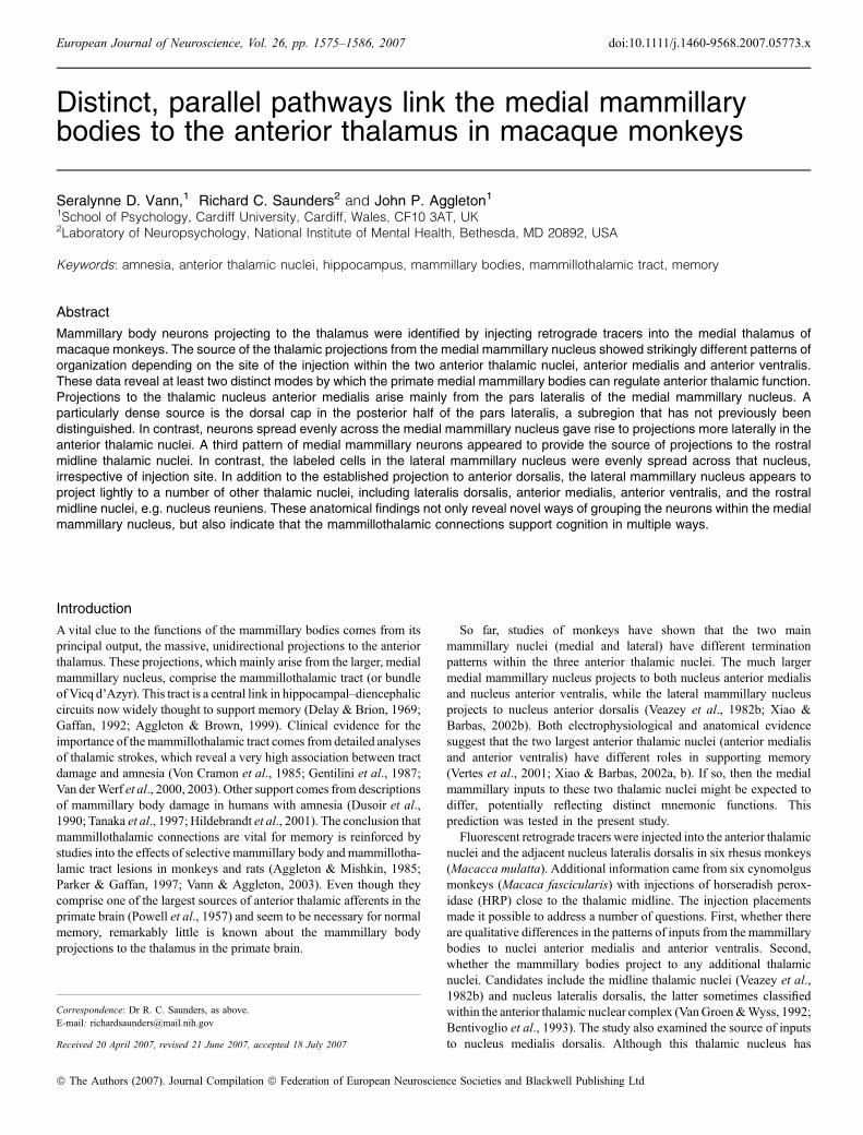
In one case (ACy26) an injection of HRP was centered in the rostralthalamicmidline just ventral to nucleus anteriormedialis, so that the tractpassed through the latter nucleus (Fig. 2). The needle tip was placed inventral nucleus centralis latocellularis, and so the core of the injectioninvolved nucleus reuniens alongwith some of nucleus anterior medialis.Large numbers of HRP-retrogradely labeled cells were observed in boththe medial mammillary nucleus and lateral nucleus of the mammillary
Fig. 6. Series of drawings of the mammillary bodies showing the location of retrogradely labeled cells (black dots) in three cases with tracer injections centered inthe anterior nuclei. The figures down the middle depict the center of each injection site (from Fig. 2). The numbers refer to the anterior–posterior levels fromOlszewski (1952; see also Fig. 1A). Abbreviations: AD, nucleus anterior dorsalis; AV, nucleus anterior ventralis; cdc, nucleus centralis densocellularis; Dy,diamidino yellow; Fb, fast blue; MTT, mammillothalamic tract; Re, nucleus reuniens.
1582 S. D. Vann et al.
ª The Authors (2007). Journal Compilation ª Federation of European Neuroscience Societies and Blackwell Publishing LtdEuropean Journal of Neuroscience, 26, 1575–1586
bodies (Figs 5F and 7). A dense aggregation of labeled cells wasrestricted to pars lateralis of the medial mammillary nucleus. Theselabeled cells were, for the most part, restricted to the ventral, central andlateral parts of pars lateralis (Figs 5F and 7). This aggregation waspresent throughout the anterior–posterior extent of the nucleus. Little orno label was observed in the dorsal cap of pars lateralis. Compared withother cases, larger numbers of labeled cells were scattered across thelateral nucleus of the mammillary bodies, with a slight increase in themedial half of this nucleus. The same patterns of mammillary body labelwere observed in both hemispheres (Fig. 7).
Injections in nucleus lateralis dorsalis
Two animals received bilateral injections of fluorescent tracerscentered in nucleus lateralis dorsalis (BRh5, BRh6; Fig. 3). BRh5had Dy in the left and Fb in the right nucleus lateralis dorsalis, as wellas Fb in nucleus anterior ventralis (Table 1). Potential mammillaryinputs to nucleus lateralis dorsalis can therefore only be inferred fromthe Dy injection in BRh5. While no Dy-positive cells were seen in themedial mammillary nucleus, a few Dy-positive cells were present inthe lateral nucleus of the mammillary bodies.
BRh6 had bilateral injections of Dy in nucleus lateralis dorsalis, andan injection of Fb along the midline involving the midline thalamicnuclei and the nucleus medialis dorsalis (Table 1). Once again, only avery few Dy cells were found in lateral nucleus of the mammillarybodies and none in medial mammillary nucleus.
Injections in MD and ⁄ or caudal midline nuclei
Animals with injections placed in nucleus medialis dorsalis helpedto clarify the findings from the two cases with multiple tracer
injections (BRh3, BRh4) involving the anterior thalamic nuclei andnucleus medialis dorsalis (Table 1, Figs 2 and 3). Three cynomol-gus monkeys had single injections of HRP centered in magnocel-lular nucleus medialis dorsalis (ACy3, ACy5, ACy7). There wasvariable medial spread across the midline, but little evidence ofrostral spread into the anterior thalamic nuclei (Fig. 3). Additionalinformation came from three cases with fluorescent tracer injections(BRh1, BRh2, BRh6), where nucleus medialis dorsalis was the onlytarget for one specific tracer. In none of these six cases was thereevidence of a projection from the medial mammillary nucleus. Inview of the placement of these injections (Figs 2 and 3), it can beconcluded that the medial mammillary nucleus does not project toeither nucleus medialis dorsalis or nucleus centralis intermedialis,the midline thalamic nucleus running between the left and rightnucleus medialis dorsalis at this level.The findings for the lateral nucleus of the mammillary bodies
nucleus were not so consistent after the injections involving nucleusmedialis dorsalis. No labeled cells were observed in the lateral nucleusof the mammillary bodies nucleus in cases ACy7 (HRP), BRh1 (Fb) orBRh2 (Dy). In case BRh6, with a Fb injection into midline thalamusand nucleus medialis dorsalis, a couple of cells were present in lateralnucleus of the mammillary bodies. Consistent with this finding, anextremely small number of HRP-positive cells were seen in the lateralnucleus of the mammillary bodies in cases ACy3 and ACy5. Acommon feature of the injections in these three cases was the greaterinvolvement of the midline thalamic nuclei at the level of the rostralhalf of nucleus medialis dorsalis. This pattern of results suggested thepresence of an extremely light projection to one or more midlinethalamic nuclei, either at the level of nucleus medialis dorsalis orimmediately rostral to this nucleus (e.g. centralis intermedialis,centralis superior or centralis densocellularis).
Fig. 7. Series of drawings of the mammillary bodies showing the location of retrogradely labeled cells (black dots) in two cases with injections centered in thecaudal nucleus AV (BRh5) or the rostral midline thalamus, including nucleus reuniens (ACy26). The figures down the middle depict the center of each injectionsite (from Fig. 2). The numbers refer to the anterior–posterior levels from Olszewski (1952; see also Fig. 1A). Abbreviations: Fb, fast blue; HRP, horseradishperoxidase.
Mammillothalamic connections in the monkey 1583
ª The Authors (2007). Journal Compilation ª Federation of European Neuroscience Societies and Blackwell Publishing LtdEuropean Journal of Neuroscience, 26, 1575–1586

Discussion
The thalamus is the principal efferent target of the mammillary bodies(Powell et al., 1957; Xiao & Barbas, 2002b), and so the mammillo-thalamic projections must reflect core aspects of mammillary bodyfunction. Indeed, it has been supposed that every mammillary bodyneuron might project to the thalamus (Hopkins, 2005), and the densityof label found in pars medialis and pars lateralis of the medialmammillary nucleus would seem to support this notion for thesesubregions at least. Previous studies have confirmed that the medialmammillary nucleus projects ipsilaterally to the thalamic nucleianterior medialis and anterior ventralis, while the lateral mammillarynucleus projects bilaterally to nucleus anterior dorsalis (Veazey et al.,1982b; Xiao & Barbas, 2002b). The present study has not only shownthat the pattern of connections is more complex than previouslysupposed, but has also revealed novel organizational properties withinthe medial mammillary nucleus. The latter finding is of especialinterest as the medial mammillary nucleus is the major contributor tothe mammillothalamic tract, and the medial mammillary nucleus ismost implicated by the evidence concerning the mammillary bodiesand memory (Victor et al., 1971; Vann & Aggleton, 2004).Injections of retrograde tracers into the medial thalamus in both
rhesus and cynomolgus macaque monkeys produced consistentpatterns of mammillary body label. The pars lateralis of the medialmammillary nucleus provided the large majority of the many neuronsprojecting to the medial part of nucleus anterior medialis (Fig. 8). Inmore caudal parts of the mammillary nucleus, these efferent neuronswere largely confined to the ‘dorsal cap’ of pars lateralis (see Fig. 5Aand B). In some cases every neuron within this subregion appeared tocontain label, an observation consistent with the belief that there are
no interneurons in the mammillary bodies (Hopkins, 2005). Althoughthis ‘dorsal cap’ has not been previously described (e.g. Crouch, 1934;Crosby & Showers, 1969), it is visible in Nissl-stained sections, as thecells pack more closely together (Rose, 1939, Fig. 16), reflecting therelative decrease in white matter (Fig. 1). Although other connectionaldata from studies of primates have not yet separated this subregionwithin pars lateralis, it seems likely that the dorsal cap will prove tohave other, distinct anatomical features given the importance of thethalamic projections for mammillary body function.More lateral injections within the anterior thalamic nuclei, involving
nucleus anterior ventralis and the border with nucleus anteriormedialis, produced a very different pattern of mammillary bodyefferents (Fig. 8). In these cases, retrograde label was evenlydistributed across the entire medial mammillary nucleus. As aconsequence, the cytoarchitectonic borders within the medialmammillary nucleus (Rose, 1939; Veazey et al., 1982a) were notmarked by any obvious changes in label density. It appears thereforethat the projections from both pars medialis and pars basalis primarilyterminate in nucleus anterior ventralis, while projections from parslateralis terminate in both nucleus anterior medialis and nucleusanterior ventralis, with the former projection being appreciably denser(Fig. 8). This pattern is consistent with case RM9 described by Veazeyet al. (1982b), where the injection of amino acids spread over themidline so that just pars medialis of the medial mammillary nucleuswas involved in one hemisphere. Projections from this very restrictedsource were traced to the anterior thalamic nuclei, with appreciablydenser label present in nucleus anterior ventralis than nucleus anteriormedialis. This pattern accords with the present overview as it not onlysupports the evidence of a topography within the medial mammillo-thalamic projections, but also that pars medialis only projects lightly to
Fig. 8. Summary diagram showing the origin ofthe major projections of the mammillary bodies tothe thalamus. Case BRh3 shows how all regionswithin the medial (MMB) and lateral (LMB)mammillary nuclei contribute to the thalamicprojections. Much of the projection to anteriormedialis (AM) originates from the dorsal or ‘cap’region of the pars lateralis in the MMB (e.g.BRh2). The projection to the nucleus anteriorventralis (AV) has a more distributed origin in theMMB. Additional projections originate from thecentral and lateral regions of pars lateralis ofMMB, and may project to the midline nuclei,including nucleus reuniens (Re, ACy26). There is abilateral projection from LMB to nucleus anteriordorsalis (AD; Veazey et al., 1982b), andpotentially a light input to nucleus lateralis dorsalis(LD). Abbreviations: Fb, fast blue; HRP,horseradish peroxidase; Mid, rostral midlinethalamic nuclei.
1584 S. D. Vann et al.
ª The Authors (2007). Journal Compilation ª Federation of European Neuroscience Societies and Blackwell Publishing LtdEuropean Journal of Neuroscience, 26, 1575–1586
nucleus anterior medialis, i.e. pars lateralis is the main source of thisinput.
One previous study has described mammillary body neuronsprojecting to the anterior thalamic nuclei in monkeys (Xiao & Barbas,2002b). An injection of either Dy or fluoro-ruby was placed in nucleusanterior medialis in four rhesus monkeys (Xiao & Barbas, 2002b).That study confirmed the huge numbers of neurons in the medialmammillary nucleus that project to the anterior thalamus as someindividual thalamic injections labeled over 50% of the cells in theentire medial mammillary nucleus. While only brief descriptions of thelocation of mammillary body label were provided (Xiao & Barbas,2002b), the label was typically distributed evenly across all subdivi-sions of the medial mammillary nucleus, i.e. there was no denseconcentration of label within pars lateralis or evidence of a distinctdorsal cap. Thus, the pattern of label described by Xiao & Barbas(2002b) corresponds much more closely to that seen with the morelateral anterior thalamic injections, e.g. cases BRh3Dyl and BRh4Fbr,even though their injections were centered in the anterior medalis.There are several possible explanations for this apparent discrepancy.First, in some of the cases of Xiao & Barbas (2002b) there may havebeen uptake of label by the immediately adjacent mammillothalamictract, so giving an even, dense pattern of retrograde label (as in caseBRh3Fbr). Second, there is some evidence that the anterior medialisinjections described by Xiao & Barbas (2002b) may have beenappreciably larger than indicated. Only the core of each injection sitewas depicted, while considerable retrograde label was reported in sitesnot typically linked to the anterior medalis, e.g. in the basolateralamygdala and the hippocampal subfields CA3 and CA4. If theirinjections had, in fact, been larger and so included nucleus anteriorventralis, then their study agrees more closely with the present one.
Evidence was also found in the present study for mammillary bodyprojections to the rostral midline nuclei of the thalamus. Numerouslabeled cells were found in the medial and lateral mammillary bodiesafter an HRP injection (case ACy26) that involved nucleus reuniens. Adense patch of label was found in the central and lateral portions ofpars lateralis in the medial mammillary nucleus, and this location wasdifferent to that seen after immediately dorsal injections in the midlinepart of nucleus anterior medialis (e.g. case ACy2). This differencesuggests a separate projection to nucleus reuniens or the midline nucleiabove. Consistent with this interpretation, Veazey et al. (1982b) notedappreciable anterograde label in the rostral midline nuclei, especiallyafter an injection in the region of the lateral mammillary nucleus, butthese authors were uncertain of its origin. As some of this midlinethalamic label was in fibers (Veazey et al., 1982b), care must be takenover the possibility that some HRP was picked up by fibers of passage.Nevertheless, the appearance of a third, different pattern of labelwithin the medial mammillary nucleus again emphasizes the potentialheterogeneity of function within this nucleus.
The lateral mammillary nucleus appeared to project to severalthalamic nuclei. It has long been known that there are dense, bilateralprojections from the lateral mammillary nucleus to nucleus anteriordorsalis (Fry et al., 1963; Veazey et al., 1982b), but the present studysuggested additional, light projections to the rostral midline nuclei,anterior medialis, anterior ventralis and lateralis dorsalis. The prox-imity of anterior dorsalis to anterior ventralis means, however, that anyinput to the latter nucleus requires additional confirmation. Thepossible projection from the lateral mammillary nucleus to lateralisdorsalis is especially interesting, as drawings from the study byVeazey et al. (1982b) indicate such a projection, although it is notstated in the text. Furthermore, electrophysiological studies with ratslink these two nuclei together as both the lateral mammillary nucleusand lateralis dorsalis contain ‘head-direction’ cells (Taube, 1998).
Label in the lateral mammillary nucleus was always scattered acrossthe structure. This lack of topography accords both with the diffusepattern of hippocampal terminations across this nucleus (Aggletonet al., 2005) and the absence of recognized subdivisions within thenucleus.The most important finding in the present study was the strikingly
different patterns of mammillary body label after tracer injections inmedial anterior medialis and those more lateral injections involvinganterior ventralis. This finding introduces the idea that the medialmammillary nucleus nucleus has at least two functions, and that thesemap onto different processes within nuclei anterior ventralis andanterior medialis. Previous studies of the inputs to the rat (Hopkins,2005) and monkey (Aggleton et al., 2005) mammillary bodies indicatethat the projections from the subiculum are distributed in a horizontalfashion. In contrast, the inputs from the tegmentum are distributed inthe dorsal axis, i.e. orthogonal to those from the subiculum (Hopkins,2005). It can be seen that the diffuse origin of the inputs from themedial mammillary nucleus nucleus to anterior ventralis should ensurethat this connection is influenced by all of the subicular projections. Incontrast, the projections to anterior medialis should be more selectivelyinfluenced by the caudal subiculum, as this part projects most denselyupon the dorsal mammillary bodies (Aggleton et al., 2005).The notion that nuclei anterior medialis and anterior ventralis have
distinct functions receives further anatomical support. Of the twothalamic nuclei, anterior medialis is preferentially connected with theprefrontal cortex (Kievet & Kuypers, 1977; Carmichael & Price, 1995;Barbas et al., 1991; Xiao & Barbas, 2002a). Furthermore, the densethalamic afferents from the hippocampus terminate bilaterally inanterior medialis, but only ipsilaterally in anterior ventralis (Aggletonet al., 1986). Other evidence of functional differences between thesetwo thalamic nuclei comes from electrophysiological studies in ratsshowing a much higher percentage of neurons in anterior ventralis(40%) that discharge rhythmically and in synchrony with hippocampaltheta (Albo et al., 2003) than those in nucleus anterior medialis(5.7%). Similarly in the medial mammillary nucleus nucleus, asubpopulation of cells also show theta-related activity (Kocsis &Vertes, 1994; Kirk, 1998), and they presumably project to anteriorventralis. The relaying of theta from the medial mammillary nucleusnucleus to anterior ventralis is seen as one way that the mammillarybody–anterior thalamus partnership might support memory (Kirk,1998; Hasselmo et al., 2002; Vann & Aggleton, 2004; Vertes et al.,2004). This activity leaves a separate mnemonic role for nucleusanterior medialis (Aggleton et al., 2005) and its medial mammillaryinputs from pars lateralis. Clues to this role come from the substantialprefrontal connections of anterior medialis (Kievet & Kuypers, 1977;Carmichael & Price, 1995; Barbas et al., 1991; Xiao & Barbas,2002a), placing it in a strategic position to integrate hippocampal,medial mammillary body and prefrontal activity.
Acknowledgement
The authors thank the invaluable assistance of Lorraine Woods for the figurepreparation, and the BBSRC for support to S.D.V.
Abbreviations
Dy, diamidino yellow; Fb, fast blue; HRP, horseradish peroxidase.
References
Aggleton, J.P. & Brown, M.W. (1999) Episodic memory, amnesia, and thehippocampal–anterior thalamic axis. Behav. Brain Sci., 22, 425–489.
Mammillothalamic connections in the monkey 1585
ª The Authors (2007). Journal Compilation ª Federation of European Neuroscience Societies and Blackwell Publishing LtdEuropean Journal of Neuroscience, 26, 1575–1586

Aggleton, J.P., Desimone, R. & Mishkin, M. (1986) The origin, course, andtermination of the hippocampo-thalamic projections in the macaque.J. Comp. Neurol., 243, 409–421.
Aggleton, J.P. & Mishkin, M. (1983) Memory impairments following restrictedmedial thalamic lesions in monkeys. Exp. Brain Res., 52, 199–209.
Aggleton, J.P. & Mishkin, M. (1985) Mamillary body lesions and visualrecognition in monkeys. Exp. Brain Res., 58, 190–197.
Aggleton, J.P., Vann, S.D. & Saunders, R.C. (2005) Projections from thehippocampal region to the mammillary bodies in macaque monkeys. Eur. J.Neurosci., 22, 2519–2530.
Albo, Z., Di Prisco, G.V. & Vertes, R.P. (2003) Anterior thalamic unit dischargeprofiles and coherence with hippocampal theta rhythm. Thalamus RelatedSystems, 2, 133–144.
Barbas, H., Haswell Henion, T.H. & Dermon, C.R. (1991) Diverse thalamicprojections to the prefrontal cortex in the rhesus monkey. J. Comp. Neurol.,313, 65–94.
Bentivoglio, M., Kultas-Ilinsky, K. & Ilinsky, I. (1993) Limbic thalamus:structure, intrinsic organisation, and connections. In Vogt, B.A. & Gabriel,M. (Eds), Neurobiology of the Cingulate Cortex and Limbic Thalamus.Birkhauser, Boston, pp. 71–122.
Carmichael, S.T. & Price, J.L. (1985) Limbic connections of the orbitaland medial prefrontal cortex in macaque monkeys. J. Comp. Neurol., 363,615–641.
Crosby, E.C. & Showers, M.J.C. (1969) Comparative anatomy of the preopticand hypothalamic areas. In Haymaker, W., Anderson, E. & Nauta, W.J.H.(Eds), The Hypothalamus. Charles C Thomas, Illinois, USA, pp. 61–134.
Crouch, R.L. (1934) The nuclear configuration of the hypothalamus and thesubthalamus of macacus rhesus. J. Comp. Neurol., 59, 431–485.
Cruce, J.A.F. (1975) An autoradiographic study of the projections of themammillothalamic tract in the rat. Brain Res., 85, 211–219.
Delay, J. & Brion, S. (1969) Le Syndrome de Korsakoff. Masson, Paris.Dusoir, H., Kapur, N., Brynes, D.P., McKinstry, S. & Hoare, R.D. (1990) Therole of diencephalic pathology in human memory disorder. Brain, 113,1695–1706.
Fry, W.J., Krumins, R., Fry, F.J., Thomas, G. & Borbely, S. & Ades, H. (1963)Origins and distributions of some efferent pathways from the mammillarynuclei of the cat. J. Comp. Neurol., 120, 195–257.
Gaffan, D. (1992) The role of the hippocampus-fornix-mammillary system inepisodic memory. In Squire, L.R. & Butters, N. (Eds), Neuropsychology ofMemory. 2nd Edn. Guilford Press, New York, pp. 336–346.
Gentilini, M., De Renzi, E. & Crispi, G. (1987) Bilateral paramedial thalamicinfarcts: report of eight cases. J. Neurol. Neurosurg. Psychiat., 50, 900–909.
Gold, J.J. & Squire, L.R. (2006) The anatomy of memory: neurohistologicalanalysis of three new cases. Learn. Mem., 13, 1–12.
Hardy, H. & Heimer, L. (1977) A safer and more sensitive substitute fordiaminobenzidene in the light microscopic demonstration of retrograde andanterograde transport of HRP. Neurosci. Lett., 5, 235–240.
Hasselmo, M., Cannon, R.C. & Koene, R. (2002) A simulation of parahip-pocampal and hippocampal structures guiding spatial navigation of a virtualrat in a virtual environment: a functions framework for theta theory. InWitter, M. & Wouterlood, F. (Eds), The Parahippocampal Region:Organization and Role in Cognition Functions. Oxford University Press,Oxford, UK, pp. 139–157.
Hildebrandt, H., Muller, S., Bussman-Mork, B., Goebel, S. & Eilers, N. (2001)Are some memory deficits unique to lesions of the mammillary bodies?J. Clin. Exp. Neuropsych., 23, 490–501.
Hopkins, D.A. (2005) Neuroanatomy of head direction cell circuits. In Wiener,S.I. & Taube, J.S. (Eds), Head Direction Cells and the Neural Mechanismsof Spatial Orientation. MIT Press, Cambridge, Massachusetts, pp. 17–44.
Keizer, K., Kuypers, H.G.J.M., Huisman, A.M. & Dann, O. (1983) Diamidinoyellow dihydrochloride (DY.2HCL); a new fluorescent retrograde neuronaltracer which migrates only very slowly out of the cell. Exp. Brain Res., 51,179–191.
Kievet, J. & Kuypers, H.G.J.M. (1977) Organization of the thalamo-corticalconnexions to the frontal lobe in the rhesus monkey. Exp. Brain Res., 29,299–322.
Kirk, I.J. (1998) Frequency modulation of hippocampal theta by thesupramammillary nucleus, and other hypothalamo–hippocampal interactions:mechanisms and functional implications. Neurosci. Biobehav. Rev., 22,291–302.
Kocsis, K. & Vertes, R.P. (1994) Characterization of neurons of thesupramammillary nucleus and mammillary body that discharge rhythmicallywith the hippocampal theta rhythm in the rat. J. Neurosci., 14, 7040–7052.
Kuypers, H.G.J.M., Bentivoglio, M., Catsman-Berrevoets, C.E. & Bharos, A.T.(1980) Double retrograde neuronal labeling through divergent axon collat-
erals using two fluorescent tracers with the same excitation wavelengthwhich label different features of the cell. Exp. Brain Res., 40, 383–392.
Loftus, M., Knight, R.T. & Amaral, D.G. (2000) An analysis of atrophy in themedial mammillary nucleus following hippocampal and fornix lesions inhumans and nonhuman primates. Exp. Neurol., 163, 180–190.
Markowitsch, H.J. (1982) Thalamic mediodorsal nucleus and memory: acritical evaluation of studies in animals and man. Neurosci. Biobehav. Rev.,6, 351–380.
Mesulam, M. (1978) Tetramethyl benzidene for horseradish peroxidaseneurohistochemistry: a noncarcinogenic blue reaction-product with superiorsensitivity for visualizing neural afferents and efferents. J. Histochem.Cytochem., 26, 106–117.
Nauta, W.J.H. & Haymaker, W. (1969) Hypothalamic nuclei and fibreconnections. In Haymaker, W., Anderson, E. & Nauta, W.J.H. (Eds), TheHypothalamus. Charles C Thomas, Illinois, USA, pp. 136–203.
Olszewski, I. (1952) The Thalamus of the Macaca Mulatta. S. Karger, Basel.Parker, A. & Gaffan, D. (1997) Mammillary body lesions in monkeys impair
object-in-place memory: functional unity of the fornix-mamillary system.J. Cogn. Neurosci., 9, 512–521.
Powell, T.P.S., Guillery, R.W. & Cowan, W.M. (1957) A quantitative study ofthe fornix-mamillo-thalamic system. J. Anat., 91, 419–437.
Rose, J. (1939) The cell structure of the mamillary body in the mammals and inman. J. Anat., 74, 91–115.
Rosene, D., Roy, N.J. & Davis, B.J. (1986) A cryoprotection method thatfacilitates cutting frozen sections of whole monkey brains for histologicaland histochemical processing without freezing artifact. J. Histochem.Cytochem., 34, 1301–1315.
Saunders, R.C., Mishkin, M. & Aggleton, J.P. (2005) Projections from theentorhinal cortex, perirhinal cortex, presubiculum, and parasubiculum to themedial thalamus in macaque monkeys: identifying different pathways usingdisconnection techniques. Exp. Brain Res., 167, 1–16.
Tanaka, Y., Miyazawa, Y., Akaoka, F. & Yamada, T. (1997) Amnesia followingdamage to the mammillary bodies. Neurology, 48, 160–165.
Taube, J.S. (1998) Head direction cells and the neurophysiological basis for asense of direction. Prog. Neurobiol., 55, 225–256.
Van der Werf, Y., Scheltens, P., Lindeboom, J., Witter, M.P., Uylings, H.B.M. &Jolles, J. (2003) Deficits of memory, executive functioning and attentionfollowing infarction in the thalamus; a study of 22 cases with localisedlesions. Neuropsychologia, 41, 1330–1344.
Van der Werf, Y., Witter, M.P., Uylings, H.B.M. & Jolles, J. (2000)Neuropsychology of infarctions in the thalamus: a review. Neuropsycholo-gia, 38, 613–627.
Van Groen, T. & Wyss, J.M. (1992) Projections from the laterodorsal nucleusof the thalamus to the limbic and visual cortices. J. Comp. Neurol., 324,427–448.
Vann, S.D. & Aggleton, J.P. (2003) Evidence of a spatial encoding deficit inrats with lesions of the mammillary bodies or mammillothalamic tract.J. Neurosci., 23, 3506–3514.
Vann, S.D. & Aggleton, J.P. (2004) The mammillary bodies – two memorysystems in one? Nat. Rev. Neurosci., 5, 35–44.
Veazey, R.B., Amaral, D.G. & Cowan, W.M. (1982a) The morphology andconnections of the posterior hypothalamus in the cynomolgus monkey(Macaca fascicularis). I. Cytoarchitectonic organization. J. Comp. Neurol.,207, 114–134.
Veazey, R.B., Amaral, D.G. & Cowan, W.M. (1982b) The morphology andconnections of the posterior hypothalamus in the cynomolgus monkey(Macaca fascicularis). II. Efferent connections. J. Comp. Neurol., 207, 135–156.
Vertes, R.P., Albo, Z. & Di Prisco, G.V. (2001) Theta-rhythmically firingneurons in the anterior thalamus: implications for mnemonic functions ofPapez’s circuit. Neuroscience, 104, 619–625.
Vertes, R.P., Hoover, W.B. & Di Prisco, G.V. (2004) Theta rhythm of thehippocampus: subcortical control and functional significance. Behav. Cogn.Neurosci. Rev., 3, 173–200.
Victor, M., Adams, R.D. & Collins, G.H. (1971) The Wernicke–KorsakoffSyndrome. Davis, Philadelphia.
Von Cramon, D.Y., Hebel, N. & Schuri, U. (1985) A contribution to theanatomical basis of thalamic amnesia. Brain, 108, 993–1008.
Xiao, D. & Barbas, H. (2002a) Pathways for emotions and memory. I. Inputand output zones linking the anterior thalamic nuclei with prefrontal corticesin the rhesus monkey. Thalamus Related Systems, 2, 21–32.
Xiao, D. & Barbas, H. (2002b) Pathways for emotions and memory. II. Afferentinput to the anterior thalamic nuclei from prefrontal, temporal, hypothalamicareas and the basal ganglia in the rhesus monkey. Thalamus Related Systems,2, 33–48.
1586 S. D. Vann et al.
ª The Authors (2007). Journal Compilation ª Federation of European Neuroscience Societies and Blackwell Publishing LtdEuropean Journal of Neuroscience, 26, 1575–1586