distinct circuit states enable state-dependent flexibility in a
TRANSCRIPT
University of PennsylvaniaScholarlyCommons
Publicly Accessible Penn Dissertations
1-1-2014
Distinct Circuit States Enable State-dependentFlexibility in a Rhythm Generating NetworkJason Christopher RodriguezUniversity of Pennsylvania, [email protected]
Follow this and additional works at: http://repository.upenn.edu/edissertations
Part of the Neuroscience and Neurobiology Commons
This paper is posted at ScholarlyCommons. http://repository.upenn.edu/edissertations/1422For more information, please contact [email protected].
Recommended CitationRodriguez, Jason Christopher, "Distinct Circuit States Enable State-dependent Flexibility in a Rhythm Generating Network" (2014).Publicly Accessible Penn Dissertations. 1422.http://repository.upenn.edu/edissertations/1422
Distinct Circuit States Enable State-dependent Flexibility in a RhythmGenerating Network
AbstractMy thesis aimed to elucidate general organizing principles underlying the modulation of neural circuits. Thesecircuits are flexible constructs that, when modulated, can occupy many distinct states and produce differentoutput patterns. Distinct circuit states can also produce the same output pattern in some cases. However,understanding the mechanisms and consequences of this latter phenomenon is impossible to achieve withoutthe capability to observe and manipulate the cellular and synaptic properties of all circuit neurons. This worktakes advantage of our detailed, cellular-level access to the central pattern generator (CPG) circuits found inthe decapod crustacean stomatogastric nervous system, a specialized extension of the CNS dedicated tointernal feeding-related behaviors. As CPGs are rhythmically active networks, much of this work focuses onthe ability of such circuits to produce rhythmic output patterns (i.e. rhythm generation). Using this system, Ifound that distinct circuit states (configured by MCN1 projection neuron stimulation and CabPK peptideapplication) can enable comparable rhythm generation by recruiting distinct ionic conductances withoverlapping functional roles (i.e. IMI and ITrans-LTS), each being regulated by synaptic inhibition to producephasic excitatory drive to a pivotal circuit neuron (LG). In one case (MCN1 stimulation), the conductance isactivated by a modulatory peptide transmitter whose release is regulated by presynaptic feedback inhibition.In the other case (CabPK application), the conductance has a slow inactivation property that is removed byhyperpolarization caused by synaptic inhibition. I also describe the consequences of having different circuitstates that produce identical outputs by assaying their responses to the same, well-defined modulatory inputs -peptide (CCAP) hormone modulation and sensory feedback (GPR neuron). I found that hormonalmodulation produced opposite effects on these two circuits states even though the cellular-level hormonalaction is likely the same in both states. In contrast, I found these circuits were similarly sensitive to sensoryfeedback, despite this feedback acting via different synapses under each condition. My work thereby providesthe first mechanistic understanding of input-pathway specific rhythm generators that produce convergentoutput patterns and the flexibility enabled by these circuit states when responding to additional modulatoryinputs.
Degree TypeDissertation
Degree NameDoctor of Philosophy (PhD)
Graduate GroupNeuroscience
First AdvisorMichael P. Nusbaum
Subject CategoriesNeuroscience and Neurobiology
This dissertation is available at ScholarlyCommons: http://repository.upenn.edu/edissertations/1422

This dissertation is available at ScholarlyCommons: http://repository.upenn.edu/edissertations/1422
DISTINCT CIRCUIT STATES ENABLE STATE-DEPENDENT FLEXIBILITY IN
A RHYTHM GENERATING NETWORK
Jason Christopher Rodriguez
A DISSERTATION
in
Neuroscience
Presented to the Faculties of the University of Pennsylvania
in
Partial Fulfillment of the Requirements for the
Degree of Doctor of Philosophy
2014
Supervisor of Dissertation
_______________________
Michael P. Nusbaum, Ph.D., Professor of Neuroscience
Graduate Group Chairperson
_______________________
Joshua I. Gold, Ph.D., Associate Professor of Neuroscience
Dissertation Committee
Minghong Ma, Ph.D., Associate Professor of Neuroscience and Thesis Committee Chair
Douglas Coulter, Ph.D., Professor of Pediatrics
Diego Contreras, Ph.D., Professor of Neuroscience
Farzan Nadim, Ph.D., Professor of Biology & Mathematics, Rutgers University, New Jersey
Institute of Technology
ii
ABSTRACT
DISTINCT CIRCUIT STATES ENABLE STATE-DEPENDENT FLEXIBILITY IN A
RHYTHM GENERATING NETWORK
Jason C. Rodriguez
Michael P. Nusbaum
My thesis aimed to elucidate general organizing principles underlying the
modulation of neural circuits. These circuits are flexible constructs that, when
modulated, can occupy many distinct states and produce different output
patterns. Distinct circuit states can also produce the same output pattern in
some cases. However, understanding the mechanisms and consequences of
this latter phenomenon is impossible to achieve without the capability to observe
and manipulate the cellular and synaptic properties of all circuit neurons. This
work takes advantage of our detailed, cellular-level access to the central pattern
generator (CPG) circuits found in the decapod crustacean stomatogastric
nervous system, a specialized extension of the CNS dedicated to internal
feeding-related behaviors. As CPGs are rhythmically active networks, much of
this work focuses on the ability of such circuits to produce rhythmic output
patterns (i.e. rhythm generation). Using this system, I found that distinct circuit
states (configured by MCN1 projection neuron stimulation and CabPK peptide
application) can enable comparable rhythm generation by recruiting distinct ionic
conductances with overlapping functional roles (i.e. IMI and ITrans-LTS), each being
regulated by synaptic inhibition to produce phasic excitatory drive to a pivotal
iii
circuit neuron (LG). In one case (MCN1 stimulation), the conductance is
activated by a modulatory peptide transmitter whose release is regulated by
presynaptic feedback inhibition. In the other case (CabPK application), the
conductance has a slow inactivation property that is removed by
hyperpolarization caused by synaptic inhibition. I also describe the
consequences of having different circuit states that produce identical outputs by
assaying their responses to the same, well-defined modulatory inputs – peptide
(CCAP) hormone modulation and sensory feedback (GPR neuron). I found that
hormonal modulation produced opposite effects on these two circuits states even
though the cellular-level hormonal action is likely the same in both states. In
contrast, I found these circuits were similarly sensitive to sensory feedback,
despite this feedback acting via different synapses under each condition. My
work thereby provides the first mechanistic understanding of input-pathway
specific rhythm generators that produce convergent output patterns and the
flexibility enabled by these circuit states when responding to additional
modulatory inputs.
iv
TABLE OF CONTENTS
ABSTRACT …………………………………………………………………...… LIST OF ILLUSTRATIONS ……………………………………………………. LIST OF ABBREVIATIONS …………………………………………………… CHAPTER 1: Introduction …………………………………………………....... CHAPTER 2: Convergent Rhythm Generation from Divergent Cellular Mechanisms ……………………………………………………………………... CHAPTER 3: Differential Sensitivity to Modulatory Input of Different Circuits Generating the Same Motor Pattern .……………………………….. CHAPTER 4: Conclusion ………………………………………………………
PAGE ii v vii 1 28 107 153
v
LIST OF ILLUSTRATIONS
FIGURE CHAPTER 1 Introduction
1. Neuromodulation enables flexible circuit states and output patterns…
2. The feeding-related compartments and associated behaviors of the
crab foregut……………………………………………………………….….
3. Neuromodulation in the crab foregut and stomatogastric nervous system………………………………………………………………………...
4. The gastric mill and pyloric circuits within the C. borealis are composed of 13 neuron types that include 22 of the 26 neurons in the STG……………………………………………………………………………
5. Gastric mill motor patterns are monitored using intracellular and extracellular recordings…………………………………………………......
6. Core mechanisms of gastric mill rhythm generation during MCN1 stimulation and CabPK superfusion………………………………………
7. The MCN1- and CabPK-configured circuit states flexibly respond to additional modulation from hormones (CCAP) and sensory feedback (GPR)………………………………………………………………………..
CHAPTER 2 Convergent Rhythm Generation from Divergent Cellular Mechanisms
1. The projection neuron MCN1 and bath-applied CabPK peptide
configure different gastric mill circuits but elicit the same gastric mill motor pattern…………………………………………………………………
2. Bath-applied CabPK peptide elicits a sustained, subthreshold depolarization in the isolated LG neuron by activating the voltage-dependent, modulator-activated inward current (IMI)………………….....
3. CabPK application influences both transient and sustained voltage-dependent inward currents in the LG neuron…………………………….
PAGE 18 19 20 22 24 25 27 84 86 88
vi
FIGURE
4. CabPK-activated ITrans-LTS in the LG neuron exhibits a time-dependent deinactivation……………………………………….………………………..
5. CabPK-activated ITrans-LTS is sensitive to changes in extracellular Ca2+ and extracellular Na+………………………………………….…………….
6. CabPK enables the LG neuron to generate post-inhibitory rebound (PIR) bursts……………………………………………......…………………
7. PIR in the LG neuron persists in the presence of TTX during either
CabPK application or dynamic clamp co-injection of artificial IMI plus ITrans-LTS………………………………………………………………………..
8. CabPK-like gastric mill rhythm generation output from a computational model that includes models of the LG, Int1 and AB neurons…………..
9. Selective elimination of either gMI or gTrans-LTS is sufficient to suppress the gastric mill rhythm in a computational model of the CabPK-gastric mill rhythm generator…………………………………………..……………
10. Selectively nullifying CabPK-activated gMI or gTrans-LTS via dynamic clamp injection of a negative version of that conductance suppressed an ongoing CabPK-gastric mill rhythm…………………………………….
11. Gastric mill rhythm-equivalent bursting in the LG neuron during application of the IMI activator OXO plus dynamic clamp injection of ITrans-LTS………………………………………………………………………..
12. Dynamic clamp injection of all CabPK-gastric mill rhythm generator conductances into the LG neuron elicits gastric mill rhythm-like bursting in LG………………………………………………………………...
13. The range of the gastric mill rhythm-related parameters in LG are comparable during the biological CabPK-gastric mill rhythm and when artificial versions of synaptic inhibition plus the CabPK-activated conductances are co-injected into LG………………………………………………………………………..……
14. MCN1 stimulation and CabPK superfusion activate different conductances to perform the same function, and activate the same conductance to perform different functions, during gastric mill rhythm generation………………………………………………………………..…..
PAGE 90 92 94 95 97 99 101 102 103 104 105
vii
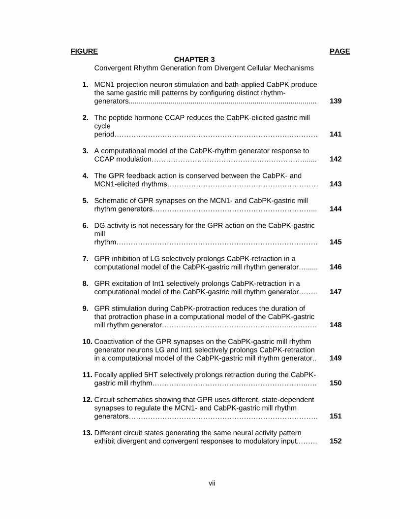
FIGURE CHAPTER 3
Convergent Rhythm Generation from Divergent Cellular Mechanisms
1. MCN1 projection neuron stimulation and bath-applied CabPK produce the same gastric mill patterns by configuring distinct rhythm-generators..............................................................................................
2. The peptide hormone CCAP reduces the CabPK-elicited gastric mill cycle period……………………………………………………………….…………
3. A computational model of the CabPK-rhythm generator response to CCAP modulation……………………………………….………………......
4. The GPR feedback action is conserved between the CabPK- and MCN1-elicited rhythms………………………………………………………
5. Schematic of GPR synapses on the MCN1- and CabPK-gastric mill rhythm generators…………………………………………………………...
6. DG activity is not necessary for the GPR action on the CabPK-gastric mill rhythm…………………………………………………………………………
7. GPR inhibition of LG selectively prolongs CabPK-retraction in a
computational model of the CabPK-gastric mill rhythm generator…...... 8. GPR excitation of Int1 selectively prolongs CabPK-retraction in a
computational model of the CabPK-gastric mill rhythm generator…….. 9. GPR stimulation during CabPK-protraction reduces the duration of
that protraction phase in a computational model of the CabPK-gastric mill rhythm generator……………………………………………..…………
10. Coactivation of the GPR synapses on the CabPK-gastric mill rhythm
generator neurons LG and Int1 selectively prolongs CabPK-retraction in a computational model of the CabPK-gastric mill rhythm generator..
11. Focally applied 5HT selectively prolongs retraction during the CabPK-
gastric mill rhythm………………………………………………………..…. 12. Circuit schematics showing that GPR uses different, state-dependent
synapses to regulate the MCN1- and CabPK-gastric mill rhythm generators…………………………………………………………………….
13. Different circuit states generating the same neural activity pattern
exhibit divergent and convergent responses to modulatory input..…….
PAGE 139 141 142 143 144 145 146 147 148 149 150 151 152
viii
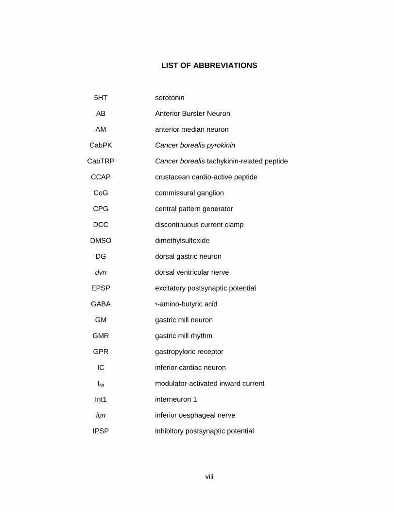
LIST OF ABBREVIATIONS
5HT
AB
AM
CabPK
CabTRP
CCAP
CoG
CPG
DCC
DMSO
DG
dvn
EPSP
GABA
GM
GMR
GPR
IC
IMI
Int1
ion
IPSP
serotonin
Anterior Burster Neuron
anterior median neuron
Cancer borealis pyrokinin
Cancer borealis tachykinin-related peptide
crustacean cardio-active peptide
commissural ganglion
central pattern generator
discontinuous current clamp
dimethylsulfoxide
dorsal gastric neuron
dorsal ventricular nerve
excitatory postsynaptic potential
ᵞ-amino-butyric acid
gastric mill neuron
gastric mill rhythm
gastropyloric receptor
inferior cardiac neuron
modulator-activated inward current
interneuron 1
inferior oesphageal nerve
inhibitory postsynaptic potential
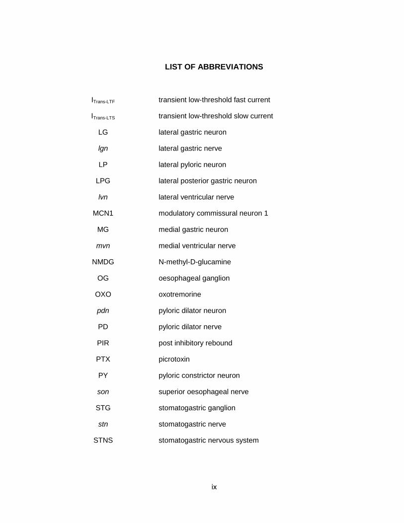
ix
LIST OF ABBREVIATIONS
ITrans-LTF
ITrans-LTS
LG
lgn
LP
LPG
lvn
MCN1
MG
mvn
NMDG
OG
OXO
pdn
PD
PIR
PTX
PY
son
STG
stn
STNS
transient low-threshold fast current
transient low-threshold slow current
lateral gastric neuron
lateral gastric nerve
lateral pyloric neuron
lateral posterior gastric neuron
lateral ventricular nerve
modulatory commissural neuron 1
medial gastric neuron
medial ventricular nerve
N-methyl-D-glucamine
oesophageal ganglion
oxotremorine
pyloric dilator neuron
pyloric dilator nerve
post inhibitory rebound
picrotoxin
pyloric constrictor neuron
superior oesophageal nerve
stomatogastric ganglion
stomatogastric nerve
stomatogastric nervous system
x
LIST OF ABBREVIATIONS
TG
TTX
VCN
VD
thoracic ganglion
tetrodotoxin
ventral cardiac neurons
ventricular dilator
1
CHAPTER 1
INTRODUCTION
Neural Circuit Modulation
The main goal of my thesis is to elucidate novel mechanistic insights
regarding neural circuit modulation, and how different modulatory states vary in
their sensitivity to hormonal inputs and sensory feedback.
Neural circuits are flexible constructs that exhibit short- and long-term
changes in dynamics in response to synaptic- and paracrine/endocrine-mediated
metabotropic actions (Getting, 1989; Marder, 2012). Synaptic inputs are subject
to homo- or heterosynaptic influences that result in processes such as synaptic
depression, facilitation and potentiation. Metabotropic (often termed modulatory)
actions provide the opportunity for state-dependence, wherein different
metabotropic actions configure different circuit states from the same network
(Kupfermann, 1979; Getting, 1989; Doi and Ramirez, 2008; Marder, 2012;
Nusbaum and Blitz, 2012). These latter inputs commonly act through
intracellular signaling cascades (e.g. G-protein signaling) to produce cellular- and
circuit-level changes that persist well past the initiating event, lasting for minutes,
hours, or even days.
Neuromodulation is a key mechanism for achieving behavioral states in all
organisms (Köhler et al., 2011; Lee and Dan, 2012; Taghert and Nitabach, 2012;
Patton and Mistlberger, 2013). This state involves the coordination of cellular
and network properties for the purpose of achieving some goal. For example, a
collection of neuromodulatory peptides coordinates feeding behaviors in
2
mammals (Jobst et al., 2004). While the importance of neuromodulation is
appreciated in all systems, there are still only a few systems sufficiently
accessible to study the consequences of neuromodulation in a cellular and circuit
context.
Central pattern generator (CPG) networks provide convenient systems for
studying neuromodulation (Marder, 2012; Nusbaum and Blitz, 2012). These
specialized neural circuits produce the rhythmic neuronal activity patterns that
underlie rhythmic behaviors (e.g. locomotion, respiration, mastication), and they
continue to generate these rhythmic activity patterns even after isolation from the
rest of the nervous system. This allows researchers to have better access for
recording and manipulating the network neurons and to more readily record the
physiologically relevant network activity in the context of different experimental
manipulations. Experimental results from small systems have provided insight
into larger CPG networks, as highlighted by studies showing that CPGs in all
animals, regardless of the particular rhythmic behavior, share several general
operating principles (Marder and Calabrese, 1996; Marder, 2012). For instance,
as indicated above, CPGs can generate at least a basic form of their in vivo
pattern even in isolation from the rest of the CNS. Additional shared principles
include CPGs generating rhythmic activity in response to non-rhythmic input, and
the fact that they are multi-functional (i.e. one network generates different activity
patterns in response to different modulatory inputs). Lastly, in the few cases
where sufficient information is available, it appears common for CPGs to be
functionally subdivided into a circuit responsible for rhythm generation and one
3
responsible for pattern generation (Guertin, 2009). These two functions can be
served by separate or overlapping sets of neurons (Guertin, 2009).
Modulatory inputs select particular output patterns from a neural circuit
(Dickinson, 2006; Briggman and Kristan, 2008; Doi and Ramirez, 2008; Harris-
Warrick, 2011; Marder, 2012). They do so by altering the synaptic and ionic
conductances of circuit neurons. These actions cause changes in the cellular
properties and synaptic dynamics of the affected neurons, enabling generation of
specific output patterns. It is appreciated in many systems that different
neuromodulators can produce distinct output patterns from the same neural
circuit (Fig. 1) (Getting, 1989; Stein, 2009; Marder, 2012; Nusbaum and Blitz,
2012). However, via a variety of mechanisms, different neuromodulators can
also elicit the same output pattern from a particular network (Fig. 1) (Di Prisco et
al., 2000; Korn and Faber, 2005; Saideman et al., 2007b; Derjean et al., 2010;
Doi and Ramirez, 2010; White and Nusbaum, 2011).
As suggested above, distinct circuit states do not necessarily produce
different output patterns. At the single neuron level, modulation of different sets
of ionic conductances can produce similar cellular consequences (Swensen and
Bean, 2005; Goaillard et al., 2009). This phenomenon also holds true at the level
of neural circuits, where different combinations of ionic and synaptic
conductances can produce identical output patterns (Golowasch et al., 1999;
Prinz et al., 2004). Obviously, this level of understanding requires access to ionic
and synaptic conductances of identified circuit neurons, the ability to selectively
activate particular input pathways, and comprehensive monitoring of the resulting
4
circuit output. There remain few systems in which these criteria can be fulfilled.
For this reason, my work was performed in a very well-defined and uniquely
accessible system called the stomatogastric nervous system (STNS) of decapod
crustaceans (Fig. 2) (Marder & Bucher, 2007; Stein, 2009).
The Cancer borealis stomatogastric nervous system
The STNS is an extension of the CNS that controls the feeding-associated
behaviors of the foregut. The decapod crustacean foregut has four distinct
structures, including the oesophagus (swallows food), cardiac sac (stores food),
gastric mill (chews food), and pylorus (filters chewed food) (Fig. 2) (Johnson and
Hooper, 1992). During feeding, swallowed food passes through the oesophagus
into the cardiac sac, where it is stored. The food is then squeezed into the
gastric mill compartment where it is chewed by the rhythmic movements of the
paired lateral teeth and unpaired medial tooth. Once food particles are
sufficiently small, they pass through the pylorus, which is continually pumping in
a series of front to rearward peristaltic waves, and into the midgut for further
digestion and absorption. These behaviors are coordinately executed by the
CPG circuits within the STNS.
The STNS is composed of four ganglia plus their connecting and
peripheral nerves (Fig. 3) (Marder and Bucher, 2007; Stein, 2009). The ganglia
include the paired commissural ganglia (CoGs: in C. borealis, contains ~600
neurons), oesophageal ganglion (OG: in C. borealis, contains 14 neurons), and
5
stomatogastric ganglion (STG: in C. borealis, contains 26 neurons) (Kilman and
Marder, 1996). Most CPG studies in this system focus on the STG, where the
gastric mill and pyloric circuits are located (see below). The CoGs and OG
contain, among other functional types of neurons, the cell bodies of projection
neurons that regulate/modulate the STG neurons (Coleman et al., 1992).
The STG circuits are very accessible, such that all STG neurons are identified
(Marder and Bucher, 2007). In the crab Cancer borealis, where my work was
performed, nearly all of the STG neurons (22 of 26) contribute to the gastric mill
and/or pyloric CPG. Among these 22 neurons are 13 different neuron types, with
9 of them present as single copies and 4 present as multiple, apparently
equivalent copies (2-5 copies, depending on the neuron type) (Fig. 4). The
neuronal cell bodies in the STG are organized in a single layer surrounding a
central neuropil (Fig. 4). Once the glial sheath that covers the STG is removed,
sharp electrode recordings are readily obtained from the relatively large STG
neuron somata, with minimal damage to the surrounding nervous system. Aside
from two STG interneurons (AB, Int1 neurons) which have relatively small
diameter somata (~35 µm), the STG neuron somata range from ~50 µm to ~120
µm in diameter. Most of the STG neurons are motor neurons that project their
axons through a stereotyped set of peripheral nerves to innervate the 30 muscles
of the foregut (Fig. 3). Consequently, their action potentials are readily recorded
extracellularly as they propagate through these peripheral nerves. Dissecting
6
and recording from smaller nerve branches enables the activity of individual
neuron types to be recorded in isolation (e.g. Fig. 5).
The aforementioned level of accessibility has enabled an extensive
characterization of this system. Not only are each of the 26 STG neurons
physiologically-identified in C. borealis (as well as the related crab C. pagurus
and two lobsters, Panulirus interruptus and Homarus americanus), but so are
their transmitters and synapses (Marder and Bucher, 2007). Additionally, the
sensitivity of individual STG neurons to various applied neuromodulators,
including amines, muscarinic agonists and numerous neuropeptides, as well as
several identified sensory and projection neurons are established (Dickinson,
2006; Marder, 2012; Blitz and Nusbaum, 2011; Nusbaum and Blitz, 2012).
As is the case for CPG networks in other systems, both the gastric mill
and pyloric CPGs can be subdivided into core rhythm generator and pattern
generator modules. In each case, the rhythm generator neurons also contribute
to pattern generation. The pyloric rhythm generator is a pacemaker-driven,
electrically-coupled ensemble whose core includes the AB and PD neurons
(Marder and Bucher, 2007). The core gastric mill rhythm generator is network-
driven, and includes the reciprocally inhibitory neurons LG and Int1 (Coleman et
al., 1995; Bartos et al., 1999; Saideman et al., 2007b). A few additional, different
neurons also contribute to gastric mill rhythm generation under different
modulatory states. This work focuses on the gastric mill rhythm generator during
two specific modulatory states, as described below.
7
The Gastric Mill Rhythm
All characterized gastric mill rhythms in C. borealis require activation of
the half-center formed by reciprocal inhibitory synapses between LG and Int1
(Fig. 4) (Bartos et al., 1999; Saideman et al., 2007b; White and Nusbaum, 2011).
The term “half-center” indicates pairs or populations of reciprocally inhibitory
neurons which, under appropriate conditions, generate a rhythmic repeating
bursting pattern during which each neuron or population is active for ~half of
each cycle (Marder and Calabrese, 1996). Prior to modulation, both in vivo and
in the isolated STNS, the LG/Int1 half-center is asymmetric, with LG being silent
and Int1 exhibiting a pyloric-timed activity pattern (Fig. 5) (Bartos et al., 1999;
Beenhakker and Nusbaum, 2004; Blitz et al., 2004; Diehl et al., 2013). Int1
activity is self-generated, enabling it to fire tonically at ~10 Hz, with this activity
pattern being rhythmically interrupted by synaptic inhibition from the pyloric
pacemaker neuron AB (Bartos et al., 1999; Saideman et al., 2007b). Each Int1
action potential elicits an ionotropic, glutamatergic inhibitory post-synaptic
potential (IPSP) in the LG neuron. In normal saline, each pyloric-timed Int1 burst
causes an LG hyperpolarization. When LG is released from each of these
inhibitory events, it repolarizes to its subthreshold membrane potential.
Neuromodulators that elicit a gastric mill rhythm do so by activating LG
and thereby balancing the LG/Int1 half-center (i.e. enabling them to burst
rhythmically in an alternating pattern) (Bartos et al., 1999; Saideman et al.,
8
2007b). The rhythmic alternation of Int1 and LG activity, and their synaptic
actions on the other gastric mill motor neurons, then coordinates a biphasic
chewing pattern (Bartos et al., 1999; Saideman et al., 2007b). In the best
characterized versions of the gastric mill rhythm, the basic operation of the
activated LG-Int1 half-center involves LG acquiring the ability (from a modulatory
input) to periodically escape or rebound from Int1 inhibition and fire a self-
terminating burst. The gastric mill pattern is, therefore, a biphasic alternation
between protraction (LG-timed) and retraction (Int1-timed) neurons, driving the
rhythmically alternating protraction and retraction movements of the teeth that
define chewing behavior. This work will focus on two gastric mill rhythm
generating mechanisms that result from two distinct modulatory input pathways,
including modulatory commissural neuron 1 (MCN1: Fig. 5) and the CabPK
(Cancer borealis pyrokinin) peptide-containing projections neurons.
Cancer borealis pyrokinins (CabPKs)
The CabPKs are two bioactive peptides found in the STNS (Saideman et
al., 2007a). They include CabPK I (TNFAFSPRLamide) and CabPK II
(SGGFAFSPRLamide). The CabPKs are PK/PBAN peptide family members,
which all contain the FXPRL amino acid sequence at their n-terminus (Rafaeli,
2009). Antisera raised against a shrimp PK peptide (pevpyrokinin) were used to
localize the CabPKs. These peptides were thereby immunolocalized in both a
9
neurohemal structure, called the pericardial organ, and within the STNS
(Saideman et al., 2007a).
CabPK immunoreactive (CabPK-IR) neuronal somata in the C. borealis STNS
included 2 or 3 STG-innervating projection neurons within the CoGs (Saideman
et al., 2007a). The STG-innervating CabPK-IR projection neurons project
through the bilateral superior oesophageal nerve (son) and unpaired
stomatogastric nerve (stn) to innervate the STG (Fig. 5). Within the STG,
CabPK-IR was limited to elaborate neuropilar processes, presumably
representing the axonal terminations of the CabPK projection neurons. No STG
somata were CabPK-IR (Saideman et al., 2007a).
Despite being localized to the CoGs, the CabPK-projection neurons
remain to be physiologically identified. Moreover, there is currently no way to
selectively activate them extracellularly, because all but two of the 15-20 CoG
projection neurons that innervate the STG project through the sons (Coleman et
al., 1992). Therefore, in my experiments CabPK neuron activity is simulated by
CabPK bath application. This approach has been used successfully in previous
studies with other neuropeptides. For example, bath application of the
neuropeptide proctolin (10-6 M) mimics the influence of selective stimulation of
the proctolin-containing projection neuron MPN (modulatory proctolin neuron) on
the C. borealis pyloric rhythm (Nusbaum and Marder, 1989ab). However, it is
important to indicate that the actions of bath-applied neuropeptide do not always
10
mimic those of the neuron(s) containing that peptide (Blitz and Nusbaum, 1999;
Blitz et al., 1999). There are several reasons for the latter situation, including
synaptic specificity, extracellular peptidase activity, actions of cotransmitters and
receptor desensitization (Wood et al., 2000; Nusbaum et al., 2001; Nusbaum,
2002; Wood and Nusbaum, 2002).
Convergent Gastric Mill Output Patterns, Divergent Mechanisms
CabPK bath-application at concentrations (≥ 10-7 M) that likely mimic
neuronal release activate a gastric mill rhythm (Saideman et al., 2007ab).
Surprisingly, CabPK application and stimulation of the CoG projection neuron
MCN1 elicit the same gastric mill motor pattern, even though MCN1 neither
contains CabPK nor is necessary for the CabPK-elicited gastric mill rhythm
(Saideman et al., 2007ab). In contrast, other pathways that activate the gastric
mill rhythm elicit distinct gastric mill motor patterns (Beenhakker and Nusbaum,
2004; Blitz et al., 2004; Christie et al., 2004; White and Nusbaum, 2011). Not
only are the basic CabPK- and MCN1-elicited gastric mill rhythm parameters the
same (e.g. retraction duration, protraction duration, cycle period), but so are
additional details of these motor patterns (Saideman et al., 2007b). For example,
the firing rates and activity patterns of all neurons measured were the same, as
were most phase relationships, during the CabPK- and MCN1-elicited gastric mill
patterns. Additionally, the same two gastric mill motor neurons (GM, AM) were
11
not activated by either CabPK or MCN1, even though they are activated by other
pathways.
Given the extent of the concurrence between these two gastric mill
rhythms, it was surprising that the network states and even aspects of the basic
gastric mill rhythm-generating mechanism varied when CabPK was applied or
MCN1 was stimulated (Fig. 6) (Saideman et al., 2007b). For example, a pivotal
aspect of MCN1-driven gastric mill rhythm generation is a slow MCN1-elicited
metabotropic (peptidergic) excitation of LG that is regulated by feedback
inhibition from LG (Bartos et al., 1999; DeLong et al., 2009). Specifically, MCN1
excites LG through the release of C. borealis tachykinin-related peptide Ia
(CabTRP Ia). MCN1-released CabTRP Ia causes a slow depolarization of LG by
activating the voltage-dependent, modulator-activated inward current (IMI)
(DeLong et al., 2009). When IMI becomes sufficiently large, LG overcomes the
inhibition that it receives from Int1 and fires a burst. While LG is active, it
presynaptically inhibits the MCN1 terminals in the STG (MCN1STG), reducing
MCN1 transmitter release and initiating a decay of IMI availability which eventually
terminates the LG burst. LG burst termination results in the resumption of MCN1
transmitter release and Int1 activity. Insofar as the feedback inhibition onto
MCN1STG and its consequences for MCN1 signaling are necessary for gastric mill
rhythm generation, MCN1STG itself is part of this version of the gastric mill rhythm
generator, as well as being its activator. Additionally, the pyloric pacemaker
neuron AB regulates the MCN1-gastric mill cycle period, although it is not
necessary for rhythm generation (Bartos et al., 1999).
12
The CabPK-gastric mill rhythm generating mechanism cannot be the
same as that used by MCN1 for several reasons (Saideman et al., 2007b). First,
the core neurons involved in rhythm generation are different. MCN1 is not
involved in the CabPK-gastric mill rhythm, nor does it release the CabPK peptide.
Second, the pyloric-timed inhibitory synapse from the AB neuron onto Int1 is
necessary only for CabPK-gastric mill rhythm generation. Third, the retraction
motor neuron DG regulates the CabPK-gastric mill rhythm, but not the MCN1-
gastric mill rhythm.
My dissertation research focused on the CabPK-gastric mill rhythm
generating mechanism and its sensitivity to hormonal and sensory influences,
including a comparison to the previously determined comparable conditions (and
underlying mechanisms) during the MCN1-gastric mill rhythm. As presented in
the following chapters, my findings establish that (1) distinct modulators can
produce convergent output patterns by recruiting different ionic conductances to
perform overlapping roles in rhythm generation, (2) the same ionic conductance
in a single neuron can contribute to rhythm generation in a state-dependent
manner, (3) circuit states that produce the same output pattern can flexibly
respond to hormonal input (Fig. 7), (4) distinct circuit states can generate
invariant responses to sensory feedback despite using different synaptic
mechanisms (Fig. 7). As discussed in the following chapters, each of these
findings provides novel insights into the degree of flexibility intrinsic to
rhythmically active neuronal circuits. Based on the fact that many previous
findings regarding circuit dynamics in the STNS have been subsequently

13
established in other model invertebrate and vertebrate systems, it is likely that
the present findings will also resonate with the operation of other such networks.
14
REFERENCES
Bartos M, Manor Y, Nadim F, Marder E, Nusbaum MP (1999) Coordination of fast and slow rhythmic neuronal circuits. J Neurosci 19:6650–6660.
Beenhakker MP, Kirby MS, Nusbaum MP (2007) Mechanosensory gating of proprioceptor input to modulatory projection neurons. J Neurosci 27:14308–14316.
Beenhakker MP, Nusbaum MP (2004) Mechanosensory activation of a motor circuit by coactivation of two projection neurons. J Neurosci 24:6741–6750.
Blitz DM, Beenhakker MP, Nusbaum MP (2004) Different sensory systems share projection neurons but elicit distinct motor patterns. J Neurosci 24:11381–11390.
Blitz DM, Christie a E, Coleman MJ, Norris BJ, Marder E, Nusbaum MP (1999) Different proctolin neurons elicit distinct motor patterns from a multifunctional neuronal network. J Neurosci 19:5449–5463.
Briggman KL, Kristan WB (2008) Multifunctional pattern-generating circuits. Annu Rev Neurosci 31:271–294.
Christie AE, Stein W, Quinlan JE, Beenhakker MP, Marder E, Nusbaum MP (2004) Actions of a histaminergic/peptidergic projection neuron on rhythmic motor patterns in the stomatogastric nervous system of the crab Cancer borealis. J Comp Neurol 469:153–169.
Coleman MJ, Nusbaum MP, Cournil I, Claiborne BJ (1992) Distribution of modulatory inputs to the stomatogastric ganglion of the crab, Cancer borealis. J Comp Neurol 325:581–594.
DeLong ND, Beenhakker MP, Nusbaum MP (2009) Presynaptic inhibition selectively weakens peptidergic cotransmission in a small motor system. J Neurophysiol 102:3492–3504.
Derjean D, Moussaddy A, Atallah E, St-Pierre M, Auclair F, Chang S, Ren X, Zielinski B, Dubuc R (2010) A novel neural substrate for the transformation of olfactory inputs into motor output. PLoS Biol 8:e1000567.
Dickinson PS (2006) Neuromodulation of central pattern generators in invertebrates and vertebrates. Curr Opin Neurobiol 16:604–614.
Doi A, Ramirez J-M (2008) Neuromodulation and the orchestration of the respiratory rhythm. Respir Physiol Neurobiol 164:96–104.
15
Doi A, Ramirez J-M (2010) State-dependent interactions between excitatory neuromodulators in the neuronal control of breathing. J Neurosci 30:8251–8262.
Getting PA (1989) Emerging principles governing the operation of neural networks. Annu Rev Neurosci 12:185–204.
Goaillard J-M, Taylor AL, Schulz DJ, Marder E (2009) Functional consequences of animal-to-animal variation in circuit parameters. Nat Neurosci 12:1424–1430.
Golowasch J, Abbott LF, Marder E (1999) Activity-dependent regulation of potassium currents in an identified neuron of the stomatogastric ganglion of the crab Cancer borealis. J Neurosci 19:RC33.
Guertin P a (2009) The mammalian central pattern generator for locomotion. Brain Res Rev 62:45–56.
Harris-Warrick RM (2011) Neuromodulation and flexibility in Central Pattern Generator networks. Curr Opin Neurobiol 21:685–692.
Jobst EE, Enriori PJ, Cowley M a (2004) The electrophysiology of feeding circuits. Trends Endocrinol Metab 15:488–499.
Johnson BR, Hooper SL (1992) Johnson and Hooper (1992) Overview of STNS.pdf. In: Dynamic Biological Networks: The Stomatogastric Nervous System (Harris-Warrick RM, Marder E, Selverston AI, Moulins M, eds), pp.1–30. Cambridge, MA: MIT Press.
Kilman VL, Marder E (1996) Ultrastructure of the stomatogastric ganglion neuropil of the crab, Cancer borealis. J Comp Neurol 374:362–375.
Kirby MS, Nusbaum MP (2007) Peptide hormone modulation of a neuronally modulated motor circuit. J Neurophysiol 98:3206–3220.
Köhler CA, da Silva WC, Benetti F, Bonini JS (2011) Histaminergic mechanisms for modulation of memory systems. Neural Plast 2011:328602.
Korn H, Faber DS (2005) The Mauthner cell half a century later: a neurobiological model for decision-making? Neuron 47:13–28.
Kupfermann I (1979) Modulatory actions of neurotransmitters. Annu Rev Neurosci 2:447–465.
Lee S-H, Dan Y (2012) Neuromodulation of brain states. Neuron 76:209–222.
16
Marder E (2012) Neuromodulation of neuronal circuits: back to the future. Neuron 76:1–11.
Marder E, Bucher D (2007) Understanding circuit dynamics using the stomatogastric nervous system of lobsters and crabs. Annu Rev Physiol 69:291–316.
Marder E, Calabrese RL (1996) Principles of rhythmic motor pattern generation. Physiol Rev 76:687–717.
Nusbaum MP, Blitz DM (2012) Neuropeptide modulation of microcircuits. Curr Opin Neurobiol 22:592–601.
Nusbaum MP, Blitz DM, Swensen AM, Wood D, Marder E (2001) The roles of co-transmission in neural network modulation. Trends Neurosci 24:146–154.
Patton DF, Mistlberger RE (2013) Circadian adaptations to meal timing: neuroendocrine mechanisms. Front Neurosci 7:185.
Prinz AA, Bucher D, Marder E (2004) Similar network activity from disparate circuit parameters. Nat Neurosci 7:1345–1352.
Di Prisco G V, Pearlstein E, Le Ray D, Robitaille R, Dubuc R (2000) A cellular mechanism for the transformation of a sensory input into a motor command. J Neurosci 20:8169–8176.
Rafaeli A (2009) Pheromone biosynthesis activating neuropeptide (PBAN): regulatory role and mode of action. Gen Comp Endocrinol 162:69–78.
Saideman SR, Ma M, Kutz-Naber KK, Cook A, Torfs P, Schoofs L, Li L, Nusbaum MP (2007a) Modulation of rhythmic motor activity by pyrokinin peptides. J Neurophysiol 97:579–595.
Saideman SR, Blitz DM, Nusbaum MP (2007b) Convergent motor patterns from divergent circuits. J Neurosci 27:6664–6674.
Stein W (2009) Modulation of stomatogastric rhythms. J Comp Physiol A Neuroethol Sens Neural Behav Physiol 195:989–1009.
Swensen AM, Bean BP (2005) Robustness of burst firing in dissociated purkinje neurons with acute or long-term reductions in sodium conductance. J Neurosci 25:3509–3520.
Taghert PH, Nitabach MN (2012) Peptide neuromodulation in invertebrate model systems. Neuron 76:82–97.
17
White RS, Nusbaum MP (2011) The same core rhythm generator underlies different rhythmic motor patterns. J Neurosci 31:11484–11494.
Wood DE, Nusbaum MP (2002) Extracellular peptidase activity tunes motor pattern modulation. J Neurosci 22:4185–4195.
Wood DE, Stein W, Nusbaum MP (2000) Projection neurons with shared cotransmitters elicit different motor patterns from the same neural circuit. J Neurosci 20:8943–8953.
18
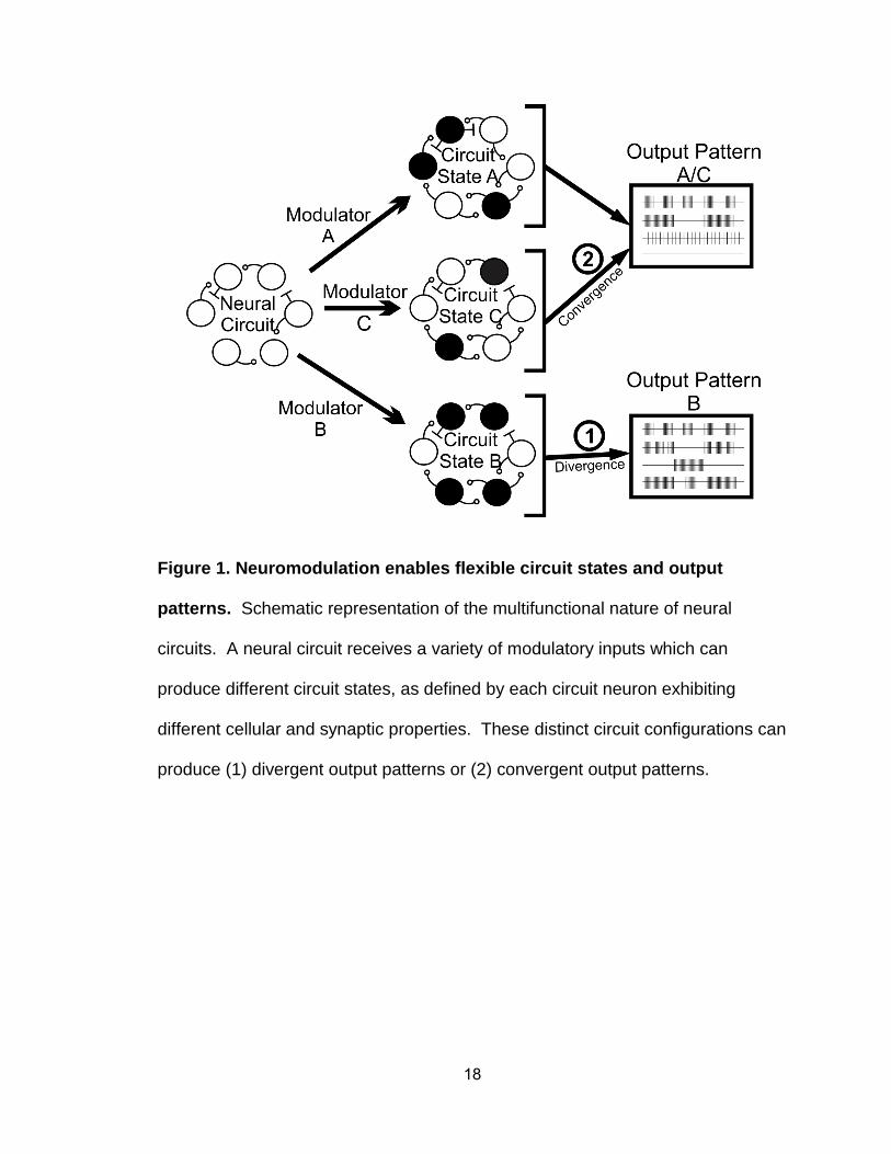
Figure 1. Neuromodulation enables flexible circuit states and output
patterns. Schematic representation of the multifunctional nature of neural
circuits. A neural circuit receives a variety of modulatory inputs which can
produce different circuit states, as defined by each circuit neuron exhibiting
different cellular and synaptic properties. These distinct circuit configurations can
produce (1) divergent output patterns or (2) convergent output patterns.
19
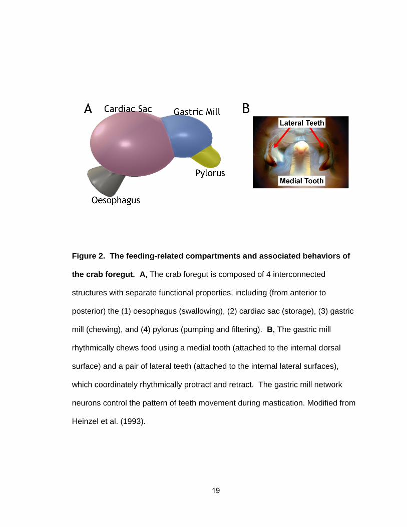
Figure 2. The feeding-related compartments and associated behaviors of
the crab foregut. A, The crab foregut is composed of 4 interconnected
structures with separate functional properties, including (from anterior to
posterior) the (1) oesophagus (swallowing), (2) cardiac sac (storage), (3) gastric
mill (chewing), and (4) pylorus (pumping and filtering). B, The gastric mill
rhythmically chews food using a medial tooth (attached to the internal dorsal
surface) and a pair of lateral teeth (attached to the internal lateral surfaces),
which coordinately rhythmically protract and retract. The gastric mill network
neurons control the pattern of teeth movement during mastication. Modified from
Heinzel et al. (1993).
20
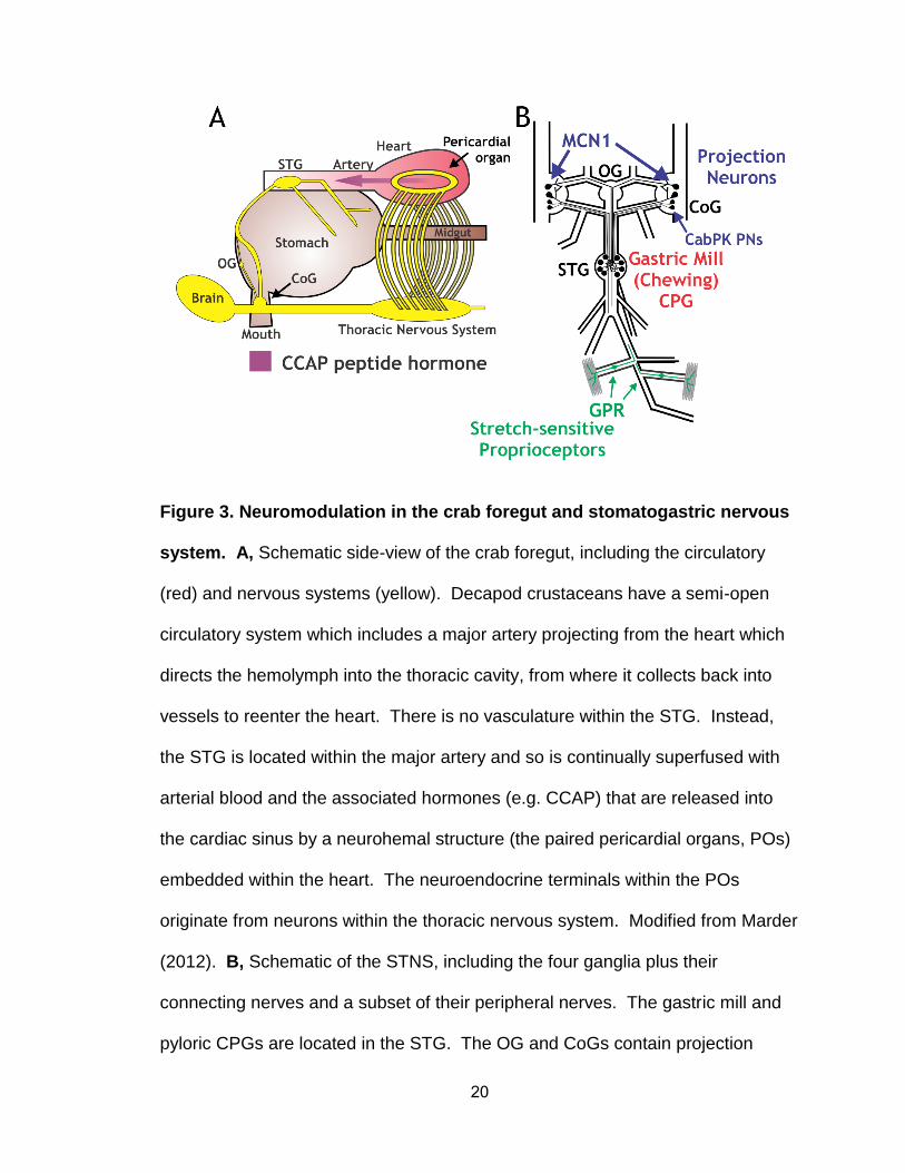
Figure 3. Neuromodulation in the crab foregut and stomatogastric nervous
system. A, Schematic side-view of the crab foregut, including the circulatory
(red) and nervous systems (yellow). Decapod crustaceans have a semi-open
circulatory system which includes a major artery projecting from the heart which
directs the hemolymph into the thoracic cavity, from where it collects back into
vessels to reenter the heart. There is no vasculature within the STG. Instead,
the STG is located within the major artery and so is continually superfused with
arterial blood and the associated hormones (e.g. CCAP) that are released into
the cardiac sinus by a neurohemal structure (the paired pericardial organs, POs)
embedded within the heart. The neuroendocrine terminals within the POs
originate from neurons within the thoracic nervous system. Modified from Marder
(2012). B, Schematic of the STNS, including the four ganglia plus their
connecting nerves and a subset of their peripheral nerves. The gastric mill and
pyloric CPGs are located in the STG. The OG and CoGs contain projection
21
neurons (e.g. MCN1, CabPK PNs) that innervate and modulate the STG
networks. GPRs are a bilaterally symmetric pair of proprioceptors that modulate
the STG networks in response to changes in muscle length and tension.
Abbreviations – CCAP, crustacean cardio-active peptide; CoG, commissural
ganglia; CPG, central pattern generator; GPR, gastro-pyloric receptor; OG,
oesophageal ganglion; STG, stomatogastric ganglion.
22
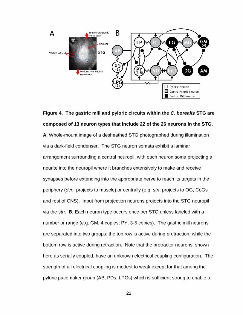
Figure 4. The gastric mill and pyloric circuits within the C. borealis STG are
composed of 13 neuron types that include 22 of the 26 neurons in the STG.
A, Whole-mount image of a desheathed STG photographed during illumination
via a dark-field condenser. The STG neuron somata exhibit a laminar
arrangement surrounding a central neuropil, with each neuron soma projecting a
neurite into the neuropil where it branches extensively to make and receive
synapses before extending into the appropriate nerve to reach its targets in the
periphery (dvn: projects to muscle) or centrally (e.g. stn: projects to OG, CoGs
and rest of CNS). Input from projection neurons projects into the STG neuropil
via the stn. B, Each neuron type occurs once per STG unless labeled with a
number or range (e.g. GM, 4 copies; PY, 3-5 copies). The gastric mill neurons
are separated into two groups: the top row is active during protraction, while the
bottom row is active during retraction. Note that the protractor neurons, shown
here as serially coupled, have an unknown electrical coupling configuration. The
strength of all electrical coupling is modest to weak except for that among the
pyloric pacemaker group (AB, PDs, LPGs) which is sufficient strong to enable to

23
them to oscillate together. Abbreviations: AB, anterior burster; AM, anterior
median; GM, gastric mill; IC, inferior cardiac; Int1, interneuron 1; LG, lateral
gastric; LP, lateral pyloric; LPG, lateral posterior gastric; MG, medial gastric; PD,
pyloric dilator; PY, pyloric; VD, ventricular dilator.
24
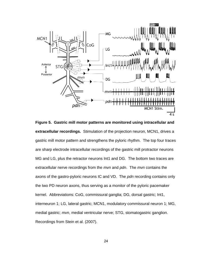
Figure 5. Gastric mill motor patterns are monitored using intracellular and
extracellular recordings. Stimulation of the projection neuron, MCN1, drives a
gastric mill motor pattern and strengthens the pyloric rhythm. The top four traces
are sharp electrode intracellular recordings of the gastric mill protractor neurons
MG and LG, plus the retractor neurons Int1 and DG. The bottom two traces are
extracellular nerve recordings from the mvn and pdn. The mvn contains the
axons of the gastro-pyloric neurons IC and VD. The pdn recording contains only
the two PD neuron axons, thus serving as a monitor of the pyloric pacemaker
kernel. Abbreviations: CoG, commissural ganglia; DG, dorsal gastric; Int1,
interneuron 1; LG, lateral gastric; MCN1, modulatory commissural neuron 1; MG,
medial gastric; mvn, medial ventricular nerve; STG, stomatogastric ganglion.
Recordings from Stein et al. (2007).
25
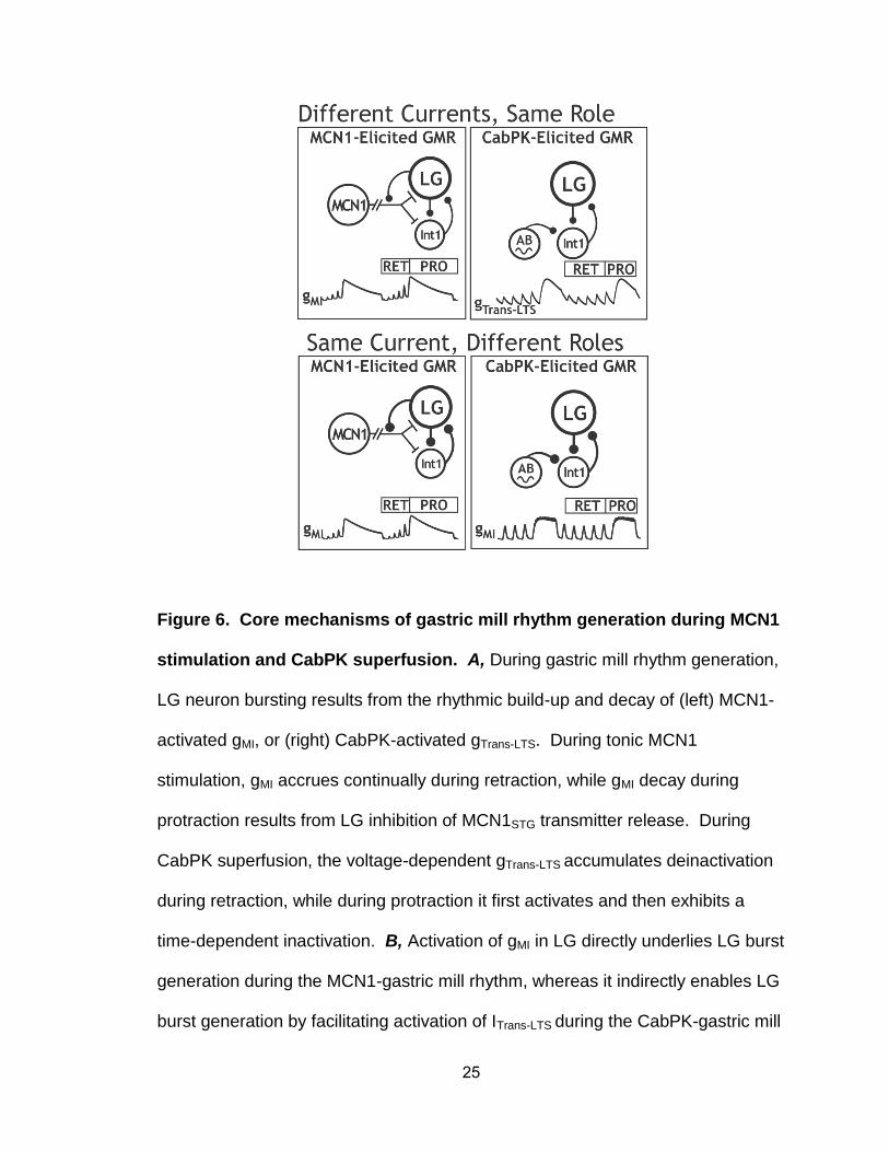
Figure 6. Core mechanisms of gastric mill rhythm generation during MCN1
stimulation and CabPK superfusion. A, During gastric mill rhythm generation,
LG neuron bursting results from the rhythmic build-up and decay of (left) MCN1-
activated gMI, or (right) CabPK-activated gTrans-LTS. During tonic MCN1
stimulation, gMI accrues continually during retraction, while gMI decay during
protraction results from LG inhibition of MCN1STG transmitter release. During
CabPK superfusion, the voltage-dependent gTrans-LTS accumulates deinactivation
during retraction, while during protraction it first activates and then exhibits a
time-dependent inactivation. B, Activation of gMI in LG directly underlies LG burst
generation during the MCN1-gastric mill rhythm, whereas it indirectly enables LG
burst generation by facilitating activation of ITrans-LTS during the CabPK-gastric mill

26
rhythm. Note the different gMI trajectories during protraction, which result from its
being both voltage- and synaptic inhibition-dependent during the MCN1-gastric
mill rhythm but only regulated by membrane potential during the CabPK-gastric
mill rhythm. Both panels from Rodriguez et al. (2013).
27
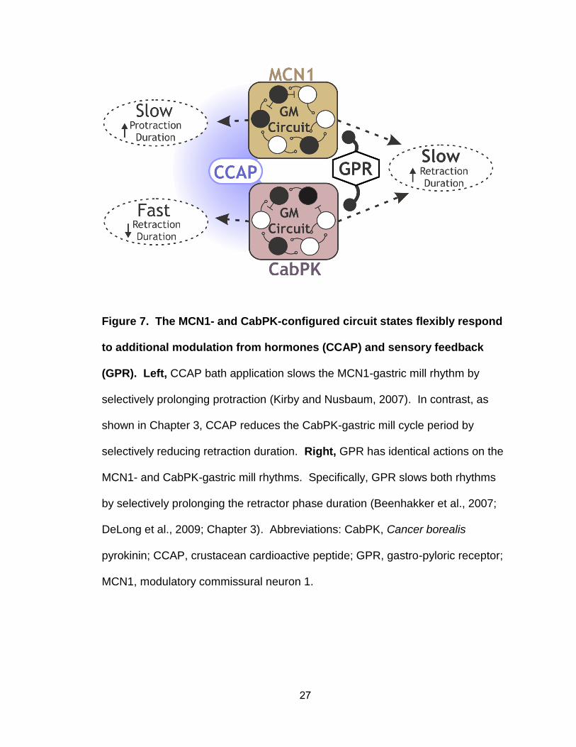
Figure 7. The MCN1- and CabPK-configured circuit states flexibly respond
to additional modulation from hormones (CCAP) and sensory feedback
(GPR). Left, CCAP bath application slows the MCN1-gastric mill rhythm by
selectively prolonging protraction (Kirby and Nusbaum, 2007). In contrast, as
shown in Chapter 3, CCAP reduces the CabPK-gastric mill cycle period by
selectively reducing retraction duration. Right, GPR has identical actions on the
MCN1- and CabPK-gastric mill rhythms. Specifically, GPR slows both rhythms
by selectively prolonging the retractor phase duration (Beenhakker et al., 2007;
DeLong et al., 2009; Chapter 3). Abbreviations: CabPK, Cancer borealis
pyrokinin; CCAP, crustacean cardioactive peptide; GPR, gastro-pyloric receptor;
MCN1, modulatory commissural neuron 1.
28
CHAPTER 2
Convergent Rhythm Generation from Divergent Cellular Mechanisms
Jason C. Rodriguez
Dawn M. Blitz
Michael P. Nusbaum
Published:
Journal of Neuroscience, 2013
46: 18047 - 18064
29
Sept 17, 2013
Convergent Rhythm Generation from Divergent Cellular Mechanisms
Jason C. Rodriguez
Dawn M. Blitz1
Michael P. Nusbaum*
Dept. of Neuroscience, 215 Stemmler Hall, Perelman School of Medicine, Univ.
of Pennsylvania, Philadelphia, PA 19104
1Current Address: Department of Biology, Miami University, 242 Pearson Hall,
Oxford, OH 45056
Running title: Rhythm generation by distinct mechanisms
Abstract: 250/250 Words
Introduction: 493/500 Words
Discussion: 1497/1500 Words
Pages: 61
30
Figures: 14
Tables: 2
*To whom correspondence should be addressed:
Michael P. Nusbaum, PhD
Dept. of Neuroscience
215 Stemmler Hall
Perelman School of Medicine, Univ. of Pennsylvania
Philadelphia, PA 19104-6074
Phone: (215) 898-1585
FAX: (215) 573-9050
Email: [email protected]
Acknowledgments: This work was supported by National Institute of
Neurological Disorders and Stroke Grant R37-NS 29436 (M.P.N.), National
Science Foundation Grant IOS-1153417 (D.M.B.), and the Behavioral and
Cognitive Neurosciences Training Grant T32-MH 17168.

31
Keywords: Stomatogastric, Postinhibitory Rebound, Modulation, Central Pattern
Generator, Reciprocal Inhibition
32
ABSTRACT
Different modulatory inputs commonly elicit distinct rhythmic motor patterns from
a central pattern generator (CPG), but they can instead elicit the same pattern.
We are determining the rhythm-generating mechanisms in this latter situation,
using the gastric mill (chewing) CPG in the crab (Cancer borealis) stomatogastric
ganglion where stimulating the projection neuron MCN1 or bath-applying CabPK
peptide elicits the same gastric mill motor pattern, despite configuring different
gastric mill circuits. In both cases, the core rhythm generator includes the same
reciprocally inhibitory neurons (LG, Int1), but the pyloric (food filtering) circuit
pacemaker neuron AB is additionally necessary only for CabPK rhythm
generation. MCN1 drives this rhythm generator by activating in LG the
modulator-activated inward current (IMI), which waxes and wanes periodically due
to phasic feedback inhibition of MCN1 transmitter release. Each buildup of IMI
enables LG to generate a self-terminating burst and thereby alternate with Int1
activity. Here we establish that CabPK drives gastric mill rhythm generation by
activating in LG IMI plus a slowly activating transient, low threshold inward current
(ITrans-LTS) that is voltage-, time- and Ca2+-dependent. Unlike MCN1, CabPK
maintains a steady IMI activation, causing a subthreshold depolarization in LG
that facilitates a periodic postinhibitory rebound (PIR) burst caused by the regular
buildup and decay of availability of ITrans-LTS. Thus, different modulatory inputs
can use different rhythm generating mechanisms to drive the same neuronal
rhythm. Additionally, the same ionic current (IMI) can play different roles under
these different conditions, while different currents (IMI, ITrans-LTS) can play the

33
same role.
34
INTRODUCTION
Different modulatory inputs enable individual neuronal networks to
generate different output patterns by changing the intrinsic and synaptic
properties of network neurons (Dickinson, 2006; Doi and Ramirez, 2008;
Briggman and Kristan, 2008; Rauscent et al., 2009; Harris-Warrick, 2011,
Marder, 2012). However, different modulatory inputs can also elicit the same
activity pattern from that network (Saideman et al., 2007b). Determining how
different modulatory pathways influence network activity is challenging, because
these different pathways can converge onto the same direct input(s) to a network
(Viana di Prisco et al., 2000; Korn and Faber, 2005; Derjean et al., 2010; White
and Nusbaum, 2011), comparably modulate the same network (Doi and Ramirez,
2010), distinctly alter multiple cellular and synaptic properties in the same circuit
neurons (MacLean et al., 2003; Prinz et al., 2004a; Goaillard et al., 2009;
Calabrese et al., 2011; Marder, 2012) and/or configure different circuits
(Saideman et al., 2007b). The cellular mechanisms underlying the last of these
processes are not determined in any system.
We are determining the cellular mechanisms that enable two differently
configured, network-driven central pattern generator (CPG) circuits to generate
the same biphasic motor pattern, using the isolated crab stomatogastric ganglion
(STG) (Marder and Bucher, 2007; Stein, 2009). These two gastric mill (chewing)
circuits are configured by the projection neuron MCN1 (modulatory commissural
neuron 1) and bath-applied CabPK (Cancer borealis pyrokinin) peptide
(Saideman et al., 2007a,b).
35
The core rhythm generator for both gastric mill circuits includes the
reciprocally inhibitory neurons LG (lateral gastric) and Int1 (interneuron 1).
Rhythmic MCN1 transmitter release is also necessary for the MCN1-gastric mill
rhythm, while the pyloric pacemaker neuron AB (anterior burster) is necessary for
the CabPK-rhythm. The cellular and synaptic mechanisms underlying MCN1-
gastric mill rhythm generation are established (Coleman et al., 1995; Bartos et
al., 1999; DeLong et al., 2009a,b). A key MCN1 rhythm-generating mechanism
is its activation of IMI (modulator-activated, voltage-dependent inward current) in
LG, which waxes and wanes periodically due to rhythmic feedback inhibition of
MCN1 transmitter release by LG. These events enable LG to periodically fire a
self-terminating burst and alternate with Int1 activity.
Here we identify two CabPK-activated currents in LG that are necessary
and sufficient for gastric mill rhythm generation. These currents include IMI and a
transient, low threshold, slowly-activating inward current (ITrans-LTS). ITrans-LTS
exhibits voltage- and time-dependent properties. CabPK-gastric mill rhythm
generation results from IMI providing a constant depolarizing drive that enables
periodic postinhibitory rebound (PIR) bursting, triggered by ITrans-LTS. The
rhythmic nature of the PIR burst generation results from the time- and voltage-
dependent properties of ITrans-LTS. Computational modeling and dynamic clamp
manipulations of these two currents support their necessity and sufficiency for
CabPK-gastric mill rhythm generation, and reveal that the pyloric rhythm (AB)-
timed influence on LG is necessary for triggering each PIR burst. Thus, distinct
rhythm-generating mechanisms enable distinct circuits to generate the same

36
rhythmic activity. Additionally, the same ionic current (IMI) plays a different role
under these two conditions, whereas different currents (IMI, ITrans-LTS) play a
comparable role.
37
METHODS
Animals. Male Jonah crabs (Cancer borealis) were purchased from commercial
suppliers (Fresh Lobster; Marine Biological Laboratory) and maintained in
aerated, filtered artificial seawater at 10 – 12° C. Animals were cold anesthetized
by packing in ice for at least 30 min before dissection, after which the foregut was
removed, in physiological saline at ~4° C, and the STNS isolated.
Solutions. C. borealis physiological saline contained (in mM): 440 NaCl, 26
MgCl2, 13 CaCl2, 11 KCl, 10 Trisma base, 5 maleic acid, 5 glucose, pH 7.4 – 7.6.
All preparations were superfused continuously with C. borealis saline (8 – 12° C).
CabPK-I or CabPK-II (Saideman et al., 2007a) (Biotechnology Center, Univ. of
Wisconsin, Madison, WI) was diluted from a stock solution (10-3 M) into
physiological saline or voltage clamp saline immediately before use. Bottles
containing C. borealis saline and CabPK saline were connected to the same
switching manifold for rapid solution changes. Oxotremorine (OXO: 10-5 M;
Sigma Chemical Co.), a muscarinic agonist, was applied in the same manner.
For voltage clamp experiments, tetrodotoxin (TTX: 10-7 M, Sigma),
picrotoxin (PTX: 10-5 M, Sigma) and tetraethylammonium chloride (TEACl: 10-2
M, Sigma) were added to C. borealis saline (i.e. voltage clamp saline). These
substances were used to suppress voltage-dependent Na+ currents (TTX),
glutamatergic inhibitory synaptic transmission (PTX), and a subset of K+ currents
(TEACl) (Marder and Eisen, 1984; Golowasch and Marder, 1992a). In some
38
experiments, the microelectrode was filled with a solution of CsCl (1 M; Sigma)
and TEACl (1 M) to additionally suppress a subset of K+ currents. To test the
sensitivity of CabPK-influenced currents to extracellular Na+, in some
experiments Na+ in the saline was replaced by NMDG+ (n-methyl, d-glucamine;
Fluka) (Golowasch and Marder, 1992). The NMDG+ was added to the solution
first and then neutralized with HCl before adding all other components.
Additionally, in some experiments we used flufenamic acid (FFA: 10-5 M; Sigma),
an inhibitor of ICAN (Ca2+-activated, non-specific cation current), dissolved in
dimethyl sulfoxide (DMSO) and added directly to Cancer saline. The final DMSO
concentration never exceeded 1%.
Electrophysiology. Electrophysiology experiments were performed using
standard techniques for this system (Beenhakker and Nusbaum, 2004). In brief,
the isolated STNS (Fig. 1A) was pinned into a silicone elastomer (Sylgard 184,
KR Anderson)-lined Petri dish. Extracellular nerve recordings were obtained
using pairs of stainless steel wire electrodes (reference and recording) whose
ends were pressed into the Sylgard-coated dish. A differential AC amplifier
(Model 1700: AM Systems) amplified the voltage difference between the
reference wire, in the main bath compartment, and the recording wire, isolated
with a section of an individual nerve from the main bath compartment by
petroleum jelly (Vaseline, Lab Safety Supply). This signal was then further
amplified and filtered (Model 410 Amplifier: Brownlee Precision). For
extracellular nerve stimulation, the pair of wires used to record nerve activity was
39
placed into a stimulus isolation unit (SIU 5: Astromed/Grass Instruments)
connected to a stimulator (Model S88: Astromed/Grass Instruments).
For current clamp experiments, intrasomatic recordings of STG neurons
were made with sharp glass microelectrodes (15 – 30 MΩ) filled with either
K2SO4 (0.6 M) plus KCl (10 mM) or KCl (1 M). For voltage clamp experiments,
neurons were impaled with separate recording and current injection electrodes.
The recording electrode, in most experiments, contained CsCl (1 M) and TEACl
(1 M) to suppress additional K+ currents. The current injection electrode was
filled with KCl (2.5 M). All intracellular recordings were amplified using Axoclamp
900A amplifiers (Molecular Devices) in bridge mode or discontinuous current
clamp mode (2 – 5 kHz sampling rate) and digitized at 5 kHz using a Micro 1401
data acquisition interface and Spike2 software (Cambridge Electronic Design).
To facilitate intracellular recording, the desheathed STG was viewed with light
transmitted through a dark-field condenser (Nikon). In all experiments, the STG
was isolated from the commissural ganglia (CoGs) by bisecting the inferior
(ions)- and superior oesophageal nerves (sons) (Fig. 1A). Individual STNS
neurons were identified by their axonal pathways, activity patterns and
interactions with other neurons (Weimann et al., 1991; Blitz et al., 1999;
Beenhakker and Nusbaum, 2004).
During the gastric mill rhythm, the LG burst defines the protractor phase
while its interburst duration, which is equivalent to the duration of Int1 activity,
defines the retractor phase (Coleman et al., 1995; Bartos et al., 1999; Diehl et al.
2013). In experiments where Int1 activity was suppressed by hyperpolarizing
40
current injection to trigger PIR in LG, the current was usually injected into the VD
(ventricular dilator) neuron instead of directly into Int1. These two neurons are
electrically coupled, VD has a larger soma and therefore is easier to impale and
manipulate, and VD has no direct synapse onto LG (Fig. 1B). Suppressing Int1
activity via this approach was routinely confirmed by the absence of unitary
IPSPs in LG, insofar as Int1 is the only source of unitary IPSPs in LG in the
isolated STG. The hyperpolarizing current duration used to elicit PIR was
standardized at 5 s, which approximates the retraction phase (Int1 active)
duration of the gastric mill rhythm (Saideman et al., 2007b). The PIR burst was
defined as having a minimum of three spikes with inter-spike intervals ≤ 2 s. In
the TTX experiments, the PIR response was measured as the amount of
depolarization following the hyperpolarizing step relative to the baseline voltage
prior to the step.
Two Electrode Voltage Clamp (TEVC). We used TEVC to record currents in the
LG neuron. In these experiments, LG was impaled with designated recording
and current-injecting electrodes. Recordings were used only if they exhibited a
minimum input resistance (Rinput) of 5 MΩ. The range of Rinput was 5 MΩ – 15
MΩ. Protocols were developed and injected using pClamp software (Molecular
Devices).
Modulator-activated currents were identified using two basic voltage clamp
protocols, including ramps and steps. IMI was isolated by injecting voltage ramps
41
into LG (-90 to 0 mV at 75 mV/s) in the presence of CabPK peptide (10-6 M) and
saline, after which the currents recorded in control saline were subtracted from
those recorded with peptide present (Swensen and Marder, 2000; DeLong et al.,
2009a). In some experiments, voltage ramps were used instead to identify IMI
activated by OXO (10-5 M) application. IMI was originally described by Golowasch
and Marder (1992) as a proctolin-activated current and thus designated Iproct.
However, many modulators are now known to activate this current in the STG
(Swensen and Marder 2000, 2001), so it is now designated as IMI (Grashow et
al., 2009; DeLong et al., 2009a).
Transient currents cannot be reliably identified with ramp protocols so, to
determine if any transient currents were influenced by CabPK, we also
implemented a standard pre-step voltage-clamp step protocol. To characterize
CabPK-influenced transient currents, we obtained estimations of their m and h
parameters by independently varying the holding voltage, pre-step voltage, pre-
step duration and step voltage, focusing primarily on the physiological range of
LG membrane potentials (-65 mV to -30 mV). To measure the voltage-
dependence of activation, LG was hyperpolarized to a pre-step voltage of -80 mV
for 10 s followed by a step depolarization to a voltage between -65 to 0 mV, in 5
mV increments. To determine the voltage dependence of deinactivation of the
CabPK-influenced transient currents, LG was held at -45 mV and given a
hyperpolarizing pre-step to -50, -60, -70 or -80 mV for 10 s, and then stepped
back to -45 mV for 6 s. To measure the time-dependence of deinactivation, LG
was given a hyperpolarizing pre-step to -80 mV for a range of durations (1 – 13
42
s) and then depolarized to -40 mV for 6 s. Currents measured in normal saline
were subtracted from those measured during CabPK bath application (10-6 M).
In all figures, unless otherwise indicated, the subtracted currents (CabPK saline
minus normal saline) are displayed. We assessed the sensitivity of the transient,
low threshold slow inward current (ITrans-LTS) to Ca2+ influx by replacing most of
the Ca2+ in the saline with equimolar Mn2+ (0.1X Ca2+ saline). We also
determined the sensitivity of ITrans-LTS to Na+ influx by replacing Na+ with an
equimolar concentration of NMDG+.
Dynamic Clamp. We used the dynamic clamp to inject artificial versions of ionic
(IMI, ITrans-LTF, ITrans-LTS) and synaptic (Int1-mediated inhibition) currents into the LG
neuron (Sharp et al, 1993; Bartos et al., 1999; Prinz et al, 2004b; Beenhakker et
al., 2005; DeLong et al., 2009a,b; DeLong and Nusbaum, 2010; Blitz and
Nusbaum, 2012). The dynamic clamp software used the intracellularly recorded
LG membrane potential to calculate and continually update an artificial, dynamic
clamp current (Idyn), using a predetermined reversal potential (Erev) and a
conductance [gdyn(t)] that was numerically computed. The injected current was
based on real time computations, updated in each time step (0.2 ms) according
to the new values of recorded membrane potential, and injected back into the LG
neuron. The currents were computed according to the following equations:
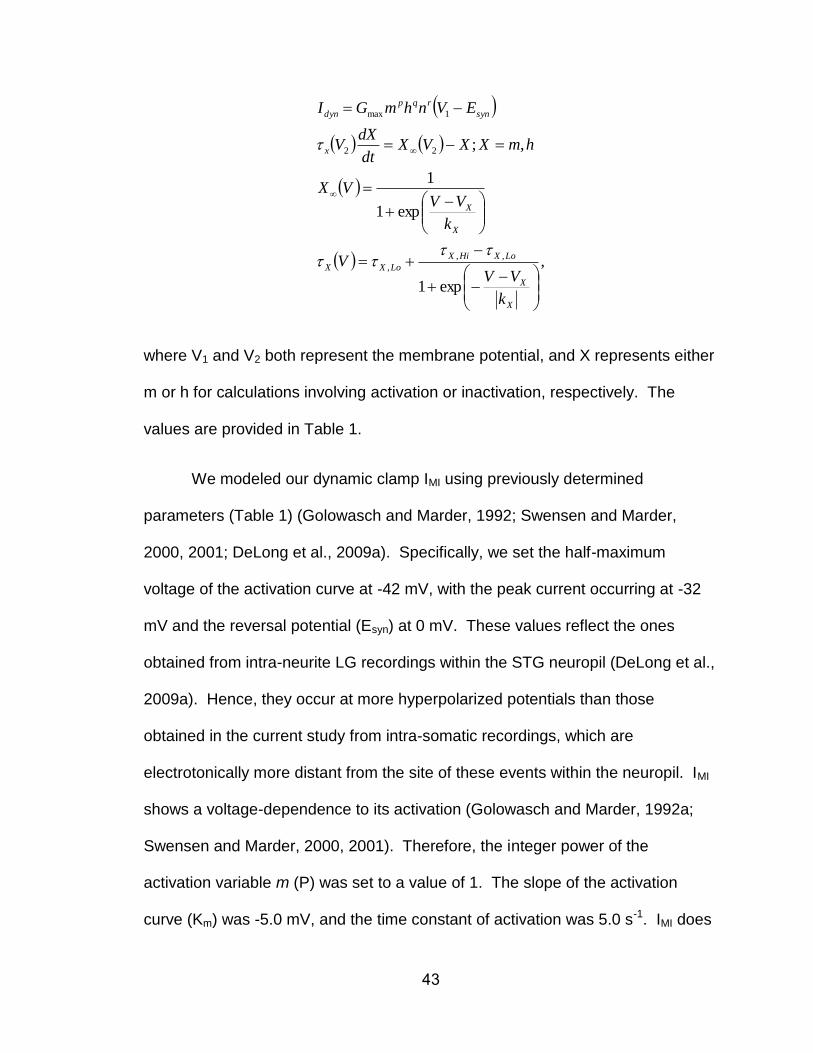
43
,
exp1
exp1
1
,;
,,
,
22
1max
X
X
LoXHiX
LoXX
X
X
x
syn
rqp
dyn
k
VVV
k
VVVX
hmXXVXdt
dXV
EVnhmGI
where V1 and V2 both represent the membrane potential, and X represents either
m or h for calculations involving activation or inactivation, respectively. The
values are provided in Table 1.
We modeled our dynamic clamp IMI using previously determined
parameters (Table 1) (Golowasch and Marder, 1992; Swensen and Marder,
2000, 2001; DeLong et al., 2009a). Specifically, we set the half-maximum
voltage of the activation curve at -42 mV, with the peak current occurring at -32
mV and the reversal potential (Esyn) at 0 mV. These values reflect the ones
obtained from intra-neurite LG recordings within the STG neuropil (DeLong et al.,
2009a). Hence, they occur at more hyperpolarized potentials than those
obtained in the current study from intra-somatic recordings, which are
electrotonically more distant from the site of these events within the neuropil. IMI
shows a voltage-dependence to its activation (Golowasch and Marder, 1992a;
Swensen and Marder, 2000, 2001). Therefore, the integer power of the
activation variable m (P) was set to a value of 1. The slope of the activation
curve (Km) was -5.0 mV, and the time constant of activation was 5.0 s-1. IMI does
44
not inactivate, so the integer power of the inactivation variable h (abbreviated ‘q’
above) was set to 0. The conductance value at maximum activation (Gmax)
varied between 50 and 200 nS. In all of our dynamic clamp experiments, the
maximum current injected into the LG neuron never exceeded 3 nA (see
Results). Synaptic conductances were modeled in a manner similar to intrinsic
conductances, except that activation depended on the presynaptic neuron
voltage and was more depolarized than the presynaptic action potential
threshold. These synapses have been well documented and incorporated into
previous models of gastric mill rhythm generation (Nadim et al., 1998; Bartos et
al., 1999; Kintos et al., 2008; DeLong and Nusbaum, 2010; Blitz and Nusbaum,
2012), and the Gmax could be readily scaled to match the observed physiological
synapses.
Our dynamic clamp model for both of the low threshold transient currents
was based on the aforementioned voltage clamp step protocol experiments. The
results from activation protocols were manually fit to Hodgkin-Huxley equations
using HHfit (Version 3.2) software developed by the Nadim lab (NJIT and
Rutgers University, Newark, NJ; available at http://stg.rutgers.edu/software/).
Occasionally, the resting Vm and action potential threshold coordinately varied
between preparations, possibly due to impalement quality. Therefore, the
dynamic clamp parameters were linked to the resting Vm. Table 1 contains a full
parameter set for a neuron resting at -60 mV.
We used two versions of the dynamic clamp on a personal computer (PC)
running Windows XP/7 and a NI PCI-6070-E data acquisition board (National
45
Instruments). The first version was developed in the Nadim laboratory (NJIT and
Rutgers University, Newark, NJ; available at http://stg.rutgers.edu/software/). The
second version was developed by E. Brady Trexler (Fishberg Dept. of
Neuroscience, Mt. Sinai School of Medicine; freely available through Gotham
Scientific: http://gothamsci.com/NetClamp/). Dynamic clamp current injections were
performed while recording in single-electrode, DCC mode (sampling rates 2 – 5
kHz) or with separate voltage recording and current-injecting electrodes.
Data analysis. Data were collected onto a computer, with later playback onto a
chart recorder (Astro-Med Everest). Acquisition onto computer (sampling rate 5
kHz) used the Spike2 data acquisition and analysis system (Cambridge
Electronic Design). Some analyses, including CabPK-gastric mill rhythm
parameters, were conducted on the digitized data using a custom-written Spike2
program (The Crab Analyzer: freely available at http://www.uni-
ulm.de/~wstein/spike2/index.html).
Voltage-clamp data analysis was performed using PClamp (version 9:
Molecular Devices), Spike2 (CED), and Igor Pro (Wavemetrics) software. For
ramps, total neuron currents were determined by averaging 10 ramps in each
condition and subtracting the control from the experimental condition. For pre-
step protocols, the protocols were run once in each condition and the control
currents were subtracted prior to analysis.
For gastric mill rhythm analyses, unless otherwise stated, each data point
46
in a data set was derived by determining the mean for the analyzed parameter
from 10 consecutive gastric mill cycles. One gastric mill cycle was defined as
extending from the onset of consecutive LG neuron action potential bursts
(Beenhakker and Nusbaum, 2004; Wood et al., 2004). Thus, the gastric mill
cycle period was measured as the duration (s) between the onset of two
successive LG neuron bursts. The protractor phase was measured as the LG
burst duration, while the retractor phase was measured as the LG interburst
duration. The gastric mill rhythm-timed LG burst duration was defined as the
duration (s) between the onset of the first and last action potential within an
impulse burst, during which no inter-spike interval was longer than 1.5 s
(approximately one pyloric cycle period during the CabPK-gastric mill rhythm and
briefer than the duration of each gastric mill phase; Saideman et al., 2007b). The
intraburst firing rate of LG was defined as the number of action potentials minus
one, divided by the burst duration.
Data were plotted with Igor Pro (version 6.10A). Figures were produced
using CorelDraw (version 13.0 for Windows). Statistical analyses were
performed with Microsoft Excel (Microsoft) and SigmaStat 3.0 (SPSS).
Comparisons were made to determine statistical significance using the paired
Student’s t-test or Analysis of Variance with Repeated Measures (RM-ANOVA)
followed by the Student-Newman-Keuls (SNK) post-hoc test. In all experiments,
the effect of each manipulation was reversible, and there was no significant
difference between the pre- and post-manipulation groups. Data are expressed
as the mean ± standard error (SE).
47
Gastric Mill Model. We constructed a computational model of the CabPK gastric
mill rhythm generator modified from an existing conductance-based model of the
MCN1-gastric mill rhythm generator (Nadim et al., 1998; Beenhakker et al., 2005;
DeLong et al., 2009a,b). The previously published version modeled the LG, Int1,
and MCN1 neurons as having multiple compartments separated by an axial
resistance, with each compartment possessing intrinsic and/or synaptic
conductances. The parameters of the CabPK-gastric mill rhythm generator
model were based on both previously published voltage clamp analyses in STG
neurons (including LG) and on the LG neuron voltage clamp results obtained in
this paper (Golowasch and Marder, 1992a; Swensen and Marder, 2000, 2001;
DeLong et al., 2009a). To mimic the effects of CabPK bath application to the
biological system, we added IMI to the LG neuron dendrite compartment as an
intrinsic (non-synaptically activated) current (Table 2). This approach was based
on the fact that CabPK excites LG by activating IMI (this paper) and that CabPK
was constantly present during its application. To more realistically mimic the
biological system, in this version of the model we modified the CabPK-activated
GMI (GMI-CabPK) in the LG dendrite compartment to include a voltage dependence
(Table 2). Based on data collected in this paper, we also added a CabPK-
activated ITrans-LTS to LG (Table 2). The time- and voltage dependence of ITrans-LTS
were empirically determined with voltage-clamp, while a canonical reversal
potential was used for ICa (ECa = 45 mV) (Zhang and Harris-Warrick, 1995).
48
Simulations were performed on a PC with the freely available Ubuntu
Linux operating system (www.ubuntu.com). We used the Network simulation
software developed in the Nadim laboratory
(http://stg.rutgers.edu/software/network.htm). This included using a fourth-order
Runge–Kutta numerical integration method with time steps of 0.05 and 0.01 ms.
Results were visualized by plotting outputted data points using the freely
available Gnuplot software package (www.gnuplot.info). In most figures showing
the model output, we present conductance (g) instead of the associated current
(I) to more clearly display the trajectory during the gastric mill retractor and
protractor phases. The main difference between “g” and “I” is that the former
lacks the fast transient changes that occur in the latter during each LG action
potential (DeLong et al., 2009a). In particular, the relatively slow kinetics of the
CabPK-activated conductances make them insensitive to these fast transient
changes in voltage.
The presentation of currents in the model and dynamic clamp figures
represent different conventions. Specifically, the model output uses the standard
voltage clamp convention, whereas the dynamic clamp output uses the standard
current clamp convention. For example, depolarizing current has a downward
trajectory in the model output figures but has an upward trajectory in the dynamic
clamp output figures.
49
RESULTS
In the isolated crab STG, tonic MCN1 stimulation and bath applied CabPK
(≥ 10-7 M) elicit comparable gastric mill motor patterns, despite configuring
different gastric mill circuits (Fig. 1B) (Saideman et al., 2007b). MCN1 does not
contain CabPK, and the CabPK-gastric mill rhythm can occur without MCN1
activity. CabPK is present in two or three pairs of CoG projection neurons which
innervate the STG, although these neurons are not physiologically identified
(Saideman et al., 2007a). However, bath-applied peptide can mimic the actions
resulting from its neuronal release. For example, in the crab STG, bath
application of the peptide proctolin (10-6 M) and direct stimulation of the
modulatory proctolin neuron (MPN) elicit comparable responses from the pyloric
CPG, despite the fact that MPN contains a small molecule co-transmitter
(Nusbaum and Marder, 1989ab; Blitz et al., 1999).
There are also at least several additional gastric mill motor patterns in C.
borealis, each distinct from the pattern elicited by MCN1 and CabPK and driven
by a different input pathway (Beenhakker and Nusbaum, 2004; Blitz et al., 2004;
Christie et al., 2004; White and Nusbaum, 2011). These different rhythms all
share the same basic structure, which includes a biphasic motor pattern
exhibiting rhythmic alternating bursting of protraction- and retraction-related
neurons across an overlapping range of cycle periods (~5 – 20 s) (Fig. 1C).
They differ in the relative timing, intensity, duration and pattern of activity in the
component neurons. There are 7 gastric mill motor neurons, including 4
protractor motor neurons and 3 retractor motor neurons, plus a single retraction-
50
timed interneuron (Int1) (Fig. 1B). As discussed below, the pyloric CPG
pacemaker neuron AB also influences these gastric mill rhythms (Fig. 1B). Like
the gastric mill rhythm, the pyloric (filtering of chewed food) rhythm is generated
in the STG (Marder and Bucher, 2007).
A core component of the rhythm generator for the MCN1- and CabPK-
gastric mill rhythms is the half-center formed by the reciprocally inhibitory
protraction neuron LG and retraction neuron Int1 (Saideman et al., 2007b) (Fig.
1B). The biphasic rhythm generated by these two neurons is then imposed on
the other gastric mill neurons by synaptic actions from the rhythm generator plus
the influences of MCN1 or CabPK. Under baseline conditions LG is silent (Fig.
1C) and Int1 is spontaneously active, exhibiting a pyloric rhythm-timed activity
pattern due to inhibitory input it receives from the AB neuron (Fig. 1B) (Bartos et
al., 1999; Saideman et al., 2007b). The pivotal event for enabling gastric mill
rhythm generation is the acquisition by LG of the ability to fire rhythmic bursts.
The cellular and synaptic mechanisms underlying MCN1 activation of the
gastric mill rhythm generator are known (Coleman et al., 1995; Bartos et al.,
1999; DeLong et al., 2009a). In brief, during the MCN1-gastric mill rhythm, there
is a rhythmic release of the MCN1 cotransmitters, which includes the peptides
proctolin and CabTRP Ia (Cancer borealis tachykinin-related peptide Ia) plus
GABA (Blitz et al., 1999). MCN1 uses only CabTRP Ia to influence LG (slow
excitation) and only GABA to influence Int1 (fast excitation) (Wood et al., 2000;
Stein et al., 2007). MCN1 cotransmitter release is rhythmic, even when MCN1 is
tonically active, because its STG terminals (MCN1STG) receive ionotropic synaptic
51
inhibition from LG (Fig. 1B) (Coleman and Nusbaum, 1994). Thus, during
retraction, continuous MCN1 release of CabTRP Ia drives a steady buildup of IMI
in LG that eventually is sufficient to enable LG to fire an action potential burst
(DeLong et al., 2009a). During protraction, when MCN1STG cotransmitter release
is inhibited by LG, there is a steady decline in the amount of IMI in LG until it can
no longer sustain the LG burst. This rhythmic activation of IMI in LG appears to
be sufficient to drive the gastric mill rhythm across the physiological range of
MCN1 firing frequencies (DeLong et al., 2009a,b; DeLong and Nusbaum, 2010).
MCN1-driven gastric mill rhythm generation is also facilitated by the pyloric
rhythm (cycle period ~1 s), because every LG burst initiates, after sufficient IMI
has accrued, during a pyloric-timed depolarization (i.e. disinhibition) that results
from AB inhibition of Int1 (Bartos et al., 1999; DeLong et al., 2009a). These
disinhibitions reduce the MCN1-gastric mill cycle period by reducing the amount
of IMI-mediated depolarization needed to enable LG to fire a burst. However, this
rhythm does persist, with a longer cycle period, when there is no pyloric rhythm
(Bartos et al., 1999).
The cellular and synaptic mechanisms underlying the CabPK-gastric mill
rhythm were not known, although it was determined previously that activity in LG,
Int1 and AB was necessary to enable this rhythm (Saideman et al., 2007b).
Additionally, as during the MCN1-gastric mill rhythm, it appeared that direct
CabPK excitation of LG was a pivotal event for rhythm generation. Thus, we
identified CabPK-influenced ionic current(s) in LG.
52
CabPK activates three voltage-dependent inward currents in the LG neuron
CabPK application (10-6 M) provides a depolarizing drive to LG from its
resting potential (-57.5 ± 1.5 mV; n=8). For example, under the most reduced
conditions, with LG isolated from synaptic input, CabPK consistently elicited in
LG a steady 5-10 mV depolarization. This depolarizing response occurred when
LG was isolated by either hyperpolarizing Int1 (dep. response: 8.5 ± 1.1 mV; n=8)
(Fig. 2A), or suppressing all glutamatergic inhibitory input to LG by bath-applying
PTX (10-5 M; dep. response: 10.7 ± 0.54 mV; n=11). The CabPK-mediated
depolarization moved the LG membrane potential closer to its spike threshold,
which was not changed by CabPK (Saline: -42.4 ± 1.3 mV; n=9; CabPK: -44.5 ±
1.4 mV; n=9, p=0.15). When Int1 was active, the LG membrane potential
exhibited subthreshold, pyloric-timed oscillations which exhibited a more
depolarized peak in the presence of CabPK (Saline: -57.5 ± 1.4 mV; n=10;
CabPK: -48.7 ± 1.7 mV; n=10, p<0.01) (Fig. 2B). These depolarized peaks
remained subthreshold prior to the onset of the gastric mill rhythm, as well as
during the ensuing gastric mill retraction phase. In contrast, CabPK did not alter
the membrane potential at the trough of these LG oscillations (Saline: -59.8 ± 1.8
mV; n=10; CabPK: -59.1 ± 1.7 mV; n=10, p=0.4) (Fig. 2B; see below). Thus, the
increased pyloric-timed oscillation amplitude was due to a more depolarized
peak.
Based on the assumption that the sustained depolarizing drive in LG
during CabPK application resulted from CabPK influence on a persistent current,
we isolated CabPK-sensitive currents using a voltage ramp protocol (see
53
Methods) in TEVC. Difference currents between CabPK and control solutions
revealed a voltage-dependent, inward net current at potentials more
hyperpolarized than 0 mV (Fig. 2C). This inward current exhibited a small,
relatively constant amplitude at membrane potentials more hyperpolarized than
~-60 mV, whereas in the depolarizing direction from ~-60 mV, the I-V plot for this
current displayed a trajectory reminiscent of voltage-dependent inward currents.
Specifically, it displayed a steadily increasing inward current that peaked at -8.1 ±
1.4 mV (peak amplitude: -4.4 ± 0.6 nA, n=7), after which the amplitude steadily
decreased (Fig. 2C). It was not possible to determine its reversal potential, likely
due to an inability to completely clamp the residual, relatively large K+ currents at
more depolarized potentials (DeLong et al., 2009a). This I-V relationship was
comparable to that of the previously identified IMI, which is activated by several
different neuromodulators in crab STG neurons including the LG neuron
(Golowasch and Marder, 1992; Swensen and Marder, 2000, 2001; DeLong et al.,
2009a).
To further establish that the CabPK-activated, voltage-dependent inward
current in the LG neuron that we identified in voltage ramp protocols was IMI, we
performed an occlusion experiment with a known IMI activator in LG, crustacean
cardioactive peptide (CCAP: DeLong et al., 2009a). CCAP application occludes
the ability of the MCN1 peptide CabTRP Ia to activate IMI in LG (DeLong et al.,
2009a). In these experiments, CabPK (10-4 M) was first pressure applied onto
the desheathed STG neuropil while recording LG in TEVC. CCAP (10-6 M) was
then bath applied to activate IMI, during which time CabPK (10-4 M) was again
54
puffed onto the STG neuropil. As shown in Figure 2C, the maximal CabPK-
activated current amplitude was decreased substantially during CCAP bath
application (CabPK pre-CCAP: -4.2 ± 0.6 nA; CabPK during CCAP application: -
0.9 ± 0.1 nA; CabPK post-CCAP: -3.5 ± 0.9; p=0.01: CabPK pre-CCAP or CabPK
post-CCAP vs CabPK during CCAP; p=0.33: CabPK pre-CCAP vs. CabPK post-
CCAP; One-way RM ANOVA with SNK post-hoc test; n=3, F(2,8) = 16.8). This
occlusion effect of CCAP thus supported the hypothesis that the aforementioned
CabPK-activated inward current in the LG neuron was IMI. In contrast to the
CabPK condition, in normal saline IMI was either not expressed or was present at
low levels, insofar as the LG resting potential in normal saline was ~-60 mV (see
above) and even modest levels of modulator-activated IMI elicit a more
depolarized LG membrane potential (Kirby and Nusbaum, 2007; DeLong et al.,
2009a).
IMI is also sensitive to changes in extracellular Ca2+ (Golowasch and
Marder, 1992). Specifically, replacing most of the extracellular Ca2+ (0.1X normal
Ca2+) in the saline with additional Mg2+, to maintain the total divalent cation
concentration, linearizes the I-V curve for IMI at hyperpolarized potentials. This
linearization in reduced Ca2+/added Mg2+ saline also occurred for the CabPK-
sensitive current (Current measured at -90 mV: CabPK saline, -0.21 ± 0.3 nA;
CabPK w/reduced Ca2+ saline, -5.67 ± 1.3 nA, n=3, p<0.05), further supporting
the hypothesis that CabPK activates IMI in LG.
To test the hypothesis that IMI was responsible for the aforementioned,
CabPK-mediated depolarization in LG, we used the dynamic clamp (DClamp) to
55
inject an artificial version of IMI into LG in normal saline (Fig. 2D). Doing so using
DClamp conductances comparable to those identified in voltage clamp (50-300
nS), while Int1 was silent or only weakly active, consistently depolarized the LG
resting potential to the same extent as CabPK application (CabPK: 10.68 ± 0.5
mV; DClamp IMI: 10.3 ± 2 mV, n=6, p=0.4) (Fig. 2A,D).
CabPK also activated other voltage-dependent inward currents in LG.
These additional currents were not evident with our voltage ramp protocol, but
they were present during a TEVC voltage step protocol. Their absence during
our voltage ramp manipulations was likely due to their time-dependent
inactivation (see below). We identified these other currents using a pre-step
hyperpolarization (-80 mV) whose duration was similar to the gastric mill
retraction phase (see Methods). Using this approach, with relatively prolonged
depolarizing steps (6 s) comparable to the gastric mill protraction phase during
which LG is depolarized and spiking, we identified three inward currents (Figs. 3
– 5). These currents included a (1) transient, low threshold, fast inward current
(ITrans-LTF), (2) transient, low threshold but more slowly developing inward current
(ITrans-LTS), and (3) sustained inward current. The ITrans-LTF was not evident in the
raw current recordings obtained during CabPK superfusion (Fig, 3A), due to
overlap with the capacitative current, but was readily evident in the difference
current traces (Figs. 3B,4,5). In contrast, ITrans-LTS was identifiable in both the
raw-CabPK and difference currents (Figs. 3 – 5), but was not evidently
expressed under control conditions (Fig. 3A). Lastly, in the raw CabPK
recordings the sustained inward current was evident as a smaller amplitude
56
outward current relative to the control recordings (Fig. 3A).
The CabPK-activated, sustained inward current was predominantly IMI.
During the last 3 s of the voltage step, we consistently observed a voltage-
dependent, time-independent inward current, as anticipated from our voltage
ramp experiments that identified CabPK activation of IMI (Fig. 3B). There were
three features of this sustained current in the voltage step protocols, however,
that were distinct from IMI measured from the voltage ramp protocol: 1) the peak
amplitude was smaller (Step: -1.6 ± 0.4 nA, n=9; Ramp: -4.6 ± 0.4 nA, n=9;
p<0.01), 2) the current exhibited less voltage-dependence at depolarized
potentials (data not shown), and 3) in some recordings, particularly with steps
more depolarized than -40 mV, a reduced inward current or small outward
current was evident immediately following ITrans-LTS, relative to the current
amplitude at the end of the step (e.g. Figs. 3A,B, 4B, 5B). These features
suggested that CabPK also activated a voltage- and time-dependent outward
current. We did not, however, further isolate and characterize this additional
component insofar as it did not appear to be necessary for the CabPK actions on
gastric mill rhythm generation (see below).
The fast transient inward current exhibited a relatively rapid time to peak
(32.9 ± 1.9 ms, n=16) and small peak amplitude (~0.5 nA), which occurred at ~-
10 mV. It exhibited an apparent voltage threshold of ~-45 mV (range: -50 to -30
mV; n=16). Only an approximate peak current amplitude is provided for ITrans-LTF
because we could not isolate this current from the other two CabPK-activated
inward currents, and these other currents appeared to contribute substantially to

57
the fast transient peak. In this context, it is noteworthy that IMI reaches its peak
current level relatively quickly in response to a depolarizing voltage step (Fig.
5A). We did not further characterize ITrans-LTF, insofar as it was not necessary for
the ability of CabPK to enable gastric mill rhythm generation (see below).
ITrans-LTS exhibited a longer time to peak than ITrans-LTF (time to peak at -45
mV: 633 ± 48 ms, n=9, p<0.01) as well as a larger peak amplitude (-5.3 ± 0.6 nA,
measured at -15 ± 3.1 mV, n=9) (Fig. 3B,C). It exhibited a voltage threshold of ~-
55 mV (range: -60 to -50 mV, n=9) and its amplitude increased with
depolarization up to ~-30 mV (n=9) (Fig. 3C). Note that this reported peak
amplitude value includes the CabPK-activated IMI amplitude and, at the more
depolarized steps, likely also includes the aforementioned voltage- and time-
dependent outward current. The unusually shallow slope of the I-V curve
between -40 mV and -10 mV likely results, at least partly, from the contribution of
this outward current (Fig. 3C). The ITrans-LTS voltage threshold and time to peak
suggested that this current was likely to be activated during the CabPK-gastric
mill rhythm, during which time the LG membrane potential exhibits rhythmic
oscillations between ~-70 mV and -40 mV (Saideman et al., 2007b). We could
not measure the full time course of the ITrans-LTS decay to the baseline because it
merged into the sustained inward current, which persisted for the remainder of
each step (Figs. 3B, 4A). ITrans-LTS exhibited a reversal potential that was more
depolarized than 0 mV (n=16 each) (ITrans-LTS: Fig. 3C), suggesting that it is
primarily carried by ions with positive equilibrium potentials (e.g. Ca2+ and/or
Na+). Both of the CabPK-activated transient currents were clearly distinct from
58
IMI, because IMI exhibits no time-dependent decrease in amplitude (e.g. Fig. 5)
(Golowasch and Marder, 1992).
ITrans-LTS not only exhibited the property of inactivation, but it also exhibited
deinactivation. This deinactivation was sensitive to the time and voltage range
that occurs in LG during the CabPK-gastric mill rhythm. We therefore
characterized this property by varying parameters of the voltage step protocol in
voltage clamp experiments. To determine the time-dependence of this property,
we maintained LG at a holding potential of -40 mV and systematically
hyperpolarized it to -80 mV for a range of durations (1 s to 13 s), stepping the
voltage back to -40 mV after each hyperpolarization. We then measured the
maximum amplitude of the slow transient inward current after the return to -40
mV (Fig. 4). These data were then fit with a sigmoid curve (Igor Pro), from which
two parameters were identified, including the midpoint and slope. There was a
relatively long time-dependence for ITrans-LTS deinactivation (midpoint: 7.4 ± 0.4 s,
slope: 2.4 ± 0.2 s-1, n=3) (Fig. 4B).
We determined the voltage-dependence of the ITrans-LTS deinactivation by
varying the pre-step voltage across a range (-75 to -50 mV) of membrane
potentials, while maintaining the pre-step duration (8 s) and subsequent step
potential (-45 mV) (midpoint: -60.9 ± 2 mV; slope: -5.3 ± 0.5 mV-1, n=3). This
midpoint value was well within the normal LG membrane potential range (-55 mV
to -70 mV) during the gastric mill retraction phase (Saideman et al., 2007b).
Thus, based on the time- and voltage dependence of this transient current in LG,
and the LG membrane potential trajectories during the gastric mill rhythm
59
(Saideman et al., 2007a,b; this study), it likely exhibits considerable inactivation
during the course of each gastric mill protraction phase and deinactivation during
the retractor phase. These correlations support the hypothesis that the CabPK-
activated slow transient inward current helps enable LG to generate a periodic
burst during a pyloric-timed membrane potential depolarization produced by the
combination of IMI and the periodic (AB-mediated) removal of synaptic inhibition
from Int1 (see below).
Both CabPK-activated transient inward currents were extracellular Ca2+-
dependent. Replacing most of the extracellular Ca2+ with Mn2+, a Ca2+ channel
antagonist (Turrigiano et al., 1995), consistently resulted in no measureable fast
(n=4) or slow transient inward currents (peak ITrans-LTS at -50 mV: CabPK alone, -
1.6 ± 0.1 nA; CabPK with 0.1 mM Ca2+/10.9 mM Mn2+, 0 ± 0 nA; CabPK post-
reduced Ca2+, -0.8 ± 0.02 nA; n=4) (Fig. 5A). This manipulation only had a
moderate effect on IMI (Fig. 5A), due to the divalent cation sensitivity of this
current being approximately equivalent for Ca2+ and Mn2+ (Golowasch and
Marder, 1992). In addition, replacing extracellular Na+ with a non-permeant ionic
species (NMDG+) also consistently resulted in no measureable ITrans-LTS (Max.
amplitude: Control, -3.8 ± 0.9 nA; NMDG saline, 0 ± 0 nA, n=7) (Fig. 5B). In
contrast, in all four of these NMDG+ experiments where there was a discernible
ITrans-LTF peak in the control recordings, this peak persisted in the NMDG+
condition (data not shown). In the three experiments where there was no
distinguishable ITrans-LTF peak in the control recording, it was not possible to
determine whether it was influenced by the NMDG+ substitution (e.g. Fig. 5B).
60
In sum, these results suggested that ITrans-LTS is either permeable to both
Na+ and Ca2+, is a Ca2+-sensitive INa, or is a Ca2+-activated nonselective cation
current (ICAN), although we also did not rule out the possibility that NMDG+ acts
instead as an inhibitor of this current. ICAN was identified previously in C.
borealis, both in the stomatogastric nervous system and cardiac ganglion, where
it exhibited a Vrev of ~-30 mV and was insensitive to changes in extracellular Na+
but sensitive to caffeine application (10-2 M), which stimulates intracellular Ca2+
release, and the ICAN antagonist flufenamic acid (FFA: 10-5 M) (Zhang et al.,
1995; Kadiri et al., 2011; Ransdell et al., 2013). However, bath applied FFA did
not alter any of the CabPK-activated currents (n=2). Insofar as ITrans-LTS exhibited
a Vrev > 0 mV, sensitivity to extracellular Na+ and insensitivity to FFA, it was not
likely to be an ICAN.
CabPK-activated transient inward currents enable post-inhibitory rebound
in the LG neuron
To determine how the CabPK-activated currents might contribute to LG
burst generation during the CabPK-gastric mill rhythm, we examined intrinsic
properties in LG. Specifically, we found that brief hyperpolarizing current
injections into LG were followed by passive responses during saline superfusion
but elicited a post-inhibitory rebound (PIR) burst during CabPK superfusion.
Under control conditions (saline), we depolarized LG to a membrane potential
that was comparable to its CabPK-mediated baseline potential (~-50 mV) using
61
either direct depolarizing current injection or DClamp IMI injection. Despite this
depolarized baseline potential, following a period of hyperpolarization there was
no evidence of PIR (n>10) (Fig. 6). In contrast to its passive response during
saline superfusion, LG hyperpolarization from its CabPK-mediated depolarized
resting potential was consistently followed by a PIR burst (Fig. 6). Specifically,
during CabPK superfusion, injecting a modest hyperpolarizing current (-1 nA) into
LG, which caused a 5 – 10 mV hyperpolarization, consistently elicited a PIR burst
when the current injection was terminated (PIR burst: Duration, 5.14 ± 0.2 s;
Number of Spikes: 8.22 ± 0.2, n=9). We used hyperpolarizing durations (5 s) and
amplitudes that were similar to those experienced by LG during the gastric mill
retraction phase. The trough of the subthreshold LG oscillations during retraction
ranged from -55 mV to -65 mV across experiments (Saideman et al., 2007a,b).
LG also readily exhibited PIR bursts during CabPK application after an
episode of synaptic inhibition from Int1 (n=5) (Fig. 6). To establish Int1-mediated
PIR in LG, Int1 activity was suppressed via hyperpolarizing current injection and
periodically released from this hyperpolarization to fire action potentials for 5 s,
comparable to its active period during the gastric mill rhythm. The resulting
inhibition in LG caused it to hyperpolarize by 10.2 ± 1.5 mV (n=5), comparable to
its response to Int1 during gastric mill retraction (Saideman et al., 2007a,b). At
the end of each inhibitory episode, LG generated a PIR burst comparable to
those resulting from hyperpolarizing current injection (PIR Burst Duration: 5.3 ± 1
s; Number of Spikes: 8.2 ± 0.6, n=5; p>0.5 for both parameters).
PIR bursts are often driven at least partly by the hyperpolarization-
62
activated inward current (Ih) (McCormick and Bal, 1997; Sekirnjak and du Lac,
2002; Robinson and Siegelbaum, 2003; Sangrey and Jaeger, 2010; Engbers et
al., 2011; Felix et al., 2011). However, Ih did not appear to contribute to the
CabPK-enabled PIR bursts in LG insofar as neither hyperpolarizing current
injection nor synaptic inhibition from Int1 revealed any evidence of a depolarizing
sag potential during saline superfusion (n>10) or CabPK application (n=18/20)
(Figs. 6,7). Similarly, there was no evidence in our voltage-clamp experiments
for a sag current (n>10). However, the CabPK-activated ITrans-LTS, with its
voltage- and time-dependent properties of inactivation and deinactivation, was a
candidate for the ionic current underlying PIR burst generation.
We first assessed the contribution of this CabPK-activated transient
inward current to PIR in LG by simplifying the preparation with TTX (10-7 M)
saline to silence all neurons. There was no evidence for a PIR response after LG
was hyperpolarized in TTX saline either from its resting potential (-59.1 ± 1.1 mV)
(n=18) or from a depolarized membrane potential (-47.4 ± 1 mV) (n=13) (Fig.
7A). In contrast, during CabPK (10-6 M) application under this condition, LG
again exhibited a maintained depolarization (-48.4 ± 1.2 mV, n=18) from which it
displayed PIR in response to hyperpolarizing pulses (-1 nA), albeit without
associated action potentials (n=18) (Fig. 7A). Thus, this CabPK-mediated PIR
did not require activation of TTX-sensitive INa. However, these PIR events were
briefer than the PIR bursts that occurred during normal CabPK saline (2.0 ± 0.1
s, n=18; p<0.01), suggesting that a TTX-sensitive INa might prolong this
response.
63
We tested the role in PIR generation played by the IMI-associated
depolarization in LG during CabPK application. To this end, during CabPK
application in TTX saline, we injected constant amplitude hyperpolarizing current
to return the LG membrane potential to its pre-CabPK resting potential. That is,
we eliminated the effect of CabPK activated IMI, which underlies the steady LG
depolarization (see above). From that resting potential, we again injected
hyperpolarizing current pulses as above. Doing so reduced the PIR amplitude to
~25% of the control amplitude (Control: Vm = -51.1 ± 2 mV, PIR Amplitude = 7.2
± 0.4 mV; Hyperpolarized: Vm = -59.1 ± 2 mV, PIR Amplitude = 1.8 ± 0.7 mV;
n=7; p<0.01), supporting the hypothesis that the IMI-mediated depolarization in
LG strengthens PIR generation mediated by activation of the transient inward
current (see below).
We next assessed whether this CabPK-enabled PIR event in TTX saline
was Ca2+-sensitive by applying CabPK after replacing most of the Ca2+ (0.1X
normal) in the saline with an equimolar concentration of Mn2+. Under these
conditions, the PIR amplitude was reversibly reduced (Pre-Control: 7.5 ± 0.7 mV;
Reduced Ca2+/Added Mn2+ saline: 0.4 ± 0.1 mV, n=6, p<0.01) (Fig. 7A). These
results suggested that the CabPK-elicited PIR was mediated by activation of the
CabPK-activated low threshold inward currents.
We tested the hypothesis that the CabPK-activated IMI plus ITrans-LTS was
sufficient to enable PIR in LG. To this end, in TTX saline, we injected into LG a
dynamic clamp version of these two currents. This manipulation did indeed
enable LG to express PIR in response to the same hyperpolarizing current
64
injections used in the presence of CabPK (PIR amplitude: 8.7 ± 1.5 mV; PIR
duration: 4.5 ± 0.03 s, n=4) (Fig. 7B). Performing the same manipulation using
only the DClamp version of IMI or ITrans-LTS did not elicit PIR (n=4) (Fig. 7B). The
absence of PIR during the latter manipulation likely resulted from the absence of
a depolarized LG resting potential, which in turn limited the activation of the
transient inward current after the hyperpolarizing current injection. Thus, the
ability of CabPK to enable LG to generate PIR bursts apparently results from its
coactivation of IMI, to depolarize LG, and ITrans-LTS, to provide the drive for the PIR
burst.
CabPK-activated inward currents in the LG neuron are necessary for
gastric mill rhythm generation
We tested the ability of the identified CabPK-activated inward currents in
the LG neuron to enable gastric mill rhythm generation in a computational model,
after which we tested the predictions of the model in the biological system. We
developed a computational model of the CabPK-gastric mill rhythm generator
(LG, Int1 and AB neurons) in which LG contained CabPK-activated IMI plus ITrans-
LTS (see Methods and Table 2). This model was based on one of three previously
published models (PK Mechanism 1: IPlat) that were focused on distinct candidate
mechanisms for CabPK-mediated gastric mill rhythm generation (Kintos et al.,
2008). As shown in Figure 8A, our model produced gastric mill rhythm-like
alternating bursting in LG and Int1. The model rhythm exhibited a cycle period
65
(10.8 ± 2 x 10⁻⁴ s), LG burst duration (2.75 ± 1 x 10⁻⁴ s) and LG interburst
duration (8.09 ± 1 x 10⁻⁴ s) similar to the biological CabPK-gastric mill rhythm
(Cycle Period: 11.96 ± 1.1 s; LG burst duration: CabPK: 3.2 ± 0.3 s; LG interburst
duration: 8.8 ± 1.1 s, n=12). Additionally, the average protraction and retraction
duty cycles (DC: fraction of the cycle) were comparable (Prot. DC: model, 0.25;
biol., 0.27; Ret. DC: model, 0.75; biol., 0.73). Note that, during the model rhythm,
gMI and gTrans-LTS followed the LG voltage trajectory (Fig. 8A), insofar as their
activation was voltage-dependent. The deinactivation (h) state of gTrans-LTS also
tracked the LG membrane potential, rising during the retractor phase when LG
was rhythmically hyperpolarized by Int1 inhibition.
The model CabPK-gastric mill rhythm was also comparable to the
biological rhythm in that it was suppressed by eliminating the AB inhibition of Int1
(Fig. 8B). In the biological system, this manipulation terminates the CabPK-
gastric mill rhythm, but not the MCN1-gastric mill rhythm (Bartos et al., 1999;
Saideman et al., 2007b). As discussed above, during the gastric mill rhythm
each fast rhythmic depolarization (disinhibition) in LG results from the fast
rhythmic AB inhibition of Int1 unmasking the depolarizing drive in LG due to
CabPK-activated IMI and ITrans-LTS. When the AB inhibition of Int1 is suppressed,
Int1 fires tonically (Bartos et al., 1999). Selectively silencing AB in the model
CabPK-gastric mill rhythm generating circuit did cause Int1 to fire tonically and
eliminated the rhythmic disinhibitions in LG that normally provide the trigger for
each LG burst (Fig. 8B). Silencing AB also resulted in the Int1 inhibition
dominating the LG membrane potential, keeping LG too hyperpolarized to
66
activate sufficient gMI and gTrans-LTS (Fig. 8B).
Removing either IMI or ITrans-LTS from the model LG neuron eliminated
gastric mill rhythm generation, supporting the hypothesis that these currents are
necessary for this process (Fig. 9A,B). When either the model gMI or gTrans-LTS
was removed (i.e. set to 0 nS), the pyloric-timed LG membrane potential
oscillation peak amplitude was reduced, preventing LG from reaching action
potential threshold and terminating the model gastric mill rhythm (Fig. 9A,B). The
peak amplitude of these subthreshold oscillations in LG was larger after
selectively removing gTrans-LTS (12.92 ± 3.4 mV; Fig. 9B) than after selectively
removing gMI (7.32 ± 1.6 mV; Fig. 9A). This result was not surprising given that
with IMI absent the subthreshold oscillations peaked at a membrane potential
close to threshold for ITrans-LTS activation (Fig. 3).
We evaluated the predictions of the computational model in the biological
system using dynamic clamp current injections into LG. We tested the necessity
of IMI and ITrans-LTS for CabPK-gastric mill rhythm generation by selectively
nullifying each one during the CabPK-gastric mill rhythm. We performed each
such manipulation by using the dynamic clamp to provide negative versions of
each conductance, enabling injection of artificial IMI or ITrans-LTS that was
approximately equal in amplitude and opposite in sign to the CabPK-activated
version of that same current (DeLong and Nusbaum, 2010).
As was the case during the comparable manipulations in our
computational model (Fig. 9), moderate levels of negative conductance injection
67
(range: 50 – 200 pS) of either gMI or gTrans-LTS caused only a modest reduction in
the peak depolarization of the pyloric-timed oscillations in LG, but it was sufficient
to arrest LG bursting and terminate the ongoing gastric mill rhythm for the
duration of the manipulation (gMI: n=7; gTrans-LTS: n=5) (Fig. 10). In most
experiments, we tested a 2-fold range of negative conductance values and found
they all effectively suppressed the ongoing rhythm. These manipulations not
only suppressed rhythmic bursting in LG but they eliminated the entire gastric mill
rhythm. For example, the combined pyloric- and gastric mill-timed patterns of the
IC and VD neurons were replaced by an exclusively pyloric rhythm-timed pattern
(n=3 of 3 preparations) (Fig. 10). The gastric mill rhythm consistently resumed
when the dynamic clamp injection was terminated (Fig. 10). Thus, both CabPK-
activated IMI and ITrans-LTS in the LG neuron were necessary for gastric mill rhythm
generation.
CabPK-activated inward currents in the LG neuron are sufficient for gastric
mill rhythm generation
To determine whether the CabPK-activated IMI and ITrans-LTS in the LG
neuron were sufficient for gastric mill rhythm generation, as was the case in the
computational model, we first combined a dynamic clamp injection of ITrans-LTS into
LG with bath-application of the muscarinic agonist oxotremorine (OXO: 10-5 M),
an IMI activator in pyloric circuit neurons (Swensen and Marder, 2000, 2001). We
established via voltage clamp experiments that OXO consistently activated IMI in

68
LG as well (n=5). In parallel current clamp experiments in which hyperpolarizing
current steps were injected into LG during OXO superfusion, PIR was not elicited
(n=5). The OXO-activated IMI characteristics in LG were comparable to those
resulting from CabPK and CCAP application (e.g. a relatively steady and small
amplitude inward current at potentials more hyperpolarized than -60 mV [-0.3 ±
0.04 nA, n=5], and a similar Vm for peak amplitude [-7.6 ± 3.8 mV, n=5], although
the peak amplitude was lower relative to these peptides [-1.2 ± 0.27 nA, n=5])
(DeLong et al., 2009a; this paper). OXO (10-5 M) superfusion consistently
increased the amplitude of the subthreshold pyloric-timed oscillations in LG by 5
– 10 mV (n=10), by selectively increasing their depolarized peak Vm (Peak Vm:
Control, -54.2 ± 1.7 mV; OXO, 45.8 ± 2 mV; n=10, p<0.01; Trough Vm: Control, -
55.2 ± 1.8 mV; OXO, -55.7 ± 1.9; n=10, p=0.13), without activating LG bursting
(Fig. 11A). This result was comparable to that occurring during CabPK
applications with dynamic clamp nullification of ITrans-LTS, when the only CabPK-
activated inward current influencing the LG membrane potential was IMI (Fig.
10B).
During OXO (10-5 M) superfusion, injecting Idyn,Trans-LTS (50 – 100 nS; peak
current: 1 to 3 nA) enabled LG to generate gastric mill rhythm-like bursting (n=5)
(Fig. 11B). This rhythmic bursting in LG exhibited characteristics that were
similar to those occurring during the CabPK-gastric mill rhythm (n=12), such as
its intraburst firing frequency (OXO/DClamp: 3.9 ± 0.6 Hz; CabPK: 2.7 ± 0.2 Hz,
p=0.14), burst duration (OXO/DClamp: 2.3 ± 0.3 s; CabPK: 3.2 ± 0.3 s, p=0.17)
and interburst duration (OXO/DClamp: 6.4 ± 0.8 s; CabPK: 8.8 ± 1.1 s, p=0.41).
69
These results were consistent across a 2-fold range of dynamic clamp
conductances (e.g. 50 – 100 nS) within the same experiments.
In the above OXO experiments, it remained possible that rhythm
generation resulted in part from additional OXO actions on other neurons (e.g.
Int1, AB) necessary for gastric mill rhythm generation. Therefore, to determine
whether the CabPK-activated inward currents in LG were likely to be truly
sufficient to enable a gastric mill rhythm-like pattern in LG, we co-injected into the
biological LG neuron dynamic clamp versions of IMI, ITrans-LTS plus the Int1-
mediated synaptic inhibition. These experiments were performed in PTX saline
in order to isolate the LG neuron. These dynamic clamp co-injections
consistently elicited gastric mill rhythm-like bursting in LG (n=4) (Fig. 12). The
resulting cycle period (8.92 ± 1.7 s, p>0.05), LG burst duration (1.83 ± 0.2 s,
p>0.05), interburst duration (7.09 ± 0.8 s, p>0.05), and number of spikes per
burst (11.38 ± 0.9, p>0.05) were all similar to the above-reported values during
the CabPK-gastric mill rhythm (Fig. 13). These experiments therefore supported
the hypothesis that the CabPK-activated IMI plus ITrans-LTS in the LG neuron were
sufficient to enable gastric mill rhythm-equivalent alternating bursting in the core
gastric mill rhythm generator neurons LG and Int1.
70
DISCUSSION
We have identified the cellular mechanisms underlying CabPK
neuropeptide activation of a rhythmic motor pattern, thereby establishing that
these mechanisms are distinct from those by which a previously studied
modulatory pathway activates the same motor pattern. Specifically, bath
applying CabPK or stimulating the projection neuron MCN1 configure different
gastric mill circuits yet elicit the same gastric mill motor pattern (Fig. 1B)
(Saideman et al., 2007b). Here we demonstrated that CabPK peptide-elicited
gastric mill rhythm generation results from its persistent recruitment of at least
two voltage-dependent inward currents (IMI, ITrans-LTS) in the rhythm generator
neuron LG. These currents conjointly enable LG to rhythmically generate a PIR
burst and thereby produce an alternating activity pattern with the rhythm
generator neuron Int1, which they then impose on the other gastric mill motor
neurons via their synaptic actions (Fig. 1B).
Our computational model and dynamic clamp manipulations support the
hypothesis that the CabPK-elicited rhythmic LG bursting results from IMI
depolarizing LG closer to its spike threshold, enabling the voltage- and time-
dependent properties of ITrans-LTS to periodically generate a PIR burst. The
projection neuron MCN1 also activates the gastric mill rhythm generator via IMI
activation in LG (DeLong et al., 2009a). However, the MCN1-activated IMI grows
and decays during the gastric mill retractor and protractor phases, respectively,
due to continual neuropeptide release from MCN1 during retraction and
presynaptic feedback inhibition of that release during protraction. This MCN1-
71
mediated rhythmic buildup of IMI is sufficiently strong to enable LG to periodically
reach spike threshold and generate a self-terminating burst (Coleman et al.,
1995; Bartos et al., 1999; DeLong et al., 2009a).
Different processes can potentially enable the same rhythmic motor
pattern to be elicited by different modulatory inputs. These processes include
convergent activation of the same direct input to a circuit (Viana di Prisco et al.,
2000; Korn and Faber, 2005; Derjean et al., 2010), convergent modulation of the
same properties in the same network neurons (Doi and Ramirez, 2010),
divergent modulation in network neurons of multiple baseline intrinsic and
synaptic conductances which functionally compensate for one another (MacLean
et al., 2003; Prinz et al., 2004a; Goaillard et al., 2009; Grashow et al., 2010;
Norris et al., 2011), or configuring different circuits by activating distinct
conductances that enable different intrinsic properties in network neurons
(Saideman et al., 2007b; this paper). The crab gastric mill system appears to
provide the first example of the latter of these mechanisms.
Despite MCN1 and CabPK configuring different rhythm generating circuits,
at their core both circuits include the reciprocally inhibitory neurons LG and Int1.
These two neurons establish the protractor and retractor phases, respectively,
and in each case the pivotal rhythm-generating event is LG activation,
presumably because Int1 is spontaneously active (Bartos et al., 1999; Saideman
et al., 2007b). However, despite these similarities and the fact that MCN1 and
CabPK each recruit IMI, they enable different active properties in LG. These
different properties result from the distinct temporal dynamics of IMI activation by
72
these two pathways, the likelihood that MCN1 activates more IMI than CabPK
(see below), and the fact that only CabPK appears to activate ITrans-LTS (Bartos et
al., 1999; DeLong et al., 2009a; this paper).
PIR bursts often involve the complementary influence of multiple voltage-
dependent inward currents, as during CabPK modulation of the LG neuron
(McCormick and Bal, 1997; Sekirnjak and du Lac, 2002; Angstadt et al., 2005;
Sangrey and Jaeger, 2010; Wang et al., 2010; Engbers et al., 2011; Felix et al.,
2011; Zheng and Raman, 2011). One inward current that often contributes to the
initial PIR depolarization is Ih (McCormick and Bal, 1997; Sekirnjak and du Lac,
2002; Robinson and Siegelbaum, 2003; Sangrey and Jaeger, 2010; Engbers et
al., 2011; Felix et al., 2011). The Ih-mediated rebound depolarization facilitates
activation of other voltage-dependent inward currents that elicit or strengthen the
PIR burst. These latter currents often have time- and voltage-dependent
properties similar to those of CabPK-activated ITrans-LTS. In many neurons, the
additional PIR-generating current is some type of ICa (McCormick and Bal, 1997;
Angstadt et al., 2005; Sangrey and Jaeger, 2010; Wang et al., 2010; Engbers et
al., 2011; Felix et al., 2011). The role of Ih in PIR bursts is similar to that of
CabPK-activated IMI in LG, which depolarizes LG after a period of inhibition and
enables sufficient ITrans-LTS activation to generate the PIR burst. Also, similar to
the CabPK comodulation of IMI and ITrans-LTS, the complementary PIR-generating
currents in some other systems can be comodulated (Harris-Warrick et al., 1995;
Angstadt et al., 2005; Wang et al., 2010).
MCN1- and CabPK activation of the gastric mill rhythm generator involves
73
both divergent actions of the same ionic current (IMI) and a conserved function
mediated by distinct ionic currents (IMI, ITrans-LTS) (Fig. 14). As discussed above,
IMI is the burst-generating conductance during MCN1-rhythm generation,
whereas during CabPK-rhythm generation there is insufficient IMI to directly
enable LG bursting. In this latter condition, IMI instead facilitates burst generation
by ITrans-LTS. The shared role of IMI and ITrans-LTS during MCN1- and CabPK-rhythm
generation, respectively, results from each current exhibiting a phase-dependent
growth and decay process that is pivotal to determining the LG burst (protraction)
and interburst (retraction) durations (Fig. 14) (Bartos et al., 1999; DeLong et al.,
2009a; this paper). The growth and decay process for IMI during MCN1
stimulation results from its synaptic regulation, as discussed above. In contrast,
the availability of CabPK-activated ITrans-LTS grows and decays during gastric mill
retraction and protraction, respectively, due to its voltage- and time-dependent
properties. During retraction, when LG is hyperpolarized by rhythmic synaptic
inhibition from Int1, ITrans-LTS exhibits a buildup of deinactivation which increases
its availability, while during protraction it first enables and then limits the LG burst
duration due to its depolarization- and time-dependent inactivation.
Despite these differences in the rhythm generating process, during both
gastric mill rhythms the LG burst initiates during a pyloric rhythm-timed
depolarization that results from AB inhibition of Int1 (Bartos et al., 1999;
Saideman et al., 2007b). However, these pyloric-timed depolarizations are
necessary only for CabPK-rhythm generation (Saideman et al., 2007b). This is
because, when the pyloric rhythm is suppressed during MCN1 stimulation, the
74
MCN1-activated IMI amplitude continues to grow during each prolonged retraction
phase until it eventually becomes large enough to enable LG to escape from Int1
inhibition and generate a burst (Bartos et al., 1999). In contrast, CabPK-
activated IMI alone is insufficient to enable LG to depolarize and trigger ITrans-LTS
activation in the presence of persistent Int1 inhibition.
The phase transitions occurring during rhythmic alternating bursting by
reciprocally inhibitory neurons commonly result from the inhibited neuron either
escaping from continuing inhibition (“escape” mode) or waiting until it is released
from that inhibition (“release” mode) (Skinner et al., 1994; Marder and Calabrese,
1996; McCormick and Bal, 1997). Each mode can also mediate the same
transition under different conditions (Bartos et al., 1999; Sorensen et al., 2004;
Kristan et al., 2005). During the MCN1- and CabPK-gastric mill rhythms the
protraction to retraction transition appears to occur via the release mechanism,
as the LG burst self-terminates and releases Int1 from inhibition. In contrast, for
reasons discussed above, during the retraction to protraction transition the
CabPK-rhythm only operates via the release mode whereas the MCN1-rhythm
can operate either in its normal “release” mode or, if the pyloric rhythm is very
slow or suppressed, in the “escape” mode.
A neural network might have the ability to configure distinct circuits
generating the same activity pattern because it needs to generate the same core
behavior under different conditions. These differently configured circuits,
however, might be differentially sensitive to particular inputs. This, for example,
is evident for the pyloric pacemaker neuron AB influence on these two gastric mill
75
circuits (i.e. it is a necessary rhythm generating component only for the CabPK-
gastric mill circuit). Such differential sensitivity likely also occurs in circuits that
generate a consistent output pattern despite extensive, albeit compensatory,
changes in the baseline intrinsic and synaptic conductances of circuit neurons
(Prinz et al., 2004a; Grashow et al., 2009, 2010; Calabrese et al., 2011;
Guttierrez et al., 2013). Two interesting tests of this hypothesis for the gastric
mill circuit will be the influence of the gastro-pyloric receptor neuron, a muscle
proprioceptor, and the peptide hormone CCAP (crustacean cardioactive peptide)
on the CabPK-gastric mill rhythm. Both of these inputs regulate the MCN1-
gastric mill rhythm via actions that involve MCN1, which does not participate in
the CabPK-rhythm (Beenhakker et al., 2005; DeLong et al., 2009a,b).
This study highlights an alternative consequence to the well-established
flexibility in neural circuit output imparted by their neuromodulatory inputs. Here,
neuromodulation configures distinct circuits/mechanisms that generate a
conserved output rather than generating distinct outputs. As part of this process,
different modulatory pathways activate the same ionic current in the same
neuron but use it in different ways, and they use different currents in the same
neuron to perform the same function (Fig. 14). Whether the conserved output
pattern resulting from these distinct mechanisms belies latent differences in
responsiveness to the same perturbation remains to be determined.
76
REFERENCES
Angstadt JD, Grassmann JL, Theriault KM, Levasseur SM (2005) Mechanisms of postinhibitory rebound and its modulation by serotonin in excitatory swim motor neurons of the medicinal leech. J Comp Physiol A Neuroethol Sens Neural Behav Physiol 191:715 – 732.
Bal T, Nagy F, Moulins M (1994) Muscarinic modulation of a pattern-generating network: control of neuronal properties. J Neurosci 14:3019 – 3035.
Bartos M, Nusbaum MP (1997) Intercircuit control of motor pattern modulation by presynaptic inhibition. J Neurosci 17:2247 – 2256.
Bartos M, Manor Y, Nadim F, Marder E, Nusbaum MP (1999) Coordination of fast and slow rhythmic neuronal circuits. J Neurosci 19:6650 – 6660.
Beenhakker MP, Nusbaum MP (2004) Mechanosensory activation of a motor circuit by coactivation of two projection neurons. J Neurosci 24:6741 – 6750.
Beenhakker MP, Blitz DM, Nusbaum MP (2004) Long-lasting activation of rhythmic neuronal activity by a novel mechanosensory system in the crustacean stomatogastric nervous system. J Neurophysiol 91:78 – 91.
Beenhakker MP, DeLong ND, Saideman SR, Nadim F, Nusbaum MP (2005) Proprioceptor regulation of motor circuit activity by presynaptic inhibition of a modulatory projection neuron. J Neurosci 25:8794 – 8806.
Blitz DM, Christie AE, Coleman MJ, Norris BJ, Marder E, Nusbaum MP (1999) Different proctolin neurons elicit distinct motor patterns from a multifunctional neuronal network. J Neurosci 19:5449 – 5463.
Blitz DM, Beenhakker MP, Nusbaum MP (2004) Different sensory systems share projection neurons but elicit distinct motor patterns. J Neurosci 24:11381 – 11390.
Briggman KL, Kristan WB (2008) Multifunctional pattern-generating circuits. Annu Rev Neurosci 31:271 – 294.
Bucher D, Prinz AA, Marder E (2005) Animal-to-animal variability in motor pattern production in adults and during growth. J Neurosci 25:1611 – 1619.
Calabrese RL, Norris BJ, Wenning A, Wright TM (2011) Coping with variability in small neuronal networks. Integr Comp Biol 51:845 – 855.
Coleman MJ, Nusbaum MP (1994) Functional consequences of compartmentalization of synaptic input. J Neurosci 14:6544 – 6552.
Coleman MJ, Meyrand P, Nusbaum MP (1995) Presynaptic inhibition mediates a switch between two modes of synaptic transmission. Nature 378:502 – 505.
77
DeLong ND, Kirby MS, Blitz DM, Nusbaum MP (2009a) Parallel regulation of a modulator-activated current via distinct dynamics underlies comodulation of motor circuit output. J Neurosci 29:12355 – 12367.
DeLong ND, Beenhakker MP, Nusbaum MP (2009b) Presynaptic inhibition selectively weakens peptidergic cotransmission in a small motor system. J Neurophysiol 102:3492 – 3504.
DeLong ND, Nusbaum MP (2010) Hormonal modulation of sensorimotor integration. J Neurosci 30:2418 – 2427.
Derjean D, Moussaddy A, Atallah E, St-Pierre M, Auclair F, Chang S, Ren X, Zielinski B, Dubuc R (2010) A novel neural substrate for the transformation of olfactory inputs into motor output. PLoS Biol 8:e1000567.
Diehl F, White RS, Stein W, Nusbaum MP (2013) Motor circuit-specific burst patterns drive different muscle and movement patterns. J Neurosci 33: 12013 – 12029.
Doi A, Ramirez JM (2008) Neuromodulation and the orchestration of the respiratory rhythm. Respir Physiol Neurobiol 164:96 – 104.
Doi A, Ramirez JM (2010) State-dependent interactions between excitatory neuromodulators in the neuronal control of breathing. J Neurosci 30:8251 – 8262.
Engbers JD, Anderson D, Tadayonnejad R, Mehaffey WH, Molineux ML, Turner RW (2011) Distinct roles for I(T) and I(H) in controlling the frequency and timing of rebound spike responses. J Physiol 589:5391 – 5413.
Felix RA 2nd, Fridberger A, Leijon S, Berrebi AS, Magnusson AK (2011) Sound rhythms are encoded by postinhibitory rebound spiking in the superior paraolivary nucleus. J Neurosci 31:12566 – 12578.
Goaillard JM, Taylor AL, Schulz DJ, Marder E (2009) Functional consequences of animal-to-animal variation in circuit parameters. Nat Neurosci 12:1424 – 1430.
Golowasch J, Marder E (1992) Proctolin activates an inward current whose voltage dependence is modified by extracellular Ca2+. J Neurosci 12:810 – 817.
Grashow R, Brookings T, Marder E (2009) Reliable neuromodulation from circuits with variable underlying structure. Proc Natl Acad Sci USA 106:11742 – 11746.
Grashow R, Brookings T, Marder E (2010) Compensation for variable intrinsic neuronal excitability by circuit-synaptic interactions. J Neurosci 30:9145 – 9156.
78
Gutierrez GJ, O'Leary T, Marder E (2013) Multiple mechanisms switch an electrically coupled, synaptically inhibited neuron between competing rhythmic oscillators. Neuron 77:845 – 858.
Harris-Warrick RM (2011) Neuromodulation and flexibility in central pattern generator networks. Curr Opin Neurobiol 21:685 – 692.
Harris-Warrick RM, Coniglio LM, Levini RM, Gueron S, Guckenheimer J (1995) Dopamine modulation of two subthreshold currents produces phase shifts in activity of an identified motoneuron. J Neurophysiol 74:1404 – 1420.
Kadiri LR, Kwan AC, Webb WW, Harris-Warrick RM (2011) Dopamine-induced oscillations of the pyloric pacemaker neuron rely on release of calcium from intracellular stores. J Neurophysiol 106:1288 – 1298.
Kintos N, Nusbaum MP, Nadim F (2008) A modeling comparison of projection neuron- and neuromodulator-elicited oscillations in a central pattern generating network. J Comput Neurosci 24:374 – 397.
Kirby MS, Nusbaum MP (2007) Peptide hormone modulation of a neuronally modulated motor circuit. J Neurophysiol 98: 3206 – 3220.
Korn H, Faber DS (2005) The Mauthner cell half a century later: a neurobiological model for decision-making? Neuron 47:13 – 28.
Kristan WB Jr, Calabrese RL, Friesen WO (2005) Neuronal control of leech behavior. Prog Neurobiol 76:279 – 327.
Li WC, Moult PR (2012) The control of locomotor frequency by excitation and inhibition. J Neurosci 32:6220 – 6230.
MacLean JN, Zhang Y, Johnson BR, Harris-Warrick RM (2003) Activity-independent homeostasis in rhythmically active neurons. Neuron 37:109 – 120.
Marder E (2012) Neuromodulation of neuronal circuits: back to the future. Neuron 76:1 – 11.
Marder E, Bucher D (2001) Central pattern generators and the control of rhythmic movements. Curr Biol 11: R986 – R996.
Marder E, Bucher D (2007) Understanding circuit dynamics using the stomatogastric nervous system of lobsters and crabs. Annu Rev Physiol 69:291 – 316.
Marder E, Calabrese RL (1996) Principles of rhythmic motor pattern generation. Physiol Rev 76:687 – 717.
Marder E, Eisen JS (1984) Transmitter identification of pyloric neurons: electrically coupled neurons use different transmitters. J Neurophysiol
79
51:1345 – 1361.
Marder E, Bucher D, Schulz DJ, Taylor AL (2005) Invertebrate central pattern generation moves along. Curr Biol 15:R685 – R699.
McCormick DA, Bal T (1997) Sleep and arousal: thalamocortical mechanisms. Annu Rev Neurosci 20:185 – 215.
Nadim F, Manor Y, Nusbaum MP, Marder E (1998) Frequency regulation of a slow rhythm by a fast periodic input. J Neurosci 18:5053 – 5067.
Norris BJ, Coleman MJ, Nusbaum MP (1994) Recruitment of a projection neuron determines gastric mill motor pattern selection in the stomatogastric nervous system of the crab, Cancer borealis. J Neurophys 72:1451 – 1463.
Norris BJ, Wenning A, Wright TM, Calabrese RL (2011) Constancy and variability in the output of a central pattern generator. J Neurosci 31:4663 – 4674.
Nusbaum MP, Marder E (1989a) A modulatory proctolin-containing neuron (MPN). I. Identification and characterization. J Neurosci 9:1591 – 1599.
Nusbaum MP, Marder E (1989b) A modulatory proctolin-containing neuron (MPN). II. State-dependent modulation of rhythmic motor activity. J Neurosci 9:1600 – 1607.
Nusbaum MP, Beenhakker MP (2002) A small systems approach to motor pattern generation. Nature 417:343 – 350.
Pirtle TJ, Willingham K, Satterlie RA (2010) A hyperpolarization-activated inward current alters swim frequency of the pteropod mollusk Clione limacina. Comp Biochem Physiol A Mol Integr Physiol 157:319 – 327.
Prinz AA, Abbott LF, Marder E (2004b) The dynamic clamp comes of age. Trends Neurosci 27:218 – 224.
Prinz AA, Bucher D, Marder E (2004a) Similar network activity from disparate circuit parameters. Nat Neurosci 7:1345 – 1352.
Ransdell JL, Temporal S, West NL, Leyrer ML, Schulz DJ (2013) Characterization of inward currents and channels underlying burst activity in motoneurons of crab cardiac ganglion. J Neurophysiol 110:42 – 54.
Rauscent A, Einum J, Le Ray D, Simmers J, Combes D (2009) Opposing aminergic modulation of distinct spinal locomotor circuits and their functional coupling during amphibian metamorphosis. J Neurosci 29:1163 – 1174.
Roberts A, Li WC, Soffe SR (2008) Roles for inhibition: studies on networks controlling swimming in young frog tadpoles. J Comp Physiol A Neuroethol Sens Neural Behav Physiol 194:185 – 193.
Saideman SR, Ma M, Kutz-Naber KK, Cook A, Torfs P, Schoofs L, Li L,
80
Nusbaum MP (2007a) Modulation of rhythmic motor activity by pyrokinin peptides. J Neurophysiol 97:579 – 595.
Saideman SR, Blitz DM, Nusbaum MP (2007b) Convergent motor patterns from divergent circuits. J Neurosci 27:6664 – 6674.
Sangrey T, Jaeger D (2010) Analysis of distinct short and prolonged components in rebound spiking of deep cerebellar nucleus neurons. Eur J Neurosci 32:1646 – 1657.
Satterlie RA (1985) Reciprocal inhibition and postinhibitory rebound produce reverberation in a locomotor pattern generator. Science 229:402 – 404.
Sekirnjak C, du Lac S (2002) Intrinsic firing dynamics of vestibular nucleus neurons. J Neurosci 22:2083 – 2095.
Sorensen M, DeWeerth S, Cymbalyuk G, Calabrese RL (2004) Using a hybrid neural system to reveal regulation of neuronal network activity by an intrinsic current. J Neurosci 24:5427 – 5438.
Sharp AA, O’Neil MB, Abbott LF, Marder E (1993) The dynamic clamp: artificial conductances in biological neurons. Trends Neurosci 16:389 – 394.
Skinner FK, Kopell N, Marder E (1994) Mechanisms for oscillation and frequency control in reciprocally inhibitory model neural networks. J Comput Neurosci 1:69 – 87.
Stein W (2009) Modulation of stomatogastric rhythms. J Comp Physiol A 195: 989 – 1009.
Viana di Prisco G, Pearlstein E, Le Ray D, Robitaille R, Dubuc R (2000) A cellular mechanism for the transformation of a sensory input into a motor command. J Neurosci 20:8169 – 8176.
Wang D, Grillner S, Wallén P (2011) 5-HT and dopamine modulates CaV1.3 calcium channels involved in postinhibitory rebound in the spinal network for locomotion in lamprey. J Neurophysiol 105:1212 – 1224.
Weimann JM, Meyrand P, Marder E (1991) Neurons that form multiple pattern generators: identification and multiple activity patterns of gastric/pyloric neurons in the crab stomatogastric system. J Neurophysiol 65:111 – 122.
Wood DE, Manor Y, Nadim F, Nusbaum MP (2004) Intercircuit control via rhythmic regulation of projection neuron activity. J Neurosci 24:7455 – 7463.
Zhang B, Harris-Warrick RM (1995) Calcium-dependent plateau potentials in a crab stomatogastric ganglion motor neuron. I. Calcium current and its modulation by serotonin. J Neurophysiol 74:1929 – 1937.
Zheng N, Raman IM (2011) Prolonged postinhibitory rebound firing in the

81
cerebellar nuclei mediated by group I metabotropic glutamate receptor potentiation of L-type calcium currents. J Neurosci 31:10283 – 10292.
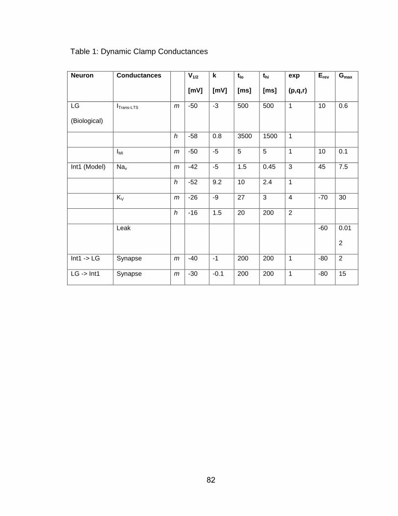
82
Table 1: Dynamic Clamp Conductances
Neuron Conductances V1/2
[mV]
k
[mV]
tlo
[ms]
thi
[ms]
exp
(p,q,r)
Erev Gmax
LG
(Biological)
ITrans-LTS m -50 -3 500 500 1 10 0.6
h -58 0.8 3500 1500 1
IMI m -50 -5 5 5 1 10 0.1
Int1 (Model) Nav m -42 -5 1.5 0.45 3 45 7.5
h -52 9.2 10 2.4 1
KV m -26 -9 27 3 4 -70 30
h -16 1.5 20 200 2
Leak -60 0.01
2
Int1 -> LG Synapse m -40 -1 200 200 1 -80 2
LG -> Int1 Synapse m -30 -0.1 200 200 1 -80 15
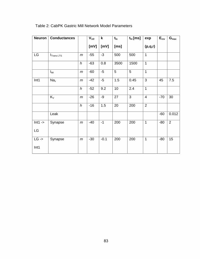
83
Table 2: CabPK Gastric Mill Network Model Parameters
Neuron Conductances V1/2
[mV]
k
[mV]
tlo
[ms]
thi [ms] exp
(p,q,r)
Erev Gmax
LG ITrans-LTS m -55 -3 500 500 1
h -63 0.8 3500 1500 1
IMI m -60 -5 5 5 1
Int1 Nav m -42 -5 1.5 0.45 3 45 7.5
h -52 9.2 10 2.4 1
KV m -26 -9 27 3 4 -70 30
h -16 1.5 20 200 2
Leak -60 0.012
Int1 ->
LG
Synapse m -40 -1 200 200 1 -80 2
LG ->
Int1
Synapse m -30 -0.1 200 200 1 -80 15
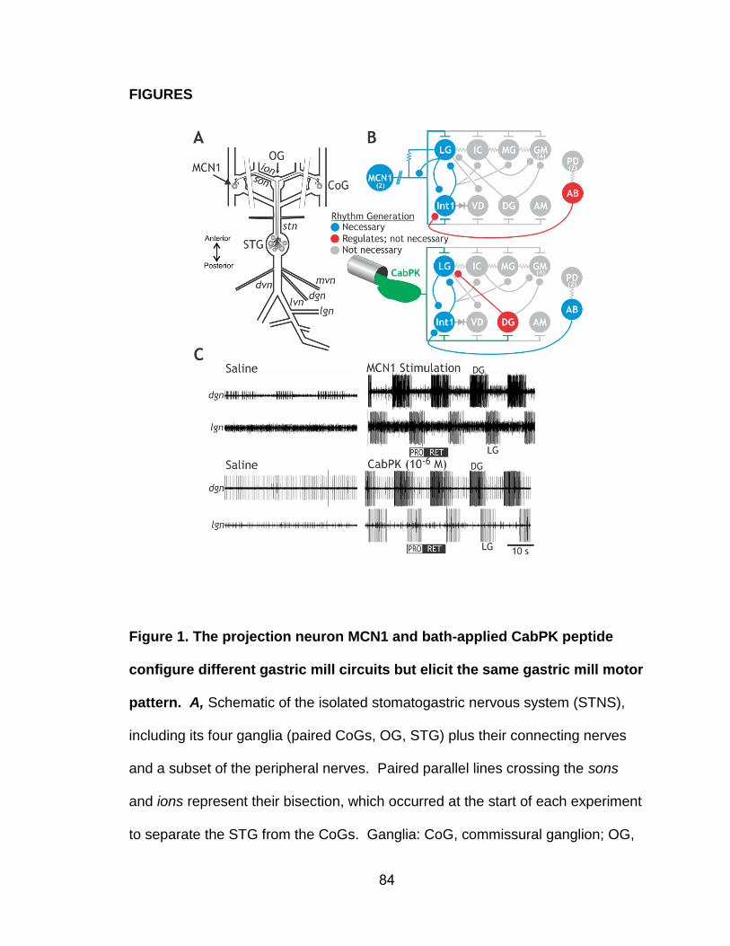
84
FIGURES
Figure 1. The projection neuron MCN1 and bath-applied CabPK peptide
configure different gastric mill circuits but elicit the same gastric mill motor
pattern. A, Schematic of the isolated stomatogastric nervous system (STNS),
including its four ganglia (paired CoGs, OG, STG) plus their connecting nerves
and a subset of the peripheral nerves. Paired parallel lines crossing the sons
and ions represent their bisection, which occurred at the start of each experiment
to separate the STG from the CoGs. Ganglia: CoG, commissural ganglion; OG,
85
oesophageal ganglion; STG, stomatogastric ganglion. Nerves: dgn, dorsal gastric
nerve; dvn, dorsal ventricular nerve; ion, inferior oesophageal nerve; lgn, lateral
gastric nerve; lvn, lateral ventricular nerve; mvn, medial ventricular nerve; son,
superior oesophageal nerve; stn, stomatogastric nerve. Neuron: MCN1,
modulatory commissural neuron 1. B, Schematic of the gastric mill circuit
configured by (top) MCN1 stimulation and (bottom) CabPK peptide superfusion.
Synapse symbols: t-bars, excitation; filled circles, inhibition; resistors, non-
rectifying electrical coupling; diode, rectifying electrical coupling. Parallel lines
crossing the MCN1 axon represent additional distance between the MCN1 soma
in each CoG and its axon terminals in the STG. Numbers in parentheses
indicate the copy number per STNS for each neuron type when it is present as
more than 1 copy. Modified from: Saideman et al. (2007b). C, MCN1 stimulation
and CabPK bath application elicit the same gastric mill motor pattern. These two
manipulations were performed in different preparations. From: Saideman et al.
(2007b).
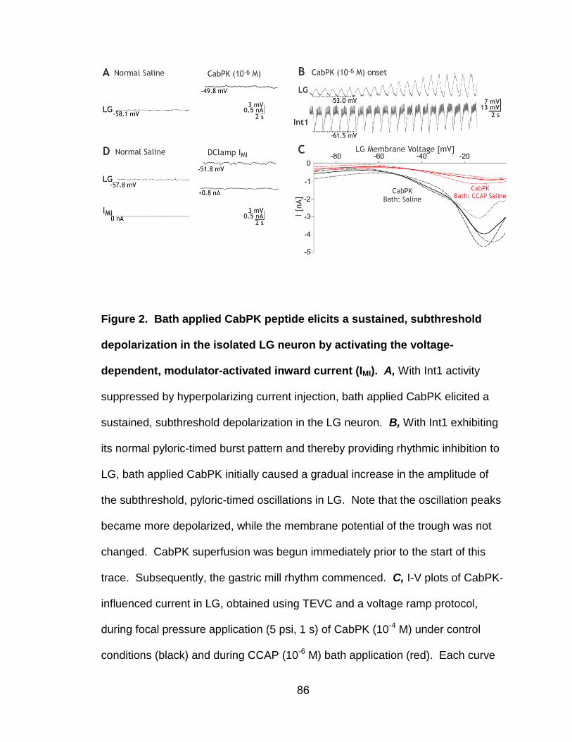
86
Figure 2. Bath applied CabPK peptide elicits a sustained, subthreshold
depolarization in the isolated LG neuron by activating the voltage-
dependent, modulator-activated inward current (IMI). A, With Int1 activity
suppressed by hyperpolarizing current injection, bath applied CabPK elicited a
sustained, subthreshold depolarization in the LG neuron. B, With Int1 exhibiting
its normal pyloric-timed burst pattern and thereby providing rhythmic inhibition to
LG, bath applied CabPK initially caused a gradual increase in the amplitude of
the subthreshold, pyloric-timed oscillations in LG. Note that the oscillation peaks
became more depolarized, while the membrane potential of the trough was not
changed. CabPK superfusion was begun immediately prior to the start of this
trace. Subsequently, the gastric mill rhythm commenced. C, I-V plots of CabPK-
influenced current in LG, obtained using TEVC and a voltage ramp protocol,
during focal pressure application (5 psi, 1 s) of CabPK (10-4 M) under control
conditions (black) and during CCAP (10-6 M) bath application (red). Each curve
87
represents the difference current (CabPK minus control or CCAP condition) as
indicated. Solid curves represent the mean values for each condition; broken
lines represent 3 individual experiments. D, Injection of artificial IMI (gMI = 100
nS) into LG, via the dynamic clamp, in a preparation where Int1 activity was weak
(<5 Hz) caused a sustained depolarization comparable to that resulting from
CabPK application.
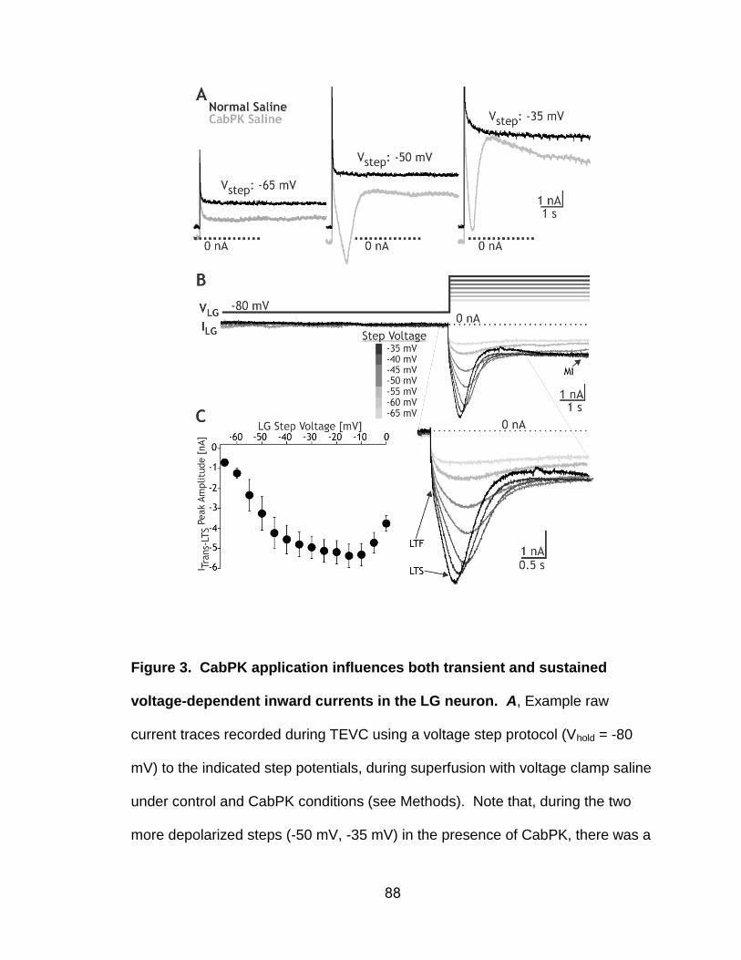
88
Figure 3. CabPK application influences both transient and sustained
voltage-dependent inward currents in the LG neuron. A, Example raw
current traces recorded during TEVC using a voltage step protocol (Vhold = -80
mV) to the indicated step potentials, during superfusion with voltage clamp saline
under control and CabPK conditions (see Methods). Note that, during the two
more depolarized steps (-50 mV, -35 mV) in the presence of CabPK, there was a
89
relatively large amplitude transient inward current, while during all three steps
there was a reduction in the amplitude of the sustained outward current relative
to the control condition. B, (top) CabPK-influenced currents (CabPK condition
minus control condition) resulting from a voltage step protocol during TEVC
included a low threshold, fast transient current (ITrans-LTF), low threshold, slow
transient current (ITrans-LTS) and sustained current (IMI). ITrans-LTS was responsible
for the transient current, while IMI was predominantly responsible for the reduced
outward current in Panel A. ITrans-LTF did not exhibit a distinct, separate peak in
this experiment (see Figs. 4A, 5A), but is evident as an initial steep inward slope
prior to the shallower rising slope representing ITrans-LTS. Entire voltage step
protocol (hold at -80 mV: 10s; step to test voltage: 6s) is shown. (bottom)
Expansion of the current traces to highlight the events occurring during each
voltage step. C, I-V plot of the current (mean ± SE) at the peak of ITrans-LTS for the
step protocol used in experiments such as that in Panel B (n=9).
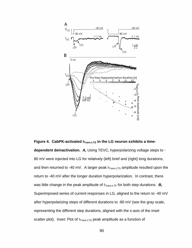
90
Figure 4. CabPK-activated ITrans-LTS in the LG neuron exhibits a time-
dependent deinactivation. A, Using TEVC, hyperpolarizing voltage steps to -
80 mV were injected into LG for relatively (left) brief and (right) long durations,
and then returned to -40 mV. A larger peak ITrans-LTS amplitude resulted upon the
return to -40 mV after the longer duration hyperpolarization. In contrast, there
was little change in the peak amplitude of ITrans-LTF for both step durations. B,
Superimposed series of current responses in LG, aligned to the return to -40 mV
after hyperpolarizing steps of different durations to -80 mV (see the gray scale,
representing the different step durations, aligned with the x-axis of the inset
scatter plot). Inset: Plot of ITrans-LTS peak amplitude as a function of

91
hyperpolarizing step duration from 3 experiments. Each symbol represents a
different experiment. The line represents the sigmoid fit to the data (Igor Pro),
and the arrow represents the calculated midpoint.
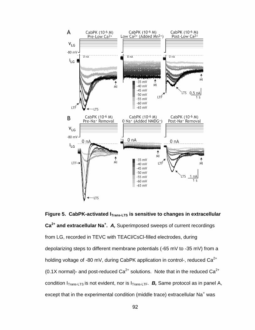
92
Figure 5. CabPK-activated ITrans-LTS is sensitive to changes in extracellular
Ca2+ and extracellular Na+. A, Superimposed sweeps of current recordings
from LG, recorded in TEVC with TEACl/CsCl-filled electrodes, during
depolarizing steps to different membrane potentials (-65 mV to -35 mV) from a
holding voltage of -80 mV, during CabPK application in control-, reduced Ca2+
(0.1X normal)- and post-reduced Ca2+ solutions. Note that in the reduced Ca2+
condition ITrans-LTS is not evident, nor is ITrans-LTF. B, Same protocol as in panel A,
except that in the experimental condition (middle trace) extracellular Na+ was

93
replaced with an equimolar concentration of NMDG+. Note the absence of ITrans-
LTS in the presence of NMDG+.
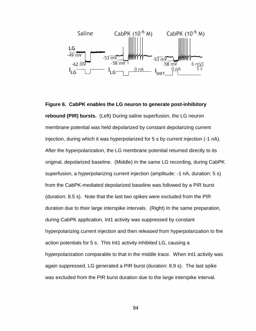
94
Figure 6. CabPK enables the LG neuron to generate post-inhibitory
rebound (PIR) bursts. (Left) During saline superfusion, the LG neuron
membrane potential was held depolarized by constant depolarizing current
injection, during which it was hyperpolarized for 5 s by current injection (-1 nA).
After the hyperpolarization, the LG membrane potential returned directly to its
original, depolarized baseline. (Middle) In the same LG recording, during CabPK
superfusion, a hyperpolarizing current injection (amplitude: -1 nA, duration: 5 s)
from the CabPK-mediated depolarized baseline was followed by a PIR burst
(duration: 8.5 s). Note that the last two spikes were excluded from the PIR
duration due to their large interspike intervals. (Right) In the same preparation,
during CabPK application, Int1 activity was suppressed by constant
hyperpolarizing current injection and then released from hyperpolarization to fire
action potentials for 5 s. This Int1 activity inhibited LG, causing a
hyperpolarization comparable to that in the middle trace. When Int1 activity was
again suppressed, LG generated a PIR burst (duration: 8.9 s). The last spike
was excluded from the PIR burst duration due to the large interspike interval.
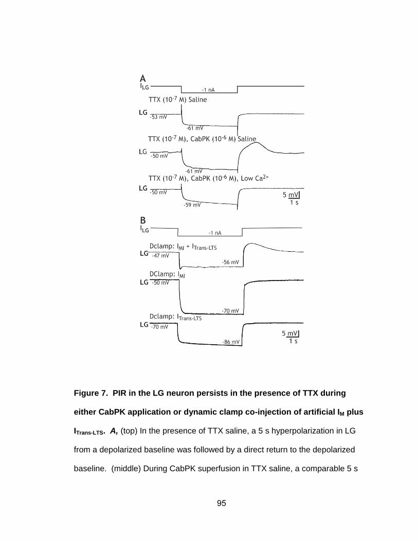
95
Figure 7. PIR in the LG neuron persists in the presence of TTX during
either CabPK application or dynamic clamp co-injection of artificial IM plus
ITrans-LTS. A, (top) In the presence of TTX saline, a 5 s hyperpolarization in LG
from a depolarized baseline was followed by a direct return to the depolarized
baseline. (middle) During CabPK superfusion in TTX saline, a comparable 5 s
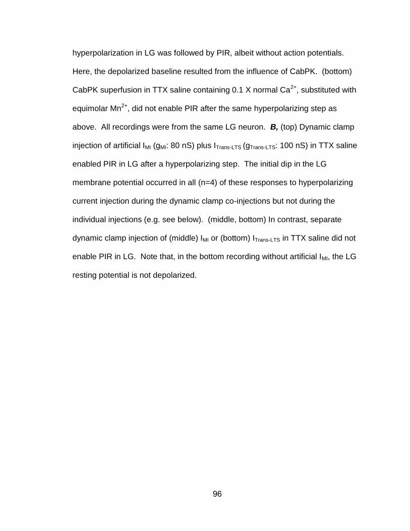
96
hyperpolarization in LG was followed by PIR, albeit without action potentials.
Here, the depolarized baseline resulted from the influence of CabPK. (bottom)
CabPK superfusion in TTX saline containing 0.1 X normal Ca2+, substituted with
equimolar Mn2+, did not enable PIR after the same hyperpolarizing step as
above. All recordings were from the same LG neuron. B, (top) Dynamic clamp
injection of artificial IMI (gMI: 80 nS) plus ITrans-LTS (gTrans-LTS: 100 nS) in TTX saline
enabled PIR in LG after a hyperpolarizing step. The initial dip in the LG
membrane potential occurred in all (n=4) of these responses to hyperpolarizing
current injection during the dynamic clamp co-injections but not during the
individual injections (e.g. see below). (middle, bottom) In contrast, separate
dynamic clamp injection of (middle) IMI or (bottom) ITrans-LTS in TTX saline did not
enable PIR in LG. Note that, in the bottom recording without artificial IMI, the LG
resting potential is not depolarized.
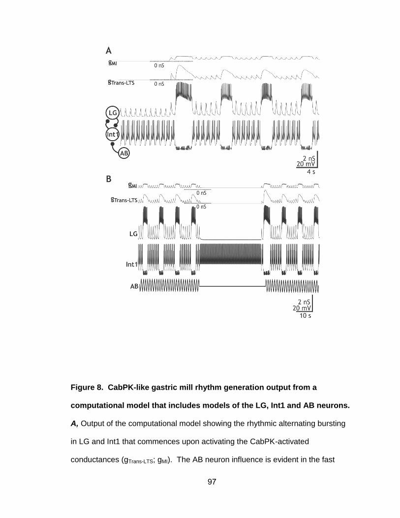
97
Figure 8. CabPK-like gastric mill rhythm generation output from a
computational model that includes models of the LG, Int1 and AB neurons.
A, Output of the computational model showing the rhythmic alternating bursting
in LG and Int1 that commences upon activating the CabPK-activated
conductances (gTrans-LTS; gMI). The AB neuron influence is evident in the fast
98
rhythmic Int1 hyperpolarizations and the associated subthreshold depolarizations
(removal of Int1 inhibition) in LG. Note that the CabPK-activated conductances
all track the membrane potential changes in LG, while gTrans-LTS also exhibits time-
dependent processes, including inactivation during the LG burst and an
increasing level of activation (due to deinactivation) during the LG interburst.
Most hyperpolarized membrane potentials: Int1, -60.5 mV; LG, -58.4 mV.
Synaptic symbol: filled circle, inhibition. B, Suppressing the AB inhibition of Int1
terminates the ongoing gastric mill rhythm in the computational model of the
CabPK-gastric mill rhythm generator, as occurs in the biological system (Bartos
et al., 1999). When the AB activity was temporarily terminated (horizontal line,
middle of the AB trace), Int1 activity changed from a fast rhythmic pattern to tonic
firing, while the LG membrane potential exhibited a steady hyperpolarized
potential. Note that, due to the hyperpolarized LG membrane potential, gTrans-LTS
was at 0 nS while gMI was maintained at a reduced level. Most hyperpolarized
membrane potentials: Int1, -47.7 mV; LG, -58.4 mV.
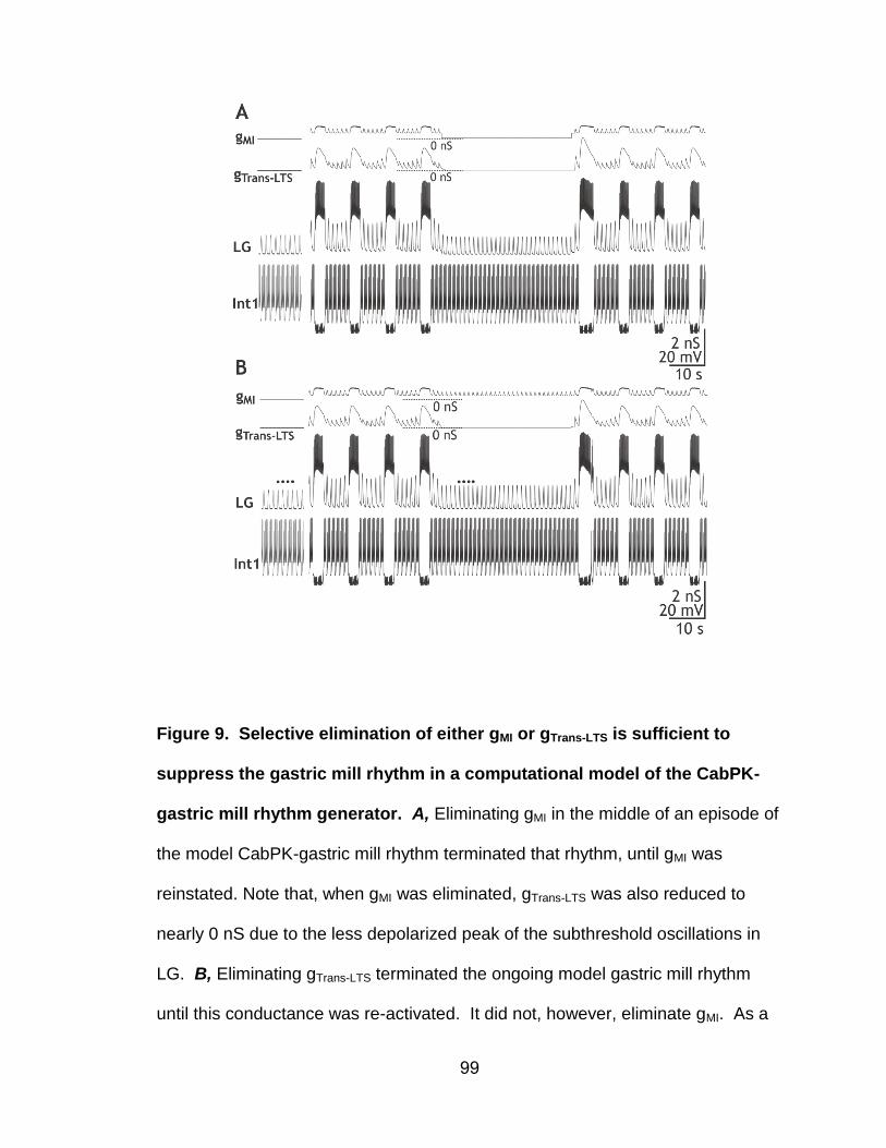
99
Figure 9. Selective elimination of either gMI or gTrans-LTS is sufficient to
suppress the gastric mill rhythm in a computational model of the CabPK-
gastric mill rhythm generator. A, Eliminating gMI in the middle of an episode of
the model CabPK-gastric mill rhythm terminated that rhythm, until gMI was
reinstated. Note that, when gMI was eliminated, gTrans-LTS was also reduced to
nearly 0 nS due to the less depolarized peak of the subthreshold oscillations in
LG. B, Eliminating gTrans-LTS terminated the ongoing model gastric mill rhythm
until this conductance was re-activated. It did not, however, eliminate gMI. As a

100
result, the peaks of the subthreshold oscillations remained larger than prior to the
onset of the CabPK influence (black dots). However, gMI alone was not sufficient
to enable LG to fire a burst.
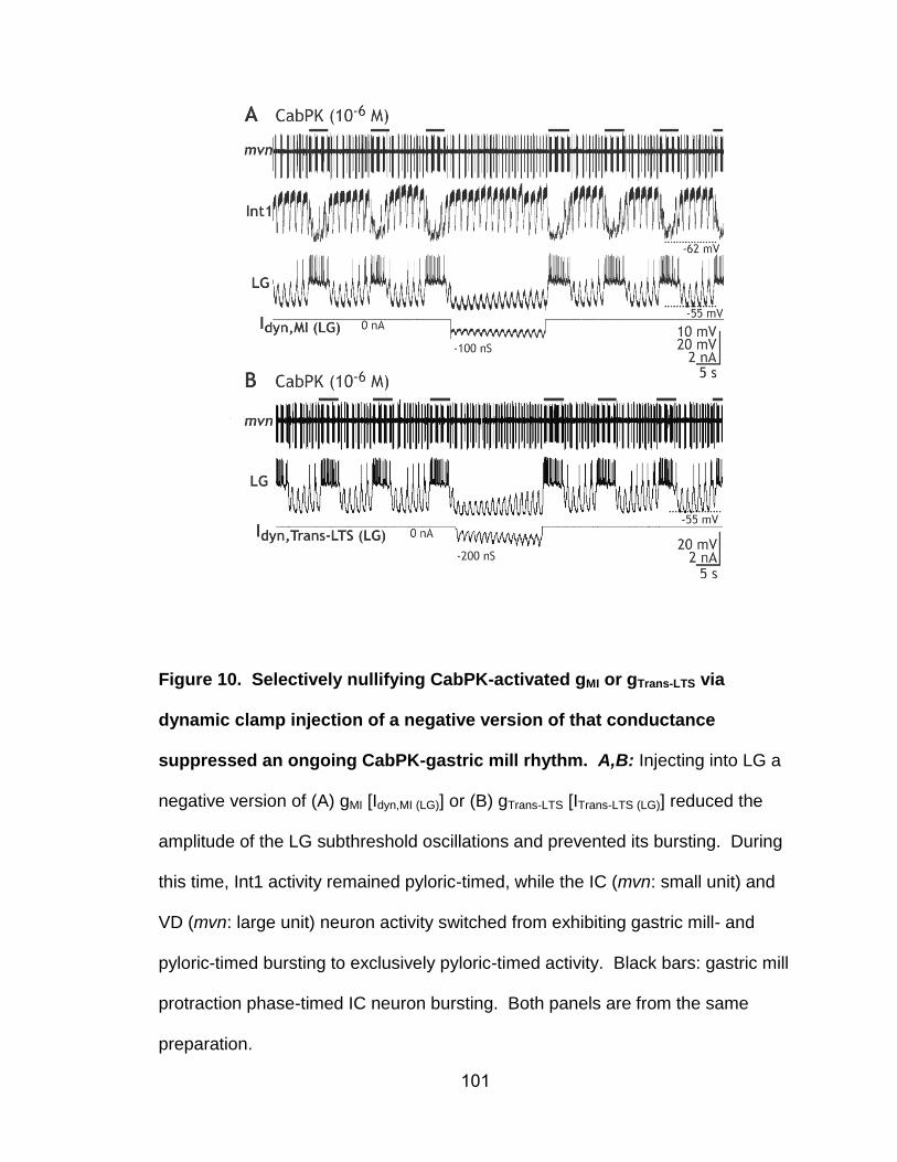
101
Figure 10. Selectively nullifying CabPK-activated gMI or gTrans-LTS via
dynamic clamp injection of a negative version of that conductance
suppressed an ongoing CabPK-gastric mill rhythm. A,B: Injecting into LG a
negative version of (A) gMI [Idyn,MI (LG)] or (B) gTrans-LTS [ITrans-LTS (LG)] reduced the
amplitude of the LG subthreshold oscillations and prevented its bursting. During
this time, Int1 activity remained pyloric-timed, while the IC (mvn: small unit) and
VD (mvn: large unit) neuron activity switched from exhibiting gastric mill- and
pyloric-timed bursting to exclusively pyloric-timed activity. Black bars: gastric mill
protraction phase-timed IC neuron bursting. Both panels are from the same
preparation.
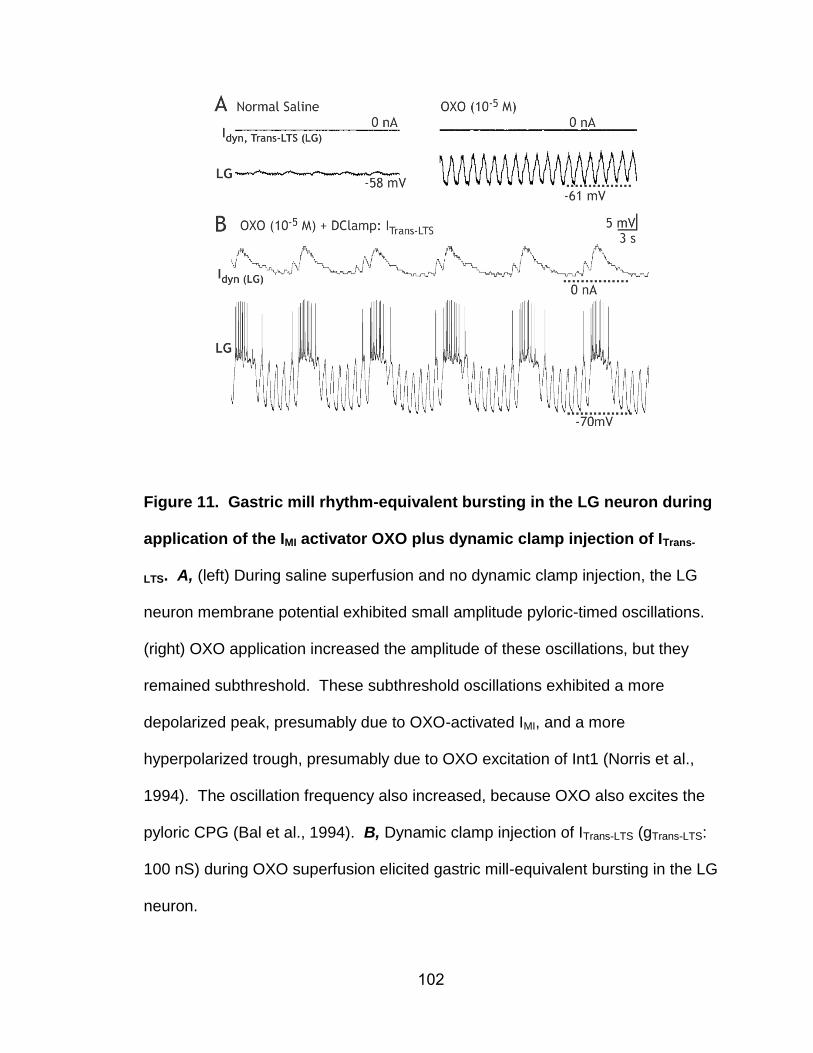
102
Figure 11. Gastric mill rhythm-equivalent bursting in the LG neuron during
application of the IMI activator OXO plus dynamic clamp injection of ITrans-
LTS. A, (left) During saline superfusion and no dynamic clamp injection, the LG
neuron membrane potential exhibited small amplitude pyloric-timed oscillations.
(right) OXO application increased the amplitude of these oscillations, but they
remained subthreshold. These subthreshold oscillations exhibited a more
depolarized peak, presumably due to OXO-activated IMI, and a more
hyperpolarized trough, presumably due to OXO excitation of Int1 (Norris et al.,
1994). The oscillation frequency also increased, because OXO also excites the
pyloric CPG (Bal et al., 1994). B, Dynamic clamp injection of ITrans-LTS (gTrans-LTS:
100 nS) during OXO superfusion elicited gastric mill-equivalent bursting in the LG
neuron.
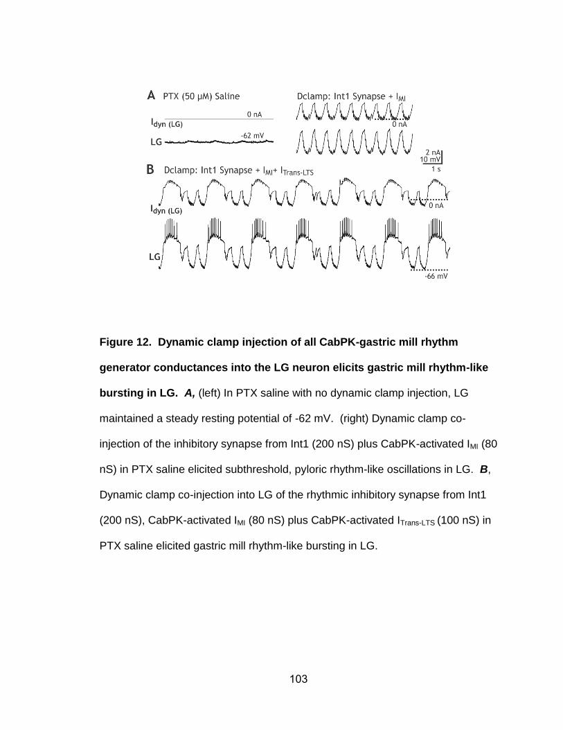
103
Figure 12. Dynamic clamp injection of all CabPK-gastric mill rhythm
generator conductances into the LG neuron elicits gastric mill rhythm-like
bursting in LG. A, (left) In PTX saline with no dynamic clamp injection, LG
maintained a steady resting potential of -62 mV. (right) Dynamic clamp co-
injection of the inhibitory synapse from Int1 (200 nS) plus CabPK-activated IMI (80
nS) in PTX saline elicited subthreshold, pyloric rhythm-like oscillations in LG. B,
Dynamic clamp co-injection into LG of the rhythmic inhibitory synapse from Int1
(200 nS), CabPK-activated IMI (80 nS) plus CabPK-activated ITrans-LTS (100 nS) in
PTX saline elicited gastric mill rhythm-like bursting in LG.
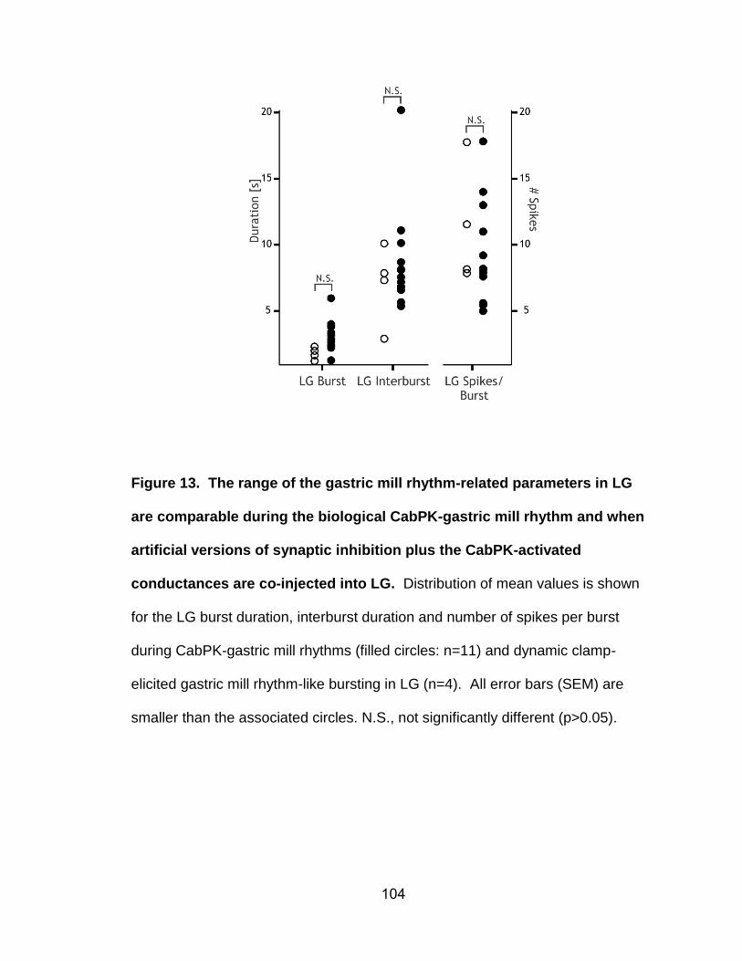
104
Figure 13. The range of the gastric mill rhythm-related parameters in LG
are comparable during the biological CabPK-gastric mill rhythm and when
artificial versions of synaptic inhibition plus the CabPK-activated
conductances are co-injected into LG. Distribution of mean values is shown
for the LG burst duration, interburst duration and number of spikes per burst
during CabPK-gastric mill rhythms (filled circles: n=11) and dynamic clamp-
elicited gastric mill rhythm-like bursting in LG (n=4). All error bars (SEM) are
smaller than the associated circles. N.S., not significantly different (p>0.05).
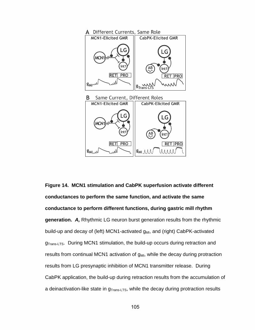
105
Figure 14. MCN1 stimulation and CabPK superfusion activate different
conductances to perform the same function, and activate the same
conductance to perform different functions, during gastric mill rhythm
generation. A, Rhythmic LG neuron burst generation results from the rhythmic
build-up and decay of (left) MCN1-activated gMI, and (right) CabPK-activated
gTrans-LTS. During MCN1 stimulation, the build-up occurs during retraction and
results from continual MCN1 activation of gMI, while the decay during protraction
results from LG presynaptic inhibition of MCN1 transmitter release. During
CabPK application, the build-up during retraction results from the accumulation of
a deinactivation-like state in gTrans-LTS, while the decay during protraction results
106
from the time-dependent inactivation of this conductance. B, The gMI in LG is the
burst generating conductance during the MCN1-gastric mill rhythm, whereas it
provides a sustained but subthreshold depolarizing drive that facilitates PIR burst
generation by ITrans-LTS during the CabPK-gastric mill rhythm. Note that the
different gMI trajectories during protraction result from gMI being both voltage- and
synaptic inhibition-dependent during the MCN1-gastric mill rhythm whereas
during the CabPK-gastric mill rhythm it is only voltage-dependent.
107
CHAPTER 3
Differential Sensitivity to Modulatory Input of Different Circuits Generating the
Same Motor Pattern
Jason C. Rodriguez
Nicholas D. DeLong
Matthew S. Kirby
Michael P. Nusbaum*
Dept. of Neuroscience, 215 Stemmler Hall, Perelman School of Medicine, Univ.
of Pennsylvania, Philadelphia, PA 19104-6074
Running Header: State-dependent responses of a conserved motor pattern
Abstract: 247/250 Words
Introduction: 500/500 Words
Discussion: 771/1500 Words
Pages: 39
Figures: 13
*Corresponding Author:
108
Dr. Michael P. Nusbaum
Dept. of Neuroscience
215 Stemmler Hall
Perelman School of Medicine
Univ. of Pennsylvania
Philadelphia, PA 19104-6074
Phone: 215-898-1585
FAX: 215-573-9050
Email: [email protected]
Acknowledgments: This work was supported by National Institute of Neurological
Diseases and Stroke Grant No. R37-NS029436 (M.P.N.).
109
ABSTRACT
Different modulatory inputs can elicit the same output pattern from a central
pattern generator (CPG) despite configuring different circuits. We are assessing
the functional consequences of this organization by determining whether such
circuits are differentially sensitive to additional input. We use the gastric mill
(chewing) CPG, which generates the biphasic (tooth protraction, retraction)
gastric mill motor pattern in the crab stomatogastric ganglion. Specifically,
stimulating the projection neuron MCN1 or superfusing the neuropeptide CabPK
elicits the same gastric mill motor pattern via distinct cellular and synaptic
mechanisms. The MCN1-driven gastric mill rhythm is regulated by sensory
feedback and circulating hormones. For example, the muscle stretch-sensitive
GPR neurons slow this rhythm by prolonging tooth retraction, while the peptide
hormone CCAP slows it by prolonging protraction. We show here that, relative to
the MCN1-gastric mill rhythm, the CabPK-rhythm exhibits a similar response to
GPR stimulation but responds differently to CCAP application. First, GPR slows
the MCN1-rhythm via presynaptic inhibition of MCN1, whereas it slows the
CabPK-rhythm by its synaptic action on one or more gastric mill rhythm
generator neurons. Second, the response threshold to CCAP is higher for
CabPK (~10-7 M) than for MCN1 (10-10 M). Third, CCAP decreases the CabPK-
rhythm cycle period by reducing retraction duration, while it increases the MCN1-
rhythm cycle period by increasing protraction duration. These results
demonstrate that different network states that produce the same steady-state

110
output can be differentially sensitive to some inputs while maintaining
responsiveness to others, albeit via different cellular and synaptic mechanisms.
111
INTRODUCTION
Different modulatory inputs commonly elicit distinct outputs from a central pattern
generator (CPG) network (Getting, 1989; Stein, 2009; Marder, 2012; Nusbaum
and Blitz, 2012). In some cases, however, distinct modulators configure different
CPG circuit states but elicit the same output pattern (Saideman et al., 2007b;
Goaillard et al., 2009; Grashow et al., 2009; Rodriguez et al., 2013). This latter
condition suggests the need for caution when interpreting the consequences of
CPG activation by different pathways in systems where the CPG is not
accessible to a cellular-level analysis. Additionally, different CPG circuit states
generating the same motor pattern could exhibit distinct responses to particular
inputs, such as those from sensory feedback and circulating hormones.
We are assessing this latter issue using the gastric mill (chewing) circuit in
the crab (Cancer borealis) stomatogastric ganglion (STG). Specifically,
selectively stimulating the projection neuron MCN1 (modulatory commissural
neuron 1) and superfusing the neuropeptide CabPK (C. borealis pyrokinin) elicit
the same gastric mill motor pattern, despite MCN1 not containing CabPK and the
CabPK-gastric mill rhythm occurring without MCN1 activity (Saideman et al.,
2007b). MCN1 and CabPK provide distinct modulatory influences to the same
gastric mill rhythm generator neuron (LG, lateral gastric), which results in
different gastric mill circuits and rhythm-generating mechanisms (Saideman et
al., 2007b; Rodriguez et al., 2013).
The MCN1-driven gastric mill rhythm is influenced by the muscle stretch-
sensitive GPR (gastropyloric receptor) sensory neuron and the peptide hormone
112
CCAP (crustacean cardioactive peptide) (Beenhakker et al., 2005; Kirby and
Nusbaum, 2007; DeLong et al., 2009ab; DeLong and Nusbaum, 2010). In brief,
GPR stimulation slows this rhythm by selectively prolonging the retractor phase.
This effect results from GPR presynaptic inhibition of the STG terminals of MCN1
(MCN1STG), which inhibits MCN1 neuropeptide release but not GABA corelease.
CCAP also slows this gastric mill rhythm, but by selectively prolonging
protraction. This effect results from convergent activation of the same ionic
current (IMI, modulator-activated inward current) in the LG neuron by CCAP and
the MCN1-released neuropeptide CabTRP Ia (C. borealis tachykinin-related
peptide Ia).
Here, we determine the influence of GPR and CCAP on the CabPK-
gastric mill rhythm. GPR stimulation slowed this rhythm by selectively prolonging
retraction, as during the MCN1-rhythm, albeit via a distinct synaptic pathway.
Interestingly, despite operating via distinct synapses, during both rhythms the
GPR action was mimicked by focal application of the GPR cotransmitter
serotonin (5HT) (DeLong et al., 2009a). 5HT causes a slow inhibition of LG but
has no direct effect on the gastric mill rhythm generator neuron Int1 (DeLong et
al., 2009a). CCAP superfusion did not influence the CabPK-gastric mill rhythm
unless it was applied at ≥ 10-7 M. In contrast, the threshold CCAP action on the
MCN1-rhythm is ~10-10 M. Furthermore, in contrast to the MCN1-rhythm, when
CCAP was applied at effective concentrations the CabPK-rhythm cycled faster,
due to a reduced retraction duration. Thus, the same motor pattern driven by

113
distinct circuit states can be equally sensitive to a particular input, albeit via
different synaptic mechanisms, and can also respond differently to other inputs.
114
MATERIALS AND METHODS
Animals. Male Jonah crabs (Cancer borealis) were purchased from commercial
suppliers (Fresh Lobster; Marine Biological Laboratory) and maintained in
aerated, filtered artificial seawater at 10 – 12° C. Animals were cold anesthetized
by packing in ice for at least 30 min before dissection, after which the foregut was
removed, in physiological saline at ~4° C, and the STNS isolated.
Solutions. C. borealis physiological saline contained (in mM): 440 NaCl, 26
MgCl2, 13 CaCl2, 11 KCl, 10 Trisma base, 5 maleic acid, 5 glucose, pH 7.4 – 7.6.
All preparations were superfused continuously with C. borealis saline (8 – 12° C).
CabPK-I, CabPK-II (Saideman et al., 2007a) (Biotechnology Center, Univ. of
Wisconsin, Madison, WI), and CCAP (DeLong and Nusbaum, 2010) (Bachem)
were each diluted from a stock solution (10-3 M) into physiological saline
immediately before use. Bottles containing C. borealis saline, CabPK saline,
CCAP saline, and CabPK/CCAP saline were connected to the same switching
manifold for rapid solution changes.
Electrophysiology. Electrophysiology experiments were performed using
standard techniques for this system (Beenhakker and Nusbaum, 2004). In brief,
the isolated STNS (Fig. 1A) was pinned into a silicone elastomer (Sylgard 184,
KR Anderson)-lined Petri dish. Extracellular nerve recordings were obtained
using pairs of stainless steel wire electrodes (reference and recording) whose
ends were pressed into the Sylgard-coated dish. A differential AC amplifier
115
(Model 1700: AM Systems) amplified the voltage difference between the
reference wire, in the main bath compartment, and the recording wire, isolated
with a section of an individual nerve from the main bath compartment by
petroleum jelly (Vaseline, Lab Safety Supply). This signal was then further
amplified and filtered (Model 410 Amplifier: Brownlee Precision). For
extracellular nerve stimulation, the pair of wires used to record nerve activity was
placed into a stimulus isolation unit (Model SIU 5: Astromed/Grass Instruments)
connected to a stimulator (Model S88: Astromed/Grass Instruments).
For current clamp experiments, intrasomatic recordings of STG neurons
were made with sharp glass microelectrodes (15 – 30 MΩ) filled with either
K2SO4 (0.6 M) plus KCl (10 mM) or KCl (1 M). All intracellular recordings were
amplified using Axoclamp 900A amplifiers (Molecular Devices) in bridge mode or
discontinuous current clamp mode (2 – 5 kHz sampling rate) and digitized at 5
kHz using a Micro 1401 data acquisition interface and Spike2 software
(Cambridge Electronic Design). To facilitate intracellular recording, the
desheathed STG was viewed with light transmitted through a dark-field
condenser (Nikon). In all experiments, the STG was isolated from the
commissural ganglia (CoGs) by bisecting the inferior (ions)- and superior
oesophageal nerves (sons) (Fig. 1A). Individual STNS neurons were identified
by their axonal pathways, activity patterns and interactions with other neurons
(Weimann et al., 1991; Beenhakker and Nusbaum, 2004).
During the gastric mill rhythm, the LG burst defines the protractor phase
while its interburst duration, which is equivalent to the duration of Int1 activity,
116
defines the retractor phase (Coleman et al., 1995; Bartos et al., 1999; Diehl et al.,
2013).
Data analysis. Data were collected onto a computer, with later playback onto a
chart recorder (Astro-Med Everest). Acquisition onto computer (sampling rate 5
kHz) used the Spike2 data acquisition and analysis system (Cambridge
Electronic Design). Analysis of CabPK-gastric mill rhythm parameters was
conducted on the digitized data using a custom-written Spike2 program (The
Crab Analyzer: freely available at http://www.uni-ulm.de/~wstein/spike2/index.html).
For gastric mill rhythm analyses, unless otherwise stated, each data point
in a data set was derived by determining the mean for the analyzed parameter
from 10 consecutive gastric mill cycles. One gastric mill cycle was defined as
extending from the onset of consecutive LG neuron action potential bursts
(Beenhakker et al., 2004; Wood et al., 2004) . Thus, the gastric mill cycle period
was measured as the duration (s) between the onset of two successive LG
neuron bursts. The protractor phase was measured as the LG burst duration,
while the retractor phase was measured as the LG interburst duration. The
gastric mill rhythm-timed LG burst duration was defined as the duration (s)
between the onset of the first and last action potential within an impulse burst,
during which no inter-spike interval was longer than 1.5 s (a duration that is ~one
pyloric cycle period during the CabPK-gastric mill rhythm, and briefer than the
duration of each gastric mill phase; Saideman et al., 2007a). The intraburst firing
rate of LG was defined as the number of action potentials minus one, divided by
the burst duration.
117
Data were plotted with Igor Pro (version 6.10A). Figures were produced
using CorelDraw (version 16.0 for Windows). Statistical analyses were
performed with Microsoft Excel (Microsoft) and SigmaStat 3.0 (SPSS).
Comparisons were made to determine statistical significance using the paired
Student’s t-. In all experiments, the effect of each manipulation was reversible,
and there was no significant difference between the pre- and post-manipulation
groups. Data are expressed as the mean ± standard error (SE).
Gastric Mill Model. We modified an existing computational model of the CabPK-
gastric mill rhythm generator (Nadim et al., 1998; Beenhakker et al., 2005;
Rodriguez et al., 2013). The previously published version modeled the LG and
Int1 neurons as having multiple compartments separated by an axial resistance,
with each compartment possessing intrinsic and/or synaptic conductances. The
parameters of the CabPK-gastric mill rhythm generator model were based on
both previously published voltage clamp analyses in STG neurons (including LG)
and on the LG neuron voltage clamp results obtained with CabPK modulation
(Golowasch and Marder, 1992; Swensen and Marder, 2000, 2001; DeLong et al.,
2009b; Rodriguez et al., 2013). To mimic in the computational model the effects
of CCAP bath application to the biological system, we added CCAP-activated IMI
to the LG neuron and/or Int1 neuron dendrite compartment as an intrinsic (non-
synaptically activated) current (Table 1). To mimic the effects of GPR, we
imported the GPR neuron and synapses from a published model (Beenhakker et
al., 2005). To assess the impact of the model GPR neuron activation on the
118
CabPK-GMR, we either stimulated GPR during protraction (10 Hz; 2 s) or during
retraction (10 Hz; 15 s).
Simulations were performed on a PC with the freely available Ubuntu
Linux operating system (www.ubuntu.com). We used the Network simulation
software developed in the Nadim laboratory
(http://stg.rutgers.edu/software/network.htm). This included using a fourth-order
Runge–Kutta numerical integration method with time steps of 0.05 and 0.01 ms.
Results were visualized by plotting outputted data points using the freely
available Gnuplot software package (www.gnuplot.info). In most figures showing
the model output, we present conductance (g) instead of the associated current
(I) to more clearly display the trajectory during the gastric mill retractor and
protractor phases. The main difference between “g” and “I” is that the former
lacks the fast transient changes that occur in the latter during each LG action
potential (DeLong et al., 2009a). In particular, the relatively slow kinetics of the
CabPK-activated conductances make them insensitive to these fast transient
changes in voltage.
119
RESULTS
The gastric mill rhythm is an episodic motor pattern, in vivo and in vitro, which is
driven by modulatory inputs that are not spontaneously active (Nusbaum and
Beenhakker, 2002; Beenhakker et al., 2004; White and Nusbaum, 2011;
Nusbaum and Blitz, 2012; Diehl et al., 2013; Rodriguez et al., 2013). There are
at least several distinct gastric mill motor patterns, driven by different pathways,
but in all cases the core rhythm generator appears to include the reciprocally
inhibitory protractor motor neuron LG and retractor interneuron Int1 (interneuron
1) (Saideman et al., 2007; White and Nusbaum, 2011). When the gastric mill
rhythm is not occurring, Int1 is spontaneously active while LG is silent.
Consequently, a pivotal action of any gastric mill rhythm-activating pathway is to
enable LG to become active and burst in alternation with Int1.
In contrast to the ability of most gastric mill-activating pathways to drive
different gastric mill motor patterns, tonic MCN1 stimulation and bath applied
CabPK (10-6 M) elicit the same gastric mill motor pattern (Fig. 1B) (Saideman et
al., 2007b). However, despite the ability of MCN1 and CabPK to each drive the
gastric mill rhythm primarily by enabling rhythmic bursting in the LG neuron, the
associated cellular and synaptic mechanisms for gastric mill rhythm generation
are distinct (Fig. 1C) (Rodriguez et al., 2013). In brief, MCN1 provides the LG
neuron with metabotropic excitation (during retraction) that culminates in a LG
burst (during protraction), with the transition from retraction to protraction
including LG presynaptic inhibition of MCN1STG (Coleman et al., 1995; Bartos et
al., 1999; DeLong et al., 2009a). During retraction, MCN1-released CabTRP Ia
120
elicits a continual build-up of IMI in LG until there is sufficient depolarizing drive to
trigger a LG burst. During protraction, LG presynaptically inhibits MCN1
transmitter release, causing a slow decay of IMI. During this time, however, LG
also receives relatively large amplitude electrical EPSPs (eEPSPs) from MCN1
which strengthens LG activity until the LG membrane potential repolarizes and
self-terminates, due to IMI decay.
Unlike the MCN1 influence on the LG neuron, CabPK enables LG to
periodically generate a post-inhibitory rebound (PIR) burst after a sufficient
duration of inhibitory input from Int1 (Rodriguez et al., 2013). These PIR bursts
result from CabPK activation of IMI plus a transient, low threshold slow inward
current (ITrans-LTS). IMI, which is the burst-generating current during the MCN1-
rhythm, instead enables a subthreshold depolarization in LG during the CabPK-
rhythm which facilitates each PIR burst generated by ITrans-LTS. Thus, MCN1 and
CabPK configure different gastric mill network states. We therefore tested the
hypothesis that these different states would enable the same gastric mill motor
pattern to be altered differently by additional input, such as that provided by
circulating hormones or sensory input.
CCAP distinctly influences the CabPK- and MCN1-gastric mill rhythms
CCAP (Pro-Phe-Cys-Asn-Ala-Phe-Thr-Gly-CysNH2) is a highly conserved
neuropeptide transmitter and hormone localized in neurons and neurohemal
structures of many arthropods, including C. borealis (Trube et al., 1994).
Simulating hormonal CCAP release by bath application modulates the MCN1-
121
gastric mill rhythm (Kirby and Nusbaum, 2007; DeLong et al., 2009a; DeLong
and Nusbaum, 2010). Specifically, CCAP slows the MCN1-gastric mill rhythm by
selectively prolonging protraction. This action results from CCAP activation of IMI
in the LG neuron (DeLong et al., 2009a). Insofar as the CabPK-gastric mill
rhythm is also driven primarily through actions on the LG neuron, we anticipated
that CCAP would also influence this motor pattern.
Bath-application of a likely hormonal CCAP concentration (10-8 M) did not
consistently change the CabPK-elicited gastric mill cycle period (Control: 14.04 ±
2.1 s; CCAP: 12.57 ± 1.6 s, n=7, p=0.13), protraction duration (Control: 3.54 ±
0.4 s; CCAP: 3.62 ± 0.4 s, n=7, p=0.70) or retraction duration (Control: 10.50 ±
2.0 s; CCAP: 8.95 ± 1.6 s, n=7, p=0.12). Furthermore, despite the ability of
CCAP to activate IMI in LG, there was no change in the LG firing frequency
(Control: 4.11 ± 0.3 Hz; CCAP: 4.29 ± 0.2 Hz, n=7, p=0.39). Insofar as the
CCAP influence on the MCN1-gastric mill rhythm has a considerably lower
threshold (10-10 M), the CabPK-gastric mill rhythm was relatively insensitive to
CCAP modulation.
Applying a relatively high CCAP concentration (10-6 M) did consistently
influence the CabPK-gastric mill rhythm (Fig. 2). Specifically, its presence
decreased the CabPK-gastric mill cycle period (Control: 16.45 ± 2.8 s; CCAP:
12.93 ± 2.2 s, n=4, p<0.05). This action was accompanied by a briefer retraction
phase (Control: 11.38 ± 1.9 s; CCAP: 8.06 ± 1.5 s, n=4, p<0.05), with no change
in protraction duration (Control: 5.07 ± 0.8 s; CCAP: 4.87 ± 0.7 s, n=4, p=0.68).
In parallel with the unchanged LG burst duration, the presence of CCAP did not
122
alter the LG intraburst firing frequency (Control: 3.37 ± 0.3 Hz; CCAP: 3.65 ± 0.4,
n=4, p=0.48).
The gastric mill rhythm generator neurons (LG, Int1) are each directly
excited by CCAP (Kirby and Nusbaum, 2007). Therefore, we determined
whether the gastric mill rhythm response to 10-6 M CCAP likely resulted from
peptide modulation of LG and/or Int1, by importing these actions into our
previously developed computational model of the CabPK-gastric mill rhythm
generator (Rodriguez et al., 2013). The computational model was adapted to
simulate CCAP peptide modulation by its activation of IMI in LG and/or Int1.
Previous work established that CCAP activates IMI in LG, and we made the
assumption that its excitatory action on Int1 also results from IMI activation
because CCAP exclusively activates IMI in several pyloric circuit neurons as well
as LG (Swensen and Marder, 2000, 2001; DeLong et al., 2009b). To assess its
effectiveness and robustness, we added CCAP-activated IMI to LG or Int1 across
a range of gMI values (0 – 20 pS).
Adding gMI (range: 0 – 10 pS) to Int1 modestly but consistently increased
the Int1 intraburst firing frequency (Control: 5.9 ± 0.0 Hz; Added IMI: 6.1 ± 0.0 Hz;
n=3; p<0.01). This effect was accompanied by an increased retraction duration
(Control: 9.5 ± 0.08 s; Added IMI: 12.2 ± 0.0004 s; n=3; p<0.01) with no change in
protraction duration (Control: 2.7 ± 0.09 s; Added IMI: 2.7 ± 0.0004 s; n=3;
p=0.23) (Fig. 3). This result was opposite to the biological CabPK-gastric mill
rhythm response to CCAP, where the cycle period was decreased due to a
selective decrease in retraction duration (Fig. 2). This result suggested that
123
CCAP excitation of Int1 was not the dominant CCAP action during the CabPK-
rhythm.
Adding gMI to the LG neuron in the CabPK-gastric mill model selectively
decreased the retractor phase duration (Control: 12.2 ± 0.0004 s; Added IMI: 10.8
± 0.0005 s; n=3; p<0.01) over a range of conductance values (0-5 pS), without
changing the LG firing frequency or burst duration. Therefore, IMI activation by
CCAP in the LG neuron was sufficient to replicate the effect of bath-applied
CCAP on the biological CabPK-gastric mill rhythm suggesting that, as for the
MCN1-gastric mill rhythm, LG is the primary target by which CCAP regulates this
rhythm.
Comparable regulation of the CabPK- and MC1-gastric mill rhythms
The GPR neuron dendrites arborize in specific lateral tooth protractor
muscles (Katz et al., 1989). As a result, stretch of these muscles during the
gastric mill retraction phase activates GPR (Katz et al., 1989; Birmingham et al.,
1999). Selectively stimulating GPR during the retraction phase of the MCN1-
gastric mill rhythm in the isolated STNS, by electrically stimulating the nerve
(gpn) through which its axon projects towards the STG, consistently prolongs
retraction without altering the subsequent protraction phase (Beenhakker et al.,
2005, 2007).
GPR is a multi-transmitter neuron which contains serotonin (5HT),
acetylcholine (ACh) and an allatostatin (AST) peptide (Katz et al., 1989; Skiebe
and Schneider, 1994). It also directly inhibits MCN1STG and LG, and directly
124
excites Int1 (Beenhakker et al., 2005; DeLong et al., 2009b). However, the GPR
action on the MCN1-gastric mill rhythm results exclusively from its slow,
serotonergic inhibition of MCN1STG, which selectively inhibits neuropeptide
release from MCN1STG and thereby reduces the rate of IMI build-up in LG
(DeLong et al., 2009a). Insofar as MCN1 does not participate in the CabPK-
gastric mill rhythm, we anticipated that GPR stimulation would influence this latter
motor pattern differently.
Stimulating GPR during the CabPK-gastric mill rhythm, using the same
stimulation parameters as during the MCN1-rhythm (5 Hz tonic stimulation during
each retraction phase), had a comparable effect to that occurring during the
MCN1-rhythm. Specifically, GPR stimulation prolonged the CaPK-gastric mill
cycle period (Control: 15.9 ± 3.3 s; GPR: 35.8 ± 8.9 s; n=6; p<0.05) by prolonging
the retraction phase (Control: 11.6 ± 2.8 s; GPR: 31.4 ± 8.8 s; n=6; p<0.05)
without altering protraction phase duration (Control: 4.4 ± 0.7 s; GPR: 4.4 ± 0.8 s;
n=6; p=0.49) (Fig. 4). Insofar as MCN1 activity is not necessary for the CabPK-
gastric mill rhythm, it was unlikely that the comparable GPR action during these
two gastric mill rhythms occurred via the same synaptic mechanism (i.e.
presynaptic inhibition of MCN1STG).
Although GPR regulates the MCN1-gastric mill rhythm exclusively by its
presynaptic inhibition of MCN1STG, as noted above GPR has additional synapses
onto the gastric mill rhythm generator which are functionally ineffective during the
MCN1-rhythm (Fig. 5). These additional GPR synapses include a slow
serotonergic inhibition of LG and a fast, non-serotonergic ionotropic excitation of
125
Int1 (DeLong et al., 2009b). The transmitter(s) responsible for GPR excitation of
Int1 is not known, but it is not likely serotonin because Int1 is not responsive to
5HT application (DeLong et al., 2009b). GPR also has a strong, serotonergic
excitatory action on the DG neuron (Katz and Harris-Warrick, 1989; Kiehn and
Harris-Warrick, 1992; Zhang and Harris-Warrick, 1995). DG does not influence
the MCN1-gastric mill rhythm generator, but it does influence the rhythm
generator during the CabPK-gastric mill rhythm (Saideman et al., 2007b).
Any or all of the aforementioned GPR actions could contribute to the
selective GPR prolongation of the CabPK-retraction phase. For example, its
direct inhibition of LG could impede the LG ability to depolarize sufficiently during
retraction to generate its PIR burst and transition the rhythm to the protraction
phase. Additionally, insofar as both Int1 and DG are active during the CabPK-
gastric mill retraction phase and inhibit LG at this time, GPR excitation of either
(or both) of these neurons could strengthen their inhibition of LG and slow the
onset of the LG PIR burst.
Suppressing DG activity with hyperpolarizing current injection during the
CabPK-rhythm did not alter the ability of GPR to prolong the retraction phase
(Control: 5.1 ± 1.3 s; GPR: 16.3 ± 3.4 s; n=5; p<0.05) (Fig. 6). Thus, DG activity
was not necessary for the GPR action on the CabPK-gastric mill rhythm.
It was not feasible to assess the relative contribution of the GPR actions
on LG and Int1 to its influence on the CabPK-gastric mill rhythm by selectively
suppressing each ones activity, as we did for the DG neuron, because
suppressing LG or Int1 activity terminates the CabPK-gastric mill rhythm
126
(Saideman et al., 2007b). Therefore, we first assessed the consequences of
these GPR synaptic actions in our computational model of the CabPK-gastric mill
rhythm generator. To this end, we incorporated into this model the previously
developed model GPR synapses that were used to evaluate the likely site of
GPR action during the MCN1-gastric mill rhythm (Delong et al., 2009b). Using
this approach, we obtained the output from three different versions of the model
CabPK-rhythm generator, including ones containing only GPR excitation of Int1
or inhibition of LG, and one in which both synapses were active.
The computational model output that occurred with either GPR excitation
of Int1 or GPR inhibition of LG were equally effective at prolonging the CabPK-
gastric mill retractor phase (Control: 9.5 ± 0.1 s; GPR to Int1 synapse active: 29.0
± 0.8 s, n=3, p<0.01; GPR to LG synapse active: 29.0 ± 0.8 s; n=3, p<0.01) (Fig.
7,8). Additionally, there was no change in protraction duration when either GPR
synapse was activated (Control: 2.7 ± 0.1 s; GPR to Int1 synapse active: 2.8 ±
0.01 s; GPR to LG synapse active: 2.8 ± 0.01 s; p=0.1). For both GPR synapses
we implemented a 5-fold range of conductance values and found they had similar
effectiveness and potency for prolonging the LG interburst. Interestingly, when
either GPR synapse was activated during protraction, instead of retraction, it
prematurely terminated the LG burst (LG burst duration: Control, 2.7 ± 0.10 s;
GPR to Int1 synapse, 1.5 ± 0.08 s; GPR to LG synapse 1.4 ± 0.10 s; n=3,
p<0.01), even when implemented at a relatively low conductance level (e.g. 0.3
nS) (Fig. 9). In contrast, GPR stimulation during MCN1-protraction did not
influence either protraction or the subsequent retraction phase (DeLong et al.,
127
2009b). These results suggested that the GPR synapses onto both LG and Int1
are candidates for mediating the GPR influence on the CabPK-gastric mill
rhythm.
To test whether coactivation of both GPR synapses (i.e. excitation of Int1,
inhibition of LG) might also selectively prolong the CabPK-gastric mill retraction
phase, we ran the model with both synapses active. This model output did
indeed produce comparable results to the separate GPR actions on LG and Int1
(Fig. 10), producing a prolonged retractor phase (Control: 9.5 ± 0.09 s; GPR
synapses active: 33.4 ± 6.49 s; n=3, p<0.5), without any change in protraction
(Control: 2.7 ± 0.10 s; GPR synapses active: 2.8 ± 0.01 s; n=3, p=0.1). We next
determined the impact of these synapse on the CabPK-rhythm in the biological
preparation.
We examined the Int1 response to GPR stimulation during CabPK-GMR
indirectly, by determining the frequency of Int1-mediated IPSPs in LG when DG
was hyperpolarized, to eliminate the confound of DG-mediated IPSPs in LG.
Under these conditions, Int1 is the sole source of IPSPs in LG. The presence of
CabPK did not alter the frequency of these IPSPs in LG (Control: 14.5 ± 1.2 Hz;
CabPK: 17.0 ± 1.7 Hz; n=3; p=0.1). It should be noted that, because CabPK also
directly excites Int1 (Saideman et al., 2007a), the apparent lack of GPR
excitation of Int1 might result from a ceiling effect due to the CabPK excitation of
Int1.
Although GPR stimulation did not alter the frequency of Int1-mediated
IPSPs in LG during the CabPK-gastric mill rhythm, it might have nevertheless
128
strengthened the Int1 synaptic action (e.g. by an activity-independent increase in
Int1 transmitter release). Therefore, we determined whether a strengthened Int1
inhibition of LG during the CabPK-elicited gastric mill rhythm reproduced the
result of GPR stimulation. To this end, we injected depolarizing current into the
VD neuron, which is electrically-coupled to Int1 but has no direct synaptic action
on LG. During these manipulations, the Int1 firing frequency was moderately but
consistently increased (Control vs. VD dep.: 5.7 to 6.1 Hz; 8.3 to 9.6 Hz; n=2).
When Int1 activity was increased, there was an increased LG hyperpolarization
(Control vs. VD dep.: -57 to -61 mV; -65 to -67 mV; n=2). Despite the
strengthened inhibitory response in LG, this manipulation did not dramatically
increase CabPK-retraction phase duration (Control vs. VD dep.: 5.7 to 6.8 s;
16.4s to 19.5 s; n=2) and, hence, did not mimic the GPR action on the CabPK-
gastric mill rhythm. Insofar as the GPR influence on neither DG nor Int1
appeared necessary for the GPR-mediated prolongation of the gastric mill
retractor phase, we determined whether GPR inhibition of LG prolonged gastric
mill retraction by substituting focal 5HT application for GPR stimulation with DG
hyperpolarized.
Focal application of 5HT (10-4 M: 5 psi, 1 s) onto the desheathed STG
neuropil selectively prolongs the MCN1-gastric mill retractor phase (DeLong et
al., 2009b). This 5HT action results from its selective inhibition of MCN1
neuropeptide release (DeLong et al., 2009b). In our initial experiments focally
applying 5HT (10-4 M) during the CabPK-gastric mill rhythm again prolonged the
retractor phase (Control vs. 5HT: 4.2 to 69.5 s; 3.6 to 25.0 s) (Fig. 11). This 5HT
129
action was similar to the GPR action on the CabPK-retractor phase (Control: 11.6
± 2.8 s; GPR: 31.4 ± 8.8 s, n=6). This response was likely a direct action of 5-
HT, insofar as 5-HT directly inhibits LG and has no influence on Int1 (DeLong et
al., 2009b). These data support the hypothesis that GPR selectively prolongs the
CabPK-elicited gastric mill retractor phase by its serotonergic inhibition of LG.
130
DISCUSSION
In this paper we have shown that distinct circuit states generating the
same motor pattern are differentially sensitive to some, but not all modulatory
inputs. Specifically, the comparable gastric mill rhythms elicited by the
projection neuron MCN1 and bath applied CabPK peptide respond differently to
the peptide hormone CCAP but exhibit the same response to the modulatory
action of the GPR proprioceptor neuron.
CCAP application increases the MCN1-gastric mill cycle period by
selectively prolonging protraction, via a low threshold (10-10 M) action (Kirby and
Nusbaum, 2007). In contrast, it decreases the CabPK-gastric mill cycle period by
selectively shortening retraction, via a high threshold (10-7 M) action (this paper).
The CCAP action on the MCN1-rhythm results from the convergent activation of
IMI in the LG neuron by CCAP and MCN1-released CabTRP Ia, which not only
prolongs protraction but prevents prolongation of retraction (DeLong et al.,
2009b). The mechanism underlying CCAP modulation of the CabPK-rhythm
remains to be determined, although the results of our computational modeling
suggest that CCAP activation of IMI in LG is again the pivotal event. Presumably
the consequences for the CabPK-rhythm involve the impact of increased IMI
availability on the influence of ITrans-LTS, the CabPK-activated current that enables
LG to generate the periodic PIR bursts that define the CabPK-protraction phase
(Rodriguez et al., 2013).
Surprisingly, sensory feedback mediated by the GPR neuron selectively
prolonged retraction during both of these gastric mill rhythms, despite the fact
131
that GPR acted via different synapses under each condition. GPR also
selectively prolongs retraction during a distinct gastric mill motor pattern triggered
by the VCN (ventral cardiac neurons) mechanosensory neurons (Beenhakker et
al., 2007). As is the case during the MCN1- and CabPK-gastric mill rhythms, an
overlapping but distinct subset of GPR synapses is functionally ineffective during
the VCN-rhythm (Beenhakker et al., 2007).
Degeneracy increases robustness and/or degrees of freedom?
This invariant GPR action is an example of degeneracy at the network
level. Degeneracy is a concept wherein different elements of a circuit perform the
same function (Marder and Taylor, 2011). In this case, GPR seemingly exhibits
degeneracy in that it can perform the same network function with any of its
multiple points of influence on the same circuit. This allows GPR to provide an
input that is both state-specific and consequence-invariant. The state[s] where
the GPR excitation of Int1 and DG is pivotal is not yet known, but, given the
model prediction, it could be a state similar to that produced by CabPK
modulation. There are at least several additional versions of the gastric mill
rhythm, during which GPR may act via these additional synapses (Blitz et al.,
2004, 2008; Christie et al., 2004).
As suggested above, CCAP likely influences the CabPK- and MCN1-
elicited circuit states by activating IMI in the LG neuron, but the consequences for
the motor pattern are clearly state-dependent. If true, CCAP would be activating
the same conductance in the same neuron (LG) during these two circuit states
132
yet having opposite actions on the resulting motor patterns. Such an outcome
has been predicted in computational modeling and computational-biological
hybrid systems, but no study has verified this prediction using a purely biological
system (Grashow et al., 2009; Kispersky et al., 2012; Britton et al., 2013). This
result would provide evidence against the notion that the convergent MCN1- and
CabPK-gastric mill motor patterns are an example of degeneracy and support the
idea that these are different states with distinct stimulus-response relationships.
The fact that these network states have overlapping outputs could simply be an
artifact of sampling a limited region of the input space. It is, therefore, possible
that degeneracy is itself an artifact of a small input sample size and that subtle
differences between states are important for adequate function. Regardless of
the function of degeneracy, this work provides guidance for systems, like the
feeding circuits found in the mammalian brain, where many neuromodulators are
identified (e.g. various peptide transmitters systems) yet the interaction between
these modulators is still unknown (Jobst et al., 2004).
The present study also highlights a prevalent limitation in neural circuit
research. First, it supports previous modeling and experimental work
demonstrating that it is inappropriate to use network output as a proxy for
network state (Prinz et al., 2004; Marder and Goaillard, 2006; Grashow et al.,
2009; Norris et al., 2011). Second, it exemplifies the consequences of such an
assumption by demonstrating that, even in a small system with relatively few
inputs, the circuit-level details are critical for the circuit state response to a given
input. These results, therefore, represent a cautionary note to investigators

133
working in less well-defined systems without adequate access to cellular-level
events.
134
REFERENCES
Bartos M, Manor Y, Nadim F, Marder E, Nusbaum MP (1999) Coordination of fast and slow rhythmic neuronal circuits. J Neurosci 19:6650–6660.
Beenhakker MP, Blitz DM, Nusbaum MP (2004) Long-lasting activation of rhythmic neuronal activity by a novel mechanosensory system in the crustacean stomatogastric nervous system. J Neurophysiol 91:78–91.
Beenhakker MP, DeLong ND, Saideman SR, Nadim F, Nusbaum MP (2005) Proprioceptor regulation of motor circuit activity by presynaptic inhibition of a modulatory projection neuron. J Neurosci 25:8794–8806.
Beenhakker MP, Kirby MS, Nusbaum MP (2007) Mechanosensory gating of proprioceptor input to modulatory projection neurons. J Neurosci 27:14308–14316.
Beenhakker MP, Nusbaum MP (2004) Mechanosensory activation of a motor circuit by coactivation of two projection neurons. J Neurosci 24:6741–6750.
Birmingham JT, Szuts ZB, Abbott LF, Marder E (1999) Encoding of muscle movement on two time scales by a sensory neuron that switches between spiking and bursting modes. J Neurophysiol 82:2786–2797.
Blitz DM, Beenhakker MP, Nusbaum MP (2004) Different sensory systems share projection neurons but elicit distinct motor patterns. J Neurosci 24:11381–11390.
Blitz DM, White RS, Saideman SR, Cook A, Christie AE, Nadim F, Nusbaum MP (2008) A newly identified extrinsic input triggers a distinct gastric mill rhythm via activation of modulatory projection neurons. J Exp Biol 211:1000–1011.
Britton OJ, Bueno-Orovio A, Van Ammel K, Lu HR, Towart R, Gallacher DJ, Rodriguez B (2013) Experimentally calibrated population of models predicts and explains intersubject variability in cardiac cellular electrophysiology. Proc Natl Acad Sci U S A 110:E2098–105.
Christie AE, Stein W, Quinlan JE, Beenhakker MP, Marder E, Nusbaum MP (2004) Actions of a histaminergic/peptidergic projection neuron on rhythmic motor patterns in the stomatogastric nervous system of the crab Cancer borealis. J Comp Neurol 469:153–169.
Coleman MJ, Meyrand P, Nusbaum MP (1995) A switch between two modes of synaptic transmission mediated by presynaptic inhibition. Nature 378:502–505.
DeLong ND, Kirby MS, Blitz DM, Nusbaum MP (2009a) Parallel regulation of a modulator-activated current via distinct dynamics underlies comodulation of motor circuit output. J Neurosci 29:12355–12367.
135
DeLong ND, Beenhakker MP, Nusbaum MP (2009b) Presynaptic inhibition selectively weakens peptidergic cotransmission in a small motor system. J Neurophysiol 102:3492–3504.
DeLong ND, Nusbaum MP (2010) Hormonal modulation of sensorimotor integration. J Neurosci 30:2418–2427.
Diehl F, White RS, Stein W, Nusbaum MP (2013) Motor circuit-specific burst patterns drive different muscle and behavior patterns. J Neurosci 33:12013–12029.
Getting PA (1989) Emerging principles governing the operation of neural networks. Annu Rev Neurosci 12:185–204.
Goaillard J-M, Taylor AL, Schulz DJ, Marder E (2009) Functional consequences of animal-to-animal variation in circuit parameters. Nat Neurosci 12:1424–1430.
Golowasch J, Marder E (1992) Proctolin activates an inward current whose voltage dependence is modified by extracellular Ca2+. J Neurosci 12:810–817.
Grashow R, Brookings T, Marder E (2009) Reliable neuromodulation from circuits with variable underlying structure. Proc Natl Acad Sci U S A 106:11742–11746.
Jobst EE, Enriori PJ, Cowley M a (2004) The electrophysiology of feeding circuits. Trends Endocrinol Metab 15:488–499.
Katz PS, Eigg MH, Harris-Warrick RM (1989) Serotonergic/cholinergic muscle receptor cells in the crab stomatogastric nervous system. I. Identification and characterization of the gastropyloric receptor cells. J Neurophysiol 62:558–570.
Katz PS, Harris-Warrick RM (1989) Serotonergic/cholinergic muscle receptor cells in the crab stomatogastric nervous system. II. Rapid nicotinic and prolonged modulatory effects on neurons in the stomatogastric ganglion. J Neurophysiol 62:571–581.
Kiehn O, Harris-Warrick RM (1992) Serotonergic stretch receptors induce plateau properties in a crustacean motor neuron by a dual-conductance mechanism. J Neurophysiol 68:485–495.
Kirby MS, Nusbaum MP (2007) Peptide hormone modulation of a neuronally modulated motor circuit. J Neurophysiol 98:3206–3220.
Kispersky TJ, Caplan JS, Marder E (2012) Increase in sodium conductance decreases firing rate and gain in model neurons. J Neurosci 32:10995–11004.
Marder E (2012) Neuromodulation of neuronal circuits: back to the future. Neuron 76:1–11.
136
Marder E, Goaillard J-MM (2006) Variability, compensation and homeostasis in neuron and network function. Nat Rev Neurosci 7:563–574.
Marder E, Taylor AL (2011) Multiple models to capture the variability in biological neurons and networks. Nat Neurosci 14:133–138.
Nadim F, Manor Y, Nusbaum MP, Marder E (1998) Frequency regulation of a slow rhythm by a fast periodic input. J Neurosci 18:5053–5067.
Norris BJ, Wenning A, Wright TM, Calabrese RL (2011) Constancy and variability in the output of a central pattern generator. J Neurosci 31:4663–4674.
Nusbaum MP, Beenhakker MP (2002) A small-systems approach to motor pattern generation. Nature 417:343–350.
Nusbaum MP, Blitz DM (2012) Neuropeptide modulation of microcircuits. Curr Opin Neurobiol 22:592–601.
Prinz AA, Bucher D, Marder E (2004) Similar network activity from disparate circuit parameters. Nat Neurosci 7:1345–1352.
Rodriguez JC, Blitz DM, Nusbaum MP (2013) Convergent Rhythm Generation from Divergent Cellular Mechanisms. J Neurosci 33:18047–18064.
Saideman SR, Ma M, Kutz-Naber KK, Cook A, Torfs P, Schoofs L, Li L, Nusbaum MP (2007a) Modulation of rhythmic motor activity by pyrokinin peptides. J Neurophysiol 97:579–595.
Saideman SR, Blitz DM, Nusbaum MP (2007b) Convergent motor patterns from divergent circuits. J Neurosci 27:6664–6674.
Skiebe P, Schneider H (1994) Allatostatin peptides in the crab stomatogastric nervous system: inhibition of the pyloric motor pattern and distribution of allatostatin-like immunoreactivity. J Exp Biol 194:195–208.
Stein W (2009) Modulation of stomatogastric rhythms. J Comp Physiol A Neuroethol Sens Neural Behav Physiol 195:989–1009.
Swensen AM, Marder E (2000) Multiple peptides converge to activate the same voltage-dependent current in a central pattern-generating circuit. J Neurosci 20:6752–6759.
Swensen AM, Marder E (2001) Modulators with convergent cellular actions elicit distinct circuit outputs. J Neurosci 21:4050–4058.
Trube A, Audehm U, Dircksen H (1994) Crustacean cardioactive peptide-immunoreactive neurons in the ventral nervous system of crayfish. J Comp Neurol 348:80–93.
137
Weimann JM, Meyrand P, Marder E (1991) Neurons that form multiple pattern generators: identification and multiple activity patterns of gastric/pyloric neurons in the crab stomatogastric system. J Neurophysiol 65:111–122.
White RS, Nusbaum MP (2011) The same core rhythm generator underlies different rhythmic motor patterns. J Neurosci 31:11484–11494.
Wood DE, Manor Y, Nadim F, Nusbaum MP (2004) Intercircuit control via rhythmic regulation of projection neuron activity. J Neurosci 24:7455–7463.
Zhang B, Harris-Warrick RM (1995) Calcium-dependent plateau potentials in a crab stomatogastric ganglion motor neuron. I. Calcium current and its modulation by serotonin. J Neurophysiol 74:1929–1937.
138
Table 1: CabPK Gastric Mill Network Model Parameters
Neuron Conductances V1/2
[mV]
k
[mV]
tlo
[ms]
thi
[ms]
exp
(p,q,r)
Erev
[mV]
Gmax
[nS]
LG ITrans-LTS m -55 -3 500 500 1 10 0.4
h -63 0.8 3500 1500 1
IMI m -60 -5 5 5 1 10 0.11
LG/Int1 CCAP- IMI m -60 -5 5 5 1 10 0- 20
pS
Int1 Nav m -42 -5 1.5 0.45 3 45 7.5
h -52 9.2 10 2.4 1
KV m -26 -9 27 3 4 -70 18
h -16 1.5 20 200 2
Leak -60 0.012
Int1 ->
LG
Synapse m -40 -1 200 200 1 -80 2
LG ->
Int1
Synapse m -30 -0.1 200 200 1 -80 15
GPR ->
Int1/LG
Synapse m -60 1 1000 2000 1 0
(Int1)
-80
(LG)
0-0.5
139
Figure 1. MCN1 projection neuron stimulation and bath-applied CabPK
produce the same gastric mill pattern by configuring distinct rhythm-
generators. A, Schematic of the STNS, including its four ganglia (paired CoGs,
OG, and STG), interconnecting nerves, and a selection of peripheral nerves.
The parallel lines that disrupt the continuity in the ions and sons represent
locations where these nerves were bisected before each experiment to prevent
projection neuron activity in the CoGs from influencing STG neurons. B, Bath-
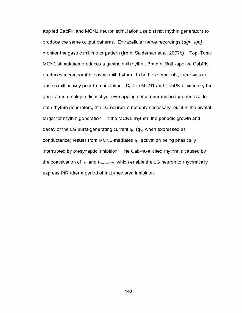
140
applied CabPK and MCN1 neuron stimulation use distinct rhythm generators to
produce the same output patterns. Extracellular nerve recordings (dgn, lgn)
monitor the gastric mill motor pattern (from: Saideman et al. 2007b) . Top, Tonic
MCN1 stimulation produces a gastric mill rhythm. Bottom, Bath-applied CabPK
produces a comparable gastric mill rhythm. In both experiments, there was no
gastric mill activity prior to modulation. C, The MCN1 and CabPK-elicited rhythm
generators employ a distinct yet overlapping set of neurons and properties. In
both rhythm generators, the LG neuron is not only necessary, but it is the pivotal
target for rhythm generation. In the MCN1-rhythm, the periodic growth and
decay of the LG burst-generating current IMI (gMI when expressed as
conductance) results from MCN1-mediated IMI activation being phasically
interrupted by presynaptic inhibition. The CabPK-elicited rhythm is caused by
the coactivation of IMI and ITrans-LTS, which enable the LG neuron to rhythmically
express PIR after a period of Int1-mediated inhibition.
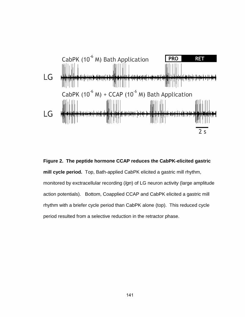
141
Figure 2. The peptide hormone CCAP reduces the CabPK-elicited gastric
mill cycle period. Top, Bath-applied CabPK elicited a gastric mill rhythm,
monitored by exctracellular recording (lgn) of LG neuron activity (large amplitude
action potentials). Bottom, Coapplied CCAP and CabPK elicited a gastric mill
rhythm with a briefer cycle period than CabPK alone (top). This reduced cycle
period resulted from a selective reduction in the retractor phase.
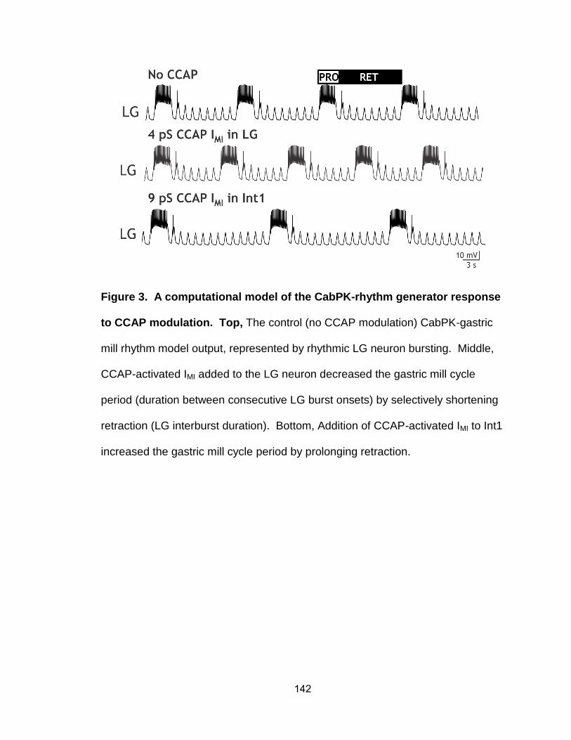
142
Figure 3. A computational model of the CabPK-rhythm generator response
to CCAP modulation. Top, The control (no CCAP modulation) CabPK-gastric
mill rhythm model output, represented by rhythmic LG neuron bursting. Middle,
CCAP-activated IMI added to the LG neuron decreased the gastric mill cycle
period (duration between consecutive LG burst onsets) by selectively shortening
retraction (LG interburst duration). Bottom, Addition of CCAP-activated IMI to Int1
increased the gastric mill cycle period by prolonging retraction.
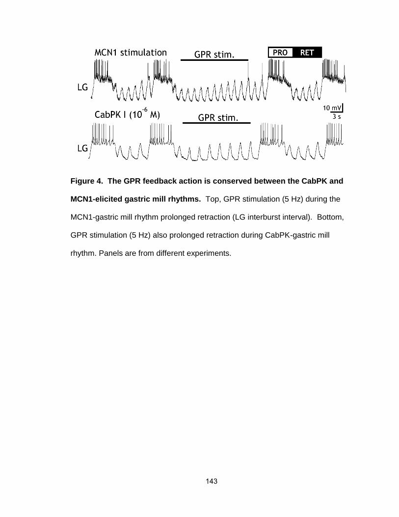
143
Figure 4. The GPR feedback action is conserved between the CabPK and
MCN1-elicited gastric mill rhythms. Top, GPR stimulation (5 Hz) during the
MCN1-gastric mill rhythm prolonged retraction (LG interburst interval). Bottom,
GPR stimulation (5 Hz) also prolonged retraction during CabPK-gastric mill
rhythm. Panels are from different experiments.
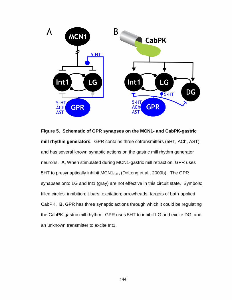
144
Figure 5. Schematic of GPR synapses on the MCN1- and CabPK-gastric
mill rhythm generators. GPR contains three cotransmitters (5HT, ACh, AST)
and has several known synaptic actions on the gastric mill rhythm generator
neurons. A, When stimulated during MCN1-gastric mill retraction, GPR uses
5HT to presynaptically inhibit MCN1STG (DeLong et al., 2009b). The GPR
synapses onto LG and Int1 (gray) are not effective in this circuit state. Symbols:
filled circles, inhibition; t-bars, excitation; arrowheads, targets of bath-applied
CabPK. B, GPR has three synaptic actions through which it could be regulating
the CabPK-gastric mill rhythm. GPR uses 5HT to inhibit LG and excite DG, and
an unknown transmitter to excite Int1.
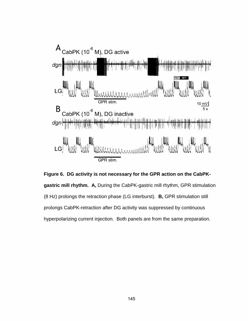
145
Figure 6. DG activity is not necessary for the GPR action on the CabPK-
gastric mill rhythm. A, During the CabPK-gastric mill rhythm, GPR stimulation
(8 Hz) prolongs the retraction phase (LG interburst). B, GPR stimulation still
prolongs CabPK-retraction after DG activity was suppressed by continuous
hyperpolarizing current injection. Both panels are from the same preparation.
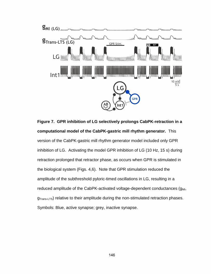
146
Figure 7. GPR inhibition of LG selectively prolongs CabPK-retraction in a
computational model of the CabPK-gastric mill rhythm generator. This
version of the CabPK-gastric mill rhythm generator model included only GPR
inhibition of LG. Activating the model GPR inhibition of LG (10 Hz, 15 s) during
retraction prolonged that retractor phase, as occurs when GPR is stimulated in
the biological system (Figs. 4,6). Note that GPR stimulation reduced the
amplitude of the subthreshold pyloric-timed oscillations in LG, resulting in a
reduced amplitude of the CabPK-activated voltage-dependent conductances (gMI,
gTrans-LTS) relative to their amplitude during the non-stimulated retraction phases.
Symbols: Blue, active synapse; grey, inactive synapse.
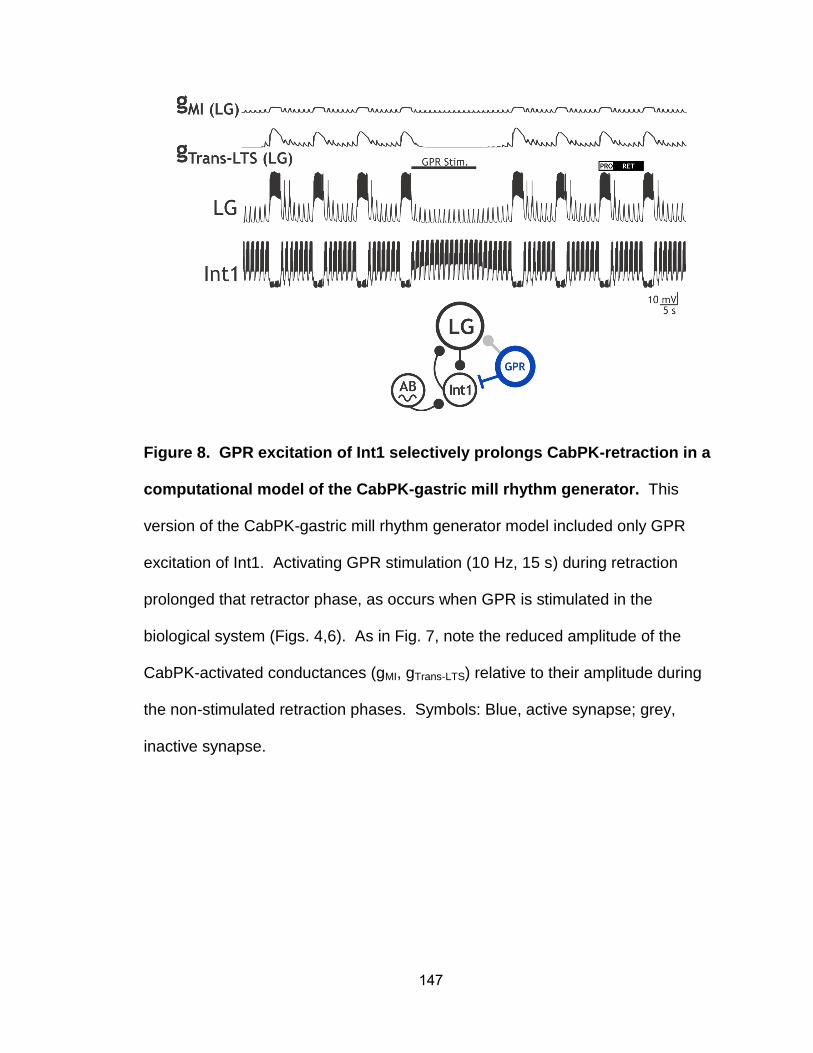
147
Figure 8. GPR excitation of Int1 selectively prolongs CabPK-retraction in a
computational model of the CabPK-gastric mill rhythm generator. This
version of the CabPK-gastric mill rhythm generator model included only GPR
excitation of Int1. Activating GPR stimulation (10 Hz, 15 s) during retraction
prolonged that retractor phase, as occurs when GPR is stimulated in the
biological system (Figs. 4,6). As in Fig. 7, note the reduced amplitude of the
CabPK-activated conductances (gMI, gTrans-LTS) relative to their amplitude during
the non-stimulated retraction phases. Symbols: Blue, active synapse; grey,
inactive synapse.
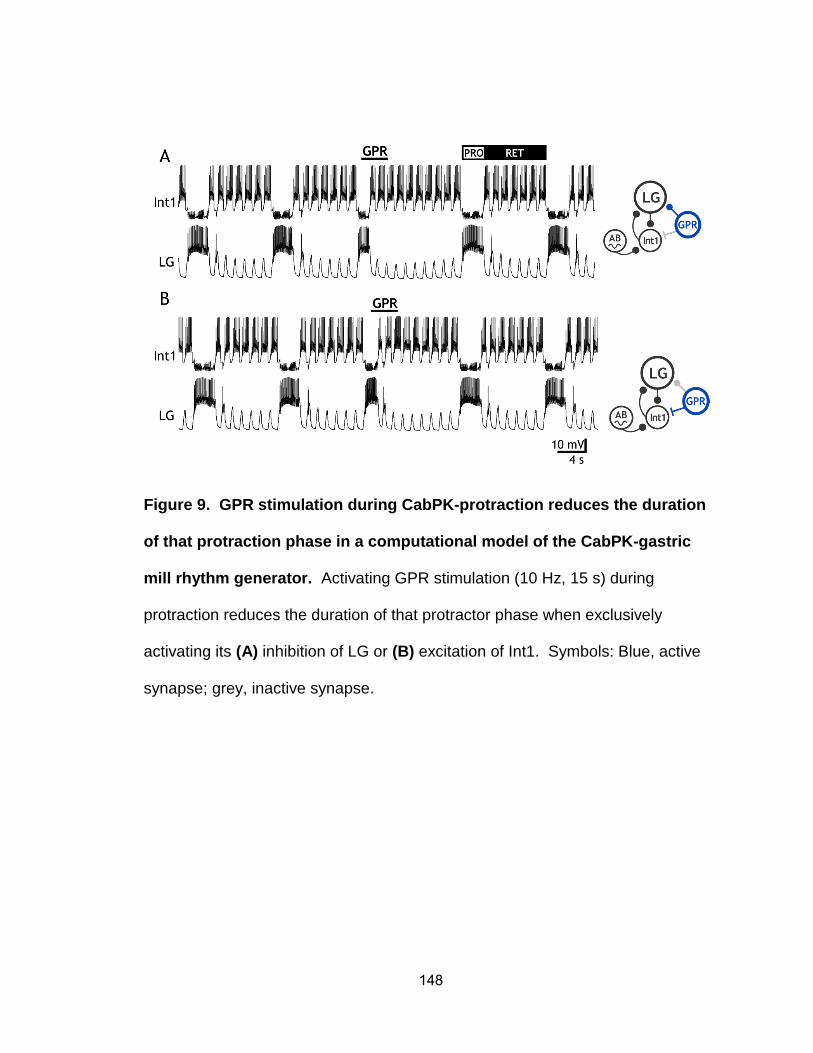
148
Figure 9. GPR stimulation during CabPK-protraction reduces the duration
of that protraction phase in a computational model of the CabPK-gastric
mill rhythm generator. Activating GPR stimulation (10 Hz, 15 s) during
protraction reduces the duration of that protractor phase when exclusively
activating its (A) inhibition of LG or (B) excitation of Int1. Symbols: Blue, active
synapse; grey, inactive synapse.
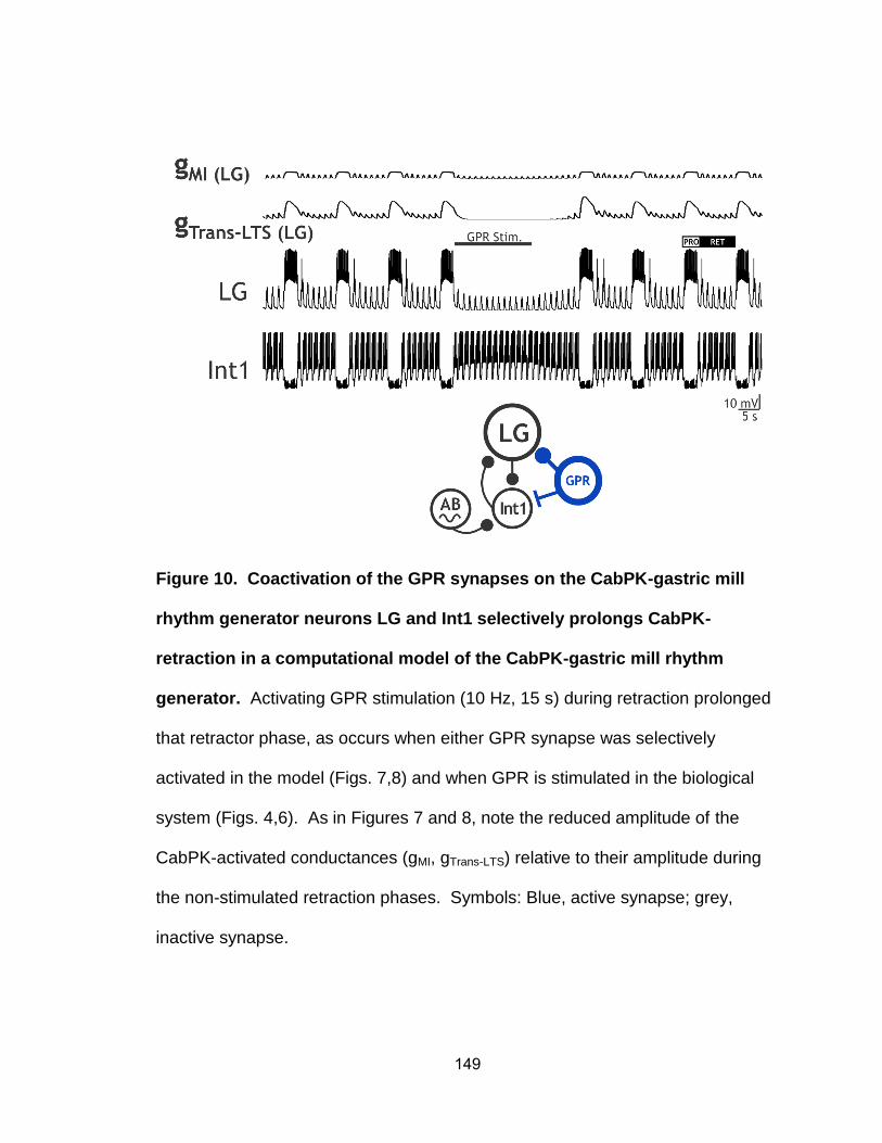
149
Figure 10. Coactivation of the GPR synapses on the CabPK-gastric mill
rhythm generator neurons LG and Int1 selectively prolongs CabPK-
retraction in a computational model of the CabPK-gastric mill rhythm
generator. Activating GPR stimulation (10 Hz, 15 s) during retraction prolonged
that retractor phase, as occurs when either GPR synapse was selectively
activated in the model (Figs. 7,8) and when GPR is stimulated in the biological
system (Figs. 4,6). As in Figures 7 and 8, note the reduced amplitude of the
CabPK-activated conductances (gMI, gTrans-LTS) relative to their amplitude during
the non-stimulated retraction phases. Symbols: Blue, active synapse; grey,
inactive synapse.
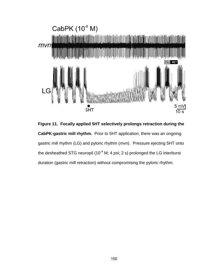
150
Figure 11. Focally applied 5HT selectively prolongs retraction during the
CabPK-gastric mill rhythm. Prior to 5HT application, there was an ongoing
gastric mill rhythm (LG) and pyloric rhythm (mvn). Pressure ejecting 5HT onto
the desheathed STG neuropil (10-4 M; 4 psi; 2 s) prolonged the LG interburst
duration (gastric mill retraction) without compromising the pyloric rhythm.
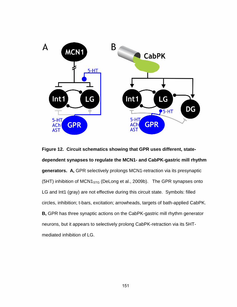
151
Figure 12. Circuit schematics showing that GPR uses different, state-
dependent synapses to regulate the MCN1- and CabPK-gastric mill rhythm
generators. A, GPR selectively prolongs MCN1-retraction via its presynaptic
(5HT) inhibition of MCN1STG (DeLong et al., 2009b). The GPR synapses onto
LG and Int1 (gray) are not effective during this circuit state. Symbols: filled
circles, inhibition; t-bars, excitation; arrowheads, targets of bath-applied CabPK.
B, GPR has three synaptic actions on the CabPK-gastric mill rhythm generator
neurons, but it appears to selectively prolong CabPK-retraction via its 5HT-
mediated inhibition of LG.
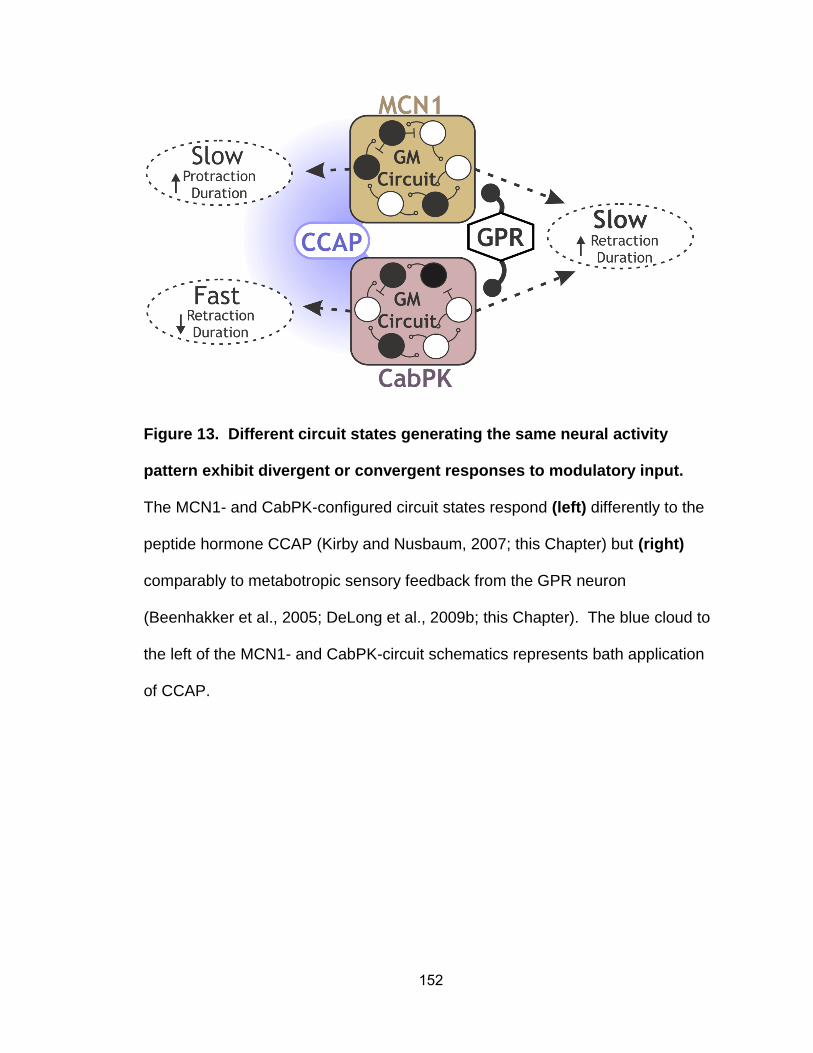
152
Figure 13. Different circuit states generating the same neural activity
pattern exhibit divergent or convergent responses to modulatory input.
The MCN1- and CabPK-configured circuit states respond (left) differently to the
peptide hormone CCAP (Kirby and Nusbaum, 2007; this Chapter) but (right)
comparably to metabotropic sensory feedback from the GPR neuron
(Beenhakker et al., 2005; DeLong et al., 2009b; this Chapter). The blue cloud to
the left of the MCN1- and CabPK-circuit schematics represents bath application
of CCAP.
153
CHAPTER 4
CONCLUSIONS
In Chapter 2, I used electrophysiological (i.e. voltage clamp) methods to
characterize the CabPK-activated ionic currents that underlie rhythmic LG neuron
bursting during the CabPK-gastric mill rhythm. These currents (ITrans-LTS, IMI) were then
injected into the LG neuron using the dynamic clamp and imported into a computational
model of the CabPK-gastric mill rhythm, to demonstrate that they are necessary and
sufficient for enabling post-inhibitory rebound bursting in LG and, as a consequence,
producing this version of the gastric mill rhythm.
Insofar as the CabPK-gastric mill rhythm was shown previously to be nearly
identical to the MCN1-gastric mill rhythm, and distinct from all other characterized gastric
mill rhythms, elucidating the cellular mechanisms by which CabPK elicits this rhythm
enabled me to compare and contrast rhythm generating mechanisms during CabPK
application and MCN1 stimulation. This comparison revealed that different burst-
generating currents [IMI (MCN1), ITrans-LTS (CabPK)] were activated in LG under these two
conditions, but their time course during gastric mill protraction and retraction were
remarkably similar. This was the case despite being regulated by different mechanisms
[inhibitory synaptic feedback (during MCN1) vs. intrinsic voltage-dependent properties
(during CabPK)]. Additionally, the same current (IMI), activated by both pathways, played
different roles during each gastric mill rhythm. These observations provide novel insight
into the degrees of freedom, and cellular mechanisms, available to distinct modulatory
pathways that influence the same neural circuit. Additionally, it is the first detailed
characterization of distinct inputs eliciting the same motor output by configuring different
circuits. As such, it highlights the need for caution when interpreting the results of
154
comparable manipulations performed on systems where there is not sufficient access to
the cellular-level operation of the studied circuit.
In Chapter 3, I explored the consequences of having divergent circuit states that
produce convergent output patterns by assaying their response to extrinsic inputs.
Specifically, I tested the hypothesis that the divergent circuit states would be
differentially sensitive to a given input. The inputs used included sensory feedback from
the muscle stretch-sensitive GPR neurons, and hormonal modulation by CCAP. The
results stress the importance of knowing circuit level details, because there was no
consistent pattern to how these circuits responded to extrinsic inputs. First, consistent
with the hypothesis, CCAP had distinct actions on the MCN1- and CabPK-gastric mill
rhythms. It prolonged the MCN1-gastric mill cycle period (via a low threshold [10-10 M]
action), and shortened the CabPK-gastric mill cycle period (via a high threshold [10-7 M]
action). This was the case despite it being likely that under both conditions the primary
CCAP action was to activate IMI in the LG neuron. Second, in contrast to the hypothesis,
GPR had the same action on the MCN1- and CabPK-gastric mill rhythms, selectively
prolonging retraction. This was the case despite GPR influencing each rhythm via a
different synapse. Presumably, the behavioral function of the GPR feedback is
sufficiently important under different versions of the gastric mill rhythm that the system
has ensured its persistence. It is perhaps not surprising that there could be regions
within the dynamic operating range of distinct circuit states where a particular influence
would evoke a similar change in both circuits, but we had not anticipated that a shared
response from the same input (GPR neuron) could be achieved with different synapses.
This novel outcome provides additional flexibility to the state-dependent responses of
neural circuits.
155
FUTURE DIRECTIONS
The primary goals for my thesis research were to determine the cellular and
synaptic mechanisms by which different modulatory inputs configure different circuit
states to generate the same neural activity pattern, and to assess the relative sensitivity
of these circuit states to additional modulatory inputs. As a result of achieving these
goals, additional interesting issues were revealed. With respect to the relative influence
of MCN1 and CabPK on the gastric mill circuit, it is noteworthy that CabPK is not only
neurally-released but is also a circulating hormone released by the pericardial organs
into the hemolymph. Whereas hormonal levels (e.g. <10-7 M) do not activate the gastric
mill rhythm, they may well still activate IMI and/or ITrans-LTS in LG. As a result, it would be
interesting to determine the influence of hormonal CabPK levels on the MCN1-gastric
mill rhythm, and to compare that influence to the previously determined CCAP influence
on that rhythm (Kirby and Nusbaum, 2007; DeLong and Nusbaum, 2010). If there is
indeed a hormonal-type of CabPK action on the MCN1-rhythm, then it would be
worthwhile to determine whether IMI and/or ITrans-LTS is activated at those lower
concentrations and, if so, then to what amplitude. Additionally, the balance of IMI to ITrans-
LTS might be concentration-dependent, which could in turn underlie a concentration-
dependent action on the MCN1-gastric mill rhythm. In contrast, the CCAP influence on
the MCN1-gastric mill rhythm is qualitatively the same, but quantitatively distinct, at
different concentrations (10-10 M to 10-7 M) (Kirby and Nusbaum, 2007).
The MCN1- and CabPK-gastric mill motor patterns involve more than just rhythm
generation (the focus of this work). There is also the associated issue of how pattern
generation is achieved under these two conditions. Pattern generation involves both the
rhythm generator neuron synapses on the other gastric mill neurons, as well as
synapses between these other neurons. Both MCN1 and CabPK have direct actions on
156
most or all of these other gastric mill neurons (Saideman et al., 2007a; Stein et al.,
2007). Their contributions to the convergent pattern generation during these two gastric
mill rhythms remains to be determined. The MCN1 influence likely involves IMI activation,
insofar as its two peptide cotransmitters (CabTRP Ia, proctolin) are known to activate IMI
in some pyloric circuit neurons and the gastro-pyloric neurons IC and VD, but whether
additional currents are activated is not yet known. Similarly, the influence of CabPK on
the gastric mill pattern generator neurons is currently unknown. Determining the
mechanisms underlying convergent pattern generation will be a more extensive project
than the one for determining rhythm generation insofar as the number of gastric mill
neurons involved (6 vs. 2) is larger.
Another potential fruitful direction to pursue pertains to the mechanism underlying
CCAP modulation of the CabPK-gastric mill rhythm. My work thus far suggests that
CCAP influences this rhythm by activating IMI in LG, as it does during the MCN1-rhythm.
However, this has yet to be established, and CCAP does influence additional gastric mill
neurons, including Int1 (Kirby and Nusbaum, 2007). Moreover, previous voltage clamp
analysis of the CCAP action on LG (and other STG neurons) was performed using only
a ramp protocol (Swensen and Marder, 2000; DeLong et al., 2009). As my voltage
clamp analysis of the CabPK-influenced currents in LG has established, while only IMI
was identified using the voltage ramp protocol, additional transient (due to time-
dependent inactivation) currents were identified using a voltage step protocol.
Therefore, one logical next step is to fully characterize the CCAP modulation of the
CabPK circuit by using the voltage step protocol to assess whether additional currents
are influenced by CabPK in LG and/or in other relevant gastric mill neurons. Any
measured changes imparted by CCAP modulation could be modeled and injected back
into LG with the dynamic clamp in order to test sufficiency.
157
With respect to the GPR influence on the CabPK-gastric mill rhythm, in the short-
term the mechanism underlying this action remains to be determined. My preliminary
data support the hypothesis that this action is due to serotonergic inhibition of LG by
GPR. However, this hypothesis remains to be tested. One potentially fruitful approach
to testing it is to use voltage clamp to identify the current(s) influenced in LG by 5HT,
and then import them into the computational model of the CabPK-gastric mill rhythm and
into the dynamic clamp software. Using the dynamic clamp, we can test whether the
CabPK-influenced current(s) is necessary (nullify the GPR-activated current with
dynamic clamp negative conductance injections) and sufficient (use positive
conductance injections in place of GPR stimulation) to mimic the GPR action on this
gastric mill rhythm. If these manipulations are not sufficient to explain this GPR action,
then we would proceed to determine the role of GPR excitation of Int1 in this process.
GPR has two modes of activity, including spontaneous bursting and
stretch/tension-related activity, in semi-intact preparations (Katz et al., 1989; Birmingham
et al., 1999). In this study, I focused on the stretch-related bursting by stimulating GPR
during the retractor phase, which as indicated above had the same effect on the CabPK-
and MCN1-gastric mill rhythms. It is possible that the influence of GPR on these distinct
circuit states when it is expressing spontaneous bursting might differentially influence
these circuit states. If so, then one possible mechanism underlying such a changed
influence results from GPR containing three cotransmitters, including 5HT, ACh, and the
peptide AST (allatostatin) (Katz and Harris-Warrick, 1989; Skiebe and Schneider, 1994).
In at least some instances, cotransmitter-containing neurons can alter the relative
amount of released cotransmitters during different firing rates and/or patterns.
My thesis work extends the concept known as degeneracy to the level of neural
circuit operation. In this context, degeneracy pertains to the ability of a neural network to
158
use different mechanisms to reach the same endpoint, such as the ability of the distinct
MCN1- and CabPK-circuit states to elicit the same gastric mill rhythm. This concept also
extends to the shared response of these two circuit states to sensory (GPR) feedback.
There are some points where degeneracy breaks down (e.g. CCAP modulation) after
only sampling a relatively small proportion of modulatory inputs into these two networks.
There are many influences on neural circuits, even small ones such as the gastric mill
CPG (Blitz and Nusbaum, 2011; Marder, 2012). The concept of degeneracy is often
couched in terms of a limited context, such as the aforementioned two gastric mill
rhythms in the isolated STG. In most systems, it remains to be determined whether
degeneracy in circuit operation is a mechanism to ensure resilience of circuit function or
whether it is an artifact of working with reduced systems receiving only a limited set of
their full behaviorally-relevant repertoire of inputs. The crab STNS is one system that is
sufficiently accessible and well-defined to it may well be possible to address this issue in
some detail.