direct perfusion improves redifferentiation of human ... - mdpi
TRANSCRIPT
applied sciences
Article
Direct Perfusion Improves Redifferentiation of HumanChondrocytes in Fibrin Hydrogel with the Deposition ofCartilage Pericellular Matrix
Alexandre Dufour , Frédéric Mallein-Gerin and Emeline Perrier-Groult *
�����������������
Citation: Dufour, A.; Mallein-Gerin,
F.; Perrier-Groult, E. Direct Perfusion
Improves Redifferentiation of Human
Chondrocytes in Fibrin Hydrogel
with the Deposition of Cartilage
Pericellular Matrix. Appl. Sci. 2021,
11, 8923. https://doi.org/10.3390/
app11198923
Academic Editor: Ming Pei
Received: 28 July 2021
Accepted: 23 September 2021
Published: 24 September 2021
Publisher’s Note: MDPI stays neutral
with regard to jurisdictional claims in
published maps and institutional affil-
iations.
Copyright: © 2021 by the authors.
Licensee MDPI, Basel, Switzerland.
This article is an open access article
distributed under the terms and
conditions of the Creative Commons
Attribution (CC BY) license (https://
creativecommons.org/licenses/by/
4.0/).
Laboratory of Tissue Biology and Therapeutic Engineering (LBTI), Institute for Biology and Chemistry of Proteins,CNRS, UMR 5305, 69007 Lyon, France; [email protected] (A.D.); [email protected] (F.M.-G.)* Correspondence: [email protected]
Abstract: Articular cartilage has limited potential for self-repair, and cell-based strategies combiningscaffolds and chondrocytes are currently used to treat cartilage injuries. However, achieving a satisfy-ing level of cell redifferentiation following expansion remains challenging. Hydrogels and perfusionbioreactors are known to exert beneficial cues on chondrocytes; however, the effect of a combinedapproach on the quality of cartilage matrix deposited by cells is not fully understood. Here, wecombined soluble factors (BMP-2, Insulin, and Triiodothyronine, that is, BIT), fibrin hydrogel, directperfusion and human articular chondrocytes (HACs) to engineer large cartilage tissues. Followingcell expansion, cells were embedded in fibrin gels and cultivated under either static or perfusionconditions. The nature of the matrix synthesized was assessed by Western blotting and immunohis-tochemistry. The stability of cartilage grafts and integration with native tissue were also investigatedby subcutaneous implantation of human osteochondral cylinders in nude mice. Perfusion precon-ditioning improved matrix quality and spatial distribution. Specifically, perfusion preconditioningresulted in a matrix rich in type II collagen but not in type I collagen, indicating the reconstructionof hyaline cartilage. Remarkably, the production of type VI collagen, the main component of thepericellular matrix, was also increased, indicating that chondrocytes were connecting to the hyalinematrix they produced.
Keywords: chondrocyte; cartilage tissue engineering; perfusion bioreactor; fibrin; type VI collagen
1. Introduction
Articular cartilage is a hyaline type of cartilage covering the extremities of long bonesin synovial joints. The unique ability of this tissue to repeatedly absorb and transmit theload applied to the joint is due to the specific composition and structural organizationof its extracellular matrix (ECM). Cartilage ECM is composed of proteoglycans, waterand a collagen network, where type II collagen is the most abundant [1]. However, theavascular nature of cartilage together with the low metabolic rate of the chondrocyteslimit its intrinsic capability for self-repairing [2]. Therefore, effective therapies for thetreatment of articular cartilage lesions rely on surgical intervention and engraftments witha composition comparable to articular cartilage.
Current strategies for repairing articular cartilage include microfracture (i.e., marrowstimulation), mosaicplasty (i.e., osteochondral autograft), and autologous chondrocyteimplantation (ACI) [3]. Microfracture and mosaicplasty remain unsatisfactory treatmentsdue to the occurrence of type I collagen-rich fibrocartilage in the repaired defect [4,5].ACI is a cell-based surgical procedure currently practiced as a popular second-line treat-ment for relatively large articular cartilage lesions [6,7]. However, the loss of therapeuticchondrocytes due to the liquid nature of the implant severely affects the quality of thehealed defect [8,9]. As a consequence, scaffold-based approaches have been investigated toguide matrix synthesis and deliver chondrocytes to the cartilage defect site. Hydrogels are
Appl. Sci. 2021, 11, 8923. https://doi.org/10.3390/app11198923 https://www.mdpi.com/journal/applsci
Appl. Sci. 2021, 11, 8923 2 of 15
excellent candidates for this due to their ability to encapsulate the cells, along with a highintrinsic water content (60–90%) [10], a feature found in native cartilage [11] which offersthe tissue its “shock absorber” capabilities.
Nutrient availability to cells throughout three-dimensional (3D) scaffolds occursin vitro by diffusion, and therefore depends on the distance from the surface, diffusionalconstraints, and cellular utilization at the periphery [12]. These diffusional limitationsbecome exacerbated when attempting to engineer larger cartilage tissues. Perfusion biore-actors have been shown to enhance cell access to oxygen and nutrients as well as thehomogeneity of neo-synthesized ECM in 3D scaffolds [13,14]; however, understanding ofits impact on the quality of the ECM deposited by chondrocytes encapsulated in hydrogelsremains limited [15–19].
Following this view, this work aimed to evaluate whether direct perfusion precon-ditioning could enhance cartilage matrix deposition by human articular chondrocytes(HACs) in fibrin hydrogel, a clinically relevant material that has been shown to supportand maintain chondrocyte redifferentiation [20,21]. We used cocktails of specific factors toexpand the cells on plastic (fibroblast growth factor (FGF)-2 and insulin, designated FI) andto allow their redifferentiation in fibrin hydrogel (bone morphogenetic protein (BMP)-2,insulin, and triiodothyronine T3, designated BIT). This sequential addition of factors hasbeen proven efficient to amplify human chondrocytes isolated from diverse anatomicalsites (including articular cartilage) and to induce their redifferentiation in collagen [22],agarose [23], self-assembling peptide [24,25] gels and collagen sponges [26,27]. Specialattention was given to the quality and spatial distribution of the neosynthesized matrixthrough analysis of the pericellular matrix (PCM) components.
2. Materials and Methods2.1. Cell Isolation and Expansion
HACs were isolated from macroscopically non-fibrillated zones of osteoarthritic jointsfrom donors undergoing total or partial knee joint replacement (age range: 60–90; EthicsCommittee for research with human samples, CODECOH: DC-2014-2325). Cartilagesamples were sliced into small pieces (2 mm3) and digested overnight at 37 ◦C in culturemedium composed of DMEM/F-12 (Invitrogen, Waltham, MA, USA) with 0.5 mg/mLcollagenase A (Roche Applied Science, Penzberg, Germany). The cell suspension wasfiltered, and isolated chondrocytes were seeded at a density of 1.5 × 10⁴ cells/cm2 inDMEM/F-12 supplemented with 10% fetal bovine serum (FBS) (Gibco-Invitrogen) and50 µg/mL streptomycin (Panpharma, La Selle-en-Luitré, France) at 37 ◦C in 5% CO2. When48 h had elapsed post-seeding, the medium was replaced and supplemented with 5 ng/mLfibroblast growing factor-2 (FGF-2) (R&D Systems, Minneapolis, MN, USA) and 5 µg/mLinsulin (Umuline Rapide, Lilly). The culture medium was then replaced twice per week fortwo weeks.
2.2. Preparation of Fibrin Hydrogels
Human fibrinogen (Millipore, Burlington, MA, USA) was dissolved in 10 mM HEPES(ThermoFisher Scientific, Waltham, MA, USA) (pH 7.4) to obtain a 50 mg/mL fibrin solution.Likewise, human thrombin (Millipore) was dissolved in 10 mM HEPES (pH 6.5) supple-mented with 0.1% BSA to prepare a 2 U/mL thrombin solution. A solution containing 3 Msodium chloride (NaCl) and 0.4 M calcium chloride (CaCl2) was also prepared in 10 mMHEPES (pH 7.4). A 1.6 mL chondrocyte-fibrin suspension was then prepared by mixing3.2 × 106 cells laden in 880 µL DMEM/F-12 with 320 µL fibrinogen, 320 µL thrombinand 80 µL NaCl/CaCl2, yielding a final concentration of 10 mg/mL fibrinogen, 0.4 U/mLthrombin, 150 mM NaCl, 20 mM CaCl2 and 2 × 10⁶ cells/mL. In parallel, 2.5% low meltingagarose (w/v) (Seaplaque, Cambrex BioScience) was poured in a petri dish and punchedwith a 10 mm biopsy punch to create cylindrical molds in which 314 µL of the chondro-cyte/fibrin mixture was poured. The cell-fibrin mixture was then allowed to gel at 37 ◦C in5% CO2 for 10 min to obtain cylindrical constructs of 10 mm diameter and 4 mm thick.
Appl. Sci. 2021, 11, 8923 3 of 15
2.3. Static Culture
HAC-seeded fibrin scaffolds were placed in a 24-well plate and fresh chondrogenicmedium containing 50 µg/mL 2-phospho-L-ascorbic acid, 200 ng/mL recombinant humanBMP-2 (dibotermin-alpha, drug form of BMP-2, InductOs kit, Wyeth, Madison, WI, USA),5 µg/mL insulin and 100 nM triiodothyronine T3 (Sigma Aldrich, Saint Quentin Fallavier,France), that is, the BIT cocktail [27], was added. Medium was replaced twice a week overa culture period of 21 days.
2.4. Perfusion Culture
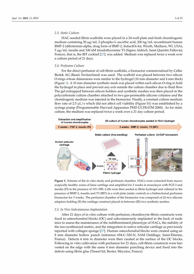
For the direct perfusion of cell-fibrin scaffolds, a bioreactor commercialized by CellecBiotek AG (Basel, Switzerland) was used. The scaffold was placed between two siliconO-rings whose dimensions were similar to the hydrogel (10 mm diameter and 4 mm thick)(Figure 1). A 10 mm diameter synthetic mesh was placed within each silicon O-ring to holdthe hydrogel in place and prevent any exit outside the culture chamber due to fluid flow.The gel entrapped between silicon holders and synthetic meshes was then placed in thepolycarbonate culture chamber attached to two gas-permeable silicone columns and thechondrogenic medium was injected in the bioreactor. Finally, a constant culture mediumflow rate of 2.5 µL/s which did not affect cell viability (Figure S1) was established by asyringe pump (Programmable Harvard Apparatus PHD ULTRATM 2000). As for staticculture, the medium was replaced twice a week over a 21 day culture period.
Appl. Sci. 2021, 11, 8923 3 of 15
314 µL of the chondrocyte/fibrin mixture was poured. The cell-fibrin mixture was then allowed to gel at 37 °C in 5% CO2 for 10 min to obtain cylindrical constructs of 10 mm diameter and 4 mm thick.
2.3. Static Culture HAC-seeded fibrin scaffolds were placed in a 24-well plate and fresh chondrogenic
medium containing 50 µg/mL 2-phospho-L-ascorbic acid, 200 ng/mL recombinant human BMP-2 (dibotermin-alpha, drug form of BMP-2, InductOs kit, Wyeth, Madison, WI, USA), 5 µg/mL insulin and 100 nM triiodothyronine T3 (Sigma Aldrich, Saint Quentin Fallavier, France), that is, the BIT cocktail [27], was added. Medium was replaced twice a week over a culture period of 21 days.
2.4. Perfusion Culture For the direct perfusion of cell-fibrin scaffolds, a bioreactor commercialized by Cellec
Biotek AG (Basel, Switzerland) was used. The scaffold was placed between two silicon O-rings whose dimensions were similar to the hydrogel (10 mm diameter and 4 mm thick) (Figure 1). A 10 mm diameter synthetic mesh was placed within each silicon O-ring to hold the hydrogel in place and prevent any exit outside the culture chamber due to fluid flow. The gel entrapped between silicon holders and synthetic meshes was then placed in the polycarbonate culture chamber attached to two gas-permeable silicone columns and the chondrogenic medium was injected in the bioreactor. Finally, a constant culture medium flow rate of 2.5 µL/sec which did not affect cell viability (Figure S1) was established by a syringe pump (Programmable Harvard Apparatus PHD ULTRATM 2000). As for static culture, the medium was replaced twice a week over a 21 day culture period.
Figure 1. Scheme of the in vitro study and perfusion chamber. HACs were extracted from macroscopically healthy zones of knee cartilage and amplified for 2 weeks in monolayer with FGF-2 and insulin (FI) in the presence of 10% FBS. Cells were then seeded in fibrin hydrogel and cultured in the presence of BMP-2, insulin and T3 (BIT) in a well plate (static control) or a bidirectional perfusion bioreactor for 3 weeks. The perfusion chamber of the bioreactor was composed of (I) two silicone adaptors holding (II) the cartilage construct placed in between (III) two synthetic meshes.
2.5. In Vivo Subcutaneous Implantation After 21 days of in vitro culture with perfusion, chondrocyte-fibrin constructs were
fixed in osteochondral blocks (OC) and subcutaneously implanted in the back of nude mice to assess the maintenance of the redifferentiated phenotype of HACs, the stability of the neo-synthesized matrix, and the integration to native articular cartilage as previously reported with collagen sponge [27]. Human osteochondral blocks were created using an 8 mm diameter hollow punch (reference 694-C-320-N, SAM Outillage, Saint-Étienne ,
Figure 1. Scheme of the in vitro study and perfusion chamber. HACs were extracted from macro-scopically healthy zones of knee cartilage and amplified for 2 weeks in monolayer with FGF-2 andinsulin (FI) in the presence of 10% FBS. Cells were then seeded in fibrin hydrogel and cultured in thepresence of BMP-2, insulin and T3 (BIT) in a well plate (static control) or a bidirectional perfusionbioreactor for 3 weeks. The perfusion chamber of the bioreactor was composed of (I) two siliconeadaptors holding (II) the cartilage construct placed in between (III) two synthetic meshes.
2.5. In Vivo Subcutaneous Implantation
After 21 days of in vitro culture with perfusion, chondrocyte-fibrin constructs werefixed in osteochondral blocks (OC) and subcutaneously implanted in the back of nudemice to assess the maintenance of the redifferentiated phenotype of HACs, the stability ofthe neo-synthesized matrix, and the integration to native articular cartilage as previouslyreported with collagen sponge [27]. Human osteochondral blocks were created using an8 mm diameter hollow punch (reference 694-C-320-N, SAM Outillage, Saint-Étienne,France). Defects 4 mm in diameter were then created at the surface of the OC blocks.Following in vitro cultivation with perfusion for 21 days, cell-fibrin constructs were har-vested on the edge with the same 4 mm diameter punching device and fixed into thedefects using fibrin glue (Tisseel kit, Baxter, Meyzieu, France).
Appl. Sci. 2021, 11, 8923 4 of 15
The animal experimentation was performed in strict accordance with the officialregulation on animal experimentation (Directive 2010/63/EU and its national transposition)and ethical guidelines for care and use of mice of the Plateau de Biologie Expérimentale dela Souris (PBES, UMS 3444) at Ecole Normale Supérieure (ENS, Lyon, France). The in vivostudy was approved by the Committee on the Ethics of Animal Experiments of ENS deLyon (approval number: ENS_2018_007). Female nude mice (six weeks old) were obtainedfrom Charles River Laboratories and maintained in the Animal Care Facilities of PBESunder pathogen-free conditions with food and water ad libitum. The animals were regularlymonitored by staff responsible for general animal health and welfare supervision. Allsurgeries were performed under general anesthesia and all efforts were made to minimizesuffering. Animals were anesthetized with isoflurane gas, and surgeries were performedunder a laminar flow hood in sterile conditions. A subcutaneous pocket was created onthe dorsum of each mouse by blunt dissection through cranial and caudal skin incisions.The osteochondral block was inserted in the pocket and the skin was closed with surgicalstaples (Harvard Apparatus, ref 52-3746). After six weeks, the mice were euthanized bycervical dislocation. At explantation, all blocks were dissected from the mice and processedfor histology.
2.6. Western Blot Analysis
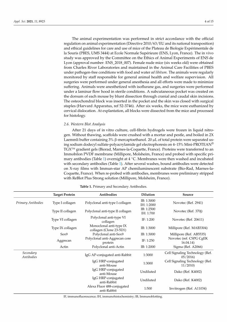
After 21 days of in vitro culture, cell-fibrin hydrogels were frozen in liquid nitro-gen. Without thawing, scaffolds were crushed with a mortar and pestle, and boiled in 2XLaemmli buffer containing 3% β-mercaptoethanol. 20 µL of total protein were separated us-ing sodium dodecyl sulfate-polyacrylamide gel electrophoresis on 4–15% Mini-PROTEAN®
TGX™ gradient gels (Biorad, Marnes-la-Coquette, France). Proteins were transferred to anImmobilon PVDF membrane (Millipore, Molsheim, France) and probed with specific pri-mary antibodies (Table 1) overnight at 4 ◦C. Membranes were then washed and incubatedwith secondary antibodies (Table 1). After several washes, bound antibodies were detectedon X-ray films with Immun-star AP chemiluminescent substrate (Bio-Rad, Marnes-la-Coquette, France). When re-probed with antibodies, membranes were preliminary strippedwith ReBlot Plus Strong solution (Millipore, Molsheim, France).
Table 1. Primary and Secondary Antibodies.
Target Protein Antibodies Dilution Source
Primary Antibodies Type I collagen Polyclonal anti-type I collagen IB: 1:3000IH: 1:2000 Novotec (Ref. 2941)
Type II collagen Polyclonal anti-type II collagen IB: 1:2500IH: 1:700 Novotec (Ref. 370j)
Type VI collagen Polyclonal anti-type VIcollagen IF: 1:200 Novotec (Ref. 20611)
Type IX collagen Monoclonal anti-type IXcollagen (Clone 23-5D1) IB: 1:3000 Millipore (Ref. MAB3304)
Sox9 Polyclonal anti-Sox9 IB: 1:3000 Millipore (Ref. AB5535)
Aggrecan Polyclonal anti-Aggrecan coreprotein IF: 1:250 Novotec (ref. CSPG CgEK
16.04.14)Actin Polyclonal anti-Actin IB: 1:2000 Sigma (Ref. A2066)
SecondaryAntibodies IgG AP-conjugated anti-Rabbit 1:3000 Cell Signaling Technology (Ref.
05/2016)IgG HRP-conjugated
anti-Mouse 1:3000 Cell Signaling Technology (Ref.11/2010)
IgG HRP-conjugatedanti-Mouse Undiluted Dako (Ref. K4002)
IgG HRP-conjugatedanti-Rabbit Undiluted Dako (Ref. K4002)
Alexa Fluor 488-conjugatedanti-Rabbit 1:500 Invitrogen (Ref. A11034)
IF, immunofluorescence; IH, immunohistochemistry; IB, Immunoblotting.
Appl. Sci. 2021, 11, 8923 5 of 15
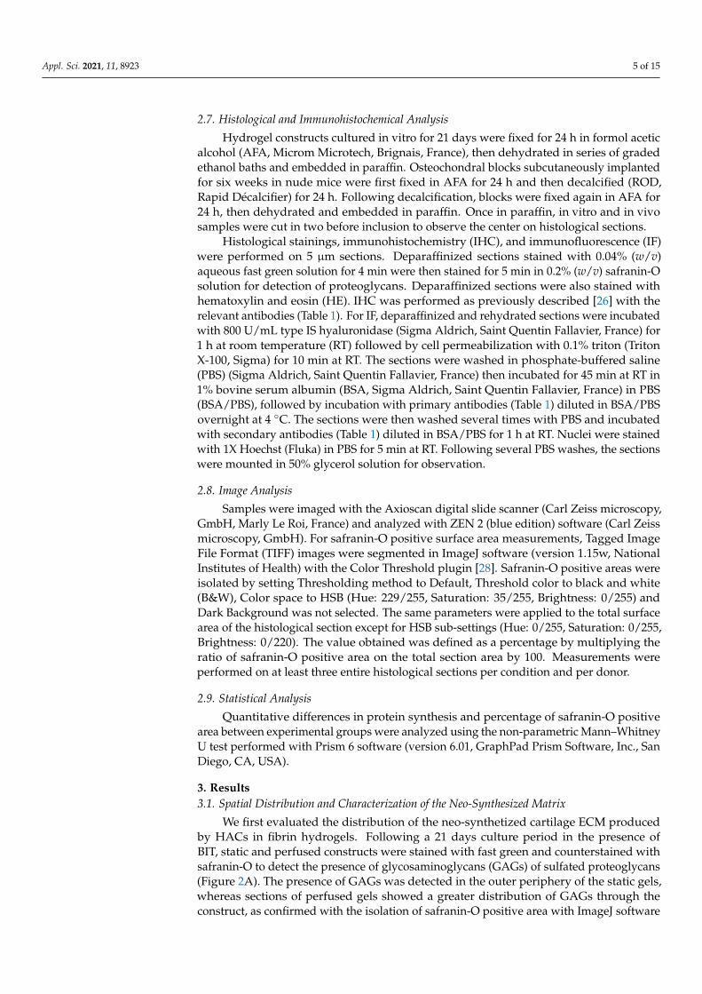
2.7. Histological and Immunohistochemical Analysis
Hydrogel constructs cultured in vitro for 21 days were fixed for 24 h in formol aceticalcohol (AFA, Microm Microtech, Brignais, France), then dehydrated in series of gradedethanol baths and embedded in paraffin. Osteochondral blocks subcutaneously implantedfor six weeks in nude mice were first fixed in AFA for 24 h and then decalcified (ROD,Rapid Décalcifier) for 24 h. Following decalcification, blocks were fixed again in AFA for24 h, then dehydrated and embedded in paraffin. Once in paraffin, in vitro and in vivosamples were cut in two before inclusion to observe the center on histological sections.
Histological stainings, immunohistochemistry (IHC), and immunofluorescence (IF)were performed on 5 µm sections. Deparaffinized sections stained with 0.04% (w/v)aqueous fast green solution for 4 min were then stained for 5 min in 0.2% (w/v) safranin-Osolution for detection of proteoglycans. Deparaffinized sections were also stained withhematoxylin and eosin (HE). IHC was performed as previously described [26] with therelevant antibodies (Table 1). For IF, deparaffinized and rehydrated sections were incubatedwith 800 U/mL type IS hyaluronidase (Sigma Aldrich, Saint Quentin Fallavier, France) for1 h at room temperature (RT) followed by cell permeabilization with 0.1% triton (TritonX-100, Sigma) for 10 min at RT. The sections were washed in phosphate-buffered saline(PBS) (Sigma Aldrich, Saint Quentin Fallavier, France) then incubated for 45 min at RT in1% bovine serum albumin (BSA, Sigma Aldrich, Saint Quentin Fallavier, France) in PBS(BSA/PBS), followed by incubation with primary antibodies (Table 1) diluted in BSA/PBSovernight at 4 ◦C. The sections were then washed several times with PBS and incubatedwith secondary antibodies (Table 1) diluted in BSA/PBS for 1 h at RT. Nuclei were stainedwith 1X Hoechst (Fluka) in PBS for 5 min at RT. Following several PBS washes, the sectionswere mounted in 50% glycerol solution for observation.
2.8. Image Analysis
Samples were imaged with the Axioscan digital slide scanner (Carl Zeiss microscopy,GmbH, Marly Le Roi, France) and analyzed with ZEN 2 (blue edition) software (Carl Zeissmicroscopy, GmbH). For safranin-O positive surface area measurements, Tagged ImageFile Format (TIFF) images were segmented in ImageJ software (version 1.15w, NationalInstitutes of Health) with the Color Threshold plugin [28]. Safranin-O positive areas wereisolated by setting Thresholding method to Default, Threshold color to black and white(B&W), Color space to HSB (Hue: 229/255, Saturation: 35/255, Brightness: 0/255) andDark Background was not selected. The same parameters were applied to the total surfacearea of the histological section except for HSB sub-settings (Hue: 0/255, Saturation: 0/255,Brightness: 0/220). The value obtained was defined as a percentage by multiplying theratio of safranin-O positive area on the total section area by 100. Measurements wereperformed on at least three entire histological sections per condition and per donor.
2.9. Statistical Analysis
Quantitative differences in protein synthesis and percentage of safranin-O positivearea between experimental groups were analyzed using the non-parametric Mann–WhitneyU test performed with Prism 6 software (version 6.01, GraphPad Prism Software, Inc., SanDiego, CA, USA).
3. Results3.1. Spatial Distribution and Characterization of the Neo-Synthesized Matrix
We first evaluated the distribution of the neo-synthetized cartilage ECM producedby HACs in fibrin hydrogels. Following a 21 days culture period in the presence ofBIT, static and perfused constructs were stained with fast green and counterstained withsafranin-O to detect the presence of glycosaminoglycans (GAGs) of sulfated proteoglycans(Figure 2A). The presence of GAGs was detected in the outer periphery of the static gels,whereas sections of perfused gels showed a greater distribution of GAGs through theconstruct, as confirmed with the isolation of safranin-O positive area with ImageJ software
Appl. Sci. 2021, 11, 8923 6 of 15
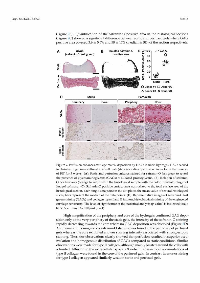
(Figure 2B). Quantification of the safranin-O positive area in the histological sections(Figure 2C) showed a significant difference between static and perfused gels where GAGpositive area covered 3.6 ± 5.5% and 58 ± 17% (median ± SD) of the section respectively.
Appl. Sci. 2021, 11, 8923 6 of 15
through the construct, as confirmed with the isolation of safranin-O positive area with ImageJ software (Figure 2B). Quantification of the safranin-O positive area in the histological sections (Figure 2C) showed a significant difference between static and perfused gels where GAG positive area covered 3.6 ± 5.5% and 58 ± 17% (median ± SD) of the section respectively.
High magnification of the periphery and core of the hydrogels confirmed GAG deposition only at the very periphery of the static gels, the intensity of the safranin-O staining rapidly decreasing towards the core where no GAG deposition was observed (Figure 2D). An intense and homogeneous safranin-O staining was found at the periphery of perfused gels whereas the core exhibited a lower staining intensity associated with strong ectopic staining. Thus, our observations clearly showed that perfusion resulted in superior accumulation and homogenous distribution of GAGs compared to static conditions. Similar observations were made for type II collagen, although mainly located around the cells with a limited diffusion in the extracellular space. Of note, intense ectopic accumulations of type II collagen were found in the core of the perfused gels. In contrast, immunostaining for type I collagen appeared similarly weak in static and perfused gels.
Figure 2. Perfusion enhances cartilage matrix deposition by HACs in fibrin hydrogel. HACs seeded in fibrin hydrogel were cultured in a well plate (static) or a direct perfusion bioreactor in the presence of BIT for 3 weeks. (A): Static and perfusion cultures stained for safranin-O fast green to reveal the presence of glycosaminoglycans (GAGs) of sulfated proteoglycans. (B): Isolation of safranin-O positive area (orange to red) within the histological sample with the color threshold plugin of ImageJ software. (C): Safranin-O positive surface area normalized to the total surface area of the histological section. Each single data point in the dot plot is the mean value of several histological slices; bars represent the median of the data points. (D): Representative images of safranin-O fast green staining (GAGs) and collagen types I and II immunohistochemical staining of the engineered cartilage constructs. The level of significance of the statistical analysis (p-value) is indicated (scale bars: A = 1 mm, D = 100 µm) (n = 4).
Figure 2. Perfusion enhances cartilage matrix deposition by HACs in fibrin hydrogel. HACs seededin fibrin hydrogel were cultured in a well plate (static) or a direct perfusion bioreactor in the presenceof BIT for 3 weeks. (A): Static and perfusion cultures stained for safranin-O fast green to revealthe presence of glycosaminoglycans (GAGs) of sulfated proteoglycans. (B): Isolation of safranin-O positive area (orange to red) within the histological sample with the color threshold plugin ofImageJ software. (C): Safranin-O positive surface area normalized to the total surface area of thehistological section. Each single data point in the dot plot is the mean value of several histologicalslices; bars represent the median of the data points. (D): Representative images of safranin-O fastgreen staining (GAGs) and collagen types I and II immunohistochemical staining of the engineeredcartilage constructs. The level of significance of the statistical analysis (p-value) is indicated (scalebars: A = 1 mm, D = 100 µm) (n = 4).
High magnification of the periphery and core of the hydrogels confirmed GAG depo-sition only at the very periphery of the static gels, the intensity of the safranin-O stainingrapidly decreasing towards the core where no GAG deposition was observed (Figure 2D).An intense and homogeneous safranin-O staining was found at the periphery of perfusedgels whereas the core exhibited a lower staining intensity associated with strong ectopicstaining. Thus, our observations clearly showed that perfusion resulted in superior accu-mulation and homogenous distribution of GAGs compared to static conditions. Similarobservations were made for type II collagen, although mainly located around the cells witha limited diffusion in the extracellular space. Of note, intense ectopic accumulations oftype II collagen were found in the core of the perfused gels. In contrast, immunostainingfor type I collagen appeared similarly weak in static and perfused gels.
Appl. Sci. 2021, 11, 8923 7 of 15
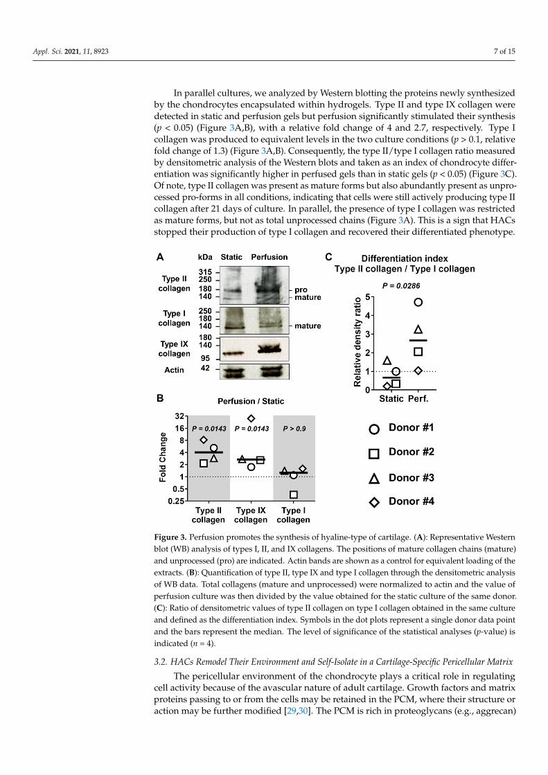
In parallel cultures, we analyzed by Western blotting the proteins newly synthesizedby the chondrocytes encapsulated within hydrogels. Type II and type IX collagen weredetected in static and perfusion gels but perfusion significantly stimulated their synthesis(p < 0.05) (Figure 3A,B), with a relative fold change of 4 and 2.7, respectively. Type Icollagen was produced to equivalent levels in the two culture conditions (p > 0.1, relativefold change of 1.3) (Figure 3A,B). Consequently, the type II/type I collagen ratio measuredby densitometric analysis of the Western blots and taken as an index of chondrocyte differ-entiation was significantly higher in perfused gels than in static gels (p < 0.05) (Figure 3C).Of note, type II collagen was present as mature forms but also abundantly present as unpro-cessed pro-forms in all conditions, indicating that cells were still actively producing type IIcollagen after 21 days of culture. In parallel, the presence of type I collagen was restrictedas mature forms, but not as total unprocessed chains (Figure 3A). This is a sign that HACsstopped their production of type I collagen and recovered their differentiated phenotype.
Appl. Sci. 2021, 11, 8923 7 of 15
In parallel cultures, we analyzed by Western blotting the proteins newly synthesized by the chondrocytes encapsulated within hydrogels. Type II and type IX collagen were detected in static and perfusion gels but perfusion significantly stimulated their synthesis (p < 0.05) (Figure 3A,B), with a relative fold change of 4 and 2.7, respectively. Type I collagen was produced to equivalent levels in the two culture conditions (p > 0.1, relative fold change of 1.3) (Figure 3A,B). Consequently, the type II/type I collagen ratio measured by densitometric analysis of the Western blots and taken as an index of chondrocyte differentiation was significantly higher in perfused gels than in static gels (p < 0.05) (Figure 3C). Of note, type II collagen was present as mature forms but also abundantly present as unprocessed pro-forms in all conditions, indicating that cells were still actively producing type II collagen after 21 days of culture. In parallel, the presence of type I collagen was restricted as mature forms, but not as total unprocessed chains (Figure 3A). This is a sign that HACs stopped their production of type I collagen and recovered their differentiated phenotype.
Figure 3. Perfusion promotes the synthesis of hyaline-type of cartilage. (A): Representative Western blot (WB) analysis of types I, II, and IX collagens. The positions of mature collagen chains (mature) and unprocessed (pro) are indicated. Actin bands are shown as a control for equivalent loading of the extracts. (B): Quantification of type II, type IX and type I collagen through the densitometric analysis of WB data. Total collagens (mature and unprocessed) were normalized to actin and the value of perfusion culture was then divided by the value obtained for the static culture of the same donor. (C): Ratio of densitometric values of type II collagen on type I collagen obtained in the same culture and defined as the differentiation index. Symbols in the dot plots represent a single donor data point and the bars represent the median. The level of significance of the statistical analyses (p-value) is indicated (n = 4).
Figure 3. Perfusion promotes the synthesis of hyaline-type of cartilage. (A): Representative Westernblot (WB) analysis of types I, II, and IX collagens. The positions of mature collagen chains (mature)and unprocessed (pro) are indicated. Actin bands are shown as a control for equivalent loading of theextracts. (B): Quantification of type II, type IX and type I collagen through the densitometric analysisof WB data. Total collagens (mature and unprocessed) were normalized to actin and the value ofperfusion culture was then divided by the value obtained for the static culture of the same donor.(C): Ratio of densitometric values of type II collagen on type I collagen obtained in the same cultureand defined as the differentiation index. Symbols in the dot plots represent a single donor data pointand the bars represent the median. The level of significance of the statistical analyses (p-value) isindicated (n = 4).
3.2. HACs Remodel Their Environment and Self-Isolate in a Cartilage-Specific Pericellular Matrix
The pericellular environment of the chondrocyte plays a critical role in regulatingcell activity because of the avascular nature of adult cartilage. Growth factors and matrixproteins passing to or from the cells may be retained in the PCM, where their structure oraction may be further modified [29,30]. The PCM is rich in proteoglycans (e.g., aggrecan)
Appl. Sci. 2021, 11, 8923 8 of 15
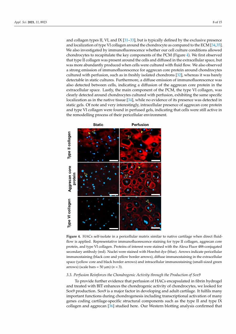
and collagen types II, VI, and IX [31–33], but is typically defined by the exclusive presenceand localization of type VI collagen around the chondrocyte as compared to the ECM [34,35].We also investigated by immunofluorescence whether our cell culture conditions allowedchondrocytes to recapitulate the key components of the PCM (Figure 4). We first observedthat type II collagen was present around the cells and diffused in the extracellular space, butwas more abundantly produced when cells were cultured with fluid flow. We also observeda strong emission of immunofluorescence for aggrecan core protein around chondrocytescultured with perfusion, such as in freshly isolated chondrons [32], whereas it was barelydetectable in static cultures. Furthermore, a diffuse emission of immunofluorescence wasalso detected between cells, indicating a diffusion of the aggrecan core protein in theextracellular space. Lastly, the main component of the PCM, the type VI collagen, wasclearly detected around chondrocytes cultured with perfusion, exhibiting the same specificlocalization as in the native tissue [34], while no evidence of its presence was detected instatic gels. Of note and very interestingly, intracellular presence of aggrecan core proteinand type VI collagen were found in perfused gels, indicating that cells were still active inthe remodelling process of their pericellular environment.
Appl. Sci. 2021, 11, 8923 8 of 15
3.2. HACs Remodel Their Environment and Self-Isolate in a Cartilage-Specific Pericellular Matrix
The pericellular environment of the chondrocyte plays a critical role in regulating cell activity because of the avascular nature of adult cartilage. Growth factors and matrix proteins passing to or from the cells may be retained in the PCM, where their structure or action may be further modified [29,30]. The PCM is rich in proteoglycans (e.g., aggrecan) and collagen types II, VI, and IX [31–33], but is typically defined by the exclusive presence and localization of type VI collagen around the chondrocyte as compared to the ECM [34,35]. We also investigated by immunofluorescence whether our cell culture conditions allowed chondrocytes to recapitulate the key components of the PCM (Figure 4). We first observed that type II collagen was present around the cells and diffused in the extracellular space, but was more abundantly produced when cells were cultured with fluid flow. We also observed a strong emission of immunofluorescence for aggrecan core protein around chondrocytes cultured with perfusion, such as in freshly isolated chondrons [32], whereas it was barely detectable in static cultures. Furthermore, a diffuse emission of immunofluorescence was also detected between cells, indicating a diffusion of the aggrecan core protein in the extracellular space. Lastly, the main component of the PCM, the type VI collagen, was clearly detected around chondrocytes cultured with perfusion, exhibiting the same specific localization as in the native tissue [34], while no evidence of its presence was detected in static gels. Of note and very interestingly, intracellular presence of aggrecan core protein and type VI collagen were found in perfused gels, indicating that cells were still active in the remodelling process of their pericellular environment.
Figure 4. HACs self-isolate in a pericellular matrix similar to native cartilage when direct fluid-flow is applied. Representative immunofluorescence staining for type II collagen, aggrecan core protein, and type VI collagen. Proteins of interest were stained with the Alexa Fluor 488-conjugated secondary antibody (red). Nuclei were stained with Hoechst dye (blue). Arrows indicate pericellular immunostaining (black core and yellow border arrows), diffuse immunostaining in the extracellular space (yellow core and black border arrows) and intracellular immunostaining (small-sized green arrows) (scale bars = 50 µm) (n = 3).
Figure 4. HACs self-isolate in a pericellular matrix similar to native cartilage when direct fluid-flow is applied. Representative immunofluorescence staining for type II collagen, aggrecan coreprotein, and type VI collagen. Proteins of interest were stained with the Alexa Fluor 488-conjugatedsecondary antibody (red). Nuclei were stained with Hoechst dye (blue). Arrows indicate pericellularimmunostaining (black core and yellow border arrows), diffuse immunostaining in the extracellularspace (yellow core and black border arrows) and intracellular immunostaining (small-sized greenarrows) (scale bars = 50 µm) (n = 3).
3.3. Perfusion Reinforces the Chondrogenic Activity through the Production of Sox9
To provide further evidence that perfusion of HACs encapsulated in fibrin hydrogeland treated with BIT enhances the chondrogenic activity of chondrocytes, we looked forSox9 production. Sox9 is a major factor in developing and adult cartilage. It fulfils manyimportant functions during chondrogenesis including transcriptional activation of manygenes coding cartilage-specific structural components such as the type II and type IXcollagen and aggrecan [36] studied here. Our Western blotting analysis confirmed that
Appl. Sci. 2021, 11, 8923 9 of 15
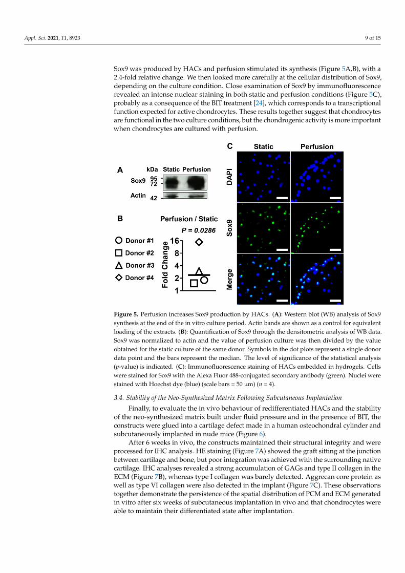
Sox9 was produced by HACs and perfusion stimulated its synthesis (Figure 5A,B), with a2.4-fold relative change. We then looked more carefully at the cellular distribution of Sox9,depending on the culture condition. Close examination of Sox9 by immunofluorescencerevealed an intense nuclear staining in both static and perfusion conditions (Figure 5C),probably as a consequence of the BIT treatment [24], which corresponds to a transcriptionalfunction expected for active chondrocytes. These results together suggest that chondrocytesare functional in the two culture conditions, but the chondrogenic activity is more importantwhen chondrocytes are cultured with perfusion.
Appl. Sci. 2021, 11, 8923 9 of 15
3.3. Perfusion Reinforces the Chondrogenic Activity through the Production of Sox9 To provide further evidence that perfusion of HACs encapsulated in fibrin hydrogel
and treated with BIT enhances the chondrogenic activity of chondrocytes, we looked for Sox9 production. Sox9 is a major factor in developing and adult cartilage. It fulfils many important functions during chondrogenesis including transcriptional activation of many genes coding cartilage-specific structural components such as the type II and type IX collagen and aggrecan [36] studied here. Our Western blotting analysis confirmed that Sox9 was produced by HACs and perfusion stimulated its synthesis (Figure 5A,B), with a 2.4-fold relative change. We then looked more carefully at the cellular distribution of Sox9, depending on the culture condition. Close examination of Sox9 by immunofluorescence revealed an intense nuclear staining in both static and perfusion conditions (Figure 5C), probably as a consequence of the BIT treatment [24], which corresponds to a transcriptional function expected for active chondrocytes. These results together suggest that chondrocytes are functional in the two culture conditions, but the chondrogenic activity is more important when chondrocytes are cultured with perfusion.
Figure 5. Perfusion increases Sox9 production by HACs. (A): Western blot (WB) analysis of Sox9 synthesis at the end of the in vitro culture period. Actin bands are shown as a control for equivalent loading of the extracts. (B): Quantification of Sox9 through the densitometric analysis of WB data. Sox9 was normalized to actin and the value of perfusion culture was then divided by the value obtained for the static culture of the same donor. Symbols in the dot plots represent a single donor data point and the bars represent the median. The level of significance of the statistical analysis (p-value) is indicated. (C): Immunofluorescence staining of HACs embedded in hydrogels. Cells were stained for Sox9 with the Alexa Fluor 488-conjugated secondary antibody (green). Nuclei were stained with Hoechst dye (blue) (scale bars = 50 µm) (n = 4).
3.4. Stability of the Neo-Synthesized Matrix Following Subcutaneous Implantation Finally, to evaluate the in vivo behaviour of redifferentiated HACs and the stability
of the neo-synthesized matrix built under fluid pressure and in the presence of BIT, the constructs were glued into a cartilage defect made in a human osteochondral cylinder and subcutaneously implanted in nude mice (Figure 6).
Figure 5. Perfusion increases Sox9 production by HACs. (A): Western blot (WB) analysis of Sox9synthesis at the end of the in vitro culture period. Actin bands are shown as a control for equivalentloading of the extracts. (B): Quantification of Sox9 through the densitometric analysis of WB data.Sox9 was normalized to actin and the value of perfusion culture was then divided by the valueobtained for the static culture of the same donor. Symbols in the dot plots represent a single donordata point and the bars represent the median. The level of significance of the statistical analysis(p-value) is indicated. (C): Immunofluorescence staining of HACs embedded in hydrogels. Cellswere stained for Sox9 with the Alexa Fluor 488-conjugated secondary antibody (green). Nuclei werestained with Hoechst dye (blue) (scale bars = 50 µm) (n = 4).
3.4. Stability of the Neo-Synthesized Matrix Following Subcutaneous Implantation
Finally, to evaluate the in vivo behaviour of redifferentiated HACs and the stabilityof the neo-synthesized matrix built under fluid pressure and in the presence of BIT, theconstructs were glued into a cartilage defect made in a human osteochondral cylinder andsubcutaneously implanted in nude mice (Figure 6).
After 6 weeks in vivo, the constructs maintained their structural integrity and wereprocessed for IHC analysis. HE staining (Figure 7A) showed the graft sitting at the junctionbetween cartilage and bone, but poor integration was achieved with the surrounding nativecartilage. IHC analyses revealed a strong accumulation of GAGs and type II collagen in theECM (Figure 7B), whereas type I collagen was barely detected. Aggrecan core protein aswell as type VI collagen were also detected in the implant (Figure 7C). These observationstogether demonstrate the persistence of the spatial distribution of PCM and ECM generatedin vitro after six weeks of subcutaneous implantation in vivo and that chondrocytes wereable to maintain their differentiated state after implantation.

Appl. Sci. 2021, 11, 8923 10 of 15Appl. Sci. 2021, 11, 8923 10 of 15
Figure 6. Scheme of the in vivo study. (A): Following extraction and monolayer expansion, HACs were seeded in fibrin hydrogel and cultured for 3 weeks in a direct perfusion bioreactor. Cartilage constructs were then fixed in fresh human osteochondral blocks with fibrin sealant and hybrid constructs were implanted into the subcutaneous pockets of nude mice for 6 weeks. The stability of the neo-synthesized matrix was assessed by histology. (B): Macroscopic pictures showing (left to right) the cartilage gel fixed in the human osteochondral block from top and side views as well as the hybrid construct implanted under mouse skin.
After 6 weeks in vivo, the constructs maintained their structural integrity and were processed for IHC analysis. HE staining (Figure 7A) showed the graft sitting at the junction between cartilage and bone, but poor integration was achieved with the surrounding native cartilage. IHC analyses revealed a strong accumulation of GAGs and type II collagen in the ECM (Figure 7B), whereas type I collagen was barely detected. Aggrecan core protein as well as type VI collagen were also detected in the implant (Figure 7C). These observations together demonstrate the persistence of the spatial distribution of PCM and ECM generated in vitro after six weeks of subcutaneous implantation in vivo and that chondrocytes were able to maintain their differentiated state after implantation.
Figure 7. The cartilage construct remains stable after 6 weeks of subcutaneous implantation in nude mice. Following 21 days of culture in a bidirectional perfusion bioreactor in the presence of BIT, fibrin hydrogels seeded with HACs were placed into a cartilage defect made in human osteochondral blocks and then implanted into the subcutaneous pockets of
Figure 6. Scheme of the in vivo study. (A): Following extraction and monolayer expansion, HACswere seeded in fibrin hydrogel and cultured for 3 weeks in a direct perfusion bioreactor. Cartilageconstructs were then fixed in fresh human osteochondral blocks with fibrin sealant and hybridconstructs were implanted into the subcutaneous pockets of nude mice for 6 weeks. The stabilityof the neo-synthesized matrix was assessed by histology. (B): Macroscopic pictures showing (left toright) the cartilage gel fixed in the human osteochondral block from top and side views as well as thehybrid construct implanted under mouse skin.
Appl. Sci. 2021, 11, 8923 10 of 15
Figure 6. Scheme of the in vivo study. (A): Following extraction and monolayer expansion, HACs were seeded in fibrin hydrogel and cultured for 3 weeks in a direct perfusion bioreactor. Cartilage constructs were then fixed in fresh human osteochondral blocks with fibrin sealant and hybrid constructs were implanted into the subcutaneous pockets of nude mice for 6 weeks. The stability of the neo-synthesized matrix was assessed by histology. (B): Macroscopic pictures showing (left to right) the cartilage gel fixed in the human osteochondral block from top and side views as well as the hybrid construct implanted under mouse skin.
After 6 weeks in vivo, the constructs maintained their structural integrity and were processed for IHC analysis. HE staining (Figure 7A) showed the graft sitting at the junction between cartilage and bone, but poor integration was achieved with the surrounding native cartilage. IHC analyses revealed a strong accumulation of GAGs and type II collagen in the ECM (Figure 7B), whereas type I collagen was barely detected. Aggrecan core protein as well as type VI collagen were also detected in the implant (Figure 7C). These observations together demonstrate the persistence of the spatial distribution of PCM and ECM generated in vitro after six weeks of subcutaneous implantation in vivo and that chondrocytes were able to maintain their differentiated state after implantation.
Figure 7. The cartilage construct remains stable after 6 weeks of subcutaneous implantation in nude mice. Following 21 days of culture in a bidirectional perfusion bioreactor in the presence of BIT, fibrin hydrogels seeded with HACs were placed into a cartilage defect made in human osteochondral blocks and then implanted into the subcutaneous pockets of
Figure 7. The cartilage construct remains stable after 6 weeks of subcutaneous implantation in nude mice. Following21 days of culture in a bidirectional perfusion bioreactor in the presence of BIT, fibrin hydrogels seeded with HACs wereplaced into a cartilage defect made in human osteochondral blocks and then implanted into the subcutaneous pocketsof nude mice. (A): hematoxylin and eosin staining of the cartilage construct in the defect after 6 weeks of subcutaneousimplantation. (B,C): Immunohistochemical analyses of the implant. (B): Cartilage implant stained for safranin-O fast green(GAGs) as well as for collagen types I and II. (C): Aggrecan core protein and type VI collagen immunofluorescence stainingof the cartilage graft. Proteins of interest were stained with the Alexa Fluor 488-conjugated secondary antibody (red). Nucleiwere stained with Hoechst dye (blue) (scale bars: A = 1 mm, B-C = 100 µm).
4. Discussion
We investigated here the combined influence of a bidirectional perfusion bioreactorand an optimized sequential combination of soluble growth factors for the redifferentiationof HACs in fibrin hydrogel. The device we used in this study, namely the U-CUP bioreactor,
Appl. Sci. 2021, 11, 8923 11 of 15
is a commercially available perfusion bioreactor that takes into account important featuresof clinical translation, such as the ability to perform maturation of multiple constructsin individual chambers in parallel, automation and ease of use. Here, we applied arelatively low flow velocity based on previous studies. Freyria et al. defined an optimalflow rate between 1.6 and 5 µL/s for direct perfusion of bovine chondrocytes seeded incollagen sponge and showed that a higher perfusion velocity (16 µL/s) decreased GAGdeposition [37]. Other studies have shown that a flow velocity of 0.13 or 8.3 µL/s increasedGAG [15,16,18] and aggrecan [16] syntheses in agarose hydrogels by animal chondrocytescompared to static cultures. However, Grogan et al. reported that a perfusion velocityof 1.7 µL/s reduced the production of GAGs and link protein by human chondrocytesin alginate hydrogels [17]. Here, our results demonstrate an increased production andimproved quality of neo-synthesized cartilage matrix by HACs in fibrin hydrogels withlow perfusion velocity (2.5 µL/s) compared to static cultures.
Perfusion preconditioning was applied to HAC-seeded gels of clinically relevantdiameter, since 10 mm is the minimum defect diameter recommended for ACI [6,7] andis often reported as the average cartilage defect diameter in clinical studies involvingACI [38]. Perfusion clearly increased the quantity of cartilage ECM deposited in the largeengineered tissues, with up to three quarters of the graft cross-section positively stainingfor GAG deposition. However, a delayed maturation of the core was indicated by ectopiccartilage ECM-positive areas, whereas the periphery of the gel seemed to have achievedits maturation as evidenced by homogeneous cartilage ECM deposition. This may beexplained by an insufficient mass transfer of nutrients and growth factors to the core,as well as by lower fluid-induced shear stresses within the scaffolds which could haveunderstimulated the cells.
In this study, we paid particular attention to the nature of the newly synthesized matrix.Our WB analyses showed that type II collagen, a typical marker of native cartilage [39], wasproduced both in static and in dynamic conditions. Undoubtedly, the combination of BITand perfusion did enhance the deposition of type II collagen; it also enhanced that of typeIX collagen, these being two main components of the collagen fibrils present in hyaline typeof cartilage [40]. It is well-known that the amplification of HACs induces dedifferentiation,resulting in a progressive increase in type I collagen at the expense of type II collagenexpression [41]. This “side effect” of HAC expansion is exacerbated with the FI cocktaildespites its proliferative effect on HACs [27]. Thus, the chondrocytes were active in type Icollagen production at the start of their culture in fibrine gel. However, no sign of precursorforms of type I collagen was detected, probably as a result of BIT treatment [24]. This stopin type I collagen production is particularly relevant since it indicates that the combinationof fibrin and BIT favors the production of hyaline type cartilage as articular cartilageand not fibrocartilage (which has inferior mechanical properties). In a previous work,we showed that perfusion inhibited type I collagen production from HACs in collagensponges [26]. Here, we observed such an effect only for one donor of four, type I collagenproduction being mainly equivalent between static and dynamic conditions. However,our differentiation index (type II collagen/type I collagen ratio) is in favor of a higherproduction of type II collagen and a differentiated phenotype in dynamic cultures. Thisis well supported by our histological and immunohistochemical analyses showing theabundant presence of GAGs, type II collagen, aggrecan core protein, and type VI collagen,whereas type I collagen was barely detected in dynamic cultures.
The particular interest in type VI collagen in the current study arises from its uniquestructure and location. In articular cartilage, type VI collagen is almost exclusively locatedin the PCM. Its amino acid structure contains sequences that are active in binding tocellular receptors [42], and other sequences that are involved in the creation of multimericfibrillar assemblies with collagen type II and aggrecan [43], which connect the chondrocytesto the macromolecular framework of the ECM. Such intimate interactions between thePCM components and the cell surface are likely involved in mechanotransduction to thecell surface; engineered constructs with non-native PCM structure could exhibit altered
Appl. Sci. 2021, 11, 8923 12 of 15
metabolic responses to physicochemical signals, affecting the ultimate success of suchapproaches. As a consequence, the presence of type VI collagen in our tissue-engineeredconstructs is an important indicator of advanced tissue maturation and demonstratesthat cells emancipate themself from the biomaterial to create intimate contacts with thematrix they produce. The absence of type VI collagen in static cultures also indicatesthat perfusion was the main determinant of the type VI collagen secretion observed inour study. These results together demonstrate that the combination of perfusion and BITsubstantially improved the reconstruction of hyaline cartilage-specific ECM and PCM infibrin hydrogels.
Perfusion can enhance the transport of molecules through large gels [15], but it alsoexerts mechanical forces such as fluid shear stress [44] and the expression of several ECMproteins, including type II and IX collagen and aggrecan, as well as transcription factors likeSox9, is sensitive to the mechanical forces in cartilage [45]. We also observed a statisticallysignificant increase in the expression of type II collagen, type IX collagen, aggrecan, andSox9 with direct perfusion (Figure S2). Thus, we can hypothesize that the fluid flowperfused here may have mechanically stimulated the chondrocytes seeded within fibringels. Importantly, the nuclear translocation of Sox9, the abundant expression of type IIprocollagen forms, and the presence of intracellular staining for aggrecan core protein andtype VI collagen occurring in cells cultivated under direct perfusion attest that cells wereactive at the end of the culture period (Figure S3).
Although good stability of the neo-cartilage and maintenance of chondrocyte differen-tiated phenotype was observed after six weeks in the nude mouse model, pre-clinical trialswith long-term implantation in cartilage lesions in a large animal model are required toassess the integration of the implant and mechanical stability in living joint. As to the latter,mechanical characterization of constructs was not performed here and this represents alimitation of the present study. Indeed, developing constructs with mechanical propertiesapproaching those of native tissue is a key challenge in cartilage tissue engineering and theimpact of perfusion preconditioning on the mechanical properties of the implant will bethus investigated in future studies.
Nevertheless, our results already demonstrate the value of a multi-factorial approachcombining HACs from osteoarthritic joints, clinically relevant fibrin hydrogel, BIT cocktail,and perfusion preconditioning. This strategy leads to a high degree of cell redifferentiationby inducing the specific production and spatial distribution of hyaline matrix. This resultis particularly relevant since it indicates that such a multi-factorial approach promotesthe production of cartilage PCM, a key feature of functional cartilage. Of note, specialefforts were made to develop an approach employing clinical-grade growth factors andscaffolds which, combined with the intrinsic features of the U-CUP bioreactor, can be easilytranslated into clinical practice. Lastly, since 3D bioprinting has the potential to modifythe microarchitecture of large tissue-engineered constructs [46], it would be pertinentto investigate whether the introduction of microchannels within the graft or a positivegradient of fibrin concentration from the periphery to the core (i.e., leading to a progressiveincrease in microfiber spacing toward the core) could balance access to growth factors andfluid-induced shear stresses between the center and the periphery of the graft.
5. Conclusions
The present study reports that perfusion preconditioning enhances redifferentiationof chondrocytes in fibrin hydrogel. Here, cells were extracted from human articular car-tilage tissue, expanded, and cultured in fibrin gel. From a translational perspective, theapproval by the Food and Drug Administration (FDA) of MACI®® (Vericel, Cambridge,MA, USA)—where cells from the patient’s knee are expanded, placed on a scaffold, andthen reimplanted into the patient—shows that tissue engineering strategies can be trans-lated into clinical practice and are commercially feasible. Preconditioned grafts containedchondrocytes expressing a better level of redifferentiation at the gene and protein levels,adding potential therapeutic value. While this last point needs to be assessed in a large
Appl. Sci. 2021, 11, 8923 13 of 15
animal model, our results herein demonstrate that perfusion preconditioning of HACs infibrin hydrogels generates cartilage-like tissues containing typical components of cartilageextra and pericellular matrix.
Supplementary Materials: The following are available online at https://www.mdpi.com/article/10.3390/app11198923/s1, Figure S1: cell viability in static and perfused gels after a 7 day culture period,Figure S2: Gene expression characterization of human articular chondrocytes cultivated for 21 daysin fibrin hydrogel in free swelling (static) or a direct perfusion bioreactor, Figure S3: Quantification ofaggrecan core protein and type VI collagen immunofluorescence at the single-cell level.
Author Contributions: Conceptualization, A.D. and E.P.-G.; methodology, A.D. and E.P.-G.; formalanalysis, A.D.; data curation, A.D.; writing—original draft preparation, A.D.; writing—review andediting, A.D., F.M.-G. and E.P.-G.; supervision, F.M.-G. and E.P.-G.; project administration, F.M.-G. and E.P.-G.; funding acquisition, F.M.-G. and E.P.-G. All authors have read and agreed to thepublished version of the manuscript.
Funding: This research was funded by CNRS, Lyon 1 University, and by Fondation de l’Avenir. A.D.was supported by a Ph.D. fellowship from the Région Auvergne-Rhône-Alpes (ARC2).
Institutional Review Board Statement: The study protocol involving human tissues was conductedin accordance with the Declaration of Helsinki and was approved by the ethics committee of COnser-vation D’ELéments du COrps Humain (CODECOH: DC-2014–2325) for preservation and researchwith human samples. The cartilage samples were collected after written informed consent of thedonors. The study involving animals was conducted according to the guidelines of the Directive2010/63/EU and its national transposition and ethical guidelines for care and use of mice of thePlateau de Biologie Expérimentale de la Souris (PBES, UMS 3444) at Ecole Normale Supérieure (ENS,Lyon) and approved by the Institutional Review Board of ENS Lyon (Committee on the Ethics ofAnimal Experiments, approval number: ENS_2018_007).
Informed Consent Statement: Informed consent was obtained from all human donors involved inthe study.
Data Availability Statement: The data presented in this study are available on request from thecorresponding author.
Acknowledgments: Authors are grateful to the technical facilities of SFR Biosciences Gerland-Lyon(UMS3444/US8) for the quantitative PCR analyses and to EL KHOLTI Naima for her assistance withpreparing and processing histological samples.
Conflicts of Interest: The authors declare no conflict of interest.
References1. Sophia Fox, A.J.; Bedi, A.; Rodeo, S.A. The basic science of articular cartilage: Structure, composition, and function. Sports Health
2009, 1, 461–468. [CrossRef]2. Huey, D.J.; Hu, J.C.; Athanasiou, K.A. Unlike bone, cartilage regeneration remains elusive. Science (80-.) 2012, 338, 917–921.
[CrossRef]3. Richter, D.L.; Schenck, R.C.; Wascher, D.C.; Treme, G. Knee Articular Cartilage Repair and Restoration Techniques: A Review of
the Literature. Sports Health 2016, 8, 153–160. [CrossRef] [PubMed]4. Hangody, L.; Füles, P. Autologous osteochondral mosaicplasty for the treatment of full-thickness defects of weight-bearing joints:
Ten years of experimental and clinical experience. J. Bone Jt. Surg.-Ser. A 2003, 85, 25–32. [CrossRef] [PubMed]5. Hunziker, E.B. Articular cartilage repair: Basic science and clinical progress. A review of the current status and prospects.
Osteoarthr. Cartil. 2002, 10, 432–463. [CrossRef] [PubMed]6. Niemeyer, P.; Albrecht, D.; Andereya, S.; Angele, P.; Ateschrang, A.; Aurich, M.; Baumann, M.; Bosch, U.; Erggelet, C.; Fickert, S.;
et al. Autologous chondrocyte implantation (ACI) for cartilage defects of the knee: A guideline by the working group “ClinicalTissue Regeneration” of the German Society of Orthopaedics and Trauma (DGOU). Knee 2016, 23, 426–435. [CrossRef] [PubMed]
7. Makris, E.A.; Gomoll, A.H.; Malizos, K.N.; Hu, J.C.; Athanasiou, K.A. Repair and tissue engineering techniques for articularcartilage. Nat. Rev. Rheumatol. 2015, 11, 21–34. [CrossRef] [PubMed]
8. McCarthy, H.S.; Richardson, J.B.; Parker, J.C.E.; Roberts, S. Evaluating Joint Morbidity after Chondral Harvest for AutologousChondrocyte Implantation (ACI): A Study of ACI-Treated Ankles and Hips with a Knee Chondral Harvest. Cartilage 2016, 7, 7–15.[CrossRef]
9. Sharpe, J.R.; Ahmed, S.U.; Fleetcroft, J.P.; Martin, R. The treatment of osteochondral lesions using a combination of autologouschondrocyte implantation and autograft. Three-year follow-up. J. Bone Jt. Surg.-Ser. B 2005, 87, 730–735. [CrossRef]
Appl. Sci. 2021, 11, 8923 14 of 15
10. Zhao, W.; Jin, X.; Cong, Y.; Liu, Y.; Fu, J. Degradable natural polymer hydrogels for articular cartilage tissue engineering. J. Chem.Technol. Biotechnol. 2013, 88, 327–339. [CrossRef]
11. Flégeau, K.; Pace, R.; Gautier, H.; Rethore, G.; Guicheux, J.; Le Visage, C.; Weiss, P. Toward the development of biomimeticinjectable and macroporous biohydrogels for regenerative medicine. Adv. Colloid Interface Sci. 2017, 247, 589–609. [CrossRef]
12. Farrell, M.J.; Shin, J.I.; Smith, L.J.; Mauck, R.L. Functional consequences of glucose and oxygen deprivation onengineeredmesenchymal stem cell-based cartilage constructs. Osteoarthr. Cartil. 2015, 23, 134–142. [CrossRef] [PubMed]
13. Wendt, D.; Stroebel, S.; Jakob, M.; John, G.T.; Martin, I. Uniform tissues engineered by seeding and culturing cells in 3D scaffoldsunder perfusion at defined oxygen tensions. Biorheology 2006, 43, 481–488. [PubMed]
14. Santoro, R.; Olivares, A.L.; Brans, G.; Wirz, D.; Longinotti, C.; Lacroix, D.; Martin, I.; Wendt, D. Bioreactor based engineering oflarge-scale human cartilage grafts for joint resurfacing. Biomaterials 2010, 31, 8946–8952. [CrossRef] [PubMed]
15. Eniwumide, J.O.; Lee, D.A.; Bader, D.L. The development of a bioreactor to perfuse radially-confined hydrogel constructs: Designand characterization of mass transport properties. Biorheology 2009, 46, 417–437. [CrossRef] [PubMed]
16. Schulz, R.M.; Wüstneck, N.; Van Donkelaar, C.C.; Shelton, J.C.; Bader, A. Development and validation of a novel bioreactorsystem for load- and perfusion-controlled tissue engineering of chondrocyte-constructs. Biotechnol. Bioeng. 2008, 101, 714–728.[CrossRef]
17. Grogan, S.P.; Sovani, S.; Pauli, C.; Chen, J.; Hartmann, A.; Colwell, C.W.; Lotz, M.K.; D’Lima, D.D. Effects of perfusion anddynamic loading on human neocartilage formation in alginate hydrogels. Tissue Eng.-Part A 2012, 18, 1784–1792. [CrossRef]
18. Nazempour, A.; Quisenberry, C.R.; Abu-Lail, N.I.; Van Wie, B.J. Combined effects of oscillating hydrostatic pressure, perfusionand encapsulation in a novel bioreactor for enhancing extracellular matrix synthesis by bovine chondrocytes. Cell Tissue Res.2017, 370, 179–193. [CrossRef]
19. Yu, L.; Ferlin, K.M.; Nguyen, B.N.B.; Fisher, J.P. Tubular perfusion system for chondrocyte culture and superficial zone proteinexpression. J. Biomed. Mater. Res.-Part A 2015, 103, 1864–1874. [CrossRef]
20. Bianchi, V.J.; Lee, A.; Anderson, J.; Parreno, J.; Theodoropoulos, J.; Backstein, D.; Kandel, R. Redifferentiated Chondrocytes inFibrin Gel for the Repair of Articular Cartilage Lesions. Am. J. Sports Med. 2019, 47, 2348–2359. [CrossRef]
21. Vogt, S.; Wexel, G.; Tischer, T.; Schillinger, U.; Ueblacker, P.; Wagner, B.; Hensler, D.; Wilisch, J.; Geis, C.; Wübbenhorst, D.; et al.The influence of the stable expression of BMP2 in fibrin clots on the remodelling and repair of osteochondral defects. Biomaterials2009, 30, 2385–2392. [CrossRef]
22. Liu, G.; Kawaguchi, H.; Ogasawara, T.; Asawa, Y.; Kishimoto, J.; Takahashi, T.; Chung, U.I.; Yamaoka, H.; Asato, H.; Nakamura,K.; et al. Optimal combination of soluble factors for tissue engineering of permanent cartilage from cultured human chondrocytes.J. Biol. Chem. 2007, 282, 20407–20415. [CrossRef]
23. Durbec, M.; Mayer, N.; Vertu-Ciolino, D.; Disant, F.; Mallein-Gerin, F.; Perrier-Groult, E. Reconstruction of nasal cartilage defectsusing a tissue engineering technique based on combination of high-density polyethylene and hydrogel. Pathol. Biol. 2014, 62,137–145. [CrossRef]
24. Dufour, A.; Buffier, M.; Vertu-Ciolino, D.; Disant, F.; Mallein-Gerin, F.; Perrier-Groult, E. Combination of bioactive factors andIEIK13 self-assembling peptide hydrogel promotes cartilage matrix production by human nasal chondrocytes. J. Biomed. Mater.Res. Part A 2019. [CrossRef]
25. Dufour, A.; Lafont, J.E.; Buffier, M.; Verset, M.; Cohendet, A.; Contamin, H.; Confais, J.; Sankar, S.; Rioult, M.; Groult, E.P.; et al.Repair of full-thickness articular cartilage defects using IEIK13 self-assembling peptide hydrogel in a non-human primate model.Sci. Rep. 2021, 1–17. [CrossRef]
26. Mayer, N.; Lopa, S.; Talò, G.; Lovati, A.B.; Pasdeloup, M.; Riboldi, S.A.; Moretti, M.; Mallein-Gerin, F. Interstitial perfusion culturewith specific soluble factors inhibits type i collagen production from human osteoarthritic chondrocytes in clinical-grade collagensponges. PLoS ONE 2016, 11, e0161479. [CrossRef]
27. Claus, S.; Mayer, N.; Aubert-Foucher, E.; Chajra, H.; Perrier-Groult, E.; Lafont, J.; Piperno, M.; Damour, O.; Mallein-Gerin, F.Cartilage-Characteristic Matrix Reconstruction by Sequential Addition of Soluble Factors During Expansion of Human ArticularChondrocytes and Their Cultivation in Collagen Sponges. Tissue Eng. Part C Methods 2012, 18, 104–112. [CrossRef]
28. Wang, J.H.; Lee, B.H. Mediolateral Differences of Proteoglycans Distribution at the ACL Tibial Footprint: Experimental Study of16 Cadaveric Knees. Biomed Res. Int. 2018, 2018. [CrossRef] [PubMed]
29. Ruoslahti, E.; Yamaguchi, Y. Proteoglycans as modulators of growth factor activities. Cell 1991, 64, 867–869. [CrossRef]30. Vincourt, J.B.; Etienne, S.; Grossin, L.; Cottet, J.; Bantsimba-Malanda, C.; Netter, P.; Mainard, D.; Libante, V.; Gillet, P.; Magdalou,
J. Matrilin-3 switches from anti- to pro-anabolic upon integration to the extracellular matrix. Matrix Biol. 2012, 31, 290–298.[CrossRef] [PubMed]
31. Poole, C.A.; Honda, T.; Skinner, S.J.M.; Schofield, J.R.; Hyde, K.F.; Shinkai, H. Chondrons from articular cartilage (II): Analysis ofthe glycosaminoglycans in the cellular microenvironment of isolated canine chondrons. Connect. Tissue Res. 1990, 24, 319–330.[CrossRef]
32. Poole, C.A.; Glant, T.T.; Schofield, J.R. Chondrons from articular cartilage. (IV) Immunolocalization of proteoglycan epitopes inisolated canine tibial chondrons. J. Histochem. Cytochem. 1991, 39, 1175–1187. [CrossRef] [PubMed]
33. Poole, C.A.; Gilbert, R.T.; Herbage, D.; Hartmann, D.J. Immunolocalization of type IX collagen in normal and spontaneouslyosteoarthritic canine tibial cartilage and isolated chondrons. Osteoarthr. Cartil. 1997, 5, 191–204. [CrossRef]
Appl. Sci. 2021, 11, 8923 15 of 15
34. Poole, C.A.; Ayad, S.; Schofield, J.R. Chondrons from articular cartilage: I. Immunolocalization of type VI collagen in thepericellular capsule of isolated canine tibial chondrons. J. Cell Sci. 1988, 90 Pt 4, 635–643. [CrossRef] [PubMed]
35. Poole, C.A.; Flint, M.H.; Beaumont, B.W. Chondrons in cartilage: Ultrastructural analysis of the pericellular microenvironment inadult human articular cartilages. J. Orthop. Res. 1987, 5, 509–522. [CrossRef] [PubMed]
36. Lefebvre, V.; Dvir-Ginzberg, M. SOX9 and the many facets of its regulation in the chondrocyte lineage. Connect. Tissue Res. 2017,58, 2–14. [CrossRef] [PubMed]
37. Freyria, A.-M.; Yang, Y.; Chajra, H.; Rousseau, C.F.; Ronzière, M.-C.; Herbage, D.; Haj, A.J. El Optimization of Dynamic CultureConditions: Effects on Biosynthetic Activities of Chondrocytes Grown in Collagen Sponges. Tissue Eng. 2005, 11, 674–684.[CrossRef] [PubMed]
38. Na, Y.; Shi, Y.; Liu, W.; Jia, Y.; Kong, L.; Zhang, T.; Han, C.; Ren, Y. Is implantation of autologous chondrocytes superior tomicrofracture for articular-cartilage defects of the knee? A systematic review of 5-year follow-up data. Int. J. Surg. 2019, 68, 56–62.[CrossRef]
39. Gouttenoire, J.; Valcourt, U.; Ronzière, M.C.; Aubert-Foucher, E.; Mallein-Gerin, F.; Herbage, D. Modulation of collagen synthesisin normal and osteoarthritic cartilage. Biorheology 2004, 41, 535–542.
40. Mendler, M.; Eich-Bender, S.G.; Vaughan, L.; Winterhalter, K.H.; Bruckner, P. Cartilage contains mixed fibrils of collagen types II,IX, and XI. J. Cell Biol. 1989, 108, 191–197. [CrossRef]
41. Von Der Mark, K.; Gauss, V.; Von Der Mark, H.; Müller, P. Relationship between cell shape and type of collagen synthesised aschondrocytes lose their cartilage phenotype in culture. Nature 1977, 267, 531–532. [CrossRef] [PubMed]
42. Bonaldo, P.; Russo, V.; Bucciotti, F.; Doliana, R.; Colombatti, A. Structural and Functional Features of the α3 Chain Indicate aBridging Role for Chicken Collagen VI in Connective Tissues. Biochemistry 1990, 29, 1245–1254. [CrossRef]
43. Wiberg, C.; Klatt, A.R.; Wagener, R.; Paulsson, M.; Bateman, J.F.; Heinegård, D.; Mörgelin, M. Complexes of matrilin-1 andbiglycan or decorin connect collagen VI microfibrils to both collagen II and aggrecan. J. Biol. Chem. 2003, 278, 37698–37704.[CrossRef]
44. Moretti, M.; Freed, L.E.; Padera, R.F.; Laganà, K.; Boschetti, F.; Raimondi, M.T. An integrated experimental-computationalapproach for the study of engineered cartilage constructs subjected to combined regimens of hydrostatic pressure and interstitialperfusion. Biomed. Mater. Eng. 2008, 18, 273–278. [CrossRef]
45. Fitzgerald, J.B.; Jin, M.; Chai, D.H.; Siparsky, P.; Fanning, P.; Grodzinsky, A.J. Shear- and compression-induced chondrocytetranscription requires MAPK activation in cartilage explants. J. Biol. Chem. 2008, 283, 6735–6743. [CrossRef] [PubMed]
46. Murphy, S.V.; De Coppi, P.; Atala, A. Opportunities and challenges of translational 3D bioprinting. Nat. Biomed. Eng. 2020, 4,370–380. [CrossRef] [PubMed]