dynamic head perfusion - aapm
TRANSCRIPT
Adult Brain Perfusion CT Protocols Version 2.0 3/1/2016
DISCLAIMER: TO THE EXTENT ALLOWED BY LOCAL LAW, THIS INFORMATION IS PROVIDED TO YOU BY THE AMERICAN ASSOCIATION OF PHYSICISTS IN MEDICINE, A NON-PROFIT ORGANIZATION ORGANIZED TO PROMOTE THE APPLICATION OF PHYSICS TO MEDICINE AND BIOLOGY, ENCOURAGE INTEREST AND TRAINING IN MEDICAL PHYSICS AND RELATED FIELDS ("AAPM"), 'AS IS' WITHOUT WARRANTIES OR CONDITIONS OF ANY KIND, WHETHER ORAL OR WRITTEN, EXPRESS OR IMPLIED. AAPM SPECIFICALLY DISCLAIMS ANY IMPLIED WARRANTIES OR CONDITIONS OF MERCHANTABILITY, SATISFACTORY QUALITY, NONINFRINGEMENT AND FITNESS FOR A PARTICULAR PURPOSE. SOME JURISDICTIONS DO NOT ALLOW EXCLUSIONS OF IMPLIED WARRANTIES OR CONDITIONS, SO THE ABOVE EXCLUSION MAY NOT APPLY TO YOU. YOU MAY HAVE OTHER RIGHTS THAT VARY ACCORDING TO LOCAL LAW. TO THE EXTENT ALLOWED BY LOCAL LAW, IN NO EVENT WILL AAPM OR ITS SUBSIDIARIES, AFFILIATES OR VENDORS BE LIABLE FOR DIRECT, SPECIAL, INCIDENTAL, CONSEQUENTIAL OR OTHER DAMAGES (INCLUDING LOST PROFIT, LOST DATA, OR DOWNTIME COSTS), ARISING OUT OF THE USE, INABILITY TO USE, OR THE RESULTS OF USE OF THE PROVIDED INFORMATION, WHETHER BASED IN WARRANTY, CONTRACT, TORT OR OTHER LEGAL THEORY, AND WHETHER OR NOT ADVISED OF THE POSSIBILITY OF SUCH DAMAGES. YOUR USE OF THE INFORMATION IS ENTIRELY AT YOUR OWN RISK. THIS INFORMATION IS NOT MEANT TO BE USED AS A SUBSTITUTE FOR THE REVIEW OF SCAN PROTOCOL PARAMETERS BY A QUALIFIED AND CERTIFIED PROFESSIONAL. USERS ARE CAUTIONED TO SEEK THE ADVICE OF A QUALIFIED AND CERTIFIED PROFESSIONAL BEFORE USING ANY PROTOCOL BASED ON THE PROVIDED INFORMATION. AAPM IS NOT RESPONSIBLE FOR A USER'S FAILURE TO VERIFY OR CONFIRM APPROPRIATE PERFORMANCE OF THE PROVIDED SCAN PARAMETERS. SOME JURISDICTIONS DO NOT ALLOW THE EXCLUSION OR LIMITATION OF LIABILITY FOR DAMAGES, SO THE ABOVE LIMITATION MAY NOT APPLY TO YOU.
The disclaimer on page 1 is an integral part of this document.
1
Copyright © March 1, 2016 by AAPM. All rights reserved.
Adult Brain Perfusion CT Protocols Version 2.0 3/1/2016
ADULT BRAIN PERFUSION CT Indications
Suspected acute infarction; Assessment of reperfusion after treatment of acute stroke; Vasculitis; New neurological symptoms after subarachnoid hemorrhage suggesting vasospasm; Evaluation of the hemodynamic significance of a carotid stenosis; Transient ischemic attack; Evaluation of the cerebral vascular reserve using acetazolamide challenge; Evaluation of brain perfusion after significant head trauma; Brain tumor.
Diagnostic Task • Detect brain ischemia in stroke, transient ischemic attack, vasculitis; • Distinguish already-infarcted brain from brain at risk of infarction; • Identify regions of brain made ischemic by vasospasm; • Detect altered brain perfusion downstream a significant carotid stenosis; • Assess altered cerebral vascular reserve in patients with ischemic symptoms; • Assess altered cerebral perfusion after traumatic brain injury; • Identify early brain tumor recurrence and higher-grade tumor components.
Key Elements • Time-resolved scans are used to track the flow of iodinated contrast media through the brain; • Multiple images (20-40) are acquired over the same section of anatomy; • Patients must be able to remain still during the exam in order to avoid motion misregistration; • The table may remain stationary during the entire exam, or move back and forth between a few table
positions; • Whole-brain perfusion CT can be accomplished using CT systems with wide detector arrays (8-16 cm);
alternatively, scan modes that move the patient back and forth over the desired scan volumes can be used;
• Acquisitions are repeated at predetermined time intervals (e.g. every second to every 2-3 seconds) for a predetermined duration (e.g. 40-90 seconds);
• Relatively thick image widths are used to minimize image noise (5-10 mm is common); • Image quality is inferior to a routine head CT. That is, images are noisier and thicker. • Data are used to generate color maps of hemodynamic significance:
--Blood volume (BV) and flow (BF), mean transit time (MTT), time to peak perfusion (TPP); • A non-contrast-enhanced head CT and/or a CT angiogram may be combined with a perfusion CT scan.
Dose Management • 80 kV should be used to increase iodine signal brightness; • Low dose per single scan (i.e. one tube rotation) is critical, since repeated scanning will result in a
relatively high cumulative dose; • Time interval between scans, and hence the total number of scans over the exam duration, should be
set carefully, taking into account the analysis algorithm (some approaches require relatively dense data points);
• Dose (tube current) modulation should not be used, as it may interfere with the calculation of the BV and BF parameters;
Additional Resources • ACR Practice Guideline for the Performance of Computed Tomography (CT) Perfusion in
Neuroradiologic Imaging. (www.acr.org/Quality-Safety/Standards-Guidelines/Practice-Guidelines-by-Modality/CT);
• AJNR Special Collection. Radiation Dose in Neuroradiology CT Protocols. Collection Editors: Max Wintermark and Michael H. Lev (available at www.ajnr.org/specCol/specCollPCTToc.dtl).
The disclaimer on page 1 is an integral part of this document.
2
Copyright © March 1, 2016 by AAPM. All rights reserved.
Adult Brain Perfusion CT Protocols Version 2.0 3/1/2016
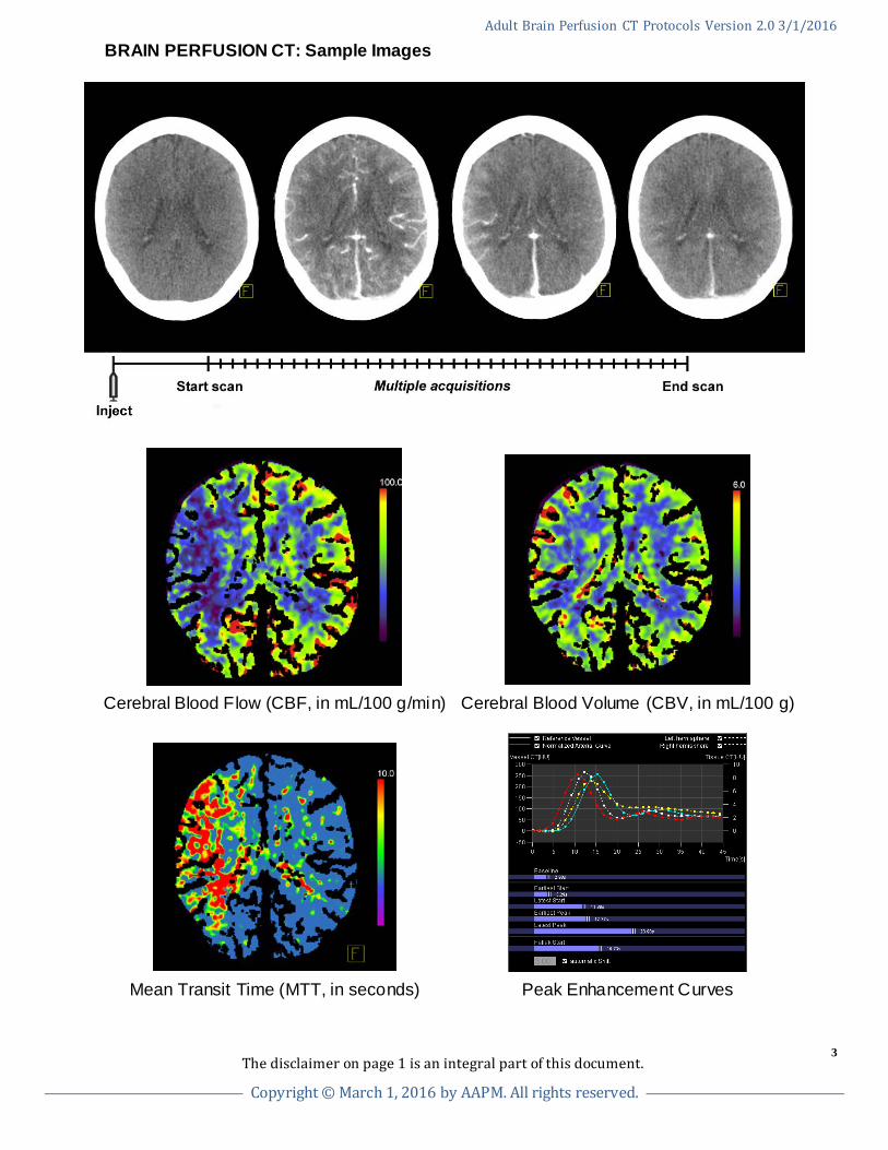
BRAIN PERFUSION CT: Sample Images
Cerebral Blood Flow (CBF, in mL/100 g/min) Cerebral Blood Volume (CBV, in mL/100 g)
Mean Transit Time (MTT, in seconds) Peak Enhancement Curves
The disclaimer on page 1 is an integral part of this document.
3
Copyright © March 1, 2016 by AAPM. All rights reserved.
Adult Brain Perfusion CT Protocols Version 2.0 3/1/2016
INDEX OF ADULT BRAIN PERFUSION CT PROTOCOLS (by manufacturer)
GE
Hitachi
Neurologica
Neusoft
Philips
Siemens
Toshiba
The disclaimer on page 1 is an integral part of this document.
4
Copyright © March 1, 2016 by AAPM. All rights reserved.
Adult Brain Perfusion CT Protocols Version 2.0 3/1/2016
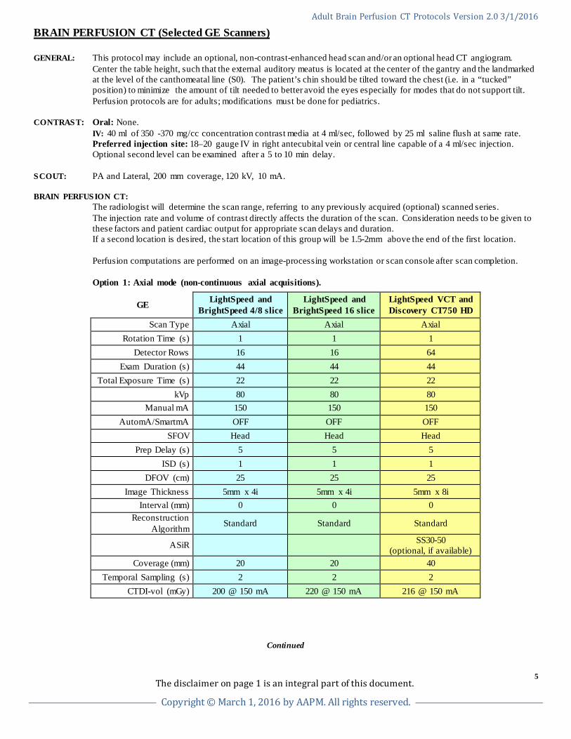
BRAIN PERFUSION CT (Selected GE Scanners) GENERAL: This protocol may include an optional, non-contrast-enhanced head scan and/or an optional head CT angiogram. Center the table height, such that the external auditory meatus is located at the center of the gantry and the landmarked
at the level of the canthomeatal line (S0). The patient’s chin should be tilted toward the chest (i.e. in a “tucked” position) to minimize the amount of tilt needed to better avoid the eyes especially for modes that do not support tilt.
Perfusion protocols are for adults; modifications must be done for pediatrics.
CONTRAST: Oral: None. IV: 40 ml of 350 -370 mg/cc concentration contrast media at 4 ml/sec, followed by 25 ml saline flush at same rate. Preferred injection site: 18–20 gauge IV in right antecubital vein or central line capable of a 4 ml/sec injection. Optional second level can be examined after a 5 to 10 min delay. SCOUT: PA and Lateral, 200 mm coverage, 120 kV, 10 mA. BRAIN PERFUSION CT: The radiologist will determine the scan range, referring to any previously acquired (optional) scanned series. The injection rate and volume of contrast directly affects the duration of the scan. Consideration needs to be given to
these factors and patient cardiac output for appropriate scan delays and duration. If a second location is desired, the start location of this group will be 1.5-2mm above the end of the first location.
Perfusion computations are performed on an image-processing workstation or scan console after scan completion. Option 1: Axial mode (non-continuous axial acquisitions).
GE LightSpeed and BrightSpeed 4/8 slice
LightSpeed and BrightSpeed 16 slice
LightSpeed VCT and Discovery CT750 HD
Scan Type Axial Axial Axial Rotation Time (s) 1 1 1
Detector Rows 16 16 64 Exam Duration (s) 44 44 44
Total Exposure Time (s) 22 22 22 kVp 80 80 80
Manual mA 150 150 150 AutomA/SmartmA OFF OFF OFF
SFOV Head Head Head Prep Delay (s) 5 5 5
ISD (s) 1 1 1 DFOV (cm) 25 25 25
Image Thickness 5mm x 4i 5mm x 4i 5mm x 8i Interval (mm) 0 0 0
Reconstruction Algorithm Standard Standard Standard
ASiR SS30-50 (optional, if available)
Coverage (mm) 20 20 40 Temporal Sampling (s) 2 2 2
CTDI-vol (mGy) 200 @ 150 mA 220 @ 150 mA 216 @ 150 mA
Continued
The disclaimer on page 1 is an integral part of this document.
5
Copyright © March 1, 2016 by AAPM. All rights reserved.
Adult Brain Perfusion CT Protocols Version 2.0 3/1/2016
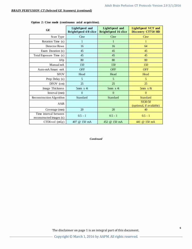
BRAIN PERFUSION CT (Selected GE Scanners) (continued) Option 2: Cine mode (continuous axial acquisition).
GE LightSpeed and BrightSpeed 4/8 slice
LightSpeed and BrightSpeed 16 slice
LightSpeed VCT and Discovery CT750 HD
Scan Type Cine Cine Cine Rotation Time (s) 1 1 1
Detector Rows 16 16 64 Exam Duration (s) 45 45 45
Total Exposure Time (s) 45 45 45 kVp 80 80 80
Manual mA 150 150 150 Auto-mA/Smart -mA OFF OFF OFF
SFOV Head Head Head Prep Delay (s) 5 5 5
DFOV (cm) 25 25 25 Image Thickness 5mm x 4i 5mm x 4i 5mm x 8i
Interval (mm) 0 0 0 Reconstruction Algorithm Standard Standard Standard
ASiR SS30-50 (optional, if available)
Coverage (mm) 20 20 40 Time interval between
reconstructed images (s) 0.5 - 1 0.5 - 1 0.5 - 1
CTDI-vol (mGy) 407 @ 150 mA 452 @ 150 mA 441 @ 150 mA
Continued
The disclaimer on page 1 is an integral part of this document.
6
Copyright © March 1, 2016 by AAPM. All rights reserved.
Adult Brain Perfusion CT Protocols Version 2.0 3/1/2016
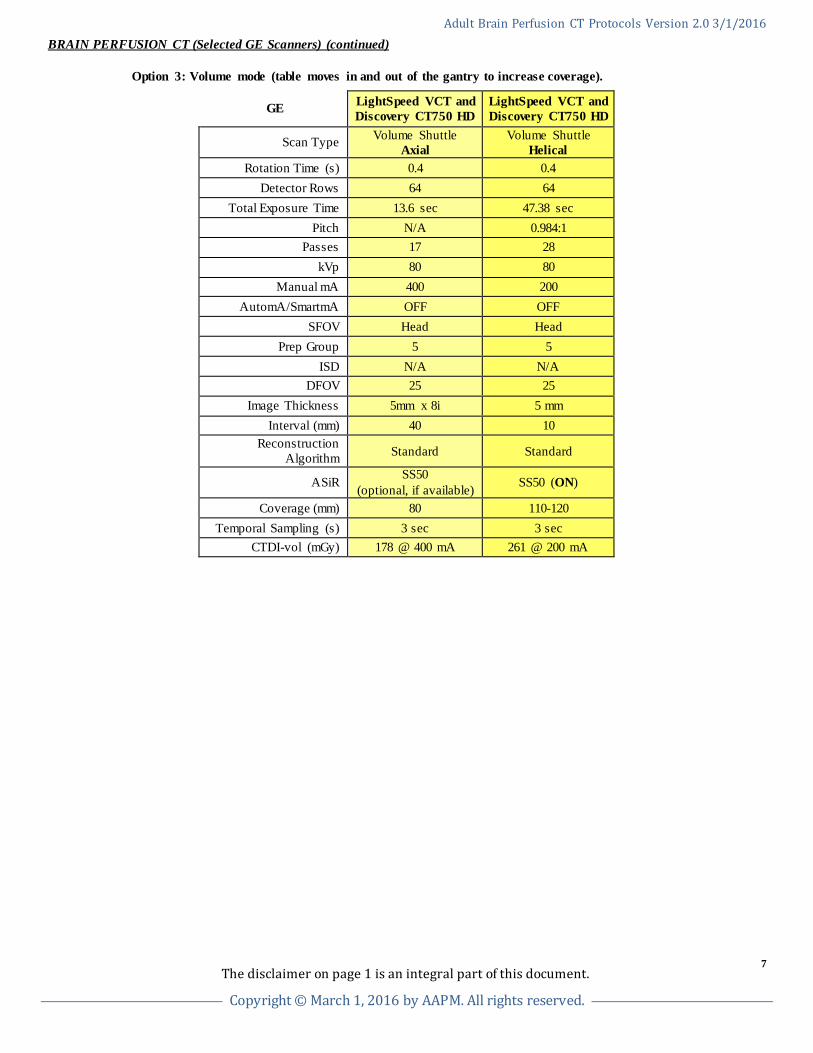
BRAIN PERFUSION CT (Selected GE Scanners) (continued) Option 3: Volume mode (table moves in and out of the gantry to increase coverage).
GE LightSpeed VCT and Discovery CT750 HD
LightSpeed VCT and Discovery CT750 HD
Scan Type Volume Shuttle Axial
Volume Shuttle Helical
Rotation Time (s) 0.4 0.4 Detector Rows 64 64
Total Exposure Time 13.6 sec 47.38 sec Pitch N/A 0.984:1
Passes 17 28 kVp 80 80
Manual mA 400 200 AutomA/SmartmA OFF OFF
SFOV Head Head Prep Group 5 5
ISD N/A N/A DFOV 25 25
Image Thickness 5mm x 8i 5 mm Interval (mm) 40 10
Reconstruction Algorithm Standard Standard
ASiR SS50 (optional, if available) SS50 (ON)
Coverage (mm) 80 110-120 Temporal Sampling (s) 3 sec 3 sec
CTDI-vol (mGy) 178 @ 400 mA 261 @ 200 mA
The disclaimer on page 1 is an integral part of this document.
7
Copyright © March 1, 2016 by AAPM. All rights reserved.
Adult Brain Perfusion CT Protocols Version 2.0 3/1/2016
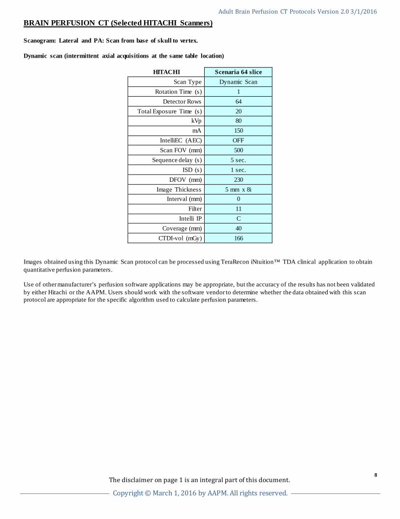
BRAIN PERFUSION CT (Selected HITACHI Scanners) Scanogram: Lateral and PA: Scan from base of skull to vertex. Dynamic scan (intermittent axial acquisitions at the same table location)
HITACHI Scenaria 64 slice Scan Type Dynamic Scan
Rotation Time (s) 1 Detector Rows 64
Total Exposure Time (s) 20 kVp 80 mA 150
IntelliEC (AEC) OFF Scan FOV (mm) 500
Sequence delay (s) 5 sec. ISD (s) 1 sec.
DFOV (mm) 230 Image Thickness 5 mm x 8i
Interval (mm) 0 Filter 11
Intelli IP C Coverage (mm) 40
CTDI-vol (mGy) 166 Images obtained using this Dynamic Scan protocol can be processed using TeraRecon iNtuition™ TDA clinical application to obtain quantitative perfusion parameters. Use of other manufacturer’s perfusion software applications may be appropriate, but the accuracy of the results has not been validated by either Hitachi or the AAPM. Users should work with the software vendor to determine whether the data obtained with this scan protocol are appropriate for the specific algorithm used to calculate perfusion parameters.
The disclaimer on page 1 is an integral part of this document.
8
Copyright © March 1, 2016 by AAPM. All rights reserved.
Adult Brain Perfusion CT Protocols Version 2.0 3/1/2016
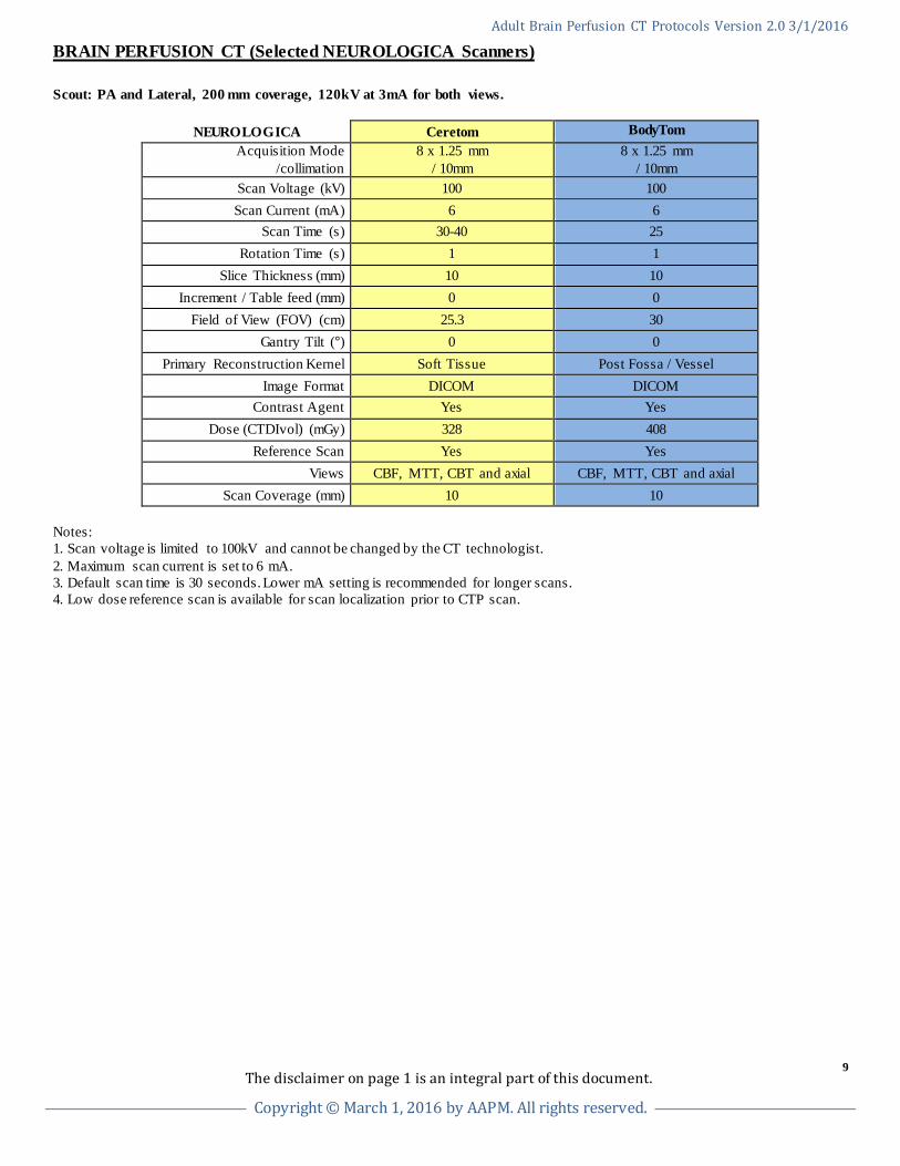
BRAIN PERFUSION CT (Selected NEUROLOGICA Scanners) Scout: PA and Lateral, 200 mm coverage, 120kV at 3mA for both views.
NEUROLOGICA Ceretom BodyTom Acquisition Mode
/collimation 8 x 1.25 mm
/ 10mm 8 x 1.25 mm
/ 10mm Scan Voltage (kV) 100 100 Scan Current (mA) 6 6
Scan Time (s) 30-40 25 Rotation Time (s) 1 1
Slice Thickness (mm) 10 10 Increment / Table feed (mm) 0 0
Field of View (FOV) (cm) 25.3 30 Gantry Tilt (°) 0 0
Primary Reconstruction Kernel Soft Tissue Post Fossa / Vessel Image Format DICOM DICOM
Contrast Agent Yes Yes Dose (CTDIvol) (mGy) 328 408
Reference Scan Yes Yes Views CBF, MTT, CBT and axial CBF, MTT, CBT and axial
Scan Coverage (mm) 10 10 Notes: 1. Scan voltage is limited to 100kV and cannot be changed by the CT technologist. 2. Maximum scan current is set to 6 mA. 3. Default scan time is 30 seconds. Lower mA setting is recommended for longer scans. 4. Low dose reference scan is available for scan localization prior to CTP scan.
The disclaimer on page 1 is an integral part of this document.
9
Copyright © March 1, 2016 by AAPM. All rights reserved.
Adult Brain Perfusion CT Protocols Version 2.0 3/1/2016
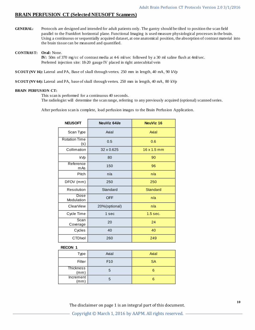
BRAIN PERFUSION CT (Selected NEUSOFT Scanners) GENERAL: Protocols are designed and intended for adult patients only. The gantry should be tilted to position the scan field
parallel to the Frankfort horizontal plane. Functional Imaging is used measure physiological processes in the brain. Using a continuous or sequentially acquired dataset, at one anatomical position, the absorption of contrast material into the brain tissue can be measured and quantified.
CONTRAST: Oral: None. IV: 50m of 370 mg/cc of contrast media at 4-6 ml/sec followed by a 30 ml saline flush at 4ml/sec. Preferred injection site: 18-20 gauge IV placed in right antecubital vein SCOUT (NV 16): Lateral and PA, Base of skull through vertex. 250 mm in length, 40 mA, 90 kVp SCOUT (NV 64): Lateral and PA, base of skull through vertex. 250 mm in length, 40 mA, 80 kVp BRAIN PERFUSION CT: This scan is performed for a continuous 40 seconds. The radiologist will determine the scan range, referring to any previously acquired (optional) scanned series. After perfusion scan is complete, load perfusion images to the Brain Perfusion Application.
NEUSOFT NeuViz 64i/e NeuViz 16
Scan Type Axial Axial
Rotation Time (s) 0.5 0.6
Collimation 32 x 0.625 16 x 1.5 mm
kVp 80 90
Reference mAs 150 96
Pitch n/a n/a
DFOV (mm) 250 250
Resolution Standard Standard Dose
Modulation OFF n/a
ClearView 20%(optional) n/a
Cycle Time 1 sec 1.5 sec.
Scan Coverage 20 24
Cycles 40 40
CTDIvol 260 249
RECON 1
Type Axial Axial
Filter F10 SA
Thickness (mm) 5 6
Increment (mm) 5 6
The disclaimer on page 1 is an integral part of this document.
10
Copyright © March 1, 2016 by AAPM. All rights reserved.
Adult Brain Perfusion CT Protocols Version 2.0 3/1/2016
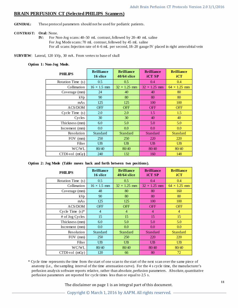
BRAIN PERFUSION CT (Selected PHILIPS Scanners) GENERAL: These protocol parameters should not be used for pediatric patients.
CONTRAST: Oral: None. IV: For Non-Jog scans: 40–50 mL contrast, followed by 20–40 mL saline For Jog Mode scans: 70 mL contrast, followed by 45 mL saline For all scans: Injection rate of 4–6 mL per second, 18–20 gauge IV placed in right antecubital vein SURVIEW: Lateral, 120 kVp, 30 mA. From vertex to base of skull Option 1: Non-Jog Mode.
PHILIPS Brilliance 16 slice
Brilliance 40/64 slice
Brilliance iCT SP
Brilliance iCT
Rotation Time (s) 0.5 0.5 0.4 0.4 Collimation 16 × 1.5 mm 32 × 1.25 mm 32 × 1.25 mm 64 × 1.25 mm
Coverage (mm) 24 40 40 80 kVp 90 80 80 80
mAs 125 125 100 100 ACS/DOM OFF OFF OFF OFF
Cycle Time (s) 2.0 2.0 1.5 1.5 Cycles 30 30 40 40
Thickness (mm) 6.0 5.0 5.0 5.0 Increment (mm) 0.0 0.0 0.0 0.0
Resolution Standard Standard Standard Standard FOV (mm) 250 250 220 220
Filter UB UB UB UB WC/WL 80/40 80/40 80/40 80/40
CTDI-vol (mGy) 240 132 160 148 Option 2: Jog Mode (Table moves back and forth between two positions).
PHILIPS Brilliance 16 slice
Brilliance 40/64 slice
Brilliance iCT SP
Brilliance iCT
Rotation Time (s) 0.5 0.5 0.4 0.4 Collimation 16 × 1.5 mm 32 × 1.25 mm 32 × 1.25 mm 64 × 1.25 mm
Coverage (mm) 48 80 80 160 kVp 90 80 80 80
mAs 125 125 100 100 ACS/DOM OFF OFF OFF OFF
Cycle Time (s)* 4 4 4 4 # of Jog Cycles 15 15 15 15 Thickness (mm) 6.0 5.0 5.0 5.0 Increment (mm) 0.0 0.0 0.0 0.0
Resolution Standard Standard Standard Standard FOV (mm) 250 250 220 220
Filter UB UB UB UB WC/WL 80/40 80/40 80/40 80/40
CTDI-vol (mGy) 120 66 80 72
* Cycle time represents the time from the start of one scan to the start of the next scan over the same piece of anatomy (i.e., the sampling interval of the time attenuation curve). For the 4 s cycle time, the manufacturer’s perfusion analysis software reports relative, rather than absolute, perfusion parameters. Absolute, quantitative perfusion parameters are reported for cycle times less than or equal to 2.5 s.
The disclaimer on page 1 is an integral part of this document.
11
Copyright © March 1, 2016 by AAPM. All rights reserved.
Adult Brain Perfusion CT Protocols Version 2.0 3/1/2016
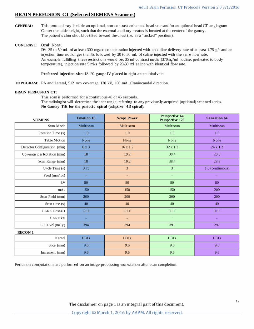
BRAIN PERFUSION CT (Selected SIEMENS Scanners) GENERAL: This protocol may include an optional, non-contrast-enhanced head scan and/or an optional head CT angiogram Center the table height, such that the external auditory meatus is located at the center of the gantry. The patient’s chin should be tilted toward the chest (i.e. in a “tucked” position).
CONTRAST: Oral: None. IV: 35 to 50 mL of at least 300 mg/cc concentration injected with an iodine delivery rate of at least 1.75 g/s and an
injection time not longer than 8s followed by 20 to 30 mL of saline injected with the same flow rate. An example fulfilling these restrictions would be: 35 ml contrast media (370mg/ml iodine, preheated to body
temperature), injection rate 5 ml/s followed by 20-30 ml saline with identical flow rate. Preferred injection site: 18–20 gauge IV placed in right antecubital vein TOPOGRAM: PA and Lateral, 512 mm coverage, 120 kV, 100 mA. Craniocaudal direction. BRAIN PERFUSION CT: This scan is performed for a continuous 40 or 45 seconds. The radiologist will determine the scan range, referring to any previously-acquired (optional) scanned series. No Gantry Tilt for the periodic spiral (adaptive 4D spiral).
SIEMENS Emotion 16 Scope Power Perspective 64 Perspective 128 Sensation 64
Scan Mode Multiscan Multiscan Multiscan Multiscan
Rotation Time (s) 1.0 1.0 1.0 1.0
Table Motion None None None None
Detector Configuration (mm) 6 x 3 16 x 1.2 32 x 1.2 24 x 1.2
Coverage per Rotation (mm) 18 19.2 38.4 28.8
Scan Range (mm) 18 19.2 38.4 28.8
Cycle Time (s) 3.75 3 3 1.0 (continuous)
Feed (mm/rot) - - - -
kV 80 80 80 80
mAs 150 150 150 200
Scan Field (mm) 200 200 200 200
Scan time (s) 40 40 40 40
CARE Dose4D OFF OFF OFF OFF
CARE kV - - - -
CTDIvol (mGy) 394 394 391 297
RECON 1
Kernel H31s H31s H31s H31s
Slice (mm) 9.6 9.6 9.6 9.6
Increment (mm) 9.6 9.6 9.6 9.6
Perfusion computations are performed on an image-processing workstation after scan completion.
The disclaimer on page 1 is an integral part of this document.
12
Copyright © March 1, 2016 by AAPM. All rights reserved.
Adult Brain Perfusion CT Protocols Version 2.0 3/1/2016
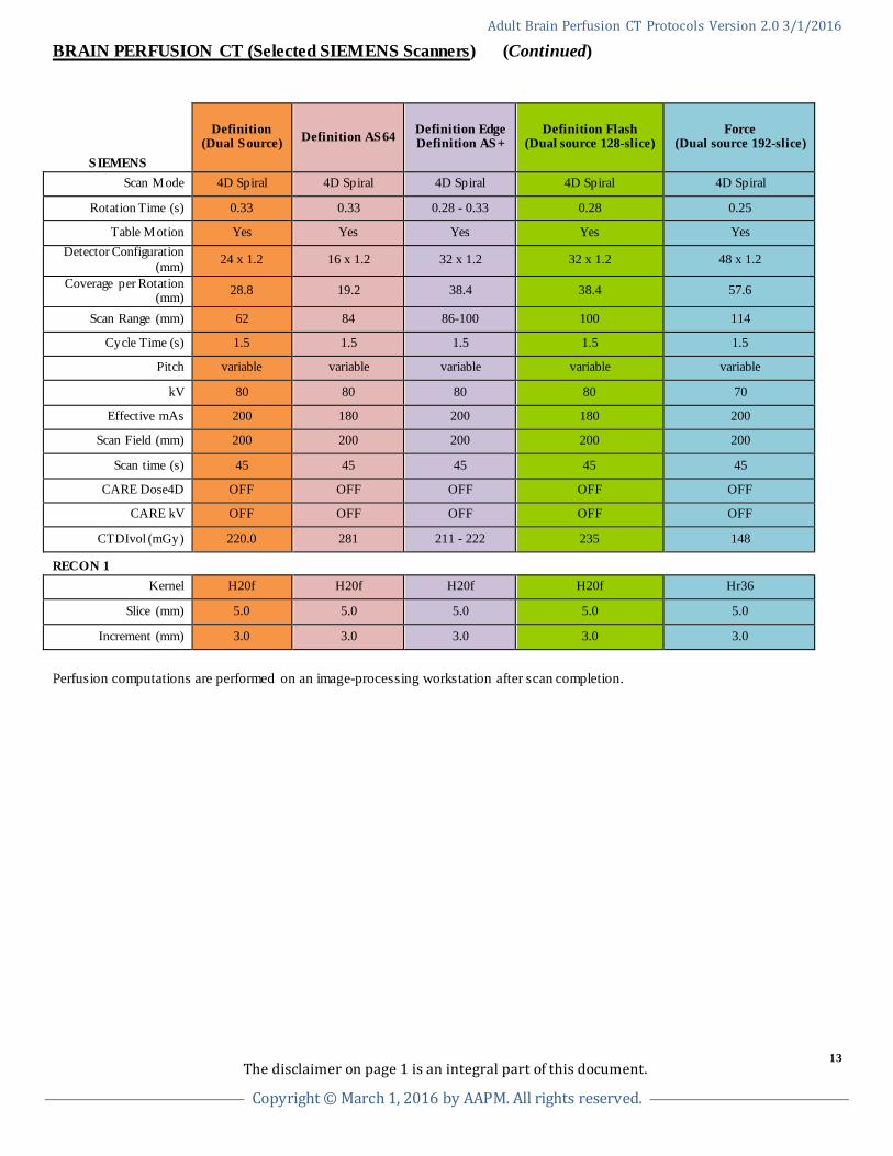
BRAIN PERFUSION CT (Selected SIEMENS Scanners) (Continued)
SIEMENS
Definition (Dual Source) Definition AS64 Definition Edge
Definition AS+ Definition Flash
(Dual source 128-slice) Force
(Dual source 192-slice)
Scan Mode 4D Spiral 4D Spiral 4D Spiral 4D Spiral 4D Spiral
Rotation Time (s) 0.33 0.33 0.28 - 0.33 0.28 0.25
Table Motion Yes Yes Yes Yes Yes Detector Configuration
(mm) 24 x 1.2 16 x 1.2 32 x 1.2 32 x 1.2 48 x 1.2
Coverage per Rotation (mm) 28.8 19.2 38.4 38.4 57.6
Scan Range (mm) 62 84 86-100 100 114
Cycle Time (s) 1.5 1.5 1.5 1.5 1.5
Pitch variable variable variable variable variable
kV 80 80 80 80 70
Effective mAs 200 180 200 180 200
Scan Field (mm) 200 200 200 200 200
Scan time (s) 45 45 45 45 45
CARE Dose4D OFF OFF OFF OFF OFF
CARE kV OFF OFF OFF OFF OFF
CTDIvol (mGy) 220.0 281 211 - 222 235 148
RECON 1
Kernel H20f H20f H20f H20f Hr36
Slice (mm) 5.0 5.0 5.0 5.0 5.0
Increment (mm) 3.0 3.0 3.0 3.0 3.0
Perfusion computations are performed on an image-processing workstation after scan completion.
The disclaimer on page 1 is an integral part of this document.
13
Copyright © March 1, 2016 by AAPM. All rights reserved.
Adult Brain Perfusion CT Protocols Version 2.0 3/1/2016
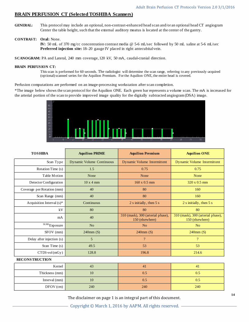
BRAIN PERFUSION CT (Selected TOSHIBA Scanners) GENERAL: This protocol may include an optional, non-contrast-enhanced head scan and/or an optional head CT angiogram Center the table height, such that the external auditory meatus is located at the center of the gantry. CONTRAST: Oral: None. IV: 50 mL of 370 mg/cc concentration contrast media @ 5-6 mL/sec followed by 50 mL saline at 5-6 mL/sec Preferred injection site: 18–20 gauge IV placed in right antecubital vein. SCANOGRAM: PA and Lateral, 240 mm coverage, 120 kV, 50 mA, caudal-cranial direction. BRAIN PERFUSION CT: This scan is performed for 60 seconds. The radiologist will determine the scan range, referring to any previously-acquired
(optional) scanned series for the Aquilion Premium. For the Aquilion ONE, the entire head is covered. Perfusion computations are performed on an image-processing workstation after scan completion. *The image below shows the scan protocol for the Aquilion ONE. Each green bar represents a volume scan. The mA is increased for the arterial portion of the scan to provide improved image quality for the digitally subtracted angiogram (DSA) image.
TOSHIBA Aquilion PRIME Aquilion Premium Aquilion ONE
Scan Type Dynamic Volume Continuous Dynamic Volume Intermittent Dynamic Volume Intermittent
Rotation Time (s) 1.5 0.75 0.75
Table Motion None None None
Detector Configuration 10 x 4 mm 160 x 0.5 mm 320 x 0.5 mm
Coverage per Rotation (mm) 40 80 160
Scan Range (mm) 40 80 160
Acquisition Interval (s)* Continuous 2 s initially, then 5 s 2 s initially, then 5 s
kV 80 80 80
mA 40 310 (mask), 300 (arterial phase), 150 (elsewhere)
310 (mask), 300 (arterial phase), 150 (elsewhere)
SUREExposure No No No
SFOV (mm) 240mm (S) 240mm (S) 240mm (S)
Delay after injection (s) 5 7 7
Scan Time (s) 49.5 53 53
CTDI-vol (mGy) 128.8 196.8 214.6
RECONSTRUCTION
Kernel 43 41 41
Thickness (mm) 10 0.5 0.5
Interval (mm) 10 0.5 0.5
DFOV (cm) 240 240 240
The disclaimer on page 1 is an integral part of this document.
14
Copyright © March 1, 2016 by AAPM. All rights reserved.