diflavin oxidoreductases activate the bioreductive prodrug pr-104a under hypoxia
TRANSCRIPT
Diflavin Oxidoreductases Activate the Bioreductive ProdrugPR-104A under Hypoxia□S
Christopher P. Guise, Maria R. Abbattista, Smitha R. Tipparaju, Neil K. Lambie,Jiechuang Su, Dan Li, William R. Wilson, Gabi U. Dachs, and Adam V. PattersonAuckland Cancer Society Research Centre, School of Medical Sciences (C.P.G., M.R.A., S.R.T., J.S., D.L., W.R.W., A.V.P.) andMaurice Wilkins Centre for Molecular Biodiscovery (C.P.G., W.R.W., G.U.D., A.V.P.), the University of Auckland, Auckland, NewZealand; Department of Anatomical Pathology, Prince of Wales Hospital, Randwick, New South Wales, Australia (N.K.L.); andDepartment of Pathology, University of Otago Christchurch, Christchurch, New Zealand (N.K.L., G.U.D.)
Received May 24, 2011; accepted October 7, 2011
ABSTRACTThe clinical agent PR-104 is converted systemically to PR-104A, a nitrogen mustard prodrug designed to target tumorhypoxia. Reductive activation of PR-104A is initiated by one-electron oxidoreductases in a process reversed by oxygen. Theidentity of these oxidoreductases is unknown, with the excep-tion of cytochrome P450 reductase (POR). To identify otherhypoxia-selective PR-104A reductases, nine candidate oxi-doreductases were expressed in HCT116 cells. Increased PR-104A-cytotoxicity was observed in cells expressing methioninesynthase reductase (MTRR), novel diflavin oxidoreductase 1(NDOR1), and inducible nitric-oxide synthase (NOS2A), inaddition to POR. Plasmid-based expression of these diflavinoxidoreductases also enhanced bioreductive metabolism ofPR-104A in an anoxia-specific manner. Diflavin oxidoreduc-
tase-dependent PR-104A metabolism was suppressed �90%by pan-flavoenzyme inhibition with diphenyliodonium chloride.Antibodies were used to quantify endogenous POR, MTRR,NDOR1, and NOS2A expression in 23 human tumor cell lines;however, only POR protein was detectable and its expressioncorrelated with anoxic PR-104A reduction (r2 � 0.712). Ananti-POR monoclonal antibody was used to probe expressionusing human tissue microarrays; 13 of 19 cancer types ex-pressed detectable POR with 21% of cores (185 of 874) stain-ing positive; this heterogeneity suggests that POR is a usefulbiomarker for PR-104A activation. Immunostaining for carbonicanhydrase 9 (CAIX), reportedly an endogenous marker of hyp-oxia, revealed only moderate coexpression (9.6%) of both CAIXand POR across a subset of five cancer types.
IntroductionHypoxia is a common characteristic of solid tumors that is as-
sociated with poor patient prognosis and resistance to treatment(Vaupel and Mayer, 2007). A number of bioreductive prodrugs
have been developed to exploit tumor hypoxia; examples that haveadvanced to clinical trial include the N-oxides 3-amino-1,2,4-benzotriazine-1,4 dioxide (tirapazamine) (Rischin et al., 2010) andbanoxantrone (Patterson and McKeown, 2000), and the nitroaro-matics 2-((2-bromoethyl)(2,4-dinitro-6-((2-(phosphonooxy)ethyl)carbamoyl)phenyl)amino)ethyl methanesulfonate (PR-104)(Jameson et al., 2010) and N,N�-bis(2-bromoethyl)-(1-methyl-2-nitro-1H-imidazol-5-yl)phosphorodiamidic acid methyl ester (TH-302) (Weiss et al., 2011). All share a common mechanism of acti-vation, undergoing reductive metabolism by intracellularoxidoreductases to form cytotoxic species. The capacity for prod-rugs to act as exogenous electron acceptors is opposed by molecularoxygen, predominantly via redox cycling, and consequently they
This research was supported by the Health Research Council of New Zea-land [Grant 08/103].
W.R.W. is a founding scientist, stockholder, and consultant to Proacta Inc.,which is undertaking the clinical development of PR-104. A.V.P. is a consul-tant to and has stock options in Proacta Inc.
Article, publication date, and citation information can be found athttp://molpharm.aspetjournals.org.
http://dx.doi.org/10.1124/mol.111.073759.□S The online version of this article (available at http://molpharm.
aspetjournals.org) contains supplemental material.
ABBREVIATIONS: PR-104, 2-((2-bromoethyl)(2,4-dinitro-6-((2-(phosphonooxy)ethyl)carbamoyl)phenyl)amino)ethyl methanesulfonate; PR-104A, 2-((2-bromoethyl)-2-{[(2-hydroxyethyl) amino]carbonyl}-4,6-dinitroanilino)ethyl methanesulfonate; TH-302, N,N�-bis(2-bromoethyl)-(1-methyl-2-nitro-1H-imidazol-5-yl)phosphorodiamidic acid methyl ester; RH1, 2,5-di(aziridin-1-yl)-3-(hydroxymethyl)-6-methylcyclohexa-2,5-diene-1,4-dione; AKR1C3, aldo-keto reductase 1C3; POR, cytochrome P450 reductase; DPI, diphenyliodonium; FDXR, adrenodoxin oxidoreductase; CYB5R, cytochrome B5 reductase;NOS, nitric-oxide synthase; NOS2A, inducible nitric-oxide synthase; NQO1, NAD(P)H quinone oxidoreductase 1; NQO2, NAD(P)H quinone oxidoreduc-tase 1; XD, xanthine dehydrogenase; MTRR, methionine synthase reductase; NDOR1, novel diflavin oxidoreductase 1; FFPE, formalin fixed paraffinembedded; LC, liquid chromatography; MS/MS, tandem mass spectrometry; mAb, monoclonal antibody; WT, wild type; IHC, immunohistochemistry;TMA, tissue microarray; CAIX, carbonic anhydrase 9; H score, Histo score; HIF-1, hypoxia-inducible factor 1; UPR, unfolded protein response;SN30000/CEN-209, 3-(3-morpholinopropyl)-7,8-dihydro-6H-indeno[5,6-e][1,2,4]triazine-1,4-dioxide; CB1954, 5-(aziridin-1-yl)-2,4-dinitrobenzamide.
1521-0111/12/8101-31–40$25.00MOLECULAR PHARMACOLOGY Vol. 81, No. 1Copyright © 2012 The American Society for Pharmacology and Experimental Therapeutics 73759/3737534Mol Pharmacol 81:31–40, 2012
31
at University of A
uckland on January 3, 2013m
olpharm.aspetjournals.org
Dow
nloaded from
3759.DC1.html http://molpharm.aspetjournals.org/content/suppl/2011/10/07/mol.111.07Supplemental Material can be found at:
function as direct oxygen sensors. Whereas several mammalianenzymes have been implicated in prodrug reduction, the enzymol-ogy of hypoxic drug metabolism is poorly defined (Patterson et al.,1998; Chen and Hu, 2009; Wilson and Hay, 2011).
PR-104 is a phosphate ester preprodrug that undergoessystemic hydrolysis in vivo to the 3,5-dinitrobenzamide-2-mustard prodrug, 2-((2-bromoethyl)-2-{[(2-hydroxyethyl)amino]carbonyl}-4,6-dinitroanilino)ethyl methanesulfonate(PR-104A). Nitroreduction of PR-104A leads to the formationof the cytotoxic hydroxylamine (PR-104H) and amine (PR-104M) metabolites (Patterson et al., 2007), which contributeto antitumor activity through the formation of DNA inter-strand cross-links (Singleton et al., 2009). Cellular sensitiv-ity to PR-104A is determined by hypoxia, the expression ofrelevant oxidoreductases, and the functional status of DNArepair pathways in the cell (Gu et al., 2009). Only one-electron oxidoreductases will generate the initial PR-104Anitro-radical anion that is sensitive to back oxidation bymolecular oxygen. Cytotoxicity of PR-104A is increased un-der hypoxia in vitro in all cell lines tested (Patterson et al.,2007) but with a wide range (5- to 120-fold) because of vari-able expression of aldo-keto reductase 1C3 (AKR1C3), a two-electron PR-104A oxidoreductase, which fails to generate theoxygen-sensitive nitro-radical species (Guise et al., 2010). Todate only a single oxygen-sensitive PR-104A reductase, cyto-chrome P450 oxidoreductase (POR), has been identified ascatalyzing the reduction of PR-104A via one-electron transferunder anoxia (Guise et al., 2007). However, quantitativeknockdown of POR by antisense and RNA interference hasshown that �40% of the anoxic PR-104A reduction in SiHacells is due to other enzymes (Guise et al., 2007). To under-stand the relative contribution of POR and other one-electronoxidoreductases to PR-104A metabolism in human tumors itis necessary to identify these oxidoreductases and to assesstheir expression.
We previously observed that the irreversible flavoenzymeinhibitor diphenyliodonium chloride (DPI) suppresses 90% ofanoxic PR-104A metabolism in SiHa cells (Guise et al., 2007);the remainder is probably attributable to oxygen-insensitivereduction by AKR1C3, which is expressed in this cell line(Guise et al., 2010). The essentially complete inhibition ofPR-104A reduction by DPI under hypoxia suggests that theunidentified one-electron oxidoreductases in SiHa cells areflavoproteins. To date, a number of flavoenzymes have beenreported to have activity as xenobiotic nitroreductases, includ-ing adrenodoxin oxidoreductase (FDXR), cytochrome b5 reduc-tase (CYB5R), inducible nitric-oxide synthase (NOS2A), NAD(P)Hquinone oxidoreductase 1 (NQO1), NAD(P)H quinone oxidoreduc-tase 2 (NQO2), and xanthine dehydrogenase (XD). NOS2A has ahigh degree of homology to POR, and both belong to a smallfamily of proteins termed the diflavin oxidoreductases thatuse the cofactors FAD and FMN for electron transfer fromNADPH to acceptors (Murataliev et al., 2004). The otherhuman diflavin oxidoreductase family members are methio-nine synthase reductase (MTRR), novel diflavin oxidoreduc-tase 1 (NDOR1), and the isoforms of nitric-oxide synthase(NOS1 and NOS3). Thus, the members of the diflavin oxi-doreductase family also represent potential candidates foranoxic PR-104A activation. Details of the substrates knownto be metabolically reduced by these enzymes are provided inSupplemental Table S1.
We tested these 10 candidate flavoenzymes and identified
three diflavin oxidoreductases, MTRR, NDOR1, and NOS2A,in addition to POR, as anoxic PR-104A reductases. Afteridentification of specific antibodies against POR, MTRR,NDOR1, and NOS2A, we examined expression in a panel of23 human cancer cell lines and showed that POR is the onlydiflavin oxidoreductase expressed at readily detectable lev-els. We demonstrated that POR, MTRR, NDOR1, andNOS2A overexpression elevated metabolism of PR-104A andthat the flavoenzyme poison DPI ablated anoxic PR-104Ametabolism across all diflavin oxidoreductase-expressingcells. Next we critically tested the available antibodies on for-malin-fixed paraffin-embedded (FFPE) cell pellets and xeno-grafts, confirming that only the anti-POR monoclonal antibodywas suitable. Last, we examined POR expression in a set ofsurgical tumor samples in tissue microarrays from 19 commoncancers and showed heterogeneous expression patterns.
Materials and MethodsCompounds. PR-104A, PR-104H, and tetradeuterated deriva-
tives were synthesized, purified, and stored as described previously(Guise et al., 2010). DPI (100 mM; Sigma-Aldrich, St. Louis, MO) wasprepared as a dimethyl sulfoxide stock and stored at �80°C.
Cell Lines, Cytotoxicity Assays, and PR-104A Metabolism.Cells were maintained in culture under humidified atmosphericconditions with 5% CO2 as described previously (Patterson et al.,2007) with �6 months of cumulative passage from sources (Supple-mental Table S2). Antiproliferative assays were performed in �-min-imal essential medium under oxic or anoxic conditions, the latterusing a 5% H2/palladium catalyst scrubbed Bactron anaerobic cham-ber (Sheldon Manufacturing, Cornelius, OR) to achieve severe an-oxia (�10 ppm O2 gas phase) during PR-104A exposure as describedpreviously (Patterson et al., 2007). Total exposure to anoxia did notexceed 6 h, and cells were allowed to regrow for 4 days underdrug-free aerobic conditions. Cellular metabolism of PR-104A (100�M, 1 h, 5 � 105 cells/well in 24-well plates) to hydroxylaminePR-104H and amine PR-104M was measured by LC-MS/MS as be-fore (Singleton et al., 2009).
Candidate Gene Expression. Plasmids encoding sequence-con-firmed open reading frames for CYB5R, FDXR, MTRR, NDOR1,NOS2A, NOS3, NQO1, NQO2, POR, and XD were purchased (Sup-plemental Table S3). Open reading frames were cloned into theGateway compatible vector F527-V5 and transfected into HCT116cells as described previously (Guise et al., 2010).
Western Immunoblot Analysis. Cell lysates were prepared inradioimmunoprecipitation assay buffer (Guise et al., 2010). Then 30�g of protein was loaded on SDS-polyacrylamide gel electrophoresisgels (4–12% gradient gels; Invitrogen, Carlsbad, CA), transferred,blocked, and probed with primary antibodies against POR (SantaCruz Biotechnology, Inc., Santa Cruz, CA), MTRR (Abnova Corpora-tion, Taipei City, Taiwan), NDOR1 (Abnova Corporation), andNOS2A (Santa Cruz Biotechnology, Inc.). Full details of antibodiesare listed in Supplemental Table S4. Bands were detected usingchemiluminescent ECL detection (SuperSignal; Thermo Fisher Sci-entific, Waltham, MA) and quantified using ImageJ (version 1.42 ofthe public domain software).
Immunostaining of Cell Pellets, Xenografts, and HumanTumor Tissue Microarrays. For all diflavin oxidoreductase-over-expressing cell lines, a dense pellet was formed by centrifugation(107 cells), fixed in 4% paraformaldehyde (30 h, room temperature),embedded, cut (5 �m), and mounted on glass. Antibodies were eval-uated for antigen specificity across a dilution range (10–0.5 �g/ml).For additional validation of POR monoclonal antibody (mAb), xeno-grafts were established in NIHIII nude mice (University of Aucklandanimal ethics committee approval number C830) from parentalHCT116 WT cells, HCT116 POR cells engineered to overexpress
32 Guise et al.
at University of A
uckland on January 3, 2013m
olpharm.aspetjournals.org
Dow
nloaded from
POR as described previously (Guise et al., 2010), and HCT116 cellsnegative for POR protein as a result of mutation of both POR allelesusing zinc finger nuclease technology (HCT116-PORnull; develop-ment of this cell line will be reported elsewhere). FFPE tissue of eachxenograft was prepared for IHC as described previously (Guise et al.,2010). Commercial human tissue microarrays (TMAs) were from thesources shown in Supplemental Table S5. A total of 874 individualcores (685 cases) representing 19 cancer types and 135 cores (77cases) representing 34 normal tissues were analyzed across 12TMAs. Optimized antigen retrieval was performed in citrate buffer,pH 6.0, (30 min, 120°C). Slides (3 �m) were immunostained for PORand carbonic anhydrase 9 (CAIX) (Novus Biologicals, Inc., Littleton,CO) as for tumor xenografts, and cores were scored for stainingintensity and proportion of positive neoplastic cells by a certifiedpathologist (N.K.L.). IHC for CAIX was performed using Cell andTissue Staining Kits (R&D Systems, Minneapolis, MN) as describedpreviously (Dachs et al., 2010). Intensity was scored using a semi-quantitative measure on a four-point scale ranging from negative(score 0) to strong staining (score 3). Histo scores (H scores) for PORand CAIX were determined using the following equation: (percent-age of cells exhibiting intensity 1 staining � 1) � (percentage of cellsexhibiting intensity 2 staining � 2) � (percentage of cells exhibitingintensity 3 staining � 3) � H score (maximum � 300). This measurewas applied to the neoplastic cell element of the tumors within thetissue microarrays and the epithelial elements within the normaltissue microarray.
Statistical Tests. All data were tested for significance by un-paired t test.
ResultsIdentification of Diflavin Oxidoreductase Enzymes
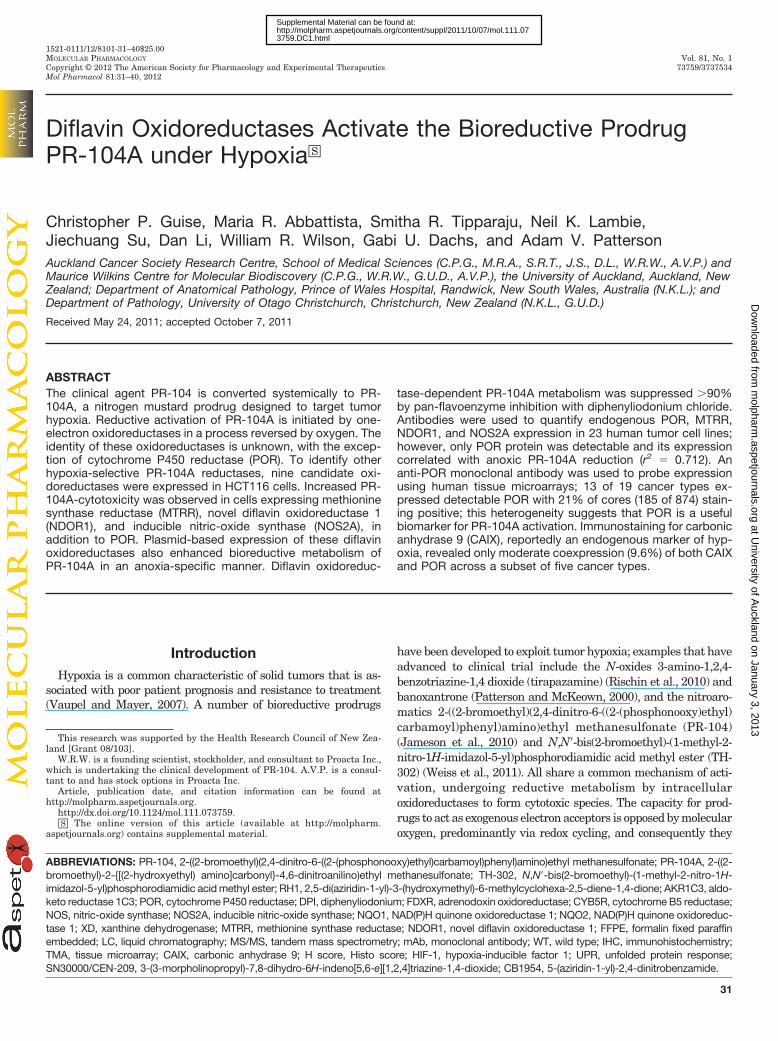
As Novel Hypoxic PR-104A Reductases. We have previ-ously demonstrated the importance of DPI-sensitive flavoen-zymes in general and POR in particular in the activation ofPR-104A by SiHa cells under anoxic conditions (Guise et al.,2007). To identify other flavoenzymes that catalyze the con-version of PR-104A to the cytotoxic para-nitro reductionproducts, PR-104H and PR-104M, 10 flavoenzymes were ex-pressed from sequence-confirmed cDNAs after plasmidtransfer in HCT116 cells to generate pooled populations. Useof a bicistronic expression cassette enforces homogeneousoxidoreductase expression in these puromycin-resistant cells.The candidate enzymes selected for this study consisted offour enzymes with homology to POR (MTRR, NDOR1,NOS2A, and NOS3) and five enzymes previously implicatedin bioreductive metabolism (CYB5R, FDXR, NQO1, NQO2,and XD). HCT116 cells overexpressing POR were also gen-erated as a positive control. Flavoenzyme expression wasconfirmed by immunodetection of a common C-terminal V5tag induced by TAG suppressor tRNA-mediated translation(Tag-On-Demand; Invitrogen) (Fig. 1A). Expression was ob-served for all proteins, although only weak bands were ob-served for NOS3 and XD. All V5-tagged proteins were of theexpected molecular mass except for XD, which showed asingle band of approximately 100 kDa rather than the ex-pected 150 kDa product. In addition to the anticipated 130kDa band for NOS2A, a second band of approximately 75 kDawas observed. The ability of these enzymes to activate PR-104A under anoxic conditions was examined by monitoringformation of reduced PR-104A metabolites by a sensitiveLC-MS/MS assay relative to the HCT116 WT controls (Fig.1B). Significant increases in PR-104A metabolism were de-tected in cells expressing POR, MTRR, NDOR1, and NOS2A
under anoxia (all p � 0.05) but not under aerobic conditions(Supplemental Fig. S1).
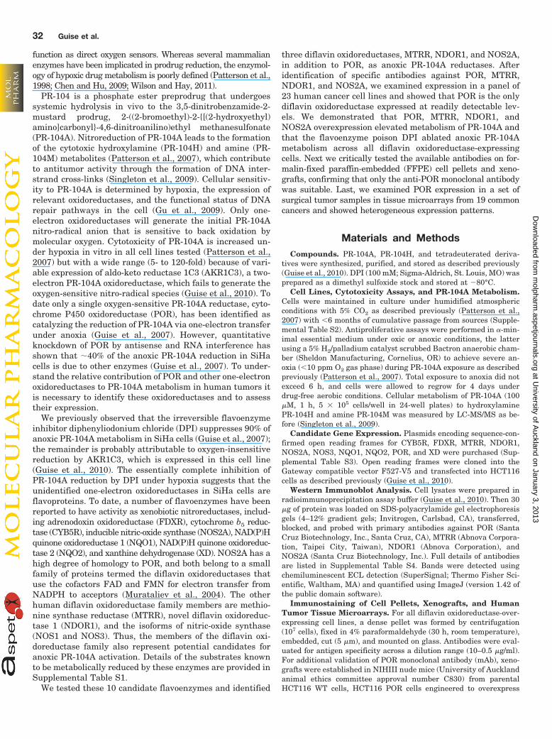
The four flavoenzymes able to activate PR-104A underanoxic conditions, namely POR, MTRR, NDOR1, andNOS2A, all belong to the diflavin oxidoreductase proteinfamily. Figure 2A shows a schematic representation of theseenzymes along with two additional family members, NOS1and NOS3, and highlights the structural homology of thediflavin oxidoreductase family. We sought to determinewhether the increases in anoxic PR-104A metabolism inHCT116 POR, MTRR, NDOR1, and NOS2A cells resulted inincreased anoxic sensitivity of these cells to PR-104A. Thesensitivity to PR-104A of WT and overexpressing cell lineswas determined under aerobic and anoxic conditions using asulforhodamine B-based antiproliferative assay. As expected,no significant increases in sensitivity to PR-104A were ob-
Fig. 1. PR-104A is activated under anoxic conditions by four diflavinoxidoreductase family members. A, detection of candidate reductase en-zymes in overexpressing HCT116 cell lines by Western blotting. COOH-terminal V5 tags were transiently expressed using an adenoviral TAGsuppressor tRNA (Tag-On-Demand; multiplicity of infection 50, 24 h). B,formation of reduced metabolites of PR-104A (sum of PR-104H and PR-104M) under anoxic conditions in HCT116 WT cells and HCT116 cellsoverexpressing candidate mammalian reductases. Metabolite formationwas calculated after addition of PR-104A (100 �M, 1 h); values are themean changes in anoxic metabolism relative to HCT116 WT controls (n �3). The average value for reduced PR-104H and PR-104M metabolites inHCT116 WT cells under anoxic conditions was 406 38 pmol/106 cells(mean S.E.M., n � 12). Error bars represent the S.E.M.. �, p � 0.05; ��,p � 0.01.
PR-104A Activation by Diflavin Oxidoreductases 33
at University of A
uckland on January 3, 2013m
olpharm.aspetjournals.org
Dow
nloaded from
served in these cell lines under aerobic conditions (Fig. 2B).Anoxic exposure resulted in significant increases in sensitiv-ity to PR-104A in POR (p � 0.003)-, MTRR (p � 0.006)-,NDOR1 (p � 0.029)-, and NOS2A (p � 0.028)-expressing cellscompared with WT cells (Fig. 2C). Increases in hypoxic cyto-toxicity ratios (HCR � aerobic IC50 value divided by anoxicIC50 value) were also observed, ranging from 20-fold in theparental cell line to 158-fold (POR), 88-fold (MTRR), 64-fold(NDOR1), and 60-fold (NOS2A) (Fig. 2). Consistent with themetabolism data (Fig. 1B), there was no significant change inanoxic PR-104A sensitivity in cells expressing NOS3 (p �0.38), and this cell line exhibited a hypoxic cytotoxicity ratiosimilar to that of the WT (30-fold) (Fig. 2).
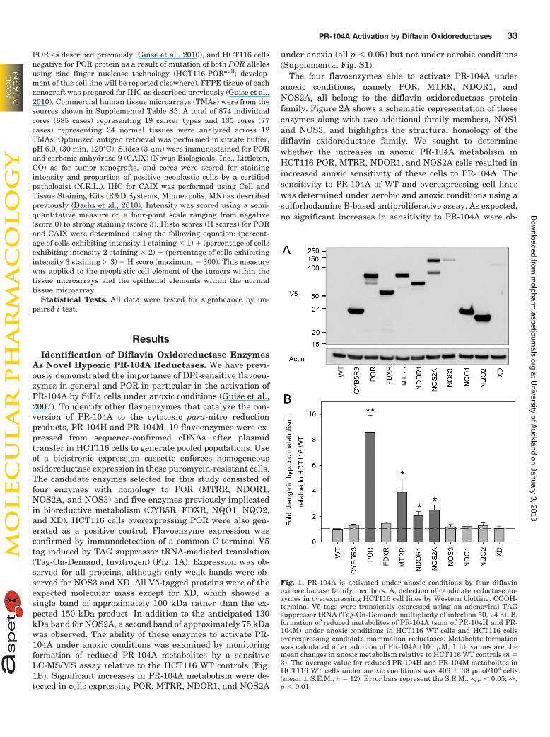
Anoxic Metabolism Correlates with POR Expressionacross a Panel of 23 Cancer Cell Lines. To test for thepresence of each diflavin oxidoreductase candidate in humanneoplastic cultured cell lines, we first validated antibodiesfrom commercial sources (Supplemental Table S4) by West-ern blotting using lysates obtained from parental and stablytransfected HCT116 cell lines. We identified an anti-PORmAb, which was more specific than the polyclonal antibodywe used previously (Guise et al., 2007). Three NOS2A anti-bodies were analyzed by Western blot, only one of whichshowed NOS2A specific binding. We also identified mAbstargeting MTRR and NDOR1, which together with the PORand NOS2A antibodies described above, were found specificfor each of the four oxidoreductases (Fig. 3A). Backgroundendogenous expression in parental HCT116 cells was seenfor POR but not for the other diflavin oxidoreductase candi-dates; despite this, the HCT116 POR cell line showed clearexcess of the enzyme with an 8.5-fold increase (POR/�-actinratio) in protein levels by Western blotting (Fig. 3A). Weconfirmed uniform expression of POR in the HCT116 WTcells during exposure to anoxia (Supplemental Fig. S2). Inaddition, the maximum 6-h exposure time used in the IC50
assays yielded no change in plasmid-based expression levelsof POR, MTRR, NDOR1, or NOS2A (Supplemental Fig. S2).
We next examined the expression of the four diflavin oxi-doreductases in a panel of 23 cancer cell lines (Fig. 3B). OnlyPOR was found to be present at readily detectable levels
Fig. 2. Cells expressing diflavin oxidoreductases are sensitized to PR-104A under anoxic conditions. A, schematic representation of the sixhuman diflavin oxidoreductase family members. B and C, IC50 values ofPR-104A after a 4-h aerobic (B) or anoxic (C) exposure in HCT116 WT andoverexpressing cell lines (n � 5). Error bars represent the S.E.M. �, p �0.05; ��, p � 0.01.
Fig. 3. Comparison between diflavin oxi-doreductase expression profiles and anoxic-specific metabolism of PR-104A in a hu-man cancer cell line panel. A, detection ofdiflavin oxidoreductases in overexpress-ing cell lines by Western blotting usingspecific antibodies and V5 tag detection.COOH-terminal V5 tags were transientlyexpressed using and adenoviral TAG sup-pressor tRNA (Tag-On-Demand; multi-plicity of infection 50, 24 h). B, detectionof POR, MTRR, NDOR1, and NOS2A in apanel of cancer cell lines by Western blot-ting. Lysates of HCT116 cells overex-pressing diflavin reductases were in-cluded in the control lanes C, anoxia-specific metabolism of PR-104A (100 �M,1 h) in the same panel of cell lines byLC-MS/MS quantification of PR-104Hand PR-104M. Values are means, and er-ror bars show the S.E.M. for total metab-olites from two to eight experiments. D,correlation between anoxia-specific PR-104A metabolism and POR expression(normalized to actin; values are meansand S.E.M. from three Western blots).
34 Guise et al.
at University of A
uckland on January 3, 2013m
olpharm.aspetjournals.org
Dow
nloaded from
across all 23 cell lines; expression varied considerably acrossthe cell lines but POR mAb immunoreactivity was observedin all cases (Fig. 3B). No detectable bands were observed forMTRR, NDOR1, or NOS2A during initial analysis (Fig. 3B).A more aggressive analysis, in which antibody concentra-tions higher than recommended were used along with ex-tended chemiluminescent substrate exposure times, showedmodest expression of NDOR1 in all 23 cell lines and weakbands corresponding to MTRR in several cell lines (mostapparent in Du145, A549, H69, MDA231, and PC3). No ex-pression of NOS2A was observed. A number of nonspecificbands were observed for all three antibodies under theseconditions (Supplemental Fig. S3).
We have previously reported the rates of oxic metabolismof PR-104A in a 23-cell line panel (Guise et al., 2010). Herewe measure the anoxia-specific (one-electron) metabolism ofPR-104A in each cell line by measuring the rates of metabo-lism under anoxic conditions (Supplemental Fig. S4A) andsubtracting the paired rate of metabolism under oxic condi-tions (Guise et al., 2010) (reproduced in Supplemental Fig.S4B). The calculated rates of anoxia-specific PR-104A reduc-tion across the 23-cell line panel are shown in Fig. 3C. Therate of anoxic metabolism covered a 52-fold range, with amedian value 17-fold higher than that under oxic conditions.We next sought to determine whether expression of PORcorrelated with levels of one-electron reduction of PR-104A inthe 23-cell line panel; analysis was based on the POR/�-actinratios obtained from three independent Western blots of the23-cell line panel. The hepatoma cell line HepG2 was a no-table anomaly (Supplemental Fig. S5) and was excluded fromthe analysis, whereupon the coefficient of determination forthe remaining 22 cell lines was 0.712 (p � 0.001) (Fig. 3D).The rate of PR-104A metabolism by the HCT116 POR cellline was consistent with this overall relationship (Supple-mental Fig. S5).
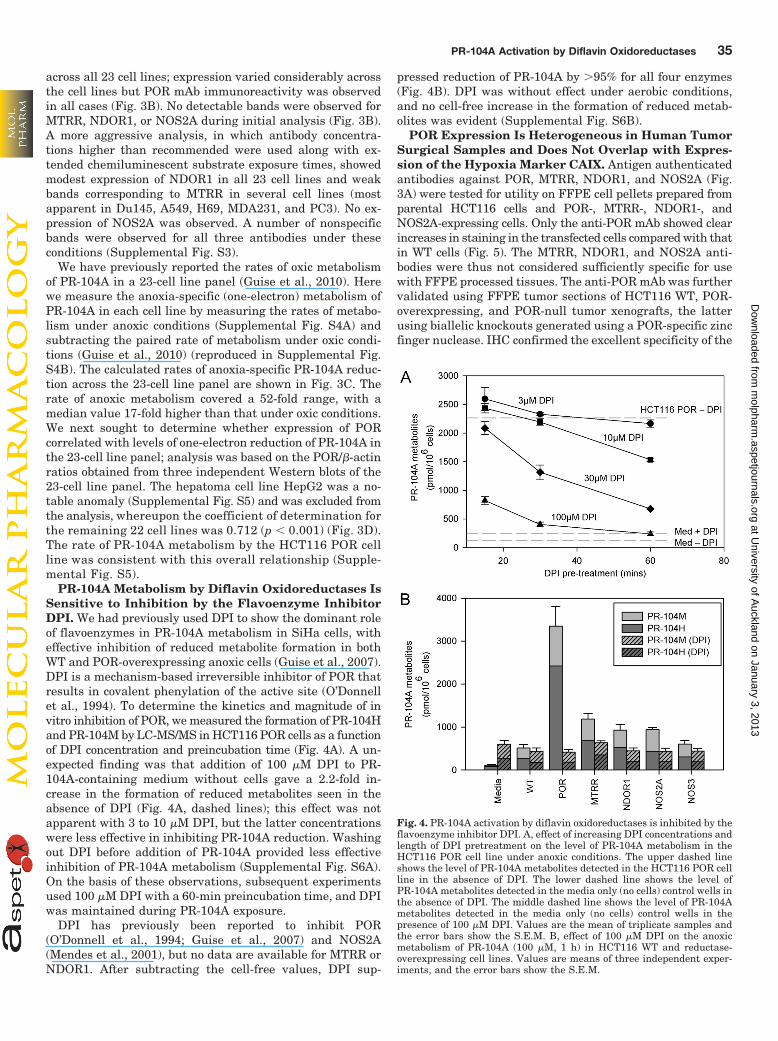
PR-104A Metabolism by Diflavin Oxidoreductases IsSensitive to Inhibition by the Flavoenzyme InhibitorDPI. We had previously used DPI to show the dominant roleof flavoenzymes in PR-104A metabolism in SiHa cells, witheffective inhibition of reduced metabolite formation in bothWT and POR-overexpressing anoxic cells (Guise et al., 2007).DPI is a mechanism-based irreversible inhibitor of POR thatresults in covalent phenylation of the active site (O’Donnellet al., 1994). To determine the kinetics and magnitude of invitro inhibition of POR, we measured the formation of PR-104Hand PR-104M by LC-MS/MS in HCT116 POR cells as a functionof DPI concentration and preincubation time (Fig. 4A). A un-expected finding was that addition of 100 �M DPI to PR-104A-containing medium without cells gave a 2.2-fold in-crease in the formation of reduced metabolites seen in theabsence of DPI (Fig. 4A, dashed lines); this effect was notapparent with 3 to 10 �M DPI, but the latter concentrationswere less effective in inhibiting PR-104A reduction. Washingout DPI before addition of PR-104A provided less effectiveinhibition of PR-104A metabolism (Supplemental Fig. S6A).On the basis of these observations, subsequent experimentsused 100 �M DPI with a 60-min preincubation time, and DPIwas maintained during PR-104A exposure.
DPI has previously been reported to inhibit POR(O’Donnell et al., 1994; Guise et al., 2007) and NOS2A(Mendes et al., 2001), but no data are available for MTRR orNDOR1. After subtracting the cell-free values, DPI sup-
pressed reduction of PR-104A by �95% for all four enzymes(Fig. 4B). DPI was without effect under aerobic conditions,and no cell-free increase in the formation of reduced metab-olites was evident (Supplemental Fig. S6B).
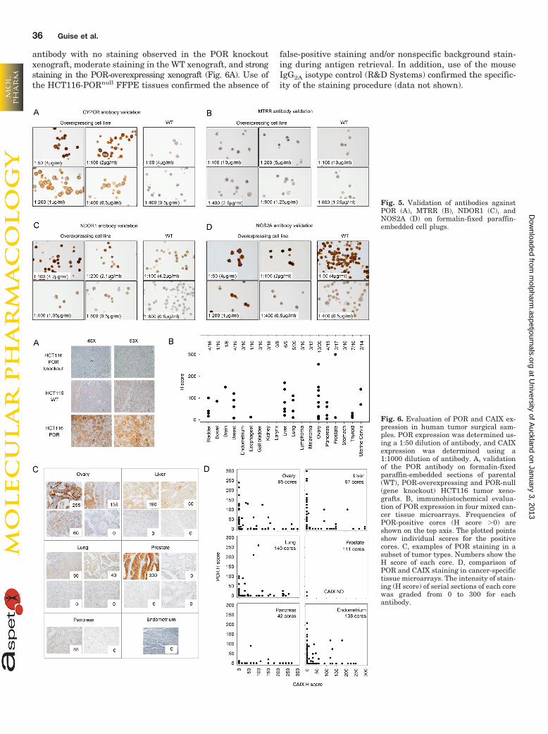
POR Expression Is Heterogeneous in Human TumorSurgical Samples and Does Not Overlap with Expres-sion of the Hypoxia Marker CAIX. Antigen authenticatedantibodies against POR, MTRR, NDOR1, and NOS2A (Fig.3A) were tested for utility on FFPE cell pellets prepared fromparental HCT116 cells and POR-, MTRR-, NDOR1-, andNOS2A-expressing cells. Only the anti-POR mAb showed clearincreases in staining in the transfected cells compared with thatin WT cells (Fig. 5). The MTRR, NDOR1, and NOS2A anti-bodies were thus not considered sufficiently specific for usewith FFPE processed tissues. The anti-POR mAb was furthervalidated using FFPE tumor sections of HCT116 WT, POR-overexpressing, and POR-null tumor xenografts, the latterusing biallelic knockouts generated using a POR-specific zincfinger nuclease. IHC confirmed the excellent specificity of the
Fig. 4. PR-104A activation by diflavin oxidoreductases is inhibited by theflavoenzyme inhibitor DPI. A, effect of increasing DPI concentrations andlength of DPI pretreatment on the level of PR-104A metabolism in theHCT116 POR cell line under anoxic conditions. The upper dashed lineshows the level of PR-104A metabolites detected in the HCT116 POR cellline in the absence of DPI. The lower dashed line shows the level ofPR-104A metabolites detected in the media only (no cells) control wells inthe absence of DPI. The middle dashed line shows the level of PR-104Ametabolites detected in the media only (no cells) control wells in thepresence of 100 �M DPI. Values are the mean of triplicate samples andthe error bars show the S.E.M. B, effect of 100 �M DPI on the anoxicmetabolism of PR-104A (100 �M, 1 h) in HCT116 WT and reductase-overexpressing cell lines. Values are means of three independent exper-iments, and the error bars show the S.E.M.
PR-104A Activation by Diflavin Oxidoreductases 35
at University of A
uckland on January 3, 2013m
olpharm.aspetjournals.org
Dow
nloaded from
antibody with no staining observed in the POR knockoutxenograft, moderate staining in the WT xenograft, and strongstaining in the POR-overexpressing xenograft (Fig. 6A). Use ofthe HCT116-PORnull FFPE tissues confirmed the absence of
false-positive staining and/or nonspecific background stain-ing during antigen retrieval. In addition, use of the mouseIgG2A isotype control (R&D Systems) confirmed the specific-ity of the staining procedure (data not shown).
Fig. 5. Validation of antibodies againstPOR (A), MTRR (B), NDOR1 (C), andNOS2A (D) on formalin-fixed paraffin-embedded cell plugs.
Fig. 6. Evaluation of POR and CAIX ex-pression in human tumor surgical sam-ples. POR expression was determined us-ing a 1:50 dilution of antibody, and CAIXexpression was determined using a1:1000 dilution of antibody. A, validationof the POR antibody on formalin-fixedparaffin-embedded sections of parental(WT), POR-overexpressing and POR-null(gene knockout) HCT116 tumor xeno-grafts. B, immunohistochemical evalua-tion of POR expression in four mixed can-cer tissue microarrays. Frequencies ofPOR-positive cores (H score �0) areshown on the top axis. The plotted pointsshow individual scores for the positivecores. C, examples of POR staining in asubset of tumor types. Numbers show theH score of each core. D, comparison ofPOR and CAIX staining in cancer-specifictissue microarrays. The intensity of stain-ing (H score) of serial sections of each corewas graded from 0 to 300 for eachantibody.
36 Guise et al.
at University of A
uckland on January 3, 2013m
olpharm.aspetjournals.org
Dow
nloaded from
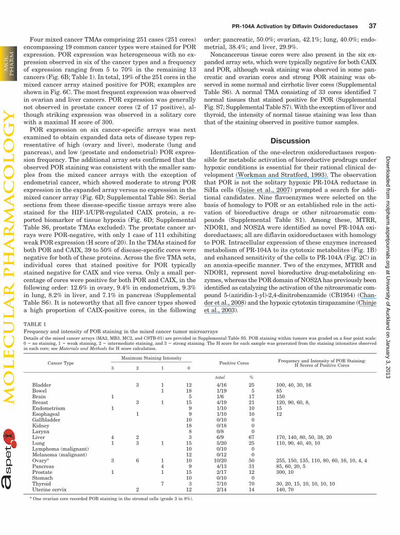
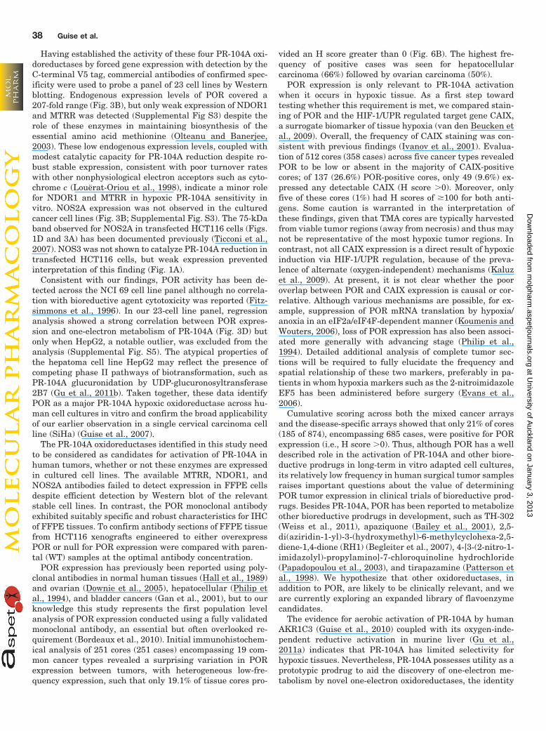
Four mixed cancer TMAs comprising 251 cases (251 cores)encompassing 19 common cancer types were stained for PORexpression. POR expression was heterogeneous with no ex-pression observed in six of the cancer types and a frequencyof expression ranging from 5 to 70% in the remaining 13cancers (Fig. 6B; Table 1). In total, 19% of the 251 cores in themixed cancer array stained positive for POR; examples areshown in Fig. 6C. The most frequent expression was observedin ovarian and liver cancers. POR expression was generallynot observed in prostate cancer cores (2 of 17 positive), al-though striking expression was observed in a solitary corewith a maximal H score of 300.
POR expression on six cancer-specific arrays was nextexamined to obtain expanded data sets of disease types rep-resentative of high (ovary and liver), moderate (lung andpancreas), and low (prostate and endometrial) POR expres-sion frequency. The additional array sets confirmed that theobserved POR staining was consistent with the smaller sam-ples from the mixed cancer arrays with the exception ofendometrial cancer, which showed moderate to strong PORexpression in the expanded array versus no expression in themixed cancer array (Fig. 6D; Supplemental Table S6). Serialsections from these disease-specific tissue arrays were alsostained for the HIF-1/UPR-regulated CAIX protein, a re-ported biomarker of tissue hypoxia (Fig. 6D; SupplementalTable S6, prostate TMAs excluded). The prostate cancer ar-rays were POR-negative, with only 1 case of 111 exhibitingweak POR expression (H score of 20). In the TMAs stained forboth POR and CAIX, 39 to 50% of disease-specific cores werenegative for both of these proteins. Across the five TMA sets,individual cores that stained positive for POR typicallystained negative for CAIX and vice versa. Only a small per-centage of cores were positive for both POR and CAIX, in thefollowing order: 12.6% in ovary, 9.4% in endometrium, 9.3%in lung, 8.2% in liver, and 7.1% in pancreas (SupplementalTable S6). It is noteworthy that all five cancer types showeda high proportion of CAIX-positive cores, in the following
order: pancreatic, 50.0%; ovarian, 42.1%; lung, 40.0%; endo-metrial, 38.4%; and liver, 29.9%.
Noncancerous tissue cores were also present in the six ex-panded array sets, which were typically negative for both CAIXand POR, although weak staining was observed in some pan-creatic and ovarian cores and strong POR staining was ob-served in some normal and cirrhotic liver cores (SupplementalTable S6). A normal TMA consisting of 33 cores identified 7normal tissues that stained positive for POR (SupplementalFig. S7; Supplemental Table S7). With the exception of liver andthyroid, the intensity of normal tissue staining was less thanthat of the staining observed in positive tumor samples.
DiscussionIdentification of the one-electron oxidoreductases respon-
sible for metabolic activation of bioreductive prodrugs underhypoxic conditions is essential for their rational clinical de-velopment (Workman and Stratford, 1993). The observationthat POR is not the solitary hypoxic PR-104A reductase inSiHa cells (Guise et al., 2007) prompted a search for addi-tional candidates. Nine flavoenzymes were selected on thebasis of homology to POR or an established role in the acti-vation of bioreductive drugs or other nitroaromatic com-pounds (Supplemental Table S1). Among these, MTRR,NDOR1, and NOS2A were identified as novel PR-104A oxi-doreductases; all are diflavin oxidoreductases with homologyto POR. Intracellular expression of these enzymes increasedmetabolism of PR-104A to its cytotoxic metabolites (Fig. 1B)and enhanced sensitivity of the cells to PR-104A (Fig. 2C) inan anoxia-specific manner. Two of the enzymes, MTRR andNDOR1, represent novel bioreductive drug-metabolizing en-zymes, whereas the POR domain of NOS2A has previously beenidentified as catalyzing the activation of the nitroaromatic com-pound 5-(aziridin-1-yl)-2,4-dinitrobenzamide (CB1954) (Chan-dor et al., 2008) and the hypoxic cytotoxin tirapazamine (Chinjeet al., 2003).
TABLE 1Frequency and intensity of POR staining in the mixed cancer tumor microarraysDetails of the mixed cancer arrays (MA2, MB3, MC2, and CSTB-01) are provided in Supplemental Table S5. POR staining within tumors was graded on a four point scale:0 � no staining, 1 � weak staining, 2 � intermediate staining, and 3 � strong staining. The H score for each sample was generated from the staining intensities observedin each core; see Materials and Methods for H score calculation.
Cancer TypeMaximum Staining Intensity
Positive Cores Frequency and Intensity of POR Staining:H Scores of Positive Cores
3 2 1 0
total %
Bladder 3 1 12 4/16 25 100, 40, 30, 16Bowel 1 18 1/19 5 85Brain 1 5 1/6 17 150Breast 3 1 15 4/19 21 120, 90, 60, 8,Endometrium 1 9 1/10 10 15Esophageal 1 9 1/10 10 12Gallbladder 10 0/10 0Kidney 18 0/18 0Larynx 8 0/8 0Liver 4 2 3 6/9 67 170, 140, 80, 50, 38, 20Lung 1 3 1 15 5/20 25 110, 90, 40, 40, 10Lymphoma (malignant) 10 0/10 0Melanoma (malignant) 12 0/12 0Ovarya 3 6 1 10 10/20 50 255, 150, 135, 110, 80, 60, 16, 10, 4, 4Pancreas 4 9 4/13 31 85, 60, 20, 5Prostate 1 1 15 2/17 12 300, 10Stomach 10 0/10 0Thyroid 7 3 7/10 70 30, 20, 15, 10, 10, 10, 10Uterine cervix 2 12 2/14 14 140, 70
a One ovarian core recorded POR staining in the stromal cells (grade 3 in 8%).
PR-104A Activation by Diflavin Oxidoreductases 37
at University of A
uckland on January 3, 2013m
olpharm.aspetjournals.org
Dow
nloaded from
Having established the activity of these four PR-104A oxi-doreductases by forced gene expression with detection by theC-terminal V5 tag, commercial antibodies of confirmed spec-ificity were used to probe a panel of 23 cell lines by Westernblotting. Endogenous expression levels of POR covered a207-fold range (Fig. 3B), but only weak expression of NDOR1and MTRR was detected (Supplemental Fig S3) despite therole of these enzymes in maintaining biosynthesis of theessential amino acid methionine (Olteanu and Banerjee,2003). These low endogenous expression levels, coupled withmodest catalytic capacity for PR-104A reduction despite ro-bust stable expression, consistent with poor turnover rateswith other nonphysiological electron acceptors such as cyto-chrome c (Louerat-Oriou et al., 1998), indicate a minor rolefor NDOR1 and MTRR in hypoxic PR-104A sensitivity invitro. NOS2A expression was not observed in the culturedcancer cell lines (Fig. 3B; Supplemental Fig. S3). The 75-kDaband observed for NOS2A in transfected HCT116 cells (Figs.1D and 3A) has been documented previously (Ticconi et al.,2007). NOS3 was not shown to catalyze PR-104A reduction intransfected HCT116 cells, but weak expression preventedinterpretation of this finding (Fig. 1A).
Consistent with our findings, POR activity has been de-tected across the NCI 69 cell line panel although no correla-tion with bioreductive agent cytotoxicity was reported (Fitz-simmons et al., 1996). In our 23-cell line panel, regressionanalysis showed a strong correlation between POR expres-sion and one-electron metabolism of PR-104A (Fig. 3D) butonly when HepG2, a notable outlier, was excluded from theanalysis (Supplemental Fig. S5). The atypical properties ofthe hepatoma cell line HepG2 may reflect the presence ofcompeting phase II pathways of biotransformation, such asPR-104A glucuronidation by UDP-glucuronosyltransferase2B7 (Gu et al., 2011b). Taken together, these data identifyPOR as a major PR-104A hypoxic oxidoreductase across hu-man cell cultures in vitro and confirm the broad applicabilityof our earlier observation in a single cervical carcinoma cellline (SiHa) (Guise et al., 2007).
The PR-104A oxidoreductases identified in this study needto be considered as candidates for activation of PR-104A inhuman tumors, whether or not these enzymes are expressedin cultured cell lines. The available MTRR, NDOR1, andNOS2A antibodies failed to detect expression in FFPE cellsdespite efficient detection by Western blot of the relevantstable cell lines. In contrast, the POR monoclonal antibodyexhibited suitably specific and robust characteristics for IHCof FFPE tissues. To confirm antibody sections of FFPE tissuefrom HCT116 xenografts engineered to either overexpressPOR or null for POR expression were compared with paren-tal (WT) samples at the optimal antibody concentration.
POR expression has previously been reported using poly-clonal antibodies in normal human tissues (Hall et al., 1989)and ovarian (Downie et al., 2005), hepatocellular (Philip etal., 1994), and bladder cancers (Gan et al., 2001), but to ourknowledge this study represents the first population levelanalysis of POR expression conducted using a fully validatedmonoclonal antibody, an essential but often overlooked re-quirement (Bordeaux et al., 2010). Initial immunohistochem-ical analysis of 251 cores (251 cases) encompassing 19 com-mon cancer types revealed a surprising variation in PORexpression between tumors, with heterogeneous low-fre-quency expression, such that only 19.1% of tissue cores pro-
vided an H score greater than 0 (Fig. 6B). The highest fre-quency of positive cases was seen for hepatocellularcarcinoma (66%) followed by ovarian carcinoma (50%).
POR expression is only relevant to PR-104A activationwhen it occurs in hypoxic tissue. As a first step towardtesting whether this requirement is met, we compared stain-ing of POR and the HIF-1/UPR regulated target gene CAIX,a surrogate biomarker of tissue hypoxia (van den Beucken etal., 2009). Overall, the frequency of CAIX staining was con-sistent with previous findings (Ivanov et al., 2001). Evalua-tion of 512 cores (358 cases) across five cancer types revealedPOR to be low or absent in the majority of CAIX-positivecores; of 137 (26.6%) POR-positive cores, only 49 (9.6%) ex-pressed any detectable CAIX (H score �0). Moreover, onlyfive of these cores (1%) had H scores of �100 for both anti-gens. Some caution is warranted in the interpretation ofthese findings, given that TMA cores are typically harvestedfrom viable tumor regions (away from necrosis) and thus maynot be representative of the most hypoxic tumor regions. Incontrast, not all CAIX expression is a direct result of hypoxicinduction via HIF-1/UPR regulation, because of the preva-lence of alternate (oxygen-independent) mechanisms (Kaluzet al., 2009). At present, it is not clear whether the pooroverlap between POR and CAIX expression is causal or cor-relative. Although various mechanisms are possible, for ex-ample, suppression of POR mRNA translation by hypoxia/anoxia in an eIF2a/eIF4F-dependent manner (Koumenis andWouters, 2006), loss of POR expression has also been associ-ated more generally with advancing stage (Philip et al.,1994). Detailed additional analysis of complete tumor sec-tions will be required to fully elucidate the frequency andspatial relationship of these two markers, preferably in pa-tients in whom hypoxia markers such as the 2-nitroimidazoleEF5 has been administered before surgery (Evans et al.,2006).
Cumulative scoring across both the mixed cancer arraysand the disease-specific arrays showed that only 21% of cores(185 of 874), encompassing 685 cases, were positive for PORexpression (i.e., H score �0). Thus, although POR has a welldescribed role in the activation of PR-104A and other biore-ductive prodrugs in long-term in vitro adapted cell cultures,its relatively low frequency in human surgical tumor samplesraises important questions about the value of determiningPOR tumor expression in clinical trials of bioreductive prod-rugs. Besides PR-104A, POR has been reported to metabolizeother bioreductive prodrugs in development, such as TH-302(Weiss et al., 2011), apaziquone (Bailey et al., 2001), 2,5-di(aziridin-1-yl)-3-(hydroxymethyl)-6-methylcyclohexa-2,5-diene-1,4-dione (RH1) (Begleiter et al., 2007), 4-[3-(2-nitro-1-imidazolyl)-propylamino]-7-chloroquinoline hydrochloride(Papadopoulou et al., 2003), and tirapazamine (Patterson etal., 1998). We hypothesize that other oxidoreductases, inaddition to POR, are likely to be clinically relevant, and weare currently exploring an expanded library of flavoenzymecandidates.
The evidence for aerobic activation of PR-104A by humanAKR1C3 (Guise et al., 2010) coupled with its oxygen-inde-pendent reductive activation in murine liver (Gu et al.,2011a) indicates that PR-104A has limited selectivity forhypoxic tissues. Nevertheless, PR-104A possesses utility as aprototypic prodrug to aid the discovery of one-electron me-tabolism by novel one-electron oxidoreductases, the identity
38 Guise et al.
at University of A
uckland on January 3, 2013m
olpharm.aspetjournals.org
Dow
nloaded from
of which has broad applicability to bioreductive agents ingeneral. For example, we have observed that MTRR-express-ing HCT116 cells are more sensitive than POR-expressingcells to TH-302 and tirapazamine under anoxia, suggestingthat more detailed evaluation of MTRR enzymology and tis-sue distribution is warranted in the context of these agents(G. P. Guise and A. V. Patterson, unpublished observations).At present, the lack of informative antibodies for detection ofMTRR in FFPE tissues is a limitation. Open access data-bases such as oncomine (http://www.oncomine.org) show thatMTRR mRNA is widely (although variably) expressed inmany human tumors, often at levels well above the mediantranscript. TH-302, a hypoxia-selective 2-nitroimidazole-trig-gered isophosphoramidate mustard currently undergoingphase II/III clinical evaluation, shows considerable promiseyet the identity of the activating oxidoreductases is un-known. An analog of tirapazamine [3-(3-morpholinopropyl)-7,8-dihydro-6H-indeno[5,6-e][1,2,4]triazine-1,4-dioxide(SN30000/CEN-209)] optimized for maximal tissue penetra-tion is in late preclinical development (Hicks et al., 2010), butagain the activating enzymes have not been elucidated.
The future clinical utility of hypoxia-activated cytotoxinswill inevitably depend on identifying the confluence of all keydeterminants of sensitivity including the presence of thera-py-limiting hypoxic cells, the complement of one-electron oxi-doreductases necessary for metabolic reduction of prodrugsin these cells, and the intrinsic sensitivity of these cells to theresulting prodrug-mediated DNA damage. Only when thisindividualization of treatment is achieved will hypoxia-acti-vated cytotoxins be well placed to improve treatment out-comes.
Acknowledgments
We thank Annika Foehrenbacher for assistance with tumor im-plantations and Graham Atwell and Professor William Denny forprovision of PR-104A.
Authorship Contributions
Participated in research design: Guise, Abbattista, Tipparaju, Wil-son, Dachs, Patterson.
Conducted experiments: Guise, Abbattista, Tipparaju, and Dachs.Contributed new reagents or analytic tools: Guise, Abbattista, Su,
Li, Wilson, and Dachs.Performed data analysis: Guise, Lambie, Dachs, and Patterson.Wrote or contributed to the writing of the manuscript: Guise, Wil-
son, Dachs, and Patterson.
ReferencesBailey SM, Lewis AD, Patterson LH, Fisher GR, Knox RJ, and Workman P (2001)
Involvement of NADPH:cytochrome P450 reductase in the activation of indoloqui-none EO9 to free radical and DNA damaging species. Biochem Pharmacol 62:461–468.
Begleiter A, Leith MK, Patel D, and Hasinoff BB (2007) Role of NADPH cytochromeP450 reductase in activation of RH1. Cancer Chemother Pharmacol 60:713–723.
Bordeaux J, Welsh A, Agarwal S, Killiam E, Baquero M, Hanna J, Anagnostou V,and Rimm D (2010) Antibody validation [published erratum appears in Biotech-niques 48:351, 2010]. Biotechniques 48:197–209.
Chandor A, Dijols S, Ramassamy B, Frapart Y, Mansuy D, Stuehr D, Helsby N, andBoucher JL (2008) Metabolic activation of the antitumor drug 5-(aziridin-1-yl)-2,4-dinitrobenzamide (CB1954) by NO synthases. Chem Res Toxicol 21:836–843.
Chen Y and Hu L (2009) Design of anticancer prodrugs for reductive activation. MedRes Rev 29:29–64.
Chinje EC, Cowen RL, Feng J, Sharma SP, Wind NS, Harris AL, and Stratford IJ(2003) Non-nuclear localized human NOSII enhances the bioactivation and toxic-ity of tirapazamine (SR4233) in vitro. Mol Pharmacol 63:1248–1255.
Dachs GU, Kano M, Volkova E, Morrin HR, Davey VC, Harris GC, Cheale M,Frampton C, Currie MJ, Wells JE, et al. (2010) A profile of prognostic andmolecular factors in European and Ma�ori breast cancer patients. BMC Cancer10:543.
Downie D, McFadyen MC, Rooney PH, Cruickshank ME, Parkin DE, Miller ID,Telfer C, Melvin WT, and Murray GI (2005) Profiling cytochrome P450 expressionin ovarian cancer: identification of prognostic markers. Clin Cancer Res 11:7369–7375.
Evans SM, Schrlau AE, Chalian AA, Zhang P, and Koch CJ (2006) Oxygen levels innormal and previously irradiated human skin as assessed by EF5 binding. J InvestDermatol 126:2596–2606.
Fitzsimmons SA, Workman P, Grever M, Paull K, Camalier R, and Lewis AD (1996)Reductase enzyme expression across the National Cancer Institute Tumor CellLine Panel: correlation with sensitivity to mitomycin C and EO9. J Natl CancerInst 88:259–269.
Gan Y, Mo Y, Kalns JE, Lu J, Danenberg K, Danenberg P, Wientjes MG, and Au JL(2001) Expression of DT-diaphorase and cytochrome P450 reductase correlateswith mitomycin C activity in human bladder tumors. Clin Cancer Res 7:1313–1319.
Gu Y, Guise CP, Patel K, Abbattista MR, Li J, Lie J, Sun X, Atwell GJ, Boyd M,Patterson AV, et al. (2011a) Reductive metabolism of the dinitrobenzamide mus-tard anticancer prodrug PR-104 in mice. Cancer Chemother Pharmacol 67:543–555.
Gu Y, Patterson AV, Atwell GJ, Chernikova SB, Brown JM, Thompson LH, andWilson WR (2009) Roles of DNA repair and reductase activity in the cytotoxicity ofthe hypoxia-activated dinitrobenzamide mustard PR-104A. Mol Cancer Ther8:1714–1723.
Gu Y, Tingle MD, and Wilson WR (2011b) Glucuronidation of anticancer prodrugPR-104A: species differences, identification of human UDP-glucuronosyltrans-ferases, and implications for therapy. J Pharmacol Exp Ther 337:692–702.
Guise CP, Abbattista MR, Singleton RS, Holford SD, Connolly J, Dachs GU, Fox SB,Pollock R, Harvey J, Guilford P, et al. (2010) The bioreductive prodrug PR-104A isactivated under aerobic conditions by human aldo-keto reductase 1C3. Cancer Res70:1573–1584.
Guise CP, Wang AT, Theil A, Bridewell DJ, Wilson WR, and Patterson AV (2007)Identification of human reductases that activate the dinitrobenzamide mustardprodrug PR-104A: a role for NADPH:cytochrome P450 oxidoreductase under hyp-oxia. Biochem Pharmacol 74:810–820.
Hall PM, Stupans I, Burgess W, Birkett DJ, and McManus ME (1989) Immunohis-tochemical Localization of NADPH-Cytochrome P450 Reductase in Human Tis-sues. Carcinogenesis 10:521–530.
Hicks KO, Siim BG, Jaiswal JK, Pruijn FB, Fraser AM, Patel R, Hogg A, LiyanageHD, Dorie MJ, Brown JM, et al. (2010) Pharmacokinetic/pharmacodynamic mod-eling identifies SN30000 and SN29751 as tirapazamine analogues with improvedtissue penetration and hypoxic cell killing in tumors. Clin Cancer Res 16:4946–4957.
Ivanov S, Liao SY, Ivanova A, Danilkovitch-Miagkova A, Tarasova N, Weirich G,Merrill MJ, Proescholdt MA, Oldfield EH, Lee J, et al. (2001) Expression ofhypoxia-inducible cell-surface transmembrane carbonic anhydrases in human can-cer. Am J Pathol 158:905–919.
Jameson MB, Rischin D, Pegram M, Gutheil J, Patterson AV, Denny WA, and WilsonWR (2010) A phase I trial of PR-104, a nitrogen mustard prodrug activated by bothhypoxia and aldo-keto reductase 1C3, in patients with solid tumors. Cancer Che-mother Pharmacol 65:791–801.
Kaluz S, Kaluzova M, Liao SY, Lerman M, and Stanbridge EJ (2009) Transcriptionalcontrol of the tumor- and hypoxia-marker carbonic anhydrase 9: a one transcrip-tion factor (HIF-1) show? Biochim Biophys Acta 1795:162–172.
Koumenis C and Wouters BG (2006) “Translating” tumor hypoxia: unfolded proteinresponse (UPR)-dependent and UPR-independent pathways. Mol Cancer Res4:423–436.
Louerat-Oriou B, Perret A, and Pompon D (1998) Differential redox and electron-transfer properties of purified yeast, plant and human NADPH-cytochrome P-450reductases highly modulate cytochrome P-450 activities. Eur J Biochem 258:1040–1049.
Mendes AF, Carvalho AP, Caramona MM, and Lopes MC (2001) Diphenyleneiodo-nium inhibits NF-�B activation and INOS expression induced by IL-1�: involve-ment of reactive oxygen species. Mediators Inflamm 10:209–215.
Murataliev MB, Feyereisen R, and Walker FA (2004). Electron transfer by diflavinreductases. Biochim Biophys Acta 1698:1–26.
O’Donnell VB, Smith GC, and Jones OT (1994) Involvement of phenyl radicals iniodonium inhibition of flavoenzymes. Mol Pharmacol 46:778–785.
Olteanu H and Banerjee R (2003) Redundancy in the pathway for redox regulationof mammalian methionine synthase: reductive activation by the dual flavoprotein,novel reductase 1. J Biol Chem 278:38310–38314.
Papadopoulou MV, Ji M, Rao MK, and Bloomer WD (2003) Reductive metabolism ofthe nitroimidazole-based hypoxia-selective cytotoxin NLCQ-1 (NSC 709257). On-col Res 14:21–29.
Patterson AV, Ferry DM, Edmunds SJ, Gu Y, Singleton RS, Patel K, Pullen SM,Hicks KO, Syddall SP, Atwell GJ, et al. (2007) Mechanism of action and preclinicalantitumor activity of the novel hypoxia-activated DNA cross-linking agent PR-104.Clin Cancer Res 13:3922–3932.
Patterson AV, Saunders MP, Chinje EC, Patterson LH, and Stratford IJ (1998)Enzymology of tirapazamine metabolism: a review. Anticancer Drug Des 13:541–573.
Patterson LH and McKeown SR (2000) AQ4N: a new approach to hypoxia-activatedcancer chemotherapy. Br J Cancer 83:1589–1593.
Philip PA, Kaklamanis L, Ryley N, Stratford I, Wolf R, Harris A, and Carmichael J(1994) Expression of xenobiotic-metabolizing enzymes by primary and secondaryhepatic tumors in man. Int J Radiat Oncol Biol Phys 29:277–283.
Rischin D, Peters LJ, O’Sullivan B, Giralt J, Fisher R, Yuen K, Trotti A, Bernier J,Bourhis J, Ringash J, et al. (2010) Tirapazamine, cisplatin, and radiation versuscisplatin and radiation for advanced squamous cell carcinoma of the head and neck(TROG 02.02, HeadSTART): a phase III trial of the Trans-Tasman RadiationOncology Group. J Clin Oncol 28:2989–2995.
PR-104A Activation by Diflavin Oxidoreductases 39
at University of A
uckland on January 3, 2013m
olpharm.aspetjournals.org
Dow
nloaded from
Singleton RS, Guise CP, Ferry DM, Pullen SM, Dorie MJ, Brown JM, Patterson AV,and Wilson WR (2009) DNA cross-links in human tumor cells exposed to theprodrug PR-104A: relationships to hypoxia, bioreductive metabolism, and cytotox-icity. Cancer Res 69:3884–3891.
Ticconi C, Zicari A, Belmonte A, Realacci M, Rao ChV, and Piccione E (2007)Pregnancy-promoting actions of HCG in human myometrium and fetal mem-branes. Placenta 28 (Suppl A):S137–S143.
van den Beucken T, Koritzinsky M, Niessen H, Dubois L, Savelkouls K, Mujcic H,Jutten B, Kopacek J, Pastorekova S, van der Kogel AJ, et al. (2009) Hypoxia-induced expression of carbonic anhydrase 9 is dependent on the unfolded proteinresponse. J Biol Chem 284:24204–24212.
Vaupel P and Mayer A (2007) Hypoxia in cancer: significance and impact on clinicaloutcome. Cancer Metastasis Rev 26:225–239.
Weiss GJ, Infante JR, Chiorean EG, Borad MJ, Bendell JC, Molina JR, Tibes R,Ramanathan RK, Lewandowski K, Jones SF, et al. (2011) Phase 1 study of thesafety, tolerability, and pharmacokinetics of TH-302, a hypoxia-activated prodrug,in patients with advanced solid malignancies. Clin Cancer Res 17:2997–3004.
Wilson WR and Hay MP (2011) Targeting hypoxia in cancer therapy. Nat Rev Cancer11:393–410.
Workman P and Stratford IJ (1993) The experimental development of bioreductivedrugs and their role in cancer therapy. Cancer Metastasis Rev 12:73–82.
Address correspondence to: Dr. Adam V. Patterson, Auckland CancerSociety Research Centre, The University of Auckland, Private Bag 92019,Auckland, New Zealand. E-mail [email protected]
40 Guise et al.
at University of A
uckland on January 3, 2013m
olpharm.aspetjournals.org
Dow
nloaded from