diagnosis and management of moderate and severe
TRANSCRIPT
MILITARY MEDICINE, 177, 8:76, 2012
Diagnosis and Management of Moderate and Severe TraumaticBrain Injury Sustained in Combat
MAJ Scott A. Marshall, MC USA*†; Ronald G. Riechers II, MD‡§
ABSTRACT Traumatic brain injury exists in a spectrum of severity among wounded personnel. The evaluation andclinical presentation, initial management, and treatment interventions to prevent secondary injury processes for combat-associated moderate and severe traumatic brain injury are reviewed. Promising therapies are discussed, and a currentreview of the literature is provided.
INTRODUCTIONTraumatic brain injury (TBI) is principally classified by either
mechanism (closed vs. penetrating) or clinical severity. Sever-
ity classification ranges from mild, moderate, to severe and is
based largely on the presenting Glasgow Coma Scale (GCS)
score postinitial resuscitation and the duration of typical post-
TBI neurologic findings (Tables I and II). Patients with mild
TBI have an admission GCS score of 13 to 15. A further
discussion of the management of mild TBI is included else-
where in this supplement. Moderate TBI is defined as an
admission GCS score of 9 to 12, and is often associated with
prolonged loss of consciousness, abnormal neuroimaging,
and neurological deficits.1 Patients with moderate TBI will
require rapid removal from the area of operations, subse-
quent theater hospitalization, and may need neurosurgical
evaluation or intervention. Patients with GCS scores of 8 or
less have significant neurological injury and are classified as
having a severe TBI. Typically, these patients have abnor-
mal neuroimaging to include computed tomography (CT)
scan findings, such as a skull fracture, traumatic intracranial
hemorrhage, or contusional injury.2 These patients require
rapid evacuation from the point of injury to the combat
support hospital (CSH) and admission to the intensive care
unit for immediate airway control, mechanical ventilation,
neurosurgical evaluation, and consideration for intracranial
pressure (ICP) monitoring.
The U.S. Army and the Institute of Surgical Research
publishes clinical practice guidelines for the management
of severe head injury as part of the Joint Theater Trauma
System, which is updated annually.3 This is available for pub-
lic distribution at usaisr.amedd.army.mil/clinical_practice_
guidelines.html. Guidelines for the management of severe
TBI published by the Brain Trauma Foundation have been
instrumental in improving care through guiding therapy with
evidence-based recommendations.4 Guidelines are also avail-
able for the prehospital management of severe TBI, field
management of combat-related head trauma, and surgical
management of TBI. All guidelines can be obtained online
from the Brain Trauma Foundation at braintrauma.org. The
latest version of the Guidelines for the Management of Severe
Traumatic Brain Injury will be published in an updated online
version. An interactive guideline compliance tool is available
at tbiclickandlearn.com.
Goals in the theater management of a patient who sustains
a moderate or severe TBI include the arrest of any ongoing
injury, preservation of neurological function, prevention of
medical complications of critical illness, and improvement
in overall outcome. The presence of a brain injury must be
suspected in any case of severe trauma, especially with a
blast-related mechanism of injury. TBI patients should be
recognized early and evacuated to the CSH or trauma center
with available specialized neurological care, such as neuro-
surgery and neurointensivist care. Once evacuated and clini-
cally stable, TBI patients are transitioned to a posture of
in-hospital rehabilitation with physical therapy, occupational
therapy, and speech–cognitive therapy.
CLINICAL EVALUATIONThe clinical examination of a patient with a suspected TBI
is critically important. The examination has both prognostic
and management implications, particularly in the early treat-
ment of TBI.4 Treatment begins in the field or combat
setting with the first responder and continues to the CSH.
Decisions made in the hyperacute period after brain
injury are essential for optimal outcome. The Guidelines
for Field Management of Combat-Related Head Trauma
and Advanced Trauma Life Support are both excellent
resources for providers who treat TBI.5 The Brain Trauma
Foundation also publishes guidelines, including the surgical
*Division of Critical Care Medicine; Department of Neurology, San
Antonio Military Medical Center, 3551 Roger Brooke Drive, Fort Sam
Houston, TX 78234.
†Department of Neurology, Uniformed Services University, 4301 Jones
Bridge Road, Bethesda, MD 20814.
‡Polytrauma Team, Cleveland VA Medical Center, 10701 East
Boulevard, Cleveland, OH 44106.
§Department of Neurology, Case Western Reserve University School of
Medicine, 10900 Euclid Avenue, Cleveland, OH 44106.
The opinions and views expressed herein belong solely to those of
the authors. They are not nor should they be implied as being endorsed by
the Uniformed Services University of the Health Sciences, Department of the
Army, Department of Defense or any other branch of the Federal Government
of the United States.
MILITARY MEDICINE, Vol. 177, August Supplement 201276
Dow
nloaded from https://academ
ic.oup.com/m
ilmed/article/177/suppl_8/76/4345427 by guest on 11 February 2022
management of TBI and prehospital management of TBI
(both are available at braintrauma.org).
In keeping with the care of all trauma patients, stabiliza-
tion of circulation, airway, and breathing is followed by a
rapid initial neurological evaluation and determination of the
GCS score1,6 (Table I). The GCS score is important for triage
and is a quantifiable measure of impairment, which can help
decide early management sequences. A more detailed neuro-
logic assessment also helps prognosticate outcome of moder-
ate and severe TBI and is important to document in the
trauma record before paralysis or sedation if possible.7,8
Initial Emergency Departmentand Field Management
First responders must recognize the importance of circula-
tion, airway, and breathing to optimize cerebral oxygenation
and perfusion. It is well established that the duration and
severity of hypoxia and hypotension in this critical early
period has dramatic consequences on ultimate clinical out-
come.4,8 Published guidelines support goals of oxygen satu-
ration greater than 90% and avoiding hypotension of systolic
blood pressure (SBP) <90 mmHg. Airway protection and
ventilator support are needed in many moderate and in most
severe TBI patients. Attention is directed to maintaining
normoxemia to mild hyperoxemia as recent work has shown
extreme hyperoxemia to be associated with an increased risk
of mortality in severe TBI.9 Support of circulation starts with
hemorrhage control and is followed by fluid resuscitation
with blood products or crystalloids.
The head should be kept in midline position and elevated
to 30�. This is to allow optimal venous drainage which, if
compromised, can exacerbate intracranial hypertension. It is
wise to assume an occult cervical spine injury in any TBI
patient with altered mental status or blunt injury above the
clavicle until ruled out by radiographic imaging.1 Spinal inju-
ries concomitant with TBI are not uncommon, as a recent
retrospective review of head injury casualties from the con-
flicts in Iraq and Afghanistan included a 16% incidence of
spinal column trauma of various types.10
Secondary Survey and Neuroimaging
The secondary survey in Advanced Trauma Life Support
guidance includes a more detailed but rapid neurologic or
disability examination. The presence of a TBI is made based
on clinical grounds, with neuroimaging offering hypothesis
testing for clinical suspicion of a brain injury. Altered mental
status or obtundation may be a result of other causes, including
oxygenation or ventilatory insufficiency, glycemic derange-
ment, medication/toxin exposure or hypoperfusion, sometimes
concomitant with obvious or occult head injury.1
Neuroimaging is clearly integral to the complete evalua-
tion of patients with moderate and severe TBI. CT imaging of
the brain will generally provide sufficient information to ini-
tiate appropriate initial clinical management. CT imaging is
based on the attenuation of X-rays by tissue density and,
subsequently, is most effective in identifying acute blood
products. Given the high frequency of hemorrhage after trau-
matic injury and the rapid acquisition time of CT scanning,
it is considered the first-line imaging technique for TBI
patients. Hemorrhage appears hyperdense or bright on CT
and can be detected extra-axially (epidural hematoma, sub-
dural hematoma, subarachnoid hemorrhage) or intra-axially
(hemorrhagic contusions, intracerebral hemorrhage, petechial
hemorrhage from traumatic axonal injury). CT can also
detect bland contusions, which are common after closed head
injury, and help identify embedded fragments resulting from
penetrating injuries. Figures 1–5 demonstrate some typical
CT findings in patients with combat-related TBI. Magnetic
resonance imaging (MRI) can be helpful during the evalua-
tion of TBI, but generally in the subacute or chronic phases of
TABLE I. Glasgow Coma Scale12
Eye Motor Verbal
Eyes Open Spontaneously 4 Follows Commands 6 Oriented, Alert 5
Localizes 5
Withdraws 4 Confused, Appropriate 4
Eyes Open to Voice 3 Flexion 3 Disoriented, Inappropriate 3
Eyes Open to Pain 2 Extension 2 Incomprehensible Speech 2
No Response 1 No Response 1 No Response 1
TABLE II. TBI Severity
Severity GCS LOC AOC PTA
Mild 13–15 <30 minutes <24 hours <24 hours
Moderate 9–13 >30 minutes <24 hours >24 hours >24 hours <7 days
Severe 3–8 >24 hours >24 hours >7 days
LOC, loss of consciousness; AOC, alteration of consciousness; PTA, post-traumatic amnesia.
MILITARY MEDICINE, Vol. 177, August Supplement 2012 77
Diagnosis and Management of Moderate and Severe TBI
Dow
nloaded from https://academ
ic.oup.com/m
ilmed/article/177/suppl_8/76/4345427 by guest on 11 February 2022
injury. MRI should not be used in the imaging of unstable
patients or acute penetrating TBI (pTBI) from metallic pro-
jectiles secondary to potential displacement of retained for-
eign bodies by the MRI magnetic field. MRI is clearly more
sensitive to the detection of traumatic axonal injury and pro-
vides better ability to quantify chronic sequelae of TBI.11
Currently, MRI capabilities exist at multiple sites in the
Afghanistan theater (A. Larsen, personal communication). If
a vascular injury is suspected, then cerebral angiography is
indicated as the incidence of vasospasm in the setting of
blast-related pTBI has been reported as high as 50%.12 Thus,
it is recommended that patients with acute pTBI from explo-
sives undergo surveillance assessment via transcranial Dopp-
ler and CT angiography or catheter angiography for definitive
diagnosis and endovascular-based treatments.12
CLINICAL SYNDROMESThe clinical presentation of moderate–severe TBI can be as
heterogeneous as the underlying tissue injuries seen. Patients
with pTBI often have focal deficits referable to the location
of the projectile’s path, whereas closed TBI patients can have
both focal and diffuse deficits. The biomechanics of closed
injuries (acceleration–deceleration and rotational forces)
place the fibers of the ascending reticular activating system
and thalamic projections at particular risk. Damage to these
structures is what typically results in disorders of conscious-
ness such as coma. Classically, epidural hematomas, often
seen in association with skull fractures, result in the classic
“talk-then-die” phenomenon in which a patient has a precip-
itous decline after build up of arterial blood compressing the
brain focally. Traumatic intracerebral hematomas can present
with focal neurologic deficits such as unilateral motor or
sensory findings.
Patients with brain injury who develop intracranial hyper-
tension may progress to a cerebral herniation event. The
clinical manifestations of increased intracranial pressure
(ICP) are important to recognize while managing these
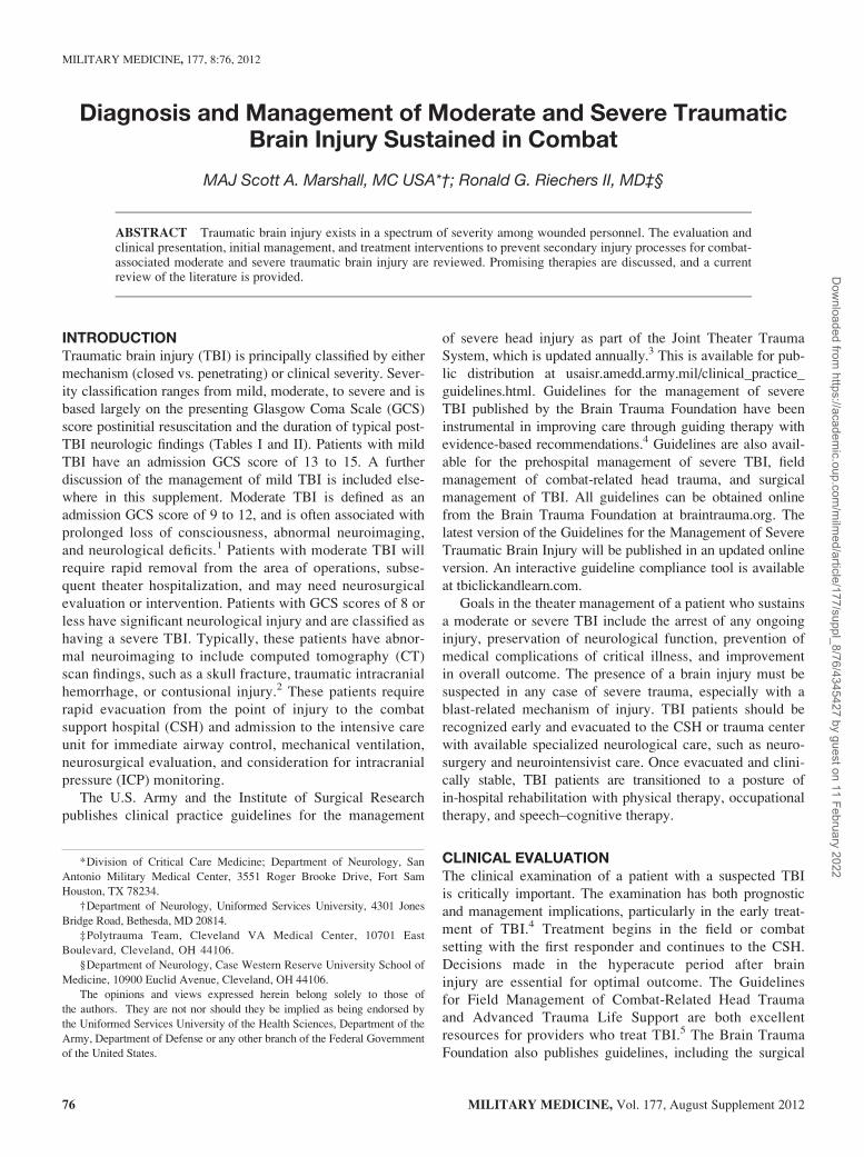
FIGURE 1. Traumatic subarachnoid hemorrhage (tSAH). (a) Penetrating supratentorial injury to the left temporal and parietal lobes with subsequentbihemispheric injury. (b) Axial bone windows of same image, which help delineate projectile (arrows) from primary intracerebral hemorrhage. (c)Subarachnoid blood layering in the basal cisterns (arrows). This mechanism of tSAH would result in a high risk of subsequent vasospasm and delayedcerebral ischemia. This patient did not survive.
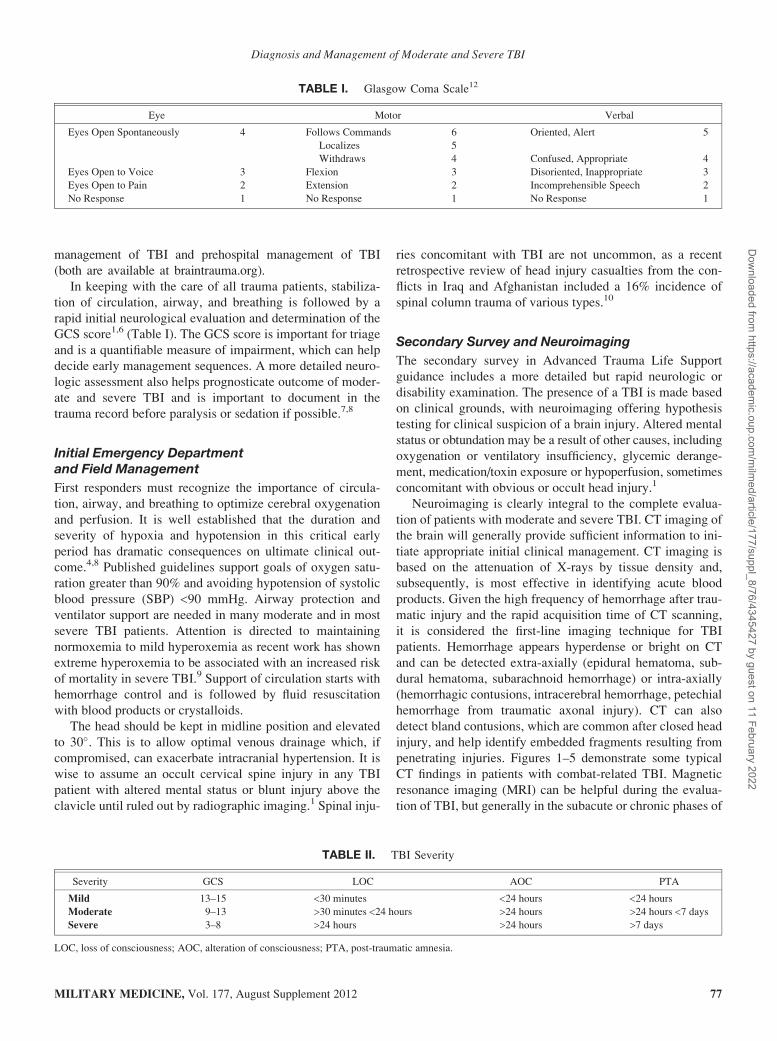
FIGURE 2. Nonpenetrating blast TBI. (a) Cytotoxic edema in the occip-ital lobes (white arrow) and hemorrhagic occipital contusion (black arrow).(b) In this, more caudal image note the subdural layering of hemorrhage overthe tentorium (white arrows).
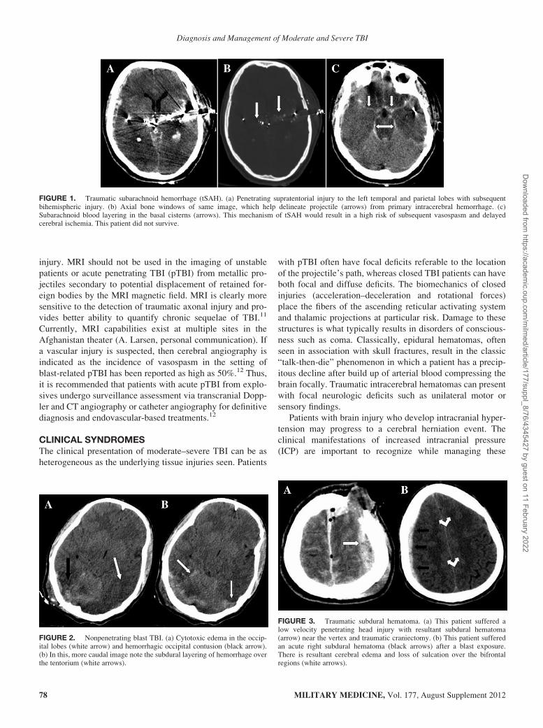
FIGURE 3. Traumatic subdural hematoma. (a) This patient suffered alow velocity penetrating head injury with resultant subdural hematoma(arrow) near the vertex and traumatic craniectomy. (b) This patient sufferedan acute right subdural hematoma (black arrows) after a blast exposure.There is resultant cerebral edema and loss of sulcation over the bifrontalregions (white arrows).
MILITARY MEDICINE, Vol. 177, August Supplement 201278
Diagnosis and Management of Moderate and Severe TBI
Dow
nloaded from https://academ
ic.oup.com/m
ilmed/article/177/suppl_8/76/4345427 by guest on 11 February 2022
patients. When compensatory mechanisms are exceeded, a
herniation may occur, which will manifest in a variety of
neurologic syndromes. Although specific clinical presenta-
tions and focal findings may occur, many patients simply
begin to become more lethargic and may drop points in their
GCS score. Herniation syndromes, which should be recog-
nizable at the bedside, include the transtentorial or uncal
herniation syndrome where the medial portion of the tempo-
ral lobe compresses the lateral midbrain and third nerve.
Because of the anatomic organization of this nerve, para-
sympathetic fibers are compressed initially, which results
in unilateral pupillary dilation. Additionally, patients can
develop weakness/paralysis affecting the body ipsilateral to
the herniation. With more diffuse edema, central herniation
can occur where the dorsal midbrain is compressed, result-
ing in forced downward gaze or other alterations of vertical
gaze control. As elevations in ICP progress or with poste-
rior fossa lesions, cerebellar tonsillar herniation can occur
whereby the caudal medulla is compressed, often a terminal
event caused by disruption of brainstem cardiorespiratory
centers. A specific type of herniation bears mention because
of the large number of military patients treated with decom-
pressive craniectomy (DC). Paradoxical herniation has been
reported during lumbar puncture in this setting and occurs
by downward movement of brain in the setting of an over-
all lowered ICP.13 This can also follow severe sodium
dysregulation and hypernatremia. If the pressure change is a
result of lumbar drainage and subsequent cerebral spinal fluid
(CSF) leak, the use of a blood patch has been reported to be
lifesaving.14 In addition to the brain parenchymal damage,
which can occur from herniation, herniation syndromes can
result in cerebral infarcts secondary to compression of proxi-
mate vessels, e.g., posterior cerebral artery infarction associ-
ated with uncal herniation. These infarcts further increase
edema and impact long-term morbidity.
It is an important point to emphasize that numerous
authors have published reports regarding outcome data of
patients with clinical and radiographic herniation who have
survived to discharge with variable disability.14–18 Poor
prognostic assessment of these patients who have not had
aggressive medical management may be inappropriate.
MANAGEMENT OF TBI
Blood Pressure and Fluid Management
Maintenance of euvolemia and adequate brain perfusion is
the overall objective of hemodynamic therapy in TBI. Cere-
bral perfusion pressure (CPP) is a measure of the overall
adequacy of global brain perfusion and is calculated by the
mean arterial pressure (MAP) minus ICP. Recommended
treatment goals are SBP >90 mmHg and CPP >60 mmHg.4
The patient with TBI may be in hemorrhagic shock
because of accompanying polytrauma and hypotension.
As such, hypotension (SBP < 90 mmHg) in the setting of
TBI is independently associated with poor outcome and
mortality from TBI.19–21 When compared to hypoxia, low
SBP is associated relatively with an even worse outcome.19
Autoregulation of the neurovasculature is impaired, and
regional cerebral blood flow becomes directly dependent
on systemic blood pressure.22 Experimental models show
that the injured brain is highly susceptible to even subtle
ischemic states.21 Current evidence grows for the prac-
tice of hypotensive resuscitation in the care of trauma
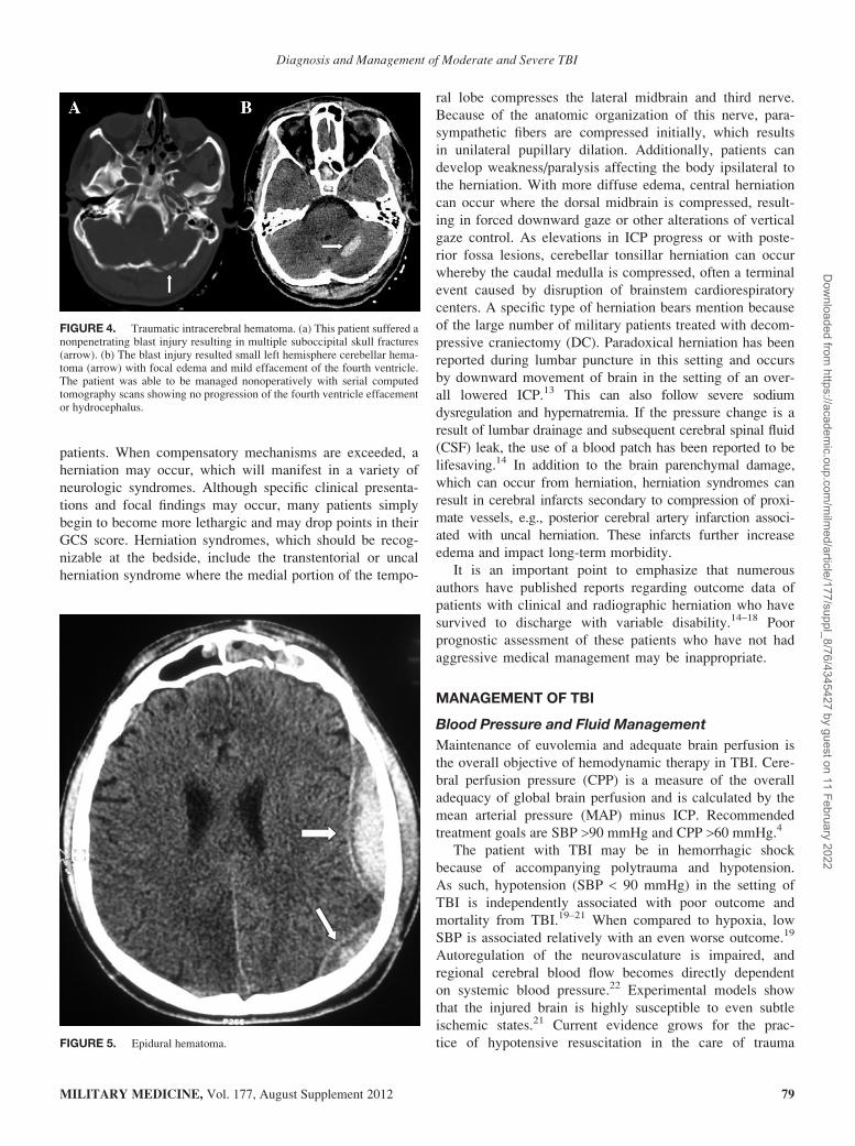
FIGURE 4. Traumatic intracerebral hematoma. (a) This patient suffered anonpenetrating blast injury resulting in multiple suboccipital skull fractures(arrow). (b) The blast injury resulted small left hemisphere cerebellar hema-toma (arrow) with focal edema and mild effacement of the fourth ventricle.The patient was able to be managed nonoperatively with serial computedtomography scans showing no progression of the fourth ventricle effacementor hydrocephalus.
FIGURE 5. Epidural hematoma.
MILITARY MEDICINE, Vol. 177, August Supplement 2012 79
Diagnosis and Management of Moderate and Severe TBI
Dow
nloaded from https://academ
ic.oup.com/m
ilmed/article/177/suppl_8/76/4345427 by guest on 11 February 2022
patients; however, it should be noted that TBI patients
are often specifically excluded from studies examining
this practice.16
Limited amounts of crystalloid fluids are used by U.S.
Army medic and Navy corpsmen protocols for field resusci-
tation in TBI/polytrauma. Once the patients are evaluated in
field hospital facilities, blood products are the preferred
resuscitation fluid. From the conflicts in Afghanistan and
Iraq, it has been reported that hemorrhagic shock is ideally
treated with red blood cells and plasma using a 1:1 ratio
based on volume.23,24 Colloid fluids are relatively contrain-
dicated in TBI.25 Hypotonic fluids, such as 1/2 normal saline
and Lactated Ringer’s, have the potential of exacerbating
cerebral edema and should be avoided.22 Fluid balance of TBI
patients is important as patients with TBI who were fluid
balance negative by approximately 600 cc had worse proximal
outcomes in an often referenced study.26 The Lund Concept
is a practice contrary to this premise, which advocates fluid
restriction as a means to control cerebral edema and nor-
malize ICP.27
CPP goals may initially be satisfied with intravenous (IV)
fluids, but if CPP cannot be maintained with IV fluids alone,
vasoactive pharmacologic agents may be considered. Norepi-
nephrine and phenylephrine are commonly used. Continuous
hemodynamic monitoring is needed with both a central
venous pressure catheter and a peripheral arterial pressure
catheter with pressor therapy.28 Aggressive use of these
agents has been associated with increased incidence of acute
respiratory distress syndrome (ARDS); however, this compli-
cation potentially could have been the result of exceeding
CPP levels of 70 mmHg.29 Beneficial effects of the Lund
Concept may be attributed to the lower incidence of pulmo-
nary complications, such as ARDS, in patients with more
judicious fluid management.30
Ventilation and Airway Management
Oxygenation and ventilation goals are established early
to maintain adequate oxygenation with the partial pres-
sure of oxygen in arterial blood (PaO2) remaining above
60 mmHg, and normocarbia.3,4,31 Avoidance of hypoxemia
or extreme hyperoxemia (PaO2 > 487 mmHg) reduces mor-
tality in TBI.9 In the field, oxygen saturation should be
less than 90%.32 Indications for placement of an artificial
airway are a GCS score of 8 or less or clinical concern
that the patient’s ability to ventilate or protect his or her
airway is in jeopardy.22 Overaggressive hyperventilation
should be avoided because of the potential for decreased
cerebral perfusion at PCO2 <25 mmHg.4,33 Newer ventila-
tor management strategies, such as airway pressure release
ventilation (APRV) aimed at improving oxygenation at the
expense of ventilation, require further study for use in the
setting of head injury and must be used with caution
because of the possibility of hypercapnea. A review of
APRV is referenced.34
Intracerebral Pressure (ICP) Management
Much of neurocritical and neurosurgical care of TBI is
concerned with ICP. If ICP progresses unchecked, it can
result in herniation and ischemia. Conservative measures
should be instituted in moderate and severe TBI patients to
minimize elevations in ICP. Simple interventions include
raising the head of the bed to 30�, keeping the head midline,
avoiding any circumferential neck dressings for securing the
endotracheal tube, and avoiding placement of internal jugular
(IJ) central venous lines into the dominant IJ. All these will
optimize venous outflow from the head. Central lines that
require the Trendelenburg position should not be used for
central access during a herniation event as placing the patient
in Trendelenburg may increase ICP further.35 Alternatively,
femoral lines may be temporarily appropriate. Aggressive
treatment of fever, seizures, pain, and agitation can help
prevent elevations in ICP.4
Indications for ICP Monitoring
Severe TBI patients with a strong clinical suspicion of
increased ICP should have a monitor placed. There are a
number of options that include the extraventricular drain
(EVD), intraparenchymal fiber optic monitor, subdural bolt,
and epidural fiber optic catheters. If hydrocephalus is seen
on imaging, an EVD is obviously the best option. Clear
indications exist for placing an ICP monitor. If the patient
has a GCS score of 8 or less (after resuscitation) and an
acute abnormality on CT, such as traumatic intracerebral
hemorrhage, compression of the basal cisterns, evidence of
contusion or herniation, then an ICP monitor should be
placed.4 If a patient has two of the following: SBP £90 mmHg,
motor posturing on examination, and/or is 40 years of age
or older, then an ICP monitor should likewise be placed or
strongly considered.4
Other invasive monitoring devices, such as brain tissue
oxygenation monitors, microdialysis catheters, and jugu-
lar venous saturation monitors, can be used to tailor
therapy. Routine application of these devices awaits fur-
ther study.4 The use of brain tissue oxygen monitors have
recently been reported to be associated with increased fluid
and vasopressor use and pulmonary complications such
as ARDS.30
ICP Treatment Goals
The goal ICP in TBI is to maintain a normal pressure
state, which is generally less than 20 cmH2O or 15 mmHg.
Elevations to more than 25 mmHg are associated with poor
outcome. Current guidelines recommend instituting mea-
sures to control ICP when pressures of 20 mmHg are
reached and aggressive means employed to prevent ICP
elevations to more than 25 mmHg.4 Awareness of CPP is
important as many interventions to decrease ICP may
also have systemic effects on peripheral hemodynamics.
The maintenance of a CPP of at least 60 mmHg with
MILITARY MEDICINE, Vol. 177, August Supplement 201280
Diagnosis and Management of Moderate and Severe TBI
Dow
nloaded from https://academ
ic.oup.com/m
ilmed/article/177/suppl_8/76/4345427 by guest on 11 February 2022
IV fluids or vasopressors is strongly recommended.4 Pul-
monary complications, including higher incidence of ARDS,
may result from overshooting the goal CPP to greater than
70 mmHg.29,30
Medical Treatment Options for Elevated ICP
Hyperventilation
Hyperventilation for ICP reduction may be used only as
an emergent and temporary intervention. Prolonged hyper-
ventilation has been clearly associated with exacerbation
of cerebral ischemia.36 Short durations of hyperventilation
are acceptable as a temporizing measure until other meth-
ods of managing ICP are available. If hyperventilation is
continued for longer than 12 hours, metabolic compensa-
tion negates any helpful effects of hyperventilation. The
recommend goal for a chronic PCO2 is 35 to 40 mmHg.
During a herniation event, hyperventilation will acutely
and reliably lower PCO2, as well as ICP, within seconds.
The current recommended PCO2 is to strictly avoid levels
below 25 mmHg.4,22
Hyperosmolar Therapy
Pharmacologic creation of an osmotic gradient causes move-
ment of water from intracellular and extracellular com-
partments of the brain into the vasculature where it reduces
the volume of the overall cranial compartment.35 Several
agents have been used for this purpose in the past but, cur-
rently, mannitol and hypertonic saline (HTS) are the main-
stays of hyperosmolar therapy. Mannitol should be given
intravenously via a peripheral or central IV line at a dose of
0.25 to 1.0 g/kg. Small doses of mannitol (0.25 g/kg) have
been shown to effectively reduce ICP in patients with TBI.37
Earlier data shows that mannitol use in TBI correlates with
decreased ICP and improvements in cerebral blood flow
and CPP.38 Mannitol can be given while following serum
osmolality, where a serum osmolality of 320 mOsm/L is
generally accepted as a treatment endpoint. Some investi-
gators advocate that slightly higher levels can be obtained
with caution.39
Another option for hyperosmolar therapy is HTS. Studies
using 7.5 and 23.4% HTS provide evidence of efficacy.
Recent evidence supports the use of bolus doses of 30 to
60mLof 23.4%HTS to emergently reverse a herniation event.18
About 23.4% HTS ameliorative effect on ICP lasts longer
than mannitol.40 High concentrations of HTS must be admin-
istered via a central venous line during a 10 to 15 minutes
time period to prevent phlebitis and hypotension. A com-
monly used initial treatment goal is to achieve serum sodium
levels 145 to 155 mEq/L, equivalent to a serum osmolality of
300 to 320 mOsm/L in most patients.35 Recent evidence
shows 23.4% HTS to be effective in reducing ICP by a mean
value of more than 8 mmHg when given for ICP >20 mmHg
and can increase CPP values by 6 mmHg when pretreatment
values are <70 mmHg.41 A continuous IV infusion of 2 or 3%
HTS can be used to maintain high serum osmolality, and
solutions of 3% or higher HTS should be given via a central
line. If continuous infusions of HTS are used, serum sodium
should be monitored every 4 hours although avoiding rapid
changes in serum sodium so as not to precipitate cerebral
edema or central pontine myelinolysis.35 A recent review
discusses frequent questions clinicians have regarding the
use of HTS in the setting of intracranial hypertension.42
Agents to Reduce the Cerebral Metabolic Rate ofOxygen (CMRO2)
If ICP remains poorly controlled, then reduction in the meta-
bolic rate of the brain via pharmacologic coma can be con-
sidered. The postulated effect by which ICP is reduced is
though a reduction in cerebral metabolism, resulting in reduc-
tions in cerebral blood flow and reduced tissue oxygen
demand. Pentobarbital is the most widespread agent in use
for induction of pharmacological coma. Pentobarbital is
administered intravenously at a loading dose of 10 mg/kg
during a 30-mintue time period, followed by a 5 mg/kg/h
infusion for 3 hours, and maintenance therapy of 1 mg/kg/h
titrated to therapeutic goals of either burst suppression on
continuous electroencephalography (EEG) monitoring or a
satisfactory reduction in ICP.28 If burst suppression is not
obtained with this dose, then a smaller loading dose and
increased rate can be given until an EEG tracing consistent
with burst suppression is seen or ICP is controlled. In the
past, other barbiturates such as the much shorter acting thio-
pental were used for acute exacerbations of ICP.38 This
medication is not currently available in the United States
but may be available to deployed forces overseas (A. Holley,
personal communication).
Propofol represents an alternative therapy. Propofol is
given at an IV loading dose of 2 mg/kg, followed by a titrated
infusion of up to 100 mg/kg/min. The use of propofol for this
clinical indication is controversial, and this drug has several
side effects, including severe systemic hypotension. A study
using propofol for ICP reduction showed a failure of an
improvement in 6-month outcome, and long-term and high-
dose propofol infusions have been associated with the devel-
opment of propofol infusion syndrome, which consists of
renal failure, rhabdomyolysis, hyperkalemia, myocardial fail-
ure, metabolic acidosis, lipemia, hepatomegaly, and often
death.43 Vigilance for this condition is wise for any patient
receiving an infusion for more than 48 hours.4 Continuous
EEG monitoring may be helpful to monitor for burst suppres-
sion or better control of ICP.
FUTURE DIRECTIONSInduced hypothermia to improve outcomes in TBI is prom-
ising but controversial. Recent animal data show induced
hypothermia to be associated with improved neurophysio-
logic metrics in an hypoxic brain injury model.44 There is
data in brain trauma that induced mild hypothermia may
MILITARY MEDICINE, Vol. 177, August Supplement 2012 81
Diagnosis and Management of Moderate and Severe TBI
Dow
nloaded from https://academ
ic.oup.com/m
ilmed/article/177/suppl_8/76/4345427 by guest on 11 February 2022
improve outcome post-TBI.45 Current use of induced hypo-
thermia for treatment of ICP in severe TBI is a second-tier
therapy but may be helpful in refractory cases.35 The goal of
maintaining normothermia and avoiding hyperthermia in
TBI patients is strongly recommended.46 The potential of
coagulopathy and antiplatelet effects of induced hypo-
thermia should be considered, especially in the setting of
hemorrhagic TBI.47–51
Older preclinical data suggests a difference between out-
comes of models of brain injury based on gender.52 Further
work defined high levels of progesterone as potentially pro-
tective from the standpoint of developing cerebral edema,
and as supportive data began to accumulate, interest in
investigating the potential benefits of progesterone therapy
in TBI grew.53 A synopsis of both preclinical and epide-
miologic studies regarding progesterone is provided.54 Cur-
rently, there are two ongoing clinical trials attempting
to clarify the benefit of progesterone therapy in TBI, the
ProTECT III (Progesterone for Traumatic Brain Injury,
Experimental Clinical Trial III), and SyNAPSE (Study
of the Neuroprotective Activity of Progesterone in Severe
Traumatic Brain Injury). Both have a planned enrollment of
more than 1,100 patients and expected completion dates are
2015 and 2012, respectively.55
SURGICAL TREATMENT OPTIONS
Decompressive Craniectomy
DC represents another clinical approach to the early interven-
tion and management of TBI. The reported experience to date
is conflicting.56–58 The role of DC in treating brief elevations
in ICP in the setting of diffuse non-pTBI was evaluated in the
recently published Decompressive Craniectomy (DECRA)
trial.59 ICP control was significantly improved in the surgical
treatment arm, but 6-month outcomes were worsened com-
pared with medical therapy. The surgical procedure per-
formed was a bilateral DC, and patients with focal space
occupying lesions were excluded from the study. Of note,
patients in the surgical arm had a statistically significant
difference in loss of pupil reactivity compared with the med-
ically treated patients. This fundamental difference in the
treatment arm, combined with the fact that bilateral DC is
not the most often used surgical procedure in treatment of
refractory elevations in ICP, appears to limit the ability to
generalize the study’s conclusions. The Randomized Evalua-
tion of Surgery with Craniectomy for Uncontrollable Eleva-
tion of Intra-Cranial Pressure (RESCUEicp) may help further
define the role DC may have in the management of severe
TBI.60 RESCUEicp recently completed its enrolment of
approximately 400 patients comparing DC to medical man-
agement (including barbiturates) in severe TBI.
The U.S. military neurosurgical experience in Operation
Enduring Freedom and Operation Iraqi Freedom used early
hemicraniectomy for treating some cases of severe TBI with
concerns for imminent elevations in ICP, whether from pen-
etrating, blunt injury, or blast induced.61,62 This population is
unique because of long, fixed-wing evacuation flights and
exposure to relative altitude proximal to the incident trauma.
In a recent study of this population comparing GCS scores of
patients at the time of head trauma and at discharge, TBI
patients who underwent a craniectomy had a lower initial
GCS score than those who underwent craniotomy, but at
discharge their GCS score was not significantly different.12
A similar article with a smaller sample size and follow-up at
11 months and the use of the Extended Glasgow Outcome
Scale (GOSE) as the outcome metric is referenced.63 Antici-
pated randomized studies on large cohorts of patients may
clarify the role of this treatment option.
Common Clinical Approach for ElevatedICP Management
The management of acute elevations in ICP initially involves
ensuring that the waveform and ICP reading is accurate.
Seizures, fever, metabolic and respiratory derangements need
to be ruled out if suspected. Brain CT imaging should be
considered in any new episode of increased ICP without
explanation. Maneuvers such as repositioning the head to
midline, sitting the patient up at 30�, establishing normother-
mia and cessation of suctioning or other noxious stimuli may
help lower temporary spikes in ICP. If this is unsuccessful
and the ICP is felt to be accurate, very brief hyperventilation
of intubated patients may be performed. If central access
exists, then 30 cc of 23.4% HTS may be given via a central
line during a 10-minute period. If given faster, CPP may need
to be augmented with small doses of phenylephrine. As an
alternative to HTS, mannitol may be given via a peripheral
line with the dose tailored to the clinical situation. A dose of
1 g/kg is given for clinical signs of herniation and doses of
0.25 to 0.5 g/kg for less severe increases in ICP. In a hernia-
tion event, central access should be obtained with consider-
ation of a femoral central venous catheter or avoidance of
extreme Trendelenburg positioning for subclavian lines.35 If
ICP continues to be elevated after these maneuvers, then
additional HTS can be given as well as further boluses of
mannitol, treating up to a serum osmolality of approximately
320 mOsm/L. Standing infusions of 3% HTS can be used,
with goal sodium values that may exceed 155 to 160 mEq/L
if required. Further medical management includes use of
bolus doses of propofol, and consideration given to pharma-
cologic coma induced hypothermia or surgical intervention.
OTHER ASPECTS OF CARE
Anticonvulsants
TBI patients, both penetrating and nonpenetrating, are at risk
for both early (less than 7 days) and late (more than 7 days)
post-traumatic seizures. A seizure in the acute phase can
exacerbate the injury. Phenytoin, a well-established anti-
epileptic drug (AED), has been shown to be beneficial in
reducing the risk of seizures during the first week after
MILITARY MEDICINE, Vol. 177, August Supplement 201282
Diagnosis and Management of Moderate and Severe TBI
Dow
nloaded from https://academ
ic.oup.com/m
ilmed/article/177/suppl_8/76/4345427 by guest on 11 February 2022
TBI.64,65 Carbamazepine, phenobarbital, and valproate are
also effective AEDs.64 Unfortunately, no AED has been
shown to prevent the development of late post-traumatic sei-
zures. The recommended approach is to stop AED therapy
after the first 7 days, and only reinstitute treatment should late
seizures manifest.4 Data regarding cognitive side effects of
phenytoin make prolonged prophylactic use of this medica-
tion, in particular, less attractive.66 If a patient requires IV
medications, alternatives include valproate, lacosamide, and
levetiracetam. Levetiracetam has been shown to be highly
effective in preclinical TBI models, and in randomized trials
it has been shown to be as effective as phenytoin with
improved GOSE at 6 months.67,68 The American Academy
of Neurology has published guidelines for the use of AEDs in
the setting of severe TBI recommending treatment with
AEDs prophylactically for the first 7 days following injury.69
Secondary Complications of the Critically Ill
Prevention of secondary complications of critical illness
includes consideration of venous thromboembolism (VTE),
stress ulceration, and skin breakdown. Any critically ill and
immobilized patient is at high risk for developing deep
venous thrombosis (DVT) with subsequent VTE. The opti-
mal approach for VTE/DVT prophylaxis in severe TBI com-
plicated by intracranial hemorrhage is unclear. Sequential
compression devices (SCD) on the lower extremities are
minimally invasive and are not associated with worsening
intracranial hemorrhage. The timing for introduction of
unfractionated or low molecular weight heparin (LMWH)
for VTE prophylaxis in this population is an individualized
decision. Lacking a contraindication to pharmacologic DVT
prophylaxis, heparin, or LMWH should be started ideally
within the first 36 hours after injury.70 The routine placement
of inferior vena cava filters currently has limited support.70,71
Gastric stress ulcers may be prevented using either H2 recep-
tor antagonists or proton pump inhibitors (PPIs). Potential
comorbidities, resulting from the indiscriminate use of PPIs
and drug-to-drug interactions, require consideration of alter-
native means of gastric ulcer prophylaxis, particularly in the
setting where a TBI patient requires an antiplatelet regi-
men.72 Either one of these medications should be considered
for gastric stress ulceration prophylaxis in severe TBI
patients, although the tendency for H2 blockers to cause
thrombocytopenia may limit their usefulness.73 Also, preven-
tion of skin breakdown is important in all severe trauma
patients, and care must be taken to reduce the likelihood of
such by appropriate skin hygiene and proper nursing care
with scheduled repositioning.
CONCLUSIONSThe management of patients with combat-related moderate
and severe brain injury is challenging, and medical and sur-
gical means of treatment are indicated to reduce secondary
injury from brain trauma. Maintaining brain perfusion, con-
trolling ICP, and preventing morbidity associated with criti-
cal illness are principle to care. As new medical and surgical
approaches are introduced, there will be increasing opportu-
nity to better manage these patients and improve neurologic
outcomes in the near and long term.
REFERENCES
1. American College of Surgeons. Advanced Trauma Life Support: Program
for Doctors, Ed 11. Chicago, IL, American College of Surgeons, 2004.
2. Geocadin R: Traumatic brain injury. In: Handbook of Neuro Critical
Care, pp 73–89. Edited by Bhardwaj A, Mirski M, Ulatowski J. Totowa,
NJ, Humana Press, 2004.
3. USAISR. Clinical Practice Guidelines. Available at USAISR.amedd
.army.mil/clinical_practice_guidelines.html; accessed December 9, 2011.
4. Bratton SL, Chestnut RM, Ghajar J, et al: Guidelines for the manage-
ment of severe traumatic brain injury. J Neurotrauma 2007; 24(Suppl 1):
S21–5.
5. Brain Trauma Foundation. Online TBI Guidelines. New York, NY,
BTF. Available at www.tbiguidelines.org; accessed February 2011.
6. Teasdale G, Jennett B: Assessment of coma and impaired conscious-
ness: a practical scale. Lancet 1974; 2: 81–4.
7. Patel HC, Tebbs S, Menon DK, Hawker R, Hutchinson PJ, Kirkpatrick
PJ: Specialist neurocritical care and outcome from head injury. Intensive
Care Med 2002; 28: 547–53.
8. Perel P, Arango M, Clayton T, et al: Predicting outcome after traumatic
brain injury: practical prognostic models based on large cohort of inter-
national patients. BMJ 2008; 336: 425–9.
9. David DP, Meade W, Sise MJ, et al: Both hypoxemia and extreme
hyperoxemia may be detrimental in patients with severe traumatic brain
injury. J Neurotrauma 2009; 26: 2217–23.
10. Bell RS, Vo AH, Neal CJ, et al: Military traumatic brain and spinal column
injury: a 5 year study of the impact blast and other military grade weaponry
on the central nervous system. J Trauma 2009; 66(4 suppl): S104–11.
11. Le TH, Gean AD: Neuroimaging of traumatic brain injury. Mt Sinai J
Med 2009; 76: 145–62.
12. Armonda RA, Bell RS, Vo AH, et al: Wartime traumatic cerebral vaso-
spasm: recent review of combat casualties. Neurosurgery 2006; 59:
1215–25.
13. Vilela MD: Delayed paradoxical herniation after a decompressive
craniectomy: case report. Surg Neurol 2008; 69: 293–6.
14. Muehlschlegel S, Voetsch B, Sorond FA: Emergent epidural blood
patch: lifesaving treatment of paradoxical herniation. Arch Neurol
2009; 66: 670–1.
15. Seinfeld J, Sawyer M, Rabb CH: Successful treatment of paradoxical
cerebral herniation by lumbar epidural blood patch placement: technical
case report. Neurosurgery 2007; 61(3 suppl): E175.
16. Stiver SI, Gean AD, Manley GT: Survival with good outcome after
cerebral herniation and duret hemorrhage caused by traumatic brain
injury. J Neurosurg 2009; 110: 1242–6.
17. Qureshi AI, Suarez JI: More evidence supporting a brain code pro-
tocol for reversal of transtentorial herniation. Neurology 2008; 70:
990–1.
18. Koenig MA, Bryan M, Lewin JL III, Mirski MA, Geocadin RG, Stevens
RD: Reversal of transtentorial herniation with hypertonic saline. Neu-
rology 2008; 70: 1023–9.
19. Schreiber MA, Aoki N, Scott BG, Beck JR: Determinants of mortality in
patients with severe blunt head injury. Arch Surg 2002; 137: 285–90.
20. Chesnut RM, Marshall LF, Klaubeer MR, et al: The role of secondary
brain injury in determining outcome from severe head injury. J Trauma
1993; 24: 216–22.
21. Jenkins LW, Moszynshi K, Lyeth BG, et al: Increased vulnerability of
the mildly traumatized rat brain to cerebral ischemia: the use of con-
trolled secondary ischemia as a research tool to identify common or
MILITARY MEDICINE, Vol. 177, August Supplement 2012 83
Diagnosis and Management of Moderate and Severe TBI
Dow
nloaded from https://academ
ic.oup.com/m
ilmed/article/177/suppl_8/76/4345427 by guest on 11 February 2022
different mechanisms contributing to mechanical and ischemic brain
injury. Brain Res 1989; 477: 211–24.
22. Stiver SI, Manley GT: Prehospital management of traumatic brain
injury. Neurosurg Focus 2008; 25: E5.
23. Gonzalez EA, Moore FA, Holcomb JB, et al: Fresh frozen plasma
should be given earlier to patients requiring massive transfusion. J
Trauma 2007; 62: 112–9.
24. Holcomb JB, McMullin NR, Pearse L, et al: Causes of death in U.S.
Special Operations Forces in the global war on terrorism. Ann Surg
2007; 245: 986–91.
25. SAFE Study Investigators; Australian and New Zealand Intensive Care
Society Clinical Trials Group; Australian Red Cross Blood Service;
George Institute for International Health; Myburgh J, Cooper DJ,
Finfer S, et al: Saline or albumin for fluid resuscitation in patients with
traumatic brain injury. N Engl J Med 2007; 357: 874–84.
26. Clifton GL, Miller ER, Choi SC, Levin HS: Fluid thresholds and out-
come from severe brain injury. Crit Care Med 2002; 30: 739–45.
27. Grande PO: The “Lund Concept” for the treatment of severe head
trauma—physiological principles and clinical application. Intensive
Care Med 2006; 32: 1475–84.
28. Ling GS, Marshall SA: Management of traumatic brain injury in the
intensive care unit. Neurol Clin 2008; 26: 409–26.
29. Contant CF, Valadka AB, Gopinath SP, Hannay HJ, Robertson CS:
Adult respiratory distress syndrome: a complication of induced hyper-
tension after severe head injury. J Neurosurg 2001; 95: 560–8.
30. Fletcher JJ, Bergman K, Blostein PA, Kramer AH: Fluid balance, com-
plications, and brain tissue oxygen tension monitoring following severe
traumatic brain injury. Neurocrit Care 2010; 13: 47–56.
31. Warner KJ, Cuschieri J, Compass MK, Jurkovisch GJ, Bulger EM:
Emergency department ventilation effects outcome in severe traumatic
brain injury. J Trauma 2008; 64: 341–7.
32. McHugh GS, Engel DC, Butcher I, et al: Prognostic value of secondary
insults in traumatic brain injury: results from the IMPACT study. J
Neurotrauma 2007; 24: 287–93.
33. Muizelaar JP, Marmarou A, Ward JD, et al: Adverse effects of
prolonged hyperventilation in patients with severe head injury: a ran-
domized clinical trial. J Neurosurg 1991; 75: 731–9.
34. Habashi NM: Other approaches to open-lung ventilation: airway pres-
sure release ventilation. Crit Care Med 2005; 33(3 suppl): S228–40.
35. Raslan A, Bhardwaj A: Medical management of cerebral edema.
Neurosurg Focus 2007; 22: E12.
36. Marion DW, Firlik A, McLaughlin MR: Hyperventilation therapy for
severe traumatic brain injury. New Horiz 1995; 3: 439–47.
37. Marshall LF, Smith RW, Rauscher LA, Shapiro HM: Mannitol dose
requirements in brain-injured patients. J Neurosurg 1978; 48: 169–72.
38. Mendelow AD, Teasdale GM, Russell T, Flood J, Patterson J, Murray
GD: Effect of mannitol on cerebral blood flow and cerebral perfusion
pressure in human head injury. J Neurosurg 1985; 63: 43–8.
39. Diringer MN, Zazulia AR: Osmotic therapy: fact and fiction. Neurocrit
Care 2004; 1: 219–33.
40. Ware ML, Nemani VM, Meeker M, Lee C, Morabito DJ, Manley GT:
Effects of 23.4% sodium chloride solution in reducing intracranial pres-
sure in patients with traumatic brain injury: a preliminary study. Neuro-
surgery 2005; 57: 727–36.
41. Rockswold GL, Solid CA, Paredes-Andrade E, Rockswold SB, Jancik
JT, Quickel RR: Hypertonic saline and its effect on intracranial pressure,
cerebral perfusion pressure, and brain tissue oxygen. Neurosurgery
2009; 65: 1035–41.
42. Marko N: Hyperosmolar therapy for intracranial hypertension: time to
dispel antiquated myths. Am J Respir Crit Care Med 2012; 185(5): 467–8.
43. Kelly DF, Goodale DB, Williams J, et al: Propofol in the treatment of
moderate and severe head injury: a randomized, prospective double-
blinded pilot trial. J Neurosurg 1999; 90: 1042–52.
44. Jia X, Koenig MA, Shin HC, et al: Improving neurological outcomes
post-cardiac arrest in a rat model: immediate hypothermia and quantita-
tive EEG monitoring. Resuscitation 2008; 76: 431–42.
45. Qiu WS, Liu WG, Shen H, et al: Therapeutic effect of mild hypo-
thermia on severe traumatic head injury. Chin J Traumatol 2005; 8:
27–32.
46. Maas AI, Stocchetti N, Bullock R: Moderate and severe traumatic brain
injury in adults. Lancet Neurol 2008; 7: 728–41.
47. Polderman KH: Application of therapeutic hypothermia in the intensive
care unit. Opportunities and pitfalls of a promising treatment modality—
part 2: practical aspects and side effects. Intensive Care Med 2004;
30(5): 757–69.
48. Watts DD, Trask A, Soeken K, Perdue P, Dols S, Kaufmann C: Hypo-
thermic coagulopathy in trauma: effect of varying levels of hypothermia
on enzyme speed, pletelet function, and fibrinolytic activity. J Trauma
1998; 44: 846–54.
49. Valeri CR, Feingold H, Cassidy G, Ragno G, Khuri S, Altschule MD, et al:
Hypothermia—induced reversible platelet dysfunction. Ann Surg 1987;
205: 175–81.
50. Mossad EB, Machado S, Apostolakis J: Bleeding following deep hypo-
thermia and circulatory arrest in children. Semin Cardiothorac Vasc
Anesth 2007; 11: 34–46.
51. Michelson AD, MacGregor H, Barnard MR, et al: Hypothermia—
induced reversible platelet dysfunction. Thromb Haemost 1994; 71:
633–40.
52. Attella MJ, Nattinville A, Stein DG: Hormonal state affects recovery
from frontal cortex lesions in adult female rats. Behav Neural Biol 1987;
48: 352–67.
53. Roof RL, Duvdevani R, Stein DG: Gender influences outcome of
brain injury: progesterone plays a protective role. Brain Res 1993;
607: 333–6.
54. Stein DG, Wright DW, Kellermann AL: Does progesterone have
neuroprotective properties? Ann Emerg Med 2008; 51(2): 164–72.
55. Clinical Trials. Available at http://clinicaltrials.gov; accessed December
19, 2011.
56. Albanese J, Leone M, Alliez JR, et al: Decompressive craniectomy for
severe traumatic brain injury: evaluation of the effects at one year. Crit
Care Med 2003; 31: 2535–8.
57. Guerra WK, Gaab MR, Dietz H, et al: Surgical decompression for
traumatic brain swelling: indications and results. J Neurosurg 1999; 90:
187–96.
58. Pompucci A, Bonis PD, Pettorini B, Petrella G, Di Chirico A, Anile C:
Decompressive craniectomy for traumatic brain injury: patient age and
outcome. J Neurotrauma 2007; 24: 1182–8.
59. Cooper DJ, Rosenfeld JV, Murray L, et al: Decompressive craniectomy
in diffuse traumatic brain injury. N Engl J Med 2011; 364: 1493–502.
60. Schlifka B: Lessons learned from OIF: a neurosurgical perspective.
J Trauma 2007; 62(6 Suppl): S103–4.
61. Bell RS, Mossop CM, Dirks MS, et al: Early decompressive
craniectomy for severe penetrating and closed head injury during war-
time. Neurosurg Focus 2010; 28: E1.
62. Schlifka B: A neurosurgical perspective. J Trauma 2010; 62: S103–4.
63. Howard JL, Cipolle MD, Anderson M, et al: Outcome after decom-
pressive craniectomy for the treatment of severe traumatic brain injury.
J Trauma 2008; 65(2): 380–5.
64. Temkin NR: Antiepileptogenesis and seizure prevention trials with
antiepileptic drugs: meta-analysis of controlled trials. Epilepsia 2001;
42: 515–24.
65. Temkin NR, Haglund MM, Winn HR: Causes, prevention, and treatment
of post-traumatic epilepsy. New Horiz 1995; 3: 518–22.
66. Naidech AM, Kreiter KT, Janjua N, et al: Phenytoin exposure is associ-
ated with functional and cognitive disability after subarachnoid hemor-
rhage. Stroke 2005; 36: 583–7.
67. Szaflarski JP, Sangha KS, Shutter LA: Prospective, randomized, single-
blinded comparative trial of intravenous levetiracetam versus phenytoin
for seizure prophylaxis. Neurocrit Care 2010; 12(2): 165–72.
68. Wang H, Gao J, Lassiter TF, et al: Levetiracetam is neuroprotective in
murine models of closed head injury and subarachnoid hemorrhage.
Neurocrit Care 2006; 5: 71–8.
MILITARY MEDICINE, Vol. 177, August Supplement 201284
Diagnosis and Management of Moderate and Severe TBI
Dow
nloaded from https://academ
ic.oup.com/m
ilmed/article/177/suppl_8/76/4345427 by guest on 11 February 2022

69. Chang BS, Lowenstein DH: Practice parameter: antiepileptic drug pro-
phylaxis in severe traumatic brain injury: report of the Quality Standards
Subcommittee of the American Academy of Neurology. Neurology
2003; 60: 10–6.
70. Rogers FB, Cipolle MD, Velmahos G, Rozycki G, Luchette FA: Practice
management guidelines for the prevention of venous thromboembolism
in trauma patients: the east Practice Management Guidelines Work
Group. J Trauma 2002; 53: 142–64.
71. Marion DW: Head and spinal cord injury. Neurol Clin 1998; 16:
485–502.
72. Juhasz M, Herszenyi L, Tulassay Z: Current standings of the proton
pump inhibitor and clopidogrel co-therapy: review on evolving field
with the eyes of the gastroenterologist. Digestion 2010; 81(1): 10–5.
73. Ropper AH, Gress DR, Diringer MN, et al: Neurological and neurosur-
gical intensive care, Ed. 4. Philadelphia, PA, Lippincott Williams &
Wilkins, 2004.
MILITARY MEDICINE, Vol. 177, August Supplement 2012 85
Diagnosis and Management of Moderate and Severe TBI
Dow
nloaded from https://academ
ic.oup.com/m
ilmed/article/177/suppl_8/76/4345427 by guest on 11 February 2022