diacylglycerol kinases terminate diacylglycerol signaling during the respiratory burst leading to...
TRANSCRIPT

Sergio Grinstein and Gregory D. FairnSteinberg, Takehiko Ueyama, Guangwei Du,Alexandros Chatilialoglu, Benjamin E. Daniel Schlam, Michal Bohdanowicz, Oxidase ActivationHeterogeneous Phagosomal NADPH Respiratory Burst Leading toDiacylglycerol Signaling during the Diacylglycerol Kinases TerminateLipids:
doi: 10.1074/jbc.M113.457606 originally published online June 27, 20132013, 288:23090-23104.J. Biol. Chem.
10.1074/jbc.M113.457606Access the most updated version of this article at doi:
.JBC Affinity SitesFind articles, minireviews, Reflections and Classics on similar topics on the
Alerts:
When a correction for this article is posted•
When this article is cited•
to choose from all of JBC's e-mail alertsClick here
Supplemental material:
http://www.jbc.org/content/suppl/2013/06/27/M113.457606.DC1.html
http://www.jbc.org/content/288/32/23090.full.html#ref-list-1
This article cites 48 references, 26 of which can be accessed free at
at Univ of Toronto - OCUL on August 29, 2013http://www.jbc.org/Downloaded from
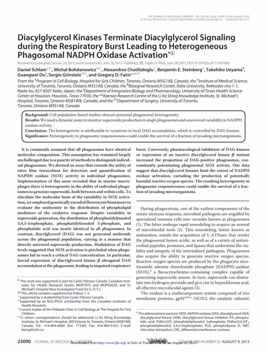
Diacylglycerol Kinases Terminate Diacylglycerol Signalingduring the Respiratory Burst Leading to HeterogeneousPhagosomal NADPH Oxidase Activation*□S
Received for publication, January 29, 2013, and in revised form, June 26, 2013 Published, JBC Papers in Press, June 28, 2013, DOI 10.1074/jbc.M113.457606
Daniel Schlam‡§1, Michal Bohdanowicz‡§2, Alexandros Chatilialoglu‡, Benjamin E. Steinberg‡, Takehiko Ueyama¶,Guangwei Du�, Sergio Grinstein‡§3, and Gregory D. Fairn**‡‡4
From the ‡Program in Cell Biology, Hospital for Sick Children, Toronto, Ontario M5G1X8, Canada, the §Institute of Medical Science,University of Toronto, Toronto, Ontario M5S1A8, Canada, the ¶Biosignal Research Center, Kobe University, Rokkodai-cho 1-1,Nada-ku, 657-8501 Kobe, Japan, the �Department of Integrative Biology and Pharmacology, University of Texas Health ScienceCenter at Houston, Houston, Texas 77030, the **Keenan Research Centre of the Li Ka Shing Knowledge Institute, St. Michael’sHospital, Toronto, Ontario M5B1W8, Canada, and the ‡‡Department of Surgery, University of Toronto,Toronto, Ontario M5S1A8, Canada
Background: Cell population-based studies obscure potential phagosomal heterogeneity.Results:Weused a dynamic assay tomonitor superoxide production in single phagosomes and uncovered variability inNADPHoxidase activity.Conclusion: The heterogeneity is attributable to variations in local DAG accumulation, which is controlled by DAG kinases.Significance:Heterogeneity in phagosome responsiveness could enable the survival of a fraction of invading microorganisms.
It is commonly assumed that all phagosomes have identicalmolecular composition. This assumption has remained largelyunchallengeddue to a paucity ofmethods to distinguish individ-ual phagosomes. We devised an assay that extends the utility ofnitro blue tetrazolium for detection and quantification ofNAPDH oxidase (NOX) activity in individual phagosomes.Implementation of this assay revealed that in murine macro-phages there is heterogeneity in the ability of individual phago-somes to generate superoxide, bothbetween andwithin cells. Toelucidate the molecular basis of the variability in NOX activa-tion,we employed genetically encoded fluorescent biosensors toevaluate the uniformity in the distribution of phospholipidmediators of the oxidative response. Despite variability insuperoxide generation, the distribution of phosphatidylinositol3,4,5-trisphosphate, phosphatidylinositol 3-phosphate, andphosphatidic acid was nearly identical in all phagosomes. Incontrast, diacylglycerol (DAG) was not generated uniformlyacross the phagosomal population, varying in a manner thatdirectly mirrored superoxide production. Modulation of DAGlevels suggested that NOX activation is precluded when phago-somes fail to reach a critical DAG concentration. In particular,forced expression of diacylglycerol kinase � abrogated DAGaccumulation at thephagosome, leading to impaired respiratory
burst. Conversely, pharmacological inhibition of DAG kinasesor expression of an inactive diacylglycerol kinase � mutantincreased the proportion of DAG-positive phagosomes, con-comitantly potentiating phagosomal NOX activity. Our datasuggest that diacylglycerol kinases limit the extent of NADPHoxidase activation, curtailing the production of potentiallyharmful reactive oxygen species. The resulting heterogeneity inphagosome responsiveness could enable the survival of a frac-tion of invading microorganisms.
During phagocytosis, one of the earliest components of theinnate immune response, microbial pathogens are engulfed byspecialized immune cells into vacuoles known as phagosomes(1). The latter undergo rapid remodeling to acquire an arsenalof microbicidal tools (2). This remodeling, better known asmaturation, entails the acquisition of V-ATPases that renderthe phagosomal lumen acidic, as well as of a variety of antimi-crobial peptides, proteases, and lipases that undermine the via-bility and integrity of the internalized pathogens. Phagosomesalso acquire the ability to generate reactive oxygen species.Reactive oxygen species are produced by the phagocyte nico-tinamide adenine dinucleotide phosphate (NADPH)-oxidase(NOX),5 a flavocytochrome-containing complex capable ofgenerating superoxide anions. In turn, superoxide can dismu-tate into hydrogen peroxide and give rise to hypochlorous acid,all effective microbicidal agents (3).The oxidase is a multicomponent system composed of two
membrane proteins, gp91phox (NOX2; the catalytic subunit)
* This work was supported in part by Cystic Fibrosis Canada, Canadian Insti-tutes for Health Research Grants MOP7075 and MOP93634, and St.Michael’s Hospital New Investigator Fund (to G. D. F.).
□S This article contains supplemental Videos 1–5.1 Supported by a studentship from Cystic Fibrosis Canada.2 Supported by an M.D./Ph.D. scholarship from the Canadian Institutes of
Health Research.3 Current holder of the Pitblado Chair in Cell Biology at The Hospital for Sick
Children.4 To whom correspondence should be addressed: Li Ka Shing Knowledge
Institute, St. Michael’s Hospital, 209 Victoria St, Toronto, Ontario M5B1W8,Canada. Tel.: 416-864-6060 (Ext. 77330); Fax: 416-864-5167; E-mail:[email protected].
5 The abbreviations used are: NOX, NAPDH oxidase; DAG, diacylglycerol; DGK,diacylglycerol kinase; DGKi, diacylglycerol kinase inhibitor; PA, phospha-tidic acid; PtdIns(3)P, phosphatidylinositol 3-phosphate; PtdIns(3,4,5)P3,phosphatidylinositol 3,4,5-trisphosphate; PLD, phospholipase D; NBT,nitro blue tetrazolium; DIC, differential interference contrast.
THE JOURNAL OF BIOLOGICAL CHEMISTRY VOL. 288, NO. 32, pp. 23090 –23104, August 9, 2013© 2013 by The American Society for Biochemistry and Molecular Biology, Inc. Published in the U.S.A.
23090 JOURNAL OF BIOLOGICAL CHEMISTRY VOLUME 288 • NUMBER 32 • AUGUST 9, 2013 at Univ of Toronto - OCUL on August 29, 2013http://www.jbc.org/Downloaded from

and p22phox, three cytosolic regulators, p40phox, p47phox, andp67phox, and a RhoGTPase (either Rac1 or Rac2). During phag-ocytosis, critical inflammatory signals trigger recruitment ofthe cytosolic components to the plasmalemma, where theyinteract with the flavocytochrome, thereby inducing formationof an active NOX complex (4).Several membrane lipids have been implicated in the regula-
tion of NOX activity, including diacylglycerol (DAG) and phos-phatidic acid (PA) (5). DAG can be generated through hydrol-ysis of phosphoinositides by phospholipase C or by thecombined actions of phospholipase D (PLD) and PA phospho-hydrolases. DAG promotes recruitment of PKC to the phago-somal membrane and its subsequent activation, which allowsfor phosphorylation of p47phox (6, 7) and assembly of the activeoxidative complex. PA, which can be produced by the hydro-lytic action of PLD on phosphatidylcholine and through phos-phorylation of DAG by diacylglycerol kinases (DGKs), has alsobeen implicated as a second messenger in the activation of therespiratory burst oxidase (8).Although much progress has been made in the characteriza-
tion of the molecular signals upstream of NOX activation, thevast majority of these studies have relied on population-basedassays. Such analyses obscure potential heterogeneity in theoxidative response and inherently assume that all phagosomesrespond similarly to pathogenic invaders. However, compellingnovel evidence suggests that each phagosome is a discrete,independent compartment whose fate is dictated by the uniqueset of signaling cues that originate at its membrane (9). Theobservation of heterogeneity in the phagosomal pool promptedus to investigate NOX activity in individual phagosomes. Tothis end, we devised a dynamic assay to quantitatively monitoroxidase activity in single phagosomes of live cells during thecourse of particle ingestion. These measurements revealed astriking variability between phagosomes in different cells andeven within the same cell. We proceeded to analyze the sourceof the heterogeneity, placing particular emphasis on the lipidmediators of oxidase activation. Our data indicate that varia-tions in the local accumulation of phagosomal DAG are themain source of the variable oxidative response and that phos-phorylation by DGK is a critical determinant of DAG concen-tration at the phagocytic cup. These observations have impor-tant implications regarding the fate of pathogens that areconfronted by professional phagocytes.
EXPERIMENTAL PROCEDURES
Reagents—Zymosan A particles (from Saccharomyces cerevi-siae), human serum IgG, nitro blue tetrazolium (NBT), andlipopolysaccharide (LPS from Salmonella enterica 595) werepurchased from Sigma. 16% paraformaldehyde (used in PBS at4% v/v) was from ElectronMicroscopy Sciences (Hatfield, PA).Alexa Fluor 555-succinimidyl ester was from Invitrogen. Cy5-conjugated and DyLight 488-conjugated donkey anti-humansecondary antibody and donkey serum were from JacksonImmunoResearch. Compounds R59 022 (DGKi I, 30 �M) andR59 949 (DGKi II, 30 �M) were purchased from Enzo Life Sci-ences, Inc.Dioctanoyl ethylene glycol (100�M)was fromTocris(Ellisville, MO). Ionomycin (1 �M) was from Calbiochem. PA(L-�-phosphatidic acid; 840074) was purchased from Avanti
(Alabaster, AL) and dried under nitrogen gas before beingresuspended in serum-freeDMEMcomplementedwith 4mgofessentially fatty acid-free BSA (Sigma).Cell Culture and Transfection—RAW 264.7 macrophages
and HeLa cells were obtained from the American Type CultureCollection (ATCC) and grown in DMEM supplemented with10% heat-inactivated fetal bovine serum (Wisent, St. Bruno,Canada) at 37 °C in a 5% CO2-regulated incubator. Cells platedon glass coverslips were transiently transfected using FuGENEHD (Roche Applied Science) according to the manufacturer’sprotocol. Each well of a 12-well plate was treated with 1 �g ofplasmid cDNA and 3�l of FuGENEHD. Transfected cells weretypically used 18–24 h after transfection. Where indicated,macrophages were activated by overnight treatment with 100ng/ml LPS. For live imaging, cells were incubated in HEPES-buffered RMPI at 37 °C.Constructs—The DAG biosensor consisted of an N-terminal
GFP fused to the C1 domain of PKC� in the pEGFP(C1) vector.Where indicated, an mCherry fluorophore was used to replaceGFP in this vector. The PA biosensor consisted of GFP fused totwo PA-binding domains of the yeast protein Spo20p,described previously (10, 11), arranged in tandem, and modi-fied by including theN-terminal nuclear export signal of PKI-�.Plasmids encoding HA-tagged PLD1, wild-type PLD2, anddominant-negative PLD2(K758R) were provided by Dr. J. Bru-mell (Hospital for Sick Children, Toronto, Ontario, Canada).The surface charge probe, RFP-R-pre, as well as the PtdIns(3)Pand PtdIns(3,4,5)P3 probes were described earlier (12, 13).DGK-GFP isoforms �, �, �, �, �, and � have also been reportedpreviously (14). DGK�, originally encoded in the pCMV-SPORT6 vector and provided by RIKEN BRC through theNational Bio-Resource Project of MEXT, Japan (15), wasdigested with EcoRI and XhoI and directionally subcloned intothe EcoRI and SalI sites of pEGFP(C3).Site-directed Mutagenesis of DGK�—An inactive (kinase-
dead) form of diacylglycerol kinase � was generated by primer-directed mutagenesis of the enzyme’s ATP-binding site usingthe QuikChange site-directed mutagenesis kit (Stratagene),conforming to themanufacturer’s instructions. Briefly, forwardand reversemutagenic primers (each carrying two complemen-tary point mutations) were utilized to introduce a glycine toaspartic acid transition at residue 495. 10 ng of template DNA(wild-type DGK� carried in the vector pEGFP(C1)) were usedfor PCR-based amplification (16 cycles total) of the mutantconstruct. PCRs were subsequently incubated with the restric-tion enzyme DpnI to digest the parental wild-type vector whilemaintaining the integrity of unmethylated linear amplicons.Finally, mutant vectors were transformed into XL1-Blue super-competent cells for nick repair and plasmid replication. Thefollowing were used: DGK�(G495D) forward, 5�-tagcatgcggtg-gagatgatactgtgggctggattttg-3�, and DGK�(G495D) reverse,5�-caaaatccagcccacagtatcatctccaccgcatgcta-3�.RT-PCR—One-step RT-PCR was employed to detect tran-
scription of distinct murine DGK isoforms (� through ) usingRNA purified from RAW 264.7 macrophages as template forthe cDNA generation. For each reaction, RNA was isolatedfrom 8 � 105 cells using the Qiagen RNeasy mini kit, asinstructed by the manufacturer. The Invitrogen OneStep RT-
Diacylglycerol Kinases Attenuate Phagosomal NOX Activation
AUGUST 9, 2013 • VOLUME 288 • NUMBER 32 JOURNAL OF BIOLOGICAL CHEMISTRY 23091 at Univ of Toronto - OCUL on August 29, 2013http://www.jbc.org/Downloaded from
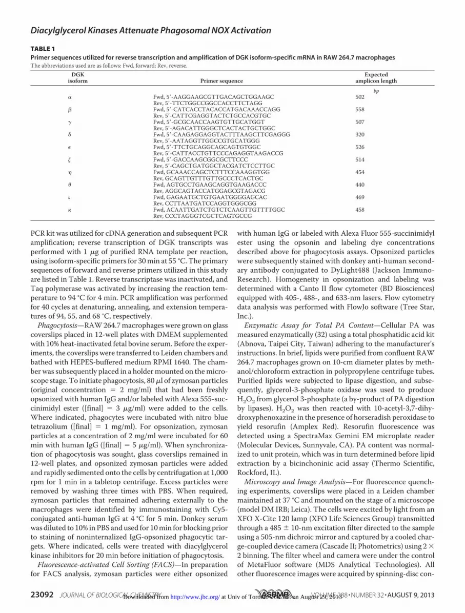
PCR kit was utilized for cDNA generation and subsequent PCRamplification; reverse transcription of DGK transcripts wasperformed with 1 �g of purified RNA template per reaction,using isoform-specific primers for 30min at 55 °C. The primarysequences of forward and reverse primers utilized in this studyare listed in Table 1. Reverse transcriptase was inactivated, andTaq polymerase was activated by increasing the reaction tem-perature to 94 °C for 4 min. PCR amplification was performedfor 40 cycles at denaturing, annealing, and extension tempera-tures of 94, 55, and 68 °C, respectively.Phagocytosis—RAW264.7macrophageswere grown on glass
coverslips placed in 12-well plates with DMEM supplementedwith 10% heat-inactivated fetal bovine serum. Before the exper-iments, the coverslips were transferred to Leiden chambers andbathed with HEPES-buffered medium RPMI 1640. The cham-ber was subsequently placed in a holdermounted on themicro-scope stage. To initiate phagocytosis, 80�l of zymosan particles(original concentration � 2 mg/ml) that had been freshlyopsonized with human IgG and/or labeled with Alexa 555-suc-cinimidyl ester ([final] � 3 �g/ml) were added to the cells.Where indicated, phagocytes were incubated with nitro bluetetrazolium ([final] � 1 mg/ml). For opsonization, zymosanparticles at a concentration of 2 mg/ml were incubated for 60min with human IgG ([final] � 5 �g/ml). When synchroniza-tion of phagocytosis was sought, glass coverslips remained in12-well plates, and opsonized zymosan particles were addedand rapidly sedimented onto the cells by centrifugation at 1,000rpm for 1 min in a tabletop centrifuge. Excess particles wereremoved by washing three times with PBS. When required,zymosan particles that remained adhering externally to themacrophages were identified by immunostaining with Cy5-conjugated anti-human IgG at 4 °C for 5 min. Donkey serumwas diluted to 10% inPBS andused for 10min for blocking priorto staining of noninternalized IgG-opsonized phagocytic tar-gets. Where indicated, cells were treated with diacylglycerolkinase inhibitors for 20 min before initiation of phagocytosis.Fluorescence-activated Cell Sorting (FACS)—In preparation
for FACS analysis, zymosan particles were either opsonized
with human IgG or labeled with Alexa Fluor 555-succinimidylester using the opsonin and labeling dye concentrationsdescribed above for phagocytosis assays. Opsonized particleswere subsequently stained with donkey anti-human second-ary antibody conjugated to DyLight488 (Jackson Immuno-Research). Homogeneity in opsonization and labeling wasdetermined with a Canto II flow cytometer (BD Biosciences)equipped with 405-, 488-, and 633-nm lasers. Flow cytometrydata analysis was performed with FlowJo software (Tree Star,Inc.).Enzymatic Assay for Total PA Content—Cellular PA was
measured enzymatically (32) using a total phosphatidic acid kit(Abnova, Taipei City, Taiwan) adhering to the manufacturer’sinstructions. In brief, lipids were purified from confluent RAW264.7 macrophages grown on 10-cm diameter plates by meth-anol/chloroform extraction in polypropylene centrifuge tubes.Purified lipids were subjected to lipase digestion, and subse-quently, glycerol-3-phosphate oxidase was used to produceH2O2 from glycerol 3-phosphate (a by-product of PA digestionby lipases). H2O2 was then reacted with 10-acetyl-3,7-dihy-droxyphenoxazine in the presence of horseradish peroxidase toyield resorufin (Amplex Red). Resorufin fluorescence wasdetected using a SpectraMax Gemini EM microplate reader(Molecular Devices, Sunnyvale, CA). PA content was normal-ized to unit protein, which was in turn determined before lipidextraction by a bicinchoninic acid assay (Thermo Scientific,Rockford, IL).Microscopy and Image Analysis—For fluorescence quench-
ing experiments, coverslips were placed in a Leiden chambermaintained at 37 °C and mounted on the stage of a microscope(model DM IRB; Leica). The cells were excited by light from anXFO X-Cite 120 lamp (XFO Life Sciences Group) transmittedthrough a 485 � 10-nm excitation filter directed to the sampleusing a 505-nm dichroic mirror and captured by a cooled char-ge-coupled device camera (Cascade II; Photometrics) using 2�2 binning. The filter wheel and camera were under the controlof MetaFluor software (MDS Analytical Technologies). Allother fluorescence images were acquired by spinning-disc con-
TABLE 1Primer sequences utilized for reverse transcription and amplification of DGK isoform-specific mRNA in RAW 264.7 macrophagesThe abbreviations used are as follows: Fwd, forward; Rev, reverse.
DGKisoform Primer sequence
Expectedamplicon length
bp� Fwd, 5�-AAGGAAGCGTTGACAGCTGGAAGC 502
Rev, 5�-TTCTGGCCGGCCACCTTCTAGG� Fwd, 5�-CATCACCTACACCATGACAAACCAGG 558
Rev, 5�-CATTCGAGGTACTCTGCCACGTGC� Fwd, 5�-GCGCAACCAAGTGTTGCATGGT 507
Rev, 5�-AGACATTGGGCTCACTACTGCTGGC� Fwd, 5�-CAAGAGGAGGTACTTTAAGCTTCGAGGG 320
Rev, 5�-AATAGGTTGGCCGTGCATGGG� Fwd, 5�-TTCTGCAGGCAGCAGTGTGGC 526
Rev, 5�-CATTACCTGTTCCCAGAGGTAAGACCG� Fwd, 5�-GACCAAGCGGCGCTTCCC 514
Rev, 5�-CAGCTGATGGCTACGATCTCCTTGC Fwd, GCAAACCAGCTCTTTCCAAAGGTGG 454
Rev, GCAGTTGTTTGTTGCCCTCACTGC� Fwd, AGTGCCTGAAGCAGGTGAAGACCC 440
Rev, AGGCAGTACCATGGAGCGTAGACG� Fwd, GAGAATGCTGTGAATGGGGAGCAC 469
Rev, CCTTAATGATCCAGGTGGGCGG Fwd, ACAATTGATCTGTCTCAAGTTGTTTTGGC 458
Rev, CCCTAGGGTCGCTCAGTGCCG
Diacylglycerol Kinases Attenuate Phagosomal NOX Activation
23092 JOURNAL OF BIOLOGICAL CHEMISTRY VOLUME 288 • NUMBER 32 • AUGUST 9, 2013 at Univ of Toronto - OCUL on August 29, 2013http://www.jbc.org/Downloaded from

focal microscopy. The systems in use in our laboratory (Quo-rum) are based on a microscope (Axiovert 200 M; Carl Zeiss,Inc.) with �63 or �100 objectives and a �1.5 magnifying lens.The units are equipped with diode-pumped solid-state lasers(440, 491, 561, 638, and 655 nm; Spectral Applied Research), amotorizedXY stage (Applied Scientific Instrumentation), and apiezo focus drive. Images were acquired using back-thinned,electron-multiplied, or conventional cooled charge-coupleddevice cameras (Hamamatsu Photonics) driven by the Volocitysoftware (version 4.1.1; PerkinElmer Life Sciences).Statistical Analysis—Statistical parameters were deter-
mined using GraphPad Prism 5c software (GraphPad Soft-ware, Inc.). To assess significance of differences, all compar-isons between means of matched groups were two-tailedunpaired t tests relying on the analysis routines built inGraphPad Prism.
RESULTS
Identification and Quantification of Superoxide Productionwithin Single Phagosomes through NBT-mediated FluorescenceQuenching—We devised a fluorescence quenching assay (illus-trated diagrammatically in Fig. 1A) tomonitorNADPHoxidaseactivity continuouslywithin single phagosomes. To this end,wetook advantage of the previously described deposition ofdiformazan generated fromNBT at sites of superoxide produc-tion (16).Directmeasurement by bright-fieldmicroscopy of thechanges in light absorbance caused by diformazan deposition iscomplicated by the absorbance and scattering contributed bythe cells and by the phagocytic targets, which vary as the parti-cles are engulfed. To circumvent these problems, zymosan par-
ticles were labeled with Alexa 555-succinimidyl ester. Theresulting labeled particles (referred to hereafter as Zymo-Alexa555) were opsonized with human IgG and used as phagocyticprey. Phagocytosis was performed in the presence of NBT,which is membrane-permeable and capable of traversing boththe plasma and phagosomal membranes, effectively reachingthe phagosomal lumen. Upon activation of NOX, intraphago-somal deposition of the black-purple diformazan effectivelyquenches the fluorescence emitted by labeled phagocytic tar-gets, interfering with the passage of both excitation and emis-sion of light (Fig. 1, A and B). The amount of remaining fluo-rescence is therefore inversely proportional to the cumulativeactivity of the NAPDH oxidase. This is illustrated in Fig. 1C;although the fluorescence of internalized particles decays rap-idly, the fluorescence of particles that remain extracellular islargely unaffected, showing only a minimal decrease attributa-ble to photobleaching. Accordingly, little fluorescence quench-ing was observed when labeled particles underwent phagocyto-sis in the absence of NBT (Fig. 1B). It is noteworthy that theAlexa Fluor 555 is stable, and its fluorescence is unaffectedbetween pH 4 and 10 (17), enabling measuring superoxide pro-duction independently of phagosomal acidification. Together,these observations indicate that the quenching assay is a suita-ble continuous measure of the production of superoxide byindividual phagosomes. This notion was validated using 10 �M
diphenyleneiodonium, a potent inhibitor of the NADPH oxi-dase that precluded fluorescence quenching (Fig. 2, A and B).NADPH Oxidase Activation Is Heterogeneous—By imple-
menting the assay described above, we detected considerable
e) 0 5 10 15 200.0
0.2
0.4
0.6
0.8
1.0ExternalInternal
(A.U
.)
Time (min)
Nor
mal
ized
fluo
resc
ence
inte
nsity
DIC IgG-Zymo-Alexa 555C
ontr
ol+
NB
T
A
(i) (ii)
Labelling
(iii)
Opsonization Phagocytosis
Diformazan precipitate
Fluorescence quenching
(iv)
Alexa 555-SE IgG NBT
CB
FIGURE 1. Detection of superoxide production within individual phagosomes by NBT-mediated fluorescence quenching. A, schematic of the experi-mental protocol. Zymosan particles labeled with Alexa 555-SE (i) and opsonized with human IgG (ii) were used as phagocytic targets for RAW macrophages inthe presence of the membrane permeable dye NBT (iii). Uptake of opsonized, labeled zymosan (IgG-Zymo-Alexa 555) and the ensuing activation of NOX2results in the generation of superoxide anions capable of reducing NBT to diformazan crystals in the phagosomal lumen. Diformazan precipitates in the formof blue/black deposits (iv) and effectively quenches the fluorescence signal emitted by the fluorescently labeled zymosan particles. B, representative differ-ential interference contrast (DIC, left panels) and corresponding epifluorescence (right panels) images of RAW macrophages fixed 25 min after exposure toIgG-opsonized zymo-Alexa 555. Where indicated, cells were incubated with NBT prior to and throughout phagocytosis. Double arrowheads point to noninter-nalized particles, and diformazan-negative and -positive phagosomes are indicated by dashed or solid arrows, respectively. Scale bar, 5 �m. C, quantification offluorescence intensity as a function of time during phagocytosis. Open circles and closed diamonds correspond to external and internalized IgG-Zymo-Alexa555, respectively. Images in B and the results in C are representative of at least six and two individual experiments of each kind, respectively. ***, p �0.005.
Diacylglycerol Kinases Attenuate Phagosomal NOX Activation
AUGUST 9, 2013 • VOLUME 288 • NUMBER 32 JOURNAL OF BIOLOGICAL CHEMISTRY 23093 at Univ of Toronto - OCUL on August 29, 2013http://www.jbc.org/Downloaded from
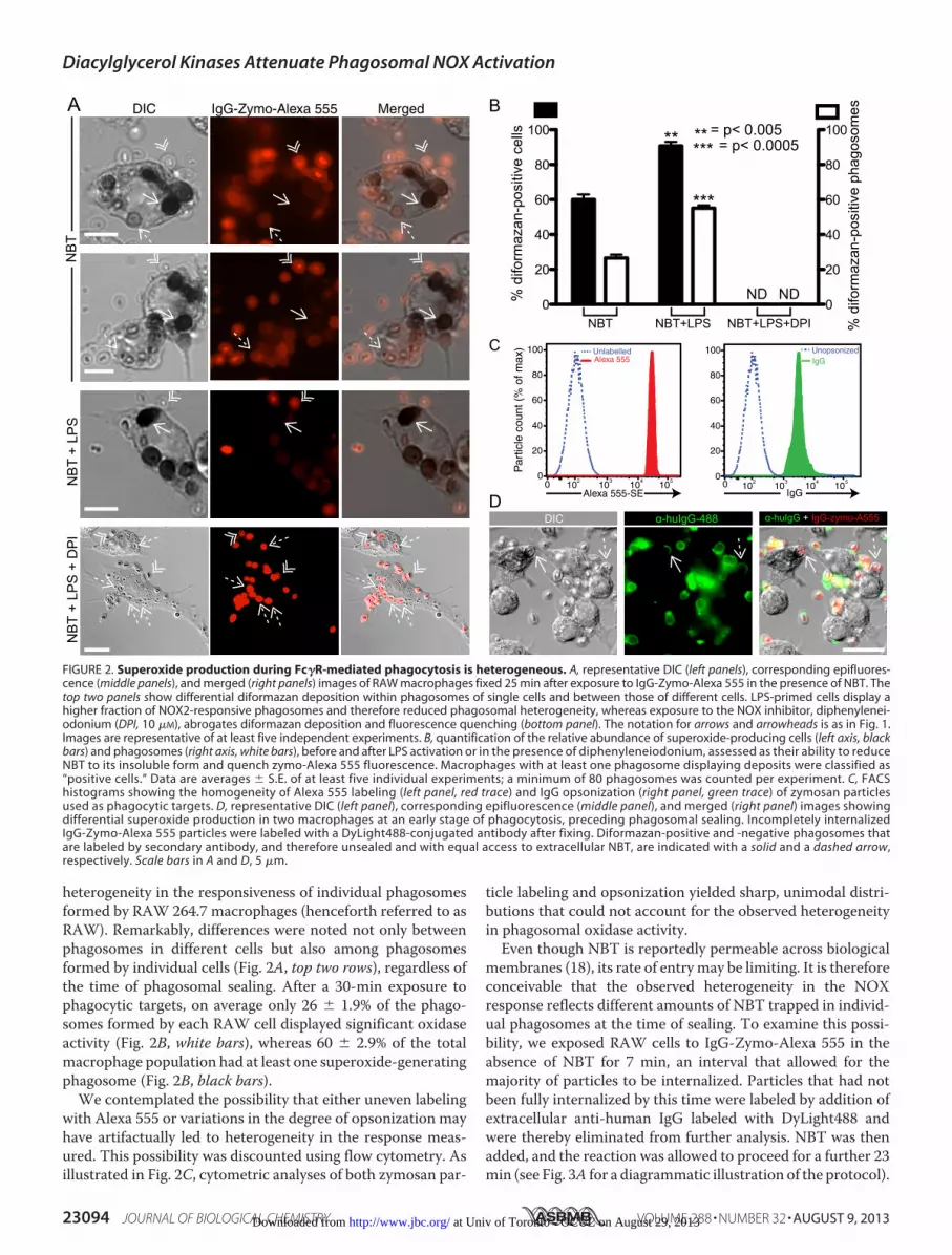
heterogeneity in the responsiveness of individual phagosomesformed by RAW 264.7 macrophages (henceforth referred to asRAW). Remarkably, differences were noted not only betweenphagosomes in different cells but also among phagosomesformed by individual cells (Fig. 2A, top two rows), regardless ofthe time of phagosomal sealing. After a 30-min exposure tophagocytic targets, on average only 26 � 1.9% of the phago-somes formed by each RAW cell displayed significant oxidaseactivity (Fig. 2B, white bars), whereas 60 � 2.9% of the totalmacrophage population had at least one superoxide-generatingphagosome (Fig. 2B, black bars).
We contemplated the possibility that either uneven labelingwith Alexa 555 or variations in the degree of opsonization mayhave artifactually led to heterogeneity in the response meas-ured. This possibility was discounted using flow cytometry. Asillustrated in Fig. 2C, cytometric analyses of both zymosan par-
ticle labeling and opsonization yielded sharp, unimodal distri-butions that could not account for the observed heterogeneityin phagosomal oxidase activity.Even though NBT is reportedly permeable across biological
membranes (18), its rate of entrymay be limiting. It is thereforeconceivable that the observed heterogeneity in the NOXresponse reflects different amounts of NBT trapped in individ-ual phagosomes at the time of sealing. To examine this possi-bility, we exposed RAW cells to IgG-Zymo-Alexa 555 in theabsence of NBT for 7 min, an interval that allowed for themajority of particles to be internalized. Particles that had notbeen fully internalized by this time were labeled by addition ofextracellular anti-human IgG labeled with DyLight488 andwere thereby eliminated from further analysis. NBT was thenadded, and the reaction was allowed to proceed for a further 23min (see Fig. 3A for a diagrammatic illustration of the protocol).
A DIC IgG-Zymo-Alexa 555 Merged
NB
TN
BT
+ LP
SN
BT
+ LP
S +
DP
I
0 102
103
104
105
0
20
40
60
80
100
Alexa 555-SE IgG
Alexa 555Unlabelled
Par
ticle
cou
nt (
% o
f max
)C
0
20
40
60
80
100
0
20
40
60
80
100
ND ND% d
iform
azan
-pos
itive
cel
ls
% d
iform
azan
-pos
itive
pha
goso
mesB
***
** = p< 0.005** *** = p< 0.0005
NBT NBT+LPS NBT+LPS+DPI
DDIC α-huIgG-488 α-huIgG + IgG-zymo-A555
20
40
60
80
100IgGUnopsonized
0 102
103
104
105
0
FIGURE 2. Superoxide production during Fc�R-mediated phagocytosis is heterogeneous. A, representative DIC (left panels), corresponding epifluores-cence (middle panels), and merged (right panels) images of RAW macrophages fixed 25 min after exposure to IgG-Zymo-Alexa 555 in the presence of NBT. Thetop two panels show differential diformazan deposition within phagosomes of single cells and between those of different cells. LPS-primed cells display ahigher fraction of NOX2-responsive phagosomes and therefore reduced phagosomal heterogeneity, whereas exposure to the NOX inhibitor, diphenylenei-odonium (DPI, 10 �M), abrogates diformazan deposition and fluorescence quenching (bottom panel). The notation for arrows and arrowheads is as in Fig. 1.Images are representative of at least five independent experiments. B, quantification of the relative abundance of superoxide-producing cells (left axis, blackbars) and phagosomes (right axis, white bars), before and after LPS activation or in the presence of diphenyleneiodonium, assessed as their ability to reduceNBT to its insoluble form and quench zymo-Alexa 555 fluorescence. Macrophages with at least one phagosome displaying deposits were classified as“positive cells.” Data are averages � S.E. of at least five individual experiments; a minimum of 80 phagosomes was counted per experiment. C, FACShistograms showing the homogeneity of Alexa 555 labeling (left panel, red trace) and IgG opsonization (right panel, green trace) of zymosan particlesused as phagocytic targets. D, representative DIC (left panel), corresponding epifluorescence (middle panel), and merged (right panel) images showingdifferential superoxide production in two macrophages at an early stage of phagocytosis, preceding phagosomal sealing. Incompletely internalizedIgG-Zymo-Alexa 555 particles were labeled with a DyLight488-conjugated antibody after fixing. Diformazan-positive and -negative phagosomes thatare labeled by secondary antibody, and therefore unsealed and with equal access to extracellular NBT, are indicated with a solid and a dashed arrow,respectively. Scale bars in A and D, 5 �m.
Diacylglycerol Kinases Attenuate Phagosomal NOX Activation
23094 JOURNAL OF BIOLOGICAL CHEMISTRY VOLUME 288 • NUMBER 32 • AUGUST 9, 2013 at Univ of Toronto - OCUL on August 29, 2013http://www.jbc.org/Downloaded from

Distinct heterogeneity in NOX activity was still observedamong the phagosomes that had sealed prior to the addition ofNBT (Fig. 3B). These data validate the notion that NBT can
readily cross biological membranes and imply that differentialtrapping of NBT during sealing cannot explain the variability ofthe responses.
A
B
0 min
D
IgG-Zymo-Alexa 555 α-huIgG-488 DIC merge
*
*
**
*
*
**
*
*
**
*
***
Fluorescence quenching
(ii)
(imaged at 30 min)Phagocytosis IgG-Zymo-Alexa 555
[0-7] min
(i)
added at 7 minNBT+ -huIgG-488
0 min
*
IgG-Zymo-Alexa 555 α-huIgG-488 DIC merge
* **
Phagocytosis IgG-Zymo-Alexa 555 + NBT[0-3] min
(i)
C
(ii)
added at 3 minα-huIgG-488 Fluorescence quenching
(imaged at 30 min)
*
*
FIGURE 3. Neither differential accumulation of NBT in individual phagosomes nor time-dependence of NOX can account for the observed phagosomalheterogeneity. A, schematic of an experimental protocol designed to determine whether accessibility of NBT, the quenching agent, to phagosomal com-partments is variable. RAW macrophages were challenged at 0 min with human IgG-opsonized zymosan particles labeled with Alexa 555-SE (IgG-Zymo-Alexa555), and phagocytosis was allowed to continue for 7 min, at which point NBT was included in the reaction. A secondary 488 anti-human IgG was also addedat this 7 min to mark phagocytic targets that had not been internalized at the time of first exposure of macrophages to NBT. Phagocytosis proceeded for anadditional 23 min after addition of NBT, and cells were fixed at 30 min for imaging. B, representative epifluorescence (left and middle panels) and DIC (right panel)images of the experiment described in A. Phagocytic targets that fluoresce both in the red and green channels (indicated with an asterisk) had not yet beeninternalized at the time of NBT addition, whereas targets that emit only in the red channel had been engulfed prior to NBT addition. The right panel showsdiformazan-positive targets (indicated with a solid arrow) and diformazan-negative targets (indicated with an arrowhead) that resided within sealed phago-somes in the same cell when the quenching dye was included in the reaction and thus had comparable access to NBT. C, schematic of an experimental protocoldesigned to determine whether time dependence of NOX could be responsible for phagosomal heterogeneity. Both NBT and IgG-Zymo-Alexa 555 were addedto macrophages at 0 min, and phagocytosis was allowed to proceed for only 3 min before addition of a secondary 488 anti-human IgG to mark the 3-min timepoint. Phagocytosis then continued for an additional 27 min, and cells were image-fixed at 30 min. D, representative epifluorescence (left and middle panels)and DIC (right panel) images of the experiment are described in C. Phagocytic targets that fluoresce in the red, but not in the green, were internalized within thefirst 3-min window (out of a total 30 min). The right panel shows diformazan-positive and -negative phagocytic phagosomes that were internalized within said3-min window and therefore had equivalent time intervals for phagosomal maturation and NOX activation. The notation for asterisks, arrows, and arrowheadsis as in A. Scale bars, 10 �m.
Diacylglycerol Kinases Attenuate Phagosomal NOX Activation
AUGUST 9, 2013 • VOLUME 288 • NUMBER 32 JOURNAL OF BIOLOGICAL CHEMISTRY 23095 at Univ of Toronto - OCUL on August 29, 2013http://www.jbc.org/Downloaded from

It is also worth noting that we routinely encountered nascentphagosomes that had not yet sealed. Some but not all of thesedisplayed diformazan precipitates at the base of the formingcup (e.g. Fig. 2D). Because at this stage access to NBT is unhin-dered, the variable response cannot be attributed to insufficientexposure to NBT; it also rules out differential permeability ofthe phagosomal membrane to NBT as the source of the hetero-geneity illustrated in Fig. 3B.Finally, we considered the distinct possibility that the hetero-
geneity in superoxide production was a consequence of asyn-chrony of the phagocytic process and/or of the activation of theoxidase. To address this, RAWcells were allowed to ingest IgG-Zymo-Alexa 555 in the presence of NBT for 30 min, as in Figs.1 and 2. However, in these experiments particles that had notbeen internalized after the first 3 min were marked by additionof a fluorescently labeled secondary antibody (see Fig. 3C for adiagrammatic illustration of the protocol). By excluding thosetargets that were ingested after addition of the antibody, wewere able to analyze the response of only those phagosomesformedwithin a narrow timewindow, i.e. the initial 3min; thesewere then allowed to mount an oxidative response for an addi-tional 27 min. The integrated cumulative response over thiscomparatively long period should have minimized the conse-quences of the slight phagocytic asynchrony incurred duringthe initial 3 min. Notably, considerable heterogeneity was nev-ertheless recorded under these conditions (Fig. 3D). Jointly,these experiments imply that the variable responsiveness of thephagosomal oxidase is neither an artifact caused by unevenexposure to NBT nor a reflection of asynchrony. Instead, theoxidative response of phagosomes appears to be intrinsicallyheterogeneous.It is widely accepted that bacterial endotoxin can robustly
prime phagocytes for superoxide production (19), although theunderlying mechanism is still incompletely understood (20).We sought to determine whether this reflects increased pro-duction of superoxide by a constant number of phagosomes oran increased fraction of responding phagosomes. Overnightincubation of RAW macrophages with the toll-like receptor-4(TLR-4) agonist, lipopolysaccharide (LPS, 100 ng/ml), notice-ably increased the fraction of phagosomes that mounted aneffective respiratory burst from 26� 1.9 to 55� 1.6%, as well asthe number of cells with responsive phagosomes from 60 � 2.9to 91 � 2.3% (Fig. 2, A and B). Thus, priming reduced the het-erogeneity of the respiratory response.Assessment of the Roles of PtdIns(3,4,5)P3, Phosphatidic Acid,
and PtdIns(3)P in the Genesis of the Functional Heterogeneity—Several lipid intermediates are required for the initiation andprogression of phagocytosis, including the deployment of theNADPH oxidase. Engulfment of large particles and completionof maturation require activation of class I phosphatidylinositol3-kinases (21). Because different degrees of receptor engage-ment can influence the extent of PtdIns(3,4,5)P3 generation(22), we analyzed whether differential production of this phos-phoinositide accounted for the variable respiratory burstresponse. We used a genetically encoded biosensor, a chimericconstruct consisting of the pleckstrin homology domain ofAkt/PKB tagged with GFP, to monitor the formation ofPtdIns(3,4,5)P3 during the course of phagocytosis of IgG-op-
sonized zymosan. Spinning disc confocal microscopy was usedto monitor translocation of the biosensor to the phagosomalmembrane over time. Consistent with earlier findings (23), wecould readily detect recruitment of PH-Akt-GFP to nascent andrecently formed phagosomes, followed by rapid loss of theprobe after sealing (Fig. 4B, top). Of note, virtually 100% of thephagosomes induced by IgG-zymosan accumulated the probeto a comparable degree (Fig. 4C). Thus, failure to generatePtdIns(3,4,5)P3 cannot account for the inability of a consider-able fraction of phagosomes to activate the NADPH oxidase.A similar approachwas used to evaluate the role of PA. In this
instance, we used a probe composed of two tandem copies ofthe PA-binding domain of the yeast protein Spo20p, which hasbeen used in the past to monitor PA distribution (10, 11). ThePAprobe (hereafter referred to as 2PABD-GFP), was optimizedby adding a nuclear export signal N-terminally to the PABDdomains, as the probe tends to partition to the nuclear com-partment otherwise.6 To ensure that this modification did notinterfere with the ligand specificity of the probe, we monitoredthe distribution of 2PABD-GFP in HeLa cells before and afteraltering the PA content by pharmacological, biochemical, orenzymatic means. Unlike phagocytes (24), unstimulated HeLacells have little PA in their plasma membrane. Exposure toexogenous PA (100�M for 10min) induced rapid association of2PABD-GFP with the plasma membrane (supplemental video2), validating the responsiveness of the probe. Recruitment of2PABD-GFP to the membrane was also observed when HeLacells were treatedwith 1�M ionomycin for 2min (supplementalvideo 3); calcium entering through this ionophore is known toactivate phospholipase C, generating DAG, a precursor of PAgeneration by DGK. Moreover, recruitment of 2PABD-GFPwas also observed when HeLa cells were co-transfected withwild-type PLD2, a plasma membrane-associated enzyme thatconverts phosphatidylcholine to PA (data not shown). Of note,expression of neither an inactive form of PLD2 nor of PLD1(which is not found at the membrane and instead has a vesicu-lar, punctate distribution) caused recruitment of the PA probeto the membrane. Jointly, these results provide evidence thatthe 2PABD biosensor utilized in this study is a suitable probefor PA.In contrast to HeLa cells, a sizable amount of the PA probe
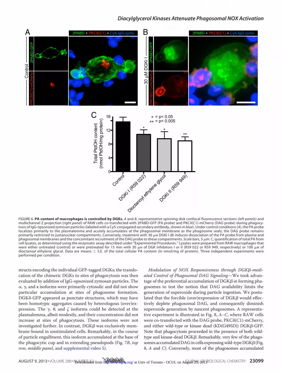
was found to associate with the plasma membrane of unstimu-lated RAW macrophages. The bound probe was rapidlyreleased to the cytosol when RAW cells were treated with theDGK inhibitor R59 022 (DGKi I, 30 �M), suggesting that PA iscontinuously synthesized by phosphorylation of DAG (supple-mental video 4). Accordingly, addition of DGKi I caused a con-comitant accumulation of DAG in themembrane of RAW cells(supplemental video 4) and a reduction in the cellular contentof PA measured biochemically (Fig. 6C). It is worth noting thatthis assay cannot distinguish lyso-PA from diacylated PA, butthe former is less abundant and is expected to contribute mar-ginally to the determinations. These observations further vali-date the reliability of the 2PABD biosensor. Despite being pres-ent at the membrane of unstimulated cells, 2PABD-GFP
6 F. Zhang and G. Du, manuscript in preparation.
Diacylglycerol Kinases Attenuate Phagosomal NOX Activation
23096 JOURNAL OF BIOLOGICAL CHEMISTRY VOLUME 288 • NUMBER 32 • AUGUST 9, 2013 at Univ of Toronto - OCUL on August 29, 2013http://www.jbc.org/Downloaded from

accumulated further at the phagocytic cup (Fig. 4B, middle).The accumulation dissipated rapidly after phagosome closure,with PA being only marginally detectable in the phagosomalmembrane 3min after sealing. As in the case of PtdIns(3,4,5)P3,essentially all phagosomes analyzed accumulated considerableamounts of PA, judged by the recruitment of the Spo20p probe(Fig. 4C). Therefore, the inability of a large fraction of phago-somes to form superoxide cannot be attributed to lack of PAaccumulation.PtdIns(3)P, a product of the class III phosphatidylinositol
3-kinase, was recently shown to be required for phagosomalsuperoxide generation, likely by recruitment of the p40phox sub-unit of the oxidase via its PX domain (25). A probe based on theFYVE domain of EEA1 was used to quantify the fraction ofphagosomes that accumulate PtdIns(3)P. As described before(2, 26), PtdIns(3)P was not detectable at the phagosomal cupand was only observable �1 min after sealing, persisting fornearly 10 min at the phagosomal membrane. Two lines of evi-dence suggest that PtdIns(3)P is not responsible for the heter-ogeneous respiratory burst. First, as for the two other lipids,PtdIns(3)P was found in every phagosome studied (Fig. 4, B andC). Second, PtdIns(3)P is seen 2–5 min post-phagosome seal-ing, at which time heterogeneity is already observed.Moreover,as illustrated in Fig. 4A and supplemental video 1, diformazandeposits initially form at the base of the cup, preceding phago-some closure (see also Fig. 2D). Even at this stage, heterogeneityis readily apparent. This excludes PtdIns(3)P, which formsonly later and is likely important for the maintenance but notthe initiation of the response, as the source of the variable res-piratory burst. Jointly, these results suggest that neitherPtdIns(3,4,5)P3, PA, nor PtdIns(3)P are responsible for the het-erogeneous activation of the NADPH oxidase during IgG-me-diated phagocytosis.Diacylglycerol Heterogeneity Correlates with NADPH Oxi-
dase Activity—DAG stimulates conventional and novel PKCisoforms (27, 28) that phosphorylate and activate the cytosoliccomponents of the NOX complex (29–31). We thereforehypothesized that variations in the phagosomal content ofDAG could account for the observed fluctuations in superoxideproduction. To test this hypothesis, we monitored DAGdynamics using a genetically encoded biosensor consisting ofthe C1 domain of PKC� fused to GFP. Remarkably, this probewas recruited to some but not all the phagosomes (Fig. 5A);heterogeneity was observed not only across cells, but alsobetween phagosomes formed by individual cells. In otherwiseuntreated macrophages, only 26 � 2.5% of the phagosomesinduced by IgG-coated zymosan were visibly endowed withDAG. In contrast, overnight activation of macrophages withLPS (100 ng/ml) increased the fraction of DAG-positive phago-somes to 60 � 2.6% (Fig. 5B). Of note, the fraction of phago-somes displayingmeasurable NADPHoxidase activity (Fig. 5C)
Akt
(PH
)-G
FP
[Ptd
Ins(
3,4,
5)P
3]
(2F
YV
E)-
GF
PP
tdIn
s(3)
P]
-30 sec [0-30] sec 150 sec
NB
TA
B
C
***
**
**
*
*
-30 sec 25 min [0-30] sec
Diform
azan
PtdIn
s(3,
4,5)
P3PA
PtdIn
s(3)
P0
20
40
60
80
100
% p
osi
tive
ph
ag
oso
me
s
***
***
(2P
AB
D)-
GF
P(P
A)
*
**
*
FIGURE 4. Phagosomal population is uniformly endowed withPtdIns(3,4,5)P3, PA, and PtdIns(3)P during the respiratory burst. A, repre-sentative DIC images of live RAW macrophages during phagocytosis of IgG-opsonized zymosan in the presence of NBT. The phagocytosis time course isdelineated to three stages as follows: pseudopod extension (minus 30 s, leftpanel), phagosome sealing (0 –30 s, middle panel), and phagosome matura-tion (plus 25 min, right panel). B, spinning disk confocal fluorescence imagesof RAW macrophages challenged with IgG-opsonized zymosan particleslabeled with Cy5-conjugated secondary antibody (blue). Cells were trans-fected with constructs encoding fluorescently tagged domains that served asbiosensors for PtdIns(3,4,5)P3 (Akt(PH)-GFP, top panel), PA (2PABD-GFP, mid-dle panel), and PtdIns(3)P (2FYVE-GFP, bottom panel). Recruitment of theprobes to the phagosomal membrane was monitored during pseudopodextension (minus 30 s), phagosome sealing (0 –30 s), and early stages of mat-uration (plus 150 s). Asterisks indicate phagosomes. Note that the �30-simage for PA was underexposed compared with the other times, to avoidsaturation of the fluorescence accumulated at the cup. Scale bars in A and B, 5�m. C, quantification of the fraction of phagosomes displaying formazan
deposits and accumulation of PtdIns(3,4,5)P3, PA, or PtdIns(3)P. Data indicatethe average fraction of positive phagosomes per cell for each condition at thefollowing time points: diformazan (20 min after exposure to zymosan);PtdIns(3,4,5)P3 (0 –30 s after phagosome sealing); PA (30 s before phagosomesealing); PtdIns(3)P (150 s after sealing). Data are representative of at least fiveindependent experiments; a minimum of 100 phagosomes was counted perexperiment.
Diacylglycerol Kinases Attenuate Phagosomal NOX Activation
AUGUST 9, 2013 • VOLUME 288 • NUMBER 32 JOURNAL OF BIOLOGICAL CHEMISTRY 23097 at Univ of Toronto - OCUL on August 29, 2013http://www.jbc.org/Downloaded from
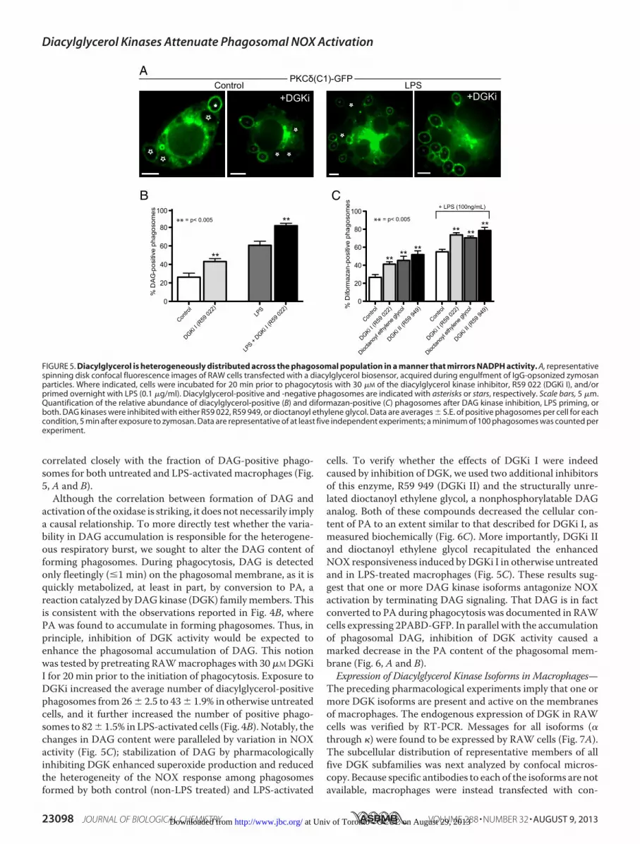
correlated closely with the fraction of DAG-positive phago-somes for both untreated and LPS-activated macrophages (Fig.5, A and B).Although the correlation between formation of DAG and
activation of the oxidase is striking, it does not necessarily implya causal relationship. To more directly test whether the varia-bility in DAG accumulation is responsible for the heterogene-ous respiratory burst, we sought to alter the DAG content offorming phagosomes. During phagocytosis, DAG is detectedonly fleetingly ( 1 min) on the phagosomal membrane, as it isquickly metabolized, at least in part, by conversion to PA, areaction catalyzed byDAGkinase (DGK) familymembers. Thisis consistent with the observations reported in Fig. 4B, wherePA was found to accumulate in forming phagosomes. Thus, inprinciple, inhibition of DGK activity would be expected toenhance the phagosomal accumulation of DAG. This notionwas tested by pretreating RAWmacrophages with 30 �MDGKiI for 20 min prior to the initiation of phagocytosis. Exposure toDGKi increased the average number of diacylglycerol-positivephagosomes from 26� 2.5 to 43� 1.9% in otherwise untreatedcells, and it further increased the number of positive phago-somes to 82� 1.5% in LPS-activated cells (Fig. 4B). Notably, thechanges in DAG content were paralleled by variation in NOXactivity (Fig. 5C); stabilization of DAG by pharmacologicallyinhibiting DGK enhanced superoxide production and reducedthe heterogeneity of the NOX response among phagosomesformed by both control (non-LPS treated) and LPS-activated
cells. To verify whether the effects of DGKi I were indeedcaused by inhibition of DGK, we used two additional inhibitorsof this enzyme, R59 949 (DGKi II) and the structurally unre-lated dioctanoyl ethylene glycol, a nonphosphorylatable DAGanalog. Both of these compounds decreased the cellular con-tent of PA to an extent similar to that described for DGKi I, asmeasured biochemically (Fig. 6C). More importantly, DGKi IIand dioctanoyl ethylene glycol recapitulated the enhancedNOX responsiveness induced byDGKi I in otherwise untreatedand in LPS-treated macrophages (Fig. 5C). These results sug-gest that one or more DAG kinase isoforms antagonize NOXactivation by terminating DAG signaling. That DAG is in factconverted to PA during phagocytosis was documented in RAWcells expressing 2PABD-GFP. In parallel with the accumulationof phagosomal DAG, inhibition of DGK activity caused amarked decrease in the PA content of the phagosomal mem-brane (Fig. 6, A and B).Expression of Diacylglycerol Kinase Isoforms in Macrophages—
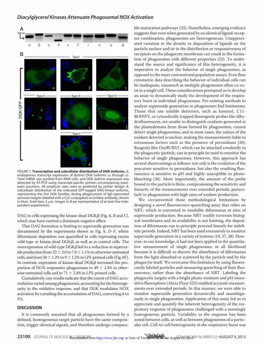
The preceding pharmacological experiments imply that one ormore DGK isoforms are present and active on the membranesof macrophages. The endogenous expression of DGK in RAWcells was verified by RT-PCR. Messages for all isoforms (�through ) were found to be expressed by RAW cells (Fig. 7A).The subcellular distribution of representative members of allfive DGK subfamilies was next analyzed by confocal micros-copy. Because specific antibodies to each of the isoforms are notavailable, macrophages were instead transfected with con-
B
Contro
l
DGKi I (R
59 02
2)
Dioctan
oyl e
thylen
e glyc
ol
DGKi II (R
59 94
9)0
20
40
60
80
100
% D
iform
azan
-pos
itive
pha
goso
mes
** ** **
** ****
Contro
l
DGKi I (R
59 02
2)LP
S
LPS +
DGKi I (R
59 02
2)0
20
40
60
80
100
% D
AG
-pos
itive
pha
goso
mes
**
**
C
PKCδ(C1)-GFPA
ControI LPS
****
**
*
*
**
***
*
*
*
+DGKi
**
*
+DGKi
** = p< 0.005** = p< 0.005
Contro
l
DGKi I (R
59 02
2)
Dioctan
oyl e
thylen
e glyc
ol
DGKi II (R
59 94
9)
+ LPS (100ng/mL)
FIGURE 5. Diacylglycerol is heterogeneously distributed across the phagosomal population in a manner that mirrors NADPH activity. A, representativespinning disk confocal fluorescence images of RAW cells transfected with a diacylglycerol biosensor, acquired during engulfment of IgG-opsonized zymosanparticles. Where indicated, cells were incubated for 20 min prior to phagocytosis with 30 �M of the diacylglycerol kinase inhibitor, R59 022 (DGKi I), and/orprimed overnight with LPS (0.1 �g/ml). Diacylglycerol-positive and -negative phagosomes are indicated with asterisks or stars, respectively. Scale bars, 5 �m.Quantification of the relative abundance of diacylglycerol-positive (B) and diformazan-positive (C) phagosomes after DAG kinase inhibition, LPS priming, orboth. DAG kinases were inhibited with either R59 022, R59 949, or dioctanoyl ethylene glycol. Data are averages � S.E. of positive phagosomes per cell for eachcondition, 5 min after exposure to zymosan. Data are representative of at least five independent experiments; a minimum of 100 phagosomes was counted perexperiment.
Diacylglycerol Kinases Attenuate Phagosomal NOX Activation
23098 JOURNAL OF BIOLOGICAL CHEMISTRY VOLUME 288 • NUMBER 32 • AUGUST 9, 2013 at Univ of Toronto - OCUL on August 29, 2013http://www.jbc.org/Downloaded from
structs encoding the individual GFP-tagged DGKs; the translo-cation of the chimeric DGKs to sites of phagocytosis was thenevaluated by addition of IgG-opsonized zymosan particles. The�, �, and � isoforms were primarily cytosolic and did not showparticular accumulation at sites of phagosome formation.DGK�-GFP appeared as punctate structures, which may havebeen homotypic aggregates caused by heterologous (over)ex-pression. The �, �, and � isoforms could be detected at theplasmalemma, albeit modestly, and their concentration did notincrease at sites of phagocytosis. These isoforms were notinvestigated further. In contrast, DGK� was exclusively mem-brane-bound in unstimulated cells. Remarkably, in the courseof particle engulfment, this isoform accumulated at the base ofthe phagocytic cup and in extending pseudopods (Fig. 7B, toprow, middle panel, and supplemental video 5).
Modulation of NOX Responsiveness through DGK�-medi-ated Control of Phagosomal DAG Signaling—We took advan-tage of the preferential accumulation of DGK� in forming pha-gosomes to test the notion that DAG availability limits thegeneration of superoxide during particle ingestion. We postu-lated that the forcible (over)expression of DGK� would effec-tively deplete phagosomal DAG, and consequently diminishsuperoxide generation by nascent phagosomes. A representa-tive experiment is illustrated in Fig. 8, A–C, where RAW cellswere co-transfected with the DAG probe, PKC�(C1)-mCherry,and either wild-type or kinase dead (KD(G495D)) DGK�-GFP.Note that phagocytosis proceeded in the presence of both wild-type and kinase-dead DGK�. Remarkably, very few of the phago-somesaccumulatedDAGincells expressingwild-typeDGK� (Fig.8, A and C). Conversely, most of the phagosomes accumulated
2PA
BD
-GFP
PK
Cδ(
C1)
-mC
herr
yC
ontro
lA B
2PA
BD
-GFP
PK
Cδ(
C1)
-mC
herr
y30
µM
DG
Ki I
16 * = p< 0.05* = p< 0.05
Contro
l
DGKi I (R
59 02
2)
Dioc
tanoy
l ethy
lene g
lycol
DGKi II (R
59 94
9)0
4
8
12
Tota
l Ptd
OH
con
tent
(n
mol
Ptd
OH
/mg
prot
ein)
* ***
** = p< 0.005C
2PABD + PKCδ(C1) + Cy5-IgG-zymo 2PABD + PKCδ(C1) + Cy5-IgG-zymo
FIGURE 6. PA content of macrophages is controlled by DGKs. A and B, representative spinning disk confocal fluorescence sections (left panels) andmultichannel Z projection (right panel) of RAW cells co-transfected with 2PABD-GFP (PA probe) and PKC�(C1)-mCherry (DAG probe) during phagocy-tosis of IgG-opsonized zymosan particles (labeled with a Cy5-conjugated secondary antibody, shown in blue). Under control conditions (A), the PA probelocalizes primarily to the plasmalemma and acutely accumulates at the phagosomal membrane as the phagosome seals; the DAG probe remainsprimarily restricted to juxtanuclear compartments. Conversely, treatment with 30 �M DGKi I (B) induces dissociation of the PA probe from plasma andphagosomal membranes and the concomitant recruitment of the DAG probe to these compartments. Scale bars, 5 �m. C, quantification of total PA fromcell lysates, as determined using the enzymatic assay described under “Experimental Procedures.” Lysates were prepared from RAW macrophages thatwere either untreated (control) or were pretreated for 15 min with 30 �M of DGK inhibitors I or II (R59 022 or R59 949, respectively) or 100 �M ofdioctanoyl ethylene glycol. Data are means � S.E. of the total cellular PA content (in nmol/mg of protein). Three independent experiments wereperformed per condition.
Diacylglycerol Kinases Attenuate Phagosomal NOX Activation
AUGUST 9, 2013 • VOLUME 288 • NUMBER 32 JOURNAL OF BIOLOGICAL CHEMISTRY 23099 at Univ of Toronto - OCUL on August 29, 2013http://www.jbc.org/Downloaded from
DAG in cells expressing the kinase-dead DGK� (Fig. 8, B and C),whichmay have exerted a dominant-negative effect.That DAG formation is limiting to superoxide generation was
documented by the experiments shown in Fig. 8, D–F, wherediformazan deposition was quantified in cells expressing excesswild-type or kinase-dead DGK�, as well as in control cells. Theoverexpression of wild-type DGK� led to a reduction in superox-ide production from23� 2.0 to 7.0� 1.1% inotherwise untreatedcells, and from58�1.2% to9�1.2% inLPS-primedcells (Fig. 8F).In contrast, expression of kinase-dead DGK� increased the pro-portion of NOX-responsive phagosomes to 49 � 2.4% in other-wise untreated cells and to 71 � 2.4% in LPS-primed cells.
Cumulatively, our results indicate that the extent ofDAGaccu-mulation varied amongphagosomes, accounting for theheteroge-neity in the oxidative response, and that DGK modulates NOXactivation by curtailing the accumulation ofDAG, converting it toPA.
DISCUSSION
It is commonly assumed that all phagosomes formed by adefined, homogeneous target particle have the same composi-tion, trigger identical signals, and therefore undergo compara-
ble maturation pathways (33). Nonetheless, emerging evidencesuggests that evenwhen generated by an identical ligand-recep-tor combination, phagosomes are heterogeneous. Unappreci-ated variation in the density or disposition of ligands on theparticle surface and/or in the distribution or responsiveness ofreceptors on the phagocyte membrane can result in the forma-tion of phagosomes with different properties (22). To under-stand the source and significance of this heterogeneity, it isimperative to analyze the behavior of single phagosomes, asopposed to themore conventional population assays. Even flowcytometric data describing the behavior of individual cells canbe inadequate, inasmuch as multiple phagosomes often co-ex-ist in a single cell. These considerations prompted us to developan assay to dynamically study the development of the respira-tory burst in individual phagosomes. Pre-existing methods toanalyze superoxide generation in phagosomes had limitations.Those that use soluble detectors, such as luminol, C11-BODIPY, or cytosolically trapped fluorogenic probes like dihy-drofluorescein, are unable to distinguish oxidants generated atthe plasmalemma from those formed by phagosomes, cannotdetect single phagosomes, and in most cases, the nature of theoxidant detected is unclear, making themeasurements liable toextraneous factors such as the presence of peroxidases (20).Reagents like OxyBURST, which can be attached covalently tothe phagocytic particle, can in principle be used to monitor thebehavior of single phagosomes. However, this approach hasseveral shortcomings as follows: not only is the oxidation of theprecursor sensitive to peroxidases, but also the resulting fluo-rescence is sensitive to pH and highly susceptible to photo-bleaching (36). More importantly, the amount of the probebound to the particle is finite, compromising the sensitivity andlinearity of the measurements over extended periods, particu-larly in phagosomes with high rates of oxidant production.We circumvented these methodological limitations by
designing a novel fluorescence-quenching assay that relies onNBT, which is converted to insoluble diformazan at sites ofsuperoxide production. Because NBT readily traverses biolog-ical membranes and its availability is not limiting, the deposi-tion of diformazan can in principle proceed linearly for indefi-nite periods. Indeed, NBT has been used extensively tomonitorsuperoxide generation in a variety of systems (18, 37, 38). How-ever, to our knowledge, it had not been applied to the quantita-tive measurement of single phagosomes, in all likelihoodbecause it is difficult to discern the absorbance of diformazanfrom the light absorbed or scattered by the particle and by thephagocyte itself. We overcame this limitation by using fluores-cently labeled particles and measuring quenching of their fluo-rescence, rather than the absorbance of NBT. Labeling thephagocytic targets with a bright photo-resistant and pH-insen-sitive fluorophore (Alexa Fluor 555) enabled accuratemeasure-ments over extended periods. In this manner, we were able tomonitor superoxide generation dynamically and unambigu-ously in single phagosomes. Application of this assay led us toappreciate and quantify the inherent heterogeneity of the res-piratory response of phagosomes challenged with a seeminglyhomogeneous particle. Variability in the response has beennoted between cells, as well as between phagosomes of a partic-ular cell. Cell-to-cell heterogeneity in the respiratory burst was
β ζγ δα ε θηκι
β
ζ
γ
δ ε
α
DG
K-G
FP
500 bpA
θ
BType I
Type II Type III Type IV
Type V
FIGURE 7. Transcription and subcellular distribution of DGK isoforms. A,endogenous transcript expression of distinct DGK isoforms (� through ).Total mRNA was purified from RAW cells, and DGK isoform expression wasdetected by RT-PCR using transcript-specific primers encompassing exon-exon junctions. All amplicon sizes were as predicted by primer design. B,subcellular distribution of the indicated GFP-tagged DAG kinase isoforms,representing the five DGK families, during phagocytosis of IgG-opsonizedzymosan targets (labeled with a Cy5-conjugated secondary antibody, shownin blue). Scale bars, 5 �m. Images in B are representative of at least five inde-pendent experiments.
Diacylglycerol Kinases Attenuate Phagosomal NOX Activation
23100 JOURNAL OF BIOLOGICAL CHEMISTRY VOLUME 288 • NUMBER 32 • AUGUST 9, 2013 at Univ of Toronto - OCUL on August 29, 2013http://www.jbc.org/Downloaded from

described earlier (40), and phagosome-to-phagosome differ-ences were parenthetically reported for PLB-985 cells (41), butthe source of this variability had not been defined.In an effort to establish the source of the heterogeneity, we
turned our attention to key bioactive lipids that are critical formounting the respiratory burst. Phosphoinositides are of par-ticular importance to the activation of the oxidase. The PXdomain of p40phox selectively binds to PtdIns(3)P, and thisinteraction is most likely required for sustaining superoxideproduction in phagosomes (25, 42). In contrast, the PX domainof p47phox binds to PtdIns(3,4)P2 and PtdIns(3,4,5)P3 (31). Thisdomain is unusual in that in contains two distinct lipid-binding
pockets, allowing it to simultaneously and cooperatively bind toa phosphoinositide and a second small lipid such as PA (43).Thus, it has been postulated that by sensing multiple inputs,p47phox acts as a master integrator of PI3K and PLD signaling(29). We hypothesized that variations in the metabolism ofthese bioactive lipids could underlie the observed heterogeneityin NOX activity across the phagosomal population. However,although only a subpopulation of the phagosomes produceddetectable amounts of superoxide, virtually all of themrecruited comparable levels of the PtdIns(3,4,5)P3, PtdIns(3)P,and PA biosensors (Fig. 4). We cannot rule out that subtle dif-ferences in the levels of these mediators contribute to the het-
D
C
KD(G495D)DGKβB
**
*
*
*
**
*
PK
Cδ(
C1)
-mC
herr
y
**
*
*
*
**
*
A
DG
KβW
T-G
FPP
KC
δ(C
1)-m
Che
rry
wtDGKβ-GFP wtDGKβ-GFP
DG
KβW
T-G
FPD
IC
E
***
***
*
*
DIC
DG
Kβ(
G49
5D)-
GFP
DG
Kβ(
G49
5D)-
GFP
KD(G495D)DGKβ
F
Contro
l
wtDGK
KD(G49
5D)D
GK
LPS-ac
tivate
d
LPS +
wtDGK
LPS +
KD(G49
5D)D
GK
0
20
40
60
80
% D
iform
azan
-pos
itive
pha
goso
mes
** = p< 0.005
**
**
**
* = p< 0.01 *
Contro
l (PM-G
FP)
wtDGK
KD(G49
5D)D
GK
0
20
40
60
80
% D
AG-p
ositi
ve p
hago
som
es * = p< 0.01
*
** = p< 0.005**
DGKβWT + PKCδ(C1) + Cy5-IgG-zymo DGKβWT + IgG-zymo-Alexa 555
KD-DGKβ + PKCδ(C1) + Cy5-IgG-zymo KD-DGKβ + IgG-zymo-Alexa 555
FIGURE 8. Modulation of NOX activity through diacylglycerol kinase �-mediated control of phagosomal DAG. A and B, representative confocal sections(left panels) and multichannel Z projection (right panels) of RAW cells co-transfected with PKC�(C1)-mCherry (DAG probe) and either (A) wild-type (wt) or (B)kinase-dead (KD(G495D)) DGK�-GFP during phagocytosis of IgG-opsonized zymosan particles (labeled with a Cy5-conjugated secondary antibody, shown inblue). Asterisks indicate phagosomes. Scale bars, 10 �m. C, quantification of the portion of phagosomes endowed with DAG in RAW cells expressing eitherwild-type or kinase-dead DGK� relative to control cells transfected with the plasma membrane marker PM-GFP. Data are averages � S.E. of positive phago-somes per cell for each condition, 3 min after exposure to zymosan. At least three independent experiments were performed and 80 phagosomes counted foreach condition. D and E, representative epifluorescence and DIC (left panels) and merged (right panel) images of RAW cells transfected with either (D) wild-typeor (E) kinase-dead DGK�-GFP during phagocytosis of IgG-opsonized zymosan labeled with Alexa 555-SE (shown in red). RAW cells were fixed and imaged 25min after exposure to NBT and IgG-opsonized Zymo-Alexa 555. F, quantification of data from experiments shown in D and E. Data are averages � S.E. ofdiformazan-positive phagosomes per cell for each condition, 30 min after exposure to zymosan. At least three independent experiments were performed, and60 phagosomes counted for each condition.
Diacylglycerol Kinases Attenuate Phagosomal NOX Activation
AUGUST 9, 2013 • VOLUME 288 • NUMBER 32 JOURNAL OF BIOLOGICAL CHEMISTRY 23101 at Univ of Toronto - OCUL on August 29, 2013http://www.jbc.org/Downloaded from

erogeneity, but ourmeasurements provided no evidence to thiseffect. Although it has been suggested that phagosomes areunevenly endowed with PtdIns(3)P (33), such heterogeneitywas reported to become apparent 20 min after particle engulf-ment, long after the NOX complex is initially activated. Indeed,our observations indicate that the phagocytic oxidase is acti-vated, and heterogeneously so, even prior to phagosome sealing(Figs. 2D and 3A and supplemental video 1).In contrast to the consistent behavior and relatively long last-
ing presence of PA and the phosphoinositides, we found theincidence of DAG to vary greatly, both across phagosomes indifferent cells and within single cells. Of note, previous studieshave conclusively demonstrated that DAG is a known activatorof PKC signaling and hence of NOX assembly (7). Together,these observations led us to hypothesize that modulation ofDAG production in phagocytes would have reciprocal effectson the observed heterogeneity in NOX responsiveness. Indeed,interfering with PKC or DAG signaling has profound effects onsuperoxide production at the cell population level in residentmacrophages of the central nervous system (microglia) (44).The latter observation suggests that heterogeneity in phago-somal DAG signaling is a universal phenomenon for phago-cytes and that DAG metabolism can be finely tuned accordingto the context of the immunological stimuli.To elucidate whether DAG is in fact responsible for the het-
erogeneous activation of the NADPH oxidase, we utilized acombination of pharmacological and molecular biologicalmethods. Given the redundancy in the number of DGKs andpotential masking effects that could arise when knocking downany particular DGK isoform, we utilized DGK inhibitors topotentiate the accumulation of phagosomal DAG. Impairmentof DGK activity not only augmented overall superoxide gener-ation, it also reduced the heterogeneity of the response, facili-tating the activation of phagosomes that would otherwiseremain inactive. Similar effects were noted when expressing akinase-dead DGK�. Conversely, overexpression of functionalDGK� diminished the content of phagosomal DAG and, moresignificantly, reduced the fraction of responsive phagosomes.Jointly, these findings indicate that limiting amounts of DAGcan account for the variable response of the NADPH oxidase inphagosomes.Experiments using cells primed with bacterial endotoxin
(LPS) are also consistentwith this conclusion. Prior exposure toLPS is known to dramatically augment NOX activity (45). Wecomplemented these observations by demonstrating that het-erogeneity diminishes in parallel, as the fraction of unrespon-sive phagosomes was greatly reduced in LPS-treated macro-phages and diminished even further upon treatmentwithDGKi(Figs. 2 and 5). The elevated responsiveness was accompaniedby an increased fraction of DAG-positive phagosomes and amore intense and prolonged recruitment of PKC�(C1), theDAG biosensor. The mechanism behind LPS priming of NOXcould thus be attributed, at least partly, to an increase in thelevels of the second messenger, DAG.The phagosomal NADPH oxidase has been reported to
remain active for �30min (20), although DAG is detectable onphagosomes only during the first minor two following particleengagement (46). DAG therefore seems to be essential for ini-
tiation of the response but not for its maintenance. The latterrole may be served by PtdIns(3)P, which was shown to beinvolved in the recruitment of p40phox and in supporting super-oxide formation in sealed phagosomes (25, 33).In summary, we demonstrate that variable DAG production
is associated with, and can account for, the heterogeneousnature of the respiratory burst. Rather than insufficient DAGgeneration, it appears that rapid conversion to PA by DGK pre-cludes DAG accumulation in all phagosomes. This suggeststhat DGKs may operate as regulatory nodes in the signalingpathways leading to NOX activation. In this regard, it is inter-esting that DGK activity is significantly impaired in neutrophilsfrom patients with localized juvenile periodontitis, which haveelevated DAG levels and mount exaggerated oxidativeresponses (47). DGKs have been implicated in a variety ofimmunomodulatory processes, including attenuation of cyto-kine production in mast cells (48), down-regulation of T-cellreceptor signaling (49), and the induction of anergic and tolero-genic profiles in lymphocytes (34, 35, 39). Our observationsindicate that DGKs play an analogous immunoregulatory rolein macrophages by executing the critical function of balancingeffective microbicidal activity while preventing excessive gen-eration of potentially harmful reactive oxygen species.
REFERENCES1. Kamen, L. A., Schlessinger, J., and Lowell, C. A. (2011) Pyk2 is required for
neutrophil degranulation and host defense responses to bacterial infec-tion. J. Immunol. 186, 1656–1665
2. Yeung, T., Ozdamar, B., Paroutis, P., andGrinstein, S. (2006) Lipidmetab-olism and dynamics during phagocytosis. Curr. Opin. Cell Biol. 18,429–437
3. Aratani, Y., Kura, F., Watanabe, H., Akagawa, H., Takano, Y., Suzuki, K.,Dinauer, M. C., Maeda, N., and Koyama, H. (2002) Relative contributionsof myeloperoxidase and NADPH oxidase to the early host defense againstpulmonary infections with Candida albicans and Aspergillus fumigatus.Med. Mycol. 40, 557–563
4. Casbon,A.-J., Allen, L.-A., Dunn, K.W., andDinauer,M.C. (2009)Macro-phage NADPH oxidase flavocytochrome B localizes to the plasma mem-brane and Rab11-positive recycling endosomes. J. Immunol. 182,2325–2339
5. Qualliotine-Mann, D., Agwu, D. E., Ellenburg, M. D., McCall, C. E., andMcPhail, L. C. (1993) Phosphatidic acid and diacylglycerol synergize in acell-free system for activation of NADPH oxidase from human neutro-phils. J. Biol. Chem. 268, 23843–23849
6. He, R., Nanamori, M., Sang, H., Yin, H., Dinauer, M. C., and Ye, R. D.(2004) Reconstitution of chemotactic peptide-induced nicotinamide ade-nine dinucleotide phosphate (reduced) oxidase activation in transgenicCOS-phox cells. J. Immunol. 173, 7462–7470
7. Cheng, N., He, R., Tian, J., Dinauer, M. C., and Ye, R. D. (2007) A criticalrole of protein kinase C� activation loop phosphorylation in formyl-me-thionyl-leucyl-phenylalanine-induced phosphorylation of p47(phox) andrapid activation of nicotinamide adenine dinucleotide phosphate oxidase.J. Immunol. 179, 7720–7728
8. Regier, D. S., Waite, K. A., Wallin, R., and McPhail, L. C. (1999) A phos-phatidic acid-activated protein kinase and conventional protein kinase Cisoforms phosphorylate p22(phox), an NADPH oxidase component.J. Biol. Chem. 274, 36601–36608
9. Griffiths, G. (2004) On phagosome individuality andmembrane signallingnetworks. Trends Cell Biol. 14, 343–351
10. Zeniou-Meyer, M., Zabari, N., Ashery, U., Chasserot-Golaz, S., Haeberlé,A.-M., Demais, V., Bailly, Y., Gottfried, I., Nakanishi, H., Neiman, A. M.,Du, G., Frohman,M. A., Bader,M.-F., and Vitale, N. (2007) PhospholipaseD1 production of phosphatidic acid at the plasma membrane promotesexocytosis of large dense-core granules at a late stage. J. Biol. Chem. 282,
Diacylglycerol Kinases Attenuate Phagosomal NOX Activation
23102 JOURNAL OF BIOLOGICAL CHEMISTRY VOLUME 288 • NUMBER 32 • AUGUST 9, 2013 at Univ of Toronto - OCUL on August 29, 2013http://www.jbc.org/Downloaded from
21746–2175711. Du, G., and Frohman, M. A. (2009) A lipid-signaled myosin phosphatase
surge disperses cortical contractile force early in cell spreading.Mol. Biol.Cell 20, 200–208
12. Yeung, T., Terebiznik, M., Yu, L., Silvius, J., Abidi, W. M., Philips, M.,Levine, T., Kapus, A., and Grinstein, S. (2006) Receptor activation altersinner surface potential during phagocytosis. Science 313, 347–351
13. Flannagan, R. S., and Grinstein, S. (2010) The application of fluorescentprobes for the analysis of lipid dynamics during phagocytosis. MethodsMol. Biol. 591, 121–134
14. Shindo, M., Irie, K., Masuda, A., Ohigashi, H., Shirai, Y., Miyasaka, K., andSaito, N. (2003) Synthesis and phorbol ester binding of the cysteine-richdomains of diacylglycerol kinase (DGK) isozymes DGK� and DGK� arenew targets of tumor-promoting phorbol esters. J. Biol. Chem. 278,18448–18454
15. Ota, T., Suzuki, Y., Nishikawa, T., Otsuki, T., Sugiyama, T., Irie, R., Waka-matsu, A., Hayashi, K., Sato, H., Nagai, K., Kimura, K., Makita, H., Sekine,M., Obayashi,M., Nishi, T., Shibahara, T., Tanaka, T., Ishii, S., Yamamoto,J., Saito, K., Kawai, Y., Isono, Y., Nakamura, Y., Nagahari, K., Murakami,K., Yasuda, T., Iwayanagi, T., Wagatsuma, M., Shiratori, A., Sudo, H.,Hosoiri, T., Kaku, Y., Kodaira, H., Kondo, H., Sugawara, M., Takahashi,M., Kanda, K., Yokoi, T., Furuya, T., Kikkawa, E., Omura, Y., Abe, K.,Kamihara, K., Katsuta, N., Sato, K., Tanikawa, M., Yamazaki, M., Ni-nomiya, K., Ishibashi, T., Yamashita, H., Murakawa, K., Fujimori, K., Ta-nai, H., Kimata, M., Watanabe, M., Hiraoka, S., Chiba, Y., Ishida, S., Ono,Y., Takiguchi, S., Watanabe, S., Yosida, M., Hotuta, T., Kusano, J., Kane-hori, K., Takahashi-Fujii, A., Hara, H., Tanase, T., Nomura, Y., Togiya, S.,Komai, F., Hara, R., Takeuchi, K., Arita, M., Imose, N., Musashino, K.,Yuuki, H., Oshima, A., Sasaki, N., Aotsuka, S., Yoshikawa, Y., Matsunawa,H., Ichihara, T., Shiohata, N., Sano, S., Moriya, S., Momiyama, H., Satoh,N., Takami, S., Terashima, Y., Suzuki, O., Nakagawa, S., Senoh, A., Mizo-guchi, H., Goto, Y., Shimizu, F., Wakebe, H., Hishigaki, H., Watanabe, T.,Sugiyama, A., Takemoto, M., Kawakami, B., Yamazaki, M., Watanabe, K.,Kumagai, A., Itakura, S., Fukuzumi, Y., Fujimori, Y., Komiyama, M.,Tashiro, H., Tanigami, A., Fujiwara, T., Ono, T., Yamada, K., Fujii, Y.,Ozaki, K., Hirao, M., Ohmori, Y., Kawabata, A., Hikiji, T., Kobatake, N.,Inagaki, H., Ikema, Y., Okamoto, S., Okitani, R., Kawakami, T., Noguchi,S., Itoh, T., Shigeta, K., Senba, T., Matsumura, K., Nakajima, Y., Mizuno,T.,Morinaga,M., Sasaki,M., Togashi, T., Oyama,M., Hata, H.,Watanabe,M., Komatsu, T., Mizushima-Sugano, J., Satoh, T., Shirai, Y., Takahashi,Y., Nakagawa, K., Okumura, K., Nagase, T., Nomura, N., Kikuchi, H.,Masuho, Y., Yamashita, R., Nakai, K., Yada, T., Nakamura, Y., Ohara, O.,Isogai, T., and Sugano, S. (2004) Complete sequencing and characteriza-tion of 21,243 full-length human cDNAs. Nat. Genet. 36, 40–45
16. Baehner, R. L., and Nathan, D. G. (1968) Quantitative nitro blue tetrazo-lium test in chronic granulomatous disease.N. Engl. J. Med. 278, 971–976
17. Panchuk-Voloshina, N., Haugland, R. P., Bishop-Stewart, J., Bhalgat,M. K., Millard, P. J., Mao, F., Leung, W.-Y., and Haugland, R. P. (1999)Alexa dyes, a series of new fluorescent dyes that yield exceptionally bright,photostable conjugates. J. Histochem. Cytochem. 47, 1179–1188
18. Choi, H. S., Kim, J.W., Cha, Y.-N., and Kim, C. (2006) A quantitative nitroblue tetrazolium assay for determining intracellular superoxide anion pro-duction in phagocytic cells. J. Immunoassay Immunochem. 27, 31–44
19. Check, J., Byrd, C. L., Menio, J., Rippe, R. A., Hines, I. N., and Wheeler,M. D. (2010) Src kinase participates in LPS-induced activation of NADPHoxidase.Mol. Immunol. 47, 756–762
20. Russell, D. G., Vanderven, B. C., Glennie, S., Mwandumba, H., and Hey-derman, R. S. (2009) The macrophage marches on its phagosome: dy-namic assays of phagosome function. Nat. Rev. Immunol. 9, 594–600
21. Zhu, Q.-S., Xia, L., Mills, G. B., Lowell, C. A., Touw, I. P., and Corey, S. J.(2006) G-CSF induced reactive oxygen species involves Lyn-PI3-kinase-Akt and contributes to myeloid cell growth. Blood 107, 1847–1856
22. Zhang, Y., Hoppe, A. D., and Swanson, J. A. (2010) Coordination of Fcreceptor signaling regulates cellular commitment to phagocytosis. Proc.Natl. Acad. Sci. U.S.A. 107, 19332–19337
23. Marshall, J. G., Booth, J. W., Stambolic, V., Mak, T., Balla, T., Schreiber,A.D.,Meyer, T., andGrinstein, S. (2001) Restricted accumulation of phos-phatidylinositol 3-kinase products in a plasmalemmal subdomain during
Fc � receptor-mediated phagocytosis. J. Cell Biol. 153, 1369–138024. Bohdanowicz, M., Schlam, D., Hermansson, M., Rizzuti, D., Fairn, G. D.,
Ueyama, T., Somerharju, P., Du, G., and Grinstein, S. (2013) Phosphatidicacid is required for the constitutive ruffling and macropinocytosis ofphagocytes.Mol. Biol. Cell 24, 1700–1712
25. Suh, C.-I., Stull, N. D., Li, X. J., Tian, W., Price, M. O., Grinstein, S., Yaffe,M. B., Atkinson, S., and Dinauer, M. C. (2006) The phosphoinositide-binding protein p40phox activates the NADPH oxidase during Fc�IIAreceptor-induced phagocytosis. J. Exp. Med. 203, 1915–1925
26. Vieira, O. V., Botelho, R. J., Rameh, L., Brachmann, S. M., Matsuo, T.,Davidson, H.W., Schreiber, A., Backer, J.M., Cantley, L. C., andGrinstein,S. (2001) Distinct roles of class I and class III phosphatidylinositol 3-ki-nases in phagosome formation and maturation. J. Cell Biol. 155, 19–25
27. Inanami, O., Johnson, J. L., McAdara, J. K., Benna, J. E., Faust, L. R., New-burger, P. E., and Babior, B.M. (1998) Activation of the leukocyteNADPHoxidase by phorbol ester requires the phosphorylation of p47PHOX onserine 303 or 304. J. Biol. Chem. 273, 9539–9543
28. Oancea, E., andMeyer, T. (1998) Protein kinase C as a molecular machinefor decoding calcium and diacylglycerol signals. Cell 95, 307–318
29. Perisic, O.,Wilson,M. I., Karathanassis, D., Bravo, J., Pacold,M. E., Ellson,C. D., Hawkins, P. T., Stephens, L., and Williams, R. L. (2004) The role ofphosphoinositides and phosphorylation in regulation of NADPH oxidase.Adv. Enzyme Regul. 44, 279–298
30. Nauseef, W. M. (2004) Assembly of the phagocyte NADPH oxidase. His-tochem. Cell Biol. 122, 277–291
31. Brown, G. E., Stewart, M. Q., Liu, H., Ha, V.-L., and Yaffe, M. B. (2003) Anovel assay system implicates PtdIns(3,4)P2, PtdIns(3)P, and PKC� in in-tracellular production of reactive oxygen species by the NADPH oxidase.Mol. Cell 11, 35–47
32. Morita, S. Y., Ueda, K., andKitagawa, S. (2009) Enzymaticmeasurement ofphosphatidic acid in cultured cells. J. Lipid Res. 50, 1945–1952
33. Henry, R. M., Hoppe, A. D., Joshi, N., and Swanson, J. A. (2004) Theuniformity of phagosome maturation in macrophages. J. Cell Biol. 164,185–194
34. Zha, Y., Marks, R., Ho, A. W., Peterson, A. C., Janardhan, S., Brown, I.,Praveen, K., Stang, S., Stone, J. C., and Gajewski, T. F. (2006) T cell anergyis reversed by active Ras and is regulated by diacylglycerol kinase-�. Nat.Immunol. 7, 1166–1173
35. Zhong, X. P., Guo, R., Zhou, H., Liu, C., and Wan, C. K. (2008) Diacylg-lycerol kinases in immune cell function and self-tolerance. Immunol. Rev.224, 249–264
36. Afzal, M., Matsugo, S., Sasai, M., Xu, B., Aoyama, K., and Takeuchi, T.(2003) Method to overcome photoreaction, a serious drawback to the useof dichlorofluorescin in evaluation of reactive oxygen species. Biochem.Biophys. Res. Commun. 304, 619–624
37. DiGregorio, K. A., Cilento, E. V., and Lantz, R. C. (1987) Measurement ofsuperoxide release from single pulmonary alveolar macrophages. Am. J.Physiol. 252, C677–C683
38. Goebel, W. S., and Dinauer, M. C. (2002) Retroviral-mediated gene trans-fer and nonmyeloablative conditioning: studies in a murine X-linkedchronic granulomatous disease model. J. Pediatr. Hematol. Oncol. 24,787–790
39. Wattenberg, B.W., and Raben, D.M. (2007)Diacylglycerol kinases put thebrakes on immune function. Sci. STKE 2007, pe43
40. DiGregorio, K. A., Cilento, E. V., and Lantz, R. C. (1987) Measurement ofsuperoxide release from single pulmonary alveolar macrophages. Am. J.Physiol. 252, C677–C683
41. Li, X. J., Tian, W., Stull, N. D., Grinstein, S., Atkinson, S., and Dinauer,M. C. (2009) A fluorescently tagged C-terminal fragment of p47phox de-tects NADPH oxidase dynamics during phagocytosis. Mol. Biol. Cell 20,1520–1532
42. Ueyama, T., Kusakabe, T., Karasawa, S., Kawasaki, T., Shimizu, A., Son, J.,Leto, T. L., Miyawaki, A., and Saito, N. (2008) Sequential binding of cyto-solic Phox complex to phagosomes through regulated adaptor proteins:evaluation using the novel monomeric Kusabira-Green System and liveimaging of phagocytosis. J. Immunol. 181, 629–640
43. Yaffe, M. B. (2002) The p47phox PX domain: two heads are better thanone! Structure 10, 1288–1290
Diacylglycerol Kinases Attenuate Phagosomal NOX Activation
AUGUST 9, 2013 • VOLUME 288 • NUMBER 32 JOURNAL OF BIOLOGICAL CHEMISTRY 23103 at Univ of Toronto - OCUL on August 29, 2013http://www.jbc.org/Downloaded from

44. Ueyama, T., Lennartz,M. R., Noda, Y., Kobayashi, T., Shirai, Y., Rikitake,K., Yamasaki, T., Hayashi, S., Sakai, N., Seguchi, H., Sawada, M., Sum-imoto, H., and Saito, N. (2004) Superoxide production at phagosomalcup/phagosome through �I protein kinase C during Fc�R-mediatedphagocytosis in microglia. J. Immunol. 173, 4582–4589
45. Perry, D. K., Hand, W. L., Edmondson, D. E., and Lambeth, J. D. (1992)Role of phospholipase D-derived diradylglycerol in the activation ofthe human neutrophil respiratory burst oxidase. Inhibition by phos-phatidic acid phosphohydrolase inhibitors. J. Immunol. 149,2749–2758
46. Scott, C. C., Dobson, W., Botelho, R. J., Coady-Osberg, N., Chavrier, P.,Knecht, D. A., Heath, C., Stahl, P., and Grinstein, S. (2005) Phosphatidy-
linositol-4,5-bisphosphate hydrolysis directs actin remodeling duringphagocytosis. J. Cell Biol. 169, 139–149
47. Tyagi, S. R., Uhlinger, D. J., Lambeth, J. D., Champagne, C., and Van Dyke,T. E. (1992) Altered diacylglycerol level and metabolism in neutrophilsfrom patients with localized juvenile periodontitis. Infect. Immun. 60,2481–2487
48. White, J. R., and Zembryki, D. (1989) Differentiation of secondmessengersystems in mast cell activation. Agents Actions 27, 410–413
49. Zhong, X.-P., Hainey, E. A., Olenchock, B. A., Jordan, M. S., Maltzman,J. S., Nichols, K. E., Shen, H., and Koretzky, G. A. (2003) Enhanced T cellresponses due to diacylglycerol kinase � deficiency. Nat. Immunol. 4,882–890
Diacylglycerol Kinases Attenuate Phagosomal NOX Activation
23104 JOURNAL OF BIOLOGICAL CHEMISTRY VOLUME 288 • NUMBER 32 • AUGUST 9, 2013 at Univ of Toronto - OCUL on August 29, 2013http://www.jbc.org/Downloaded from