development of biodegradable nanoparticles for delivery of quercetin
TRANSCRIPT
This article appeared in a journal published by Elsevier. The attachedcopy is furnished to the author for internal non-commercial researchand education use, including for instruction at the authors institution
and sharing with colleagues.
Other uses, including reproduction and distribution, or selling orlicensing copies, or posting to personal, institutional or third party
websites are prohibited.
In most cases authors are permitted to post their version of thearticle (e.g. in Word or Tex form) to their personal website orinstitutional repository. Authors requiring further information
regarding Elsevier’s archiving and manuscript policies areencouraged to visit:
http://www.elsevier.com/copyright
Author's personal copy
Colloids and Surfaces B: Biointerfaces 80 (2010) 184–192
Contents lists available at ScienceDirect
Colloids and Surfaces B: Biointerfaces
journa l homepage: www.e lsev ier .com/ locate /co lsur fb
Development of biodegradable nanoparticles for delivery of quercetin
Avnesh Kumaria, Sudesh Kumar Yadava, Yogesh B. Pakadeb, Bikram Singhc, Subhash Chandra Yadava,∗
a Biotechnology Division, Institute of Himalayan Bioresource Technology, CSIR, Palampur 176061, HP, Indiab Hill Area Tea Science Division, Institute of Himalayan Bioresource Technology, CSIR, Palampur 176061, HP, Indiac Natural Plant Product Division, Institute of Himalayan Bioresource Technology, CSIR, Palampur 176061, HP, India
a r t i c l e i n f o
Article history:Received 8 February 2010Received in revised form 31 May 2010Accepted 3 June 2010Available online 11 June 2010
Keywords:QuercetinPLANanoparticlesEncapsulation efficiencyAntioxidantIn vitro release
a b s t r a c t
The antioxidant molecule quercetin has been encapsulated on poly-d,l-lactide (PLA) nanoparticles bysolvent evaporation method for the improvement of its poor aqueous solubility and stability. The surfacemorphology and average size of PLA and quercetin loaded PLA nanoparticles are 170 ± 25 and 130 ± 30 nmrespectively. The antioxidant activities of the PLA encapsulated quercetin nanomedicine are identical tofree quercetin. The nanoencapsulation efficiency of quercetin evaluated by HPLC and antioxidant assayis 96.7%. The in vitro release kinetics under physiological condition show initial burst release followedby slow and sustained release. The complete release and maximum retention of quercetin is 72 and96 h respectively. The less fluorescence quenching efficiency of quercetin–PLA nanoparticles than freequercetin on BSA confirms the controlled release of quercetin from PLA nanoparticles. These propertiesof PLA encapsulated quercetin molecule pave way for encapsulating various therapeutically less usefulhighly active antioxidant molecules towards the development of better therapeutic compounds.
© 2010 Elsevier B.V. All rights reserved.
1. Introduction
Quercetin, 3,3′,4′,5′-7-pentahydroxy flavone (Fig. 1) is one ofthe most abundant flavonoid in plants. It is abundantly found invarying concentrations in berries between 53 and 153 mg kg−1ofdry weight of plant material. This molecule is an important con-stituent of wine and its concentration varies from 1 to 33 �M.The eating of fried onions (equivalent to 225 �mol quercetin)and apples (equivalent to 325 �mol quercetin) increases the peakplasma levels of quercetin up to 0.74 and 0.30 �M respectively[12]. The antioxidant activity of this molecule is higher thanwell-known antioxidant molecules ascorbyl, trolox and rutin [14].This is due to the number and position of the free hydroxylgroups in the quercetin molecule [1]. The flavonoid glycosides arerapidly hydrolysed in the small intestine or by bacterial activityin the colon to generate the quercetin aglycones, which is furthermetabolized into the glucuronidated or sulfated form of quercetin[6]. This molecule is retained in the large intestine for approx-imately 6 h after oral administration. However, it is chemicallyunstable, especially in aqueous alkaline medium, which possiblyinvolves attack of hydroxyl ions on the C-ring of quercetin [11].Apart from the antioxidant activity, this molecule shows anti-
∗ Corresponding author at: Nanobiology Lab, Biotechnology Division, Institute ofHimalayan Bioresource Technology, CSIR, Post Box No. 6, Palampur 176061, HP,India. Tel.: +91 9418096177; fax: +91 1894230428.
E-mail address: [email protected] (S.C. Yadav).
cancer and antiviral activities also [2,20,29]. In spite of this widespectrum of pharmacological properties, the use of quercetin inpharmaceutical field is limited due to its low aqueous solubilityand instability in physiological medium [16]. These properties ofquercetin result in poor bioavailability, poor permeability, insta-bility and extensive first pass metabolism before reaching thesystemic circulation [18]. One-way to circumvent these problemsare to entrap/adsorb these molecule into biodegradable polymericnanoparticles. Among the biodegradable polymeric nanoparticles[8]. PLA is extensively used for the encapsulation of many ther-apeutic agents due to its high hydrophobicity, biodegradability,biocompatibility, low toxicity, strong mechanical strength and slowdrug release [9]. Quercetin (synthetic) molecule has been success-fully encapsulated into liposomes [17] and chitosan nanoparticles[27]. However, the detailed characterisations of liposome and chi-tosan nanoencapsulated quercetin molecule are not reported. Inthis study, we have investigated the feasibility of encapsulatingsynthetic quercetin molecule into PLA nanoparticles. The solventevaporation method has been used for the encapsulation of thismolecule on polymeric PLA nanoparticles. The quercetin loaded PLAnanoparticles have been characterised by scanning electron micro-scope, atomic force microscope, UV–vis spectrophotometer. Effectof quercetin loaded PLA nanoparticles on fluorescence quenchingof BSA protein has also been evaluated. The quantification of encap-sulation efficiency, antioxidant activity and in vitro release wasalso carried to enhance its application in pharmaceutical field. PLAencapsulated quercetin molecule shows higher aqueous solubilityand sustained release. Thus it is speculated that the PLA nanoencap-
0927-7765/$ – see front matter © 2010 Elsevier B.V. All rights reserved.doi:10.1016/j.colsurfb.2010.06.002
Author's personal copy
A. Kumari et al. / Colloids and Surfaces B: Biointerfaces 80 (2010) 184–192 185
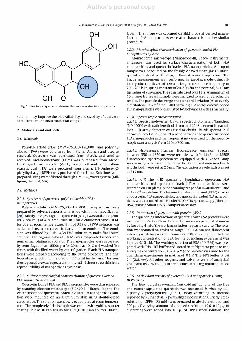
Fig. 1. Structure of quercetin: showing the molecular structure of quercetin.
sulation may improve the bioavailability and stability of quercetinand other similar small molecular drugs.
2. Materials and methods
2.1. Materials
Poly-d,l-lactide (PLA) (MW = 75,000–120,000) and polyvinylalcohol (PVA) were purchased from Sigma–Aldrich and used asreceived. Quercetin was purchased from Merck, and used asreceived. Dichloromethane (DCM) was purchased from Merck.HPLC grade acetonitrile (ACN), water, ethanol and trifluo-roacetic acid (TFA) were procured from Sigma. 1,1-Diphenyl-2-picrylhydrazyl (DPPH) was purchased from Fluka. Solutions wereprepared using water filtered through a Milli-Q water system (Mil-lipore, Bedford, MA).
2.2. Methods
2.2.1. Synthesis of quercetin–poly(d,l-lactide) (PLA)nanoparticles
Poly(d,l-lactide) (MW = 75,000–120,000) nanoparticles wereprepared by solvent evaporation method with minor modification[26]. Briefly, PLA (50 mg) and quercetin (5 mg) was sonicated (Son-ics Vibra cell) at 40% amplitude in 2 ml dichloromethane (DCM)for 30 s at room temperature. 4 ml of 3% (w/v) PVA solution wasadded and again sonicated similarly to form emulsion. The emul-sion was diluted by 0.1% (w/v) PVA solution to make final 80 mlsolution. The organic solvent (DCM) was evaporated under vac-uum using rotating evaporator. The nanoparticles were separatedby centrifugation at 16500 rpm for 20 min at 10 ◦C and washed fivetimes with distilled water by centrifugation. Blank PLA nanopar-ticles were prepared according to the same procedure. The finallyophilized product was stored at 4 ◦C until further use. This syn-thesis procedure was repeated minimum 3–4 times to establish thereproducibility of nanoparticles synthesis.
2.2.2. Surface morphological characterisation of quercetin loadedPLA nanoparticles by SEM
Quercetin loaded PLA and PLA nanoparticles were characterisedby scanning electron microscope (S-3400 N, Hitachi, Japan). Thewater suspended quercetin loaded PLA and PLA nanoparticles solu-tion were mounted on an aluminium stub using double-sidedcarbon tape. The solution was slowly evaporated at room tempera-ture. The completely dried sample was coated with gold by sputtercoating unit at 10 Pa vacuum for 10 s (E1010 ion sputter Hitachi,
Japan). The image was captured on SEM mode at desired magni-fication. PLA nanoparticles were also characterised using similarprocedure.
2.2.3. Morphological characterisation of quercetin loaded PLAnanoparticles by AFM
Atomic force microscope (Nanoscope-III, Veeco Instruments,Singapore) was used for surface characterisation of both PLAnanoparticles and quercetin loaded PLA nanoparticles. A drop ofsample was deposited on the freshly cleaved clean glass surface,spread and dried with nitrogen flow at room temperature. Theimage measurement was performed in tapping mode using sil-icon probe cantilever of 125 �m length, resonance frequency of209–286 kHz, spring constant of 20–80 N/m and nominal, 5–10 nmtip radius of curvature. The scan rate used was 1 Hz. A minimum of10 images from each sample were analyzed to assure reproducibleresults. The particle size range and standard deviation (�) of evenlydistributed (∼3 �m2 area/∼400 particles) PLA and quercetin loadedPLA nanoparticles were calculated by software as well as manually.
2.2.4. Spectroscopic characterisation2.2.4.1. Spectrophotometer. UV–vis spectrophotometer, Nanodrop(ND 1000) with path length of 1 mm and 2048 element linear sil-icon CCD array detector was used to obtain UV–vis spectra. 2 �lof each quercetin solution, PLA nanoparticles and quercetin loadedPLA nanoparticles and their supernatant were used for the spectro-scopic scan analysis from 220 to 700 nm.
2.2.4.2. Fluorescence. Intrinsic fluorescence emission spectrabetween 350 and 650 nm were recorded with Perkin Elmer LS50Bfluorescence spectrophotometer equipped with a xenon lampsource using a 3-D scanning mode. Excitation and emission band-widths both were set at 2.5 nm. The excitation wavelength was setat 617 nm.
2.2.4.3. FTIR. The FTIR spectra of lyophilized quercetin, PLAnanoparticles and quercetin loaded PLA nanoparticles wererecorded on KBr plates in the scanning range of 400–4000 cm−1 andat 1 cm−1 resolution. The Fourier transform infrared (FTIR) spectraof quercetin, PLA nanoparticles, and quercetin loaded PLA nanopar-ticles were recorded on a Nicolet 5700 FTIR spectroscopy (Thermo,USA) using a Smart OMNI-sampler accessory.
2.2.5. Interaction of quercetin with proteins (BSA)The quenching interactions of quercetin with BSA proteins were
recorded on Perkin Elmer LS50B fluorescence spectrophotometerby keeping 4 ml of the working solution in quartz cuvette. The solu-tion was scanned on emission range 290–450 nm and fluorescentintensity at 340 nm was determined on 280 nm excitation. The finalworking concentration of BSA for the quenching experiment waskept as 0.10 �M. The working solution of BSA (10−6 M) was pre-pared with Tris–HCl buffer and stored in refrigerator prior to use.A varying concentration (0.0–90 �M) of quercetin was used for thequenching experiments in methanol–0.1 M Tris–HCl buffer at pH7.4 (2:8, v/v). All other reagents and solvents were of analyticalgrade and used without further purification using double distilledwater.
2.2.6. Antioxidant activity of quercetin–PLA nanoparticles usingDPPH assay
The free radical scavenging (antioxidant) activity of the freeand nanoencapsulated quercetin was measured in vitro by 1,1-diphenyl-2-picrylhydrazyl (DPPH) assay according to methodreported by Kumar et al. [7] with slight modifications. Briefly, stocksolution of DPPH (0.2 mM) was prepared in absolute ethanol and300 �l of varying amount of quercetin solution (0.6–0.12 �g ofquercetin) were added into 100 �l of DPPH stock solution. The
Author's personal copy
186 A. Kumari et al. / Colloids and Surfaces B: Biointerfaces 80 (2010) 184–192
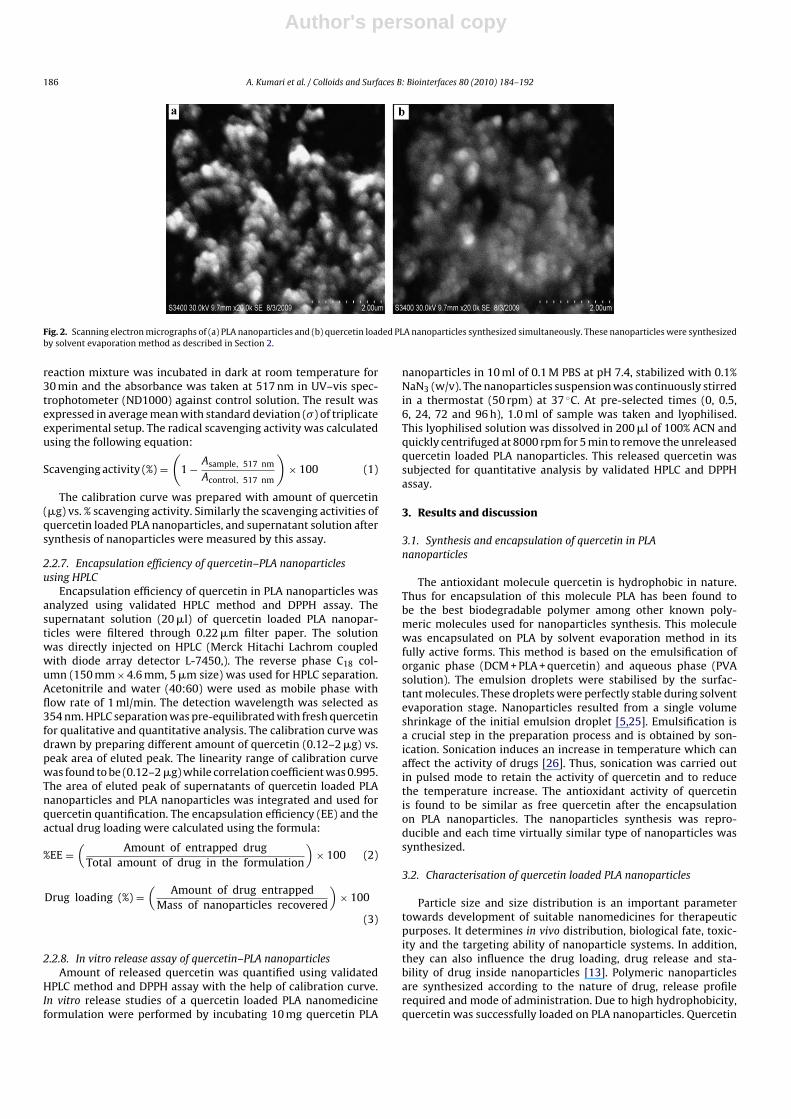
Fig. 2. Scanning electron micrographs of (a) PLA nanoparticles and (b) quercetin loaded PLA nanoparticles synthesized simultaneously. These nanoparticles were synthesizedby solvent evaporation method as described in Section 2.
reaction mixture was incubated in dark at room temperature for30 min and the absorbance was taken at 517 nm in UV–vis spec-trophotometer (ND1000) against control solution. The result wasexpressed in average mean with standard deviation (�) of triplicateexperimental setup. The radical scavenging activity was calculatedusing the following equation:
Scavenging activity (%) =(
1 − Asample, 517 nm
Acontrol, 517 nm
)× 100 (1)
The calibration curve was prepared with amount of quercetin(�g) vs. % scavenging activity. Similarly the scavenging activities ofquercetin loaded PLA nanoparticles, and supernatant solution aftersynthesis of nanoparticles were measured by this assay.
2.2.7. Encapsulation efficiency of quercetin–PLA nanoparticlesusing HPLC
Encapsulation efficiency of quercetin in PLA nanoparticles wasanalyzed using validated HPLC method and DPPH assay. Thesupernatant solution (20 �l) of quercetin loaded PLA nanopar-ticles were filtered through 0.22 �m filter paper. The solutionwas directly injected on HPLC (Merck Hitachi Lachrom coupledwith diode array detector L-7450,). The reverse phase C18 col-umn (150 mm × 4.6 mm, 5 �m size) was used for HPLC separation.Acetonitrile and water (40:60) were used as mobile phase withflow rate of 1 ml/min. The detection wavelength was selected as354 nm. HPLC separation was pre-equilibrated with fresh quercetinfor qualitative and quantitative analysis. The calibration curve wasdrawn by preparing different amount of quercetin (0.12–2 �g) vs.peak area of eluted peak. The linearity range of calibration curvewas found to be (0.12–2 �g) while correlation coefficient was 0.995.The area of eluted peak of supernatants of quercetin loaded PLAnanoparticles and PLA nanoparticles was integrated and used forquercetin quantification. The encapsulation efficiency (EE) and theactual drug loading were calculated using the formula:
%EE =(
Amount of entrapped drugTotal amount of drug in the formulation
)× 100 (2)
Drug loading (%) =(
Amount of drug entrappedMass of nanoparticles recovered
)× 100
(3)
2.2.8. In vitro release assay of quercetin–PLA nanoparticlesAmount of released quercetin was quantified using validated
HPLC method and DPPH assay with the help of calibration curve.In vitro release studies of a quercetin loaded PLA nanomedicineformulation were performed by incubating 10 mg quercetin PLA
nanoparticles in 10 ml of 0.1 M PBS at pH 7.4, stabilized with 0.1%NaN3 (w/v). The nanoparticles suspension was continuously stirredin a thermostat (50 rpm) at 37 ◦C. At pre-selected times (0, 0.5,6, 24, 72 and 96 h), 1.0 ml of sample was taken and lyophilised.This lyophilised solution was dissolved in 200 �l of 100% ACN andquickly centrifuged at 8000 rpm for 5 min to remove the unreleasedquercetin loaded PLA nanoparticles. This released quercetin wassubjected for quantitative analysis by validated HPLC and DPPHassay.
3. Results and discussion
3.1. Synthesis and encapsulation of quercetin in PLAnanoparticles
The antioxidant molecule quercetin is hydrophobic in nature.Thus for encapsulation of this molecule PLA has been found tobe the best biodegradable polymer among other known poly-meric molecules used for nanoparticles synthesis. This moleculewas encapsulated on PLA by solvent evaporation method in itsfully active forms. This method is based on the emulsification oforganic phase (DCM + PLA + quercetin) and aqueous phase (PVAsolution). The emulsion droplets were stabilised by the surfac-tant molecules. These droplets were perfectly stable during solventevaporation stage. Nanoparticles resulted from a single volumeshrinkage of the initial emulsion droplet [5,25]. Emulsification isa crucial step in the preparation process and is obtained by son-ication. Sonication induces an increase in temperature which canaffect the activity of drugs [26]. Thus, sonication was carried outin pulsed mode to retain the activity of quercetin and to reducethe temperature increase. The antioxidant activity of quercetinis found to be similar as free quercetin after the encapsulationon PLA nanoparticles. The nanoparticles synthesis was repro-ducible and each time virtually similar type of nanoparticles wassynthesized.
3.2. Characterisation of quercetin loaded PLA nanoparticles
Particle size and size distribution is an important parametertowards development of suitable nanomedicines for therapeuticpurposes. It determines in vivo distribution, biological fate, toxic-ity and the targeting ability of nanoparticle systems. In addition,they can also influence the drug loading, drug release and sta-bility of drug inside nanoparticles [13]. Polymeric nanoparticlesare synthesized according to the nature of drug, release profilerequired and mode of administration. Due to high hydrophobicity,quercetin was successfully loaded on PLA nanoparticles. Quercetin
Author's personal copy
A. Kumari et al. / Colloids and Surfaces B: Biointerfaces 80 (2010) 184–192 187
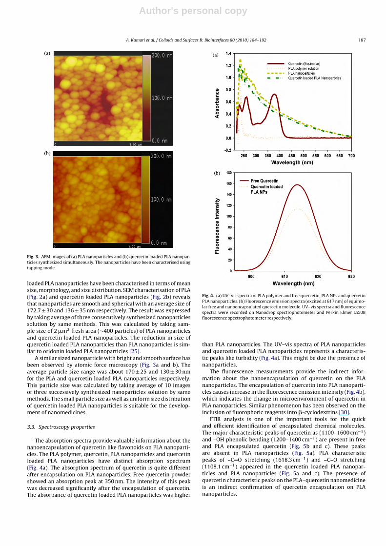
Fig. 3. AFM images of (a) PLA nanoparticles and (b) quercetin loaded PLA nanopar-ticles synthesized simultaneously. The nanoparticles have been characterised usingtapping mode.
loaded PLA nanoparticles have been characterised in terms of meansize, morphology, and size distribution. SEM characterisation of PLA(Fig. 2a) and quercetin loaded PLA nanoparticles (Fig. 2b) revealsthat nanoparticles are smooth and spherical with an average size of172.7 ± 30 and 136 ± 35 nm respectively. The result was expressedby taking average of three consecutively synthesized nanoparticlessolution by same methods. This was calculated by taking sam-ple size of 2 �m2 fresh area (∼400 particles) of PLA nanoparticlesand quercetin loaded PLA nanoparticles. The reduction in size ofquercetin loaded PLA nanoparticles than PLA nanoparticles is sim-ilar to oridonin loaded PLA nanoparticles [25].
A similar sized nanoparticle with bright and smooth surface hasbeen observed by atomic force microscopy (Fig. 3a and b). Theaverage particle size range was about 170 ± 25 and 130 ± 30 nmfor the PLA and quercetin loaded PLA nanoparticles respectively.This particle size was calculated by taking average of 10 imagesof three successively synthesized nanoparticles solution by samemethods. The small particle size as well as uniform size distributionof quercetin loaded PLA nanoparticles is suitable for the develop-ment of nanomedicines.
3.3. Spectroscopy properties
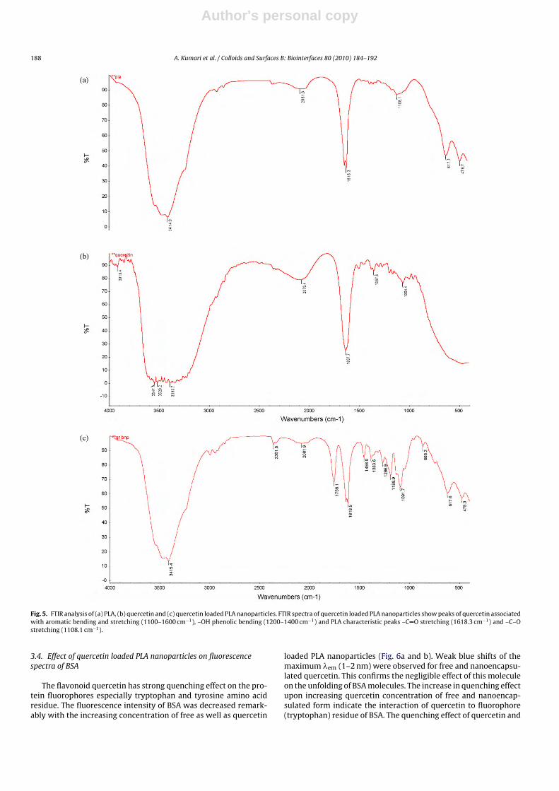
The absorption spectra provide valuable information about thenanoencapsulation of quercetin like flavonoids on PLA nanoparti-cles. The PLA polymer, quercetin, PLA nanoparticles and quercetinloaded PLA nanoparticles have distinct absorption spectrum(Fig. 4a). The absorption spectrum of quercetin is quite differentafter encapsulation on PLA nanoparticles. Free quercetin powdershowed an absorption peak at 350 nm. The intensity of this peakwas decreased significantly after the encapsulation of quercetin.The absorbance of quercetin loaded PLA nanoparticles was higher
Fig. 4. (a) UV–vis spectra of PLA polymer and free quercetin, PLA NPs and quercetinPLA nanoparticles. (b) Fluorescence emission spectra (excited at 617 nm) of equimo-lar free and nanoencapsulated quercetin molecule. UV–vis spectra and fluorescencespectra were recorded on Nanodrop spectrophotometer and Perkin Elmer LS50Bfluorescence spectrophotometer respectively.
than PLA nanoparticles. The UV–vis spectra of PLA nanoparticlesand quercetin loaded PLA nanoparticles represents a characteris-tic peaks like turbidity (Fig. 4a). This might be due the presence ofnanoparticles.
The fluorescence measurements provide the indirect infor-mation about the nanoencapsulation of quercetin on the PLAnanoparticles. The encapsulation of quercetin into PLA nanoparti-cles causes increase in the fluorescence emission intensity (Fig. 4b),which indicates the change in microenvironment of quercetin inPLA nanoparticles. Similar phenomenon has been observed on theinclusion of fluorophoric reagents into �-cyclodextrins [30].
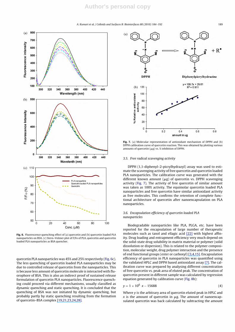
FTIR analysis is one of the important tools for the quickand efficient identification of encapsulated chemical molecules.The major characteristic peaks of quercetin as (1100–1600 cm−1)and –OH phenolic bending (1200–1400 cm−1) are present in freeand PLA encapsulated quercetin (Fig. 5b and c). These peaksare absent in PLA nanoparticles (Fig. 5a). PLA characteristicpeaks of –C O stretching (1618.3 cm−1) and –C–O stretching(1108.1 cm−1) appeared in the quercetin loaded PLA nanopar-ticles and PLA nanoparticles (Fig. 5a and c). The presence ofquercetin characteristic peaks on the PLA–quercetin nanomedicineis an indirect confirmation of quercetin encapsulation on PLAnanoparticles.
Author's personal copy
188 A. Kumari et al. / Colloids and Surfaces B: Biointerfaces 80 (2010) 184–192
Fig. 5. FTIR analysis of (a) PLA, (b) quercetin and (c) quercetin loaded PLA nanoparticles. FTIR spectra of quercetin loaded PLA nanoparticles show peaks of quercetin associatedwith aromatic bending and stretching (1100–1600 cm−1), –OH phenolic bending (1200–1400 cm−1) and PLA characteristic peaks –C O stretching (1618.3 cm−1) and –C–Ostretching (1108.1 cm−1).
3.4. Effect of quercetin loaded PLA nanoparticles on fluorescencespectra of BSA
The flavonoid quercetin has strong quenching effect on the pro-tein fluorophores especially tryptophan and tyrosine amino acidresidue. The fluorescence intensity of BSA was decreased remark-ably with the increasing concentration of free as well as quercetin
loaded PLA nanoparticles (Fig. 6a and b). Weak blue shifts of themaximum �em (1–2 nm) were observed for free and nanoencapsu-lated quercetin. This confirms the negligible effect of this moleculeon the unfolding of BSA molecules. The increase in quenching effectupon increasing quercetin concentration of free and nanoencap-sulated form indicate the interaction of quercetin to fluorophore(tryptophan) residue of BSA. The quenching effect of quercetin and
Author's personal copy
A. Kumari et al. / Colloids and Surfaces B: Biointerfaces 80 (2010) 184–192 189
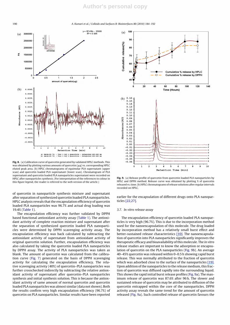
Fig. 6. Fluorescence quenching effect of (a) quercetin and (b) quercetin loaded PLAnanoparticles on BSA. (c) Stern–Volmer plot of F/Fo of PLA, quercetin and quercetinloaded PLA nanoparticles as BSA quencher.
quercetin PLA nanoparticles was 45% and 25% respectively (Fig. 6c).The less quenching of quercetin loaded PLA nanoparticles may bedue to controlled release of quercetin from the nanoparticles. Thisis because less amount of quercetin molecule is interacted with flu-orophore of BSA. This is also an indirect proof of sustained releaseformulation of quercetin PLA nanoparticles. Fluorescence quench-ing could proceed via different mechanisms, usually classified asdynamic quenching and static quenching. It is concluded that thequenching of BSA was not initiated by dynamic quenching, butprobably partly by static quenching resulting from the formationof quercetin–BSA complex [19,21,23,24,28].
Fig. 7. (a) Molecular representation of antioxidant mechanism of DPPH and (b)DPPH calibration curve of quercetin reaction. This was obtained by plotting variousamounts of quercetin (�g) vs. % inhibition of DPPH.
3.5. Free radical scavenging activity
DPPH (1,1-diphenyl–2-picrylhydrazyl) assay was used to esti-mate the scavenging activity of free quercetin and quercetin loadedPLA nanoparticles. The calibration curve was generated with thedifferent known amount (�g) of quercetin vs. DPPH scavengingactivity (Fig. 7). The activity of free quercetin of similar amountwas taken as 100% activity. The equimolar quercetin loaded PLAnanoparticles and free quercetin have similar antioxidant activityas free molecules. This confirms the retention of complete func-tional architecture of quercetin after nanoencapsulation on PLAnanoparticles.
3.6. Encapsulation efficiency of quercetin loaded PLAnanoparticles
Biodegradable nanoparticles like PLA, PLGA, etc. have beenreported for the encapsulation of large number of therapeuticmolecules such as taxol and ellagic acid [22] with highest affin-ity. Drug loading and entrapment efficiency very much depend onthe solid-state drug solubility in matrix material or polymer (soliddissolution or dispersion). This is related to the polymer composi-tion, molecular weight, drug polymer interaction and the presenceof end functional groups (ester or carboxyl) [3,4,15]. Encapsulationefficiency of quercetin in PLA nanoparticles was quantified usingthe validated HPLC and DPPH based antioxidant assay [7]. The cal-ibration curve was prepared by analysing different concentrationof free quercetin vs. peak area of eluted peak. The concentration ofquercetin present in different sample was calculated by regressionequation generated by calibration curve (Fig. 8b):
y = 1 × 10⁶ x − 15688 (4)
Where y is the arbitrary area of quercetin eluted peak in HPLC andx is the amount of quercetin in �g. The amount of nanoencap-sulated quercetin was back calculated by subtracting the amount
Author's personal copy
190 A. Kumari et al. / Colloids and Surfaces B: Biointerfaces 80 (2010) 184–192
Fig. 8. (a) Calibration curve of quercetin generated by validated HPLC methods. Thiswas obtained by plotting various amounts of quercetin (�g) vs. corresponding HPLCeluted peak area. (b) HPLC chromatograms of equimolar PLA supernatant (upperscan) and quercetin loaded PLA supernatant (lower scan). Chromatogram of PLAsupernatant and quercetin loaded PLA nanoparticles supernatant were recorded onHPLC after nanoparticles synthesis. (For interpretation of the references to colour inthis figure legend, the reader is referred to the web version of the article.)
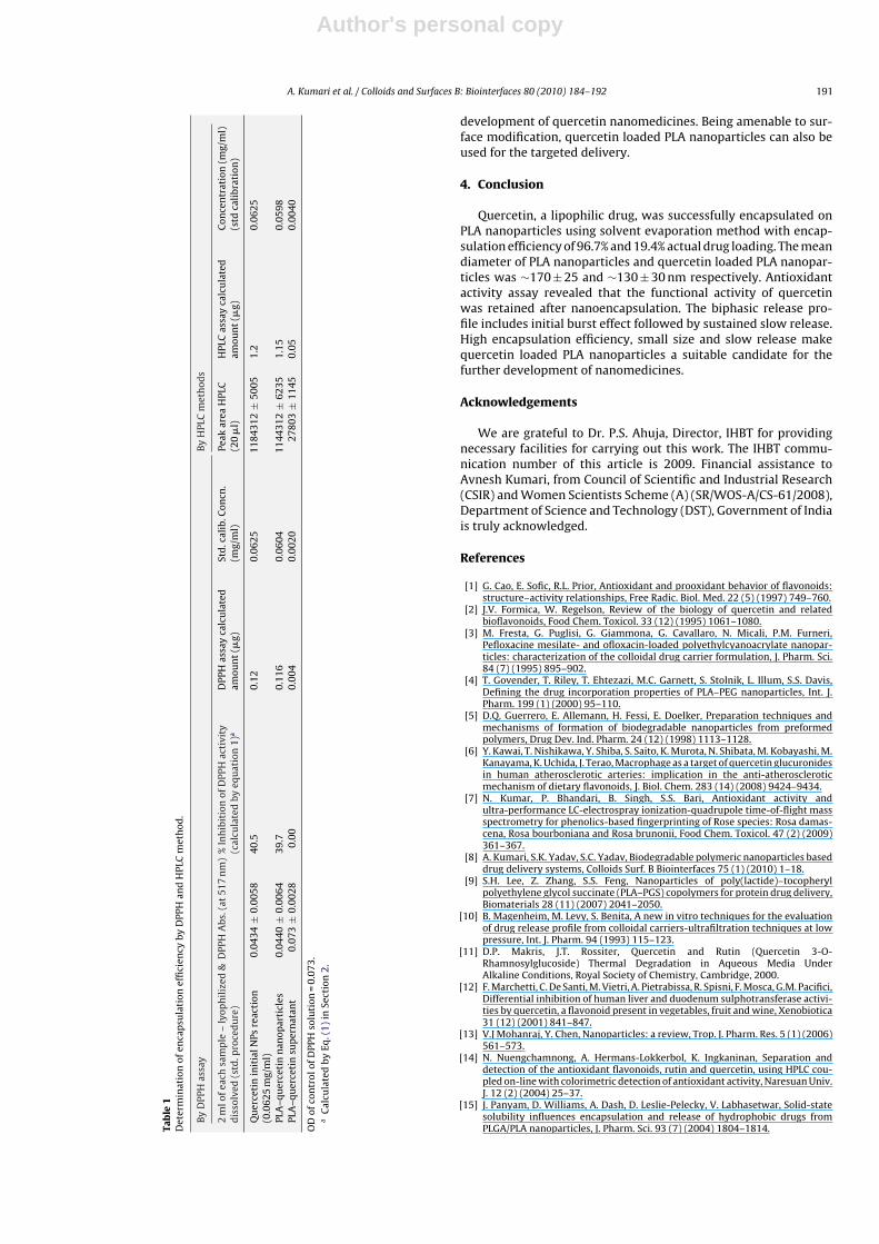
of quercetin in nanoparticle synthesis mixture and supernatantafter separation of synthesized quercetin loaded PLA nanoparticles.HPLC analysis reveals that the encapsulation efficiency of quercetinloaded PLA nanoparticles was 96.7% and actual drug loading was19.4% (Table 1).
The encapsulation efficiency was further validated by DPPHbased functional antioxidant activity assay (Table 1). The antioxi-dant activity of complete reaction mixture and supernatant afterthe separation of synthesized quercetin loaded PLA nanoparti-cles were determined by DPPH scavenging activity assay. Theencapsulation efficiency was back calculated by subtracting theantioxidant activity of supernatant from antioxidant activity oforiginal quercetin solution. Further, encapsulation efficiency wasalso calculated by taking the quercetin loaded PLA nanoparticlesby DPPH assay. The activity of PLA nanoparticles was taken asblank. The amount of quercetin was calculated from the calibra-tion curve (Fig. 7) generated on the basis of DPPH scavengingactivity for calculating the encapsulation efficiency. The rela-tive scavenging activity (40%) of quercetin–PLA nanoparticles wasfurther crosschecked indirectly by subtracting the relative antiox-idant activity of supernatant after quercetin–PLA nanoparticlessynthesis and initial synthesis solution. This is because the antiox-idant activity of same amount of normal quercetin and quercetinloaded PLA nanoparticles was almost similar (data not shown). Boththe results confirm very high encapsulation efficiency (96.7%) ofquercetin on PLA nanoparticles. Similar results have been reported
Fig. 9. (a) Release profile of quercetin from quercetin loaded PLA nanoparticles byHPLC and DPPH method. Release curve was obtained by plotting % of quercetinreleased vs. time. (b) HPLC chromatograms of release solutions after regular intervalsrecorded on HPLC.
earlier for the encapsulation of different drugs onto PLA nanopar-ticles [22,27].
3.7. In vitro release assay
The encapsulation efficiency of quercetin loaded PLA nanopar-ticles is very high (96.7%). This is due to the incorporation methodused for the nanoencapsulation of this molecule. The drug loadedby incorporation method has a relatively small burst effect andbetter-sustained release characteristics [10]. The nanoencapsula-tion of quercetin into PLA nanoparticles significantly improves thetherapeutic efficacy and bioavailability of this molecule. The in vitrorelease studies are important to know the adsorption or encapsu-lation of quercetin on the PLA nanoparticles (Fig. 9b). An average40–45% quercetin was released within 0–0.5 h showing rapid burstrelease. This was normally attributed to the fraction of quercetinwhich was adsorbed close to the surface of the nanoparticles [19].Upon addition of the nanoparticles to the release medium, this frac-tion of quercetin was diffused rapidly into the surrounding liquid.This shows the rapid initial burst release profiles (Fig. 9a). The max-imum release of quercetin was 87.6% after 96 h. The slower andsustained release of quercetin may be attributed to diffusion of thequercetin entrapped within the core of the nanoparticles. DPPHactivity assay reveals the same trend for the amount of quercetinreleased (Fig. 9a). Such controlled release of quercetin favours the
Author's personal copy
A. Kumari et al. / Colloids and Surfaces B: Biointerfaces 80 (2010) 184–192 191
Tab
le1
Det
erm
inat
ion
ofen
cap
sula
tion
effi
cien
cyby
DPP
Han
dH
PLC
met
hod
.
By
DPP
Has
say
By
HPL
Cm
eth
ods
2m
lofe
ach
sam
ple
–ly
oph
iliz
ed&
dis
solv
ed(s
td.p
roce
du
re)
DPP
HA
bs.(
at51
7n
m)
%In
hib
itio
nof
DPP
Hac
tivi
ty(c
alcu
late
dby
equ
atio
n1)
aD
PPH
assa
yca
lcu
late
dam
oun
t(�
g)St
d.c
alib
.Con
cn.
(mg/
ml)
Peak
area
HPL
C(2
0�
l)H
PLC
assa
yca
lcu
late
dam
oun
t(�
g)C
once
ntr
atio
n(m
g/m
l)(s
tdca
libr
atio
n)
Qu
erce
tin
init
ialN
Psre
acti
on(0
.062
5m
g/m
l)0.
0434
±0.
0058
40.5
0.12
0.06
2511
8431
2±
5005
1.2
0.06
25
PLA
–qu
erce
tin
nan
opar
ticl
es0.
0440
±0.
0064
39.7
0.11
60.
0604
1144
312
±62
351.
150.
0598
PLA
–qu
erce
tin
sup
ern
atan
t0.
073
±0.
0028
0.00
0.00
40.
0020
2780
3±
1145
0.05
0.00
40
OD
ofco
ntr
olof
DPP
Hso
luti
on=
0.07
3.a
Cal
cula
ted
byEq
.(1)
inSe
ctio
n2.
development of quercetin nanomedicines. Being amenable to sur-face modification, quercetin loaded PLA nanoparticles can also beused for the targeted delivery.
4. Conclusion
Quercetin, a lipophilic drug, was successfully encapsulated onPLA nanoparticles using solvent evaporation method with encap-sulation efficiency of 96.7% and 19.4% actual drug loading. The meandiameter of PLA nanoparticles and quercetin loaded PLA nanopar-ticles was ∼170 ± 25 and ∼130 ± 30 nm respectively. Antioxidantactivity assay revealed that the functional activity of quercetinwas retained after nanoencapsulation. The biphasic release pro-file includes initial burst effect followed by sustained slow release.High encapsulation efficiency, small size and slow release makequercetin loaded PLA nanoparticles a suitable candidate for thefurther development of nanomedicines.
Acknowledgements
We are grateful to Dr. P.S. Ahuja, Director, IHBT for providingnecessary facilities for carrying out this work. The IHBT commu-nication number of this article is 2009. Financial assistance toAvnesh Kumari, from Council of Scientific and Industrial Research(CSIR) and Women Scientists Scheme (A) (SR/WOS-A/CS-61/2008),Department of Science and Technology (DST), Government of Indiais truly acknowledged.
References
[1] G. Cao, E. Sofic, R.L. Prior, Antioxidant and prooxidant behavior of flavonoids:structure–activity relationships, Free Radic. Biol. Med. 22 (5) (1997) 749–760.
[2] J.V. Formica, W. Regelson, Review of the biology of quercetin and relatedbioflavonoids, Food Chem. Toxicol. 33 (12) (1995) 1061–1080.
[3] M. Fresta, G. Puglisi, G. Giammona, G. Cavallaro, N. Micali, P.M. Furneri,Pefloxacine mesilate- and ofloxacin-loaded polyethylcyanoacrylate nanopar-ticles: characterization of the colloidal drug carrier formulation, J. Pharm. Sci.84 (7) (1995) 895–902.
[4] T. Govender, T. Riley, T. Ehtezazi, M.C. Garnett, S. Stolnik, L. Illum, S.S. Davis,Defining the drug incorporation properties of PLA–PEG nanoparticles, Int. J.Pharm. 199 (1) (2000) 95–110.
[5] D.Q. Guerrero, E. Allemann, H. Fessi, E. Doelker, Preparation techniques andmechanisms of formation of biodegradable nanoparticles from preformedpolymers, Drug Dev. Ind. Pharm. 24 (12) (1998) 1113–1128.
[6] Y. Kawai, T. Nishikawa, Y. Shiba, S. Saito, K. Murota, N. Shibata, M. Kobayashi, M.Kanayama, K. Uchida, J. Terao, Macrophage as a target of quercetin glucuronidesin human atherosclerotic arteries: implication in the anti-atheroscleroticmechanism of dietary flavonoids, J. Biol. Chem. 283 (14) (2008) 9424–9434.
[7] N. Kumar, P. Bhandari, B. Singh, S.S. Bari, Antioxidant activity andultra-performance LC-electrospray ionization-quadrupole time-of-flight massspectrometry for phenolics-based fingerprinting of Rose species: Rosa damas-cena, Rosa bourboniana and Rosa brunonii, Food Chem. Toxicol. 47 (2) (2009)361–367.
[8] A. Kumari, S.K. Yadav, S.C. Yadav, Biodegradable polymeric nanoparticles baseddrug delivery systems, Colloids Surf. B Biointerfaces 75 (1) (2010) 1–18.
[9] S.H. Lee, Z. Zhang, S.S. Feng, Nanoparticles of poly(lactide)–tocopherylpolyethylene glycol succinate (PLA–PGS) copolymers for protein drug delivery,Biomaterials 28 (11) (2007) 2041–2050.
[10] B. Magenheim, M. Levy, S. Benita, A new in vitro techniques for the evaluationof drug release profile from colloidal carriers-ultrafiltration techniques at lowpressure, Int. J. Pharm. 94 (1993) 115–123.
[11] D.P. Makris, J.T. Rossiter, Quercetin and Rutin (Quercetin 3-O-Rhamnosylglucoside) Thermal Degradation in Aqueous Media UnderAlkaline Conditions, Royal Society of Chemistry, Cambridge, 2000.
[12] F. Marchetti, C. De Santi, M. Vietri, A. Pietrabissa, R. Spisni, F. Mosca, G.M. Pacifici,Differential inhibition of human liver and duodenum sulphotransferase activi-ties by quercetin, a flavonoid present in vegetables, fruit and wine, Xenobiotica31 (12) (2001) 841–847.
[13] V.J Mohanraj, Y. Chen, Nanoparticles: a review, Trop. J. Pharm. Res. 5 (1) (2006)561–573.
[14] N. Nuengchamnong, A. Hermans-Lokkerbol, K. Ingkaninan, Separation anddetection of the antioxidant flavonoids, rutin and quercetin, using HPLC cou-pled on-line with colorimetric detection of antioxidant activity, Naresuan Univ.J. 12 (2) (2004) 25–37.
[15] J. Panyam, D. Williams, A. Dash, D. Leslie-Pelecky, V. Labhasetwar, Solid-statesolubility influences encapsulation and release of hydrophobic drugs fromPLGA/PLA nanoparticles, J. Pharm. Sci. 93 (7) (2004) 1804–1814.
Author's personal copy
192 A. Kumari et al. / Colloids and Surfaces B: Biointerfaces 80 (2010) 184–192
[16] T. Pralhad, K. Rajendrakumar, Study of freeze-dried quercetin–cyclodextrinbinary systems by DSC, FT-IR, X-ray diffraction and SEM analysis, J. Pharm.Biomed. Anal. 34 (2) (2004) 333–339.
[17] A. Priprem, J. Watanatorn, S. Sutthiparinyanont, W. Phachonpai, S. Muchima-pura, Anxiety and cognitive effects of quercetin liposomes in rats,Nanomedicine 4 (1) (2008) 70–78.
[18] D.V. Ratnam, D.D. Ankola, V. Bhardwaj, D.K. Sahana, M.N. Kumar, Role of antiox-idants in prophylaxis and therapy: a pharmaceutical perspective, J. Control.Release 113 (3) (2006) 189–207.
[19] L.H. Riihimaki, M.J. Vainio, J.M.S. Heikura, K.H. Valkonen, V.T. Virta-nen, P.M. Vuorela, Binding of phenolic compounds and their derivativesto bovine and reindeer b-lactoglobulin, J. Agric. Food Chem. 56 (2008)7721–7729.
[20] M.R. Schaab, B.M. Barney, W.A. Francisco, Kinetic and spectroscopic studieson the quercetin 2,3-dioxygenase from Bacillus subtilis, Biochemistry 45 (3)(2006) 1009–1016.
[21] S. Soares, N. Mateus, V.D. Freitas, Interaction of different polyphenols with BSAand human salivary a-amylase (HSA) by fluorescence quenching, J. Agric. FoodChem. 55 (16) (2007) 6726–6735.
[22] K. Sonaje, J.L. Italia, G. Sharma, V. Bhardwaj, K. Tikoo, M.N. Kumar, Developmentof biodegradable nanoparticles for oral delivery of ellagic acid and evaluationof their antioxidant efficacy against cyclosporine A-induced nephrotoxicity inrats, Pharm. Res. 24 (5) (2007) 899–908.
[23] J.B. Xiao, J.W. Chen, H. Cao, F.L. Ren, C.S. Yang, Y. Chen, M. Xu, Study of theinteraction between baicalin and bovine serum albumin by multi spectroscopicmethod, J. Photochem. Photobiol. 191 (2007) 222–227.
[24] J.B. Xiao, X.Q. Chen, X.Y. Jiang, M. Hilczer, M. Tachiya, Probing the interaction oftrans-resveratrol with bovine serum albumin: a fluorescence quenching studywith Tachiya model, J. Fluoresc. 18 (3–4) (2008) 671–678.
[25] J. Xing, D. Zhang, T. Tan, Studies on the oridonin-loaded poly(d,l-lactic acid)nanoparticles in vitro and in vivo, Int. J. Biol. Macromol. 40 (2) (2007) 153–158.
[26] M.F. Zambaux, F. Bonneaux, R. Gref, E. Dellacherie, C. Vigneron, Preparation andcharacterization of protein C-loaded PLA nanoparticles, J. Control. Release 60(2-3) (1999) 179–188.
[27] Y. Zhang, Y. Yang, K. Tang, X. Hu, G. Zou, Physicochemical characterization andantioxidant activity of quercetin-loaded chitosan nanoparticles, J. Appl. Polym.Sci. 107 (2008) 891–897.
[28] Y.Z. Zhang, B. Zhou, Y.X. Liu, C.X. Zhou, X.L. Ding, Y. Liu, Fluorescence study onthe interaction of bovine serum albumin with p-aminoazobenzene, J. Fluoresc.18 (1) (2008) 109–118.
[29] Y. Zheng, I.S. Haworth, Z. Zuo, M.S. Chow, A.H. Chow, Physicochemicaland structural characterization of quercetin-beta-cyclodextrin complexes, J.Pharm. Sci. 94 (5) (2005) 1079–1089.
[30] L. Zhu, Z. Qi, Z. Lu, H. Jing, W. Qi, Comaprative study of fluorescence enhance-ment of some fluorescence systems in different cyclodextrin derivatives andcyclodextrin media, Microchem. J. 53 (1996) 361–370.