development and validation of a model to predict severe
TRANSCRIPT
Journal of
Clinical Medicine
Article
Development and Validation of a Model to Predict SevereHospital-Acquired Acute Kidney Injury in Non-CriticallyIll Patients
Jacqueline Del Carpio 1,2,3,*, Maria Paz Marco 1,3, Maria Luisa Martin 1,3, Natalia Ramos 4 , Judith de la Torre 4,5,Joana Prat 6,7, Maria J. Torres 6,8, Bruno Montoro 9, Mercedes Ibarz 3,10 , Silvia Pico 3,10, Gloria Falcon 11,Marina Canales 11, Elisard Huertas 12, Iñaki Romero 13, Nacho Nieto 6,8, Ricard Gavaldà 14 and Alfons Segarra 1,4,†
�����������������
Citation: Carpio, J.D.; Marco, M.P.;
Martin, M.L.; Ramos, N.; de la Torre,
J.; Prat, J.; Torres, M.J.; Montoro, B.;
Ibarz, M.; Pico, S.; et al. Development
and Validation of a Model to Predict
Severe Hospital-Acquired Acute
Kidney Injury in Non-Critically Ill
Patients. J. Clin. Med. 2021, 10, 3959.
https://doi.org/10.3390/jcm10173959
Academic Editor: Ersilia Lucenteforte
Received: 31 May 2021
Accepted: 24 August 2021
Published: 31 August 2021
Publisher’s Note: MDPI stays neutral
with regard to jurisdictional claims in
published maps and institutional affil-
iations.
Copyright: © 2021 by the authors.
Licensee MDPI, Basel, Switzerland.
This article is an open access article
distributed under the terms and
conditions of the Creative Commons
Attribution (CC BY) license (https://
creativecommons.org/licenses/by/
4.0/).
1 Department of Nephrology, Arnau de Vilanova University Hospital, 25198 Lleida, Spain;[email protected] (M.P.M.); [email protected] (M.L.M.);[email protected] (A.S.)
2 Department of Medicine, Autonomous University of Barcelona, 08193 Barcelona, Spain3 Institute of Biomedical Research (IRBLleida), 25198 Lleida, Spain; [email protected] (M.I.);
[email protected] (S.P.)4 Department of Nephrology, Vall d’Hebron University Hospital, 08035 Barcelona, Spain;
[email protected] (N.R.); [email protected] (J.d.l.T.)5 Department of Nephrology, Althaia Foundation, 08243 Manresa, Spain6 Department of Informatics, Vall d’Hebron University Hospital, 08035 Barcelona, Spain;
[email protected] (J.P.); [email protected] (M.J.T.); [email protected] (N.N.)7 Department of Development, Parc Salut Hospital, 08019 Barcelona, Spain8 Department of Information, Southern Metropolitan Territorial Management, 08028 Barcelona, Spain9 Department of Hospital Pharmacy, Vall d’Hebron University Hospital, 08035 Barcelona, Spain;
[email protected] Laboratory Department, Arnau de Vilanova University Hospital, 25198 Lleida, Spain11 Technical Secretary and Territorial Management of Lleida-Pirineus, 25198 Lleida, Spain;
[email protected] (G.F.); [email protected] (M.C.)12 Informatic Unit of the Catalonian Institute of Health—Territorial Management, 25198 Lleida, Spain;
[email protected] Territorial Management Information Systems, Catalonian Institute of Health, 25198 Lleida, Spain;
[email protected] Amalfi Analytics S.A, 08018 Barcelona, Spain; [email protected]* Correspondence: [email protected]; Tel.: +34-9737-05274† Principal investigator.
Abstract: Background. The current models developed to predict hospital-acquired AKI (HA-AKI) innon-critically ill fail to identify the patients at risk of severe HA-AKI stage 3. Objective. To developand externally validate a model to predict the individual probability of developing HA-AKI stage 3through the integration of electronic health databases. Methods. Study set: 165,893 non-critically illhospitalized patients. Using stepwise logistic regression analyses, including demography, chroniccomorbidities, and exposure to risk factors prior to AKI detection, we developed a multivariate modelto predict HA-AKI stage 3. This model was then externally validated in 43,569 non-critical patientsadmitted to the validation center. Results. The incidence of HA-AKI stage 3 in the study set was 0.6%.Among chronic comorbidities, the highest odds ratios were conferred by ischemic heart disease,ischemic cerebrovascular disease, chronic congestive heart failure, chronic obstructive pulmonarydisease, chronic kidney disease and liver disease. Among acute complications, the highest odd ratioswere associated with acute respiratory failure, major surgery and exposure to nephrotoxic drugs.The model showed an AUC of 0.906 (95% CI 0.904 to 0.908), a sensitivity of 89.1 (95% CI 87.0–91.0)and a specificity of 80.5 (95% CI 80.2–80.7) to predict HA-AKI stage 3, but tended to overestimatethe risk at low-risk categories with an adequate goodness-of-fit for all risk categories (Chi2: 16.4,p: 0.034). In the validation set, incidence of HA-AKI stage 3 was 0.62%. The model showed anAUC of 0.861 (95% CI 0.859–0.863), a sensitivity of 83.0 (95% CI 80.5–85.3) and a specificity of 76.5(95% CI 76.2–76.8) to predict HA-AKI stage 3 with an adequate goodness of fit for all risk categories(Chi2: 15.42, p: 0.052). Conclusions. Our study provides a model that can be used in clinical practice
J. Clin. Med. 2021, 10, 3959. https://doi.org/10.3390/jcm10173959 https://www.mdpi.com/journal/jcm
J. Clin. Med. 2021, 10, 3959 2 of 13
to obtain an accurate dynamic assessment of the individual risk of HA-AKI stage 3 along the hospitalstay period in non-critically ill patients.
Keywords: acute kidney injury; hospital-acquired; electronic health data records; risk score
1. Introduction
Acute kidney injury (AKI) is a global concern with a high incidence among hospi-talized patients [1,2]. The incidence of hospital-acquired AKI (HA-AKI) ranges between5 and 15% or 30–45 cases/1000 hospital admissions/year but shows an increasing trendas hospitalized patients are older and subjected to more interventional diagnostic andtreatment techniques, and exposed to the effects of nephrotoxic drugs [3–5]. In addition,AKI has been associated with significant increases in health care resource utilization andcosts in patients who are hospitalized, and with long-term morbidity and mortality afterhospital discharge [6–11]. Numerous studies on AKI have been published in patientsadmitted to intensive care units, in which the causes, risk factors, mortality, and the influ-ence of different treatment strategies have been identified [12–17]. The epidemiology ofacute renal failure in patients admitted to conventional hospitalization wards is much lessknown [18]. Since a large part of the AKI episodes are due to potentially avoidable causes,knowing as accurately as possible the individual risk at any time of hospital stay couldhelp decision making and implementation of preventive measures to reduce the incidenceof hospital AKI [19,20]. The diagnostic approach to in-hospital AKI has undergone asignificant change over time. The old detection models were based on the communicationof the cases at the time of the diagnosis, by conventional analytical controls, and weresubject to the influence of multiple sources of error that motivated avoidable delays in theidentification of cases and in the adoption of treatment measures [21]. With the appearanceof electronic laboratory data records, electronic alert systems were designed. These systemsallow the detection of all cases at an early stage, but they do not allow to adopt preventivemeasures since they detect the problem once it has occurred [22]. The evolution of themanagement systems of the in-hospital AKI has gone in the direction of the development ofpredictive models of individual risk, whose purpose is to be able to anticipate the episodeof AKI and to carry out prevention measures appropriate to the particular situation of eachpatient [23]. In recent years, several models have been developed and validated to allowthe estimation of the risk of suffering AKI during hospitalization, but the results of earlydiagnosis and intensive interventions in terms of reduction of morbidity and mortalityhave been discordant and inconclusive [24]. The studies analyzing the epidemiology andrisk factors associated with AKI in non-critically ill patients have two main limitations toidentify accurately the risk factors associated with HA-AKI. First, most of them are basedon demographic characteristics and comorbidities that have been registered retrospectively,from the discharge administrative codes, and therefore, are subject to a potential bias inthe collection of coded information [25]. Secondly, they do not allow to know whether theexposure to risk factors preceded or not the detection of the AKI episode [26]. Thirdly, theydo not allow to identify the categories of severe AKI. Wu L. et al. recently published an arti-cle where the risk factors that predict the presentation of severe AKI were defined, but theyincluded both ICU and non-ICU patients and no external validation was performed [27].Our group recently developed a model that overcame some of those limitations and pro-vides an accurate dynamic assessment of the individual risk of suffering AKI along thewhole hospital stay period in patients admitted into non-critical hospitalization wards [28].However, although this model allows AKI to be accurately predicted, because of a lack ofstatistical power, it does not allow to detect the risk of developing AKI-3 severity stage,which is the one associated with greater morbidity, related to the severity of complicationsand, in many cases, to the need for replacement of kidney function. The aim of our study
J. Clin. Med. 2021, 10, 3959 3 of 13
was to develop and externally validate a model to predict the risk of HA-AKI stage 3 inhospital-acquired AKI in non-critically ill patients.
2. Methods
This study was performed at two different hospital centers. The first center developedthe predictive model (study set) and the second center performed the external validationof the predictive model (validation set).
2.1. Study Set
The study set included patients admitted to the Vall d’Hebron hospital from January2011 to December 2017. Vall d’Hebron is a tertiary hospital that provides assistance to apopulation of 500,000 habitants in Barcelona, Spain, and develops all kinds of medical andsurgical procedures, including neurosurgery, cardiac surgery, endovascular catheter-guidedprocedures as well as lung, liver, kidney and bone marrow transplantation programs. Weincluded all patients >18 years of age who were admitted to hospital along this periodand did not meet any of the following exclusion criteria: 1.- admission for community-acquired AKI, 2.- hospital stay < 24 h, 3.- admission for elective heart surgery, 4.- directadmission from the emergency room to the intensive care units (ICUs), 5.- admission as arecipient of renal, lung, liver or bone marrow transplant, 6.- absence of serum creatininemeasurements done at least 12 months after hospital admission, 7.- chronic hemodialysistreatment and 8.- denial to give a written consent to participate in the study. Community-acquired AKI was diagnosed whenever patients met the AKI criteria within the first 24 hof hospital admission. Patients initially admitted to conventional hospitalization wardswho afterwards required admission into ICUs were only included if the AKI episode wasdetected while they were admitted in non-critically ill wards, prior to their admission intothe ICUs.
2.2. Baseline Kidney Function
Our patient care system integrates the laboratory databases of the hospital and pri-mary care registers, thus allowing historical data to be obtained for all patients who arehospitalized, provided that these data appear in those registers. Baseline kidney functionwas obtained from the electronic laboratory data records of primary health care and definedas the most recent glomerular filtration rate, estimated by the CKD-EPI equation, withinthe 12 months prior to hospital admission.
2.3. Definition of AKI Severe
AKI was defined and classified in severity stages according to the KDIGO (KidneyDisease Improving Global Outcomes) clinical practice guidelines [29]. Severe AKI HA-AKIwas defined as an increase serum creatinine of at least ×3 over the baseline or ≥4 mg/dL,occurring from the first 24 h to any time within hospital admission.
2.4. AKI Detection
A software integrated into the electronic laboratory database was used to performrepeated comparisons among all serum creatinine levels available for each patient duringhospital stay and generated an identification code, assigning a 1 when the HA-AKI stage3 criteria were met and a 0 when not. The date of HA-AKI stage 3 detection was alsorecorded. The number of the admission episode was used as a filter so that patients withmore than one HA-AKI stage 3 episode during hospital stay were entered into the databaseonly once, corresponding with the more severe episode.
2.5. Clinical Evaluation at Hospital Admission and during Hospital Stay
Patient comorbidities and diagnosis codes were obtained from the electronic medicaldata records and classified according to the International Classification of Diseases, NinthRevision, Clinical Modification (ICD-9-CM). During hospital stay, the data of six electronic
J. Clin. Med. 2021, 10, 3959 4 of 13
health databases, namely, vital signs, laboratory, pharmacy prescription, interventionalradiology, interventional cardiology and surgery, were integrated together using the num-ber of the admission episode, which is unique for each patient and common to all thesedatabases. Overall, the information extracted from these databases included: hemoglobinlevels, leukocyte count, oxygen saturation, body temperature, blood pressure, heart rateand respiratory rate as well as a complete list of nephrotoxic drugs (detailed in Table S1),and exposure to contrast dyes or major surgery. Every 24 h, updated information of allthese data was dumped into the general study database which contained as well the co-morbidity data and all available values of serum creatinine of each patient. From thesedata, a software generated classification codes for anemia, hypoxemic acute respiratoryfailure, Systemic Inflammatory Response Syndrome, shock, exposure to nephrotoxic drugs,contrast dyes or major surgery. Using these codes, the exposure to all these risk factors wasclassified as positive = 1, when the system detected at least one exposure during hospitalstay, or negative = 0 when no exposure was detected. In all cases, the system recorded thedata of exposure to each and one of these variables as well as the number of exposuresto them. In patients with a code of AKI = 1, the exposure to these risk factors only wasclassified as =1 when it occurred within a maximum period of time prior to HA-AKI stage3 detection (48 h for anemia, SIRS and shock, 72 h for contrast dyes and surgery and 7 daysfor nephrotoxic drugs). The procedures for the interrelation among the different electronicdatabases carried out to obtain the information on the clinical variables along hospital stayhave been detailed in a previous report [28]. Unlike the hemoglobin level, arterial oxygensaturation, heart rate, respiratory rate or blood pressure level, that being numerical vari-ables could be directly transferred to the general database, both circulatory shock and SIRSare complex variables that, to be automatically detected using a software-guided detectioncode, required the integration of data from various electronic records and the definitionof classification algorithms. In both cases, before using them in statistical analyses, weanalyzed the accuracy of the automatic detection systems in a sample of 3426 patients, aspreviously detailed [28].
2.6. Validation Set
The predictive model obtained at study set was externally validated in patients ad-mitted at Arnau de Vilanova Hospital of Lleida between June 2017 and December 2019.Arnau de Vilanova hospital is a high-complexity teaching center and provides assistanceto 490,000 habitants. This center develops similar activities as the study set with the excep-tions of transplant programs and cardiac surgery. The selection of patients and the studyprocedures were done according to the same criteria stated for the study set. The externalvalidation study was performed by an independent research team that did not participatein the development of the predictive model.
2.7. Statistics
The incidence and prevalence calculations were referred the total number of admis-sions. For patients who developed more than one AKI episode along hospital admission,only the most severe episode was included in the study. Patients were considered to beat risk each time they were admitted to the hospital and, therefore, patients who duringthe study period were admitted two or more times were included in the calculationson each admission, except when readmission occurred within the 30 days after hospitaldischarge. Results are given as the mean ± SD or median and [P25–P75]. Differences inrisk factors between groups were calculated by the Student’s unpaired T or ANOVA tests.Qualitative variables were compared using the Chi-squared test. Concordance analysesbetween qualitative variables was done by the Kappa coefficient. A p value of less than 0.05was considered statistically significant. To determine which variables were independentlyassociated with AKI, we carried out a univariate analysis comparing patients with andwithout AKI. All the variables with p values under 0.1 in the univariate analysis wereentered into stepwise multiple logistic regression analysis with a forward selection method
J. Clin. Med. 2021, 10, 3959 5 of 13
based on changes in the likelihood ratio (LR). Odds ratios (OR) were calculated from theregression coefficients as an approximation of the relative risk. The predictive value of thelogistic model was evaluated using the C statistic, Cox & Snell R2 and Nagelgerkes’ R2.Model over-fitting was prevented using the Akaike Information Criterion (AIC) [30,31].The Hosmer–Lemeshow’s test [32] was used as well to calculate the discrimination powerand goodness of fit of the logistic model. Results are presented according to the TRIPODguidelines for risk-prediction models [33,34]. Once obtained in the study set, the predic-tive logistic model was blindly tested on the external validation set by an independentgroup of researchers who did not participate in the development of the predictive model.Statistical analyses were performed with the Statistical Package for the Social Sciences forWindows 20.0.
3. Results3.1. Study Set
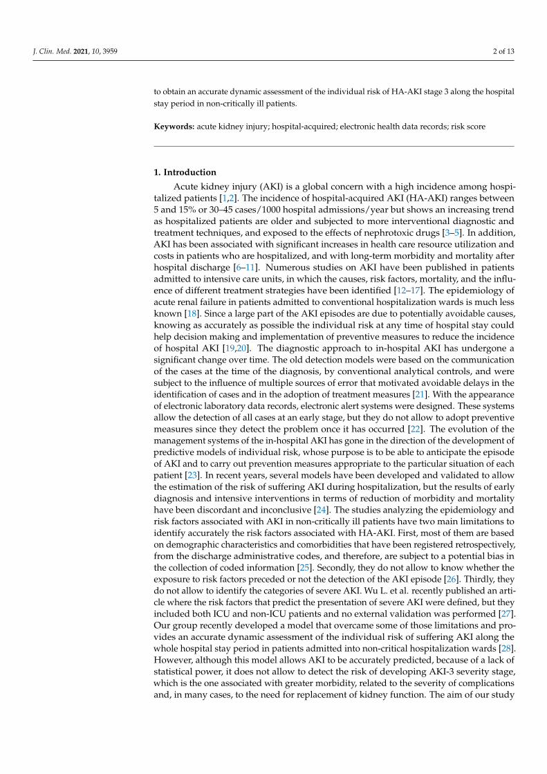
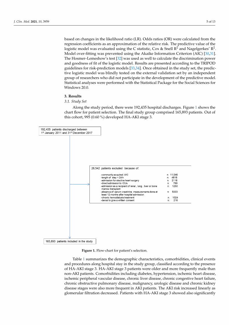
Along the study period, there were 192,435 hospital discharges. Figure 1 shows thechart flow for patient selection. The final study group comprised 165,893 patients. Out ofthis cohort, 995 (0.60 %) developed HA-AKI stage 3.
J. Clin. Med. 2021, 10, 3959 5 of 13
Concordance analyses between qualitative variables was done by the Kappa coefficient. A p value of less than 0.05 was considered statistically significant. To determine which variables were independently associated with AKI, we carried out a univariate analysis comparing patients with and without AKI. All the variables with p values under 0.1 in the univariate analysis were entered into stepwise multiple logistic regression analysis with a forward selection method based on changes in the likelihood ratio (LR). Odds ratios (OR) were calculated from the regression coefficients as an approximation of the relative risk. The predictive value of the logistic model was evaluated using the C statistic, Cox & Snell R2 and Nagelgerkes’ R2. Model over-fitting was prevented using the Akaike Information Criterion (AIC) [30,31]. The Hosmer–Lemeshow’s test [32] was used as well to calculate the discrimination power and goodness of fit of the logistic model. Results are presented according to the TRIPOD guidelines for risk-prediction models [33,34]. Once obtained in the study set, the predictive logistic model was blindly tested on the external validation set by an independent group of researchers who did not participate in the development of the predictive model. Statistical analyses were performed with the Statistical Package for the Social Sciences for Windows 20.0
3. Results 3.1. Study Set
Along the study period, there were 192,435 hospital discharges. Figure 1 shows the chart flow for patient selection. The final study group comprised 165,893 patients. Out of this cohort, 995 (0.60 %) developed HA-AKI stage 3.
Figure 1. Flow-chart for patient’s selection.
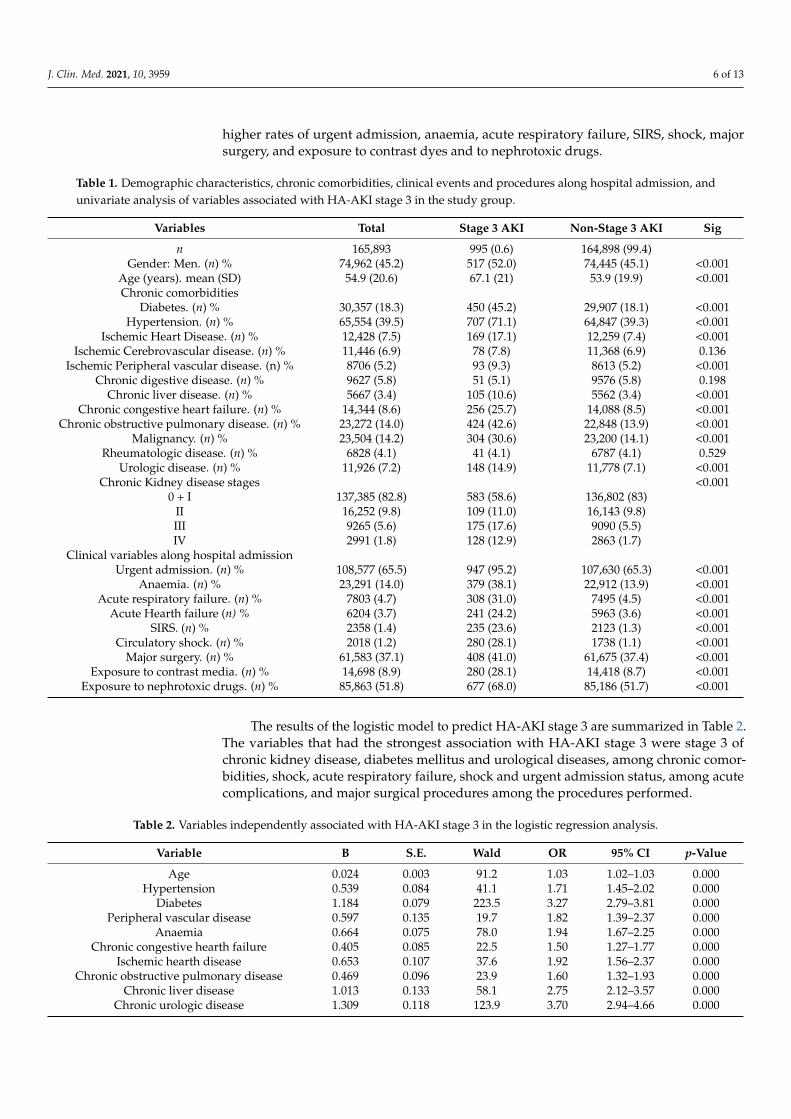
Table 1 summarizes the demographic characteristics, comorbidities, clinical events and procedures along hospital stay in the study group, classified according to the presence of HA-AKI stage 3. HA-AKI stage 3 patients were older and more frequently male than non-AKI patients. Comorbidities including diabetes, hypertension, ischemic heart disease, ischemic peripheral vascular disease, chronic liver disease, chronic congestive heart failure, chronic obstructive pulmonary disease, malignancy, urologic disease and chronic kidney disease stages were also more frequent in AKI patients. The AKI risk
Figure 1. Flow-chart for patient’s selection.
Table 1 summarizes the demographic characteristics, comorbidities, clinical eventsand procedures along hospital stay in the study group, classified according to the presenceof HA-AKI stage 3. HA-AKI stage 3 patients were older and more frequently male thannon-AKI patients. Comorbidities including diabetes, hypertension, ischemic heart disease,ischemic peripheral vascular disease, chronic liver disease, chronic congestive heart failure,chronic obstructive pulmonary disease, malignancy, urologic disease and chronic kidneydisease stages were also more frequent in AKI patients. The AKI risk increased linearly asglomerular filtration decreased. Patients with HA-AKI stage 3 showed also significantly
J. Clin. Med. 2021, 10, 3959 6 of 13
higher rates of urgent admission, anaemia, acute respiratory failure, SIRS, shock, majorsurgery, and exposure to contrast dyes and to nephrotoxic drugs.
Table 1. Demographic characteristics, chronic comorbidities, clinical events and procedures along hospital admission, andunivariate analysis of variables associated with HA-AKI stage 3 in the study group.
Variables Total Stage 3 AKI Non-Stage 3 AKI Sig
n 165,893 995 (0.6) 164,898 (99.4)Gender: Men. (n) % 74,962 (45.2) 517 (52.0) 74,445 (45.1) <0.001
Age (years). mean (SD) 54.9 (20.6) 67.1 (21) 53.9 (19.9) <0.001Chronic comorbidities
Diabetes. (n) % 30,357 (18.3) 450 (45.2) 29,907 (18.1) <0.001Hypertension. (n) % 65,554 (39.5) 707 (71.1) 64,847 (39.3) <0.001
Ischemic Heart Disease. (n) % 12,428 (7.5) 169 (17.1) 12,259 (7.4) <0.001Ischemic Cerebrovascular disease. (n) % 11,446 (6.9) 78 (7.8) 11,368 (6.9) 0.136
Ischemic Peripheral vascular disease. (n) % 8706 (5.2) 93 (9.3) 8613 (5.2) <0.001Chronic digestive disease. (n) % 9627 (5.8) 51 (5.1) 9576 (5.8) 0.198
Chronic liver disease. (n) % 5667 (3.4) 105 (10.6) 5562 (3.4) <0.001Chronic congestive heart failure. (n) % 14,344 (8.6) 256 (25.7) 14,088 (8.5) <0.001
Chronic obstructive pulmonary disease. (n) % 23,272 (14.0) 424 (42.6) 22,848 (13.9) <0.001Malignancy. (n) % 23,504 (14.2) 304 (30.6) 23,200 (14.1) <0.001
Rheumatologic disease. (n) % 6828 (4.1) 41 (4.1) 6787 (4.1) 0.529Urologic disease. (n) % 11,926 (7.2) 148 (14.9) 11,778 (7.1) <0.001
Chronic Kidney disease stages <0.0010 + I 137,385 (82.8) 583 (58.6) 136,802 (83)
II 16,252 (9.8) 109 (11.0) 16,143 (9.8)III 9265 (5.6) 175 (17.6) 9090 (5.5)IV 2991 (1.8) 128 (12.9) 2863 (1.7)
Clinical variables along hospital admissionUrgent admission. (n) % 108,577 (65.5) 947 (95.2) 107,630 (65.3) <0.001
Anaemia. (n) % 23,291 (14.0) 379 (38.1) 22,912 (13.9) <0.001Acute respiratory failure. (n) % 7803 (4.7) 308 (31.0) 7495 (4.5) <0.001
Acute Hearth failure (n) % 6204 (3.7) 241 (24.2) 5963 (3.6) <0.001SIRS. (n) % 2358 (1.4) 235 (23.6) 2123 (1.3) <0.001
Circulatory shock. (n) % 2018 (1.2) 280 (28.1) 1738 (1.1) <0.001Major surgery. (n) % 61,583 (37.1) 408 (41.0) 61,675 (37.4) <0.001
Exposure to contrast media. (n) % 14,698 (8.9) 280 (28.1) 14,418 (8.7) <0.001Exposure to nephrotoxic drugs. (n) % 85,863 (51.8) 677 (68.0) 85,186 (51.7) <0.001
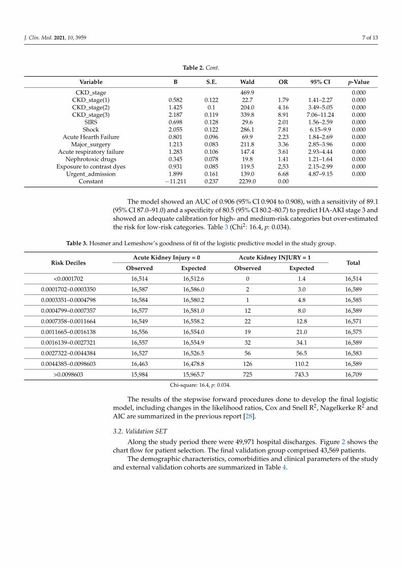
The results of the logistic model to predict HA-AKI stage 3 are summarized in Table 2.The variables that had the strongest association with HA-AKI stage 3 were stage 3 ofchronic kidney disease, diabetes mellitus and urological diseases, among chronic comor-bidities, shock, acute respiratory failure, shock and urgent admission status, among acutecomplications, and major surgical procedures among the procedures performed.
Table 2. Variables independently associated with HA-AKI stage 3 in the logistic regression analysis.
Variable B S.E. Wald OR 95% CI p-Value
Age 0.024 0.003 91.2 1.03 1.02–1.03 0.000Hypertension 0.539 0.084 41.1 1.71 1.45–2.02 0.000
Diabetes 1.184 0.079 223.5 3.27 2.79–3.81 0.000Peripheral vascular disease 0.597 0.135 19.7 1.82 1.39–2.37 0.000
Anaemia 0.664 0.075 78.0 1.94 1.67–2.25 0.000Chronic congestive hearth failure 0.405 0.085 22.5 1.50 1.27–1.77 0.000
Ischemic hearth disease 0.653 0.107 37.6 1.92 1.56–2.37 0.000Chronic obstructive pulmonary disease 0.469 0.096 23.9 1.60 1.32–1.93 0.000
Chronic liver disease 1.013 0.133 58.1 2.75 2.12–3.57 0.000Chronic urologic disease 1.309 0.118 123.9 3.70 2.94–4.66 0.000
J. Clin. Med. 2021, 10, 3959 7 of 13
Table 2. Cont.
Variable B S.E. Wald OR 95% CI p-Value
CKD_stage 469.9 0.000CKD_stage(1) 0.582 0.122 22.7 1.79 1.41–2.27 0.000CKD_stage(2) 1.425 0.1 204.0 4.16 3.49–5.05 0.000CKD_stage(3) 2.187 0.119 339.8 8.91 7.06–11.24 0.000
SIRS 0.698 0.128 29.6 2.01 1.56–2.59 0.000Shock 2.055 0.122 286.1 7.81 6.15–9.9 0.000
Acute Hearth Failure 0.801 0.096 69.9 2.23 1.84–2.69 0.000Major_surgery 1.213 0.083 211.8 3.36 2.85–3.96 0.000
Acute respiratory failure 1.283 0.106 147.4 3.61 2.93–4.44 0.000Nephrotoxic drugs 0.345 0.078 19.8 1.41 1.21–1.64 0.000
Exposure to contrast dyes 0.931 0.085 119.5 2,53 2.15–2.99 0.000Urgent_admission 1.899 0.161 139.0 6.68 4.87–9.15 0.000
Constant −11.211 0.237 2239.0 0.00
The model showed an AUC of 0.906 (95% CI 0.904 to 0.908), with a sensitivity of 89.1(95% CI 87.0–91.0) and a specificity of 80.5 (95% CI 80.2–80.7) to predict HA-AKI stage 3 andshowed an adequate calibration for high- and medium-risk categories but over-estimatedthe risk for low-risk categories. Table 3 (Chi2: 16.4, p: 0.034).
Table 3. Hosmer and Lemeshow’s goodness of fit of the logistic predictive model in the study group.
Risk DecilesAcute Kidney Injury = 0 Acute Kidney INJURY = 1
TotalObserved Expected Observed Expected
<0.0001702 16,514 16,512.6 0 1.4 16,514
0.0001702–0.0003350 16,587 16,586.0 2 3.0 16,589
0.0003351–0.0004798 16,584 16,580.2 1 4.8 16,585
0.0004799–0.0007357 16,577 16,581.0 12 8.0 16,589
0.0007358–0.0011664 16,549 16,558.2 22 12.8 16,571
0.0011665–0.0016138 16,556 16,554.0 19 21.0 16,575
0.0016139–0.0027321 16,557 16,554.9 32 34.1 16,589
0.0027322–0.0044384 16,527 16,526.5 56 56.5 16,583
0.0044385–0.0098603 16,463 16,478.8 126 110.2 16,589
>0.0098603 15,984 15,965.7 725 743.3 16,709
Chi-square: 16.4, p: 0.034.
The results of the stepwise forward procedures done to develop the final logisticmodel, including changes in the likelihood ratios, Cox and Snell R2, Nagelkerke R2 andAIC are summarized in the previous report [28].
3.2. Validation SET
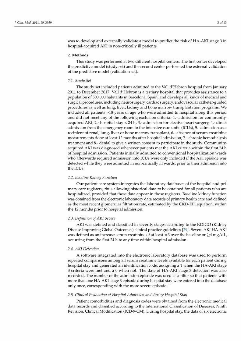
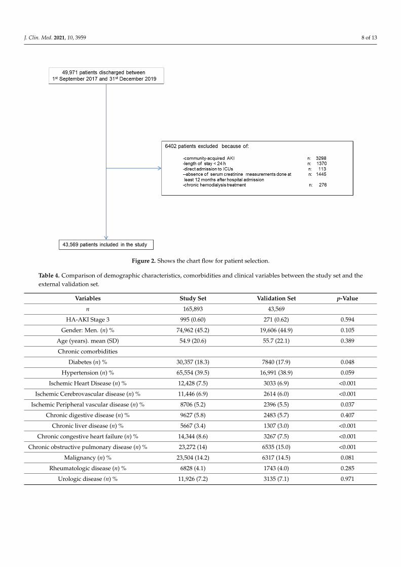
Along the study period there were 49,971 hospital discharges. Figure 2 shows thechart flow for patient selection. The final validation group comprised 43,569 patients.
The demographic characteristics, comorbidities and clinical parameters of the studyand external validation cohorts are summarized in Table 4.
J. Clin. Med. 2021, 10, 3959 8 of 13J. Clin. Med. 2021, 10, 3959 8 of 13
Figure 2. Shows the chart flow for patient selection.
The demographic characteristics, comorbidities and clinical parameters of the study and external validation cohorts are summarized in Table 4.
Table 4. Comparison of demographic characteristics, comorbidities and clinical variables between the study set and the external validation set.
Variables Study Set Validation Set p-Value n 165,893 43,569
HA-AKI Stage 3 995 (0.60) 271 (0.62) 0.594 Gender: Men. (n) % 74,962 (45.2) 19,606 (44.9) 0.105
Age (years). mean (SD) 54.9 (20.6) 55.7 (22.1) 0.389 Chronic comorbidities
Diabetes (n) % 30,357 (18.3) 7840 (17.9) 0.048 Hypertension (n) % 65,554 (39.5) 16,991 (38.9) 0.059
Ischemic Heart Disease (n) % 12,428 (7.5) 3033 (6.9) <0.001 Ischemic Cerebrovascular disease (n) % 11,446 (6.9) 2614 (6.0) <0.001
Ischemic Peripheral vascular disease (n) % 8706 (5.2) 2396 (5.5) 0.037 Chronic digestive disease (n) % 9627 (5.8) 2483 (5.7) 0.407
Chronic liver disease (n) % 5667 (3.4) 1307 (3.0) <0.001 Chronic congestive heart failure (n) % 14,344 (8.6) 3267 (7.5) <0.001
Chronic obstructive pulmonary disease (n) % 23,272 (14) 6535 (15.0) <0.001 Malignancy (n) % 23,504 (14.2) 6317 (14.5) 0.081
Rheumatologic disease (n) % 6828 (4.1) 1743 (4.0) 0.285 Urologic disease (n) % 11,926 (7.2) 3135 (7.1) 0.971
Chronic Kidney Disease stages 0.2758 0 + I 137,385 (82.8) 36,162 (83.0)
II 16,252 (9.8) 4182 (9.6) III 9265 (5.6) 2396 (5.5) IV 2991 (1.8) 829 (1.9)
Clinical variables along hospital admission Urgent admission (n) % 108,577 (65.5) 28,319 (65.0) 0.077
Figure 2. Shows the chart flow for patient selection.
Table 4. Comparison of demographic characteristics, comorbidities and clinical variables between the study set and theexternal validation set.
Variables Study Set Validation Set p-Value
n 165,893 43,569
HA-AKI Stage 3 995 (0.60) 271 (0.62) 0.594
Gender: Men. (n) % 74,962 (45.2) 19,606 (44.9) 0.105
Age (years). mean (SD) 54.9 (20.6) 55.7 (22.1) 0.389
Chronic comorbidities
Diabetes (n) % 30,357 (18.3) 7840 (17.9) 0.048
Hypertension (n) % 65,554 (39.5) 16,991 (38.9) 0.059
Ischemic Heart Disease (n) % 12,428 (7.5) 3033 (6.9) <0.001
Ischemic Cerebrovascular disease (n) % 11,446 (6.9) 2614 (6.0) <0.001
Ischemic Peripheral vascular disease (n) % 8706 (5.2) 2396 (5.5) 0.037
Chronic digestive disease (n) % 9627 (5.8) 2483 (5.7) 0.407
Chronic liver disease (n) % 5667 (3.4) 1307 (3.0) <0.001
Chronic congestive heart failure (n) % 14,344 (8.6) 3267 (7.5) <0.001
Chronic obstructive pulmonary disease (n) % 23,272 (14) 6535 (15.0) <0.001
Malignancy (n) % 23,504 (14.2) 6317 (14.5) 0.081
Rheumatologic disease (n) % 6828 (4.1) 1743 (4.0) 0.285
Urologic disease (n) % 11,926 (7.2) 3135 (7.1) 0.971
J. Clin. Med. 2021, 10, 3959 9 of 13
Table 4. Cont.
Variables Study Set Validation Set p-Value
Chronic Kidney Disease stages 0.2758
0 + I 137,385 (82.8) 36,162 (83.0)
II 16,252 (9.8) 4182 (9.6)
III 9265 (5.6) 2396 (5.5)
IV 2991 (1.8) 829 (1.9)
Clinical variables along hospital admission
Urgent admission (n) % 108,577 (65.5) 28,319 (65.0) 0.077
Anaemia (n) % 23,291 (14.0) 6186 (14.2) 0.397
Acute respiratory failure (n) % 7803 (4.7) 2178 (5.0) 0.011
Acute Hearth failure (n) % 6204 (3.7) 1655 (3.8) 0.565
SIRS (n) % 2358 (1.4) 653 (1.5) 0.227
Circulatory shock (n) % 2018 (1.2) 566 (1.3) 0.167
Major surgery (n) % 61,583 (37.1) 13,942 (32.0) <0.001
Exposure to contrast dyes (n) % 14,698 (8.9) 3.921 (9.0) 0.36
Exposure to nephrotoxic drugs (n) % 85,863 (51.8) 23,135 (53.1) <0.001
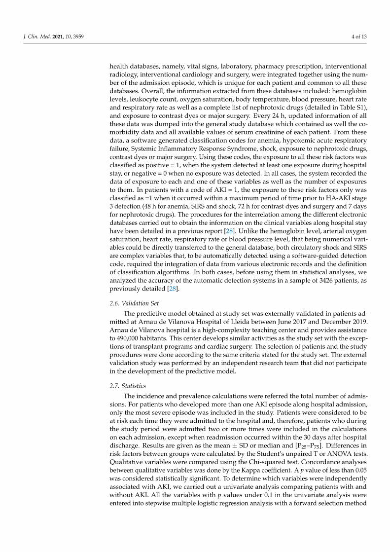
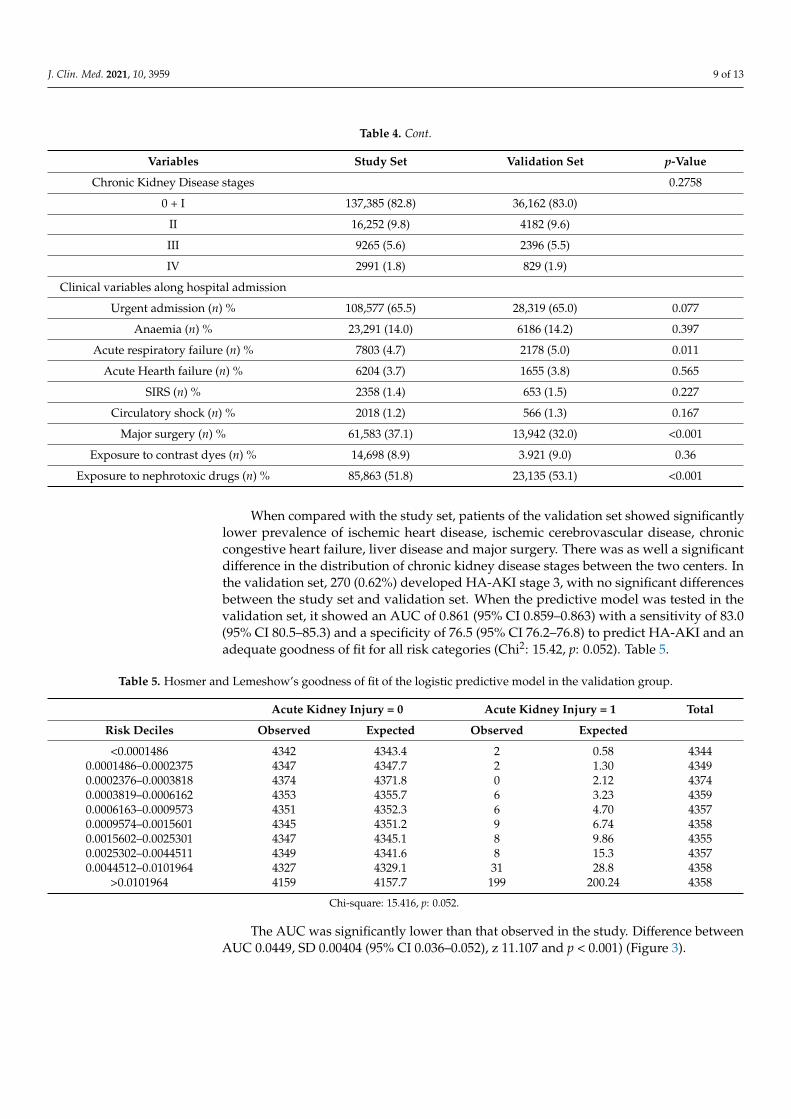
When compared with the study set, patients of the validation set showed significantlylower prevalence of ischemic heart disease, ischemic cerebrovascular disease, chroniccongestive heart failure, liver disease and major surgery. There was as well a significantdifference in the distribution of chronic kidney disease stages between the two centers. Inthe validation set, 270 (0.62%) developed HA-AKI stage 3, with no significant differencesbetween the study set and validation set. When the predictive model was tested in thevalidation set, it showed an AUC of 0.861 (95% CI 0.859–0.863) with a sensitivity of 83.0(95% CI 80.5–85.3) and a specificity of 76.5 (95% CI 76.2–76.8) to predict HA-AKI and anadequate goodness of fit for all risk categories (Chi2: 15.42, p: 0.052). Table 5.
Table 5. Hosmer and Lemeshow’s goodness of fit of the logistic predictive model in the validation group.
Acute Kidney Injury = 0 Acute Kidney Injury = 1 Total
Risk Deciles Observed Expected Observed Expected
<0.0001486 4342 4343.4 2 0.58 43440.0001486–0.0002375 4347 4347.7 2 1.30 43490.0002376–0.0003818 4374 4371.8 0 2.12 43740.0003819–0.0006162 4353 4355.7 6 3.23 43590.0006163–0.0009573 4351 4352.3 6 4.70 43570.0009574–0.0015601 4345 4351.2 9 6.74 43580.0015602–0.0025301 4347 4345.1 8 9.86 43550.0025302–0.0044511 4349 4341.6 8 15.3 43570.0044512–0.0101964 4327 4329.1 31 28.8 4358
>0.0101964 4159 4157.7 199 200.24 4358
Chi-square: 15.416, p: 0.052.
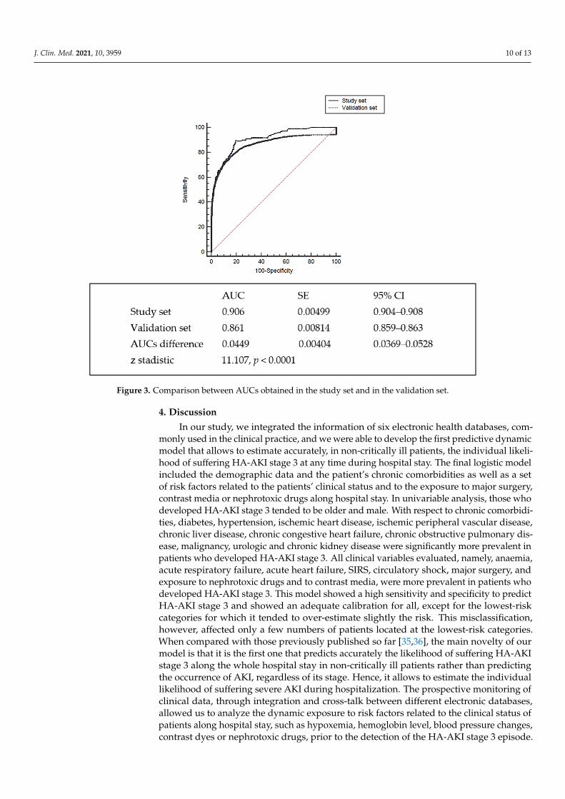
The AUC was significantly lower than that observed in the study. Difference betweenAUC 0.0449, SD 0.00404 (95% CI 0.036–0.052), z 11.107 and p < 0.001) (Figure 3).
J. Clin. Med. 2021, 10, 3959 10 of 13
J. Clin. Med. 2021, 10, 3959 9 of 13
Anaemia (n) % 23,291 (14.0) 6186 (14.2) 0.397 Acute respiratory failure (n) % 7803 (4.7) 2178 (5.0) 0.011
Acute Hearth failure (n) % 6204 (3.7) 1655 (3.8) 0.565 SIRS (n) % 2358 (1.4) 653 (1.5) 0.227
Circulatory shock (n) % 2018 (1.2) 566 (1.3) 0.167 Major surgery (n) % 61,583 (37.1) 13,942 (32.0) <0.001
Exposure to contrast dyes (n) % 14,698 (8.9) 3.921 (9.0) 0.36 Exposure to nephrotoxic drugs (n) % 85,863 (51.8) 23,135 (53.1) <0.001
When compared with the study set, patients of the validation set showed significantly lower prevalence of ischemic heart disease, ischemic cerebrovascular disease, chronic congestive heart failure, liver disease and major surgery. There was as well a significant difference in the distribution of chronic kidney disease stages between the two centers. In the validation set, 270 (0.62%) developed HA-AKI stage 3, with no significant differences between the study set and validation set. When the predictive model was tested in the validation set, it showed an AUC of 0.861 (95% CI 0.859–0.863) with a sensitivity of 83.0 (95% CI 80.5–85.3) and a specificity of 76.5 (95% CI 76.2–76.8) to predict HA-AKI and an adequate goodness of fit for all risk categories (Chi2:15.42, p: 0.052). Table 5.
Table 5. Hosmer and Lemeshow’s goodness of fit of the logistic predictive model in the validation group.
Acute Kidney Injury = 0 Acute Kidney Injury = 1 Total Risk Deciles Observed Expected Observed Expected <0.0001486 4342 4343,4 2 0.58 4344
0.0001486–0.0002375 4347 4347,7 2 1.30 4349 0.0002376–0.0003818 4374 4371,8 0 2.12 4374 0.0003819–0.0006162 4353 4355,7 6 3.23 4359 0.0006163–0.0009573 4351 4352,3 6 4.70 4357 0.0009574–0.0015601 4345 4351,2 9 6.74 4358 0.0015602–0.0025301 4347 4345,1 8 9.86 4355 0.0025302–0.0044511 4349 4341,6 8 15.3 4357 0.0044512–0.0101964 4327 4329,1 31 28.8 4358
>0.0101964 4159 4157,7 199 200.24 4358 Chi-square: 15.416, p: 0.052.
The AUC was significantly lower than that observed in the study. Difference between AUC 0.0449, SD 0.00404 (95% CI 0.036–0.052), z 11.107 and p < 0.001) (Figure 3).
J. Clin. Med. 2021, 10, 3959 10 of 13
Figure 3. Comparison between AUCs obtained in the study set and in the validation set.
4. Discussion In our study, we integrated the information of six electronic health databases,
commonly used in the clinical practice, and we were able to develop the first predictive dynamic model that allows to estimate accurately, in non-critically ill patients, the individual likelihood of suffering HA-AKI stage 3 at any time during hospital stay. The final logistic model included the demographic data and the patient’s chronic comorbidities as well as a set of risk factors related to the patients’ clinical status and to the exposure to major surgery, contrast media or nephrotoxic drugs along hospital stay. In univariable analysis, those who developed HA-AKI stage 3 tended to be older and male. With respect to chronic comorbidities, diabetes, hypertension, ischemic heart disease, ischemic peripheral vascular disease, chronic liver disease, chronic congestive heart failure, chronic obstructive pulmonary disease, malignancy, urologic and chronic kidney disease were significantly more prevalent in patients who developed HA-AKI stage 3. All clinical variables evaluated, namely, anaemia, acute respiratory failure, acute heart failure, SIRS, circulatory shock, major surgery, and exposure to nephrotoxic drugs and to contrast media, were more prevalent in patients who developed HA-AKI stage 3. This model showed a high sensitivity and specificity to predict HA-AKI stage 3 and showed an adequate calibration for all, except for the lowest-risk categories for which it tended to over-estimate slightly the risk. This misclassification, however, affected only a few numbers of patients located at the lowest-risk categories. When compared with those previously published so far [35,36], the main novelty of our model is that it is the first one that predicts accurately the likelihood of suffering HA-AKI stage 3 along the whole hospital stay in non-critically ill patients rather than predicting the occurrence of AKI, regardless of its stage. Hence, it allows to estimate the individual likelihood of suffering severe AKI during hospitalization. The prospective monitoring of clinical data, through integration and cross-talk between different electronic databases, allowed us to analyze the dynamic exposure to risk factors related to the clinical status of patients along hospital stay, such as hypoxemia, hemoglobin level, blood pressure changes, contrast dyes or nephrotoxic drugs, prior to the detection of the HA-AKI stage 3 episode. This integration allowed as well to perform an accurate and reliable transformation of single variables such as blood pressure, heart rate, arterial oxygen saturation, prescription of vasoactive drugs or blood leukocyte counts into more complex variables defining specific syndromes such as SIRS and circulatory shock. Electronic records also permitted us to record the exposure to the same variables and risk factors in patients who did not develop HA-AKI stage 3 during hospital admission. This approach made it possible to estimate the individual risk, based on the actual exposure to each and one of risk factors. Since our predictive model was developed from the values of risk factors assessed prior to HA-AKI stage 3 detection, it allows to perform a dynamic monitoring of risk and even to predict the changes in the individual risk that are expected to happen every time the value of the different predictive risk factors changes. In order to obtain a predictive model that could be exportable to hospitals with different case-mix, patients who were admitted for programs and/or procedures such as cardiac surgery, solid organ or bone marrow transplantation, that are not commonly available to all hospital centers, were deliberately excluded from the study set. When comparing the study and the validation sets, we still observed statistically
Figure 3. Comparison between AUCs obtained in the study set and in the validation set.
4. Discussion
In our study, we integrated the information of six electronic health databases, com-monly used in the clinical practice, and we were able to develop the first predictive dynamicmodel that allows to estimate accurately, in non-critically ill patients, the individual likeli-hood of suffering HA-AKI stage 3 at any time during hospital stay. The final logistic modelincluded the demographic data and the patient’s chronic comorbidities as well as a setof risk factors related to the patients’ clinical status and to the exposure to major surgery,contrast media or nephrotoxic drugs along hospital stay. In univariable analysis, those whodeveloped HA-AKI stage 3 tended to be older and male. With respect to chronic comorbidi-ties, diabetes, hypertension, ischemic heart disease, ischemic peripheral vascular disease,chronic liver disease, chronic congestive heart failure, chronic obstructive pulmonary dis-ease, malignancy, urologic and chronic kidney disease were significantly more prevalent inpatients who developed HA-AKI stage 3. All clinical variables evaluated, namely, anaemia,acute respiratory failure, acute heart failure, SIRS, circulatory shock, major surgery, andexposure to nephrotoxic drugs and to contrast media, were more prevalent in patients whodeveloped HA-AKI stage 3. This model showed a high sensitivity and specificity to predictHA-AKI stage 3 and showed an adequate calibration for all, except for the lowest-riskcategories for which it tended to over-estimate slightly the risk. This misclassification,however, affected only a few numbers of patients located at the lowest-risk categories.When compared with those previously published so far [35,36], the main novelty of ourmodel is that it is the first one that predicts accurately the likelihood of suffering HA-AKIstage 3 along the whole hospital stay in non-critically ill patients rather than predictingthe occurrence of AKI, regardless of its stage. Hence, it allows to estimate the individuallikelihood of suffering severe AKI during hospitalization. The prospective monitoring ofclinical data, through integration and cross-talk between different electronic databases,allowed us to analyze the dynamic exposure to risk factors related to the clinical status ofpatients along hospital stay, such as hypoxemia, hemoglobin level, blood pressure changes,contrast dyes or nephrotoxic drugs, prior to the detection of the HA-AKI stage 3 episode.
J. Clin. Med. 2021, 10, 3959 11 of 13
This integration allowed as well to perform an accurate and reliable transformation ofsingle variables such as blood pressure, heart rate, arterial oxygen saturation, prescriptionof vasoactive drugs or blood leukocyte counts into more complex variables defining specificsyndromes such as SIRS and circulatory shock. Electronic records also permitted us torecord the exposure to the same variables and risk factors in patients who did not developHA-AKI stage 3 during hospital admission. This approach made it possible to estimatethe individual risk, based on the actual exposure to each and one of risk factors. Since ourpredictive model was developed from the values of risk factors assessed prior to HA-AKIstage 3 detection, it allows to perform a dynamic monitoring of risk and even to predictthe changes in the individual risk that are expected to happen every time the value of thedifferent predictive risk factors changes. In order to obtain a predictive model that could beexportable to hospitals with different case-mix, patients who were admitted for programsand/or procedures such as cardiac surgery, solid organ or bone marrow transplantation,that are not commonly available to all hospital centers, were deliberately excluded from thestudy set. When comparing the study and the validation sets, we still observed statisticallysignificant differences in the prevalence of several chronic comorbidities, in spite of the factthat, in both cohorts, we used the same ICD-9 codes to classy them. These differences maybe due to dissimilarities in the case mix between both hospitals, but may also be caused bybiases associated with potential discrepancies in assigning administrative codes to clinicalconditions [37]. There were also between-group differences in other variables involvedin the calculation of the risk of HA-AKI stage 3, such as the total percentage of urgent orsurgical admissions. The discrimination ability of the model in the validation cohort wasslightly but significantly lower than that observed in the original cohort. These differencesare expected to be found when a predictive model is externally validated, and may bepartially attributable to some degree of overfitting of the derivation modeling [38,39]. Thecalibration of the model in the external validation cohort showed a similar trend to thatobserved in the derivation cohort. Overall, the differences in the performance of the modelbetween the study set and the validation set were small, which supports the potentialscalability of the predictive model to fewer complex centers.
Our model has some limitations that must be highlighted. First, the record of clinicalvariables such as blood pressure, heart rate, respiratory rate or oxygen saturation wereautomatically dumped into the study database; however, these values are not withoutpotential error related to the variability in the manual introduction of these variablesinto their corresponding databases. Second, the model obtained in our study is not theonly one that can be obtained with the combination of data obtained from electronicrecords. As exposure to each of the acute complications or nephrotoxic agents can occur atdifferent times after hospital admission, in order to relate the exposure to them with thedevelopment of HA-AKI stage 3, it was necessary to define a maximum period of timebetween exposure and detection of HA-AKI stage 3. In our study, the duration of thisperiod of time was defined by consensus of the research group, using pathophysiologicalcriteria. The definition of other periods of time, based on alternative criteria, would modifythe prevalence of exposure to these risk factors and, consequently, the magnitude of theassociations found between these variables and HA-AKI stage 3.
In conclusion, our study provides the first model, based on demographic data, specificcomorbidities, acute clinical conditions and procedures, that can be used in clinical practiceto obtain an accurate dynamic assessment of the individual risk of suffering HA-AKI stage3 along the whole hospital stay period in patients admitted into non-critical hospitalizationwards. This model allows from performing a repeated manual risk estimation, using theprediction algorithm, to providing an automatic risk measure updated in real time, in thosecenters where it is possible to carry out a complete integration among the health databasescontaining the necessary information. We anticipate that our study sets the cornerstoneto a change in the management of hospital acute renal failure, by using a dynamic modelof integration of electronic records with the aim of awareness of the physician in charge
J. Clin. Med. 2021, 10, 3959 12 of 13
to these patients at high risk for AKI 3. It should be the aim to take special care to thesepatients at high risk to prevent acute renal failure and thus avoid fatal outcomes.
The anonymized database is available for reproduction as long as the requestorattaches a document endorsed by an ethical committee.
Supplementary Materials: The following are available online at https://www.mdpi.com/article/10.3390/jcm10173959/s1, Table S1: List of nephrotoxic drugs included.
Author Contributions: A.S.: Principal investigator. J.D.C.: correspondence author. A.S. and J.D.C.conceptualized and performed the tests and wrote the manuscript. The rest of authors super-vised and reviewed the manuscript. All authors have read and agreed to the published version ofthe manuscript.
Funding: This research received no external funding.
Institutional Review Board Statement: The ethics committee of the Arnau de Vilanova Hospitalwas consulted and they decided that an informed consent was not necessary for the validation of themodel given that no type of intervention was carried out on the patients.
Informed Consent Statement: Not applicable.
Data Availability Statement: The anonymized database is available for reproduction as long as therequestor attaches a document endorsed by an ethical committee.
Conflicts of Interest: The authors declare no conflict of interest.
References1. Kashani, K.; Rosner, M.H.; Haase, M.; Lewington, A.J.P.; O’Donoghue, D.J.; Wilson, F.P.; Nadim, M.K.; Silver, S.A.; Zarbock, A.;
Ostermann, M.; et al. Quality Improvement Goals for Acute Kidney Injury. Clin. J. Am. Soc. Nephrol. 2019, 14, 941–953. [CrossRef][PubMed]
2. Mehta, R.L.; Burdmann, E.A.; Cerdá, J.; Feehally, J.; Finkelstein, F.; García-García, G.; Godin, M.; Jha, V.; Lameire, N.H.; Levin,N.W.; et al. Recognition and Management of Acute Kidney Injury in the International Society of Nephrology 0by25 GlobalSnapshot: A Multinational Cross-Sectional Study. Lancet 2016, 387, 2017–2025. [CrossRef]
3. Wonnacott, A.; Meran, S.; Amphlett, B.; Talabani, B.; Phillips, A. Epidemiology and Outcomes in Community-Acquired versusHospital-Acquired AKI. Clin. J. Am. Soc. Nephrol. 2014, 9, 1007–1014. [CrossRef] [PubMed]
4. Meier, P.; Bonfils, R.M.; Vogt, B.; Burnand, B.; Burnier, M. Referral Patterns and Outcomes in Noncritically Ill Patients withHospital-Acquired Acute Kidney Injury. Clin. J. Am. Soc. Nephrol. 2011, 6, 2215–2225. [CrossRef]
5. Yong, K.; Dogra, G.; Boudville, N.; Pinder, M.; Lim, W. Acute Kidney Injury: Controversies Revisited. Int. J. Nephrol. 2011,2011, 762634. [CrossRef]
6. Pruchnicki, M.C.; Dasta, J.F. Acute Renal Failure in Hospitalized Patients: Part I. Ann. Pharmacother. 2002, 36, 1261–1267.[CrossRef]
7. Patschan, D.; Müller, G.A. Acute Kidney Injury. J. Inj. Violence Res. 2015, 7, 19–26. [CrossRef]8. Forni, L.G.; Darmon, M.; Ostermann, M.; Oudemans-van Straaten, H.M.; Pettilä, V.; Prowle, J.R.; Schetz, M.; Joannidis, M. Renal
Recovery after Acute Kidney Injury. Intensive Care Med. 2017, 43, 855–866. [CrossRef] [PubMed]9. Bucaloiu, I.D.; Kirchner, H.L.; Norfolk, E.R.; Hartle, J.E.; Perkins, R.M. Increased Risk of Death and de Novo Chronic Kidney
Disease Following Reversible Acute Kidney Injury. Kidney Int. 2012, 81, 477–485. [CrossRef] [PubMed]10. Collister, D.; Pannu, N.; Ye, F.; James, M.; Hemmelgarn, B.; Chui, B.; Manns, B.; Klarenbach, S. Alberta Kidney Disease Network.
Health Care Costs Associated with AKI. Clin. J. Am. Soc. Nephrol. 2017, 12, 1733–1743. [CrossRef]11. Chertow, G.M.; Burdick, E.; Honour, M.; Bonventre, J.V.; Bates, D.W. Acute Kidney Injury, Mortality, Length of Stay, and Costs in
Hospitalized Patients. JASN 2005, 16, 3365–3370. [CrossRef]12. Susantitaphong, P.; Cruz, D.N.; Cerda, J.; Abulfaraj, M.; Alqahtani, F.; Koulouridis, I.; Jaber, B.L. Acute Kidney Injury Advisory
Group of the American Society of Nephrology. World Incidence of AKI: A Meta-Analysis. Clin. J. Am. Soc. Nephrol. 2013, 8,1482–1493. [CrossRef]
13. Ostermann, M. Acute Kidney Injury on Admission to the Intensive Care Unit: Where to Go from Here? Crit. Care 2008, 12, 189.[CrossRef]
14. Mas-Font, S.; Ros-Martinez, J.; Pérez-Calvo, C.; Villa-Díaz, P.; Aldunate-Calvo, S.; Moreno-Clari, E. Prevention of acute kidneyinjury in intensive care units. Med. Intensiva 2017, 41, 116–126. [CrossRef] [PubMed]
15. Seller-Pérez, G.; Más-Font, S.; Pérez-Calvo, C.; Villa-Díaz, P.; Celaya-López, M.; Herrera-Gutiérrez, M.E. Acute kidney injury:Renal disease in the ICU. Med. Intensiva 2016, 40, 374–382. [CrossRef]
J. Clin. Med. 2021, 10, 3959 13 of 13
16. Hoste, E.A.; Bagshaw, S.M.; Bellomo, R.; Cely, C.M.; Colman, R.; Cruz, D.N.; Edipidis, K.; Forni, L.G.; Gomersall, C.D.; Govil, D.;et al. Epidemiology of acute kidney injury in critically ill patients: The multinational AKI-EPI study. Intensive Care Med. 2015, 41,1411–1423. [CrossRef]
17. Hsu, C.-Y.; McCulloch, C.E.; Fan, D.; Ordoñez, J.D.; Chertow, G.M.; Go, A.S. Community-Based Incidence of Acute Renal Failure.Kidney Int. 2007, 72, 208–212. [CrossRef] [PubMed]
18. Barrantes, F.; Feng, Y.; Ivanov, O.; Yalamanchili, H.B.; Patel, J.; Buenafe, X.; Cheng, V.; Dijeh, S.; Amoateng-Adjepong, Y.; Manthous,C.A. Acute Kidney Injury Predicts Outcomes of Non-Critically Ill Patients. Mayo Clin. Proc. 2009, 84, 410–416. [CrossRef]
19. Harty, J. Prevention and Management of Acute Kidney Injury. Ulster Med. J. 2014, 83, 149–157. [PubMed]20. Cheng, P.; Waitman, L.R.; Hu, Y.; Liu, M. Predicting Inpatient Acute Kidney Injury over Different Time Horizons: How Early and
Accurate? AMIA Annu. Symp. Proc. 2017, 2017, 565–574.21. Bedford, M.; Stevens, P.; Coulton, S.; Billings, J.; Farr, M.; Wheeler, T.; Kalli, M.; Mottishaw, T.; Farmer, C. Development of Risk
Models for the Prediction of New or Worsening Acute Kidney Injury on or during Hospital Admission: A Cohort and Nested Study; HealthServices and Delivery Research; NIHR Journals Library: Southampton, UK, 2016.
22. James, M.T.; Hobson, C.E.; Darmon, M.; Mohan, S.; Hudson, D.; Goldstein, S.L.; Ronco, C.; Kellum, J.A.; Bagshaw, S.M. AcuteDialysis Quality Initiative (ADQI) Consensus Group. Applications for Detection of Acute Kidney Injury Using Electronic MedicalRecords and Clinical Information Systems: Workgroup Statements from the 15(Th) ADQI Consensus Conference. Can. J. KidneyHealth Dis. 2016, 3, 9. [CrossRef] [PubMed]
23. Koyner, J.L.; Adhikari, R.; Edelson, D.P.; Churpek, M.M. Development of a Multicenter Ward-Based AKI Prediction Model. Clin.J. Am. Soc. Nephrol. 2016, 11, 1935–1943. [CrossRef]
24. Hodgson, L.E.; Dimitrov, B.D.; Roderick, P.J.; Venn, R.; Forni, L.G. Predicting AKI in Emergency Admissions: An ExternalValidation Study of the Acute Kidney Injury Prediction Score (APS). BMJ Open 2017, 7, e013511. [CrossRef] [PubMed]
25. O’Malley, K.J.; Cook, K.F.; Price, M.D.; Wildes, K.R.; Hurdle, J.F.; Ashton, C.M. Measuring Diagnoses: ICD Code Accuracy. HealthServ. Res. 2005, 40 Pt 2, 1620–1639. [CrossRef]
26. Hodgson, L.E.; Sarnowski, A.; Roderick, P.J.; Dimitrov, B.D.; Venn, R.M.; Forni, L.G. Systematic Review of Prognostic PredictionModels for Acute Kidney Injury (AKI) in General Hospital Populations. BMJ Open 2017, 7, e016591. [CrossRef]
27. Wu, L.; Hu, Y.; Yuan, B.; Zhang, X.; Chen, W.; Liu, K.; Liu, M. Which Risk Predictors Are More Likely to Indicate Severe AKI inHospitalized Patients? Int. J. Med. Inform. 2020, 143, 104270. [CrossRef]
28. Segarra, A.; Del Carpio, J.; Marco, M.P.; Jatem, E.; Gonzalez, J.; Chang, P.; Ramos, N.; de la Torre, J.; Prat, J.; Torres, M.J.; et al.Integrating Electronic Health Data Records to Develop and Validate a Predictive Model of Hospital-Acquired Acute KidneyInjury in Non-Critically Ill Patients. Clin. Kidney J. 2021, sfab094. [CrossRef]
29. Kidney Disease: Improving Global Outcomes (KDIGO) Acute Kidney Injury Work Group. KDIGO Clinical Practice Guideline forAcute Kidney Injury. Kidney Int. 2012, 2 (Suppl. 1), 1–138.
30. Akaike, H. Likelihood of a Model and Information Criteria. J. Econom. 1981, 16, 3–14. [CrossRef]31. Cavanaugh, J.E. Unifying the Derivations for the Akaike and Corrected Akaike Information Criteria. Stat. Probab. Lett. 1997, 33,
201–208. [CrossRef]32. Hosmer, D.W.; Lemeshow, S. Confidence Interval Estimates of an Index of Quality Performance Based on Logistic Regression
Models. Stat. Med. 1995, 14, 2161–2172. [CrossRef]33. Moons, K.G.M.; Altman, D.G.; Reitsma, J.B.; Ioannidis, J.P.A.; Macaskill, P.; Steyerberg, E.W.; Vickers, A.J.; Ransohoff, D.F.; Collins,
G.S. Transparent Reporting of a Multivariable Prediction Model for Individual Prognosis or Diagnosis (TRIPOD): Explanationand Elaboration. Ann. Intern. Med. 2015, 162, W1–W73. [CrossRef]
34. Pencina, M.J.; D’Agostino, R.B.; D’Agostino, R.B.; Vasan, R.S. Evaluating the Added Predictive Ability of a New Marker: FromArea under the ROC Curve to Reclassification and Beyond. Stat. Med. 2008, 27, 157–172. [CrossRef] [PubMed]
35. Martin-Cleary, C.; Molinero-Casares, L.M.; Ortiz, A.; Arce-Obieta, J.M. Development and Internal Validation of a PredictionModel for Hospital-Acquired Acute Kidney Injury. Clin. Kidney J. 2021, 14, 309–316. [CrossRef]
36. Tomašev, N.; Glorot, X.; Rae, J.W.; Zielinski, M.; Askham, H.; Saraiva, A.; Mottram, A.; Meyer, C.; Ravuri, S.; Protsyuk, I.; et al.A Clinically Applicable Approach to Continuous Prediction of Future Acute Kidney Injury. Nature 2019, 572, 116–119. [CrossRef]
37. Bell, S.; James, M.T.; Farmer, C.K.T.; Tan, Z.; de Souza, N.; Witham, M.D. Development and External Validation of an AcuteKidney Injury Risk Score for Use in the General Population. Clin. Kidney J. 2020, 13, 402–412. [CrossRef]
38. Steyerberg, E.W.; Vickers, A.J.; Cook, N.R.; Gerds, T.; Gonen, M.; Obuchowski, N.; Pencina, M.J.; Kattan, M.W. Assessing thePerformance of Prediction Models: A Framework for Traditional and Novel Measures. Epidemiology 2010, 21, 128–138. [CrossRef][PubMed]
39. Austin, P.C.; Steyerberg, E.W. Graphical Assessment of Internal and External Calibration of Logistic Regression Models by UsingLoess Smoothers. Stat. Med. 2014, 33, 517–535. [CrossRef]