determining intestinal parasitic infections (ipis) in inmates
TRANSCRIPT
RESEARCH ARTICLE Open Access
Determining intestinal parasitic infections(IPIs) in inmates from Kajang Prison,Selangor, Malaysia for improved prisonmanagementLorainne Angal1, Rohela Mahmud1, Sajideh Samin2, Nan-Jiun Yap1, Romano Ngui1, Amirah Amir1, Init Ithoi1,Adeeba Kamarulzaman3 and Yvonne AL Lim1*
Abstract
Background: The prison management in Malaysia is proactively seeking to improve the health status of the prisoninmates. Intestinal parasitic infections (IPIs) are widely distributed throughout the world and are still gaining great concerndue to their significant morbidity and mortality among infected humans. In Malaysia, there is a paucity of information onIPIs among prison inmates. In order to further enhance the current health strategies employed, the present study aimsto establish firm data on the prevalence and diversity of IPIs among HIV-infected and non-HIV-infected individuals in aprison, an area in which informed knowledge is still very limited.
Methods: Samples were subjected to microscopy examination and serological test (only for Strongyloides). Speciationfor parasites on microscopy-positive samples and seropositive samples for Strongyloides were further determined viapolymerase chain reaction. SPSS was used for statistical analysis.
Results: A total of 294 stool and blood samples each were successfully collected, involving 131 HIV positive and 163 HIVnegative adult male inmates whose age ranged from 21 to 69-years-old. Overall prevalence showed 26.5 % was positivefor various IPIs. The IPIs detected included Blastocystis sp., Strongyloides stercoralis, Entamoeba spp., Cryptosporidium spp.,Giardia spp., and Trichuris trichiura. Comparatively, the rate of IPIs was slightly higher among the HIV positive inmates(27.5 %) than HIV negative inmates (25.8 %). Interestingly, seropositivity for S. stercoralis was more predominant in HIVnegative inmates (10.4 %) compared to HIV-infected inmates (6.9 %), however these findings were not statisticallysignificant. Polymerase chain reaction (PCR) confirmed the presence of Blastocystis, Strongyloides, Entamoeba histolyticaand E. dispar.
Conclusions: These data will enable the health care providers and prison management staff to understand the trendand epidemiological situations in HIV/parasitic co-infections in a prison. This information will further assist in providingevidence-based guidance to improve prevention, control and management strategies of IPIs co-infections among bothHIV positive and HIV negative inmates in a prison environment.
Keywords: HIV, Intestinal parasites, Prison inmates, Malaysia
* Correspondence: [email protected] of Parasitology, Faculty of Medicine, University of Malaya, KualaLumpur, MalaysiaFull list of author information is available at the end of the article
© 2015 Angal et al. Open Access This article is distributed under the terms of the Creative Commons Attribution 4.0International License (http://creativecommons.org/licenses/by/4.0/), which permits unrestricted use, distribution, andreproduction in any medium, provided you give appropriate credit to the original author(s) and the source, provide a link tothe Creative Commons license, and indicate if changes were made. The Creative Commons Public Domain Dedication waiver(http://creativecommons.org/publicdomain/zero/1.0/) applies to the data made available in this article, unless otherwise stated.
Angal et al. BMC Infectious Diseases (2015) 15:467 DOI 10.1186/s12879-015-1178-3

BackgroundIntestinal parasitic infections (IPIs) are highly transmis-sible in a crowded environment and in a closed-contactcommunity such as in day-care centres and prisons. InMalaysia, high prevalence of parasitic infections amongrural communities are commonly reported, however thereis no available information on the occurrence of theseinfections among prison inmates. The only study amongincarcerated population was conducted by Kamel et al.(1994) [1] on cryptosporidiosis among human immuno-deficiency virus (HIV) positive drug addicts in TampinDrug Rehabilitation Centre, Malaysia, reporting a preva-lence of 23 %. Other foreign studies in Canada, Hollandand Nigeria reported high prevalence of IPIs amongprison inmates ranging from 27–77 % [2-5]. Ishaleku andMamman (2014) [6] postulated that prison inmates havehigher risk of acquiring IPIs due to the environment fromwhich they come from and by the prison in which theylive. Most professionals (e.g. clinicians and researchers)working in prison setup usually encountered problemssuch as lack of nutrition and concern, inadequate healthcare facilities and expertise which may further aggravatethe spread of parasitic infections among prisoners.Furthermore, prisoners also usually have high burden
of HIV/AIDS. HIV prevalence among prisoners is be-tween six to fifty times higher than that of the generaladult population. Prisons have high prevalence of HIV-infected and at-risk populations, due to overcrowding,poor nutrition, limited access to health care, continueddrug use, unsafe injecting practices, unprotected sex andtattooing. Furthermore, majority of the inmates comefrom marginalized populations with an elevated risk forHIV such as intravenous drug users (IVDUs) [7]. Bothparasitic infections and HIV/AIDS are common prob-lems present among prison inmates [8].IPIs are known to be prevalent among the immuno-
competent individuals in Malaysia [9-14]. However,the epidemiological status of these infections amongimmunocompromised people (i.e. HIV positive) is stillscarce. Studies on correlation between cryptosporidi-osis [1, 15-17], microsporidiosis [18], strongyloidiasis[19] and IPIs [20] among immunocompromised indi-viduals (i.e. HIV-infected populations) are available.But most of the studies were among hospitalised indi-viduals and one among the drug addicts. Currently,there are no studies on IPIs among the prison inmatesin Malaysia.The prison management in Malaysia is proactive in
wanting to improve the health conditions of the prisoninmates. Thus, the present study highlights the study ofIPIs among the HIV-infected and non-HIV-infected in-mates in a prison in the state of Selangor, Malaysia. Soil-transmitted helminths (STHs) including Ascaris, Trichurisand hookworms are the most common IPIs reported not
only in Selangor but generally in Malaysia [21, 22]. It isimportant to determine and compare the IPIs between theHIV-infected and non-HIV-infected inmates because thereis a high burden of HIV in the prison inmates and theparasitic infections among these two groups may differ.In conjunction with that, the study aims to establish a
platform to understand the trend and epidemiologicalsituations in HIV positive, HIV negative and intestinalparasites co-infections in a prison setup. The prisonmanagement can consider for improved preventive andcontrol strategies for the wellbeing of the inmates par-ticularly pertaining to parasitic infections. Besides, thepresent study will help to contribute towards increaseawareness among health care personnels, the prisoninmates and public, the importance of providing guid-ance on better management of parasites co-infectionsin this population, and therefore help in reducing theadverse effects. It is also important to identify the asso-ciated factors which may favour the transmission ofparasitic infections among the prison inmates in orderto curb transmission.
MethodsEthical approval and consentEthical approval was obtained prior to the commencementof this study from the Ethics Committee of the Universityof Malaya Medical Centre, Malaysia (UMMC; reference no.890.10). Once approved, a briefing on the objectives andmethodology of the study was presented to the MalaysianPrison Committee Board to obtain an official permissionletter. Prior to sample collection, a briefing was carried outinvolving the researcher explaining the objectives andmethodology of the study to the participants. Participants’written consent were obtained before sampling com-menced. The results of the present study have been sub-mitted to the Kajang Prison authorities for further action.Treatment for positive cases will be administered by theauthorized doctors in Kajang Prison.
Sample sizeThe sample size required for this study was calculatedbased on the latest study on prevalence of IPIs amongprison inmates [5] which is 77.0 % according to the for-mula by Leedy (1993) [23] as stated below. Taking intoconsideration a significance level of 5 % and confidencelevel of 95 %, a minimum sample size required for thisstudy is 273. In the present study, 294 out of 314 inmatesapproached gave their consent to participate.
n ¼ z=mð Þ2xp 1‐pð Þ
n = sample size; z = standard core (1.96); m = rate ofsampling error (5 %); p = estimated rate or case whichhappened in the population.
Angal et al. BMC Infectious Diseases (2015) 15:467 Page 2 of 11
Study area and populationThe target population are the prison inmates who areeither HIV positive or HIV negative regardless of theirrace and nationality. This study was carried out at theKajang Prison, Selangor, Malaysia from June 2012 untilJanuary 2013 involving adult male inmates ranging from21 to 70-years-old. Kajang Prison (3°0‘14“N 101°48’43”E)is situated next to the Prison Headquaters and is one ofthe 38 prison institutions under the administration ofthe Prison Department of Malaysia. It was established in1975 and began full operation in 1985. It is one of themain prisons in Malaysia and was chosen as the study sitedue to its accessibility from University of Malaya (conveni-ent for samples transportation) and high population ofinmates. The prison management is very supportive ofmeasures to further improve the wellbeing of their in-mates. In general, the inmates at Kajang Prison encompassa majority of Malaysian citizens with a small compositionof immigrants (i.e. Burmese and Indonesian).The prison was divided into four divisions namely male
division (also known as the main division), female division,treatment and drug rehabilitation division as well as thepre-free inmates division. At the time of samples collec-tion, the male division accommodated an estimation of3400 inmates, with approximately 5 % were infected withHIV. It can be further categorized into adults (21-years-old and above) and juveniles (below 21-years-old). Presentstudy only involved the adult males.Generally, there are about four to twelve inmates who
are placed in the same cell. They are provided with basicfacilities such as mattresses, latrine and tap water. Mealsare given three times a day. Regimented cleaning schedulesuch as sweeping, mopping, washing all areas of theprison (e.g. own cells, kitchen, cafeteria) on daily basis isimplemented for the inmates to keep the prison areaclean. HIV-infected inmates are placed in one block, sep-arately from non-HIV-infected inmates. It is an obligationfor all inmates to undergo HIV test at the early stage oftheir sentence period.
Questionnaires and samples collectionFollowing the participant’s written consent, questionnaireson demographic data, risks factors of having HIV, symp-toms, disease history and condition in the prison’s cell wasperformed. During every collection visit, the researcher wasaccompanied by prison wardens who provided protectionand assistance. CD4 count for HIV inmates was retrievedfrom their medical records at the prison clinic. Later, pre-labelled plastic stool containers were handed out to all par-ticipants. Over the next two days, filled stool containerswere collected and blood-drawing session was carried-out.The fresh faecal samples (ambient temperature) and bloodsamples in plain bottles (placed in ice box) were transferredto the laboratory in the Department of Parasitology, Faculty
of Medicine, University of Malaya within 2–4 h post-collection. In the present study, only one stool specimenwas collected from each participant. Once the specimensarrived at the laboratory, a small portion of each fresh fae-cal sample was transferred into a 1.5 ml microcentrifugetube respectively and the rest was preserved in 2.5 % potas-sium dichromate [24] and kept at 4 °C until further used.Blood samples in plain tubes were centrifuged at 5000 rpmfor 10 min to obtain the serum. Serum was stored in −20 °C until further analysis. A total of 294 stool and blood sam-ples each were successfully collected.
Laboratory proceduresMicroscopy examinationPreserved faecal samples were processed by formalin etherconcentration technique followed by iodine staining andwere examined via microscopy (100× and 400× magnifica-tion) for the presence of intestinal protozoa and helminths.For detection and confirmation of Cryptosporidium spp.,Isospora belli and Cyclospora cayetanensis oocysts, modifiedZiehl-Neelsen staining was performed.In addition, approximately 50 mg of the 294 fresh faecal
samples were cultured into a 15-ml screw-cap tube con-taining 5 ml of Jones’ medium (consisting of sodium phos-phate, potassium hydrogen phosphate, yeast extract andwater) supplemented with 10 % horse serum [25] for theisolation of Blastocystis sp. The presence of Blastocystis sp.was observed daily for 14 days of cultivation, by placing 1drop of the cultured sediment onto a glass slide, coveredwith a cover-slip and viewed (100× and 400× magnification)under light microscopy. Positive cultures were defined bythe detection of any form of Blastocystis sp. (i.e. vacuolar,granular, amoeboid, and cystic forms) [26].
Serological test of strongyloidiasisSera from all blood samples were subjected to StrongyloidesELISA Kit (Diagnostic Automation Inc., USA) for the quali-tative detection of IgG antibodies toward S. stecoralis. Theassay was performed according to the manufacturer’sinstruction.
Molecular characterization of microscopy-positive sam-ples and seropositive samplesDNA was extracted from microscopy-positive andserology-positive faecal samples using Macherey NagelNucleoSpin® Soil Kit (MACHEREY-NAGEL GmbH &Co. KG, Germany) according to the manufacturer’sprotocol. Extracted samples were subjected to PCRaccording to the protocol for detection of respectivespecies (Table 1). All PCR amplifications were doneusing MyCycler thermal cycler (Bio-Rad, Hercules,USA). Primers sequences, polymerase chain reaction(PCR) conditions and band size for respective speciesare tabulated in Table 1.
Angal et al. BMC Infectious Diseases (2015) 15:467 Page 3 of 11
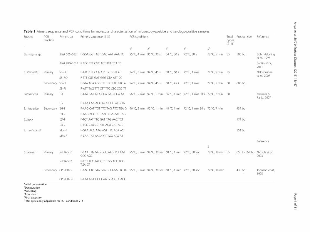
Table 1 Primers sequence and PCR conditions for molecular characterization of microscopy-positive and serology-positive samples
Species PCRreaction
Primers set Primers sequence (5’-3’) PCR conditions Totalcycles(2–4)f
Product size Reference
1a 2b 3c 4d 5e
Blastocystis sp. Blast 505–532 F-GGA GGT AGT GAC AAT AAA TC 95 °C, 4 min 95 °C, 30 s 54 °C, 30 s 72 °C, 30 s 72 °C, 5 min 35 500 bp Böhm-Gloninget al., 1997
Blast 998–1017 R TGC TTT CGC ACT TGT TCA TC Santin et al.,2011
S. stercoralis Primary SS–FO F-ATC CTT CCA ATC GCT GTT GT 94 °C, 5 min 94 °C, 45 s 58 °C, 60 s 72 °C, 1 min 72 °C, 5 min 35 Nilforoushanet al., 2007
SS–RO R-TTT CGT GAT GGG CTA ATT CC
Secondary SS–FI F-GTA ACA AGG TTT TCG TAG GTG A 94 °C, 2 min 94 °C, 45 s 60 °C, 45 s 72 °C, 1 min 72 °C, 5 min 30 680 bp
SS–RI R-ATT TAG TTT CTT TTC CTC CGC TT
Entamoeba Primary E-1 F-TAA GAT GCA CGA GAG CGA AA 96 °C, 2 min 92 °C, 1 min 56 °C, 1 min 72 °C, 1 min 30 s 72 °C, 7 min 30 Khairnar &Parija, 2007
E-2 R-GTA CAA AGG GCA GGG ACG TA
E. histolytica Secondary EH-1 F-AAG CAT TGT TTC TAG ATC TGA G 96 °C, 2 min 92 °C, 1 min 48 °C, 1 min 72 °C, 1 min 30 s 72 °C, 7 min 439 bp
EH-2 R-AAG AGG TCT AAC CGA AAT TAG
E.dispar ED-1 F-TCT AAT TTC GAT TAG AAC TCT 174 bp
ED-2 R-TCC CTA CCTATT AGA CAT AGC
E. moshkovskii Mos-1 F-GAA ACC AAG AGT TTC ACA AC 553 bp
Mos-2 R-CAA TAT AAG GCT TGG ATG AT
Reference
5
C. parvum Primary N-DIAGF2 F-CAA TTG GAG GGC AAG TCT GGTGCC AGC
95 °C, 5 min 94 °C, 30 sec 68 °C, 1 min 72 °C, 30 sec 72 °C, 10 min 35 655 to 667 bp Nichols et al.,2003
N-DIAGR2 R-CCT TCC TAT GTC TGG ACC TGGTGA GT
Secondary CPB-DIAGF F-AAG CTC GTA GTA GTT GGA TTC TG 95 °C, 5 min 94 °C, 30 sec 60 °C, 1 min 72 °C, 30 sec 72 °C, 10 min 435 bp Johnson et al.,1995
CPB-DIAGR R-TAA GGT GCT GAA GGA GTA AGGaInitial denaturationbDenaturationcAnnealingdExtensioneFinal extensionfTotal cycles only applicable for PCR conditions 2–4
Angalet
al.BMCInfectious
Diseases
(2015) 15:467 Page
4of
11
Microscopy-positive samples for Blastocystis sp. weresubjected to single-step PCR protocol for specific amplifi-cation of Blastocystis sp. [27, 28]. Nested multiplex PCRtargeting a 16S-like rRNA gene was carried out on posi-tive faecal samples for molecular characterization of E.histolytica, E.dispar and E. moshkovskii [29]. Primary PCRwas performed for amplification of Entamoeba genus inthe positive faecal samples. Subsequently, primary PCRproduct was subjected to secondary PCR reaction forspecies-specific identification. Nested PCR protocolwas performed targeting the SSU rRNA gene for the de-tection of Cryptosporidium in the microscopy-positivesamples [30, 31]. Seropositive samples for strongyloida-sis were subjected to nested PCR targeting the internaltranscribed spacer 1 (ITS1) region of the ribosomalDNA gene [32].In all the PCR reaction, positive controls were used
according to the species studied and distilled water wasused as negative controls. PCR products were analyzed on1 % agarose gel (Blastocystis sp.) and 2 % agarose gel (Ent-amoeba spp., C. parvum and S. stercoralis) visualizedusing UV transiluminator after staining with SYBR® SafeDNA (Invitrogen, Auckland, New Zealand). PCR-positivesamples for Blastocystis sp. were sent for sequencing andanalyzed with BLAST (Basic Local Alignment Search Tool)for further determination of Blastocystis subtypes.
Data analysisSPSS software (Statistical Package for the Social Sciences)program for Windows version 22 (SPSS, Chicago, IL,USA) was employed for data entry and statistical analysis.Descriptive statistics were mainly used to describe thecharacteristics of the study population including preva-lence of the IPIs. Qualitative data were determined andpresented as frequencies and percentages. Statistical sig-nificance of differences in proportions was evaluated byChi-Square test with significant value of p < 0.05 used forall tests. Univariate analysis was run to determine theassociation of variables with IPIs infections.
ResultsDemographic distributions and clinical information ofparticipating prison inmatesFrom a total of 294 consented inmates, there were 131(44.6 %) HIV positive inmates and 163 (55.4 %) HIV nega-tive inmates. The inmates aged ranging from 20 to 69-years-old. Participants included Malay, Chinese, Indian,Sabah and Sarawak Bumiputera ethnics, with a small com-position of immigrants (i.e. Burmese and Indonesian). Ma-jority of them had secondary education. Symptoms relatedto IPIs and other diseases presences were also noted.Intravenous drug users (IVDUs) were identified to be the
main mode of HIV transmission among the 131 HIV posi-tive inmates. Duration of having HIV ranged from 0 to
more than 21 years. As some of the HIV positive inmates’medical files could not be retrieved from the record section,CD4 counts were only available for 93 out of the 131 HIVpositive inmates, with 24 (25.8 % of 93) having CD4counts < 200 cells/mm3 and 69 (64.2 %) with CD4counts > 200 cells/mm3. All HIV positive inmates were of-fered to be included in the highly active antiretroviraltherapy (HAART) programme by the prison’s clinic. How-ever, there were only 7 (5.3 %) who were willing toundergo HAART due to reasons such as side effects andnon-compliance. Table 2 shows the details of demographicdistributions and clinical information of participatingprison inmates according to HIV status.
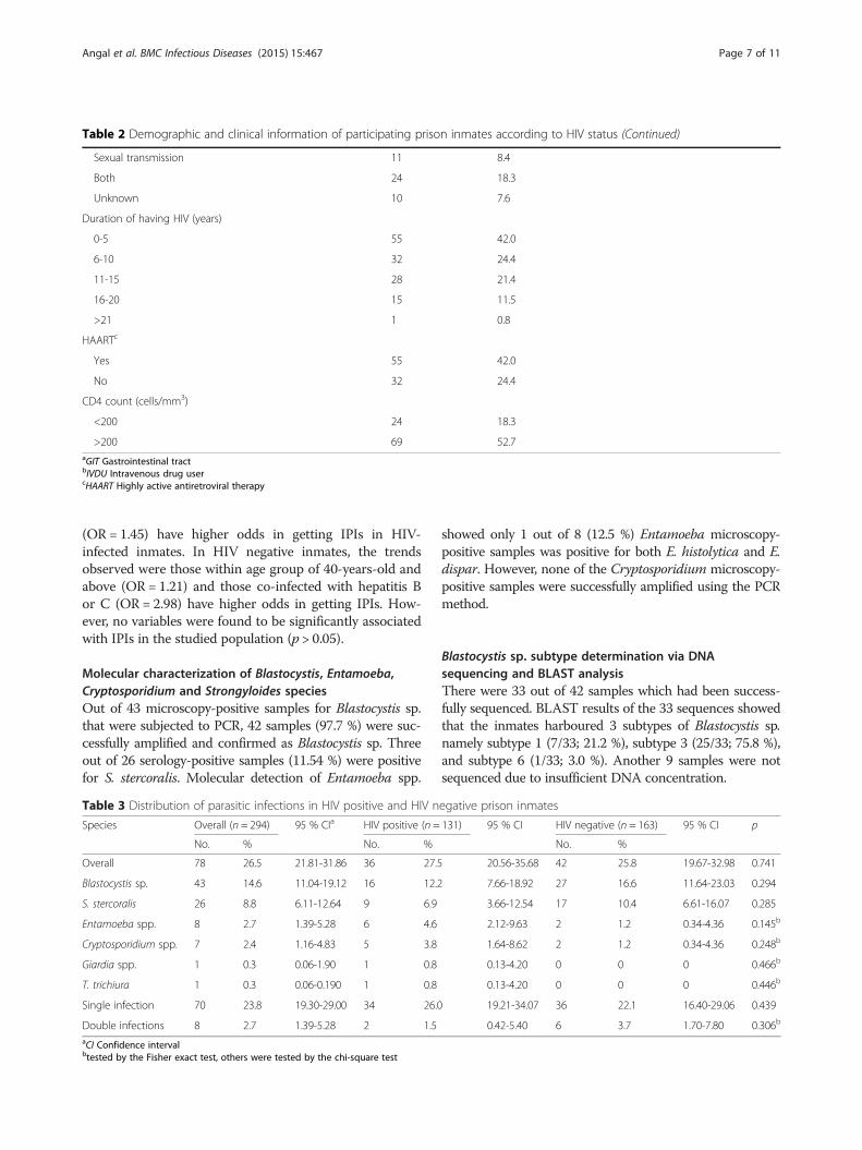
Intestinal parasitic infections (IPIs) among prison inmatesTable 3 shows the overall prevalence of IPIs among the294 studied inmates was 26.5 % (78/294). Intestinal para-sites detected included Blastocystis sp. in 43 inmates(14.6 %), S. stercoralis with seropositivity in 26 inmates(8.8 %), Entamoeba spp. in 8 inmates (2.7 %), Cryptospor-idium spp. in 7 inmates (2.4 %) and both Giardia spp. andT. trichiura in only 1 inmate each (0.3 %). Majority ofthem, 23.8 % (70/294) had single infections and there was2.7 % (8/294) who were infected with two parasites.With regards to IPIs according to HIV status, the inmates
who were HIV positive had slightly higher rates of IPIs with27.5 % (36/131) compared to HIV negative inmates with25.8 % (42/163), however it was not statistically significant(p = 0.741). Higher prevalence of both Entamoeba spp.,4.6 % (6/131; p = 0.145) and Cryptosporidium spp., 3.8 %(5/131; p = 0.248) were seen in HIV positive inmates. Simi-lar prevalence of Giardia spp., 0.8 % (1/131) and Trichuristrichiura, 0.8 % (1/131) were also observed among thisgroup. In contrast, Blastocystis sp. infection, 16.6 % (27/163; p = 0.294) was slightly higher in HIV negative inmatescompared to HIV positive inmates with 12.2 % (16/131).Interestingly, seropositive of S. stercoralis was higher inHIV negative inmates, 10.4 % (17/163; p = 0.285) comparedto HIV positive inmates, 6.9 % (9/131).
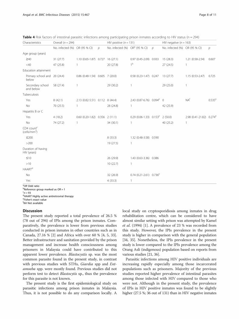
Risk factors for IPIs in prison inmatesUnivariate analysis for the association of parasitic infectionswith sociodemographic characteristics (i.e. age group, edu-cation attainment), presence of tuberculosis and presenceof hepatitis B or C among the overall inmates, HIV positiveinmates and HIV negative inmates are shown in Table 4.Besides that, factors including duration of having HIV,HAART treatment, and CD4 count were also analysed inHIV positive inmates. Only those co-infected with tubercu-losis appeared to have higher odds (i.e., 2.13) in getting IPIscompared to those without among the overall inmates. Thesame trends were observed in which those co-infected withtuberculosis (OR = 2.43), those with CD4 count < 200 cells/mm (OR= 1.32) and those having HIV less than 10 years
Angal et al. BMC Infectious Diseases (2015) 15:467 Page 5 of 11
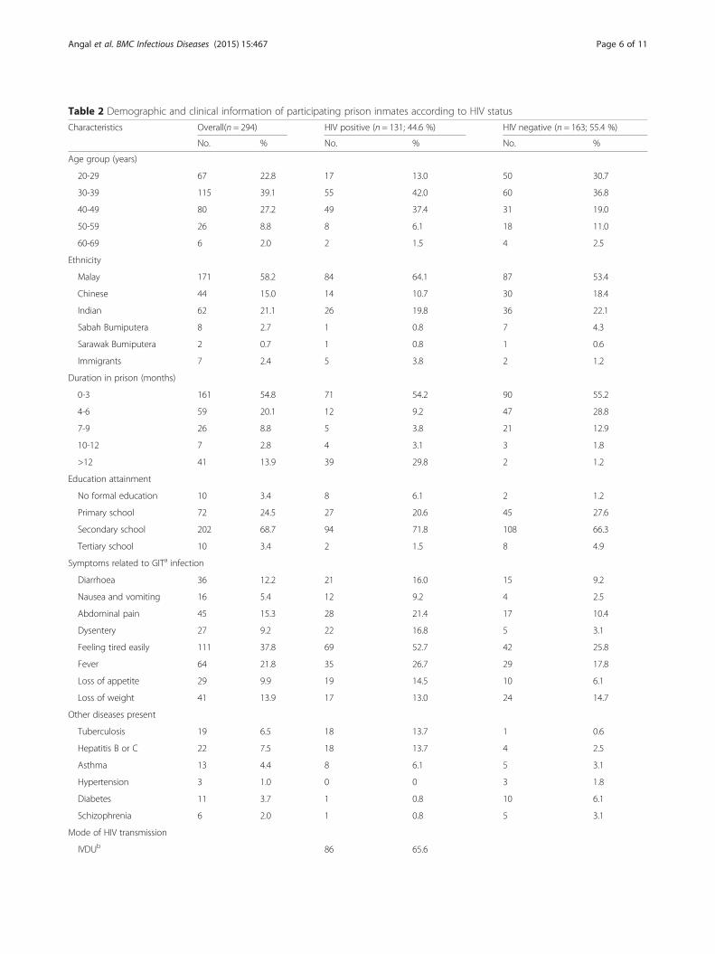
Table 2 Demographic and clinical information of participating prison inmates according to HIV status
Characteristics Overall(n = 294) HIV positive (n = 131; 44.6 %) HIV negative (n = 163; 55.4 %)
No. % No. % No. %
Age group (years)
20-29 67 22.8 17 13.0 50 30.7
30-39 115 39.1 55 42.0 60 36.8
40-49 80 27.2 49 37.4 31 19.0
50-59 26 8.8 8 6.1 18 11.0
60-69 6 2.0 2 1.5 4 2.5
Ethnicity
Malay 171 58.2 84 64.1 87 53.4
Chinese 44 15.0 14 10.7 30 18.4
Indian 62 21.1 26 19.8 36 22.1
Sabah Bumiputera 8 2.7 1 0.8 7 4.3
Sarawak Bumiputera 2 0.7 1 0.8 1 0.6
Immigrants 7 2.4 5 3.8 2 1.2
Duration in prison (months)
0-3 161 54.8 71 54.2 90 55.2
4-6 59 20.1 12 9.2 47 28.8
7-9 26 8.8 5 3.8 21 12.9
10-12 7 2.8 4 3.1 3 1.8
>12 41 13.9 39 29.8 2 1.2
Education attainment
No formal education 10 3.4 8 6.1 2 1.2
Primary school 72 24.5 27 20.6 45 27.6
Secondary school 202 68.7 94 71.8 108 66.3
Tertiary school 10 3.4 2 1.5 8 4.9
Symptoms related to GITa infection
Diarrhoea 36 12.2 21 16.0 15 9.2
Nausea and vomiting 16 5.4 12 9.2 4 2.5
Abdominal pain 45 15.3 28 21.4 17 10.4
Dysentery 27 9.2 22 16.8 5 3.1
Feeling tired easily 111 37.8 69 52.7 42 25.8
Fever 64 21.8 35 26.7 29 17.8
Loss of appetite 29 9.9 19 14.5 10 6.1
Loss of weight 41 13.9 17 13.0 24 14.7
Other diseases present
Tuberculosis 19 6.5 18 13.7 1 0.6
Hepatitis B or C 22 7.5 18 13.7 4 2.5
Asthma 13 4.4 8 6.1 5 3.1
Hypertension 3 1.0 0 0 3 1.8
Diabetes 11 3.7 1 0.8 10 6.1
Schizophrenia 6 2.0 1 0.8 5 3.1
Mode of HIV transmission
IVDUb 86 65.6
Angal et al. BMC Infectious Diseases (2015) 15:467 Page 6 of 11
(OR = 1.45) have higher odds in getting IPIs in HIV-infected inmates. In HIV negative inmates, the trendsobserved were those within age group of 40-years-old andabove (OR = 1.21) and those co-infected with hepatitis Bor C (OR = 2.98) have higher odds in getting IPIs. How-ever, no variables were found to be significantly associatedwith IPIs in the studied population (p > 0.05).
Molecular characterization of Blastocystis, Entamoeba,Cryptosporidium and Strongyloides speciesOut of 43 microscopy-positive samples for Blastocystis sp.that were subjected to PCR, 42 samples (97.7 %) were suc-cessfully amplified and confirmed as Blastocystis sp. Threeout of 26 serology-positive samples (11.54 %) were positivefor S. stercoralis. Molecular detection of Entamoeba spp.
showed only 1 out of 8 (12.5 %) Entamoeba microscopy-positive samples was positive for both E. histolytica and E.dispar. However, none of the Cryptosporidium microscopy-positive samples were successfully amplified using the PCRmethod.
Blastocystis sp. subtype determination via DNAsequencing and BLAST analysisThere were 33 out of 42 samples which had been success-fully sequenced. BLAST results of the 33 sequences showedthat the inmates harboured 3 subtypes of Blastocystis sp.namely subtype 1 (7/33; 21.2 %), subtype 3 (25/33; 75.8 %),and subtype 6 (1/33; 3.0 %). Another 9 samples were notsequenced due to insufficient DNA concentration.
Table 2 Demographic and clinical information of participating prison inmates according to HIV status (Continued)
Sexual transmission 11 8.4
Both 24 18.3
Unknown 10 7.6
Duration of having HIV (years)
0-5 55 42.0
6-10 32 24.4
11-15 28 21.4
16-20 15 11.5
>21 1 0.8
HAARTc
Yes 55 42.0
No 32 24.4
CD4 count (cells/mm3)
<200 24 18.3
>200 69 52.7aGIT Gastrointestinal tractbIVDU Intravenous drug usercHAART Highly active antiretroviral therapy
Table 3 Distribution of parasitic infections in HIV positive and HIV negative prison inmates
Species Overall (n = 294) 95 % CIa HIV positive (n = 131) 95 % CI HIV negative (n = 163) 95 % CI p
No. % No. % No. %
Overall 78 26.5 21.81-31.86 36 27.5 20.56-35.68 42 25.8 19.67-32.98 0.741
Blastocystis sp. 43 14.6 11.04-19.12 16 12.2 7.66-18.92 27 16.6 11.64-23.03 0.294
S. stercoralis 26 8.8 6.11-12.64 9 6.9 3.66-12.54 17 10.4 6.61-16.07 0.285
Entamoeba spp. 8 2.7 1.39-5.28 6 4.6 2.12-9.63 2 1.2 0.34-4.36 0.145b
Cryptosporidium spp. 7 2.4 1.16-4.83 5 3.8 1.64-8.62 2 1.2 0.34-4.36 0.248b
Giardia spp. 1 0.3 0.06-1.90 1 0.8 0.13-4.20 0 0 0 0.466b
T. trichiura 1 0.3 0.06-0.190 1 0.8 0.13-4.20 0 0 0 0.446b
Single infection 70 23.8 19.30-29.00 34 26.0 19.21-34.07 36 22.1 16.40-29.06 0.439
Double infections 8 2.7 1.39-5.28 2 1.5 0.42-5.40 6 3.7 1.70-7.80 0.306b
aCI Confidence intervalbtested by the Fisher exact test, others were tested by the chi-square test
Angal et al. BMC Infectious Diseases (2015) 15:467 Page 7 of 11
DiscussionThe present study reported a total prevalence of 26.5 %(78 out of 294) of IPIs among the prison inmates. Com-paratively, the prevalence is lower from previous studiesconducted in prison inmates in other countries such as inCanada, 27.16 % [2] and Africa with over 60 % [4, 5, 33].Better infrastructure and sanitation provided by the prisonmanagement and increase health consciousness amongprisoners in Malaysia could have contributed to thisapparent lower prevalence. Blastocystis sp. was the mostcommon parasite found in the present study, in contrastwith previous studies with STHs, Giardia spp and Ent-amoeba spp. were mostly found. Previous studies did notperform test to detect Blastocytis sp., thus the prevalencefor this parasite is not known.The present study is the first epidemiological study on
parasitic infections among prison inmates in Malaysia.Thus, it is not possible to do any comparison locally. A
local study on cryptosporidiosis among inmates in drugrehabilitation centre, which can be considered to havealmost similar setting with prison was attempted by Kamelet al. (1994) [1]. A prevalence of 23 % was recorded fromthis study. However, the IPIs prevalence in the presentstudy is higher in comparison with the general population[34, 35]. Nonetheless, the IPIs prevalence in the presentstudy is lower compared to the IPIs prevalence among theOrang Asli (indigenous) population based on reports fromvarious studies [21, 36].Parasitic infections among HIV positive individuals are
increasing rapidly especially among those incarceratedpopulations such as prisoners. Majority of the previousstudies reported higher prevalence of intestinal parasitesamong those infected with HIV compared to those whowere not. Although in the present study, the prevalenceof IPIs in HIV positive inmates was found to be slightlyhigher (27.5 %; 36 out of 131) than in HIV negative inmates
Table 4 Risk factors of intestinal parasitic infections among participating prison inmates according to HIV status (n = 294)
Characteristics Overall (n = 294) HIV positive (n = 131) HIV negative (n = 163)
No. infected (%) OR (95 % CI) p No. infected (%) ORa (95 % CI) p No. infected (%) OR (95 % CI) p
Age group (years)
≥40 31 (27.7) 1.10 (0.65-1.87) 0.727 16 (27.1) 0.97 (0.45-2.09) 0.933 15 (28.3) 1.21 (0.58-2.54) 0.607
<40 47 (25.8) 1 20 (27.8) 1b 27 (24.5) 1
Education attainment
Primary school andbelow
20 (24.4) 0.86 (0.48-1.54) 0.605 7 (20.0) 0.58 (0.23-1.47) 0.247 13 (27.7) 1.15 (0.53-2.47) 0.725
Secondary schooland below
58 (27.4) 1 29 (30.2) 1 29 (25.0) 1
Tuberculosis
Yes 8 (42.1) 2.13 (0.82-5.51) 0.112 8 (44.4) 2.43 (0.87-6.76) 0.094e 0 NAf 0.535e
No 70 (25.5) 1 28 (24.8) 1 42 (25.9)
Hepatitis B or C
Yes 4 (18.2) 0.60 (0.20-1.82) 0.356 2 (11.1) 0.29 (0.06-1.33) 0.153e 2 (50.0) 2.98 (0.41-21.82) 0.274e
No 74 (27.2) 1 34 (30.1) 1 40 (25.2) 1
CD4 countc
(cells/mm3)
≤200 8 (33.3) 1.32 (0.48-3.58) 0.590
>200 19 (27.5) 1
Duration of havingHIV (years)
≤10 26 (29.9) 1.43 (0.63-3.36) 0.386
>10 10 (22.7) 1
HAARTd
No 32 (26.9) 0.74 (0.21-2.61) 0.736e
Yes 4 (33.3) 1aOR Odd ratiobReference group marked as OR = 1cn = 93dHAART Highly active antiretroviral therapyeFisher’s exact valuefNA Not available
Angal et al. BMC Infectious Diseases (2015) 15:467 Page 8 of 11
(25.8 %; 42 out of 163), it was not statistically significant.HIV positive status of some of the prisoners did not play amajor role in increasing the IPIs prevalence.Presence of Blastocystis sp. in the present study was
higher in HIV negative inmates than HIV positive in-mates with 16.6 % (27/163) and 12.2 % (16/131), respect-ively. Though many authors have given credit to it as apathogen, however there is still much controversy sur-rounding the role of Blastocystis in causing human dis-ease. The most common symptoms associated withBlastocystis infection include diarrhoea, abdominal painand vomiting. The significance of Blastocystis sp.in HIV/AIDS populations has not been ascertained, but there havebeen several studies with varying results on the prevalenceof intestinal parasites and particularly the incidence ofBlastocystis in these populations [37, 38]. Based on theseprevious studies, Blastocystis infection may have a signifi-cant role in causing diarrhoea especially in the immuno-suppressed patients.In the present study, Blastocystis sp. subtype 3 was
found to be the most common subtype, a finding inagreement with the previous studies [39, 40]. Occurrenceof Blastocystis sp. subtype 1 (ST1) following ST3 was notsurprising. There were various reports which showed ST1was the common subtype found in human, together withST2, ST3 and ST4 [39, 41, 42]. The single infection ofBlastocystis subtype 6 (ST6) may reflect zoonotic trans-mission, as this subtype has been reported among theavians [43, 44]. Jantermtor et al. (2013) [40] also detectedST6 among the Thais in their study. However, ST6 israrely reported in Asia.S. stercoralis was negative microscopically in the present
study. This may be due to a one stool sample collectionfrom each participant. Multiple stool specimens are re-quired for microscopy diagnosis of S. strongyloides becausethe parasite is excreted periodically and in low numbers. Inthe present study, ELISA test showed high seroprevalenceof S. stercoralis in HIV negative inmates (10.4 %; 17/163)compared to in HIV positive inmates (6.9 %; 9/131). Thistrend may be due to the inmate’s geographical origin,hygiene and occupation before being imprisoned. Therewere also a few studies which reported low prevalence of S.stercoralis in HIV positive individuals [45, 46]. However,ELISA test is unable to differentiate current from pastinfection. Based on PCR of stool samples, S. stercoralis wasdetected in 3 non-HIV inmates. Since PCR analysis con-firmed current infection, these positive individuals shouldbe treated to prevent transmission and hyperinfection.Opportunistic infection of Cryptosporidium spp. was
more predominant (3.8 %; 5/131) in HIV positive inmatesthan in non-HIV inmates (1.2 %; 2/163), which was in con-cordance with the study by Tian et al. (2012) [47]. Crypto-sporidiosis is categorized by CDC as AIDS-defining illness(ADI) due to its ability in causing diarrhoea and poses a
public health problem in HIV/AIDS patients [48]. In clin-ical cryptosporidiosis, chronic diarrhoea with watery stools,weight loss and dehydration are the prominent features insymptomatic patients [49]. Seven (7) inmates were micro-scopically positive for Cryptosporidium spp. Detection ofCryptopsoridium by Ziehl-Neelsen stain is a definitive diag-nosis. Of these, none of the samples were successfully amp-lified using nested PCR [31]. These microscopy-positivesamples failed to be detected maybe due to low DNA con-centration of the parasites or degradation of DNA.Thepresent study reported Entamoeba spp. was more prevalentamong the HIV positive inmates (4.6 %; 6/131) in compari-son with the non-HIV inmates (1.2 %; 2/163) microscopic-ally. This finding was in concordance with the previousstudy [38]. However, E. histolytica cannot be differentiatedmicroscopically with the other two non-pathogenic speciesnamely E. dispar and E. moshkovskii due to similarity intheir morphology. Thus, PCR test was performed in orderto distinguish the pathogenic from non-pathogenic species.Among all the intestinal parasites reported in the present
study, S. stercoralis and T. trichiura are categorized as soil-transmitted helminths (STHs). Thus, to prevent the infec-tion of these parasites, (i) regular anthelminthic treatment,(ii) health education, (iii) sanitation and personal hygienecan be implemented. However, in the present study setting,prevalence of STHs is low. This may be due to the goodpractice of sanitation and personal hygiene by the inmates.With regards to CD4 counts, those who had CD4
counts < 200 cells/mm3 were having higher odds in gettingIPIs compared to those with CD4 counts > 200 cells/mm3.These findings were consistent with other study thatshowed parasitic infections usually occurred in individualshaving CD4 counts of < 200 cells/mm3 [20]. Furthermore,those newly acquired HIV infection between 0 to 10 yearshad higher IPIs compared to those who acquired HIV in-fection >10 years. Inmate’s lack of information on hygiene,sanitation and of the infection could be the reasons forthis outcome. In addition, majority of this group of in-mates were also not on HAART. Those HIV infected in-mates who were undergoing HAART have lower levels ofIPIs in comparison to those who were not. Hung et al.(2007) [50] addressed the importance of HAART amongHIV positive individuals in restoring their immune func-tions, thus improving the immunity level towards IPIs.Tuberculosis (TB) and hepatitis B or C (hep B or C)
are the most common co-infections among HIV positivepeople worldwide. WHO (2015) [51] estimated at leastone-third of people living with HIV worldwide in 2013were infected with TB whereas almost 80 % of peoplewith HIV who inject drugs also have hepatitis B or Cvirus [52]. Among the HIV negative inmates, those co-infected with hep B or C had higher probability of ac-quiring IPIs compared to those without. On the otherhand, among overall inmates, those co-infected with TB
Angal et al. BMC Infectious Diseases (2015) 15:467 Page 9 of 11

have higher odds in getting IPIs in comparison to thosewithout TB regardless of their HIV status. This affirmedfurther that TB and hep B or C worsened the immunitylevel, thus increasing the individual’s susceptibility to-wards parasitic infections in this study.Few limitations were encountered in the present study
including unretrievable CD4 count for some of the in-mates as their clinical records were not found in the rec-ord section; conditions of the prison cells were notobserved as these areas were restricted only for the in-mates, staffs and doctors in-charge and comparison onparasitic infections with the female inmates could not bedone as permission to take samples was only given forthe male inmates.
ConclusionOverall prevalence of IPIs among inmates was 26.5 %(78 out of 294). With regards to the HIV status, HIVpositive inmates had slightly higher rates of IPIs with27.5 % compared to HIV negative inmates with 25.8 %.The present study is the first study in Malaysia whichemphasized on the parasites-HIV co-infections in theprison setting. Non-HIV inmates were also included tocompare the prevalence of parasites between these twogroups. It was noted in the present study that IPIsprevalence is lower compared to previous studies onprison inmates conducted in other countries. MalaysianPrison Department is proactively improving the healthstatus of prison inmates in Malaysia. The informationgathered from the study will enable the health care pro-viders and prison management staff to understand thetrend and epidemiological situations of parasitic co-infections in a prison setup. It also provides evidence-based guidance to improve prevention and control (i.e.food preparation, boiled drinking water) through healtheducation as well as treatment management strategies ofIPIs co-infections (i.e. parasite infections screening, anti-parasitic treatments). More importantly, the inmatesthemselves should be aware of the importance of goodpersonal hygiene to prevent them from acquiring ortransmitting infections to others. In addition, doctorsmanaging these infected individuals should also haveheightened awareness of the possibility of co-infectionswith parasites, bacteria and viruses.
AbbreviationsAIDS: Acquired immunodeficiency syndrome; BLAST: Basic local alignmentsearch tool; CI: Confidence interval; DNA: Deoxyribonucleic acid;GIT: Gastrointestinal tract; HAART: Highly active antiretroviral therapy; Hep Bor C: Hepatitis B or C; HIV: Human immunodeficiency virus; IPIs: Intestinalparasitic infections; IVDUs: Intravenous drug users; OR: Odds ratio;PCR: Polymerase chain reaction; Spp: Species; SPSS: Statistical Package for theSocial Sciences; SSU rDNA: Small subunit ribosomal DNA; ST: Subtype;STI: Sexually transmitted infections; STHs: Soil-transmitted helminths;TB: Tuberculosis; UMMC: University Malaya Medical Centre; UNAIDS: UnitedNations & AIDS; WHO: World Health Organization.
Competing interestThe authors declare that they have no competing interests.
Authors’ contributionsLA was involved in the study design, data collection, data analysis and writeup of the manuscript; YALL and RM involved in the study design, supervisedthe study, and revised the manuscript; RN was involved in the statisticalanalysis of data; SS, RN, YNJ and AA were involved in the collection ofsamples. II and AK involved in the study design. All authors read andapproved the final manuscript.
AcknowledgementsThe authors express their sincere gratitude to the management of KajangPrison for approving and giving permission to conduct this study. Specialthanks to the staff of Kajang Prison who have assisted with samplescollection during the course of the study.
FundingThis study was supported by the University of Malaya Postgraduate ResearchGrant (PG117-2012B) and UM/MoHE High Impact Research (UM.C/625/1/HIR/MOHE/MED/23). The funders had no role in study design, data collection,analysis and preparation or decision to publish the manuscript.
Author details1Department of Parasitology, Faculty of Medicine, University of Malaya, KualaLumpur, Malaysia. 2Penjara Utama Kajang, Kajang, Selangor, Malaysia.3Department of Medicine, Faculty of Medicine, University of Malaya, KualaLumpur, Malaysia.
Received: 7 May 2015 Accepted: 5 October 2015
References1. Kamel AGM, Maning N, Arulmainathan S, Murad S, Nasuruddin A, Lai KP.
Cryptosporidiosis among HIV positive intravenous drug users in Malaysia.Southeast Asian J Trop Med Public Health. 1994;25:650–3.
2. Mecroritch E, Eaton RD. Outbreak of amoebiasis among Indian inmates inNorth Western Sackatchewan. Canada Am J Trop Med Hyg. 1965;4:719–23.
3. Anderson RM. Dynamic aspects of parasite population ecology. In: KennedyCR, editor. Ecological aspects of parasitology. Amsterdam: North HollandPublishing Co; 1976. p. 431–62.
4. Amuga G, Usmand D, Onwuliri COE. Human intestinal parasites amonginmates of Keffi Prison, Nasarawa State. Int J of Nat – Applied Sc. 2006;2:7–10.
5. Okolie N. Intestinal parasites distribution among inmates of Owerri Prison.The Internet Journal of Parasitic Diseases. 2008;4:1.
6. Ishaleku D, Mamman AS. Co-infection of malaria and helminthes infectionamong prison inmates. Res Rev J Microbiol Virol. 2014;2(1):1–5.
7. World Health Organization (WHO) Europe. HIV in prisons. 2015.http://www.euro.who.int/en/health-topics/communicable-diseases/hivaids/policy. Accessed February 2015.
8. Weishburch JC. Prison health. In: Last JM, Wallace RB, editors. Public healthand preventive medicine. 13th ed. California: Appleton and Lange;1992. p. 1159–62.
9. Li CF. Hookworm infection and protein energy malnutrition: Transverseevidence from two Malaysian ecological groups. Trop Geogr Med.1990;42:8–12.
10. Chan L, Kan SP, Bundy DA. The effect of repeated chemotheraphy on theprevalence and intensity infection of Ascaris lumbricoides and Trichuristrichiura. Parasitology. 1992;104:371–7.
11. DuPont HL, Chapell CL, Sterling CR, Okhuysen PC, Rose JB, Jakubowski W.The infectivity of Cryptosporidium parvum in healthy volunteers. New Engl JMed. 1995;332:855–9.
12. Kan SP. Soil-transmitted helminthiases among inhabitants of an oil palmplantation in West Malaysia. J Trop Med Hygiene. 1998;92:263–9.
13. Norhayati M, Fatmah MS, Yusof S. Intestinal parasitic infections in man: Areview. Med J Malaysia. 2003;58:2–10.
14. Ngui R, Ishak S, Chuen CS, Mahmud R, Lim YAL. Prevalence and risk factorsof intestinal parasitism in rural and remote West Malaysia. PLoS Negl TropDis. 2011;5:e974.
Angal et al. BMC Infectious Diseases (2015) 15:467 Page 10 of 11

15. Lim YAL, Rohela M, Sim BLH, Jamaiah I, Nurbayah M. Prevalence ofcryptosporidiosis in HIV-infected patients in Kajang Hospital, Selangor. SoutheastAsian J Trop Med Public Health. 2005;36:30–2.
16. Zaidah AR, Chan YY, Asma SH, Abdullah S, Nurhaslindawati AR, Salleh M, etal. Detection of Cryptosporidium parvum in HIVinfected patients in Malaysiausing molecular approach. Southeast Asian J Trop Med Public Health.2008;39:511–6.
17. Asma I, Lim YAL, Johari S, Sim BLH. High diversity of Cryptosporidiumsubgenotypes identified in Malaysian HIV/AIDS individuals targeting gp60gene. Plos ONE. 2012;7(2):e31139.
18. Lono A, Kumar S, Chye TT. Detection of microsporidia in local HIV-positivepopulation in Malaysia. Trans R Soc Trop Med Hyg. 2011;105(7):409–13.
19. Azira NMS, Abdel Rahman MZ, Zeehaida M. Review of patients withStrongyloides stercoralis infestation in a tertiary teaching hospital. KelantanMalays J Pathol. 2013;35(1):71–6.
20. Asma I, Johari S, Benedict SLH, Yvonne LAL. How common is intestinal parasitismin HIV-infected patients in Malaysia? Trop Biomed. 2011;28(2):400–10.
21. Lee SC, Ngui R, Tan TK, Aidil RM, Lim YAL. Neglected tropical diseasesamong two indigenous subtribes in Peninsular Malaysia: Highlightingdifferences and co-infection of helminthiasis and sarcocystosis. Plos One.2014;9(9):e107980.
22. Lim YAL, Ngui R, Nicholas C, Chow SC, Smith HV. Intestinal parasiticinfections amongst Orang Asli (indigenous) in Malaysia: Has socioeconomicdevelopment alleviated the problem? Trop Biomed. 2009;26(2):110–22.
23. Leedy PD. Practical research: planning and design. 5th ed. New York:MacMilan Publishing Company; 1993.
24. Kuk S, Yazar S, Cetinkaya U. Stool sample storage conditions for the preservationof Giardia intestinalis DNA. Mem Inst Oswaldo Cruz. 2012;107(8):965–8.
25. Pipatsatitpong D, Rangsin R, Leelayoova S, Naaglor T, Mungthin M.Incidence and risk factors of Blastocystis infection in an orphanage inBangkok Thailand. Parasit Vectors. 2012;14:37.
26. Abdulsalam AM, Ithoi I, Al-Mekhlafi HM, Ahmed A, Surin J, Mak JW. Drinkingwater is a significant predictor of Blastocystis infection among ruralMalaysian primary schoolchildren. Parasitology. 2012;139:1014–20.
27. Böhm-Gloning B, Knobloch J, Walderich B. Five subgroups of Blastocystishominis isolates from symptomatic and asymptomatic patients revealedby restriction site analysis of PCR-amplified 16S-like rDNA. Trop Med Int.1997;2:771–8.
28. Santín M, Gómez-Muñoz MT, Solano-Aguilar G, Fayer R. Development ofa new protocol to detect and subtype Blastocystis spp. from humansand animals. Parasitol Res. 2011;109:205–12.
29. Khairnar K, Parija SC. A novel nested multiplex polymerase chainreaction (PCR) assay for differential detection of Entameoba histolytica,E. moshkovskii and E. dispar DNA in stool samples. BMC Microbiol.2007;7:47.
30. Johnson DW, Pieniazek NJ, Griffin DW, Misener L, Rose JB. Developmentof a PCR protocol for sensitive detection of Cryptosporidium oocysts inwater samples. Appl Environ Microbiol. 1995;61(11):3849–55.
31. Nichols RAB, Campbell BM, Smith HV. Identification of Cryptosporidiumspp. oocysts in United Kingdom noncarbonated natural mineral watersand drinking waters by using a modified nested PCR-restrictionfragment length polymorphism assay. Appl Environ Microbiol.2003;69:4183–9.
32. Nilforoushan MR, Mirhendi H, Rezaie S, Rezaian M, Meamar AR, Kia EB. ADNA-based identification of Strongyloides stercoralis isolates from Iran.Iranian J Publ Health. 2007;36(3):16–20.
33. Mamo H. Intestinal parasitic infections among prison inmates andtobacco farm workers in Shewa Robit. North-Central Ethiopia PLoS ONE.2014;9(6):e99559.
34. Jamaiah I, Rohela M. Prevalence of intestinal parasites among members ofthe public in Kuala Lumpur, Malaysia. Southeast Asian J Trop Med PublicHealth. 2005;36(1):68–71.
35. Nissapatorn V, Lim YAL, Jamaiah I, Rohela M, Khairul AA. Parasitic infection:A recurring phenomenon in Malaysia. Southeast Asian J Trop Med PublicHealth. 2007;38(1):181–90.
36. Tengku SA, Siti NA, Fatmah MS, Norhayati M. Molecular epidemiology ofgiardiasis among Orang Asli in Malaysia: application of the triosephosphateisomerase gene. BMC Infect Dis. 2014;14:78.
37. Gassama A, Sow PS, Fall F, Camara P, Gueye-N’diaye A, Seng R, et al.Ordinary and opportunistic enteropathogens associated with diarrhea in
Senegalese adults in relation to human immunodeficiency virus serostatus.Int J Infect Dis. 2001;5:192–8.
38. Tian LG, Wang TP, Lv S, Wang FF, Guo J, Yin XM, et al. HIV and intestinalparasite co-infections among a Chinese population: An immunologicalprofile. Infectious Diseases of Poverty. 2013;2:18.
39. Vassalos CM, Spanakos G, Vassalou E, Papadopoulou C, Vakalis N. Differencesin clinical significance and morphologic features of Blastocystis sp subtype3. Am J Clin Pathol. 2010;133:251–8.
40. Jantermtor S, Pinlaor P, Sawadpanich K, Pinlaor S, Sangka A, Wilailuckana C,et al. Subtype identification of Blastocystis spp. isolated from patients in amajor hospital in northeastern Thailand. Parasitol Res. 2013;112:1781–6.
41. Sardarian K, Hajilooi M, Maghsood A, Moghimbeigi A, Alikhani M. A study ofthe genetic variability of Blastocystis hominis isolates in Hamadan. West ofIran Jundishapur J Microbiol. 2012;5(4):555–9.
42. Ramírez JD, Sánchez LV, Bautista DC, Corredor AF, Flórez AC, Stensvold CR.Blastocystis subtypes detected in humans and animals from Colombia. InfectGenet Evol. 2013;22:223–8.
43. Yoshikawa H, Abe N, Wu Z. PCR-based identification of zoonotic isolates ofBlastocystis from mammals and birds. Microbiology. 2004;150:1147–51.
44. Noël C, Dufernez F, Gerbod D, Edgcomb VP, Delgado-Viscogliosi P, Ho LC,et al. Molecular phylogenies of Blastocystis isolates from different hosts:Implications for genetic diversity, identification of species, and zoonosis. JClin Microbiol. 2005;43:348–55.
45. Fontanet AL, Sahlu T, Rinke de Wit T, Messele T, Masho W. Epidemiology ofinfections with intestinal parasites and human immunodeficiency virus (HIV)among sugar-estate residents in Ethiopia. Ann Trop Med Parasitol.2000;94:269–78.
46. Lebbad M, Norrgren H, Naucler A, Dias F, Andersson S. Intestinal parasites inHIV-2 associated AIDS cases with chronic diarrhoea in Guinea-Bissau. ActaTrop. 2001;80:45–9.
47. Tian LG, Chen JX, Wang TP, Cheng GJ, Steinmann P, et al. Co-infection ofHIV and intestinal parasites in rural area of China. Parasit Vectors. 2012;5:36.
48. CDC. Revision of the CDC surveillance case definition for acquiredimmunodeficiency syndrome. MMWR Surveill Summ. 1987;36:1–15.
49. Kosek M, Alcantara C, Lima AA, Guerrant RL. Cryptosporidiosis: an update.Lancet Infect Dis. 2001;1:262–9.50. Adamu H, Wegayehu T, Petros B. Highprevalence of diarrhoegenic intestinal parasite infections among non-ARTHIV patients in Fitche Hospital, Ethiopia. PLoS One. 2013;8(8):e72634.
50. Hung CC, Tsaihong JC, Lee YT, Deng HY, Hsiao WH, Chang SY, et al.Prevalence of intestinal infection due to Cryptosporidium species amongTaiwanese patients with human immunodeficiency virus infection. J FormosMed Assoc. 2007;106:31–5.
51. WHO. HIV/TB co-infection. 2015. www.who.int/topics/tuberculosis/en/. AccessedMarch 2015.
52. CDC. HIV and Viral Hepatitis. 2014. http://www.cdc.gov/hiv/pdf/library_factsheets_HIV_and_viral_Hepatitis.pdf. Accessed March 2014.
Submit your next manuscript to BioMed Centraland take full advantage of:
• Convenient online submission
• Thorough peer review
• No space constraints or color figure charges
• Immediate publication on acceptance
• Inclusion in PubMed, CAS, Scopus and Google Scholar
• Research which is freely available for redistribution
Submit your manuscript at www.biomedcentral.com/submit
Angal et al. BMC Infectious Diseases (2015) 15:467 Page 11 of 11