detection of lung cancer using image ... - infona
TRANSCRIPT
http://www.iaeme.com/IJEET/index.asp 7 [email protected]
International Journal of Electrical Engineering & Technology (IJEET)
Volume 11, Issue 2, March-April 2020, pp. 7-16, Article ID: IJEET_11_02_002
Available online at http://www.iaeme.com/IJEET/issues.asp?JType=IJEET&VType=11&IType=2
ISSN Print: 0976-6545 and ISSN Online: 0976-6553
Journal Impact Factor (2020): 10.1935 (Calculated by GISI) www.jifactor.com
© IAEME Publication
DETECTION OF LUNG CANCER USING IMAGE
SEGMENTATION
V. Kannan
Professors, Department of ECE,
GMR Institute of Technology, Rajam, Andhra Pradesh, India
V. Jagan Naveen
Professors, Department of ECE,
GMR Institute of Technology, Rajam, Andhra Pradesh, India
ABSTRACT
In modern days, image processing methods are widely adopted in the medical field
for enhancing the earlier detection of certain abnormalities, such as the breast cancer,
lung cancer, brain cancer and so on. This paper mainly concentrates on the
segmentation of lung cancer tumors from X-ray images, Computed Tomography (CT)
images and MRI images. Image processing methods are adopted in segmenting the
images. In the pre-processing stage mean and median filters are used. In the image
segmentation stage, Otsu's thresholding and k-Means clustering segmentation
approaches are used to segment the lung images and locate the tumors. To evaluate
the performance of the methods used for segmentation, the performance evaluation
parameters such as Signal to noise Ratio(SNR) ,Mean Square Error (MSE) and Peak
Signal Noise to Ratio (PSNR)) are computed on the segmented images of the two
different segmentation methods used for segmentation. Better results are obtained for
the K-Means segmentation irrespective of the images.
Keywords: Lung Cancer, Computed Tomography, MRI, Thresolding, K-Means
Clustering, MSE, PSNR
Cite this Article: V. Kannan and V. Jagan Naveen, Detection of Lung Cancer using
Image Segmentation, International Journal of Electrical Engineering & Technology,
11(2), 2020, pp. 7-16.
http://www.iaeme.com/IJEET/issues.asp?JType=IJEET&VType=11&IType=2
1. INTRODUCTION
In the modern world, there is an increase in Lung cancer which is due to abnormal
multiplication of certain cell multiplying and growing into tumor. Maximum cancers that
begin within the lungs are called carcinomas. Two kinds of cancers are observed as small-cell
lung carcinoma and nonsmall - cell lung carcinoma [1].Tobacco smoking for long years is the
primary cause for 85% of lung cancers [2]. Approximately 10–15% of cases arise in humans
who never smoked, but the illness is due to air pollution, 2nd hand smoking, asbestos, radon
V. Kannan and V. Jagan Naveen
http://www.iaeme.com/IJEET/index.asp 8 [email protected]
fuel and so on. X-Ray imaging and Computer tomography (CT) are the traditional techniques
to discover the affected lungs with cancer. The detection is done through biopsy that is
commonly performed with the aid of bronchoscopy or CT scan. The cause of cancer-
associated loss of life amongst men is specifically due to lung cancer. For this reason, it is
crucial to determine a new strong method to diagnose the lung cancers at an earlier stage [3].
Lung cancer, also known as lung carcinoma, is a malignant tumor characterized with the aid
of out of control growth of the tissue cells of the lungs. It is compulsory to treat this cancer to
avoid spreading in to other parts of the body [5].
Cancer cells may be carried away from the lungs through blood, or lymph fluid that
surrounds the tissues of lungs. Lymph fluid flows through lymphatic vessels and get drained
into lymph nodes positioned inside the lungs and at the center of the chest. Lung cancers
frequently spreads towards the center of the chest due to the fact that the herbal drift of lymph
fluid flows out of the lungs is towards the center of the chest. Progression takes place when
the cancerous cell leaves the site where it is originated and moves into a lymph node or to any
other a part of the body via the blood circulation [4]. Cancerous tumor that got developed
within the lung is referred to as primary lung cancer.
CT and X-rays are the traditional techniques to detect the existence of lung cancers. The
scanning is confirmed by using biopsy, that is generally finished by using bronchoscopy or
CT experiment. The reason of most cancers-associated demise among men is specifically due
to lung cancers.
1.1. Lungs and Lung Cancer
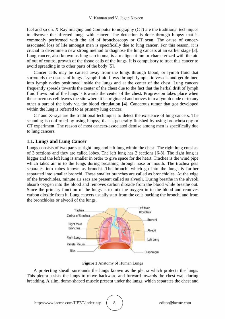
Lungs consists of two parts as right lung and left lung within the chest. The right lung consists
of 3 sections and they are called lobes. The left lung has 2 sections [6-8]. The right lung is
bigger and the left lung is smaller in order to give space for the heart. Trachea is the wind pipe
which takes air in to the lungs during breathing through nose or mouth. The trachea gets
separates into tubes known as bronchi. The bronchi which go into the lungs is further
separated into smaller bronchi. These smaller branches are called as bronchioles. At the edge
of the bronchioles, minute air sacs are present called as alveoli. During breathe in the alveoli
absorb oxygen into the blood and removes carbon dioxide from the blood while breathe out.
Since the primary function of the lungs is to mix the oxygen in to the blood and removes
carbon dioxide from it. Lung cancers usually start from the cells backing the bronchi and from
the bronchioles or alveoli of the lungs.
Figure 1 Anatomy of Human Lungs
A protecting sheath surrounds the lungs known as the pleura which protects the lungs.
This pleura assists the lungs to move backward and forward towards the chest wall during
breathing. A slim, dome-shaped muscle present under the lungs, which separates the chest and
Detection of Lung Cancer using Image Segmentation
http://www.iaeme.com/IJEET/index.asp 9 [email protected]
the abdomen is called diaphragm. During breathing, the air is forced in and out of the lungs by
the up and down movement of the diaphragm.
Most cancers begin, when there is an abnormal growth of cells in the body. A form of
cancer that is found in the lungs are called Lung cancers. The two major types of lung cancer
are small cell lung cancers and Non-small cell lung cancers (NSCLC).It is found that 85% of
the lung cancers are NSCLC [9-10]. The NSCLC are further classified as large cell
carcinoma, adenocarcinoma and squamous cell carcinomas.
Adenocarcinomas start in the cells that secrete typical substance along with mucus [11].
Lung cancers are diagnosed mostly with people who smoke [12]. Lung cancers are also
visible in people who do not smoke [13] which is due to second hand smoking. It is more
common in ladies whose husbands smoke.
Adenocarcinoma occurs in the exterior of the lung and it may be detected before it
spreads. Squamous cells are flat cells that rim the inner way of the airways within the lungs.
The cancer occur in it is called as Squamous cell carcinomas. This cancer is mostly related to
the his-tory of smoking. Any part of the lung may be affected by large cell carcinoma. It is a
dangerous cancer which may develop and spread quickly, making the treatment tougher.
Large cell neuroendocrine carcinoma is a subtype of large cell carcinoma, which is a quick-
growing lung cancer. The cancer that start form the lung only is called as lung cancer .The
cancers which spread from different organs like breast, kidney and pancreas to the lungs are
not lung cancers [14].
1.2. Causes of Lung Cancer
The main cause of present days deaths are due to Lung cancers in both women and men. To-
day, nine out of 10 lung cancer deaths are caused by smoking. The various causes of lung
cancers are discussed below
1.2.1. Smoking
Cigarettes [15] are packed with cancer-inflicting chemical substances. In addition they spoil
the lungs' natural protection system. To protect the lungs, the airways of the lungs are lined
with tiny hairs known as cilia. They sweep out pollution, bacteria, and viruses from the air
while breathing. The cilia is affected by the tobacco smoke and stops working in doing its job.
This is the main reason for the cancer-causing chemicals to build up in the lungs and. the
cancers starts quietly. During the early stages, no symptoms or warning are found due to
cancer .But when it get worse, the following symptoms may be noticed
Chest pain, during deep breathing
Cough that won't go away
Bloody phlegm when Coughing
Wheezing or shortness of breath,
Fatigue
1.2.2. Second hand Smoke
Even though the main cause of lung cancers is smoking, breathing in the second hand smoke
released by a smoker is also danger. This second hand smoke which occur at domestic or at
work place also may increase the risk of danger [16- 17]. It is found that woman‟s who are
married to a smoking person are 20% to 30% more likely to get lung cancer when compared
the woman‟s of non-smokers.
1.2.3. Risky work
Some of the working environments may also be the cause of lung cancers . People who are
working with the metals such as uranium, arsenic, and other chemical substances have more
V. Kannan and V. Jagan Naveen
http://www.iaeme.com/IJEET/index.asp 10 [email protected]
chances for cancer and they should try to limit their exposure with such metals. People work-
ing with asbestos, for insulation, may also prone to lung cancer.
1.2.4. Radon Gas
This natural radioactive gas [18-20] which can build up inside the houses may increase the
risk of lung cancers. It is found to be the second leading cause of lung cancer in some
countries. This gas can‟t be smelt or seen, but the presence of it can be identified using test
kit.
1.2.5. Air Pollution
Air pollution is also a cause for cancer, but it less prone when compared to smoking, but for
good health it is better to avoid air pollutants [21-23]. It is found that pollutants from vehicles,
power plants and factories also affect the lungs like second hand smoke does. Also it is cited
that each and every car corporation is producing minimum of two hundred Cars per day in
India which adds air pollution.
2. EARLY DETECTION OF LUNG CANCERS
The method used to check or discover a disorder in an individual, when there is no signs and
symptoms is called Screening. Initially X-Rays were studied for lung cancer screening. In
recent years, CT test, MRI test has been taken to find people at a higher risk of getting lung
cancer [24]. These scans can be used to locate the specific areas inside the lungs that can be
cancerous. Research has shown that people at higher danger of lung cancers and screened
with CT scans were saved at large, when compared to chest x-rays. For the people having
higher risk of getting cancer may be screened yearly with CT or MRI scans. So that before the
symptoms starts and it will help them in decreasing the risk of dying from lung cancer.
Presently, Image processing methods are extensively used in a number of medical are-as.
Since the early diagnosis of cancer can help in saving the life, Image processing methods are
being developed in locating the cancerous tissues for treatment in the lungs, breast, and
pancreas and so on. Image quality is very much important, which is obtained through X-Ray,
CT and MRI Scans. The accuracy in locating the cancerous tumors may be obtained through
Image processing methods such as image segmentation.
3. METHODOLOGY
Lung cancers are found to be the most risky and widespread cancer in the world which cause
more deaths. So there is a need for the early detection of the disease. Early detection can save
the people from death by giving treatment at the early stage itself.
The work is divided in to two stages 1. Image Filtering stage: Even though the images of
the present age are more clear, in order to make the image better and accurate, image filtering
is done to keep the image from noise, corruption or interference free. The Mean and median
filters are used for filtering the images from noise. 2. Image Segmentation stage: In order to
locate the cancerous issues in the lungs, a method called image segmentation is performed.
Segmentation is a method of partitioning an image into regions which are greater
meaningful for a specific cause. It is one of the steps to investigate and explain the image
[25]. In the field of medicine, segmented images are often used to make it easy in analyzing
the size, shape and borders of the selected organ of the patient. But, the process of image
segmentation is not so easy as we think. The problems such as highlights, shadows,
transparency, and item occlusion [26] are also to be considered during the segmentation
process. Two different methods are used for segmenting the images in this research, they are
Otsu‟s Thresholding approach and K-Means Clustering Segmentation approach.
Detection of Lung Cancer using Image Segmentation
http://www.iaeme.com/IJEET/index.asp 11 [email protected]
The Otsu‟s Thresholding algorithm is based on a single intensity threshold .This threshold
separate pixels into background and foreground pixels. The threshold is determined by
minimizing the intra-class intensity variance and by maximizing the inter-class variance. In k-
means clustering, the objects are divided into several clusters mentioned by the number „K.‟
Here, the features or characteristics are compared, and all objects having similar
characteristics are clustered together.
The lung is a vital organ that has to be monitored often. For monitoring, the lungs Images
may be obtained through any one of the imaging methods discussed. Then by using any one
of the segmentation algorithms discussed, the cancerous tissues if any may be identified and
treat-ed. The accuracy of segmentation methods plays an important role in identifying the
lung disorders [27].
For evaluating the methods used for segmentation, some parameters of the processed
images are calculated. Some of parameters used for evaluating the image processing
techniques are signal to noise ratio (SNR), Peak signal to noise ratio (PSNR), timing run,
histograms, Mean Square Errors (MSE), specificity, sensitivity, accuracy and so on. In this
research the evaluation parameters considered are MSE [28], SNR [29] and PSNR[30]. The
parameters of MSE, SNR and PSNR are evaluated using the equations as follows
∑ ∑ ) )
(1)
∑ ∑ )
∑ ∑ ) )
(2)
(3)
Where, p(i, j) = the original image
q(i, j)=the segmented output image
MxN= the number of rows and columns in the images
4. RESULTS AND DISCUSSIONS
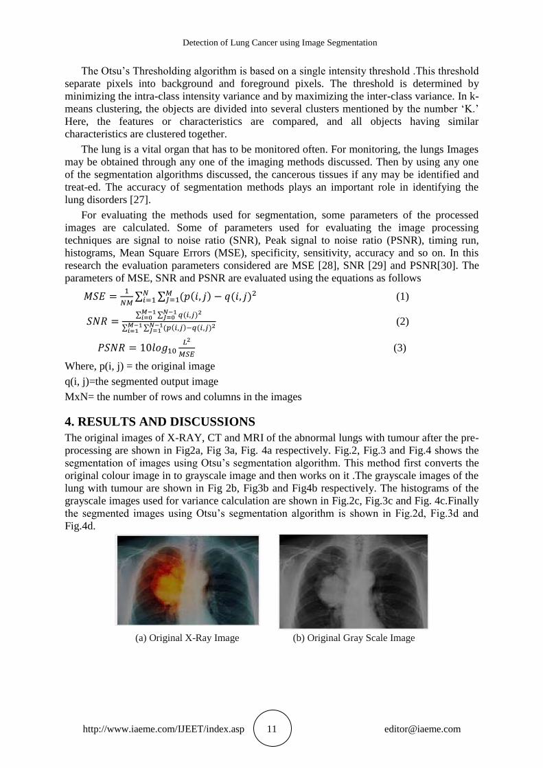
The original images of X-RAY, CT and MRI of the abnormal lungs with tumour after the pre-
processing are shown in Fig2a, Fig 3a, Fig. 4a respectively. Fig.2, Fig.3 and Fig.4 shows the
segmentation of images using Otsu‟s segmentation algorithm. This method first converts the
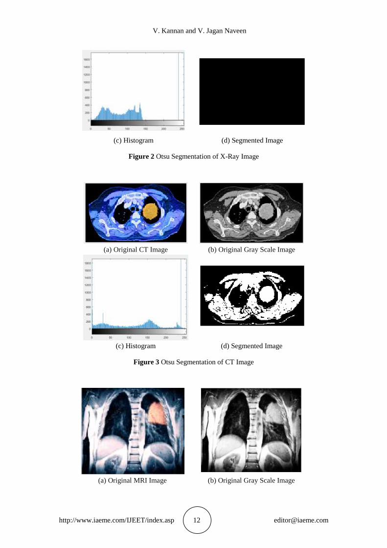
original colour image in to grayscale image and then works on it .The grayscale images of the
lung with tumour are shown in Fig 2b, Fig3b and Fig4b respectively. The histograms of the
grayscale images used for variance calculation are shown in Fig.2c, Fig.3c and Fig. 4c.Finally
the segmented images using Otsu‟s segmentation algorithm is shown in Fig.2d, Fig.3d and
Fig.4d.
(a) Original X-Ray Image (b) Original Gray Scale Image
V. Kannan and V. Jagan Naveen
http://www.iaeme.com/IJEET/index.asp 12 [email protected]
(c) Histogram (d) Segmented Image
Figure 2 Otsu Segmentation of X-Ray Image
(a) Original CT Image (b) Original Gray Scale Image
(c) Histogram (d) Segmented Image
Figure 3 Otsu Segmentation of CT Image
(a) Original MRI Image (b) Original Gray Scale Image
Detection of Lung Cancer using Image Segmentation
http://www.iaeme.com/IJEET/index.asp 13 [email protected]
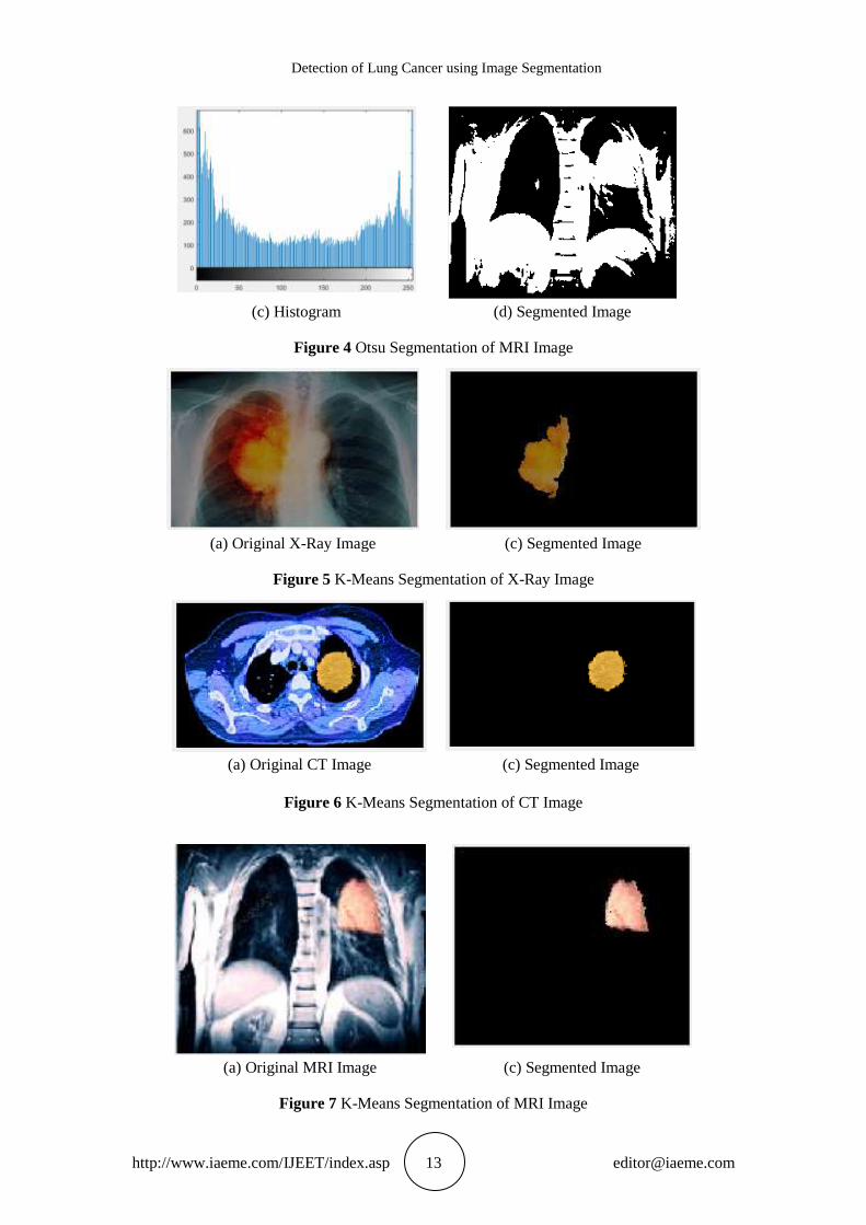
(c) Histogram (d) Segmented Image
Figure 4 Otsu Segmentation of MRI Image
(a) Original X-Ray Image (c) Segmented Image
Figure 5 K-Means Segmentation of X-Ray Image
(a) Original CT Image (c) Segmented Image
Figure 6 K-Means Segmentation of CT Image
(a) Original MRI Image (c) Segmented Image
Figure 7 K-Means Segmentation of MRI Image
V. Kannan and V. Jagan Naveen
http://www.iaeme.com/IJEET/index.asp 14 [email protected]
Fig. 5, Fig. 6 and Fig. 7 shows the segmentation of images using K-Means Clustering
algorithm. The segmented images using K-Means Clustering algorithm are shown in Fig. 5b,
Fig.6b and Fig.7b respectively.
It is found that by viewing the image itself, comparing the two image segmentation
algorithms used in this research, K-Means Clustering algorithm shows a better result in
segmenting the lung tumor. Also Otsu‟s segmentation algorithm has segmented to a certain
extent on the CT and MRI images as shown in Fig2d and Fig3d and its performance is poor
on X-Ray image as shown in Fig4d.
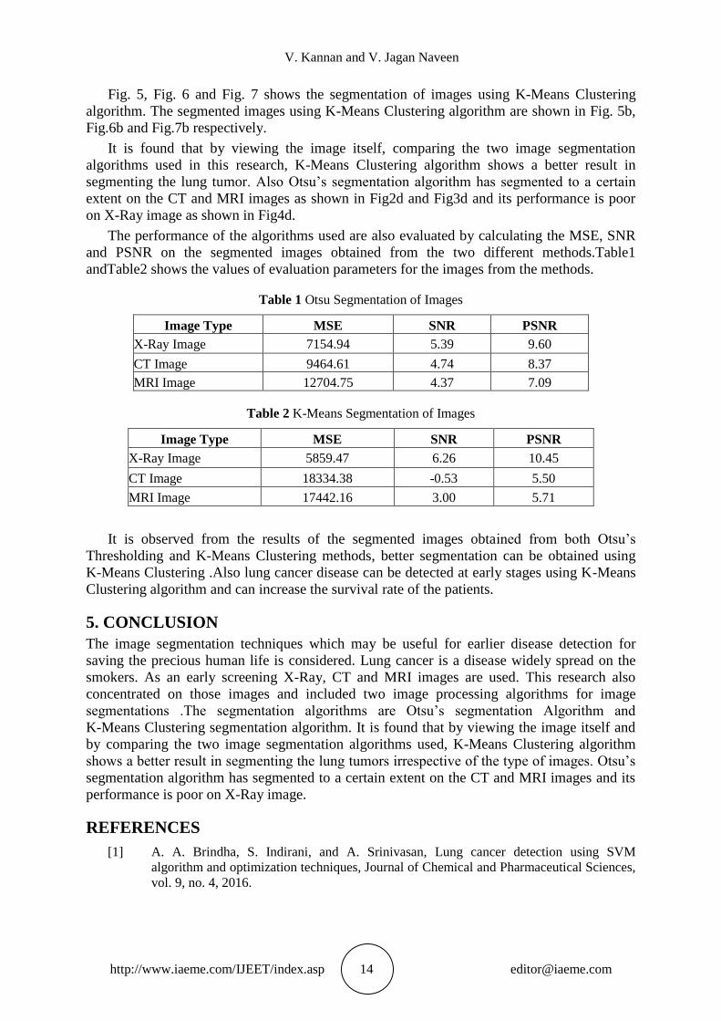
The performance of the algorithms used are also evaluated by calculating the MSE, SNR
and PSNR on the segmented images obtained from the two different methods.Table1
andTable2 shows the values of evaluation parameters for the images from the methods.
Table 1 Otsu Segmentation of Images
Image Type MSE SNR PSNR
X-Ray Image 7154.94 5.39 9.60
CT Image 9464.61 4.74 8.37
MRI Image 12704.75 4.37 7.09
Table 2 K-Means Segmentation of Images
Image Type MSE SNR PSNR
X-Ray Image 5859.47 6.26 10.45
CT Image 18334.38 -0.53 5.50
MRI Image 17442.16 3.00 5.71
It is observed from the results of the segmented images obtained from both Otsu‟s
Thresholding and K-Means Clustering methods, better segmentation can be obtained using
K-Means Clustering .Also lung cancer disease can be detected at early stages using K-Means
Clustering algorithm and can increase the survival rate of the patients.
5. CONCLUSION
The image segmentation techniques which may be useful for earlier disease detection for
saving the precious human life is considered. Lung cancer is a disease widely spread on the
smokers. As an early screening X-Ray, CT and MRI images are used. This research also
concentrated on those images and included two image processing algorithms for image
segmentations .The segmentation algorithms are Otsu‟s segmentation Algorithm and
K-Means Clustering segmentation algorithm. It is found that by viewing the image itself and
by comparing the two image segmentation algorithms used, K-Means Clustering algorithm
shows a better result in segmenting the lung tumors irrespective of the type of images. Otsu‟s
segmentation algorithm has segmented to a certain extent on the CT and MRI images and its
performance is poor on X-Ray image.
REFERENCES
[1] A. A. Brindha, S. Indirani, and A. Srinivasan, Lung cancer detection using SVM
algorithm and optimization techniques, Journal of Chemical and Pharmaceutical Sciences,
vol. 9, no. 4, 2016.
Detection of Lung Cancer using Image Segmentation
http://www.iaeme.com/IJEET/index.asp 15 [email protected]
[2] M. Kurkure and A. +akare, Introducing automated system for lung cancer detection us-ing
Evolutionary Approach, International Journal of Engineering and Computer Science, vol.
5, no. 5, pp. 16736–16739, 2016.
[3] B. Rani, A. K. Goel, and R. Kaur, A modified approach for lung cancer detection using
bacterial forging optimization algorithm, International Journal of Scientific Research
Engineering and Technology, vol. 5, no. 1, 2016.
[4] Charles S, Dela Cruz, Lynn. T. Tanoue and Richard A Mattay, Lung Cancer:
Epidemiology, Etiology and Prevention, Clinics inchest medicine, 32, 4, 2011.
[5] Murray N, Coy P, Pater JL, Hodson I, Arnold A, Zee BC, Payne D, Kostashuk EC, Evans
WK, Dixon P, et al. Importance of timing for thoracic irradiation in the combined
modality treatment of limited-stage small-cell lung cancer. The national cancer institute of
canada clinical trials group. Journal of clinical oncology: official journal of the American
Society of Clinical Oncology. 11(2):336–344. 1993
[6] Tucker WD, Burns B. Anatomy, Thorax, Heart Pulmonary Arteries, Stat Pearls ,Stat
Pearls Publishing; Treasure Island (FL): Dec 9, 2018.
[7] Burlew JT, Banks KP. Stat Pearls, StatPearls Publishing; Treasure Island (FL): Feb 16,
Anatomy, Thorax, Mediastinal Lymph Nodes. 2019.
[8] Donley ER, Loyd JW. StatPearls, Stat Pearls Publishing; Treasure Island (FL): Feb 10,
Anatomy, Thorax, Wall Movements. 2019
[9] S. Dearden, J. Stevens, Y.-L. Wu & D. Blowers, Mutation incidence and coincidence in
non small-cell lung cancer: meta-analyses by ethnicity and histology (mutMap), Annals of
Oncology 00: 1–6, 2013 doi:10.1093/annonc/mdt205
[10] Irene Ferrer, Jon Zugazagoitia, Stephan Herbertz, William John, Review of KRAS-Mutant
non-small cell lung cancer: From biology to therapy, Lung Cancer, Volume 124, October
Pages 53-64. 2018
[11] Kim H, Goo JM, Suh YJ, Park CM, Kim YT. Implication of total tumor size on the
prognosis of patients with clinical stage IA lung adenocarcinomas appearing as part-solid
nodules: Does only the solid portion size matter? Eur Radiol. Mar; 29 (3):1586-1594.
2019
[12] Muhammad Furrukh, Tobacco Smoking and Lung Cancer Perception-changing facts,
Sultan Qaboos Univ Med J. Aug; 13(3): 345–358. 2013
[13] Jonathan M. Samet, Erika Avila-Tang, Paolo Boffetta, Lindsay M. Hannan, Susan Olivo-
Marston, Michael J. Thun, and Charles M. Rudin, Lung Cancer in Never Smokers:
Clinical Epidemiology and Environmental Risk Factors, Clin Cancer Res. Sep 15; 15(18):
5626–5645. 2009
[14] Tracey A. Martin, Lin Ye, Andrew J. Sanders, Jane Lane, and Wen G. Jiang, Cancer
Invasion and Metastasis: Molecular and Cellular Perspective, Metastatic Cancer: Clinical
and Biological Perspectives edited by Rahul Jandial. Landes Bioscience. ©2013
[15] Sibu P Saha, Deepak K Bhalla, Thomas F Whayne, and CG Gairola, Cigarette smoke and
adverse health effects: An overview of research trends and future needs, Int J Angiol.
Autumn; 16(3): 77–83. 2007
[16] Kofi Asomaning, David P. Miller, Geoffrey Liu, John C. Wain, Thomas J. Lynch, Li Su,
and David C. Christiani, Second hand smoke, age of exposure and lung cancer risk, Lung
Cancer. Jul; 61(1): 13–20. 2008
[17] Leslie Stayner, James Bena, Annie J. Sasco, Randall Smith, Kyle Steenland, Michaela
Kreuzer and Kurt Straif, Lung Cancer Risk and Workplace Exposure to Environmental
Tobacco Smoke, Am J Public Health. March; 97(3): 545–551. 2007
V. Kannan and V. Jagan Naveen
http://www.iaeme.com/IJEET/index.asp 16 [email protected]
[18] IA RC Monographs on the Evaluation of Carcinogenic Risks to Hu-mans. Lung. In:
Ionizing Radiation, Part 1: X- and Gamma (y)-Radiation, and Neutrons. Vol 75. Lyon,
World Health Organization/IARC, pp. 253–254. 2000
[19] S Darby et.al. Radon in homes and risk of lung cancer: collaborative analysis of individual
data from 13 European case-control studies, BMJ; 330:223. 2005
[20] Krewski D, Lubin JH, Zielinski JM, et al. A combined analysis of North American case-
control studies of residential radon and lung cancer. J Toxicol Environ Health Part A; 69:
533–597. 2006
[21] A J Cohen and C A Pope, Lung cancer and air pollution, Environ Health Perspect. Nov;
103 (Suppl 8): 219–224. 1995
[22] Sandrah P Eckel, Myles Cockburn, Yu-Hsiang Shu, Huiyu Deng, Frederick W. Lurmann,
Lihua Liu, and Frank D Gilliland, Air Pollution Affects Lung Cancer Survival, Thorax.
Oct; 71(10): 891–898. 2016
[23] Xu X, Ha S, Kan H, Hu H, Curbow BA, Lissaker CT. Health effects of air pollution on
length of respiratory cancer survival. BMC Public Health; 13(1):1–9. 2013
[24] N. Hollings, P. Shaw, Diagnostic Imaging of Lung Cancer, European Respiratory Journal
19: 722-742. 2002
[25] Sharma N, Aggrawal L M, “Automated medical image segmentation techniques”, Journal
of Medical Physics, Jan-Mar, vol 35(1), pp 3-14. 2010
[26] Gonzalez R.C., Woods R.E., "Digital Image Processing using MATLAB", Upper Saddle
River, NJ Prentice Hall, 2008.
[27] Mokhled S. Al-Tarawneh, “Lung Cancer Detection Using Image Processing Techniques”,
Leonardo Electronic Journal of Practices and Technologies, Issue 20, January- June 2012,
p.147-158. ISSN 1583-1078
[28] Moreno et al. Towards no-reference of peak signal to noise ratio estimation based on
chromatic induction model International Journal of Advanced Computer Science and
Applications, 2013
[29] Zuzana Krbcova, Jaromir Kukal, Relationship between entropy and SNR changes in
image enhancement, EURASIP Journal on Image and Video Processing. (1), 2017
[30] Wang, Zhou, Bovik and Alan C. Modern image quality assessment (USA: Morgan &
Claypool). 2006
[31] Sreedhar T and Dr. Sathappan S, A Comparative Analysis of Image Segmentation
Techniques. International Journal of Computer Engineering and Technology, 9(5), pp. 64-
69. 2018
[32] Regonda Nagaraju and M. Janga Reddy, Review of Medical Image Segmentation with
Statistical Approach - State of the Art and Analysis. International Journal of Computer
Engineering & Technology, 8(2), pp.36–55. 2017
[33] Amel H. Abbas, Aryan A. Kareem, Mohammed Y. Kami, Breast Cancer Image
Segmentation Using Morphological Operations, International Journal of Electronics and
Communication Engineering and Technology, 6(4), pp. 8–14. 2015