detection of helminths by loop-mediated isothermal
TRANSCRIPT
SCOPING REVIEW Open Access
Detection of helminths by loop-mediatedisothermal amplification assay: a review ofupdated technology and future outlookMiao-Han Deng1,2†, Lan-Yi Zhong1,2†, Okanurak Kamolnetr3, Yanin Limpanont3* and Zhi-Yue Lv1,2,4,5*
Abstract
Background: Helminths are endemic in more than half of the world’s countries, raising serious public health concerns.Accurate diagnosis of helminth infection is crucial to control strategies. Traditional parasitological methods, serologicaltests and PCR-based assays are the major means of the diagnosis of helminth infection, but they are time-consumingand/or expensive, and sometimes provide inaccurate results. Loop mediated isothermal amplification (LAMP) assay, asensitive, simple and rapid method was therefore developed for detection of helminths. This study aims to discuss thecurrent status of application of LAMP on helminths detection and to make a comprehensive evaluation aboutthis updated technology and its future outlook by comparing with several other diagnostic methods.
Main body: This review summarizes LAMP assay applied for helminth detection and helminthiasis surveillance. Thebasic principle of LAMP is introduced to help better understand its characteristics and each reported assay is assessedmainly based on its detection sensitivity, specificity and limitations, in comparison with other common diagnostic tests.Moreover, we discuss the limitations of the assays so as to clarify some potential ways of improvement.
Conclusions: Here, we summarize and discuss the advantages, disadvantages and promising future of LAMP inheliminth detection, which is expected to help update current knowledge and future perspectives of LAMP inhighly sensitive and specific diagnosis and surveillance of helminthiasis and other parasitic diseases, and cancontribute to the elimination of the diseases from endemic areas.
Keywords: Loop-mediated isothermal amplification, Helminth, Point-of-care-test, Epidemiological surveillance,Field survey
Multilingual abstractsPlease see Additional file 1 for translations of the abstractinto the five official working languages of the UnitedNations.
BackgroundHelminths, including trematodes (flukes), nematodes(roundworms) and cestodes (tapeworms), are associatedwith substantial morbidity and economic losses worldwide[1–3]. Approximately one-sixth of the worlds’ populationis infected with helminths [4], with an estimated 15 billion
individuals, particularly in low socio-economic regions,suffered from soil-transmitted helminth (STH) infections[5, 6]. Although most of helminths have been well investi-gated epidemiologically [7], actual distributions of themare still unknown and accurate diagnosis is urgentlyneeded because of their generally non-specific and similarsymptoms (nausea and/or vomiting, diarrhoea, abdominalpain, and fever) between the causative species [8, 9].The approaches to clinical diagnosis and epidemio-
logical surveillance of helminthiasis vary according to thesamples, infectious stages, life cycle, morphological char-acteristics of helminths. Although the methods are diversi-fied, there is not an ideal and reliable point-of-care (POC)diagnostic method that can eminently meet the WorldHealth Orgnization (WHO)’s expectation of characteris-tics of affordable, sensitive, specific, user-friendly, rapidand equipment-delivered (ASSURED) [10, 11]. Though
© The Author(s). 2019 Open Access This article is distributed under the terms of the Creative Commons Attribution 4.0International License (http://creativecommons.org/licenses/by/4.0/), which permits unrestricted use, distribution, andreproduction in any medium, provided you give appropriate credit to the original author(s) and the source, provide a link tothe Creative Commons license, and indicate if changes were made. The Creative Commons Public Domain Dedication waiver(http://creativecommons.org/publicdomain/zero/1.0/) applies to the data made available in this article, unless otherwise stated.
* Correspondence: [email protected]; [email protected] Deng and Lan-yi Zhong are the joint first authors.3Faculty of Tropical Medicine, Mahidol University, Bangkok 10400, Thailand1Zhongshan School of Medicine, Sun Yat-sen University, Guangzhou 510080,ChinaFull list of author information is available at the end of the article
Deng et al. Infectious Diseases of Poverty (2019) 8:20 https://doi.org/10.1186/s40249-019-0530-z

the simple and cost-effective morphological identificationof parasites has been commonly employed in clinical diag-nosis and field survey, it shows poor sensitivity inlow-density parasite infections [12–16]. Furthermore, withrespect to the discernment of parasite eggs that aremorphologically similar, it will lose its specificity [12–16].In addition, its prerequisite for a considerable quality andquantity of manpower also makes it unadaptable as aPOC tool [17]. To avoid misdiagnosis and misseddiagnosis, particularly in low-grade infections and inlow-intensity regions, enzyme-linked immunosorbentassay (ELISA), as a representative of serological tests, hasbeen applied [18, 19]. However, the major drawbacks withthe use of ELIAS are clear due to its inability to distin-guish between past and present infections, relatively highfalse-positive rate, and cross reactions [16, 19, 20]. Alter-natively, a series of polymerase chain reaction (PCR)-based techniques, which are both specific and sensitive,started a new era for nucleic acid-based molecular detec-tion of helminths. The 1990s witnessed the inception ofvarious amplification techniques, e.g., nucleic acidsequence-based amplification [21], strand displacementamplification [22], and rolling circle amplification [23].But none of these methods manages to conquer the inher-ent weakness of heavy dependence on a particular instru-ment or elaborate detection methods [24, 25]. As a result,their application is restricted where they are urgentlyneeded, such as in primary medical institutions, under-developed areas and field studies [16, 26, 27]. As LAMP, anucleic acid amplification method with extremely highsensitivity and specificity, appears to promise an appealingresolution for almost all of issues mentioned above, thisreview investigates the recent research progress in use ofLAMP in helminth detection and make an comprehensiveevaluation about this updated technology and highlightsthe future perspectives regarding the possible applicationsof LAMP in diagnosis of parasitic diseases, comparingwith etiological detection, serological tests and other mo-lecular assay.In the present paper, we reviewed published studies
between 2001 and 2018 to identify studies exploitingLAMP in helminth detection. A comprehensive searchstrategy was developed in PubMed, proper key wordsand free text terms employed. Search terms were “(hel-minth” [All fields] OR nematode [All fields] OR cestode[All fields] OR trematode [All fields]) AND (“loop-me-diated isothermal amplification” [All fields] OR “LAMP”[All fields]). In brief, information was collected and ana-lyzed from 54 articles in Chinese or English.
Main textPrinciple of LAMPUsing the sophisticated mechanism of auto-cycling stranddisplacement DNA synthesis, LAMP was developed as a
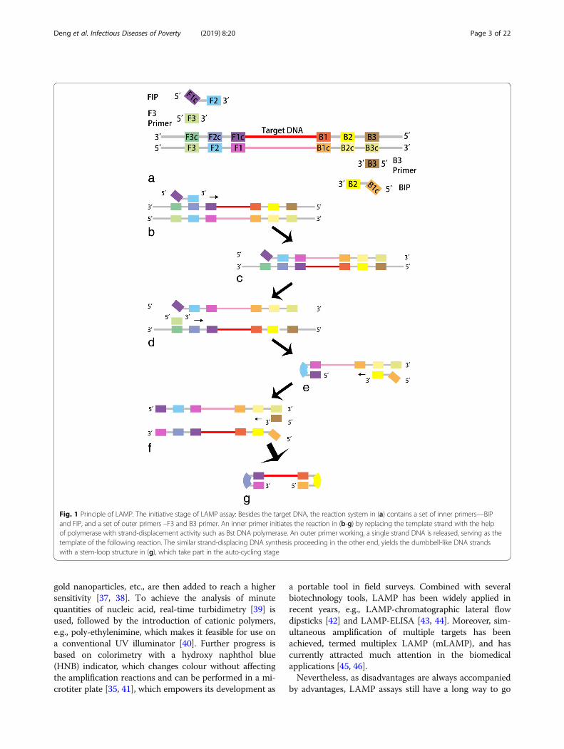
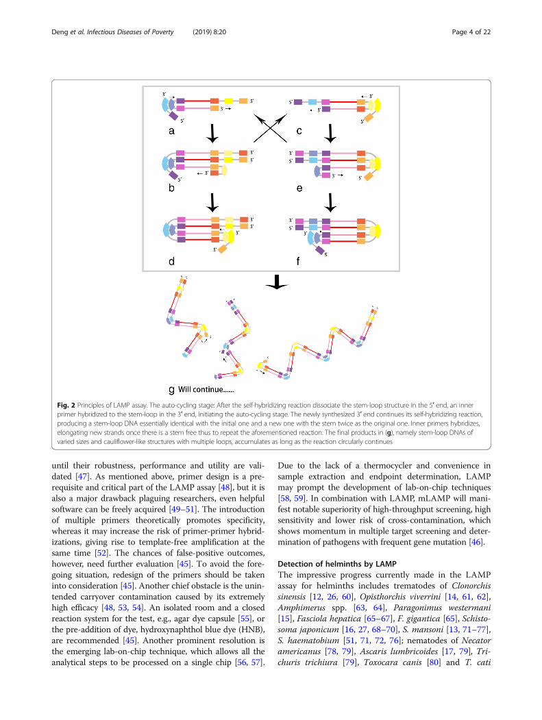
novel method requiring minimal instrumentation [25]. Aninner primer, termed forward inner primer (FIP), contain-ing sequences corresponding to the sense and antisensesequences of the target DNA, initiates the reaction [25].An outer primer primes the subsequent strand displace-ment DNA synthesis [25]. As a result, a single-strandedDNA molecule is released, serving as the template forsimilar DNA synthesis primed by another set of primers atthe other end of the target DNA [25]. In the initial step,dumbbell-like DNA strands with a stem-loop structure areproduced (Fig. 1) [25]. In the following cycling step, DNAsynthesis is triggered by an inner primer hybridizing to theloop on the product, which produces an identicalstem-loop structure [25]. Released by the strand displace-ment reaction, the 3′ end of the original stem-loop DNAmolecule is able to complete the self-primed DNA synthe-sis, yielding a new stem-loop DNA molecule with the stemtwice length as the original one [25]. The above reactionscircularly repeat during the entire cycling step (Fig. 2) [25].Without a thermocycler [28], target DNA is amplified
by employing Bst DNA polymerase under a constanttemperature of 60–65 °C, and accumulated 109 copies oftarget DNA in less than an hour, with a detection limitof a few copies [24, 25, 29]. Properly designed primersare given [30], as four different primers recognize 6 dis-tinct sequences in a target DNA. The process will beblocked once non-specific recognition occurs, hencehigh selectiveness [29]. If supplemented with loopprimers, stem primers and swarm primers, an evenhigher reaction speed can be expected [31–33]. The finalproducts of the LAMP reaction are stem-loop DNAsinverted with a large amount of repeats of the target andcauliflower-like structures with multiple loops. The ap-proaches of endpoint monitoring differ according to var-ied purposes. Sometimes agarose gel electrophoresis isemployed as the gold standard, but it is not always com-pulsory [25, 34, 35]. And turbidity determination is moresuitable for field research [24]. As a pyrophosphate ionis released once a nucleotide is added to the DNAstrands, a large number of target DNA will be accumu-late by the end of the assay, forming visible white precip-itates of magnesium pyrophosphate, which is used todetermine whether the target nucleic acid was amplifiedor not [36]. Based on the principle mentioned above,LAMP is characteristically able to meet the ASSUREDneeds, since it is a one-step process running within 1 hwhen there is Bst polymerase and a simple heatingblock, and the result can be read by the naked eyes. Fur-thermore, LAMP has also reported to be more tolerantthan PCR for some biological inhibitors. Therefore, itcan detect DNA in some specific clinical samples, suchas swabs, without DNA extraction [28].For further improvement, the fluorescent probe calcein,
DNA-binding dye SYBR Green I, DNA-functionalized
Deng et al. Infectious Diseases of Poverty (2019) 8:20 Page 2 of 22
gold nanoparticles, etc., are then added to reach a highersensitivity [37, 38]. To achieve the analysis of minutequantities of nucleic acid, real-time turbidimetry [39] isused, followed by the introduction of cationic polymers,e.g., poly-ethylenimine, which makes it feasible for use ona conventional UV illuminator [40]. Further progress isbased on colorimetry with a hydroxy naphthol blue(HNB) indicator, which changes colour without affectingthe amplification reactions and can be performed in a mi-crotiter plate [35, 41], which empowers its development as
a portable tool in field surveys. Combined with severalbiotechnology tools, LAMP has been widely applied inrecent years, e.g., LAMP-chromatographic lateral flowdipsticks [42] and LAMP-ELISA [43, 44]. Moreover, sim-ultaneous amplification of multiple targets has beenachieved, termed multiplex LAMP (mLAMP), and hascurrently attracted much attention in the biomedicalapplications [45, 46].Nevertheless, as disadvantages are always accompanied
by advantages, LAMP assays still have a long way to go
Fig. 1 Principle of LAMP. The initiative stage of LAMP assay: Besides the target DNA, the reaction system in (a) contains a set of inner primers—BIPand FIP, and a set of outer primers –F3 and B3 primer. An inner primer initiates the reaction in (b-g) by replacing the template strand with the helpof polymerase with strand-displacement activity such as Bst DNA polymerase. An outer primer working, a single strand DNA is released, serving as thetemplate of the following reaction. The similar strand-displacing DNA synthesis proceeding in the other end, yields the dumbbell-like DNA strandswith a stem-loop structure in (g), which take part in the auto-cycling stage
Deng et al. Infectious Diseases of Poverty (2019) 8:20 Page 3 of 22
until their robustness, performance and utility are vali-dated [47]. As mentioned above, primer design is a pre-requisite and critical part of the LAMP assay [48], but it isalso a major drawback plaguing researchers, even helpfulsoftware can be freely acquired [49–51]. The introductionof multiple primers theoretically promotes specificity,whereas it may increase the risk of primer-primer hybrid-izations, giving rise to template-free amplification at thesame time [52]. The chances of false-positive outcomes,however, need further evaluation [45]. To avoid the fore-going situation, redesign of the primers should be takeninto consideration [45]. Another chief obstacle is the unin-tended carryover contamination caused by its extremelyhigh efficacy [48, 53, 54]. An isolated room and a closedreaction system for the test, e.g., agar dye capsule [55], orthe pre-addition of dye, hydroxynaphthol blue dye (HNB),are recommended [45]. Another prominent resolution isthe emerging lab-on-chip technique, which allows all theanalytical steps to be processed on a single chip [56, 57].
Due to the lack of a thermocycler and convenience insample extraction and endpoint determination, LAMPmay prompt the development of lab-on-chip techniques[58, 59]. In combination with LAMP, mLAMP will mani-fest notable superiority of high-throughput screening, highsensitivity and lower risk of cross-contamination, whichshows momentum in multiple target screening and deter-mination of pathogens with frequent gene mutation [46].
Detection of helminths by LAMPThe impressive progress currently made in the LAMPassay for helminths includes trematodes of Clonorchissinensis [12, 26, 60], Opisthorchis viverrini [14, 61, 62],Amphimerus spp. [63, 64], Paragonimus westermani[15], Fasciola hepatica [65–67], F. gigantica [65], Schisto-soma japonicum [16, 27, 68–70], S. mansoni [13, 71–77],S. haematobium [51, 71, 72, 76]; nematodes of Necatoramericanus [78, 79], Ascaris lumbricoides [17, 79], Tri-churis trichiura [79], Toxocara canis [80] and T. cati
Fig. 2 Principles of LAMP assay. The auto-cycling stage: After the self-hybridizing reaction dissociate the stem-loop structure in the 5′ end, an innerprimer hybridized to the stem-loop in the 3′ end, initiating the auto-cycling stage. The newly synthesized 3′ end continues its self-hybridizing reaction,producing a stem-loop DNA essentially identical with the initial one and a new one with the stem twice as the original one. Inner primers hybridizes,elongating new strands once there is a stem free thus to repeat the aforementioned reaction. The final products in (g), namely stem-loop DNAs ofvaried sizes and cauliflower-like structures with multiple loops, accumulates as long as the reaction circularly continues
Deng et al. Infectious Diseases of Poverty (2019) 8:20 Page 4 of 22
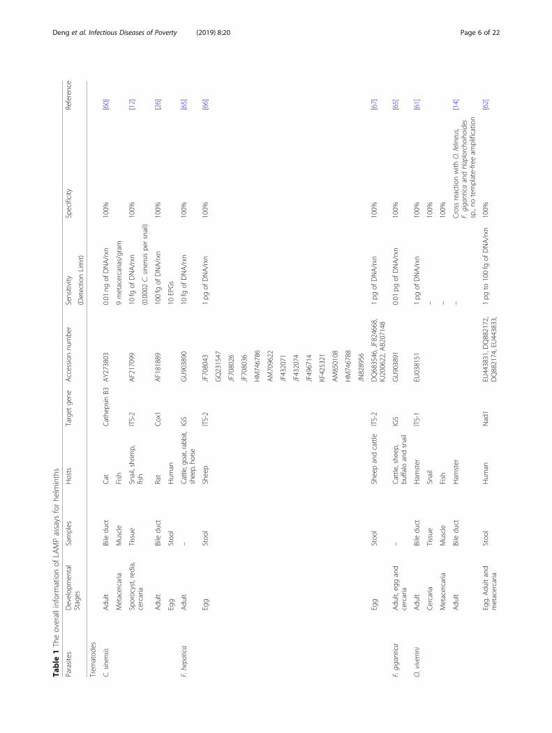
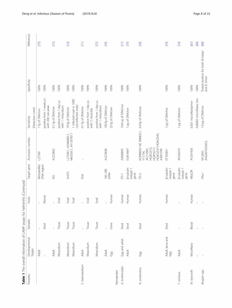
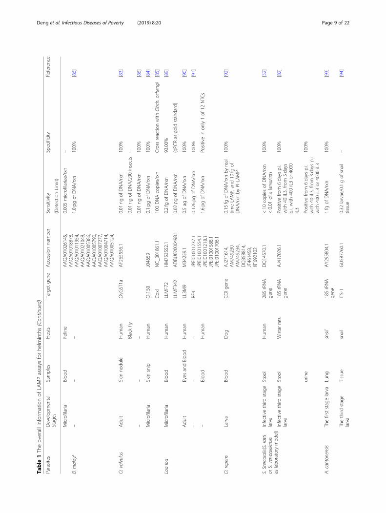
[81], Strongyloides stercoralis [52, 82], Onchocerca volvu-lus [83–86], Wuchereria bancrofti [86, 87], Brugiamalayi [86, 88], B. tomori [88], Loa loa [89–91], Dirofi-laria repens [92], Angiostrongylus cantonensis [93, 94],Trichinella spiralis [95, 96], Bursaphelenchus xylophilus[97], and Haemonchus contortus [98, 99]; cestodes of T.solium [44, 100–103], T. saginata [44, 100–103], T. asia-tica [44, 100–103], T. hydatigena [104], T. multiceps[104], T. pisiformis [104] and T. crassiceps [104], Echino-coccus granulosus [104–106], E. multilocularis [104,107], E. equinus [108], E. canadensis [108], E. felidi[108], E. ortleppi [108, 109] and E. shiquicus [104], havebeen covered in this review for further insight into itsadoption for clinical diagnosis, field surveys and surveil-lance of helminths. The sensitivity and specificity of de-tection of helminths by LAMP are shown in Table 1.
Detection of trematodes by LAMPFoodborne trematode infections remain a serious globalhealth burden, resulting in 2 million disability-adjustedlife years lost annually [110, 111].Clonorchiasis and opisthorchiasis, being mainly preva-
lent in Asia and Europe, are characterised by significantpathological hepatobiliary changes caused by C. sinensis,O. viverrini and O. felineus [110, 112]. Both C. sinensisand O. viverrini, classified as class one carcinogens ofhuman cholangiocarcinoma by the International Agencyfor Research on Cancer, are cancerogenic after years ofinfestation in bile ducts of the host [112, 113]. As devel-oped as biotechnology tools are, microscopic egg count-ing in stool samples continues to be the routine methodof diagnosis, which is simple but lacks sensitivity in earlyand light infections [112, 114, 115]. How to accuratelydifferentiate between liver flukes and intestinal flukes inareas where they coexist remains an unsolved problem[116]. In endemic areas where residents become infectedby consuming raw fish with metacercariae, the epi-demiological investigation of C. sinensis infection infreshwater fish is an important part of clonorchiasissupervision. The current epidemiological method in fishpartly depends on the labour-intensive microscopic in-spection of fish muscle, which may lead to missed detec-tion of low worm burden or cross-border contamination[117, 118]. Hence, LAMP, as an innovative techniquethat is sensitive and convenient, will help to solve theseproblems. The LAMP assay has been devised to detectDNA of C. sinensis and O. viverrini in freshwater snails[12], the second intermediate fish hosts [14, 60, 61] andpatient faeces [26, 61, 62].In the detection of C. sinensis infection in fish, the
respective detection limit of LAMP and PCR were10− 8 ng/μL and 10− 6 ng/μL, respectively, demonstratingthat LAMP was 100-fold more sensitive than PCR [60].When the true positive and negative results of LAMP
were in 100% agreement with the conventional micro-scopic examination, this approach shows the potential toreplace the conventional method in the investigation offluke invasion in the fish industry [14, 60, 61]. In addition,LAMP is sensitive enough to examine up to 0.0002 cer-cariae in a snail, and it is promising to be a prominent fig-ure in epidemiological surveillance for snail controlintervention [12]. In human faecal samples, LAMP-basedtechnology was established to detect C. sinensis with infec-tion intensity as low as 1 egg per 100mg. Further evalu-ation of the LAMP-based diagnosis test showed asensitivity of 97.1% and specificity of 100% as confirmedby the Kato-Katz (KK) method as well as real-time PCR(RT-PCR) [26]. However, it also perceived five additionalpositive samples of 13 microscopically negative samples inO. viverrini determination [61]. Future studies are expectedto assess the valid detection limit of this method in com-parison with the KK method and RT-PCR as well as itsfeasibility as a routine standard method [26]. Similar LAMPassays were also developed in O. viverrini, with the vari-ation of sensitivity and specificity relating to the repetitionof different target genes when detecting copro-DNA [14,61, 62]. For example, LAMP is highly sensitive when target-ing internal transcribed spacer 1 (ITS1) of O. viverrini, butspecificity cannot be guaranteed for ITS1 cross-amplifyinggenes from O. felineus, F. gigantica and Haplorchoihoidesspp. [61, 62]. When amplifying the mitochondrial genenad1 of O. viverrini in 100% specificity, the sensitivity forLAMP was between 1 petagram (pg) and 100 femtograms(fg), whereas it was 10 pg for PCR [62].Amphimeriasis, caused by Amphimerus spp., has been
recently reported as an emerging zoonotic fish-borne tre-matodisasis affecting indigenous inhabitants and domesticanimals in the tropical Pacific side of Ecuador [119]. Todate, a novel LAMP assay (namely LAMPhimerus) is de-vised for the first time to detect internal transcribed spa-cer 2 (ITS2) of Amphimerus spp. DNA in patient faecalsamples, with detection limit (1 pg) identical to conven-tional PCR [63]. LAMPhimerus was more sensitive thantraditional parasitological techniques, including direct mi-croscopy detection, formalin-ether concentration, simplesedimentation technique, Kato-Katz technique, fecal eggcount [63]. Of 44 human stool samples, the LAMPhi-merus method achieved 76.67% sensitivity; 80.77% specifi-city; 82.14% positive predict value (PPV) and 75.00%negative predict value (NPV) [63]. As the current scarcegenomic information of Amphimerus spp. is scarce, fur-ther enhancement of the assay could be based on the ex-ploitation of different DNA target [63]. The procedure, incombination with the air-dried faecal specimens on com-mon filter paper as source of DNA, is superior in feasiblecollection, long-term preservation and transportation, andpotentially applicable as an effective diagnostic or epi-demiological tool in amphimeriasis-endemic regions [64].
Deng et al. Infectious Diseases of Poverty (2019) 8:20 Page 5 of 22
Table
1Theoverallinformationof
LAMPassays
forhe
lminths
Parasites
Develop
men
tal
Stages
Samples
Hosts
Target
gene
Accession
numbe
rSensitivity
Specificity
Reference
(Detectio
nLimit)
Trem
atod
es
C.sin
ensis
Adu
ltBiledu
ctCat
Cathe
psin
B3AY273803
0.01
ngof
DNA/rxn
100%
[60]
Metacercaria
Muscle
Fish
9metacercarias/gram
Sporocyst,redia,
cercaria
Tissue
Snail,shrim
p,fish
ITS-2
AF217099
10fg
ofDNA/rxn
100%
[12]
(0.0002C.
sinensis
persnail)
Adu
ltBiledu
ctRat
Cox1
AF181889
100fg
ofDNA/rxn
100%
[26]
Egg
Stoo
lHum
an10
EPGs
F.hepatica
Adu
lt–
Cattle,go
at,rabbit,
sheep,ho
rse
IGS
GU903890
10fg
ofDNA/rxn
100%
[65]
Egg
Stoo
lSheep
ITS-2
JF708043
1pg
ofDNA/rxn
100%
[66]
GQ231547
JF708026
JF708036
HM746786
AM709622
JF432071
JF432074
JF496714
KF425321
AM850108
HM746788
JN828956
Egg
Stoo
lSheepandcattle
ITS-2
DQ683546,JF824668,
KJ200622,A
B207148
1pg
ofDNA/rxn
100%
[67]
F.gigantica
Adu
lt,eg
gand
cercaria
–Cattle,she
ep,
buffalo
andsnail
IGS
GU903891
0.01
pgof
DNA/rxn
100%
[65]
O.viverrini
Adu
ltBiledu
ctHam
ster
ITS-1
EU038151
1pg
ofDNA/rxn
100%
[61]
Cercaria
Tissue
Snail
–100%
Metacercaria
Muscle
Fish
–100%
Adu
ltBiledu
ctHam
ster
–Cross
reactio
nwith
O.felineus,
F.giganticaandHaplorcho
ihoides
sp.,no
template-fre
eam
plificatio
n
[14]
Egg,
Adu
ltand
metacercaria
Stoo
lHum
anNad1
EU443831,D
Q882172,
DQ882174,EU443833,
1pg
to100fg
ofDNA/rxn
100%
[62]
Deng et al. Infectious Diseases of Poverty (2019) 8:20 Page 6 of 22

Table
1Theoverallinformationof
LAMPassays
forhe
lminths(Con
tinued)
Parasites
Develop
men
tal
Stages
Samples
Hosts
Target
gene
Accession
numbe
rSensitivity
Specificity
Reference
(Detectio
nLimit)
EU443832,D
Q882175,
GQ401025,G
Q401064,
GQ401046,G
Q401060,
GQ401082,G
Q401096,
EU022343,EU022346,
EU022348,EU022350
Metacercaria
and
metacercarialcyst
Muscle
Fish
–100%
Egg
Stoo
lHum
an–
100%
Adu
ltBiledu
ctHam
ster
OvM
S6DQ144069
1pg
ofDNA/rxn
100%
[14]
Amph
imerus
spp.
Adu
ltLiver
Catsanddo
gsITS-2
AB678442.1
1pg
ofDNA/rxn
100%
[64]
Egg
Stoo
lHum
an–
–
P.westerm
ani
Metacercaria
Muscle
Freshw
ater
crab
andcrayfish
ITS-2
AF159604
0.01
fgof
DNA/rxn
100%
[15]
1metacercaria/gram
Egg
Sputum
and
pleuralfluid
Hum
an–
100%
S.japonicum
Adu
lt,eg
g,cercaria
Liverho
mog
enate,
stoo
land
serum
Rabb
itSjR2
AF412221
0.08
fgof
DNA/rxn
100%
[16]
SjR2
AY027869
–Serum
Rabb
itpo
sitivefro
m1weekp.i.
with
500cercariae
–
Adu
lt–
–100fg
ofDNA/rxn
Cross
reactio
nwith
Sch.man
soni;
notemplate-fre
eam
plificatio
n[69]
–Serum
Rabb
itpo
sitivefro
m1weekp.i.
with
200cercariae
–
–Serum
Rabb
itSjR2
AF412221
positivefro
m3days
p.i.
with
30cercariae
–[70]
Miracidium
Tissue
Snail
28SrDNA
Z46504
100fg
ofDNA/rxn
100%
[68]
Miracidium
Tissue
Snail
positivefro
m1dayp.iw
ith1miracidium
–
1infected
snailin100no
n-infected
snails
Miracidium
Tissue
Snail
100fg
ofDNA/rxn
100%
[27]
S.man
soni
Adu
lt–
Mou
seSm
1–7
M36086
0.1fg
ofDNA/rxn
100%
[71]
Miracidium
Tissue
Snail
positivefro
m1dayp.i.with
10miracidia
–
Miracidium
Tissue
Snail
positivefro
m1dayp.i.
with
1miracidium
–[72]
–Plasma/Serum
Mou
se0.5fg
ofDNA/rxn
100%
[74]
positivefro
m1weekp.i.
with
200cercariae
Deng et al. Infectious Diseases of Poverty (2019) 8:20 Page 7 of 22
Table
1Theoverallinformationof
LAMPassays
forhe
lminths(Con
tinued)
Parasites
Develop
men
tal
Stages
Samples
Hosts
Target
gene
Accession
numbe
rSensitivity
Specificity
Reference
(Detectio
nLimit)
Adu
lt–
–Minisatellite
DNAregion
L27240
1fg
ofDNA/rxn
100%
[73]
–Stoo
lMou
sepo
sitivefro
m1weekp.i.
with
200cercariae.
100%
Adu
lt–
–IGS
AJ223842
0.1fg
ofDNA/rxn
100%
[75]
Miracidium
Tissue
Snail
positivefro
m1dayp.i.
with
1miracidium
100%
Miracidium
Tissue
Snail
SmITS
L27240.1,A
Y446082.1,
M63265.1,AF130787.1
70fg
ofDNA/rxn
100%
[13]
Miracidium
Tissue
Snail
1infected
snailin1000
non-infected
snails
100%
S.ha
ematobium
Adu
lt–
–DraI
–0.1fg
ofDNA/rxn
100%
[71]
Miracidium
Tissue
Snail
positivefro
m1dayp.i.
with
5miracidia
100%
Miracidium
Tissue
Snail
positivefro
m1dayp.i.
with
1miracidium
100%
[72]
Adu
lt–
–18S–28S
rDNA
AJ223838
100fg
ofDNA/rxn
100%
[76]
Egg
Urin
eHum
an25
fgof
DNA/rxn
100%
Nem
atod
es
A.lumbricoides
Eggandadult
Stoo
lHum
anITS-1
AJ000895
10.8ng
ofDNA/rxn
100%
[17]
Adu
ltStoo
lHum
anβ-tubu
linisotype1
gene
EU814697
1pg
ofDNA/rxn
100%
[79]
N.american
usEgg
Stoo
lHum
anITS-2
KC896820.1-KC896825.1,
Y11734,
AF217891,
HQ452515,
HQ455217,
HQ452537-HQ452543,
AJ001599
0.4fg
ofDNA/rxn
100%
[78]
Adu
lt,larvaand
egg
Stoo
lHum
anβ-tubu
linisotype1
gene
EF392851
1pg
ofDNA/rxn
100%
[79]
T.trichiura
Adu
lt–
–β-tubu
linisotype1
gene
AF034219
1pg
ofDNA/rxn
100%
[79]
W.ban
crofti
Microfilaria
Bloo
dHum
anWbLDR
AY297458
0.001microfilariae/rxn
100%
[87 ]
––
–0.00002microfilaria/rxn
100%
[86]
Brugianspp.
––
–Hha
IM12691,
AAQA01025653,
1.0pg
ofDNA/rxn
Tested
positiveforbo
thB.malayi
andB.tim
ori
[88]
Deng et al. Infectious Diseases of Poverty (2019) 8:20 Page 8 of 22
Table
1Theoverallinformationof
LAMPassays
forhe
lminths(Con
tinued)
Parasites
Develop
men
tal
Stages
Samples
Hosts
Target
gene
Accession
numbe
rSensitivity
Specificity
Reference
(Detectio
nLimit)
AAQA01026145,
AAQA01018878,
AAQA01011954,
AAQA01021048,
AAQA01005386,
AAQA01005790,
AAQA01007277,
AAQA01004714,
AAQA01005124,
Microfilaria
Bloo
dFeline
0.005microfilariae/rxn
–
B.malayi
––
–1.0pg
ofDNA/rxn
100%
[86]
O.volvulus
Adu
ltSkin
nodu
leHum
anOvG
ST1a
AF265556.1
0.01
ngof
DNA/rxn
100%
[83]
Blackfiy
0.01
ngof
DNA/200
insects
–
––
–0.01
ngof
DNA/rxn
100%
[86]
Microfilaria
Skin
snip
Hum
anO-150
J04659
0.1pg
ofDNA/rxn
100%
[84]
Cox1
NC_001861.1
100DNAcopies/rxn
Cross
reactio
nwith
Ohch.ocheng
i[85]
Loaloa
Microfilaria
Bloo
dHum
anLLMF72
HM753552.1
0.2fg
ofDNA/rxn
93.00%
[89]
LLMF342
ADBU
02000498.1
0.02
pgof
DNA/rxn
(qPC
Ras
gold
standard)
Adu
ltEyes
andBloo
dHum
anLL3M
9M34259.1
0.5ag
ofDNA/rxn
100%
[90]
––
–RF4
JPEI01001237.1
JPEI01001554.1
JPEI01001218.1
JPEI01001588.1
JPEI01001706.1
0.126pg
ofDNA/rxn
100%
[91]
–Bloo
dHum
an1.6pg
ofDNA/rxn
Positivein
only1of
12NTC
s
D.repens
Larva
Bloo
dDog
COIg
ene
AJ271614,
AM749230-
AM749234,
DQ358814,
JF461458,
KF692102
0.15
fgof
DNA/rxn
byreal
time-LA
MP,and10
fgof
DNA/rxn
byPI-LAMP
100%
[92]
S.Stercoralis(S.ratti
orS.venezuelensis
aslabo
ratory
mod
el)
Infectivethird
stage
larva
Stoo
lHum
an28SrRNA
gene
DQ14570.1
<10
copies
ofDNA/rxn
<0.01
ofalarva/rxn
100%
[52]
Infectivethird
stage
larva
Stoo
lWistarrats
18SrRNA
gene
AJ417026.1
Positivefro
m6days
p.i.
with
40iL3,fro
m5days
p.i.with
400iL3or
4000
iL3
100%
[82]
urine
Positivefro
m6days
p.i.
with
40iL3,fro
m3days
p.i.
with
400iL3or
4000
iL3
100%
A.canton
ensis
Thefirststagelarva
Lung
snail
18SrRNA
gene
AY295804.1
1fg
ofDNA/rxn
100%
[93]
Thethird
stage
larva
Tissue
snail
ITS-1
GU587760.1
0.32
larvae/0.1gof
snail
tissue
–[94]
Deng et al. Infectious Diseases of Poverty (2019) 8:20 Page 9 of 22

Table
1Theoverallinformationof
LAMPassays
forhe
lminths(Con
tinued)
Parasites
Develop
men
tal
Stages
Samples
Hosts
Target
gene
Accession
numbe
rSensitivity
Specificity
Reference
(Detectio
nLimit)
Adu
ltTissue
snail
0.01
ngof
DNA/rxn
100%
T.spiralis
Larva
Muscle
Mice
Repe
titive
DNA
X06625
0.724fg
ofDNA/rxn
Cross
reactio
nwith
positive
controls,including
Tri.na
tiva,Tri.
pseudospiralis
andTri.nelso
ni,no
crossreactio
nwith
heterologo
usspecies,no
template-fre
eam
plificatio
n
[95]
0.002larvae/rxn
0.01
larvae/g
ofmuscle
tissue
mt-lsrDNA
GU339148.1
0.1pg
/rxn
100%
[96]
T.canis
Egg
––
ITS-2
AJ002440,
0.1pg
ofDNA/rxn
100%
[81]
Egg
Sand
–3eg
gs/10gof
sand
Adu
ltStoo
lDog
0.1pg
ofDNA/rxn
100%
[80]
3eg
gs/30gof
stoo
ls
T.catti
Egg
Sand
–ITS-2
AJ002441
0.1pg
/rxn
100%
[81]
B.xyloph
ilus
––
–ITS
AB500146-
AB500156
(accessedin
DDBJ)
10copies
ofDNA/rxn
100%
[97]
–Woo
d–
2.5×10^(−5)
ofa
nematod
e/rxn
–
H.con
tortus
Egg
Stoo
lSheep
ITS-1
–5pg
ofDNA/rxn
100%
[98]
Adu
lt–
Goat
ITS-2
X78803.1
1pg
ofDNA/rxn
100%
[99]
Cestode
s
T.solium
Prog
lottid
and
cysticercus
Cystfluid
Mou
seCox1
AB086256
–100%
[100]
Prog
lottid
and
cysticercus
Cystfluid
Mou
seClp
AB441815
1copy
ofDNA/rxn
100%
T.sagina
taProg
lottid
and
cysticercus
Cystfluid
Mou
seCox1
AY684274
–100%
[100]
Egg
Stoo
lHum
an5EPG
100%
Prog
lottid
and
cysticercus
Cystfluid
Mou
seClp
AB441816
1copy
ofDNA/rxn
100%
Egg
Stoo
lHum
anmorethan
10EPG
97.4%(con
firmed
bymultip
lexPC
Rwith
Cox1ge
nes)
T.asiatica
Prog
lottid
and
cysticercus
Cystfluid
Mou
seCox1
AF445798
–100%
[100]
Egg
Stoo
lHum
an5EPG
100%
E.gran
ulosus
Protoscolex
Liver
Sheep
Repe
atregion
sequ
ence
DQ157697
100fg
DNA/200
μl100%
[105]
Egg
Stoo
lDog
1pg
/200
mgfeces
–
5EPG
Deng et al. Infectious Diseases of Poverty (2019) 8:20 Page 10 of 22

Table
1Theoverallinformationof
LAMPassays
forhe
lminths(Con
tinued)
Parasites
Develop
men
tal
Stages
Samples
Hosts
Target
gene
Accession
numbe
rSensitivity
Specificity
Reference
(Detectio
nLimit)
Egg
Stoo
lDog
Nad5
AF297617
1pg
ofDNA/rxn
100%
[106]
Egg
Stoo
lDog
positivefro
m22
days
p.i.
with
10000protoscoleces
–
Eggandlarva
Stoo
lDog
10pg
ofDNA/rxn
100%
[104]
E.gran
ulosus
sensu
stricto
Protoscolex
––
Nad1
AF297617
1/10
or1/50
ofon
eproscolex
E.gran
ulosus
G1,E.gran
ulosus
G3
positive;
[108]
100%
E.equinu
sProtoscolex
––
Nad1
AF346403
1/10
or1/50
ofon
eproscolex
100%
[108]
E.Ca
nadensis
Protoscolex
––
Nad1
AB208063
1/10
or1/50
ofon
eproscolex
E.Ca
nadensisG6,E.Ca
nadensisG7,
E.Ca
nadensisG8,E.Ca
nadensis
G10
positive;
[108]
100%
E.felidi
Egg
––
Nad1
EF558357
1/10
and1/50
egg
100%
[105]
E.ortleppi
Protoscolex
––
Nad1
AB235846
1/10
or1/50
ofon
eproscolex
100%
[105]
Protoscolexand
associated
germ
inal
layer
Hydatid
cyst
Cam
eland
human
Nad1
JN637177
10pg
ofDNA/rxn
100%
[109]
E.multilocularis
Protoscolex
Multilocular
cystic
masses
Mou
seNad5
AB031351
1pg
ofDNA/rxn
100%
[107]
Egg
Stoo
lDog
positivefro
m12
days
p.i.
with
10000protoscoleces
100%
Larva
–Hum
anCox1
AB46141
1pg
ofDNA/rxn
100%
[104]
Egg
Stoo
lDog
5eg
gof
DNAextractio
n100%
E.shiquicus
Adu
lt–
Fox
Cox1
JF90613
10pg
ofDNA/rxn
100%
[104]
T.hydatigena
Adu
lt–
Fox
Cox1
JN83129
10pg
ofDNA/rxn
100%
[104]
Egg
Stoo
lDog
1eg
gof
DNAextractio
n100%
T.multiceps
Larva
–Sheep
Nad1
KC79480
1pg
ofDNA/rxn
100%
[104]
Egg
Stoo
lDog
2eg
gof
DNAextractio
n100%
T.crassiceps
Larvae
–Gerbil
Cox1
EU54454
10pg
ofDNA/rxn
100%
[104]
T.pisio
fmi
Adu
lt–
Dog
Cox1
JX67796
10pg
ofDNA/rxn
100%
[104]
ITS-1Internal
tran
scrib
edspacer
1,ITS-2Internal
tran
scrib
edspacer
2,IGSIntergen
icspacer,C
ox1Cytochrom
ecoxidasesubu
nit1ge
ne,C
lpcathep
sinL-likecysteine
peptidase,
Nad
1Th
emito
chon
drialN
ADH
dehy
drog
enasesubu
nit1(Nad
1)ge
ne,N
ad5Th
emito
chon
drialN
ADHde
hydrog
enasesubu
nit5(Nad
5)ge
ne,O
vMS6
Opistho
rchisviverrinim
icrosatellite
6,SjR2
Schistosom
ajapo
nicum
retrotranspo
son2,
p.iP
ost-
infection,
EPGEg
gpe
rgram
offeces-:u
navailable,
MtTh
emito
chon
drialN
ad5ge
ne,W
bLDRW.b
ancroftiLo
ngDNArepe
at,O
vGST
Oncho
cercavolvulus
glutathion
eS-tran
sferase,
RF4Re
peat
family
4,NTC
Non
-tem
plate
Con
trol,M
t-lsrDNATh
emito
chon
drial-large
subu
nitrib
osom
alDNA
Deng et al. Infectious Diseases of Poverty (2019) 8:20 Page 11 of 22
Furthermore, the system ‘air-dried stool sample on filterpaper’-LAMP assay would be practical in large-scale mo-lecular investigation of the other helminthiasis [64].Given the infection of the genus Fasciola, fascioliasis
mainly affects ruminants and only occasionally humans,raising public health and economic concerns due to a re-duction in output [120–122]. Triclabendazole-resistantF. hepatica, an emerging problem, calls for reliable as-sessment of efficacy or resistance after deworming ther-apy [122]. Serological ELISA is applied in the detectionof cattle and sheep, but it is unreliable for species dis-tinction and the effectiveness of drug therapy [123].Coproantigen ELISA is appropriate for monitoring adultinfection, whereas it is insufficient correlation with larvalstage invasion until 6 weeks post treatment [124]. LAMPtargeting ribosomal intergenic spacer seems to be an op-tional detection method that overcomes the difficulty intaxonomical classification of F. hepatica and F. gigantica.It can amplify genes from adults, eggs and juvenilestages with a sensitivity 10 000-fold higher than PCR,while running an hour faster in the laboratory [65].Other LAMP-based assays amplifying sequences of thesecond internal transcribed spacer (ITS2) show their in-ability to distinguish between the two Fasciola species,F. hepatica and F. gigantica [66, 67]. Under field condi-tions, the LAMP assay can identify infected sheep in thefirst week post-infection and 30 days post-therapy, whileELISA cannot detect infections until 6 weeks and is in-sufficient to discriminate current and past infections, in-dicating the practical and applicable determination ofdrug efficacy or resistance [66]. In contrast, M.I. Arifinet al. reported poor performance of LAMP and PCR incomparison with other conventional methods for thediagnosis of F. hepatica in naturally infected sheep andcattle in the field. Of the 64 animals examined, LAMPand PCR had low sensitivities of 17.9 and 10.7%, respect-ively, and high specificities of 97.2 and 100%, respect-ively, with faecal egg count (FEC) and coproantigenELISA as composite reference standards. The failure ofLAMP and PCR may be due to factors including insuffi-ciency of DNA sample, possibly in relation to the choiceof DNA extraction method, amount of faeces substantiallyused, and uneven egg distribution in faeces of differenthost species [67]. If promoted in the future, such a test isstill suitable for early diagnosis, thus reducing veterinarycosts and the loss of livestock due to fascioliasis [65–67].To the best of our knowledge, LAMP has not yet beenused for the detection of human fascioliasis.Paragonimiasis, also known as lung fluke disease, is a pul-
monary inflammation caused by Paragonimus species [125,126], of which P. westermani is the most epidemiologicallyrelevant in Asia and sporadically in American and Africancountries [127]. The conventional immunological diagnosismethod is sensitive in human paragonimiasis but
unsustainable in epidemiological surveys when intermedi-ate hosts are detected [128]. A LAMP assay has successfullyamplified the gene sequence of P. westermani eggs in spu-tum and pleura fluid from patients, as well as metacercariaein freshwater crabs and crayfish. With a detection limit of1 × 10− 8 ng/μL, LAMP is close to 100 times more sensitivethan PCR. The LAMP method also yields positive andnegative results coinciding with those from parasitologytests, acting as an excellent candidate for field surveys andclinical diagnoses of paragonimiasis [15].Schistosomiasis ranks on the list of neglected tropical
diseases (NTD) for its impacts on an estimated numberof over 200 million individuals in more than 70 coun-tries [126, 129, 130]. Of the five Schistosoma spp. thatusually cause human schistosomiasis, S. japonicum isprevalent in Asia, while S. mansoni and S. haematobiumare mainly concurrent in Africa and the Middle East[130]. Currently, infection and reinfection continue tobe global challenges, particularly in poverty-stricken andinsanitary communities [131, 132] and in other regionsdue to transmission by tourists and immigrants whocome into contact with infested water [130, 132]. Mean-while, low-density infection remains after dewormingprogrammes, which still demands an affordable diagnos-tic approach for pre-patent infection and massive epi-demiological surveillance despite current parasitological,immunological and molecular diagnostic methods [131–134]. The KK method is the current mainstay of schisto-somiasis diagnosis, and its drawback of day-to-day vari-ation is inevitable in massive surveillance [9, 130, 131,134]. In addition, it is of great importance to overcomethe limitation of serological methods and their incap-acity to discriminate between past and present infectionsdue to the persistent existence of circular antibodies inthe patient even after an effective cure [135].As the control of intermediate host snails considerably
contributes to the monitoring of schistosomiasis [126],LAMP assays were established to detect S. japonicum inOncomelania hupensis [27, 68], S. mansoni in Biompha-laria spp. [13, 71, 72, 75] and S. haematobium in othersnails [71, 72]. LAMP assays are sensitive and specific inpooled samples, with a detection limit of up to one posi-tive in 100 negative O. hupensis (expecting for a largersample) [68] as well as one snail infected with S. man-soni in 1000 normal snails [13]. In addition, a snail in-vaded by a single miracidium can be detected only 1 dayafter exposure [68, 72, 132]. Therefore, LAMP was usedto construct the risk map of schistosomiasis based on in-fected O. hupensis in a field survey and readily adaptedto predict the prevalence tendency [27]. What’s more,there is another work of LAMP (named SmMIT-LAMP)assessing not only infected snails but also human stoolin low-transmission area of S. mansoni in Brazil, wherethe incidence was corresponded to what has been
Deng et al. Infectious Diseases of Poverty (2019) 8:20 Page 12 of 22
reported, ascertaining the foci of schistosomiasis trans-mission and helping build risk maps of schistosomiasis[77]. Furthermore, LAMP was developed to detect S.japonicum in rabbit models [16, 69, 70] and S. mansoniin murine models [71, 73, 74]. This approach detectedpositive results as early as 1 week [16, 69], and even 3days, after low-intensity infection in rabbit models [70],tested negative as late as 12 weeks post treatment, whichis consistent with PCR in early diagnosis, and testednegative 2 weeks later than PCR [70], thereby possessingpotential in early diagnosis, treatment and assessment ofthe efficacy after chemotherapy [16, 69, 70]. LAMP isalso readily adopted in the clinical determination of S.japonicum in human serum samples [16, 70], S. mansoniin stool samples [77], as well as S. mansoni and S. hae-matobium in urine samples [51, 76]. In human sera withlight to mediate infection, LAMP achieves the sensitivity,specificity, PPV and NPV of 95.5, 100, 100 and 89.4%,respectively, whereas those for S. mansoni and S. haema-tobium in urine sample are 90–100% [76]. Additionally,the sensitivity (92.86%), specificity (80.11%), and NPV(99.33%) of SmMIT-LAMP in human stool samples areoverall acceptable, but the PPV is 26.00%, which can beexplained by the higher sensibility of LAMP over thereference standard (KK), especially in patient with lowinfection levels [77]. In addition, without any need forcostly laboratory instrumentation and highly skilledpersonnel, the refinement of DNA extraction (i.e., LAM-Pellet, NaOH and heat lysis [51]), the harness of a port-able plasma separator [136] and the utility of auser-friendly chip [74] fulfil the requirements of thePOC test and are estimated to have a competitiveper-person cost, with less than $7.25 for the circulatingcathodic antigen test and no more than $7.00 for a sin-gle KK test [74]. Accordingly, further evaluation is re-quired for POC use in endemic areas [51, 74, 76].
Detection of nematodes by LAMPNemathelminthiasis, caused by nematodes, is a globallyrampant parasitic disease. The pathogenic nematode in-fecting human includes STH, S. stercoralis, Toxocara spp.,filariae, and other nematodes with distinctive life cycles,namely, A. cantonensis and Trichinella. Nematodes in vet-erinary and agricultural fields are also included.STH, including A. lumbricoides, hookworms, and whip-
worms, mainly occur in tropical and subtropical regions[137]. The KK method is currently the most commonmethod in STH diagnosis and is recommended by theWHO to conduct STH surveys [17, 78, 79, 138]. However,for the false negative results brought about by the reduc-tion of egg production after chemotherapy or the hatchingof eggs due to the delay of examination [139, 140], it is ac-tually a suboptimal choice in a mass drug administration(MDA) programme where post-chemotherapy evaluation
is needed. In contrast, the LAMP assay is superior to theparasitological and unspecific serological approaches inthat it tests positive when there is merely a single ovum[17], without cross reactivity or non-template positive [17,78, 79]. In terms of the quantity of DNA, the SmartAmp2assay amplifies the STH β-tubulin gene provided thatthere is one pg of DNA [79], and hookworm detection tar-geting the ITS-2 gene can even succeed with 0.4 fg ofDNA [78]. None of the false positives is observed in theseLAMPs, which is important, as multiple helminthiasesmay coexist in individuals in endemic areas [17]. In simu-lated clinical samples, the LAMP assays exhibit greatagreement with the KK method in which the kappa coeffi-cient is calculated to be 0.72 for A. lumbricoides determin-ation targeting ITS-1 [79] and 0.9 for hookwormmeasuring targeting ITS-2 [17, 78]. In the SmartAmp2assay, the pre-addition of HNB dye achieves even betteraccuracy by providing a closed system to avoid contamin-ation in post-reaction manipulation using SYBR Green[79]. Bovine serum albumin was added, and it performswell in crudely prepared stool samples despite the pres-ence of inhibitors, which is undoubtedly a competitive ad-vantage for a POC tool, though it still needs furthercomparison [79]. However, the vulnerability of HNB topH changes may be a challenge for its stability but can beresolved by standardizing reaction conditions [79].S. stercoralis, acting as one of the opportunistic nema-
todes transmitted by soil, is the causative agent of humanstrongyloidiasis. It usually contributes to asymptomatic in-fection but is a deadly uncontrolled hyperinfection syn-drome in immunocompromised patients [141–145], witha mortality rate of up to 87% [146, 147]. There is no singlegold standard for its detection, as the microscopic exam-ination of larvae in stool samples is insufficiently sensitiveeven when supplemented with enrichment techniques.Serological tests are sensitive but lack specificity [148–151]. PCR-based techniques, though sufficiently specific,are not diagnostically superior to parasitological tech-niques because of their unsatisfactory sensitivity, which ispresumably attributed to the irregular larval output inchronic strongyloidiasis, the uneven distribution in stoolspecimens, the DNA extraction process, the existence ofinhibitors in stool samples, etc. [151]. Generally, the de-finitive diagnosis of strongyloidiasis is made by parasito-logical examinations based on clinical symptoms,serological evidence, etc. [52, 82]. Compared with mor-phological examination, nucleic acid tests are advanta-geous in that they can detect specimens where parasiteshad been killed [52]. In 2014, the LAMP assay for S. ster-coralis was first reported to be capable of amplifying lessthan ten 0 DNA copies of larvae per reaction, or 10− 2 di-lution of one spiked larva in stool samples, comparable tothe results of PCR [52]. Unfortunately, the foregoing fac-tors that may influence PCR-based techniques, e.g., the
Deng et al. Infectious Diseases of Poverty (2019) 8:20 Page 13 of 22
DNA extraction process, also may impact it [52]. Aimingat surmounting the shortcomings of common stool sam-ples, urine samples from rodent models were used in anovel LAMP assay named Strong-LAMP [82]. The cre-ative introduction of urine samples may possess predom-inant advantages in collection, storage and processingover stool samples. Furthermore, when employing urinesamples of the rodent model, Strong-LAMP shows posi-tive results from 5 days after infection of 40 third-stage(L3) infective larvae (1 day earlier than employing stoolsamples) to 3 days after infection of 400 or 4000 L3 infect-ive larvae (2 days earlier than employing stool samples).Nevertheless, since requests for urine samples in S. ster-coralis detection are rare, its clinical value in latent infec-tion of humans needs further study [82].The larvae of T. canis and T. cati are responsible for
human toxocariasis. Children specifically tend to acquirethese kinds of telluric zoonosis and saprozoonosis by en-vironmental exposure to Toxocara spp. [152], whichmakes it one of the most common cosmopolitan helmin-thiases [153]. The prevention of its transmission dependson the condition of the environmental contaminationlevels and the accurate determination of its sources [81].However, Toxocara identification by traditional micros-copy of stools from pets or environmental samples re-mains a methodological concern due to its insensitivityin low-burden cases and its difficulty in distinguishing T.canis from T. cati eggs [80, 81]. PCR assays have beendesigned to discern Toxocara spp. in stools [154] or en-vironmental samples [155] and to distinguish between T.canis and T. cati in soil samples [156]. Thespecies-specific LAMP assay targeting ITS-2 was vali-dated by two groups and found to be ten-fold more sen-sitive than PCR without cross reactivity in the laboratorybetween Toxocara spp. and is applied in domesticateddogs and sand samples [80, 81]. In the context of envir-onmental specimens, LAMP manifests a detection limitof 3 eggs/10 g of sand and less than 3 eggs/30 g of stools,compared with the 6 eggs/10 g of sand and more than 2eggs/30 g of stools detection limit of PCR [80, 81]. In afield survey of soil contamination, LAMP yield a positiverate of 42.7% versus 7.7% of PCR [157]. In another fieldstudy, even LAMP fails to identify very low contamin-ation, which is a pitfall that may be attributed to thecrude processing of DNA extraction in LAMP comparedwith that of PCR [81], the LAMP assay successfully de-creased the standard examination time by 50% com-pared to that of PCR [81].As one of the most debilitating infectious diseases in
the world, lymphatic filariasis, which is caused by bru-gian filariae and W. bancrofti, is also regarded as a ser-ious public health concern for 856 million people in 52countries around the world [158]. The WHO MDAprogramme effectively reduces morbidity, raising new
concerns about diagnosis and surveillance in the controlareas and determination of the treatment endpoint inthe post-MDA stage [8, 83, 87, 88, 159]. So far, the diag-nosis largely counts on the microfilaraemia test, whichemploys night blood samples [86, 88] and is recom-mended by the WHO to conduct a transmission assess-ment survey (TAS) where Brugia spp. is endemic. It isused as the minimum in TAS but suffers from the re-duction of sensitivity in response to the prevalence de-crease in the post-MDA era. Simultaneously, moreaccurate methods, such as antibody tests and PCR, arerestricted by their inherent shortcomings. The antige-naemia tests recommended to map W. bancrofti endem-icity, namely, immunochromatography card test andfilariasis test strip [160, 161], are unavailable for brugianfilariae and may cross react with Loa loa [160, 162, 163].Alternatively, as a competitive candidate in the presentstudy, LAMP assays manifest cheerful outcomes in bothlaboratory and clinical tests [87, 88]. For instance, theW. bancrofti LAMP test, with a determination limit of0.1 pg per reaction equivalent to that of PCR, costs over$1.38 less than the latter [87]. It is estimated that thereis approximately 200 pg and 100 pg of DNA inside a sin-gle microfilaria of W. bancrofti or Brugia spp., respect-ively [164]; that is to say, the detection limit of theLAMP assay exceeds the theoretical detection limit ofmicrofilariae per ml via microscopic inspection [165].Furthermore, compared with the serological tests thatare inadequately specific, almost all the LAMP assays forlymphatic filaria diagnosis are species-specific, exceptone detecting brugian filariae for both B. timori and B.malayi [86–88].A similar methodological handicap is used to eliminate
O. volvulus, another major public health concern mostlyrampant in sub-Saharan Africa [83, 166]. Following theimpediment to onchocerciasis transmission, the chal-lenge emerges in that the conventional diagnosticmethod of skin snip microscopy and the primary diag-nostic antibody test, the Ov-16 rapid diagnostic test, islosing its sensitivity in low-prevalence settings [167,168]. Alternatively, nucleic acid-based assays can beemployed in both diagnosis and xenomonitoring withextreme sensitivity and specificity. O-150 PCR, therefore,is recommended by the WHO to undertake vector sur-veillance but is limited in resource-limited areas [84,169]. Using the economical LAMP assay as a diagnosticoption manifests sensitivity just slightly lower than theutmost sensitive qPCR when targeting cox1 but is tentimes higher than conventional PCR in O-150 assay atthe same time [84, 85]. In terms of specificity, the cox1assay is reported to cross react with O. chengi, a sympat-ric cattle parasite transmitted by black flies, or rather,the cox1 assay can be used only in clinical diagnosisusing skin biopsy samples unless significant progress is
Deng et al. Infectious Diseases of Poverty (2019) 8:20 Page 14 of 22
made to improve specificity [85]. However, whether theother set of primers designed for O-150 can amplify theheterologous sequence from O. chengi remains to be de-termined [84], as the PCR targeting O-150 has beenproven to cross react with O. chengi unless a specificDNA probe is added [170]. In addition, an elaboratecomparison is designed between the HNB and neutralred dyes, and the latter improves the sensitivity 10-fold,which sheds light on a new approach for parasite LAMPamelioration, maximizing its usefulness in a world witha changing global landscape of infection [84].In contrast to other parasites, in the post-MDA surveil-
lance of filariae, the exploitation of samples from mos-quito vectors is considered timelier, more operationallyfeasible and more ethically accepted than detection usingspecimens from humans [8, 159, 168, 169, 171]. As ento-mological inspection via field dissension is expensive, timeconsuming and unable to distinguish O. volvulus from O.chengi, O-150 PCR using vector samples is currentlywidely accepted to determine the interruption of filariae[8, 87, 159, 167–169]. LAMP can also act as an excellentsurrogate for PCR in this case. As shown in O. volvulusdetection targeting OvGST1a, without crossreactivity withO. chengi or other filariae, LAMP tests positive withmerely 0.01 ng of DNA spiked in 200 insects, which ismore sensitive than PCR, which tests positive in 0.01 ng/50insects [83]. Based on the conventional LAMP assays, animproved non-instrumented nucleic acid-LAMP was de-veloped, devised as a single portable electricity free devicewith comparable or even higher sensitivity than a normalassay, demonstrating that it is more suited for field surveys[86]. Whereas the existing LAMP assays for vector moni-toring are designed to utilize the DNA extracted frominfective-stage larvae (L3), there are great hurdles in xeno-monitoring, where the DNA test cannot identify DNAfrom L3 larvae from immature stage parasites (L1 or L2) invectors, which actually distinguishes xenomonitoring fromentomological monitoring of transmission [159]. As thediscrimination between infectious and immature parasiteswill clarify whether the positive result is due to adultfilariae not responding to drug treatment or recent in-fection indicating active transmission, it is taking onincreasing significance in the assessment afterlarge-scale drug treatment [8, 171]. For O. volvulus, inwhich the infectious stage parasites are located in thehead capsule isolated from the immature stage larvaein the abdomen and thoracic muscle, the obstacle canbe overcome by the separation of the head and bodyand therefore provide accurate evaluation of transmission[159, 172]. On the other hand, although there are specificL3-stage RT-PCR tests that are capable of indirectly deter-mining the infection potential and transmission dynamicsof lymphatic filariae via RNA [173, 174], dissection re-mains more common for the detection of infectious stage
lymphatic filariae [159]. However, it can be expected thatthe development of RT-LAMP in parasitology may favourthis technique to replace RT-PCR and conventional dis-section to precisely predict the transmission potentialeven in low-resource areas.Loa loa is a long-neglected filariae that is reported to
cause deadly serious adverse events after ivermectintreatment [86, 89–91, 175, 176] at a low threshold ofmicrofilaria (mf) burden [175], where determination ofthe mf burden before MDA programme is especially im-portant. Unfortunately, the routine diagnosis and quanti-fication in remote areas rely on microscopic inspectionof midday blood samples, which requires expertise andprocessing of a considerable number of samples and isunqualified to serve as a POC or a large-scale screeningtool. Among the existing LAMPs, one amplifies theLL3M9 gene and exhibits the lowest detection limit of0.5 ag/reaction, far lower than the formerly reported 0.1pg/reaction for W. bancrofti [87, 90]. Considering thepractical significance of Loa loa mf burden quantitationin MDA practice, Loa loa LAMP targeting LLMF72 wasassessed for its potential for semi-quantitation. As a re-sult, a correlation was observed between the time toLAMP reaction positivity (minutes) and the mf concen-tration in the blood, allowing the naked-eye determin-ation of whether the mf burden is above or below thespecific threshold. For example, the run time to positiv-ity is 15 min at the threshold of > 30 000 mf/mL, 20 minat threshold of > 5000 mf/mL, and 25 min at the thresh-old of >v100 mf/mL, which is promising for applicationin the Loa loa microfilaraemia assessment before iver-mectin treatment and thus facilitating the elimination offilariasis [89]. Since the LL3M9 comprises multiple cop-ies of a simple nematode-conserved repeat, and LLMF72is a single copy gene, which may exert an impact on thesensitivity and specificity, a new bioinformatic pipeline isdesigned to mine a new species-specific sequence that ismore suitable for MDA practice. Consequently, RF4 is anew biomarker with specificity; however, it lacks sensi-tivity compared with the LL3M9 or LLMF72 assays.Nevertheless, the bioinformatic pipeline remains a cre-ative and robust method to further explore the potentialof LAMP [91].Dirofilariasis caused by D. repens, another species of
mosquito-borne filariae [177], is regarded as an emergingzoonotic disease calling for more accurate diagnosis. Thetraditional diagnostic method relies on microscopic exam-ination of blood from the hosts [178]. Serological screen-ings [179] and PCR tests have been designed [180, 181].The LAMP assay targeting the COI gene was devised as 2versions for further evaluation. With respect to sensitivity,the detection limits of reverse transcriptase LAMP(RT-LAMP) and propidium iodide LAMP (PI-LAMP) are0.15 fg and 10 fg, respectively, versus the detection limit of
Deng et al. Infectious Diseases of Poverty (2019) 8:20 Page 15 of 22
15 fg for conventional PCR. With a lower limit, the LAMPassays yield amplicons within approximately 40min, whileconventional PCR takes 2 h. Generally, both versions ofLAMP prevail over conventional PCR in both sensitivityand efficiency, while all of them are species-specific in thecurrent study. Considering practical value, while RT-LAMP employs a RT-PCR instrument, PI-LAMP, byintroducing propidium iodide, permits visualization of theamplification as UV fluorescence, meriting more wide-spread application in field surveys and clinical diagnoses[92]. Because of its combination of sensitivity, specificity,rapidity and convenience, it may be a promising ancillarytool in dirofilariasis surveillance and prevention, such aslarge-scale travelling animal quarantine inspection or culi-cid mosquito screening.A. cantonensis infects people on the Pacific islands and
Southeast Asia. It is the major cause of an eosinophilicmeningitis in humans in endemic areas [182]. The lackof standardization of a diagnostic procedure and thecurrent situation of being overlooked in accounts for theuse of a presumptive diagnosis, which is primarily basedon the combination of patient history and clinical cri-teria, e.g., morphological examination of adult worms orlarvae in cerebrospinal fluid, of which the positive rate isbetween 2%~ 12% [183], are unable to meet the expect-ation of either clinical diagnosis or large-scale surveil-lance [184, 185]. In an effort to help establish asurveillance system, two LAMP assays were developedto detect the L3 larvae in molluscan hosts. One amplify-ing the ITS-1 gene manifests a detection limit of 1 fg/re-action [94]. The other test targeting the 18S rRNA geneis inferior, with a detection limit of 10 pg/reaction [93],while both have higher sensitivity than PCR, which candetect DNA > 100 pg/reaction [93, 94]. In a similar fieldsurvey, the ITS-1 LAMP assay demonstrates detectionrates of 6.7 and 4.4% higher than the standard digestionmethod and PCR, respectively [94]. In summary, all ofthe above information exhibits considerable potentialand superiority in replacing existing approaches inlarge-scale field surveys and clinical diagnoses [93, 94].Trichinellosis is a significant zoonotic disease caused
by the ingestion of raw or insufficiently cooked meatcontaining Trichinella spp., for which the inadequacy ofveterinary control is one factor to blame. There hadbeen no detailed and systematic reports of the sensitivityand conditions of the assays for Trichinella determin-ation by 2012, when 2 LAMP assays were designed [95,96], amplifying mitochondrial large ribosomal subunitDNA (mt-lsrDNA) and a 1.6 kb repetitive sequence fromthe larvae, respectively. Both assays manifest sensitivity10-fold stronger than conventional PCR [95, 96], but theone targeting mt-lsrDNA turns out to be 10-fold lesssensitive than RT-PCR [96]. Further exploration couldbe made to improve the sensitivity of LAMP to make it
an optimal methodology for trichinellosis detection inpractice, e.g., meat quarantine or field survey.In addition to the human medical nematoda men-
tioned above, the application of LAMP has spread to theveterinary [98, 99] and agricultural fields [97], whichmakes it a promising detection tool shared by all fieldsof bioscience.
Detection of cestodes by LAMPTaenia species (T. solium, T. saginata and T. asiatica), thecausative pathogens of taeniasis, can be sympatrically en-demic in Asia, such as in China and Thailand [186]. T.solium, normally transmitted between pigs and humans,results in neurocysticercosis with a range of manifesta-tions, especially epilepsy and seizures [7]. Conventionalproglottid examination, as a common diagnostic methodfor taeniasis, fails to morphologically differentiate the eggsof Taenia species. Multiplex PCR and nested PCR openthe door for characteristic discrimination [187, 188] butare unrealistically applied in field surveys for high expenseand time considerations. Therefore, a LAMP assay withthe cytochrome c oxidase subunit 1 (cox1) primer set wasdeveloped for the differentiation of Taenia spp. at the spe-cies level in the laboratory and in the field, managing todetect eggs in traditional faecal samples in epidemiologicalsurveys with high specificity and even higher sensitivitythan PCR [100–103]. Ranging from five to ten eggs pergram (EPG) of faeces, the detection limit of LAMP iscomparable to that of five EPG and 40 EPG of multiplexPCR and nested PCR, respectively [100, 187, 188]. Thespecificity is approximately 100%, with only two in 76(2.6%) T. saginata recognized as T. asiatica in faecal sam-ples [100]. Out of 51 proglottids expelled from 35 carriers,consistent results were obtained by LAMP under fieldconditions and in the laboratory, except for one sample[102]. Thus, the tedious procedure of simultaneously iden-tifying Taenia species is expected to be simplified to re-duce the possibility of cross contamination and to savetime, while the handy copro-DNA extraction methodis expected to take the place of centrifugation. Re-markably, the modification of mLAMP combined withdot-ELISA has succeeded in specific amplification in asingle tube, demonstrating an easier and more prac-tical POC diagnostic method for real-time humanTaenia species confirmation [44].Widely distributed in pastoral areas worldwide but
often neglected, echinococcosis, especially cystic echino-coccosis and alveolar echinococcosis, attracts enormousattention by posing as a threat to both humans and ani-mals and results in economic loss [189–193]. An on-siteapproach is expected to replace the ethically-challengedpost-mortem inspection as the gold standard in suscep-tible Echinococcus-infected definitive canid hosts [189,193]. In addition, a more practical and available tool is
Deng et al. Infectious Diseases of Poverty (2019) 8:20 Page 16 of 22

sought to solve the problem of copro-ELISA lacking sen-sitivity in latent infection monitoring [194] and to sus-tain reliability of copro-PCR while reducing the expense[195, 196] in epidemiological surveillance in endemicareas at the same time. LAMP was exploited to detect E.granulosus s. s. (G1-G3) copro-DNA in dogs [104–106]and then cysts in camels and humans [109]. It standsout for its high sensitivity in detecting infection incopro-samples from definitive hosts 22 days after expos-ure, which is equivalent to 3 days, 4 days and 47 daysearlier than ELISA, conventional PCR and light micros-copy, respectively [106]. A similar advance in E. multilo-cularis determination depicts LAMP as a substantialalternative for field surveillance of AE in areas of en-demicity [107]. LAMP was also applied in other cestodesof veterinary relevance, including E. equinus (G4), E.canadensis (G6-G10), E. felidi (lion strain), E. ortleppi(G5) [108], E. shiquicus, T. hydatigena, T. multiceps, T.pisiformis and T. crassiceps [104]. Furthermore, it wassensitive enough to distinguish different Echinococcusspecies, achieving sensitivity down to 2% of a single pro-toscolex or egg per reaction [104, 108], but failed to dis-criminate at the genotype level [108]. There are not yetinadequate data to relate intrastrain genetic variants todifferent life cycles, pathogenicity or any other practicalrelative features [191, 192, 194, 197, 198]. Subsequently,LAMP has great potential to become a new tool for fu-ture perspectives on molecular epidemiology in echino-coccosis surveillance at this stage. Additionally, thereal-time LAMP assay gave 100% concordance with theresults obtained by nested RT-PCR when testing parasiteDNA extracted from hydatid cysts from domestic ani-mals and humans, which highlights a brilliant future inclinical diagnosis of CE [108, 109]. Recently, LAMP wasfirst reported to determine Taenia species in an epi-demiological survey in Mongolia [199]. Above all, therapid, sensitive and accurate LAMP is sufficient to facili-tate a large-scale epidemiological survey.
Application of LAMP in field researchAs discussed above, the LAMP assay is a robust and ver-satile tool that is capable of meeting the WHO’s require-ments for ideal POC tools of ASSURED and possessesthe potential to become an appealing option for field re-search, which was substantiated by a series of laboratoryand diagnostic tests.From the perspective of field application, major
achievements were made for LAMP assays for malariaand tuberculosis [200, 201]; in both cases, scientistsworked extensively with the WHO for the implementa-tion of the tests in field, and their standardized reagentkits have been used in developing countries as patientside tools [202]. For protozoa, bacteria and fungi, severalcommercial reagent kits have been put on the market
and have performed excellently [203, 204]. With respectto helminths, considerable significance is attached to fil-ariae. LAMP assays for the detection filariae havealready come to MDA management practice in Guinea,Nigeria and Southeast Asia [205–207]. In a recent epi-demiological survey in Mongolia, LAMP also played asignificant part [199].
ConclusionsTo sum up, though presently in its infancy, the LAMPassay is a groundbreaking DNA amplification techniquewith prominent advantages. Its ASSURED characteristicsand its versatility in adapting to various circumstancesmake it an ideal POC tool and friendly to field surveys.The chief shortcoming of LAMP is the false-positive re-sult caused by primer-primer reaction and contamin-ation. The former needs further evaluation, and thelatter can be solved by the amelioration of the reactionsystem, detection approaches, etc. Another handicap inLAMP development is the difficulty in primer design.However, its merits outweigh its weakness, and LAMPhas blossomed in the detection of microorganisms andprotozoa detection and has already entered into themarket and epidemiological surveys. Overall, the meth-odology will be improved in the future, and the activerole of LAMP in clinical and epidemiological practice isforeseeable.
Additional file
Additional file 1: Multilingual abstract in the five official workinglanguages of the United Nations. (PDF 590 kb)
AbbreviationsASSURED: Affordable, Sensitive, Specific, User-friendly, Rapid and equipment-delivered; ELISA: Enzyme-linked immunosorbent assay; EPG: Egg (or eggs)per gram; FIP: Forward inner primer; HNB: Hydroxy naphthol blue; KK: Kato-Katz; LAMP: Loop-mediated isothermal amplification; MDA: Mass drugadministration; mf: Microfilariae; mLAMP: Multiplex LAMP; NPV: Negativepredict value; NTD: Neglected tropical diseases; PCR: Polymerase chainreaction; PEI: Poly-ethylenimine; POC: Point-of-care; PPV: Positive predictvalue; RT-PCR: Real-time PCR; STH: Soil-transmitted helminth;TAS: Transmission assessment survey
AcknowledgementsWe sincerely appreciate that Prof. Heinz Mehlhorn polished the revisedmanuscript.
FundingThis work was supported by grants from the Project of Basic Platform ofNational Science and Technology Resources of the Ministry of Sciences andTechnology of China (grant no. TDRC-2017-22), the National Key Researchand Development Program of China (grant no. 2016YFC1202003,2016YFC1202005 and 2016YFC1200500), the National Natural ScienceFoundation of China (grant No. 81371836, 81572023 and 81271855), theGuangdong Natural Science Foundation (grant No. 2014A030313134), theScience and Technology Planning Project of Guangdong Province (grantNo. 2016A050502008), the Science and Technology Planning Project ofGuangzhou (grant No. 201607010029), the “111” Project (grant No. B12003),the Undergraduates Innovation Training Program of Guangdong Province
Deng et al. Infectious Diseases of Poverty (2019) 8:20 Page 17 of 22
(grant No. 201410558274 and 201601084) and the Teaching Reform Projectof Sun Yat-sen University (grant No. 2016012).
Availability of data and materialsThe datasets used and/or analysed during the current study are availablefrom the corresponding author on reasonable request.
Authors’ contributionsDMH, ZLY and KO conceived and drafted the review; YN and LZY revised thesubsequent version of the review. All authors read and approved the finalversion of the review.
Ethics approval and consent to participateNot applicable.
Consent for publicationNot applicable.
Competing interestsThe authors declare that they have no competing interests.
Author details1Zhongshan School of Medicine, Sun Yat-sen University, Guangzhou 510080,China. 2Key Laboratory of Tropical Disease Control (Sun Yat-sen University),Ministry of Education, Guangzhou 510080, China. 3Faculty of TropicalMedicine, Mahidol University, Bangkok 10400, Thailand. 4ProvincialEngineering Technology Research Center for Biological Vector Control,Guangzhou 510080, China. 5Fifth Affiliated Hospital, Sun Yat-sen University,Guangzhou 519000, China.
Received: 5 September 2018 Accepted: 7 March 2019
References1. Cox FE. History of human parasitology. Clin Microbiol Rev. 2002;15:595–612.2. Botelho MC, Machado JC, Brindley PJ, Correia da Costa JM. Targeting
molecular signaling pathways of Schistosoma haemotobium infection inbladder cancer. Virulence. 2014;2:267–79.
3. Cardoso R, Alves H, Richter J, Botelho MC. Parasites in forensic science: ahistoric perspective. Ann Parasitol. 2017;63:235–41.
4. World Health Organization: Soil-transmitted helminth infections. 2017.http://www.who.int/mediacentre/factsheets/fs366/en/, Accessed 23 Feb2018.
5. World Health Organization. Parasitic Hazards. In: World Health Organization,editor. WHO estimates the global burden of foodborne disease. Swissfrancs: WHO Press; 2015. p. 35–38.
6. Ndimubanzi PC, Carabin H, Budke CM, Nguyen H, Qian YJ, Rainwater E, et al.A systematic review of the frequency of neurocyticercosis with a focus onpeople with epilepsy. PLoS Negl Trop Dis. 2010;4:e870.
7. Carabin H, Ndimubanzi PC, Budke CM, Nguyen H, Qian Y, Cowan LD, et al.Clinical manifestations associated with neurocysticercosis: a systematicreview. PLoS Negl Trop Dis. 2011;5:e1152.
8. Okorie PN, de Souza DK. Prospects, drawbacks and future needs ofxenomonitoring for the endpoint evaluation of lymphatic filariasiselimination programs in Africa. Trans R Soc Trop Med Hyg. 2016;110:90–7.
9. Spear RC, Seto EY, Carlton EJ, Liang S, Remais JV, Zhong B, et al. Thechallenge of effective surveillance in moving from low transmission toelimination of schistosomiasis in China. Int J Parasitol. 2011;41:1243–7.
10. Njiru ZK. Loop-mediated isothermal amplification technology: towards pointof care diagnostics. PLoS Negl Trop Dis. 2012;6:e1572.
11. Mabey D, Peeling RW, Ustianowski A, Perkins MD. Diagnostics for thedeveloping world. Nat Rev Microbiol. 2004;2:231–40.
12. Chen Y, Wen T, Lai DH, Wen YZ, Wu ZD, Yang TB, et al. Development andevaluation of loop-mediated isothermal amplification (LAMP) for rapiddetection of Clonorchis sinensis from its first intermediate hosts, freshwatersnails. Parasitology. 2013;140:1377–83.
13. Caldeira RL, Jannotti-Passos LK, Dos Santos Carvalho O. Use of molecularmethods for the rapid mass detection of Schistosoma mansoni(Platyhelminthes: Trematoda) in Biomphalaria spp. (Gastropoda:Planorbidae). J Trop Med. 2017;2017:8628971.
14. Arimatsu Y, Kaewkes S, Laha T, Sripa B. Specific diagnosis of Opisthorchisviverrini using loop-mediated isothermal amplification (LAMP) targetingparasite microsatellites. Acta Trop. 2015;141:368–71.
15. Chen MX, Ai L, Zhang RL, Xia JJ, Wang K, Chen SH, et al. Sensitive and rapiddetection of Paragonimus westermani infection in humans and animals byloop-mediated isothermal amplification (LAMP). Parasitol Res. 2011;108:1193–8.
16. Xu J, Rong R, Zhang HQ, Shi CJ, Zhu XQ, Xia CM. Sensitive and rapiddetection of Schistosoma japonicum DNA by loop-mediated isothermalamplification (LAMP). Int J Parasitol. 2010;40:327–31.
17. Shiraho EA, Eric AL, Mwangi IN, Maina GM, Kinuthia JM, Mutuku MW, et al.Development of a loop mediated isothermal amplification for diagnosis ofAscaris lumbricoides in fecal samples. J Parasitol Res. 2016;2016:7376207.
18. Jin Y, Kim EM, Choi MH, Oh MD, Hong ST. Significance of serology by multi-antigenELISA for tissue helminthiases in Korea. J Korean Med Sci. 2017;32:1118–23.
19. Doenhoff MJ, Chiodini PL, Hamilton JV. Specific and sensitive diagnosis ofschistosome infection: can it be done with antibodies? Trends Parasitol.2004;20:35–9.
20. Xu X, Zhang Y, Lin D, Zhang J, Xu J, Liu YM, et al. Serodiagnosis ofSchistosoma japonicum infection: genome-wide identification of a proteinmarker, and assessment of its diagnostic validity in a field study in China.Lancet Infect Dis. 2014;14:489–97.
21. Compton J. Nucleic acid sequence-based amplification. Nature. 1991;350:91–2.
22. Walker GT, Fraiser MS, Schram JL, Little MC, Nadeau JG, Malinowski DP.Strand displacement amplification--an isothermal, in vitro DNA amplificationtechnique. Nucleic Acids Res. 1992;20:1691–6.
23. Lizardi PM, Huang X, Zhu Z, Bray-Ward P, Thomas DC, Ward DC. Mutationdetection and single-molecule counting using isothermal rolling-circleamplification. Nat Genet. 1998;19:225–32.
24. X-e F, Li J, Chen Q. One new method of nucleic acid amplification—loop-mediated isothermal amplification of DNA. Virol Sin. 2008;23:167–72.
25. Notomi T, Okayama H, Masubuchi H, Yonekawa T, Watanabe K, Amino N, etal. Loop-mediated isothermal amplification of DNA. Nucleic Acids Res. 2000;28:e63-e.
26. Rahman SMM, Song HB, Jin Y, Oh JK, Lim MK, Hong ST, et al. Application ofa loop-mediated isothermal amplification (LAMP) assay targeting cox1 genefor the detection of Clonorchis sinensis in human fecal samples. PLoS NeglTrop Dis. 2017;11:e0005995.
27. Tong QB, Chen R, Zhang Y, Yang GJ, Kumagai T, Furushima-Shimogawara R,et al. A new surveillance and response tool: risk map of infectedOncomelania hupensis detected by loop-mediated isothermal amplification(LAMP) from pooled samples. Acta Trop. 2015;141:170–7.
28. Kaneko H, Kawana T, Fukushima E, Suzutani T. Tolerance of loop-mediatedisothermal amplification to a culture medium and biological substances. JBiochem Biophys Methods. 2007;70:499–501.
29. Li Y, Fan P, Zhou S, Zhang L. Loop-mediated isothermal amplification(LAMP): a novel rapid detection platform for pathogens. Microb Pathog.2017;107:54–61.
30. Chang CC, Chen CC, Wei SC, Lu HH, Liang YH, Lin CW. Diagnostic devicesfor isothermal nucleic acid amplification. Sensors (Basel). 2012;12:8319–37.
31. Nagamine K, Hase T, Notomi T. Accelerated reaction by loop-mediatedisothermal amplification using loop primers. Mol Cell Probes. 2002;16:223–9.
32. Gandelman O, Jackson R, Kiddle G, Tisi L. Loop-mediated amplificationaccelerated by stem primers. Int J Mol Sci. 2011;12:9108–24.
33. Martineau RL, Murray SA, Ci S, Gao W, Chao SH, Meldrum DR. Improvedperformance of loop-mediated isothermal amplification assays via swarmpriming. Anal Chem. 2017;89:625–32.
34. Zhang X, Lowe SB, Gooding JJ. Brief review of monitoring methods forloop-mediated isothermal amplification (LAMP). Biosens Bioelectron. 2014;61:491–9.
35. Abdul-Ghani R, Al-Mekhlafi AM, Karanis P. Loop-mediated isothermalamplification (LAMP) for malarial parasites of humans: would it come toclinical reality as a point-of-care test? Acta Trop. 2012;122:233–40.
36. Mori Y, Nagamine K, Tomita N, Notomi T. Detection of loop-mediatedisothermal amplification reaction by turbidity derived from magnesiumpyrophosphate formation. Biochem Biophys Res Commun. 2001;289:150–4.
37. Tomita N, Mori Y, Kanda H, Notomi T. Loop-mediated isothermalamplification (LAMP) of gene sequences and simple visual detection ofproducts. Nat Protoc. 2008;3:877–82.
38. Seetang-Nun Y, Jaroenram W, Sriurairatana S, Suebsing R, KiatpathomchaiW. Visual detection of white spot syndrome virus using DNA-functionalized
Deng et al. Infectious Diseases of Poverty (2019) 8:20 Page 18 of 22
gold nanoparticles as probes combined with loop-mediated isothermalamplification. Mol Cell Probes. 2013;27:71–9.
39. Mori Y, Kitao M, Tomita N, Notomi T. Real-time turbidimetry of LAMP reactionfor quantifying template DNA. J Biochem Biophys Methods. 2004;59:145–57.
40. Mori Y, Hirano T, Notomi T. Sequence specific visual detection of LAMPreactions by addition of cationic polymers. BMC Biotechnol. 2006;6:3.
41. Goto M, Honda E, Ogura A, Nomoto A, Hanaki K. Colorimetric detection ofloop-mediated isothermal amplification reaction by using hydroxy naphtholblue. Biotechniques. 2009;46:167–72.
42. Lalle M, Possenti A, Dubey JP, Pozio E. Loop-mediated isothermalamplification-lateral-flowdipstick (LAMP-LFD) to detect Toxoplasma gondii oocyst in ready-to-eat salad. Food Microbiol. 2018;70:137–42.
43. Ravan H, Yazdanparast R. Development and evaluation of a loop-mediatedisothermal amplification method in conjunction with an enzyme-linkedimmunosorbent assay for specific detection of Salmonella serogroup D.Anal Chim Acta. 2012;733:64–70.
44. Nkouawa A, Sako Y, Okamoto M, Ito A. Simple identification of humanTaenia species by multiplex loop-mediated isothermal amplification incombination with dot enzyme-linked immunosorbent assay. Am J TropMed Hyg. 2016;94:1318–23.
45. Wong YP, Othman S, Lau YL, Radu S, Chee HY. Loop-mediated isothermalamplification (LAMP): a versatile technique for detection of micro-organisms. J Appl Microbiol. 2018;124:626–43.
46. Lin WH, Zou BJ, Song QX, Zhou GH. Progress in multiplex loop-mediatedisothermal amplification technology. Yi Chuan. 2015;37:899–910.
47. Han ET. Loop-mediated isothermal amplification test for the moleculardiagnosis of malaria. Expert Rev Mol Diagn. 2013;13:205–18.
48. Dhama K, Karthik K, Chakraborty S, Tiwari R, Kapoor S, Kumar A, et al. Loop-mediated isothermal amplification of DNA (LAMP): a new diagnostic toollights the world of diagnosis of animal and human pathogens: a review.Pak J Biol Sci. 2014;17:151–66.
49. Biswas G, Sakai M. Loop-mediated isothermal amplification (LAMP) assaysfor detection and identification of aquaculture pathogens: current state andperspectives. Appl Microbiol Biotechnol. 2014;98:2881–95.
50. Law JW, Ab Mutalib NS, Chan KG, Lee LH. Rapid methods for the detectionof foodborne bacterial pathogens: principles, applications, advantages andlimitations. Front Microbiol. 2014;5:770.
51. Gandasegui J, Fernandez-Soto P, Carranza-Rodriguez C, Perez-Arellano JL,Vicente B, Lopez-Aban J, et al. The rapid-heat LAMPellet method: a potentialdiagnostic method for human urogenital schistosomiasis. PLoS Negl TropDis. 2015;9:e0003963.
52. Watts MR, James G, Sultana Y, Ginn AN, Outhred AC, Kong F, et al. A loop-mediated isothermal amplification (LAMP) assay for Strongyloides stercoralisin stool that uses a visual detection method with SYTO-82 fluorescent dye.Am J Trop Med Hyg. 2014;90:306–11.
53. Bi A, Nakajima C, Fukushima Y, Tamaru A, Sugawara I, Kimura A, et al. A rapidloop-mediated isothermal amplification assay targeting hspX for the detectionof Mycobacterium tuberculosis complex. Jpn J Infect Dis. 2012;65:247–51.
54. Fischbach J, Xander NC, Frohme M, Glokler JF. Shining a light on LAMPassays--a comparison of LAMP visualization methods including the noveluse of berberine. Biotechniques. 2015;58:189–94.
55. Karthik K, Rathore R, Thomas P, Arun TR, Viswas KN, Dhama K, et al. Newclosed tube loop mediated isothermal amplification assay for prevention ofproduct cross-contamination. MethodsX. 2014;1:137–43.
56. Kopp MU, AJd M. Manz a. Chemical amplification: continuous-flow PCR on achip. Science. 1998;280:1046–8.
57. Whitesides GM. The origins and the future of microfluidics. Nature. 2006;442(7101):368–73.
58. Notomi T, Mori Y, Tomita N, Kanda H. Loop-mediated isothermal amplification(LAMP): principle, features, and future prospects. J Microbiol. 2015;53:1–5.
59. Mori Y, Notomi T. Loop-mediated isothermal amplification (LAMP): a rapid,accurate, and cost-effective diagnostic method for infectious diseases. JInfect Chemother. 2009;15:62–9.
60. Cai XQ, Xu MJ, Wang YH, Qiu DY, Liu GX, Lin A, et al. Sensitive and rapiddetection of Clonorchis sinensis infection in fish by loop-mediatedisothermal amplification (LAMP). Parasitol Res. 2010;106:1379–83.
61. Arimatsu Y, Kaewkes S, Laha T, Hong SJ, Sripa B. Rapid detection ofOpisthorchis viverrini copro-DNA using loop-mediated isothermalamplification (LAMP). Parasitol Int. 2012;61:178–82.
62. Le TH, Nguyen NT, Truong NH, De NV. Development of mitochondrial loop-mediated isothermal amplification for detection of the small liver fluke
Opisthorchis viverrini (Opisthorchiidae; Trematoda; Platyhelminthes). J ClinMicrobiol. 2012;50:1178–84.
63. Cevallos W, Fernandez-Soto P, Calvopina M, Fontecha-Cuenca C, Sugiyama H,Sato M, et al. LAMPhimerus: a novel LAMP assay for detecting Amphimerus sp.DNA in human stool samples. PLoS Negl Trop Dis. 2017;11:e0005672.
64. Cevallos W, Fernandez-Soto P, Calvopina M, Buendia-Sanchez M, Lopez-Aban J, Vicente B, et al. Diagnosis of amphimeriasis by LAMPhimerus assayin human stool samples long-term storage onto filter paper. PLoS One.2018;13:e0192637.
65. Ai L, Li C, Elsheikha HM, Hong SJ, Chen JX, Chen SH, et al. Rapididentification and differentiation of Fasciola hepatica and Fasciola giganticaby a loop-mediated isothermal amplification (LAMP) assay. Vet Parasitol.2010;174:228–33.
66. Martinez-Valladares M, Rojo-Vazquez FA. Loop-mediated isothermalamplification (LAMP) assay for the diagnosis of fasciolosis in sheep and itsapplication under field conditions. Parasit Vectors. 2016;9:73.
67. Arifin MI, Hoglund J, Novobilsky A. Comparison of molecular andconventional methods for the diagnosis of Fasciola hepatica infection in thefield. Vet Parasitol. 2016;232:8–11.
68. Kumagai T, Furushima-Shimogawara R, Ohmae H, Wang TP, Lu S, Chen R, et al.Detection of early and single infections of Schistosoma japonicum in theintermediate host snail, Oncomelania hupensis, by PCR and loop-mediatedisothermal amplification (LAMP) assay. Am J Trop Med Hyg. 2010;83:542–8.
69. Wang C, Chen L, Yin X, Hua W, Hou M, Ji M, et al. Application of DNA-baseddiagnostics in detection of schistosomal DNA in early infection and afterdrug treatment. Parasit Vectors. 2011;4:164.
70. Xu J, Guan ZX, Zhao B, Wang YY, Cao Y, Zhang HQ, et al. DNA detection ofSchistosoma japonicum: diagnostic validity of a LAMP assay for low-intensityinfection and effects of chemotherapy in humans. PLoS Negl Trop Dis. 2015;9:e0003668.
71. Abbasi I, King CH, Muchiri EM, Hamburger J. Detection of Schistosomamansoni and Schistosoma haematobium DNA by loop-mediated isothermalamplification: identification of infected snails from early prepatency. Am JTrop Med Hyg. 2010;83:427–32.
72. Hamburger J, Abbasi I, Kariuki C, Wanjala A, Mzungu E, Mungai P, et al.Evaluation of loop-mediated isothermal amplification suitable for molecularmonitoring of schistosome-infected snails in field laboratories. Am J TropMed Hyg. 2013;88:344–51.
73. Fernandez-Soto P, Gandasegui Arahuetes J, Sanchez Hernandez A, LopezAban J, Vicente Santiago B, Muro A. A loop-mediated isothermalamplification (LAMP) assay for early detection of Schistosoma mansoni instool samples: a diagnostic approach in a murine model. PLoS Negl TropDis. 2014;8:e3126.
74. Song J, Liu C, Bais S, Mauk MG, Bau HH, Greenberg RM. Molecular detectionof schistosome infections with a disposable microfluidic cassette. PLoS NeglTrop Dis. 2015;9:e0004318.
75. Gandasegui J, Fernandez-Soto P, Hernandez-Goenaga J, Lopez-Aban J,Vicente B, Muro A. Biompha-LAMP: a new rapid loop-mediated isothermalamplification assay for detecting Schistosoma mansoni in Biomphalariaglabrata snail host. PLoS Negl Trop Dis. 2016;10:e0005225.
76. Lodh N, Mikita K, Bosompem KM, Anyan WK, Quartey JK, Otchere J, et al.Point of care diagnosis of multiple schistosome parasites: species-specificDNA detection in urine by loop-mediated isothermal amplification (LAMP).Acta Trop. 2017;173:125–9.
77. Gandasegui J, Fernandez-Soto P, Muro A, Simoes Barbosa C, Lopes de Melo F,Loyo R, et al. A field survey using LAMP assay for detection of Schistosomamansoni in a low-transmission area of schistosomiasis in Umbuzeiro, Brazil:assessment in human and snail samples. PLoS Negl Trop Dis. 2018;12:e0006314.
78. Mugambi RM, Agola EL, Mwangi IN, Kinyua J, Shiraho EA, Mkoji GM.Development and evaluation of a loop mediated isothermal amplification(LAMP) technique for the detection of hookworm (Necator americanus)infection in fecal samples. Parasit Vectors. 2015;8:574.
79. Rashwan N, Diawara A, Scott ME, Prichard RK. Isothermal diagnostic assaysfor the detection of soil-transmitted helminths based on the SmartAmp2method. Parasit Vectors. 2017;10:496.
80. Khoshakhlagh P, Spotin A, Mahami-Oskouei M, Shahbazi A, Ozlati M. Loop-mediated isothermal amplification as a reliable assay for Toxocara canisinfection in pet dogs. Parasitol Res. 2017;116:2591–7.
81. Macuhova K, Kumagai T, Akao N, Ohta N. Loop-mediated isothermalamplification assay for detection and discrimination of Toxocara canis andToxocara cati eggs directly from sand samples. J Parasitol. 2010;96:1224–7.
Deng et al. Infectious Diseases of Poverty (2019) 8:20 Page 19 of 22
82. Fernandez-Soto P, Sanchez-Hernandez A, Gandasegui J, Bajo Santos C,Lopez-Aban J, Saugar JM, et al. Strong-LAMP: A LAMP Assay forStrongyloides spp detection in stool and urine samples Towards thediagnosis of human strongyloidiasis starting from a rodent model. PLoSNegl Trop Dis. 2016;10:e0004836.
83. Alhassan A, Makepeace BL, LaCourse EJ, Osei-Atweneboana MY, Carlow CK.A simple isothermal DNA amplification method to screen black flies forOnchocerca volvulus infection. PLoS One. 2014;9:e108927.
84. Alhassan A, Osei-Atweneboana MY, Kyeremeh KF, Poole CB, Li Z, Tettevi E,et al. Comparison of a new visual isothermal nucleic acid amplification testwith PCR and skin snip analysis for diagnosis of onchocerciasis in humans.Mol Biochem Parasitol. 2016;210:10–2.
85. Lagatie O, Merino M, Batsa Debrah L, Debrah AY, Stuyver LJ. An isothermalDNA amplification method for detection of Onchocerca volvulus infection inskin biopsies. Parasit Vectors. 2016;9:624.
86. Poole CB, Li Z, Alhassan A, Guelig D, Diesburg S, Tanner NA, et al.Colorimetric tests for diagnosis of filarial infection and vector surveillanceusing non-instrumented nucleic acid loop-mediated isothermalamplification (NINA-LAMP). PLoS One. 2017;12:e0169011.
87. Takagi H, Itoh M, Kasai S, Yahathugoda TC, Weerasooriya MV, Kimura E.Development of loop-mediated isothermal amplification method fordetecting Wuchereria bancrofti DNA in human blood and vectormosquitoes. Parasitol Int. 2011;60:493–7.
88. Poole CB, Tanner NA, Zhang Y, Evans TC Jr, Carlow CK. Diagnosis of brugianfilariasis by loop-mediated isothermal amplification. PLoS Negl Trop Dis.2012;6:e1948.
89. Drame PM, Fink DL, Kamgno J, Herrick JA, Nutman TB. Loop-mediatedisothermal amplification for rapid and semiquantitative detection of Loa loainfection. J Clin Microbiol. 2014;52:2071–7.
90. Fernandez-Soto P, Mvoulouga PO, Akue JP, Aban JL, Santiago BV, Sanchez MC, etal. Development of a highly sensitive loop-mediated isothermal amplification(LAMP) method for the detection of Loa loa. PLoS One. 2014;9:e94664.
91. Poole CB, Ettwiller L, Tanner NA, Evans TC Jr, Wanji S, Carlow CK. Genomefiltering for new DNA biomarkers of Loa loa infection suitable for loop-mediated isothermal amplification. PLoS One. 2015;10:e0139286.
92. Raele DA, Pugliese N, Galante D, Latorre LM, Cafiero MA. Development andapplication of a loop-mediated isothermal amplification (LAMP) approachfor the rapid detection of Dirofilaria repens from biological samples. PLoSNegl Trop Dis. 2016;10:e0004789.
93. Chen R, Tong Q, Zhang Y, Lou D, Kong Q, Lv S, et al. Loop-mediatedisothermal amplification: rapid detection of Angiostrongylus cantonensisinfection in Pomacea canaliculata. Parasit Vectors. 2011;4:204.
94. Liu CY, Song HQ, Zhang RL, Chen MX, Xu MJ, Ai L, et al. Specific detectionof Angiostrongylus cantonensis in the snail Achatina fulica using a loop-mediated isothermal amplification (LAMP) assay. Mol Cell Probes. 2011;25:164–7.
95. Li X, Liu W, Wang J, Zou D, Wang X, Yang Z, et al. Rapid detection ofTrichinella spiralis larvae in muscles by loop-mediated isothermalamplification. Int J Parasitol. 2012;42:1119–26.
96. Lin Z, Cao J, Zhang H, Zhou Y, Deng M, Li G, et al. Comparison of threemolecular detection methods for detection of Trichinella in infected pigs.Parasitol Res. 2013;112:2087–93.
97. Kikuchi T, Aikawa T, Oeda Y, Karim N, Kanzaki N. A rapid and precisediagnostic method for detecting the pinewood nematode Bursaphelenchusxylophilus by loop-mediated isothermal amplification. Phytopathology. 2009;99:1365–9.
98. Melville L, Kenyon F, Javed S, McElarney I, Demeler J, Skuce P. Developmentof a loop-mediated isothermal amplification (LAMP) assay for the sensitivedetection of Haemonchus contortus eggs in ovine faecal samples. VetParasitol. 2014;206:308–12.
99. Yang X, Qi MW, Zhang ZZ, Gao C, Wang CQ, Lei WQ, et al. Developmentand evaluation of a loop-mediated isothermal amplification (LAMP) assayfor the detection of Haemonchus contortus in goat fecal samples. J Parasitol.2017;103:161–7.
100. Nkouawa A, Sako Y, Nakao M, Nakaya K, Ito A. Loop-mediated isothermalamplification method for differentiation and rapid detection of Taeniaspecies. J Clin Microbiol. 2009;47:168–74.
101. Nkouawa A, Sako Y, Li T, Chen X, Wandra T, Swastika IK, et al. Evaluation ofa loop-mediated isothermal amplification method using fecal specimens fordifferential detection of Taenia species from humans. J Clin Microbiol. 2010;48:3350–2.
102. Nkouawa A, Sako Y, Li T, Chen X, Nakao M, Yanagida T, et al. A loop-mediatedisothermal amplification method for a differential identification of Taeniatapeworms from human: application to a field survey. Parasitol Int. 2012;61:723–5.
103. Sako Y, Nkouawa A, Yanagida T, Ito A. Loop-mediated isothermalamplification method for a differential identification of human Taeniatapeworms. Methods Mol Biol. 2013;1039:109–20.
104. Feng K, Li W, Guo Z, Duo H, Fu Y, Shen X, et al. Development of LAMPassays for the molecular detection of taeniid infection in canine in Tibetanrural area. J Vet Med Sci. 2017;79:1986–93.
105. Salant H, Abbasi I, Hamburger J. The development of a loop-mediatedisothermal amplification method (LAMP) for Echinococcus granulosus[corrected] coprodetection. Am J Trop Med Hyg. 2012;87:883–7.
106. Ni XW, McManus DP, Lou ZZ, Yang JF, Yan HB, Li L, et al. A comparison ofloop-mediated isothermal amplification (LAMP) with other surveillance toolsfor Echinococcus granulosus diagnosis in canine definitive hosts. PLoS One.2014;9:e100877.
107. Ni X, McManus DP, Yan H, Yang J, Lou Z, Li H, et al. Loop-mediatedisothermal amplification (LAMP) assay for the identification ofEchinococcus multilocularis infections in canine definitive hosts. ParasitVectors. 2014;7:254.
108. Wassermann M, Mackenstedt U, Romig T. A loop-mediated isothermalamplification (LAMP) method for the identification of species within theEchinococcus granulosus complex. Vet Parasitol. 2014;200:97–103.
109. Ahmed ME, Eldigail MH, Elamin FM, Ali IA, Grobusch MP, Aradaib IE.Development and evaluation of real-time loop-mediated isothermalamplification assay for rapid detection of cystic echinococcosis. BMC VetRes. 2016;12:202.
110. World Health Organization: Foodborne trematodiases. 2018. http://www.who.int/news-room/fact-sheets/detail/foodborne-trematodiases, Accessed 8Feb 2018.
111. World Health Orgnization. Expert consultation to accelerate control offoodborne trematode infections, taeniasis and cysticercosis, Seoul, Republicof Korea, 17–19 May 2017.2017.
112. Saijuntha W, Sithithaworn P, Kaitsopit N, Andrews RH, Petney TN. Liverflukes: Clonorchis and Opisthorchis. In: Toledo R, Fried B, editors. DigeneticTrematodes. New York: Springer New York; 2014. p. 153–99.
113. IARC working group on the evaluation of carcinogenic risk to humans.Biological agents. In: A review of human carcinogens, vol. 100 B. Lyon (FR):International agency for research on cancer; 2012. p. 341–70.
114. Rim HJ. Clonorchiasis: an update. J Helminthol. 2005;79:269–81.115. Hong ST, Fang Y. Clonorchis sinensis and clonorchiasis, an update. Parasitol
Int. 2012;61:17–24.116. Traub RJ, Macaranas J, Mungthin M, Leelayoova S, Cribb T, Murrell KD, et al.
A new PCR-based approach indicates the range of Clonorchis sinensis nowextends to Central Thailand. PLoS Negl Trop Dis. 2009;3:e367.
117. Furst T, Keiser J, Utzinger J. Global burden of human food-borne trematodiasis:a systematic review and meta-analysis. Lancet Infect Dis. 2012;12:210–21.
118. Chen D, Chen J, Huang J, Chen X, Feng D, Liang B, et al. Epidemiologicalinvestigation of Clonorchis sinensis infection in freshwater fishes in the PearlRiver Delta. Parasitol Res. 2010;107:835–9.
119. Calvopina M, Cevallos W, Kumazawa H, Eisenberg J. High prevalence ofhuman liver infection by Amphimerus spp. flukes, Ecuador. Emerg Infect Dis.2011;17:2331–4.
120. Mas-Coma S, Valero MA, Bargues MD. Fascioliasis. In: Toledo R, Fried B, editors.Digenetic trematodes. New York: Springer New York; 2014. p. 77–114.
121. El-Tahawy AS, Bazh EK, Khalafalla RE. Epidemiology of bovine fascioliasis inthe Nile Delta region of Egypt: its prevalence, evaluation of risk factors, andits economic significance. Vet World. 2017;10:1241–9.
122. Fairweather I. Reducing the future threat from (liver) fluke: realistic prospector quixotic fantasy? Vet Parasitol. 2011;180:133–43.
123. Salimi-Bejestani MR, McGarry JW, Felstead S, Ortiz P, Akca A, Williams DJ.Development of an antibody-detection ELISA for Fasciola hepatica and itsevaluation against a commercially available test. Res Vet Sci. 2005;78:177–81.
124. George SD, Vanhoff K, Baker K, Lake L, Rolfe PF, Seewald W, et al. Application ofa coproantigen ELISA as an indicator of efficacy against multiple life stages ofFasciola hepatica infections in sheep. Vet Parasitol. 2017;246:60–9.
125. Blair D, Xu Z-B, Agatsuma T. Paragonimiasis and the genus Paragonimus.Adv Parasitol. 1999;42:113–222.
126. Lu XT, Gu QY, Limpanont Y, Song LG, Wu ZD, Okanurak K, et al. Snail-borneparasitic diseases: an update on global epidemiological distribution,transmission interruption and control methods. Infect Dis Poverty. 2018;7:28.
Deng et al. Infectious Diseases of Poverty (2019) 8:20 Page 20 of 22

127. Yoshida A, Matsuo K, Moribe J, Tanaka R, Kikuchi T, Nagayasu E, et al.Venison, another source of Paragonimus westermani infection. Parasitol Int.2016;65:607–12.
128. Lee JS, Lee J, Kim SH, Yong TS. Molecular cloning and characterization of amajor egg antigen in Paragonimus westermani and its use in ELISA for theimmunodiagnosis of paragonimiasis. Parasitol Res. 2007;100:677–81.
129. World Health Organization: Preventive chemotherapy (PC) data portal. 2017.http://apps.who.int/gho/cabinet/pc.jsp, Accessed 8 Jun 2018.
130. World Health Organization: Schistosomiasis. 2018. http://www.who.int/en/news-room/fact-sheets/detail/schistosomiasis, Accessed 8 Jun 2018.
131. Adenowo AF, Oyinloye BE, Ogunyinka BI, Kappo AP. Impact of humanschistosomiasis in sub-Saharan Africa. Braz J Infect Dis. 2015;19:196–205.
132. Savioli L, Fenwick A, Rollinson D, Albonico M, Ame SM. An achievable goal:control and elimination of schistosomiasis. Lancet. 2015;386:739.
133. Ross AG, Sleigh AC, Li Y, Davis GM, Williams GM, Jiang Z, et al.Schistosomiasis in the People's Republic of China: prospects and challengesfor the 21st century. Clin Microbiol Rev. 2001;14:270–95.
134. Ross AG, Chau TN, Inobaya MT, Olveda RM, Li Y, Harn DA. A new globalstrategy for the elimination of schistosomiasis. Int J Infect Dis. 2017;54:130–7.
135. Cai YC, Xu JF, Steinmann P, Chen SH, Chu YH, Tian LG, et al. Fieldcomparison of circulating antibody assays versus circulating antigen assaysfor the detection of schistosomiasis japonica in endemic areas of China.Parasit Vectors. 2014;7:138.
136. Liu C, Liao SC, Song J, Mauk MG, Li X, Wu G, et al. A high-efficiencysuperhydrophobic plasma separator. Lab Chip. 2016;16:553–60.
137. WHO/Department of control of neglected tropical diseases. Soil-transmittedhelminthiases. In: Holmes P, editor. Investing to overcome the global impact ofneglected tropical diseases. Swiss franc: WHO Press; 2015. p. 161–7.
138. WHO/Department of control of neglected tropical diseases. Assessing theepidemiology of STH during a TAS. In: Montresor A, editor. Assessing theepidemiology of soil-transmitted helminths during a transmissionassessment survey (TAS). Rome: WHO Press; 2015. p. 1–16.
139. Dacombe RJ, Crampin AC, Floyd S, Randall A, Ndhlovu R, Bickle Q, et al.Time delays between patient and laboratory selectively affect accuracy ofhelminth diagnosis. Trans R Soc Trop Med Hyg. 2007;101:140–5.
140. Phuphisut O, Yoonuan T, Sanguankiat S, Chaisiri K, Maipanich W, Pubampen S, etal. Triplex polymerase chain reaction assay for detection of major soil-transmittedhelminths, Ascaris lumbricoides, Trichuris trichiura, Necator americanus, in fecalsamples. Southeast Asian J Trop Med Public Health. 2014;45:267–75.
141. Olsen A, van Lieshout L, Marti H, Polderman T, Polman K, Steinmann P, et al.Strongyloidiasis--the most neglected of the neglected tropical diseases?Trans R Soc Trop Med Hyg. 2009;103:967–72.
142. Siddiqui AA, Berk SL. Diagnosis of Strongyloides stercoralis infection. ClinInfect Dis. 2001;33:1040–7.
143. Schar F, Trostdorf U, Giardina F, Khieu V, Muth S, Marti H, et al. Strongyloidesstercoralis: global distribution and risk factors. PLoS Negl Trop Dis. 2013;7:e2288.
144. Montes M, Sawhney C, Barros N. Strongyloides stercoralis: there but not seen.Curr Opin Infect Dis. 2010;23:500–4.
145. Paula FM, Costa-Cruz JM. Epidemiological aspects of strongyloidiasis inBrazil. Parasitology. 2011;138:1331–40.
146. Marcos LA, Terashima A, Dupont HL, Gotuzzo E. Strongyloides hyperinfectionsyndrome: an emerging global infectious disease. Trans R Soc Trop MedHyg. 2008;102:314–8.
147. Marcos LA, Terashima A, Canales M, Gotuzzo E. Update on strongyloidiasisin the immunocompromised host. Curr Infect Dis Rep. 2011;13:35–46.
148. Requena-Mendez A, Chiodini P, Bisoffi Z, Buonfrate D, Gotuzzo E, Munoz J.The laboratory diagnosis and follow up of strongyloidiasis: a systematicreview. PLoS Negl Trop Dis. 2013;7:e2002.
149. Campo Polanco L, Gutierrez LA, Cardona AJ. Diagnosis of Strongyloidesstercoralis infection: meta-analysis on evaluation of conventional parasitologicalmethods (1980-2013). Rev Esp Salud Publica. 2014;88:581–600.
150. Bisoffi Z, Buonfrate D, Sequi M, Mejia R, Cimino RO, Krolewiecki AJ, et al.Diagnostic accuracy of five serologic tests for Strongyloides stercoralisinfection. PLoS Negl Trop Dis. 2014;8:e2640.
151. Buonfrate D, Requena-Mendez A, Angheben A, Cinquini M, Cruciani M,Fittipaldo A, et al. Accuracy of molecular biology techniques for thediagnosis of Strongyloides stercoralis infection-a systematic review and meta-analysis. PLoS Negl Trop Dis. 2018;12:e0006229.
152. Gawor J, Borecka A, Zarnowska H, Marczynska M, Dobosz S. Environmentaland personal risk factors for toxocariasis in children with diagnosed diseasein urban and rural areas of Central Poland. Vet Parasitol. 2008;155:217–22.
153. Fillaux J, Magnaval JF. Laboratory diagnosis of human toxocariasis. VetParasitol. 2013;193:327–36.
154. Jacobs DE, Zhu X, Gasser RB, Chilton NB. PCR-based methods foridentification of potentially zoonotic ascaridoid parasites of the dog, foxand cat. Acta Trop. 1997;68:191–200.
155. Borecka A, Gawor J. Modification of gDNA extraction from soil for PCRdesigned for the routine examination of soil samples contaminated withToxocara spp. eggs. J Helminthol. 2008;82:119–22.
156. Fogt-Wyrwas R, Jarosz W, Mizgajska-Wiktor H. Utilizing a polymerase chainreaction method for the detection of Toxocara canis and T. cati eggs in soil.J Helminthol. 2007;81:75–8.
157. Ozlati M, Spotin A, Shahbazi A, Mahami-Oskouei M, Hazratian T, Adibpor M,et al. Genetic variability and discrimination of low doses of Toxocara spp.from public areas soil inferred by loop-mediated isothermal amplificationassay as a field-friendly molecular tool. Vet World. 2016;9:1471–7.
158. World Health Organization: Lymphatic filariasis. 2018. http://www.who.int/en/news-room/fact-sheets/detail/lymphatic-filariasis, Accessed 26 May 2018.
159. Pilotte N, Unnasch TR, Williams SA. The current status of molecularxenomonitoring for lymphatic filariasis and onchocerciasis. Trends Parasitol.2017;33:788–98.
160. WHO/Department of control of neglected tropical diseases. Comparison oftests for lymphatic filariasisantigen. In: King DJ, editor. Strengthening theassessment of lymphatic filariasis transmission and documenting theachievement of elimination. France: WHO Press; 2016. p. 8–15.
161. WHO/Department of control of neglected tropical diseases. Responding tofailed transmission assessment surveys. Report of an ad hoc meeting.Washington: WHO Press; 2016.
162. Weil GJ, Lammie PJ, Weiss N. The ICT Filariasis test: a rapid-format antigentest for diagnosis of bancroftian filariasis. Parasitol Today. 1997;13:401–4.
163. McCarthy JS, Lustigman S, Yang GJ, Barakat RM, Garcia HH, Sripa B, et al. Aresearch agenda for helminth diseases of humans: diagnostics for controland elimination programmes. PLoS Negl Trop Dis. 2012;6:e1601.
164. Rao RU, Atkinson LJ, Ramzy RMR, Helmy H, Farid HA, Bockarie MJ, et al. Areal-time PCR-based assay for detection of Wcuhereria bancrofti DNA inblood and mosquitoes. Am J Trop Med Hyg. 2006;74:826–32.
165. Melrose WD. Lymphatic filariasis: new insights into an old disease. Int JParasitol. 2002;32:947–60.
166. World Health Organization: Onchocerciasis. 2018. http://www.who.int/news-room/fact-sheets/detail/onchocerciasis. Accessed 18 August 2018.
167. World Health Organization: Report of the 1st meeting of the WHOOnchocerciasis technical advisory subgroup. Edited by Cantey DP. Geneva:World Health Organization; 2017.
168. Unnasch TR, Golden A, Cama V, Cantey PT. Diagnostics for onchocerciasis inthe era of elimination. Int Health. 2018;10:i20–i6.
169. WHO/Department of control of neglected tropical diseases. Guidelines forstopping mass drug administration and verifying elimination of humanonchocerciasis. Geneva: WHO Press; 2016.
170. Merriweather A, Unnasch TR. Onchocerca volvulus: development of a speciesspecific polymerase chain reaction-based assay. Exp Parasitol. 1996;83:164–6.
171. Alhassan A, Li Z, Poole CB, Carlow CK. Expanding the MDx toolbox forfilarial diagnosis and surveillance. Trends Parasitol. 2015;31:391–400.
172. World Health Organization. Defining the roles of vector control andxenomonitoring in the Global Programme to Eliminate Lymphatic Filariasis:report of the informal consultation WHO/HQ, Geneva, 29–31 January 2002.In: IRIS: World Health Organization. p. 2002. http://apps.who.int/iris/handle/10665/67340. Accessed 4 Jan 2019.
173. Laney SJ, Buttaro CJ, Visconti S, Pilotte N, Ramzy RMR, Weil GJ, et al. Areverse transcriptase-PCR assay for detecting filarial infective larvae inmosquitoes. PLoS Negl Trop Dis. 2008;2:e251.
174. Laney SJ, Ramzy RM, Helmy HH, Farid HA, Ashour AA, Weil GJ, et al. Detectionof Wuchereria bancrofti L3 larvae in mosquitoes: a reverse transcriptase PCRassay evaluating infection and infectivity. PLoS Negl Trop Dis. 2010;4:e602.
175. Gardon J, Gardon-Wendel N, Demanga N, Kamgno J, Chippaux JP,Boussinesq M. Serious reactions after mass treatment of onchocerciasis withivermectin in an area endemic for Loa loa infection. Lancet. 1997;350:18–22.
176. Chippaux JP, Boussinesq M, Gardon J, Gardon-Wendel N, Ernould JC. Severeadverse reaction risks during mass treatment with ivermectin in loiasis-endemic areas. Parasitol Today. 1996;12:448–50.
177. Pampiglione S, Rivasi F, Angeli G, Boldorini R, Incensati RM, Pastormerlo M,et al. Dirofilariasis due to Dirofilaria repens in Italy, an emergent zoonosis:report of 60 new cases. Histopathology. 2001;38:344–54.
Deng et al. Infectious Diseases of Poverty (2019) 8:20 Page 21 of 22
178. Magnis J, Lorentz S, Guardone L, Grimm F, Magi M, Naucke TJ, et al.Morphometric analyses of canine blood microfilariae isolated by the Knott’s testenables Dirofilaria immitis and D. repens species-specific and Acanthocheilonema(syn. Dipetalonema) genus-specific diagnosis. Parasit Vectors. 2013;6:48.
179. Tasić-Otašević SA, Gabrielli SV, Tasić AV, MiladinovićTasić NL, Kostić JT,Ignjatović AM, et al. Seroreactivity to Dirofilaria antigens in people fromdifferent areas of Serbia. BMC Infect Dis. 2014;14:68.
180. Latrofa MS, Montarsi F, Ciocchetta S, Annoscia G, Dantas-Torres F, RavagnanS, et al. Molecular xenomonitoring of Dirofilaria immitis and Dirofilaria repensin mosquitoes from North-Eastern Italy by real-time PCR coupled withmelting curve analysis. Parasit Vectors. 2012;5:76.
181. Rishniw M, Barr SC, Simpson KW, Frongillo MF, Franz M, Dominguez AlpizarJL. Discrimination between six species of canine microfilariae by a singlepolymerase chain reaction. Vet Parasitol. 2006;135:303–14.
182. Alicata JE. Angiostrongyliasis cantonensis (eosinophilic meningitis): historicalevents in its recognition as a new parasitic disease of man. J Wash Acad Sci.1988;78:38–46.
183. Rodpai R, Intapan PM, Thanchomnang T, Sanpool O, Sadaow L,Laymanivong S, et al. Angiostrongylus cantonensis and A. malaysiensisbroadly overlap in Thailand, Lao PDR, Cambodia and Myanmar: a molecularsurvey of larvae in land snails. PLoS One. 2016;11:e0161128.
184. Barratt J, Chan D, Sandaradura I, Malik R, Spielman D, Lee R, et al.Angiostrongylus cantonensis: a review of its distribution, molecularbiology and clinical significance as a human pathogen. Parasitology.2016;143:1087–118.
185. Murphy GS, Johnson S. Clinical aspects of eosinophilic meningitis andmeningoencephalitis caused by Angiostrongylus cantonensis, the ratlungworm. Hawaii J Med Public Health. 2013;72(Suppl 2):35–40.
186. Li T, Craig PS, Ito A, Chen X, Qiu D, Qiu J, et al. Taeniasis/cysticercosis in aTibetan population in Sichuan Province. China Acta Trop. 2006;100:223–31.
187. Yamasaki H, Allan JC, Sato MO, Nakao M, Sako Y, Nakaya K, et al. DNAdifferential diagnosis of taeniasis and cysticercosis by multiplex PCR. J ClinMicrobiol. 2004;42:548–53.
188. Mayta H, Gilman RH, Prendergast E, Castillo JP, Tinoco YO, Garcia HH, et al.Nested PCR for specific diagnosis of Taenia solium taeniasis. J Clin Microbiol.2008;46:286–9.
189. Mag JE, Meslin F-X, Pawłowski ZS, World Health Organization. WHO/OIEmanual on echinococcosis in humans and animals: a public health problemof global concern. In: IRIS: World Health Organization; 2001. https://apps.who.int/iris/handle/10665/42427. Accessed 30 Jan 2019.
190. McManus DP, Zhang W, Li J, Bartley PB. Echinococcosis. Lancet. 2003;362:1295–304.
191. Tigre W, Deresa B, Haile A, Gabriel S, Victor B, Pelt JV, et al. Molecularcharacterization of Echinococcus granulosus s.L. cysts from cattle, camels,goats and pigs in Ethiopia. Vet Parasitol. 2016;215:17–21.
192. Roinioti E, Papathanassopoulou A, Theodoropoulou I, Simsek S,Theodoropoulos G. Molecular identification of Echinococcus granulosusisolates from ruminants in Greece. Vet Parasitol. 2016;226:138–44.
193. Eckert J. Predictive values and quality control of techniques for thediagnosis of Echinococcus multilocularis in definitive hosts. Acta Trop. 2003;85:157–63.
194. Ito A, Nakao M, Lavikainen A, Hoberg E. Cystic echinococcosis: futureperspectives of molecular epidemiology. Acta Trop. 2017;165:3–9.
195. Bowles J, Blair D, McManus D. Genetic variants within the genusEchinococcus identified by mitochondrial DNA sequencing. Mol BiochemParasitol. 1992;54:165–73.
196. Huttner M, Nakao M, Wassermann T, Siefert L, Boomker JD, Dinkel A, et al.Genetic characterization and phylogenetic position of Echinococcus felidis(Cestoda: Taeniidae) from the African lion. Int J Parasitol. 2008;38:861–8.
197. Romig T, Ebi D, Wassermann M. Taxonomy and molecular epidemiology ofEchinococcus granulosus sensu lato. Vet Parasitol. 2015;213:76–84.
198. Avila HG, Santos GB, Cucher MA, Macchiaroli N, Perez MG, Baldi G, etal. Implementation of new tools in molecular epidemiology studies ofEchinococcus granulosus sensu lato in South America. Parasitol Int. 2017;66:250–7.
199. Davaasuren A, Dorjsuren T, Yanagida T, Sako Y, Nakaya K, Davaajav A, et al.Recent situation of taeniasis in Mongolia (2002-2012). Korean J Parasitol.2014;52:211–4.
200. World Health Organization. The use of loop-mediated isothermalamplification (TB-LAMP) for the diagnosis of pulmonary tuberculosis: policyguidance. Geneva: WHO Press; 2016.
201. Patel JC, Lucchi NW, Srivastava P, Lin JT, Sug-Aram R, Aruncharus S, et al.Field evaluation of a real-time fluorescence loop-mediated isothermalamplification assay, RealAmp, for the diagnosis of malaria in Thailand andIndia. J Infect Dis. 2014;210:1180–7.
202. Mansour SM, Ali H, Chase CC, Cepica A. Loop-mediated isothermalamplification for diagnosis of 18 world Organization for Animal Health (OIE)notifiable viral diseases of ruminants, swine and poultry. Anim Health ResRev. 2015;16:89–106.
203. Mori Y, Kanda H, Notomi T. Loop-mediated isothermal amplification (LAMP):recent progress in research and development. J Infect Chemother. 2013;19:404–11.
204. Niessen L. Current state and future perspectives of loop-mediatedisothermal amplification (LAMP)-based diagnosis of filamentous fungi andyeasts. Appl Microbiol Biotechnol. 2015;99:553–74.
205. Kouassi BL, de Souza DK, Goepogui A, Narh CA, King SA, Mamadou BS, et al.Assessing the presence of Wuchereria bancrofti in vector and humanpopulations from urban communities in Conakry. Guinea Parasit Vectors.2015;8:492.
206. Pam DD, de Souza DK, D'Souza S, Opoku M, Sanda S, Nazaradden I, et al. Ismass drug administration against lymphatic filariasis required in urbansettings? The experience in Kano. Nigeria PLoS Negl Trop Dis. 2017;11:e0006004.
207. Dorkenoo MA, de Souza DK, Apetogbo Y, Oboussoumi K, Yehadji D, TchalimM, et al. Molecular xenomonitoring for post-validation surveillance oflymphatic filariasis in Togo: no evidence for active transmission. ParasitVectors. 2018;11:52.
Deng et al. Infectious Diseases of Poverty (2019) 8:20 Page 22 of 22