evaluation of non-instrumented nucleic acid amplification by loop-mediated isothermal amplification...
TRANSCRIPT
Sema et al. Malaria Journal (2015) 14:44 DOI 10.1186/s12936-015-0559-9
RESEARCH Open Access
Evaluation of non-instrumented nucleic acidamplification by loop-mediated isothermalamplification (NINA-LAMP) for the diagnosis ofmalaria in Northwest EthiopiaMeslo Sema1*, Abebe Alemu2, Abebe Genetu Bayih3, Sisay Getie4, Gebeyaw Getnet4, Dylan Guelig5,Robert Burton5, Paul LaBarre5 and Dylan R Pillai3,4
Abstract
Background: Malaria is a major public health problem in sub-Saharan African countries including Ethiopia. Earlyand accurate diagnosis followed by prompt and effective treatment is among the various tools available for prevention,control and elimination of malaria. This study aimed to evaluate the performance of non-instrumented nucleic acidamplification loop-mediated isothermal amplification (NINA-LAMP) compared to standard thick and thin filmmicroscopy and nested PCR as gold standard for the sensitive diagnosis of malaria in Northwest Ethiopia.
Methods: A cross-sectional study was conducted in North Gondar, Ethiopia from March to July 2014. Eighty-twoblood samples were collected from malaria suspected patients visiting Kola Diba Health Centre and analysed forPlasmodium parasites by microscopy, NINA-LAMP and nested PCR. The NINA-LAMP method was performed usingthe Loopamp™ Malaria Pan/Pf detection kits for detecting DNA of the genus Plasmodium and more specificallyPlasmodium falciparum using an electricity-free heater. Diagnostic accuracy outcome measures (analytical sensitivity,specificity, predictive values, and Kappa scores) of NINA-LAMP and microscopy were compared to nested PCR.
Results: A total of 82 samples were tested in the primary analysis. Using nested PCR as reference, the sensitivity andspecificity of the primary NINA-LAMP assay were 96.8% (95% confidence interval (CI), 83.2% - 99.5%) and 84.3% (95% CI,71.4% - 92.9%), respectively for detection of Plasmodium genus, and 100% (95% CI, 75.1% - 100%) and 81.2% (95% CI,69.9% - 89.6%), respectively for detection of P. falciparum parasite. Microscopy demonstrated sensitivity and specificityof 93.6% (95% CI, 78.5% - 99.0%) and 98.0% (95% CI, 89.5% - 99.7%), respectively for the detection of Plasmodiumparasites. Post-hoc repeat NINA-LAMP analysis showed improvement in diagnostic accuracy, which was comparable tonested PCR performance and superior to microscopy for detection at both the Plasmodium genus level andP. falciparum parasites.
Conclusion: NINA-LAMP is highly sensitive for the diagnosis of malaria and detection of Plasmodiumparasite infection at both the genus and species level when compared to nested PCR. NINA-LAMP is moresensitive than microscopy for the detection of P. falciparum and differentiation from non-falciparum speciesand may be a critical diagnostic modality in efforts to eradicate malaria from areas of low endemicity.
* Correspondence: [email protected] of Medical Laboratory Sciences, College of Medicine andHealth Sciences, Wollo University, Dessie, EthiopiaFull list of author information is available at the end of the article
© 2015 Sema et al.; licensee BioMed Central. This is an Open Access article distributed under the terms of the CreativeCommons Attribution License (http://creativecommons.org/licenses/by/4.0), which permits unrestricted use, distribution, andreproduction in any medium, provided the original work is properly credited. The Creative Commons Public DomainDedication waiver (http://creativecommons.org/publicdomain/zero/1.0/) applies to the data made available in this article,unless otherwise stated.

Sema et al. Malaria Journal (2015) 14:44 Page 2 of 9
BackgroundMalaria is an infectious disease caused by protozoan par-asites of the genus Plasmodium that continues to exact alarge human toll in endemic areas [1]. Although the in-cidence and malaria specific mortality rate is decliningworldwide due to concerted global malaria control ef-forts, malaria remains a major public health issue insub-Saharan African countries with occasional epidemicsleading to significant mortality. Children less than fiveyears of age and pregnant mothers bear the greatest bur-den of illness [2-4].Malaria contributes to 12% of outpatient consultations
and 10% hospital admissions in Ethiopia [5,6]. To reducethis impact, the Ethiopian government is implementinga five-year National Strategic Plan for Malaria Controland Prevention, starting 2011. Achieving zero malariatransmission in malarious areas and malaria eliminationin low transmission areas of the country are the two majorgoals of the strategic plan. To achieve these goals, thestrategy calls for early and accurate diagnosis followedby prompt treatment and case management of patientswith malaria [7,8].Clinical diagnosis and parasitological confirmation by
microscopy using Giemsa-stained blood films (‘Giemsamicroscopy’) or rapid diagnostic test (RDT) are the mal-aria diagnostic approaches currently employed throughoutEthiopia. Giemsa microscopy is considered the gold stand-ard and RDTs are alternatively used for the diagnosis ofmalaria in all health facilities or through rural health ex-tension and outreach. RDTs are relatively easier to per-form and used for screening of malaria in remote areaswhere electricity and other resources are limited [9,10].However, microscopy and RDTs cannot reliably detectlower-density parasitaemia (<100 parasites/μL) [11]. More-over, microscopy requires experience and intensive trainingon the part of the microscopist and needs careful prepar-ation and application of reagents to ensure quality controland assurance [12-14].A recent study in Ethiopia showed that a high rate of
sub-microscopic Plasmodium parasite infection was de-tectable by polymerase chain reaction (PCR) [15]. Anotherstudy also showed that, compared to nested PCR, micros-copy resulted in a high degree of misidentification andmisclassification of Plasmodium parasites in Ethiopia [16].Similarly, the RDT methods reveal inconsistency of per-formance (sensitivity, 20% to 99%) and stability problemsin rural health facilities where storage temperatures mayexceed 30°C [14].Recently, nucleic acid amplification tests (NAATs) are
being considered as a point of care test (POCT) for diag-nosis of malaria. These methods can detect the presenceof parasite in low-level infections which otherwise wouldbe missed by microscopy or RDT [17]. NAATs are usedfor the detection of submicroscopic infections and to
increase the power of surveys at low transmission set-tings [18]. Nested PCR is commonly used for malariaepidemiological surveys with a detection limit of ~0.2parasites/μL blood [19]. However, the method is proneto contamination and reagents must be stored in coldconditions to preserve function. The technique is alsosophisticated, requires training, capital investment andexpensive reagents. Therefore, PCR assays are less feas-ible to be used as a POCT for malaria diagnosis in de-veloping countries where malaria is endemic. Because ofits high sensitivity and specificity, PCR assays have rec-ognized value in research settings and can serve as ref-erence method in the evaluation of other diagnosticmethods [17,20].Loop-mediated isothermal amplification (LAMP) can
amplify DNA/RNA with high specificity, efficiency andrapidity under isothermal conditions. The method em-ploys DNA polymerase and four or six primers recogniz-ing distinct gene sequences targeting mitochondrialDNA of the parasite. The method can detect parasit-aemia as few as 5 parasites/μl of blood, below the detec-tion limit of microscopy or RDT [21,22].Numerous attempts have been made to develop sim-
plified molecular diagnostics for malaria appropriate forlow-resource or resource compromised settings [23,24].PATH has recently developed a variety of non-instrumented nucleic acid amplification (NINA) heaterconfigurations to facilitate pathogen detection via iso-thermal nucleic acid amplification assays, such asLAMP. The low-cost, electricity-free, reusable NINAplatform heater enables pathogen detection in low-resource settings where there is no access to electricityand/or instrumentation [25-28]. LAMP executed in aNINA heater is rapid and simple and can be accom-plished by minimally-trained health workers. Results canbe read simply by observing fluorescence or turbidityvisually in the reaction tube with no additional process-ing [21,29,30].In Ethiopia, availability of more rapid, easy, sensitive
and specific method of diagnosis is crucial to the successof the National Strategic plan to eradicate malaria. Thediagnostic performance of LAMP has not yet been eval-uated as a laboratory diagnosis tool in Ethiopia. Thisstudy sought to examine the diagnostic performance ofNINA-LAMP compared to microscopy and nested PCRfor the diagnosis of malaria at Kola Diba Health Centre,northwest Ethiopia.
MethodsStudy design and study areaA cross-sectional diagnostic evaluation study was con-ducted at Kola Diba Health Centre, Dembia District ofNorth Gondar administrative zone, northwest Ethiopia.The district has an altitude range between 1,750 and
Sema et al. Malaria Journal (2015) 14:44 Page 3 of 9
2,100 m above sea level and lies close to Lake Tana. Malariais the most prevalent seasonal disease in the area and ac-counts for the second most common reported disease inthe health centre. Plasmodium vivax, Plasmodium falcip-arum and Plasmodium ovale are all reported in the area[31,32]. Malaria clinical diagnosis is based on Giemsa mi-croscopy in the health centre.
Study subjects and inclusion criteriaStudy participants were recruited consecutively (con-venient sampling) from malaria suspected febrile outpa-tients based on self-reported history of fever within theprevious 24 hours and referred to the laboratory for mal-aria testing using microscopy. Male and female febrilepatients of any age were enrolled in the study. Study par-ticipants were not involved in the decision to be referredto the laboratory or in any decision regarding clinicalmanagement. Patients who had received anti-malarialdrugs during the past four weeks and critically ill pa-tients were excluded from participation. Since the preva-lence of malaria during data collection (March to April)was very low, a total of 200 study participants wererecruited to increase the number of positive cases forassuring the reliability of diagnostic test evaluation. Allmicroscopy positives (30) and 52 negatives from a total170 negatives were included in the study. The 52 nega-tives were selected by identifying one in every three (ap-proximately) microscopy negative patients for moleculartesting by NINA-LAMP and nested PCR.
Blood collection and microscopic diagnosis of malariaCapillary blood was taken from 200 study participants,and both thick and thin blood film was prepared on aslide for microscopic detection of Plasmodium parasites.After air-drying, the thin blood films were fixed inmethanol. Thin and thick blood films were stained with10% Giemsa solution for 10 min and examined by experi-enced laboratory personnel using manual for laboratorydiagnosis of malaria in Ethiopia (2012). The presence ofPlasmodium infection was ruled out if no parasites wereobserved after examining at least 100 microscopic fieldswith 100X objective. Parasitaemia was estimated by count-ing the number of parasites per 200 white blood cells in athick blood film and then calculated as parasite count permicrolitre by assuming a total specimen white blood cellcount of 8,000/μl [33].Giemsa microscopy was performed by two experi-
enced laboratory technicians and verified by a study-blinded third expert to resolve any discordance betweenthe two readers. The reported parasite density is theaverage of the two laboratory personnel’s parasite count.If more than 10% discrepancy was observed between thetwo readers parasite count, the third expert blindlycounted parasite load and recorded as correct result.
Sample collection for molecular analysisApproximately four millilitres of venous blood was drawnfrom 82 microscopically confirmed study participants(30 positives and 52 negatives participants). Soon aftercollection, two separate drops of blood were placed onWhatman filter paper 903 (GE Healthcare) and air driedindividually to avoid any chance of contamination. Theremaining blood was dispensed into 5 ml tubes contain-ing 0.08 ml of 10% ethylenediaminetetraacetic acid(EDTA) solution and stored at 2-8°C in Kola Diba healthcentre up to four days. The patient code and date of col-lection were recorded on filter paper and EDTA tube.Samples collected at the health centre were transportedonce a week to the University of Gondar laboratory onice. Upon arrival, venous blood samples were storedin −80°C freezer for subsequent LAMP analysis. Thedried filter papers were individually inserted into smallzip locked plastic bags and packed within a larger plasticbag for transportation to University of Calgary, Canadafor nested PCR analysis.
Malaria LAMP assay using NINAThe LAMP assay used in this study utilize primers foramplification of parasite mitochondrial DNA. Loopamp™malaria Pan/Pf detection kits (Eiken Chemicals, Tokyo,Japan) consisting of plastic reaction tubes containingthermostable vacuum-dried reagents used to amplifyPlasmodium/ P. falciparum DNA. The parasite DNAwas extracted by a boil and spin method as follows. SixtyμL of EDTA blood was added to 60 μL of extraction so-lution (400 mM NaCl, 40 mM Tris pH 6.5, and 0.4% so-dium dodecyl sulfate) in an Eppendorf tube, heated for5 minutes at 95°C with a water bath, and centrifuged at10,000 × g for 3 minute; 30 μL of the clear supernatantwas pipetted into a dilution tube containing 345 μl ster-ile water. Then, 30 μl of diluted DNA sample was dis-pensed into the reaction tube and mixed well withreagent for use in the NINA-LAMP assay.The NINA H.V6 prototype heater was used to produce
isothermal conditions suitable for LAMP procedure,after which the tubes were removed and analysed. TheNINA device operation was performed following theprototype manual provided by PATH (Seattle, USA). Anexothermic chemical reaction coupled with a phase-change material (PCM) provides temperature control tothe NINA device (Figure 1). To activate the heater, 5 mL0.9% saline (Medline Industries Inc, USA) is combinedwith 0.9 g MgFe fuel pack, provided by PATH inside ofthe thermos cup. Post-activation, the device requires ap-proximately 15 minutes for reaching isothermal condi-tions of amplification after initiation of exothermicreaction and maintains 64 ± 1°C for 60 minutes. DaqPRO5300 data recorder containing instrumented PCR tubeswas used to measure and record temperature profile of
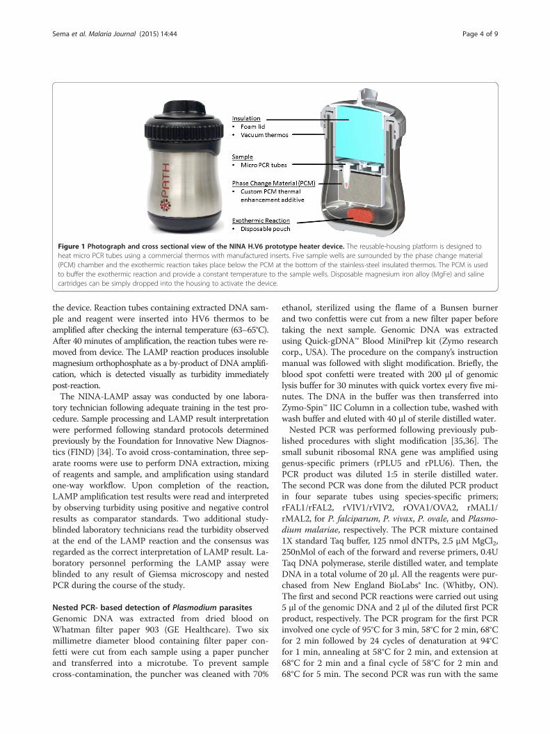
Figure 1 Photograph and cross sectional view of the NINA H.V6 prototype heater device. The reusable-housing platform is designed toheat micro PCR tubes using a commercial thermos with manufactured inserts. Five sample wells are surrounded by the phase change material(PCM) chamber and the exothermic reaction takes place below the PCM at the bottom of the stainless-steel insulated thermos. The PCM is usedto buffer the exothermic reaction and provide a constant temperature to the sample wells. Disposable magnesium iron alloy (MgFe) and salinecartridges can be simply dropped into the housing to activate the device.
Sema et al. Malaria Journal (2015) 14:44 Page 4 of 9
the device. Reaction tubes containing extracted DNA sam-ple and reagent were inserted into HV6 thermos to beamplified after checking the internal temperature (63–65°C).After 40 minutes of amplification, the reaction tubes were re-moved from device. The LAMP reaction produces insolublemagnesium orthophosphate as a by-product of DNA amplifi-cation, which is detected visually as turbidity immediatelypost-reaction.The NINA-LAMP assay was conducted by one labora-
tory technician following adequate training in the test pro-cedure. Sample processing and LAMP result interpretationwere performed following standard protocols determinedpreviously by the Foundation for Innovative New Diagnos-tics (FIND) [34]. To avoid cross-contamination, three sep-arate rooms were use to perform DNA extraction, mixingof reagents and sample, and amplification using standardone-way workflow. Upon completion of the reaction,LAMP amplification test results were read and interpretedby observing turbidity using positive and negative controlresults as comparator standards. Two additional study-blinded laboratory technicians read the turbidity observedat the end of the LAMP reaction and the consensus wasregarded as the correct interpretation of LAMP result. La-boratory personnel performing the LAMP assay wereblinded to any result of Giemsa microscopy and nestedPCR during the course of the study.
Nested PCR- based detection of Plasmodium parasitesGenomic DNA was extracted from dried blood onWhatman filter paper 903 (GE Healthcare). Two sixmillimetre diameter blood containing filter paper con-fetti were cut from each sample using a paper puncherand transferred into a microtube. To prevent samplecross-contamination, the puncher was cleaned with 70%
ethanol, sterilized using the flame of a Bunsen burnerand two confettis were cut from a new filter paper beforetaking the next sample. Genomic DNA was extractedusing Quick-gDNA™ Blood MiniPrep kit (Zymo researchcorp., USA). The procedure on the company’s instructionmanual was followed with slight modification. Briefly, theblood spot confetti were treated with 200 μl of genomiclysis buffer for 30 minutes with quick vortex every five mi-nutes. The DNA in the buffer was then transferred intoZymo-Spin™ IIC Column in a collection tube, washed withwash buffer and eluted with 40 μl of sterile distilled water.Nested PCR was performed following previously pub-
lished procedures with slight modification [35,36]. Thesmall subunit ribosomal RNA gene was amplified usinggenus-specific primers (rPLU5 and rPLU6). Then, thePCR product was diluted 1:5 in sterile distilled water.The second PCR was done from the diluted PCR productin four separate tubes using species-specific primers;rFAL1/rFAL2, rVIV1/rVIV2, rOVA1/OVA2, rMAL1/rMAL2, for P. falciparum, P. vivax, P. ovale, and Plasmo-dium malariae, respectively. The PCR mixture contained1X standard Taq buffer, 125 nmol dNTPs, 2.5 μM MgCl2,250nMol of each of the forward and reverse primers, 0.4UTaq DNA polymerase, sterile distilled water, and templateDNA in a total volume of 20 μl. All the reagents were pur-chased from New England BioLabs® Inc. (Whitby, ON).The first and second PCR reactions were carried out using5 μl of the genomic DNA and 2 μl of the diluted first PCRproduct, respectively. The PCR program for the first PCRinvolved one cycle of 95°C for 3 min, 58°C for 2 min, 68°Cfor 2 min followed by 24 cycles of denaturation at 94°Cfor 1 min, annealing at 58°C for 2 min, and extension at68°C for 2 min and a final cycle of 58°C for 2 min and68°C for 5 min. The second PCR was run with the same
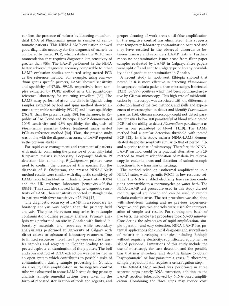
Table 1 NINA- LAMP result compared with Giemsa-stainedmicroscopy and nested PCR for the detection ofPlasmodium parasite in 82 patient samples at Kola Dibahealth centre, Northwest Ethiopia, 2014
Giemsa microscopy (n) Primary NINA- LAMPresult (n)
Nested PCR (n)
Positives (30), Positives (38), Positives (31),
P. falciparum (13) P. falciparum (26) P. falciparum (13)
P.vivax (17) Non- P. falciparum (12) P. vivax (11), P. ovale (7)
Negatives (52) Negatives (44) Negatives (51)
n = number of blood samples examined; NINA = Non-instrumented nucleicacid amplification; LAMP = Loop-mediated isothermal amplification; nestedPCR = nested polymerase chain reaction.
Sema et al. Malaria Journal (2015) 14:44 Page 5 of 9
program but 30 cycles. The final PCR product was thenrun on a 2% agarose gel and visualized using a Gel Doc™(Bio-Rad, Mississauga, ON).
Data analysisAfter assuring data completeness, data were analysed bySPSS version 20 and MedCalc easy to use online statis-tical software version 13.3 used for diagnostic test evalu-ation. Sensitivity, specificity, and positive and negativepredictive values of NINA- LAMP and Giemsa micros-copy were determined using nested PCR as the goldstandard for diagnosis of malaria. The concordance re-sponse rate (percentage of responses with both positiveand both negative results) and Kappa value (k) was de-termined to measure degree of agreements between twodiagnostic test results. Secondary analysis (repeat testingfrom original samples) was performed at University ofCalgary for samples which the original nested PCR andmalaria NINA-LAMP results disagreed.
Ethical considerationThe study protocol was reviewed and approved byResearch and Ethical Review Committee of School ofBiomedical and Laboratory Sciences, College of Medicineand Health Sciences, University of Gondar (SBLS; refer-ence No 525/06). Permission to conduct the study wasalso obtained from Dembia district health bureau. Allstudy participants signed written informed consent be-fore enrollment. Patients found to be positive for malariaparasite were treated according to the current treatmentguideline for malaria in the country.
ResultsParasite positivity by Giemsa microscopy and NINA-LAMPand nested PCRGiemsa microscopy for 200 febrile malaria suspected pa-tients resulted in 15.0% (30/200) confirmed positive forPlasmodium parasites with a median parasite density of9,800 parasites/μL (parasitaemia range = 420–186,800parasites/μL). Of those positives, 43.3% (13/30) and56.7% (17/30) had P. falciparum and P. vivax infection,respectively. Mixed infection with Plasmodium specieswas not identified by microscopy. From the total of 200study participants, samples of 82 patients (30 positivesand 52 negatives) were analysed by NINA-LAMP andnested PCR methods. During primary analysis, NINA-LAMP method identified 38 positives from total 82 sam-ples performed in Gondar using Loopamp™ malaria PanDetection kits (46.3%). From those positives by NINA-LAMP, 68.4% (26/38) tested positive for P. falciparumusing Loopamp™ malaria Pf detection kits. The nestedPCR analysis performed using four Plasmodium speciesprimers detected 31 positives for Plasmodium parasites
from 82 patient samples analysed. Of those positives,41.9% (13/31), 35.5% (11/31) and 22.6% (7/31) were dueto P. falciparum, P.vivax and P. ovale, respectively. AllP. ovale positive samples diagnosed by nested PCR weremisclassified as P vivax by microscopy. Table 1 presentsthe parasite positivity by Giemsa microscopy, NINA-LAMP and nested PCR for the diagnosis of Plasmo-dium parasites.
Discordant analysis of microscopy, NINA-LAMP andnested PCR for diagnosis of Plasmodium parasitesDuring primary NINA-LAMP analysis in Gondar, falsepositives were observed in detecting both genus Plasmo-dium and P. falciparum. Discordant results were observedfrom ten patient samples using the three diagnostic testsfor detecting Plasmodium parasites in Gondar. The NINA-LAMP method identified eight patient samples as positivefor Plasmodium parasites, which were classified as nega-tive by microscopy. Nested PCR detected one positive andseven negative from those discordant results by NINA-LAMP and Giemsa microscopy. Additionally, one samplewas identified as positive by nested PCR from previouslynegative samples by microscopy and NINA–LAMP.Nested PCR also detected one negative result from otherpositive sample by Giemsa microscopy and NINA-LAMP.The NINA-LAMP method detected 13 additional positivesamples for P. falciparum using nested PCR as referencemethod (Table 2). In post hoc analysis, nested PCR andNINA-LAMP discordant samples were retested in a con-trolled laboratory setting in the University of Calgary,Canada. Specifically, NINA-LAMP was repeated for four-teen samples to detect Plasmodium genus and/or P. fal-ciparum. All samples with primary positive results inGondar were subsequently identified as NINA-LAMPnegative for Plasmodium genus and P. falciparum. AfterNINA-LAMP repeat testing, only results of two sampleswere discordant with results of nested PCR for detectinggenus Plasmodium (Table 2).
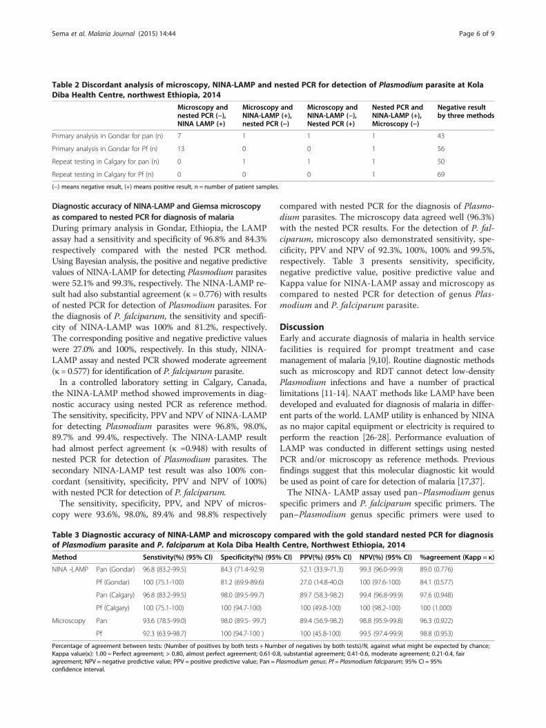
Table 2 Discordant analysis of microscopy, NINA-LAMP and nested PCR for detection of Plasmodium parasite at KolaDiba Health Centre, northwest Ethiopia, 2014
Microscopy andnested PCR (−),NINA LAMP (+)
Microscopy andNINA-LAMP (+),nested PCR (−)
Microscopy andNINA-LAMP (−),Nested PCR (+)
Nested PCR andNINA-LAMP (+),Microscopy (−)
Negative resultby three methods
Primary analysis in Gondar for pan (n) 7 1 1 1 43
Primary analysis in Gondar for Pf (n) 13 0 0 1 56
Repeat testing in Calgary for pan (n) 0 1 1 1 50
Repeat testing in Calgary for Pf (n) 0 0 0 1 69
(−) means negative result, (+) means positive result, n = number of patient samples.
Sema et al. Malaria Journal (2015) 14:44 Page 6 of 9
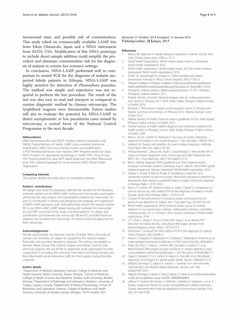
Diagnostic accuracy of NINA-LAMP and Giemsa microscopyas compared to nested PCR for diagnosis of malariaDuring primary analysis in Gondar, Ethiopia, the LAMPassay had a sensitivity and specificity of 96.8% and 84.3%respectively compared with the nested PCR method.Using Bayesian analysis, the positive and negative predictivevalues of NINA-LAMP for detecting Plasmodium parasiteswere 52.1% and 99.3%, respectively. The NINA-LAMP re-sult had also substantial agreement (κ = 0.776) with resultsof nested PCR for detection of Plasmodium parasites. Forthe diagnosis of P. falciparum, the sensitivity and specifi-city of NINA-LAMP was 100% and 81.2%, respectively.The corresponding positive and negative predictive valueswere 27.0% and 100%, respectively. In this study, NINA-LAMP assay and nested PCR showed moderate agreement(κ = 0.577) for identification of P. falciparum parasite.In a controlled laboratory setting in Calgary, Canada,
the NINA-LAMP method showed improvements in diag-nostic accuracy using nested PCR as reference method.The sensitivity, specificity, PPV and NPV of NINA-LAMPfor detecting Plasmodium parasites were 96.8%, 98.0%,89.7% and 99.4%, respectively. The NINA-LAMP resulthad almost perfect agreement (κ =0.948) with results ofnested PCR for detection of Plasmodium parasites. Thesecondary NINA-LAMP test result was also 100% con-cordant (sensitivity, specificity, PPV and NPV of 100%)with nested PCR for detection of P. falciparum.The sensitivity, specificity, PPV, and NPV of micros-
copy were 93.6%, 98.0%, 89.4% and 98.8% respectively
Table 3 Diagnostic accuracy of NINA-LAMP and microscopy cof Plasmodium parasite and P. falciparum at Kola Diba Health
Method Senstivity(%) (95% CI) Specificity(%) (95%
NINA -LAMP Pan (Gondar) 96.8 (83.2-99.5) 84.3 (71.4-92.9)
Pf (Gondar) 100 (75.1-100) 81.2 (69.9-89.6)
Pan (Calgary) 96.8 (83.2-99.5) 98.0 (89.5-99.7)
Pf (Calgary) 100 (75.1-100) 100 (94.7-100)
Microscopy Pan 93.6 (78.5-99.0) 98.0 (89.5- 99.7)
Pf 92.3 (63.9-98.7) 100 (94.7-100 )
Percentage of agreement between tests: (Number of positives by both tests + NumKappa value(κ): 1.00 = Perfect agreement; > 0.80, almost perfect agreement; 0.61-0.8agreement; NPV = negative predictive value; PPV = positive predictive value; Pan = Pconfidence interval.
compared with nested PCR for the diagnosis of Plasmo-dium parasites. The microscopy data agreed well (96.3%)with the nested PCR results. For the detection of P. fal-ciparum, microscopy also demonstrated sensitivity, spe-cificity, PPV and NPV of 92.3%, 100%, 100% and 99.5%,respectively. Table 3 presents sensitivity, specificity,negative predictive value, positive predictive value andKappa value for NINA-LAMP assay and microscopy ascompared to nested PCR for detection of genus Plas-modium and P. falciparum parasite.
DiscussionEarly and accurate diagnosis of malaria in health servicefacilities is required for prompt treatment and casemanagement of malaria [9,10]. Routine diagnostic methodssuch as microscopy and RDT cannot detect low-densityPlasmodium infections and have a number of practicallimitations [11-14]. NAAT methods like LAMP have beendeveloped and evaluated for diagnosis of malaria in differ-ent parts of the world. LAMP utility is enhanced by NINAas no major capital equipment or electricity is required toperform the reaction [26-28]. Performance evaluation ofLAMP was conducted in different settings using nestedPCR and/or microscopy as reference methods. Previousfindings suggest that this molecular diagnostic kit wouldbe used as point of care for detection of malaria [17,37].The NINA- LAMP assay used pan–Plasmodium genus
specific primers and P. falciparum specific primers. Thepan–Plasmodium genus specific primers were used to
ompared with the gold standard nested PCR for diagnosisCentre, Northwest Ethiopia, 2014
CI) PPV(%) (95% CI) NPV(%) (95% CI) %agreement (Kapp = κ)
52.1 (33.9-71.3) 99.3 (96.0-99.9) 89.0 (0.776)
27.0 (14.8-40.0) 100 (97.6-100) 84.1 (0.577)
89.7 (58.3-98.2) 99.4 (96.8-99.9) 97.6 (0.948)
100 (49.8-100) 100 (98.2-100) 100 (1.000)
89.4 (56.9-98.2) 98.8 (95.9-99.8) 96.3 (0.922)
100 (45.8-100) 99.5 (97.4-99.9) 98.8 (0.953)
ber of negatives by both tests)/N, against what might be expected by chance;, substantial agreement; 0.41-0.6, moderate agreement; 0.21-0.4, fairlasmodium genus; Pf = Plasmodium falciparum; 95% CI = 95%
Sema et al. Malaria Journal (2015) 14:44 Page 7 of 9
confirm the presence of malaria by detecting mitochon-drial DNA of Plasmodium genus in samples of symp-tomatic patients. This NINA-LAMP evaluation showedgood diagnostic accuracy for the diagnosis of malaria ascompared to nested PCR, which satisfies the WHO rec-ommendation that requires diagnostic kits sensitivity ofgreater than 95%. The LAMP performed in the NINAheater achieved diagnostic accuracy comparable to otherLAMP evaluation studies conducted using nested PCRas the reference method. For example, using Plasmo-dium genus specific primers, LAMP showed sensitivityand specificity of 97.0%, 99.2%, respectively from sam-ples extracted by PURE method in a UK parasitologyreference laboratory for returning travellers [38]. TheLAMP assay performed at remote clinic in Uganda usingsamples extracted by boil and spins method showed al-most comparable sensitivity (93.9%) and lower specificity(76.3%) than the present study [39]. Furthermore, in Re-public of São Tomé and Príncipe, LAMP demonstrated100% sensitivity and 98% specificity for diagnosis ofPlasmodium parasites before treatment using nestedPCR as reference method [40]. Thus, the present studywas in line with the diagnostic accuracy of LAMP reportedin the previous studies.For rapid case management and treatment of patients
with malaria, confirming the presence of potentially fatalfalciparum malaria is necessary. Loopamp™ Malaria Pfdetection kits containing P falciparum primers wereused to confirm the presence of this species. For thediagnosis of P. falciparum, the present NINA-LAMPmethod results were similar with diagnostic sensitivity ofLAMP reported in Northern Thailand (sensitivity = 100%)and the UK reference laboratory (sensitivity = 98.4%)[38,41]. This study also showed far higher diagnostic sensi-tivity of LAMP than sensitivity reported in Bangladeshiin-patients with fever (sensitivity =76.1%) [42].The diagnostic accuracy of LAMP in a secondary la-
boratory analysis was higher than the primary fieldanalysis. The possible reason may arise from samplecontamination during primary analysis. Primary ana-lysis was performed on site in Gondar with limited la-boratory materials and resources while secondaryanalysis was performed at University of Calgary withdirect access to substantial laboratory resources. Dueto limited resources, non-filter tips were used to trans-fer samples and reagents in Gondar, leading to sus-pected aspirate contamination of the pipettes. The boiland spin method of DNA extraction was performed inan open system which contributes to possible risks ofcontamination during sample processing in Gondar.As a result, false amplification in the negative controltube was observed in some LAMP tests during primaryanalysis. Simple remedial actions were taken in theform of repeated sterilization of tools and regents, and
proper cleaning of work areas until false amplificationin the negative control was eliminated. This suggeststhat temporary laboratory contamination occurred andmay have resulted in the observed discordance be-tween primary and secondary LAMP testing. Further-more, no contamination issues arose from filter papersamples evaluated by LAMP in Calgary. Filter paperswere split off and sent to Calgary prior to any possibil-ity of end product contamination in Gondar.A recent study in northwest Ethiopia showed that
nested PCR is more effective in detecting Plasmodiumin suspected malaria patients than microscopy. It detected13.1% (39/297) positives which had been confirmed nega-tive by Giemsa microscopy. This high rate of misidentifi-cation by microscopy was associated with the difference indetection limit of the two methods, and skills and experi-ences of microscopists to detect and identify Plasmodiumparasites [16]. Giemsa microscopy could not detect para-site densities below 100 parasites/μl of blood while nestedPCR had the ability to detect Plasmodium parasitaemia asfew as one parasite/μl of blood [11,19]. The LAMPmethod had a similar detection threshold with nestedPCR [22]. In this study, malaria NINA-LAMP demon-strated diagnostic sensitivity similar to that of nested PCRand superior to that of microscopy. Therefore, the NINA-LAMP method could be a practical alternative to PCRmethod to avoid misidentification of malaria by micros-copy in endemic areas and detection of submicroscopicinfections in low transmission settings.The method relied on isothermal amplification in a
NINA heater, which permits POCT in low resource set-tings. The NINA enabled electricity free reaction condi-tions comparable to a thermocycler or water bath. TheNINA-LAMP test procedure used in this study did notrequire special equipment and can be used in remotemalaria endemic areas. The test procedure was also donewith short-term training and no previous experience.Negative and positive controls were used for interpret-ation of sample test results. For running one batch offive tests, the whole test procedure took 60–80 minutes.Considering the advantages of rapid amplification, sim-ple operation and easy detection, NINA-LAMP has po-tential applications for clinical diagnosis and surveillanceof malaria in developing countries including Ethiopiawithout requiring electricity, sophisticated equipment orskilled personnel. Limitations of this study include theuse of microscopy for case detection and the possiblebias that may introduce, and also the failure to obtain“asymptomatic” or low parasitemia cases. Furthermore,sample preparation still requires a centrifugation step.The NINA-LAMP method was performed in three
separate steps namely DNA extraction, addition to theLAMP reaction tube, followed by NINA-based amplifi-cation. Combining the three steps may reduce cost,
Sema et al. Malaria Journal (2015) 14:44 Page 8 of 9
turnaround time, and possible risk of contamination.This study relied on commercially available LAMP kitsfrom Eiken Chemicals, Japan and a NINA instrumentfrom PATH, USA. Modification of this NINA prototypeto include direct sample addition could simplify the pro-cedure and eliminate contamination risk for the diagno-sis of malaria in remote low-resource settings.In conclusion, NINA-LAMP performed well in com-
parison to nested PCR for the diagnosis of malaria sus-pected febrile patients in Ethiopia. NINA-LAMP washighly sensitive for detection of Plasmodium parasites.The method was simple and experience was not re-quired to perform the test procedure. The result of thetest was also easy to read and interpret as compared toroutine diagnostic method by Giemsa microscopy. Thelyophilized reagents were thermostable. Future studieswill aim to evaluate the potential for NINA-LAMP todetect asymptomatic or low parasitemia cases missed bymicroscopy, a central focus of the National ControlProgramme in the next decade.
AbbreviationsDNA: Deoxyribonucleic acid; EDTA: Ethylene diamine tetraacetic acid;FMOH: Federal Ministry of Health; LAMP: Loop-mediated isothermalamplification; NINA: Non-instrumented nucleic acid amplification;n-PCR: Nested-polymerase chain reaction; NPV: Negative predictive value;PATH: Program for appropriate technology in health; POC: Point of care;PPV: Positive predictive value; RDT: Rapid diagnostic test; RNA: Ribonucleicacid; SPSS: Statistical package for social sciences; WHO: World HealthOrganization.
Competing interestsThe authors declare that they have no competing interests.
Authors’ contributionsMS design and wrote the proposal, collected the samples for all laboratorymethods, carried out the NINA-LAMP method and microscopic examination,performed the data analysis and interpretation; drafted the manuscript. GGand SG contributed in writing and designing the proposal, and supervisionof NINA-LAMP laboratory work. AGB performed nested PCR analysis, trainedMS to do NINA-LAMP, LAMP repeat testing and reviewed the manuscript.AA and DRP conceived of the study, and participated in its design andcoordination and reviewed the manuscript. RB and PL provided technicalexpertise and reviewed the manuscript. All authors read and approved thefinal manuscript.
AcknowledgementsWe like acknowledge the National Institute of Health (NIH) ,University ofGondar and University of Calgary for supporting this research projectfinancially and providing laboratory materials. The authors are grateful toAbrham Abere, Amare Kifle, Getenet Ayalew and Habitie Tesfa for theirtechnical supports. We would like to appreciate study participants for theircooperation in providing the necessary information and blood sample, andKola Diba health centre laboratory staffs for their support during the datacollection.
Author details1Department of Medical Laboratory Sciences, College of Medicine andHealth Sciences, Wollo University, Dessie, Ethiopia. 2School of Medicine,College of Health Sciences and Medicine, Wolaita Sodo University, Wolaita,Ethiopia. 3Department of Pathology and Laboratory Medicine, University ofCalgary, Calgary, Canada. 4Department of Medical Parasitology, School ofBiomedical and Laboratory Sciences, College of Medicine and HealthSciences, University of Gondar, Gondar, Ethiopia. 5PATH, Seattle, USA.
Received: 21 October 2014 Accepted: 13 January 2015
References1. Marcus BA, Babcock H. Deadly diseases & epidemics: malaria. 2nd ed. New
York: Chelsea house press; 2009. p. 12–3.2. World Health Organization. World malaria report. Geneva, Switzerland:
World Health Organization; 2012.3. World health organization. World malaria report 2013 fact sheet. Geneva,
Switzerland: World health organization; 2013.4. Smith TA, Leuenberger R, Lengeler C. Child mortality and malaria
transmission intensity in Africa. Trends Parasitol. 2001;17:145–9.5. Adugna A. Malaria in Ethiopia. Available at http://www.ethiodemographyand
health.org/MedVectoredDiseasesMalaria.pdf [accessed on December 4, 2013].6. President’s malaria initiative. Malaria operational plan FY 2011. Ethiopia:
President’s malaria initiative; 2011.7. Federal ministry of health. National strategic plan for malaria prevention
and control in Ethiopia, 2011–2015. Addis Ababa, Ethiopia: Federal ministryof health; 2010.
8. Richards S. Third annual malaria control program review in Ethiopia andNigeria: Summary proceedings, 24 February 2012. Atlanta, Georgia: CarterCenter; 2012.
9. Federal ministry of health. National malaria guidelines. 3rd ed. Addis Ababa,Ethiopia: Federal ministry of health; 2012.
10. Federal ministry of health. Malaria diagnosis and treatment guidelines forhealth workers in Ethiopia. 2nd ed. Addis Ababa, Ethiopia: Federal ministryof health; 2004.
11. Milne L, Kyi M, Chiodini P, Warhurst D. Accuracy of routine laboratorydiagnosis of malaria in the United Kingdom. J Clin Microbiol. 1994;47:740–2.
12. Haditsch M. Quality and reliability of current malaria diagnostic methods.Travel Med Infect Dis. 2004;2:149–60.
13. Wongsrichanalai C, Barcus MJ, Muth S, Sutamihardja A, Wernsdorfer WH. Areview of malaria diagnostic tools: microscopy and rapid diagnostic test(RDT). Am J Trop Med Hyg. 2007;77(6 Suppl):119–27.
14. Bell D. Malaria diagnosis: WHO guidelines and their implementation.European commission research workshop, Aug 31–sept 01, 2010. WHO–globalmalaria programme. Geneva, Switzerland: World health organization; 2010.
15. Golassa L, Enweji N, Erko B, Assefa A, Swedberg G. Detection of asubstantial number of sub-microscopic Plasmodium falciparum infections bypolymerase chain reaction: a potential threat to malaria control and diagnosisin Ethiopia. Malar J. 2013;12:352.
16. Alemu A, Fuehrer HP, Getnet G, Kassu A, Getie S, Noedl H. Comparison ofGiemsa microscopy with nested PCR for the diagnosis of malaria in NorthGondar, north-west Ethiopia. Malar J. 2014;13:174.
17. Cordray M, Richards-Kortum R. Emerging nucleic acid-based tests forpoint-of-care detection of malaria. Am J Trop Med Hyg. 2012;87:223–30.
18. World health organization. WHO evidence review group on malariadiagnosis in low transmission settings: malaria policy advisory committeemeeting session 10, 12–14 March 2014. Geneva; Switzerland: World healthorganization; 2014.
19. Li P, Zhao Z, Wang Y, Xing H, Parker DM, Yang Z, et al. Nested PCRdetection of malaria directly using blood filter paper samples fromepidemiological surveys. Malar J. 2014;13:175.
20. Hänscheid T, Grobusch M. How useful is PCR in the diagnosis of malaria?Trends Parasitol. 2002;18:395–8.
21. Notomi T, Okayama H, Masubuchi H, Yonekawa T, Watanabe K, Amino N, et al.Loop-mediated isothermal amplification of DNA. Nucl Acids Res. 2000;28:E63.
22. Polley SD, Mori Y, Watson J, Perkins MD, González IJ, Notomi T, et al.Mitochondrial DNA targets increase sensitivity of malaria detection usingLoop-mediated isothermal amplification. J Clin Microbiol. 2010;48:2866–71.
23. Yager P, Edwards T, Fu E, Helton K, Nelson K, Tam MR, et al. Microfluidicdiagnostic technologies for global public health. Nature. 2006;442:412–8.
24. Weigl B, Domingo G, LaBarre P, Gerlach J. Towards non- and minimallyinstrumented, microfluidics-based diagnostic devices. Lab Chip.2008;8:1999–2014.
25. Weigl B, Domingo G, Gerlach J, Tang D, Harvey D, Talwar N, et al. Non-instrumentednucleic-acid amplification assay. ProcSPIE. 2008;6886:688604.
26. LaBarre P, Hawkins KR, Gerlach J, Wilmoth J, Beddoe A, Singleton J, et al. ASimple, inexpensive device for nucleic acid amplification without electricity -Toward instrument-free molecular diagnostics in low-resource settings. PLoSOne. 2011;6:e19738.

Sema et al. Malaria Journal (2015) 14:44 Page 9 of 9
27. LaBarre P, Gerlach J, Wilmoth J, Beddoe A, Singleton J, Weigl B.Non-instrumented nucleic acid amplification (NINA): instrument-freemolecular malaria diagnostics for low-resource settings. Conf Proc IEEEEng Med Biol Soc. 2010;2010:1097–9.
28. Singleton J, Osborn JL, Lillis L, Hawkins K, Guelig D, Price W, et al. Electricity-freeamplification and detection for molecular point-of-care diagnosis of HIV-1. PLoSOne. 2014;9:e113693.
29. Curtis KA, Rudolph DL, Nejad I, Singleton J, Beddoe A, Weigl B, et al.Isothermal amplification using a chemical heating device for point of caredetection of HIV-1. PLoS One. 2012;7:e31432.
30. Han ET. Loop-mediated isothermal amplification test for the moleculardiagnosis of malaria. Expert Rev Mol Diagn. 2013;13:205–18.
31. Alemu A, Muluye D, Mihret M, Adugna M, Gebeyaw M. Ten year trendanalysis of malaria prevalence in Kola Diba, North Gondar, NorthwestEthiopia. Parasit Vectors. 2012;5:173.
32. Alemu A, Fuehrer H, Getnet G, Tessema B, Noedl H. Plasmodium ovale curtisiand Plasmodium ovale wallikeri in North-West Ethiopia. Malar J. 2013;12:346.
33. Federal Ministry of Health. Manual for laboratory diagnosis of malaria. 1sted. Addis Ababa, Ethiopia: Federal Ministry of Health; 2012.
34. FIND Malaria Programme. Manual of standard operating procedures formalaria LAMP Version one. Geneva, Switzerland: Foundation for newinnovative diagnostic (FIND); 2012.
35. Snounou G, Viriyakosol S, Zhu XP, Jarra W, Pinheiro L, do Rosario VE, et al.High sensitivity detection of human malaria parasites by the use of nestedpolymerase chain reaction. Mol Biochem Parastiol. 1993;61:315–20.
36. Singh B, Bobogare A, Cox-Singh J, Snounou G, Abdullah MS, Rahman HA. Agenus- and species-specific nested polymerase chain reaction malariadetection assay for epidemiologic studies. J Trop Med Hyg. 1999;60:687–92.
37. Abdul-Ghani R, Al-Mekhlafi A, Karanis P. Loop-mediated isothermal amplification(LAMP) for malarial parasites of humans: would it come to clinical reality as apoint-of-care test? Acta Trop. 2012;122:233–40.
38. Polley SD, González IJ, Mohamed D, Daly R, Bowers K, Watson J, et al.Clinical evaluation of a loop-mediated amplification kit for diagnosis ofimported malaria. J Infect Dis. 2013;208:637–44.
39. Hopkins H, González IJ, Polley SD, Angutoko P, Ategeka J, Asiimwe C, et al.Highly sensitive detection of malaria parasitemia in a malaria-endemicsetting: performance of a new loop-mediated isothermal amplification kit ina remote clinic in Uganda. J Infect Dis. 2013;208:645–52.
40. Lee PW, Ji DD, Liu CT, Rampao HS, do Rosario VE, Lin IF, et al. Application ofloop-mediated isothermal amplification for malaria diagnosis during afollow-up study in Sao Tome. Malar J. 2012;11:408.
41. Pöschl B, Waneesorn J, Thekisoe O, Chutipongvivate S, Panagiotis K.Comparative diagnosis of malaria infections by microscopy, nested PCR, andLAMP in Northern Thailand. Am J Trop Med Hyg. 2010;83:56–60.
42. Paris DH, Imwong M, Faiz AM, Hasan M, Yunus EB, Silamut K, et al.Loop-mediated isothermal PCR (LAMP) for the diagnosis of falciparummalaria. Am J Trop Med Hyg. 2007;77:972–6.
Submit your next manuscript to BioMed Centraland take full advantage of:
• Convenient online submission
• Thorough peer review
• No space constraints or color figure charges
• Immediate publication on acceptance
• Inclusion in PubMed, CAS, Scopus and Google Scholar
• Research which is freely available for redistribution
Submit your manuscript at www.biomedcentral.com/submit