designer self-assembling peptide materials
TRANSCRIPT
Feature Article
Designer Self-Assembling PeptideMaterials
Xiaojun Zhao,* Shuguang Zhang*
Understanding of macromolecular materials at the molecular level is becoming increasinglyimportant for a new generation of nanomaterials for nanobiotechnology and other disciplines,namely, the design, synthesis, and fabrication of nanodevices at the molecular scale frombottom up. Basic engineering principles for microfabricationcan be learned through fully grasping the molecular self-assembly and programmed assembly phenomena. Self- andprogrammed-assembly phenomena are ubiquitous in nature.Two key elements in molecular macrobiological materialproductions are chemical complementarity and structuralcompatibility, both of which require weak and non-covalentinteractions that bring building blocks together during self-assembly. Significant advances have been made during the1990s at the interface ofmaterials chemistry andbiology. Theyinclude the design of helical ribbons, peptide nanofiber scaf-folds for three-dimensional cell cultures and tissue engineer-ing, peptide surfactants for solubilizing and stabilizing diversetypes of membrane proteins and their complexes, and molecular ink peptides for arbitraryprinting and coating surfaces as well as coiled-coil helical peptides for multi- length scale fractalstructures. These designer self-assembling peptides have far reaching implications in a broadspectrum of applications in biology, medicine, nanobiotechnology, and nanobiomedical techno-logy, some of which are beyond our current imaginations.
X. ZhaoInstitute for Nanobiomedical Technology andMembrane Biology,West China Hospital, Sichuan University, No.1, Ke Yuan 4th Street,Gao Peng Road, Chengdu, 610041, Sichuan, ChinaFax: þ86 28 8516 4073; E-mail: [email protected] Key Lab of Biotherapy of Human Diseases, Cancer Center,West China Medical School, West China Hospital, SichuanUniversity, Guo Xue Xiang, 37, Chengdu, 610041, Sichuan, ChinaS. ZhangCenter for Biomedical Engineering, and Center for Bits & Atoms,NE47-379, Massachusetts Institute of Technology, Cambridge,MA02139-4307, USAE-mail: [email protected]
Macromol. Biosci. 2007, 7, 13–22
� 2007 WILEY-VCH Verlag GmbH & Co. KGaA, Weinheim
Introduction
Molecular self-assembly is ubiquitous in Nature, from
lipids that form oil droplets in water, and surfactants that
form micelles and other complex structures in water, to
sophisticated multiunit ribosome and virus assemblies.
Nature is a grand master who utilizes the strategy to
bottom-up build hierarchical materials. These elegant
molecular self-assembly systems lie at the interface be-
tween biochemistry, molecular biology, peptide and
protein chemistry, macromolecular science, materials
science, and engineering. The key elements are chemical
DOI: 10.1002/mabi.200600230 13
X. Zhao, S. Zhang
Xiaojun Zhao is Professor, Chief Scientist, andExecutive Director at a newly established Institutefor NanoBiomedical Technology and MembraneBiology, Sichuan University, Chengdu, P. R. China.He received his Ph.D. in Biological Chemistry fromthe University of California at Los Angeles, wherehe studied the structure and function of DNArecombination and the repair enzyme RecA pro-tein. He then worked on mitochondrial disease atCalifornia Institute of Technology and studied themechanism of neuronal synaptogenesis and pep-tide self assembling at Massachusetts Institute ofTechnology. His lab is currently working with var-ious self-assembling peptide systems to developnew classes of biological materials, which includepeptide matrix scaffolds for regenerative medi-cine, tissue engineering, biological surface engi-neering, and peptide surfactant nanotubes forstabilizing membrane proteins and their com-plexes. He is also interested in developing scaffoldsfor controlling stem cells differentiation and pro-liferation, tumor cells in vitro proliferation anddrug therapy, virus proliferation and growth inhi-bition in modified peptide gel scaffolds, and so on.Shuguang Zhang is at the Center for BiomedicalEngineering and Center for Bits & Atoms, Mas-sachusetts Institute of Technology, Cambridge,Massachusetts, USA. He received his B.Sc. fromthe Sichuan University in China and a Ph.D. inBiochemistry & Molecular Biology from Univer-sity of California at Santa Barbara, USA. He wasan American Cancer Society Postdoctoral Fellowat MIT and was a Whitaker Foundation Investi-gator. He was a 2003 Fellow of Japan Society forPromotion of Science (JSPS fellow). He is also aChang Jiang distinguished scholar in China. Hisdiscovery and work on designer self-assemblingpeptide scaffolds won the 2004 R&D100 award.He and his colleagues’ work on bio-solar energywas selected to be one of the 10 finalists of the2005 Saatchi & Saatchi Award for World Chan-ging Ideas. He is one of the 2006 John SimonGuggenheim Fellows. He is the recipient of the2006Wilhelm ExnerMedal fromVienna, Austria.
14
complementarity and structural compatibility through
numerous non-covalent weak interactions.
Learning from nature of the sophisticated fabrication
and construction of diverse biological materials, which
often is elegantly built one molecule and one atom at a
time, we have developed many self-assembling systems,
ranging from models for studying protein folding and
protein conformational-related diseases, to molecular mate-
rials for producing peptide nanofibers, peptide scaffolds,
peptide surfactants, and peptide ink (Figure 1).[1,2] Molecular
self-assembly systems represent a significant advance in
the molecular engineering of simple molecular building
Macromol. Biosci. 2007, 7, 13–22
� 2007 WILEY-VCH Verlag GmbH & Co. KGaA, Weinheim
blocks for a wide range of material and device applica-
tions.[3–6] In this article wewill only focus on designer Lego
peptides and surfactant peptides. Other interesting work
can be found from earlier reviews elsewhere.[3–6]
Lego TM Peptides
The first member of the Lego peptides was serendipitously
discovered from a segment in a left-handed Z-DNA binding
protein in yeast, Zuotin (Zuo means Left in Chinese, tin
means protein in biology).[7] Inspired by its special struc-
ture, a class of ‘Lego peptides’ was designed. On the
nanometer scale, these peptides resemble the Lego
bricks that have both pegs and holes in a precisely
determinedmanner. They can be programmed to assemble
in well-formed structures. This class of Lego peptide
undergoes spontaneous assembly into well-organized
nanofibers.[8]
Various Modulus Peptides
Because of their beta-sheet structures in aqueous solution,
modulus peptides have two distinct sides, one is hydro-
philic and the other is hydrophobic, like the pegs and holes
in Lego bricks. The hydrophobic sides shield themselves
fromwater and thus facilitate their self-assembly inwater,
similar to that seen in the case of protein folding. In the
case of protein folding, the driving force is intramolecular
interactions where side chains of the amino acids form a
hydrophobic core; but in the peptide self-assembly, the
driving force is intermolecular interactions. The unique
structural feature of these Lego peptides is that they form
complementary ionic bonds with regular repeats on the
hydrophilic surface. The complementary ionic sides have
been classified into several moduli, i.e., modulus I, II, III, IV,
etc., and mixed moduli. This classification is based on the
hydrophilic surface of the molecules that have alternating
positively (þ) and negatively (�) charged amino acid
residues, either alternating by 1, 2, 3, 4, and so on. For
example, charge arrangements are for modulus I,
SRSRSRSR; modulus II, SSRRSSRR; modulus
III, SSSRRR; and modulus IV, SSSSRRRR. The
charge orientation can also be designed in the reverse
orientation to yield entirely different molecules. These
structurally well-defined peptides undergo ordered assem-
bly, and may resemble some polymer assemblies.[1,2]
Dynamic Behavior of the Peptide Re-Assemblies
The self-assembly process is reversible and dynamic
(Figure 2)[9] since these peptides are short and simple,
DOI: 10.1002/mabi.200600230
Designer Self-Assembling Peptide Materials
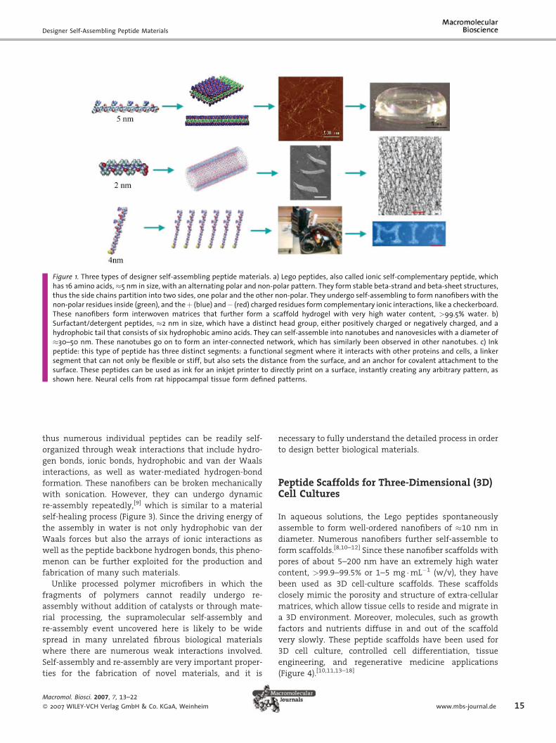
Figure 1. Three types of designer self-assembling peptide materials. a) Lego peptides, also called ionic self-complementary peptide, whichhas 16 amino acids,�5 nm in size, with an alternating polar and non-polar pattern. They form stable beta-strand and beta-sheet structures,thus the side chains partition into two sides, one polar and the other non-polar. They undergo self-assembling to form nanofibers with thenon-polar residues inside (green), and theþ (blue) and� (red) charged residues form complementary ionic interactions, like a checkerboard.These nanofibers form interwoven matrices that further form a scaffold hydrogel with very high water content, >99.5% water. b)Surfactant/detergent peptides, �2 nm in size, which have a distinct head group, either positively charged or negatively charged, and ahydrophobic tail that consists of six hydrophobic amino acids. They can self-assemble into nanotubes and nanovesicles with a diameter of�30–50 nm. These nanotubes go on to form an inter-connected network, which has similarly been observed in other nanotubes. c) Inkpeptide: this type of peptide has three distinct segments: a functional segment where it interacts with other proteins and cells, a linkersegment that can not only be flexible or stiff, but also sets the distance from the surface, and an anchor for covalent attachment to thesurface. These peptides can be used as ink for an inkjet printer to directly print on a surface, instantly creating any arbitrary pattern, asshown here. Neural cells from rat hippocampal tissue form defined patterns.
thus numerous individual peptides can be readily self-
organized through weak interactions that include hydro-
gen bonds, ionic bonds, hydrophobic and van der Waals
interactions, as well as water-mediated hydrogen-bond
formation. These nanofibers can be broken mechanically
with sonication. However, they can undergo dynamic
re-assembly repeatedly,[9] which is similar to a material
self-healing process (Figure 3). Since the driving energy of
the assembly in water is not only hydrophobic van der
Waals forces but also the arrays of ionic interactions as
well as the peptide backbone hydrogen bonds, this pheno-
menon can be further exploited for the production and
fabrication of many such materials.
Unlike processed polymer microfibers in which the
fragments of polymers cannot readily undergo re-
assembly without addition of catalysts or through mate-
rial processing, the supramolecular self-assembly and
re-assembly event uncovered here is likely to be wide
spread in many unrelated fibrous biological materials
where there are numerous weak interactions involved.
Self-assembly and re-assembly are very important proper-
ties for the fabrication of novel materials, and it is
Macromol. Biosci. 2007, 7, 13–22
� 2007 WILEY-VCH Verlag GmbH & Co. KGaA, Weinheim
necessary to fully understand the detailed process in order
to design better biological materials.
Peptide Scaffolds for Three-Dimensional (3D)Cell Cultures
In aqueous solutions, the Lego peptides spontaneously
assemble to form well-ordered nanofibers of �10 nm in
diameter. Numerous nanofibers further self-assemble to
form scaffolds.[8,10–12] Since these nanofiber scaffolds with
pores of about 5–200 nm have an extremely high water
content, >99.9–99.5% or 1–5 mg �mL�1 (w/v), they have
been used as 3D cell-culture scaffolds. These scaffolds
closely mimic the porosity and structure of extra-cellular
matrices, which allow tissue cells to reside and migrate in
a 3D environment. Moreover, molecules, such as growth
factors and nutrients diffuse in and out of the scaffold
very slowly. These peptide scaffolds have been used for
3D cell culture, controlled cell differentiation, tissue
engineering, and regenerative medicine applications
(Figure 4).[10,11,13–18]
www.mbs-journal.de 15
X. Zhao, S. Zhang
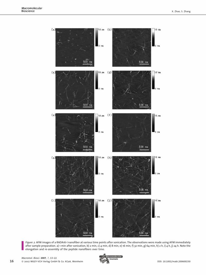
Figure 2. AFM images of a RADA16-I nanofiber at various time points after sonication. The observations were made using AFM immediatelyafter sample preparation. a) 1 min after sonication, b) 2 min, c) 4 min, d) 8 min, e) 16 min, f) 32 min, g) 64 min, h) 2 h, i) 4 h, j) 24 h. Note theelongation and re-assembly of the peptide nanofibers over time.
16Macromol. Biosci. 2007, 7, 13–22
� 2007 WILEY-VCH Verlag GmbH & Co. KGaA, Weinheim DOI: 10.1002/mabi.200600230
Designer Self-Assembling Peptide Materials
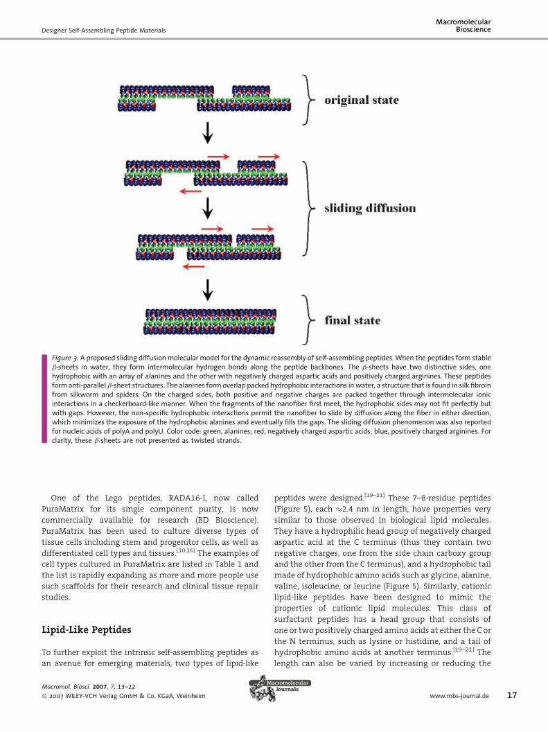
Figure 3. A proposed sliding diffusionmolecular model for the dynamic reassembly of self-assembling peptides.When the peptides form stableb-sheets in water, they form intermolecular hydrogen bonds along the peptide backbones. The b-sheets have two distinctive sides, onehydrophobic with an array of alanines and the other with negatively charged aspartic acids and positively charged arginines. These peptidesformanti-parallelb-sheet structures. The alanines formoverlap packedhydrophobic interactions inwater, a structure that is found in silk fibroinfrom silkworm and spiders. On the charged sides, both positive and negative charges are packed together through intermolecular ionicinteractions in a checkerboard-like manner. When the fragments of the nanofiber first meet, the hydrophobic sides may not fit perfectly butwith gaps. However, the non-specific hydrophobic interactions permit the nanofiber to slide by diffusion along the fiber in either direction,which minimizes the exposure of the hydrophobic alanines and eventually fills the gaps. The sliding diffusion phenomenon was also reportedfor nucleic acids of polyA and polyU. Color code: green, alanines; red, negatively charged aspartic acids; blue, positively charged arginines. Forclarity, these b-sheets are not presented as twisted strands.
One of the Lego peptides, RADA16-I, now called
PuraMatrix for its single component purity, is now
commercially available for research (BD Bioscience).
PuraMatrix has been used to culture diverse types of
tissue cells including stem and progenitor cells, as well as
differentiated cell types and tissues.[10,16] The examples of
cell types cultured in PuraMatrix are listed in Table 1 and
the list is rapidly expanding as more and more people use
such scaffolds for their research and clinical tissue repair
studies.
Lipid-Like Peptides
To further exploit the intrinsic self-assembling peptides as
an avenue for emerging materials, two types of lipid-like
Macromol. Biosci. 2007, 7, 13–22
� 2007 WILEY-VCH Verlag GmbH & Co. KGaA, Weinheim
peptides were designed.[19–21] These 7–8-residue peptides
(Figure 5), each �2.4 nm in length, have properties very
similar to those observed in biological lipid molecules.
They have a hydrophilic head group of negatively charged
aspartic acid at the C terminus (thus they contain two
negative charges, one from the side chain carboxy group
and the other from the C terminus), and a hydrophobic tail
made of hydrophobic amino acids such as glycine, alanine,
valine, isoleucine, or leucine (Figure 5). Similarly, cationic
lipid-like peptides have been designed to mimic the
properties of cationic lipid molecules. This class of
surfactant peptides has a head group that consists of
one or two positively charged amino acids at either the C or
the N terminus, such as lysine or histidine, and a tail of
hydrophobic amino acids at another terminus.[19–21] The
length can also be varied by increasing or reducing the
www.mbs-journal.de 17

X. Zhao, S. Zhang
Figure 4. From designer self-assembling peptides to nanofiber scaffolds for tissues regenerations. a) Active synapses on the peptide surface.Primary rat hippocampal neurons form active synapses on peptide scaffolds. The confocal images shown bright discrete green dot labelingindicative of synaptically activemembranes after incubation of neurons with the fluorescent lipophilic probe FM-143. FM-143 can selectivelytrace synaptic vesicle turnover during the process of synaptic transmission. The active synapses on the peptide scaffold are fully functional,indicating that the peptide scaffold is a permissible material for neurite outgrowth and active synapse formation. b) Adult mouse neuralstem cells embedded in a 3D scaffold (image courtesy of F. Gelain). c) Brain damage repair in a hamster. The peptide scaffold was injectedinto the optical nerve area of brain that was first severed with a knife. The cut was sealed by the migrating cells after two days. A greatnumber of neurons form synapses (image courtesy of R. Ellis-Behnke). d) Peptide KLD12 (KLDLKLDLKLDL), chondrocytes in the peptidescaffold and cartilage. The chondrocytes stainedwith TB show abundant GAG production (left panel) and antibody to type II collagen, whichdemonstrates abundant Type II collagen production (right panel). A piece of premolded cartilage with encapsulated chondrocytes in thepeptide nanofiber scaffold. The cartilage formed over a 3–4 week period after the initial seeding of the chondrocytes (image courtesy of J.Kisiday). e) Von Kossa staining showing transverse sections of primary osteoblast cells on HA-PHP-RADA16-I self-assembling peptidenanofiber scaffold. Scale bar¼0.1 mm. The intensely stained black areas represent the formation of bone nodules (image courtesy of M.Bokhari).
18
number of amino acids, one at a time to a desired
length.[20]
Nanotubes and Nanovesicles of Lipid-Like Peptides
When dissolved in water, these lipid-like peptides tend to
self-assemble to isolate the hydrophobic tail from contact
with water. The process of surfactant self-assembly is an
enthalpy driven process of energy minimization in which
the individual monomers pack together to sequester their
hydrophobic tails from water. The peptide surfactants are
demonstrated to form a curved bilayer. Within this curved
bilayer, the peptides stack so that their hydrophilic heads
are exposed to the water with their hydrophobic tails
packed within. These proposed bilayers are approximately
5 nm in thickness because of a 2.4 nm length of the single
Macromol. Biosci. 2007, 7, 13–22
� 2007 WILEY-VCH Verlag GmbH & Co. KGaA, Weinheim
peptides and, because of both the peptide shape and
the electrostatic repulsion among the head groups, curve
to form both nanotubes and nanovesicles having an
average diameter of 30–50 nm. These nanostructures have
been observed using quick-freeze/deep-etch transmission
electron microscopy (TEM) (Figure 6).[19–21] This self-
assembling phenomenon is very similar to well-studied
events in lipids.[22–24]
Lipid-like Peptide Surfactants StabilizeMembrane Proteins
These lipid-like peptide surfactants have been found to be
excellentmaterials not only for solubilizing and stabilizing
several diverse membrane proteins and membrane
protein complexes but for also crystallizing onemembrane
DOI: 10.1002/mabi.200600230
Designer Self-Assembling Peptide Materials
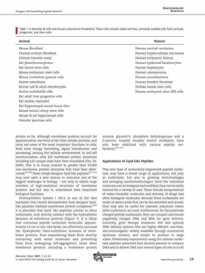
Table 1. A diversity of cells and tissues cultured on PuraMatrix. These cells include stable cell lines, primarily isolated cells from animals,progenitor, and stem cells.
Animal Human
Mouse fibroblast Human cervical carcinoma
Chicken embryo fibroblast Human hepato-cellular carcinoma
Chinese hamster ovary Human embryonic Kidney
Rat pheochromocytoma Human epidermal keratinocytes
Rat neural stem cells Human hepatocytes
Mouse embryonic stem cells Human osteosarcoma
Mouse cerebellum granule cells Human neuroblastoma
Bovine osteoblasts Human foreskin fibroblast
Bovine calf & adult chondrocytes Human neural stem cells
Bovine endothelial cells Human embryonic stem (ES) cells
Rat adult liver progenitor cells
Rat cardiac myocytes
Rat hippocampal neural tissue slice
Mouse neural colony stem cells
Mouse & rat hippocampal cells
Hamster pancreas cells
protein so far. Although membrane proteins account for
approximately one-third of the total cellular proteins, and
carry out some of the most important functions in cells,
from solar energy harvesting, signal transduction and
processing, sensing the cellular environment, to cell-cell
communication, only 225 membrane protein structures
including 116 unique ones have been elucidated (Oct. 26,
2006). This is in sharp contrast to greater than 35 000
non-membrane protein structures that have been deter-
mined.[25,26] These simple designer lipid-like peptides[27–31]
may now open a new avenue to overcome one of the
biggest challenges in biology – not only to obtain large
numbers of high-resolution structures of membrane
proteins and but also to understand their important
biological functions.
Photosynthetic system I (PS-I) is one of the first
examples that clearly demonstrates how designer lipid-
like peptides stabilize membrane protein complexes.[29]
It is plausible that lipid- like peptides, similar to other
surfactants, may directly interact with the hydrophobic
domains of membrane proteins (Figure 7). It is likely
that numerous peptide surfactant molecules, approxi-
mately 2.4 nm in size, like lipids, can effectively surround
the hydrophobic trans-membrane domains of mem-
brane proteins, thus sequestering them from directly
interacting with water molecules and preventing
them from undergoing self-aggregation. Some other
membrane proteins, including a membrane protein
Macromol. Biosci. 2007, 7, 13–22
� 2007 WILEY-VCH Verlag GmbH & Co. KGaA, Weinheim
enzyme glycerol-3- phosphate dehydrogenase and a
G-protein coupled receptor bovine rhodopsin, have
also been stabilized with various peptide sur-
factants.[29–31]
Applications of Lipid-Like Peptides
This new type of molecularly engineered peptide surfac-
tant may have a broad range of applications, not only
as surfactants, but also in growing biotechnologies
and emerging nanobiotechnologies. Since the individual
molecules can be designed andmodified, they can be easily
tailored for a variety of uses. These include encapsulation
of water-insoluble molecules and delivery of drugs and
other biological molecules. Because these surfactants are
made of amino acids that can be bio-absorbed and reused,
they may also be useful for cosmetic industries where
other surfactants are used. Furthermore, for the positively
charged peptide surfactants, they can compact and encase
negatively charged DNA and RNA for gene delivery.
Currently, gene therapy endeavors still lack optimal
DNA delivery systems that are highly efficient, non-toxic,
non-immunogenic, widely available through commercial
synthesis vendors, and simple to produce in large
scales. Preliminary experiments using the cationic surfac-
tant peptides presented here showed promise to compact
DNA and to deliver DNA into several types of cells in a cell
www.mbs-journal.de 19
X. Zhao, S. Zhang
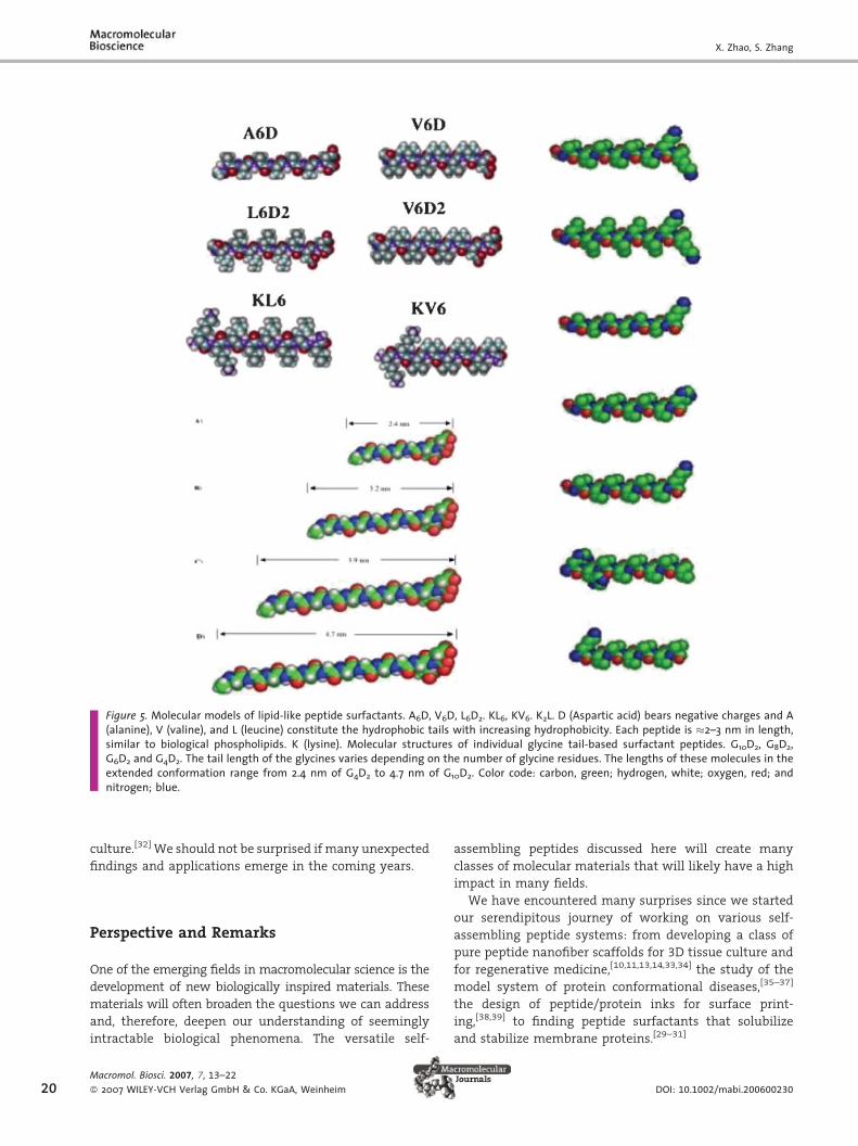
Figure 5. Molecular models of lipid-like peptide surfactants. A6D, V6D, L6D2. KL6, KV6. K2L. D (Aspartic acid) bears negative charges and A(alanine), V (valine), and L (leucine) constitute the hydrophobic tails with increasing hydrophobicity. Each peptide is �2–3 nm in length,similar to biological phospholipids. K (lysine). Molecular structures of individual glycine tail-based surfactant peptides. G10D2, G8D2,G6D2 and G4D2. The tail length of the glycines varies depending on the number of glycine residues. The lengths of these molecules in theextended conformation range from 2.4 nm of G4D2 to 4.7 nm of G10D2. Color code: carbon, green; hydrogen, white; oxygen, red; andnitrogen; blue.
20
culture.[32] We should not be surprised if many unexpected
findings and applications emerge in the coming years.
Perspective and Remarks
One of the emerging fields in macromolecular science is the
development of new biologically inspired materials. These
materials will often broaden the questions we can address
and, therefore, deepen our understanding of seemingly
intractable biological phenomena. The versatile self-
Macromol. Biosci. 2007, 7, 13–22
� 2007 WILEY-VCH Verlag GmbH & Co. KGaA, Weinheim
assembling peptides discussed here will create many
classes of molecular materials that will likely have a high
impact in many fields.
We have encountered many surprises since we started
our serendipitous journey of working on various self-
assembling peptide systems: from developing a class of
pure peptide nanofiber scaffolds for 3D tissue culture and
for regenerative medicine,[10,11,13,14,33,34] the study of the
model system of protein conformational diseases,[35–37]
the design of peptide/protein inks for surface print-
ing,[38,39] to finding peptide surfactants that solubilize
and stabilize membrane proteins.[29–31]
DOI: 10.1002/mabi.200600230
Designer Self-Assembling Peptide Materials
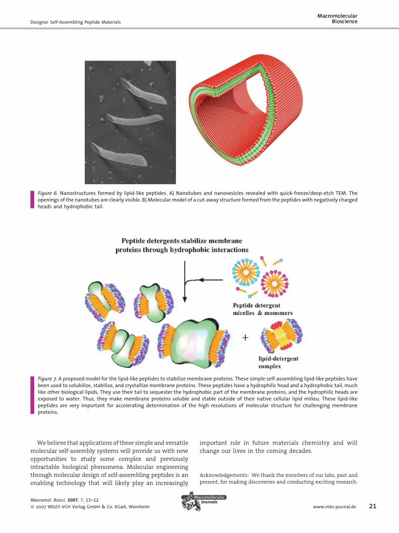
Figure 6. Nanostructures formed by lipid-like peptides. A) Nanotubes and nanovesicles revealed with quick-freeze/deep-etch TEM. Theopenings of the nanotubes are clearly visible. B) Molecularmodel of a cut-away structure formed from the peptideswith negatively chargedheads and hydrophobic tail.
Figure 7. A proposedmodel for the lipid-like peptides to stabilize membrane proteins. These simple self-assembling lipid-like peptides havebeen used to solubilize, stabilize, and crystallize membrane proteins. These peptides have a hydrophilic head and a hydrophobic tail, muchlike other biological lipids. They use their tail to sequester the hydrophobic part of the membrane proteins, and the hydrophilic heads areexposed to water. Thus, they make membrane proteins soluble and stable outside of their native cellular lipid milieu. These lipid-likepeptides are very important for accelerating determination of the high resolutions of molecular structure for challenging membraneproteins.
Webelieve that applications of these simple and versatile
molecular self-assembly systems will provide us with new
opportunities to study some complex and previously
intractable biological phenomena. Molecular engineering
through molecular design of self-assembling peptides is an
enabling technology that will likely play an increasingly
Macromol. Biosci. 2007, 7, 13–22
� 2007 WILEY-VCH Verlag GmbH & Co. KGaA, Weinheim
important role in future materials chemistry and will
change our lives in the coming decades.
Acknowledgements: We thank the members of our labs, past andpresent, for making discoveries and conducting exciting research.
www.mbs-journal.de 21

X. Zhao, S. Zhang
22
We gratefully acknowledge the support by grants from ARO, ONR,DARPA (BioComputing), DARPA/Naval Research Labs, DARPA/AFOSR, MURI/AFO, NIH, NSF-MIT BPEC and NSF CCR-0122419 toMIT Media Lab’s Center for Bits & Atoms, the WhitakerFoundation, Du Pont-MIT Alliance, Menicon, Ltd, Japan, OlympusCorp. Japan, Mitsubishi Chemical Research Center, Japan, andROHM Corp, Japan. We also acknowledge the Intel Corporationeducational donation of a computing cluster to the Center forBiomedical Engineering at MIT.
Received: October 17, 2006; Revised: October 26, 2006; Accepted:October 31, 2006; DOI: 10.1002/mabi.200600230
Keywords: designer nanofiber scaffolds; membrane protein;peptide surfactants; peptides; regenerative medicine; self-assembly
[1] S. Zhang, Biotechnol. Adv. 2002, 20, 321.[2] S. Zhang, Nat. Biotechnol. 2003, 21, 1171.[3] Y. C. Yu, T. Pakalns, Y. Dori, J. B. McCarthy, M. Tirrell,
G. B. Fields, Meth. Enzymol. 1997, 289, 571.[4] G. B. Fields, Bioorg. Med. Chem. 1999, 7, 75.[5] G. M. Whitesides, J. P. Mathias, C. T. Seto, Science 1991, 254,
1312.[6] G. M. Whitesides, B. Grzybowski, Science 2002, 295, 2418.[7] S. Zhang, C. Lockshin, A. Herbert, E. Winter, A. Rich, EMBO J.
1992, 11, 3787.[8] S. Zhang, T. Holmes, C. Lockshin, A. Rich, Proc. Natl. Acad. Sci.
USA 1993, 90, 3334.[9] H. Yokoi, T. Kinoshita, S. Zhang, Proc. Natl. Acad. Sci. USA 2005,
102, 8414.[10] S. Zhang, T. Holmes, C. Dipersio, R. Hynes, X. Su, A. Rich,
Biomaterials 1995, 16, 1385.[11] T. Holmes, S. Lacalle, X. Su, G. Liu, A. Rich, S. Zhang, Proc. Natl.
Acad. Sci. USA 2000, 97, 6728.[12] D. Marini, W. Hwang, D. A. Lauffenburger, S. Zhang,
D. K. Roger, Nano Lett. 2002, 2, 295.[13] J. Kisiday, M. Jin, B. Kurz, H. Hung, C. Semino, S. Zhang,
A. J. Grodzinsky, Proc. Natl. Acad. Sci. USA 2002, 99, 9996.[14] M. A. Bokhari, G. Akay, S. Zhang, M. A. Birch, Biomaterials
2005, 26, 5198.[15] S. Zhang, F. Gelain, X. Zhao, Seminars Cancer Biol. 2005, 15,
413.
Macromol. Biosci. 2007, 7, 13–22
� 2007 WILEY-VCH Verlag GmbH & Co. KGaA, Weinheim
[16] S. Zhang, X. Zhao, L. Spirio, ‘‘Scaffolding in Tissue Engineer-ing’’, P. Ma, J. Elisseeff, Eds., CRC Press, Boca Raton 2005,pp. 217–238.
[17] R. Ellis-Behnke, Y. Liang, S. You, D. K. C. Tay, S. Zhang, K. So,G. E. Schneider, Proc. Natl. Acad. Sci. USA 2006, 103, 5054.
[18] Y. Nagai, L. D. Unsworth, S. Koutsopoulos, S. Zhang,J. Controlled Release 2006, 115, 18.
[19] S. Vauthey, S. Santoso, H. Gong, N. Watson, S. Zhang, Proc.Natl. Acad. Sci. USA 2002, 99, 5355.
[20] S. Santoso, W. Hwang, H. Hartman, S. Zhang, Nano Lett. 2002,2, 687.
[21] G. von Maltzahn, S. Vauthey, S. Santoso, S. Zhang, Langmuir.2003, 19, 4332.
[22] J. N. Israelachvili, D. J. Mitchell, B. W. Ninham, J. Chem. Soc.1976, 72, 1525.
[23] M. S. Spector, K. R. Easwaran, G. Jyothi, J. V. Selinger, A. Singh,J. M. Schnur, Proc. Natl. Acad. Sci. USA 1996, 93, 12943.
[24] J. V. Selinger, F. C. MacKintosh, J. M. Schnur, Phys. Rev. E: Stat.Phys. 1996, 53, 3804.
[25] E. Wallin, G. von Heijne, Protein Sci. 1998, 7, 1029.[26] P. J. Loll, J. Struct. Biol. 2003, 142, 144.[27] S. Santoso, S. Zhang, ‘‘Handbook of Nanostructured Biomater-
ials and their Applications in Nanobiotechnology’’, H. S.Nalwa, Ed., American Scientific Publishers, StevensonRanch, CA. 2004, pp. 459–471.
[28] S. Yang, S. Zhang, Supramol. Chem. 2006, 18, 389.[29] P. Kiley, X. Zhao,M. Baldo, B. Bruce, S. Zhang, PLoS Biol. 2005, 3,
1181.[30] J. I. Yeh, S. Du, A. Tordajada, J. Paulo, S. Zhang, Biochemistry
2005, 44, 16912.[31] X. Zhao, Y. Nagai, P. Reeves, P. Kiley, H. Khorana, S. Zhang,
Proc. Natl. Acad. Sci. USA 2006, 103, 11707.[32] G. von Maltzhan, S. Zhang, unpublished results.[33] S. Zhang, Nat. Biotechnol. 2004, 22, 151.[34] M. E. Davis, P. C. H. Hsieh, T. Takahashi, Q. Song, S. Zhang,
R. D. Kamm, A. J. Grodzinsky, P. Anversa, R. T. Lee, Proc. Natl.Acad. Sci. USA 2006, 103, 8155.
[35] S. Zhang, A. Rich, Proc. Natl. Acad. Sci. USA 1997, 94, 23.[36] M. Altman, P. Lee, A. Rich, S. Zhang, Protein Sci. 2000, 9,
1095.[37] S. Zhang, M. Altman, A. Rich, ‘‘Structural Plasticity of Peptides
and Proteins’’, in: Diseases of Conformation—A Compen-dium, E. Katzir, B. Solomon, A. Taraboulos, Eds., Bialik Insti-tute, N. Ben-Zvi Printing Enterprises Ltd., Jerusalem, Israel2001, pp. 63–72.
[38] S. Zhang, L. Yan, M. Altman, M. Lassle, H. Nugent, F. Frankel,D. Lauffenburger, G. M. Whitesides, A. Rich, Biomaterials1999, 20, 1213.
[39] N. Sanjana, S. B. Fuller, J. Neurosci. Meth. 2004, 136, 151.
DOI: 10.1002/mabi.200600230