design and synthesis of beta-hairpin peptidomimetics for modulating integrin mediated cell adhesion,...
TRANSCRIPT

University of South FloridaScholar Commons
Graduate Theses and Dissertations Graduate School
12-31-2010
Design and Synthesis of Beta-HairpinPeptidomimetics for Modulating IntegrinMediated Cell Adhesion, Abeta Fibrillogenesis andp53-MDM2 Protein-Protein InteractionsPriyesh JainUniversity of South Florida
Follow this and additional works at: http://scholarcommons.usf.edu/etd
Part of the American Studies Commons, and the Chemistry Commons
This Dissertation is brought to you for free and open access by the Graduate School at Scholar Commons. It has been accepted for inclusion inGraduate Theses and Dissertations by an authorized administrator of Scholar Commons. For more information, please [email protected].
Scholar Commons CitationJain, Priyesh, "Design and Synthesis of Beta-Hairpin Peptidomimetics for Modulating Integrin Mediated Cell Adhesion, AbetaFibrillogenesis and p53-MDM2 Protein-Protein Interactions" (2010). Graduate Theses and Dissertations.http://scholarcommons.usf.edu/etd/3458

Design & Synthesis of β-hairpin Peptidomimetics for Modulating Integrin Mediated Cell
Adhesion, Abeta Fibrillogenesis and p53-MDM2 Protein-Protein Interactions
by
Priyesh Jain
A dissertation submitted in partial fulfillment
of the requirements for the degree of
Doctor of Philosophy
Department of Chemistry
College of Arts and Sciences
University of South Florida
Major Professor: Mark McLaughlin, Ph.D.
Jon Antilla, Ph.D.
Roman Manetsch, Ph.D.
Jianfeng Cai, Ph.D.
Date of Approval:
July 6, 2010
Keywords: β-sheet conformation, Cyclic Peptides, Cyclic III peptide, Multiple Myeloma,
Peptide Nucleic Acids
Copyright © 2010, Priyesh Jain

DEDICATION
To my father late Shri Jayantiprasad Jain and
mother Smt. Jinendraprabha Jain
To my brothers and sisters
To my wife Parul
To my mentors Mark and Malshe
To my friends

ACKNOWLEDGEMENTS
I would like to thank my eldest brother, Pradeep Jain, my mother Jinendraprabha and
family members who had inspired me to pursue the Doctorate degree. I would also thank
my beloved wife, Parul, who has always supported me and helped me to accomplish this
deed. I also thank my mentor, Dr. McLaughlin for his support, motivation, invaluable
ideas and advise during my graduate career. He has helped me a lot in molding my career
from polymer chemist to medicinal chemist. I also thank Dr. Lori Hazlehurst and her
group members for performing biological activity studies for cyclic III peptide project. I
am very thankful to Dr. Vasudha Sharma for agreeing to chair my Defense Committee. I
would also like to acknowledge Dr. Sergiy Borysov at USF Health Byrd Alzheimer’s
Institute for carrying out biological assays for abeta fibrillogenesis inhibitors project. I
thank all my committee members, Dr. Antilla, Dr. Roman and Dr. Cai for their invaluable
suggestions. I would also like to thank Daniel Santiago who helped me with molecular
modeling studies. I would like to acknowledge Dr. Ted Gauthier and Phil Murray for
teaching me solid phase peptide synthesis and characterization. I also thank David
Badger for performing all 2D NMR experiments for the cyclic peptidomimetics. I also
thank my previous and current group members: Dr. Woogie, Sridhar, Dr. Laura, Missy,
Mingzhou, Mehul, Dr. Kiran, Dr. Stephanie, Dr. Tanaji, Dr. Mohan, Hyun Joo, Yi and
Fenger for their constant support and encouragement. Finally I would like to thank the
Department of Chemistry and Moffitt Cancer Research Center for providing me support
and facilities to carry out my research work.

i
TABLE OF CONTENTS
LIST OF TABLES ...............................................................................................................v
LIST OF FIGURES ........................................................................................................... vi
LIST OF SCHEMES............................................................................................................x
LIST OF ABBREVIATIONS ............................................................................................ xi
ABSTRACT .......................................................................................................................xv
CHAPTER ONE: INTRODUCTION ..................................................................................1
1.1 General overview of peptides and proteins ........................................................1
1.2 β-Sheet and β-hairpins .......................................................................................3
1.3 Cyclic β-hairpin peptidomimetics ....................................................................10
1.4 Solid Phase Peptide Synthesis .........................................................................18
1.5 Cysteine based Peptide Nucleic Acid (CPNA) ................................................22
1.6 Structural Analysis of Peptides ........................................................................23
1.7 References ........................................................................................................25
CHAPTER TWO: NOVEL CYCLIC III PEPTIDES TARGETING INTEGRIN
MEDIATED CELL ADHESION IN MULTIPLE MYELOMA ........29
2.1 Introduction ......................................................................................................29
2.2 Results & Discussion .......................................................................................33
2.2.1 Peptide design ...................................................................................33
2.2.2 Structure Activity Relationship of cyclic III peptide ........................36

ii
2.2.3 Structure Activity Relationship of Retro-inverso cyclic III
analog ................................................................................................39
2.2.4 Design of cyclic HYD1 analogs with constrained ether-
peptidomimetic beta turn promoter ...................................................39
2.2.5 Optimization of Non-recognition strand ...........................................41
2.2.6 Synthesis of β- turn promoters and cyclic III peptides .....................44
2.2.7 Conformational studies of cyclic Peptides using Circular
Dicroism ............................................................................................49
2.2.8 NMR studies for determination of structure of cyclic peptides
in solution..........................................................................................52
2.2.9 Analysis and Characterization of Our Novel Turn Promoter. ..........53
2.2.10 Peptide Structural Characterization via NOE. ................................59
2.3 Experimental Procedures .................................................................................62
2.3.1 Materials and methods ......................................................................62
2.3.2 Peptide Synthesis & Purification ......................................................63
2.4 References ........................................................................................................93
CHAPTER THREE: CYCLIC β- HAIRPIN PEPTIDOMIMETICS AS POTENTIAL
Aβ FIBRILLOGENESIS INHIBITOR ..........................................98
3.1 Introduction ......................................................................................................98
3.2 Results & Discussion .....................................................................................103
3.2.1 Peptide Design ................................................................................103
3.2.2 Conformational studies of cyclic Peptides using Circular
Dichroism ........................................................................................113

iii
3.3 Experimental Procedures ...............................................................................114
3.3.1 Materials and Methods ....................................................................114
3.3.2 Peptide Synthesis & Purification ....................................................115
3.4 References ......................................................................................................124
CHAPTER FOUR: CYCLIC β-HAIRPIN PEPTIDOMIMETICS: TARGETING
p53-MDM2 PROTEIN-PROTEIN INTERACTIONS ...................129
4.1 Introduction ....................................................................................................129
4.2 Results & Discussion .....................................................................................132
4.2.1 Peptide Design ................................................................................132
4.3 Experimental Procedures ...............................................................................143
4.3.1 Materials and Methods ....................................................................143
4.3.2 Peptide Synthesis & Purification ....................................................143
4.4 References ......................................................................................................157
CHAPTER FIVE: DESIGN AND SYNTHESIS OF PEPTIDE NUCLEIC ACID
OLIGOMERS ....................................................................................159
5.1 Introduction ....................................................................................................159
5.2 Results & Discussion .....................................................................................162
5.2.1 Synthesis of CPNA monomers .......................................................162
5.2.2 Synthesis of standard PNA monomers ...........................................166
5.3 Experimental Procedures ...............................................................................170
5.3.1 Materials and Methods ....................................................................170
5.4 Solid Phase PNA oligomerization using Fmoc/Cbz protecting group
Strategy ..........................................................................................................183

iv
5.5 References ......................................................................................................184
APPENDICES .................................................................................................................186
Appendix A: Selected 1H and
13C NMR Spectra .................................................187
Appendix B: HPLC Chromatogram and MALDI-TOF Spectra for cyclic
peptides ..........................................................................................240
About the Author ................................................................................................... End Page

v
LIST OF TABLES
Table 2.1 Turn Promoters used in synthesis of cyclic peptidomimetics. 36
Table 2.2 Structure-Activity Relationship studies of cyclic III peptide analogs. 38
Table 2.3 NMR assignments for peptide 2.4. 52
Table 2.4 NMR assignments for peptide 2.17. 53
Table 2.5 NMR assignments for peptide 2.1. 80
Table 2.6a NMR assignments for peptide 2.6. 81
Table 2.6b NMR assignments for peptide 2.7. 81
Table 2.6c NMR assignments for peptide 2.10. 81
Table 3.1 Primary sequences of cyclic Aβ-hairpin peptidomimetics. 106
Table 4.1 Structure activity relationship of cyclic β-hairpin peptidomimetics. 135
Table 4.2 QuikPro calculations for LogP data for cyclic abeta and cyclic III
peptides. 136
Table 5.1 PNA sequences synthesized on PTI synthesizer using different
protecting group strategies. 184

vi
LIST OF FIGURES
Figure 1.1 (a) Resonance in planar peptide bond and (b) various dihedral angles
observed in linear peptide chain. 1
Figure 1.2 Schematic representation of (A) parallel and (B) antiparallel β-sheets. 4
Figure 1.3 The β-turn. 5
Figure 1.4 β-hairpin. 5
Figure 1.5 16 amino acid residue peptide with Asn-Gly dipeptide template as turn
promoter. 6
Figure 1.6 12 amino acid residue Gellman peptide with DPro-Gly dipeptide
template. 7
Figure 1.7 Hammer’s design of β-hairpin structures with (a) Aib-Gly and
(B)Aib-DAla dipeptide unit introduced as β-turn inducer in Gellman’s
peptide sequence. 7
Figure 1.8 Balaram’s design of β-hairpins with extended α,β, α,γ and α,δ turn
promoter. 8
Figure 1.9 Different β-turn promoters for inducing β-hairpin structures for linear
peptides. 9
Figure 1.10 Parallel β-sheet formation using CHDA-Gly diacid linker. 9
Figure 1.11 Artificial β-sheets consisting of parallel β-sheet dimer with succinic
diacid linkers and β-strand and β-turn mimic. 10
Figure 1.12 Robinson’s cyclic β-hairpin design as inhibitors of p53/MDM2
interaction. 11
Figure 1.13 β-hairpin mimetic of cationic antimicrobial peptide Protegrin I. 12
Figure 1.14 β-hairpin mimetic of antimicrobial peptides with mixed peptide-peptoid
backbone. 13
Figure 1.15a Nowick’s design for triple stranded artificial β-sheet. 14

vii
Figure 1.15b 42-membered macrocyclic β-sheet peptide mimic containing β-strand
& β-turn mimic. 14
Figure 1.15c Macrocyclic β-sheet peptide mimic containing β-strand & β-turn
mimic. 14
Figure 1.16a Designed methylsulfonamide aminoethyl glycine and proline based
ether-peptidomimetic amino acid turn promoters. 16
Figure 1.16b Cyclic β-hairpin peptidomimetic with aminoethyl glycine turn
promoter to inhibit adhesion of leukemia cells to extracellular matrix. 16
Figure 1.16c Cyclic β-hairpin peptidomimetic with proline based
ether-peptidomimetic amino acid turn promoter. 16
Figure 1.17 Proposed cyclic β-hairpin mimetic of α-helix of p53. 17
Figure 1.18 Cyclic β-hairpin peptidomimetic targeting hydrophobic core region
(17-21) of Aβ peptide. 18
Figure 1.19 General protocol for solid phase peptide synthesis. 19
Figure 1.20 Common coupling reagents used in SPPS. 21
Figure 1.21 CD spectra of secondary structural elements in peptides and proteins. 24
Figure 2.1 Proposed cyclic III peptide analogs of linear HYD1 peptide with
L-WSVVM and D-MVVSW as key residues on recognition strand. 32
Figure 2.2 Stereo drawing of cyclic abeta peptides obtained afer the
enery- minimization procedure. 34
Figure 2.3 Cyclic Abeta peptide designed with different β-turn promoters. 35
Figure 2.4 Cyclic III analogs with constrained β-turn promoter. 41
Figure 2.5a Retro-inverso cyclic III analog with ether-peptidomimetic amino
acid turn promoter and N-methylated glycine residue in non-recognition
strand. 42
Figure 2.5b Cyclic III analog with membrane seeking linker attached to cysteine
residue in non-recognition strand. 42
Figure 2.6 Circular Dichroism studies for cyclic III peptides in 7mM sodium
acetate buffer at a concentration of 200 µM at pH 7. 50

viii
Figure 2.7 Circular dichroism studies for validating the linkers (A) methyl-
sulfonamido aminoethyl glycine and (B) ether-peptidomimetic amino
acid in inducing secondary structures in Gellman peptide. 51
Figure 2.8 Labeled positions on the methylsulfonamido aminoethyl glycine turn. 56
Figure 2.9 Newman projections of the β-turn viewed down the δ-γ bond. 58
Figure 2.10 Newman projection of the structurally locked β-turn viewed down the
δ-γ bond. 59
Figure 2.11 Peptide 2.1 NOEs. 83
Figure 2.12 Peptide 2.4 NOEs. 85
Figure 2.13 Peptide 2.6 NOEs. 87
Figure 2.14 Peptide 2.7 NOEs. 89
Figure 2.15 Peptide 2.10 NOEs. 91
Figure 2.16 Peptide 2.17 NOEs. 93
Figure 3.1 Examples of some potential Aβ aggregation inhibitors. 99
Figure 3.2 (a) Proposed cyclic peptidomimetic fibrillogenesis inhibitor (b) Aβ
peptide aggregation mechanism and fibril inhibition using proposed
cyclic peptide. 102
Figure 3.3 (a) Anti-parallel & (b) parallel interactions of proposed cyclic Aβ
peptide with growing Aβ fibril. 105
Figure 3.4 Effects of cyclic Aβ peptides 3.1, 3.2, 3.4 and 3.6 on oligomerization
of Aβ 1-42 in vitro. 108
Figure 3.5 Proposed cyclic Aβ peptide with KLVFF as core residues in recognition
strand interacting with fragment 16-20 of growing Aβ fibril. 109
Figure 3.6 Synthesis protocol for coupling step for peptide 3.5 on PTI symphony
synthesizer. 110
Figure 3.7 CD spectra for determining β-sheet conformation, for different
concentration of cyclic Aβ hairpin 3.2 prepared in 7 mM phosphate
buffer. 114
Figure 4.1 Modulation of p53-MDM2 interactions. 130

ix
Figure 4.2 Small molecule and peptidomimetic inhibitors of p53/MDM2
interactions. 131
Figure 4.3a Crystal structure of complex consisting of a p53-derived peptide and
MDM2. 133
Figure 4.3b Proposed β-hairpin mimetic scaffold with novel methylsulfonamido
aminoethyl glycine turn promoters. 134
Figure 4.4 Cyclic HYD1 analogs with proline derived ether-peptidomimetic turn
promoter. 137
Figure 4.5a Proposed cyclic p53-MDM2 peptidomimetics with chiral reduced
amide dipeptide β-turn promoter with leucine residues. 138
Figure 4.5b Reduced amide dipeptides β-turn promoter superimposed with ligand
(PDB 2axi). 138
Figure 4.5c Proposed cyclic p53-MDM2 hairpin peptide docked into the active
site of MDM2 (PDB 2axi). 139
Figure 5.1 Structures of PNA and DNA. 160
Figure 5.2 Proposed CPNA design for antisense applications. 161

x
LIST OF SCHEMES
Scheme 2.1 Solid Phase synthesis of cylic III analogs. 44
Scheme 2.2 Synthesis of methylsulfonamido aminoethyl glycine beta turn promoter. 45
Scheme 2.3 Synthesis of ether-peptidomimetic amino acid. 46
Scheme 2.4 Synthesis of N-ethylated beta turn promoter. 46
Scheme 2.5 Synthetic routes for preparing (a) membrane-seeking linkers and
(b) bivalent linkers for making dimeric or oligomeric cyclic III analogs. 48
Scheme 2.6 Synthesis of Fmoc-Lys(lauroyl)-OH and Fmoc-Lys(biotin)-OH. 49
Scheme 3.1 Synthesis of N-methylated amino acids via reduction of 5-oxazolidines. 111
Scheme 3.2 Synthesis of Fmoc α-N-methyl Lys(Boc)-OH. 112
Scheme 3.3 Solid Phase Synthesis of Fmoc α-N-Methyl-Lys(Boc)-OH. 113
Scheme 4.1 Synthesis of Morpholino derivative of lysine. 140
Scheme 4.2 Synthetic routes for morpholino derivative of serine (a) using Cbz
protecting groups (b) using Boc/Cbz protecting group strategy. 141
Scheme 4.3 Synthesis of proposed reduced amide dipeptides β-turn promoter
populated with Leu side chains. 142
Scheme 5.1 Synthetic strategy for making CPNA monomers. 163
Scheme 5.2 Synthesis of Cbz protected adenine acetic acid nucleobase for CPNA.165
Scheme 5.3 Synthesis of standard PNA monomers using Fmoc/t-butyl strategy. 167
Scheme 5.4 Synthesis of Cyclic Bts PNA monomers. 168
Scheme 5.5 Solid phase synthesis of standard PNA using cyclic Bts protecting
group. 169
xi
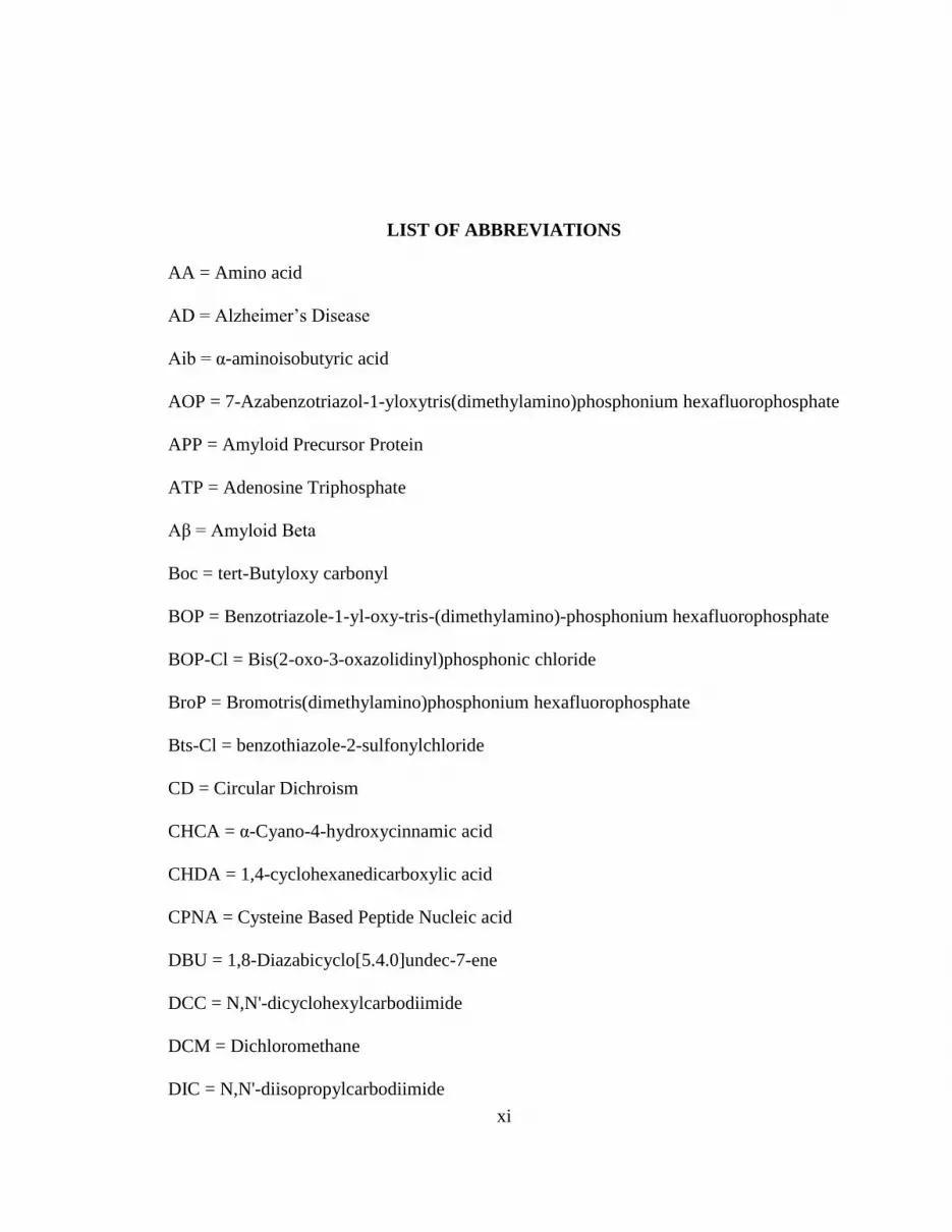
LIST OF ABBREVIATIONS
AA = Amino acid
AD = Alzheimer’s Disease
Aib = α-aminoisobutyric acid
AOP = 7-Azabenzotriazol-1-yloxytris(dimethylamino)phosphonium hexafluorophosphate
APP = Amyloid Precursor Protein
ATP = Adenosine Triphosphate
Aβ = Amyloid Beta
Boc = tert-Butyloxy carbonyl
BOP = Benzotriazole-1-yl-oxy-tris-(dimethylamino)-phosphonium hexafluorophosphate
BOP-Cl = Bis(2-oxo-3-oxazolidinyl)phosphonic chloride
BroP = Bromotris(dimethylamino)phosphonium hexafluorophosphate
Bts-Cl = benzothiazole-2-sulfonylchloride
CD = Circular Dichroism
CHCA = α-Cyano-4-hydroxycinnamic acid
CHDA = 1,4-cyclohexanedicarboxylic acid
CPNA = Cysteine Based Peptide Nucleic acid
DBU = 1,8-Diazabicyclo[5.4.0]undec-7-ene
DCC = N,N'-dicyclohexylcarbodiimide
DCM = Dichloromethane
DIC = N,N'-diisopropylcarbodiimide

xii
DIEA = N,N-Diisopropylethylamine
DMF = N,N-Dimethyl Formamide
DMSO = Dimethylsulfoxide
DPPA = Azidodiphenoxyoxophosphorane
DNA = Deoxyribonucleic acid
ECM = Extracellular matrix
EDC = 1-ethyl-3-[3-(dimethylamino)propyl]carbodiimide
ELISA = Enzyme-linked immunosorbent assay
Et3SiH = Triethylsilane
Fmoc = 9-Fluorenylmethoxycarbonyl
Fmoc-Cl = 9-Fluorenylmethoxycarbonyl chloride
Fmoc-OSu = 9-Fluorenylmethyl N-succinimidyl carbonate
FN = Fibronectin
FTIR = Fourier Transform InfraRed
GPNA = Guanidine based Peptide Nucleic acid
HATU = N,N,N′,N′-Tetramethyl-O-(7-azabenzotriazol-1-yl)uronium
hexafluorophosphate
HBTU = 2-(1H-Benzotriazole-1-yl)-1,1,3,3-tetramethyluronium hexafluorophosphate
HCTU = N,N,N′,N′-Tetramethyl-O-(6-chloro-1H-benzotriazol-1-yl)uronium
hexafluorophosphate
HOBT = 1-hydroxybenzotriazole
HOPfp = pentafluorophenol
HPLC = High-performance liquid chromatography
III = Integerin Interaction Inhibitors

xiii
KDa = kilodalton
MBHA = 4 - Methylbenzhydrylamine resin
MDM2 = murine double minute 2
MDR = Multi-drug resistant
Meu = α-N-methylated Leucine
MM = Multiple myeloma
MRD = Minimal Residual Disease
m-RNA = Messenger RNA
NMM = N-Methylmorpholine
NMP = N-Methylpyrrolidone
NOE = Nuclear Overhauser effect
ONBSCl = ortho-nitrobenezenesulfonyl chloride
PDB = Protein Data Bank
PNA = Peptide Nucleic acid
PTSCl = p-Toluenesulfonyl chloride
PyAOP = 7-Azabenzotriazol-1-yloxy)tripyrrolidinophosphonium hexafluorophosphate
PyBOP = (1-Hydroxy-1H-benzotriazolato-O)tri-1-yrrolidinylphosphorus(1+)
hexafluorophosphate(1-)
ROS = Reactive Oxygen Species
SN2 = Nucleophilic Substitution bimolecular
SPPS = Solid Phase Peptide Synthesis
TBAI = Tetrabutylammonium iodide
TATU = 2-(7-Azabenzotriazole-1-yl)-1,1,3,3-tetramethyluronium tetrafluoroborate

xiv
TBTU = 2-(1H-Benzotriazole-1-yl)-1,1,3,3-tetramethyluronium tetrafluoroborate
TEA = Triethylamine
TEMPO = 2,2,6,6-tetramethylpiperidinoxyl
TFA = Trifluoroacetic acid
TFMSA = Trifluoromethanesulfonic acid
THF = Tetrahydrofuran
ThT = Thioflavin T
TLC = Thin Layer Chromatography
TMS = Tetramethylsilane
TMSCl = Chlorotrimethylsilane

xv
Design & Synthesis of β-hairpin Peptidomimetics for Modulating Integrin Mediated
Cell Adhesion, Abeta Fibrillogenesis and p53-MDM2 Protein-Protein Interactions
Priyesh Jain
ABSTRACT
Inhibiting therapeutically important protein-protein interactions has been a tremendous
challenge for medicinal chemists. The folded 3D structures of peptides and proteins,
mainly comprise secondary structural elements i.e α-helices and β-sheet have created an
opportunity to design small molecules and peptidomimetic inhibitors of protein-protein
interaction (PPI). Hence, information about the formation and stabilization of these
secondary structures is vital for designing future drugs. In this dissertation, several cyclic
beta-hairpin peptidomimetics that mimic the recognition surface have been designed and
synthesized as inhibitors for different targets such as integrin mediated extracellular
matrix -cell adhesion in multiple myeloma, p53-MDM2 PPI, amyloid beta fibrillogenesis
inhibitor. Cyclization of linear peptides to restrict the number of conformations available
to the linear peptide can increase its affinity for the target as well as increase its
proteolytic resistance. In this study, different beta turn promoters that increase the
propensity of cyclic peptides to adopt beta-sheet structures have been designed and
synthesized. Chapter two discusses the design and synthesis of several cyclic III (Integrin
Interaction Inhibitor) peptides that block adhesion of integrins to extracellular matrix
components in Multiple Myeloma tumor cells. These cyclic peptides, as assayed by

xvi
TOPRO 3 assay were more potent than the parent linear peptide with a bio-activity of
1.08 µM. We have also studied structure activity relationships (SAR) of these cyclic III
peptide analogs to increase the potency and bioavailability of these peptides.
Chapter three describes the application of cyclic beta-hairpin peptidomimetics to inhibit
abeta fibrillogenesis that is responsible for Alzheimer’s disease. We have successfully
designed and synthesized cyclic peptides that target the hydrophobic region (17-21) of
abeta fibril which is believed to cause self aggregation and plaque formation. We have
also successfully explored these cyclic beta-hairpin peptides to disrupt p53-MDM2
interactions. Chapter five discusses the design and synthesis of novel cysteine based
Peptide Nucleic Acid (PNA) monomers that are aimed to increase cellular uptake by
introducing positively charged species attached to the cysteine side chain. We have
successfully synthesized CPNA monomers and made efforts to make PNA oligomers.

1
CHAPTER ONE:
INTRODUCTION
1.1 General overview of peptides and proteins
Proteins are macromolecules consisting of 20 different natural amino acids linked
together by amide bonds to form linear chains. Linear chains consisting of 2 to 100 amino
acids with molecular weight up to 10 KDa are usually termed as peptides whereas longer
polypeptides with defined structures are classified as proteins. The total number of
different proteins that can be made from 20 different amino acids is huge. A 10 amino
acids chain can have up to 2010
possible sequences or 10 trillion structurally different
molecules. The biological activity of a protein depends on its three dimensional
conformation. Peptides often have many different conformations and can randomly
change whereas proteins are relatively rigid with one or a handful of preferred
conformations. The primary structure of a polypeptide or protein is the sequence of
amino acids linked together by peptide bond. As shown in fig. 1.1a, the peptide C-N bond
has partial double bond character due to resonance, which restricts free rotation around
the peptide bond (1). Proteins fold themselves into well defined secondary and tertiary
structures. The secondary structure of proteins usually stems from the geometry of bond
angle between amino acids and hydrogen bonds betweens adjacent amino acids residues.
The main secondary structures of protein are: the α-helix, the β-pleated sheet and the β-
turn. The tertiary structure of a protein describes the overall three-dimensional

2
arrangement of the atoms or shape of a protein. As shown in Fig. 1.1b, the peptide
structure is always interpreted in terms of dihedral angles between adjacent planar
peptide groups. The dihedral angle for free rotation around Cα
–N bond is represented by
angle Φ whereas rotation about Cα
–C is denoted by Ψ.
Fig. 1.1 (a) Resonance in planar peptide bonds (b) Various dihedral angles observed in
linear peptide chains.
Table 1.1 Dihedral angles for secondary structural elements in peptides
Conformation Dihedral angle (degree)
Ф Ψ
Residues per turn
Right-handed α-helix -57 -47 3.6
Anti-parallel β-sheet -139 +135 2.0
Parallel β-sheet -119 +113 2.0
Type I β-turn* -60(-90) -30(0) 3.0
Type II β-turn* -60(+90) +120(0) 3.0
For the β-turn, angle outside parenthesis is for ith+1 residue and within parenthesis is for
ith+2 residue of the turn (2).

3
The right handed α-helix accounts for approximately 31% of all protein structures
whereas β-sheets and β-turns accounts for 28% and 25% respectively (3). These
secondary and tertiary structures are stabilized by noncovalent interactions such as
hydrogen bonds, Van der Waals interactions, hydrophobic effects and steric interactions.
The folded 3D structures of peptides and proteins have created an opportunity to design
small molecules and peptidomimetics to mimic the recognition surface involved in
various protein-protein interactions responsible for causing several diseases.
1.2 β-Sheet and β-hairpins
β-Sheet and β-hairpins are key recognition motifs that bind to many protein-
protein and protein-DNA interactions that are important for many biological functions
and also in causing some diseases. So far, the α-helix secondary structure has been
widely studied. The study of β-sheets was not widely adopted due to the fact that these
peptides quickly undergo self-association to form large, insoluble, quaternary β-sheet
structures, which makes detailed thermodynamic and structural characterization of these
secondary structures very difficult (4,5). β-sheets consist of two or more paired β-strands
arranged in parallel or antiparallel fashion. In parallel β-sheets, the individual β-strands
run in the same direction and are usually characterized by 12 atom hydrogen bonded
rings (Fig.1.2a). In antiparallel β-sheets, the individual β-strands run in the opposite
directions and are usually characterized by alternating 10 and 14 atom hydrogen bonded
rings (Fig.1.2b). Table 1.1 clearly depicts the dihedral angles found in parallel and anti-
parallel beta sheets. Antiparallel β-sheets have shorter (linear) H bonds whereas parallel
β-sheet have slightly longer H bonds. Both parallel and antiparallel β-sheets have
adjacent side chain residues running in opposite directions, thereby exhibiting an

4
extended conformation. Thus β-sheets offer three different recognition sites: interaction
either via top or bottom faces of the sheet or via hydrogen bonding with amide hydrogen
on each side of the sheet. Parallel β-sheets are usually seen in the hydrophobic interior of
proteins whereas antiparallel β-sheets with amphipathic character are usually found at the
surface of proteins.
Fig. 1.2 Schematic representation of (A) parallel and (B) antiparallel β-sheets. H bonds
are indicated by dashed lines whereas side chains of amino acids are represented by R.
The arrow head indicates direction from N to C terminus of the β-sheet.
The β-turn is another important protein structure which allows the polypeptide
chain to change the direction abruptly by 180o and allows the protein to adopt a more
globular compact shape (6-8). β-turns usually consists of loops formed from four amino

5
acid residues where the hydrogen bonding from ith and ith +3 residue is important in
stabilizing the turn (Fig. 1.3).
Fig. 1.3 The β-turn
Another very interesting secondary structure found at protein-protein interfaces is
the β-hairpin motif (9-11). A β-hairpin is a simple motif that contains two antiparallel
strands connected by a short connecting loop sequence of two to five amino residues that
stabilizes the hairpin structure (Fig. 1.4). The β-hairpin scaffold is very interesting since it
is used by many proteins for bimolecular recognition. 3D structure analysis of β-hairpins
revealed that β-hairpins vary widely in hydrogen-bonding pattern between strands and
most of hairpin loops have less than 5 residues (12).
Fig. 1.4 β-hairpin

6
The simplest approach to design a β-hairpin mimetic is to transplant a hairpin
loop of protein with a known structure onto a template that stabilizes the β-hairpin to
form a conformationally defined macrocyclic structure. The template serves as a β-turn
promoter and nucleates the cyclic β-sheet formation. The template structure plays an
important role on macrocylic β-hairpin stability. Various research groups have
incorporated natural or unnatural amino acids in the β-turn position of a β-hairpin and
have studied β-hairpin folding and the stability of linear peptides in aqueous solution (13-
18). As shown in Fig. 1.5, Blanco and co-workers have used the L-Asn-Gly dipeptide
template as a β-turn promoter which folds and forms β-hairpin structure in water.
Fig. 1.5 16 amino acid residue peptide with Asn-Gly dipeptide template as turn promoter
Gellman and co-workers have utilized D-Pro-Gly template to form stable β-
hairpins in aqueous solution (Fig. 1.6) (14). They found that the D-Pro-Gly template was
better than Asn-Gly and use of D-amino acid increases the propensity of linear peptides
to adopt the β-hairpin conformation.

7
Fig. 1.6 12 amino acid residue Gellman peptide with D-Pro-Gly dipeptide template
It has been reported that achiral α-aminoisobutyric acid (Aib) forms a β-turn when
used with D-α-amino acid or achiral α-amino acids. Balaram and co-workers have shown
that peptides with the Aib-Xaa (Xaa= D-Ala, D-Val, D-Pro, D-Gly) dipeptide template
adopts β-turn conformations in organic solvents (19). Hammer, Barany, Veglia and co-
workers have shown that Aib-D-Ala and Aib-Gly adopts a β-turn conformation in water
(20). They have compared the conformation of Gellman’s peptide (Fig. 1.6) by replacing
the D-Pro-Gly dipeptide β-turn inducer by Aib-D-Ala (Balram’s dipeptide template),
Aib-Gly and demonstrated by NMR studies that these peptides (Fig. 1.7) adopt type I’ β-
turn conformations in water.
Fig. 1.7 Hammer’s design of β-hairpin structures with (a) Aib-Gly and (B)Aib-D-Ala
dipeptide units introduced as a β-turn inducer in Gellman’s peptide sequence.

8
Balaram and co-workers have also reported several expanded β-turn mimics with
D-Pro-Xaa (Xaa= β-,γ-, and δ- amino acid) dipeptide template (21). They have shown
that peptides with α,β and α,γ residues at the turn adopt β-hairpin structures in methanol.
Fig. 1.8 Balaram’s design of β-hairpins with extended α,β, α,γ and α,δ turn promoter.
Guan have reported a 1,4-disubstituted 1,2,3-triazole based β-turn promoter that
induces β-hairpin structures for peptides in chloroform (22). Silvani have reported

9
tetrahydroisoquinoline based turn promoter to induce β-hairpin structures for smaller
peptides in chloroform (23). Several research groups have reported new β-turn promoter
that induce β-hairpin formation for linear peptides (24). Fig.1.10 lists some of the β-turn
promoters reported for inducing β-sheet structure formation.
Fig. 1.9 Different β-turn promoters for inducing β-hairpin structures for linear peptides
Fig. 1.10 Parallel β-sheet formation using CHDA-Gly diacid linker

10
Fig. 1.11 Artificial β-sheets consisting of parallel β-sheet dimer with succinic diacid
linkers and β-strand and β-turn mimic.
Recently Gellman and co-workers have reported the first diacid linker, CHDA-
Gly turn promoter that promotes β-sheet formation in water (25). The CHDA-Gly turn
promoter aligns two β-strands in parallel orientation as shown in Fig.1.10. Nowick and
Levine have recently reported the synthesis of artificial β-sheets that dimerize through
parallel interstrand β-sheet interations in chloroform (26). Parallel β-sheet interactions are
important in the aggregation of peptides such as Aβ peptide aggregation observed in
Alzheimer’s disease.
1.3 Cyclic β-hairpin peptidomimetics
Generally the bioactivity of proteins arises from a small local segment of protein
surface formed by these secondary structures, so molecules based on these structures
would in principle serve as antagonists. Hence a simple approach for medicinal chemists

11
is to design drugs would mimic the β-sheet/strand regions of proteins recognized by other
proteins or DNA. However the linear peptides have the disadvantages of being
conformationally flexible, existing as random structures in aqueous solution. Linear
peptides have low bioavailability, are easily susceptible to degradation by proteases and
poor pharmacological properties. There are two strategies for solving these issues. The
first approach involves restricting the degrees of freedom available to the linear peptide
through cyclization to form macrocycles. Molecules which are conformationally
preorganized or have fixed shape that can be recognized by a receptor will have higher
affinity for that receptor due to a diminished loss of entropy for adopting that shape.
Nature frequently uses cyclization technique to force peptides to adopt bioactive
conformations. Naturally occurring peptide antibiotics such as gramicidin S and θ-
defensin have been used and studied extensively for creating β-sheet structures. Cyclic
peptides also cannot as readily be recognized by proteolytic enzymes.
Fig. 1.12 Robinson’s cyclic β-hairpin design as inhibitors of p53/MDM2 interaction.

12
As shown in Fig.1.12, Robinson and co-workers have utilized this approach in
mimicking the α-helix of p53 with a cyclic β-hairpin to inhibit p53/MDM2 interaction.
They have designed a cyclic beta-hairpin scaffold mounted on a D-Pro-L-Pro beta-turn
promoter template that holds the side chains of phenylalanine and tryptophan residues in
the correct relative positions, so that they could interact with their respective binding sites
on MDM2 protein (27). This dipeptide template adopts a stable type II’ β-turn that is
ideal for inducing β-hairpin conformations. Kopple and co-workers have also used this
template to design cyclic hexapeptides that exhibit β-sheet structures with type II’ β-turn
(28). This cyclic β-hairpin peptide design developed by Robinson has been explored to
design inhibitors for several bimolecular targets. Shankaramma have synthesized a cyclic
macrocylic β-hairpin mimetic of the cationic antimicrobial peptide Protegrin I by using
the DPro-LPro dipeptide template (Fig.1.13) (29). Robinson & co-workers have also
reported β-hairpin mimetics with a mixed peptide-peptoid backbone that have shown
excellent antimicrobial activity (Fig.1.14) (30). They have also synthesized a series of
cyclic β-hairpin mimetics based on a trypsin inhibitor from sunflower seeds (31).
Fig. 1.13 β-hairpin mimetic of cationic antimicrobial peptide Protegrin I.

13
Fig. 1.14 β-hairpin mimetic of antimicrobial peptides with mixed peptide-peptoid
backbone.
Nowick and co-workers have reported the synthesis of a triple-stranded artificial
β-sheet that adopts β-sheet like conformation in organic solvents (Fig.1.15a)(32).
Recently, they have developed several macrocyclic peptides containing a pentapeptide
strand, β-strand mimic and two ornithine based β-turn promoters (33-34). These 42-
membered cyclic peptides adopt β-sheet conformations in water (Fig.1.15b). Ornithine α-

14
amino groups provide a site that allows synthesis of bivalent peptides connected by a
linker of suitable length (Fig.1.15c).
Fig. 1.15a Nowick’s design for triple stranded artificial β-sheet.
Fig. 1.15b 42-membered macrocyclic β-sheet peptide mimic containing β-strand & β-
turn mimic.
Fig. 1.15c Macrocyclic β-sheet peptide mimic containing β-strand & β-turn mimic.

15
As shown in Fig.1.16, the methylsulfonamido aminoethyl glycine and proline
based ether-peptidomimetic amino acid have been designed and synthesized to induce β-
sheet conformations for synthesizing cyclic β-haipin peptidomimetics to inhibit protein-
protein interactions. The core residues mimicking the recognition surface are placed in
the recognition strand whereas non-recognition strand residues can be modified to
optimize the bioavailability and bioactivity of the designed peptide. N- to C- terminus
cyclization of linear peptides to restrict the number of conformations available to the
linear peptide increases the affinity of the cyclized peptide for its target. Peptidomimetics
obtained after incorportation of methylsulfonamido aminoethyl glycine turn promoters
are believed to adopt β-sheet extended structures. Replacement of the aminoethyl glycine
turn promoter with the D-Proline derived ether-peptidomimetic turn promoter should
further constrain the β-hairpin and increase its binding affinity for the target.
It is known through cited literature that adhesion of leukemia tumor cell to
extracellular matrix components via integrin influences cell survival and inhibits drug
induced apoptosis. Various research groups have made efforts to design drugs that mimic
integrin recognition surfaces, inhibiting protein-protein interaction and thereby inducing
cell death through apoptotic pathways. A linear 10 amino acid residue peptide referred as
D-HYD1 has been reported to block adhesion of tumor cells to extracellular matrix. Our
proposed strategy of cyclic β-hairpin peptidomimetics design that carries the core region
of D-HYD1 in the recognition strand are expected to block adhesion of integrins
(glycoprotein) to extracellular matrix components in prostate, multiple myeloma, and
lung tumor cells.

16
(a)
(b)
(c)
Fig. 1.16 (a) Designed methylsulfonamide aminoethyl glycine and proline based ether-
peptidomimetic amino acid turn promoters (b) Proposed cyclic β-hairpin peptidomimetic
with aminoethyl glycine turn promoter to inhibit adhesion of leukemia cells to
extracellular matrix (c) Cyclic β-hairpin peptidomimetic with proline based ether-
peptidomimetic amino acid turn promoter.
Another well known example is the p53/MDM2 protein-protein interactions
observed in many cancers. Various research groups have developed drugs that mimic the
p53 recognition surface that binds to MDM2 and inhibits the protein-protein interactions.
Fig.1.17 shows the 9-membered cyclic peptide that has been designed to mimic the α-
helix of p53 with cyclic β-hairpin to inhibit p53/MDM2 interaction.

17
Fig. 1.17 Proposed cyclic β-hairpin mimetic of α-helix of p53.
In the last two decades many neurological disorders such as Alzheimer’s disease
(AD), Parkinson’s disease, mad-cow disease, Huntington’s disease and many other prion
diseases have been to be caused by protein misfolding without any alteration of the
protein ‘primary structure’. It was found that aggregation or self-association of amyloid
fibrils, consisting of 40-42 amino acid Aβ peptides formed insoluble β-sheet plaques that
were responsible for causing AD. Many researchers have shown that the hydrophobic
core region (residues 17-21) of Aβ peptide is critical for fibril formation. Based on these
reports we propose the design and synthesis of cyclic β-hairpin peptides that bind
specifically to this hydrophobic region and prevent abeta β-sheet aggregation (Fig.1.18).
The recognition strand of the designed cyclic peptide would interact with the growing Aβ
fibril whereas the non-recognition strand would have residues that would inhibit
aggregation from the other face of the assembly.

18
Fig. 1.18 Cyclic β-hairpin peptidomimetic targeting hydrophobic core region (17-21) of
Aβ peptide.
1.4 Solid Phase Peptide Synthesis
In 1959, Merrifield conceived the idea that if peptide is bound to an insoluble
support then any unreacted reagents left at the end of each reaction step can be simply
removed by general solvent wash, thereby reducing the time required for purification and
synthesis of the desired compound. This idea led him to pioneer a new field in chemistry
called solid-phase synthesis for which he was awarded Nobel Prize in 1984. Solid-phase
synthesis is widely used by chemist to carry out chemical reactions on a solid support to
prepare libraries of organic compounds, oligomers and complex molecules that are
difficult to synthesize using solution-phase synthesis. During the last six decades, Solid-
Phase Peptide Synthesis (SPPS) has evolved as the primary route for synthesizing a great
diversity of peptides and proteins. The basic concept of synthesizing peptides using SPPS
is described in Fig.1.19. The peptide chain is assembled on polymeric resin solid support
by stepwise addition of α-amino acid residues in the C to N direction. Fmoc and Boc
protecting group strategies are widely used for synthesizing peptides on resin. Fmoc

19
chemistry has been widely adopted for most solid-phase peptide synthesis since this
strategy has been shown to be more reliable. The greatest advantage of employing Fmoc
SPPS strategy is that it is chemically compatible with most of the commercially available
peptide synthesizers. The use of extremely hazardous and corrosive chemicals for
deprotection step in Boc SPPS limits its application on an automated synthesizer.
Fig. 1.19 General protocol for solid-phase peptide synthesis.

20
After base treatment, the peptide is washed several times with a mixture of
solvents and then a mixture consisting of an activated amino acid and base is added to the
peptide in the reactor to couple the next amino acid. There are four different kinds of
coupling agents used for carboxyl activation in SPPS:
1. Carbodiimides
2. Symmetrical anhydrides
3. Activated Esters
4. Uronium based coupling reagents
Carbodiimides are widely preferred activation reagents (35). These reagents
produce insoluble ureas in some of the solvents that are used in SPPS and thus interfere
in peptide purification. Symmetrical anhydrides can be generated in situ from two
equivalents of amino acid and one equivalent of carbodiimide reagent. The generated
urea can be filtered off and the synthesized coupling agents can then be used for
coupling. The most commonly used activated esters for peptide coupling are those
derived from pentafluorophenol (HOPfp) and HODhbt (36). During last three decades,
coupling reagents based on phosphonium and uronium salts have been widely used for
synthesizing peptides. These in situ activation reagents are highly efficient in peptide
couplings and reduces racemization or side-product formation. Fig.1.20 lists some of the
common activating agents used. Other coupling reagents used for peptide coupling
include amino acid halides and azides (37, 38). The activated acids can be prepared and
purified prior to coupling or can be generated in situ be addition of reagents such as
cyanuric chloride or fluoride to give acid chlorides or fluorides. Diphenyl
phosphorylazide directly converts carboxyl groups to an acylazide intermediate.

21
Fig. 1.20 Common coupling reagents used in SPPS.

22
All cyclic β-hairpin peptidomimetics designed for the different targets (chapter 2,
3 &4) will be synthesized on the Symphony peptide synthesizer, Protein Technologies
Instrument, using Fmoc SPPS strategy. Typically, a linear precursor will be assembled on
solid support, 2-Chlorotrityl chloride resin, and then macrocyclized and deprotected in
solution phase. SPPS strategy has also been adopted to synthesize PNA oligomers which
is described in detail in chapter five.
1.5 Cysteine based Peptide Nucleic Acid (CPNA)
PNA (Peptide Nucleic Acids) are DNA mimics in which the deoxyribose
phosphate diester backbone is replaced by a pseudo-peptide backbone while the four
natural nucleobases are retained. PNA consists of repeating N-(2-aminoethyl)-glycine
units in which the nucleobase is attached to the glycine nitrogen via a methylene carbonyl
linker. The acyclic, achiral and neutral backbone makes PNA set apart from DNA. The
neutral backbone of PNA allows stronger binding between complementary PNA/DNA
strands than between complementary DNA/DNA strands. PNAs have been demonstrated
as potential candidates for gene-targeting drugs but some disadvantages have limited its
applications. These include low cellular uptake caused by poor cell membrane
permeability and poor aqueous solubility. To increase cellular uptake we propose the
design of novel CPNA (cysteine based PNA) scaffolds by introducing positive charge
species attached to the cysteine side chains. An attempt to synthesize cysteine based
Peptide nucleic acid (CPNA) monomers and oligomers containing all four nucleobases is
described in chapter five.

23
1.6 Structural Analysis of Peptides
The 3D structural analysis of peptides and proteins can be carried out by several
experimental techniques such as X-ray crystallography, solution nuclear magnetic
resonance (NMR) spectroscopy, circular dichroism (CD) spectroscopy and Fourier
transform infrared spectroscopy (FTIR). X-ray analysis is a widely preferred technique
for structural elucidation of peptides. Solution NMR spectroscopy is another powerful
tool used since the last three decades. The NMR input for conformational studies of
peptides depends on NOE (nuclear Overhauser effects) and coupling constants. NOE
studies detect the interactions involved by measuring interproton distances of 5 Ǻ or less
while the coupling constants give information about torsion angles. The whole set of
NOE constraints is then plugged in computational software and an energy minimization
experiment is carried out to search for optimized solution structures of peptides and
proteins.
Circular Dichroism (CD) is another important technique for determining the
conformations of peptides and proteins in solution. CD spectroscopy measures the
difference in absorbance of right- and left-circularly polarized light by a substance. Bands
in the far UV region of CD spectra (260-190 nm) are analyzed for the different secondary
structural of peptides and proteins i.e. alpha helix, parallel and antiparallel beta sheet,
beta turn, etc. Fig.1.21. shows various secondary structural elements in CD spectra of
polypeptides in solution (39). All cyclic β-hairpin peptides synthesized will be
characterized for their secondary structures using CD and NMR experiments.

24
Fig. 1.21 CD spectra of secondary structural elements in peptides and proteins.
FTIR technique is used for examining peptides that shows self-association or
aggregation behavior. In IR spectra each compounds has its own set of absorption bands.
In FTIR, characterstic bands for peptides and proteins include Amide I and Amide II
arising from the amide bond (40). The stretching vibrations of C=O leads to Amide I
bands where bending vibrations of the N-H bonds leads to amide II bands. The location
of both amide I and amide II bands are very sensitive to the secondary structure of
peptides and proteins.

25
1.7 References
1. Patrick, G. L., An Introduction to Medicinal Chemistry, 2nd Edition. Oxford
University Press, New York, 2005; pp.24. 2. Creighton, T. E. Proteins: Structure and Molecular Properties; 2nd ed; W.
H.Freeman and Co.: New York, 1993; pp. 65-68.
3. Walton, A. G. Polypeptides and Protein Structure; Elsevier: New York, 1981;
pp. 49-55.
4. Pauling, L.; Corey, R. B., Configurations of polypeptide chains with favored
orientations around single bonds: two new pleated sheets. Proc. Natl.
Acad. Sci. U. S. A. 1951, 37, 729-40.
5. Pauling, L.; Corey, R. B.; Branson, H. R., The structure of proteins: two
hydrogen-bonded helical configurations of the polypeptide chain. Proc.
Natl. Acad. Sci. U. S. A. 1951, 37, 205-11.
6. Dyson, H. J.; Rance, M.; Houghten, R. A.; Lerner, R. A.; Wright, P. E., Folding
of immunogenic peptide fragments of proteins in water solution. I.
Sequence requirements for the formation of a reverse turn. J. Mol. Biol.
1988, 201 (1), 161-200.
7. Hutchinson, E. G.; Thornton, J. M., A revised set of potentials for β-turn
formation in proteins. Protein Sci. 1994, 3 (12), 2207-16.
8. Sibanda, B. L.; Blundell, T. L.; Thornton, J. M., Conformation of beta-hairpins
in protein structures. A systematic classification with applications to
modelling by homology, electron density fitting and protein engineering.
J. Mol. Biol. 1989, 206 (4), 759-77.
9. Blanco, F. J.; Jimenez, M. A.; Herranz, J.; Rico, M.; Santoro, J.; Nieto, J. L.,
NMR evidence of a short linear peptide that folds into a beta -hairpin in
aqueous solution. J. Am. Chem. Soc. 1993, 115 (13), 5887-8.
10. Blanco, F. J.; Rivas, G.; Serrano, L., A short linear peptide that folds into a
native stable beta-hairpin in aqueous solution. Nat Struct Biol 1994, 1 (9),
584-90.
11. Searle, M. S.; Williams, D. H.; Packman, L. C., A short linear peptide derived
from the N-terminal sequence of ubiquitin folds into a water-stable non-
native beta-hairpin. Nat Struct Biol 1995, 2 (11), 999-1006.
12. Sibanda, B. L.; Thornton, J. M., Beta-hairpin families in globular proteins.
Nature 1985, 316 (6024), 170-4.

26
13. Ramirez-Alvarado, M.; Blanco Francisco, J.; Serrano, L., De novo design and
structural analysis of a model beta -hairpin peptide system. Nat Struct Biol
1996, 3 (7), 604-612.
14. Gellman, S. H., Minimal model systems for beta sheet secondary structure in
proteins. Curr. Opin. Chem. Biol. 1998, 2 (6), 717-725.
15. Searle, M. S., Peptide models of protein beta -sheets: design, folding and
insights into stabilising weak interactions. J. Chem. Soc., Perkin Trans. 2
2001, (7), 1011-1020.
16. Venkatraman, J.; Shankaramma, S. C.; Balaram, P., Design of Folded
Peptides. Chem. Rev. (Washington, D. C.) 2001, 101 (10), 3131-3152.
17. Searle, M. S.; Ciani, B., Design of beta -sheet systems for understanding the
thermodynamics and kinetics of protein folding. Curr. Opin. Struct. Biol.
2004, 14 (4), 458-464.
18. Hughes, R. M.; Waters, M. L., Model systems for beta -hairpins and beta -
sheets. Curr. Opin. Struct. Biol. 2006, 16 (4), 514-524.
19. Aravinda, S.; Shamala, N.; Rajkishore, R.; Gopi, H. N.; Balaram, P., A
crystalline beta -hairpin peptide nucleated by a type I' Aib-D-Ala beta -
turn: evidence for cross-strand aromatic interactions. Angew. Chem., Int.
Ed. 2002, 41 (20), 3863-3865.
20. Masterson, L. R.; Etienne, M. A.; Porcelli, F.; Barany, G.; Hammer, R. P.;
Veglia, G., Nonstereogenic alpha -aminoisobutyryl-glycyl dipeptidyl unit
nucleates type I' beta -turn in linear peptides in aqueous solution.
Biopolymers 2007, 88 (5), 746-753.
21. Rai, R.; Vasudev, P. G.; Ananda, K.; Raghothama, S.; Shamala, N.; Karle, I.
L.; Balaram, P., Hybrid peptides: expanding the beta turn in peptide
hairpins by the insertion of beta -, gamma -, and delta -residues. Chem.--
Eur. J. 2007, 13 (20), 5917-5926.
22. Oh, K.; Guan, Z., A convergent synthesis of new beta -turn mimics by click
chemistry. Chem. Commun. (Cambridge, U. K.) 2006, (29), 3069-3071.
23. Lesma, G.; Meschini, E.; Recca, T.; Sacchetti, A.; Silvani, A., Synthesis of
tetrahydroisoquinoline-based pseudopeptides and their characterization as
suitable reverse turn mimetics. Tetrahedron 2007, 63 (25), 5567-5578.
24. Ressurreicao, A. S. M.; Bordessa, A.; Civera, M.; Belvisi, L.; Gennari, C.;
Piarulli, U., Synthesis and Conformational Studies of Peptidomimetics
Containing a New Bifunctional Diketopiperazine Scaffold Acting as a beta
-Hairpin Inducer. J. Org. Chem. 2008, 73 (2), 652-660.

27
25. Freire, F.; Fisk, J. D.; Peoples, A. J.; Ivancic, M.; Guzei, I. A.; Gellman, S. H.,
Diacid Linkers That Promote Parallel beta -Sheet Secondary Structure in
Water. J. Am. Chem. Soc. 2008, 130 (25), 7839-7841.
26. Levin, S.; Nowick, J. S., An Artificial beta -Sheet That Dimerizes through
Parallel beta -Sheet Interactions. J. Am. Chem. Soc. 2007, 129 (43),
13043-13048.
27. Fasan, R.; Dias, R. L. A.; Moehle, K.; Zerbe, O.; Vrijbloed, J. W.; Obrecht,
D.; Robinson, J. A., Using a beta -hairpin to mimic an alpha -helix: Cyclic
peptidomimetic inhibitors of the p53-HDM2 protein-protein interaction.
Angew. Chem., Int. Ed. 2004, 43 (16), 2109-2112.
28. Bean, J. W.; Kopple, K. D.; Peishoff, C. E., Conformational analysis of cyclic
hexapeptides containing the D-Pro-L-Pro sequence to fix beta -turn
positions. J. Am. Chem. Soc. 1992, 114 (13), 5328-34.
29. Shankaramma, S. C.; Athanassiou, Z.; Zerbe, O.; Moehle, K.; Mouton, C.;
Bernardini, F.; Vrijbloed, J. W.; Obrecht, D.; Robinson, J. A., Macrocyclic
hairpin mimetics of the cationic antimicrobial peptide protegrin I: a new
family of broad-spectrum antibiotics. ChemBioChem 2002, 3 (11), 1126-
1133.
30. Shankaramma, S. C.; Moehle, K.; James, S.; Vrijbloed, J. W.; Obrecht, D.;
Robinson, J. A., A family of macrocyclic antibiotics with a mixed peptide-
peptoid beta -hairpin backbone conformation. Chem. Commun.
(Cambridge, U. K.) 2003, (15), 1842-1843.
31. Descours, A.; Moehle, K.; Renard, A.; Robinson, J. A., A new family of beta -
hairpin mimetics based on a trypsin inhibitor from sunflower seeds.
ChemBioChem 2002, 3 (4), 318-323.
32. Nowick, J. S.; Cary, J. M.; Tsai, J. H., A Triply Templated Artificial beta -
Sheet. J. Am. Chem. Soc. 2001, 123 (22), 5176-5180.
33. Woods, R. J.; Brower, J. O.; Castellanos, E.; Hashemzadeh, M.; Khakshoor,
O.; Russu, W. A.; Nowick, J. S., Cyclic Modular beta -Sheets. J. Am.
Chem. Soc. 2007, 129 (9), 2548-2558.
34. Khakshoor, O.; Demeler, B.; Nowick, J. S., Macrocyclic beta -Sheet Peptides
That Mimic Protein Quaternary Structure through Intermolecular beta -
Sheet Interactions. J. Am. Chem. Soc. 2007, 129 (17), 5558-5569.
35. Barany, G.; Merrifield, R.B. The Peptides: Analysis, Synthesis, Biology;
Gross, E., Meienhofer, J. Eds.; Academic Press: New York, NY, 1979;
Vol. 2.
36. Kovacs, J.; Kisfaludy, L.; Ceprini, M. Q., Optical purity of peptide active
esters prepared by N,N'-dicyclohexylcarbodiimide and complexes of N,N'-

28
dicyclohexylcarbodiimide-pentachlorophenol and N,N'-
dicyclohexylcarbodiimide-pentafluorophenol. J. Am. Chem. Soc. 1967, 89
(1), 183-4.
37. Carpino, L. A.; Beyermann, M.; Wenschuh, H.; Bienert; Michael, Peptide
Synthesis via Amino Acid Halides. Acc. Chem. Res. 1996, 29 (6), 268-
274.
38. Ninomiya, K.; Shioiri, T.; Yamada, S., Amino acids and peptides. XII.
Phosphorus in organic synthesis. VIII. Reaction of malonic acid half esters
with diphenyl phosphorazidate. Chem. Pharm. Bull. 1974, 22 (6), 1398-
1404.
39. Greenfield, N. J.; Fasman, G. D., Computed circular dichroism spectra for the
evaluation of protein conformation. Biochemistry 1969, 8 (10), 4108-16.
40. Haris, P. I.; Chapman, D., The conformational analysis of peptides using
Fourier transform IR spectroscopy. Biopolymers 1995, 37 (4), 251-63.

29
CHAPTER TWO:
NOVEL CYCLIC III PEPTIDES TARGETING INTEGRIN MEDIATED CELL
ADHESION IN MULTIPLE MYELOMA
2.1 Introduction
Multiple Myeloma (MM) is an incurable malignancy which frequently exhibits
relapse due to unsuccessful elimination of minimal residual disease (MRD). Current
standard chemotherapeutic treatments that target apoptotic cell death pathways have
proven to be unsuccessful in treating this disease, due to the strong multi-drug resistance
(MDR) emergence by tumor cells (1). MRD is found in the bone marrow indicating that
the bone marrow microenvironment is essential for tumor cell survival (2). The bone
marrow is rich in extracellular matrices. Hazlehurst and co-workers have shown that
adhesion of leukemia and multiple myeloma cells to the extracellular matrix component
fibronectin (FN) influences cell survival and inhibits drug induced apoptotic cell death
(3-9). Various research groups have targeted integrin mediated cell adhesion to increase
the efficacy of standard cytotoxic drugs that predominantly use the apoptotic cell death
pathways. Cell deaths in mammals have been reported to occur mainly via three different
pathways. Type I cell death or apoptosis usually occurs with alteration of mitochondrial
membrane potential, followed by subsequent release of cytochrome C and activation of
caspases, ultimately leading to shut down of cell machinery. Type II cell death or
autophagy is independent of activation of caspases and is usually observed with the

30
formation of double membrane vesicles (autophagosomes) (10, 11). It is usually the
default cell death pathway and may occur when apoptotic mechanisms fails. Type III cell
death pathway or necrosis is seen with rupture of plasma membrane, swelling of
cytoplasmic organelles, decrease in ATP levels and an increase in reactive oxygen
species (ROS) (12). The majority of cancer cells follow the apoptotic pathway to induce
cell death. Hence, the majority of current standard chemotherapeutic agents are
developed to induce apoptosis in tumor cells. With the emergence of multi-drug
resistance of tumor cells for currently used standard cytotoxic drugs that follow the
apoptotic pathway, development of cytotoxic drugs that follow an alternative cell death
pathway would be an effective strategy in tumor cell killing.
Using combinatorial peptide libraries and a functional binding assay, Kit Lam and
Anne Cress and co-workers were able to identify several peptides that inhibited β1
integrin mediated adhesion of prostate cancer cells to fibronectin, laminin and collagen
IV (13). They identified a synthetic all D-amino acid peptide referred as
HYD1(kikmviswkg) that blocks binding of epithelial prostate carcinoma cells to
extracellular matrix components (14,15). Hazlehurst and co-workers have shown that use
of HYD1 peptide in blocking β1 integrin mediated adhesion increased the efficacy of
standard cytotoxic drugs used to treat Multiple myeloma (16). HYD1 induces cell death
in multiple myeloma as a single agent in vitro and in vivo. HYD1 treatment did not
trigger the activation of caspases nor did it induce DNA fragmentation. HYD1 treatment
seemed to cause necrotic cell death as shown by loss of mitochondrial membrane
potential, loss of cellular ATP and an increase in ROS production.

31
Hazlehurst and co-workers performed truncation studies on the C and N terminus
and identified MVISW as a likely core region of linear D-HYD1 responsible for
biological activity. They have demonstrated that HYD1 has an IC50 value 33 μM when
assayed for H929 multiple myeloma tumor cells using FACS analysis for cell death. It is
well known that any molecule which is conformationally preorganized or fixed into a
shape which can be recognized by a target will have higher affinity for that target, at the
expense of entropy loss for adopting that conformation (17). Cyclization of linear
peptides to form cyclic peptidomimetics, imposes constraints in the number of
conformations available to the linear peptide. The constraint further stabilizes the bound
conformation of the peptide. Also, cyclization of the linear peptide to form cyclic
peptides makes the amide bonds less recognizable by various proteases.
Based on the structural information of D-HYD1 peptide, we proposed the design
and synthesis of novel cyclic III (Integrin Interaction Inhibitors) peptide analogs for
optimizing the efficacy of the parent peptide (Fig.2.1). The introduction of a novel beta
turn promoter in the cyclic peptide design will further constrain the recognition portion of
cyclic peptide specifically into an extended or beta-sheet like conformation. All cyclic
peptides were more potent as compared to linear D-HYD1 when assayed using TOPRO3
protocol. The alanine scan of the recognition strand carrying core residues has also been
investigated to determine critical residues responsible for bioactivity of cyclic peptides.
Furthermore we have also proved that all cyclic peptidomimetics adopt beta sheet
conformation as confirmed by circular dichroism and NMR studies.

32
(A) Cyclic D-HYD1 2.18 (B) Cyclic III Peptide 2.17
(C) Cyclic III Peptide 2.1 (D) Cyclic III Peptide 2.7
(E) Retro-inverso Cyclic III Peptide 2.10
Fig. 2.1 Proposed cyclic III peptide analogs of linear HYD1 peptide with L-WSVVM and
D-MVVSW as key residues on recognition strand.

33
2.2 Results & Discussion
2.2.1 Peptide design
Hazlehurst and co-workers have systematically carried out truncated N and C
terminus studies and identified MVISW as the likely core region of linear D-HYD1
required for biological activity. Using this information and the finding that valine for
isoleucine replacement gave a more active D-HYD1 analog, we developed a cyclized
version of D-HYD1 that is designed to display the core sequence (MVVSW) in the
recognition strand and (KLKLK) as the non-recognition strand. The designed cyclic
peptidomimetic exhibited an extended or beta-sheet-like conformation (Fig. 2.1). N- to C-
terminus cyclization of linear peptides to restrict the number of conformations available
to the linear peptide increases the affinity of the cyclized peptide for its target when the
constraint stabilizes the bound conformation of the peptide. As a starting point for the
rational design, we did energy minimization studies on similar designed beta-hairpin
cyclic peptides for inhibiting abeta fibrillogenesis (Fig. 2.2 & Fig. 2.3). Energy
minimizations were carried out by keeping all backbone atoms fixed to refine the spatial
position of side chain residues. Energy minimization studies suggested that introduction
of the novel turn promoter and the Robinson template (D-Pro-L-Pro) at the turns gave
stable hairpin peptide with all of the internal H-bonds intact. Table 2.1. depicts the
energy minization values for different turn promoters used in designing these stable
cyclic peptides. Based on energy minimization studies data, we synthesized cyclic
D-HYD1 with the methylsulfonamide aminoethyl glycine as turn promoters. As per the
cell inhibition assay TOPRO 3, the cyclic D-HYD1 peptide was found to be twice as
active as linear D-HYD1.

34
Fig. 2.2 Stereo drawing of cyclic abeta peptides obtained after energy minimization
procedure. Cyclic Aβ peptides designed using (a) L-Pro-D-Pro and methylsulfonamido
aminoethyl glycine (b) two methylsulfonamido aminoethyl glycine units and (c) L-Pro-
D-Pro and L-Pro-Aib turn promoters.
(a)
(b)
(c)

35
Fig. 2.3 Cyclic Abeta peptide designed with different β-turn promoters. (a) Turn 1 (b)
Turn 2 (c) top view (d) side view of cyclic abeta peptide with the methylsulfonamido
aminoethyl glycine turns and (e) super-imposed pose for cyclic abeta peptides with
different turn promoters.
(c) (d)
(a) (b)
(e)

36
Table 2.1 Turn Promoters used in synthesis of cyclic peptidomimetics.
Turn1 Turn 2 Energy
Minimization
1 Methylsulfonamido aminoethyl
glycine
D-Pro-L-Pro -2100 Kcal
2 Methylsulfonamido aminoethyl
glycine
Methylsulfonamido aminoethyl
glycine
-2400 Kcal
3 L-Pro-Aib D-Pro-L-Pro -1400 Kcal
We also further investigated the inverso- and retro-inverso- cyclic III peptide
analogs for their potential to block beta integrin mediated cell adhesion. Retro-inverso
design of biologically active peptides is a well known strategy to design all D-amino acid
peptides from potentially bioactive all L-peptide sequences with increased stability (18-
21). Retro-inverso peptide analogs have a similar placement of side chain residues as
observed for cyclic D-HYD1 and hence similar or better effect in bioactivity was
anticipated for retro-inverso analogs. It was surprisingly found that retro-inverso analogs
had better bioactivity than cyclic D-HYD1 analogs whereas cyclic III was twice more
active as cyclic D-HYD1 (Table 2.2). In an effort to optimize the bioactivity of cyclic III,
it was essential to determine the key residues most critical to the bioactivity of cyclic III
peptide.
2.2.2 Structure Activity Relationship of cyclic III peptide
The key residues of cyclic III peptide responsible for biological activity have been
identified by performing sequential alanine substitution analysis on the recognition strand
of the peptide analog. We expected two or more of the recognition strand residues to be
responsible for bio-activity of resulting cyclic III analogs. As anticipated, bioactivity data

37
revealed tryptophan, valine and methionine in peptides 2.2, 2.4 and 2.6 respectively as
key residues critical for binding of cyclic III to integrins (Table 2.2). Replacement of
alanine for the serine in peptide 2.2 drastically improved the bioactivity of cyclic III
analog. Oxidation of methionine side chain has been observed during peptide isolation
for some cyclic III analogs through HPLC. This problem was overcome by replacing the
methionine side chain with a structurally similar and chemically stable side chain such as
norleucine. To our surprise, introduction of the norleucine into the recognition strand of
peptide 2.7 increased bioactivity drastically. We therefore anticipated that the recognition
strand WAVVN (N= Norleucine) would further improve the bioactivity of the cyclic III
analogs. After the determination of critical residues responsible for bioactivity of cyclic
III peptide, efforts have been made to further enhance the bioactivity by making slight
changes such as increased hydrophobicity or slightly decreased hydrophobicity in the
recognition strand. As shown in Table 2.2, replacement of norleucine in peptide 2.9 with
more hydrophobic tryptophan did not alter the bioactivity appreciably. Cress and
co-workers have previously reported that another peptide RZ-3 (KMVIYWKAG) similar
to HYD1 inhibited adhesion of prostate tumor cells to ECM proteins or human dermal
fibroblasts (22). To further optimize our scaffold for enhancement of bioactivity of cyclic
III peptide design, we synthesized cyclic peptide 2.8 with the RZ-3 (WYVVN) core
sequence in the recognition strand. Peptide 2.8 had similar bioactivity as our optimized
cyclic III analog.

38
Table 2.2 Structure-Activity Relationship studies of cyclic III peptide analogs.
Peptide R1 R2 R3 R4 R5 R6 R7 R8 R9 R10 IC50
2.1 K L K L K W S V V M 15.5±7.7
2.2 K L K L K A S V V M 57.1±22
2.3 K L K L K W A V V M 4.1±1.9
2.4 K L K L K W S A V M 19.0±6.9
2.5 K L K L K W S V A M 6.2±2.7
2.6 K L K L K W S V V A 31.1±7.6
2.7 K L K L K W S V V N* 2.6±1.3
2.8 K L K L K W Y V V N* 2.9±1.3
2.9 K L K L K W A V V W 5.9
2.10 K L K L K M V V S W 5.9±3.4
2.11 K L K L K N* V V A W 12.3
2.12 K L K L K A V V A W 21.9
2.13 K L K L K N* A V A W 25.9
2.14 K L K L K N* V A A W 41.3
2.15 K L K L K N* V V A A 2.8
2.16 K L K L K F V V A W 9.7
N* = Nor-Leucine

39
2.2.3 Structure Activity Relationship of Retro-inverso cyclic III analog
After determining that serine and methionine replacement with alanine and
tryptophan residues yielded cyclic III peptides with improved bioactivity, we attempted
to study structure activity relationship for the retro-inverso cyclic III analog. A sequential
alanine scan was carried out with NVVAW as the core sequence in the recognition
strand. It was found that replacement of norleucine (peptide 2.12) and valine in peptide
2.13 & 2.14 are critical for the bioactivity of the retro-inverso peptides. There was an
unexpected improvement in bioactivity for peptide 2.15 where tryptophan was substituted
for alanine.
2.2.4 Design of cyclic HYD1 analogs with constrained ether-peptidomimetic β-turn
promoter
After optimizing the recognition strand for obtaining increased bioactivity for
cyclic III analogs, efforts were made to further constrain the cyclic peptide by
introduction of a constrained oxygenated turn promoter (Fig.2.4a) at one turn and the
methylsulfonamido aminoethyl glycine as the other turn. The introduction of an ether-
peptidomimetic amino acid (proline or 2-piperidine carboxylic acid derivative) as a
constrained turn promoter will probably further reduce the degrees of freedom available
to the cyclic peptide and possibly increase its affinity for binding to the target.
Conformational search and energy minimization studies were carried out using GLIDE
software to ensure that all intramolecular hydrogen bonds are sustained within the cyclic
beta-hairpin like scaffold (Fig. 2.4 b-d). Conformational search and energy minimization
studies suggested that introduction of the five membered ring D-Proline derivatized
ether-peptidomimetic was favorable in stabilizing and sustaining the intramolecular
hydrogen-bonding within the cyclic III analog. Based on this information, we synthesized

40
cyclic III analog with the proline derived ether-peptidomimetic at one turn and
methylsulfonamide amino ethyl glycine as other turn gave an impressive bioactivity of
1.08 µM when assayed using TOPRO3 protocol.
(b)
(c)
(a)

41
Fig. 2.4 Cyclic III analogs with constrained β-turn promoter. (a) proline derived ether-
peptidomimetic turn promoter, (b), (c) and (d) represents conformational search for the
ether-peptidomimetic turn promoter, thioether peptidomimetic turn promoter and
piperidine derived ether-peptidomimetic turn promoter.
As shown in figure 2.4d, computational studies suggested that when ether
peptidomimetic was replaced with the thioether derivative it disrupts internal hydrogen
bonding within the cyclic III peptide.
2.2.5 Optimization of Non-recognition strand
The non-recognition strand of cyclic III peptide was optimized to determine if it
has any effect on bioactivity. A similar bioactivity observed for cyclic D-HYD1 and the
cyclic III peptides suggest extensive peptide backbone interactions are absent or minimal
since these two analogs have opposite backbone sequences. This hypothesis was tested
by replacing the amino acid residues that have exo amide hydrogens with N-methylated
amino acid residues. We first replaced all leucine residues in the non-recognition strand
with N-methyl glycine (sarcosine) (Fig.2.5a). We anticipated that N-methylation of the
exo amides will not significantly change III conformation but it should stabilize cyclic
beta-hairpin and eliminate possible peptide aggregation due to beta-sheet like
(d)

42
dimerization or oligomerization of one or more cyclic III analogs. Surprisingly, we found
that this cyclic III analog with sarcosine residues in the non-recognition strand and the
ether-peptidomimetic amino acid as the turn promoter did not show any bioactivity. We
believe that the introduction of too many constraints in the molecule might have caused
disruption of the internal hydrogen bonding which stabilizes the cyclic peptide.
(a)
(b)
Fig. 2.5 (a) Retro-inverso cyclic III analog with ether-peptidomimetic amino acid turn
promoter and N-methylated glycine residue in non-recognition strand. (b) cyclic III
analog with membrane seeking linker attached to the cysteine residue in the non-
recognition strand.

43
Cyclic HYD1 analogs with membrane seeking linker in the non-recognition strand
It is presumed that the target of the cyclic III monomer is extracellular. We
hypothesize that integrin clustering could enable an avidity effect on the cyclic III analog
binding. Hence attachment of a long membrane-seeking fatty acid tail connected via
oligoethyleneoxide linker to the cysteine side chain in the non-recognition strand should
enhance the bioactivity of these III analogs (Fig. 2.5b). We have attempted to attach
laurate, H3C(CH2)11-(OCH2CH2)5-OCH2CH2NHCOCH2Br to the cysteine side chain in
the non-recognition strand as per reported procedure (23). MALDI-TOF data clearly
suggested that the membrane seeking linker was attached to the cysteine side chain but
we could not get appreciable yield to carry out bioassay experiments. A lysine monomer
with a laurate side chain attached to the ε-nitrogen has also been synthesized with an aim
to incorporate these monomers directly during solid phase peptide synthesis. This
strategy should improve the bioavailability of these cyclic III analogs. Recently, several
reports have appeared in the literature describing the use of bivalent linkers to target G-
protein coupled receptors (24-29). The use of bivalent linkers exhibited increased potency
as compared to monovalent counterparts. Based on these cited reports, we propose the
design of dimeric cyclic III analogs separated by the bivalent hexaethylene glycol derived
linker BrCH2CONHCH2CH2(OCH2CH2)4CH2CH2NHCOCH2Br. These membrane-
seeking dimeric cyclic III analogs should probably be more bioactive than underivatized
cyclic III analogs. We have been successful in synthesizing bivalent electrophilic linkers
for making dimeric cyclic III analogs but have failed in attaching these linkers to the thiol
side chain of the cysteine in the non-recognition strand.

44
2.2.6 Synthesis of β- turn promoters and cyclic III peptides
All cyclic peptidomimetics were synthesized on 2-chlorotrityl chloride resin as
solid support and Fmoc solid phase peptide synthesis strategy as shown in scheme 2.1.
The linear peptides were synthesized and selectively cleaved from the resin without
cleaving side chain Boc-groups using trifluoroethanol as the cleaving agent. The linear
peptide is then cyclized in solution under dilute conditions to afford crude cyclized
peptide in modest yields. We have made a few attempts to do on-resin cylization of linear
peptides but failed in our endeavor to synthesize cyclic peptides.
Scheme 2.1 Solid-Phase synthesis of cylic III analogs.
Scheme 2.2 describes the synthesis of the methylsulfonamide aminoethyl glycine
turn promoter. Selective mono-alkylation of excess ethylene diamine with tert-butyl
bromoacetate was carried out under dilute conditions to give compound 2.2 in 85% yield
(30). Compound 2.2 is taken up in the next step without further purification and selective
Fmoc protection of the primary amine is achieved to give crude fmoc-protected

45
aminoethyl glycinate 2.3. The crude reaction mixture is then washed with dilute
hydrochloric acid, stored overnight in the deep freezer, resulting in precipitation of pure
compound 2.3 as the hydrochloride salt that can be stored for several months in the
refrigerator (Scheme 2.2). Mesylation of the secondary amine with methanesulfonyl
chloride afforded compound 2.4 that precipitates from ethyl acetate solution under cold
conditions. Deprotection of the t-butyl group was achieved by employing 4M HCl in
dioxane to give the desired compound 2.5 in excellent yield.
Scheme 2.2 Synthesis of the methylsulfonamido aminoethyl glycine beta turn promoter.
As shown in scheme 2.3, the ether peptidomimetic amino acid was prepared from
commercially available Boc-D-Prolinol. First, O-alkylation of 2.6 with tert-butyl
bromoacetate afforded compound 2.7 in 77% yield (31, 32). Selective removal of the Boc
protecting group of compound 2.7 using trifluoroacetic acid in DCM (1:4) gave
compound 2.8. Fmoc-group protection of the secondary amine 2.8 with FmocOSu
followed by acidic cleavage of the tert-butyl ester group gave the proline derived ether-
peptidomimetic 2.10 in 79% yield. The N-ethylated beta turn promoter 2.15 was
synthesized to determine if the additional constraint further stabilizes the secondary

46
structure of the cyclic peptide and increase its affinity for the target. Compound 2.15 was
prepared from 2-ethyl ethanolamine as shown in scheme 2.4. Boc protection of the
secondary amine 2.11 followed by O-alkylation with tert-butyl bromoacetate gave the
fully protected compound 2.12 in 89% yield. Selective deprotection of the Boc group of
compound 2.12 followed by Fmoc protection of the resulting amine gave compound 2.14.
Final cleavage of the tert-butyl ester in compound 2.14 afforded desired N-ethylated turn
promoter 2.15 in 85% yield.
Scheme 2.3 Synthesis of ether-peptidomimetic amino acid.
Scheme 2.4 Synthesis of the N-Ethylated beta turn promoter.

47
The membrane-seeking linkers and bivalent linkers proposed for making
membrane-seeking and dimeric/oligomeric cyclic III analogs were synthesized to target
avidity effect arising due to integrin clustering near the cell membrane surface (scheme
2.5). Synthesis of azides derived from the ethylene glycol derivatives have been
previously reported by Bonger et. al.(33). Azide 2.18 was prepared from the
corresponding ethylene glycol derivative 2.16 by transformation to the p-toluenesulfonate
ester 2.17 followed by displacement of the esters with sodium azide. Hydrogenation of
compound 2.18 with 10% Pd/C in methanol afforded primary amine 2.19 in quantitative
yield. Acetylation of primary amine 2.19 with bromoacetyl bromide afforded the desired
linker 2.20 in 93% yield. Bis-azide 2.23 required for making bivalent linkers was
prepared from the corresoponding hexaethylene glycol using the above mentioned
strategy in 85% yield. The diamine 2.24 was finally acetylated to give bis-
bromoacetylated hexaethylene glycol 2.25 in 81% yield. Compound 2.20 and 2.25 can be
stored in refrigerator at -200C for several weeks.
Another attractive strategy proposed for making membrane-seeking linkers
involved synthesis of lysine monomers with the side chain acetylated with lauroyl
chloride. These modified amino acids would be incorporated into the sequence like other
residues for the synthesis of the linear peptide on the resin. Based on these proposed
ideas, lauryl and biotin groups have been attached to the ε-nitrogen of Fmoc-lysine to
give compounds 2.28 and 2.29, respectively (scheme 2.6).

48
(A)
(B)
Scheme 2.5 Synthetic routes for preparing (a) membrane-seeking linkers and (b) bivalent
linkers for making dimeric or oligomeric cyclic III analogs.

49
Scheme 2.6 Synthesis of Fmoc-Lys(lauroyl)-OH and Fmoc-Lys(biotin)-OH.
2.2.7 Conformational studies of cyclic Peptides using Circular Dicroism
Circular dichroism (CD) is a sensitive measure of the secondary structure of
peptides and proteins. CD spectroscopy measures the difference in absorbance of right-
and left-circularly polarized light by a substance. Peptides usually show absorption in the
ultraviolet region of the spectrum from the peptide bonds and side chains of the amino
acids in the sequence. Various reports cited in the literature have shown that CD spectra
from 260-190 nm is analyzed for the different secondary structural of peptides and
proteins i.e. alpha helix, parallel and antiparallel beta sheet, beta turn, etc (34, 35). The
lowest energy transition in the peptide chromophore is an n - Π* transition observed at
210-220 nm with weak intensity. The peptide with a β-sheet structure usually exhibits an
absorption minima around 210 nm and a relatively strong absorption maxima around 190
nm. Peptide conformational studies using CD are mainly characterized by comparing
analysis of the overall shape of the CD spectra with standard peptide spectra.
The conformational study of all cyclic III peptides were carried out in buffered
aqueous solutions. The CD spectra of the cyclic peptide with Robinson’s template and the
novel methylsulfonamide glycine turn promoter and peptide 2.1 are shown in Fig.2.6a.

50
Fig. 2.6b shows the CD spectra for cyclic peptides 2.4, 2.6, 2.7 and 2.10 in 7 mM sodium
acetate buffer at a concentration of 200 µM at pH 7 and 250C. All these cyclic peptides
displayed similar, but different characteristic CD bands: negative absorption minima
around 200 nm and strong positive absorption maxima around 185-190 nm. These
characteristic CD bands are strongly indicative of a beta-sheet or beta-turn secondary
structures. These peptides shows strong absorption at higher concentration indicating a
possible dimeric or oligomeric species in aqueous buffered solutions.
(A)
(B)
Fig. 2.6 Circular dichroism studies for cyclic III (A) peptides 2.1, 2.17 and (B) peptides
2.4, 2.6, 2.7, 2.10 in 7 mM sodium acetate buffer at a concentration of 200 µM at pH 7.

51
To check the validity of turn promoters to induce β-hairpin structure, turn
promoters 2.5 and 2.10 were replaced by D-Pro-Gly in linear Gellman’s peptide
(fig. 2.7). CD spectra of these peptides indicated that oxygenated turn promoter produced
well defined β-hairpin secondary structures in linear peptide as compared to
methylsulfonamido aminoethyl glycine turn promoter.
(A) (B)
Fig. 2.7. Circular dichroism studies for validating the linkers (A) methylsulfonamido
aminoethyl glycine and (B) ether-peptidomimetic amino acid in inducing secondary
structures in Gellman peptide.

52
2.2.8 NMR studies for determination of structure of cyclic peptides in solution†
Complete peak assignments were only done for cyclic peptides 2.4 and 2.17 and
are shown in Table 2.3 and Table 2.4. Assignments for both the recognition sequence
and the non-recognition sequence, omitting the turns, were done for the remainder of the
peptides. In an effort to de-clutter the 2D spectra sets, the NMR experiments were run in
100% D2O to remove the exchangeable amide and Lysine ε-NH protons from the
spectrum. Even without information from the amide and lysine ε-NH protons, the results
from our NMR experiments clearly show that the peptides have all adopted a β-hairpin
structure.
Table 2.3 NMR assignments for peptide 2.4.
α β γ δ ε
PNA Turn 3.465S, 4.047
R
3.490S, 3.529
R 3.334 SO2Me = 3.050
Lys-1 4.423 1.657, 1.770 1.305 1.589 2.879
Leu-2 4.042 1.994 1.955 0.904, 0.929
Lys-3 4.072 1.613, 1.740 1.149, 1.281 1.506 2.776
Leu-4 4.160 1.418, 1.525 1.359 0.645, 0.768
Lys-5 4.311 1.823 1.452 1.691 2.996
PNA Turn 3.891S, 4.062
R
3.500S, 3.612
R 3.236 SO2Me = 3.045
Trp-6 4.609 3.123, 3.206 7.151, 7.160, 7.239, 7.473, 7.654 (5H, 2H, 6H, 7H, 4H)
Ser-7 4.599 3.720, 3.822
Ala-8 4.531 1.183
Val-9 4.575 1.618 0.797, 0.924
Met-10 4.599 1.999, 2.234 2.532, 2.664 2.077 R = Pro-R, S = Pro-S
† All NMR run and analyzed by David B. Badger

53
Table 2.4 NMR assignments for peptide 2.17.
α β γ δ ε
L-Pro-1 4.531 2.111, 2.219 1.940, 2.077 3.705, 3.949
D-Pro-2 4.741 2.155, 2.287 1.896, 2.004 3.539, 3.793
Lys-3 4.541 1.662, 1.789 1.242, 1.349 1.564 2.864
Leu-4 4.985 1.730 1.530 0.777, 0.851
Lys-5 4.291 1.667, 1.794 1.261, 1.403 1.564 2.947
Leu-6 4.536 1.569 1.505 0.811, 0.812
Lys-7 4.379 1.745, 1.784 1.403 1.667 2.977
PNA Turn 3.563
S, 3.866
R
3.348 3.162 SO2Me = 3.030
Trp-10 4.692 3.128, 3.226 7.121, 7.146, 7.229, 7.473, 7.561 (5H, 2H, 6H, 7H, 4H)
Ser-11 4.599 3.837
Val-12 4.267 1.989 0.802, 0.880
Val-13 4.130 1.940 0.665, 0.875
Met-14 4.482 2.107 2.434, 2.551 2.121 R = Pro-R, S = Pro-S
Supporting our CD results, both the chemical shifts of the amino acid α-hydrogen
protons (Hα) and the NOE data indicate that the peptides are in a β-hairpin conformation.
Our NMR results agree with previous empirical analysis which has shown that when β-
sheets are formed, there is a downfield shift in the Hα resonances (36, 37). The majority
of the amino acid Hα’s in our peptides are shifted significantly downfield such that their
values indicate a β-hairpin conformation.
2.2.9 Analysis and Characterization of Our Novel Turn Promoter
Hα and NOE NMR analysis of peptide 2.17, which contains both the Robinson β-
hairpin turn promoter template (38) (D-Pro-L-Pro) and the methylsulfonamido
aminoethyl glycine turn, confirms the structure of this peptide as a β-sheet. This peptide

54
was then remade using the methylsulfonamido aminoethyl glycine in place of the
Robinson template as the β-hairpin turn promoter to give peptide 2.1. The resulting Hα
chemical shifts for this peptide show it is also a β-sheet.
A comparison between the Hα shifts of these two peptides reveals a many
similarities. Most of the Hα’s on the non-recognition side of peptide 2.1 have shifted
upfield relative to 2.17, suggesting that the structure is less like a β-sheet. While 3 of
these residues have only shifted about 0.2 ppm or less upfield, Leu2’s shift of about 0.7
ppm suggests a fair amount of structural change. Interestingly, the only Hα on the non-
recognition strand that has an upfield shift is Lys1. More importantly however, all but
one of the Hα’s on the recognition side of the peptide have shifted downfield indicating a
β-sheet conformation. The Hα of Ser7 is the only one that has shifted upfield. Looking at
the fact that the Hα’s on residues Met10 and Lys1 have shifted downfield after the β-
hairpin turn promoter was changed from the Robinson template to our
methylsulfonamido aminoethyl glycine turn, it suggests that our turn promoter allows for
more β-hairpin-like character at this end of the peptide. Thus, our turn may be a better β-
hairpin promoter for certain peptide sequences.
Although most of the Hα shifts were small, about 0.2 ppm or less, there was a
large shift in two of the Hα’s which appears to be highly structurally significant. While
the Leu2’s Hα shifted upfield 0.704 ppm, Val9’s Hα shifted downfield 0.532 ppm. This
suggests that the leucine α proton is not predominatly adopting a β-sheet conformation.
This is most likely due to steric interactions from the γ-protons of Val9 which is directly
across from Leu2. Presumably as a direct result of Leu2’s structural conformational

55
change, Lys3’s Hα is shifted 0.215ppm (the second largest shift) upfield which removes a
small amount of its β-sheet character and thus is further evidence supporting this claim.
An examination of the other peptides reveals a similar phenomenon. The Leu2
Hα is always shifted upfield and the residue in position 9 is always shifted downfield,
with the exception of peptide 2.10, in comparison with peptide 2.17. This can be
explained by the fact that the D-Pro-L-Pro turn in the Robinson template is very
structurally rigid, forcing those residues close to it into a β-hairpin conformation.
However, our turn is much more flexible, thus allowing the residues close to the turn to
be less rigid in their orientations. It is probably this flexibility that allows the Leu2 Hα to
deviate from the β-sheet configuration. NOE analysis supports this view. In peptide
2.17, strong NOEs were observed between the Hα’s of Leu4 and Val13 as well as a small
NOE between the Hα’s of Leu4 and Val12. However, in peptide 2.1 the NOE between
the Hα’s of Leu2 and Val9 was only of low intensity. In its place, there was a strong
NOE between the Hα’s of Leu2 and Leu4. Additionally, a semi-strong NOE was
observed between the Hα of Leu2 and only 1 γ-CH3 group of Val9. This significant
reduction in NOE cross-peak intensity between the Leu and Val Hα’s combined with the
appearance of a new strong cross-peak between the two Leu Hα’s is strong evidence in
support of this structural picture.
The replacement of the Robinson template by our turn slightly increases the
distance between the two sides of the β-sheet. This is due to the fact that this turn isn’t
rigidly fixed into a certain conformation thereby allowing the chain to expand and
contract, much like an accordion. It is this accordion-like action that allows the distance
between the two sides of the β-sheet to change. This change in distance can be seen by

56
the decrease in NOE intensity between the Leu and Val residues mentioned previously.
NOEs also show the disappearance of the following; a strong NOE between the ε protons
of Met14 and the Hα of Lys3, a strong NOE between the γ1 protons of Val12 and the β
and β′ protons of Lys5. Also, the intensity of the NOE interaction between the γ2
protons of Val12 and the β proton of Lys5 dropped from being semi-strong to being quite
low upon replacement of the Robinson turn.
The methylsulfonamido aminoethyl glycine turn itself can be broken down into
two parts for discussions sake. The first part is the CH2 side between the carbonyl and
the N-Mesyl group, which we will refer to as the α-protons. The second part is the CH2-
CH2 side between the N-Mesyl group and the amide NH, which will be referred to as the
γ and δ-protons as shown in Figure 2.7.
Fig. 2.8 Labeled positions on the methylsulfonamido aminoethyl glycine turn.
The flexibility of the turn is not just limited to adjusting the distance between the
two sides. Rather, NOE analysis shows the existence of 2 distinct conformations.
Newman projections of the two different conformations viewed down the δ-γ bond

57
showing specific steric interactions can be seen in Figure 2.8. The first conformation (a)
exists as an eclipsed conformer which puts it in the higher energy state of the two.
However, the bulky N-Ms group points down and away from the β-sheet, eliminating all
steric interactions and placing it in the lowest energy state. The second conformation (b)
exists as the staggered conformer, thus being the lower energy of the two. This time
however, the bulky N-Ms group points directly into the center of the β-sheet causing
large amounts of steric interactions with the backbone forcing the N-Ms group into a very
high energy state. Therefore, it can be assumed that the bulky N-Ms group drives the
turn’s preference for the (a) conformation, picking the lower total energy conformer with
the least amount of steric interactions.
Interestingly, conformer (a) may also be slightly favored because the Pro-R γ-
proton (labeled as H’) is eclipsed with the amide NH and the Pro-R δ-proton is eclipsed
with the nitrogen from the N-Ms group. The proton attached to the amide NH is pointed
out away from the backbone of the peptide while the lone pair on this nitrogen is pointed
upwards towards the inside of the β-sheet. Additionally, the lone pair on the nitrogen
from the N-Ms group points out towards the middle of the two δ-protons. There is the
potential for a favorable attractive interaction (39,40) between the lone pairs on these
nitrogens and their respective protons. This would forgive some of the strain caused
from being in the eclipsed conformation further reducing the total energy of the (a)
conformation making it more favorable.
Indeed, this favorable attractive interaction is supported by the chemical shifts of
the respective protons. In peptide 2.4 for both turns, the Pro-R γ-protons are shifted

58
downfield relative to the Pro-S γ-protons meaning a decrease in shielding as is expected
with the interaction of the lone pair of electrons on the amide nitrogen.
Fig. 2.9 Newman Projections of the β-turn viewed down the δ-γ bond: (a) N-Ms pointing
down and away from the β-sheet; (b) N-Ms pointing into the center of the
β-sheet.
For all but one of the peptides, the turn’s geminal γ-protons are non-identical
giving rise to a Pro-R proton and a Pro-S proton. Peptide 2.17 is the only one where
these protons are identical. This is most likely caused by the Robinson turn locking the
methylsulfonamido aminoethyl glycine turn into a more rigid conformation.
Observations that support this analysis can be seen by the significant (0.08ppm and
greater) upfield shift of both the γ and δ resonances from 2.4 to 2.17. The α protons, with
the exception of the equatorial one in 2.17, experience a much larger (0.18ppm and
greater) upfield shift than either of these two. Thus, this side of the peptide looks less
like a β-sheet when the Robinson turn is used to induce the β-hairpin.
Because only 1 peak is seen for these protons, the turn’s γ-protons must exist in
somewhat similar environments and the turn’s conformation must be different enough
from the two mentioned above. Fig.2.9 is a Newman projection viewed down the δ-γ
bond of what this altered conformation might look like which is supported by NOEs.
N
H'
H
NHH
H
H'
H N
H
NHH
MeO2S SO2Me
a) b)

59
Fig. 2.10. Newman Projection of the structurally locked β-turn viewed down the
δ-γ bond.
2.2.10 Peptide Structural Characterization via NOE
In conjunction with the chemical shifts of the α-protons, the NOE data was used
to help determine the 3D structure of peptides. Peptide 2.4 was used as a general model
for all the peptides. Cross-strand analysis reveals many NOEs between the Tryp6 and
Lys5 residues, specifically between Tryp4H-LysεH, Tryp5H-LysβH, Tryp5H-LysεH,
Tryp6H-LysγH and TrypβH-LysεH to name a few. These suggest that the tryptophan
ring sits between the two strands at an angle with the indole ring facing the rest of the
peptide. Additionally, peptide 2.1 also shows a NOE between TrypβH-LysβH, evidence
that the ring spends part of its time in an alternate position between the two strands.
Chemical shift analysis supports these two pictures, showing that the tryptophan
ring occupies one of two positions depending on the adjacent residues. With the
exception of the Hα’s, all of the protons in Lys5 are downfield of their respective ones in
either Lys3 or Lys1 even though Lys5 is cross-strand from Tryp6 which would suggest
an upfield shift due to interaction with the aromatic rings. The first picture deals with
those peptides that contain a valine at residue 8. Here, the aromatic ring of Tryp6 and the
hydrophobic γ-methyl groups Val8 are oriented with each other such that one face of the
H
N H
H
NHH
MeO2S

60
tryptophan ring is interacting with the valine via intra-pair van der Waal’s contacts (41)
while the other face is interacting slightly with the Lys5 protons via cation-π interaction
(42) causing them to slightly shift upfield. This is supported by the chemical shifts of the
valine in position 8 because the protons interacting with the tryptophan ring shift upfield
as is expected due to the increased shielding from the ring. The chemical shifts of Lys5
also show a small amount of upfield shift which indicates packing against the tryptophan
ring. However, peptide 2.6 does not follow this model. While the Val8 Hα does shift
upfield, the β and γ-protons shift downfield which means the face of the Tryptophan ring
is not interacting with that valine. Instead, all of Lys3’s protons shift upfield with the
exception of Hα and new prominent NOEs can be seen between Lys3 and Tryp6. This
combined with the fact that there was very little shift, up or downfield, of Lys5’s protons
means one face of the Tryptophan ring is now interacting with Lys3 rather than with
Val8. This is probably due to the replacement of the methionine with an alanine thus
reducing the van der Waal’s interactions with Lys3.
The second picture is for peptide 2.4, which lacks a valine at residue 8. In this
picture, the tryptophan ring sits between the two strands partially over the turn and is at
an angle with the indole ring facing the rest of the peptide. Here, there is less interaction
between the face of the tryptophan ring and the protons of Lys5. Therefore, the Lys5
resonances are shifted slightly downfield. In either picture however, the Lys1 and Lys3
are more upfield due to interactions with each other and the methionine.
Interestingly, peptide 2.4 shows only a small NOE between the Hα’s of Leu4 and
Ser7, much like the Leu2-Val9 interaction mentioned above. There are however a few
notable NOEs between serine and leucine4 which include SerβH-LeuδH, SerβH-LeuβH

61
and SerβH-Leuβ′H. These imply that the leucine is oriented such that the β-protons point
into the β-sheet while the δ-methyl’s are pointing down and away from the β-sheet. It is
also important to note the chemical shift of the serine Hα. There is no difference in the
serine Hα chemical shift between peptides 2.17 and 2.4, this is due to the fact that in 2.17,
the Robinson turn helps to keep everything in a tight β-sheet and in 2.4, the tryptophan
ring is over the turn. However, in peptides 2.1 and 2.7, the tryptophan ring is above the
serine Hα shielding it and causing an upfield shift. In peptide 2.6, the serine Hα is
significantly downfield suggesting that the tryptophan ring isn’t above it; this is
supported by the NOE data.
The NOEs between the Ala8 and Lys3 residues are of significant intensity, The
alanine β-proton shows an NOE with the β, β′, δ and γ-protons of Lys3. Although the
other peptides possess a valine at position 8, they show the same NOEs with Lys3 and
some even show NOEs to Lys1 and Lys5. These cross-strand and diagonal cross-strand
NOEs imply that all of the Lysine’s are oriented over the β-sheet itself and that when
there is a valine in position 8, it’s γ-methyl groups are aligned with the β-sheet and point
in opposite directions.
The valine in position 9 has several NOEs, while most are with Leu2 there is one
with Leu4 which is quite intense. A few of those with Leu2 include Valγ2H-LeuγH,
Valγ2H-LeuδH, Valγ2H-LeuβH, ValβH-LeuαH and ValαH-LeuδH. The NOE with Leu4
is between Valγ1H and LeuδH. Although the intensity of the NOE between the Hα’s of
Val9 and Leu2 is quite low, the strengths of the NOEs just mentioned provide compelling
evidence that this part of the peptide is indeed a β-sheet.

62
NOEs from Met10 describe an interesting side-chain structure and a particular
orientation with Lys1. Some of these include MetεH-LysαH, Metβ′H-Lysβ′H, Metβ′H-
LysεH and MetαH-LysεH. Since these residues are attached to either side of the turn,
their cross-strand NOEs are proof that the turn does in fact make a β-sheet rather than a
random coil. Additionally, NOEs are also observed between Met10 and Lys3. Some of
the most significant ones are MetεH-LysεH, MetγH-LysεH and MetβH-LysεH. These
diagonal cross-strand NOEs help to reinforce the fact that these peptides exist as β-sheets
despite the lack of a strong NOE between the Hα’s of Leu2 and Val9. These NOEs
suggest that the methionine side chain is specifically interacting with the two lysine side
chains. In addition to the standard Van der Waals interactions, weak hydrogen bonding
(43) may exist between the methionine sulfur and the lysine ε-NH protons holding the
chains closer in space thus giving rise to more and stronger NOEs between the chains.
This idea is supported by the fact that when methionine is replaced by nor-leucine in 2.7,
both the amount and the intensity of the cross-strand NOEs decrease significantly.
Furthermore, both the ε and γ-methylenes of the Lys1 experience a significant downfield
shift upon the replacement with norleucine. Replacing the methionine residue with a
more hydrophobic one removes the hydrogen bonding and causes a change in the side
chain conformation, which is evident in 2.7.
2.3 Experimental Procedures
2.3.1 Materials and Methods
Organic and inorganic reagents (ACS grade) were obtained from commercial
sources and used without further purification, unless otherwise noted. Fmoc-protected
amino acids and the coupling agent HCTU were obtained from Protein Technologies,

63
Calbiochem-Novabiochem, or Chem-impex International. 2-Chlorotrityl chloride resin
was purchased from Anaspec Inc. All linear peptides were synthesized on the Symphony
peptide synthesizer, Protein Technologies Instruments. Solvents for peptide synthesis and
reverse-phase HPLC were obtained from Applied Biosystems. Other chemicals used
were obtained from Aldrich and were of the highest purity commercially available. Thin
layer chromatography (TLC) was performed on glass plates (Whatman) coated with 0.25
mm thickness of silica gel 60Å (# 70-230 mesh). All 1H and
13C NMR spectra were
recorded on Bruker 250 MHz, Varian INOVA 400 MHz spectrometer in CDCl3 or unless
otherwise specified and chemical shifts are reported in ppm (δ) relative to internal
standard tetramethylsilane (TMS). High resolution mass spectra were obtained on an
Agilent LC-MSD-TOF.
2.3.2 Peptide Synthesis & Purification
Cyclic III peptide with D-Pro-L-Pro and N-(2-aminoethyl)-N-methylsulfonamido
glycine beta-turn promoters.
2-Chlorotrityl chloride resin was treated with Fmoc-Pro-OH and then
immediately Fmoc-deprotected using 20% piperidine/2% DBU in DMF. Fmoc
quantification of resin indicated a loading of 0.19 mmol/g of resin. For a 25 μmol
synthesis, 132 mg of resin was charged to the peptide reaction vessel on a Protein
Technologies Symphony Peptide Synthesizer. For each coupling step, 5 equivalents of
Fmoc-amino acid and 7.5 equivalents of HCTU are dissolved in 0.4 M NMM in DMF to
equal 20 equivalents of NMM, which is added to the reactor. Each coupling reaction was
carried out for 10 mins followed by NMP washes. Fmoc deprotection was done using
20% piperidine/2% DBU in DMF for (2 x 2.5 mins). The amino acids used for peptide
synthesis were coupled in the following order: Fmoc-D-Pro-OH, Fmoc-Lys(Boc)-OH,

64
Fmoc-Leu-OH, Fmoc-Lys(Boc)-OH, Fmoc-Leu-OH, Fmoc-Lys(Boc)-OH, Fmoc-
NHCH2CH2N(O2SCH3)CH2COOH, Fmoc-Trp(Boc)-OH, Fmoc-Ser(t-Bu)-OH, Fmoc-
Val-OH, Fmoc-Val-OH, and Fmoc-Met-OH. After synthesis of the protected linear
HYD1, the resin was transferred to a manual peptide synthesis vessel and treated with 5
mL of a cleavage solution of 20% trifluoroethanol in DCM for 2 hours. The resin was
filtered and washed with 5 mL of cleavage solution. This cleavage cycle was repeated
twice. The combined organic filtrates were concentrated to give crude protected linear III
peptide. The crude III peptide was dissolved in 15 mL of 1% v/v DIEA in DMF and
treated with 4 equivalents of HCTU for one hour. After one hour, the reaction mixture
was concentrated to give crude protected cyclized III peptidomimetic. The crude
peptidomimetic was then treated with a 10 mL solution of 87.5% TFA/5% H2O/5%
phenol/2.5% triethylsilane for 30 mins. The reaction mixture was concentrated and the
thick viscous liquid was triturated twice with 10 mL of cold diethyl ether. The reaction
contents were centrifuged to give crude cyclic III peptidomimetic. The crude
peptidomimetic was dissolved in a solution of 0.1% TFA in H2O and freeze-dried to give
a white fluffy powder. All cyclic III peptides and peptidomimetics were purified using
semi-preparative reverse phase HPLC (5 µM particle size C18 AAPPTEC spirit column,
25 x 2.12 cm) with eluents: A = 0.1% HCO2H in H2O, B = 0.1% HCO2H in H3CCN. The
purification was carried out using a gradient of 5-50% B Buffer over 40 min with a flow
rate 20 mL/minute using 222 nm UV detection. All peaks with retention times expected
for peptides were collected and lyophilized. The purified peptides were analyzed using
similar analytical HPLC conditions and found to have >95% purity and were structurally
characterized using a Bruker Autoflex MALDI-TOF instrument with α-cyano hydroxyl

65
cinnamic acid (CHCA) as matrix. We have also characterized the secondary structure of
selected cyclic III peptidomimetics and they show concentration independent CD spectra
in pH 7.0 sodium acetate buffer at concentrations of 200 µM indicative of beta-sheet-like
conformations with a minima around 200 nm for cyclic III and a maxima around 190 nm
as expected. This supports the assertion of cyclic beta-hairpin-like structure.
Cyclic III peptides with two N-(2-aminoethyl)-N-methylsulfonamidoglycine beta-
turn promoters.
2-Chlorotrityl chloride resin was treated with Fmoc-Met-OH and then
immediately Fmoc-deprotected using 20% piperidine/2% DBU in DMF. Fmoc
quantification of resin indicated a loading of 0.24 mmol/g of resin. For a 25 μmol
synthesis, 104 mg of resin was charged to the peptide reaction vessel on a Protein
Technologies Symphony Peptide Synthesizer. Everything else was the same as above
except the amino acids were coupled in the following order:
Fmoc-NHCH2CH2N(O2SCH3)CH2COOH, Fmoc-Lys(Boc)-OH, Fmoc-Leu-OH,
Fmoc-Lys(Boc)-OH, Fmoc-Leu-OH, Fmoc-Lys(Boc)-OH, Fmoc-
NHCH2CH2N(O2SCH3)CH2COOH, Fmoc-Trp(Boc)-OH, Fmoc-Ser(t-Bu)-OH,
Fmoc-Val-OH, and Fmoc-Val-OH. We saw no evidence of Met C-terminal racemization
from the C-terminal peptide cyclization step, which can be detected by the appearance of
diastereomeric peptide side products in the HPLC analysis.
Synthesis of cyclic HYD1 analogs with membrane seeking linker 20 attached to the
cysteine side chain on the non-recognition strand.
A stock solution of linker 2.20 (2.3 mg, 4 µmol) in 600 µL DMF was prepared
and 42 µL (0.28 µmol) of this stock solution was added to cyclic peptide (1.8 mg, 1.1
µmol). 20% (v/v) DMF in 0.1M Tris-HCl buffer (pH = 8) was then added and the

66
mixture was stirred for 3-4 hours under inert atmosphere. 200 µL DMF/ mg peptide was
used. After completion of the reaction, the reaction contents were quenched by addition
of 1M HCl to pH 2. MALDI-TOF analysis of the crude mixture showed the product peak
along with the unreacted peptide peak .
tert-Butyl N-(2-aminoethyl) glycine 2.2
A solution of tert-butyl bromoacetate (27.6 mL, 0.18 mol) in 150 mL DCM was added
dropwise to a solution of ethylenediamine (100 mL, 1.5 mol) in 700 mL DCM at 0oC for
a period of 30 mins. The reaction mixture was allowed to warm to room temperature and
stirred for 15 hours. The reaction mixture was then washed with (2 x 150 mL) water. The
aqueous layer was re-extracted with DCM (3 x 100 mL). The combined organic washes
were dried using sodium sulfate and then filtered. The solution was concentrated in vacuo
to dryness and was used in next step without further purification (27.4 gm, 85%). 1
H
NMR (250 MHz, CDCl3) δ 3.30 (s, 2H), 2.83 – 2.76 (m, 2H), 2.72 – 2.64 (m, 2H), 1.60
(b, 3H), 1.47 (s, 9H). 13
C NMR (63 MHz, CDCl3) ppm 171.54, 80.59, 51.70, 51.21,
41.34, 27.78.
tert-Butyl N-[2-(N-9-fluorenylmethoxycarbonyl)aminoethyl]glycinate hydrochloride
2.3

67
Compound 2.2 (22 gm, 0.13 mol) was dissolved along with DIEA (22 mL, 0.13 mol) in
DCM (1000 mL) and N-(9-fluorenylmethoxycarbonyloxy) succinimide (41 gm, 0.12
mol) in 300 mL DCM was added dropwise over one hour. The reaction contents were
stirred overnight and washed with (3 x 100 mL) 1M HCl solution and brine solution
(100 mL). The organic contents were dried using Na2SO4 and filtered. The solution was
partially concentrated to 50 mL and cooled in deep freezer (-20oC) for overnight. The
white precipitate formed was filtered and washed with DCM. The precipitates were
vacuum dried to give compound 2.3 as the hydrochloride salt (43.1 gm, 90%). 1
H NMR
(400 MHz, DMSO d6) δ 9.51 (s, 2H), 7.88 (d, J = 7.5 Hz, 2H), 7.70 (d, J = 7.4 Hz, 2H),
7.65 (d, J = 5.5 Hz, 1H), 7.41 (t, J = 7.4 Hz, 2H), 7.33 (t, J = 7.3 Hz, 2H), 4.31 (d, J = 6.7
Hz, 2H), 4.23 (d, J = 6.6 Hz, 1H), 3.86 (s, 2H), 3.49 – 3.26 (m, 2H), 3.02 (t, J = 5.9 Hz,
2H), 1.45 (s, 9H). 13
C NMR (101 MHz, DMSO d6) ppm 165.52, 156.21, 143.77, 140.70,
127.60, 127.04, 125.17, 120.09, 82.91, 65.64, 54.91, 47.19, 46.63, 46.37, 36.59, 27.59.
tert-Butyl N-[2-(N-9-fluorenylmethoxycarbonyl)aminoethyl N-methylsulfonamido
glycinate 2.4
Compound 2.3 (5.0 gm, 11.5 mmol) was suspended in 50 mL of THF and DIEA (4.0 mL,
23.1 mmol) was added to it at 0oC. Methanesulfonyl chloride (0.9 mL, 11.5 mmol) was
added dropwise for period of 10 mins. The reaction mixture was stirred for two hours and
allowed to warm to room temperature. The mixture was evaporated to dryness in vacuum
and the residue was partitioned between DCM and water. The organic layer was partially
concentrated and kept in the refrigerator overnight. The white precipitate that formed was

68
filtered and dried in vacuuo. (4.8 gm, 88%) 1
H NMR (250 MHz, CDCl3) δ 7.76 (d, J =
7.1 Hz, 2H), 7.61 (d, J = 7.3 Hz, 2H), 7.44 – 7.27 (m, 5H), 5.48 (s, 1H), 4.38 (d, J = 7.1
Hz, 2H), 4.28 – 4.18 (m, 1H), 4.02 (s, 2H), 3.38 (d, J = 12.6 Hz, 4H), 3.01 (s, 3H), 1.48
(s, 9H).13
C NMR (63 MHz, CDCl3) ppm 169.04, 156.58, 143.93, 141.30, 127.69, 127.09,
125.18, 119.96, 82.97, 66.95, 49.45, 47.94, 47.19, 39.71, 39.20, 28.04.
2-(N-(2-(((9H-fluoren-9-yl)methoxy)carbonylamino)ethyl)methylsulfonamido)acetic
acid 2.5
Compound 2.4 (4.8 gm, 10 mmol) was dissolved in 1,4-dioxane and 4M HCl was added
to it. After completion of the reaction, reaction contents were filtered to give compound
2.5 as white solid in quantitative yield (4.1 gm). 1H NMR (400 MHz, DMSO d6) δ 12.93
(s, 1H), 7.90 (d, J = 7.5 Hz, 2H), 7.69 (d, J = 7.4 Hz, 2H), 7.43 (t, J = 7.4 Hz, 2H), 7.34
(t, J = 7.3 Hz, 2H), 4.32 (d, J = 6.9 Hz, 2H), 4.23 (t, J = 6.8 Hz, 1H), 4.01 (s, 2H), 3.36 (s,
1H), 3.29 (t, J = 6.3 Hz, 2H), 3.20 (dd, J = 12.0, 6.0 Hz, 2H), 2.98 (s, 3H). 13
C NMR (101
MHz, DMSO) ppm 170.99, 156.13, 143.88, 140.73, 127.60, 127.06, 125.13, 120.10,
65.45, 48.25, 46.98, 46.74, 39.02, 38.87.
I-tert-butyl 2-((2-tert-butoxy-2-oxoethoxy)methyl)pyrrolidine-1-carboxylate 2.7
To a solution of 2.6 (0.5 gm, 2.5 mmol) in toluene (10 mL) were added 30% NaOH
solution (6 mL), tert-butyl bromoacetate (0.73 mL, 5.0 mmol) and TBAI (0.46 gm, 1.2
mmol) at 0oC. The reaction was carried out for 3 hrs until the TLC showed complete

69
consumption of starting material. The reaction mixture was diluted with water (5 mL)
before extracting with ethyl acetate (3 x 20 mL). The combined organic layer was washed
with 1M HCl (10 mL), brine (10 mL) and dried over Na2SO4. The organic layer was
concentrated to leave a residue which was further purified by column chromatography to
give compound 2.7 (0.6 gm) in 77% yield. 1H NMR (250 MHz, CDCl3) δ 3.89 (s, 2H),
3.88 – 3.81 (m, 1H), 3.55 (d, J = 6.1 Hz, 1H), 3.26 (s, 2H), 1.88 (tdt, J = 28.0, 23.0, 9.0
Hz, 4H), 1.40 (s, 9H), 1.39 (s, 9H). 13
C NMR (63 MHz, CDCl3) ppm 169.69, 154.47,
81.45, 79.21, 72.04, 68.96, 56.30, 46.40, 28.49, 28.08, 23.78, 22.88.
1-tert-butyl 2-(pyrrolidin-2-ylmethoxy)acetate 2.8
Compound 2.7 (0.6 gm, 1.8 mmol) was dissolved in 15 mL DCM and 5 mL
trifluoroacetic acid was added to it. The reaction contents were stirred until the starting
material was completely consumed. The reaction mixture was concentrated to dryness in
vacuo and was used in the next step without further purification.
I-(9H-fluoren-9-yl)methyl 2-((2-tert-butoxy-2-oxoethoxy)methyl)pyrrolidine-1-
carboxylate 2.9
Compound 2.8 (0.5 gm, 1.8 mmol) was dissolved in 20 mL DCM and DIEA (1.0 mL, 5.4
mmol) was added to it. The reaction mixture was cooled to 0oC and FmocOSu (0.6 gm,
1.8 mmol) was added to it. The reaction contents were allowed to warm to room

70
temperature and stirred for two hours. The reaction mixture was concentrated in vacuo
and residue was partitioned between DCM (20 mL) and H2O (15 mL). The organic layer
was dried, filtered, concentrated and chromatographed using EtOAc/Hexane (4:1) as
eluent to give compound 2.9 (0.55 gm) in 79%yield. 1H NMR (250 MHz, CDCl3) δ 7.61
(d, J = 7.4 Hz, 2H), 7.45 (d, J = 7.1 Hz, 2H), 7.28 – 7.13 (m, 4H), 4.39 (d, J = 5.6 Hz,
1H), 4.22 (d, J = 5.8 Hz, 1H), 4.08 (t, J = 6.9 Hz, 1H), 3.91 – 3.78 (m, 2H), 3.59 (d, J =
3.6 Hz, 1H), 3.55 – 3.48 (m, 1H), 3.47 – 3.38 (m, 1H), 3.33 – 3.17 (m, 2H), 1.87 – 1.43
(m, 4H), 1.32 (s, 9H).
I-2-((1-(((9H-fluoren-9-yl)methoxy)carbonyl)pyrrolidin-2-yl)methoxy)acetic acid
2.10
Compound 2.9 (0.6 gm, 1.3 mmol) was dissolved in 10 mL DCM and 10 mL
trifluoroacetic acid was added to it. The reaction contents were stirred until the starting
material was completely consumed to give compound 2.10 in quantitative yield. 1H NMR
(400 MHz, CDCl3) δ 7.76 (d, J = 7.5 Hz, 2H), 7.58 (d, J = 7.3 Hz, 2H), 7.40 (t, J = 7.4
Hz, 2H), 7.32 (t, J = 7.4 Hz, 2H), 4.64 (t, J = 23.4 Hz, 1H), 4.43 (s, 1H), 4.21 (t, J = 26.2
Hz, 3H), 3.77 (d, J = 20.2 Hz, 1H), 3.61 (d, J = 23.2 Hz, 1H), 3.41 (t, J = 34.1 Hz, 3H),
3.00 (d, J = 36.5 Hz, 1H), 1.88 (d, J = 42.5 Hz, 4H). 13
C NMR (101 MHz, CDCl3) ppm
174.54, 159.10, 144.01, 127.99, 127.38, 125.22, 120.20, 72.71, 71.81, 68.49, 67.94,
47.45, 47.11, 29.93, 23.97.

71
tert-butyl 2-(2-(tert-butoxycarbonyl(ethyl)amino)ethoxy)acetate 2.12
2-ethylaminoethanol (2.2 mL, 23.7 mmol) was dissolved in DCM (10 mL) and cooled to
0oC. (Boc)2O (5.2 gm, 23.7 mmol) dissolved in DCM (20 mL) was added to it and the
reaction was monitored by TLC until the starting material was completely consumed. The
reaction mixture was partitioned between DCM (50 mL) and H2O (20 mL). The organic
layer was further washed with 1M HCl (15 mL) and brine (15 mL). The organic layer
was dried, filtered, and concentrated to give tert-butyl ethyl(2-hydroxyethyl) carbamate
(3.8 gm) in 90 % yield. To a solution of the Boc protected compound (1.0 gm, 5.3 mmol)
in toluene (15 mL) were added 30% NaOH solution (12 mL), tert-butyl bromoacetate
(1.6 mL, 10.6 mmol) and TBAI (1.0 gm, 2.65 mmol) at 0oC. The reaction was carried out
for 3 hrs until the TLC showed complete consumption of starting material. The reaction
mixture was diluted with water (10 mL) before extracting with ethyl acetate (3 x 20 mL).
The combined organic layer was washed with 1M HCl (20 mL), brine (20 mL) and dried
over Na2SO4. The organic layer was concentrated to leave a residue which was further
purified by column chromatography to give compound 2.12 in (1.45 gm) in 90 % yield.
1H NMR (400 MHz, CDCl3) δ 3.86 (d, J = 8.6 Hz, 2H), 3.52 (s, 2H), 3.25 (d, J = 37.1
Hz, 4H), 1.36 (t, J = 8.7 Hz, 18H), 0.99 (dd, J = 14.7, 7.1 Hz, 3H). 13
C NMR (101 MHz,
CDCl3) ppm 169.62, 155.68, 81.63, 79.45, 70.32, 69.05, 46.63, 43.41, 42.88, 28.56,
28.19, 13.76.

72
tert-butyl 2-(2-(ethylamino)ethoxy)acetate 2.13
Compound 2.12 (1.5 gm, 5.1 mmol) was dissolved in 15 mL DCM and 5 mL
trifluoroacetic acid was added to it. The reaction contents were stirred until the starting
material was completely consumed. The reaction mixture was concentrated to dryness in
vacuo and was used directly in the next step without further purification.
tert-butyl 2-(2-((((9H-fluoren-9-yl)methoxy)carbonyl)(ethyl)amino)ethoxy)acetate
2.14
Compound 2.13 (1.6 gm, 5.1 mmol) was dissolved in 20 mL DCM and DIEA (2.7 mL,
15.4 mmol) was added to it. The reaction mixture was cooled to 0oC and FmocOSu (1.7
gm, 5.1 mmol) was added to it. The reaction contents were allowed to warm to room
temperature and stirred for two hours. The reaction mixture was concentrated in vacuo
and the residue was partitioned between DCM (40 mL) and H2O (15 mL). The organic
layer was dried, filtered, concentrated, and chromatographed to give compound 2.14 in
(1.78 gm) 85% yield. 1H NMR (250 MHz, CDCl3) δ 7.78 (d, J = 7.2 Hz, 2H), 7.62 (d, J =
6.4 Hz, 2H), 7.46 – 7.28 (m, 4H), 4.58 – 4.39 (m, 2H), 4.32 – 4.20 (m, 1H), 3.91 (d, J =
38.7 Hz, 2H), 3.68 (d, J = 4.3 Hz, 1H), 3.49 (b, 1H), 3.40 – 3.17 (m, 4H), 1.51 (s, 9H),
1.16 – 0.96 (m, 3H). 13
C NMR (63 MHz, CDCl3) ppm 169.50, 156.15, 144.18, 141.40,
127.64, 127.06, 124.94, 119.95, 81.61, 69.99, 68.94, 67.11, 47.42, 47.05, 43.45, 28.13,
13.62.

73
2-(2-((((9H-fluoren-9-yl)methoxy)carbonyl)(ethyl)amino)ethoxy)acetic acid 2.15
Compound 2.14 (1.8 gm, 4.2 mmol) was dissolved in 10 mL DCM and 10 mL of
trifluoroacetic acid was added to it. The reaction contents were stirred until the starting
material was completely consumed to give compound 2.15 in quantitative yield. 1H NMR
(400 MHz, DMSO d6) δ 7.87 (d, J = 7.4 Hz, 2H), 7.64 (d, J = 7.3 Hz, 2H), 7.40 (t, J = 7.4
Hz, 2H), 7.33 (t, J = 7.1 Hz, 2H), 4.41 (d, J = 5.8 Hz, 2H), 4.29 – 4.25 (m, 1H), 3.99 –
3.86 (m, 2H), 3.49 (s, 1H), 3.29 (s, 1H), 3.26 – 3.12 (m, 4H), 3.11 – 3.02 (m, 1H), 0.89
(d, J = 67.3 Hz, 3H). 13
C NMR (101 MHz, DMSO d6) ppm 171.49, 155.15, 154.81,
144.03, 140.85, 127.53, 127.06, 124.82, 120.02, 68.84, 68.55, 67.47, 66.19, 51.35, 46.82,
46.09, 45.70, 42.38, 41.94, 13.32, 12.95.
3,6,9,12,15,18-hexaoxatriacontyl 4-methylbenzenesulfonate 2.17
Hexaethylene glycol monododecyl ether 2.16 (1.0 gm, 2.2 mmol) and p-toluenesulfonyl
chloride (0.6 gm, 2.2 mmol) were dissolved in DCM (15 mL). Powdered KOH (0.5 gm,
8.8 mmol) was added to it in small lots so that temperature remains below 5oC. The
reaction was carried out for 3 hrs. The reaction mixture was partitioned between DCM
(15 mL) and 10% NaHCO3 (10 mL). The organic layer was dried using Na2SO4, filtered
and concentrated to give a clear oil. This oil was chromatographed using flash silica to
give compound 2.17 (1.1 gm) in 85% yield. 1H NMR (250 MHz, CDCl3) δ 7.87 – 7.73
(m, 2H), 7.35 (d, J = 8.0 Hz, 2H), 4.23 – 4.08 (m, 2H), 3.76 – 3.53 (m, 22H), 3.45 (t, J =

74
6.8 Hz, 2H), 1.56 (dd, J = 13.5, 6.7 Hz, 2H), 1.26 (s, 18H), 0.88 (t, J = 6.6 Hz, 3H). 13
C
NMR (63 MHz, CDCl3) ppm 144.80, 132.96, 129.83, 127.99, 71.54, 70.74, 70.57, 70.04,
69.26, 68.66, 31.92, 29.64, 29.50, 29.36, 26.09, 22.69, 21.66, 14.15.
1-azido-3,6,9,12,15,18-hexaoxatriacontane 2.18
Compound 2.17 (1.3 gm, 2.1 mmol) was dissolved in DMF (15 mL) and NaN3 (0.3 gm,
4.3 mmol) was added to it. The reaction mixture was heated at 80oC for 18 hrs. The
reaction mixture was concentrated in vacuo and chromatographed using EtOAc/Hexane
(4:1) as eluent to give compound 2.18 (1.1 gm) in 96% yield. 1H NMR (250 MHz,
CDCl3) δ 3.76 – 3.66 (m, 20H), 3.65 – 3.59 (m, 2H), 3.45 (dt, J = 8.1, 5.0 Hz, 4H), 1.61
(dd, J = 13.3, 6.6 Hz, 2H), 1.30 (s, 18H), 0.92 (t, J = 6.6 Hz, 3H). 13
C NMR (63 MHz,
CDCl3) ppm 71.54, 70.69, 70.66, 70.62, 70.58, 70.04, 50.66, 31.91, 29.66, 29.63, 29.61,
29.49, 29.35, 26.08, 22.68, 14.13.
3,6,9,12,15,18-hexaoxatriacontan-1-amine 2.19
Compound 2.18 (0.7 gm, 2.1 mmol) was taken up in MeOH and 10% Pd/C was added to
it. Hydrogenation was carried overnight at 50 psi. The reaction contents were filtered
over diatomaceous earth and concentrated to give compound 2.19 (0.83 gm) in 84%
yield.

75
2-bromo-N-3,6,9,12,15,18-hexaoxatriacontylacetamide 2.20
Compound 2.19 (0.7 gm, 1.6 mmol) was suspended in 8 mL DCM and 8 mL saturated
aqueous Na2CO3 solution was added to it. The reaction mixture was cooled to 0oC and
bromoacetyl bromide (0.4 gm, 1.8 mmol) was added to it. The reaction contents were
allowed to warm to room temperature and stirred for two hours. The reaction mixture was
concentrated in vacuo and residue was partitioned between EtOAc (20 mL) and H2O (15
mL). The organic layer was dried, filtered, concentrated and chromatographed using
EtOAc/Hexane (4:1) as eluent to give compound 2.20 (0.66 gm) in 93% yield. 1
H NMR
(250 MHz, CDCl3) δ 7.09 (s, 1H), 3.86 (s, 2H), 3.65 – 3.52 (m, 22H), 3.51 – 3.35 (m,
4H), 1.63 – 1.43 (m, 2H), 1.24 (s, 20H), 0.86 (t, J = 6.4 Hz, 3H). 13
C NMR (63 MHz,
CDCl3) ppm 165.79, 71.52, 70.56, 70.33, 70.02, 69.34, 39.92, 31.90, 29.60, 29.48, 29.33,
29.12, 26.07, 22.67, 14.13.
3,6,9,12,15-pentaoxaheptadecane-1,17-diyl bis(4-methylbenzenesulfonate) 2.22
Hexaethylene glycol 2.21 (1.0 gm, 3.5 mmol) and p-toluenesulfonyl chloride (1.4 gm, 7.1
mmol) were dissolved in DCM (10 mL). Powdered KOH (1.6 gm, 28.3 mmol) was added
to it in small lots so that temperature remains below 5oC. The reaction was carried out for
3 hrs. The reaction mixture was partitioned between DCM (15 mL) and 10% NaHCO3
(10 ml). The organic layer was dried using Na2SO4, filtered, and concentrated to give a
clear oil. The crude oil was chromatographed using flash silica to give compound 2.22

76
(1.54 gm) in 74% yield. 1
H NMR (250 MHz, CDCl3) δ 7.80 (d, J = 8.3 Hz, 4H), 7.35 (d,
J = 8.0 Hz, 4H), 4.20 – 4.12 (m, 4H), 3.72 – 3.65 (m, 4H), 3.62 (s, 7H), 3.58 (s, 8H), 2.45
(s, 6H). 13
C NMR (63 MHz, CDCl3) ppm 144.86, 132.91, 129.86, 127.97, 70.71, 70.59,
70.53, 70.48, 69.30, 68.65, 21.66.
1,17-diazido-3,6,9,12,15-pentaoxaheptadecane 2.23
Compound 2.22 (1.5 gm, 2.6 mmol) was dissolved in DMF (15 mL) and NaN3 (0.7 gm,
10.4 mmol) was added to it. The reaction mixture was heated at 800C for 18 hrs. The
reaction mixture was concentrated in vacuo and chromatographed using EtOAc/Hexane
(4:1) as eluent to give compound 2.23 (0.74 gm) in 85% yield. 1H NMR (250 MHz,
CDCl3) δ 3.71 – 3.63 (m, J = 5.8, 2.8 Hz, 5H), 3.39 (t, J = 5.0 Hz, 1H).
3,6,9,12,15-pentaoxaheptadecane-1,17-diamine 2.24
Compound 2.23 (0.75 gm, 2.1 mmol) was taken up in MeOH and 10% Pd/C was added to
it. Hydrogenation was carried overnight at 50 psi. The reaction contents were filtered
over diatomaceous earth and concentrated to give compound 2.24.
N,N’-(3,6,9,12,15-pentaoxaheptadecane-1,17-diyl)bis(2-bromoacetamide) 2.25
Compound 2.24 (0.6 gm, 2.1 mmol) was suspended in 10 mL DCM and 10 mL saturated
aqueous Na2CO3 solution was added to it. The reaction mixture was cooled to 0oC and

77
bromoacetyl bromide (1.0 gm, 4.7 mmol) was added to it. The reaction contents were
allowed to warm to room temperature and stirred for two hours. The reaction mixture was
concentrated in vacuo and the residue was partitioned between EtOAc (20 mL) and H2O
(15 mL). The organic layer was dried, filtered, concentrated and chromatographed using
EtOAc/Hexane (4:1) as eluent to give compound 2.25 (0.93 gm) in 81% yield. 1H NMR
(250 MHz, CDCl3) δ 3.90 (s, 4H), 3.67 (d, J = 6.2 Hz, 16H), 3.63 – 3.57 (m, 5H), 3.49
(dd, J = 10.1, 5.1 Hz, 4H). 13
C NMR (63 MHz, CDCl3) ppm 166.02, 70.56, 70.28, 69.44,
39.95, 29.17.
(S)-2-(((9H-fluoren-9-yl)methoxy)carbonylamino)-6-dodecanamidohexanoic acid
2.28
Fmoc-Lys(Boc)-OH (2.5 gm, 5.3 mmol) 2.26 was dissolved in 15 mL 1,4-dioxane and 15
mL 4M HCl was added to it. After completion of the reaction, the reaction mixture was
concentrated in vacuo. The white solid was filtered and dried to give compound 2.27 as
the hydrochloride salt in quantitative yield. Compound 2.26 (1.0 gm, 2.5 mmol) was
dissolved in 15 mL dioxane and sodium carbonate (0.8 gm, 7.4 mmol) in 15 mL H2O was
added dropwise at 0oC. After 5 min, lauroyl chloride (0.6 mL, 2.6 mmol) in 10 mL
dioxane was added to the reaction mixture. The reaction mixture was allowed to warm to
room temperature and stirred overnight. After completion of reaction, the reaction
contents were taken up in 125 mL DCM and washed with saturated NaHCO3 (1 x 20
mL), 1M HCl (1 x 10 mL) and brine (1 x 20 mL). The organic layer was dried, filtered,
concentrated, and chromatographed to give compound 2.28 as white solid in 72 %

78
yield. 1H NMR (400 MHz, cdcl3) δ 7.74 (d, J = 7.5 Hz, 2H), 7.59 (t, J = 8.0 Hz, 2H), 7.37
(t, J = 7.4 Hz, 2H), 7.30 – 7.25 (m, 2H), 5.82 (s, 2H), 4.49 – 4.30 (m, 3H), 4.19 (t, J = 7.2
Hz, 1H), 3.31 – 3.13 (m, 2H), 2.18 – 2.11 (m, 2H), 1.92 – 1.76 (m, 1H), 1.65 – 1.35 (m,
6H), 1.28 – 1.19 (m, 17H), 0.87 (dd, J = 8.5, 5.3 Hz, 3H). 13
C NMR (101 MHz, CDCl3)
ppm 175.29, 174.63, 156.53, 143.93, 141.48, 127.92, 127.30, 125.40, 120.17, 67.30,
53.83, 47.36, 39.30, 36.99, 32.13, 29.84, 29.73, 29.57, 29.52, 26.02, 22.91, 22.36, 14.35.
(S)-2-(((9H-fluoren-9-yl)methoxy)carbonylamino)-6-(5-((3aS,4S,6aR)-2-
oxohexahydro-1H-thieno[3,4-d]imidazol-4-yl)pentanamido)hexanoic acid 2.29
Compound 2.27 (0.4 gm, 1.0 mmol) was dissolved in 10 mL DCM and DIEA (0.5 mL,
2.9 mmol) was added to it. Chlorotrimethyl silane (0.25 mL, 2.0 mmol) was added and
reaction contents were heated until a homogenous solution was obtained. After 30 mins,
the reaction contents were cooled to 0oC and Biotin-Onp (0.4 gm, 1.0 mmol) were added.
The reaction was stirred for 3 hrs and washed with 5% NaHCO3 (10 mL), 1M HCl (1 x
10 mL). The organic layers were filtered, dried and concentrated. To the resulting crude
reaction mass was added 15 mL cold MeOH. The resulting gelatinous white solid was
filtered, washed with MeOH to afford pure Fmoc-Lys(Biotin)-OH 2.29 as white solid in
65% yield. 1
H NMR (250 MHz, DMSO) δ 12.64 (s, 1H), 7.91 (d, J = 7.4 Hz, 2H), 7.74
(d, J = 7.4 Hz, 2H), 7.43 (t, J = 7.1 Hz, 2H), 7.34 (t, J = 7.0 Hz, 2H), 6.45 (s, 1H), 6.39 (s,
1H), 4.27 (m, 4H), 4.16 – 4.08 (m, 1H), 3.91 (m, 1H), 3.06 (m, 3H), 2.82 (dd, J = 12.4,
5.0 Hz, 1H), 2.57 (d, J = 12.4 Hz, 1H), 2.05 (t, J = 7.1 Hz, 2H), 1.74 – 1.23 (m, 12H). 13
C
NMR (63 MHz, DMSO) ppm 174.01, 171.79, 162.68, 156.12, 143.77, 140.68, 127.62,

79
127.05, 125.28, 120.10, 65.56, 60.99, 59.13, 55.40, 53.78, 46.61, 38.14, 35.19, 30.43,
28.74, 28.20, 28.00, 25.29, 23.08.
Circular Dichroism Measurement
Circular dichroism experiments were carried out at room temperature on the Aviv
(Model # 210) spectropolarimeter flushed with nitrogen. The samples were prepared as
stock solutions in sodium acetate buffer and diluted to the desired concentration for
measurements. Each spectra was collected from 250 to 184 nm using a 0.1 cm path
length cylindrical quartz cell. Each spectrum was recorded as an average of three scans
taken at a spectral bandwidth of 1 nm. All spectra were corrected for buffer contributions
and presented in units of molar ellipticity.
NMR Spectroscopy
All deuterated reagents and solvents were purchased from Cambridge Isotopes.
NMR samples were made by dissolving 1-2 mg peptide in 100 μL D2O and then
adjusting the pD to 4.0 (uncorrected) with either 50 mM NaOAc-d3 or 50 mM AcOH-d4
to yield a final concentration between 3-7 mM. Chemical shifts are reported in parts per
million (ppm) relative to 0.5 mM DSS. NMR experiments were run and processed on a
three-channel Varian Inova 500MHz instrument at 298.1 K using a 3-mm I.D. RT probe
equipped with Z-axis PFGs running VnmrJ 2.2D. Spectra were then analyzed using
ACD labs NMR Manager version 11.0. 1D NMR spectra were collected using 32K data
points, between 16 and 64 scans were collected using a 0.5 s delay and 1 s presaturation.
2D TOCSY and NOESY experiments were run with a 5000 Hz window in both
dimensions. TOCSY experiments were run with a mixing time of 60 ms, a 0.5 s
relaxation delay followed by 1 s of presaturation and 512 increments in the ƒ1 dimension

80
with 32 transients per increment (collecting 4096 data points per transient in the ƒ2
dimension). Zero-filling was then applied using 4096 points for each dimension.
NOESY experiments were performed using a 500 ms mixing time, 1 s of presaturation
and 512 increments of 32 transients each (collecting 4096 data points per transient in the
ƒ2). Zero-filling was then applied using 4096 points for each dimension. Presaturation
was used to suppress the water resonance both during the relaxation delay and during the
mixing time. All spectra were analyzed using standard window functions (Gaussian
without shifting). Assignments were made by using standard methods as described by
Wüthrich (44).
2D NMR Data
Table 2.5 NMR assignments for peptide 2.1.
α β γ δ ε
Lys-1 4.574 1.657, 1.765 1.286 1.589 2.879
Leu-2 4.281 1.945 1.622 0.923, 0.845
Lys-3 4.076 1.642, 1.759 1.285, 1.373 1.588 2.874
Leu-4 4.349 1.554 1.427 0.782, 0.767
Lys-5 4.262 1.774, 1.823 1.402, 1.432 1.642 2.976
Trp-6 4.701 3.172 7.140, 7.121, 7.238, 7.473, 7.590 (5H, 2H, 6H, 7H, 4H)
Ser-7 4.462 3.778
Val-8 4.320 1.911 0.772, 0.816
Val-9 4.662 1.637 0.801, 0.874
Met-10 4.643 1.989, 2.155 2.522, 2.575 2.072

81
Table 2.6a NMR assignments for peptide 2.6.
α Β γ δ ε
Lys-1 4.516 1.745, 1.647 1.291 1.564 2.869
Leu-2 4.282 1.540 1.427 0.787, 0.768
Lys-3 4.081 1.750, 1.638 1.339, 1.261 1.555 2.845
Leu-4 4.311 1.946 1.892 0.836, 0.763
Lys-5 4.438 1.823, 1.779 1.423 1.662 2.977
Trp-6 4.668 3.153, 3.172 7.146, 7.141, 7.244, 7.478, 7.615 (5H, 2H, 6H, 7H, 4H)
Ser-7 4.697 3.754, 3.803
Val-8 4.184 1.950 0.860, 0.934
Val-9 4.629 1.623 0.792, 0.865
Ala-10 4.419 1.383
Table 2.6b NMR assignments for peptide 2.7.
α β γ δ ε
Lys-1 4.590 1.638, 1.731 1.310, 1.388 1.589 2.943
Leu-2 4.375 1.985 1.618 0.836, 0.914
Lys-3 4.086 1.682, 1.750 1.266, 1.369 1.584 2.869
Leu-4 4.292 1.579 1.467 0.807, 0.821
Lys-5 4.287 1.691, 1.799 1.379, 1.437 1.623 2.943
Trp-6 4.595 3.246, 3.207 7.112, 7.141, 7.239, 7.483, 7.581 (2H, 5H, 6H, 7H, 4H)
Ser-7 4.453 3.802
Val-8 4.639 1.589 0.811, 0.861
Val-9 4.673 2.038 0.880, 0.895
Nor-10 4.453 1.794 1.711 1.276 0.851
Table 2.6c NMR assignments for peptide 2.10.
α β γ δ ε
Lys-1 4.345 1.770, 1.819 1.393, 1.437 1.687 2.977
Leu-2 4.365 1.633 1.525 0.875, 0.905
Lys-3 4.052 1.608, 1.731 1.354 1.535 2.943
Leu-4 4.531 1.657 1.535 0.851, 0.895
Lys-5 4.174 1.652, 1.770 1.345 1.379 2.967
Met-6 4.453 1.941, 2.048 2.498, 2.562 2.083
Val-7 4.096 2.009 0.924, 0.939
Val-8 4.517 1.496 0.782, 0.816
Ser-9 4.512 3.774, 3.754
Trp-10 4.834 3.212, 3.436 7.141, 7.234, 7.268, 7.493, 7.630 (5H, 6H, 2H, 7H, 4H)
For the NOE figures below, arrows drawn in red indicate a weak NOE enhancement.
Arrows drawn in green indicate a medium NOE enhancement while those drawn in black
indicate a strong NOE enhancement.

82
(a)
(b)

83
Fig. 2.10 Peptide 2.1 NOEs: (a) Intra-residue NOEs; (b) Same-strand NOEs; (c) Cross-
strand NOEs.
(c)

84
(a)
(b)

85
Fig. 2.11 Peptide 2.4 NOEs: (a) Intra-residue NOEs; (b) Same-strand NOEs; (c) Cross-
strand NOEs.
(c)

86
(a)
(b)

87
Fig. 2.12 Peptide 2.6 NOEs: (a) Intra-residue NOEs; (b) Same-strand NOEs; (c) Cross-
strand NOEs.
(c)

88
(a)
(b)

89
Fig. 2.13 Peptide 2.7 NOEs: (a) Intra-residue NOEs; (b) Same-strand NOEs; (c) Cross-
strand NOEs.
(c)

90
(a)
(b)

91
Fig. 2.14 Peptide 2.10 NOEs: (a) Intra-residue NOEs; (b) Same-strand NOEs; (c) Cross-
strand NOEs.
(c)

92
(a)
(b)

93
Fig. 2.15 Peptide 2.17 NOEs: (a) Intra-residue NOEs; (b) Same-strand NOEs; (c) Cross-
strand NOEs.
2.4 Refereneces
1. Hazlehurst, L. A.; Enkemann, S. A.; Beam, C. A.; Argilagos, R. F.; Painter, J.; Shain,
K. H.; Saporta, S.; Boulware, D.; Moscinski, L.; Alsina, M.; Dalton, W. S.,
Genotypic and Phenotypic Comparisons of de Novo and Acquired Melphalan
Resistance in an Isogenic Multiple Myeloma Cell Line Model. Cancer Res. 2003,
63 (22), 7900-7906.
2. Meads, M. B.; Hazlehurst, L. A.; Dalton, W. S., The Bone Marrow Microenvironment
as a Tumor Sanctuary and Contributor to Drug Resistance. Clin. Cancer Res.
2008, 14 (9), 2519-2526.
3. Hazlehurst, L. A.; Damiano, J. S.; Buyuksal, I.; Pledger, W. J.; Dalton, W. S.,
Adhesion to fibronectin via beta 1 integrins regulates p27kip1 levels and
contributes to cell adhesion mediated drug resistance (CAM-DR). Oncogene
2000, 19 (38), 4319-4327.
(c)

94
4. Hazlehurst, L. A.; Valkov, N.; Wisner, L.; Storey, J. A.; Boulware, D.; Sullivan, D.
M.; Dalton, W. S., Reduction in drug-induced DNA double-strand breaks
associated with beta 1 integrin-mediated adhesion correlates with drug resistance
in U937 cells. Blood 2001, 98 (6), 1897-1903.
5. Hazlehurst, L. A.; Foley, N. E.; Gleason-Guzman, M. C.; Hacker, M. P.; Cress, A. E.;
Greenberger, L. W.; De Jong, M. C.; Dalton, W. S., Multiple mechanisms confer
drug resistance to mitoxantrone in the human 8226 myeloma cell line. Cancer
Res. 1999, 59 (5), 1021-1028.
6. Hazlehurst, L. A.; Argilagos, R. F.; Emmons, M.; Boulware, D.; Beam, C. A.;
Sullivan, D. M.; Dalton, W. S., Cell Adhesion to Fibronectin (CAM-DR)
Influences Acquired Mitoxantrone Resistance in U937 Cells. Cancer Res. 2006,
66 (4), 2338-2345.
7. Hazlehurst, L. A.; Argilagos, R. F.; Dalton, W. S., beta 1 integrin mediated adhesion
increases Bim protein degradation and contributes to drug resistance in leukaemia
cells. Br. J. Haematol. 2007, 136 (2), 269-275.
8. Hazlehurst, L. A.; Landowski, T. H.; Dalton, W. S., Role of the tumor
microenvironment in mediating de novo resistance to drugs and physiological
mediators of cell death. Oncogene 2003, 22 (47), 7396-7402.
9. Matsunaga, T.; Takemoto, N.; Sato, T.; Takimoto, R.; Tanaka, I.; Fujimi, A.; Akiyama,
T.; Kuroda, H.; Kawano, Y.; Kobune, M.; Kato, J.; Hirayama, Y.; Sakamaki, S.;
Kohda, K.; Miyake, K.; Niitsu, Y., Interaction between leukemic-cell VLA-4 and
stromal fibronectin is a decisive factor for minimal residual disease of acute
myelogenous leukemia. Nat. Med. (N. Y., NY, U. S.) 2003, 9 (9), 1158-1165.
10. Gozuacik, D.; Kimchi, A., Autophagy as a cell death and tumor suppressor
mechanism. Oncogene 2004, 23 (16), 2891-2906.
11. Bursch, W.; Hochegger, K.; Torok, L.; Marian, B.; Ellinger, A.; Hermann, R. S.,
Autophagic and apoptotic types of programmed cell death exhibit different fates
of cytoskeletal filaments. J. Cell Sci. 2000, 113 (7), 1189-1198.
12. Golstein, P.; Kroemer, G., Cell death by necrosis: towards a molecular definition.
Trends Biochem. Sci. 2007, 32 (1), 37-43.
13. Pennington, M. E.; Lam, K. S.; Cress, A. E., The use of a combinatorial library
method to isolate human tumor cell adhesion peptides. Mol. Diversity 1996, 2
(1/2), 19-28.

95
14. Sroka Thomas, C.; Marik, J.; Pennington Michael, E.; Lam Kit, S.; Cress Anne, E.,
The minimum element of a synthetic peptide required to block prostate tumor cell
migration. Cancer Biol Ther 2006, 5 (11), 1556-62.
15. Sroka Thomas, C.; Pennington Michael, E.; Cress Anne, E., Synthetic D-amino acid
peptide inhibits tumor cell motility on laminin-5. Carcinogenesis 2006, 27 (9),
1748-57.
16. Nair, R. R.; Emmons, M. F.; Cress, A. E.; Argilagos, R. F.; Lam, K.; Kerr, W. T.;
Wang, H.-G.; Dalton, W. S.; Hazlehurst, L. A., HYD1-induced increase in
reactive oxygen species leads to autophagy and necrotic cell death in multiple
myeloma cells. Mol. Cancer Ther. 2009, 8 (8), 2441-2451.
17. Loughlin, W. A.; Tyndall, J. D. A.; Glenn, M. P.; Fairlie, D. P., Beta-Strand
Mimetics. Chem. Rev. (Washington, DC, U. S.) 2004, 104 (12), 6085-6117.
18. Chorev, M.; Goodman, M., A dozen years of retro-inverso peptidomimetics. Acc.
Chem. Res. 1993, 26 (5), 266-73.
19. Fletcher, M. D.; Campbell, M. M., Partially Modified Retro-Inverso Peptides:
Development, Synthesis and Conformational Behavior. Chem. Rev. (Washington,
D. C.) 1998, 98 (2), 763-795.
20. Nair, D. T.; Kaur, K. J.; Singh, K.; Mukherjee, P.; Rajagopal, D.; George, A.; Bal, V.;
Rath, S.; Rao, K. V. S.; Salunke, D. M., Mimicry of native peptide antigens by the
corresponding retro-inverso analogs is dependent on their intrinsic structure and
interaction propensities. J. Immunol. 2003, 170 (3), 1362-1373.
21. Taylor, E. M.; Otero, D. A.; Banks, W. A.; O'Brien, J. S., Retro-inverso prosaptide
peptides retain bioactivity, are stable in vivo, and are blood-brain barrier
permeable. J. Pharmacol. Exp. Ther. 2000, 295 (1), 190-194.
22. DeRoock, I. B.; Pennington, M. E.; Sroka, T. C.; Lam, K. S.; Bowden, G. T.; Bair, E.
L.; Cress, A. E., Synthetic peptides inhibit adhesion of human tumor cells to
extracellular matrix proteins. Cancer Res. 2001, 61 (8), 3308-3313.
23. Xu, W.; Taylor, J. W., A template-assembled model of the N-peptide helix bundle
from HIV-1 Gp-41 with high affinity for C-peptide. Chem. Biol. Drug Des. 2007,
70 (4), 319-328.
24. Soulier, J.-L.; Russo, O.; Giner, M.; Rivail, L.; Berthouze, M.; Ongeri, S.; Maigret,
B.; Fischmeister, R.; Lezoualc'h, F.; Sicsic, S.; Berque-Bestel, I., Design and
Synthesis of Specific Probes for Human 5-HT4 Receptor Dimerization Studies. J.
Med. Chem. 2005, 48 (20), 6220-6228.
25. Carrithers, M. D.; Lerner, M. R., Synthesis and characterization of bivalent peptide
ligands targeted to G-protein-coupled receptors. Chem. Biol. 1996, 3 (7), 537-542.

96
26. Tejada, F. R.; Nagy, P. I.; Xu, M.; Wu, C.; Katz, T.; Dorsey, J.; Rieman, M.; Lawlor,
E.; Warrier, M.; Messer, W. S., Jr., Design and Synthesis of Novel Derivatives of
the Muscarinic Agonist Tetra(ethylene glycol) (3-methoxy-1,2,5-thiadiazol-4-yl)
[3-(1-Methyl-1,2,5,6-tetrahydropyrid-3-yl)-1,2,5-thiadiazol-4-yl] Ether (CDD-
0304): Effects of Structural Modifications on the Binding and Activity at
Muscarinic Receptor Subtypes and Chimeras. J. Med. Chem. 2006, 49 (25), 7518-
7531.
27. Rajeswaran, W. G.; Cao, Y.; Huang, X.-P.; Wroblewski, M. E.; Colclough, T.; Lee,
S.; Liu, F.; Nagy, P. I.; Ellis, J.; Levine, B. A.; Nocka, K. H.; Messer, W. S., Jr.,
Design, Synthesis, and Biological Characterization of Bivalent 1-Methyl-1,2,5,6-
tetrahydropyridyl-1,2,5-thiadiazole Derivatives as Selective Muscarinic Agonists.
J. Med. Chem. 2001, 44 (26), 4563-4576.
28. Peng, X.; Knapp, B. I.; Bidlack, J. M.; Neumeyer, J. L., Synthesis and Preliminary In
vitro Investigation of Bivalent Ligands Containing Homo- and Heterodimeric
Pharmacophores at micro , delta , and k Opioid Receptors. J. Med. Chem. 2006,
49 (1), 256-262.
29. Daniels, D. J.; Lenard, N. R.; Etienne, C. L.; Law, P.-Y.; Roerig, S. C.; Portoghese, P.
S., Opioid-induced tolerance and dependence in mice is modulated by the
distance between pharmacophores in a bivalent ligand series. Proc. Natl. Acad.
Sci. U. S. A. 2005, 102 (52), 19208-19213.
30. Thomson, S. A.; Josey, J. A.; Cadilla, R.; Gaul, M. D.; Hassman, C. F.; Luzzio, M. J.;
Pipe, A. J.; Reed, K. L.; Ricca, D. J.; et al., Fmoc mediated synthesis of peptide
nucleic acids. Tetrahedron 1995, 51 (22), 6179-94.
31. Kuwahara, M.; Arimitsu, M.; Shigeyasu, M.; Saeki, N.; Sisido, M., Hybridization
between Oxy-Peptide Nucleic Acids and DNAs: Dependence of Hybrid Stabilities
on the Chain-Lengths, Types of Base Pairs, and the Chain Directions. J. Am.
Chem. Soc. 2001, 123 (20), 4653-4658.
32. Kuwahara, M.; Arimitsu, M.; Sisido, M., Synthesis of delta -amino acids with an
ether linkage in the main chain and nucleobases on the side chain as monomer
units for oxy-peptide nucleic acids. Tetrahedron 1999, 55 (33), 10067-10078.
33. Bonger, K. M.; van den Berg, R. J. B. H. N.; Heitman, L. H.; Ijzerman, A. P.;
Oosterom, J.; Timmers, C. M.; Overkleeft, H. S.; van der Marel, G. A., Synthesis
and evaluation of homo-bivalent GnRHR ligands. Bioorg. Med. Chem. 2007, 15
(14), 4841-4856.
34. Creighton, T. E. Protein: Structures and Molecular Properties, 2nd ed.; W.
H.Freeman and Co.: New York, 1993.

97
35. Tilstra, L.; Mattice, W. L. In Circular Dichroism and the Conformational Analysis of
Biomolecule. Fasman, G. D. ed; Plenum Press: New York, 1996.
36. Wishart, D.S.; Sykes, B.D.; Richards, F.M. Biochemistry 1992, 31, 1647-1651
37. Wishart, D. S.; Sykes, B. D.; Richards, F. M. J. Mol. Biol. 1991, 222, 311-333
38. Favre, M.; Moehle, K.; Jiang, L.; Pfeiffer, B.; Robinson, J.A. J. Am. Chem. Soc. 1999,
121, 2679-2685
39. Bertrand, R. D.; Compton, R. D.; Verkade J.G. J. Am. Chem. Soc. 1970, 92, 2702-
2709
40. Yamamoto, Y.; Iwafune, K.; Nanai, N.; Ōsawa, A.; Chûjô, R.; Suzuki, T. Eur. J.
Biochem. 1991, 198, 299-306
41. Hutchinson, E.G; Sessions, R.B; Thornton J.M.; Woolfson D.N. Protein Sci. 1998, 7,
2287-2300
42. Hughes, R.M.; Waters M.L. J. Am. Chem. Soc. 2005, 127, 6518-6519
43. Zhou, P.; Tian, F.; Lv, F.; Shang, Z. Proteins Struct. Funct. Bioinf. 2009, 76, 151-163
44. Wüthrich, K. NMR of Proteins and Nucleic Acids; Wiley-Interscience Publication:
New York, 1986.

98
CHAPTER THREE:
CYCLIC β- HAIRPIN PEPTIDOMIMETICS AS POTENTIAL Aβ
FIBRILLOGENESIS INHIBITOR
3.1 Introduction
Substantial information amassed from genetic, animal modeling, and biochemical
data, reveals that self aggregation of fibrillar amyloid beta (Aβ) peptides is responsible
for insoluble, neuritic plaque formation in the brains of patients with Alzheimer’s disease
(AD). These Aβ peptides are byproducts of cleavage of transmembrane protein, Amyloid
Precursor Protein (APP) by successive action of two aspartyl proteases, β- and γ-
secretases (1-3). β-secretase cleaves APP to produce a membrane bound 99 amino acid
long C-terminal stub which is successively cleaved by γ-secretase releasing Aβ peptides.
Depending on the exact cleavage point by γ-secretase, Aβ exists in three isoforms: Aβ38,
Aβ40 and Aβ42. Aβ40 is more abundantly produced in nature but Aβ42 is more
fibrillogenic and associated with diseased states (4-7). The mere presence of Aβ peptides
in brains and cerebrospinal fluids does not cause neurodegenerative diseases (8). These
Aβ peptides are disordered in monomeric state, but adopt ordered beta sheet
conformations to form amyloid plaques upon aggregation (9,10). Hence disruption of Aβ
aggregation has been a major area of research in treating Alzheimer’s disease. Efforts
have been made by various research groups to design small molecule inhibitors or
peptidomimetics that could potentially inhibit Aβ aggregation, fibrillization, and plaque

99
formation. Fig. 3.1 shows some promising small molecule and peptidomimetic inhibitors
developed for inhibiting Aβ aggregation (11).
Fig. 3.1 Examples of some potential Aβ aggregation inhibitors
Peptide based inhibitors for inhibiting Aβ aggregation
Since Aβ peptides self-oligomerize, the first strategy for developing Aβ
aggregation inhibitors was to start with short peptide fragment homologous to the full
length wild-type peptide. Tjernberg and co-workers first developed synthetic peptides
homologous to the core section (16-20) (KLVFF) of full-length Aβ and showed that this
peptide was able to bind full-length Aβ, thereby preventing self-assembly into fibrils
(12). Soto also developed peptidomimetics targeting the core region of Aβ especially
residues 17-21 (LVFFA) (13,14). They developed peptides with partial homology of core

100
region 17-21 of Aβ, having key positions replaced with proline. Proline residues were
introduced to reduce the β-sheet-forming propensity of the peptide (LPFFD,
RDLPFFDVPID) without losing its hydrophobicity. The β-sheet breaker peptides
designed from all D-amino acids (LPFFD) were equally effective as the L-peptide
versions but it was more stable, smaller and hydrophobic enough to penetrate blood brain
barrier (15).
Murphy and co-workers targeted the core region (15-25) of Aβ and designed
peptide with a disrupting element attached to the C-terminus, linked to the recognition
element. The recognition element would interact specifically with Aβ whereas the
disrupting element would interfere with normal fibril self-association mechanism and
alter Aβ aggregation pathways (16-18). Residues 16-20 of the core of Aβ were more
potent as recognition elements as compared to residues 15-25. Murphy showed that the
disrupting element containing at least three lysine residues (KLVFFKKKKKK) was most
potent at preventing Aβ toxicity and caused largest change in Aβ aggregation kinetics and
aggregate morphology.
The use of N-methylated amino acids in peptides for inhibition of Aβ and
improving peptide stability in vivo have been explored by various research groups (19-
23). Momany have shown that N-methylation promotes β-sheet formation by locking the
residue into β-sheet conformation (24). Doig and co-workers have shown that N-
methylation generates soluble monomeric β-sheet peptides. One side of the N-methylated
peptide forms hydrogen bonding with the Aβ fibril whereas the outer edge having N-
methyl groups in place of backbone NH groups blocks hydrogen bonding. Meredith
reported N-methylated peptides corresponding to the core region 16-22 and 16-20 of Aβ

101
and have shown that these peptides prevented Aβ fibril formation and even dissolved pre-
formed fibrils (25, 26). N-methylated peptides have been shown to have high proteolytic
resistance, solubility, blood brain barrier permeability and a high β-sheet forming
propensity.
Based on these finding by various research groups, we aim to target the
hydrophobic core region 17-21 (LVFFA) of full length Aβ peptide. We propose the
design and synthesis of cyclic β-hairpin peptide that binds specifically to this
hydrophobic region and prevents β-sheet aggregation. Figure 3.2 clearly explains the
inhibition mechanism of Aβ fibrils using cyclic peptides. The recognition strand of the
cyclic peptide would interact with the Aβ fibril whereas the non-recognition strand would
have residues that would inhibit aggregation from the other face of the assembly. The
introduction of novel beta turn promoter in the cyclic structure stabilizes the extended
conformation and provides stability to the cyclic structure. All of our cyclic peptides
exhibit β-sheet character as characterized by circular dichroism and 1H NMR studies. We
have also designed and synthesized cyclic N-methylated peptides to prevent abeta
fibrillogenesis. The designed N-methylated β-hairpin is believed to cap the growing
fibril, prevent further oligomerization and even solubilize preformed Aβ fibrils. The Aβ
fibril inhibition of all cyclic β-hairpin peptides synthesized has been studied using
Thioflavin T fluorometry assay.

102
(a)
(b)
Fig. 3.2 (a) Proposed cyclic peptidomimetic fibrillogenesis inhibitor (b) Aβ peptide
aggregation mechanism and fibril inhibition using proposed cyclic peptide.

103
3.2 Results & Discussion
3.2.1 Peptide design
Our design of cyclic peptide inhibitors for Aβ fibril is based on the hypothesis
that peptides, which contain the hydrophobic core of Aβ (LVFFA) in the recognition
strand, can interact with the corresponding residues of Aβ growing fibril via inter-strand
hydrogen bonding and side chain-side chain interactions, whereas the non-recognition
strand could have residues that would prevent further aggregation on other face of the
assembly. Cyclization greatly restricts the number of conformations available to the
linear peptide and thus induces distinct secondary structures. The proposed cyclic Aβ
peptide is believed to exhibit secondary beta-sheet structure which we confirmed by
circular dichroism and NMR studies. Also, cyclic peptides become resistant to
proteolysis. It has been reported that short linear Aβ peptides containing the hydrophobic
core (LVFFA) with N-methylated derivatives have been effective in inhibiting fibril
formation and inducing secondary structure in short peptides (27,28). Our strategy
involves incorporation of N-methylated leucine or sarcosine in the non-recognition strand
that would eliminate any possible hydrogen bonding arising due to dangling amide
hydrogens on the opposite face. Thus we hypothesized that cyclic peptides with this
design could have strong affinity for the surfaces of β-sheet assemblies of Aβ and prevent
further aggregation by blocking a face of the assembly. Fig. 3.3 shows the proposed
design of cyclic Aβ peptidomimetic with novel designed methylsulfonamido aminoethyl
glycine turn promoter and Robinson’s template (D-Pro-L-Pro). The incorporation of a
beta turn promoter in the cyclic structure stabilizes the extended conformation and
provides proteolytic stability to the cyclic hairpin. The designed cyclic peptide could

104
interact with the growing fibril in either the parallel or anti-parallel fashion (Fig. 3.3). To
support our rational design, we did energy minimization studies on designed cyclic Aβ
peptides designed to inhibit abeta fibrillogenesis as described in the previous chapter 2.
Energy minimization studies suggested that introduction of the novel turn promoter and
the Robinson template (D-Pro-L-Pro) at the turns gave stable hairpin peptides with all
internal H-bonds intact. As per computational studies, the stability of cyclic hairpin
further improved when both turns were replaced by methylsulfonamido aminoethyl
glycine linker. Based on energy minimization studies data, we have synthesized a series
of cyclic Aβ peptides as shown in Table 3.1.
It has been previously reported that amino acid residues that are restrained to a
limited range of backbone torsion angles may be used as turn promoters in the design of
α-helix and β-hairpins (29). Conformationally constrained amino acids such as α-amino
isobutyric acid (Aib) and related Cα,α
dialkylated are strong α-helix and beta turn
promoters. D-Pro-Aib sequences strongly favor type II' turn conformations, which have
been used to design and synthesize β-hairpin structures (30). We have also synthesized
cyclic abeta peptides with D-Pro-Aib and Robinson’s template as turn promoters.

105
Fig. 3.3 (a) Anti-parallel & (b) parallel interactions of proposed cyclic Aβ peptide with
growing Aβ fibril.

106
Table 3.1 Primary sequences of cyclic Aβ-hairpin peptidomimetics.
Peptide R1 R2 R3 R4 R5 R6 R7 R8 R9 R10 R11 R12
3.1 X K L K L K O L V F F A
3.2 X K Meu K Meu K O L V F F A
3.3 O K L K L K O L V F F A
3.4 O K Meu K Meu K O L V F F A
3.5 X K Sar K Sar K O L V F F A
3.6 X K G K G K O L V F F A
3.7 O K L K L K O K L V F F
X = DPro-
LPro Meu = N-methylated Leucine Sar = Sarcosine O = methylsulfonamido
aminoethyl glycine (NH2CH2CH2N(O2SCH3)CH2COOH).
All synthesized cyclic peptides were tested for their potential to inhibit Aβ
aggregation using Thioflavin T (ThT) binding assays. Thioflavin T is known to fluoresce
upon binding to Aβ fibrils and the intensity of fluorescence is a measure of fibril content.
The extent of fibril formation of each sample was determined by addition of varying
concentrations of cyclic Aβ peptide and 5 μL of Aβ 1-42 solution to a 50 μL of a

107
thioflavin T solution. The fluorescence of the thioflavin T was measured by monitoring
emission at 528 ± 20 nm with an excitation of 485 ± 20. Multiple experiments were
conducted to determine the effect of cyclic peptides on beta-amyloid fibril formation. It
can be seen from ThT binding assay data that peptide 3.6 prevented fibril formation of
Aβ 1-42 in vitro. As can be seen in Fig. 3.4, a molar ratio of 1:2 (inhibitor: Aβ1-42) is
capable of inhibiting abeta fibrillogenesis. Designed cyclic Peptides 3.1, 3.2 & 3.4 were
less potent in inhibiting fibril formation.

108
Fig. 3.4 Effects of cyclic Aβ peptides 3.1, 3.2, 3.4 and 3.6 on oligomerization of Aβ 1-42
in vitro. Beta-amyloid (Aβ 1-42) at 5 μM monomer concentration was mixed with cyclic
peptides at 1 μM, 2 μM, 5 μM, 10 μM, and 20 μM. 50 μM Thioflavin T was then added
to Aβ peptide solution. The fibril formation was measured at various time as the change
in fluorescent emission at 528 nm with excitation at 450 nm.
In our search of more potent Aβ fibrillogenesis inhibitors we designed another
cyclic peptide that had a hydrophobic core (KLVFF) of Aβ 16-20 in the recognition
strand. We were able to synthesize this peptide but were unsuccessful in isolation.

109
Fig. 3.5 Proposed cyclic Aβ peptide with KLVFF as core residues in recognition strand
interacting with fragment 16-20 of growing Aβ fibril.
All cyclic peptidomimetics were synthesized on 2-chlorotrityl chloride resin as
solid support and standard solid phase Fmoc amino acid chemistry as described in
chapter 2 (31). Coupling was carried out in most cases using standard chemistry of
HCTU in N-methyl pyrrolidone (NMP) with double coupling for each residue. The
exceptions were cyclic peptides 3.2 & 3.4, which required extended coupling times
because of the difficulty of coupling amino acids following a N-methyl residue. These
peptides were manually synthesized first to optimize coupling conditions for attaching
the amino acid residue following the N-methyl residue. Coupling efficiency for each
amino acid was monitored using UV quantification for dibenzofulvene-derived
chromophore released on deprotection of the Fmoc-protecting group using 20%
piperidine/2% DBU in DMF. It was seen that double coupling of unmethylated amino

110
acids following N-methyl residue coupling for 3 hours gave quantitative conversion.
Only two coupling reactions had to be double coupled for 3 hrs, whereas the remaining
were coupled for 10 mins each cycle. After optimizing and successfully synthesizing
peptides 3.2 & 3.4 manually, we tried to synthesize these peptides on Protein
Technologies Symphony synthesizer using optimized conditions for manual synthesis. To
our surprise none of the peptides were synthesized. When N-methyl-Leu was replaced
with commercially available and less hindered N-methyl-Gly (Sarcosine), peptide 3.5
was synthesized on synthesizer using the same optimized conditions for manual
synthesis. All linear peptides were cleaved from the resin and cyclized under dilute
conditions as described in chapter 2. The crude cyclic peptides were then purified by
HPLC and characterized by MALDI-TOF and NMR analysis.
Fig. 3.6 Synthesis protocol for coupling step for peptide 3.5 on PTI symphony
synthesizer.
Various methods have been reported for the synthesis of α-N-methylated amino
acids (32-36). All α-N-methylated amino acids were synthesized using a two step
protocol. The first step involves 5-oxazolidinone formation by condensation of
commercially available Fmoc-amino acids with paraformaldehyde in the minimum
amount of toluene using microwave irradiation. All the reactions were carried out in an

111
unmodified domestic microwave oven operated at 2450 MHz frequency at 80% power. 5-
Oxazolidinone formation was monitored by TLC and the reaction was completed in 2
mins. Treatment of oxazolidinones with excess triethylsilane in TFA:DCM (1:1) resulted
in ring opening with reduction to give α-N-methylated amino acids 3.3a-c in good yields.
Scheme 3.1 Synthesis of N-methylated amino acids via reduction of 5-oxazolidines.
Most of the reported methodologies have limited or no applications to
N-methylated amino acids with functionalized side chains and are not suitable for Fmoc-
solid phase chemistry. We have synthesized N-methylated lysine by a second protocol
employing o-nitrobenzenesulfonyl chloride (ONBSCl) as protecting group as shown in
scheme 3.2 (37,38). Our strategy was to use the o-NBS group to protect and activate the
amino group. The acidic proton of the sulfonamide is then easily removed by a relatively
weak base such as DBU allowing softer conditions for N-methylation specifically on α-
amino group. The removal of the o-NBS protecting group is achieved with
mercaptoethanol and DBU in DMF. The resulting secondary amine is acylated with
Fmoc-Cl to give the N-methylated protected ester 3.9. The deprotection of the allyl

112
protecting group with Pd(PPh3)4 in the presence of NMM gives Fmoc-protected α-N-
methylated lysine in good yield.
Scheme 3.2 Synthesis of Fmoc α-N-methyl Lys(Boc)-OH.
We have also synthesized a N-methylated lysine derivative in good yields using a
solid phase strategy. This methodology involves the use of 2-chlorotrityl chloride resin to
temporarily protect the carboxylic group of the α-amino acid as shown in scheme 3.3.

113
Scheme 3.3 Solid Phase Synthesis of Fmoc α-N-Methyl-Lys(Boc)-OH.
3.2.2 Conformational studies of cyclic Peptides using Circular Dichroism
The conformational study of all cyclic III peptides were carried out in buffered aqueous
solutions. The CD spectra of cyclic Aβ-hairpin peptide with Robinson’s template and
novel sulfonamide glycine turn promoter (peptide 3.1) are shown in Fig. 3.7. CD spectra
for cyclic peptide 3.1 was take in 7 mM phosphate buffer at a concentration of 200 µM
at pH 7 and 250C. As expected the CD spectra showed a negative absorption minima
around 200 nm and strong positive absorption maxima around 185-190 nm. These
characterstic CD bands are strongly indicative of a beta sheet or beta turn secondary
structures.

114
Fig. 3.7 CD spectra for determining β-sheet conformation, for different concentration of
cyclic Aβ hairpin 3.2 prepared in 7 mM phosphate buffer.
3.3 Experimental Procedures
3.3.1 Materials and Methods
Organic and inorganic reagents (ACS grade) were obtained from commercial
sources and used without further purification, unless otherwise noted. Fmoc-protected
amino acids and the coupling agent HCTU were obtained from Protein Technologies,
Calbiochem-Novabiochem, or Chem-impex International. 2-Chlorotrityl chloride resin
was purchased from Anaspec Inc. All linear peptides were synthesized on the Symphony
peptide synthesizer, Protein Technologies Instruments. Solvents for peptide synthesis and
reverse-phase HPLC were obtained from Applied Biosystems. Other chemicals used
were obtained from Aldrich and were of the highest purity commercially available. Thin
layer chromatography (TLC) was performed on glass plates (Whatman) coated with 0.25
mm thickness of silica gel 60Å (# 70-230 mesh). All 1H,
13C and
2D NMR spectra were
recorded on Bruker 250 MHz, Varian INOVA 400 MHz spectrometer in CDCl3 or unless

115
otherwise specified and chemical shifts are reported in ppm (δ) relative to internal
standard tetramethylsilane (TMS). High resolution mass spectra were obtained on an
Agilent LC-MSD-TOF.
3.3.2 Peptide Synthesis & Purification
Cyclic Aβ-hairpin peptide with DPro-
LPro and N-(2-aminoethyl)-N-
methylsulfonamido glycine beta-turn promoters.
2-Chlorotrityl chloride resin was treated with Fmoc-Pro-OH and then
immediately Fmoc-deprotected using 20% piperidine/2% DBU in DMF. Fmoc
quantification of resin indicated a loading of 0.19 mmol/g of resin. For a 25 μmol
synthesis, 132 mg of resin was charged to the peptide reaction vessel on a Protein
Technologies Symphony Peptide Synthesizer. For each coupling step, 5 equivalents of
Fmoc-amino acid and 7.5 equivalents of HCTU are dissolved in 0.4 M NMM in DMF to
equal 20 equivalents of NMM, which is added to the reactor. Each coupling reaction was
carried out for 10 mins followed by NMP washes. Fmoc deprotection was done using
20% piperidine/2% DBU in DMF for (2 x 2.5 mins). The amino acids used for peptide
synthesis were coupled in the following order: Fmoc-DPro-OH, Fmoc-Lys(Boc)-OH,
Fmoc-Leu-OH, Fmoc-Lys(Boc)-OH, Fmoc-Leu-OH, Fmoc-Lys(Boc)-OH, Fmoc-
NHCH2CH2N(O2SCH3)CH2COOH, Fmoc-Leu-OH, Fmoc-Val-OH, Fmoc-Phe-OH,
Fmoc-Phe-OH, and Fmoc-Ala-OH. After synthesis of the protected linear Aβ peptide, the
resin was transferred to a manual peptide synthesis vessel and treated with 5 mL of a
cleavage solution of 20% trifluoroethanol in DCM for 2 hrs. The resin was filtered and
washed with 5 mL of cleavage solution. This cleavage cycle was repeated twice. The
combined organic filtrates were concentrated to give crude protected linear Aβ peptide.
The crude Aβ peptide was dissolved in 15 mL of 1% v/v DIEA in DMF and treated with

116
4 equivalents of HCTU for one hour. After one hour, the reaction mixture was
concentrated to give crude protected cyclized Aβ-hairpin peptidomimetic. The crude
peptidomimetic was then treated with a 10 mL solution of 87.5% TFA/5% H2O/5%
phenol/2.5% triethylsilane for 30 minutes. The reaction mixture was concentrated and the
thick viscous liquid was triturated twice with 10 mL of cold diethyl ether. The reaction
contents were centrifuged to give crude cyclic Aβ peptidomimetic. The crude
peptidomimetic was dissolved in a solution of 0.1% TFA in H2O and freeze-dried to give
a white fluffy powder. All Aβ-hairpin peptides were purified using preparative reverse
phase HPLC (5 µM particle size C18 AAPPTEC spirit column, 25 x 2.12 cm) with
eluents: A = 0.1% HCO2H in H2O, B = 0.1% HCO2H in H3CCN. The purification was
carried out using a gradient of 5-50% B Buffer over 40 min with a flow rate 20
mL/minute using 222 nm UV detection. All peaks with retention times expected for
peptides were collected and lyophilized. The purified peptides were analyzed using
similar analytical HPLC conditions and found to have >95% purity and were structurally
characterized using a Bruker Autoflex MALDI-TOF instrument with α-cyano hydroxy
cinnamic acid (CHCA) as matrix. We have also characterized the secondary structure of
selected cyclic peptidomimetics and they show concentration independent CD spectra in
pH 7.2 phosphate buffer indicative of beta-sheet-like conformations with a minima at 218
nm and a maxima at 190 nm for cyclic Aβ-hairpin peptide as expected. This supports the
assertion of cyclic beta-hairpin-like structures.
General Procedure for the synthesis of N-Fmoc-5-oxazoldinone
A mixture of Fmoc-amino acid (10 mmol), paraformaldehyde (60 mmol) and
p-toluenesulfonic acid (1 mmol) was suspended in toluene (10 mL) in a pyrex microwave

117
vial. The vial was placed in a beaker filled with sand and exposed to microwave
irradiation in a domestic microwave operating at 2450 MHz frequency and 80% power
for 45 sec. After completion of the reaction, an additional 25 mL of ethyl acetate was
added and the reaction mixture was washed with water (1 x 20 mL). The organic layer
was dried, filtered, concentrated and chromatographed using EtOAc/Hexane (1:4) as
eluent to give the 5-oxazolidinone in good yields.
(9H-fluoren-9-yl)methyl 4-isobutyl-5-oxooxazolidine-3-carboxylate 3.2a
Compound 3.2a was obtained as oil and purified by column chromatography using
EtOAc/Hexane (1:4). Yield: (3.5 gm, 95 %). 1H NMR (400 MHz, CDCl3) δ 7.72 (d, J =
7.5 Hz, 2H), 7.51 (d, J = 7.3 Hz, 2H), 7.39 – 7.34 (m, 2H), 7.32 – 7.26 (m, 2H), 5.39 –
5.15 (m, 1H), 4.97 (d, J = 4.5 Hz, 1H), 4.58 (b, 2H), 4.17 – 4.04 (m, 2H), 2.33 (s, 2H),
1.77 – 1.30 (m, 3H), 0.94 – 0.69 (m, 6H). 13
C NMR (101 MHz, CDCl3) ppm 172.55,
170.98, 143.77, 143.70, 141.61, 129.26, 128.45, 128.10, 127.37, 125.55, 124.85, 120.32,
63.87, 60.45, 53.49, 53.41, 47.45, 39.42, 39.36, 22.86, 21.09.
(9H-fluoren-9-yl)methyl4-(3-(allyloxy)-3-oxopropyl)-5-oxooxazolidine-3-carboxylate

118
Compound 3.2b was obtained as white foam and purified by column chromatography
using EtOAc/Hexane (1:1). Yield: (4.1 gm, 98 %). 1H NMR (400 MHz, CDCl3) δ 7.74
(d, J = 7.5 Hz, 2H), 7.51 (d, J = 7.4 Hz, 2H), 7.38 (td, J = 7.4, 4.0 Hz, 2H), 7.30 (t, J =
7.4 Hz, 2H), 5.94 – 5.82 (m, 1H), 5.33 – 5.19 (m, 3H), 5.02 (d, J = 4.4 Hz, 1H), 4.71 –
4.57 (m, 2H), 4.52 (d, J = 5.0 Hz, 1H), 4.19 (t, J = 5.3 Hz, 1H), 2.52 – 2.04 (m, 3H), 1.95
– 1.70 (m, 1H). 13
C NMR (101 MHz, CDCl3) ppm 171.82, 153.06, 143.51, 141.59,
132.19, 128.18, 127.47, 124.80, 120.33, 118.75, 77.72, 67.59, 65.59, 54.04, 47.34, 29.33,
25.85.
(9H-fluoren-9-yl)methyl 4-isopropyl-5-oxooxazolidine-3-carboxylate 3.2c
Compound 3.2c was obtained as pale yellow oil and purified by column chromatography
using EtOAc/Hexane (1:1). Yield: (3.2 gm, 92 %). 1H NMR (400 MHz, CDCl3) δ 7.73
(d, J = 7.5 Hz, 2H), 7.51 (d, J = 7.4 Hz, 2H), 7.38 (t, J = 7.4 Hz, 2H), 7.30 (t, J = 7.4 Hz,
2H), 5.45 – 5.17 (m, 1H), 4.97 (d, J = 4.6 Hz, 1H), 4.68 – 4.53 (m, 2H), 4.21 – 4.14 (m,
1H), 3.66 – 3.52 (m, 1H), 1.80 – 1.58 (m, 1H), 1.09 – 0.76 (m, 6H). 13
C NMR (101 MHz,
CDCl3) ppm 171.41, 153.42, 143.62, 141.65, 128.15, 127.42, 124.75, 120.30, 78.43,
67.22, 60.06, 47.41, 31.12, 17.98.
General Procedure for the synthesis of N-Fmoc-N-Methyl Amino Acid
5-Oxazolidinone (1 equiv.) was dissolved in 20 mL DCM and 20 mL TFA, triethylsilane
(3 equiv.) were added. The reaction mixture was stirred overnight at room temperature.
The reaction contents were then concentrated in vacuo and reconcentrated three times to
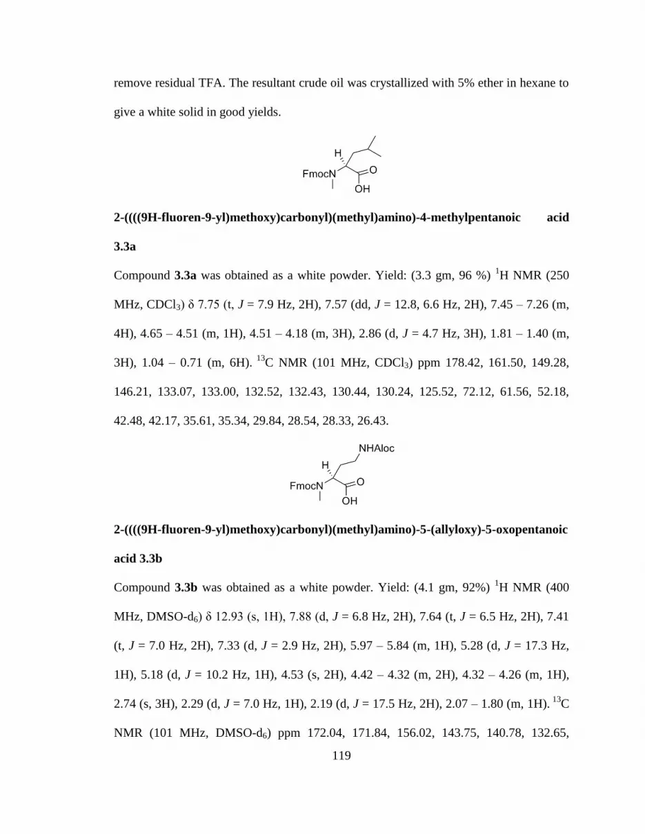
119
remove residual TFA. The resultant crude oil was crystallized with 5% ether in hexane to
give a white solid in good yields.
2-((((9H-fluoren-9-yl)methoxy)carbonyl)(methyl)amino)-4-methylpentanoic acid
3.3a
Compound 3.3a was obtained as a white powder. Yield: (3.3 gm, 96 %) 1H NMR (250
MHz, CDCl3) δ 7.75 (t, J = 7.9 Hz, 2H), 7.57 (dd, J = 12.8, 6.6 Hz, 2H), 7.45 – 7.26 (m,
4H), 4.65 – 4.51 (m, 1H), 4.51 – 4.18 (m, 3H), 2.86 (d, J = 4.7 Hz, 3H), 1.81 – 1.40 (m,
3H), 1.04 – 0.71 (m, 6H). 13
C NMR (101 MHz, CDCl3) ppm 178.42, 161.50, 149.28,
146.21, 133.07, 133.00, 132.52, 132.43, 130.44, 130.24, 125.52, 72.12, 61.56, 52.18,
42.48, 42.17, 35.61, 35.34, 29.84, 28.54, 28.33, 26.43.
2-((((9H-fluoren-9-yl)methoxy)carbonyl)(methyl)amino)-5-(allyloxy)-5-oxopentanoic
acid 3.3b
Compound 3.3b was obtained as a white powder. Yield: (4.1 gm, 92%) 1H NMR (400
MHz, DMSO-d6) δ 12.93 (s, 1H), 7.88 (d, J = 6.8 Hz, 2H), 7.64 (t, J = 6.5 Hz, 2H), 7.41
(t, J = 7.0 Hz, 2H), 7.33 (d, J = 2.9 Hz, 2H), 5.97 – 5.84 (m, 1H), 5.28 (d, J = 17.3 Hz,
1H), 5.18 (d, J = 10.2 Hz, 1H), 4.53 (s, 2H), 4.42 – 4.32 (m, 2H), 4.32 – 4.26 (m, 1H),
2.74 (s, 3H), 2.29 (d, J = 7.0 Hz, 1H), 2.19 (d, J = 17.5 Hz, 2H), 2.07 – 1.80 (m, 1H). 13
C
NMR (101 MHz, DMSO-d6) ppm 172.04, 171.84, 156.02, 143.75, 140.78, 132.65,

120
127.65, 127.10, 125.00, 120.09, 117.70, 117.65, 66.84, 64.45, 58.07, 46.74, 30.96, 30.19,
23.44.
2-((((9H-fluoren-9-yl)methoxy)carbonyl)(methyl)amino)-3-methylbutanoic acid 3.3c
Compound 3.3c was obtained as a white powder. Yield: (3.2 gm, 95%) 1H NMR (400
MHz, DMSO-d6) δ 11.29 (s, 1H), 7.85 (s, 2H), 7.63 (s, 2H), 7.35 (d, J = 29.8 Hz, 4H),
4.40 (d, J = 26.4 Hz, 2H), 4.27 (s, 2H), 2.76 (s, 3H), 2.05 (d, J = 31.9 Hz, 1H), 1.14 –
0.70 (m, 6H). 13
C NMR (101 MHz, DMSO-d6) ppm 171.76, 171.58, 155.86, 155.25,
143.65, 143.51, 140.62, 127.39, 126.84, 124.70, 119.82, 66.61, 63.58, 46.59, 46.53,
29.92, 26.75, 26.63, 19.48, 19.42, 18.58, 18.28.
allyl 1-(9H-fluoren-9-yl)-13,13-dimethyl-3,11-dioxo-2,12-dioxa-4,10-diazatetra-
decane-5-carboxylate 3.5
Fmoc-Lys(Boc)-OH (2.0 gm, 4.2 mmol) was suspended in dry methanol (12 mL) and
cesium carbonate (0.7 gm, 2.1 mmol) was added to it. After forming homogenous
solution, the reactions contents were stirred at room temperature for 2 hrs under inert
atmosphere. The mixture was then concentrated to give white foam, which was
redissolved in DMF (15 mL). Allyl bromide (3.7 mL, 42.4 mmol) was then added and
reaction was stirred overnight. After completion of reactions, the contents were taken up
in 200 mL ethyl acetate and washed with water (1 x 30 mL) and brine (1 x 30 mL). The

121
organic layer was dried over Na2SO4, filtered, concentrated and flash chromatographed to
give compound 3.5 (1.8 gm, 82%) as colorless foam. Rf = 0.25 (1:3 ethyl acetate:
hexane). 1
H NMR (400 MHz, CDCl3) δ 7.71 (d, J = 7.5 Hz, 2H), 7.61 – 7.54 (m, 2H),
7.35 (t, J = 7.4 Hz, 2H), 7.26 (t, J = 7.3 Hz, 2H), 5.85 (ddd, J = 16.3, 10.7, 5.4 Hz, 1H),
5.77 (d, J = 7.0 Hz, 1H), 5.29 (d, J = 17.2 Hz, 1H), 5.20 (d, J = 10.4 Hz, 1H), 4.80 (s,
1H), 4.60 (s, 2H), 4.40 – 4.32 (m, 2H), 4.18 (t, J = 6.8 Hz, 1H), 3.06 (s, 2H), 1.91 – 1.52
(m, 4H), 1.41 (s, 9H), 1.37 (b, 2H). 13
C NMR (101 MHz, CDCl3) ppm 172.10, 155.99,
143.79, 143.63, 141.12, 131.46, 127.55, 126.92, 124.98, 119.81, 118.69, 78.83, 66.84,
65.72, 53.75, 47.02, 39.89, 31.80, 29.42, 28.31, 22.35.
allyl 6-(tert-butoxycarbonylamino)-2-(2-nitrophenylsulfonamido)hexanoate 3.6
Compound 3.5 (1.8 gm, 3.5 mmol) was dissolved in 20 mL DCM and 5 mL 20%
piperidine / 2% DBU was added to it. After complete deprotection of the Fmoc group, the
resulting primary amine was quickly purified by passing over a pad of silica gel and used
in the next step without further purification. The amine (0.94 gm, 3.3 mmol) was taken
up in 20 mL DCM and DIEA (0.84 mL, 4.8 mmol) was added. o-Nitrobenzenesulfonyl
chloride (1.1 gm, 5.0 mmol) in 10 mL DCM was added slowly at 0 oC. The reaction was
stirred for 2 hrs by monitoring the conversion of starting material by TLC. After
completion of the reaction, the crude mixture was further diluted with 50 mL DCM and
washed with (1 x 15 mL) 5% Na2CO3 solution, water (1 x 20 mL) and brine (1 x 20 mL).
The combined organic layers were dried, filtered, concentrated and chromatographed to

122
give compound 3.6 (1.12 gm, 73%). 1H NMR (250 MHz, CDCl3) δ 8.10 – 8.04 (m, 1H),
7.95 – 7.90 (m, 1H), 7.77 – 7.70 (m, 2H), 6.12 (d, J = 9.2 Hz, 1H), 5.67 (ddt, J = 17.3,
10.2, 5.9 Hz, 1H), 5.23 – 5.19 (m, 1H), 5.15 (dd, J = 1.7, 1.2 Hz, 1H), 4.54 (s, 1H), 4.38 –
4.31 (m, 2H), 4.24 – 4.15 (m, 1H), 3.14 – 3.05 (m, 2H), 1.98 – 1.53 (m, 6H), 1.45 (s, 9H).
13C NMR (101 MHz, CDCl3) ppm 170.37, 155.72, 147.19, 133.58, 133.35, 132.61,
130.74, 130.04, 125.01, 118.61, 78.34, 65.64, 56.24, 39.65, 32.12, 28.85, 28.01, 21.96.
allyl 6-(tert-butoxycarbonylamino)-2-(N-methyl-2-nitrophenylsulfonamido)
hexanoate 3.7
Compound 3.6 (1.12 gm, 2.4 mmol) was dissolved in 15 mL DMF and (CH3)2SO4 (0.3
mL, 3.5 mmol), CH3I (0.2 mL, 3.5 mmol) was added along with DBU (0.8 mL, 4.7
mmol) as base. The reaction was completed in 15 mins, as monitored by TLC. The
solvent was removed in vacuo and crude reaction contents were purified by flash
chromatography to give compound 3.7 (1.2 gm, 93%) as a pale yellow oil. 1
H NMR (250
MHz, CDCl3) ppm 8.07 – 8.02 (m, 1H), 7.71 – 7.66 (m, 2H), 7.65 – 7.58 (m, 1H), 5.84 –
5.65 (m, 1H), 5.20 (ddd, J = 9.6, 7.4, 1.3 Hz, 2H), 4.66 (dd, J = 10.6, 4.9 Hz, 1H), 4.54
(s, 1H), 4.50 – 4.44 (m, 2H), 3.17 – 3.06 (m, 2H), 2.97 (d, J = 3.3 Hz, 3H), 1.85 – 1.55
(m, 6H), 1.44 (s, 9H).

123
allyl 1-(9H-fluoren-9-yl)-4,13,13-trimethyl-3,11-dioxo-2,12-dioxa-4,10-diaza
tetradecane-5-carboxylate 3.9
Compound 3.7 (1.15 gm, 2.4 mmol) was dissolved in 10 mL DMF and mercaptoethanol
(1.7 mL, 23.6 mmol) along with DBU (1.8 mL, 11.8 mmol) was added at 0oC. After
completion of reaction in 10 mins, the solvent was removed from reaction mixture and
the crude mixture was quickly purified over pad of silica gel to give secondary amine 3.8
(0.6 gm, 85%). This secondary amine was used without further purification. The
secondary amine (0.14 gm, 0.5 mmol) was dissolved in 6 mL DCM and Fmoc-Cl (0.26
gm, 0.9 mmol), DIEA (0.3 mL, 1.4 mmol) were added. The reaction was stirred
overnight at room temperature. The reaction mixture was concentrated in vacuo and
subjected to flash chromatography to give compound 3.9 (0.18 gm, 77%) as a white
foam. 1H NMR (400 MHz, CDCl3) δ 7.65 (d, J = 7.1 Hz, 2H), 7.57 – 7.43 (m, 2H), 7.31 –
7.19 (m, 4H), 5.86 – 5.71 (m, 1H), 5.26 – 5.09 (m, 2H), 4.96 – 4.71 (m, 1H), 4.55 – 4.42
(m, 2H), 4.40 – 4.28 (m, 2H), 4.19 – 4.08 (m, 1H), 3.07 – 2.93 (m, 2H), 2.76 (d, J = 7.1
Hz, 3H), 1.95 – 1.60 (m, 2H), 1.37 (d, J = 8.6 Hz, 9H), 1.31 – 0.99 (m, 4H). 13
C NMR
(101 MHz, CDCl3) ppm 170.67, 170.34, 156.55, 155.83, 155.70, 144.57, 143.60, 143.47,
140.89, 131.42, 131.35, 127.30, 126.93, 126.67, 126.49, 124.66, 124.61, 124.34, 119.58,
119.44, 118.04, 78.36, 67.27, 66.96, 65.17, 64.69, 58.02, 50.07, 46.82, 39.79, 30.18,
29.89, 29.02, 28.06, 27.81, 22.83.

124
1-(9H-fluoren-9-yl)-4,13,13-trimethyl-3,11-dioxo-2,12-dioxa-4,10-diazatetradecane-
5-carboxylic acid 3.10
Compound 3.9 (0.18 gm, 0.34 mmol) was dissolved in anhydrous 10 mL DCM and
Pd(PPh3)4 (39 mg, 0.03 mmol), NMM (0.17 gm, 1.7 mmol) were added to it. After 2 hrs,
another fresh lot of catalyst (39 mg, 0.03 mmol) was added and reaction was stirred for
another 2 hrs. The reaction contents were concentrated and purified by flash
chromatography to give compound 3.10 (0.13 gm, 80%) as a white solid. 1
H NMR (400
MHz, CDCl3) δ 10.30 (b, 1H), 7.75 (t, J = 8.8 Hz, 2H), 7.62 – 7.53 (m, 2H), 7.42 – 7.27
(m, 4H), 4.81 (dd, J = 10.5, 4.4 Hz, 1H), 4.69 – 4.53 (m, 1H), 4.50 – 4.38 (m, 2H), 4.30 –
4.19 (m, 1H), 3.09 (d, J = 13.2 Hz, 2H), 2.86 (d, J = 10.6 Hz, 3H), 2.03 – 1.69 (m, 2H),
1.44 (d, J = 8.8 Hz, 9H), 1.40 – 1.11 (m, 4H). 13
C NMR (101 MHz, CDCl3) ppm 175.71,
157.28, 156.53, 144.04, 143.88, 141.39, 127.76, 127.13, 125.10, 124.90, 120.02, 67.90,
67.62, 58.39, 47.31, 41.42, 40.28, 30.69, 30.48, 29.41, 28.48, 28.14, 23.37.
3.4 References
1. Haass, C.; Schlossmacher, M. G.; Hung, A. Y.; Vigo-Pelfrey, C.; Mellon, A.;
Ostaszewski, B. L.; Lieberburg, I.; Koo, E. H.; Schenk, D.; Teplow, D. B.; et al.,
Amyloid beta-peptide is produced by cultured cells during normal metabolism.
Nature 1992, 359 (6393), 322-5.
2. Seubert, P.; Vigo-Pelfrey, C.; Esch, F.; Lee, M.; Dovey, H.; Davis, D.; Sinha, S.;
Schlossmacher, M.; Whaley, J.; Swindlehurst, C.; et al., Isolation and
quantification of soluble Alzheimer's beta-peptide from biological fluids. Nature
1992, 359 (6393), 325-7.

125
3. Shoji, M.; Golde, T. E.; Ghiso, J.; Cheung, T. T.; Estus, S.; Shaffer, L. M.; Cai, X. D.;
McKay, D. M.; Tintner, R.; Frangione, B.; et al., Production of the Alzheimer
amyloid beta protein by normal proteolytic processing. Science 1992, 258 (5079),
126-9.
4. Burdick, D.; Soreghan, B.; Kwon, M.; Kosmoski, J.; Knauer, M.; Henschen, A.; Yates,
J.; Cotman, C.; Glabe, C., Assembly and aggregation properties of synthetic
Alzheimer's A4/beta amyloid peptide analogs. J Biol Chem 1992, 267 (1), 546-54.
5. Jarrett, J. T.; Berger, E. P.; Lansbury, P. T., Jr., The carboxy terminus of the beta
amyloid protein is critical for the seeding of amyloid formation: implications for
the pathogenesis of Alzheimer's disease. Biochemistry 1993, 32 (18), 4693-7.
6. Vigo-Pelfrey, C.; Lee, D.; Keim, P.; Lieberburg, I.; Schenk, D. B., Characterization of
beta-amyloid peptide from human cerebrospinal fluid. J Neurochem 1993, 61 (5),
1965-8.
7. Walsh, D. M.; Tseng, B. P.; Rydel, R. E.; Podlisny, M. B.; Selkoe, D. J., The
oligomerization of amyloid beta-protein begins intracellularly in cells derived
from human brain. Biochemistry 2000, 39 (35), 10831-9.
8. Pike, C. J.; Walencewicz, A. J.; Glabe, C. G.; Cotman, C. W., In vitro aging of beta-
amyloid protein causes peptide aggregation and neurotoxicity. Brain Res 1991,
563 (1-2), 311-4.
9. Kirschner, D. A.; Abraham, C.; Selkoe, D. J., X-ray diffraction from intraneuronal
paired helical filaments and extraneuronal amyloid fibers in Alzheimer disease
indicates cross-beta conformation. Proc. Natl. Acad. Sci. U. S. A. 1986, 83 (2),
503-7.
10. Benzinger, T. L. S.; Gregory, D. M.; Burkoth, T. S.; Miller-Auer, H.; Lynn, D. G.;
Botto, R. E.; Meredith, S. C., Two-Dimensional Structure of beta -Amyloid(10-
35) Fibrils. Biochemistry 2000, 39 (12), 3491-3499.
11. Amijee, H.; Madine, J.; Middleton, D. A.; Doig, A. J., Inhibitors of protein
aggregation and toxicity. Biochem. Soc. Trans. 2009, 37 (4), 692-696.
12. Tjernberg, L. O.; Naeslund, J.; Lindqvist, F.; Johansson, J.; Karlstroem, A. R.;
Thyberg, J.; Terenius, L.; Nordstedt, C., Arrest of beta -amyloid fibril formation
by a pentapeptide ligand. J. Biol. Chem. 1996, 271 (15), 8545-8.
13. Soto, C.; Kindy, M. S.; Baumann, M.; Frangione, B., Inhibition of Alzheimer's
amyloidosis by peptides that prevent beta -sheet conformation. Biochem. Biophys.
Res. Commun. 1996, 226 (3), 672-680.
14. Poduslo, J. F.; Curran, G. L.; Kumar, A.; Frangione, B.; Soto, C., beta -sheet breaker
peptide inhibitor of Alzheimer's amyloidogenesis with increased blood-brain

126
barrier permeability and resistance to proteolytic degradation in plasma. J.
Neurobiol. 1999, 39 (3), 371-382.
15. Soto, C.; Sigurdsson, E. M.; Morelli, L.; Kumar, R. A.; Castano, E. M.; Frangione,
B., beta -Sheet breaker peptides inhibit fibrillogenesis in a rat brain model of
amyloidosis: implications for Alzheimer's therapy. Nat. Med. (N. Y.) 1998, 4 (7),
822-826.
16. Ghanta, J.; Shen, C. L.; Kiessling, L. L.; Murphy, R. M., A strategy for designing
inhibitors of beta-amyloid toxicity. J Biol Chem 1996, 271 (47), 29525-8.
17. Pallitto, M. M.; Ghanta, J.; Heinzelman, P.; Kiessling, L. L.; Murphy, R. M.,
Recognition sequence design for peptidyl modulators of beta -amyloid
aggregation and toxicity. Biochemistry 1999, 38 (12), 3570-3578.
18. Lowe, T. L.; Strzelec, A.; Kiessling, L. L.; Murphy, R. M., Structure-Function
Relationships for Inhibitors of beta -Amyloid Toxicity Containing the
Recognition Sequence KLVFF. Biochemistry 2001, 40 (26), 7882-7889.
19. Bodles Angela, M.; El-Agnaf Omar, M. A.; Greer, B.; Guthrie David, J. S.; Irvine, G.
B., Inhibition of fibril formation and toxicity of a fragment of alpha-synuclein by
an N-methylated peptide analogue. Neurosci Lett 2004, 359 (1-2), 89-93.
20. Gordon, D. J.; Meredith, S. C., Probing the Role of Backbone Hydrogen Bonding in
beta -Amyloid Fibrils with Inhibitor Peptides Containing Ester Bonds at Alternate
Positions. Biochemistry 2003, 42 (2), 475-485.
21. Kapurniotu, A.; Schmauder, A.; Tenidis, K., Structure-based design and study of non-
amyloidogenic, double N-methylated IAPP amyloid core sequences as inhibitors
of IAPP amyloid formation and cytotoxicity. J. Mol. Biol. 2002, 315 (3), 339-350.
22. Kokkoni, N.; Stott, K.; Amijee, H.; Mason Jody, M.; Doig Andrew, J., N-Methylated
peptide inhibitors of beta-amyloid aggregation and toxicity. Optimization of the
inhibitor structure. Biochemistry 2006, 45 (32), 9906-18.
23. Yan, L.-M.; Tatarek-Nossol, M.; Velkova, A.; Kazantzis, A.; Kapurniotu, A., Design
of a mimic of nonamyloidogenic and bioactive human islet amyloid polypeptide
(IAPP) as nanomolar affinity inhibitor of IAPP cytotoxic fibrillogenesis. Proc.
Natl. Acad. Sci. U. S. A. 2006, 103 (7), 2046-2051.
24. Manavalan, P.; Momany, F. A., Conformational energy studies on N-methylated
analogs of thyrotropin releasing hormone, enkephalin, and luteinizing hormone-
releasing hormone. Biopolymers 1980, 19 (11), 1943-73.
25. Gordon, D. J.; Sciarretta, K. L.; Meredith, S. C., Inhibition of beta -Amyloid(40)
Fibrillogenesis and Disassembly of beta -Amyloid(40) Fibrils by Short beta -

127
Amyloid Congeners Containing N-Methyl Amino Acids at Alternate Residues.
Biochemistry 2001, 40 (28), 8237-8245.
26. Gordon, D. J.; Tappe, R.; Meredith, S. C., Design and characterization of a membrane
permeable N-methyl amino acid-containing peptide that inhibits Abeta 1-40
fibrillogenesis. J. Pept. Res. 2002, 60 (1), 37-55.
27. Hughes, E.; Burke, R. M.; Doig, A. J., Inhibition of toxicity in the beta -amyloid
peptide fragment beta -(25-35) using N-methylated derivatives: a general strategy
to prevent amyloid formation. J. Biol. Chem. 2000, 275 (33), 25109-25115.
28. Doig, A. J., A three stranded beta -sheet peptide in aqueous solution containing N-
methyl amino acids to prevent aggregation. Chem. Commun. (Cambridge) 1997,
(22), 2153-2154.
29. Mahalaxmi, R.; Balaram, P. Methods in Molecular Biology, vol. 340: Protein Design:
Methods and Applications, R.Guerois and M.L. de la Paz , Eds., Humana Press
Inc., Totowa, NJ.
30. Cammish, L. E.; Kates, S. A. Instrumentation for automated solid phase peptide
synthesis. In Fmoc Solid Phase Peptide Synthesis, Chan, W. C. and White, P. D.
Eds., Oxford, 2000, pp 277-302.
31. Tantry, S. J.; Kantharaju; Suresh Babu, V. V., Microwave accelerated efficient
synthesis of N-fluorenylmethoxycarbonyl/t-butoxycarbonyl/benzyloxycarbonyl-5-
oxazolidinones. Tetrahedron Lett. 2002, 43 (51), 9461-9462.
32. .Freidinger, R. M.; Hinkle, J. S.; Perlow, D. S., Synthesis of 9-
fluorenylmethyloxycarbonyl-protected N-alkyl amino acids by reduction of
oxazolidinones. J. Org. Chem. 1983, 48 (1), 77-81.
33. Ben-Ishai, D., Reaction of acylamino acids with paraformaldehyde. J. Am. Chem.
Soc. 1957, 79, 5736-8.
34. Zhang, S.; Govender, T.; Norstroem, T.; Arvidsson, P.I., An improved synthesis of
Fmoc-N-methyl-α-amino acids. J. Org. Chem. 2005, 70(17), 6918-6920.
35. Biron, E.; Chatterjee, J.; Kessler, H., Optimized selective N-methylation of peptides
on solid support. J. Pept. Sci. 2006, 12 (3), 213-219.
36. Bowman, W. R.; Coghlan, D. R., A facile method for the N-alkylation of alpha -
amino esters. Tetrahedron 1997, 53 (46), 15787-15798.
37. Reichwein, J. F.; Liskamp, R. M. J., Site-specific N-alkylation of peptides on the
solid phase. Tetrahedron Lett. 1998, 39 (10), 1243-1246.

128
38. Fukuyama, T.; Jow, C.-K.; Cheung, M., 2- and 4-nitrobenzenesulfonamides:
exceptionally versatile means for preparation of secondary amines and protection
of amines. Tetrahedron Lett. 1995, 36 (36), 6373-4.

129
CHAPTER FOUR:
CYCLIC β- HAIRPIN PEPTIDOMIMETICS: TARGETING p53-MDM2
PROTEIN-PROTEIN INTERACTIONS
4.1 Introduction
The tumor suppressor protein p53 plays a pivotal part in protection from tumor
development that arises due to various forms of cellular stress pathways such as DNA
damage, hypoxia, telomere shortening etc (1,2). p53 is a potent transcription factor that
regulates multiple downstream genes implicated in cell cycle control, apoptosis,
antiangiogenesis and senescence. It is estimated that half of all human tumors express
p53 is disabled by mutations in its DNA-binding domain and becomes inactive as a
transcription factor (3). Growth suppressive and proapoptotic activity of p53 could harm
proliferating cells that are not under stress (4). The levels of p53 in cells are controlled by
its negative regulator MDM2 (an oncoprotein) through an autoregulatory feedback loop
mechanism (5,6). Under cellular stress, nuclear p53 level gets elevated that
transcriptionally transactivates the MDM2 gene, resulting in MDM2 binding to p53
(fig.4.1). This results in an increase in MDM2 expression. In turn, MDM2 binds to p53,
blocks its transactivation domain and transports p53 to the cytoplasm where MDM2, acts
as an E3 ubiquitin protein ligase and promotes p53 ubiquitination and degradation by the
proteaosome (7-14). On response to cellular stress, p53 gets phosphorylated on specific
serine residue near the MDM2 binding domain, thereby decreasing the affinity of p53 for

130
MDM2 (15). Over expression of MDM2 inhibits p53 function, leading to uncontrolled
proliferation of tumor cells. These tumor cells become resistant to standard
chemotherapeutic treatment and do not undergo programmed cell death or apoptosis.
Fig. 4.1 Modulation of p53-MDM2 interactions. Cellular stress triggers the p53 response
pathway. The p53-MDM2 autoregulatory feedback loop modulates the amount of p53.
Over expression of MDM2 in human cancer leads to p53 ubiquitination to disable the p53
network. Designed inhibitors should complex with MDM2 and block its interaction with
p53.
MDM2 interacts through its NH2 terminal domain with an α-helix present in the
NH2 terminal transactivation domain of p53 and blocks its transcriptional activity
directly. The hydrophobic side of the p53 α-helix formed by the amino acids
phenylalanine, tryptophan and leucine fit deeply into the hydrophobic cleft of MDM2.
Since MDM2 is seen to be over expressed in certain cancers, it may reduce the functional
activity of p53-dependent cancer therapies. Hence disruption of p53/MDM2 interaction is
a viable therapeutic target for the treatment of cancer. Various small molecule inhibitors,
retroinverso peptides, peptoids, terphenyls, β-hairpins, p-oligobenzamides, β-peptides
have been developed to mimic the recognition surface of the α-helix of p53. These

131
inhibitors occupy the p53-binding pocket in MDM2, thereby preventing protein-protein
interaction, and thus, facilitate the p53 tumor suppressor function to inhibit cancer cells.
Among small molecule inhibitors, Nutlin (cis-imidazolines) emerged as a potent and
selective small molecule antagonist of p53/MDM2 interaction with both in vitro and in
vivo activity (16). Figure 4.2. shows some promising small molecule and proteomimetic
scaffold based inhibitors for p53/MDM2 interaction.
Fig. 4.2 Small molecule and peptidomimetic inhibitors of p53/MDM2 interactions.
Robinson mimicked the α-helix of p53 with a cyclic β-hairpin to inhibit
p53/MDM2 interaction. The cyclic beta-hairpin scaffold mounted on DPro-
LPro template
holds the side chains of phenylalanine and tryptophan residues in the correct relative

132
positions, so that they could interact with their respective binding sites on MDM2 (17).
This scaffold exhibited poor cellular uptake. In this present work, we have made an
attempt to synthesize cyclic β-hairpin peptidomimetics mimicking the α-helix of p53
using our novel β- turn promoter, to disrupt p53/MDM2 protein-protein interactions. The
incorporation of novel β- turn promoter will further facilitate in stabilizing the cyclic
β-hairpin. We have also made an attempt to synthesize morpholino derivatives of serine
and lysine and incorporate them into the non-recognition strand of the proposed cyclic
β-hairpin with an aim to improve the bioavailability and cellular permeability of the
cyclic peptides. The synthesis of the chiral β-turn promoters have also been attempted to
improve the binding affinity of these molecules to MDM2.
4.2 Results & Discussions
4.2.1 Peptide Design
The crystal structure of a p53-derived peptide (residues 15-29) in complex with
the inhibitory domain of MDM2 (residue 17-125) as revealed by Pavletich and workers
suggested that the peptide adopts an α-helical backbone conformation (18). The side
chains of Phe19, Trp23 and Leu26 align along one face of the helix and inserts deep into
hydrophobic clefts on the surface of MDM2 (fig.4.3a). The binding energy for this
protein-protein interaction arises due to van der Waals interactions of these three p53 side
chains with the surface of MDM2. The C-terminal end of the helix is less tightly bound
leading to a type I β-turn for the segment between Trp23 and Leu26. Robinson and
coworkers found that the distance between Cα atoms of Phe19 and Trp23 residues on one
face of the MDM2-bound p53 α-helix is same as the distance between Cα atoms of ith
and ith+2 residues along one strand (recognition strand) of a β-hairpin. They proposed a

133
cyclic β-hairpin scaffold that would hold Trp and Phe residues in the correct relative
positions so that they could interact with p53 binding site on MDM2. Our design for
making cyclic β-hairpin peptides as p53-MDM2 inhibitors involves, Phe19, Trp23 and
Leu26 residues aligned along one face of the recognition strand. We hypothesized that
incorporation of a novel sulfonamido aminoethyl glycine β-turn promoter at the turns
would further facilitate in stabilizing the β-hairpin conformation. As shown in Fig.4.3 (b),
β-hairpin mimetic designed as scaffold for p53-MDM2 inhibitors was found to be a weak
lead with an IC50 greater than 150 µM in an ELISA assay.
(A)

134
(B)
Fig. 4.3 (A) Crystal structure of complex consisting of a p53-derived peptide and MDM2.
(B) Proposed β-hairpin mimetic scaffold with novel sulfonamido aminoethyl glycine turn
promoters.
In an attempt to design bioactive peptides for inhibiting p53-MDM2 interaction
we tried to shorten the β-hairpin loop to eight residue loop as shown in (Table.4.1). The
eight residue loop with Leu and glutamic acid residues populated along the β turn was
preorganized into a regular β-hairpin. We found that this scaffold is bioactive with an
IC50 of 35 µM. With this design as our lead scaffold, we tried to study structure activity
relationship with an aim to obtain β-hairpin mimetics with enhanced bioactivity.
Table 4.1 depicts the structure activity relationship of designed β-hairpin mimetics as
p53-MDM2 inhibitors. After obtaining peptide 4.1 as lead molecule we tried to replace
aspartic acid with leucine at the β-turn. We found that this mimetic had IC50 of 2 µM. In
another attempt, we tried to populate the β-turn with Leu and Trp residues. Here we tried
to populate the three residues around the β-turn so that we could achieve maximum
binding.

135
Table 4.1 Structure activity relationship of cyclic β-hairpin peptidomimetics.
SM = Morpholine derivatized serine ND= Not determined yet
To check the bioavailability of the designed cyclic peptidomimetics, we carried
out quikPro calculation for log P data for previously designed cyclic abeta and cyclic III
peptides. We found that designed cyclic peptidomimetics are too hydrophilic with log P
outside the range of acceptable limits. We proposed to decrease the hydrophilicity by
incorporating morpholine derivatized lysine and serine derivatives into the non-
recognition strand of the peptidomimetics. This more hydrophobic lysine analog reduces
the basic character of the epsilon nitrogen and removes two of the three hydrogen bond
donors in the physiologically relevant protonated form of this side chain. As seen from
Table 4.2, incorporation of the morpholine derivative brings the log P values for cyclic
Peptide R1 R2 R3 R4 R5 R6 R7 R8 IC50 (µM)
4.1 W K F F K W D L 35
4.2 W K F F K W L L 2
4.3 W SM F F SM W L L >125
4.4 F K F L K W L W ND

136
abeta and cyclic III peptides within the range of acceptable limits. To test this idea, we
synthesized cyclic peptide 4.3 with the lysine replaced with the morpholine derivative.
The peptide was synthesized successfully as characterized initially by MALDI-TOF but
isolation and purification problems were encountered. We tested the crude peptide for
p53-MDM2 inhibition, which did not give us expected results.
Table 4.2 QuikPro calculations for LogP data for cyclic abeta and cyclic III peptides.
O= Methylsulfonamido aminoethyl glycine turn promoter
In a quest for obtaining improved bioactive peptides we further constrained the
cyclic peptide by introduction of a constrained oxygenated turn promoter (fig. 4.4) at one
turn. The introduction of an ether-peptidomimetic amino acid (proline or 2-piperidine
carboxylic acid derivative) as a constrained turn promoter will further reduce the degrees
of freedom available to the cyclic peptide and possibly increase its affinity for binding to
the target. As shown in figure 4.4, this cyclic peptidomimetic had an IC50 of 2 µM,
similar to that of peptide 4.2.
Peptide Log P (Octanol/Water)
Cyclo (7,8) LP
1 DP
2 K
3 L
4 K
5 L
6 K
7 O
8 M
9 V
10 I
11 S
12 W
13 -3.078
Cyclo (7,8) LP
1 DP
2 KM
3 L
4 KM
5 L
6 KM
7 O
8 M
9 V
10 I
11 S
12 W
13 -1.369
Cyclo (1,12) M1 V
2 I
3 S
4 W
5 O
6 K
7 L
8 K
9 L
10 K
11 O
12 -4.224
Cyclo (1,12) M1 V
2 I
3 S
4 W
5 O
6 KM
7 L
8 KM
9 L
10 KM
11 O
12 -2.445
Cyclo (7,8) LP
1 DP
2 K
3 L
4 K
5 L
6 K
7 O
8 W
9 S
10 V
11 V
12 M
13 -3.077
Cyclo (7,8) LP
1 DP
2 KM
3 L
4 KM
5 L
6 KM
7 O
8 W
9 S
10 V
11 V
12 M
13 -0.818

137
Fig. 4.4 Cyclic betahairpin peptide with proline derived ether-peptidomimetic turn
promoter.
To further improve our lead scaffold, we designed a chiral reduced amide
dipeptide β-turn promoter decorated with different alkyl groups at the turn as shown in
fig. 4.5a. To support our design, we did computational studies by incorporating these
reduced amide dipeptide β-turn promoters into the eight residue β-hairpin scaffold. It was
seen that chiral turn promoter populated with tryptophan residues at the turn inserts deep
into the hydrophobic pocket of MDM2 (fig. 4.5c). When superimposed, this mimetic had
side chain residues perfectly aligned with the corresponding ligand residues as shown in
Fig.4.5b. Fig.4.5c shows the docking and interactions of this peptide in the active site of
MDM2. The key to this mimetic lies in the synthesis of the reduced amide dipeptide β-
turn promoter. An attempt to make the reduced amide dipeptide β-turn promoter with Leu
side chains is described.

138
(A)
(B)

139
(C)
Fig. 4.5 (a) Proposed cyclic p53-MDM2 peptidomimetics with chiral reduced amide
dipeptide β-turn promoter with leucine residues (b) reduced amide dipeptides β-turn
promoter superimposed with ligand (PDB 2axi). (c) proposed cyclic p53-MDM2 hairpin
peptide docked into the active site of MDM2 (PDB 2axi).
All cyclic peptidomimetics were synthesized on 2-chlorotrityl chloride resin as
solid support and standard solid phase Fmoc amino acid chemistry as described in
chapter 2. Coupling was carried out in most cases using standard chemistry of HCTU in
N-methyl pyrrolidone (NMP) with double coupling for each residue. The synthesized
linear peptides were selectively cleaved from the resin without cleaving Boc groups on
certain residues using dilute trifluoroethanol as the cleaving agent. The linear peptide is
then cyclized in solution under dilute conditions to afford crude cyclized peptide in
modest yields.

140
Scheme 4.1 Synthesis of Morpholino derivative of lysine.
The morpholino derivative of lysine was synthesized from commercially available
CbzLys(Boc)-OH as shown in scheme 4.1. The key step in this scheme was the formation
of morpholine ring. Compound 4.2 was subjected to Boc-deprotection using TFA/DCM
followed by reflux of resulting amine with 2-bromo ethyl ether to give key compound 4.3
in 78% yield. Compound 4.3 was hydrogenated using 10% Pd/C in methanol to give
completely deprotected amino acid 4.4. The primary amine was then protected with a
Fmoc protecting group using Bolin’s procedure to give desired morpholine derivative 4.5
in low yields. Hence an alternative route of preparing morpholine derivatives of serine
was investigated using two different protecting group strategies.

141
Scheme 4.2 Synthetic routes for morpholino derivative of serine (a) using Cbz protecting
groups (b) using Boc/Cbz protecting group strategy.
In scheme 4.2(a), the morpholino compound 4.10 was synthesized by
transformation of hydroxyl group to iodide followed by SN2 displacement of iodide with
morpholine. Compound 4.10 is subjected to hydrogenation followed by Fmoc protection
of resulting amine to give desired compound 4.11 in moderate yield 49%. An alternative
strategy was formulated to maximize yield for compound 4.11. Scheme 4.2b describes
the synthesis of the morpholino derivative of serine using commercially available Boc
Ser-OH 4.12. Compound 4.15 was synthesized using the same procedure as described for

142
the preparation of compound 4.10. Compound 4.14 was then subjected to Boc
deprotection using TFA/DCM followed by protection of the resulting amine with Fmoc
group to give fully protected morpholino derivative 4.16. Hydrogenation of 4.16 with
10% Pd/C gave desired compound 4.11 in good yields.
Scheme 4.3 Synthesis of proposed reduced amide dipeptides β-turn promoter populated
with Leu side chains.
The synthesis of reduced amide dipeptides β-turn promoter 4.22 was carried out
by making a dipeptide unit 4.18. Compound 4.18 was then reduced using 65% Vitride in
toluene to give compound 4.19. The secondary amine was selectively protected with
ortho-nitrobenezenesulfonyl chloride (ONBSCl) to give compound 4.20 in good yield.
Oxidation of the resulting alcohol 4.20 using TEMPO and trichloroisocyanuric acid as
oxidizing agent gave the corresponding acid 4.21 in low yield (19). We did not achieve

143
success in synthesizing Fmoc-protected amino acid 4.22 using standard Bolin’s
procedure.
4.3. Experimental Procedures
4.3.1 Materials and Methods
Organic and inorganic reagents (ACS grade) were obtained from commercial
sources and used without further purification, unless otherwise noted. Fmoc-protected
amino acids and the coupling agent HCTU were obtained from Protein Technologies,
Calbiochem-Novabiochem, or Chem-impex International. 2-Chlorotrityl chloride resin
was purchased from Anaspec Inc. All linear peptides were synthesized on the Symphony
peptide synthesizer, Protein Technologies Instruments. Solvents for peptide synthesis and
reverse-phase HPLC were obtained from Applied Biosystems. Other chemicals used
were obtained from Aldrich and were of highest purity commercially available. Thin
layer chromatography (TLC) was performed on glass plates (Whatman) coated with 0.25
mm thickness of silica gel 60Å (# 70-230 mesh). All 1H and
13C NMR spectra were
recorded on Bruker 250 MHz, Varian INOVA 400 MHz spectrometer in CDCl3 or unless
otherwise specified and chemical shifts are reported in ppm (δ) relative to internal
standard tetramethylsilane (TMS). High Resolution mass spectra were obtained on an
Agilent LC-MSD-TOF.
4.3.2 Peptide Synthesis & Purification
Cyclic β-hairpin peptidomimetic with (2-aminoethyl)-N-methylsulfonamido glycine
beta-turn promoter.
2-Chlorotrityl chloride resin was treated with Fmoc-Phe-OH and then
immediately Fmoc-deprotected using 20% piperidine/2% DBU in DMF. Fmoc

144
quantification of resin indicated a loading of 0.18 mmol/g of resin. For a 25 μmol
synthesis, 139 mg of resin was charged to the peptide reaction vessel on a Protein
Technologies Symphony Peptide Synthesizer. For each coupling step, 5 equivalents of
Fmoc amino acid and 7.5 equivalents of HCTU are dissolved in 0.4 M NMM in DMF to
equal 20 equivalents of NMM, which is added to the reactor. Each coupling reaction was
carried out for 10 minutes followed by NMP washes. Fmoc deprotection was done using
20% piperidine/2% DBU in DMF for (2 x 2.5 minutes). The amino acids used for peptide
synthesis were coupled in the following order: Fmoc-Lys(Boc)-OH, Fmoc-Trp(Boc)-OH,
Fmoc-Leu-OH, Fmoc-Leu-OH, Fmoc-Trp(Boc)-OH Fmoc-Lys(Boc)-OH, Fmoc-Phe-OH
and Fmoc-NHCH2CH2N(O2SCH3)CH2COOH. After synthesis of the protected linear
peptide, the resin was transferred to a manual peptide synthesis vessel and treated with 5
mL of a cleavage solution of 20% trifluoroethanol in DCM for 2 hours. The resin was
filtered and washed with 5 mL of cleavage solution. This cleavage cycle was repeated
twice. The combined organic filtrates were concentrated to give crude protected linear
peptide. The crude peptide was dissolved in 15 mL of 1% v/v DIEA in DMF and treated
with 4 equivalents of HCTU for one hour. After one hour, the reaction mixture was
concentrated to give crude protected cyclized β-hairpin peptidomimetic. The crude
peptidomimetic was then treated with a 10 mL solution of 87.5% TFA/5% H2O/5%
phenol/2.5% triethylsilane for 30 minutes. The reaction mixture was concentrated and the
thick viscous liquid was triturated twice with 10 mL of cold diethyl ether. The reaction
contents were centrifuged to give crude cyclic β-hairpin peptidomimetic. The crude
peptidomimetic was dissolved in a solution of 0.1% TFA in H2O and freeze-dried to give
a white fluffy powder. All cyclic peptides were purified using preparative reverse phase

145
HPLC (5 µM particle size C18 AAPPTEC spirit column, 25 x 2.12 cm) with eluents: A =
0.1% HCO2H in H2O, B = 0.1% HCO2H in H3CCN. The purification was carried out
using a gradient of 5-50% B Buffer over 40 min with a flow rate 20 mL/minute using 222
nm UV detection. All peaks with retention times expected for peptides were collected
and lyophilized. The purified peptides were analyzed using similar analytical HPLC
conditions and found to have >95% purity and were structurally characterized using a
Bruker Autoflex MALDI-TOF instrument with α-cyano hydroxy cinnamic acid (CHCA)
as matrix.
N-α-benzyloxycarbonyl-N-ε-(tert-butoxycarbonyl)lysine benzyl ester (4.2)
N-ε-(tert-butoxycarbonyl)-N-α-carbobenzyloxylysine 4.1(1 gm, 2.6 mmol) was dissolved
in 25 mL dry acetonitrile and diisopropylethylamine (0.47 mL, 2.8 mmol) was added.
Benzyl bromide (0.33 mL, 2.8 mmol) was subsequently added and the reaction mixture
was stirred under argon at room temperature for two days. The reaction mixture was
concentrated in vacuo and the residue was partitioned between DCM (25 mL) and water
(15 mL). The organic layer was dried using Na2SO4, filtered and concentrated to give a
clear oil. The crude oil was chromatographed over flash silica with EtOAc/Hexane (1:1)
to give compound 4.2 (0.95 gm, 78%). 1
H NMR (400 MHz, CDCl3) ppm 7.43 – 7.24 (m,
10H), 5.42 (s, 1H), 5.23 – 5.06 (m, 4H), 4.53 (b, 1H), 4.40 (d, J = 4.9 Hz, 1H), 3.12 –
2.94 (m, 2H), 1.88 – 1.63 (m, 2H), 1.46 – 1.25 (m, 13H). 13
C NMR (101 MHz, CDCl3) δ
172.35, 156.13, 136.33, 135.40, 128.73, 128.61, 128.44, 128.27, 79.24, 67.23, 67.10,
53.89, 40.11, 32.25, 29.65, 28.52, 22.36.

146
(R)-benzyl 2-(benzyloxycarbonylamino)-6-morpholinohexanoate (4.3)
Compound 4.2 (0.95 gm, 2 mmol) was dissolved in 20 mL DCM and 10 mL
trifluoroacetic acid was added to it. The reaction contents were stirred until the starting
material was completely consumed. The reaction mixture was concentrated to dryness in
vacuo and used directly in the next step without further purification. The deprotected
amine was suspended in 20 mL toluene and sodium bicarbonate (0.55 gm, 6 mmol),
2-bromoethyl ether (0.56 gm, 2.4 mmol) was added to it. The system was flushed with
argon and reaction contents were refluxed for 24 hrs. The reaction mixture was
concentrated in vacuo and the residue was partitioned between EtOAc (25 mL) and dilute
NaHCO3 (15 mL). The organic layer was dried using Na2SO4, filtered and concentrated to
give crude oil. The crude oil was chromatographed over flash silica with EtOAc:Hexane
(2:1) to give compound 4.3 (0.7 gm, 79%). 1
H NMR (250 MHz, DMSO) ppm 7.80 (d, J =
7.7 Hz, 1H), 7.48 – 7.19 (m, 10H), 5.13 (t, J = 5.1 Hz, 2H), 5.09 – 4.97 (m, 2H), 4.16 –
3.98 (m, 1H), 3.62 – 3.46 (m, 4H), 2.51 (dt, J = 3.5, 1.7 Hz, 1H), 2.34 – 2.24 (m, 4H),
2.19 (t, J = 6.8 Hz, 2H), 1.69 (dt, J = 15.2, 6.3 Hz, 2H), 1.48 – 1.20 (m, 4H). 13
C NMR
(63 MHz, DMSO) ppm 172.33, 156.16, 136.89, 135.94, 128.39, 128.33, 128.03, 127.79,
127.73, 127.23, 66.14, 65.84, 65.47, 57.99, 53.89, 53.29, 30.52, 25.30, 23.29. m/z
441.2377 (M+H)+

147
(S)-2-amino-6-morpholinohexanoic acid (4.4)
Compound 4.3 (0.7 gm, 1.6 mmol) was dissolved in MeOH and 10% Pd/C was added to
it. Hydrogenation was carried overnight at 50 psi. The reaction contents were filtered
over diatomaceous earth and concentrated to give compound 4.4. This compound was
used in the next step without further purification.
(S)-2-(((9H-fluoren-9-yl)methoxy)carbonylamino)-6-morpholinohexanoic acid (4.5)
Compound 4.4 (0.32 gm, 1.5 mmol) was suspended in 15 mL anhydrous DCM and DIEA
(0.73 mL, 4.19 mmol), TMSCl (0.53 mL, 4.2 mmol) were added to it. The reaction
mixture became homogenous on gentle reflux for one hour. After one hour, the reaction
mixture was cooled to 0 0C, and Fmoc-OSu (0.71 gm, 2.1 mmol) was added. The
progress of the reaction was monitored by TLC. The reaction content was concentrated
and flash chromatographed to give compound 4.5 in low yields. m/z 439.2227 (M+H)+
(R)-2-(benzyloxycarbonylamino)-3-hydroxypropanoic acid (4.7)
To a stirred suspension of NaHCO3 (10 gm, 12 mmol) in THF:H2O (50 mL) was added
L-serine (5 gm, 47.5 mmol) in small lots. After 15 mins, benzyl chloroformate (7.5 mL,
52.5 mmol) was added dropwise over a period of 30 mins. The reaction was carried out

148
for 2 hrs and then diethyl ether (50 mL) was added to it. The aqueous layer was acidified
to pH 2~3 with 1N HCl solution and extracted with ethyl acetate (2 x 50 mL). The
combined organic layer was further washed with 1N HCl (25 mL) and brine (50 mL),
dried over Na2SO4. The reaction mixture was concentrated to dryness in vacuo and
further crystallized with ethyl acetate/hexane to give 4.7 (9.9 gm) in 87 % yield. 1H NMR
(250 MHz, MeOD) ppm 7.46 – 7.28 (m, 5H), 5.14 (s, 2H), 4.30 (t, J = 4.4 Hz, 1H), 3.97
– 3.77 (m, 2H). 13
C NMR (101 MHz, CD3OD) ppm 173.72, 158.41, 137.93, 129.37,
128.93, 128.77, 68.01, 67.68, 63.03, 57.58. m/z 240.0866 (M+H)+
(R)-benzyl 2-(benzyloxycarbonylamino)-3-hydroxypropanoate (4.8)
Compound 4.7 (5 gm, 20.8 mmol) was dissolved in dry methanol (25 mL) and Cs2CO3
(3.4 gm, 10.4 mmol) was added to it at 0 0C. After 10 mins, methanol was removed at
reduced pressure and the reaction contents were taken in DMF (30 mL). Benzyl bromide
(20 mL, 167 mmol) was added in excess and the reaction was stirred overnight at room
temperature. After completion of reaction, the reaction mixture was concentrated and
partitioned between DCM (200 mL) and water (50 mL). The organic layer was dried,
filtered, concentrated and chromatographed to give compound 4.8 (5.4 gm) in 79% yield.
1H NMR (400 MHz, CDCl3) ppm 7.46 – 7.30 (m, 10H), 5.71 (s, 1H), 5.22 (s, 2H), 5.12
(s, 2H), 4.55 – 4.43 (m, 1H), 4.09 – 3.88 (m, 3H), 2.11 (s, 1H). 13
C NMR (101 MHz,
CDCl3) ppm 170.49, 156.25, 136.25, 135.30, 128.89, 128.78, 128.49, 128.42, 128.36,
67.76, 67.46, 63.65, 56.40. m/z 330.1324 (M+H)+

149
(S)-benzyl 2-(benzyloxycarbonylamino)-3-iodopropanoate (4.9)
To a stirred solution of 4.8 (2 gm, 6 mmol) in dry DCM (10 mL) was added a pre-mixed
solution of PPh3 (2.38 gm, 9 mmol), iodine (2.31 gm, 9 mmol) and imidazole (0.75 gm,
10.9 mmol) in 30 mL dry DCM. The reaction mixture was stirred at room temperature for
2 hrs. After completion of reaction, the reaction mixture was concentrated in vacuo and
chromatographed using EtOAc:Hexane (1:9) as eluent to give compound 4.9 (1.7 gm)
70% as white solid. 1H NMR (250 MHz, CDCl3) ppm 7.67 – 7.49 (m, 10H), 5.84 (d, J =
7.2 Hz, 1H), 5.42 (s, 2H), 5.33 (d, J = 0.6 Hz, 2H), 4.82 (dt, J = 7.5, 3.7 Hz, 1H), 3.88 –
3.71 (m, 2H). 13
C NMR (63 MHz, CDCl3) ppm 169.15, 155.43, 135.94, 134.72, 128.77,
128.71, 128.66, 128.62, 128.34, 128.16, 68.17, 67.33, 54.04, 7.44. m/z 440.0345 (M+H)+
(R)-benzyl 2-(benzyloxycarbonylamino)-3-morpholinopropanoate (4.10)
To a solution of 4.9 (1.0 gm, 2.3 mmol) in dry DMF (15 mL) were added K2CO3 (0.81
gm, 5.9 mmol) and morpholine (0.45 mL, 5.3 mmol) at room temperature. The reaction
mixture was stirred overnight until TLC showed disappearance of starting material. After
completion of reaction, the reaction mixture was concentrated in vacuo and
chromatographed to give compound 4.10 (0.76 gm) 84% as pale yellow liquid. 1
H NMR
(250 MHz, DMSO d6) ppm 7.80 (d, J = 7.7 Hz, 1H), 7.48 – 7.19 (m, 10H), 5.13 (t, J =

150
5.1 Hz, 2H), 5.09 – 4.97 (m, 2H), 4.16 – 3.98 (m, 1H), 3.62 – 3.46 (m, 4H), 2.51 (dt, J =
3.5, 1.7 Hz, 1H), 2.34 – 2.24 (m, 4H), 2.19 (t, J = 6.8 Hz, 2H), 1.69 (dt, J = 15.2, 6.3 Hz,
2H), 1.48 – 1.20 (m, 4H). 13
C NMR (63 MHz, DMSO d6) ppm 171.48, 155.92, 136.84,
135.90, 128.36, 128.33, 128.00, 127.82, 127.77, 66.03, 65.87, 65.56, 58.54, 53.11, 52.11.
m/z 399.1917 (M+H)+
(S)-2-(((9H-fluoren-9-yl) methoxy) carbonylamino)-3-morpholinopropanoic acid
(4.11)
Compound 4.10 (0.9 gm, 2.3 mmol) was taken in MeOH and 10% Pd/C was added to it.
Hydrogenation was carried overnight at 50 psi. The reaction contents were filtered over
diatomaceous earth and concentrated to give the intermediate zwitterion (0.36 gm, 2.1
mmol). This compound was used in the next step without further purification. This
compound was suspended in 15 mL anhydrous DCM and DIEA (0.73 mL, 4.2 mmol),
TMSCl (0.53 mL, 4.2 mmol) was added to it. The reaction mixture became homogenous
on gentle reflux for one hour. After one hour, the reaction mixture was cooled to 0oC, and
Fmoc-OSu (0.71 gm, 2.1 mmol) was added. The progress of the reaction was monitored
by TLC. The reaction contents were concentrated and flash chromatographed to give
compound 4.11 (0.41 gm, 49%). 1H NMR (400 MHz, CDCl3) ppm 7.74 (d, J = 7.3 Hz,
2H), 7.57 (d, J = 6.6 Hz, 2H), 7.33 (dt, J = 36.2, 6.9 Hz, 4H), 6.01 (s, 1H), 4.43 – 4.31
(m, 2H), 4.27 (s, 1H), 4.17 (t, J = 6.2 Hz, 1H), 3.92 – 3.69 (m, 4H), 3.28 – 2.91 (m, 5H),
2.63 – 2.30 (m, 1H). 13
C NMR (101 MHz, CDCl3) ppm 172.57, 156.14, 144.02, 141.53,

151
127.97, 127.26, 125.27, 120.21, 67.16, 64.86, 58.07, 52.99, 50.44, 47.35. m/z 397.1755
(M+H)+.
(S)-benzyl 2-(tert-butoxycarbonylamino)-3-hydroxypropanoate (4.13)
Compound 4.12 (4.0 gm, 19 mmol) was dissolved in dry methanol (25 mL) and Cs2CO3
(3.2 gm, 9.7 mmol) was added to it at 0oC. After 10 mins, methanol was removed at
reduced pressure and the reaction contents were taken up in DMF (30 mL). Benzyl
bromide (18.6 mL, 156 mmol) was added in excess and the reaction was stirred overnight
at room temperature. After completion of reaction, the reaction mixture was concentrated
and partitioned between DCM (200 mL) and water (50 mL).The organic layer was dried,
filtered, concentrated and chromatographed to give compound 4.13 (5.1 gm) in 87%
yield.. 1H NMR (400 MHz, CDCl3) ppm 7.42 – 7.20 (m, 5H), 5.64 (d, J = 7.4 Hz, 1H),
5.18 (d, J = 20.2 Hz, 2H), 4.37 (s, 1H), 3.89 (dd, J = 41.7, 10.1 Hz, 2H), 3.17 (s, 1H),
1.40 (s, 9H). 13
C NMR (101 MHz, CDCl3) ppm 171.09, 156.05, 135.51, 128.76, 128.56,
128.32, 80.39, 67.46, 63.37, 56.08, 28.48.
(R)-benzyl 2-(tert-butoxycarbonylamino)-3-iodopropanoate (4.14)
To a stirred solution of 4.13 (5.0 gm, 17 mmol) in dry DCM (10 mL) was added a pre-
mixed solution of PPh3 (6.7 gm, 25.5 mmol), iodine (6.4 gm, 25.5 mmol) and imidazole
(0.75 gm, 30.6 mmol) in 100 mL dry DCM. The reaction mixture was stirred at room

152
temperature for 2 hrs. After completion of reaction, the reaction mixture was
concentrated in vacuo and chromatographed using EtOAc:Hexane (1:9) as eluent to give
compound 4.14 (4.8 gm) 70% as a white solid. 1H NMR (400 MHz, CDCl3) ppm 7.46 –
7.25 (m, 5H), 5.36 (d, J = 4.8 Hz, 1H), 5.20 (t, J = 5.1 Hz, 2H), 4.54 (d, J = 3.0 Hz, 1H),
3.56 (dd, J = 11.2, 6.6 Hz, 2H), 1.44 (s, 9H). 13
C NMR (101 MHz, CDCl3) ppm 169.68,
155.04, 135.06, 129.11, 128.85, 128.77, 80.68, 68.16, 53.91, 28.48, 7.99. m/z 428.0383
(M+Na)+.
(S)-benzyl 2-(tert-butoxycarbonylamino)-3-morpholinopropanoate (4.15)
To a solution of 4.13 (3.5 gm, 8.6 mmol) in dry DMF (30 mL) were added K2CO3 (3.1
gm, 22.4 mmol) and morpholine (1.71 mL, 19.8 mmol) at room temperature. The
reaction mixture was stirred overnight until TLC showed disappearance of starting
material. After completion of reaction, the reaction mixture was concentrated in vacuo
and chromatographed to give compound 4.14 (2.65 gm) 84% as a pale yellow liquid.1H
NMR (400 MHz, CDCl3) ppm 7.98 – 7.79 (m, 1H), 7.38 – 7.22 (m, 6H), 5.25 (d, J = 17.4
Hz, 1H), 5.22 – 4.93 (m, 2H), 4.56 (d, J = 18.3 Hz, 1H), 4.27 (d, J = 5.4 Hz, 1H), 3.48 (t,
J = 4.3 Hz, 3H), 2.83 (d, J = 7.5 Hz, 1H), 2.77 (s, 1H), 2.59 (t, J = 9.5 Hz, 2H), 2.35 –
2.20 (m, 4H), 1.40 – 1.25 (m, 9H). m/z 365.2109 (M+H)+.

153
(S)-benzyl 2-(((9H-fluoren-9-yl)methoxy)carbonylamino)-3-morpholinopropanoate
(4.16)
Compound 4.15 (0.79 gm, 2.2 mmol) was dissolved in 10 mL DCM and 10 mL TFA was
added to it. After completion of the reaction, the residue was reconcentrated three times
with DCM to remove residual TFA. The crude residue (1.06 gm, 2.2 mmol) was
dissolved in 20 mL DCM and DIEA (1.32 mL, 7.6 mmol), FmocOSu (0.69 gm, 2.1
mmol) was added. After completion of the reaction, the reaction contents were diluted
with 50 mL DCM and washed with water (1 x 15 mL) and brine (1 x 15 mL). The
organic layer was then dried, filtered, concentrated and subjected to flash
chromatography to give compound 4.16 (0.95 gm, 91%).1H NMR (250 MHz, CDCl3)
ppm 7.68 (d, J = 7.2 Hz, 2H), 7.52 (d, J = 6.4 Hz, 2H), 7.39 – 7.14 (m, 9H), 5.60 (d, J =
6.2 Hz, 1H), 5.11 (dd, J = 29.2, 11.8 Hz, 2H), 4.33 (d, J = 6.6 Hz, 3H), 4.16 (s, 1H), 3.49
(s, 4H), 2.64 (d, J = 4.7 Hz, 2H), 2.29 (s, 4H).13
C NMR (63 MHz, CDCl3) ppm 171.59,
155.97, 143.95, 143.77, 141.34, 135.22, 129.26, 128.63, 127.76, 127.10, 125.11, 120.04,
67.27, 67.08, 66.85, 59.09, 53.74, 52.36, 47.18 m/z 487.2095 (M+H)+.
(S)-2-(((9H-fluoren-9-yl)methoxy)carbonylamino)-3-morpholinopropanoic acid
(4.11)

154
Compound 4.16 (0.5 gm, 1.6 mmol) was taken up in MeOH and 10% Pd/C was added to
it. Hydrogenation was carried overnight at 50 psi. The reaction contents were filtered
through diatomaceous earth and concentrated to give compound 4.11 (0.35 gm, 87%).
m/z 397.1755 (M+H)+.
(S)-tert-butyl 2-((R)-2-(tert-butoxycarbonylamino)-4-methylpentanamido)-4-
methylpentanoate (4.18)
Dipeptide 4.18 was synthesized from Boc-Leu-OH monohydrate (3.0 gm, 13 mmol) and
leucine tert-butyl ester hydrochloride (2.9 gm, 13 mmol) as per the procedure for mixed
anhydride coupling using N-methylmorpholine (4.3 mL, 39 mmol) and isobutyl
chloroformate (1.7 mL, 13 mmol) using THF (60 mL) as solvent. After completion of the
reaction, reaction contents were washed with water (1 x 25 mL), brine (1 x 25 mL) and
filtered, dried and concentrated. The dipeptide 4.18 was obtained as white needles (4.48
gm, 86.4%) after crystallization using EtOAc/Hexane as solvent. 1H NMR (250 MHz,
CDCl3) ppm 6.38 (d, 1H), 4.89 (d, J = 8.3 Hz, 1H), 4.41 (td, J = 8.5, 5.4 Hz, 1H), 4.10 –
3.95 (m, 1H), 1.65 – 1.45 (m, 6H), 1.39 (d, J = 2.1 Hz, 9H), 1.38 – 1.36 (m, 9H), 0.92 (s,
1H), 0.90 – 0.84 (m, 13H). m/z 387.3225 (M+H)+.

155
tert-butyl (R)-1-((S)-1-hydroxy-4-methylpentan-2-ylamino)-4-methylpentan-2-
ylcarbamate (4.19)
Compound 4.18 (3.4 gm, 9.5 mmol) was taken up in 60 mL toluene and a solution of
65% Vitride in toluene (22 mL, 71 mmol) was added to it at 0 oC. The reaction contents
were refluxed for one hour, cooled to room temperature and poured into cold 0.5 M citric
acid solution at pH 2~3. The compound was then extracted with (2 x 100 mL) ethyl
acetate and dried over Na2SO4. The combined organic layers were concentrated in vacuo
to give compound 4.20 (2.4 gm) in 80% yield. 1H NMR (250 MHz, CDCl3) ppm 4.47 (b,
1H), 3.85 – 3.69 (m, 1H), 3.62 (dd, J = 10.7, 3.8 Hz, 1H), 3.28 – 3.18 (m, 1H), 2.73 –
2.67 (m, 1H), 2.63 (d, J = 5.6 Hz, 2H), 1.74 – 1.56 (m, 2H), 1.46 (s, 9H), 1.41 – 1.13 (m,
5H), 0.93 (dd, J = 6.5, 4.8 Hz, 12H). 13
C NMR (63 MHz, CDCl3) ppm 156.13, 63.54,
56.88, 51.68, 49.00, 42.57, 41.38, 28.40, 24.94, 23.16, 23.08, 22.71, 22.21. m/z 317.2725
(M+H)+.
tert-butyl (R)-1-(N-((S)-1-hydroxy-4-methylpentan-2-yl)-2-itrophenylsulfonamido)-
4-methylpentan-2-ylcarbamate (4.20)
To a stirred solution of NaHCO3 (2.9 gm, 34 mmol) in 10 mL water was added 4.18 (1.8
gm, 5.7 mmol) in 15 mL dioxane. The reaction contents were cooled to 0oC in ice-bath
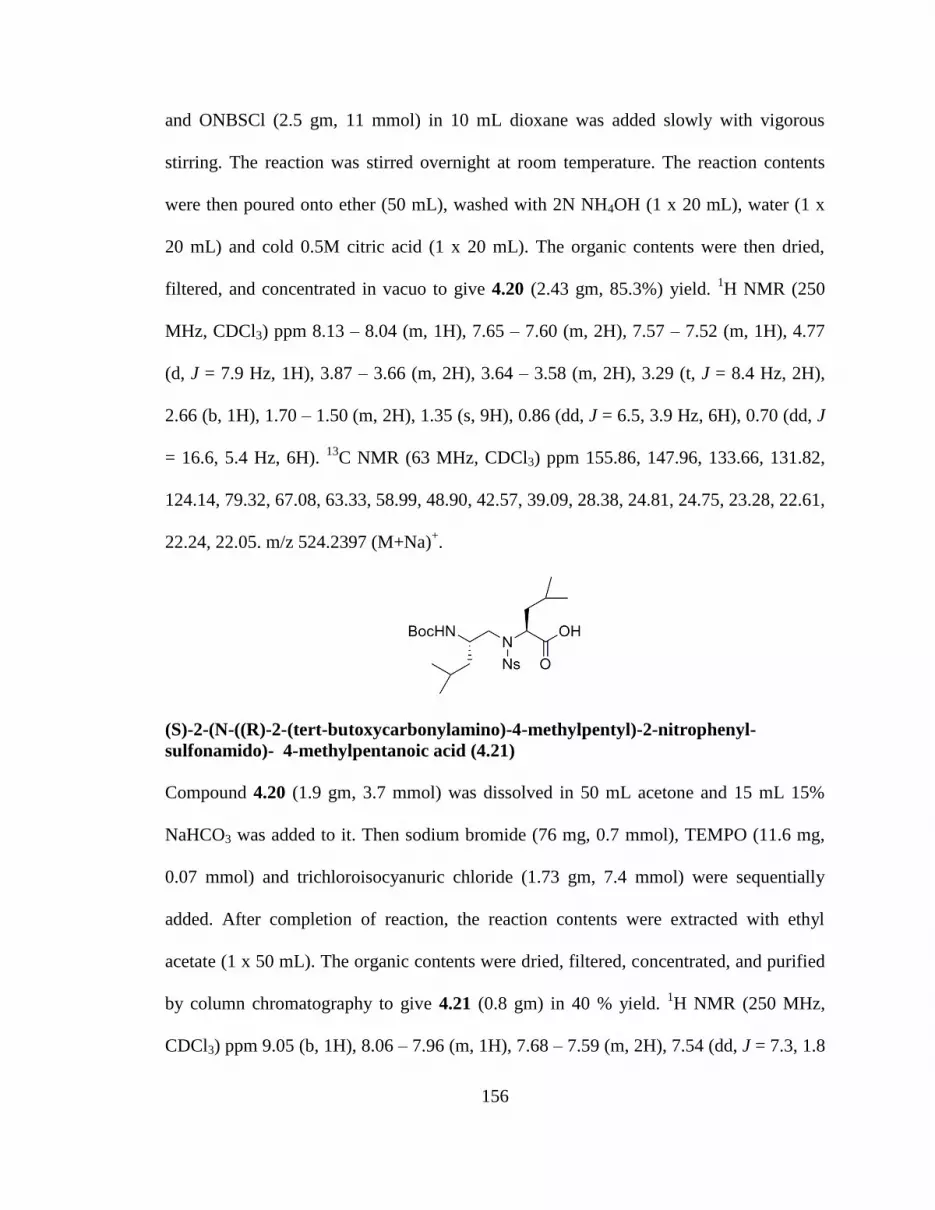
156
and ONBSCl (2.5 gm, 11 mmol) in 10 mL dioxane was added slowly with vigorous
stirring. The reaction was stirred overnight at room temperature. The reaction contents
were then poured onto ether (50 mL), washed with 2N NH4OH (1 x 20 mL), water (1 x
20 mL) and cold 0.5M citric acid (1 x 20 mL). The organic contents were then dried,
filtered, and concentrated in vacuo to give 4.20 (2.43 gm, 85.3%) yield. 1H NMR (250
MHz, CDCl3) ppm 8.13 – 8.04 (m, 1H), 7.65 – 7.60 (m, 2H), 7.57 – 7.52 (m, 1H), 4.77
(d, J = 7.9 Hz, 1H), 3.87 – 3.66 (m, 2H), 3.64 – 3.58 (m, 2H), 3.29 (t, J = 8.4 Hz, 2H),
2.66 (b, 1H), 1.70 – 1.50 (m, 2H), 1.35 (s, 9H), 0.86 (dd, J = 6.5, 3.9 Hz, 6H), 0.70 (dd, J
= 16.6, 5.4 Hz, 6H). 13
C NMR (63 MHz, CDCl3) ppm 155.86, 147.96, 133.66, 131.82,
124.14, 79.32, 67.08, 63.33, 58.99, 48.90, 42.57, 39.09, 28.38, 24.81, 24.75, 23.28, 22.61,
22.24, 22.05. m/z 524.2397 (M+Na)+.
(S)-2-(N-((R)-2-(tert-butoxycarbonylamino)-4-methylpentyl)-2-nitrophenyl-
sulfonamido)- 4-methylpentanoic acid (4.21)
Compound 4.20 (1.9 gm, 3.7 mmol) was dissolved in 50 mL acetone and 15 mL 15%
NaHCO3 was added to it. Then sodium bromide (76 mg, 0.7 mmol), TEMPO (11.6 mg,
0.07 mmol) and trichloroisocyanuric chloride (1.73 gm, 7.4 mmol) were sequentially
added. After completion of reaction, the reaction contents were extracted with ethyl
acetate (1 x 50 mL). The organic contents were dried, filtered, concentrated, and purified
by column chromatography to give 4.21 (0.8 gm) in 40 % yield. 1H NMR (250 MHz,
CDCl3) ppm 9.05 (b, 1H), 8.06 – 7.96 (m, 1H), 7.68 – 7.59 (m, 2H), 7.54 (dd, J = 7.3, 1.8

157
Hz, 1H), 4.77 (b, 1H), 4.55 – 4.42 (m, 1H), 3.98 – 3.65 (m, 1H), 3.58 – 3.04 (m, 2H),
1.46 – 1.32 (m, 11H), 0.90 – 0.79 (m, 12H). 13
C NMR (63 MHz, CDCl3) ppm 175.65,
155.84, 148.06, 134.02, 133.95, 131.70, 131.57, 131.43, 131.36, 124.31, 124.17, 81.63,
79.31, 59.42, 58.75, 50.08, 48.52, 42.79, 38.58, 28.38, 28.08, 24.65, 24.31, 23.64, 23.15,
22.55, 22.24, 21.35, 20.91. m/z 538.2193 (M+Na)+.
4.4 References
1.Vogelstein, B.; Lane, D.; Levine, A. J., Surfing the p53 network. Nature 2000, 408
(6810), 307-10.
2.Vousden, K. H.; Lu, X., Live or let die: the cell's response to p53. Nat. Rev. Cancer
2002, 2 (8), 594-604.
3.Hainaut, P.; Hollstein, M., p53 and human cancer. The first ten thousand mutations.
Adv. Cancer Res. 2000, 77, 81-137, 1 color plate.
4.Moll, U. M.; Petrenko, O., The MDM2-p53 Interaction. Mol. Cancer Res. 2003, 1 (14),
1001-1008.
5.Bond, G. L.; Hu, W.; Levine, A. J., MDM2 is a central node in the p53 pathway: 12
years and counting. Curr. Cancer Drug Targets 2005, 5 (1), 3-8.
6.Fischer Peter, M.; Lane David, P., Small-molecule inhibitors of the p53 suppressor
HDM2: have protein-protein interactions come of age as drug targets? Trends
Pharmacol Sci 2004, 25 (7), 343-6
7.Momand, J.; Zambetti, G. P.; Olson, D. C.; George, D.; Levine, A. J., The mdm-2
oncogene product forms a complex with the p53 protein and inhibits p53-
mediated transactivation. Cell (Cambridge, Mass.) 1992, 69 (7), 1237-45.
8.Roth, J.; Dobbelstein, M.; Freedman, D. A.; Shenk, T.; Levine, A. J., Nucleo-
cytoplasmic shuttling of the hdm2 oncoprotein regulates the levels of the p53
protein via a pathway used by the human immunodeficiency virus rev protein.
Embo J. 1998, 17 (2), 554-564.
9.Tao, W.; Levine, A. J., Nucleocytoplasmic shuttling of oncoprotein Hdm2 is required
for Hdm2-mediated degradation of p53. Proc Natl Acad Sci U S A 1999, 96 (6),
3077-80.

158
10.Pickart, C. M., Mechanisms underlying ubiquitination. Annu. Rev. Biochem. 2001, 70,
503-533.
11.Haupt, Y.; Maya, R.; Kazaz, A.; Oren, M., Mdm2 promotes the rapid degradation of
p53. Nature (London) 1997, 387 (6630), 296-299.
12.Honda, R.; Tanaka, H.; Yasuda, H., Oncoprotein MDM2 is a ubiquitin ligase E3 for
tumor suppressor p53. FEBS Lett. 1997, 420 (1), 25-27.
13.Kubbutat, M. H. G.; Jones, S. N.; Vousden, K. H., Regulation of p53 stability by
Mdm2. Nature (London) 1997, 387 (6630), 299-303.
14.Harris, C. C., Protein-protein interactions for cancer therapy. Proc Natl Acad Sci USA
2006, 103 (6), 1659-1660.
15.Jimenez, G. S.; Khan, S. H.; Stommel, J. M.; Wahl, G. M., p53 regulation by post-
translational modification and nuclear retention in response to diverse stresses.
Oncogene 1999, 18 (53), 7656-7665.
16.Vassilev, L. T.; Vu, B. T.; Graves, B.; Carvajal, D.; Podlaski, F.; Filipovic, Z.; Kong,
N.; Kammlott, U.; Lukacs, C.; Klein, C.; Fotouhi, N.; Liu, E. A., In Vivo
Activation of the p53 Pathway by Small-Molecule Antagonists of MDM2.
Science (Washington, DC, U. S.) 2004, 303 (5659), 844-848.
17.Fasan, R.; Dias, R. L. A.; Moehle, K.; Zerbe, O.; Vrijbloed, J. W.; Obrecht, D.;
Robinson, J. A., Using a beta-hairpin to mimic an alpha -helix: Cyclic
peptidomimetic inhibitors of the p53-HDM2 protein-protein interaction. Angew.
Chem., Int. Ed. 2004, 43 (16), 2109-2112.
18.Kussie, P. H.; Gorina, S.; Marechal, V.; Elenbaas, B.; Moreau, J.; Levine, A. J.;
Pavletich, N. P., Structure of the MDM2 oncoprotein bound to the p53 tumor
suppressor transactivation domain. Science (Washington, D. C.) 1996, 274 (5289),
948-953.
19.Luca Leonora, D.; Luca Joao Bosco Assis, D., Marie Rennotte, educator and medical
doctor: elements for a historical and biographical, social and medical study. Hist
Cienc Saude Manguinhos 2003, 10 (2), 703-25.

159
CHAPTER FIVE:
DESIGN AND SYNTHESIS OF PEPTIDE NUCLEIC ACID OLIGOMERS
5.1 Introduction
Many human diseases are caused by over-, under-, or misproduction of specific
proteins. Defects in the gene cause production of m-RNAs which codes for a non-
functional protein. Many diseases can be controlled by curbing production of these non
functional proteins (1). Thus, if single strands of DNA can be synthesized, the base
sequence could be studied and manipulated to treat various diseases. Gene expression can
be regulated by binding of an oligonucleotide or oligonucleotide analogue to double
stranded DNA or single stranded RNA. PNA (Peptide Nucleic Acids) is an extremely
good DNA mimic in which the deoxyribose phosphate diester backbone is replaced by a
pseudo-peptide backbone while the four natural nucleobases are retained. This new
backbone designed using computer models is structurally similar to the deoxyribose
phosphate backbone, synthetically accessible and amenable to automated assembly
synthesis (2, 3) (Fig. 5.1).
PNA consists of repeating N-(2-aminoethyl)-glycine units in which the
nucleobase is attached to the glycine nitrogen via a methylene carbonyl linker. The
acyclic, achiral and neutral backbone makes PNA set apart from DNA (4). The neutral
backbone of PNA allows stronger binding between complementary PNA/DNA strands
than between complementary DNA/DNA strands as determined by Tm’s (Melting

160
Temperature). This is attributed to lack of charge repulsion between the PNA strand and
the DNA strand. PNA also binds to DNA with greater specificity. A mismatch in a
PNA/DNA duplex is more destabilizing than a mismatch in a DNA/DNA duplex (5, 6).
Fig. 5.1 Structures of PNA and DNA.
PNA can bind to DNA or RNA strands in antiparallel or parallel orientations. In
antiparallel orientations, a DNA or RNA strand in 5’ to 3’ orientation binds to a
complementary PNA strand such that the carboxyl end of PNA is directed towards the 5’
end of DNA or RNA and the amino end is directed towards the 3’ end of DNA/RNA. The
neutral polyamide backbone of PNA offers enzymatic stability, making PNA resistant to
both proteases and nucleases (7). PNAs form stable triplexes PNA-DNA-PNA in which
one PNA strand hybridizes to DNA by Watson-crick base pairing while other binds the
same strand via the Hoogsten mode.
PNA DNA

161
All of the above properties makes PNA an important tool for designing gene
therapeutic drugs (antisense and antigene) and in genetic diagnostics (8). Although PNAs
have been demonstrated as potential candidates for gene-targeting drugs, some
disadvantages have limited applications. These include low cellular uptake caused by
poor cell membrane permeability and poor aqueous solubility (9, 10). This is due to
uncharged property of PNAs whereas nucleic acid oligonucleotides bearing negative
charges can penetratate the cell membrane by endocytosis (11). In order to increase the
cellular uptake of PNAs various strategies have been explored during the last decade.
These includes microinjection, electroporation, and attachment to cell penetrating
peptides (12). Since a positive charge will enhance attraction of molecules to cell
membrane, some researchers have tried to introduce positively charged residues such as
lysine and arginine into PNA molecules to increase cellular uptake of PNAs. Ly’s group
synthesized GPNA with arginine side chain attached to PNA backbone (7, 13). Inspired
by this idea to increase cellular uptake by introducing positive charge species into PNA,
an attempt to synthesize cysteine based Peptide nucleic acid (CPNA) monomers and
oligomers containing all four nucleobases was made. The proposed target molecule is
shown in figure 5.2.
Fig. 5.2 Proposed CPNA design for antisense applications.

162
CPNA monomers are derived from cysteine with positively charged guanidine units in
the side chain. The thiol group in cysteine allows us to attach various alkyl groups
designed to improve the properties of PNA. The presence of the thiol group usually
interferes in solid phase synthesis too. This might be the reason for no report of PNAs
based on cysteine. The introduction of positively charge residues on PNA is expected to
increase aqueous solubility and cellular uptake of PNAs. We have also made efforts to
synthesize standard Peptide Nucleic Acids monomers with an aim to make hybrid PNAs.
In this chapter, the synthesis of standard and CPNA monomers, modified nucleobases
and an effort to synthesize PNA oligomers is described.
5.2 Results and Discussion
5.2.1 Synthesis of CPNA monomers
Synthesis of PNA oligomers is based on solid phase peptide synthesis protocols.
The scheme for protecting amino groups of PNA monomers is based on either Boc or
Fmoc chemistry. Scheme 5.1 shows the synthesis of four CPNA monomers using Fmoc
protecting groups on primary amine of backbone and Cbz protecting groups on the
exocyclic amino groups of nucleobases. The backbone protecting groups must be
orthogonal in their requirement from removal from PNA during oligomer synthesis i.e
Cbz protecting groups on nucleobases must be stable to conditions used for removal of
Fmoc protecting group from terminal amine. The use of Fmoc protection groups offers
advantages of milder synthesis conditions and improved monomer solubility.

163
Scheme 5.1 Synthetic strategy for making CPNA monomers.
Various research groups have used the reductive amination route (14) to
synthesize PNA backbone but our PNA is synthesized by a novel SN2 route. Synthesis of
CPNA monomers involves activation of commercially available Fmoc(Cys)Trt-COOH

164
using DCC followed by attack of nucleophile to enable formation of Amide 5.2. Selective
reduction of 5.2 with BH3 in THF yields primary amine 5.3 in 65% yield. Alkylation of
terminal amine 5.3 with tert-butyl bromoacetate via SN2 reaction followed by aqueous
workup gives 5.4 in 82% yield. Deprotection of acid labile trityl protecting group in 5.4
using triflouro acetic Acid followed by alkylation of resulting thiol with various
alkylating agents (methyl iodide, guanidium) enabled us to introduce positive charge
residue on the side chain of PNAs. The next step involves coupling of nucleobase acetic
acids to unreactive secondary amine in 5.5, 5.6, 5.7 to give fully protected CPNA
monomers in 80% yield. Finally, the t-butyl ester was deprotected with HCl in dioxane to
give CPNA monomers in quantitative yield.
During synthesis of amide, the by-product N,N’-DCC urea is mostly removed by
filtration, but trace amounts still remain and is often difficult to remove. Other activating
agents such as EDC, DIC were also used as an alternative. These activating agents were
equally efficient to DCC with similar yields. Excess amount of ammonium hydroxide
should be avoided as it often leads to cleavage of base labile Fmoc group, thereby
decreasing the yields of amide. This reaction should be carried out at low temperature
~10-150C. Larger reaction time and elevated temperatures often resulted in low yields of
amide. Borane reduction of amide to give primary amine was tricky reaction.
Concentration of pure amine after column chromatography usually cleaved Fmoc
protecting group which causes a reduction in yield. This problem was eliminated by
making use of trityulation technique for purifying crude amine. During synthesis of
compound 5.4, excess amount of alkylating agent caused formation of some dialkylated
compound along with desired monoalkylated compound. Also cleavage of Fmoc

165
protecting group was seen when base slight excess of DIEA was used. This problem was
solved by using 1equivalent of alkylating agent and 1.05 equivalent of base. The reaction
temperature also determined the amount of dialkylated compound formed. A significant
amount of dialkylated compound was formed when the reaction was carried at room
temperature for longer time as well as some Fmoc cleavage was also observed. The
reaction was done at 10-150C for 10 hours and then at room temperature for two hours.
The reaction rate was slow but no dialkylated compound and Fmoc cleavage was
observed. Deprotection of trityl group in compound 5.4 and then alkylation of resulting
thiol was done in a single pot. Extreme dilute conditions were employed while using TFA
deprotection of trityl group. Dilute conditions enabled t-butyl to remain intact. Alkylation
of thiol was done using slight excess of base followed by addition of slight excess of
alkylating agent. Coupling of nucleobase acetic acids to CPNA submonomers gave
monomers in good yield. Purification of these polar based coupled CPNA monomers was
tedious, particularly Guanidine coupled CPNA monomer purification gave low yields.
Deprotection of t-butyl ester from fully protected CPNA monomer using
HCl/Dioxane gave final monomers in quantitative yields but removal of residual dioxane
from monomers was difficult.
Scheme 5.2 Synthesis of Cbz protected Adenine acetic acid nucleobase for CPNA.

166
Synthesis of Adenine Acetic Acid used for coupling to PNA backbone is shown
in scheme 5.2. Several procedures for synthesizing nucleobase acetic acids have been
reported till now (15, 16, 17). The exocyclic amine of adenine was first protected with
acid labile Cbz protecting group using benzyl chloroformate to give 5.33. Compound
5.33 was then alkylated with t-butyl bromoacetate in DMF using potassium carbonate
and cesium carbonate to give a mixture of N-7 and N-9 alkylated products. The desired
N-9 alkylated produced was separated using column chromatography in 49% yield.
Removal of the t-butyl ester with TFA in DCM gave compound 5.35 in 92% yields along
with a loss of Cbz group. The loss of Cbz group in this reaction was suppressed by using
slight excess of cation scavenger triethylsilane in the reaction mixture.
The solid phase synthesis of CPNA monomers posed a great challenge to us. We
have been able to synthesize dimers of nucleobase coupled CPNA monomers using
solution phase coupling. Synthesis of purine based CPNA monomers and their coupling
during solid phase synthesis was difficult. Solid phase coupling for obtaining CPNA
oligomers (18-mer) was attempted but it did not yield promising results. The synthesis of
standard peptide nucleic acid monomers was attempted with an aim to make hybrid
PNAs consisting of standard PNA and CPNA monomers. This strategy will help us to
modulate the positive charge on PNA, required for achieving cellular permeability.
5.2.2 Synthesis of standard PNA monomers
Scheme 5.3 depicts the synthesis of standard PNA monomers using orthogonal
Fmoc/t-butyl strategy as cited in reported literature procedures. Selective monoalkylation
of excess ethylene diamine with t-butyl bromoacetate gave compound 5.37. This material
was utilized in next step without further purification. Fmoc protection of the primary

167
amine in 5.37 followed by acidic workup gave compound 5.38 as Hydrochloride salt.
This compound can be stored indefinitely in deep freezer. Coupling of nucleobase acetic
acids to PNA backbone was achieved using HATU and DIEA in DMF to give nucleobase
coupled PNA esters. Finally deprotection of t-butyl group using HCl gas in dioxane gave
peptide nucleic acid monomers in good yields.
Scheme 5.3 Synthesis of standard PNA monomers using Fmoc/t-butyl strategy.
The solid phase synthesis of standard PNA monomers was carried out on MBHA
resin solid support using standard Fmoc protecting group strategy. We were able to
synthesize 12mer PNA oligomer with sequence NH2LysTTACATCTTTTC-COOH along
with truncated byproducts. The purification of PNA using RPLC became difficult and we
were not successful in obtaining purified PNA out of the column. We sought for
alternative strategies for making standard peptide nucleic acid. Lee and coworkers have
synthesized self-activated cyclic PNA monomers and an efficient method for

168
synthesizing PNA oligomer using benzothiazole-2-sulfonyl (Bts) protecting group.
Scheme 5.4 shows the synthesis of cyclic Bts protected PNA monomers carried out as per
cited literature procedure (18).
Scheme 5.4 Synthesis of Cyclic Bts PNA monomers.
Compound 5.48 was prepared by treating 5.47 with Bts-Cl synthesized from
2-meracaptobenzothioazole in 72% yield (19, 20). Nucleobases used for coupling were
protected with Bhoc group due to its stability towards nucleophiles and ease of
deprotection. Nucleobase acetic acids were prepared as per reported literature and
coupled to 5.48 using HATU and DIEA in DMF (21). The corresponding esters (5.49-
5.52) were hydrolysed using LiOH to give corresponding acids (5.53-5.56) in 80-90%
yield. The acids (5.53-5.56) were then readily cyclized using EDC to give cyclic Bts
protected PNA monomers in 85-90% yield. Scheme 5.5 describes the solid phase
synthesis of PNA oligomer using cyclic Bts monomers.

169
Scheme 5.5 Solid phase synthesis of standard PNA using cyclic Bts protecting group.
Bts group activates carbonyl of piperazinone to be readily attacked by primary
amine of PNA and is easily removed by thiols in presence of base after the coupling
reaction. PNA synthesis was carried out on CLEAR amino resin loaded with PAL linker
that allows cleavage of PNA under acidic conditions. Coupling was carried out at 40oC
for two hours and unreacted amine of PNA was capped by Ac2O/Lutidine. The Bts group
was removed by 1M 4-Methoxybenzethiol and 1M DIEA in DMF. The greatest
advantage of using cyclic Bts protected monomers is that they do not require
preactivation or extremely anhydrous conditions. Also we can recover unused monomers
since no coupling agents are involved. MALDI-TOF analysis of the crude PNA synthesis
suggested we were able to synthesize desired PNA alongwith truncated byproducts using
this strategy but we could not quantify it into appreciable yields.

170
5.3 Experimental Procedures
5.3.1 Materials and Methods
Organic and inorganic reagents (ACS grade) were obtained from commercial
sources and used without further purification, unless otherwise noted. Fmoc-protected
amino acids and the coupling agent HCTU were obtained from Protein Technologies,
Calbiochem-Novabiochem, or Chem-impex International. 2-Chlorotrityl chloride resin
was purchased from Anaspec Inc. All linear peptides were synthesized on the Symphony
peptide synthesizer, Protein Technologies Instruments. Solvents for peptide synthesis and
reverse-phase HPLC were obtained from Applied Biosystems. Other chemicals used
were obtained from Aldrich and were of highest purity commercially available. Thin
layer chromatography (TLC) was performed on glass plates (Whatman) coated with 0.25
mm thickness of silica gel 60Å (# 70-230 mesh). All 1H and
13C NMR spectra were
recorded on Bruker 250 MHz, Varian INOVA 400 MHz spectrometer in CDCl3 or unless
otherwise specified and chemical shifts are reported in ppm (δ) relative to internal
standard tetramethylsilane (TMS). High Resolution mass spectra were obtained on an
Agilent LC-MSD-TOF.
(R)-(9H-fluoren-9-yl)methyl1-amino-1-oxo-3-(tritylthio)propan-2-ylcarbamate (5.2)
To a solution of 5.1(10.00 g, 17.05 mmol), HOBT (2.76 gm, 20.0 mmol) and DCC (4.23
gm, 20.0 mmol) in THF (100 mL) was added 28% NH4OH (2.45 mL, 20.0 mmol) at 0°C.
After 2hr of stirring at 0°C, the reaction mixture was filtered through diatomaceous earth

171
and the filtrate was concentrated, diluted with ethyl acetate, and washed with water and
brine. The organic layer was then dried over sodium sulfate, concentrated and subjected
to flash column chromatography to give 5.2(9.25 g, 92 %) as white solid. Rf = 0.45
(50:50 ethyl acetate: hexane) ) 1H NMR (400MHz, CDCl3) δ 2.65(m, 2H), 4.12 (q, J =
7.14, 1H), 4.17 (t, J = 6.52, 1H), 4.41 (d, J = 4.92, 2H), 5.02 (d, J = 7.54, 1H), 5.48 (s,
1H), 5.74 (s, 1H), 7.23 (m, 11H), 7.38 (dd, J = 7.6 & 11.0, 8H), 7.36 (d, J = 7.36, 2H),
7.74 (m, 2H). 13
C NMR (400MHz, CDCl3) δ 25.94, 33.94, 53.5, 60.10, 67.4, 120.01,
124.99, 126.94, 127.07, 127.10, 127.75, 127.77, 128.08, 129.55, 141.35, 143.66, 144.28,
156.76, 172.72. m/z 585.2 (M+H)+
(R)- (9H-fluoren-9-yl) methyl 1-amino-3-(tritylthio) propan-2-ylcarbamate (5.3).
To a solution of 5.2(9.25 gm, 15.8 mmol) in THF(30 mL was added 1 M Borane·THF
complex (34.76 mL, 34.76 mmol) at 0°C and After stirring at 0°C for 3 hrs, reaction
mixture was then heated to 70°C and stirred for 12 hrs. Reaction mixture was cooled to
0°C and quenched by MeOH. The reaction mixture was concentrated and diluted with
MeOH and concentrated again in vacuo. The concentrated oil was subjected to flash
column chromatograph to afford 5.3(6.5 gm) in a 72 % yield. Rf = 0.50 (90:10 ethyl
acetate: methanol) ) 1H NMR (400 MHz, CDCl3) δ 0.78 (m, 2H), 2.37 (d, J = 20.25, 2H),
2.63 (s, 2H), 4.20 (t, J = 6.74, 1H), 4.40 (d, J = 6.62, 2H), 4.81 (s, 1H), 7.25 (m, 11H),
7.41 (m, 8H), 7.59 (d, J = 7.39, 2H), 7.75 (m, 2H). ). 13
C NMR (400 MHz, CDCl3) δ

172
24.94, 25.62, 33.96, 44.56, 47.30, 52.42, 66.90, 119.96 125.03, 126.81, 127.03, 127.68,
127.97, 129.58, 141.34, 143.91, 144.54, 155.92. m/z 571.2 (M+H)+
[2-(9H-Fluoren-9-ylmethoxycarbonylamino)-3-tritylsulfanyl-propylamino]-acetic
acid tert-butyl ester (5.4)
To a solution of 5.3 (1.8 gm, 3.2 mmol) in THF(20 mL) was added tert-butyl
bromoacetate (0.47 mL, 3.2 mmol) and DIEA (0.576 ml, 3.31 mmol) at 0°C. The reaction
mixture was slowly allowed to warm to room temperature. The reaction mixture was
concentrated in vacuo, diluted with EtOAc and washed with brine. The organic layer was
dried over sodium sulfate, concentrated in vacuo and subjected to a flash column
chromatography to give 5.4(2.2 gm, 82%). Rf = 0.73 (50:50 ethyl acetate: hexane) ) 1H
NMR (400 MHz, CDCl3) δ 0.868 (d, J = 7.09, 2H), 0.97 (d, J = 6.55, 1H), 1.25 (s, 2H),
1.45 (s, 9H), 2.46 (m, 4H), 3.17 (s, 2H), 4.20 (s, 1H), 4.35 (d, J = 6.71, 2H), 5.10 (m,
1H), 7.25 (m, 11H), 7.4 (m, 8H), 7.59 (d, J = 7.31, 2H), 7.75 (d, J = 7.49, 2H). 13
C NMR
(400 MHz, CDCl3) δ 29.51, 35.3, 47.6, 49.5, 50.2, 67.6, 67.52, 81.9, 120.1, 125.8,
127.43, 127.9, 128.05, 129.88, 140.7, 144.2, 145.5, 156.2, 172.1. m/z 685.2 (M+H)+

173
(R)-tert-butyl 13-(((9H-fluoren-9-yl)methoxy)carbonylamino)-3,9-dioxo-1-phenyl-2-
oxa-11-thia-4,8,15-triazaheptadecan-17-oate (5.6)
To a stirred solution of 5.4 (1.5 gm, 2.1 mmol) in DCM (50 mL) at 0°C was added TFA
(4 mL). The reaction mixture was treated with triethylsilane (0.35 mL, 2.1 mmol). The
reaction mixture was stirred for 30 min at 0°C and concentrated in vacuo. The crude
product was dissolved in EtOH (20 mL) and the solution was cooled to 00C. Aqueous
NaOH (2N, 3.3 mL, 6.6 mmol) was added slowly to the solution followed by addition of
the alkyl bromide, (0.86 gm, 2.6 mmol). The reaction was stirred for 4 hrs at 0°C. After
the removal of organic solvents in vacuo, the residue was diluted with EtOAc and washed
with water and brine. The organic layer was dried over sodium sulfate, concentrated in
vacuo and subjected to a flash column chromatography to give 5.6(1.4 gm, 92 %). Rf =
0.4 (100% ethyl acetate) 1H NMR (400 MHz, CDCl3) δ 1.44 (d, J = 7.17, 9H), 1.63 (s,
2H), 2.75 (m, 2H), 3.24 (m, 8H), 3.52 (s, 2H), 4.06 (s, 1H), 4.17 (q, 1H), 4.33 (d, 2H),
5.03 (s, 2H), 5.4 (s, 1H), 6.58 (b, 1H), 7.28 (m, 7H), 7.37 (t, J = 7.47, 2H), 7.45 (m, 1H),
7.58 (d, 2H) 7.73 (d, J = 7.52, 2H). m/z 691.2 (M+H)+
[2-(9H-Fluoren-9-ylmethoxycarbonylamino)-3-methylsulfanyl-propylamino]-acetic
acid tert-butyl ester (5.5)

174
Rf = 0.52 (50:50 ethyl acetate: hexane) ) 1H NMR (250 MHz, CDCl3) δ 1.43 (d, J = 19.9,
9H), 2.116 (s, 2H), 2.78 (m, 4H), 3.28 (s, 2H), 3.85 (s, 1H), 4.21 (t, J = 6.78, 1H), 4.39 (s,
2H), 5.55 (s, 1H), 7.33 (ddd, J = 5.57, 12.76 & 17.63, 4H), 7.61 (d, J = 7.28, 2H), 7.74 (d,
J = 7.27, 2H). 13
C NMR (400 MHz, CDCl3) δ 16.19, 28.12, 36.81, 47.28, 50.29, 51.01,
51.65, 66.58, 81.42, 119.94, 125.11, 127.03, 127.64, 141.3, 143.95, 156.13, 171.64. m/z
457.2(M+H)+
General procedure for Base coupling
(R)-tert-butyl13-(((9H-fluoren-9-yl)methoxy)carbonylamino)-15-(2-(5-methyl-2,4-
dioxo-3,4-dihydropyrimidin-1(2H)-yl)acetyl)-3,9-dioxo-1-phenyl-2-oxa-11-thia-
4,8,15-triazaheptadecan-17-oate (5.19)
To a stirring solution of 5.6(1.18 gm, 1.7 mmol), HATU (0.78 gm, 2.04mmol) and
thymine acetic acid (0.47 gm, 2.6 mmol) in DMF was added TEA (0.71 mL) at room
temperature for 4 hrs. The reaction mixture was diluted with EtOAc, washed with cold
water and brine, dried over sodium sulfate, concentrated in vacuo and subjected to a flash
column chromatography to give 5.19(1.38 gm, 95%).
{[2-(9H-Fluoren-9-ylmethoxycarbonylamino)-3-methylsulfanyl-propyl]-[2-(5-
methyl-2,4-dioxo-3,4-dihydro-2H-pyrimidin-1-yl)-acetyl]-amino}-acetic acid tert-
butyl ester (5.8).

175
Rf = 0.51 (100% ethyl acetate) ) 1H NMR (400 MHz, CDCl3) δ 1.47 (d, J = 27.2, 9H),
2.93 (s, 3H), 2.01 (s, 1H), 2.1 (d, J = 22.8, 3H), 2.6-2.72 (m, 2H), 2.8 (d, J = 0.58, 2H),
4.09 (m, 2H), 4.21 (m, 1H), 4.41 (d, J = 10.32, 2H), 4.74 (b, 1H), 7.23 (d, J = 6.09, 1H),
7.3 (d, J = 6.28, 2H), 7.51 (dd, J = 4.46 & 8.43, 1H), 7.66 (m, 2H), 7.78 (d, J = 7.27, 2H),
8.42 (dd, J = 1.36 & 8.43, 1H), 8.72 (d, J = 4.46, 1H). m/z 645.1 (M+Na)+
General procedure for deprotecting carboxylic acid
Synthesis of (R)-13-(((9H-fluoren-9-yl)methoxy)carbonylamino)-15-(2-(5-methyl-
2,4-dioxo-3,4-dihydropyrimidin-1(2H)-yl)acetyl)-3,9-dioxo-1-phenyl-2-oxa-11-thia-
4,8,15-triazaheptadecan-17-oic acid (5.23).
HCl gas was passed through a solution of 5.19(1.38 gm, 1.6 mmol) in dioxane for 20
minutes. The reaction mixture was then stirred at r.t for 2 hrs followed by concentration
in vacuo to give 5.23 (1.2 gm, quantitative yield).
6-N-(Benzyloxycarbonyl) adenine (5.33)
Sodium Hydride (6.07 gm, 252 mmol), washed with hexane (3 times), was taken up in
DMF (100 mL) in an ice-bath. Adenine (5 gm, 7 mmol), 5.32, was then added in small
lots. The suspension was stirred for 5 minutes and then benzyl chloroformate (11.6 gm,
81.5 mmol) was added dropwise. The reaction mixture was stirred for 4 hours, poured
into ice-cooled water (250 mL) and pH was adjusted to 7 with 1 N HCl. The yellow
precipitate was washed with water and ether to give crude product. Recrystallization with

176
DCM gave 5.12 gm of 5.33 in a 51 % yield. 1H NMR (400MHz, DMSO) δ 5.3 (s, 2H),
7.3-7.5 (m, 5H), 8.4 (s, 1H), 8.6 (s, 1H) m/z 270.0 (M+H)+
6-N-(Benzyloxycarbonyl)-9-(tert-butoxycarbonylmethyl) adenine (5.34)
5.33 (4 gm, 14.8 mmol) was taken up in DMF (25 mL). Anhydrous Cs2CO3 (0.48 gm,
1.48 mmol) and anhydrous K2CO3 (2.04 gm, 14.8 mmol) was added to it. After stirring
for 5 minutes, tert-butyl bromoacetate (2.64 mL, 17.8 mmol) was added dropwise. After
stirring for 24 hours, the mixture was concentrated, diluted in EtOAc, washed with water
(10 mL) and ethyl acetate (35 mL). The organic layer was dried over sodium sulfate,
concentrated, and subjected to column chromatography to give 2.85 gm of 5.34 in a 49 %
yield. Rf = 0.33, (50:50 EtOAc:Hexane) 1
H NMR (400 MHz, CDCl3) δ 8.78 (s, 1H), 8.09
(s, 1H), 7.25-7.35 (m, 5H), 5.3 (s, 2H), 4.89 (s, 2H), 1.47 (s, 9H). m/z 384.1(M+H)+
6-N-(Benzyloxycarbonyl)-9-(carboxymethyl) adenine 5.35
Triethylsilane (11.4 ml) was added to a solution of 5.34 (2.85 gm, 7.43 mmol) in
anhydrous DCM (10 mL) and then TFA (15 mL) was added dropwise at 00C. After 5
min, the reaction mixture was allowed to warm to room temperature and stirred for 10
hrs. The mixture was concentrated and any volatile impurities were removed by

177
azeotroping with CHCl3 (3 times) to give a crude product as white foam.
Recrystallization with acetone gave 2.23 g of 5.35 in a 92 % yield. 1H NMR (400 MHz,
DMSO) δ 5.05 (s, 2H), 5.2 (s, 2H), 7.2-7.3 (m, 5H), 8.4 (s, 1H), 8.6 (s, 1H), 10.7 (s, 1H).
m/z 326.0 (M-H)+
Synthesis for compound 5.38 has been reported in chapter two.
General procedure for coupling of Cbz protected nucleobases to PNA backbone
To a stirring solution of 5.38 (1.0 eq.), HATU (1.2 eq) and thymine acetic acid (1.2 eq.)
in DMF was added TEA (3 eq.) at room temperature for 4 hrs. The reaction mixture was
diluted with EtOAc, washed with cold water and brine, dried over sodium sulfate,
concentrated in vacuo and subjected to a flash column chromatography to give
corresponding esters 5.39-5.41 in 75-80% yield.
General procedure for deprotection of esters 5.39-5.41
Corresponding esters 5.39-5.41 were dissolved in 1, 4-dioxane and HCl(g) was passed into
solution for 10 min. The reaction mixture was stirred for another one hour and
concentrated in vacuo and dried to give the corresponding acids.

178
N-[2-(N-9-fluorenylmethoxycarbonyl)aminoethyl]-N-[[4-N-(benzloxycarbonyl)-
cytosine-1-yl]acetyl]glycine 5.43
Yield: 86%, White solid. 1H NMR (400 MHz, DMSO) δ 7.89 – 7.81 (m, 3H), 7.65 (d, J =
6.9 Hz, 2H), 7.42 – 7.26 (m, 10H), 7.00 – 6.93 (m, 1H), 5.16 (s, 2H), 4.79 (s, 1H), 4.60
(s, 1H), 4.34 – 4.23 (m, 2H), 4.20 (s, 2H), 3.96 (s, 1H), 3.45 – 3.01 (m, 5H).
N-[2-N-(N-9-Fluorenylmethoxycarbonyl)aminoethyl]-N-[(thymin-1-yl)acetyl]-
glycine (5.44)
Yield: 89%, White solid. 1H NMR (400 MHz, DMSO) δ 11.24 (d, J = 7.0 Hz, 1H), 7.85
(d, J = 7.5 Hz, 2H), 7.65 (d, J = 7.3 Hz, 2H), 7.38 (t, J = 7.4 Hz, 3H), 7.29 (dd, J = 7.3,
6.6 Hz, 3H), 4.62 (s, 1H), 4.44 (s, 1H), 4.28 (dd, J = 16.0, 6.9 Hz, 2H), 4.22 – 4.14 (m,
2H), 4.05 – 3.77 (m, 2H), 3.38 (t, J = 6.3 Hz, 1H), 3.30 (t, J = 6.6 Hz, 1H), 3.22 (d, J =
5.9 Hz, 1H), 3.07 (dd, J = 12.3, 6.1 Hz, 1H), 1.69 (s, 3H).

179
N-[2-(N-9-fluorenylmethoxycarbonyl)aminoethyl]-N-[[6-N-(benzyloxycarbonyl)
adenin-9-yl]glycine (5.42)
Yield: 82% White solid. 1H NMR (400 MHz, DMSO) δ 8.56 (d, J = 19.3 Hz, 1H), 8.49 –
8.45 (m, 1H), 7.84 (d, J = 7.5 Hz, 2H), 7.67 – 7.61 (m, 2H), 7.50 – 7.46 (m, 1H), 7.45 –
7.42 (m, 2H), 7.39 – 7.34 (m, 4H), 7.34 – 7.30 (m, 1H), 7.29 – 7.25 (m, 2H), 5.35 (s, 1H),
5.19 (d, J = 18.2 Hz, 3H), 4.33 (d, J = 7.1 Hz, 2H), 4.28 – 4.14 (m, 3H), 3.97 (s, 1H),
3.55 – 3.50 (m, 1H), 3.33 (d, J = 15.4 Hz, 2H).
N-(2-Aminoethyl) glycine (5.46)
Ethylene diamine (18 mL, 27 mmol) was dissolved in methanol (26 mL) and glyoxylic
acid monohydrate (8.4 gm, 91 mmol) in 2 6mL of H2O was added slowly at 0oC. After
stirring for 20 mins, 5% Pd/C is added to the mixture and hydrogenation was carried out
at 50 psi at room temperature. After filtering the mixture, the filtrate was concentrated
and residue was reconcentrated twice with toluene (2 x 25 mL). Methanol (15 mL) was
added to the crude residue stirring vigorously at 0oC. After 15 mins, the white precipitates
that formed were filtered off and washed with cold methanol. A second crop of
precipitate was obtained from concentration of the mother liquor. The white precipitates
were combined and dried to give compound 5.46 in 62% yield. 1
H NMR (250 MHz, D2O)

180
δ 3.22 (s, 2H), 2.99 (dd, J = 9.5, 3.4 Hz, 2H), 2.85 (t, J = 6.0 Hz, 2H). m/z 118.13
(M+H)+
Ethyl N-(2-aminoethyl) glycinate dihydrochloride (5.47)
Compound 5.46 (10 gm, 84.7 mmol) was suspended in ethanol and HCl gas was passed
into the suspension. The mixture was refluxed overnight and cooled to 0oC. The product
was filtered off and dried in vacuo to give compound 5.47 in quantitative yield. 1H NMR
(250 MHz, D2O) δ 4.25 (q, J = 7.2 Hz, 2H), 4.04 (s, 2H), 3.82 (s, 1H), 3.54 – 3.27 (m,
5H), 1.23 (t, J = 7.2 Hz, 3H). m/z 147.10 (M+H)+
N-[2-(Benzothiazole-2-sulfonylamino)-ethyl]-glycine ethyl ester (5.48)
Compound 5.47 (3.2 gm, 15 mmol) was suspended in 100 mL DCM and TEA (7.5 mL,
58.4 mmol) was added slowly at room temperature. Then Bts-Cl (3.5 gm, 15 mmol)
dissolved in 50 mL DCM was added over a period of 10 minutes. The reaction was
stirred for 3 hours at room temperature and washed with water (50 mL). The organic
layer was dried, filtered, concentrated and purified by column chromatography to give
compound 5.48 in 72% yield. 1H NMR (250 MHz, CDCl3) δ 8.20 – 8.13 (m, 1H), 8.01 –
7.94 (m, 1H), 7.64 – 7.51 (m, 2H), 4.13 (q, J = 7.1 Hz, 2H), 3.38 (s, 2H), 3.34 (d, J = 5.9
Hz, 2H), 2.88 – 2.81 (m, 2H), 1.23 (t, J = 7.1 Hz, 3H).

181
General Procedure for synthesizing compounds (5.53-5.56)
To a stirred solution of 5.48 (1.0 eq.), nucleobase acetic acid (1.0 eq.), HOBT (1.2 eq.)
and DCC (1.2 eq.) in DMF was added DIEA (1.5 eq.) at room temperature. The reaction
mixture was stirred overnight and concentrated to leave crude residue. The residue was
dissolved in DCM and the precipitates were filtered. The filtrate was washed with 1M
cold HCl solution, sat. NaHCO3 solution and brine. The residue was then triturated with
ethyl alcohol to give protected ester (5.49-5.52). The protected esters were suspended in
THF:H2O (1:1) mixture, solution of lithium hydroxide (2 eq.) in water was then added
and the reaction mixture was stirred at room temperature for one hour. After completion
of the reaction, the reaction contents were acidified to pH ~ 2-3 by dropwise addition of
1M HCl at 0oC. The resulting solution was extracted with ethyl acetate, dried, filtered,
and concentrated to dryness in vacuo to afford compounds 5.53-5.56 in 80-90% yield.
General procedure for synthesizing cyclic Bts protected PNA monomers
To a mixture of Bts protected PNA monomers (5.53-5.56) (1.0 eq.) in DMF was added
EDCI (1.2 eq.) and stirred for 6 hours at room temperature. After completion of the
reaction, solvent was removed by evaporation and residue was dissolved in DCM. The
solution was washed with cold 1M HCl and water. The organic contents were dried,

182
filtered, concentrated and purified by flash chromatography to give cyclized Bts protected
PNA monomers in 85-90% yield.
1-(Benzothiazole-2-sulfonyl)-4-{[6-N-(benzhydryloxycarbonyl)-adenine-9-yl]-
acetyl}-piperazin-2-one) (5.57)
Yield: 87%, white solid. 1H NMR (250 MHz, DMSO) δ 10.91 (s, 1H), 8.60 (d, J = 4.3
Hz, 1H), 8.33 (ddd, J = 13.4, 7.5, 2.2 Hz, 3H), 7.74 (dd, J = 6.4, 2.5 Hz, 2H), 7.54 (d, J =
7.1 Hz, 4H), 7.39 (t, J = 9.9, 4.6 Hz, 4H), 7.33 – 7.26 (m, 2H), 6.83 (s, 1H), 5.41 (s, 1H),
5.30 (s, 1H), 4.59 (s, 1H), 4.30 (s, 2H), 4.11 (s, 2H), 3.89 (s, 1H). m/z 683.147 (M+H)+
1-(Benzothiazole-2-sulfonyl)-4-[(thymin-1-yl)-acetyl]-piperazin-2-one (5.60)
Yield: 92%, white solid. 1H NMR (400 MHz, DMSO) δ 11.27 (d, J = 7.2 Hz, 1H), 8.32
(d, J = 4.8 Hz, 1H), 8.24 (s, 1H), 7.72 – 7.67 (m, 2H), 7.31 (s, 1H), 7.24 (s, 1H), 4.65 (s,
1H), 4.55 (s, 1H), 4.39 (s, 1H), 4.25 (s, 1H), 4.18 (s, 1H), 4.04 (s, 1H), 3.96 – 3.90 (m,
1H), 3.86 – 3.80 (m, 1H), 1.71 (s, 3H). 13
C NMR (101 MHz, DMSO) δ 167.03, 166.45,
166.31, 165.03, 164.44, 151.87, 151.73, 142.66, 142.58, 137.24, 129.10, 128.68, 125.64,
124.07, 108.89, 60.42, 49.47, 48.80, 48.62, 47.70, 46.84, 46.68, 42.40, 12.58. LCMS
(ESI) m/z 464.07 (MH+)

183
5.4 General Procedure for PNA synthesis using Fmoc/Cbz protecting group strategy
PNA oligomer synthesis was carried out on Protein Technologies Instrument PTI
Symphony synthesizer. MBHA resin was first swollen manually in NMP for 45 minutes
and downloaded with Fmoc-Lys(Boc)-OH to a substitution level of ~0.15mmol/g. Fmoc
deprotection was then carried out using 20% piperidine/2%DBU in NMP and the resin is
washed with NMP (5x), DCM (5x). Coupling was carried out using 5 equivalents of
Fmoc protected PNA monomers, 5 equivalents HCTU and 10 eq. NMM in NMP. After
coupling the resin was washed again with NMP (5x), DCM (5x). This cycle was repeated
for each base coupled. After the last coupling the Fmoc group was deprotected and PNA
oligomer was cleaved off from the resin using following procedure: The resin was
swelled in TFA once. Two solutions were prepared:
Solution 1 Solution 2
TFA/dimethylsulphide/m-cresol (1:3:1) TFA/TFMSA (9:1)
1 mL of each solution was added to resin first and swirled for one hour. Here the
protecting groups on the immobilized oligomer are removed first. Then 1 mL of a third
solution TFMSA/TFA/m-cresol (2:8:1) was added to the reaction vessel and swirled for
another 1.5 hrs. The black solution was poured out in ether (6-7 mL). The white solid was
precipitated out by centrifugation and the precipitate was washed with ether. The ether
was decanted and precipitates were freeze-dried. Different PNA oligomers synthesized
using various orthogonal protecting group strategies are listed in Table 5.1.

184
Table 5.1 PNA sequences synthesized on PTI synthesizer using different protecting
group strategies.
Sequence Protecting Group Expected Observed
LysTTCCC Fmoc/Cbz 1181.177 1180.736
Lys-TTCTTCCTTCTTC-NH2 Fmoc/Cbz 3532.132 3532.902
Lys-TTCTTCCTTCTTC-NH2 Preactivated OPf esters 3532.132 3532.071
CO2HCTTTTCTACATTLysNH2 Bts/Bhoc 3312.3477 3312.0891
GlyTTCC Fmoc/Cbz (CPNA) 1364.5131 1364.5138
5.5 References
1. Ge, R.; Heinonen, J.E.; Svahn, M.J.; Smith, C.I. The Faseb Journal, 2007, 21, 1902.
2. Nielsen, P.E.: Peptide Nucleic Acids: Protocols and Applications Horizon Scientific
Press. Norfolk, 2004.
3. Nielsen, P.E.; Egholm, M.; Buchardt, O. Bioconjugate Chem., 1994, 5, 3-7.
4. Nielsen, P.E. Letters in Peptide Science, 2003, 10, 135-147
5. Ganesh, K.N.; Nielsen, P.E. Current Organic Chemistry, 2000, 4, 931-943.
6. Shakeel, S.; Karim, S.; Arif, A. J. Chem technol biotechnol., 2006, 81, 892-899.
7. Andrasi, A.D.; Rapireddy, S.; Bhattacharya, B.; Nielsen, J.H.; Zon, G.; Ly, D.H.
J. Am. Chem. Soc., 2006, 128, 16104-16112.
8. Nielsen, P.E. Acc. Chem. Res., 1999, 32, 624-630.
9. Koning, M.C.; Marel, G.A.; Overhand, M. Current Opinion in Chemical Biology,
2003, 7, 734-740.
10. Li, X.; Zhang, L.; Lu, J.; Chen, Y.; Zhang, Li. Bioconjugate Chemistry, 2003, 14,
153-157.
11. Wang, G.; Xu, X.S. Cell Research, 2004, 14, 111-116.

185
12. Zhou, P.; Andrasi, A.D.; Bhattacharya, B.; Nielsen, J.H.; Ly, D.H. Bioorganic &
Medicinal Chemistry Letters, 2006, 16, 4931-4935.
13. Andrasi, A.D.; Rapireddy, S.; Bhattacharya, B.; Nielsen, J.H.; Zon, G.; Ly, D.H.
J. Am. Chem. Soc., 2003, 125, 6878-6879.
14. Dose, C.; Seitz, O. Organic Letters, 2005, 7, 4365-4368.
15. Kofoed, T.; Hansen, H.F.; Orum, H.; Koch, T. J. Peptide Sci., 2001, 7, 402-412.
16. Thomson, S.A.; Josey, J.A.; Cadilla, R.; Gaul, M.D.; Hassman, C.F.; Luzzio, M.J.;
Pipe, A.J.; Reed, K.L.; Ricca, D.J.; Wiethe, R.W.; Noble, S.A. Tetrahedron, 1995,
51, 6179-6194.
17. Hyrup, B.; Nielsen, P.E. Bioorganic & Medicinal Chemistry, 1996, 4, 5-23.
18. Lee, H.; Jeon, J.H.; Lim, J.C.; Choi, H.; Yoon, Y.; Kim, S.K. Organic Letters, 2007,
9(17), 3291-3293.
19. Vedejs, E.; Lin, S.; Klapars, A.; Wang, J. J. Am. Chem. Soc., 1996, 118, 9796.
20. Will, D.W.; Breipohl, G.; Langner, D.; Knolle, J.; Uhlmann, E. Tetrahedron, 1995,
51, 12069.
21. Debaene, F.; Winssinger, N. Organic Letters, 2003, 5(23), 4445.

186
APPENDICES
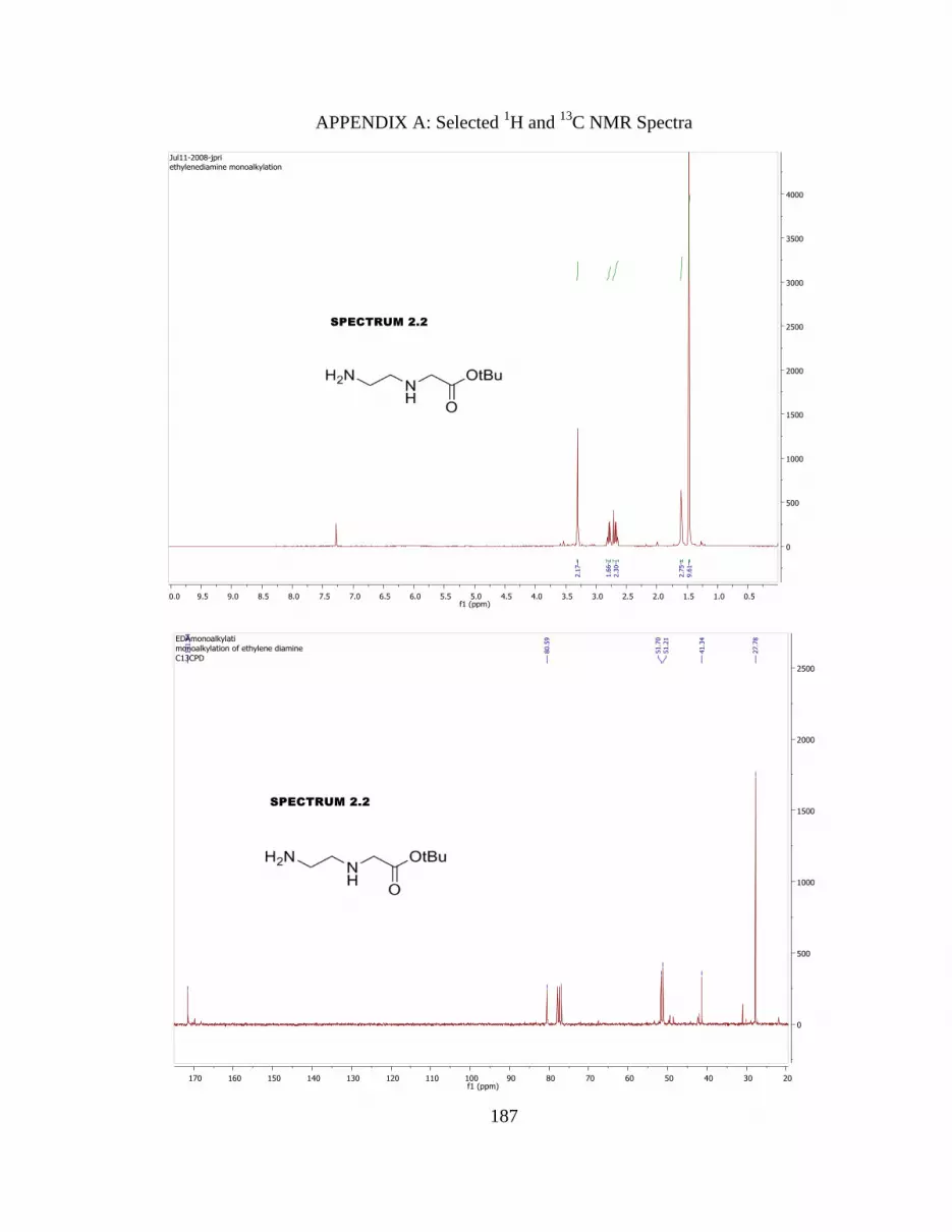
187
APPENDIX A: Selected 1H and
13C NMR Spectra

188

189

190

191

192

193

194

195

196

197
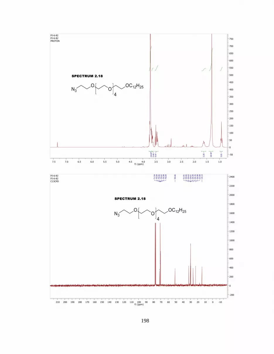
198

199

200

201

202

203

204

205

206

207

208

209

210

211

212

213

214

215

216

217

218

219

220

221

222

223

224

225

226

227

228

229

230

231

232

233

234

235

236

237

238

239
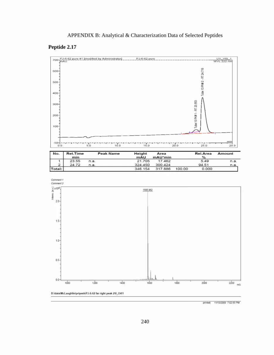
240
APPENDIX B: Analytical & Characterization Data of Selected Peptides
Peptide 2.17

241
Peptide 2.1

242
Peptide 2.2

243
Peptide 2.3

244
Peptide 2.4

245
Peptide 2.5

246
Peptide 2.6

247
Peptide 2.7

248
Peptide 2.8

249
Peptide 2.9

250
Peptide 2.10

251
Peptide 2.11

252
Peptide 2.12

253
Peptide 2.13

254
Peptide 2.14

255
Peptide 2.15

256
Peptide 2.16

257
Peptide 2.17

258
Peptide 2.18
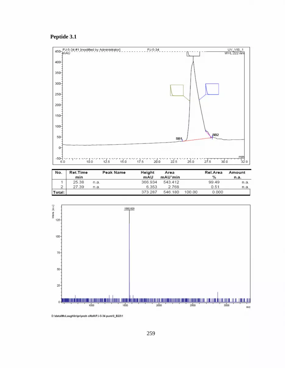
259
Peptide 3.1

260
Peptide 3.2
Peptide 3.3

261
Peptide 3.4
Peptide 3.5

262
Peptide 3.6

263
Peptide 4.1
Peptide 4.2

264
Peptide 4.3
About the author
Mr. Priyesh Jain received his Bachelor’s degree in Paints & Polymer Technology from
University Department of Chemical Technology, University of Mumbai, India in 2000.
He worked as Technical officer (R&D) in Goodlass Nerolac paints, Mumbai till
September 2001. He completed his Master’s degree in Paints & Polymer Technology
from UDCT in August 2003. Later he worked for Anmol Chemical Industries, New
Delhi, India as Assistant Manager (R&D). Mr. Jain started his career at USF in Spring
2005 as a graduate student in Dr. Mark McLaughlin’s group. He continued his research in
synthetic organic chemistry and medicinal chemistry to receive doctoral degree in
Fall 2010.