depression and antidepressants: molecular and cellular aspects
TRANSCRIPT

REVIEW
Depression and antidepressants: molecular and cellular aspects
Cristina Lanni Æ Stefano Govoni Æ Adele Lucchelli ÆCinzia Boselli
Received: 25 February 2009 / Revised: 28 April 2009 / Accepted: 20 May 2009 / Published online: 12 June 2009
� Birkhauser Verlag, Basel/Switzerland 2009
Abstract Clinical depression is viewed as a physical and
psychic disease process having a neuropathological basis,
although a clear understanding of its ethiopathology is still
missing. The observation that depressive symptoms are
influenced by pharmacological manipulation of monoam-
ines led to the hypothesis that depression results from
reduced availability or functional deficiency of monoam-
inergic transmitters in some cerebral regions. However,
there are limitations to current monoamine theories related
to mood disorders. Recently, a growing body of experi-
mental data has showed that other classes of endogenous
compounds, such as neuropeptides and amino acids, may
play a significant role in the pathophysiology of affective
disorders. With the development of neuroscience, neuronal
networks and intracellular pathways have been identified
and characterized, describing the existence of the interac-
tion between monoamines and receptors in turn able
to modulate the expression of intracellular proteins
and neurotrophic factors, suggesting that depression/
antidepressants may be intermingled with neurogenesis/
neurodegenerative processes.
Keywords Depression � Neurotransmitter �Neuropeptide � Brain plasticity � Growth factor
Introduction
Depression is a complex psychiatric disorder affecting
approximately 15% of the population with high morbidity
and mortality. Major depressive disorder (MDD) is com-
mon in the general population; it affects up to one-sixth of
the population.
Despite high prevalence and socioeconomic impact,
little is known about the etiology of depressive illnesses.
The underlying causes of most mood disorders remain
unknown. Heritability estimates demonstrate up to a 50%
genetic component based on family aggregation and con-
trasting monozygotic and dizygotic twin studies [1]. This
strong heritable component to psychiatric illnesses, when
coupled with environmental influences, results in increased
susceptibility to develop the illness in response to stressful/
adverse environmental conditions. Population genetic
studies suggest that there is an increased risk of bipolar
illness in the relatives of bipolar patients and an increased
risk of unipolar depressive illness in the relatives of both
bipolar and unipolar patients. Intensive research efforts
have been made to better characterize the genetic under-
pinnings of mental illness. Technological advances,
including the completion of the human genome inventory,
chromosome mapping, high throughput DNA sequencing,
and others, offer the promise of identifying the genetic
basis of mental illnesses. Several linkage and association
studies showed that the inheritance for major depressive
illness could reside on chromosomes 11 and 18 [2]. In
particular, on chromosome 18, two putative candidate
genes have been identified: the gene for corticotrophin
receptor (CRF1) and for Gs protein-alpha subunit. How-
ever, recent genome scan meta-analysis yielded somewhat
contrasting conclusions with a relatively low significance
for quantitative trait loci on chromosomes 9, 10, 14, and 18
C. Lanni � S. Govoni � A. Lucchelli � C. Boselli (&)
Department of Experimental and Applied Pharmacology,
Centre of Excellence in Applied Biology, University of Pavia,
Viale Taramelli 14, 27100 Pavia, Italy
e-mail: [email protected]
Cell. Mol. Life Sci. (2009) 66:2985–3008
DOI 10.1007/s00018-009-0055-x Cellular and Molecular Life Sciences

[1]. To date, unfortunately, no gene or genetic risk factor
has been convincingly identified yet for depression.
Depression and antidepressants: still matter
of neurotransmitters?
Almost 4 decades of intensive research have sought to
elucidate the neurobiological bases of depression. How-
ever, the study of biological bases of depression is still
overdue. The principal aims are to increase the knowledge
of the basis of the disease in order to better define its
pathogenic process and to identify predictive biomarkers
or, at least, markers able to support the diagnosis. Within
this context, a great variety of bioumoral parameters have
been explored as well as the existence of a genetic com-
ponent of depression. Epidemiological studies have
revealed that both genetic and environmental factors con-
tribute to the risk for depression. Adverse early life
experiences influence neurobiological systems, leading to
the neurobiological and behavioral manifestations of
depression. Several data on biological fluids from patients
affected by depression, on brain tissue from suicide sub-
jects, and the evidence of a clinical activity of the
antidepressant drugs suggested that depression is associ-
ated with an impairment mainly in monoaminergic
neurotransmission systems (see below). These observations
are mainly indirect and based on the role of the aminergic
cerebral circuits on functions, such as attention and alert-
ness, which are altered in depressive illnesses, and on
behavioral effects induced by drugs acting on aminergic
transmission.
The catecholamine/serotonin theory: a brief view
The observation that depressive symptoms were influenced
by the pharmacological manipulation of monoaminergic
system led to the hypothesis that depression results from
reduced availability or functional deficiency of monoam-
inergic transmitters in some cerebral regions [3–6] (see
Table 1 and Fig. 1 for a better comprehension of the role of
noradrenaline (NE), serotonin (5-HT), and dopamine (DA)
in depression [7–33]). This view was supported by the
pharmacological action of both tricyclic antidepressants
(TCAs) and monoamine oxidase inhibitors (MAOIs), able
to acutely increase synaptic levels of monoamines, and by
drugs, such as reserpine, to induce depression [10]. The
idea that exposure to chronic stress or cerebral disorders
could decrease levels and activity of monoamines, thus
leading to depression, took place. On this conceptual basis
the commonly used antidepressants were developed. All
the antidepressant drugs now in use modulate monoamine
neurotransmission, both acutely and chronically, although
their therapeutic effects need as long as 6 to 8 weeks to
develop, and each drug is efficacious in only 60–70% of
patients [34].
The monoaminergic hypothesis has enjoyed consider-
able support, since it attempts to provide a pathophy-
siologic explanation for the actions of antidepressants.
However, in its original form it is clearly inadequate, as it
does not provide a complete explanation for the actions of
antidepressants, and the pathophysiology of depression
itself remains unknown. The hypothesis has evolved over
the years to include, for example, adaptive changes in
receptors to explain why there should be only a gradual
clinical response to antidepressant treatment when the
increase in availability of monoamines is rapid. Still, the
monoamine hypothesis does not address key issues such as
the reason why antidepressants are also effective in other
disorders, such as panic disorder, obsessive-compulsive
disorder, and bulimia, or why all drugs that enhance
serotonergic or noradrenergic transmission are not neces-
sarily effective in depression (for example, amphetamine or
cocaine). Despite these limitations, however, it is clear that
the development of the monoamine hypothesis has been of
great importance in understanding depression and in the
development of safe and quite effective pharmacologic
agents for its treatment.
In the past 2 decades, with the development of neuro-
science, a great deal of success has been achieved in our
understanding of the neurobiological basis of depressive
syndromes and of cerebral systems’ activity, mainly con-
cerning receptor-mediated intracellular mechanisms.
Neuronal networks have been identified and characterized.
It has been understood that the interaction monoamine-
receptor is able to modulate the expression of intracellular
proteins, such as CREB (cAMP response element binding
protein) and BDNF (brain-derived neurotrophic factor).
Through a number of their receptor subtypes, the seroto-
nergic and noradrenergic systems regulate the cyclic
adenosine 30,50-monophosphate (cAMP)-mediated signal
transduction cascade, which activates protein kinases that
phosphorylate specific proteins. One of these proteins is the
transcription factor CREB, which mediates the cAMP
activation of certain genes, such as BDNF [35]. This
increased understanding of mechanims at the base of psy-
chopathologies suggests that, beyond receptors, depressive
mood is probably characterized by modifications in neu-
rotrophic factors, CREB, and BDNF, which are regulated
in turn by a modulation in gene activation.
However, despite the advances in recent years, two
critical concepts should be carefully pointed out. It is worth
noting that, in spite of an increase in knowledge about the
neurobiological basis of depressive syndromes and about
cerebral systems’ activity, no valid alternative hypothesis
to monoaminergic theory is available to date. This might be
2986 C. Lanni et al.
due to the nature of clinical studies or the experimental
models now available to examine the neuronal state of
depressed patients. On the other hand, it should be noticed
that animal models can provide crucial information on the
whole range of neuronal functions not accessible in
humans and potentially allow us to identify novel targets
for antidepressant drug development, although they do not
predict the clinical outcome and the latency time necessary
for the therapeutic response.
Other classical neurotransmitters
Recently, some important advances in a better under-
standing of the psychopathology of depression have been
made. Moving beyond monoamines, a growing body of
experimental data indicates that other classes of com-
pounds, such as neuropeptides and amino acids (the
glutamatergic and GABA-ergic systems), are likely to play
a significant role in the pathophysiology of affective
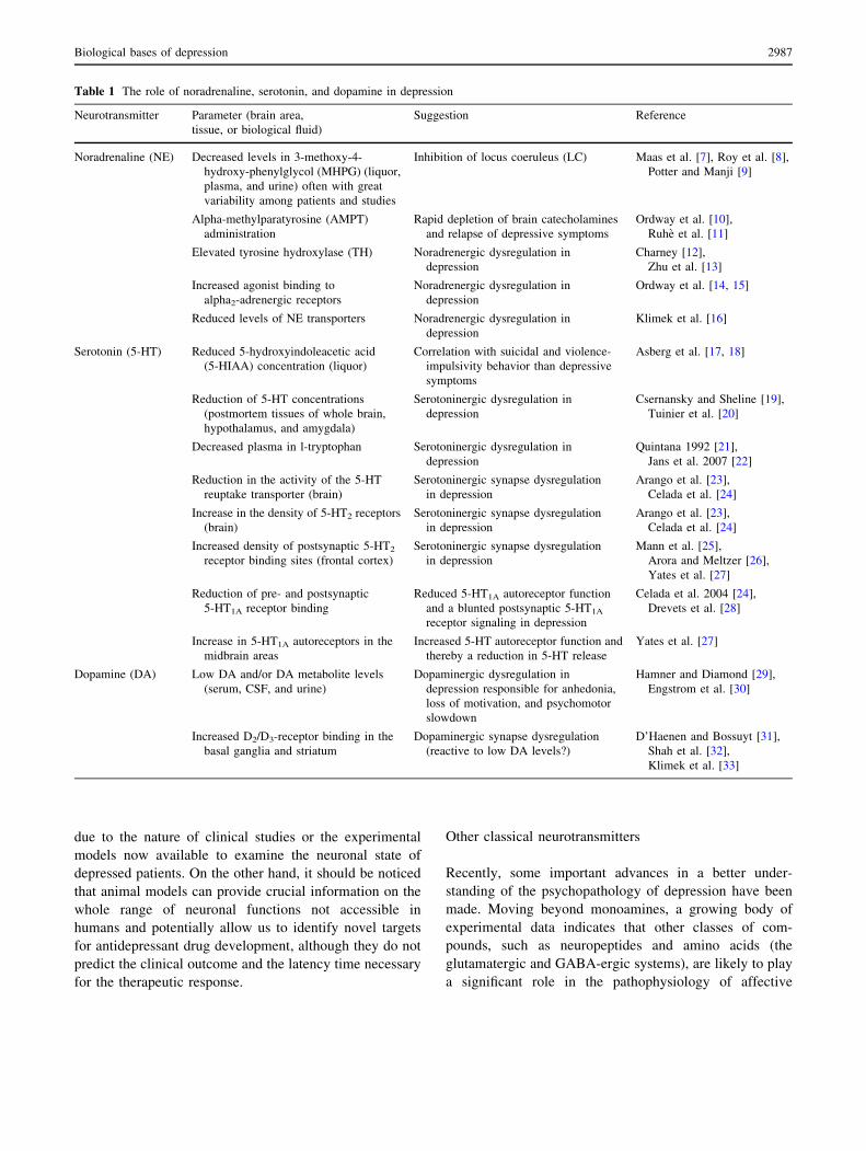
Table 1 The role of noradrenaline, serotonin, and dopamine in depression
Neurotransmitter Parameter (brain area,
tissue, or biological fluid)
Suggestion Reference
Noradrenaline (NE) Decreased levels in 3-methoxy-4-
hydroxy-phenylglycol (MHPG) (liquor,
plasma, and urine) often with great
variability among patients and studies
Inhibition of locus coeruleus (LC) Maas et al. [7], Roy et al. [8],
Potter and Manji [9]
Alpha-methylparatyrosine (AMPT)
administration
Rapid depletion of brain catecholamines
and relapse of depressive symptoms
Ordway et al. [10],
Ruhe et al. [11]
Elevated tyrosine hydroxylase (TH) Noradrenergic dysregulation in
depression
Charney [12],
Zhu et al. [13]
Increased agonist binding to
alpha2-adrenergic receptors
Noradrenergic dysregulation in
depression
Ordway et al. [14, 15]
Reduced levels of NE transporters Noradrenergic dysregulation in
depression
Klimek et al. [16]
Serotonin (5-HT) Reduced 5-hydroxyindoleacetic acid
(5-HIAA) concentration (liquor)
Correlation with suicidal and violence-
impulsivity behavior than depressive
symptoms
Asberg et al. [17, 18]
Reduction of 5-HT concentrations
(postmortem tissues of whole brain,
hypothalamus, and amygdala)
Serotoninergic dysregulation in
depression
Csernansky and Sheline [19],
Tuinier et al. [20]
Decreased plasma in l-tryptophan Serotoninergic dysregulation in
depression
Quintana 1992 [21],
Jans et al. 2007 [22]
Reduction in the activity of the 5-HT
reuptake transporter (brain)
Serotoninergic synapse dysregulation
in depression
Arango et al. [23],
Celada et al. [24]
Increase in the density of 5-HT2 receptors
(brain)
Serotoninergic synapse dysregulation
in depression
Arango et al. [23],
Celada et al. [24]
Increased density of postsynaptic 5-HT2
receptor binding sites (frontal cortex)
Serotoninergic synapse dysregulation
in depression
Mann et al. [25],
Arora and Meltzer [26],
Yates et al. [27]
Reduction of pre- and postsynaptic
5-HT1A receptor binding
Reduced 5-HT1A autoreceptor function
and a blunted postsynaptic 5-HT1A
receptor signaling in depression
Celada et al. 2004 [24],
Drevets et al. [28]
Increase in 5-HT1A autoreceptors in the
midbrain areas
Increased 5-HT autoreceptor function and
thereby a reduction in 5-HT release
Yates et al. [27]
Dopamine (DA) Low DA and/or DA metabolite levels
(serum, CSF, and urine)
Dopaminergic dysregulation in
depression responsible for anhedonia,
loss of motivation, and psychomotor
slowdown
Hamner and Diamond [29],
Engstrom et al. [30]
Increased D2/D3-receptor binding in the
basal ganglia and striatum
Dopaminergic synapse dysregulation
(reactive to low DA levels?)
D’Haenen and Bossuyt [31],
Shah et al. [32],
Klimek et al. [33]
Biological bases of depression 2987

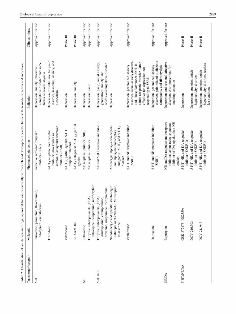
disorders. The compounds currently under investigation are
summarized in Table 2.
Glutamate
Glutamate is the major mediator of excitatory synaptic
transmission in the mammalian brain [36]. In the human
brain, glutamatergic neurons project within and from the
cortex to the subcortical regions, such as the locus coeru-
leus, raphe nucleus, and substantia nigra, where mono-
aminergic pathways are modulated. Under normal conditions,
the glutamatergic system plays a role in a wide array of
physiological functions, including memory and other cog-
nitive tasks, neurotrophic and neurotoxic actions, and the
induction of neuronal plasticity. However, in pathological
conditions it is known to be a potent neuronal excitotoxin,
triggering either rapid or delayed neurotoxicity. To date,
an abnormal function of the glutamatergic system is
known to be implicated in the pathophysiology of several
neurodegenerative disorders, such as Huntington’s chorea,
epilepsy, Alzheimer’s disease, schizophrenia, and anxiety
disorders. Recent and increasing evidence suggests that
abnormal activity of the glutamatergic system is likely to
contribute to the impairments in the synaptic and neural
plasticity observed in patients with severe mood disorders
[37]. Glutamatergic abnormalities have been reported in
plasma [38–40], serum [41], cerebrospinal fluid [42, 43],
and brain tissue [44] of subjects affected by mood disor-
ders. However, these studies are compromised by problems
with potential medication effects, lack of appropriate
control for diagnosis, post-mortem effects on metabolism,
and the inability to identify the precise source of the glu-
tamate in the peripheral tissue. Brain imaging studies
showed the presence of elevated glutamate levels in the
occipital cortex of depressed patients and decreased glu-
tamate levels in the anterior cingulate cortex of MDD
patients [37]. In addition, an increased glutamate content in
several brain regions of individuals with bipolar disorder
has also been reported. Several studies showed glutamate
receptor alterations, mainly differences related to NMDA
(N-methyl-D-aspartate) receptor expression and binding
affinities between individuals with and without mood dis-
orders. Decreased NMDA receptor binding and NR1
subunit expression have been reported in the superior
temporal cortex of subjects with MDD and bipolar disorder
[45]. Interestingly, two polymorphisms of the GRIN1 gene
coding for the NR1 subunit and two other polymorphisms
in the GRIN2B gene coding for NR2B were also recently
reported to be associated with bipolar disorder [46, 47]. On
the contrary, fewer postmortem studies have investigated
the link between AMPA (a-amino-3-hydroxy-5-methyl-4-
isoxazole propionic acid) and kainate receptors and mood
disorders. Only a single study reported an increased AMPA
binding associated with a decreased glutamate receptor 1
(GLUR1) subunit expression in the striatum in bipolar
disorder [48]. Furthermore, decreased levels of GLUR2
and GLUR3 were found in subjects with MDD [49].
Interestingly, the STAR*D (sequenced treatment alterna-
tives to relieve depression) study, the largest prospective
treatment trial for MDD, recently demonstrated associa-
tions of genetic polymorphisms within kainate and AMPA-
glutamate receptors GRIK2 and GRIA3 with treatment-
emergent suicidal ideation [50, 51], an uncommon symptom
found in some patients following antidepressant treatment
initiation. These studies, even if not yet confirmed, suggest
the existence of a relationship between antidepressant
treatment-emergent suicidal ideation and the glutamate
system [52, 53]. In addition, many studies highlighted
the importance of glial deficits in mood disorders as they
could cause a complex disturbance in glutamatergic regu-
lation. Deficits in glia function, as are found in some types
of epilepsy, appear to be associated with dysfunctional
accumulations of synaptic glutamate [54]. Postmortem
studies [55, 56] have shown cortical glial cell loss and
reduced glial density in patients with mood disorders. Glial
loss has been well-documented in several brain regions,
such as the dorsolateral prefrontal cortex [57], the orbito-
frontal cortex [57], and the amygdala [58] from subjects
with MDD and bipolar disorder. Altogheter, these data
provide a link between the glutamatergic neurotransmitter
system and the pathophysiology of mood disorders, thus
suggesting that agents acting on the glutamatergic system
can be explored as potential treatment in this field. Within
this context, in fact, ketamine, an NMDA receptor anta-
gonist, has been recently demonstrated effective in
treating antidepressant-resistant patients [59]. Ketamine is
Fig. 1 Relationship among noradrenaline, serotonin, and dopamine
and some behaviors
2988 C. Lanni et al.
Ta
ble
2C
lass
ifica
tio
no
fan
tid
epre
ssan
td
rug
s,ap
pro
ved
for
use
or
curr
entl
yin
rese
arch
and
dev
elo
pm
ent,
on
the
bas
iso
fth
eir
mo
de
of
acti
on
and
ind
icat
ion
Tra
nsp
ort
er/r
ecep
tor
Mo
lecu
leP
har
mac
olo
gic
acti
on
Ind
icat
ion
Cli
nic
alp
has
e
5-H
TF
luo
xet
ine,
par
ox
etin
e,fl
uv
ox
amin
e,
cita
lop
ram
,es
cita
lop
ram
Sel
ecti
ve
sero
ton
inre
up
tak
e
inh
ibit
or
(SS
RI)
Dep
ress
ion
,an
xie
ty,
ob
sess
ive-
com
pu
lsiv
ed
iso
rder
,an
dso
me
form
so
fse
ver
esh
yn
ess
Ap
pro
ved
for
use
Tra
zod
on
e5
-HT
2re
up
tak
ean
dre
cep
tor
inh
ibit
or;
also
kn
ow
nas
sero
ton
inan
tag
on
ist
reu
pta
ke
inh
ibit
or
(SA
RI)
Dep
ress
ion
;o
ff-l
abel
use
for
pan
ic
dis
ord
er,
inso
mn
ia,
anx
iety
,an
d
alco
ho
lism
Ap
pro
ved
for
use
Vil
azo
do
ne
5-H
T1A
par
tial
ago
nis
t,5
-HT
reu
pta
ke
inh
ibit
or
Dep
ress
ion
Ph
ase
III
Lu
AA
21
00
45
-HT
3an
tag
on
ist,
5-H
T1A
par
tial
ago
nis
t
Dep
ress
ion
,an
xie
tyP
has
eII
I
NE
Reb
ox
etin
eN
Ere
up
tak
ein
hib
ito
r(N
RI)
Dep
ress
ion
Ap
pro
ved
for
use
Tri
cycl
ican
tid
epre
ssan
ts(T
CA
):
amo
xap
ine,
des
ipra
min
e,n
ort
rip
tyli
ne
NE
reu
pta
ke
inh
ibit
or
Dep
ress
ion
,p
anic
Ap
pro
ved
for
use
5-H
T/N
ET
ricy
clic
anti
dep
ress
ants
(TC
A):
Am
itri
pty
lin
e,cl
om
ipra
min
e,
do
xep
ine,
imip
ram
ine,
trim
ipra
min
e
NE
and
5-H
Tre
up
tak
ein
hib
ito
rD
epre
ssio
n,
pan
ic,
soci
alan
xie
ty,
gen
eral
ized
anx
iety
,an
d
ob
sess
ive-
com
pu
lsiv
ed
iso
rder
Ap
pro
ved
for
use
No
rad
ren
erg
ican
dsp
ecifi
cse
roto
ner
gic
anti
dep
ress
ant
(NaS
SA
):M
irta
zap
ine,
mia
nse
rin
e
Ad
ren
erg
ical
ph
a 2-a
uto
rece
pto
rs
and
alp
ha 2
-het
ero
rece
pto
rs
anta
go
nis
t,5
-HT
2an
d5
-HT
3
blo
cker
Dep
ress
ion
Ap
pro
ved
for
use
Ven
lafa
xin
e5
-HT
and
NE
reu
pta
ke
inh
ibit
or
(SN
RI)
Dep
ress
ion
,g
ener
aliz
edan
xie
ty
dis
ord
er,
soci
alan
xie
tyd
iso
rder
,
and
,si
nce
No
vem
ber
20
05
,in
adu
lts
for
pan
icd
iso
rder
.O
ften
effe
ctiv
efo
rd
epre
ssio
nn
ot
resp
on
din
gto
SS
RIs
Ap
pro
ved
for
use
Du
lox
etin
e5
-HT
and
NE
reu
pta
ke
inh
ibit
or
(SN
RI)
Dep
ress
ion
,g
ener
aliz
edan
xie
ty
dis
ord
er,
pai
nre
late
dto
dia
bet
ic
neu
rop
ath
y,
and
fib
rom
yal
gia
Ap
pro
ved
for
use
NE
/DA
Bu
pro
pio
nN
Ean
dD
Are
up
tak
ean
dre
cep
tors
inh
ibit
or;
abo
ut
twic
eas
po
ten
t
inh
ibit
or
of
DA
up
tak
eth
anN
E
up
tak
e
Dep
ress
ion
and
seas
on
alaf
fect
ive
dis
ord
er.
Als
op
resc
rib
edfo
r
smo
kin
gce
ssat
ion
Ap
pro
ved
for
use
5-H
T/N
E/D
AG
SK
37
24
75
(NS
23
59
)5
-HT
,N
E,
and
DA
reu
pta
ke
inh
ibit
or
(SN
DR
-I)
Dep
ress
ion
Ph
ase
II
DO
V2
16
,30
35
-HT
,N
E,
and
DA
reu
pta
ke
inh
ibit
or
(SN
DR
I)
Dep
ress
ion
,at
ten
tio
nd
efici
t
hy
per
acti
vit
yd
iso
rder
Ph
ase
II
DO
V2
1,
94
75
-HT
,N
E,
and
DA
reu
pta
ke
inh
ibit
or
(SN
DR
I)
Dep
ress
ion
,at
ten
tio
nd
efici
t
hy
per
acti
vit
yd
iso
rder
,re
stle
ss
leg
syn
dro
me
Ph
ase
II
Biological bases of depression 2989

Ta
ble
2co
nti
nu
ed
Tra
nsp
ort
er/r
ecep
tor
Mo
lecu
leP
har
mac
olo
gic
acti
on
Ind
icat
ion
Cli
nic
alp
has
e
Glu
tam
ate
TIK
-10
1/D
-cy
clo
seri
ne
NM
DA
par
tial
ago
nis
tA
nx
iety
dis
ord
ers
Ph
ase
II
AZ
D6
76
5N
MD
Are
cep
tor
anta
go
nis
tD
epre
ssio
nP
has
eII
Del
uce
min
e(N
PS
15
06
)N
MD
Aan
tag
on
ist
Dep
ress
ion
,st
rok
eP
has
eI
AF
Q0
56
mG
luR
5re
cep
tor
anta
go
nis
tA
nx
iety
dis
ord
ers
Ph
ase
I
GA
BA
AZ
D7
32
5G
AB
Are
cep
tor
par
tial
ago
nis
tA
nx
iety
dis
ord
ers
Ph
ase
II
AZ
D6
28
0G
AB
Are
cep
tor
par
tial
ago
nis
tA
nx
iety
dis
ord
ers
Ph
ase
I
En
zym
eM
ole
cule
Ph
arm
aco
log
icac
tio
nIn
dic
atio
nC
lin
ical
ph
ase
Mo
no
amin
eo
xid
ase
Ipro
nia
zid
e,tr
any
lcy
pro
min
e,p
hen
elzi
ne
Irre
ver
sib
lem
on
oam
ine
ox
idas
e
inh
ibit
or
(MA
O-I
)
Dep
ress
ion
,p
anic
,an
dso
cial
ph
ob
ia
Ap
pro
ved
for
use
Mo
clo
bem
ide
Rev
ersi
ble
mo
no
amin
eo
xid
ase
Inh
ibit
or
(RIM
A)
Dep
ress
ion
,an
xie
tyA
pp
rov
edfo
ru
se
CX
15
7(T
yri
maT
M)
Rev
ersi
ble
mo
no
amin
eo
xid
ase
A
(MA
O-A
)in
hib
ito
r
Dep
ress
ion
,an
xie
tyP
has
eII
CX
26
14
Rev
ersi
ble
mo
no
amin
eo
xid
ase
inh
ibit
or
(RIM
A)
Dep
ress
ion
,an
xie
tyP
recl
inic
al
Neu
rop
epti
de
Mo
lecu
leP
har
mac
olo
gic
acti
on
Ind
icat
ion
Cli
nic
alp
has
e
Tac
hy
kin
inS
ared
uta
nt
(SR
48
96
8)
NK
2n
euro
kin
inre
cep
tor
anta
go
nis
t
Dep
ress
ion
,an
xie
tyP
has
eII
I
GW
82
32
96
(orv
epit
ant)
NK
1an
tag
on
ist
Dep
ress
ion
,an
xie
tyP
has
eI
SA
R1
02
27
9N
K2
rece
pto
ran
tag
on
ist
Dep
ress
ion
,an
xie
tyP
re-c
lin
ical
CR
FP
exac
erfo
nt
(BM
S-5
62
08
6)
CR
F1
anta
go
nis
tD
epre
ssio
n,
anx
iety
Ph
ase
III
SS
R1
25
54
3C
RF
1an
tag
on
ist
Dep
ress
ion
,an
xie
ty,
hy
per
ph
agia
Ph
ase
I
-Un
nam
ed-
CR
F1
anta
go
nis
tD
epre
ssio
n,
anx
iety
Pre
-cli
nic
al
GS
K5
61
67
9C
RF
1an
tag
on
ist
So
cial
anx
iety
dis
ord
er,
MD
DP
has
eII
GW
87
60
08
CR
F1
anta
go
nis
tS
oci
alan
xie
tyd
iso
rder
Ph
ase
II
Arg
inin
ev
aso
pre
ssin
SS
R1
49
41
5V
1b
anta
go
nis
tD
epre
ssio
n,
anx
iety
dis
ord
ers
Ph
ase
II
Inth
ista
ble
mo
od
stab
iliz
ers
and
nat
ura
lsu
bst
ance
sar
en
ot
men
tio
ned
.T
he
tab
leh
asb
een
bu
ilt
on
the
bas
iso
fav
aila
ble
dat
ash
eets
of
the
dru
gs
app
rov
edfo
ru
sean
do
nth
ese
arch
on
the
site
Cli
nic
alT
rial
.go
v(c
on
sult
ed2
Feb
ruar
y2
00
9)
and
/or
the
site
(s)
of
the
pro
du
cers
2990 C. Lanni et al.
of particular interest because it has been shown to yield a
rapid and sustained antidepressant effect and has been
reported as the only known drug leading to such a dramatic
and prolonged response with a single administration [60].
In particular, Maeng and Zarate hypothesized that the
rapid onset and the prolonged effects of ketamine are not
the consequence of neuroplastic changes from glutamate
modulation, rather to an increase in AMPA relative to
NMDA glutamatergic throughput, which results in increased
synaptic potentiation (for more detailed information,
see [60]).
However, NMDA receptor antagonists of this class may
have adverse psychiatric effects and may impair memory.
Moreover, other than ketamine, also memantine, another
NMDA receptor blocker exerting a voltage-dependent
reversible block of this ionotropic receptor, which has been
approved for the treatment of moderate-severe Alzheimer’s
disease, demonstrated early onset efficacy in patients with
MDD [61].
Gamma-aminobutyric acid
Gamma-aminobutyric acid (GABA) is the most widely
distributed inhibitory transmitter in the mammalian brain
[57]. In regions such as the cerebral cortex, hippocampus,
thalamus, basal ganglia, cerebellum, and brain stem, it
represents about one-third of the synapses [62]. GABA
transmission is present in interneurons modulating local
neuronal circuitry, such as noradrenergic, dopaminergic,
and serotonergic neurons [63]. In the past decades con-
siderable evidence has emerged that, in the brain, GABA
acts as a modulator of neuronal function and behavioral
processes, such as sleep, feeding, aggression, sexual
behavior, pain, cardiovascular regulation, thermoregula-
tion, locomotor activity, and mood [62, 64, 65]. This has
been supported by the reduced GABA content in the
plasma and cerebrospinal fluid of depressed patients [66–
69]. In vivo proton magnetic resonance spectroscopy, a
technique for a quantitative analysis of GABAergic sys-
tems with the use of brain scan, showed reduced occipital
cortex GABA concentrations in a sample of severely
depressed subjects [70]. Other studies demonstrated that
treatment of depression with electroconvulsive therapy
[71] or selective 5-HT reuptake inhibitor agents [72] was
able to increase occipital cortex GABA concentrations in
MDD patients, thus suggesting that GABA abnormalities
may be normalized by antidepressant medication. Gluta-
mate decarboxylase (GAD) and GABA aminotransferase
are the enzymes involved in GABA synthesis and degra-
dation [73]. GAD 65 and 67 isoforms were found to be
decreased in the cerebellum in samples from both bipolar
disorders and schizophrenia [74]. Additional studies indi-
cated a reduction in GABA concentrations and increased
GABAA receptor binding in the brain of depressed suicide
victims, thus suggesting an impairment in GABAergic
neurotransmission [75, 76]. The decreased GABA concen-
trations found in depression may reflect either decreased
rates of synthesis or increased rates of catabolism [77].
However, these studies were not always confirmed, proba-
bly because of the rapid postmortem changes in brain GAD
activity and GABA content. Furthermore, from a genetic
point of view, genes coding for GABAA receptor subunits
have been found to be associated with mood disorders.
Reports from the literature showed that polymorphisms of
the GABAA receptor 1 subunit (GABRA1) can be associ-
ated with mood disorders [78, 79], as well as the GABAA
receptor a6 subunit variant (Pro385Ser) [79]. However, not
all the studies found a significant association of the
GABRA1 gene with depression or bipolar disorder [80].
Despite quite consistent findings of GABA deficits in mood
disorders, GABAergic drugs, such as benzodiazepines,
acting as allosteric agonist of the GABAA receptor, have not
proven to be clinically efficacious as a monotherapy in the
treatment of MDD [53, 77].
On the other hand, anxiety is frequently a comorbid
condition in depressed patients, and some of the distur-
bances belonging to the anxiety spectrum are better treated
with the SSRI antidepressant drugs than with benzodiaze-
pines. Conversely, there are non-benzodiazepine anxiety
relievers, such as buspirone, working on serotoninergic
terminals. In addition, it should be stressed that presynaptic
GABA receptors, both GABAA and GABAB, may regulate
monoaminergic terminal activity. These observations do
suggest that there is still room to investigate both at the
clinical and preclinical level the role of GABAergic
transmission in depression and in depression with comor-
bid anxiety.
Acetylcholine
Cholinergic systems, among other neurotransmitters in
the brain, appear to be associated with various altered
behaviors, such as psychosis, depression, agitation, and
personality changes. One of the most consistent findings
in neuropsychiatry is that patients with depression have
dysfunctional neuroendocrine systems possibly resulting
from prolonged responses to stress [81, 82]. Acetylcholine
(ACh) plays a significant role in mediating neuroendocrine,
emotional, and physiological responses to stress. For
example, central ACh turnover is increased following
stress [83], and ACh facilitates the release of several stress-
sensitive neurohormones and peptides including cortico-
sterone, adrenocorticotropin (ACTH), and corticotropin-
releasing factor (CRF) [84], suggesting a dynamic interaction
between cholinergic and monoaminergic systems in the
regulation of mood and affect. As proposed by Janowsky
Biological bases of depression 2991

et al. [85, 86], depression could be a manifestation of a
central cholinergic predominance, whereas mania could be
correlated to a relative monoaminergic predominance. The
hypothesis that the overactivity of the cholinergic system
over the adrenergic one could result in depressive symp-
toms [85] has been supported by several lines of evidence.
The presence of exaggerated responses (behavioral, neu-
rochemical, sleep) to cholinergic agents in patients with
affective disorder compared to controls has been reported
[87]. The indirect cholinesterase inhibitor ACh agonist,
physostigmine, when administered to healthy subjects,
produced symptoms of dysphoria, depression, anxiety, and
irritability, but increased depressed mood in manic patients
[88]; pyridostigmine, another cholinesterase inhibitor,
increased growth hormone release to a significantly greater
extent in depressed patients than in healthy subjects, sug-
gesting cholinergic receptor supersensitivity in depressed
subjects [89]. Furthermore, central acting cholinomimetics
decreased rapid eye movement (REM) sleep latency and
increased ACTH and cortisol levels, changes observed in
depression [90–92]. Based on the ‘‘cholinergic-adrenergic
hypothesis of mania and depression,’’ small pilot trials in
humans with cholinesterase inhibitors and muscarinic
agonists suggested that stimulation of muscarinic recep-
tors may produce an antimanic effect [93]. Within this
context, Burt and coworkers [94] demonstrated that the
cholinesterase inhibitor, donepezil, when added to existing
therapy in treatment-resistant bipolar patients, improved
symptoms in over half of the patients. Bymaster and Felder
[93] proposed that the antimanic activity could be medi-
ated by activation of muscarinic M4 receptor. The
rationale for the potential use of M4 agonists in the treat-
ment of mania is based on the fact that these molecules,
similarly to mood stabilizers, decrease cAMP formation
and can block DA receptor functions (motor and mood
effects) in cortical and limbic areas, which are affected in
bipolar disorders [93]. Moreover, consistently with the
hypothesis generated by Janowsky et al. [85], also the
antidepressant effects of antimuscarinic agents, such as
scopolamine, have been reported, thus supporting the link
between muscarinic receptor function and mood disorders.
Within this context, a rapid and robust antidepressant
response to scopolamine in depressed patients with poor
prognoses has been observed [95, 96]. Although the spe-
cific mechanism through which antimuscarinic effects
may positively impact the pathogenesis of depression is
still unknown, scopolamine seems to modulate, as other
somatic antidepressant drugs, NMDA receptor function. In
particular, muscarinic receptor stimulation regulates
NMDA receptor gene expression in some brain regions,
and scopolamine has been found to decrease messenger
RNA concentrations for NMDA receptors 1A and 2A in
the rat brain [97].
Furthermore, the role of nicotinic acetylcholine recep-
tors (nAChRs) has also been investigated. Shytle et al. [98]
and Silver et al. [99] have discussed the role of antide-
pressant medications in inhibiting nAChRs and suggested
that antagonism of these receptors could contribute to their
effectiveness in reducing symptoms of depression and
mood instability in patients with comorbid depression and
bipolar disorder. Modulation of nAChRs by antidepres-
sants, in fact, plays important roles in neuronal circuits
regulating mood, by regulating stress-related effects in
hypothalamic pituitary adrenal axis, behavioral reinforce-
ment in mesolimbic DA system and citoplasticity in
hippocampus [98]. Based on the evidence supporting the
hypothesis that nicotinic receptors inhibition may reduce
depression and stabilize mood [100], future research should
fully investigate the therapeutic potential of nicotinic
receptor antagonists as a novel approach to treat affective
disorders. However, much work has to be done. It is still
controversial whether the antidepressant-like effect of
nAChRs modulation is induced by activation, desensitiza-
tion, or inhibition of central nAChRs, and to date the
specific nAChR subtype/s involved remain unknown.
Furthermore, this hypothesis is also somewhat controver-
sial, as suggested by data showing that acetylcholinesterase
inhibitors relieve depressive symptoms, even in demented
patients [101, 102]. The putative use of nicotinic antagonists
could be based on the fact that nicotinic agonists increase
agitation and anxiety, symptoms which can be signs of
depression. On the other hand, such an effect in these patients
may be linked to the action of cholinesterase inhibition on the
cognitive function.
Finally, the potential antidepressant activity of drugs
inhibiting cholinergic activity, as outlined before, should
be carefully evaluated taking into consideration the possi-
ble negative effects on the cognitive function of such
agents. A clinical observation supports this warning. Lit-
erature data show that in the elderly tricyclic antidepressant
and SSRI with muscarinic receptor blocking properties
should be avoided, if possible, because of their anticho-
linergic effect both in the periphery and on cognition [103,
104].
Other non-classical (peptidergic) transmitters
About 30 years ago the observation that neuropeptides
serve as signalling molecules in the nervous system gen-
erated great expectations for the drug industry. Much
progress has been made since then in exploiting neuro-
peptide systems pharmacologically in the psychiatric field.
Nowadays, the health burden of stress-related diseases,
including depression and anxiety disorders, is rapidly
increasing, whereas the range of available pharmacother-
apies to treat these disorders is limited. In affective
2992 C. Lanni et al.
disorders several neuropeptides seem to be causally
involved in mediating the response to stress, thus identi-
fying neuropeptide systems as attractive therapeutic targets
for depression and anxiety disorders [105]. Interestingly,
repeated administration of antidepressant drugs is able to
induce adaptive changes in non-monoaminergic systems,
such as orexinergic neurons [106]. Furthermore, other
neuropeptides involved in depression-related responses
such as tachykinins [substance P (SP) and neurokinin A],
CRF, vasopressin, neuropeptide Y, neurotensin, and gala-
nin have received recent attention as candidates for
antidepressant drug development.
Corticotropin-releasing factor (CRF)
The 41 amino acid neuropeptide CRF initiates the hypo-
thalamic-pituitary-adrenal (HPA) axis response to stress
and has been the subject of intense investigation in the
pathophysiology and treatment of depression and anxiety
disorders. A large body of evidence correlates stressful life
events with an increased vulnerability for affective and
anxiety disorders; stressful events often precede the onset
of depression, and stress has also been associated with the
severity of the illness [107, 108]. Furthermore, stressful life
events in childhood have been shown to predispose an
individual for development of mood and anxiety disorders
in adulthood. In mammalian brain, CRF is heterogeneously
distributed throughout the CNS and interacts with two
G-protein-coupled CRF receptor subtypes (CRF1 and CRF2
receptors) [109]. One of the molecular mechanisms asso-
ciated with depression is an abnormal HPA activity [110],
driven by CRF-containing neurons, which start to coex-
press vasopressin when chronically activated, in turn able
to enhance CRF actions. Dysfunction of the HPA system
has been extensively studied since various researchers
independently reported that plasma cortisol concentrations,
following a higher release in ACTH, were elevated in a
majority of patients with MDD. Moreover, synthetic glu-
cocorticoid dexamethasone did not suppress plasma ACTH
and cortisol concentrations in depressed patients [111]. In
depressed patients cortisol levels are increased mainly
during the afternoon and the evening, whereas in healthy
subjects the peaks of plasma cortisol concentrations are
detected during the morning. These abnormal cortisol
levels are due to a defect in neurotransmitter regulation of
CRF release. CRF is hypersecreted from hypothalamic as
well as from extrahypothalamic neurons in depression,
resulting in hyperactivity of the HPA axis and increase in
CSF concentrations of CRF [112]. Elevated concentrations
of CRF in CSF of drug-free patients with MDD and from
suicide victims compared with patients with other psychi-
atric disorders and healthy controls have been reported
[113, 114]. The elevations of CRF neuronal activity are
also believed to mediate certain behavioral symptoms of
depression involving sleep and appetite disturbances,
reduced libido, and psychomotor changes. However, other
studies have been unable to replicate these observations
[115, 116]. Differently from CSF data, cerebral levels of
CRF appeared not to be altered. Transgenic CRF over-
expressing mice showed an increase in anxiogenic
behavior [117] and a different ability to adapt to an envi-
ronmental stressor, probably related to individual
differences in stress reactivity [118]. Interestingly, there is
evidence that CRF1 receptors play an important role in
mediating the HPA response to stress and the extrahypo-
thalamic mediation of stress-related behaviors [119].
Within this context, some studies, showing that the anx-
iolytic-like effects of CRF1 receptor antagonists are more
pronounced in rats pre-exposed to stressors such as
immobilization and forced swimming, allowed to hypoth-
esize that CRF1 receptor antagonists might be capable of
blocking pathological CRF-mediated stress responses
without producing side effects caused by a general sup-
pression of HPA activity [120].
The potential clinical usefulness of CRF1 receptor
antagonists led to synthesizing several CRF1 antagonists
for the treatment of anxiety and depression in patients with
MDD under investigation as reported in Table 2 [121].
Finally, it is worth highlighting the amount of literature
data on ACTH abnormal levels in depression at the
expense of a lack of information on its precursor, the
proopiomelanocortin (POMC) or big-ACTH. POMC ini-
tially is synthesized as an inactive molecule that requires
posttranslational modifications to generate bioactive
products, among which are ACTH, b-endorphin, and
melanocyte stimulating hormone. It should be interesting
to evaluate whether an alteration in POMC is also asso-
ciated with depression and which neuronal structures
could be involved.
Arginine vasopressin
Vasopressin (AVP, arginine vasoPressin) seems to be a key
regulator of HPA axis, acting synergically with CRF to
secrete ACTH and other corticoid hormones in physio-
logical and stress-related conditions [122]. The vasopres-
sinergic system consists of hypothalamic AVP-containing
cells located in the paraventricular and supraoptic nucleus
and extrahypothalamic AVP-containing neurons (limbic
system and amigdala) and may be critically involved in the
ethiopathogenesis of depression as supported by post-
mortem studies on the brains of individuals with a known
history of depression [123]. In depressed patients an
overproduction of AVP has been detected, measured as
increased AVP plasma levels [124, 125], number of AVP
expressing cells in paraventricular nucleus [123], or AVP
Biological bases of depression 2993

content in the paraventricular nucleus [126]. Furthermore,
increased AVP mRNA was detected in supraoptic nuclei
of depressed patients [127]. These findings are consistent
with experimental studies in rodents showing that para-
ventricular neurons are able to coexpress both CRH and
AVP and tend to increase the relative predominance of
AVP production by stressors [128]. Furthermore, in vivo
and in vitro studies showed that a single nucleotide poly-
morphism (SNP) in the AVP locus, specifically the
SNP[A(-1276)G], resulted in overexpression of AVP and
in increased HPA axis function in a rat selected for high
anxiety behavior [129]. In addition in AVP-deficient
rodents, such as Brattleboro rats endowed with a single
nucleotide deletion (-G326) in the AVP gene, some authors
reported low levels of basal ACTH and corticosterone
[130], decreased responses to stress in terms of endocrine
response such as low ACTH response, decreased adrenal
sensitivity to ACTH [131, 132], and behavioral responses
such as decreased anxiety in a variety of models of
depression (open-field, force swimming, and sucrose
preference test) [133, 134]. As other hormones, AVP exerts
its effects through interaction with three specific receptors
(V1a, V1b, or V3 and V2) [135, 136], and the vasopressin 1b
(V1b) receptors seem to be the prominent ones involved in
depression and anxiety disorders, although also the role of
V1a subtype has been considered [137]. Studies in V1b-
receptor KO-mice reported a normal HPA axis function
[138] and a reduced aggressive behavior along with again a
normal neuroendocrine response and social response to
stress [139, 140], with no phenotypic differences in stress-
induced models compared to wild-type mice [141]. This
might be due to some complementary/compensatory
mechanism(s) probably involving other AVP or CRF
receptor subtypes, but further studies are necessary to
explore this hypothesis.
The involvement of the V1b receptor subtype is sup-
ported by experimental evidence using SR149415, a
selective V1b antagonist, at this time in phase II clinical
trials in patients with anxiety and MDD (Table 2) [142].
Preclinical studies in rodents showed that this compound is
endowed with antidepressant-like activity in different
models of depression (forced swimming test, olfactory
bulbectomized-induced hyperemotionality, DRL-72s
schedule test, unpredictable chronic mild stress) [142–146].
Its antidepressant-like effects are also supported by the data
obtained administrating SR149415 directly into the lateral
septum, the central, basolateral, or medial nuclei of amy-
gadala in the rat forced swimming test [147, 148]. Only one
study failed to detect behavioral changes [149]. Further-
more, SR149415 is able to inhibit the enhancement of
ACTH levels induced by acute restrain stress with no
influence on ACTH basal release, as expected by a V1b
receptor antagonist [150]. In addition to the vasopressin
V1b selectivity, SR149415 acts also as an oxytocin receptor
antagonist, and this may be helpful to its antidepressant-
like activity [151].
To date, conflicting data on the role of V1a receptor
subtype in depression have been reported. V1a-KO mice
show normal anxiety response to stress, and an overex-
pression of V1a receptor seems to be associated with an
enhanced anxiety behavior [152]. Opposite results, a
reduced anxiety-related behavior, have been found by
Landgraf et al. [153] in a model of V1a-deficient mice
obtained with the infusion of an antisense oligodeoxynu-
cleotide into the septum, suggesting an involvement of this
tract in anxiety onset. Recently, an antidepressant-like
action has been found in murine models of depression
using SRX251 [154], a promising V1a antagonist, at this
time, in phase I of development for other therapeutic target.
Moreover, a better understanding of the physiological role
of V1a receptor subtype will probably help to understand its
role in anxiety and depression disorders.
Complexively, the data on CRF and vasopressin point to
the existence in depression of a profound dysfunction of
the systems participating to the endocrine response to
stress, which includes their involvement both in brain and
periphery and, obviously, the coparticipation of the
monoaminergic stress system. Finally, the traits that appear
to be particularly affected by the derangement of these
systems include anxiety as a key feature deepening the
biological link between depressed mood and anxiety.
Substance P
The isolation of substance P (SP) in 1931 and the later
discovery of its preferred neurokinin (NK)1 receptor led to
intense research aimed at elucidating the biological role of
SP, mainly within the CNS. SP is an 11-amino acid peptide
belonging to the tachykinin family, and neurons containing
SP are widely distributed in the periphery and CNS [155].
To date, there is a large body of evidence supporting the
hypothesis that SP is one of the most important neuro-
transmitters and neuromodulators in the brain [156, 157].
SP has been suggested to be involved in the etiopathology
of psychiatric disorders including affective disorders,
anxiety disorders and cognitive disorders [158–160], on the
basis of data from animal and human studies. There is
some evidence for increased SP levels in the plasma [161]
and CSF [162] of depressed patients. Furthermore,
increasing evidence suggests that SP and its NK1 receptor
might play an important role in the modulation of stress-
related affective behaviors [163]. This observation is
reinforced by the fact that SP and NK1 receptor are
expressed in brain regions involved in stress, fear, and
affective response, such as the amygdala, hippocampus,
hypothalamus, and frontal cortex and that SP content in
2994 C. Lanni et al.
these areas is modified by stressful stimuli [163]. In addi-
tion, SP is colocalized with other neurotransmitters such as
5-HT [164], NE [163], DA (which are known to be
involved in the regulation of stress, mood, and anxiety),
and neuropeptides such as CRF. Therefore, it was
hypothesized that blockade of the NK1 receptor might have
anxiolytic and antidepressant effects, as demonstrated in
several animal models frequently used for assessing the
activity of antidepressant and/or anxiolytic drugs. In
addition, knockout animals for the SP-encoding gene as
well as NK1 receptor knockout mice show decreased
depression- and anxiety-related behaviors when compared
to wild-type animals [163]. The antidepressant efficacy of
the first NK1 receptor antagonist to be developed clinically,
MK0869 (Aprepitant), was originally demonstrated in
patients with MDD and high anxiety [165] and has recently
been replicated with a second compound, L759274 [166].
Aprepitant, however, failed to exert antidepressant effects
in recent phase III trials [167]. The future of NK1 receptor
antagonists as antidepressant drugs will depend on the
outcome of clinical trials with other NK1 receptor antag-
onists. The discovery of a new family of tachykinins, the
hemokinins and endokinins, which acts on NK1 receptors
and has potent effects on immune cells, has implications
for the clinical use of NK1 receptor antagonists [168].
Hence, specific therapeutic strategies may be required to
enable NK1 receptor antagonists to be introduced for
treatment of neuropsychiatric disorders. Furthermore, it has
been reported that several antidepressants (TCAs such as
clomipramine and SSRIs such as fluoxetine) did not act as
NK1 antagonists, but their action on SP-induced effects
seems to be due to their calcium-blocking properties as
reported in functional and biochemical studies in humans
and rodents [169–172].
Another member of the tachykinin family, neurokinin A,
which acts through NK2 receptors, is also under investi-
gation for its potential role in depression and anxiety
disorders [173, 174], and clinical trial to assess the anti-
depressant effects of NK2 receptor antagonists are currently
underway [163, 168].
Neuropeptide Y
Neuropeptide Y (NPY), is a 36 amino acid neuropeptide
discovered in 1980 by Tatemoto and Mutt [175]. NPY
modulates neuronal activity and HPA-axis function, influ-
ences circadian rhythms and seizure activity, and is
involved in memory processing and cognition, all of which
are dysregulated in many psychiatric states [176, 177]. It is
abundantly expressed, together with its receptors (Y1–Y5)
in numerous brain areas, including the locus coeruleus,
hypothalamus, amygdala, hippocampus, nucleus accum-
bens, and neocortex. Impaired central NPY signaling may
therefore be involved in the pathophysiology of depression,
anxiety, and schizophrenia [177]. Data from animal and
human studies have led to the hypothesis that NPY is
involved in the etiology and pathogenesis of affective
disorders. In this regard, decreased peptide and mRNA
NPY were found in hippocampus of both the genetic and
environmental rat models of depression [178]. Reduced
NPY content in the caudate nucleus and frontal cortex in
brains from suicide victims has also been found [179].
Moreover, NPY was found to be reduced in CSF of
depressed patients [177, 178]. Furthermore, antidepressive
treatments selectively increased NPY in rat hippocampus
and in human CSF. In addition, when injected to rats, NPY
showed antidepressive effects that have been antagonized
by NPY-Y1 blockers [178].
Neurotensin
Neurotensin (NT) is a neuropeptide consisting of 13
amino acids, involved in locomotion, reward, stress, and
the pathophysiology of schizophrenia and depression
[180]. NT was reported to be widely distributed in the
brain with the greatest concentrations in the hypothala-
mus and basal forebrain, intermediate levels in the basal
ganglia, brainstem and dorsal horn of the spinal cord, and
low concentrations in the thalamus and cortex [180].
Locomotor and reward effects depend on stimulation of
NT-1 receptors linking NT systems and the mesote-
lencephalic DA system. There are indications that
exogenously administered NT produces pharmacological
actions similar to those of antipsychotic drugs, particu-
larly atypical antipsychotics [181]. Moreover, NT alters
the firing rate and the metabolic activity of DA neurons,
especially in the mesolimbic system. A series of studies
showed that antipsychotic drugs increase both NT mRNA
expression and NT concentrations in specific DA terminal
areas [182–184]. The NT function as an endogenous
antipsychotic is based on clinical investigations in which
low CSF NT concentrations were found in drug-free
schizophrenic patients [185]. While the NT function has
been well documented in psychosis [186], the possible
role of endogenous NT in depression has essentially not
been fully elucidated. Cervo et al. [187] examined loco-
motor activity and behavior in the forced swim test, a
model of depression-like behavior, in rats that had
received NT in the ventral tegmental area. The results
suggested that activation of the mesolimbic DA system
through administration of NT produced antidepressant-
like effects, as evaluated by a behavioral hyperactivity
characterized by an increase in exploratory behaviors.
However, to date the NT hypothesis has not been pur-
sued, and no NT agonists have been developed for
depression.
Biological bases of depression 2995
Galanin
Galanin, isolated from the porcine gut by Viktor Mutt and
coworkers in 1983, is a 29 (30 in human) amino acid neu-
ropeptide [188], widely distributed in brain regions
implicated in the regulation of mood and anxiety, including
the ventral forebrain, amygdala, hypothalamus and brain-
stem, in a number of species [189]. Galanin is of particular
interest since it is colocalized with 5-HT in the dorsal raphe
nucleus (DRN) and with NE in the LC, nuclei known to play
a major role in affective disorders and in the action of
antidepressant drugs. The neuropeptide has been shown to
act as an inhibitory neuromodulator of these neurotrans-
mitters in the rodent brain [190, 191]. Analyses of the role of
galanin in the brain have been based on administration of the
peptide via a chronic cannula placed in the lateral ventricle
(icv), or directly into relevant brain regions, or on in vitro
application to brain slices. Both in vitro and in vivo studies
suggest that galanin could mediate its actions via multiple
receptors in brain tissue [192]. Three distinct galanin
receptor subtypes (GAL-R), GAL-R1, GAL-R2 and GAL-
R3, have been identified in the rodent brain, which are
coupled to G-protein [193]. Several pharmacological studies
suggest a role of galanin in depression-like behavior in
rodents. The first pharmacological evidence for involvement
of endogenous galanin in depression-like behavior was
obtained after galanin icv infusions into the ventral teg-
mental area, showing a significant increased immobility
time in forced swim test in rats [194], which was fully
blocked by the galanin receptor antagonist M35. Impor-
tantly, the antagonist M35 given icv produced a significant
decrease in immobility in the forced swim test, indicative of
an antidepressant-like effect [195]. Ogren et al. [196]
showed from experimental studies in rodents that in vivo
galanin is a potent modulator of brain 5-HT transmission,
and in particular 5-HT1A receptor-mediated functions. Once
given icv, galanin showed strong inhibitory interactions with
5 HT1A receptor functions, particularly in the dorsal raphe,
but also in the hippocampus. Since pre- and postsynaptic
5-HT1A receptors in the dorsal raphe and hippocampus are
implicated in the action of antidepressant drugs, it has been
hypothesized that galanin receptors may be an important
target for the development of novel antidepressant drugs.
Within this context, stimulation of GAL-R1 and/or GAL-R3
receptors resulted in depression-like phenotype, while acti-
vation of the GAL-R2 receptor attenuated depression-like
behavior [192, 197, 198]. At present, there is no information
on the role of galanin in humans, with the exception of one
short-term study, carried out in depressed patients, which
showed an acute and rapid antidepressant-like effect of
galanin following intravenous administration as demon-
strated by a decrease in Hamilton depression rating scale
score and an increase in REM latency [199].
Altogether the data on peptides, with the exclusion of
CRF and vasopressin, do not allow to suggest a general
working hypothesis and/or a hierarchy of events to establish
whether the observed changes of substance P, neurotensin,
neuropeptide Y, and galanin are primary events or are
driven by the alteration in monoaminergic or other trans-
mission systems. On the other hand, their modulatory
roles are appreciated and may be useful pharmacological
targets. Within this context, it will be important to estab-
lish whether acting upon these targets will allow to control
MDD in a large number of patients, as it is with the
presently available drugs, or only a subpopulation of
patients characterized by a specific neurochemical alter-
ation profile.
Depression: a matter of brain plasticity
Depression and synaptic strength
Long-term potentiation (LTP) and long-term depression
(LTD) regulate the strength of synaptic transmission and
the formation of new synapses in many neural networks.
Currently, a few key aspects of the LTP are well estab-
lished. A single train of stimuli initially produces an early
phase of LTP, which lasts only 1–3 h and does not require
new protein synthesis. It involves covalent modifications of
pre-existing proteins that lead to the strengthening of pre-
existing connections. Repeated trains of electrical stimuli
produce a late phase of LTP, which persists for a longer
time, up to 24 h, and requires both translation and tran-
scription. Several kinases can participate in this local
synaptic regulation, among them the protein kinase A, the
protein kinase C, and the mitogen-activated protein kinase,
playing dominant roles in activation of the transcription
factor cAMP response-element-binding protein (CREB), in
turn able to regulate a transcription cascade ultimately
involved in a process that yields synapse-specific structural
changes [200–204]. It has been proposed that long-term
synaptic plasticity or its modulation might be disturbed in
depressed patients [205, 206]. This observation is rein-
forced by the fact that different antidepressants and
electroconvulsive therapy have been shown to effectively
modulate synaptic plasticity in the dentate gyrus and the
CA1 subfield of the hippocampus and in the neostriatum
[206, 207].
The relationship between memory and depression is a
complex one since brain circuits and neurotransmitters
involved in depressed mood are also involved in attention
and memory trace formation [208]. This intermingling of
the two systems is further strengthened by the novel infor-
mation on the growth factor changes in the hippocampus, a
brain area that is key for memory (see next paragraph).
2996 C. Lanni et al.

From the clinical point of view, it is well known that, par-
ticularly in the elderly, depressed mood negatively
influences cognitive functions and that depression may, in
some cases, be prodromal to the manifestation of a cogni-
tive disorder [209]. However, at this time, while published
data suggest that antidepressants may have favorable effects
on attention and memory, no data on drugs affecting
memory support an action also on depressed mood, sug-
gesting that the two systems are indeed separate.
Growth factors
While in the past depression research has focused mainly
on the regulation of 5-HT, NE, and DA, their synthesizing
and metabolizing enzymes, transporters and receptors,
more recent studies have identified alterations of intracel-
lular signal transduction pathways and target genes that
contribute to the possible cause of depression (Fig. 2).
Studies of neurotrophic factors, particularly brain-derived
neurotrophic factor (BDNF), have been of particular
interest and have led to the ‘‘neurotrophic hypothesis’’ of
depression [210]. The ‘‘neurotrophic hypothesis’’ postu-
lates that reduced levels of neurotrophic factors, mainly
BDNF, contribute to the hippocampal atrophy, as observed
in depressed patients, whereas antidepressant treatments
exert their therapeutic effects through increased expression
of neurotrophic factors in the hippocampus [211]. BDNF,
the most abundant neurotrophic factor, promotes the
growth and development of immature neurons and enhan-
ces the survival and function of adult neurons [212]. BDNF
plays a role in the survival of neurons during hippocampal
development, and this may relate to its putative role in
depression. Antidepressant drugs increased expression of
proteins associated with synaptic plasticity [213]. There is
evidence demonstrating a requirement for BDNF in syn-
aptic plasticity for LTP [214] and for persistence of
long-term memory storage [215]. As BDNF has a role in
synaptic plasticity, alterations in BDNF signaling may well
be involved in mood disorders, such as MDD, and in the
mechanism of action of antidepressant drugs. Although
recent findings suggest a different BDNF action/function
depending on the brain area involved [216], BDNF mRNA
expression is decreased by stress and glucocorticoids in the
hippocampus, whereas several antidepressant drugs are
able to enhance it [217]. Compared with normal human
subjects, levels of BDNF have been reported to be lower in
postmortem brain tissue from depressed patients but higher
in those taking antidepressants at the time of death [218].
Moreover, brain imaging studies have documented a
reduction in hippocampal volume in depressed subjects,
which can be attenuated by antidepressant treatment [219].
Decreased volumes of the prefrontal cortex and the
amygdala brain regions have also been linked to altered
mood, anxiety, and cognition in depressed patients [210,
219]. In addition, several reports demonstrate that serum
BDNF, which likely reflects BDNF levels in the brain, is
significantly decreased in depressed patients [220, 221].
This decline in serum BDNF levels is negatively correlated
with the severity of depressive symptoms [220]. In murine
models of depression infusion of BDNF into the midbrain
or hippocampus produces antidepressant-like effects [222].
Moreover, repeated administration of both electroconvul-
sive shocks as well as antidepressant drugs increase BDNF
gene expression in rat brain [223, 224]. Exogenous appli-
cation of BDNF, which induces a long-term potentiation
(BDNF-LTP) [225], required upregulation of the immedi-
ate early gene Arc [226] and five other genes regulated
with Arc during BDNF-LTP; moreover, an upregulation of
these BDNF-LTP-associated genes only after chronic
antidepressant administration has been observed [226],
thus suggesting that antidepressant treatment can promote
gene expression responses linked to BDNF signaling and
Fig. 2 An example of some antidepressants’ activity on noradrener-
gic and serotonergic nerve endings. The figure shows the acute effect
of some antidepressants on NE and 5-HT nerve endings. TCAs, NRIs,
SSRIs, SNRIs, and SARIs modulate the reuptake of monoamines.
NaSSA antidepressants are antagonists of some 5-HT receptor
subtypes and also act as adrenergic alpha-2 receptors antagonists,
thus controlling the monoaminergic release. This pharmacological
action on 5-HT2 and 5-HT3 receptors, together with the effect on
reuptake, promotes 5-HT action on other receptor subtypes, mainly
5-HT1A, involved in the cerebral circuits deranged in depression.
Chronic effects of antidepressants involve further mechanisms on
signal transduction, second messengers formation, and transcriptional
control of specific genes, such as BDNF. Not indicated in the figure is
the dopaminergic nerve ending. Some antidepressants have a triple
action on 5-HT, NE, and DA synapses (see text). Abbreviations: TCATriCyclic antidepressant, SNRI serotonin and norepinephrine reuptake
inhibitor, SSRI selective serotonin reuptake inhibitor, SARI serotonin
antagonist reuptake inhibitor, NaSSA noradrenergic and specific
serotonergic antidepressant, MAOI monoamine oxidase inhibitor,
NET, norepinephrine transporter, SERT serotonin transporter, CREBcAMP response element binding protein, BDNF brain-derived
neurotrophic factor
Biological bases of depression 2997
long-term synaptic plasticity. In order to elucidate BDNF
dysfunctions in mood disorders, a single nucleotide poly-
morphism (SNP) was identified in the coding exon of the
BDNF gene resulting in a valine (Val) to methionine (Met)
change at amino acid position 66 (Val66Met) [227]. This
polymorphism has been demonstrated to be correlated with
a decreased hippocampal volume and abnormal hippo-
campal activation, which may underlie an enhanced
vulnerability [228]. In addition, a recent study reported that
a highly significant excess of the Met allele is present in
geriatric depressed patients [229].
Furthermore, in an animal model of a behavioral anti-
depressant, referred to as learned safety, a learning process
critical for preventing chronic stress, an increased expres-
sion of BDNF has been found in the hippocampus,
promoting the survival of newborn cells in the dentate
gyrus of the hippocampus [230].
On the contrary, the social avoidance induced by
chronic social defeat, a novel murine model of depression,
seems to be strictly dependent on BDNF signaling in the
mesolimbic area. It should be stressed that ventral teg-
mental area-nucleus accumbens (VTA-NAc) circuits play a
critical role in reward-emotion-related behaviors. In detail,
VTA-NAc increased BDNF content induced by stress or by
direct BDNF infusion may result in neuronal changes that
could be maladaptive [231]. A pro-depressant action of
BDNF in the mesolimbic circuits was demonstrated,
reversed by chronic but not acute administration of anti-
depressant [232–234]. Furthermore, NAc knock-down of
BDNF produced the same effect of antidepressant chronic
therapy [232].
This seems to suggest a more complex picture in the
BDNF implications in the pathogenesis of depression-
related disorders and to probably open new potential
strategies for pharmacologic intervention.
The observations on all neuromodulatory peptides and
growth factors introduce into the depression field the
concept that has emerged in the case of neurodegenerative
diseases, that is, that besides the neurotransmitter deficits
also growth factors and protein/protein interaction mal-
functioning may lead to the disease [235]. This view is also
supported by recent observations on the erythropoietin
alterations in depression and on the experimental antide-
pressive effects of erythropoietin [236].
Depression: a body-brain dialogue
Is the female brain depressed? Depression and sex
hormones
Dysregulation of hypothalamic-pituitary axes affecting
adrenal, thyroid, and gonadal function have also been
associated with mood disturbance. In particular, the
hypothalamic-pituitary-gonadal (HPG) axis has been the
focus of interest concerning the study of gender-based
differences in depression at various points across the life
cycle [237].
The fact that men and women differ with respect to the
prevalence of several psychiatric disorders, certain aspects
of cognitive function, and certain personality traits may
possibly also reflect an influence of sex steroids on human
behavior. In addition on their actions on their classical
targets, sex steroids are able to exert important effects on
CNS, regulating neuronal structure and synaptic plasticity.
Furthermore, at a molecular level, steroids modulate syn-
thesis and release of several transmitters as well as the
expression of neuronal growth factors and their receptors
[238].
A close functional interaction between the HPA-axis
and the HPG-axis has been reported [229], based on the
effects of sex hormones on the HPA axis. Such interactions
may be a basis for the higher prevalence of mood disorders
in women compared to men [239]. The prevalence of MDD
increases during the reproductive years, mainly during
times when sex hormone levels show rapid fluctuations, as
observed in the premenstrual period or post partum or
during the transition phase to menopause or the oral con-
traceptive treatment [240–243]. In addition, women with
histories of depression appear to be more vulnerable to
recurrent episodes of depression during periods of signifi-
cant reproductive endocrine change. In depressed women
an increased HPA axis and a parallel reduced HPG-axis
activity has been observed, which is linked to lower plasma
levels of estrogen and higher levels of androgens [244].
Reduced plasma levels of estradiol could be related to the
inhibition of the reproductive axis by the HPA axis, which
is comparable to the situation during stress or after CRF
administration [244]. Sex hormone replacement therapy
indeed seems to improve mood in elderly people and may
make women with Alzheimer’s disease less vulnerable to
depression [245, 246]. However, to date, the exact mech-
anism for an involvement of sex hormones in mood
disorders has not been fully understood yet. Apart from the
effects on the HPA axis, an interaction between sex hor-
mones and the serotonergic system has also been proposed
[244]. In animal studies, differences in estrogen concen-
tration between the sexes may modulate increased 5-HT
activity seen in female rats, including higher 5-HT levels in
the brain stem and limbic forebrain [247], 5-HT synthesis,
and 5-HT turnover [248]. Human studies showing the
ability of estrogens to modulate 5-HT function are rela-
tively sparse. Women have decreased whole brain 5-HT
synthesis [249], increased CSF 5-hydroxyindoleacetic acid
levels [250], and decreased 5-HT2 receptor binding
capacity in specific brain regions [251] compared to men.
2998 C. Lanni et al.
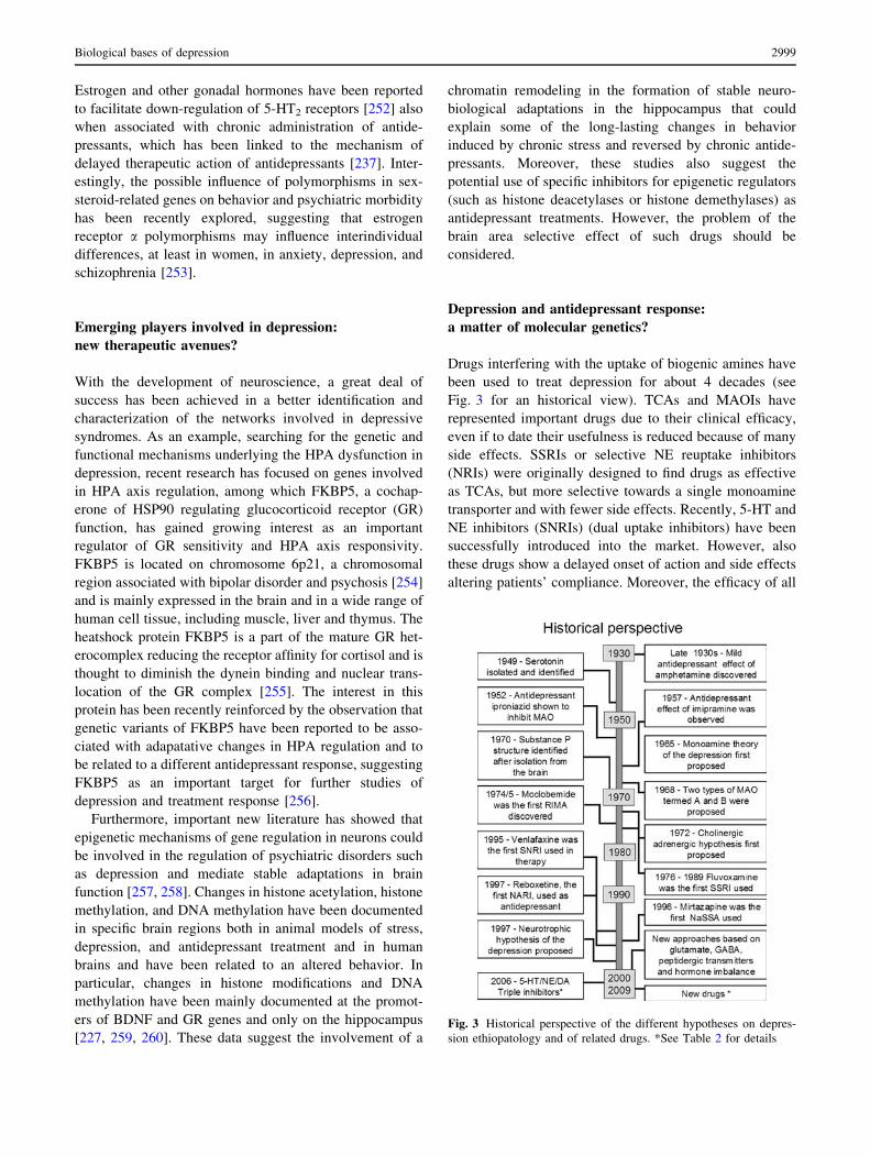
Estrogen and other gonadal hormones have been reported
to facilitate down-regulation of 5-HT2 receptors [252] also
when associated with chronic administration of antide-
pressants, which has been linked to the mechanism of
delayed therapeutic action of antidepressants [237]. Inter-
estingly, the possible influence of polymorphisms in sex-
steroid-related genes on behavior and psychiatric morbidity
has been recently explored, suggesting that estrogen
receptor a polymorphisms may influence interindividual
differences, at least in women, in anxiety, depression, and
schizophrenia [253].
Emerging players involved in depression:
new therapeutic avenues?
With the development of neuroscience, a great deal of
success has been achieved in a better identification and
characterization of the networks involved in depressive
syndromes. As an example, searching for the genetic and
functional mechanisms underlying the HPA dysfunction in
depression, recent research has focused on genes involved
in HPA axis regulation, among which FKBP5, a cochap-
erone of HSP90 regulating glucocorticoid receptor (GR)
function, has gained growing interest as an important
regulator of GR sensitivity and HPA axis responsivity.
FKBP5 is located on chromosome 6p21, a chromosomal
region associated with bipolar disorder and psychosis [254]
and is mainly expressed in the brain and in a wide range of
human cell tissue, including muscle, liver and thymus. The
heatshock protein FKBP5 is a part of the mature GR het-
erocomplex reducing the receptor affinity for cortisol and is
thought to diminish the dynein binding and nuclear trans-
location of the GR complex [255]. The interest in this
protein has been recently reinforced by the observation that
genetic variants of FKBP5 have been reported to be asso-
ciated with adapatative changes in HPA regulation and to
be related to a different antidepressant response, suggesting
FKBP5 as an important target for further studies of
depression and treatment response [256].
Furthermore, important new literature has showed that
epigenetic mechanisms of gene regulation in neurons could
be involved in the regulation of psychiatric disorders such
as depression and mediate stable adaptations in brain
function [257, 258]. Changes in histone acetylation, histone
methylation, and DNA methylation have been documented
in specific brain regions both in animal models of stress,
depression, and antidepressant treatment and in human
brains and have been related to an altered behavior. In
particular, changes in histone modifications and DNA
methylation have been mainly documented at the promot-
ers of BDNF and GR genes and only on the hippocampus
[227, 259, 260]. These data suggest the involvement of a
chromatin remodeling in the formation of stable neuro-
biological adaptations in the hippocampus that could
explain some of the long-lasting changes in behavior
induced by chronic stress and reversed by chronic antide-
pressants. Moreover, these studies also suggest the
potential use of specific inhibitors for epigenetic regulators
(such as histone deacetylases or histone demethylases) as
antidepressant treatments. However, the problem of the
brain area selective effect of such drugs should be
considered.
Depression and antidepressant response:
a matter of molecular genetics?
Drugs interfering with the uptake of biogenic amines have
been used to treat depression for about 4 decades (see
Fig. 3 for an historical view). TCAs and MAOIs have
represented important drugs due to their clinical efficacy,
even if to date their usefulness is reduced because of many
side effects. SSRIs or selective NE reuptake inhibitors
(NRIs) were originally designed to find drugs as effective
as TCAs, but more selective towards a single monoamine
transporter and with fewer side effects. Recently, 5-HT and
NE inhibitors (SNRIs) (dual uptake inhibitors) have been
successfully introduced into the market. However, also
these drugs show a delayed onset of action and side effects
altering patients’ compliance. Moreover, the efficacy of all
Fig. 3 Historical perspective of the different hypotheses on depres-
sion ethiopatology and of related drugs. *See Table 2 for details
Biological bases of depression 2999

currently used antidepressants has been questioned because
of the existence of a significant proportion of drug-resistant
patients and a debated drug/placebo effect. A recent
development has been obtained with the introduction of the
triple uptake inhibitors, designed to inhibit the uptake of all
three neurotransmitters, 5-HT, NE, and DA, mainly
involved in depression. Triple uptake inhibitors (SNDRIs)
are expected to be the next generation of drugs for the
treatment of MDD [261] (Table 2). Preclinical studies
indicate indeed that they could produce a more rapid onset
of action and possess greater efficacy than traditional
antidepressants [261]. DOV 216,303 [(±)-1-(3,4-dichloro-
phenyl)-3-azabicyclo-[3.1.0]hexane hydrochloride], the
prototype of the compounds referred to as triple uptake
inhibitors, has been demonstrated to be active in tests
predictive of antidepressant activity, such as the mouse
forced-swim test and locomotor depression [262] (Fig. 3).
To date, because of the less severe side effects than
those of other antidepressants, SSRIs represent the class of
drugs of choice for treating MDD. However, a substantial
number of patients benefit only partially or not at all from
treatment. Drug response can be influenced by genetic
factors and several physiological and environmental fac-
tors, including age, renal and liver function, nutritional
status, smoking and alcohol consumption. In particular,
genetic polymorphisms may play a major role in deter-
mining the response to SSRI treatment [263–266]. At
present the main focus of research is to gain greater
understanding of the functional polymorphism in the 5-HT
transporter (SERT) promoter region. SERT polymorphism
(referred as 5-HTTLPR) involves the presence or absence
of a 44 base-pair segment, which produces a long (L) or
short (S) allele; the short allele results in decreased trans-
porter expression and, hence, in a reduced uptake of 5-HT
[267]. 5-HTTLPR has been associated with susceptibility
to depression [268], although there is considerable heter-
ogeneity between studies [269]. The S allele has also been
associated with lower response to several SSRIs than the L
allele in several studies [270–272] and may also increase
vulnerability to SSRI side effects [273]. The large-scale
study referred as STAR*D confirmed the S variant of
5-HTTLPR to be predictive for the global burden of side
effects. Furthermore, Binder and coworkers examined 57
single nucleotide polymorphisms (SNP) in eight candidate
genes related to the HPA axis in patients afflicted with
depression searching for the SNP associated with antide-
pressant response. They observed that a polymorphism of
FKBP5, the cochaperone of HSP90 regulating glucocorti-
coid receptor function, was associated with rapid response
to antidepressants [256]. Another investigated polymorphic
gene is the 5HT2A-receptor gene. All antidepressants
induce a down-regulation of this postsynaptic receptor. The
C variant at 102T/C was found to be associated with less
antidepressant response. Murphy and coworkers observed
in elderly depressed subjects that this polymorphism may
be associated with sensitivity to the adverse effects of
mirtazapine, but was without influence on SSRI adverse
effects [273]. Antidepressants can also alter the synthesis
and release of CRF; polymorphisms in the CRF gene have
been described [274] and may be associated with MDD
[275]. For further genes, TPH1 and G-protein b3, initially
shown to be implicated in the modulation of antidepressant
response, no definite conclusions can be made [276].
Delineating the genes that are correlated to an antidepres-
sant response may be valuable in identifying the best
treatment specific for the patient.
References
1. Hamet P, Tremblay J (2005) Genetics and genomics of
depression. Metabolism 54(5 Suppl 1):10–15
2. Segurado R, Detera-Wadleigh SD, Levinson DF, Lewis CM,
Gill M, Nurnberger JI Jr, Craddock N, DePaulo JR, Baron M,
Gershon ES, Ekholm J, Cichon S, Turecki G, Claes S, Kelsoe
JR, Schofield PR, Badenhop RF, Morissette J, Coon H, Black-
wood D, McInnes LA, Foroud T, Edenberg HJ, Reich T, Rice
JP, Goate A, McInnis MG, McMahon FJ, Badner JA, Goldin
LR, Bennett P, Willour VL, Zandi PP, Liu J, Gilliam C, Juo SH,
Berrettini WH, Yoshikawa T, Peltonen L, Lonnqvist J, Nothen
MM, Schumacher J, Windemuth C, Rietschel M, Propping P,
Maier W, Alda M, Grof P, Rouleau GA, Del-Favero J, Van
Broeckhoven C, Mendlewicz J, Adolfsson R, Spence MA,
Luebbert H, Adams LJ, Donald JA, Mitchell PB, Barden N,
Shink E, Byerley W, Muir W, Visscher PM, Macgregor S,
Gurling H, Kalsi G, McQuillinm A, Escamilla MA, Reus VI,
Leon P, Freimer NB, Ewald H, Kruse TA, Mors O, Radhakrishna
U, Blouin JL, Antonarakis SE, Akarsu N (2003) Genome scan
meta-analysis of schizophrenia and bipolar disorder, part III:
bipolar disorder. Am J Hum Genet 73(1):49–62
3. Bunney WE Jr, Davis JM (1965) Norepinephrine in depressive
reactions. A review. Arch Gen Psychiatry 13(6):483–494
4. Schildkraut JJ (1965) The catecholamine hypothesis of affective
disorders: a review of supporting evidence. J Neuropsychiatry
Clin Neurosci 7:524–533
5. Coppen A (1967) The biochemistry of affective disorders. Br J
Psychiatry 113:1237–1264
6. Montgomery SA (2008) The under-recognized role of dopamine
in the treatment of major depressive disorder. Int Clin Psycho-
pharmacol 23(2):63–69
7. Maas JW, Fawcett JA, Dekirmenjian H (1972) Catecholamine
metabolism, depressive illness, and drug response. Arch Gen
Psychiatry 26(3):252–262
8. Roy A, Jimerson DC, Pickar D (1986) Plasma MHPG in
depressive disorders and relationship to the dexamethasone
suppression test. Am J Psychiatry 143(7):846–851
9. Potter WZ, Manji HK (1993) Are monoamine metabolites in
cerebrospinal fluid worth measuring? Arch Gen Psychiatry
50(8):653–656
10. Ordway GA, Smith KS, Haycock JW (1998) Monoamine dys-
function and the pathophysiology and treatment of depression.
J Clin Psychiatry 59(Suppl 14):11–14
11. Ruhe HG, Mason NS, Schene AH (2007) Mood is indirectly
related to serotonin, norepinephrine and dopamine levels in
3000 C. Lanni et al.
humans: a meta-analysis of monoamine depletion studies. Mol
Psychiatry 12(4):331–359
12. Charney DS (1994) Elevated tyrosine hydroxylase in the locus
coeruleus of suicide victims. J Neurochem 62(2):680–685
13. Zhu MY, Klimek V, Dilley GE, Haycock JW, Stockmeier C,
Overholser JC, Meltzer HY, Ordway GA (1999) Elevated levels
of tyrosine hydroxylase in the locus coeruleus in major
depression. Biol Psychiatry 46(9):1275–1286
14. Ordway GA, Widdowson PS, Smith KS, Halaris A (1994)
Agonist binding to alpha 2-adrenoceptors is elevated in the locus
coeruleus from victims of suicide. J Neurochem 63(2):617–624
15. Ordway GA, Schenk J, Stockmeier CA, May W, Klimek V
(2003) Elevated agonist binding to alpha2-adrenoceptors in
the locus coeruleus in major depression. Biol. Psychiatry.
53(4):315–323
16. Klimek V, Stockmeier C, Overholser J, Meltzer HY, Kalka S,
Dilley G, Ordway GA (1997) Reduced levels of norepinephrine
transporters in the locus coeruleus in major depression. J Neu-
rosci 17(21):8451–8458
17. Asberg M, Traskman L, Thoren P (1976) 5-HIAA in the cere-
brospinal fluid: a biochemical suicide predictor? Arch Gen
Psychiatry 33:1193–1197
18. Asberg M, Thoren L, Traskman P (1976) Serotonin depression:
a biochemical subgroup within the affective disorders. Science
191:478–480
19. Csernansky JG, Sheline YI (1993) Abnormalities of serotonin
metabolism and nonpsychotic psychiatric disorders. Ann Clin
Psychiatry 5(4):275–281
20. Tuinier S, Verhoeven WM, van Praag HM (1995) Cerebrospinal
fluid 5-hydroxyindolacetic acid and aggression: a critical reap-
praisal of the clinical data. Int Clin Psychopharmacol 10(3):147–
156
21. Quintana J (1992) Platelet serotonin and plasma tryptophan
decreases in endogenous depression: clinical, therapeutic and
biological correlations. J Affect Disord 24:58–62
22. Jans LA, Riedel WJ, Markus CR, Blokland A (2007) Seroto-
nergic vulnerability and depression: assumptions, experimental
evidence and implications. Mol Psychiatry 12(6):522–543
23. Arango V, Huang YY, Underwood MD, Mann JJ (2003)
Genetics of the serotonergic system in suicidal behavior. J
Psychiatr Res 37(5):375–386
24. Celada P, Puig M, Amargos-Bosch M, Adell A, Artigas F (2004)
The therapeutic role of 5-HT1A and 5-HT2A receptors in
depression. J Psychiatry Neurosci 29(4):252–265
25. Mann JJ, Stanley M, McBride PA, McEwen BS (1986)
Increased serotonin2 and beta-adrenergic receptor binding in the
frontal cortices of suicide victims. Arch Gen Psychiatry 43:945–
959
26. Arora RC, Meltzer HY (1989) Serotonergic measures in the
brains of suicide victims: 5-HT2 binding sites in the frontal
cortex of suicide victims and control subjects. Am J Psychiatry
146:730–736
27. Yates M, Leake A, Candy JM, Fairbairn AF, McKeith IG,
Ferrier IN (1990) 5-HT2 receptor changes in major depression.
Biol Psychiatry 27:489–496
28. Drevets WC, Frank E, Price JC, Kupfer DJ, Holt D, Greer PJ,
Huang Y, Gautier C, Mathis C (1999) PET imaging of serotonin
1A receptor binding in depression. Biol Psychiatry 46:1375–
1387
29. Hamner MB, Diamond BI (1996) Plasma dopamine and nor-
epinephrine correlations with psychomotor retardation, anxiety,
and depression in non-psychotic depressed patients: a pilot
study. Psychiatry Res 64(3):209–211
30. Engstrom G, Alling C, Blennow K, Regnell G, Traskman-Bendz
L (1999) Reduced cerebrospinal HVA concentrations and HVA/
5-HIAA ratios in suicide attempters. Monoamine metabolites in
120 suicide attempters and 47 controls. Eur Neuropsychophar-
macol 9(5):399–405
31. D’Haenen HA, Bossuyt A (1994) Dopamine D2 receptors in
depression measured with single photon emission computed
tomography. Biol Psychiatry 35(2):128–132
32. Shah PJ, Ogilvie AD, Goodwin GM, Ebmeier KP (1997) Clin-
ical and psychometric correlates of dopamine D2 binding in
depression. Psychol Med 27(6):1247–1256
33. Klimek V, Schenck JE, Han H, Stockmeier CA, Ordway GA
(2002) Dopaminergic abnormalities in amygdaloid nuclei in
major depression: a postmortem study. Biol Psychiatry
52(7):740–748
34. Sonnenberg CM, Deeg DJ, Comijs HC, van Tilburg W, Beek-
man AT (2008) Trends in antidepressant use in the older
population: Results from the LASA-study over a period of 10
years. J Affect Disord 111(2–3):299–305
35. Duman RS, Heninger GR, Nestler EJ (1997) A molecular and
cellular theory of depression. Arch Gen Psychiatry 54(7):597–
606
36. Orrego F, Villanueva S (1993) The chemical nature of the main
central excitatory transmitter: a critical appraisal based upon
release studies and synaptic vesicle localization. Neuroscience
56:539–555
37. Sanacora S, Zarate CA, Krystal JH, Manji HK (2008) Targeting
the glutamatergic system to develop novel, improved thera-
peutics for mood disorders. Nat Rev Drug Discov 7(5):426–437
38. Kim JS, Schmid-Burgk W, Claus D, Kornhuber HH (1982)
Increased serum glutamate in depressed patients. Arch Psychiatr
Nervenkr 232:299–304
39. Altamura CA, Mauri MC, Ferrara A, Moro AR, D’Andrea G,
Zamberlan F (1993) Plasma and platelet excitatory amino acids
in psychiatric disorders. Am J Psychiatry 150:1731–1733
40. Mauri MC, Ferrara A, Boscati L, Bravin S, Zamberlan F, Alecci
M, Invernizzi G (1998) Plasma and platelet amino acid con-
centrations in patients affected by major depression and under
fluvoxamine treatment. Neuropsychobiology 37:124–129
41. Mitani H, Shirayama Y, Yamada T, Maeda K, Ashby CR Jr,
Kawahara R (2006) Correlation between plasma levels of glu-
tamate, alanine and serine with severity of depression. Prog
Neuropsychopharmacol Biol Psychiatry 30:1155–1158
42. Levine J, Panchalingam K, Rapoport A, Gershon S, McClure RJ,
Pettegrew JW (2000) Increased cerebrospinal fluid glutamine
levels in depressed patients. Biol Psychiatry 47:586–593
43. Frye MA, Tsai GE, Huggins T, Coyle JT, Post RM (2006) Low
cerebrospinal fluid glutamate and glycine in refractory affective
disorder. Biol Psychiatry 61:162–166
44. Francis PT, Poynton A, Lowe SL, Najlerahim A, Bridges PK,
Bartlett JR, Procter AW, Bruton CJ, Bowen DM (1989) Brain
amino acid concentrations and Ca2?-dependent release in
intractable depression assessed antemortem. Brain Res 494:315–
324
45. Nudmamud-Thanoi S, Reynolds GP (2004) The NR1 subunit of
the glutamate/NMDA receptor in the superior temporal cortex inschizophrenia and affective disorders. Neurosci Lett 372:173–
177
46. Mundo E, Tharmalingham S, Neves-Pereira M, Dalton EJ,
Macciardi F, Parikh SV, Bolonna A, Kerwin RW, Arranz MJ,
Makoff AJ, Kennedy JL (2003) Evidence that the N-methyl-D-
aspartate subunit 1 receptor gene (GRIN1) confers susceptibility
to bipolar disorder. Mol Psychiatry 8:241–245
47. Martucci L, Wong AH, De Luca V, Likhodi O, Wong GW, King
N, Kennedy JL (2006) N-methyl-D-aspartate receptor NR2B
subunit gene GRIN2B in schizophrenia and bipolar disorder:
polymorphisms and mRNA levels. Schizophr Res 84:214–221
48. Meador-Woodruff JH, Hogg AJ Jr, Smith RE (2001) Striatal
ionotropic glutamate receptor expression in schizophrenia,
Biological bases of depression 3001
bipolar disorder, and major depressive disorder. Brain Res Bull
55:631–640
49. Beneyto M, Meador-Woodruff JH (2006) Lamina-specific
abnormalities of AMPA receptor trafficking and signaling
molecule transcripts in the prefrontal cortex in schizophrenia.
Synapse 60:585–598
50. Laje G, Paddock S, Manji H, Rush AJ, Wilson AF, Charney D,
Mc-Mahon FJ (2007) Genetic markers of suicidal ideation
emerging during citalopram treatment of major depression. Am
J Psychiatry 164:1530–1538
51. Menke A, Lucae S, Kloiber S, Horstmann S, Bettecken T, Uhr
M, Ripke S, Ising M, Muller-Myhsok B, Holsboer F, Binder EB
(2008) Genetic markers within glutamate receptors associated
with antidepressant treatment-emergent suicidal ideation. Am J
Psychiatry 165(7):917–918
52. Hasler G, van der Veen JW, Tumonis T, Meyers N, Shen J,
Drevets WC (2007) Reduced prefrontal glutamate/glutamine
and gammaaminobutyric acid levels in major depression deter-
mined using proton magnetic resonance spectroscopy. Arch Gen
Psychiatry 64:193–200
53. Kugaya A, Sanacora G (2005) Beyond monoamines: glutama-
tergic function in mood disorders. CNS Spectr 10:808–819
54. During MJ, Spencer DD (1993) Extracellular hippocampal
glutamate and spontaneous seizure in the conscious human
brain. Lancet 341:1607–1610
55. Ongur D, Drevets WC, Price JL (1998) Glial reduction in the
subgenual prefrontal cortex in mood disorders. Proc Natl Acad
Sci USA 95:13290–13295
56. Cotter D, Mackay D, Landau S, Kerwin R, Everall I (2001)
Reduced glial cell density and neuronal size in the anterior
cingulate cortex in major depressive disorder. Arch Gen Psy-
chiatry 58:545–553
57. Cotter D, Pariante CM, Rajkowska G (2002) Glial pathology
and major psychiatric disorders. In: Agam G, Everall I, Bel-
maker RH (eds) The postmortem brain in psychiatric research.
Mass Kluwer Academic Publishers, Boston, pp 49–73
58. Bowley MP, Drevets WC, Ongur D, Price JL (2002) Low glial
numbers in the amygdala in major depressive disorder. Biol
Psychiatry 52:404–412
59. Zarate CA, Singh JB, Carlson PJ, Brutsche NE, Ameli R,
Luckenbaugh DA, Charney DS, Manji HK (2006) A ran-
domized trial of an N-methyl-D-aspartate antagonist in
treatment-resistant major depression. Arch Gen Psychiatry
63:856–864
60. Maeng S, Zarate CA Jr (2007) The role of glutamate in mood
disorders: results from the ketamine in major depression study
and the presumed cellular mechanism underlying its antide-
pressant effects. Curr Psychiatry Rep 9(6):467–474
61. Ferguson JM, Shingleton RN (2007) An open-label, flexible-
dose study of memantine in major depressive disorder. Clin
Neuropharmacol 30(3):136–144
62. Paredes RG, Agmo A (1992) GABA and behavior: the role of
receptor subtypes. Neurosci Biobehav Rev 16(2):145–170
63. Petty F (1995) GABA and mood disorders: a brief review and
hypothesis. J Affect Disord 34(4):275–281
64. Sanacora G, Mason GF, Krystal JH (2000) Impairment of
GABAergic transmission in depression: new insights from
neuroimaging studies. Crit Rev Neurobiol 14:23–45
65. Krystal JH, Sanacora G, Blumberg H, Anand A, Charney DS,
Marek G, Epperson CN, Goddard A, Mason GF (2002) Gluta-
mate and GABA systems as targets for novel antidepressant and
mood-stabilizing treatments. Mol Psychiatry 7:S71–S80
66. Petty F, Kramer GL, Gullion CM, Rush AJ (1992) Low plasma
c-aminobutyric acid levels in male patients with depression. Biol
Psychiatry 32:354–363
67. Gerner R, Hare TA (1981) CSF GABA levels in normal subjects
and patients with depression, schizophrenia, mania and anorexia
nervosa. Am J Psychiatry 138:1098–1101
68. Kasa K, Otsuki S, Yamamoto M, Sato M, Kuroda H, Ogawa N
(1982) Cerebrospinal fluid c-aminobutyric acid and homova-
nillic acid in depressive disorders. Biol Psychiatry 17:877–883
69. Roy A, Dejong J, Ferraro T (1991) CSF GABA in depressed
patients and normal controls. Psychol Med 21:613–618
70. Sanacora G, Mason GF, Rothman DL, Behar KL, Hyder F,
Petroff OA, Berman RM, Charney DS, Krystal JH (1999)
Reduced cortical c-aminobutyric acid levels in depressed
patients determined by proton magnetic resonance spectroscopy.
Arch Gen Psychiatry 56:1043–1047
71. Sanacora G, Mason GF, Rothman DL, Krystal JH (2002)
Increased occipital cortex GABA concentrations in depressed
patients after therapy with selective serotonin reuptake inhibi-
tors. Am J Psychiatry 159:663–665
72. Sanacora G, Mason GF, Rothman DL, Ciarcia JJ, Ostroff RB,
Krystal JH (2003) Increased occipital cortex GABA concentra-
tions following electroconvulsive therapy in depressed patients.
Am J Psychiatry 160:577–579
73. Tunnicliff G, Ngo TT (1986) Regulation of c-aminobutyric acid
synthesis in the vertebrate nervous system. Neurochem Int
8:287–297
74. Fatemi SH, Stary JM, Earle JA, Araghi-Niknam M, Eagan E
(2005) GABAergic dysfunction in schizophrenia and mood
disorders as reflected by decreased levels of glutamic acid
decarboxylase 65 and 67 kDa and reelin proteins in cerebellum.
Schizophr Res 72:109–122
75. Benes FM, Todtenkopf MS, Logiotatos P, Williams M (2000)
Glutamate decarboxylase(65)-immunoreactive terminals in cin-
gulate and prefrontal cortices of schizophrenic and bipolar brain.
J Chem Neuroanat 20(3–4):259–269
76. Heckers S, Stone D, Walsh J, Shick J, Koul P, Benes FM (2002)
Differential hippocampal expression of glutamic acid decar-
boxylase 65 and 67 messenger RNA in bipolar disorder and
schizophrenia. Arch Gen Psychiatry 59(6):521–529
77. Sanacora G, Saricicek A (2007) GABAergic contributions to the
pathophysiology of depression and the mechanism of antide-
pressant action. CNS Neurol Disord Drug Targets 6(2):127–140
78. Horiuchi Y, Nakayama J, Ishiguro H, Ohtsuki T, Detera-Wad-
leigh SD, Toyota T, Yamada K, Nankai M, Shibuya H,
Yoshikawa T, Arinami T (2004) Possible association between a
haplotype of the GABA-A receptor alpha 1 subunit gene
(GABRA1) and mood disorders. Biol Psychiatry 55(1):40–45
79. Yamada K, Watanabe A, Iwayama-Shigeno Y, Yoshikawa T
(2003) Evidence of association between gamma-aminobutyric
acid type A receptor genes located on 5q34 and female patients
with mood disorders. Neurosci Lett 349(1):9–12
80. Serretti A, Macciardi F, Cusin C, Lattuada E, Lilli R, Di Bella
D, Catalano M, Smeraldi E (1998) GABAA alpha-1 subunit
gene not associated with depressive symptomatology in mood
disorders. Psychiatr Genet 8(4):251–254
81. Mitchell AJ (1998) The role of corticotropin releasing factor in
depressive illness: a critical review. Neurosci Biobehav Rev
22:635–651
82. Nemeroff CB (1998) The neurobiology of depression. Sci Am
278:42–49
83. Gilad GM (1987) The stress-induced response of the septo-hip-
pocampal cholinergic system. A vectorial outcome of
psychoneuroendocrinological interactions. Psychoneuroendocri-
nology 12:167–184
84. Janowsky DS, Risch SC (1984) Cholinomimetic and anticho-
linergic drugs used to investigate an acetylcholine hypothesis of
affective disorders and stress. Drug Devel Res 4:125–142
3002 C. Lanni et al.
85. Janowsky DS, El-Yousef MK, Davis JM, Sekerke HJ (1972) A
cholinergic-adrenergic hypothesis of mania and depression.
Lancet 2:632–635
86. Janowsky DS, El-Yousef MK, Davis JM, Sekerke HJ (1973)
Parasympathetic suppression of mania by physostigmine. Arch
Gen Psych 28:542–547
87. Janowsky DS, Overstreet DH, Nurnberger JI Jr (1994) Is cho-
linergic sensitivity a genetic marker for the affective disorders?
Am J Med Genet 54:335–344
88. Davis KL, Berger PA, Hollister LE, Defraites E (1978) Physo-
stigmine in man. Arch Gen Psychiatry 35:119–122
89. O’Keane V, O’Flynn K, Lucey J, Dinan TG (1992) Pyrido-
stigmine-induced growth hormone responses in healthy and
depressed subjects: evidence for cholinergic supersensitivity in
depression. Psychol Med 22:55–60
90. Gillin JC, Salin-Pascual R, Velazquez-Moctezuma J, Shiromani
P, Zoltoski R (1993) Cholinergic receptor subtypes and REM
sleep in animals and normal controls. Prog Brain Res 98:379–387
91. Janowsky DS, Risch SC, Kennedy B, Ziegler M, Huey LY
(1986) Central muscarinic effects of physostigmine on mood,
cardiovascular function, pituitary and adrenal neuroendocrine.
Psychopharmacology 89:150–154
92. Riemann D, Berger M (1992) Sleep, age depression and the
cholinergic induction test with RS 86. Prog Neuropsychophar-
macol Biol Psychiatry 16:311–316
93. Bymaster FP, Felder CC (2002) Role of the cholinergic mus-
carinic system in bipolar disorder and related mechanism of
action of antipsychotic agents. Mol Psychiatry 7:S57–S63
94. Burt T, Sachs GS, Demopulos C (1999) Donepezil in treatment-
resistant bipolar disorder. Biol Psychiatry 45:959–964
95. Furey ML, Drevets WC (2006) Antidepressant efficacy of the
antimuscarinic drug scopolamine: a randomized, placebo-con-
trolled clinical trial. Arch Gen Psychiatry 63(10):1121–1129
96. Janowsky DS (2007) Scopolamine as an antidepressant agent:
theoretical and treatment considerations. Curr Psychiatry Rep
9(6):447–448
97. Liu HF, Zhou WH, Xie XH, Cao JL, Gu J, Yang GD (2004)
Muscarinic receptors modulate the mRNA expression of NMDA
receptors in brainstem and the release of glutamate in periaqu-
eductal grey during morphine withdrawal in rats. Sheng Li Xu
Bao 56(1):95–100 (article in Chinese)
98. Shytle RD, Silver AA, Lukas RJ, Newman MB, Sheehan DV,
Sanberg P (2002) Nicotinic acetylcholine receptors as targets for
antidepressants. Mol Psychiatry 6:525–535
99. Silver AA, Shytle RD, Sheehan KH, Sheehan DV, Ramos A,
Sanberg P (2001) Multicenter, double-blind, placebo-controlled
study of mecamylamine monotherapy for Tourette’s disorder.
J Am Acad Child Adolesc Psychiatry 40:1103–1110
100. Sacco KA, Bannon KL, George TP (2004) Nicotinic receptor
mechanisms and cognition in normal states and neuropsychiatric
disorders. J Psychopharmacol 18(4):457–474
101. Rozzini L, Vicini Chilovi B, Bertoletti E, Trabucchi M, Pado-
vani A (2007) Acetylcholinesterase inhibitors and depressive
symptoms in patients with mild to moderate Alzheimer’s dis-
ease. Aging Clin Exp Res 19(3):220–223
102. Wynn ZJ, Cummings JL (2004) Cholinesterase inhibitor thera-
pies and neuropsychiatric manifestations of Alzheimer’s
disease. Dement Geriatr Cogn Disord 17(1–2):100–108
103. Salzman C (1999) Practical considerations for the treatment of
depression in elderly and very elderly long-term care patients.
J Clin Psychiatry 60(20):30–33
104. Mottram P, Wilson K, Strobl J (2006) Antidepressants for
depressed elderly. Cochrane Database Syst Rev (1):CD003491
105. Griebel G (1999) Is there a future for neuropeptide receptor
ligands in the treatment of anxiety disorders? Pharmacol Ther
82:1–61
106. Feng P, Vurbic D, Wu Z, Hu Y, Strohl KP (2008) Changes in
brain orexin levels in a rat model of depression induced by
neonatal administration of clomipramine. J Psychopharmacol
22(7):784–791
107. Brown GW, Bifulco A, Harris TO (1987) Life events, vulner-
ability and onset of depression. Br J Psychiatry 150:30–42
108. Hammen C, Davila J, Brown G, Ellicott A, Gitlin M (1992)
Psychiatric history and stress: predictors of severity of unipolar
depression. J Abnorm Psychol 101:45–52
109. Owens MJ, Nemeroff CB (1991) Physiology and pharmacology
of corticotropin-releasing factor. Pharmacol Rev 43:425–473
110. Pariante CM (2003) Depression, stress and the adrenal axis.
J Neuroendocrinol 15(8):811–812
111. Holsboer F, Barden N (1996) Antidepressants and hypotha-
lamic-pituitary-adrenocortical regulation. Endocr Rev 17:187–
205
112. Plotsky P, Owens MJ, Nemeroff CB (1995) Neuropeptide
alterations in affective disorders. In: Bloom FE, Kupfer DJ (eds)
Psychopharmacology: the fourth generation of progress. Raven
Press, New York, pp 971–981
113. Nemeroff CB, Widerlov E, Bissette G, Walleus H, Karlsson I,
Eklund K, Kilts CD, Loosen PT, Vale W (1984) Elevated
concentrations of CSF corticotropin-releasing factor-like
immunoreactivity in depressed patients. Science 226:1342–1344
114. Banki CM, Karmacsi L, Bissette G, Nemeroff CB (1992)
Cerebrospinal fluid neuropeptides in mood disorders and
dementia. J Affect Disord 25:39–46
115. Pitts AF, Samuelson SD, Meller WH, Bissette G, Nemeroff CB,
Kathol RG (1995) Cerebrospinal fluid corticotropin-releasing
hormone, vasopressin, and oxytocin concentrations in treated
patients with major depression and controls. Biol Psychiatry
38:330–335
116. Molchan SE, Hill JL, Martinez RA, Lawlor BA, Mellow AM,
Rubinow DR, Bissette G, Nemeroff CB, Sunderland T (1993)
CSF somatostatin in Alzheimer’s disease and major depression:
relationship to hypothalamic-pituitary-adrenal axis and clinical
measures. Psychoneuroendocrinology 19:509–519
117. Stenzel-Poore MP, Heinrichs SC, Rivest S, Koob GF, Vale WW
(1994) Overproduction of corticotropin-releasing factor in trans-
genic mice: a genetic model of anxiogenic behavior. J Neurosci
14(5 Pt 1):2579–2584
118. Kasahara M, Groenink L, Breuer M, Olivier B, Sarnyai Z (2007)
Altered behavioral adaptation in mice with neural corticotro-
phin-releasing factor overexpression. Genes Brain Behav
6(7):598–607
119. Holmes A, Heilig M, Rupniak NMJ, Steckler T, Griebel G
(2003) Neuropeptides systems as novel therapeutic targets for
depression and anxiety disorders. TIPS 24:580–588
120. Griebel G, Perrault G, Sanger DJ (1998) Characterization of the
behavioral profile of the non-peptide CRF receptor antagonist
CP-154, 526 in anxiety models in rodents. Comparison with
diazepam and buspirone. Psychopharmacology (Berl) 138:55–
66
121. Holsboer F, Ising M (2008) Central CRH system in depression
and anxiety—evidence from clinical studies with CRH1 receptor
antagonists. Eur J Pharmacol 583(2–3):350–357
122. Aguilera G, Rabadan-Diehl C (2000) Vasopressinergic regula-
tion of the hypothalamic-pituitary-adrenal axis: implications for
stress adaptation. Regul Pept 96:23–29
123. Merali Z, Kent P, Du L, Hrdina P, Palkovits M, Faludi G, Poulter
MO, Bedard T, Anisman H (2006) Corticotropin-releasing hor-
mone, arginine vasopressin, gastrin-releasing peptide, and
neuromedin B alterations in stress-relevant brain regions of
suicides and control subjects. Biol Psychiatry 59:594–602
124. van Londen L, Goekoop JG, van Kempen GM, Frankhuijzen-
Sierevogel AC, Wiegant VM, van der Velde EA, De Wied D
Biological bases of depression 3003

(1997) Plasma levels of arginine vasopressin elevated in patients
with major depression. Neuropsychopharmacology 17(4):284–292
125. van Londen L, Kerkhof GA, van den Berg F, Goekoop JG,
Zwinderman KH, Frankhuijzen-Sierevogel AC, Wiegant VM,
de Wied D (1998) Plasma arginine vasopressin and motor
activity in major depression. Biol Psychiatry 43(3):196–204
126. Purba JS, Hoogendijk WJ, Hofman MA, Swaab DF (1996)
Increased number of vasopressin- and oxytocin-expressing
neurons in the paraventricular nucleus of the hypothalamus in
depression. Arch Gen Psychiatry 53(2):137–143
127. Meynen G, Unmehopa UA, van Heerikhuize JJ, Hofman MA,
Swaab DF, Hoogendijk WJ (2006) Increased arginine vaso-
pressin mRNA expression in the human hypothalamus in
depression: a preliminary report. Biol Psychiatry 60(8):892–895
128. Spinedi E, Hadid R, Gaillard RC (1997) Increased vasopress-
inergic activity as a possible compensatory mechanism for a
normal hypothalamic-pituitary-adrenal axis response to stress in
BALB/c nude mice. Neuroendocrinology 66(4):287–293
129. Keck ME, Wigger A, Welt T, Muller MB, Gesing A, Reul JM,
Holsboer F, Landgraf R, Neumann ID (2002) Vasopressin
mediates the response of the combined dexamethasone/CRH test
in hyper-anxious rats: implications for pathogenesis of affective
disorders. Neuropsychopharmacology 26(1):94–105
130. Conte-Devolx B, Oliver C, Giraud P, Castanas E, Boudouresque F,
Gillioz P, Millet Y (1982) Adrenocorticotropin, and corticosterone
secretion in Brattleboro rats. Endocrinology 110(6):2097–2100
131. Wiley MK, Pearlmutter AF, Miller RE (1974) Decreased
adrenal sensitivity to ACTH in the vasopressin-deficient
(Brattleboro) rat. Neuroendocrinology 14(5):257–270
132. Fink G, Dow RC, Casley D, Johnston CI, Bennie J, Carroll S,
Dick H (1992) Atrial natriuretic peptide is involved in the
ACTH response to stress and glucocorticoid negative feedback
in the rat. J Endocrinol 135(1):37–43
133. Williams AR, Carey RJ, Miller M (1985) Altered emotionality
of the vasopressin-deficient Brattleboro rat. Peptides 6(Suppl
1):69–76
134. Mlynarik M, Zelena D, Bagdy G, Makara GB, Jezova D (2007)
Signs of attenuated depression-like behavior in vasopressin
deficient Brattleboro rats. Horm Behav 51(3):395–405
135. Jard S, Barberis C, Audigier S, Tribollet E (1987) Neurohypo-
physeal hormone receptor systems in brain and periphery. Prog
Brain Res 72:173–187
136. Birnbaumer M (2000) Vasopressin receptors. Trends Endocrinol
Metab 11(10):406–410
137. Thomson F, Craighead M (2008) Innovative approaches for the
treatment of depression: targeting the HPA axis. Neurochem Res
33(4):691–707
138. Lolait SJ, Stewart LQ, Jessop DS, Young WS 3rd, O’Carroll
AM (2007) The hypothalamic-pituitary-adrenal axis response to
stress in mice lacking functional vasopressin V1b receptors.
Endocrinology 148(2):849–856
139. Wersinger SR, Ginns EI, O’Carroll AM, Lolait SJ, Young WS
3rd (2002) Vasopressin V1b receptor knockout reduces
aggressive behavior in male mice. Mol Psychiatry 7(9):975–984
140. Wersinger SR, Kelliher KR, Zufall F, Lolait SJ, O’Carroll AM,
Young WS 3rd (2004) Social motivation is reduced in vaso-
pressin 1b receptor null mice despite normal performance in an
olfactory discrimination task. Horm Behav 46(5):638–645
141. Egashira N, Tanoue A, Higashihara F, Fuchigami H, Sano K,
Mishima K, Fukue Y, Nagai H, Takano Y, Tsujimoto G,
Stemmelin J, Griebel G, Iwasaki K, Ikeda T, Nishimura R,
Fujiwara M (2005) Disruption of the prepulse inhibition of the
startle reflex in vasopressin V1b receptor knockout mice:
reversal by antipsychotic drugs. Neuropsychopharmacology
30(11):1996–2005
142. Griebel G, Simiand J, Serradeil-Le Gal C, Wagnon J, Pascal M,
Scatton B, Maffrand JP, Soubrie P (2002) Anxiolytic- and
antidepressant-like effects of the non-peptide vasopressin V1b
receptor antagonist, SSR149415, suggest an innovative
approach for the treatment of stress-related disorders. Proc Natl
Acad Sci USA 99(9):6370–6375
143. Overstreet DH, Griebel G (2005) Antidepressant-like effects of
the vasopressin V1b receptor antagonist SSR149415 in the
Flinders Sensitive Line rat. Pharmacol Biochem Behav 82(1):
223–227
144. Iijima M, Chaki S (2007) An arginine vasopressin V1b antag-
onist, SSR149415 elicits antidepressant-like effects in an
olfactory bulbectomy model. Prog Neuropsychopharmacol Biol
Psychiatry 31(3):622–627
145. Alonso R, Griebel G, Pavone G, Stemmelin J, Le Fur G, Soubrie
P (2004) Blockade of CRF(1) or V(1b) receptors reverses stress-
induced suppression of neurogenesis in a mouse model of
depression. Mol Psychiatry 9(3):278–286, 224
146. Louis C, Cohen C, Depoortere R, Griebel G (2006) Antide-
pressant-like effects of the corticotropin-releasing factor 1
receptor antagonist, SSR125543, and the vasopressin 1b recep-
tor antagonist, SSR149415, in a DRL-72 s schedule in the rat.
Neuropsychopharmacology 31(10):2180–2187
147. Stemmelin J, Lukovic L, Salome N, Griebel G (2005) Evidence
that the lateral septum is involved in the antidepressant-like
effects of the vasopressin V1b receptor antagonist, SSR149415.
Neuropsychopharmacology 30(1):35–42
148. Salome N, Stemmelin J, Cohen C, Griebel G (2006) Differential
roles of amygdaloid nuclei in the anxiolytic- and antidepressant-
like effects of the V1b receptor antagonist, SSR149415, in rats.
Psychopharmacology (Berl). 187(2):237–244
149. Hodgson RA, Higgins GA, Guthrie DH, Lu SX, Pond AJ,
Mullins DE, Guzzi MF, Parker EM, Varty GB (2007) Com-
parison of the V1b antagonist, SSR149415, and the CRF1
antagonist, CP-154, 526, in rodent models of anxiety and
depression. Pharmacol Biochem Behav 86(3):431–440
150. Serradeil-Le Gal C, Wagnon J, Simiand J, Griebel G, Lacour C,
Guillon G, Barberis C, Brossard G, Soubrie P, Nisato D,
Pascal M, Pruss R, Scatton B, Maffrand JP, Le Fur G (2002)
Characterization of (2S, 4R)-1-[5-chloro-1-[(2, 4-dimethoxy-
phenyl)sulfonyl]-3-(2-methoxy-phenyl)-2-oxo-2, 3-dihydro-1H-
indol-3-yl]-4-hydroxy-N, N-dimethyl-2-pyrrolidine carboxamide
(SSR149415), a selective and orally active vasopressin V1b
receptor antagonist. J Pharmacol Exp Ther 300(3):1122–1130
151. Griffante C, Green A, Curcuruto O, Haslam CP, Dickinson BA,
Arban R (2005) Selectivity of d[Cha4]AVP and SSR149415 at
human vasopressin and oxytocin receptors: evidence that
SSR149415 is a mixed vasopressin V1b/oxytocin receptor
antagonist. Br J Pharmacol 146(5):744–751
152. Bielsky IF, Hu SB, Ren X, Terwilliger EF, Young LJ (2005) The
V1a vasopressin receptor is necessary and sufficient for normal
social recognition: a gene replacement study. Neuron 47(4):
503–513
153. Landgraf R, Gerstberger R, Montkowski A, Probst JC, Wotjak
CT, Holsboer F, Engelmann M (1995) V1 vasopressin receptor
antisense oligodeoxynucleotide into septum reduces vasopressin
binding, social discrimination abilities, and anxiety-related
behavior in rats. J Neurosci 15(6):4250–4258
154. Ferris CF, Lu SF, Messenger T, Guillon CD, Heindel N, Miller
M, Koppel G, Robert Bruns F, Simon NG (2006) Orally active
vasopressin V1a receptor antagonist, SRX251, selectively
blocks aggressive behavior. Pharmacol. Biochem. Behav.
83(2):169–174
155. Pioro EP, Mai JK, Cuello AC (1990) Distribution of substance P
and enkephalin immunoreactive neurons and fibers. In: Paxinos
3004 C. Lanni et al.

G (ed) The human nervous system (ed) Academic Press, San
Diego, pp 1051–1094
156. Otsuka M, Yoshioka K (1993) Neurotransmitter functions of
mammalian tachykinins. Physiol Rev 73:229–308
157. Alvaro G, Di Fabio R (2007) Neurokin 1 receptor antagonists—
current prospects. Curr Opin Drug Discov Dev 10(5):613–621
158. Herpfer I, Lieb K (2003) Substance P and Substance P receptor
antagonists in the pathogenesis and treatment of affective dis-
orders. World J Biol Psychiatry 4:56–63
159. Stout SC, Owens MJ, Nemeroff CB (2001) Neurokinin(1)
receptor antagonists as potential antidepressants. Annu Rev
Pharmacol Toxicol 41:877–906
160. Herpfer I, Katzev M, Feige B, Fiebich BL, Voderholzer U, Lieb
K (2007) Effects of substance P on memory and mood in healthy
male subjects. Hum Psychopharmacol Clin Exp 22:567–573
161. Bondy B, Baghai TC, Minov C, Schule C, Schwarz MJ,
Zwanzger P, Rupprecht R, Moller HJ (2003) Substance P serum
levels are increased in major depression: preliminary results.
Biol Psychiatry 53:538–542
162. Rimon R, Le Greves P, Nyberg F, Heikkila L, Salmela L,
Terenius L (1984) Elevation of substance P-like peptides in the
CSF of psychiatric patients. Biol Psychiatry 19:509–516
163. Herpfer I, Lieb K (2005) Substance P receptor antagonists in
psychiatry: rationale for development and therapeutic potential.
CNS drug 19(4):275–293
164. Sergeyev V, Hokfelt T, Hurd Y (1999) Serotonin and substance
P coexist in dorsal raphe neurons of the human brain. Neuro-
report 10:3967–3970
165. Kramer MS, Cutler N, Feighner J, Shrivastava R, Carman J,
Sramek JJ, Reines SA, Liu G, Snavely D, Wyatt-Knowles E,
Hale JJ, Mills SG, MacCoss M, Swain CJ, Harrison T, Hill RG,
Hefti F, Scolnick EM, Cascieri MA, Chicchi GG, Sadowski S,
Williams AR, Hewson L, Smith D, Carlson EJ, Hargreaves RJ,
Rupniak NM (1998) Distinct mechanism for antidepressant
activity by blockade of central substance P receptors. Science
281:1640–1645
166. Kramer MS, Winokur A, Kelsey J, Preskorn SH, Rothschild AJ,
Snavely D, Ghosh K, Ball WA, Reines SA, Munjack D, Apter
JT, Cunningham L, Kling M, Bari M, Getson A, Lee Y (2004)
Demonstration of the efficacy and safety of a novel substance P
(NK1) receptor antagonist in major depression. Neuropsycho-
pharmacology 29:385–392
167. Keller M, Montgomery S, Ball W, Morrison M, Snavely D, Liu
G, Hargreaves R, Hietala J, Lines C, Beebe K, Reines S (2006)
Lack of efficacy of the substance P (neurokinin1 receptor)
antagonist aprepitant in the treatment of major depressive dis-
order. Biol Psychiatry 59(3):216–223
168. Chahl LA (2006) Tachykinins and neuropsychiatric disorders.
Curr Drug Targets 7(8):993–1003
169. Boselli C, Santagostino Barbone M, Lucchelli A (2007) Older
versus newer antidepressants: substance P or calcium antago-
nism? Can J Physiol Pharmacol 85:1004–1011
170. Kim HJ, Choi JS, Lee Y, Shim EY, Hong SH, Kim M, Min DS,
Rhie D, Kim M, Jo Y, Hahn SJ, Yoon SH (2005) Fluoxetine
inhibits ATP-induced [Ca2?]i increase in PC12 cells by
inhibiting both extracellular Ca2? influx and Ca2? release from
intracellular stores. Neuropharmacology 49:265–274
171. Lavoie PA, Beauchamp G, Elie R (1990) Tricyclic antidepres-
sants inhibit voltage-dependent calcium channels and Na(?)-
Ca2? exchange in rat brain cortex synaptosomes. Can J Physiol
Pharmacol 68:1414–1418
172. Traboulsie A, Chemin J, Kupfer E, Nargeot J, Lory P (2006)
T-type calcium channels are inhibited by fluoxetine and its
metabolite norfluoxetine. Mol Pharmacol 69:1963–1968
173. Steinberg R, Alonso R, Griebel G, Bert L, Jung M, Oury-Donat
F, Poncelet M, Gueudet C, Desvignes C, Le Fur G, Soubrie P
(2001) Selective blockade of neurokinin-2 receptors produces
antidepressant-like effects associated with reduced corticotropin-
releasing factor function. J Pharmacol Exp Ther 299:449–458
174. Louis C, Stemmelin J, Boulay D, Bergis O, Cohen C, Griebel G
(2008) Additional evidence for anxiolytic- and antidepressant-
like activities of saredutant (SR48968), an antagonist at the
neurokinin-2 receptor in various rodent-models. Pharmacol
Biochem Behav 89(1):36–45
175. Tatemoto K, Mutt V (1980) Isolation of two novel candidate
hormones using a chemical method for finding naturally
occurring polypeptides. Nature 285:417–418
176. Jimenez Vasquez PA, Salmi P, Ahlenius S, Mathe AA (2000)
Neuropeptide Y in brains of the Flinders Sensitive Line rat, a
model of depression. Effects of electroconvulsive stimuli and
d-amphetamine on peptide concentrations and locomotion.
Behav Brain Res 111(1–2):115–123
177. Eaton K, Sallee FR, Sah R (2007) Relevance of neuropeptide Y
(NPY) in psychiatry. Curr Top Med Chem 7(17):1645–1659
178. Mathe AA, Husum H, El Khoury A, Jimenez-Vasquez P, Gruber
SH, Wortwein G, Nikisch G, Baumann P, Agren H, Andersson
W, Sodergren A, Angelucci F (2007) Search for biological
correlates of depression and mechanisms of action of antide-
pressant treatment modalities. Do neuropeptides play a role?
Physiol Behav 92(1–2):226–231
179. Widdowson PS, Ordway GA, Halaris AE (1992) Reduced
neuropeptide Y concentrations in suicide brain. J Neurochem
59:73–80
180. Geisler S, Berod A, Zahm DS, Rostene W (2006) Brain neu-
rotensin, psychostimulants, and stress—emphasis on neuro-
anatomical substrates. Peptides 27(10):2364–2384
181. Nemeroff CB (1980) Neurotensin: perchance an endogenous
neuroleptic. Biol Psychiatry 15:283–302
182. Govoni S, Hong JS, Yang HYT, Costa E (1980) Increase of
neurotensin content elicited by neuroleptics in nucleus accum-
bens. J Pharmacol Exp Ther 215:413–417
183. Merchant KM, Dobner PR, Dorsa DM (1992) Differential
effects of haloperidol and clozapine on neurotensin gene tran-
scription in rat neostriatum. J Neurosci 12:652–663
184. Battaini F, Govoni S, Di Giovine S, Trabucchi M (1986) Neu-
rotensin effect on dopamine release and calcium transport in rat
striatum: interactions with diphenylalkylamine calcium antago-
nists. Naunyn Schmiedebergs Arch Pharmacol 332(3):267–270
185. Sharma RP, Janicak PJ, Bissette G, Nemeroff CB (1997) CSF
neurotensin concentrations and antipsychotic treatment in
schizophrenia and schizoaffective disorder. Am J Psychiatry
154:1019–1021
186. Boules M, Fredrickson P, Richelson E (2005) Neurotensin
agonists as an alternative to antipsychotics. Expert Opin Investig
Drugs 14(4):359–369
187. Cervo L, Rossi C, Tatarczynska E, Samanin R (1992) Antide-
pressant-like effect of neurotensin administered in the ventral
tegmental area in the forced swimming test. Psychopharmacol-
ogy (Berl) 109(3):369–372
188. Tatemoto K, Rokaeus A, Jornvall H, McDonald TJ, Mutt V
(1983) Galanin—a novel biologically active peptide from
porcine intestine. FEBS Lett 164:124–128
189. Kordower JH, Le HK, Mufson EJ (1992) Galanin immunore-
activity in the primate central nervous system. J Comp Neurol
319:479–500
190. Pieribone VA, Burazin TC (1998) Electrophysiologic effects of
galanin on neurons of the central nervous system. Ann NY Acad
Sci 863:264–273
191. Kehr J, Yoshitake T, Wang FH, Razani H, Gimenez-Llort L,
Jansson A, Yamaguchi M, Ogren SO (2002) Galanin is a potent
in vivo modulator of mesencephalic serotonergic neurotrans-
mission. Neuropsychopharmacology 27:341–356
Biological bases of depression 3005
192. Lu X, Sharkey L, Bartfai T (2007) The brain galanin receptors:
targets for novel antidepressant drugs. CNS Neurolog Dis Drug
Targets 6:183–192
193. Branchek T, Smith KE, Walker MW (1998) Molecular biology
and pharmacology of galanin receptors. Ann NY Acad Sci
863:94–107
194. Weiss JM, Bonsall RW, Demetrikopoulos MK, Emery MS,
West CH (1998) Galanin: a significant role in depression? Ann
NY Acad Sci 863:364–382
195. Kuteeva E, Wardi T, Hokfelt T, Ogren SO (2007) Galanin
enhances and a galanin antagonist attenuates depression-like
behavior in the rat. Eur Neuropsychopharmacol 17:64–69
196. Ogren SO, Razani H, Elvander-Tottie E, Kehr J (2007) The
neuropeptide galanin as an in vivo modulator of brain 5-HT1A
receptors: possible relevance for affective disorders. Physiol
Behav 92(1–2):172–179
197. Ogren SO, Kuteeva E, Hokfelt T, Kehr J (2006) Galanin
receptor antagonists : a potential novel pharmacological treat-
ment for mood disorders. CNS Drugs 20(8):633–654
198. Kuteeva E, Hokfelt T, Wardi T, Ogren SO (2008) Galanin—25
years with a multitalented neuropeptide: Galanin, galanin
receptor subtypes and depression-like behavior. Cell Mol Life
Sci 65(12):1854–1863
199. Murck H, Held K, Ziegenbein M, Kunzel H, Holsboer F, Steiger
A (2004) Intravenous administration of the neuropeptide galanin
has fast antidepressant efficacy and affects the sleep EEG.
Psychoneuroendocrinology 29:1205–1211
200. Amadio M, Govoni S, Alkon DL, Pascale A (2004) Emerging
targets for the pharmacology of learning and memory. Phar-
macol Res 50(2):111–122
201. Squire LR (1992) Memory and the hippocampus: A synthesis
from findings with rats, monkeys and humans. Psychological
Rev 99:195–231
202. Fenn KM, Nusbaum HC, Margoliash D (2003) Consolidation
during sleep of perceptual learning of spoken language. Nature
425:614–616
203. Foster DJ, Wilson MA (2006) Reverse replay of behavioral
sequences in hippocampal place cells during the awake state.
Nature 440:680–683
204. Kandel ER (2004) The Molecular biology of memory storage: a
dialog between genes and synapses. Biosci Rep 24(4/5):477–522
205. Spedding M, Neau I, Harsing L (2003) Brain plasticity and
pathology in psychiatric disease: sites of action for potential
therapy. Curr Opin Pharmacol 3:33–40
206. De Murtas M, Tatarelli R, Girardi P, Vicini S (2004) Repeated
electroconvulsive stimulation impairs long-term depression in
the neostriatum. Biol Psychiatry 55:472–476
207. Shakesby AC, Anwyl R, Rowan MJ (2002) Overcoming the
effects of stress on synaptic plasticity in the intact hippocampus:
rapid actions of serotonergic and antidepressant agents. J Neu-
rosci 22:3638–3644
208. Han JH, Kushner SA, Yiu AP, Hsiang HL, Buch T, Waisman A,
Bontempi B, Neve RL, Frankland PW, Josselyn SA (2009)
Selective erasure of a fear memory. Science 323(5920):1492–
1496
209. Potter GG, Steffens DC (2007) Contribution of depression to
cognitive impairment and dementia in older adults. Neurologist
13(3):105–117
210. Duman RS (2004) Role of neurotrophic factors in the etiology
and treatment of mood disorders. Neuromolecular Med 5:11–25
211. Duman RS, Monteggia LM (2006) A neurotrophic model for
stressrelated mood disorders. Biol Psychiatry 59:1116–1127
212. Lindsay RM, Wiegand SJ, Altar CA, DiStefano PS (1994)
Neurotrophic factors: from molecule to man. Trends Neurosci
17:182–190
213. Sairanen M, O’Leary OF, Knuuttila JE, Castren E (2007)
Chronic antidepressant treatment selectively increases expres-
sion of plasticity related proteins in the hippocampus and medial
prefrontal cortex of the rat. Neuroscience 144:368–374
214. Pang PT, Teng HK, Zaitsev E, Woo NT, Sakata K, Zhen S, Teng
KK, Yung WH, Hempstead BL, Lu B (2004) Cleavage of
proBDNF by tPA/plasmin is essential for long-term hippocam-
pal plasticity. Science 306:487–491
215. Bekinschtein P, Cammarota M, Igaz LM, Bevilaqua LR,
Izquierdo I, Medina JH (2007) Persistence of long-term memory
storage requires a late protein synthesis- and BDNF- dependent
phase in the hippocampus. Neuron 53:261–277
216. Krishnan V, Nestler EJ (2008) The molecular neurobiology of
depression. Nature 455(7215):894–902
217. Nibuya M, Morinobu S, Duman RS (1995) Regulation of BDNF
and TrkB mRNA in rat brain by chronic electroconvulsive sei-
zure and antidepressant drug treatments. J Neurosci 15:7539–
7547
218. Chen B, Dowlatshahi D, MacQueen GM, Wang JF, Young LT
(2001) Increased hippocampal BDNF immunoreactivity in
subjects treated with antidepressant medication. Biol Psychiatry
50:260–265
219. Sheline YI, Gado MH, Kraemer HC (2003) Untreated depres-
sion and hippocampal volume loss. Am J Psychiatry 160:1516–
1518
220. Karege F, Schwald M, Cisse M (2002) Postnatal developmental
profile of brain-derived neurotrophic factor in rat brain and
platelets. Neurosci Lett 328:261–264
221. Shimizu E, Hashimoto K, Okamura N, Koike K, Komatsu N,
Kumakiri C, Nakazato M, Watanabe H, Shinoda N, Okada S,
Iyo M (2003) Alterations of serum levels of brain-derived
neurotrophic factor (BDNF) in depressed patients with or
without antidepressants. Biol Psychiatry 54:70–75
222. Shirayama Y, Chen AC, Nakagawa S, Russell DS, Duman RS
(2002) Brain-derived neurotrophic factor produces antidepres-
sant effects in behavioral models of depression. J Neurosci
22(8):3251–3261
223. Coppell AL, Pei Q, Zetterstrom TS (2003) Bi-phasic change in
BDNF gene expression following antidepressant drug treatment.
Neuropharmacology 44(7):903–910
224. De Foubert G, Carney SL, Robinson CS, Destexhe EJ, Toml-
inson R, Hicks CA, Murray TK, Gaillard JP, Deville C,
Xhenseval V, Thomas CE, O’Neill MJ, Zetterstrom TS (2004)
Fluoxetine-induced change in rat brain expression of brain-
derived neurotrophic factor varies depending on length of
treatment. Neuroscience 128(3):597–604
225. Messaoudi E, Ying SW, Kanhema T, Croll SD, Bramham CR
(2002) Brain-derived neurotrophic factor triggers transcription-
dependent, late phase long-term potentiation in vivo. J Neurosci
22(17):7453–7461
226. Alme MN, Wibrand K, Dagestad G, Bramham CR (2007)
Chronic fluoxetine treatment induces brain region-specific
upregulation of genes associated with BDNF-induced long-term
potentiation. Neural Plast 2007:26496
227. Martinowich K, Manji H, Lu B (2007) New insights into BDNF
function in depression and anxiety. Nat Neurosci 10(9):1089–
1093
228. Bueller JA, Aftab M, Sen S, Gomez-Hassan D, Burmeister M,
Zubieta JK (2006) BDNF Val66Met allele is associated with
reduced hippocampal volume in healthy subjects. Biol Psychi-
atry 59:812–815
229. Hwang JP, Tsai SJ, Hong CJ, Yang CH, Lirng JF, Yang YM
(2006) The Val66Met polymorphism of the brain-derived neu-
rotrophic factor gene is associated with geriatric depression.
Neurobiol Aging 27:1834–1837
3006 C. Lanni et al.

230. Pollak DD, Monje FJ, Zuckerman L, Denny CA, Drew MR,
Kandel ER (2008) An animal model of a behavioral intervention
for depression. Neuron 60(1):149–161
231. Krishnan V, Han MH, Graham DL, Berton O, Renthal W, Russo
SJ, Laplant Q, Graham A, Lutter M, Lagace DC, Ghose S,
Reister R, Tannous P, Green TA, Neve RL, Chakravarty S,
Kumar A, Eisch AJ, Self DW, Lee FS, Tamminga CA, Cooper
DC, Gershenfeld HK, Nestler EJ (2007) Molecular adaptations
underlying susceptibility and resistance to social defeat in brain
reward regions. Cell 131(2):391–404
232. Berton O, McClung CA, Dileone RJ, Krishnan V, Renthal W,
Russo SJ, Graham D, Tsankova NM, Bolanos CA, Rios M,
Monteggia LM, Self DW, Nestler EJ (2006) Essential role of
BDNF in the mesolimbic dopamine pathway in social defeat
stress. Science 311(5762):864–868
233. Tsankova NM, Berton O, Renthal W, Kumar A, Neve RL,
Nestler EJ (2006) Sustained hippocampal chromatin regulation
in a mouse model of depression and antidepressant action. Nat
Neurosci 9(4):519–525
234. Nestler EJ, Carlezon WA Jr (2006) The mesolimbic dopamine
reward circuit in depression. Biol Psychiatry 59(12):1151–1159
235. Cummings JL (2003) Toward a molecular neuropsychiatry of
neurodegenerative diseases. Ann Neurol 54(2):147–154
236. Girgenti MJ, Hunsberger J, Duman CH, Sathyanesan M,
Terwilliger R, Newton SS (2009) Erythropoietin induction by
electroconvulsive seizure, gene regulation, and antidepressant-
like behavioral effects. Biol Psychiatry. doi: 10.1016/j.biopsych.
2008.12.005
237. Joffe H, Cohen LS (1998) Estrogen, serotonin, and mood dis-
turbance: where is the therapeutic bridge? Biol Psychiatry
44:798–811
238. Sohrabji F, Greene LA, Miranda RC, Toran-Allerand CD (1994)
Reciprocal regulation of estrogen and NGF receptors by their
ligands in PC12 cells. J Neurobiol 25:974–988
239. Kessler RC, McGonagle KA, Swartz M, Blazer DG, Nelson CB
(1993) Sex and depression in the National Comorbidity Survey
I: lifetime prevalence, chronicity and recurrence. J Affect Disord
29:85–96
240. Parry BL, Newton RP (2001) Chronobiological basis of female-
specific mood disorders. Neuropsycopharmacology 25(5):S102–
S108
241. Young EA, Korszun A (2002) The hypothalamic-pituitary-
gonadal axis in mood disorders. Endocrinol Metab Clin North
Am 31(1):63–78
242. Gallicchio L, Schilling C, Miller SR, Zacur H, Flaws JA (2007)
Correlates of depressive symptoms among women undergoing
the menopausal transition. J Psychosom Res 63:263–268
243. Morsink LF, Vogelzangs N, Nicklas BJ, Beekman AT, Satter-
field S, Rubin SM, Yaffe K, Simonsick E, Newman AB,
Kritchevsky SB, Penninx BW, for the Health ABC study (2007)
Associations between sex steroid hormone levels and depressive
symptoms in elderly men and women: results from the Health
ABC study. Psychoneuroendocrinology 32:874–883
244. Young EA, Midgley AR, Carlson NE, Brown MB (2000)
Alteration in the hypothalamic-pituitary-ovarian axis in
depressed women. Arch Gen Psychiatry 57(12):1157–1162
245. Carlson LE, Sherwin BB, Chertkow HM (2000) Relationships
between mood and estradiol (E2) levels in Alzheimer’s disease
(AD) patients. J Gerontol B55:47–53
246. Bloch M, Schmidt PJ, Su TP, Tobin MB, Rubinow DR (1998)
Pituitary-adrenal hormones and testosterone across the men-
strual cycle in women with premenstrual syndrome and controls.
Biol Psychiatry 43:897–903
247. Carlsson M, Carlsson A (1988) A regional study of sex differ-
ences in rat brain serotonin. Prog Neuropsychopharmacol Biol
Psychiatry 12:53–61
248. Haleem DJ, Kennett GA, Curzon G (1990) Hippocampal
5-hydroxy-tryptamine synthesis is greater in female rats than
in males and more decreased by the 5-HT1A agonist 8-OHDPAT.
J Neural Transm Gen Sect 79:93–101
249. Nishizawa S, Benkelfat C, Young SN, Leyton M, Mzengeza S,
deMontignuy C, Blier P, Diksic M (1997) Differences between
males and females in rates of serotonin synthesis in human
brain. Proc Natl Acad Sci USA 94:5308–5313
250. Agren H, Mefford IN, Rudorfer MV, Linnoila M, Potter WZ
(1986) Interacting neurotransmitter systems: a non-experimental
approach to the 5-HIAA-HVA correlation in human CSF.
J Psychiatr Res 20:175–193
251. Biver F, Lotstra F, Monclus M, Wikler D, Damhaut P, Mend-
lewicz J, Goldman S (1996) Sex difference in 5HT2 receptor in
the living human brain. Neurosci Lett 204:25–28
252. Kendall DA, Stancel GM, Enna SJ (1981) Imipramine: effect of
ovarian steroids on modifications in serotonin receptor binding.
Science 211:1183–1185
253. Westberg L, Eriksson E (2008) Sex steroid-related candidate
genes in psychiatric disorders. J Psychiatry Neurosci 33(4):319–
330
254. Cheng R, Juo SH, Loth JE, Nee J, Iossifov I, Blumenthal R,
Sharpe L, Kanyas K, Lerer B, Lilliston B, Smith M, Trautman
K, Gilliam TC, Endicott J, Baron M (2006) Genome-wide
linkage scan in a large bipolar disorder sample from the National
Institute of Mental Health genetics initiative suggests putative
loci for bipolar disorder, psychosis, suicide, and panic disorder.
Mol Psychiatry 11:252–260
255. Wochnik GM, Ruegg J, Abel GA, Schmidt U, Holsboer F, Rein
T (2005) FK506-binding Proteins 51 and 52 differentially reg-
ulate dynein interaction and nuclear translocation of the
glucocorticoid receptor in mammalian cells. J Biol Chem
28:4609–4616
256. Binder EB, Salyakina D, Lichtner P, Wochnik GM, Ising M,
Putz B, Papiol S, Seaman S, Lucae S, Kohli MA, Nickel T,
Kunzel HE, Fuchs B, Majer M, Pfennig A, Kern N, Brunner J,
Modell S, Baghai T, Deiml T, Zill P, Bondy B, Rupprecht R,
Messer T, Kohnlein O, Dabitz H, Bruckl T, Muller N, Pfister H,
Lieb R, Mueller JC, Lohmussaar E, Strom TM, Bettecken T,
Meitinger T, Uhr M, Rein T, Holsboer F, Muller-Myhsok B
(2004) Polymorphisms in FKBP5 are associated with increased
recurrence of depressive episodes and rapid response to anti-
depressant treatment. Nat Genet 36:1319–1325
257. Tsankova N, Renthal W, Kumar A, Nestler EJ (2007) Epigenetic
regulation in psychiatric disorders. Nat Rev Neurosci 8:355–367
258. McGowan PO, Sasaki A, D’Alessio AC, Dymov S, Labonte B,
Szyf M, Turecki G, Meaney MJ (2009) Epigenetic regulation of
the glucocorticoid receptor in human brain associates with
childhood abuse. Nat Neurosci 12(3):342–348
259. Weaver IC, Cervoni N, Champagne FA, D’Alessio AC, Sharma
S, Seckl JR, Dymov S, Szyf M, Meaney MJ (2004) Epigenetic
programming by maternal behavior. Nat Neurosci 7(8):847–854
260. Levenson JM, Roth TL, Lubin FD, Miller CA, Huang IC, Desai
P, Malone LM, Sweatt JD (2006) Evidence that DNA (cytosine-
5) methyltransferase regulates synaptic plasticity in the hippo-
campus. J Biol Chem 281(23):15763–15773
261. Chen Z, Skolnick P (2007) Triple uptake inhibitors: therapeutic
potential in depression and beyond. Expert Opin Investig Drugs
16:1365–1377
262. Skolnick P, Krieter P, Tizzano J, Basile A, Popik P, Czobor P,
Lippa A (2006) Preclinical and clinical pharmacology of
DOV216, 303, a ‘‘triple’’ reuptake inhibitor. CNS Drug Rev.
12:123–134
263. Lotrich FE, Pollock BG (2005) Candidate genes for antide-
pressant response to selective serotonin reuptake inhibitors.
Neuropsychiatr Dis Treat 1(1):17–35
Biological bases of depression 3007

264. Kato T (2007) Molecular genetics of bipolar disorder and
depression. Psychiatry Clin Neurosci 61(1):3–19
265. Maier W, Zobel A (2008) Contribution of allelic variations to
the phenotype of response to antidepressants and antipsychotics.
Eur Arch Psychiatry Clin Neurosci 258(1):12–20
266. Camilleri M (2007) Pharmacogenomics and serotonergic agents:
research observations and potential clinical practice implica-
tions. Neurogastroenterol Motil 19(2):40–45
267. Lesch KP (1998) Serotonin transporter and psychiatric disor-
ders: listening to the gene. Neuroscientist 4:25–34
268. Caspi A, Sugden K, Moffitt TE, Taylor A, Craig IW, Harrington
H, McClay J, Mill J, Martin J, Braithwaite A, Poulton R (2003)
Influence of life stress on depression: moderation by a poly-
morphism in the 5-HTT gene. Science 301:368–389
269. Lotrich FE, Pollock BG (2004) Meta-analysis of serotonin
transporter polymorphisms and affective disorder. Psychiatr
Genet 14(3):121–129
270. Yu YW, Tsai SJ, Chen TJ, Lin CH, Hong CJ (2002) Association
study of the serotonin transporter promoter polymorphism and
symptomatology and antidepressant response in major depres-
sive disorders. Mol Psychiatry 7(10):1115–1119
271. Arias B, Catalan R, Gasto C, Gutierrez B, Fananas L (2003)
5-HTTLPR polymorphism of the serotonin transporter gene
predicts non-remission in major depression patients treated with
citalopram in a 12-weeks follow up study. J Clin Psychophar-
macol 23(6):563–567
272. Serretti A, Cusin C, Rossini D, Artioli P, Dotoli D, Zanardi R
(2004) Further evidence of a combined effect of SERTPR and
TPH on SSRIs response in mood disorders. Am J Med Genet B
Neuropsychiatr Genet 129:36–40
273. Murphy GM Jr, Kremer C, Rodrigues HE, Schatzberg AF
(2003) Pharmacogenetics of antidepressant medication intoler-
ance. Am J Psychiatry 160(10):1830–1835
274. Gonzalez-Gay MA, Hajeer AH, Garcia-Porrua C, Dababneh A,
Amoli MM, Botana MA, Thomson W, Llorca J, Ollier WE
(2003) Corticotropin-releasing hormone promoter polymor-
phisms in patients with rheumatoid arthritis from northwest
Spain. J Rheumatol 30(5):913–917
275. Claes S, Villafuerte S, Forsgren T, Sluijs S, Del-Favero J,
Adolfsson R, Van Broeckhoven C (2003) The corticotropin-
releasing hormone binding protein is associated with major
depression in a population from Northern Sweden. Biol Psy-
chiatry 54(9):867–872
276. Binder EB, Holsboer F (2006) Pharmacogenomics and antide-
pressant drugs. Ann Med 38:82–94
3008 C. Lanni et al.