cytotoxic, immunomodulatory, antimycotic, and antiviral activities of semisynthetic...
TRANSCRIPT

MedChemComm
CONCISE ARTICLE
aGrupo de Investigacion Dermatologica, Univ
Colombia. E-mail: [email protected] de Inmunologıa Celular e Inmunog
Investigacion Universitaria, Universidad decDepartamento de Quımica Organica, Univ
Valencia, Spain. E-mail: miguel.a.gonzalez
963543880
† Electronic supplementary informatioexperimental details of the biological assa
Cite this:Med. Chem. Commun., 2013,4, 1239
Received 3rd June 2013Accepted 1st July 2013
DOI: 10.1039/c3md00151b
www.rsc.org/medchemcomm
This journal is ª The Royal Society of
Cytotoxic, immunomodulatory, antimycotic, andantiviral activities of semisynthetic 14-hydroxyabietanederivatives and triptoquinone C-4 epimers†
Bibiana Zapata,a Mauricio Rojas,b Liliana Betancur-Galvis,a Ana Cecilia Mesa-Arango,a
David Perez-Guaitac and Miguel A. Gonzalez*c
A series of C14-hydroxy derivatives of dehydroabietic acid were synthesised from commercial abietic acid
and evaluated for their cytotoxic, antimycotic, and antiviral activities. From these C14-hydroxy derivatives,
triptoquinone C-4 epimers were obtained and their immunomodulatory activity was additionally
evaluated. None of the tested compounds showed antiviral activity against herpes simplex virus type 1
(HHV-1), and nor did they display antimycotic activity against certain Aspergillus, spp. except for one
compound, abieta-8,11,13-trien-14,18-diol. Interestingly, two triptoquinone epimers showed cytotoxic
activity, and one of them induced mitochondrial potential loss, DNA damage and cell cycle distribution
alterations in Jurkat cells, but not in human peripheral blood mononuclear cells. In addition, these
compounds inhibited monocyte's differentiation and production of pro-inflammatory cytokines, IL-1b
and TNF-a, and the anti-inflammatory cytokine IL-10 in the presence of LPS. In conclusion, one of the
triptoquinone molecules could be a promising scaffold for the development of novel anti-cancer agents,
and two of them could be potential anti-inflammatory agents.
1 Introduction
Natural abietane phenols and quinones, as well as other oxi-dised related compounds, constitute an interesting group ofditerpene metabolites, due to the signicant biological activi-ties exhibited by some of them. For example, ferruginol (1)(Fig. 1) presents interesting biological activities, such as anti-fungal and antitumor properties.1
Another interesting phenol abietane diterpene is triptinine B(2), which displays leukotriene D4 antagonism.2 Among theabietane quinones, representative examples are the antitumorand antimicrobial taxodione (3),3–5 the antileishmanial agent 12-deoxyroyleanone (4),6 and the antifungal and cytotoxic crypto-quinone (5).7 Other signicant quinones are a group of A-ringfunctionalised diterpenoids such as triptoquinones A–F (6–11),which are potent interleukin-1 inhibitors, and triptoquinone A,which also suppresses inducible nitric oxide synthase mRNAexpression mediated by bacterial lipopolysaccharides.8–10
Despite the diverse biological properties of this family of
ersidad de Antioquia, A.A1226, Medellın,m; Tel: +57 42196064
enetica. Unidad de Citometrıa, Sede de
Antioquia, A.A1226, Medellın, Colombia
ersidad de Valencia, E-46100 Burjassot,
@uv.es; Fax: +34 963544328; Tel: +34
n (ESI) available: Fig. 2–10 andys. See DOI: 10.1039/c3md00151b
Chemistry 2013
compounds, little effort has beenmade for their biological study,especially the cytotoxicity of derivatives towards tumour and non-tumour cells.
As a part of our research program towards the discovery ofbioactive terpene-based compounds, we were interested in thebiological study of some recently synthesised 14-hydrox-yabietanes and their corresponding quinones obtained by
Fig. 1 Bioactive oxidised abietane phenols and quinones.
Med. Chem. Commun., 2013, 4, 1239–1246 | 1239
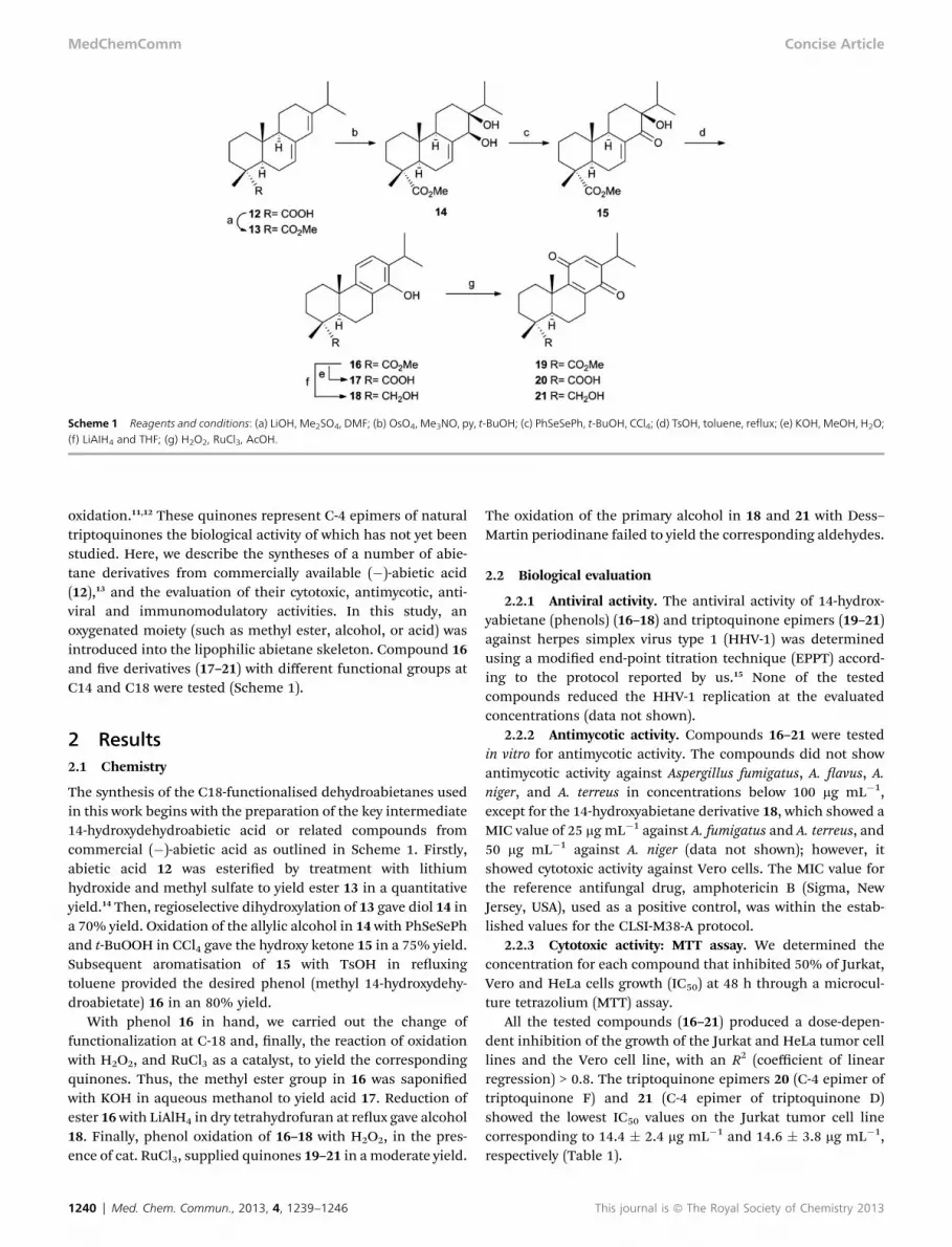
Scheme 1 Reagents and conditions: (a) LiOH, Me2SO4, DMF; (b) OsO4, Me3NO, py, t-BuOH; (c) PhSeSePh, t-BuOH, CCl4; (d) TsOH, toluene, reflux; (e) KOH, MeOH, H2O;(f) LiAIH4 and THF; (g) H2O2, RuCl3, AcOH.
MedChemComm Concise Article
oxidation.11,12 These quinones represent C-4 epimers of naturaltriptoquinones the biological activity of which has not yet beenstudied. Here, we describe the syntheses of a number of abie-tane derivatives from commercially available (�)-abietic acid(12),13 and the evaluation of their cytotoxic, antimycotic, anti-viral and immunomodulatory activities. In this study, anoxygenated moiety (such as methyl ester, alcohol, or acid) wasintroduced into the lipophilic abietane skeleton. Compound 16and ve derivatives (17–21) with different functional groups atC14 and C18 were tested (Scheme 1).
2 Results2.1 Chemistry
The synthesis of the C18-functionalised dehydroabietanes usedin this work begins with the preparation of the key intermediate14-hydroxydehydroabietic acid or related compounds fromcommercial (�)-abietic acid as outlined in Scheme 1. Firstly,abietic acid 12 was esteried by treatment with lithiumhydroxide and methyl sulfate to yield ester 13 in a quantitativeyield.14 Then, regioselective dihydroxylation of 13 gave diol 14 ina 70% yield. Oxidation of the allylic alcohol in 14 with PhSeSePhand t-BuOOH in CCl4 gave the hydroxy ketone 15 in a 75% yield.Subsequent aromatisation of 15 with TsOH in reuxingtoluene provided the desired phenol (methyl 14-hydroxydehy-droabietate) 16 in an 80% yield.
With phenol 16 in hand, we carried out the change offunctionalization at C-18 and, nally, the reaction of oxidationwith H2O2, and RuCl3 as a catalyst, to yield the correspondingquinones. Thus, the methyl ester group in 16 was saponiedwith KOH in aqueous methanol to yield acid 17. Reduction ofester 16with LiAlH4 in dry tetrahydrofuran at reux gave alcohol18. Finally, phenol oxidation of 16–18 with H2O2, in the pres-ence of cat. RuCl3, supplied quinones 19–21 in a moderate yield.
1240 | Med. Chem. Commun., 2013, 4, 1239–1246
The oxidation of the primary alcohol in 18 and 21 with Dess–Martin periodinane failed to yield the corresponding aldehydes.
2.2 Biological evaluation
2.2.1 Antiviral activity. The antiviral activity of 14-hydrox-yabietane (phenols) (16–18) and triptoquinone epimers (19–21)against herpes simplex virus type 1 (HHV-1) was determinedusing a modied end-point titration technique (EPPT) accord-ing to the protocol reported by us.15 None of the testedcompounds reduced the HHV-1 replication at the evaluatedconcentrations (data not shown).
2.2.2 Antimycotic activity. Compounds 16–21 were testedin vitro for antimycotic activity. The compounds did not showantimycotic activity against Aspergillus fumigatus, A. avus, A.niger, and A. terreus in concentrations below 100 mg mL�1,except for the 14-hydroxyabietane derivative 18, which showed aMIC value of 25 mg mL�1 against A. fumigatus and A. terreus, and50 mg mL�1 against A. niger (data not shown); however, itshowed cytotoxic activity against Vero cells. The MIC value forthe reference antifungal drug, amphotericin B (Sigma, NewJersey, USA), used as a positive control, was within the estab-lished values for the CLSI-M38-A protocol.
2.2.3 Cytotoxic activity: MTT assay. We determined theconcentration for each compound that inhibited 50% of Jurkat,Vero and HeLa cells growth (IC50) at 48 h through a microcul-ture tetrazolium (MTT) assay.
All the tested compounds (16–21) produced a dose-depen-dent inhibition of the growth of the Jurkat and HeLa tumor celllines and the Vero cell line, with an R2 (coefficient of linearregression) > 0.8. The triptoquinone epimers 20 (C-4 epimer oftriptoquinone F) and 21 (C-4 epimer of triptoquinone D)showed the lowest IC50 values on the Jurkat tumor cell linecorresponding to 14.4 � 2.4 mg mL�1 and 14.6 � 3.8 mg mL�1,respectively (Table 1).
This journal is ª The Royal Society of Chemistry 2013
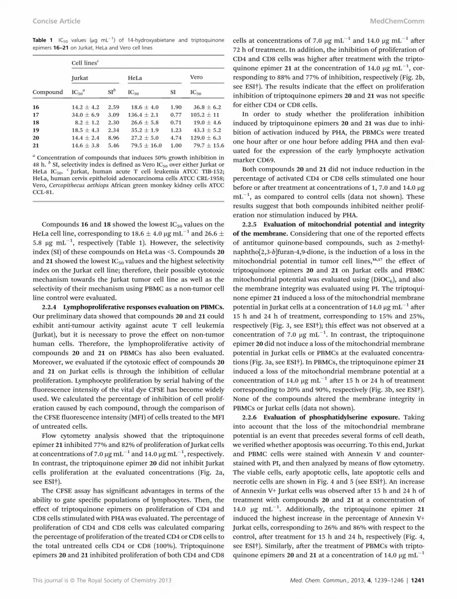
Table 1 IC50 values (mg mL�1) of 14-hydroxyabietane and triptoquinoneepimers 16–21 on Jurkat, HeLa and Vero cell lines
Compound
Cell linesc
Jurkat HeLa Vero
IC50a SIb IC50 SI IC50
16 14.2 � 4.2 2.59 18.6 � 4.0 1.90 36.8 � 6.217 34.0 � 6.9 3.09 136.4 � 2.1 0.77 105.2 � 1118 8.2 � 1.2 2.30 26.6 � 5.8 0.71 19.0 � 4.619 18.5 � 4.3 2.34 35.2 � 1.9 1.23 43.3 � 5.220 14.4 � 2.4 8.96 27.2 � 5.0 4.74 129.0 � 6.321 14.6 � 3.8 5.46 79.5 � 16.0 1.00 79.7 � 15.6
a Concentration of compounds that induces 50% growth inhibition in48 h. b SI, selectivity index is dened as Vero IC50 over either Jurkat orHeLa IC50.
c Jurkat, human acute T cell leukemia ATCC TIB-152;HeLa, human cervix epitheloid adenocarcinoma cells ATCC CRL-1958;Vero, Cercopithecus aethiops African green monkey kidney cells ATCCCCL-81.
Concise Article MedChemComm
Compounds 16 and 18 showed the lowest IC50 values on theHeLa cell line, corresponding to 18.6 � 4.0 mg mL�1 and 26.6 �5.8 mg mL�1, respectively (Table 1). However, the selectivityindex (SI) of these compounds on HeLa was <5. Compounds 20and 21 showed the lowest IC50 values and the highest selectivityindex on the Jurkat cell line; therefore, their possible cytotoxicmechanism towards the Jurkat tumor cell line as well as theselectivity of their mechanism using PBMC as a non-tumor cellline control were evaluated.
2.2.4 Lymphoproliferative responses evaluation on PBMCs.Our preliminary data showed that compounds 20 and 21 couldexhibit anti-tumour activity against acute T cell leukemia(Jurkat), but it is necessary to prove the effect on non-tumorhuman cells. Therefore, the lymphoproliferative activity ofcompounds 20 and 21 on PBMCs has also been evaluated.Moreover, we evaluated if the cytotoxic effect of compounds 20and 21 on Jurkat cells is through the inhibition of cellularproliferation. Lymphocyte proliferation by serial halving of theuorescence intensity of the vital dye CFSE has become widelyused. We calculated the percentage of inhibition of cell prolif-eration caused by each compound, through the comparison ofthe CFSE uorescence intensity (MFI) of cells treated to the MFIof untreated cells.
Flow cytometry analysis showed that the triptoquinoneepimer 21 inhibited 77% and 82% of proliferation of Jurkat cellsat concentrations of 7.0 mg mL�1 and 14.0 mgmL�1, respectively.In contrast, the triptoquinone epimer 20 did not inhibit Jurkatcells proliferation at the evaluated concentrations (Fig. 2a,see ESI†).
The CFSE assay has signicant advantages in terms of theability to gate specic populations of lymphocytes. Then, theeffect of triptoquinone epimers on proliferation of CD4 andCD8 cells stimulated with PHA was evaluated. The percentage ofproliferation of CD4 and CD8 cells was calculated comparingthe percentage of proliferation of the treated CD4 or CD8 cells tothe total untreated cells CD4 or CD8 (100%). Triptoquinoneepimers 20 and 21 inhibited proliferation of both CD4 and CD8
This journal is ª The Royal Society of Chemistry 2013
cells at concentrations of 7.0 mg mL�1 and 14.0 mg mL�1 aer72 h of treatment. In addition, the inhibition of proliferation ofCD4 and CD8 cells was higher aer treatment with the tripto-quinone epimer 21 at the concentration of 14.0 mg mL�1, cor-responding to 88% and 77% of inhibition, respectively (Fig. 2b,see ESI†). The results indicate that the effect on proliferationinhibition of triptoquinone epimers 20 and 21 was not specicfor either CD4 or CD8 cells.
In order to study whether the proliferation inhibitioninduced by triptoquinone epimers 20 and 21 was due to inhi-bition of activation induced by PHA, the PBMCs were treatedone hour aer or one hour before adding PHA and then eval-uated for the expression of the early lymphocyte activationmarker CD69.
Both compounds 20 and 21 did not induce reduction in thepercentage of activated CD4 or CD8 cells stimulated one hourbefore or aer treatment at concentrations of 1, 7.0 and 14.0 mgmL�1, as compared to control cells (data not shown). Theseresults suggest that both compounds inhibited neither prolif-eration nor stimulation induced by PHA.
2.2.5 Evaluation of mitochondrial potential and integrityof the membrane. Considering that one of the reported effectsof antitumor quinone-based compounds, such as 2-methyl-naphtho[2,3-b]furan-4,9-dione, is the induction of a loss in themitochondrial potential in tumor cell lines,16,17 the effect oftriptoquinone epimers 20 and 21 on Jurkat cells and PBMCmitochondrial potential was evaluated using (DiOC6), and alsothe membrane integrity was evaluated using PI. The triptoqui-none epimer 21 induced a loss of the mitochondrial membranepotential in Jurkat cells at a concentration of 14.0 mg mL�1 aer15 h and 24 h of treatment, corresponding to 15% and 25%,respectively (Fig. 3, see ESI†); this effect was not observed at aconcentration of 7.0 mg mL�1. In contrast, the triptoquinoneepimer 20 did not induce a loss of themitochondrial membranepotential in Jurkat cells or PBMCs at the evaluated concentra-tions (Fig. 3a, see ESI†). In PBMCs, the triptoquinone epimer 21induced a loss of the mitochondrial membrane potential at aconcentration of 14.0 mg mL�1 aer 15 h or 24 h of treatmentcorresponding to 20% and 90%, respectively (Fig. 3b, see ESI†).None of the compounds altered the membrane integrity inPBMCs or Jurkat cells (data not shown).
2.2.6 Evaluation of phosphatidylserine exposure. Takinginto account that the loss of the mitochondrial membranepotential is an event that precedes several forms of cell death,we veried whether apoptosis was occurring. To this end, Jurkatand PBMC cells were stained with Annexin V and counter-stained with PI, and then analyzed by means of ow cytometry.The viable cells, early apoptotic cells, late apoptotic cells andnecrotic cells are shown in Fig. 4 and 5 (see ESI†). An increaseof Annexin V+ Jurkat cells was observed aer 15 h and 24 h oftreatment with compounds 20 and 21 at a concentration of14.0 mg mL�1. Additionally, the triptoquinone epimer 21induced the highest increase in the percentage of Annexin V+Jurkat cells, corresponding to 26% and 86% with respect to thecontrol, aer treatment for 15 h and 24 h, respectively (Fig. 4,see ESI†). Similarly, aer the treatment of PBMCs with tripto-quinone epimers 20 and 21 at a concentration of 14.0 mg mL�1
Med. Chem. Commun., 2013, 4, 1239–1246 | 1241
MedChemComm Concise Article
for 15 h and 24 h, the percentage of positive Annexin cellsincreased in comparison to the control. The triptoquinoneepimer 21 induced the highest increase in the percentage ofAnnexin V+ PBMCs corresponding to 75% and 90% incomparison to the control, aer treatment for 15 h and 24 h,respectively (Fig. 5†). These results indicate that compounds 20and 21 induce phosphatidylserine exposure on Jurkat andPBMC cells. Compound 21 induced the highest phosphati-dylserine exposure.
2.2.7 Evaluation of cell cycle distribution. We also investi-gated whether compounds 20 and 21 induced either DNAdamage or alteration in the cell cycle distribution of Jurkat andPBMC cells, using ow cytometry analysis. Aer treating Jurkatcells with the triptoquinone epimer 21 for 15 h and 24 h, therewas a marked increase in the percentage of cells in the G2/M-phase of the cell cycle, corresponding to 13% and 15%,respectively, accompanied by a decrease in the percentage ofcells in the G0/G1-phase. In contrast, the triptoquinone epimer20 did not alter the cell cycle distribution of Jurkat cells (Fig. 6,see ESI†).
The triptoquinone epimer 21 induced hypoploidy in Jurkatcells at a concentration of 14.0 mg mL�1, aer 15 h and 24 h oftreatment, corresponding to 12% and 35%, respectively, whilethe triptoquinone epimer 20 did not induce hypoploidy at theevaluated concentrations (Fig. 7, see ESI†). In stimulatedPBMCs, triptoquinone epimers 20 and 21 did not inducealteration of the cell cycle distribution, and did not causehypoploidy at the tested concentrations (data not shown).
2.2.8 TUNEL assay. To prove that the epimer 20 did notinduce DNA damage in Jurkat and PBMC cells in comparison tothe epimer 21, we carried out a TUNEL assay. The triptoquinoneepimer 21 induced an increase of positive TUNEL Jurkat cells ata concentration of 14.0 mg mL�1, aer 15 h and 24 h of treat-ment, corresponding to 10% and 35%, respectively, but did notinduce an increase in PBMCs at the evaluated concentrations(Fig. 8b†). In contrast, the triptoquinone epimer 20 did notinduce any DNA damage in Jurkat or PBMC cells at the evalu-ated concentrations (Fig. 8, see ESI†). These results are inagreement with the results of staining with PI that weredescribed above (Fig. 7, see ESI†).
2.2.9 Evaluation of mononuclear phagocyte differentia-tion. As triptoquinone D and triptoquinone F are potent inter-leukin-1a and interleukin-1b inhibitors,8 we evaluated the anti-inammatory activity of their C-4 epimers, compounds 20 and21 respectively, in lipopolysaccharide (LPS)-stimulated mono-cyte-derived macrophages. Initially we investigated the effectof triptoquinone epimers 20 and 21 on the differentiation ofmonocytes into macrophages measuring the expression ofHLA-DR and uorescein diacetate labeling (FDA) aer 120 hof culture.
Both compounds induced a reduction in HLA-DR expression(Fig. 9a, see ESI†) and MFI of FDA (Fig. 9b, see ESI†) aertreatment at concentrations of 5.0, 10.0 and 15.0 mg mL�1, ascompared to control cells.
2.2.10 Cytokine production by monocytes. The effect oftriptoquinone epimers 20 and 21 on cytokine production, in thepresence or absence of LPS, was evaluated. Both compounds
1242 | Med. Chem. Commun., 2013, 4, 1239–1246
reduced the production of IL-1b, TNFa and IL-10 in PBMCsstimulated with LPS aer treatment at concentrations of 5.0,10.0 and 15.0 mg mL�1 (Fig. 10, see ESI†), and there were nosignicant differences in inhibition of cytokine productionbetween compounds, except for IL-1b, which was more inhibi-ted by the triptoquinone epimer 20 at concentrations of 5.0 and10.0 mg mL�1. The level of cytokines did not differ in theabsence of LPS aer treatment with both compounds (data notshown).
3 Discussion
The present study was performed to evaluate the antimycotic,antiviral and cytotoxic activity of three semisynthetic tripto-quinone epimers and three semisynthetic 14-hydroxyabietanederivatives. None of the tested compounds showed relevantantiviral or antimycotic activity, however some of them showedcytotoxic activity.
Compound 16 showed the lowest IC50 value on the HeLa cellline, but the selectivity index of this compound was <5. In theJurkat tumor cell line, compounds 20 and 21 inhibited cellulargrowth and showed the highest selectivity index (SI) values >5,corresponding to 8.96 and 5.46, respectively.
Triptoquinone epimers (19–21) were more selective to Jurkatthan HeLa cells, especially compound 20, and this is possiblydue to the presence of a molecular target in Jurkat that is notpresent in HeLa cells.
According to the National Cancer Institute (USA), a crudeextract is cytotoxic when its IC50 on normal cells is below 30 mgmL�1;18 therefore compounds 20 and 21, with IC50 values of129.0 � 6.3 and 79.7 � 15.6, respectively, are not cytotoxic inVero cells. So they were selected to evaluate their possiblemechanism of cell death induction.
Here, it was demonstrated that only the triptoquinoneepimer 21 induced proliferation inhibition in the Jurkat cell linewhen it was evaluated for the vital dye carboxyuoresceindiacetate succinimidyl (CFSE) assay. In addition, in order toidentify the selectivity on proliferation inhibition of compounds20 and 21 in tumor-lines, their effect on cell proliferation inPBMCs was determined. Both compounds 20 and 21 affectedCD4 or CD8 cells proliferation aer 72 h of treatment, but nottheir activation (CD69 expression), which is very importantconsidering that CD69 is a co-stimulatory molecule for T-cellactivation aer stimulation through the T cell antigen receptor(TCR) as well as a signal-transmitting receptor for synthesis ofcytokines (IL-2, INF-a, and TNF-a), expression of the IL-2Rsubunit, rise of intracellular calcium concentration and cellproliferation necessary for immune response.19
Desmond et al. (2005) have reported the ability of the fur-anonaphthoquinones to cause growth arrest and apoptosis in avariety of human leukemia and multiple myeloma cell lines;16
these effects were associated with a decrease in mitochondrialfunction that mediates the release of some proapoptoticproteins. In this study, it was found that only the triptoquinoneepimer 21 induced a loss of the mitochondrial membranepotential in both Jurkat cells and PBMCs at a concentration of14 mg mL�1 but not at a concentration of 7 mg mL�1, suggesting
This journal is ª The Royal Society of Chemistry 2013
Concise Article MedChemComm
that at lower concentrations, it can be more selective to Jurkatcells. Furthermore, none of the triptoquinone epimers alteredthe membrane integrity in PBMCs or Jurkat cells. This suggeststhat triptoquinone epimers 20 and 21 do not induce necrosis,which is important because pharmacological-induced necrosismight lead to tissue damages.20
Alteration of cell cycle distribution was only observed withcompound 21, which was an increase in the percentage of cellsin the G2/M-phase and a decrease in the percentage of cells inthe G0/G1-phase.
Gomathinayagam et al. (2008) observed that “Plumbagin”, anaphthoquinone, present in plants from the Droseraceae andPlumbaginaceae families, caused an increase in the percentageof cells in the G2/M-phase in human lung cancer cells (H460) bydown-regulating G2/M regulatory proteins (cyclinB1 andCdc25B), and it also induced a decrease in the percentage ofcells in the G0/G1-phase.21
Likewise, only compound 21 induced selective DNA damagein Jurkat cells at the tested concentrations. It is noteworthy tosay that this has been observed with another diterpenoidquinone, “Salvicine”, which induces DNA damage throughdouble-strand breaks by inhibition of topoisomerase II andglutathione depletion (Cai et al., 2008).22 In contrast, diterpe-noid quinones isolated from the plant Peltodon longipes andthat possess a para-quinone structure such as 7-a-acetoxyr-oyleanone, horminone, royleanone, and 7-ketoroyleanone haveshown cytotoxic activity against the human pancreatic cancercell line MIA PaCa-2, mainly through the inhibition of therelaxation activity of human topoisomerase I (induced by DNAstrand breaks) at concentrations lower than or similar to thoseof the positive control, camptothecin.23 As said by Fronza et al.(2012),23 it is possible that the structural properties of para-quinone diterpenes can inuence or determine their molecularmode of producing cell death. Therefore, these different modesof action suggest that cell death induced by abietane quinonediterpenes may not follow a single mechanism, but severalmechanisms instead.
Apoptotic cell death is a process of programmed cell deaththat includes several biochemical events, among others, proteincleavage, protein cross-linking, DNA breakdown and loss of themitochondrial membrane potential. Wong et al. (2012) haveobserved that Tanshinone IIA (Tan IIA), a diterpenoid naph-thoquinone derived from Radix salvia miltiorrhiza, inducesapoptosis in lung cancer cells (A549) and human colon adeno-carcinoma cells (Colo-205) through a mitochondrial mediatedintrinsic cell death pathway.24–26 Tan IIA has shown cytotoxicactivity against human prostate cancer cells (LNCaP) at aconcentration of 35 mM and induces a reduction of the mito-chondrial membrane potential at a concentration of 50 mM.24
In this study, the externalization of phosphatidylserineresidues in the outer plasma membrane, an early biochemicalevent that occurs in apoptotic cells, was also evaluated. It wasobserved that both compounds 20 and 21 induced phosphati-dylserine (Anexin V) exposure on both PBMCs and Jurkat cellline at a tested concentration of 14.0 mg mL�1 and compound 21induced the highest percentage of positive Annexin cells in bothPBMCs and Jurkat cell line.
This journal is ª The Royal Society of Chemistry 2013
The triptoquinones A–F (6–11)(Fig. 1) are principal compo-nents of Triptergium wilfordii and have been found to inhibitproduction or release of IL-1 by human monocytes stimulatedby endotoxin.9,27,28 Then, the effects of compounds 20 and 21 ondifferentiation of monocytes were investigated and it was foundthat both compounds interfered with monocyte differentiationas assessed by HLA-DR expression and FDA labeling.
The inhibition of production or release of IL-1b in rat thoracicaorta aer LPS treatment has been shown by the triptoquinone A.8
In addition, T. wilfordii extracts and their active compounds,tryptoquinones D and F, have also showed inhibition of IL-1bproduction in human monocytes28 and IL-1b activity in humanperipheral mononuclear cells.9 Takaishi et al. (1997) observed thattriterpenoids isolated from T. wilfordii var. Regelii have inhibitoryactivity against IL-1a and IL-1b release from lipopolysaccharide-stimulated human peripheral mononuclear cells.27
Several studies have reported that Triptolide, a puriedditerpenoid component of T. wilfordii, is able to suppress theproduction of inammatory mediators such as IL-1b and TNF-ainduced by LPS in various cell types including bronchialepithelial cells,24,29 peripheral blood mononuclear cells,30,31 andmacrophages.32
Several studies suggest that the T. wilfordii bioactivecompounds (triptolide, celastrol and tripchlorolide) exert theirimmunosuppressive and anticancer activities by modulating thetranscriptional activity of the nuclear factor-NF-kB. NF-kB activatesthe enhancer region of various genes, including the pro-inam-matory cytokines tumour necrosis factor (TNF)-a and oncogenicgenes, such as survivin, cyclinD1 and c-myc.24 Additionally, severalclinical trials have shown the efficacy of Triptolide in the T. wil-fordii extracts in the treatment of rheumatic diseases, includingrheumatoid arthritis and systemic lupus erythematosus.24,33–37
Based on this evidence, the anti-inammatory effect of trip-toquinone epimers 20 and 21was evaluated. Both compounds notonly inhibited the production of IL-1b and TNF-a, pro-inam-matory cytokines, but also IL-10 an anti-inammatory cytokine inthe presence of LPS. Therefore the compounds did not possess adifferential effect on the production of pro-inammatory andanti-inammatory cytokines.
4 Conclusions
In summary, the triptoquinone epimers 20 and 21 showeddifferent effects and selectivity, which could be due to thedifference in their chemical structure; both compounds differonly in one of their substitutions on carbon 18. Compound 20has a carboxylic acid as the functional group and compound 21has an alcohol. In addition, the triptoquinone epimer 20induced only phosphatidylserine exposure on Jurkat cells,therefore, it was not clear if this epimer induces cell death. Onthe other hand, the triptoquinone epimer 21 induced phos-phatidylserine exposure and proliferation inhibition in PBMCsbut it did not affect their activation. Moreover, it induced a lossof the mitochondrial potential at a concentration of 7.0 mgmL�1, DNA damage and alteration of cell cycle distribution inJurkat cells but not in PBMCs. These results suggest that thiscompound induces cell death in the Jurkat tumoral cell line at a
Med. Chem. Commun., 2013, 4, 1239–1246 | 1243
MedChemComm Concise Article
concentration of 7.0 mg mL�1, however, in vivo studies will benecessary to dene its possible clinical usefulness.
In addition, taking into account that both compoundsinhibited monocyte differentiation, they could be potentialimmunosuppressive and anti-inammatory agents that can beuseful in treatment of autoimmune diseases such as rheuma-toid arthritis and non-infectious inammatory processes.38
5 Materials and methods5.1 Chemistry
Optical rotations were determined with a 5 cm path-length cell,using dichloromethane as a solvent (concentration expressed ing per 100 mL). [a]D-values are given in 10�1 deg cm2 g�1. NMRspectra were recorded on a 300 MHz spectrometer with tetra-methylsilane as an internal standard. All spectra were recordedin CDCl3 as the solvent unless otherwise described. Completeassignments of 13C NMR multiplicities were made on the basisof DEPT experiments. J values are given in Hz. Mass spectra(MS) were run by electron impact (EI) at 70 eV. Reactions weremonitored by means of thin-layer chromatography (TLC) usingMerck silica gel 60 F-254 in 0.25 mm thick plates. Compoundson TLC plates were detected under UV light at 254 nm andvisualized by immersion in a 10% sulfuric acid solution andheating on a hotplate. Purications were performed by ashchromatography on Merck silica gel (230–400 mesh). All non-aqueous reactions were carried out in an argon atmosphere inoven-dried glassware. Commercial reagent grade solvents andchemicals were used as received unless otherwise noted.Combined organic extracts were washed with brine, dried overanhydrous sodium sulphate, ltered and concentrated underreduced pressure.
5.1.1 Methyl 13b,14b-dihydroxyabieta-7-en-18-oate (14). Toa solution of methyl abietate (13) (Gonzalez et al., 2009)14 (6.0 g,0.018 mol) in t-BuOH (30 mL) and pyridine (1.4 mL), Me3NO(2.8 g, 1.3 equiv.) and a 4% solution of OsO4 (2.0 mL) wereadded. The reaction mixture was stirred under an Ar atmo-sphere at reux for one week. Then, NaHSO3 (5 mL) was addedand the solvent was evaporated. The residue was dissolved inAcOEt and washed with brine. The organic layer was dried andconcentrated to yield the crude product which was puried bychromatography on silica, eluted with hexane–ethyl acetate(from 7 : 3 to 6 : 4) to yield diol 14 (4.6 g, 70%) as an orange oilthat solidies upon standing. The MS, 1H and 13C data are inagreement with the literature data.39
5.1.2 Methyl 13b-hydroxy-14-oxoabieta-7-en-18-oate (15).Diphenyl diselenide (6.2 g, 20 mmol) and 5.5 M t-BuOOH indecane (7.3 mL, 40 mmol) were added to a stirred solution of 14(5.0 g, 14.3 mmol) in dry CCl4 (100 mL), and the mixture wasreuxed under an Ar atmosphere for 4 h. Then, the solvent wasevaporated under vacuum, and the residue was puried bychromatography on silica, eluted with hexane–diethyl ether(7 : 3) to yield ketone 15 (3.7 g, 75%) as a colourless oil. The MS,1H and 13C data are in agreement with the literature data.40
5.1.3 Methyl 14-hydroxyabieta-8,11,13-trien-18-oate (16).To a solution of the ketone 15 (2.0 g, 5.7 mmol) in toluene(100 mL), p-toluenesulfonic acid (1.1 g, 5.7 mmol) was added
1244 | Med. Chem. Commun., 2013, 4, 1239–1246
and the mixture was reuxed for 15 h. Then, the solvent wasevaporated to yield a residue, which was chromatographed onsilica, eluted with hexane–ethyl acetate (8 : 2) to yield phenol 16(1.5 g, 80%) as an orange oil: [a]25D +36.0 (c 4.5) [(ref. 8) +51.7(c 0.5, CHCl3); (ref. 9) +7.7 (c 1.0, CHCl3)].
1H NMR (300 MHz) d7.00 (1H, d, J ¼ 8.2), 6.82 (1H, d, J ¼ 8.2), 3.64 (3H, s), 3.16 (1H,sept., J¼ 6.9), 2.77 (1H, dd, J¼ 16.8, 6.3), 2.67 (1H, m), 2.27 (1H,br d, J ¼ 12.9), 2.21 (1H, dd, J ¼ 12.6, 2.1), 1.27 (3H, s), 1.21 (6H,d, J¼ 6.9), 1.20 (3H, s); 13C NMR (75 MHz) dC 179.0 (s), 150.3 (s),148.0 (s), 130.3 (s), 123.1 (d), 120.7 (s), 115.9 (d), 51.7 (d), 47.4 (s),44.0 (d), 37.9 (t), 36.7 (s), 36.4 (t), 26.5 (q), 24.8 (q), 23.9 (t), 22.6(q), 22.4 (q), 20.9 (t), 18.4 (t), 16.3 (q); HRMS (EI) m/z 330.2204[M]+, calcd for C21H30O3: 330.2195.
5.1.4 14-Hydroxyabieta-8,11,13-trien-18-oic acid (17). Amixture of ester 16 (200 mg, 0.61 mmol), KOH (85%, 1.5 g, 23mmol), H2O (2 mL) and methanol (12 mL) was reuxed for 48 h.Aer this time, the reaction mixture was cooled, poured intoaqueous HCl (1.2 M, 30 mL) and extracted three times withDCM. The organic extract was dried over MgSO4 and concen-trated under reduced pressure to yield the crude acid 17, whichwas chromatographed on silica, eluted with hexane–ethylacetate (6 : 4) to yield acid 17 (144 mg, 75%) as a yellow foam:[a]25D +46.4 (c 1.2) [(ref. 8) +58.9 (c 0.5, CHCl3)].
1H NMR(300 MHz) d 7.01 (1H, d, J ¼ 8.1), 6.84 (1H, d, J ¼ 8.1), 3.14 (1H,sept., J¼ 6.9), 2.76 (1H, dd, J¼ 16.8, 6.6), 2.68 (1H, m), 2.28 (1H,d, J ¼ 12.6), 2.21 (1H, d, J ¼ 11.1), 1.27 (3H, s), 1.22 (6H, d, J ¼6.9), 1.20 (3H, s); 13C NMR (75 MHz) dC 185.2 (s), 150.1 (s), 148.1(s), 130.4 (s), 123.3 (d), 120.7 (s), 116.2 (d), 47.3 (s), 43.9 (d), 37.9(t), 36.8 (s), 36.6 (t), 26.7 (q), 25.0 (q), 23.9 (t), 22.7 (q), 22.5 (q),21.0 (t), 18.5 (t), 16.1 (q); HRMS (EI) m/z 316.2045 [M]+, calcd forC20H28O3: 316.2038.
5.1.5 Abieta-8,11,13-trien-14,18-diol (18). To a solution ofester 16 (500 mg, 1.51 mmol) in dry THF (10 mL) LiAlH4
(500 mg, 13 mmol) was added in portions, and then reuxed for15 h. Then, the mixture was cooled to 0 �C, and 0.5 mL of H2O,0.5 mL of 15% NaOH and 1.5 mL of H2O were added sequen-tially and carefully. The resulting white solid was ltered off andwashed with ethyl acetate. The extract was concentrated andpuried by means of chromatography, eluted with hexane–ethylacetate (6 : 4) to yield 380 mg (83%) of pure alcohol 18 as ayellowish foam: [a]25D +32.6 (c 2.1) [(ref. 8) +51.4 (c 0.75, CHCl3)].1H NMR (300 MHz) d 6.99 (1H, d, J ¼ 8.1), 6.83 (1H, d, J ¼ 8.1),5.03 (1H, s, OH), 3.45 (1H, d, J¼ 10.8), 3.18 (1H, d, J¼ 10.8), 3.14(1H, sept., J¼ 6.9), 2.76 (1H, dd, J¼ 16.2, 5.7), 2.62 (1H, m), 2.25(1H, br d, J¼ 12.9), 1.23 (3H, d, J¼ 6.9), 1.21 (3H, s), 1.20 (3H, d,J ¼ 6.9), 0.85 (3H, s); 13C NMR (75 MHz) dC 150.2 (s), 148.6 (s),130.2 (s), 123.0 (d), 120.8 (s), 116.2 (d), 71.9 (t), 42.9 (d), 38.4 (t),37.6 (s), 37.2 (s), 34.9 (t), 26.7 (q), 25.2 (q), 23.9 (t), 22.7 (q), 22.5(q), 18.6 (t), 18.1 (t), 17.3 (q); HRMS (EI)m/z 302.2257 [M]+, calcdfor C20H30O2: 302.2246.
5.1.6 Methyl 11,14-dioxoabieta-8,12-dien-18-oate (19). To asolution of phenol 16 (90 mg, 0.27 mmol) in AcOH (1 mL) at10 �C, a catalytic amount of RuCl3$3H2O (14 mg, 0.2 equiv.) and30% H2O2 (150 mL, 1.42 mmol) were added. The mixture wasallowed to warm to room temperature and stirred for 15 h.Then, the reaction mixture was carefully poured into saturatedNaHCO3 and extracted with ethyl acetate. The organic layer was
This journal is ª The Royal Society of Chemistry 2013
Concise Article MedChemComm
washed with brine, dried and concentrated. The residue waschromatographed on silica, eluted with hexane–ethyl acetate(8 : 2) to yield quinone 19 (56 mg, 60%) as a yellow-orange oil:[a]25D �43.6 (c 2.2) [(ref. 8) �69.7 (c 0.5, CHCl3)].
1H NMR(300 MHz) d 6.32 (1H, d, J ¼ 1.2), 3.68 (3H, s), 2.98 (1H, m), 2.78(1H, br d, J ¼ 11.1), 2.65 (1H, dd, J ¼ 20.1, 6.0), 2.40 (1H, dd, J ¼11.4, 7.5), 1.99 (1H, dd, J ¼ 12.3, 1.8), 1.31 (3H, s), 1.25 (3H, s),1.11 (3H, d, J ¼ 6.9), 1.09 (3H, d, J ¼ 6.9); 13C NMR (75 MHz) dC187.8 (s), 178.6 (s), 152.9 (s), 150.0 (s), 142.6 (s), 131.8 (d), 52.0(d), 47.7 (s), 45.7 (d), 37.8 (s), 36.4 (t), 35.5 (t), 26.3 (q), 25.4 (t),21.3 (q), 21.2 (q), 20.3 (q), 20.2 (t), 18.1 (t), 16.6 (q); HRMS (EI)m/z 344.1977 [M]+, calcd for C21H28O4: 344.1988.
5.1.7 11,14-Dioxoabieta-8,12-dien-18-oic acid (20). To asolution of phenol 17 (90 mg, 0.28 mmol) in AcOH (1 mL) at10 �C, a catalytic amount of RuCl3$3H2O (14 mg, 0.2 equiv.) and30% H2O2 (150 mL, 1.42 mmol) were added. The mixture wasallowed to warm to room temperature and stirred for 15 h.Then, the reaction mixture was poured into water and extractedwith ethyl acetate. The organic layer was washed with brine,dried and concentrated. The residue was chromatographed onsilica, eluted with hexane–ethyl acetate (6 : 4) to yield quinone20 (50mg, 55%) as a yellow-orange semi-solid: [a]25D �52.8 (c 2.5)[(ref. 8) �68.0 (c 0.5, CHCl3)].
1H NMR (300 MHz) d 6.33 (1H, d,J ¼ 1.0), 2.98 (1H, m), 2.80 (1H, br d, J ¼ 12.9), 2.67 (1H, dd, J ¼20.1, 4.8), 2.00 (1H, d, J ¼ 11.1), 1.31 (3H, s), 1.26 (3H, s), 1.10(3H, d, J ¼ 6.9), 1.09 (3H, d, J ¼ 6.9); 13C NMR (75 MHz) dC 187.8(s), 187.7 (s), 152.9 (s), 149.9 (s), 142.7 (s), 131.8 (d), 45.4 (d), 37.7(s), 36.5 (t), 35.5 (t), 26.3 (q), 25.4 (t), 21.3 (q), 21.3 (q), 20.3 (q),20.2 (t), 18.1 (t), 16.3 (q); HRMS (EI) m/z 330.1792 [M]+, calcd forC20H26O4: 330.1831.
5.1.8 11,14-Dioxoabieta-8,12-dien-18-ol (21). To a solutionof phenol 18 (250 mg, 0.82 mmol) in AcOH (2.5 mL) at 10 �C, acatalytic amount of RuCl3$3H2O (34 mg, 0.2 equiv.) and 30%H2O2 (450 mL, 1.42 mmol) were added. The mixture was allowedto warm and stirred for 15 h. Then, the reaction mixture wascarefully poured into saturated NaHCO3 and extracted withethyl acetate. The organic layer was washed with brine, driedand concentrated. The residue was chromatographed on silica,eluted with hexane–ethyl acetate (8 : 2) to yield quinone 21(150 mg, 58%) as an orange oil: [a]25D �36.5 (c 7.5) [(ref. 8) �64.1(c 0.6, CHCl3)].
1H NMR (300 MHz) d 6.31 (1H, d, J ¼ 1.0), 3.48(1H, d, J¼ 10.8), 3.13 (1H, d, J¼ 10.8), 2.97 (1H, m), 1.32 (3H, s),1.10 (3H, d, J ¼ 6.9), 1.09 (3H, d, J ¼ 6.9), 0.83 (3H, s); 13C NMR(75 MHz) dC 187.9 (s), 187.8 (s), 152.6 (s), 150.6 (s), 142.6 (s),131.7 (d), 71.4 (t), 44.5 (d), 38.2 (s), 37.6 (s), 35.8 (t), 34.5 (t), 26.1(q), 25.4 (t), 21.2 (q), 21.2 (q), 20.4 (q), 18.2 (t), 17.6 (q), 16.9 (t);HRMS (EI) m/z 316.2045 [M]+, calcd for C20H28O3: 316.2038.
5.2 Biological assays (see ESI†)
Acknowledgements
Financial support from the Spanish Ministry of Science andEducation, under a “Ramon y Cajal” research grant, and alsofrom the Generalitat Valenciana (project GV/2007/007) is grate-fully acknowledged. L. B.-G. thanks the nancial support from
This journal is ª The Royal Society of Chemistry 2013
the Antioquia University of Colombia, CENIVAM, COLCIENCIAS(Patrimonio Autonomo del Fondo Nacional de Financiamientopara la Ciencia, la Tecnologıa y la Innovacion, Francisco Jose deCaldas) Grant RC RC-245-2011 and RC 366-2011.
References
1 M. B. De Jesus, W. F. Zambuzzi, R. R. R. De Sousa, C. Areche,A. C. S. De Souza, H. Aoyama, G. Schmeda-Hirschmann,J. A. Rodrıguez, A. R. M. De Souza-Brito,M. P. Peppelenbosch, J. Den Hertog, E. De Paula andC. V. Ferreira, Biochimie, 2008, 90, 843–854.
2 J. Xu, T. Ikekawa, M. Ohkawa, I. Yokota, N. Hara andY. Fujimoto, Phytochemistry, 1997, 44, 1511–1514.
3 S. M. Kupchan, A. Karim and C. Marcks, J. Org. Chem., 1969,34, 3912–3918.
4 A. M. Zaghloul, A. A. Gohar, Z. A. Naiem and F. M. Abdel Bar,Z. Naturforsch., C: J. Biosci., 2008, 63, 355–360.
5 L. Kuzma, H. Wysokinska, M. Rozalski, A. Budzynska,M. Wieckowska-Szakiel, B. Sadowska, M. Paszkiewicz,W. Kisiel and B. Rozalska, Phytomedicine, 2012, 19, 1285–1287.
6 N. Tan, M. Kaloga, O. A. Radtke, A. F. Kiderlen, S. Oksuz,A. Ulubelen and H. Kolodziej, Phytochemistry, 2002, 61,881–884.
7 H. Kofujita, M. Ota, K. Takahashi, Y. Kawai and Y. Hayashi,Phytochemistry, 2002, 61, 895–898.
8 K. Shishido, K. Nakano, N. Wariishi, H. Tateishi,T. Omodani, M. Shibuya, K. Goto, Y. Ono and Y. Takaishi,Phytochemistry, 1994, 35, 731–737.
9 M. Niwa, Y. Tsutsumishita, Y. Kawai, H. Takahara,N. Nakamura, S. Futaki, Y. Takaishi, W. Kondoh andH. Moritoki, Biochem. Biophys. Res. Commun., 1996, 224,579–585.
10 H. Moritoki, T. Hisayama, K. Kida, W. Kondoh, S. Inoue andY. Takaishi, Life Sci., 1996, 59, 49–54.
11 Y. Matsushita, Y. Iwakiri, S. Yoshida, K. Sugamoto andT. Matsui, Tetrahedron Lett., 2005, 46, 3629–3632.
12 E. Alvarez-Manzaneda, R. Chahboun, F. Bentaleb, E. Alvarez,M. A. Escobar, S. Sad-Diki, M. J. Cano and I. Messouri,Tetrahedron, 2007, 63, 11204–11212.
13 M. A. Gonzalez, M. J. Gil-Gimeno and A. Blake, ActaCrystallogr., Sect. E: Struct. Rep. Online, 2006, 62, 3346–3347.
14 M. A. Gonzalez, J. Correa-Royero, L. Agudelo, A. Mesa-Arangoand L. Betancur-Galvis, Eur. J. Med. Chem., 2009, 44, 2468–2472.
15 L. Betancur-Galvis, C. Zuluaga, M. Arno, M. A. Gonzalez andR. J. Zaragoza, J. Nat. Prod., 2001, 64, 1318–1321.
16 J. C. Desmond, H. Kawabata, C. Mueller-Tidow,E. Simamura, D. Heber, K. Hirai and H. P. Koeffler, Br.J. Haematol., 2005, 131, 520–529.
17 E. Simamura, K. Hirai, H. Shimada, J. Pan and J. Koyama,Cancer Detect. Prev., 2003, 27, 5–13.
18 T. Hennebelle, S. Sahpaz, H. Joseph and F. Bailleul,J. Ethnopharmacol., 2008, 116, 211–222.
19 K. Abbas, H. L. Andrew and P. Shiv, Cellular and molecularimmunology, Saunders Elsevier, 2007, p. 566.
Med. Chem. Commun., 2013, 4, 1239–1246 | 1245
MedChemComm Concise Article
20 S. Fink and B. Cookson, Infect. Immun., 2005, 73, 1907–1916.21 R. Gomathinayagam, S. Sowmyalakshmi, F. Mardhatillah,
R. Kumar, M. A. Akbarsha and C. Damodaran, AnticancerRes., 2008, 28, 785–792.
22 Y. J. Cai, J. J. Lu, H. Zhu, H. Xie, M. Huang, L. P. Lin,X. W. Zhang and J. Ding, Free Radical Biol. Med., 2008, 45,627–635.
23 M. Fronza, E. Lamy, S. Gunther, B. Heinzmann, S. Lauferand I. Merfort, Phytochemistry, 2012, 78, 107–119.
24 K. F. Wong, Y. Yuan and J. M. Luk, Clin. Exp. Pharmacol.Physiol., 2012, 39, 311–320.
25 T. L. Chiu and C. C. Su, Int. J. Mol. Med., 2010, 25, 231–236.26 C. C. Su, G. W. Chen, J. C. Kang andM. H. Chan, Planta Med.,
2008, 74, 1357–1362.27 Y. Takaishi, N. Wariishi, H. Tateishi, K. Kawazoe and
K. Nakano, Phytochemistry, 1997, 45, 969–974.28 M. L. Chang, L. L. Yang, D. M. Chang, S. Y. Kuo and S. J. Chu,
Zhonghua Minguo Weishengwu Ji Mianyixue Zazhi, 1993, 26,15–24.
29 G. Zhao, L. T. Vaszar, D. Qiu, L. Shi and P. N. Kao, Am.J. Physiol.: Lung Cell. Mol. Physiol., 2000, 279, 958–966.
1246 | Med. Chem. Commun., 2013, 4, 1239–1246
30 T. Krakauer, X. Chen, O. M. Howard and H. A. Young,Immunopharmacol. Immunotoxicol., 2005, 27, 53–66.
31 W. Tang and J. P. Zuo, Acta Pharmacol. Sin., 2012, 33, 1112–1118.
32 R. Matta, X. Wang, H. Ge, W. Ray, L. D. Nelin and Y. Liu, Am.J. Transl. Res., 2009, 1, 267–282.
33 X. Tao and P. E. Lipsky, Clin. Rheum. Dis., 2000, 26,29–50.
34 N. Lin, C. Liu, C. Xiao, H. Jia, K. Imada, H. Wu and A. Ito,Biochem. Pharmacol., 2007, 73, 136–146.
35 J. Ma, M. Dey, H. Yang, A. Poulev, R. Pouleva, R. Dorn,P. E. Lipsky, E. J. Kennelly and I. Raskin, Phytochemistry,2007, 68, 1172–1178.
36 C. T. Chou, Phytother. Res., 1997, 11, 152–154.37 B. J. Chen, Lymphoma, 2001, 42, 253–265.38 J. M. Davis and E. L. Matteson, Mayo Clin. Proc., 2012, 87,
659–673.39 M. C. Costa, S. P. Alves, M. E. Correia and M. J. Marcelo-
Curto, Synthesis, 2006, 7, 1171–1175.40 A. Presser, E. Haslinger, R. Weis and A. Hufner, Monatsh.
Chem., 1998, 129, 921–930.
This journal is ª The Royal Society of Chemistry 2013