cysteamine, glutathione and ionomycin treatments improve in vitro fertilization of prepubertal goat...
TRANSCRIPT

Proof Delivery FormPlease return this form with your proof
CUP reference:
Date of delivery:
Journal and Article number: ZYG 287
Volume and Issue Number: 12(3)
Colour Figures: Nil
Number of pages (not including this page): 8
ZYGOTE – ‘Return of Proofs’ instructions
We hope you have now downloaded a proof of your paper in pdf format, and also the copyright form and the offprint orderform.
Please print out a proof of your paper and the two forms. Both the forms should be completed in hard copy, and correctionsto your proof should also be marked on a hard copy.
Please check the proofs carefully, answering any queries. Queries raised by the typesetter are listed below: the text to whichthe queries refer is flagged in the margins of the proof. Please note it is your responsibility to check the factual content of yourpaper. Only typographical and factual errors should be corrected – you may be charged for corrections of non-typographicalerrors. Mark any typesetting errors in RED ink and your own (author corrections) in BLUE or BLACK ink.
If any figure requires correction of anything other than a typographical error introduced by the printer, you must provide anew copy.
Please return the corrected proof, the Copyright form and the Offprint Order Form, within three days of receipt to:-
Nicki MarshallProduction Editor (Journals)Cambridge University PressUniversity Printing HouseShaftesbury RoadCambridge CB2 2BSUnited Kingdom
Please return these proofs by First Class mail or FEDEX courier (or similar) if necessary or if outside Europe. It may not bepossible to take your corrections into consideration if they are not received at the appropriate time. If you have any difficultieswith these proofs, please contact Nicki Marshall (details above).

Author queries:
Q1: Martino et al. (1995) is not in the References.
Typesetter queries:
T1: Disk mismatch with Msp. Disk followed.
Non-printed material:


Zygote 12 (August), pp. 1–8. C© 2004 Cambridge University PressDOI:10.1017/S0967199404002874 Printed in the United Kingdom
Cysteamine, glutathione and ionomycin treatments improve in vitrofertilization of prepubertal goat oocytes
Aixa Urdaneta, Ana Raquel Jimenez, Marıa-Teresa Paramio, and Dolors IzquierdoDepartament de Ciencia Animal i dels Aliments, Facultat de Veterinaria, Universitat Autonoma de Barcelona,08193 Bellaterra, Barcelona, Spain
Date submitted: 7.5.04. Date accepted: 8.7.04
Summary
The aim of this study was to improve in vitro embryo development of prepubertal goat oocytes by
T1
studying the effect of adding cysteamine to in vitro maturation medium, glutathione (GSH) to in vitrofertilization medium and ionomycin to the sperm capacitation medium. In experiment 1, we analysedthe effect of 1 mM GSH added to fertilization medium of oocytes matured with 400 µM cysteamine.The control group were oocytes without cysteamine and GSH. In experiment 2, oocytes matured andfertilized in the presence of 400 µM cysteamine and 1 mM GSH, respectively, were inseminated withspermatozoa treated with ionomycin or heparin. In experiment 1, the percentages of total and normalfertilized oocytes were significantly higher for oocytes supplemented with cysteamine and GSH (40.26%and 30.20%, respectively) than for oocytes from the control group (16.66%, and 10.61%, respectively).The percentage of total embryos obtained after 7 days of culture was significantly higher in the groupsupplemented with cysteamine and GSH (30.62%) than in the control group (8.09%). In experiment 2,percentages of total and normal fertilized oocytes were significantly higher for the group of spermatozoacapacitated with ionomycin (52.21% and 37.17%, respectively) than with heparin (38.62% and 28.35%,respectively). After 7 days of culture, total embryo rate was significantly higher in the group of spermcapacitated with ionomycin (44.91%) than with heparin (38.69%). However, the percentage of embryosdeveloped to the blastocyst stage was not affected by any of the treatments studied.
Keywords: Cysteamine, Glutathione, Goat, Ionomycin, Prepubertal
Introduction
In vitro matured prepubertal goat oocytes often showdeficiencies after their in vitro fertilization (IVF) asindicated by a low incidence of male pronuclearformation (Martino et al., 1995; Mogas et al., 1997),Q1which would cause a high number of haploid embryos(Villamediana et al., 2001) and a low developmentalcompetence to the blastocyst stage (Izquierdo et al.,1999).
During fertilization, the sperm nucleus is decon-densed and transformed into a male pronucleus(MPN). This transformation of the sperm nucleusduring IVF has been related to the levels of intracellular
All correspondence to: D. Izquierdo, Departament deCiencia Animal i dels Aliments, Facultat de Veterinaria,Universitat Autonoma de Barcelona, 08193 Bellaterra,Barcelona, Spain. Tel: + 34 935811456. Fax: + 34-935811494.e-mail: [email protected]
glutathione (GSH) (Sutovsky & Schatten, 1997). Thetripeptide glutathione (γ-glutamyl-cysteinyl-glycine) isa reducing agent that plays a number of importantroles in cellular metabolism (reviewed by Knapenet al., 1999), participating in various mechanisms suchas amino acid transport, DNA and protein synthesis,reduction of disulphide bonds and protection of cellsagainst oxidative damage. During the developmentand maturation of the oocyte in the ovary, GSH contentincreases as the oocyte approaches the time of ovulation(Perrault et al., 1988). When the oocytes are in vitromatured, GSH synthesis can be stimulated by theaddition of low-molecular-weight thiol compounds.
Cysteamine is a low-molecular-weight thiol whichwhen present during in vitro maturation (IVM) in-creases the intracytoplasmic oocyte GSH concentrationin cattle (De Matos et al., 1995; Luvoni et al., 1996;De Matos & Furnus, 2000), goat (Rodriguez et al.,2003), buffalo (Gasparrini et al., 2003) and ewes (DeMatos et al., 2002). Addition of cysteamine to the IVM

2 A. Urdaneta et al.
medium of cattle oocytes improves in vitro blastocystdevelopment (De Matos et al., 1995, 1996). Similarresults were found in sheep (De Matos et al., 2002),buffalo (Gasparrini et al., 2000), pig (Grupen et al., 1995;Yamauchi & Nagai, 1999) and mouse (De Matos et al.,2003). Previous studies in our laboratory (Rodrıguez-Gonzalez et al., 2003) showed that 100 µM of cysteamineadded to the IVM medium of prepubertal goat oocytesincreased intracellular GSH levels and significantlyimproved the percentages MPN formation after IVFand embryo development. Later, Urdaneta et al. (2003)concluded that the addition of 400 µM of cysteamineto the IVM medium improved embryo developmentof prepubertal goat oocytes compared with 100 µM.However, the percentage of blastocysts obtained inboth studies was low.
During fertilization, several researchers add GSH tothe sperm preparation medium (pig: Jeong & Yang,2001; cattle: Earl et al., 1997; Taneja et al., 1998) and/orIVF medium (pig: Boquest et al., 1999; cattle: VanSoom et al., 1998), obtaining an improvement in theblastocyst formation rate. The use of GSH during spermpreparation could be beneficial to membrane stabi-lization of the spermatozoa and the protection ofsperm from free radical damage, and could thus helpsubsequent fertilization and development.
Goat oocytes have significantly improved IVF rateswhen spermatozoa are capacitated with ionomycin(Wang et al., 2002). Ionomycin is a calcium ionophorethat can increase Ca2 + cycling across phospholipidmembranes and induce the acrosome reaction (Ballet al., 1983). Wang et al. (2002), studying variousfactors affecting the effectiveness of the IVM–IVFsystem of goat oocytes, concluded that spermatozoatreated with heparin plus 100–200 nM of ionomycingave significantly higher fertilization rates comparedwith the conventional goat sperm treatment of heparinalone.
The aim of this study was to improve in vitro embryodevelopment of prepubertal goat oocytes by studyingthe effect of adding 400 µM cysteamine to IVMmedium, 1 mM GSH to the IVF medium and 200 nMionomycin to the sperm capacitation medium.
Materials and methods
Oocyte collection
We obtained ovaries from prepubertal (approximately2 months old) goats from a local slaughterhouse andtransported them at 37 ◦C in Dulbecco’s phosphate-buffered saline (PBS; P-4417, Sigma Chemical, St Louis,MO, USA) supplemented with 50 µg/ml gentamicin.Oocytes were recovered by cutting the surface ofovaries with a razor blade in a 60 mm culture dish
with TCM199 (M-2520; Sigma, USA), supplementedwith 2.2 mg/ml NaHCO3 and 50 µg/ml gentamicin.We selected only oocytes with one or more completelayers of unexpanded cumulus cells and homogeneouscytoplasm.
In vitro maturation of oocytes
The oocyte maturation medium was TCM199 (M-7528;Sigma, USA) supplemented with 275 µg/ml sodiumpyruvate (P-3662; Sigma, USA), 146 µg/ml L-glutamine(G-5763; Sigma, USA), 10% (v/v) steer serum,10 µg/ml o-LH (L-5269; Sigma, USA), 10 µg/ml o-FSH(Ovagen, Immuno Chemicals Products, Auckland,New Zealand), 1 µg/ml 17β-estradiol (E-2257; Sigma,USA) and 50 µg/ml gentamicin. A group of oocyteswere supplemented with 400 µM cysteamine (M-9768;Sigma, USA). The cumulus–oocyte complexes wereculture in groups of 20–25 in 100 µl microdrops ofmaturation medium and incubated for 27 h at 38.5 ◦Cin a humidified atmosphere of 5% CO2 in air undermineral oil (M-8410; Sigma, USA).
Sperm preparation and capacitation
The oocytes were inseminated with fresh semen,at the end of the maturation period. We collectedejaculates from two Murciano-Granadino bucks ofproven fertility into artificial vaginas and transportedthem at 37–38 ◦C within 30 min to the laboratory.The motility of sperm cells was evaluated under aninverted microscope and the motile sperm fraction wasseparated by swim-up (Parrish et al., 1986). Seventymicrolitres of semen was placed in each of severalconical tubes under 2 ml Defined Medium (Brackett &Oliphant, 1975) as modified by Younis et al. (1991)and referred to here as mDM, and incubated for 45–60 min in a humidified atmosphere of 5% CO2 in airat 38.5 ◦C. After incubation, 600 µl from the top ofeach tube containing highly motile spermatozoa wasremoved and pooled in a sterile 15 ml centrifuge tubeand centrifuged at 200 g for 10 min.
After discarding the supernatant, the resulting spermpellet was treated in one of two ways: (1) thesperm pellet was resuspended 1:1 (v:v) with mDMmedium containing heparin (heparin-sodium salt;170 UI/mg; final concentration 50 µg/ml of heparin)and incubated in a humidified air atmosphere of5% CO2 at 38.5 ◦C (final concentration approximately84 × 106 sperm/ml) for 45–60 min (heparin group) or(2) the sperm pellet was resuspended 1:1 (v:v) withmDM medium containing 20 µg/ml heparin, 1 mM 8-Br-cAMP and 400 nM ionomycin for 15 min (ionomycingroup; final concentration 10 µg/ml heparin, 0.5 mM 8-Br-cAMP and 200 nM ionomycin).
Cysteamine, GSH and ionomycin 3
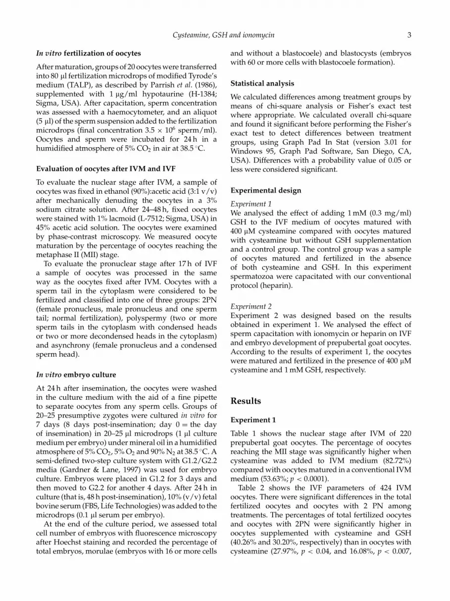
In vitro fertilization of oocytes
After maturation, groups of 20 oocytes were transferredinto 80 µl fertilization microdrops of modified Tyrode’smedium (TALP), as described by Parrish et al. (1986),supplemented with 1 µg/ml hypotaurine (H-1384;Sigma, USA). After capacitation, sperm concentrationwas assessed with a haemocytometer, and an aliquot(5 µl) of the sperm suspension added to the fertilizationmicrodrops (final concentration 3.5 × 10⁶ sperm/ml).Oocytes and sperm were incubated for 24 h in ahumidified atmosphere of 5% CO2 in air at 38.5 ◦C.
Evaluation of oocytes after IVM and IVF
To evaluate the nuclear stage after IVM, a sample ofoocytes was fixed in ethanol (90%):acetic acid (3:1 v/v)after mechanically denuding the oocytes in a 3%sodium citrate solution. After 24–48 h, fixed oocyteswere stained with 1% lacmoid (L-7512; Sigma, USA) in45% acetic acid solution. The oocytes were examinedby phase-contrast microscopy. We measured oocytematuration by the percentage of oocytes reaching themetaphase II (MII) stage.
To evaluate the pronuclear stage after 17 h of IVFa sample of oocytes was processed in the sameway as the oocytes fixed after IVM. Oocytes with asperm tail in the cytoplasm were considered to befertilized and classified into one of three groups: 2PN(female pronucleus, male pronucleus and one spermtail; normal fertilization), polyspermy (two or moresperm tails in the cytoplasm with condensed headsor two or more decondensed heads in the cytoplasm)and asynchrony (female pronucleus and a condensedsperm head).
In vitro embryo culture
At 24 h after insemination, the oocytes were washedin the culture medium with the aid of a fine pipetteto separate oocytes from any sperm cells. Groups of20–25 presumptive zygotes were cultured in vitro for7 days (8 days post-insemination; day 0 = the dayof insemination) in 20–25 µl microdrops (1 µl culturemedium per embryo) under mineral oil in a humidifiedatmosphere of 5% CO2, 5% O2 and 90% N2 at 38.5 ◦C. Asemi-defined two-step culture system with G1.2/G2.2media (Gardner & Lane, 1997) was used for embryoculture. Embryos were placed in G1.2 for 3 days andthen moved to G2.2 for another 4 days. After 24 h inculture (that is, 48 h post-insemination), 10% (v/v) fetalbovine serum (FBS, Life Technologies) was added to themicrodrops (0.1 µl serum per embryo).
At the end of the culture period, we assessed totalcell number of embryos with fluorescence microscopyafter Hoechst staining and recorded the percentage oftotal embryos, morulae (embryos with 16 or more cells
and without a blastocoele) and blastocysts (embryoswith 60 or more cells with blastocoele formation).
Statistical analysis
We calculated differences among treatment groups bymeans of chi-square analysis or Fisher’s exact testwhere appropriate. We calculated overall chi-squareand found it significant before performing the Fisher’sexact test to detect differences between treatmentgroups, using Graph Pad In Stat (version 3.01 forWindows 95, Graph Pad Software, San Diego, CA,USA). Differences with a probability value of 0.05 orless were considered significant.
Experimental design
Experiment 1We analysed the effect of adding 1 mM (0.3 mg/ml)GSH to the IVF medium of oocytes matured with400 µM cysteamine compared with oocytes maturedwith cysteamine but without GSH supplementationand a control group. The control group was a sampleof oocytes matured and fertilized in the absenceof both cysteamine and GSH. In this experimentspermatozoa were capacitated with our conventionalprotocol (heparin).
Experiment 2Experiment 2 was designed based on the resultsobtained in experiment 1. We analysed the effect ofsperm capacitation with ionomycin or heparin on IVFand embryo development of prepubertal goat oocytes.According to the results of experiment 1, the oocyteswere matured and fertilized in the presence of 400 µMcysteamine and 1 mM GSH, respectively.
Results
Experiment 1
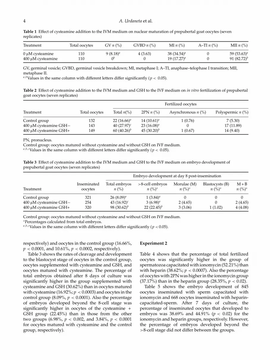
Table 1 shows the nuclear stage after IVM of 220prepubertal goat oocytes. The percentage of oocytesreaching the MII stage was significantly higher whencysteamine was added to IVM medium (82.72%)compared with oocytes matured in a conventional IVMmedium (53.63%; p < 0.0001).
Table 2 shows the IVF parameters of 424 IVMoocytes. There were significant differences in the totalfertilized oocytes and oocytes with 2 PN amongtreatments. The percentages of total fertilized oocytesand oocytes with 2PN were significantly higher inoocytes supplemented with cysteamine and GSH(40.26% and 30.20%, respectively) than in oocytes withcysteamine (27.97%, p < 0.04, and 16.08%, p < 0.007,
4 A. Urdaneta et al.
Table 1 Effect of cysteamine addition to the IVM medium on nuclear maturation of prepubertal goat oocytes (sevenreplicates)
Treatment Total oocytes GV n (%) GVBD n (%) MI n (%) A–TI n (%) MII n (%)
0 µM cysteamine 110 9 (8.18)a 4 (3.63) 38 (34.54)a 0 59 (53.63)a
400 µM cysteamine 110 0b 0 19 (17.27)a 0 91 (82.72)b
GV, germinal vesicle; GVBD, germinal vesicle breakdown; MI, metaphase I; A–TI, anaphase–telophase I transition; MII,metaphase II.a ,bValues in the same column with different letters differ significantly (p < 0.05).
Table 2 Effect of cysteamine addition to the IVM medium and GSH to the IVF medium on in vitro fertilization of prepubertalgoat oocytes (seven replicates)
Fertilized oocytes
Treatment Total oocytes Total n(%) 2PN n (%) Asynchronous n (%) Polyspermic n (%)
Control group 132 22 (16.66)a 14 (10.61)a 1 (0.76) 7 (5.30)400 µM cysteamine GSH− 143 40 (27.97)c 23 (16.08)a 0 17 (11.89)400 µM cysteamine GSH+ 149 60 (40.26)b 45 (30.20)b 1 (0.67) 14 (9.40)
PN, pronucleus.Control group: oocytes matured without cysteamine and without GSH on IVF medium.a ,b,cValues in the same column with different letters differ significantly (p < 0.05).
Table 3 Effect of cysteamine addition to the IVM medium and GSH to the IVF medium on embryo development ofprepubertal goat oocytes (seven replicates)
Embryo development at day 8 post-insemination
Inseminated Total embryos >8-cell embryos Morulae (M) Blastocysts (B) M + BTreatment oocytes n (%) n (%)∗ n (%)∗ n (%)∗ n (%)∗
Control group 321 26 (8.09)a 1 (3.84)a 0 0 0400 µM cysteamine GSH− 254 43 (16.92)c 3 (6.98)a 2 (4.65) 0 2 (4.65)400 µM cysteamine GSH+ 320 98 (30.62)b 22 (22.45)b 3 (3.06) 1 (1.02) 4 (4.08)
Control group: oocytes matured without cysteamine and without GSH on IVF medium.∗Percentages calculated from total embryos.a ,b,cValues in the same column with different letters differ significantly (p < 0.05).
respectively) and oocytes in the control group (16.66%,p < 0.0001, and 10.61%, p < 0.0002, respectively).
Table 3 shows the rates of cleavage and developmentto the blastocyst stage of oocytes in the control group,oocytes supplemented with cysteamine and GSH, andoocytes matured with cysteamine. The percentage oftotal embryos obtained after 8 days of culture wassignificantly higher in the group supplemented withcysteamine and GSH (30.62%) than in oocytes maturedwith cysteamine (16.92%; p < 0.0003) and oocytes in thecontrol group (8.09%, p < 0.0001). Also the percentageof embryos developed beyond the 8-cell stage wassignificantly higher in oocytes of the cysteamine +GSH group (22.45%) than in those from the othertwo groups (6.98%, p < 0.002, and 3.84%, p < 0.0001for oocytes matured with cysteamine and the controlgroup, respectively).
Experiment 2
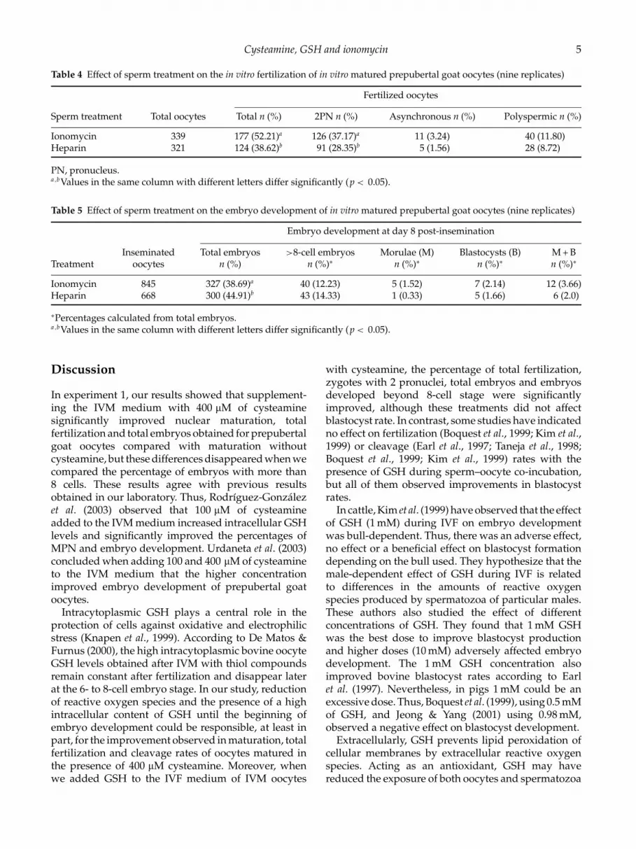
Table 4 shows that the percentage of total fertilizedoocytes was significantly higher in the group ofspermatozoa capacitated with ionomycin (52.21%) thanwith heparin (38.62%; p < 0.0007). Also the percentageof oocytes with 2PN was higher in the ionomycin group(37.17%) than in the heparin group (28.35%, p < 0.02).
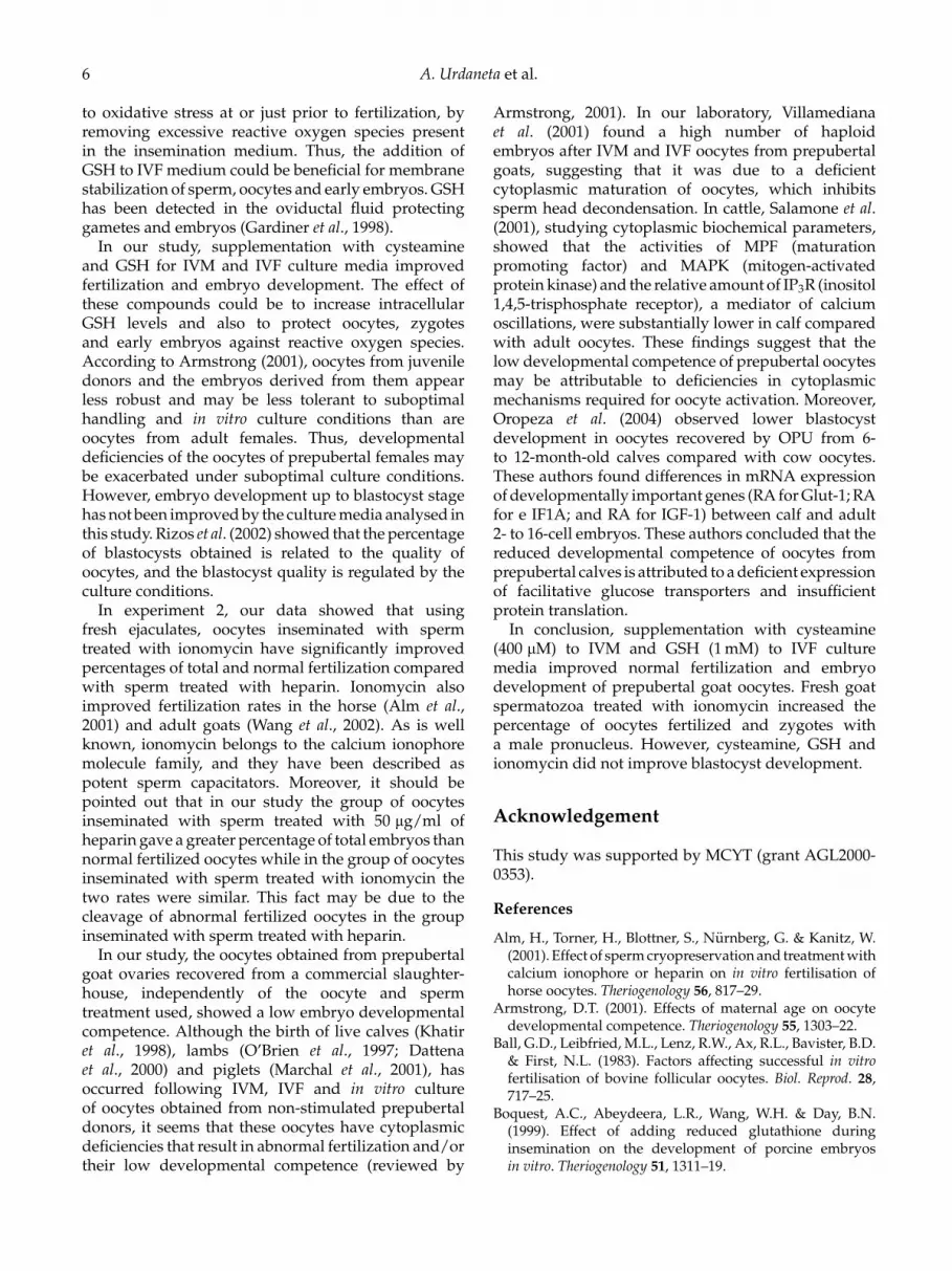
Table 5 shows the embryo development of 845oocytes inseminated with sperm capacitated withionomycin and 668 oocytes inseminated with heparin-capacitated-sperm. After 7 days of culture, thepercentage of inseminated oocytes that developed toembryos was 38.69% and 44.91% (p < 0.02) for theionomycin and heparin groups, respectively. However,the percentage of embryos developed beyond the>8-cell stage did not differ between the groups.
Cysteamine, GSH and ionomycin 5
Table 4 Effect of sperm treatment on the in vitro fertilization of in vitro matured prepubertal goat oocytes (nine replicates)
Fertilized oocytes
Sperm treatment Total oocytes Total n (%) 2PN n (%) Asynchronous n (%) Polyspermic n (%)
Ionomycin 339 177 (52.21)a 126 (37.17)a 11 (3.24) 40 (11.80)Heparin 321 124 (38.62)b 91 (28.35)b 5 (1.56) 28 (8.72)
PN, pronucleus.a ,bValues in the same column with different letters differ significantly (p < 0.05).
Table 5 Effect of sperm treatment on the embryo development of in vitro matured prepubertal goat oocytes (nine replicates)
Embryo development at day 8 post-insemination
Inseminated Total embryos >8-cell embryos Morulae (M) Blastocysts (B) M + BTreatment oocytes n (%) n (%)∗ n (%)∗ n (%)∗ n (%)∗
Ionomycin 845 327 (38.69)a 40 (12.23) 5 (1.52) 7 (2.14) 12 (3.66)Heparin 668 300 (44.91)b 43 (14.33) 1 (0.33) 5 (1.66) 6 (2.0)
∗Percentages calculated from total embryos.a ,bValues in the same column with different letters differ significantly (p < 0.05).
Discussion
In experiment 1, our results showed that supplement-ing the IVM medium with 400 µM of cysteaminesignificantly improved nuclear maturation, totalfertilization and total embryos obtained for prepubertalgoat oocytes compared with maturation withoutcysteamine, but these differences disappeared when wecompared the percentage of embryos with more than8 cells. These results agree with previous resultsobtained in our laboratory. Thus, Rodrıguez-Gonzalezet al. (2003) observed that 100 µM of cysteamineadded to the IVM medium increased intracellular GSHlevels and significantly improved the percentages ofMPN and embryo development. Urdaneta et al. (2003)concluded when adding 100 and 400 µM of cysteamineto the IVM medium that the higher concentrationimproved embryo development of prepubertal goatoocytes.
Intracytoplasmic GSH plays a central role in theprotection of cells against oxidative and electrophilicstress (Knapen et al., 1999). According to De Matos &Furnus (2000), the high intracytoplasmic bovine oocyteGSH levels obtained after IVM with thiol compoundsremain constant after fertilization and disappear laterat the 6- to 8-cell embryo stage. In our study, reductionof reactive oxygen species and the presence of a highintracellular content of GSH until the beginning ofembryo development could be responsible, at least inpart, for the improvement observed in maturation, totalfertilization and cleavage rates of oocytes matured inthe presence of 400 µM cysteamine. Moreover, whenwe added GSH to the IVF medium of IVM oocytes
with cysteamine, the percentage of total fertilization,zygotes with 2 pronuclei, total embryos and embryosdeveloped beyond 8-cell stage were significantlyimproved, although these treatments did not affectblastocyst rate. In contrast, some studies have indicatedno effect on fertilization (Boquest et al., 1999; Kim et al.,1999) or cleavage (Earl et al., 1997; Taneja et al., 1998;Boquest et al., 1999; Kim et al., 1999) rates with thepresence of GSH during sperm–oocyte co-incubation,but all of them observed improvements in blastocystrates.
In cattle, Kim et al. (1999) have observed that the effectof GSH (1 mM) during IVF on embryo developmentwas bull-dependent. Thus, there was an adverse effect,no effect or a beneficial effect on blastocyst formationdepending on the bull used. They hypothesize that themale-dependent effect of GSH during IVF is relatedto differences in the amounts of reactive oxygenspecies produced by spermatozoa of particular males.These authors also studied the effect of differentconcentrations of GSH. They found that 1 mM GSHwas the best dose to improve blastocyst productionand higher doses (10 mM) adversely affected embryodevelopment. The 1 mM GSH concentration alsoimproved bovine blastocyst rates according to Earlet al. (1997). Nevertheless, in pigs 1 mM could be anexcessive dose. Thus, Boquest et al. (1999), using 0.5 mMof GSH, and Jeong & Yang (2001) using 0.98 mM,observed a negative effect on blastocyst development.
Extracellularly, GSH prevents lipid peroxidation ofcellular membranes by extracellular reactive oxygenspecies. Acting as an antioxidant, GSH may havereduced the exposure of both oocytes and spermatozoa
6 A. Urdaneta et al.
to oxidative stress at or just prior to fertilization, byremoving excessive reactive oxygen species presentin the insemination medium. Thus, the addition ofGSH to IVF medium could be beneficial for membranestabilization of sperm, oocytes and early embryos. GSHhas been detected in the oviductal fluid protectinggametes and embryos (Gardiner et al., 1998).
In our study, supplementation with cysteamineand GSH for IVM and IVF culture media improvedfertilization and embryo development. The effect ofthese compounds could be to increase intracellularGSH levels and also to protect oocytes, zygotesand early embryos against reactive oxygen species.According to Armstrong (2001), oocytes from juveniledonors and the embryos derived from them appearless robust and may be less tolerant to suboptimalhandling and in vitro culture conditions than areoocytes from adult females. Thus, developmentaldeficiencies of the oocytes of prepubertal females maybe exacerbated under suboptimal culture conditions.However, embryo development up to blastocyst stagehas not been improved by the culture media analysed inthis study. Rizos et al. (2002) showed that the percentageof blastocysts obtained is related to the quality ofoocytes, and the blastocyst quality is regulated by theculture conditions.
In experiment 2, our data showed that usingfresh ejaculates, oocytes inseminated with spermtreated with ionomycin have significantly improvedpercentages of total and normal fertilization comparedwith sperm treated with heparin. Ionomycin alsoimproved fertilization rates in the horse (Alm et al.,2001) and adult goats (Wang et al., 2002). As is wellknown, ionomycin belongs to the calcium ionophoremolecule family, and they have been described aspotent sperm capacitators. Moreover, it should bepointed out that in our study the group of oocytesinseminated with sperm treated with 50 µg/ml ofheparin gave a greater percentage of total embryos thannormal fertilized oocytes while in the group of oocytesinseminated with sperm treated with ionomycin thetwo rates were similar. This fact may be due to thecleavage of abnormal fertilized oocytes in the groupinseminated with sperm treated with heparin.
In our study, the oocytes obtained from prepubertalgoat ovaries recovered from a commercial slaughter-house, independently of the oocyte and spermtreatment used, showed a low embryo developmentalcompetence. Although the birth of live calves (Khatiret al., 1998), lambs (O’Brien et al., 1997; Dattenaet al., 2000) and piglets (Marchal et al., 2001), hasoccurred following IVM, IVF and in vitro cultureof oocytes obtained from non-stimulated prepubertaldonors, it seems that these oocytes have cytoplasmicdeficiencies that result in abnormal fertilization and/ortheir low developmental competence (reviewed by
Armstrong, 2001). In our laboratory, Villamedianaet al. (2001) found a high number of haploidembryos after IVM and IVF oocytes from prepubertalgoats, suggesting that it was due to a deficientcytoplasmic maturation of oocytes, which inhibitssperm head decondensation. In cattle, Salamone et al.(2001), studying cytoplasmic biochemical parameters,showed that the activities of MPF (maturationpromoting factor) and MAPK (mitogen-activatedprotein kinase) and the relative amount of IP3R (inositol1,4,5-trisphosphate receptor), a mediator of calciumoscillations, were substantially lower in calf comparedwith adult oocytes. These findings suggest that thelow developmental competence of prepubertal oocytesmay be attributable to deficiencies in cytoplasmicmechanisms required for oocyte activation. Moreover,Oropeza et al. (2004) observed lower blastocystdevelopment in oocytes recovered by OPU from 6-to 12-month-old calves compared with cow oocytes.These authors found differences in mRNA expressionof developmentally important genes (RA for Glut-1; RAfor e IF1A; and RA for IGF-1) between calf and adult2- to 16-cell embryos. These authors concluded that thereduced developmental competence of oocytes fromprepubertal calves is attributed to a deficient expressionof facilitative glucose transporters and insufficientprotein translation.
In conclusion, supplementation with cysteamine(400 µM) to IVM and GSH (1 mM) to IVF culturemedia improved normal fertilization and embryodevelopment of prepubertal goat oocytes. Fresh goatspermatozoa treated with ionomycin increased thepercentage of oocytes fertilized and zygotes witha male pronucleus. However, cysteamine, GSH andionomycin did not improve blastocyst development.
Acknowledgement
This study was supported by MCYT (grant AGL2000-0353).
References
Alm, H., Torner, H., Blottner, S., Nurnberg, G. & Kanitz, W.(2001). Effect of sperm cryopreservation and treatment withcalcium ionophore or heparin on in vitro fertilisation ofhorse oocytes. Theriogenology 56, 817–29.
Armstrong, D.T. (2001). Effects of maternal age on oocytedevelopmental competence. Theriogenology 55, 1303–22.
Ball, G.D., Leibfried, M.L., Lenz, R.W., Ax, R.L., Bavister, B.D.& First, N.L. (1983). Factors affecting successful in vitrofertilisation of bovine follicular oocytes. Biol. Reprod. 28,717–25.
Boquest, A.C., Abeydeera, L.R., Wang, W.H. & Day, B.N.(1999). Effect of adding reduced glutathione duringinsemination on the development of porcine embryosin vitro. Theriogenology 51, 1311–19.

Cysteamine, GSH and ionomycin 7
Brackett, B.G. & Oliphant, G. (1975). Capacitation of rabbitspermatozoa in vitro. Biol. Reprod. 12, 260–74.
Dattena, M., Ptak, G., Loi, P. & Cappai, P. (2000). Survival andviability of vitrified in vitro and in vivo produced ovineblastocysts. Theriogenology 53, 1511–19.
De Matos, D.G. & Furnus, C.C. (2000). The importance ofhaving high glutathione (GSH) level after bovine in vitromaturation on embryo development effect of beta-mercaptoethanol, cysteine and cystine. Theriogenology 53,761–71.
De Matos, D.G., Furnus, C.C., Moses, D.F. & Baldassarre,H. (1995). Effect of cysteamine on glutathione level anddevelopmental capacity of bovine oocyte matured in vitro.Mol. Reprod. Dev. 42, 432–6.
De Matos, D.G., Furnus, C.C., Moses, D.F., Martınez, A.G. &Matkovic, M. (1996). Stimulation of glutathione synthesisof in vitro matured bovine oocytes and its effect on embryodevelopment and freezability. Mol. Reprod. Dev. 45, 451–7.
De Matos, D.G., Gasparrini, B., Pasqualini, S.R. &Thompson, J.G. (2002). Effect of glutathione synthesisstimulation during in vitro maturation of ovine oocytes onembryo development and intracellular peroxide content.Theriogenology 57, 1443–51.
De Matos, D.G., Nogueira, D., Cortvrindt, R., Herrera, C.,Adrianssens, T., Pasqualini, R.S. & Smitz, J. (2003). Capacityof adult and prepubertal mouse oocytes to undergo embryodevelopment in the presence of cysteamine. Mol. Reprod.Dev. 62, 214–18.
Driancourt, M.A., Reynaud, K. & Smitz, J. (2001). Differencesin follicular function of 3-month-old calves and maturecows. Reproduction 121, 463–74.
Earl, C.R., Kelly, J., Rowe, J. & Armstrong, D.T. (1997)Glutathione treatment of bovine sperm enhances in vitroblastocyst production rates. Theriogenology 47, 255.
Gardiner, C.S., Salmen, J.J., Brandt, C.J. & Stover, S.K. (1998).Glutathione is present in reproductive tract secretions andimproves development of mouse embryos after chemicallyinduced glutathione depletion. Biol. Reprod. 59, 431–6.
Gardner, D.K. & Lane, M. (1997). Culture and selection ofviable blastocysts: a feasible proposition for human IVF?Hum. Reprod. Update 3, 367–82.
Gasparrini, B., Negia, G., Di Palo, R., Campanile, G. &Zicarelli, L. (2000). Effect of cysteamine during IVM onbuffalo embryo development. Theriogenology 54, 1537–42.
Gasparrini, B., Sayoud H., Neglia, G., de Matos, D.G., Donnay,I. & Zicarelli, L. (2003). Glutathione synthesis duringin vitro maturation of buffalo (Bubalus bubalis) oocytes: ef-fects of cysteamine on embryo development. Theriogenology60, 943–52.
Grupen, C.G., Nagashima, H. & Nottle, M.B. (1995). Cys-teamine enhances in vitro development of porcine oocytesmatured and fertilised in vitro. Biol. Reprod. 53, 173–8.
Izquierdo, D., Villamediana, P. & Paramio, M.T. (1999). Effectof culture media on embryo development from prepubertalgoat IVM-IVF oocytes. Theriogenology 52, 847–61.
Jeong, B.S. & Yang, X. (2001). Cysteine, glutathione, andPercoll treatments improve porcine oocyte maturation andfertilisation in vitro. Mol. Reprod. Dev. 59, 330–5.
Khatir, H., Lonergan, P., Touze, J.L. & Mermillod, P. (1998).The characterization of bovine embryos obtained from
prepubertal calf oocytes and their viability after non-surgical embryo transfer. Theriogenology 50, 1201–10.
Kim, I.H., Van Langendonckt, A., Van Soom, A., Vanroose,G., Casi, A.L., Hendriksen, P.J.M. & Bevers, M.M. (1999).Effect of exogenous glutathione on the in vitro fertilisationof bovine oocytes. Theriogenology 52, 537–47.
Knapen, M.F.C.M., Zusterzeel, P.L.M., Peters, W.H.M. &Steegers, E.A.P. (1999). Glutathione and glutathione-relatedenzymes in reproduction. A review. Eur. J. Obstet. Gynecol.Reprod. Biol. 82, 171–84.
Luvoni, G.C., Keskintepe, L. & Brackett, B.G. (1996).Improvement in bovine embryo production in vitro byglutathione-containing culture media. Mol. Reprod. Dev. 43,437–43.
Marchal, R., Feugang, J.M., Perreau, C., Venturi, E., Terqui,M. & Mermillod, P. (2001). Meiotic and developmentalcompetence of prepubertal and adult swine oocytes.Theriogenology 56, 17–29.
Mogas, T., Palomo, M.J., Izquierdo, D. & Paramio, M.T.(1997). Morphological events during in vitro fertilisation ofprepubertal goat oocytes matured in vitro. Theriogenology48, 815–29.
O’Brien, J.K., Catt, S.L., Ireland, K.A., Maxwell, W.M.C. &Evans, G. (1997). In vitro and in vivo developmental capacityof oocytes from prepubertal and adult sheep. Theriogenology47, 1433–43.
Oropeza, A., Wrenzycki, C., Herrmann, D., Hadeler, K.G. &Niemann, H. (2004). Improvement of the developmentalcapacity of oocytes from prepubertal cattle by intraovarianIGF-I application. BOR Papers in Press. Published on6 February 2004 as DOI:10.1095/biolreprod.103.025494
Parrish, J.J., Susko-Parrish, J.L., Leibfried-Rutledge, M.L.,Crister, E.S., Eyeston, W.H. & First, N.L. (1986). Bovinein vitro fertilisation with frozen thawed semen.Theriogenology 25, 591–600.
Perrault, S.D., Barbee, R.R. & Slott, V.L. (1988). Importanceof glutathione in the acquisition and maintenance ofsperm nuclear decondensing activity in maturing hamsteroocytes. Dev. Biol. 125, 181–6.
Rizos, D., Lonergan, P., Boland, M.P., Arroyo-Garcıa, R.,Pintado, B., de la Fuente, J. & Gutierrez-Adan, A.(2002). Analysis of differential messenger RNA expressionbetween bovine blastocysts produced in different culturesystems: implications for blastocyst quality. Biol. Reprod. 66,589–95.
Rodrıguez-Gonzalez, E., Lopez-Bejar, M., Mertens, M.J.& Paramio, M.T. (2003). Effects on in vitro embryodevelopment and intracellular glutathione content ofthe presence of thiol compounds during maturation ofprepubertal goat oocytes. Mol. Reprod. Dev. 65, 446–53.
Salamone, D.F., Damiani, P., Fissore, R.A., Robl, J.M. & Duby,R.T. (2001). Biochemical and developmental evidence thatooplasmic maturation of prepubertal bovine oocytes iscompromised. Biol. Reprod. 64, 1761–8.
Sutovsky, P. & Schatten, G. (1997). Depletion of glutathioneduring bovine oocyte maturation reversibly blocks thedecondensation of the male pronucleus and pronuclearapposition during fertilisation. Biol. Reprod. 56, 1503–12.
Taneja, M., Kelly, J., Rowe, J. & Earl, C.R. (1998). Influenceof glutathione in the presence of heparin and caffeine onbovine in vitro fertilisation. Theriogenology 49, 298.

8 A. Urdaneta et al.
Urdaneta, A., Jimenez-Macedo, A.R., Izquierdo, D. &Paramio, M.T. (2003). Supplementation with cysteamineduring maturation and embryo culture on embryodevelopment of prepubertal goat oocytes selected by thebrilliant cresyl blue test (BCB). Zygote 11, 347–54.
Van Soom, A., Vanroose, G. & Kruif, A. (1998). Glutathioneaddition during fertilisation doubles embryo productionbut has no effect upon embryo quality in cattle.Theriogenology 49, 301.
Villamediana, P.C., Vidal, F. & Paramio, M.T. (2001).Cytogenetic analysis of caprine 2- to 4-cell embryosproduced in vitro. Zygote 9, 193–9.
Wang, B., Baldasarre, H., Tao, T., Gauthier, M., Neveu, N.,Zhou, J.F., Leduc, M., Duguay, F., Bilodeau, A.S., Lazaris,A., Keefer, C. & Karatzas, C.N. (2002). Transgenic goatsproduced by pronuclear microinjection of in vitro derivedzygotes. Mol. Reprod. Dev. 63, 437–43.
Yamauchi, N. & Nagai, T. (1999). Male pronuclear formationin denuded porcine oocytes after in vitro maturationin the presence of cysteamine. Biol. Reprod. 61, 828–33.
Younis, A.I., Zuelke, K.A., Harper, K.M., Oliveira, M.A.L. &Brackett, B.G. (1991). In vitro fertilisation of goat oocytes.Biol. Reprod. 44, 1177–82.