crystal structure of a thermostable lipase from bacillus stearothermophilus p1
TRANSCRIPT

Crystal Structure of a Thermostable Lipase fromBacillus stearothermophilus P1
Joel D. A. Tyndall1, Supachok Sinchaikul1,2, Linda A. Fothergill-Gilmore1
Paul Taylor1 and Malcolm D. Walkinshaw1*
1Structural Biochemistry GroupInstitute of Cell and MolecularBiology, University ofEdinburgh, Michael SwannBuilding, King’s BuildingsMayfield Road, Edinburgh EH93JR, Scotland, UK
2Department of ChemistryFaculty of Science, Chiang MaiUniversity, Chiang Mai 50200Thailand
We describe the first lipase structure from a thermophilic organism. Itshares less than 20% amino acid sequence identity with other lipasesfor which there are crystal structures, and shows significant insertionscompared with the typical a/b hydrolase canonical fold. The structurecontains a zinc-binding site which is unique among all lipases withknown structures, and which may play a role in enhancing thermalstability. Zinc binding is mediated by two histidine and two aspartic acidresidues. These residues are present in comparable positions in thesequences of certain lipases for which there is as yet no crystal structuralinformation, such as those from Staphylococcal species and Arabidopsisthaliana. The structure of Bacillus stearothermophilus P1 lipase provides atemplate for other thermostable lipases, and offers insight into mecha-nisms used to enhance thermal stability which may be of commercialvalue in engineering lipases for industrial uses.
q 2002 Elsevier Science Ltd. All rights reserved
Keywords: Bacillus stearothermophilus P1; bacterial thermostable lipase;lipase closed conformation; metal ion stabilisation; zinc-binding site*Corresponding author
Introduction
Lipases (triacylglycerol acylhydrolases, EC3.1.1.3) are present in many different organisms,and catalyse the hydrolysis of long-chaintriglycerides into fatty acids and glycerol. Theseenzymes are of considerable commercial interestfor biotechnological applications such as deter-gents, food production, pharmaceuticals andindustrial synthesis of fine chemicals (see reviewby Jaeger & Reetz1). In particular, thermostablelipases from thermophilic bacteria have the poten-tial to play important roles in industrial appli-cations because they possess relatively highthermodynamic stability both at elevated tempera-tures and in organic solvents.2 – 4
Bacterial lipases are members of the structuralsuperfamily of a/b hydrolases5 whose enzymic
activity results from the catalytic triad Ser-His-Asp similar to that found in serine proteinases.Generally, the serine residue occurs in the penta-peptide chain of Gly-Xaa-Ser-Xaa-Gly, except inthe Bacillus lipases where the first glycine isreplaced by an alanine. Bacterial lipases have beenclassified into families with the Bacillus lipasesfalling into subfamily five of family I (I.5). Thisfamily consists of lipases from Gram-positiveorganisms.6
To date, a total of nine bacterial lipase structureshave been solved, four of these being true lipases;one from family I.1 (Pseudomonas aeruginosa,PAL7), and three from I.2 (Pseudomonas glumae,PGL,8 Chromobacterium viscosum, CVL,9 and Pseudo-monas cepacia, PCL10 – 12). The remaining five arerelated lipolytic enzymes from families II (GDSL)(Streptomyces scabies, SsEST13), III (Streptomycesexfoliatus, SEL14), IV (HSL) (Alcaligenes eutrophus,esterase15), VI (Pseudomonas flurorescens, PFC16) andVII (Bacillus subtilis, BFAE17).
The active site of the majority of bacterial lipasesis covered by an a-helical flexible “lid” that in theclosed position maintains the catalytic triad in ahydrophobic environment isolated from aqueoussolvents. The term interfacial activation18 is usedto describe the enhancement of lipase activity thatoccurs when the lid is triggered to open by theinteraction of the enzyme with the interface of a
0022-2836/02/$ - see front matter q 2002 Elsevier Science Ltd. All rights reserved
Present address: J. D. A. Tyndall, Institute forMolecular Bioscience, University of Queensland,Brisbane 4072, Australia.
E-mail address of the corresponding author:[email protected]
Abbreviations used: RMSD, root-mean-squaredeviation; BSP, lipase from Bacillus stearothermophilus P1;CVL, lipase from Chromobacterium viscosum; PCL, lipasefrom Pseudomonas cepacia; PAL, lipase from Pseudomonasaeruginosa.
doi:10.1016/S0022-2836(02)01004-5 available online at http://www.idealibrary.com onBw
J. Mol. Biol. (2002) 323, 859–869
lipid–water bilayer. Derewenda19 describes theanatomy of the conformational changes associatedwith this flexible lid region using Rhizomucor mieheitriacylglyceride lipase18,20 as a key example. Of thebacterial lipase crystal structures released to date,PGL and CVL have both been crystallised in theclosed conformation, whereas the structures ofPCL and PAL have been crystallised in the openconformation both with and without boundligands.
Bacillus stearothermophilus P1 lipase (BSP) occursin the I.5 family along with two other highlythermostable enzymes. These bacterial lipasesisolated from Bacillus thermocatenulatus2 andB. stearothermophilus L14 have high sequence iden-tity with BSP (,95%). All possess a completesequence of 417 residues including a 29-residuesignal sequence that is cleaved to produce themature 388-residue lipase. The optimum activity
of all three thermostable lipases lies around 65 8Cand pH 8.0–9.0. Calcium-dependent stability hasbeen demonstrated by Kim and co-workers,21 whoshowed that lipase BSL1 begins to unfold at 66 8Cin the presence of calcium ions, but at 58 8C in theabsence of calcium.
We report here the first structure of a 43 kDathermostable lipase isolated from B. stearothermo-philus P1. It possesses significant differences fromthe known fold of the a/b hydrolase family,22,23
and contains both a calcium ion and a zinc ion,not previously found in lipases.
Results and Discussion
X-ray structure determination
Lipase P1 from B. stearothermophilus was purifiedfrom a recombinant expression system by anion-exchange chromatography and gel filtration.Crystals were obtained by the hanging dropvapour diffusion method and data were collectedas described.24 The closest homologue lipase forwhich there is an X-ray structure is PCL (PDBcode 4lip). However, it has only 17% amino acidsequence identity, and all attempts at usingmolecular replacement with this structure as amodel failed. The structure was finally determinedby multiple isomorphous replacement (MIR) usingthree heavy atom derivatives (with anomalousscattering). The data collection and structurerefinement statistics are given in Table 1.
The quality of the electron density map wasexcellent and allowed unambiguous tracing ofall amino acid residues in the sequence (see e.g.Figure 1).
Table 1. Data collection and phase refinement statistics
Derivativea Native K2PtCl4 HgCl2 EMTSb
Resolution (A) 2.2 2.4 2.5 2.5No. of observations 211,130 214,389 79,129 121,289No. of independentreflections
47,208 35,676 28,038 32,900
Rmergec (%) 10.6 6.3 7.3 10.9
Top shell 29.8 12.7 17.5 40.3Completeness (%) 99.2 96.8 87.4 99.7Top shell 91.9 95.5 76.7 98.3I/sI 11.2 19.2 11.2 11.4Top shell 4.2 13.6 4.6 3.1No. of sites per molecule 2 4 4
a Conditions.b Sodium ethyl mercurithiosalicylicate.c Rmerge ¼ ShSIlIhi 2 kIhll/ShSIIhi.
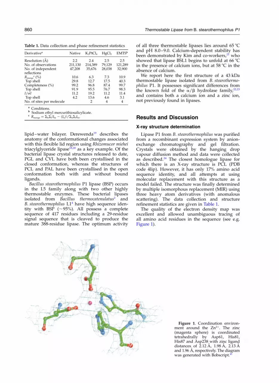
Figure 1. Coordination environ-ment around the Zn2þ. The zinc(magenta sphere) is coordinatedtetrahedrally by Asp61, His81,His87 and Asp238 with zinc liganddistances of 2.12 A, 1.98 A, 2.13 Aand 1.96 A, respectively. The diagramwas generated with Bobscript.41
860 Thermostable Lipase from B. stearothermophilus P1

The structure has been refined to 2.2 A with anRfactor of 16.6% and an Rfree of 21.7% (Table 2).There are two independent molecules per asym-metric unit, each comprising 388 residues, togetherwith 1 Ca2þ and 1 Zn2þ per molecule. X-ray fluor-escence was carried out to confirm the identity ofthe zinc ion. A clear signal was obtained at9.769 keV (Figure 2), which corresponds to theabsorption edge of zinc that has a theoreticalvalue of 9.659 keV. No signal was observed aroundthe theoretical absorption edge of copper at8.98 keV.
Description of overall protein fold
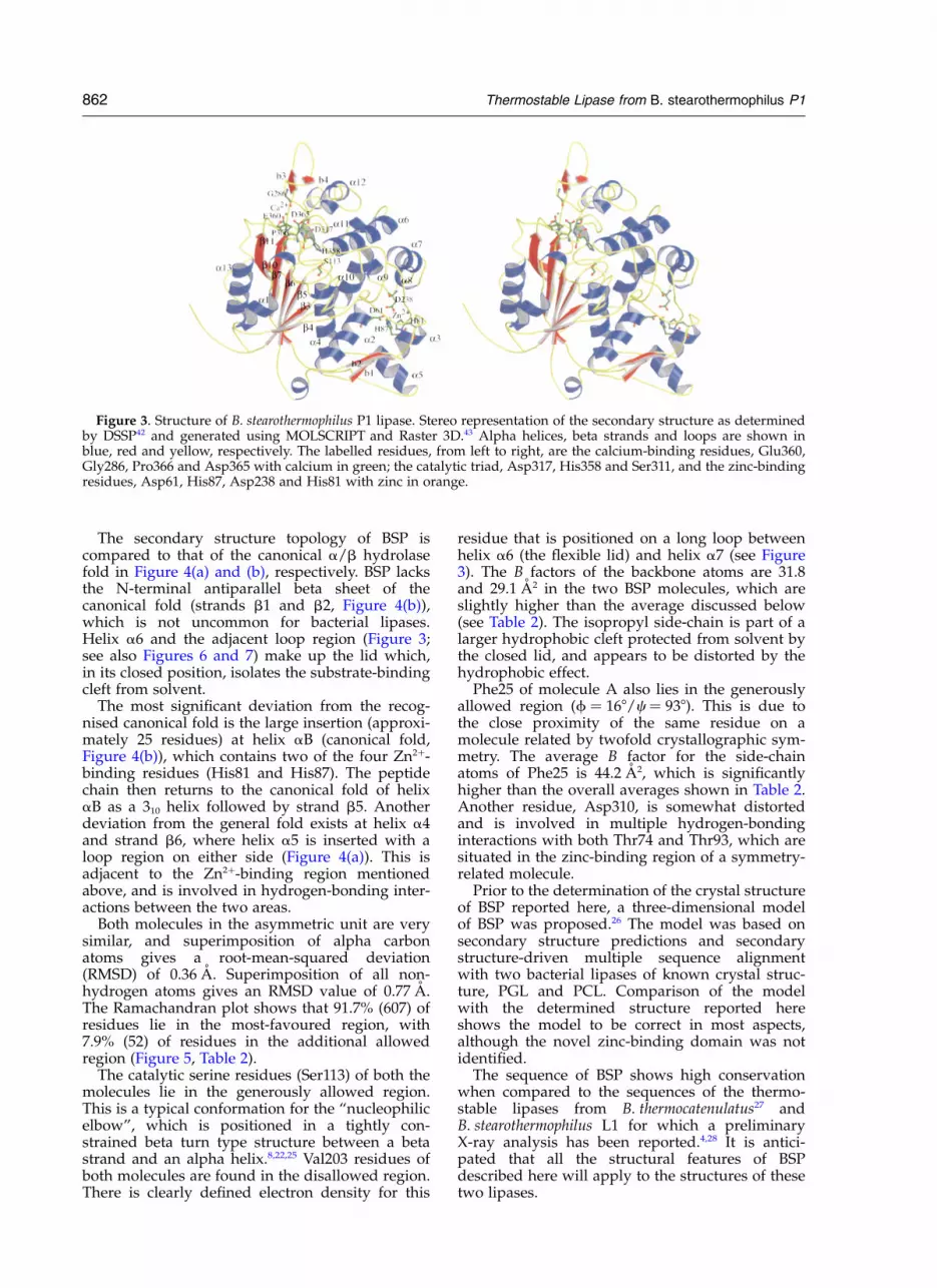
Each BSP molecule has a generally globularshape with approximate dimensions of 40 A £50 A £ 65 A (Figure 3). The a/b hydrolase fold ofBSP is similar to that seen for other lipases, thoughthere are a number of unique structural features.The core structure consists of a seven-strandedparallel sheet that is surrounded by alpha helices1 and 13 on one side, and alpha helices 2, 4, 5, 9,10, 11 and 12 on the other side.
Table 2. Refinement statistics
Resolution range (A) 40.0–2.20a (A) 118.5b (A) 81.2c (A) 99.8b (deg.) 96.3% Solvent 55.12Vm (A3 Da21) 2.76Rfactor
a (%) 16.6Rfree
b (%) 21.7Number of residues per BSP molecule 388Number of water molecules 347Number of calcium ions per BSP molecule 1Number of zinc ions per BSP molecule 1RMSDBond lengths (A) 0.218Bond angles (deg.) 1.256Ramachandran plotResidues in most-favoured region (%) 91.7Residues in additional allowed regions (%) 7.9Residues in disallowed regions (%) 0.5
Average B factors (A2) Molecule A Molecule B Overall
Main-chain atoms 26.8 24.3 25.5Side-chain atoms 31.2 28.6 30.2c
All atoms 29.0 26.4 28.0
a Rfactor ¼ ShllFobsl 2 lFcalcll/SlFobsl, where Fobs and Fcalc are the observed and calculated structure factor amplitudes, respectively.b Rfree is calculated with 5% of the diffraction data, which were not used during the refinement.c Side-chains þ 347 water molecules.
Figure 2. Fluorescence signalfrom a single crystal of BSP show-ing the presence of zinc. Synchro-tron radiation from SLS station 9.5was used to provide a mono-chromatic X-ray beam which wasscanned over the energy range9.72 keV to 9.9 keV.
Thermostable Lipase from B. stearothermophilus P1 861
The secondary structure topology of BSP iscompared to that of the canonical a/b hydrolasefold in Figure 4(a) and (b), respectively. BSP lacksthe N-terminal antiparallel beta sheet of thecanonical fold (strands b1 and b2, Figure 4(b)),which is not uncommon for bacterial lipases.Helix a6 and the adjacent loop region (Figure 3;see also Figures 6 and 7) make up the lid which,in its closed position, isolates the substrate-bindingcleft from solvent.
The most significant deviation from the recog-nised canonical fold is the large insertion (approxi-mately 25 residues) at helix aB (canonical fold,Figure 4(b)), which contains two of the four Zn2þ-binding residues (His81 and His87). The peptidechain then returns to the canonical fold of helixaB as a 310 helix followed by strand b5. Anotherdeviation from the general fold exists at helix a4and strand b6, where helix a5 is inserted with aloop region on either side (Figure 4(a)). This isadjacent to the Zn2þ-binding region mentionedabove, and is involved in hydrogen-bonding inter-actions between the two areas.
Both molecules in the asymmetric unit are verysimilar, and superimposition of alpha carbonatoms gives a root-mean-squared deviation(RMSD) of 0.36 A. Superimposition of all non-hydrogen atoms gives an RMSD value of 0.77 A.The Ramachandran plot shows that 91.7% (607) ofresidues lie in the most-favoured region, with7.9% (52) of residues in the additional allowedregion (Figure 5, Table 2).
The catalytic serine residues (Ser113) of both themolecules lie in the generously allowed region.This is a typical conformation for the “nucleophilicelbow”, which is positioned in a tightly con-strained beta turn type structure between a betastrand and an alpha helix.8,22,25 Val203 residues ofboth molecules are found in the disallowed region.There is clearly defined electron density for this
residue that is positioned on a long loop betweenhelix a6 (the flexible lid) and helix a7 (see Figure3). The B factors of the backbone atoms are 31.8and 29.1 A2 in the two BSP molecules, which areslightly higher than the average discussed below(see Table 2). The isopropyl side-chain is part of alarger hydrophobic cleft protected from solvent bythe closed lid, and appears to be distorted by thehydrophobic effect.
Phe25 of molecule A also lies in the generouslyallowed region (f ¼ 168/c ¼ 938). This is due tothe close proximity of the same residue on amolecule related by twofold crystallographic sym-metry. The average B factor for the side-chainatoms of Phe25 is 44.2 A2, which is significantlyhigher than the overall averages shown in Table 2.Another residue, Asp310, is somewhat distortedand is involved in multiple hydrogen-bondinginteractions with both Thr74 and Thr93, which aresituated in the zinc-binding region of a symmetry-related molecule.
Prior to the determination of the crystal structureof BSP reported here, a three-dimensional modelof BSP was proposed.26 The model was based onsecondary structure predictions and secondarystructure-driven multiple sequence alignmentwith two bacterial lipases of known crystal struc-ture, PGL and PCL. Comparison of the modelwith the determined structure reported hereshows the model to be correct in most aspects,although the novel zinc-binding domain was notidentified.
The sequence of BSP shows high conservationwhen compared to the sequences of the thermo-stable lipases from B. thermocatenulatus27 andB. stearothermophilus L1 for which a preliminaryX-ray analysis has been reported.4,28 It is antici-pated that all the structural features of BSPdescribed here will apply to the structures of thesetwo lipases.
Figure 3. Structure of B. stearothermophilus P1 lipase. Stereo representation of the secondary structure as determinedby DSSP42 and generated using MOLSCRIPT and Raster 3D.43 Alpha helices, beta strands and loops are shown inblue, red and yellow, respectively. The labelled residues, from left to right, are the calcium-binding residues, Glu360,Gly286, Pro366 and Asp365 with calcium in green; the catalytic triad, Asp317, His358 and Ser311, and the zinc-bindingresidues, Asp61, His87, Asp238 and His81 with zinc in orange.
862 Thermostable Lipase from B. stearothermophilus P1

Description of the active site
The catalytic triad consists of Ser113, His358and Asp317 as described by Sinchaikul andco-workers26 (see Figure 3). The catalytic serine issituated on the nucleophilic elbow between strandb5 and helix a4 deep within the core structure.In Bacillus lipases the serine is embedded withinthe Ala-Xaa-Ser-Xaa-Gly consensus sequence(where Xaa represents His and Gln, respectively).The active site is isolated from solvent by a flexiblealpha helical lid (helix a6) similar to that found inthe structures of CVL and PGL. The active site isalso bounded by helix a11 and a12 (separatedonly by Cys295). Helix a6 would most likely be
involved in a conformational change that allowsthe substrate access to the active site. The natureof these enzymes dictates that the active site isquite hydrophobic. Within the small active sitecavity there is only one water molecule that makeshydrogen-bonding interactions with Thr17 andTyr29 on the base of the cavity.
On closer investigation, the active site and theinterface between the proposed active site floorand the flexible lid region consist predominantlyof aromatic and other hydrophobic residues(Figure 6). They include Phe16 (that immediatelyfollows strand b3), Trp19, Met24, Tyr29, Pro55 andLeu56. A 310 helical turn positions Leu170 towardsthe active site, and this is followed by Phe176,
Figure 4. Comparison of the secondary structure of BSP with the canonical lipase fold. (a) Secondary structuretopology of BSP showing the general a/b hydrolase fold including the catalytic triad and the zinc-binding structuralelements and residues indicated in black and red, respectively. Alpha helices and beta strands are represented byrectangles and arrows, respectively. New structural elements, strand b1, helix a3 and strand b2 are shown in red.(b) Secondary structure topology diagram of the canonical a/b hydrolase fold. Broken lines indicate possible sites ofinsertions. The heavy line depicts the position of the new deviation from the known fold.
Thermostable Lipase from B. stearothermophilus P1 863

Phe180, Phe181, Leu183, Val187 and Leu188, whichmake up the amphipathic helix a6 (Figure 3). Onlyone of a few non-hydrophobic residues within theactive site, Gln184, stands out. The side-chainamide nitrogen of this residue forms a hydrogenbond with the main-chain carbonyl oxygen ofPhe16, possibly forming an anchor to keep the lid
from opening. Further enclosed with the lid regionare Tyr204 and Phe206, followed by Phe221,Phe225, Leu228 and Trp234 of helices a7 and a8.The apparently more rigid side is bounded byPhe290 and Leu299 of helices a11 and a12,respectively. Ile319 (that lies above the catalyticAsp317 and His358) and Ile362 make up the
Figure 5. Ramachandran plot of the two molecules of BSP in the asymmetric unit. Glycine residues are shown as tri-angles. Core regions are shaded red, with additional allowed and generously allowed zones in progressively lightershades of yellow. Disallowed zones are unshaded. See the text for description of labelled residues.
Figure 6. Stereo view of BSP with the aromatic and hydrophobic residues of the lid region coloured green. Thecatalytic triad residues (Ser113, His358 and Asp317) are shown from left to right in red, blue and red. The calciumion is yellow, and the zinc is magenta.
864 Thermostable Lipase from B. stearothermophilus P1
remainder of the hydrophobic residues within thecavity.
Inspection of a surface representation of BSPshows what appears to be a channel leadingtowards the active site. It is clear that this is aportion of the substrate-binding cleft and is sur-rounded by the hydrophobic residues Phe27,Leu33, Val187, Ala190, Leu359 and Val364. The
latter two residues are adjacent to the catalytichistidine and one of the calcium-binding residues.A comparison of BSP with PAL in complex with atriacylglycerol analogue shows that the sn-2 chainof this inhibitor does indeed bind in a similarchannel in PAL. This suggests that BSP wouldbind a triacylglycerol substrate in a similar mannerfollowing the opening of the flexible lid.
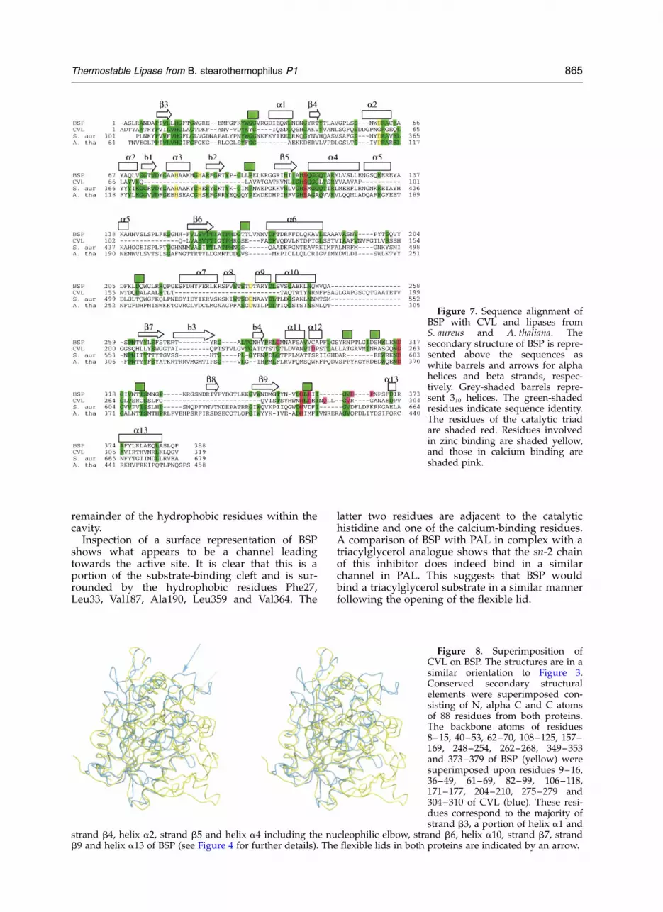
Figure 7. Sequence alignment ofBSP with CVL and lipases fromS. aureus and A. thaliana. Thesecondary structure of BSP is repre-sented above the sequences aswhite barrels and arrows for alphahelices and beta strands, respec-tively. Grey-shaded barrels repre-sent 310 helices. The green-shadedresidues indicate sequence identity.The residues of the catalytic triadare shaded red. Residues involvedin zinc binding are shaded yellow,and those in calcium binding areshaded pink.
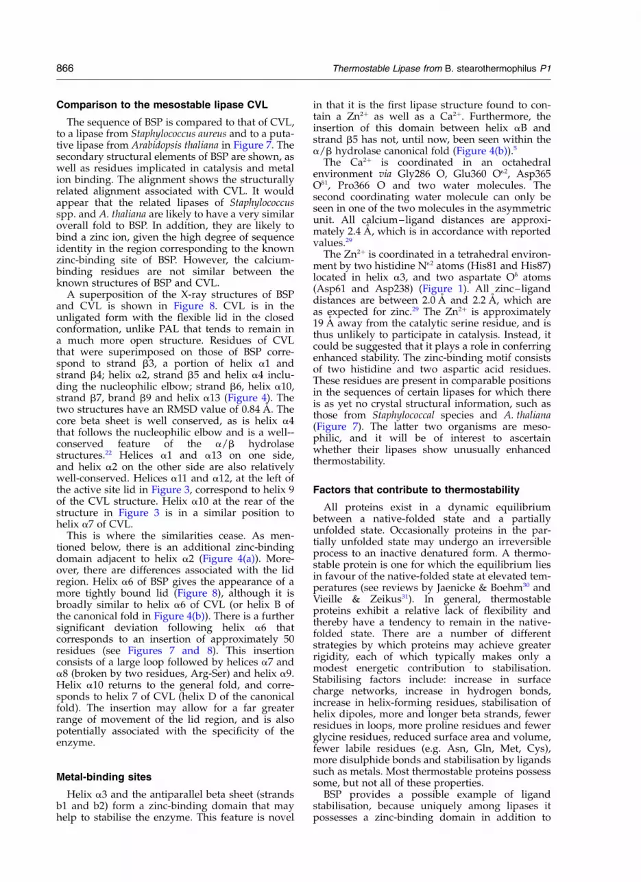
Figure 8. Superimposition ofCVL on BSP. The structures are in asimilar orientation to Figure 3.Conserved secondary structuralelements were superimposed con-sisting of N, alpha C and C atomsof 88 residues from both proteins.The backbone atoms of residues8–15, 40–53, 62–70, 108–125, 157–169, 248–254, 262–268, 349–353and 373–379 of BSP (yellow) weresuperimposed upon residues 9–16,36–49, 61–69, 82–99, 106–118,171–177, 204–210, 275–279 and304–310 of CVL (blue). These resi-dues correspond to the majority ofstrand b3, a portion of helix a1 and
strand b4, helix a2, strand b5 and helix a4 including the nucleophilic elbow, strand b6, helix a10, strand b7, strandb9 and helix a13 of BSP (see Figure 4 for further details). The flexible lids in both proteins are indicated by an arrow.
Thermostable Lipase from B. stearothermophilus P1 865
Comparison to the mesostable lipase CVL
The sequence of BSP is compared to that of CVL,to a lipase from Staphylococcus aureus and to a puta-tive lipase from Arabidopsis thaliana in Figure 7. Thesecondary structural elements of BSP are shown, aswell as residues implicated in catalysis and metalion binding. The alignment shows the structurallyrelated alignment associated with CVL. It wouldappear that the related lipases of Staphylococcusspp. and A. thaliana are likely to have a very similaroverall fold to BSP. In addition, they are likely tobind a zinc ion, given the high degree of sequenceidentity in the region corresponding to the knownzinc-binding site of BSP. However, the calcium-binding residues are not similar between theknown structures of BSP and CVL.
A superposition of the X-ray structures of BSPand CVL is shown in Figure 8. CVL is in theunligated form with the flexible lid in the closedconformation, unlike PAL that tends to remain ina much more open structure. Residues of CVLthat were superimposed on those of BSP corre-spond to strand b3, a portion of helix a1 andstrand b4; helix a2, strand b5 and helix a4 inclu-ding the nucleophilic elbow; strand b6, helix a10,strand b7, brand b9 and helix a13 (Figure 4). Thetwo structures have an RMSD value of 0.84 A. Thecore beta sheet is well conserved, as is helix a4that follows the nucleophilic elbow and is a well--conserved feature of the a/b hydrolasestructures.22 Helices a1 and a13 on one side,and helix a2 on the other side are also relativelywell-conserved. Helices a11 and a12, at the left ofthe active site lid in Figure 3, correspond to helix 9of the CVL structure. Helix a10 at the rear of thestructure in Figure 3 is in a similar position tohelix a7 of CVL.
This is where the similarities cease. As men-tioned below, there is an additional zinc-bindingdomain adjacent to helix a2 (Figure 4(a)). More-over, there are differences associated with the lidregion. Helix a6 of BSP gives the appearance of amore tightly bound lid (Figure 8), although it isbroadly similar to helix a6 of CVL (or helix B ofthe canonical fold in Figure 4(b)). There is a furthersignificant deviation following helix a6 thatcorresponds to an insertion of approximately 50residues (see Figures 7 and 8). This insertionconsists of a large loop followed by helices a7 anda8 (broken by two residues, Arg-Ser) and helix a9.Helix a10 returns to the general fold, and corre-sponds to helix 7 of CVL (helix D of the canonicalfold). The insertion may allow for a far greaterrange of movement of the lid region, and is alsopotentially associated with the specificity of theenzyme.
Metal-binding sites
Helix a3 and the antiparallel beta sheet (strandsb1 and b2) form a zinc-binding domain that mayhelp to stabilise the enzyme. This feature is novel
in that it is the first lipase structure found to con-tain a Zn2þ as well as a Ca2þ. Furthermore, theinsertion of this domain between helix aB andstrand b5 has not, until now, been seen within thea/b hydrolase canonical fold (Figure 4(b)).5
The Ca2þ is coordinated in an octahedralenvironment via Gly286 O, Glu360 O12, Asp365Od1, Pro366 O and two water molecules. Thesecond coordinating water molecule can only beseen in one of the two molecules in the asymmetricunit. All calcium–ligand distances are approxi-mately 2.4 A, which is in accordance with reportedvalues.29
The Zn2þ is coordinated in a tetrahedral environ-ment by two histidine N12 atoms (His81 and His87)located in helix a3, and two aspartate Od atoms(Asp61 and Asp238) (Figure 1). All zinc–liganddistances are between 2.0 A and 2.2 A, which areas expected for zinc.29 The Zn2þ is approximately19 A away from the catalytic serine residue, and isthus unlikely to participate in catalysis. Instead, itcould be suggested that it plays a role in conferringenhanced stability. The zinc-binding motif consistsof two histidine and two aspartic acid residues.These residues are present in comparable positionsin the sequences of certain lipases for which thereis as yet no crystal structural information, such asthose from Staphylococcal species and A. thaliana(Figure 7). The latter two organisms are meso-philic, and it will be of interest to ascertainwhether their lipases show unusually enhancedthermostability.
Factors that contribute to thermostability
All proteins exist in a dynamic equilibriumbetween a native-folded state and a partiallyunfolded state. Occasionally proteins in the par-tially unfolded state may undergo an irreversibleprocess to an inactive denatured form. A thermo-stable protein is one for which the equilibrium liesin favour of the native-folded state at elevated tem-peratures (see reviews by Jaenicke & Boehm30 andVieille & Zeikus31). In general, thermostableproteins exhibit a relative lack of flexibility andthereby have a tendency to remain in the native-folded state. There are a number of differentstrategies by which proteins may achieve greaterrigidity, each of which typically makes only amodest energetic contribution to stabilisation.Stabilising factors include: increase in surfacecharge networks, increase in hydrogen bonds,increase in helix-forming residues, stabilisation ofhelix dipoles, more and longer beta strands, fewerresidues in loops, more proline residues and fewerglycine residues, reduced surface area and volume,fewer labile residues (e.g. Asn, Gln, Met, Cys),more disulphide bonds and stabilisation by ligandssuch as metals. Most thermostable proteins possesssome, but not all of these properties.
BSP provides a possible example of ligandstabilisation, because uniquely among lipases itpossesses a zinc-binding domain in addition to
866 Thermostable Lipase from B. stearothermophilus P1
the calcium binding generally found in bacteriallipases. Other examples of thermostableand hyperthermostable enzymes that containadditional metal ions not found in their mesostableanalogues include proteinases in the subtilisinsuperfamily32,33 and ferredoxin.34 The latterprotein from the thermophile Sulfolobus possessesan additional 40-residue N-terminal domaincontaining a zinc ion not found in mesostablehomologues, but conserved in other thermoacido-philes. In addition, certain proteins contain Zn2þ
that play a purely structural role, with an obviousexample being zinc fingers found in DNA-bindingproteins. An important class of proteinases, theMMPs, contain a catalytic Zn2þ as well as structuralCa2þ and Zn2þ. It is believed that these metal ionskeep the structural elements together within thecatalytic domain, thus contributing to its stability.35
The structure of aspartate transcarbamolyase36
shows the Zn2þ playing a crucial structural rolein linking the allosteric domain to the catalyticpolypeptide.
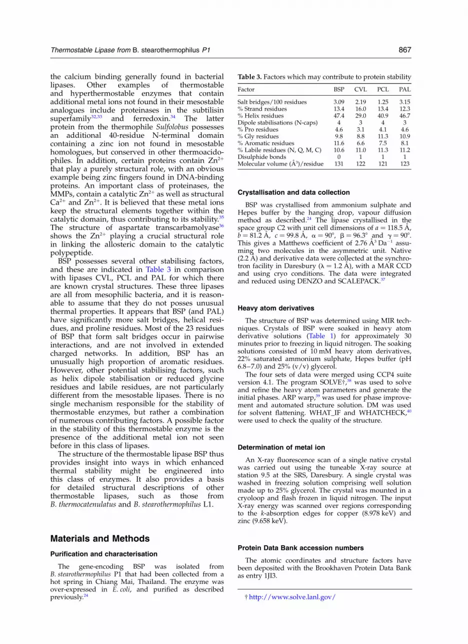
BSP possesses several other stabilising factors,and these are indicated in Table 3 in comparisonwith lipases CVL, PCL and PAL for which thereare known crystal structures. These three lipasesare all from mesophilic bacteria, and it is reason-able to assume that they do not posses unusualthermal properties. It appears that BSP (and PAL)have significantly more salt bridges, helical resi-dues, and proline residues. Most of the 23 residuesof BSP that form salt bridges occur in pairwiseinteractions, and are not involved in extendedcharged networks. In addition, BSP has anunusually high proportion of aromatic residues.However, other potential stabilising factors, suchas helix dipole stabilisation or reduced glycineresidues and labile residues, are not particularlydifferent from the mesostable lipases. There is nosingle mechanism responsible for the stability ofthermostable enzymes, but rather a combinationof numerous contributing factors. A possible factorin the stability of this thermostable enzyme is thepresence of the additional metal ion not seenbefore in this class of lipases.
The structure of the thermostable lipase BSP thusprovides insight into ways in which enhancedthermal stability might be engineered intothis class of enzymes. It also provides a basisfor detailed structural descriptions of otherthermostable lipases, such as those fromB. thermocatenulatus and B. stearothermophilus L1.
Materials and Methods
Purification and characterisation
The gene-encoding BSP was isolated fromB. stearothermophilus P1 that had been collected from ahot spring in Chiang Mai, Thailand. The enzyme wasover-expressed in E. coli, and purified as describedpreviously.24
Crystallisation and data collection
BSP was crystallised from ammonium sulphate andHepes buffer by the hanging drop, vapour diffusionmethod as described.24 The lipase crystallised in thespace group C2 with unit cell dimensions of a ¼ 118.5 A,b ¼ 81.2 A, c ¼ 99.8 A, a ¼ 908, b ¼ 96.38 and g ¼ 908.This gives a Matthews coefficient of 2.76 A3 Da21 assu-ming two molecules in the asymmetric unit. Native(2.2 A) and derivative data were collected at the synchro-tron facility in Daresbury (l ¼ 1.2 A), with a MAR CCDand using cryo conditions. The data were integratedand reduced using DENZO and SCALEPACK.37
Heavy atom derivatives
The structure of BSP was determined using MIR tech-niques. Crystals of BSP were soaked in heavy atomderivative solutions (Table 1) for approximately 30minutes prior to freezing in liquid nitrogen. The soakingsolutions consisted of 10 mM heavy atom derivatives,22% saturated ammonium sulphate, Hepes buffer (pH6.8–7.0) and 25% (v/v) glycerol.
The four sets of data were merged using CCP4 suiteversion 4.1. The program SOLVE†,38 was used to solveand refine the heavy atom parameters and generate theinitial phases. ARP warp,39 was used for phase improve-ment and automated structure solution. DM was usedfor solvent flattening. WHAT_IF and WHATCHECK,40
were used to check the quality of the structure.
Determination of metal ion
An X-ray fluorescence scan of a single native crystalwas carried out using the tuneable X-ray source atstation 9.5 at the SRS, Daresbury. A single crystal waswashed in freezing solution comprising well solutionmade up to 25% glycerol. The crystal was mounted in acryoloop and flash frozen in liquid nitrogen. The inputX-ray energy was scanned over regions correspondingto the k-absorption edges for copper (8.978 keV) andzinc (9.658 keV).
Protein Data Bank accession numbers
The atomic coordinates and structure factors havebeen deposited with the Brookhaven Protein Data Bankas entry 1JI3.
Table 3. Factors which may contribute to protein stability
Factor BSP CVL PCL PAL
Salt bridges/100 residues 3.09 2.19 1.25 3.15% Strand residues 13.4 16.0 13.4 12.3% Helix residues 47.4 29.0 40.9 46.7Dipole stabilisations (N-caps) 4 3 4 3% Pro residues 4.6 3.1 4.1 4.6% Gly residues 9.8 8.8 11.3 10.9% Aromatic residues 11.6 6.6 7.5 8.1% Labile residues (N, Q, M, C) 10.6 11.0 11.3 11.2Disulphide bonds 0 1 1 1Molecular volume (A3)/residue 131 122 121 123
† http://www.solve.lanl.gov/
Thermostable Lipase from B. stearothermophilus P1 867

Acknowledgements
This research was supported by the RoyalAcademy of Engineering (RAE), Thailand ResearchFund (TRF) and Edinburgh Protein InteractionCentre (EPIC). We are grateful to staff at theSynchrotron Radiation Source (SRS) at Daresbury.
References
1. Jaeger, K.-E. & Reetz, M. T. (1998). Microbial lipasesform versatile tools for biotechnology. TrendsBiotechnol. 16, 396–403.
2. Schmidt-Dannert, C., Rua, M., Atomi, H. & Schmid,R. D. (1996). Thermoalkalophilic lipase of Bacillusthermocatenulatus. I. Molecular cloning, nucleotidesequence, purification and some properties. Biochim.Biophys. Acta, 1301, 105–114.
3. Schmidt-Dannert, C., Rua, M., Wahl, S. & Schmid,R. D. (1997). Bacillus thermocatenulatus lipase: athermoalkalophilic lipase with interesting properties.Biochem. Soc. Trans. 25, 178–182.
4. Kim, H.-K., Park, S.-Y., Lee, J.-K. & Oh, T.-K. (1998).Gene cloning and characterization of thermostablelipase from Bacillus stearothermophilus L1. Biosci.Biotechnol. Biochem. 62, 66–71.
5. Ollis, D. L., Cheah, E., Cygler, M., Dijkstra, B.,Frolow, F., Franken, S. M. et al. (1992). The alpha/beta hydrolase fold. Protein Eng. 5, 197–211.
6. Arpigny, J. L. & Jaeger, K. E. (1999). Bacterial lipolyticenzymes: classification and properties. Biochem. J.343, 177–183.
7. Nardini, M., Lang, D. A., Jaeger, K. E. & Dijkstra,B. W. (2000). Crystal structure of Pseudomonasaeruginosa lipase in the open conformation. Theprototype for family I.1 of bacterial lipases. J. Biol.Chem. 275, 31219–31225.
8. Noble, M. E., Cleasby, A., Johnson, L. N., Egmond,M. R. & Frenken, L. G. (1993). The crystal structureof triacylglycerol lipase from Pseudomonas glumaereveals a partially redundant catalytic aspartate.FEBS Letters, 331, 123–128.
9. Lang, D., Hofmann, B., Haalck, L., Hecht, H. J.,Spener, F., Schmid, R. D. & Schomburg, D. (1996).Crystal structure of a bacterial lipase fromChromobacterium viscosum ATCC 6918 refined at1.6 A resolution. J. Mol. Biol. 259, 704–717.
10. Lang, D. A., Mannesse, M. L., de Haas, G. H., Verheij,H. M. & Dijkstraand, B. W. (1998). Structural basis ofthe chiral selectivity of Pseudomonas cepacia lipase.Eur. J. Biochem. 254, 333–340.
11. Kim, K. K., Song, H. K., Shin, D. H., Hwang, K. Y. &Suh, S. W. (1997). The crystal structure of a triacyl-glycerol lipase from Pseudomonas cepacia reveals ahighly open conformation in the absence of a boundinhibitor. Structure, 5, 173–185.
12. Schrag, J. D., Li, Y., Cygler, M., Lang, D., Burgdorf, T.,Hecht, H. J. et al. (1997). The open conformation of aPseudomonas lipase. Structure, 5, 187–202.
13. Wei, Y., Schottel, J. L., Derewenda, U., Swenson, L.,Patkar, S. & Derewenda, Z. S. (1995). A novel variantof the catalytic triad in the Streptomyces scabiesesterase. Nature Struct. Biol. 2, 218–223.
14. Wei, Y., Swenson, L., Castro, C., Derewenda, U.,Minor, W., Arai, H. et al. (1998). Structure of amicrobial homologue of mammalian platelet-activat-
ing factor acetylhydrolases: Streptomyces exfoliatuslipase at 1.9 A resolution. Structure, 6, 511–519.
15. Bourne, P. C., Isupov, M. N. & Littlechild, J. A. (2000).The atomic-resolution structure of a novel bacterialesterase. Struct. Fold. Des. 8, 143–151.
16. Kim, K. K., Song, H. K., Shin, D. H., Hwang, K. Y.,Choe, S., Yoo, O. J. & Suh, S. W. (1997). Crystalstructure of carboxylesterase from Pseudomonasfluorescens, an alpha/beta hydrolase with broad sub-strate specificity. Structure, 5, 1571–1584.
17. Wei, Y., Contreras, J. A., Sheffield, P., Osterlund, T.,Derewenda, U., Kneusel, R. E. et al. (1999). Crystalstructure of brefeldin A esterase, a bacterial homologof the mammalian hormone-sensitive lipase. NatureStruct. Biol. 6, 340–345.
18. Brzozowski, A. M., Derewenda, U., Derewenda, Z. S.,Dodson, G. G., Lawson, D. M., Turkenburg, J. P. et al.(1991). A model for interfacial activation in lipasesfrom the structure of a fungal lipase–inhibitorcomplex. Nature, 351, 491–494.
19. Derewenda, Z. S. (1994). Structure and function oflipases. Advan. Protein Chem. 45, 1–52.
20. Derewenda, U., Brzozowski, A. M., Lawson, D. M. &Derewenda, Z. S. (1992). Catalysis at the interface:the anatomy of a conformational change in a tri-glyceride lipase. Biochemistry, 31, 1532–1541.
21. Kim, M.-H., Kim, H.-K., Lee, J.-K., Park, S.-Y. & Oh,T.-K. (2000). Thermostable lipase of Bacillusstearothermophilus: high-level production, purifi-cation, and calcium-dependent thermostability.Biosci. Biotechnol. Biochem. 64, 280–286.
22. Heikinheimo, P., Goldman, A., Jeffries, C. & Ollis,D. L. (1999). Of barn owls and bankers: a lush varietyof alpha/beta hydrolases. Struct. Fold. Des. 7,R141–R146.
23. Nardini, M. & Dijkstra, B. W. (1999). Alpha/betahydrolase fold enzymes: the family keeps growing.Curr. Opin. Struct. Biol. 9, 732–737.
24. Sinchaikul, S., Tyndall, J. D. A., Fothergill-Gilmore,L. A., Taylor, P., Phutrakul, S., Chen, S.-T. &Walkinshaw, M. D. (2002). Expression, purification,crystallization and preliminary crystallographicanalysis of a thermostable lipase from Bacillusstearothermophilus P1. Acta Crystallog. sect. D, 58,182–185.
25. Derewenda, Z. S. & Sharp, A. M. (1993). News fromthe interface: the molecular structures of triacyl-glyceride lipases. Trends Biochem. Sci. 18, 20–25.
26. Sinchaikul, S., Sookkheo, B., Phutrakul, S., Wu, Y. T.,Pab, F. M. & Chen, S. T. (2001). Structural modelingand characterization of a thermostable lipase fromBacillus stearothermophilus P1. Biochem. Biophys. Res.Commun. 283, 868–875.
27. Schmidt-Dannert, C., Rua, M., Atomi, H. & Schmid,R. D. (1996). Thermoalkalophilic lipase of Bacillusthermocatenulatus. I. Molecular cloning, nucleotidesequence, purification and some properties. Biochim.Biophys. Acta, 1301, 105–114.
28. Jeong, S.-T., Kim, H.-K., Kim, S.-J., Pan, J.-G., Oh,T.-K. & Ryu, S.-E. (2001). Crystallization and prelimi-nary X-ray analysis of a thermoalkalophilic lipasefrom Bacillus stearothermophilus L1. Acta Crystallog.sect. D, 57, 1300–1302.
29. Harding, M. M. (2001). Geometry of metal–ligandinteractions in proteins. Acta Crystallog. sect. D, 57,401–411.
30. Jaenicke, R. & Boehm, G. (1998). The stability ofproteins in extreme environments. Curr. Opin. Struct.Biol. 8, 738–748.
868 Thermostable Lipase from B. stearothermophilus P1

31. Vieille, C. & Zeikus, G. (2001). Hyperthermophilicenzymes: sources, uses, and molecular mechanismsfor thermostability. Microbiol. Mol. Biol. Rev. 65, 1–43.
32. Smith, C. A., Toogood, H. S., Baker, H. M., Daniel,R. M. & Baker, E. N. (1999). Calcium-mediatedthermostability in the subtilisin superfamily: thecrystal structure of Bacillus Ak.1 protease at 1.8 Aresolution. J. Mol. Biol. 294, 1027–1040.
33. Teplyakov, A. V., Gros, P. & Hol, W. G. (1996).Crystallographic study of eglin-C binding to thermi-tase. Advan. Expt. Med. Biol. 379, 5–9.
34. Fujii, T., Hata, Y., Ooseki, M., Moriyama, H., Wakagi,T., Tanaka, N. & Oshima, T. (1997). The crystal struc-ture of zinc-containing ferredoxin from the termo-acidophilic archaeon Sulfolobus sp. strain 7.Biochemistry, 36, 1505–1513.
35. Massova, I., Pirkle, H., Edwards, B. F. P. & Mobash-ery, S. (1997). Insights into the three-dimensionalstructure of crotalase: implications for biologicalactivity and substrate specificity. Bioorg. Med. Chem.Lett. 7, 3139–3144.
36. Stevens, R. C., Gouaux, J. E. & Lipscomb, W. N.(1990). Structural consequences of effector bindingto the T-state of aspartate carbamoyltransferase: crys-tal structures of the unligated and ATP-complexed
and CTP-complexed enzymes at 2.6 A resolution.Biochemistry, 29, 7691–7701.
37. Otwinowski, Z. & Minor, W. (1997). Processing ofX-ray diffraction data collected in oscillation mode.Methods Enzymol. 276, 307–326.
38. Terwilliger, T. C. & Berendzen, J. (1999). AutomatedMAD and MIR structure solution. Acta Crystallog.sect. D, 55, 849–861.
39. Perrakis, A., Morris, R. & Lamzin, V. S. (1999).Automated protein model building combined withiterative structure refinement. Nature Struct. Biol. 6,458–463.
40. Hooft, R. W., Vriend, G., Sander, C. & Abola, E. E.(1996). Errors in protein structures. Nature, 381, 272.
41. Esnouf, R. M. (1997). An extensively modifiedversion of MolScript that includes greatly enhancedcoloring capabilities. J. Mol. Graph. 15, 132–134.
42. Kabsch, W. & Sander, C. (1983). Dictionary of proteinsecondary structure: pattern recognition ofhydrogen-bonded and geometrical features.Biopolymers, 22, 2577–2637.
43. Kraulis, P. (1991). MOLSCRIPT — a program to pro-duce both detailed and schematic plots of proteinstructures. J. Appl. Crystallog. 24, 946–950.
Edited by R. Huber
(Received 1 July 2002; received in revised form 8 September 2002; accepted 9 September 2002)
Note added in proof: The crystal structure of a similar thermostable lipase from B. stearothermophilus L1 hasrecently been published (Jeong, S.-T., Kim, H.-K., Kim, S.-J., Chi, S.-W., Pan, J.-G., Oh, T.-K. & Ryu, S.-E.(2002). J. Biol. Chem. 277, 17041–17047).
Thermostable Lipase from B. stearothermophilus P1 869