cpq orthopaedics (2021) 5:2 review article - cient periodique
TRANSCRIPT
CPQ Orthopaedics (2021) 5:2 Review Article
Latent Risk Factors Associated With the Worldwide Occurrence of Congenital Talipes Equinovarus: A Review
Vaishnavi Pandey1, Ajai Singh1*, Sabir Ali2, Amit Kumar Gond3, Salma Siddiqui4, Manish Yadav2, Archana Raikwar1 & Anamika Singh1
*Correspondence to: Dr. Ajai Singh, Department of Paediatric Orthopaedics, King George’s Medical University, Lucknow, India.
Copyright
© 2021 Dr. Ajai Singh, et al. This is an open access article distributed under the Creative Commons Attribution License, which permits unrestricted use, distribution, and reproduction in any medium, provided the original work is properly cited.
Ajai Singh, et al. (2021). Latent Risk Factors Associated With the Worldwide Occurrence of Congenital Talipes Equinovarus: A Review. CPQ Orthopaedics, 5(2), 01-19.
CIENT PERIODIQUE
Keywords: CTEV; Etiology; Genetics; Clubfoot; Congenital; Idiopathic
Received: 07 January 2021Published: 27 January 2021
Congenital Talipes Equinovarus (CTEV) is the most frequently reported congenital abnormalities that influence children’s lower limbs in particular. With the prevalence of 1-2 percent per 1000 births worldwide, it occurs mainly in males as opposed to females in a 2:1 ration and is bilateral in about half of cases. The etiology of CTEV can eventually play a role in determining the prognostic and the choice of therapeutic interventions for an individual patient. According to available literature, there was the number of risk factors reported to be associated with its occurrence. However, some of them are popularly known to the world, while some still remained latent. CTEV has been positively linked in the latest literature with the genes of Homeobox family, collagen-family, GLI3, T-Box family, muscle contractile family and apoptotic pathway genes mainly due to its unknown etiology.
Abstract
1Department of Paediatric Orthopaedics, King George’s Medical University, Lucknow, India
4Department of Biochemistry, King George’s Medical University, Lucknow, India
2Department of Orthopaedic Surgery, King George’s Medical University, Lucknow, India
3Department of Paediatrics, King George’s Medical University, Lucknow, India
CIENT PERIODIQUE
Ajai Singh, et al. (2021). Latent Risk Factors Associated With the Worldwide Occurrence of Congenital Talipes Equinovarus: A Review. CPQ Orthopaedics, 5(2), 01-19.
Ajai Singh, et al., CPQ Orthopaedics (2021) 5:2 Page 2 of 19
Introduction
CTEV: Congenital Talipes EquinovarusMTHFR: Methylene Tetra Hydro Folate Reductase HOX: Homeobox GenePITX1: Paired Like Homeodomain 1 GeneCASP10: Caspase 10 GeneCOL9A1: Collagen Type 9 Alpha 1 ChainNAT2: N-Acetyltransferase 2DNA: Deoxyribonucleic AcidHPA: Hypothalamic Pituitary Adrenal AxisUK: United KingdomANC: Antenatal CareCVS: Chorionic Villus Sampling
Clubfoot is one of the most prevalent congenital limb deformities, also called congenital talipes equinovarus. It is often an isolated congenital condition and is believed to be idiopathic [1]. It can also be acquired and is referred to as A CTEV i.e. Acquired Equinovarus Congenital Talipes (Table. 1) [1-3]. It appears with a proportion of 2:1 between males and females and is bilateral in about half of the cases [4,5]. Congenital talipes equinovarus (CTEV) affects 1-2 per 1,000 births worldwide. This 3-D deformation is recognizable after delivery. The foot is held in a fixed equinus with adduct us and cavus of the midfoot and a varus of the hindfoot [4].
Abbreviations
The recent advances in clubfoot genetics are aimed at examining and explaining the impact of how a demographic, environmental and genetic classification involves itself and how it could result in advanced strategies for individualized therapy. Also, CTEV is thought to be associated during pregnancy with certain maternal environmental interferences, dietary intake, metabolism including mainly folate metabolism, and related activities of the promoter-inducer gene. Hence, the principle objective of this study is to get an overview of all the known and latent risk factors of CTEV, based on the available studies and then update the information as well as hypothesis accordingly.
Table 1: Difference between CTEV and ATEV i.e. Congenital talipes equinovarus and Acquired talipes equinovarus
CTEV ATEV
1. Present since birth. 1. Not present from birth.
2. May be associated with spina bifida. 2. May be due to polio, cerebral palsy etc.
3. Bilateral. 3. Usually unilateral.
Ajai Singh, et al. (2021). Latent Risk Factors Associated With the Worldwide Occurrence of Congenital Talipes Equinovarus: A Review. CPQ Orthopaedics, 5(2), 01-19.
Ajai Singh, et al., CPQ Orthopaedics (2021) 5:2 Page 3 of 19
4. Skin, subcutaneous tissue, muscles are normal.
4. Tropic changes in the skin, muscles are flaccid (LMN lesion) or spastic (UMN lesion).
5. Transverse crease is seen across the sole on the medial side. 5. No transverse crease.
6. Bone are normal in thickness. 6. Bone are thinner than normal.
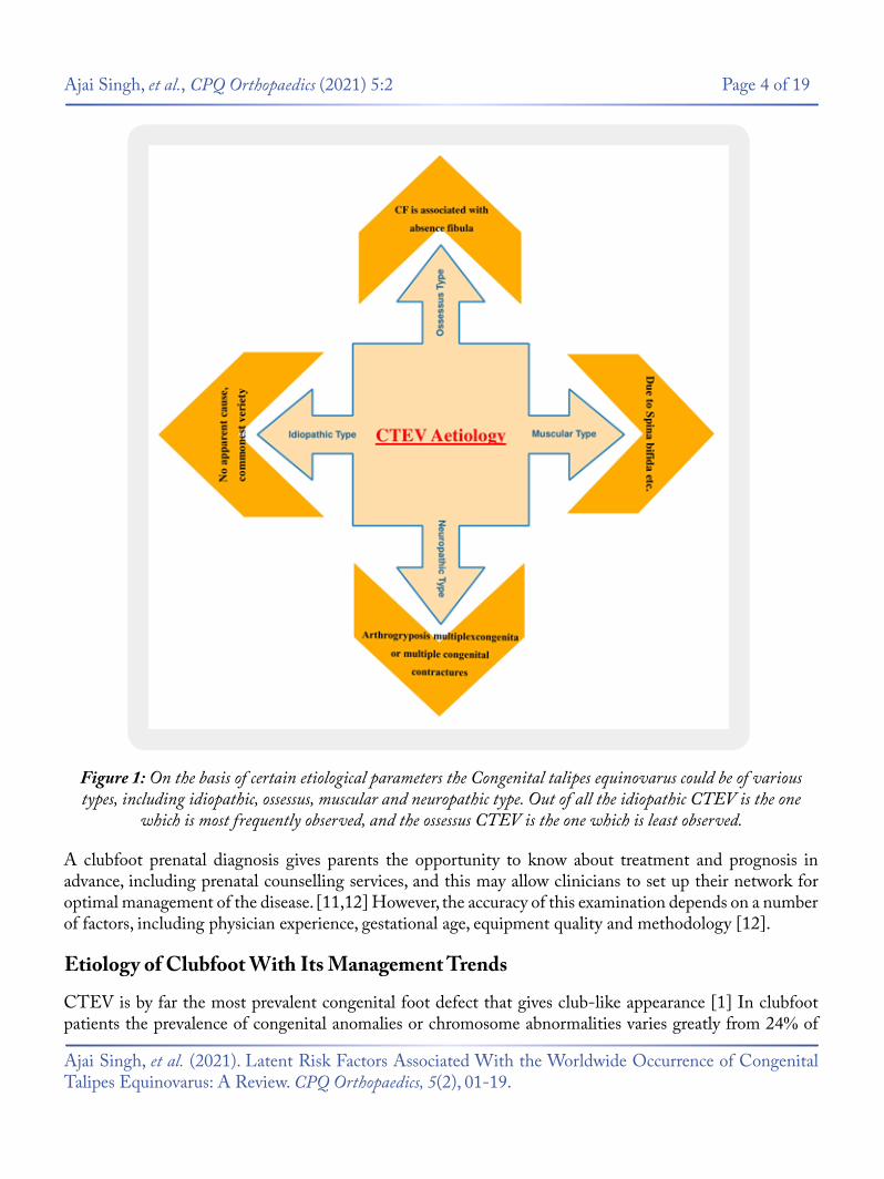
In some cases, CTEV may occur as part of a genetic syndrome in combination with other characteristics or congenital anomalies, but in the majority of cases, it may occur in isolation and is considered to be isolated CTEV (Figure 1) [3,4]. CTEV’s etiology is not yet known [5]. There was no clarification of either the environmental or genetic factors [6]. Mutations in genes involved in limb and muscle development are risk factors for clubfoot [5], specifically those encoding the contractile muscle complex and those regulating the expression of these many genes [6]. Common genetic variants such as HOX homeobox genes, insulin such as protein binding growth factor, MTHFR gene and Caspase gene family have either been reported to be directly associated with isolated clubfoot or to regulate the expression of other genes involved. According to the latest research by Wang et al. HOXD-12 and 13 gene is liable to cause congenital talipes equinovarus [7]. Many studies showed that smoking cigarettes are one of the most significant and coherent determinants in raising a child’s risk for CTEV during pregnancy [8]. In the presence of a favorable history of CTEV, smoking also raises the danger to 20 times, thus supporting the function of the gene in CTEV [9]. Environmental factors have been suggested as contributing to CTEV development, such as a small uterine cavity, mother-ingested drugs during pregnancy and herbicide aerial spraying [10].
Ajai Singh, et al. (2021). Latent Risk Factors Associated With the Worldwide Occurrence of Congenital Talipes Equinovarus: A Review. CPQ Orthopaedics, 5(2), 01-19.
Ajai Singh, et al., CPQ Orthopaedics (2021) 5:2 Page 4 of 19
Figure 1: On the basis of certain etiological parameters the Congenital talipes equinovarus could be of various types, including idiopathic, ossessus, muscular and neuropathic type. Out of all the idiopathic CTEV is the one
which is most frequently observed, and the ossessus CTEV is the one which is least observed.
A clubfoot prenatal diagnosis gives parents the opportunity to know about treatment and prognosis in advance, including prenatal counselling services, and this may allow clinicians to set up their network for optimal management of the disease. [11,12] However, the accuracy of this examination depends on a number of factors, including physician experience, gestational age, equipment quality and methodology [12].
Etiology of Clubfoot With Its Management Trends
CTEV is by far the most prevalent congenital foot defect that gives club-like appearance [1] In clubfoot patients the prevalence of congenital anomalies or chromosome abnormalities varies greatly from 24% of
Ajai Singh, et al. (2021). Latent Risk Factors Associated With the Worldwide Occurrence of Congenital Talipes Equinovarus: A Review. CPQ Orthopaedics, 5(2), 01-19.
Ajai Singh, et al., CPQ Orthopaedics (2021) 5:2 Page 5 of 19
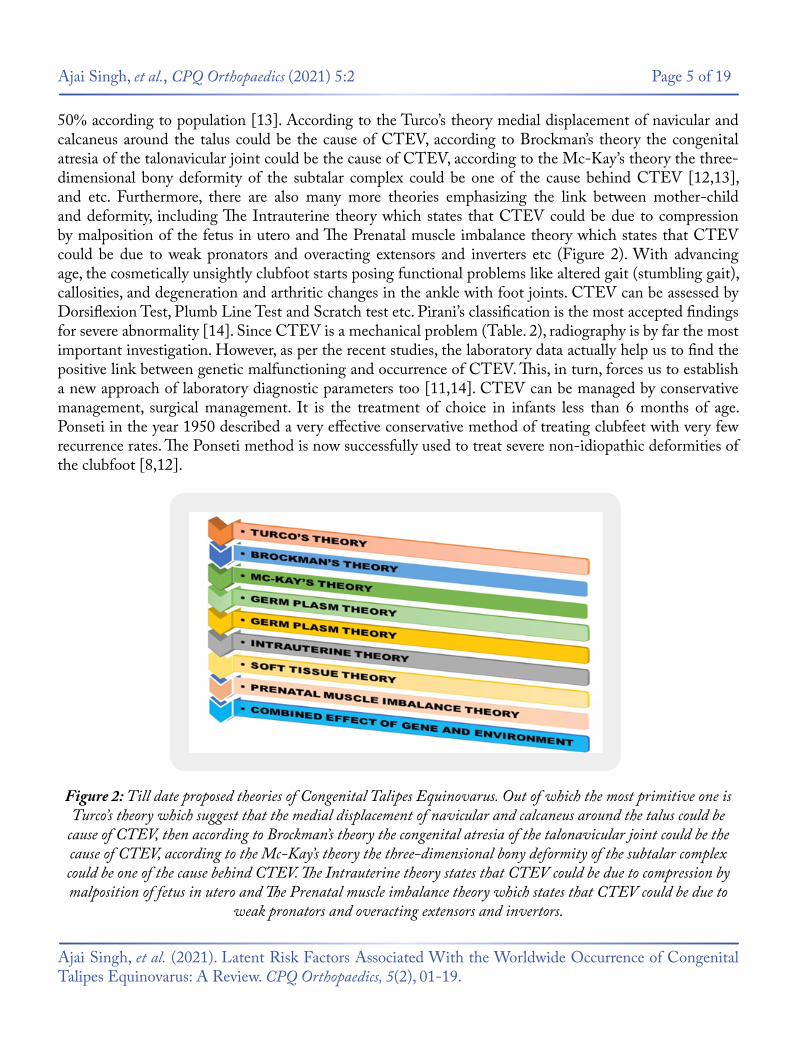
Figure 2: Till date proposed theories of Congenital Talipes Equinovarus. Out of which the most primitive one is Turco’s theory which suggest that the medial displacement of navicular and calcaneus around the talus could be
cause of CTEV, then according to Brockman’s theory the congenital atresia of the talonavicular joint could be the cause of CTEV, according to the Mc-Kay’s theory the three-dimensional bony deformity of the subtalar complex could be one of the cause behind CTEV. The Intrauterine theory states that CTEV could be due to compression by malposition of fetus in utero and The Prenatal muscle imbalance theory which states that CTEV could be due to
weak pronators and overacting extensors and invertors.
50% according to population [13]. According to the Turco’s theory medial displacement of navicular and calcaneus around the talus could be the cause of CTEV, according to Brockman’s theory the congenital atresia of the talonavicular joint could be the cause of CTEV, according to the Mc-Kay’s theory the three-dimensional bony deformity of the subtalar complex could be one of the cause behind CTEV [12,13], and etc. Furthermore, there are also many more theories emphasizing the link between mother-child and deformity, including The Intrauterine theory which states that CTEV could be due to compression by malposition of the fetus in utero and The Prenatal muscle imbalance theory which states that CTEV could be due to weak pronators and overacting extensors and inverters etc (Figure 2). With advancing age, the cosmetically unsightly clubfoot starts posing functional problems like altered gait (stumbling gait), callosities, and degeneration and arthritic changes in the ankle with foot joints. CTEV can be assessed by Dorsiflexion Test, Plumb Line Test and Scratch test etc. Pirani’s classification is the most accepted findings for severe abnormality [14]. Since CTEV is a mechanical problem (Table. 2), radiography is by far the most important investigation. However, as per the recent studies, the laboratory data actually help us to find the positive link between genetic malfunctioning and occurrence of CTEV. This, in turn, forces us to establish a new approach of laboratory diagnostic parameters too [11,14]. CTEV can be managed by conservative management, surgical management. It is the treatment of choice in infants less than 6 months of age. Ponseti in the year 1950 described a very effective conservative method of treating clubfeet with very few recurrence rates. The Ponseti method is now successfully used to treat severe non-idiopathic deformities of the clubfoot [8,12].
Ajai Singh, et al. (2021). Latent Risk Factors Associated With the Worldwide Occurrence of Congenital Talipes Equinovarus: A Review. CPQ Orthopaedics, 5(2), 01-19.
Ajai Singh, et al., CPQ Orthopaedics (2021) 5:2 Page 6 of 19
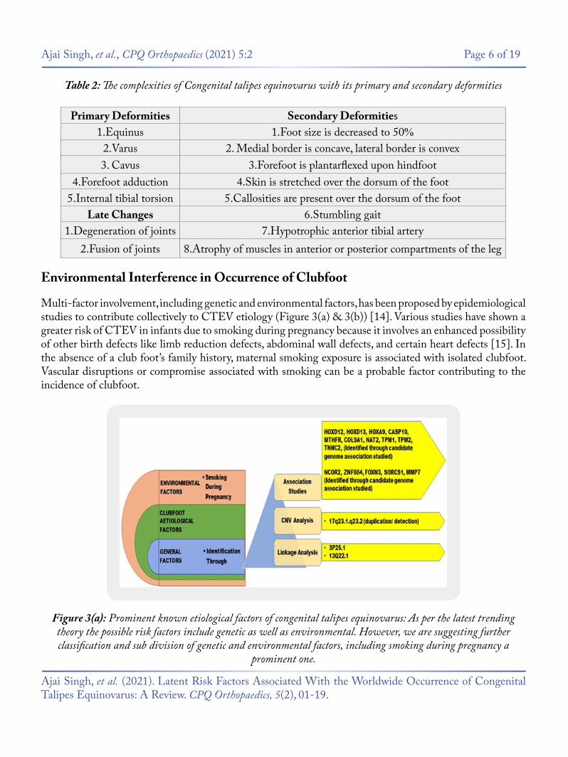
Figure 3(a): Prominent known etiological factors of congenital talipes equinovarus: As per the latest trending theory the possible risk factors include genetic as well as environmental. However, we are suggesting further classification and sub division of genetic and environmental factors, including smoking during pregnancy a
prominent one.
Multi-factor involvement, including genetic and environmental factors, has been proposed by epidemiological studies to contribute collectively to CTEV etiology (Figure 3(a) & 3(b)) [14]. Various studies have shown a greater risk of CTEV in infants due to smoking during pregnancy because it involves an enhanced possibility of other birth defects like limb reduction defects, abdominal wall defects, and certain heart defects [15]. In the absence of a club foot’s family history, maternal smoking exposure is associated with isolated clubfoot. Vascular disruptions or compromise associated with smoking can be a probable factor contributing to the incidence of clubfoot.
Table 2: The complexities of Congenital talipes equinovarus with its primary and secondary deformities
Primary Deformities Secondary Deformities1.Equinus 1.Foot size is decreased to 50%
2.Varus 2. Medial border is concave, lateral border is convex3. Cavus 3.Forefoot is plantarflexed upon hindfoot
4.Forefoot adduction 4.Skin is stretched over the dorsum of the foot5.Internal tibial torsion 5.Callosities are present over the dorsum of the foot
Late Changes 6.Stumbling gait1.Degeneration of joints 7.Hypotrophic anterior tibial artery
2.Fusion of joints 8.Atrophy of muscles in anterior or posterior compartments of the leg
Environmental Interference in Occurrence of Clubfoot
Ajai Singh, et al. (2021). Latent Risk Factors Associated With the Worldwide Occurrence of Congenital Talipes Equinovarus: A Review. CPQ Orthopaedics, 5(2), 01-19.
Ajai Singh, et al., CPQ Orthopaedics (2021) 5:2 Page 7 of 19
Genetic factors make a substantial contribution to CTEV etiology. The genetical cause indicated that CTEV is prone to segregate in families [16]. As per the available literature, it has been found that there is a link between family history of CTEV, with the occurrence of CTEV in neonates [17,18]. The significance of genes involved in the growth of premature limbs was recently identified by the discovery of an unusual mutation in the transcription factor PITX1 [19]. PITX1 is the first gene involved in clubfoot explaining the foot’s unique involvement [19]. Mainly Hox-Gene Family, MTHFR Gene, Caspase-Gene Family, etc. are the genetically identified factors through associative studies (Table. 3). Genes with apoptosis, homeobox A and D (HOXA and HOXD) and the genes involved in muscle contraction, have suggested the contribution
Genetic Mis-Functioning as a Reason Behind CTEV
The potential for gene-environment interaction in clubfoot etiology on the Asian population has not been examined yet. Indeed, available researches also demonstrate that both maternal smoking and the history of the family are essential causes for clubfoot and that these two variables can have a substantial potential relationship. It also offers a further indication of etiology of clubfoot and emphasizes the significance of considering interactions of sex, laterality, and higher-order in etiological studies of defects.
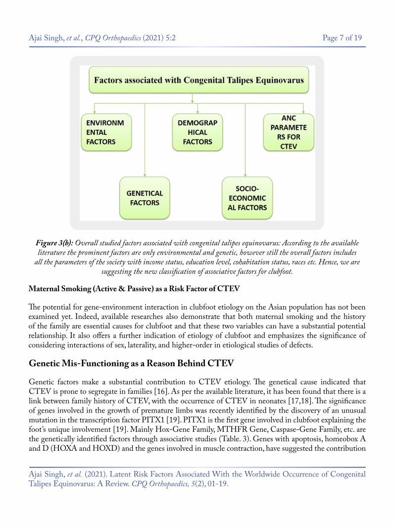
Figure 3(b): Overall studied factors associated with congenital talipes equinovarus: According to the available literature the prominent factors are only environmental and genetic, however still the overall factors includes
all the parameters of the society with income status, education level, cohabitation status, races etc. Hence, we are suggesting the new classification of associative factors for clubfoot.
Maternal Smoking (Active & Passive) as a Risk Factor of CTEV

Ajai Singh, et al. (2021). Latent Risk Factors Associated With the Worldwide Occurrence of Congenital Talipes Equinovarus: A Review. CPQ Orthopaedics, 5(2), 01-19.
Ajai Singh, et al., CPQ Orthopaedics (2021) 5:2 Page 8 of 19
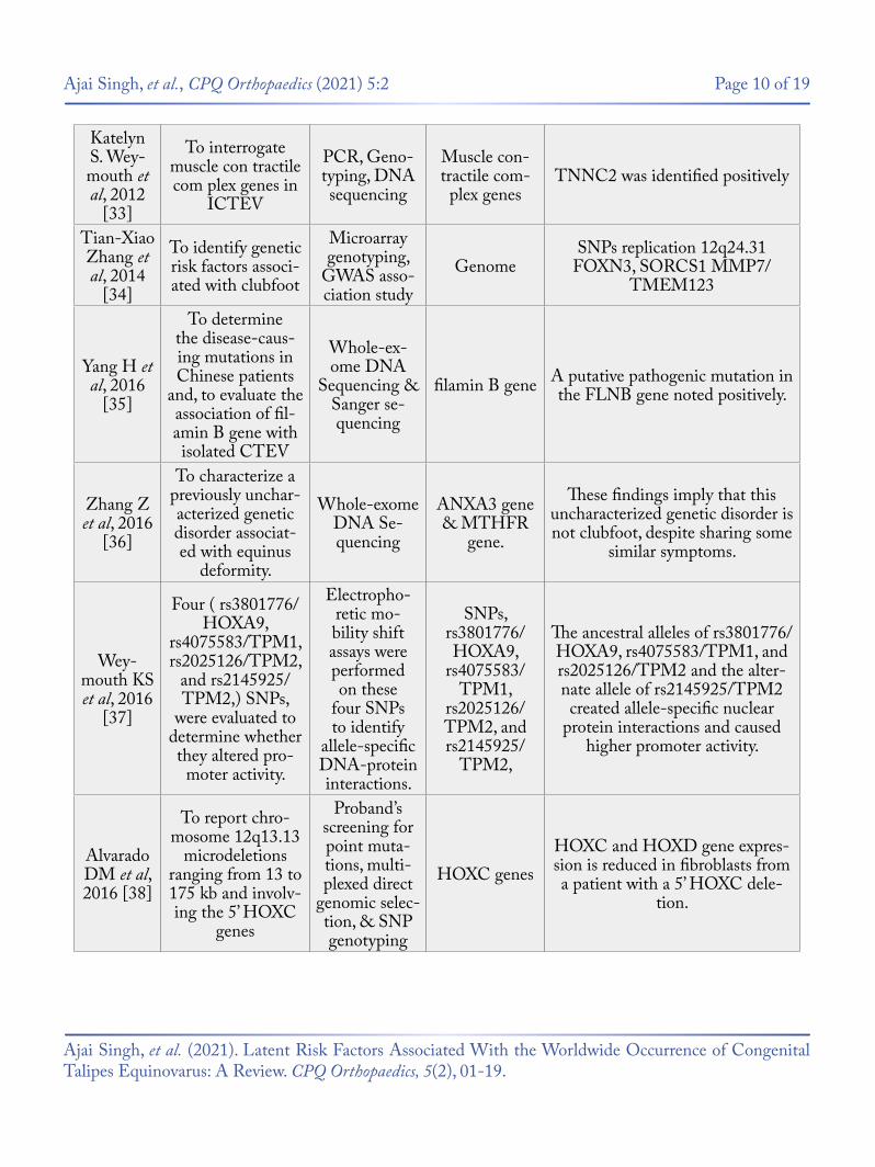
Table 3: Data representation of recent studies accomplished on CTEV in the previous years
of genetic in the accountability of CTEV [20,21]. Heck and colleagues [22] detected evidence that a rare CASP10 gene allele was linked and associated with simplex CTEV cases. Polymorphism of methylenetetrahydrofolate reductase gene (MTHFR) in mothers was also associated with CTEV [21]. The gene COL9A1 is the significantly susceptible gene for CTEV, as the elevated expression of COL9A1 was noted relative to ordinary people [20]. NAT2 (N-acetyltransferase 2) contributes to the biotransformation of N-acetylation tobacco smoke [23]. Functional analysis has shown that the variants associated with CTEV result in allele-specific interactions between nuclear proteins and cause higher promoter activity [9].
Fisrst Author Aim of the Study Methodology Targetedgene Result
L Bonafé et al, 2002
[24]
To evaluate the re-lationship between
R279 W muta-tion in DTDST
and occurrence of ICTEV
PCR, Genotyping DTDST
Linkage and association results obtained with GENEHUNTER for D5S1507 were not significant.
Li-li Wang et al, 2005
[8]
To study SNPs in HoxD10, HoxD12,
HoxD13 and haplotypes distri-bution in ICTEV
pedigree
PCR, Genotyping,
TDT
HoxD10 HoxD12 HoxD13
The rs847154 located in 5’ flank-ing sequence of HOXD12 gene and the rs13392701 located in exon 1 of HOXD13 gene were noted to have transmission dis-equilibrium in 84 nuclear pedi-
grees (P < 0.05).
Zhang X et al, 2006
[25]
To explore the association and
mutation of GLI3 gene in ICTEV
PCR, Genotyping,
TDTGLI3
rs929387ls located in exon 14 of GLI3 gene have transmission
disequilibrium in 84 nuclear pedigrees (P<0.05), and rs846266 located in exon 4 have no trans-mission disequilibrium (P>0.05). A mutation in exon 9 was detect-ed in one patient and his mother.
Sharp L et al, 2006
[26]
To study MTHFR C677Tpo lymor-phism, and ma-
ternal periconcep tional folic acid supple ment use, influenced risk of isolated clubfoot
PCR, Genotyping MTHFR
A significant trend of decreas-ing clubfoot risk with increasing number of T alleles: relative risk
for CT vs. CC = 0.75, 95% confi-dence interval: 0.57, 0.97; relative risk for TT vs. CC = 0.57, 95%
confidence interval: 0.35, 0.91; p trend = 0.006. This association was
not modified by maternal folic acid use.

Ajai Singh, et al. (2021). Latent Risk Factors Associated With the Worldwide Occurrence of Congenital Talipes Equinovarus: A Review. CPQ Orthopaedics, 5(2), 01-19.
Ajai Singh, et al., CPQ Orthopaedics (2021) 5:2 Page 9 of 19
Liu LY et al, 2007
[27]
To analyze SNPs within COL9A1 gene in ICTEV
PCR, Genotyping,
ETDTCOL9A1
The results showed that rs592121 and rs1135056 loci within CO-L9A1 gene existed transmission
disequilibrium in 84 nuclear pedi-grees (P<0.05). Expression of CO-
L9A1 on mRNA levels showed significantly higher in patients
with ICTEV than in normal per-son (t=4.7500, P<0.05) .
Zhao N et al, 2008
[28]
To detect the expressions of
COL1A1 mRNA in 20 patients with
ICTEV
PCR-DGGE, DNA se-quencing
COL1A1
Expression of COL1A1 on mRNA levels showed significantly
higher in patients with ICTEV than in normal persons (t=12.680, P < 0.05). By DNA sequencing, a -161(T--> C) heterozygous
mutation and a+ 274(C-->G) ho-mozygous mutation were detect-ed, and both were new identified mutations. These results indicated that the mutations in transcription regulator sequences of COL1A1
could cause ICTEV.
Shyy W et al, 2009
[29]
To test the hypoth-esis that CAND2 and WNT7a mu-tation associated
with ICTEV
PCR, DNA sequencing
CAND2 WN-T7a
A polymorphism was observed in each gene, but the single nu-cleotide change in CAND2 was
a silent mutation that did not alter the amino acid product, and the single nucleotide change in WNT7a was in the upstream,
non-coding or promoter region before the start codon.
Audrey R. Ester et al, 2009 [30]
To study HoxA, HoxD and IG-
FBP3 in patients with ICTEV
PCR, Ge-notyping, In
SilicoHoxA HoxD
IGFBP3Tested genes positve Interactions
with CASP3
Shyy W et al, 2010
[31]
To study MYH genes in ICTEV
patientsPCR, DNA sequencing
MYH 1 MYH 2 MYH 3 MYH 8
MYH genes not directly cause ICTEV
W. Lu et al, 2012
[32]
To assess wheth-er variation in or around TBX4 is a common cause of non syndromic
clubfoot
CGH, PCR, Genotyping,
DNA se-quencing
TBX4 TBX4 variation negative
Ajai Singh, et al. (2021). Latent Risk Factors Associated With the Worldwide Occurrence of Congenital Talipes Equinovarus: A Review. CPQ Orthopaedics, 5(2), 01-19.
Ajai Singh, et al., CPQ Orthopaedics (2021) 5:2 Page 10 of 19
Katelyn S. Wey-mouth et al, 2012
[33]
To interrogate muscle con tractile com plex genes in
ICTEV
PCR, Geno-typing, DNA sequencing
Muscle con-tractile com-
plex genesTNNC2 was identified positively
Tian-Xiao Zhang et al, 2014
[34]
To identify genetic risk factors associ-ated with clubfoot
Microarray genotyping,
GWAS asso-ciation study
GenomeSNPs replication 12q24.31
FOXN3, SORCS1 MMP7/ TMEM123
Yang H et al, 2016
[35]
To determine the disease-caus-ing mutations in Chinese patients
and, to evaluate the association of fil-amin B gene with isolated CTEV
Whole-ex-ome DNA
Sequencing & Sanger se-quencing
filamin B gene A putative pathogenic mutation in the FLNB gene noted positively.
Zhang Z et al, 2016
[36]
To characterize a previously unchar-acterized genetic disorder associat-ed with equinus
deformity.
Whole-exome DNA Se-quencing
ANXA3 gene & MTHFR
gene.
These findings imply that this uncharacterized genetic disorder is not clubfoot, despite sharing some
similar symptoms.
Wey-mouth KS et al, 2016
[37]
Four ( rs3801776/HOXA9,
rs4075583/TPM1, rs2025126/TPM2,
and rs2145925/TPM2,) SNPs,
were evaluated to determine whether they altered pro-
moter activity.
Electropho-retic mo-bility shift assays were performed on these
four SNPs to identify
allele-specific DNA-protein interactions.
SNPs, rs3801776/HOXA9,
rs4075583/TPM1,
rs2025126/TPM2, and rs2145925/
TPM2,
The ancestral alleles of rs3801776/HOXA9, rs4075583/TPM1, and rs2025126/TPM2 and the alter-nate allele of rs2145925/TPM2 created allele-specific nuclear
protein interactions and caused higher promoter activity.
Alvarado DM et al, 2016 [38]
To report chro-mosome 12q13.13
microdeletions ranging from 13 to 175 kb and involv-ing the 5’ HOXC
genes
Proband’s screening for point muta-tions, multi-plexed direct
genomic selec-tion, & SNP genotyping
HOXC genesHOXC and HOXD gene expres-sion is reduced in fibroblasts from a patient with a 5’ HOXC dele-
tion.
Ajai Singh, et al. (2021). Latent Risk Factors Associated With the Worldwide Occurrence of Congenital Talipes Equinovarus: A Review. CPQ Orthopaedics, 5(2), 01-19.
Ajai Singh, et al., CPQ Orthopaedics (2021) 5:2 Page 11 of 19
There are a number of population-based orthopedic-confirmed clubfeet patients and controls for assessing the incidence patterns [26,27,44]. The male majority reported in various studies was reported in our study,
Demographic Factors Accountable for Clubfoot
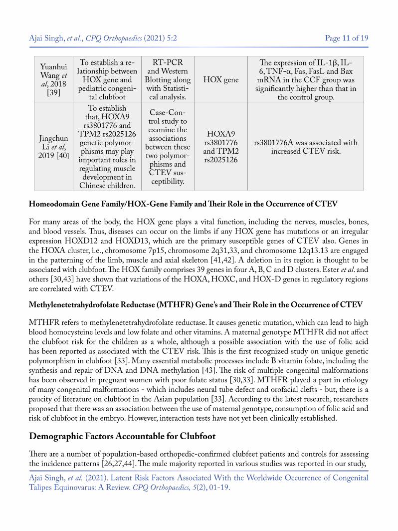
For many areas of the body, the HOX gene plays a vital function, including the nerves, muscles, bones, and blood vessels. Thus, diseases can occur on the limbs if any HOX gene has mutations or an irregular expression HOXD12 and HOXD13, which are the primary susceptible genes of CTEV also. Genes in the HOXA cluster, i.e., chromosome 7p15, chromosome 2q31,33, and chromosome 12q13.13 are engaged in the patterning of the limb, muscle and axial skeleton [41,42]. A deletion in its region is thought to be associated with clubfoot. The HOX family comprises 39 genes in four A, B, C and D clusters. Ester et al. and others [30,43] have shown that variations of the HOXA, HOXC, and HOX-D genes in regulatory regions are correlated with CTEV.
Homeodomain Gene Family/HOX-Gene Family and Their Role in the Occurrence of CTEV
Yuanhui Wang et al, 2018
[39]
To establish a re-lationship between
HOX gene and pediatric congeni-
tal clubfoot
RT-PCR and Western
Blotting along with Statisti-cal analysis.
HOX gene
The expression of IL-1β, IL-6, TNF-α, Fas, FasL and Bax
mRNA in the CCF group was significantly higher than that in
the control group.
Jingchun Li et al,
2019 [40]
To establish that, HOXA9 rs3801776 and
TPM2 rs2025126 genetic polymor-phisms may play
important roles in regulating muscle development in
Chinese children.
Case-Con-trol study to examine the associations
between these two polymor-phisms and CTEV sus-ceptibility.
HOXA9 rs3801776 and TPM2 rs2025126
rs3801776A was associated with increased CTEV risk.
MTHFR refers to methylenetetrahydrofolate reductase. It causes genetic mutation, which can lead to high blood homocysteine levels and low folate and other vitamins. A maternal genotype MTHFR did not affect the clubfoot risk for the children as a whole, although a possible association with the use of folic acid has been reported as associated with the CTEV risk. This is the first recognized study on unique genetic polymorphism in clubfoot [33]. Many essential metabolic processes include B vitamin folate, including the synthesis and repair of DNA and DNA methylation [43]. The risk of multiple congenital malformations has been observed in pregnant women with poor folate status [30,33]. MTHFR played a part in etiology of many congenital malformations - which includes neural tube defect and orofacial clefts - but, there is a paucity of literature on clubfoot in the Asian population [33]. According to the latest research, researchers proposed that there was an association between the use of maternal genotype, consumption of folic acid and risk of clubfoot in the embryo. However, interaction tests have not yet been clinically established.
Methylenetetrahydrofolate Reductase (MTHFR) Gene’s and Their Role in the Occurrence of CTEV

Ajai Singh, et al. (2021). Latent Risk Factors Associated With the Worldwide Occurrence of Congenital Talipes Equinovarus: A Review. CPQ Orthopaedics, 5(2), 01-19.
Ajai Singh, et al., CPQ Orthopaedics (2021) 5:2 Page 12 of 19
There is evidence in both animals and humans that psycho-social and environmental stressors modify the sex ratio in favour of females, possibly by activating the hypothalamic-pituitary-adrenal (HPA) axis [44]. Why more males are born with clubfoot than females remains a mystery, with countably low available literature and hence requires further studies with special emphasis. However, we may suggest that the environmental or psycho-social factors that change the male sex ratio may be worth considering as a risk factor [24].
including the demonstrations and analysis for isolated cases, those that were associated with other chief malformations, unilateral and bilateral cases, and that could be attributed to fetal constraint or genetics. However, the lowest magnitudes of masculine births have been observed among cases of further major congenital malformations, those associated with fetal constraint, and those with a positive family history in first-degree relatives, possibly due to etiological heterogeneity among them [44].
Level of education positively associated with clubfoot [9,45-47], however, other studies found no association [48-52], as we observed. Further, the combined family income and its stability source have both direct as well as indirect impact on clubfoot [9,47-49], by deciding the means of basic requirements for pregnant women and infant both such as food, water, shelter, hygiene and early age medication etc [9,47-49].
Socio-demographic factors in clubfoot have not been consistently related; however, still they have a specific impact on the child and mother both. Hence, we may consider them also as a sub-prominent factor for clubfoot.
Maternal age was reported to be inversely linked to clubfoot [9,45-47]. However, other studies found no association [48-52]. We did not use primiparity as a constraint predictor because almost half of all women fall into that category and would have weakened the sensitivity of any particular constraint. For example, no correlation has been observed for obesity since primiparity was added to the concept of ‘constraint.’ Clubfoot is well known to recur in some obese families [45], And studies suggest that clubfoot cases had a relative first degree affected [9,45,52-54].
We analyzed the status of cohabitation as a further measure of socioeconomic status in comparison to a higher risk of unmarried mothers in a UK study, and it was also not associated with clubfoot [45]. Two studies have identified a higher risk of clubfoot in White mother’s offspring compared to non-White mothers [9,46], which was not substantiated in four other studies [47,49,50,52].
Sexuality of Child as a Risk Factor of CTEV
Maternal Age and Weight During Pregnancy as a Risk Factor of CTEV
Role of Socio-Economic Factors in Occurence of Clubfoot
Impact of Parental Qualification and Combined Family Income Status on CTEV
Impact of Parental Cohabitation Status on CTEV
Ajai Singh, et al. (2021). Latent Risk Factors Associated With the Worldwide Occurrence of Congenital Talipes Equinovarus: A Review. CPQ Orthopaedics, 5(2), 01-19.
Ajai Singh, et al., CPQ Orthopaedics (2021) 5:2 Page 13 of 19
Based on clinical findings of cases involving oligohydramnios, uterine bicornuate, breech presentations, and multiple births, i.e. females with no prior births are at increased risk of clubfoot. Past epidemiological studies have, however, found contradictory findings, with positive, inverse and null associations [47-49,55]. Increased clubfoot risks associated with breech delivery and uterus bicornuate and 60 to 80% increased risks for oligohydramnios and multiple births, in favour of a framework for limiting a minority of cases [47-49,55]. The relation between clubfoot and primiparity which is regularly observed [45-52]. Was raised as evidence to support fetal pathogenesis, on the premise that after the first birth, by holding the fetus, the intrauterine area is extended [45,49].
In at least some cases, clubfoot was accused of arising from fetal constraint and also thought to be associated with ANC managements, including gestational age and medication also.
Based on more systemic or neurological developmental abnormalities, it is suggested that the Prenatally diagnosed isolated clubfoot cases will postnatally have complex clubfoot [26,27]. While advising women on isolated clubfoot prenatally diagnosed, it is necessary to reassure them that approximately 10 percent of people would have a normal foot or foot deformity that needs minimal treatment [26,33]. Clubfoot may be diagnosed prenatally on a thorough ultrasonography scan performed in the second or third trimester by visualizing Tibia and fibula in the same longitudinal plane as the foot’s lateral portion [47-49].
Another potential pathogenetic mechanism for clubfoot is the vascular disturbance, leading to increased risk of clubfoot in women with early gestational amniocentesis [55,56] Or is it exposed to abortive misoprostol [57,58]. In a few studies, we found that if there is a 16 Weeks of gestation, amniocentesis, CVS, or the fetal loss of a twin or triplet to be vascular disturbance markers. Such factors were correlated with an increased risk of clubfoot 5.6, 2.2, and 4.1 times respectively, Supporting potential vascular disruption pathogenesis [55-58].
Clubfoot is one of the most prevalent birth defects in the musculoskeletal system; its etiology and controversies related to optimal treatment strategies are still unknown. The aim of the study was to update the latest progress in the awareness of the genetic and environmental etiology of clubfoot and explore the future research required for this disorder to achieve a genetic classification scheme. The objective was also to examine the development of clubfoot management and clarify How it revolutionized the Ponseti process childcare worldwide (Figure 4). Although current methods of treatment seem to be beneficial in maximum cases, regardless of heir etiology, co-morbidity risks, i.e. hip dysplasia. Future genome associative research
Number of Previous Pregnancies as a Risk Factor of CTEV
Proposed Outcomes of Prenatally Diagnosed Clubfoot
Conclusion With Future Perspectives
Proposed ANC Parameters Associated With Clubfoot
Gestational Age as a Risk Factor of CTEV
Ajai Singh, et al. (2021). Latent Risk Factors Associated With the Worldwide Occurrence of Congenital Talipes Equinovarus: A Review. CPQ Orthopaedics, 5(2), 01-19.
Ajai Singh, et al., CPQ Orthopaedics (2021) 5:2 Page 14 of 19
will provide an uneven alternative in the Identifying Clubfoot susceptibilities, and Large Samples Using Identify the susceptibility of both major and minor genes where present. Individualized interventions based on etiology may also lead to decreased use of braces if etiology or genetic profile correlates with risk of relapse. The primary aim of treatment is to deliver a fully functional, painless foot for long-term correction. In order to achieve this, more progressive research-based studies may need to combine approaches which apply the strengths of different methods.
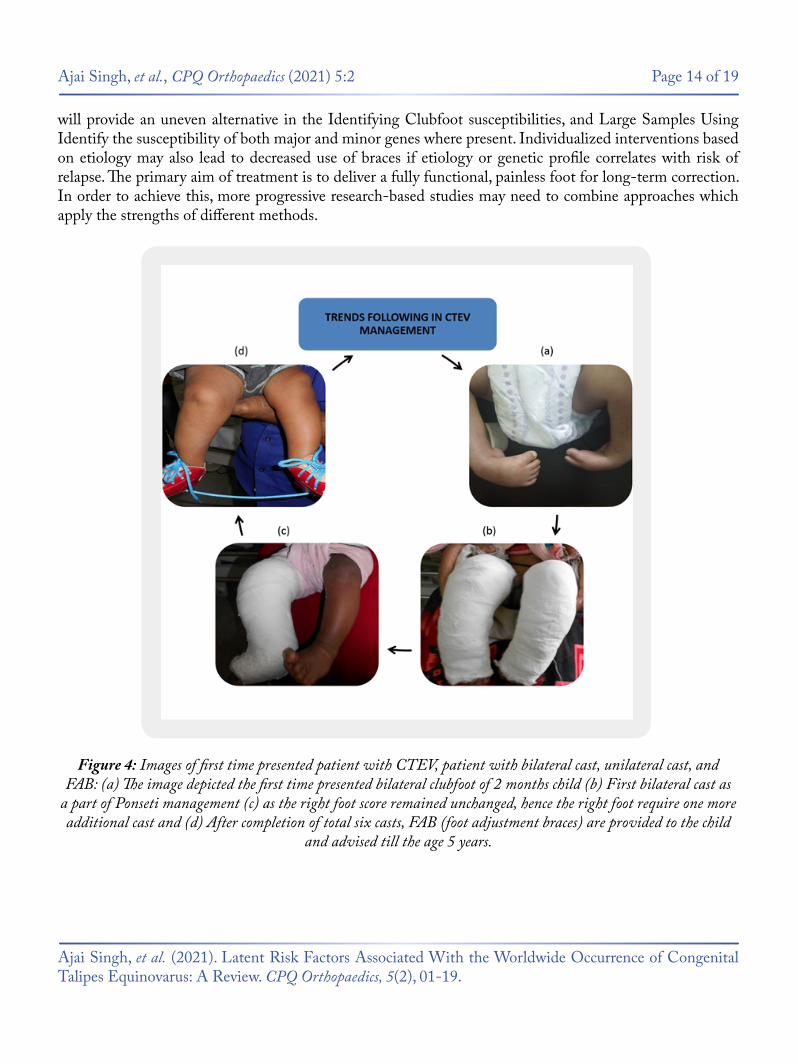
Figure 4: Images of first time presented patient with CTEV, patient with bilateral cast, unilateral cast, and FAB: (a) The image depicted the first time presented bilateral clubfoot of 2 months child (b) First bilateral cast as
a part of Ponseti management (c) as the right foot score remained unchanged, hence the right foot require one more additional cast and (d) After completion of total six casts, FAB (foot adjustment braces) are provided to the child
and advised till the age 5 years.
Ajai Singh, et al. (2021). Latent Risk Factors Associated With the Worldwide Occurrence of Congenital Talipes Equinovarus: A Review. CPQ Orthopaedics, 5(2), 01-19.
Ajai Singh, et al., CPQ Orthopaedics (2021) 5:2 Page 15 of 19
5. Jugessur, A., Wilcox, A. J., Lie, R. T., et al. (2003). Exploring the effects of methylenetetrahydrofolate reductase gene variants C677T and A1298C on the risk of orofacial clefts in 261 Norwegian case-parent triads. Am J Epidemiol., 157(12), 1083-1091.
This study was supported by Department of Paediatric Orthopaedics & Orthopaedic Surgery in collaboration with Department of Paediatrics and Department of Biochemistry, King George’s Medical University, Lucknow.
The authors declare that they have no competing interests in this article.
1. Carey, M., Bower, C., Mylvaganam, A., et al. (2003). Talipesequinovarus in Western Australia. Paediatr Perinat Epidemiol., 17(2), 187-194.
2. Chung, C. S., Nemechek, R. W., Larsen, I. J., et al. (1969). Genetic and epidemiological studies of club foot in Hawaii. General and medical considerations. Hum Hered., 19(4), 321-342.
3. Miedzybrodzka, Z. (2003). Congenital talipesequinovarus (clubfoot): a disorder of the foot but not the hand. J Anat., 202(1), 37-42.
4. Botto, L. D. & Yang, Q. (2000). 5,10-Methylenetetrahydrofolate reductase gene variants and congenital anomalies: aHuGE review. Am J Epidemiol ., 151(9), 862-877.
6. Bacino, C. A. & Hecht, J. T. (2014). Etiopathogenesis of equinovarus foot malformations. European Journal of Medical Genetics, 57(8), 473-479.
Acknowledgements
Conflicts of Interest
Bibliography
8. Wang, L. L., Jin, C. L., Liu, L. Y., Zhang, X., Ji, S. J. & Sun, K. L. (2005). Analysis of association between 5’HOXD gene and idiopathic congenital talipes equinovarus. Chinese Journal of Medical Genetics, 22(6), 653-656.
7. Ester, A. R. (2010). Analysis of Variation in Clubfoot Candidate Genes. UT GSBS Dissertations and Theses. (Pp. 1-148).
9. Honein, M. A., Paulozzi, L. J. & Moore, C. A. (2000). Family history, maternal smoking, and clubfoot: an indication of a gene-environment interaction. Am J Epidemiol., 152(7), 658-665.
10. Werler, M. M. (1997). Teratogen update: smoking and reproductive outcomes. Teratology, 55(6), 382-328.
Ajai Singh, et al. (2021). Latent Risk Factors Associated With the Worldwide Occurrence of Congenital Talipes Equinovarus: A Review. CPQ Orthopaedics, 5(2), 01-19.
Ajai Singh, et al., CPQ Orthopaedics (2021) 5:2 Page 16 of 19
23. Engell, V., Nielsen, J., Damborg, F., et al. (2014). Heritability of clubfoot: a twin study. J Child Orthop., 8(1), 37-41.
12. Wynne-Davies, R. (1964). Family studies and the cause of congenital club foot. Talipes equinovarus, talipes calcaneo-valgus and metatarsus varus. J Bone Joint Surg Br., 46, 445-463.
11. Cowell, H. R. & Wein, B. K. (1980). Genetic aspects of clubfoot. J Bone Joint Surg., 62(8), 1381-1384.
15. Bar-Hava, I., Bronshtein, M., Orvieto, R., et al. (1997). Caution: prenatal clubfoot can be both a transient and a lateonset phenomenon. Prenat Diagn., 17, 457-460.
16. Attenborough, C. G. (1966). Severe congenital talipes equinovarus. J Bone Joint Surg., 48-B, 31.
17. Barenfeld, P. A. & Wesley, M. S. (1972). Surgical treatment of congenital clubfoot. Clin Orthop., 84, 79-87.
18. McKay, D. W. (1983). New concept of and approach to clubfoot treatment. Section 111. Evaluation and results. J Paediatr Orthop., 3, 141.
20. Turc, V. J. (1971). Surgical correction of the resistant clubfoot: One stage posteromedial release with internal fixation: A preliminary report. J Bone and Joint Surg., 53(3), 477-497.
24. Bonafe, L., Blanton, S. H., Scott, A., Broussard, S., Wise, C. A., Superti-Furga, A. & Hecht, J. T. (2002). DTDST mutations are not a frequent cause of idiopathic talipes equinovarus (club foot). Journal of Medical Genetics, 39(4), e20.
22. Heck, A. L., Bray, M. S., Scott, A., et al. (2005). Variation in CASP10 gene is associated with idiopathic talipes equinovarus. J Pediatr Orthop., 25(5), 598-602.
25. Zhang, X., Jin, C. L., Liu, L. Y., Zhao, N., Zhang, L. J., Ji, S. J. & Sun, K. L. (2006). Association and mutation analysis of GLI3 gene in idiopathic congenital talipes equinovarus. Chinese Journal of Medical Genetics, 23(5), 551-554.
13. Yamamoto, H. (1979). A clinical, genetic and epidemiologic study of congenital club foot. JinruiIdengaku Zasshi., 24(1), 37-44.
14. Mahan, S. T., Yazdy, M. M., Kasser, J. R., et al. (2014). Prenatal screening for clubfoot: what factors predict prenatal detection? Prenat Diagn., 34(4), 389-393.
19. Evans, D. (1961). Relapsed clubfoot. J Bone and Joint Surg., 43-B, 722.
21. Garceau, G. J. (1954). Recurrent clubfoot. Bull Hosp Joint Dis., 15(2), 143-150.

Ajai Singh, et al. (2021). Latent Risk Factors Associated With the Worldwide Occurrence of Congenital Talipes Equinovarus: A Review. CPQ Orthopaedics, 5(2), 01-19.
Ajai Singh, et al., CPQ Orthopaedics (2021) 5:2 Page 17 of 19
35. Yang, H., Zheng, Z., Cai, H., Li, H., Ye, X., Zhang, X., Wang, Z. & Fu, Q. (2016). Three novel missense mutations in the filamin B gene are associated with isolated congenital talipes equinovarus. Human Genetics, 135(10), 1181-1189.
26. Sharp, L., Miedzybrodzka, Z., Cardy, A. H., et al. (2006). The C677T polymorphism in the methylenetetrahydrofolate reductase gene (MTHFR), maternal use of folic acid supplements, and risk of isolated clubfoot: A case-parent-triad analysis. Am J Epidemiol., 164(9), 852-861.
29. Shyy, W., Dietz, F., Dobbs, M. B., Sheffield, V. C. & Morcuende, J. A. (2009). Evaluation of CAND2 and WNT7a as candidate genes for congenital idiopathic clubfoot. Clinical Orthopaedics and Related Research, 467(5), 1201-1205.
30. Ester, A. R., Weymouth, K. S., Burt, A., et al. (2009). Altered transmission of HOX and apoptotic SNPs identify a potential common pathway for clubfoot. Am J Med Genet A., 149A(12), 2745-2752.
31. Shyy, W., Wang, K., Sheffield, V. C. & Morcuende, J. A. (2010). Evaluation of embryonic and perinatal myosin gene mutations and the etiology of congenital idiopathic clubfoot. Journal of Pediatric Orthopedics, 30(3), 231-234.
32. Lu, W., Bacino, C. A., Richards, B. S., Alvarez, C., Vander Meer, J. E., Vella, M., Ahituv, N., et al. (2012). Studies of TBX4 and chromosome 17q23. 1q23. 2: an uncommon cause of nonsyndromic clubfoot. American Journal of Medical Genetics Part A., 158A(7), 1620-1627.
36. Zhang, Z., Kong, Z., Zhu, M., Lu, W., Ni, L., Bai, Y. & Lou, Y. (2016). Whole genome sequencing identifies ANXA3 and MTHFR mutations in a large family with an unknown equinus deformity associated genetic disorder. Molecular Biology Reports, 43(10), 1147-1155.
34. Zhang, T. X., Haller, G., Lin, P., Alvarado, D. M., Hecht, J. T., Blanton, S. H., Richards, B. S., et al. (2014). Genome-wide association study identifies new disease loci for isolated clubfoot. Journal of Medical Genetics, 51(5), 334-339.
37. Weymouth, K. S., Blanton, S. H., Powell, T., Patel, C. V., Savill, S. A. & Hecht, J. T. (2016). Functional assessment of clubfoot associated HOXA9, TPM1, and TPM2 variants suggests a potential gene regulation mechanism. Clinical Orthopaedics and Related Research., 474(7), 1726-1735.
27. Liu, L. Y., Jin, C. L., Cao, D. H., et al. (2007). Analysis of association between COL9A1 gene and idiopathic congenital talipes equinovarus. Yi Chuan, 29(4), 427-432.
28. Zhao, N., Jin, C. L., Liu, L. Y., Cao, D. H., Lin, C. K., Ji, S. J. & Sun, K. L. (2008). Association study between mutations of transcription regulator sequences of COL1A1 gene and idiopathic con-genital talipes equinovarus. Yi chuan= Hereditas, 30(6), 723-727.
33. Weymouth, K. S., Blanton, S. H., Bamshad, M. J., et al. (2011). Variants in genes that encode muscle contractile proteins influence risk for isolated clubfoot. Am J Med Genet A., 155A(9), 2170-2179.

Ajai Singh, et al. (2021). Latent Risk Factors Associated With the Worldwide Occurrence of Congenital Talipes Equinovarus: A Review. CPQ Orthopaedics, 5(2), 01-19.
Ajai Singh, et al., CPQ Orthopaedics (2021) 5:2 Page 18 of 19
47. Kancherla, V., Romitti, P. A., Caspers, K. M., Puzhankara, S. & Morcuende, J. A. (2010). Epidemiology of congenital idiopathic talipes equinovarus in Iowa, 1997-2005. Am J Med Genet A., 152A(7), 1695-1700.
38. Alvarado, D. M., McCall, K., Hecht, J. T., Dobbs, M. B. & Gurnett, C. A. (2016). Deletions of 5′ HOXC genes are associated with lower extremity malformations, including clubfoot and vertical talus. Journal of Medical Genetics, 53(4), 250-255.
41. Lochmiller, C., Johnston, D., Scott, A., et al. (1998). Genetic epidemiology study of idiopathic talipes equinovarus. Am J Med Genet., 79(2), 90-96.
42. Rebbeck, T. R., Dietz, F. R., Murray, J. C., et al. (1993). A single-gene explanation for the probability of having idiopathic talipes equinovarus. Am J Hum Genet., 53, 1051-1063.
43. Idelberger, K. H. (1978). Orthopedic genetics and family counseling (proceedings). Z Orthop Ihre Grenzgeb., 116, 552-554.
44. Werler, M. M., Yazdy, M. M., Mitchell, A. A., Meyer, R. E., Druschel, C. M., Anderka, M., et al. (2013). Descriptive epidemiology of idiopathic clubfoot. American Journal of Medical Genetics Part A., 161A(7), 1569-1578.
45. Cardy, A. H., Barker, S., Chesney, D., Sharp, L., Maffulli, N. & Miedzybrodzka, Z. (2007). Pedigree analysis and epidemiological features of idiopathic congenital talipes equinovarus in the United Kingdom: a case-control study. BMC Musculoskelet Disord., 8, 62.
48. Byron-Scott, R., Sharpe, P., Hasler, C., Cundy, P., Hirte, C., Chan, A., Scott, H., et al. (2005). A South Australian population-based study of congenital talipes equinovarus. Paediatr Perinat Epidemiol., 19(3), 227-237.
46. Dickinson, K. C., Meyer, R. E. & Kotch, J. (2008). Maternal smoking and the risk for clubfoot in infants. Birth Defects Res A Clin Mol Teratol., 82(2), 86-91.
49. Carey, M., Mylvaganam, A., Rouse, I. & Bower, C. (2005). Risk factors for isolated talipes equinovarus in Western Australia, 1980-1994. Paediatr Perinat Epidemiol., 19(3), 238-245.
39. Wang, Y. (2018). Relationship between HOX gene and pediatric congenital clubfoot. Experimental and Therapeutic Medicine, 15(6), 4861-4865.
40. Li, J., Wu, J., Liu, Y., Li, Y., Xiao, Z., Jiang, X., Tang, Y. & Xu, H. (2019). HOXA9 rs3801776 G> A polymorphism increases congenital talipes equinovarus risk in a Chinese population. The Journal of Gene Medicine, 21(10), 3119.
50. Moorthi, R. N., Hashmi, S. S., Langois, P., Canfield, M., Waller, D. K. & Hecht, J. T. (2005). Idiopathic talipes equinovarus (ITEV) (clubfeet) in Texas. Am J Med Genet A., 132A(4), 376-380.

Ajai Singh, et al. (2021). Latent Risk Factors Associated With the Worldwide Occurrence of Congenital Talipes Equinovarus: A Review. CPQ Orthopaedics, 5(2), 01-19.
Ajai Singh, et al., CPQ Orthopaedics (2021) 5:2 Page 19 of 19
51. Pavone, V., Bianca, S., Grosso, G., Pavone, P., Mistretta, A., Longo, M. R., Marino, S. & Sessa, G. (2012). Congenital talipes equinovarus: an epidemiological study in Sicily. Acta Orthop., 83(3), 294-298.
54. Wynne-Davies, R. (1972). Genetic and environmental factors in the etiology of talipes equinovarus. Clin Orthop Relat Res., 84, 9-13.
55. Philip, J., Silver, R. K., Wilson, R. D., Thom, E. A., Zachary, J. M., Mohide, P., Mahoney, M. J., et al. (2004). Late first-trimester invasive prenatal diagnosis: results of an international randomized trial. Obstet Gynecol., 103(6), 1164-1173.
56. Sundberg, K., Bang, J., Smidt-Jensen, S., Brocks, V., Lundsteen, C., Parner, J., Keiding, N. & Philip, J. (1997). Randomised study of risk of fetal loss related to early amniocentesis versus chorionic villus sampling. Lancet, 350(9079), 697-703.
57. Pastuszak, A. L., Schuler, L., Speck-Martins, C. E., Coelho, K. E., Cordello, S. M., Vargas, F., Brunoni, D., et al. (1998). Use of misoprostol during pregnancy and Mobius’ syndrome in infants. N Engl J Med., 338, 1881-1885.
58. Vargas, F. R., Schuler-Faccini, L., Brunoni, D., Kim, C., Meloni, V. F., Sugayama, S. M., et al. (2000). Prenatal exposure to misoprostol and vascular disruption defects: a case-control study. Am J Med Genet., 95, 302-306.
52. Skelly, A. C., Holt, V. L., Mosca, V. S. & Alderman, B. W. (2002). Talipes equinovarus and maternal smoking: a population-based case-control study in Washington state. Teratology, 66(2), 91-100.
53. Cartlidge, I. (1984). Observations on the epidemiology of club foot in Polynesian and Caucasian populations. J Med Genet., 21(4), 290-292.