copper nanoparticles synthesized by polyol process used to control hematophagous parasites
TRANSCRIPT
ORIGINAL PAPER
Copper nanoparticles synthesized by polyol processused to control hematophagous parasites
Jeyaraman Ramyadevi & Kadarkaraithangam Jeyasubramanian &
Arumugam Marikani & Govindasamy Rajakumar & Abdul Abdul Rahuman &
Thirunavukkarasu Santhoshkumar & Arivarasan Vishnu Kirthi &Chidambaram Jayaseelan & Sampath Marimuthu
Received: 21 March 2011 /Accepted: 6 April 2011 /Published online: 28 April 2011# Springer-Verlag 2011
Abstract The present study was based on assessments ofthe anti-parasitic activities of the hematophagous (bloodfeeding) larvae of malaria vector, Anopheles subpictusGrassi, filariasis vector, Culex quinquefasciatus, Say(Diptera: Culicidae), and the larvae of cattle tick Rhipice-phalus (Boophilus) microplus, Canestrini (Acari: Ixodi-dae). The metallic copper nanoparticles (Cu NPs)synthesized by polyol process from copper acetate asprecursor and Tween 80 were used as both the mediumand the stabilizing reagent. The efficacy of synthesized CuNPs was tested against the larvae of blood-suckingparasites. UV-vis spectra characterization was performed,and peak was observed at 575 nm, which is thecharacteristic to the surface plasmon bond of Cu NPs.The strong surface plasmon absorption band observed at575 nm may be due to the formation of non-oxidized CuNPs. X-ray diffraction (XRD) spectral data showedconcentric rings corresponding to the 26.79 (111), 34.52(200), and 70.40 (220) reflections. XRD spectrum of the
copper nanoparticles exhibited 2θ values corresponding tothe copper nanocrystal. No peaks of impurities areobserved in XRD data. The scanning electron micrograph(SEM) showed structures of irregular polygonal, cylindri-cal shape, and the size range was found to be 35–80 nm.The size of the Cu NPs was measured by atomic forcemicroscope (AFM) in non-contact mode. For imaging byAFM, the sample was suspended in acetone and spinscoated on a silicon wafer. The line profile image wasdrawn by the XEI software and the horizontal line at 6 μmon a 2D AFM image. Research has demonstrated thatmetallic nanoparticles produce toxicity in aquatic organ-isms that is due largely to effects of particulates asopposed to release of dissolved ions. Copper acetatesolution tested against the parasite larvae exposed tovarying concentrations and the larval mortality wasobserved for 24 h. The larval percent mortality observedin synthesized Cu NPs were 36, 49, 75, 93,100; 32, 53,63, 73, and 100 and 36, 47, 69, 88, 100 at 0.5, 1.0, 2.0,4.0, and 8.0 mg/L against A. subpictus, C. quinquefascia-tus and R. microplus, respectively. The larval percentmortality shown in copper acetate solution were 16, 45,57, 66 and 100, 37, 58, 83, 87, and 100 and 41, 59, 79,100, and 100 at 10, 20, 30, 40, and 50 mg/L against A.subpictus, C. quinquefasciatus, and R. microplus, respec-tively. The maximum efficacy was observed in Cu NPsand copper acetate solution against the larvae of A.subpictus, C. quinquefasciatus, and R. microplus withLC50 and r2 values of 0.95 and 23.47, 1.01 and 15.24, and1.06 and 14.14 mg/L with r2=0.766; 0.957 and 0.908;0.946; and 0.816 and 0.945, respectively. The control(distilled water) showed nil mortality in the concurrentassay. The chi-square value was significant at p≤0.05level. This is the first report on anti-parasitic activity ofthe synthesized Cu NPs and copper acetate solution.
J. Ramyadevi :K. JeyasubramanianNanoscience and Technology Laboratory,Department of Mechanical Engineering,Mepco Schlenk Engineering College,Sivakasi 626 005, Tamil Nadu, India
A. MarikaniDepartment of Physics, Mepco Schlenk Engineering College,Sivakasi 626 005, Tamil Nadu, India
G. Rajakumar :A. A. Rahuman (*) : T. Santhoshkumar :A. V. Kirthi : C. Jayaseelan : S. MarimuthuUnit of Nanotechnology and Bioactive Natural Products,Post Graduate and Research Department of Zoology,C. Abdul Hakeem College,Melvisharam, 632 509 Vellore District, Tamil Nadu, Indiae-mail: [email protected]
Parasitol Res (2011) 109:1403–1415DOI 10.1007/s00436-011-2387-3
Introduction
Vector control is a serious concern in developing countrieslike India. Every year, a large of the population in the worldis affected by one or more vector-borne diseases.The mosquitoes are potential vectors of many diseases,including malaria, filariasis, dengue, brain fever, etc. Thereis an urgent need to check the proliferation of thepopulation of vector mosquitoes in order to reduce vector-borne diseases by appropriate control methods (Kuppusamyand Murugan 2009). Every year, an estimated 300–500million new infections and 1–3 million deaths result frommalaria worldwide (Muturi et al. 2008). Malaria infectsmore than 500 million humans each year, killing approx-imately 1.2–2.7 million per year. About 90% of all malariacases occur in Africa, as do approximately 90% of theworld’s malaria-related deaths (Breman et al. 2004; Snowet al. 2005). The World Health Assembly, in its annualmeeting in 2005, urged member states to establish policiesand operational plans to ensure that at least 80% of those atrisk of, or suffering from malaria and related vector-bornediseases benefit by 2010 by employing major preventiveand curative interventions, so as to ensure a reduction in theburden of malaria of at least 50% by 2010 and 75% by2015 (WHO 2005). In India, 1.67 million cases of malaria(including 0.77 million Plasmodium falciparum cases) and1,487 deaths were reported in 2006 (NVBDCP 2008). Asreported recently, 406 million Indians were at risk of stableP. falciparum transmission in 2007 with an uncertaintypoint estimate of 101.5 million clinical cases (Hay et al.2010). The WHO concludes that approximately 15,000individuals die from malaria in India each year (WHO2008), and a recent study estimates approximately 200,000malaria deaths per year in India before the age of 70 and55,000 in early childhood (Dhingra et al. 2010).
Anopheles subpictus is known to transmit malaria andfilariasis in an isolated study of multiple host-feeding infield populations, and its specific role in transmittingmalaria in Sri Lanka revealed that multiple blood feedingwithin the same gonotrophic cycle was attributed to a local“frequent feeding strategy” in this primarily zoophagic andendophilic malaria vector. On the contrary, in Indonesia, A.subpictus is a potential vector of bancroftian filariasis andfed on microfilaremia carriers harbored Wuchereria ban-crofti larvae (Hoedojo et al. 1980; Amerasinghe andAmerasinghe 1999). Panicker et al. (1981) incriminated A.subpictus as a vector of malaria in the coastal areas of southIndia. It is a widespread and abundant species in India. Itbreeds profusely in water collections and fallow rice fieldsof southern India, where the larval incidence was highthroughout the year (Rao 1984; Dhanda and Kaul 1980). A.subpictus is a complex isomorphic sibling species and isrecognized as a vector of malaria, a disease of great
socioeconomic importance, and also a vector of somehelminth and arboviruses (Chandra et al. 2010).
In India, malaria is still the most important cause ofmorbidity and mortality with approximately two to threemillion new cases arising every year (Sharma 2003).
Culex quinquefasciatus is generally known as the vectorof the worm responsible for bancroftian filariasis in tropicaland humid areas of the world. As mosquitoes are waterbreeders so their larval stages are attractive targets ofpesticides, and it is easy to deal with them in this habitat(Rawani et al. 2010). Thenmozhi et al. (2006) reported thisspecies as a vector of Japanese encephalitis virus inCuddalore, an area of Tamil Nadu, India, endemic for thedisease.
During outbreaks, Brazilian public health authoritieshave standardized the use of conventional pesticides,such as malathion, DDT, and pyrethroides. This measureonly partially controls the mosquito population since iteliminates the adult flying insects but does not eliminatethe breeding places. In these breeding sites, the larvicidesused are usually the organophosphorate temephos, which,although very slightly toxic, may cause headaches, lossof memory, and irritability (Cavalcanti et al. 2004), andrisk of toxicity to humans (Kiran et al. 2006). Nowadays,the control of vector-borne diseases is more difficult dueto the increased resistance of mosquito populations tosynthetic insecticides and even to microbial control agentsand because of the resistance of malaria parasitesto chemotherapic drugs and some economic issues(Hargreaves et al. 2000; Ranson et al. 2001; Gericke etal. 2002; Shelton et al. 2007). Control of mosquitopopulations is most effective when the aquatic stage istargeted because that is the stage where they are mostconcentrated and immobile (Cetin et al. 2010).
Rhipicephalus (Boophilus) microplus (Acari: Ixodidae)is one of the most widely distributed tick species andconstitutes a major problem for the cattle industry intropical and subtropical regions of the world. The tick isresponsible for severe losses caused by tick worry, bloodloss, hide damage, injection of toxins, and diseasestransmitted by the parasite (Sabatini et al. 2001; Ducornezet al. 2005). In India, multitick infestation on animals is acommon phenomenon. However, among the 109 tickspecies reported from India, R. microplus has beenconsidered as one of the most widely distributed tickspecies (Ghosh et al. 2006). Today, controlling ticks hasbecome a challenge to researchers around the world, whoseek a sustainable way to do it. Synthetic acaricides havebeen widely used; however, the high cost, the hazardousenvironmental effects, and the indiscriminate use of theseproducts, which has selected resistant strains of ticks maketheir use a concern, mainly because human beings are theindirect target (Furlong 1993).
1404 Parasitol Res (2011) 109:1403–1415
In recent years, nanoparticle/polymer composites havebecome important owing to their small size and largesurface area and because they exhibit unique properties notseen in bulk materials. As a result, nanoparticles (NPs) haveuseful applications in photovoltaic cells, optical andbiological sensors, conductive materials, and coatingformulations (Templeton et al. 2000). Copper is one ofthe most widely used materials in the world. It has a greatsignificance in all industries, particularly in the electricalsector due to low cost. Copper nanoparticles (Cu NPs) havebeen synthesized and characterized by different methods.Stability and reactivity are the two important factors thatimpede the use and development of the metal cluster in anew generation of nanoelectronic device (Khanna et al.2007). Several methods have been developed for thepreparation of copper nanoparticles, including thermalreduction, sono-chemical reduction (Dhas et al. 1998;Kumar et al. 2001), metal vapor synthesis (Vitulli et al.2002), chemical reduction (Huang et al. 1997), vacuumvapor deposition (Liu and Bando 2003), and laser ablation(Yeh et al. 1999).
Concerning copper oxides (CuO), for aquatic testorganisms, CuO NPs are remarkably more toxic thanbulk CuO, and they are 51-fold more toxic to Daphniamagna and 48-fold more toxic to bacteria Vibrio fischeri(Heinlaan et al. 2008), 16-fold more toxic to algaePseudokirchneriella subcapitata (Aruoja et al. 2009),and up to 23-fold more toxic to protozoa Tetrahymenathermophila (Mortimer et al. 2010). Indeed, variouscopper compounds have been used as an antifoulants forcenturies, and extensive research has been performed tounderstand how copper speciation influences bioavailabilityand toxicity (Thomas and Brooks 2010).
Yang et al. (2010) have reported that the mechanisms ofnanocopper-induced hepatotoxicity, which were identifiedfrom hepatic gene expression profiles that were phenotyp-ically anchored to conventional toxicological outcomes,and identified biomarkers of nanotoxicity caused by nano-copper. New response metrics based on chemical andbiological activity of NPs for screening assays that can beused to predict NPs toxicity in vivo and tested against thetwo cell-free and two cell-based assays were evaluated fortheir power in predicting in vivo toxicity of eight distinctparticle types with widely differing physicochemical char-acteristics, and the results indicated that Cu NPs had thegreatest activity in all assays, while TiO2 and gold NPsgenerally were the least reactive (Rushton et al. 2010).Toxic effects have been seen in mice exposed to oralnanocopper or zinc and used a single oral gavage of CuNPs in mice and reported pathologies in the kidney, spleen,and liver (Chen et al. 2006). The copper-fluoropolymernanocomposite is employed as bioactive coatings that arecapable of inhibiting the growth of target microorganisms
such as Saccharomyces cerevisiae, Escherichia coli, Staph-ylococcus aureus, and Listeria (Cioffi et al. 2005).Accordingly, nanocopper particles, similar to any of othernanomaterials, are likely to enter the environment andhuman body via different paths such as effluent, spillageduring shipping and handling, consumer products anddisposal, etc. In human body, copper is maintained inhomeostasis (Jesse and Mary 2004). If the intake ofcopper exceeds the range of the human tolerance, it wouldcause toxic effects, such as hemolysis, jaundice, and evendeath. The pathological and morphological experimentsand blood biochemical index assay in nanocopper-treatedmice groups, dwindling of splenic units, reduction inlymphocyte numbers, and the sharp decline of splenicindex were observed (Chen et al. 2006). Most recently, thestudy indicates that the overload of common copper invivo can induce a set of toxicological activities such ashepatocirrhosis (Bjorn et al. 2003), changes in lipidprofile, oxidative stress, and renal dysfunction (Galhardiet al. 2004). Mohan et al. (2011) have reported that the AgNP-grafted carbon nanotubes and Cu-grafted carbonnanotubes may be used as effective antimicrobial materi-als that find applications in biomedical devices andantibacterial controlling system. A low melting pointsoda-lime glass powder containing Cu NPs with highantibacterial (against Gram-positive and Gram-negativebacteria) and antifungal activity have been reported byEsteban-Tejeda et al. (2009). Ruparelia et al. (2008)reported that the copper nanoparticles a good negativecorrelation was observed between the inhibition zoneobserved in disk diffusion test and MIC/MBC determinedbased on liquid cultures against E. coli (four strains),Bacillus subtilis, and S. aureus (three strains).
The free copper ions (Cu+ and Cu2+) are the mostbioavailable and thus toxic to aquatic organisms, whereascopper bound to organic matter is widely considered non-bioavailable (Arnold et al. 2005). The antibacterial proper-ties of copper, silver, and zinc have been widely utilized inadvanced coating technologies, such as the design ofmaterials for biomedical devices, hospital equipment, foodprocessing and storage equipment, household materials, andantifouling paints (Mann et al. 2002). Biological experi-ments show that Cu NPs/polymer composites exhibitantimicrobial activity similar to that of conventionalcopper-based biocides (Anyaogu et al. 2008). However, assolubilisation did not fully explain the toxicity of CuO NPsto cell cultures (Karlsson et al. 2008) and yeast S. cerevisiae(Kasemets et al. 2009), other mechanisms of toxicity suchas the formation of reactive oxygen species and DNAdamage (Karlsson et al. 2008) by NPs of CuO has to beconsidered. Due to the disadvantages associated with suchsynthetic pesticides, including development of pesticideresistant strains, ecological imbalances, and harm to non-
Parasitol Res (2011) 109:1403–1415 1405
target organisms, there is a renewed effort to developsubstances of synthesized nanoparticles, which are consideredto be more environmentally friendly due to their innatebiodegradability. In the present study, Tween 80 was used asboth the medium and the stabilizing reagent. The polyolprocess is one of the widely applied techniques using non-aqueous liquid (polyol) as a solvent and reducing agent fornanoparticles preparation. In the continuation of the develop-ment of simple synthesis of nanosized nanoparticles, weherein report on the synthesis of metallic Cu NPs from asimple, low-cost, and reproducible process from copperacetate as precursor.
Materials and methods
Parasites rearing and collection
A. subpictus and C. quinquefasciatus larvae were collectedfrom rice field and stagnant water area of Melvisharam(12°56′23″ N, 79°14′23″ E) and identified in ZonalEntomological Research Centre, Vellore (12°55′48″ N,79°7′48″ E), Tamil Nadu, India. To start the colony, thecollected larvae were kept in plastic and enamel trayscontaining tap water. They were maintained and reared inthe laboratory as per the method of Kamaraj et al. (2009).
Larvicidal bioassay
During preliminary screening with the laboratory trial, thelarvae of A. subpictus and C. quinquefasciatus werecollected from the insect-rearing cage and identified in theZonal Entomological Research Centre, Vellore. For thebioassay test, mosquito larvae were taken in five batches of20 in 249 mL of water and 1.0 mL of copper acetate. Thecontrol was set up with distilled water and copper acetate.The number of dead larvae was counted after 24 h ofexposure, and the percentage of mortality was reportedfrom the average of five replicates. The experimental mediain which 100% mortality of larvae occurs alone wereselected for dose–response bioassay.
A synthesized Cu NPs and copper acetate solutiontoxicity test was performed by placing 20 mosquito larvaeinto 200 mL of sterilized double distilled water withnanoparticles into a 250 mL beaker (Borosil). The CuNPs and copper acetate solutions were diluted using doubledistilled water as a solvent according to the desiredconcentrations (8, 4, 2, 1, and 0.5 mg/L and 50, 40, 30,20, and 10 mg/L). Each test included a set of control group(distilled water only) with five replicates for each individualconcentration. Mortality was assessed after 24 h todetermine the acute toxicities on fourth instar larvae of A.subpictus and C. quinquefasciatus.
Dose–response bioassay
Based on the preliminary screening results, copper acetatesolution and synthesized Cu NPs were subjected to dose–response bioassay for larvicidal activity against the larvaeof A. subpictus and C. quinquefasciatus. Different concen-trations ranging from 10, 20, 30, 40, and 50 mg/L (forcopper acetate) and 0.5, 1, 2, 4, and 8 mg/L (forsynthesized Cu NPs) were prepared for larvicidal activityof parasites. The number of dead larvae was counted after24 h of exposure, and the percentage of mortality wasreported from the average of five replicates. However, atthe end of 24 h, the selected test samples turned out to beequal in their toxic potential.
Rhipicephalus microplus collection and bioassay test
The newly attached larvae of R. microplus were collectedfrom the softer skin inside the thigh, flanks, abdomen,brisket, and forelegs of naturally infested cattle. R. micro-plus larvae have a short, straight capitulum and a brown tocream body. The parasites were identified in the Departmentof Veterinary Parasitology, Madras Veterinary College, TamilNadu Veterinary and Animal Sciences University, Chennai,Tamil Nadu. The applied method in the present study to verifythe acaricidal activity of Cu NPs against the larvae of R.microplus was developed as per the method of FAO (2004),incorporating slight modifications to improve practicalityand efficiency of tested materials (Fernandes 2001;Fernandes et al. 2005). From the stock solution, 20 mg/Lwas prepared, and a series of filter paper envelopes (What-man filter paper no.1; 125 mm in diameter) with microporeswere treated with each concentration of copper acetatesolution. The synthesized Cu NPs were impregnated with10 mg/L of which 3 ml solution of the stock was uniformlydistributed with a pipette on internal surfaces. Five envelopeswere impregnated with each tested solution. The controlpapers were impregnated with distilled water only. Theopening of the envelopes (treated and inoculated with larvalticks) was folded (10 mm) and re-sealed with a metallic clip,with its identification mark (tested solution and concentra-tion) on the outside. The packets are placed in the BODincubator at a temperature of 28–30°C and 80–90% RH for24 h. The envelopes were opened 24 h after exposure, andthe number of live and mortality larvae was recorded(Fernandes and Freitas 2007). The experimental media, inwhich 100% mortality of larvae occurs alone, were selectedfor a dose–response bioassay.
Materials and experimental procedure for Cu NPs
Cu (II) acetate hydrate, Tween 80 [polyoxyethylene-(20)-sorbitan monooleate], and ethylene glycol are the chemicals
1406 Parasitol Res (2011) 109:1403–1415
were purchased from Sigma-Aldrich. All solvents andreagents were used without further purification. Allreagents were chemically pure and used as supplied.The particles were prepared by adding various propor-tions of Cu (II) acetate hydrate in a mixture of ethyleneglycol and an organic protective agent, Tween 80[polyoxyethylene-(20)-sorbitan monooleate]. The mix-tures were refluxed at 190–200°C under stirring in air.To determine the optimum reaction conditions, thefollowing parameters were varied: the concentration ofTween 80, the concentration of the metallic salts, and thetime of reaction. The addition of a surfactant wasnecessary to prevent particle agglomeration. To investi-gate the effect of protective agent concentration onparticle size, the concentration of Tween80 was main-tained at 75 mM, which gives best results. Theconcentration of the metallic salts in the reaction shouldbe proper. In our experiment, the concentration of themetallic salts was at the concentration of 15 mM. Thereaction time had to be carefully controlled. Thereactions were allowed to proceed for 2–3 h to ensurecomplete reduction of the metallic salts and to getuniform and monodisperse particles. Otherwise, theproducts of the reaction had a great tendency toaggregate or stick to glass walls as the reactionproceeded. At the end of all reactions, the suspensionswere cooled at room temperature, and the solids wereseparated from the supernatant by centrifugation. Subse-quently, the products were washed with alcohol severaltimes until a clear filtrate was obtained. The particleswere then dried in nitrogen. Under the same conditions,they were transferred to the characterization procedures(Zhang et al. 2003).
Characterization of Cu NPs
UV-vis spectra of these aliquots were monitored as afunction of time of reaction on a Schimadzu 1601spectrophotometer in 300–700 nm range operated at aresolution of 1 nm. For the analysis, 0.1 mL of thesample in a cuvette and was diluted to 2 ml withdeionized water. For XRD studies, dried nanoparticleswere coated on XRD grid, and the spectra was recordedby using a Philips PW 1830 instrument operating at avoltage of 40 kV and a current of 30 mA with Cu Kα1
radiation. X-ray diffraction (XRD) spectrum of the coppernanoparticles exhibited 2θ values corresponding to thecopper nanocrystal. For electron microscopic studies,25 μL of the sample was sputter coated on copper stub,and the images of nanoparticles were studied using ascanning electron microscope (SEM) (Philips® XL 30SEM at 12–15 kV with a tilt angle of 45°). Topographywas studied using AFM (PARKS scanning probe micro-scope) working in the non-contact mode. AFM imageshave been processed using XEI software given by PARKSsystem (Horcas et al. 2007).
Results
In the present study, parasite larvae were exposed tovarying concentrations of synthesized Cu NPs for 24 h.The larval percent mortality observed in synthesized CuNPs were 36, 49, 75, 93, and 100; 32, 53, 63, 73, and 100;and 36, 47, 69, 88, and 100 at 0.5, 1.0, 2.0, 4.0, and 8.0 mg/Lagainst A. subpictus, C. quinquefasciatus, and R. microplus,respectively. The maximum efficacy was observed in Cu
Species Concentration(mg/L)
Percentmortality ± SDa
LC50 (mg/L)(LCL–UCL)
Slope r2
A. subpictus 8 100±0.000 0.95 (0.73–1.23) 49 0.7664 93±0.373
2 75±0.103
1 49±0.372
0.5 36±0.113
C. quinquefasciatus 8 100±0.000 1.01 (0.76–1.33) 73 0.9084 73±0.273
2 63±0.211
1 53±0.112
0.5 32±0.101
R. microplus 8 100±0.000 1.06 (0.86–1.31) 47 0.8164 88±0.211
2 69±0.234
1 47±0.123
0.5 36±0.013
Table 1 Parasitic activityof synthesized coppernanoparticles against thelarvae of A.subpictus,C. quinquefasciatusand larvae of R.microplus
Control (distilled water)—nil mortality. P<0.05,significant levelaMean value of five replicates
Parasitol Res (2011) 109:1403–1415 1407
NPs against the larvae of A. subpictus (LC50=0.95 mg/L;r2=0.766), C. quinquefasciatus (LC50=1.01 mg/L; r2=0.908), and R. microplus (LC50=1.06 mg/L; r2=0.816)respectively. The control (distilled water) showed nilmortality in the concurrent assay. The chi-square value wassignificant at p≤0.05 level. The complete mortality wasobserved for synthesized Cu NPs for larvae of A. subpictusand C. quinquefasciatus and the adult of R. microplus at8 mg/L (Table 1).
The larval percent mortality shown in copper acetatesolution were 16, 45, 57, 66, and 100; 37, 58, 83, 87, and100; and 41, 59, 79, 100, and 100 at 10, 20, 30, 40, and50 mg/L against A. subpictus, C. quinquefasciatus, and R.microplus, respectively. The synthesized Cu NPs andcopper acetate solution showed larvicidal activity; however,the highest mortality was found in synthesized Cu NPs andcopper acetate against the larvae of A. subpictus (LC50=23.47 mg/L; r2=0.957), larvae of C. quinquefasciatus(LC50=15.24 mg/L; r2=0.946), and against R. microplus(LC50=14.14 mg/L; r2=0.945), respectively. The control(distilled water) showed nil mortality in the concurrentassay. The chi-square value was significant at p≤0.05 level.The complete mortality was observed for copper acetate forlarvae of A. subpictus and C. quinquefasciatus at 50 mg/Land the larvae of R. microplus at 40 mg/L (Table 2).
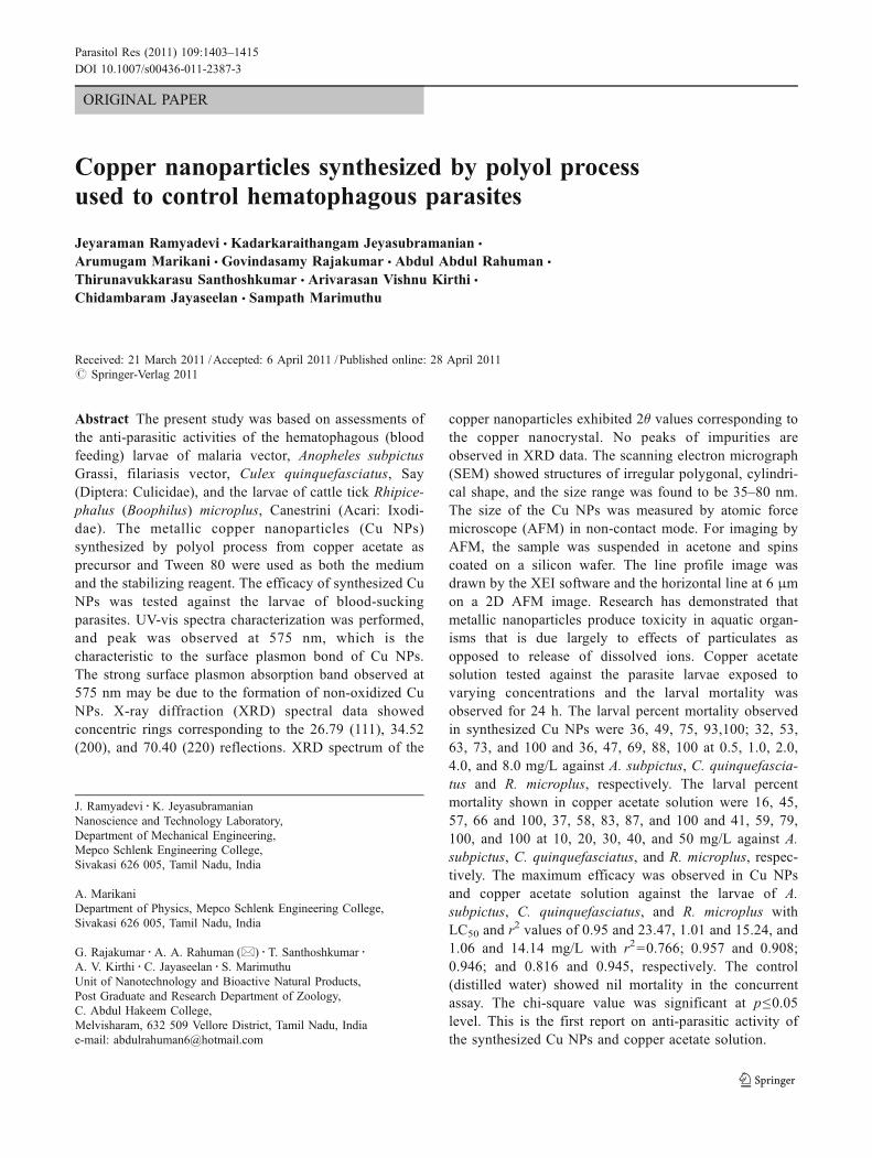
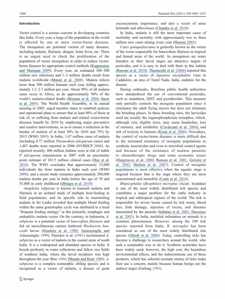
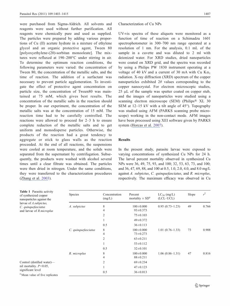
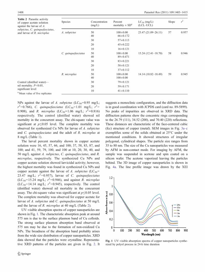
UV–visible absorption spectra of copper nanoparticles areshown in Fig. 1. The characteristic absorption peak at around575 nm is due to the surface plasmon band of Cu colloids.The strong surface plasmon absorption band observed at575 nm may be due to the formation of non-oxidized CuNPs. The broadness of the absorption band probably arisesfrom the wide size distribution of copper nanoparticles. XRDdata showed that the particles were crystalline. Representa-tive XRD patterns of the particles are given in Fig. 2. It
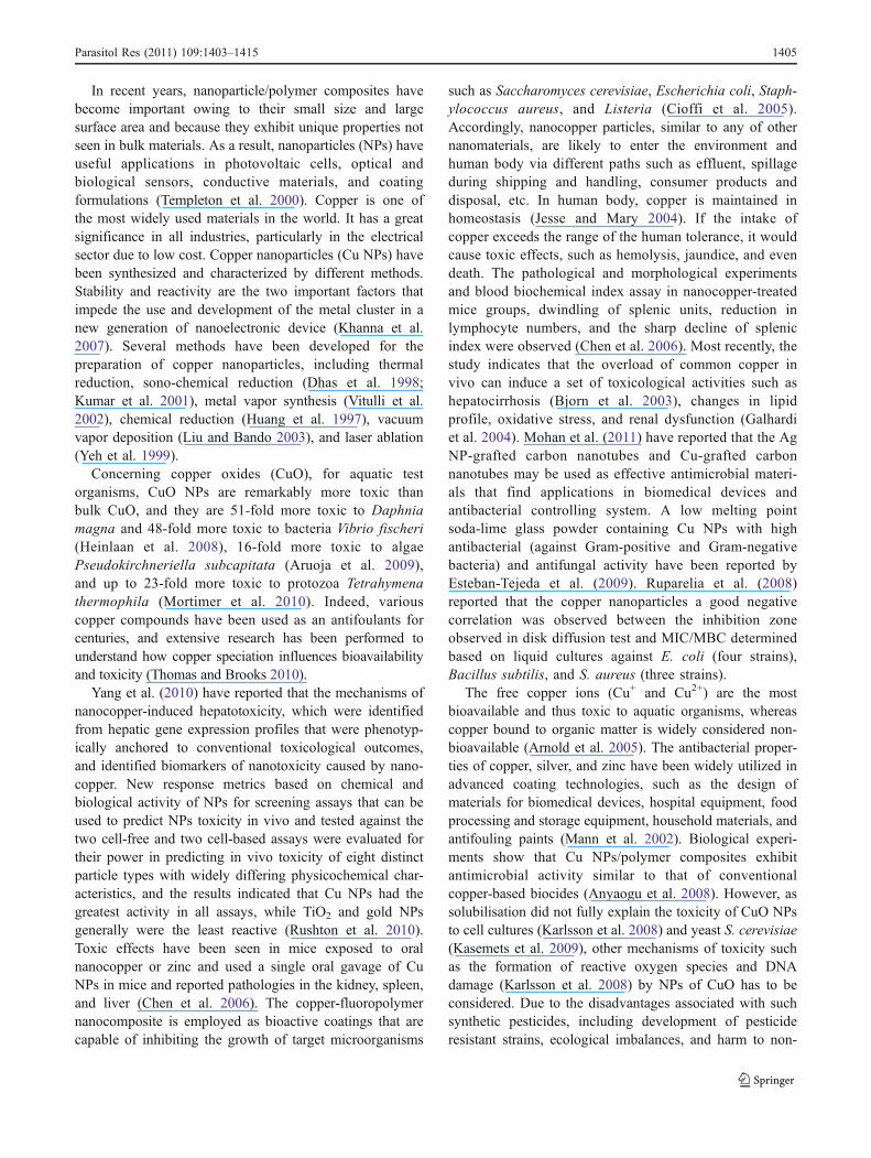
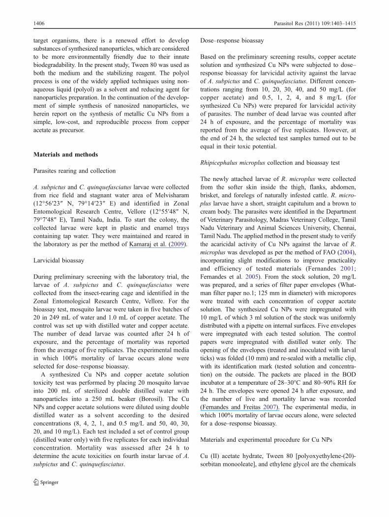
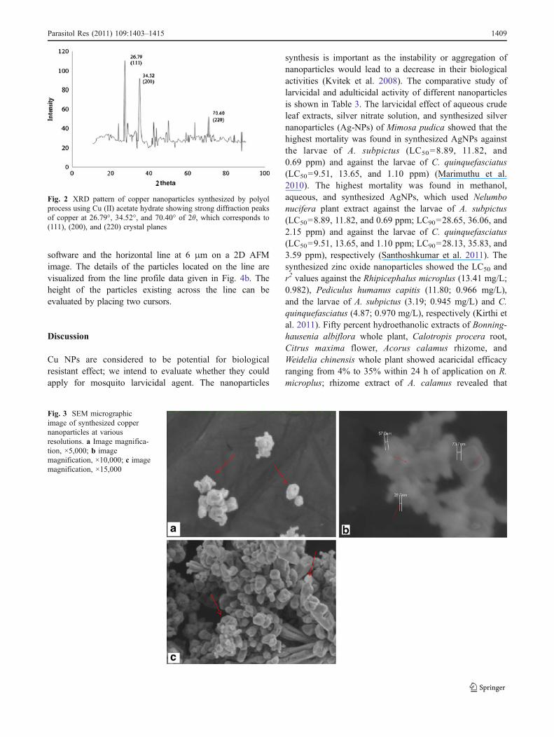
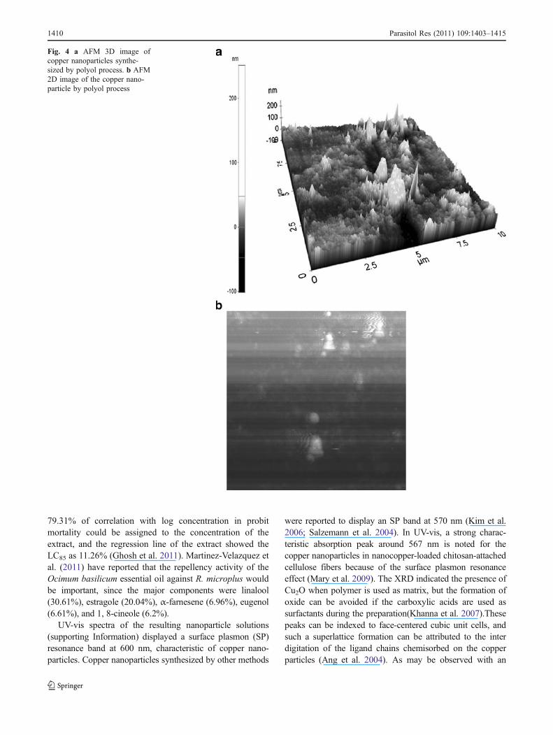
suggests a monoclinic configuration, and the diffraction datais in good coordination with JCPDS card (card no. 89-5899).No peaks of impurities are observed in XRD data. Thediffraction patterns show the concentric rings correspondingto the 26.79 (111), 34.52 (200), and 70.40 (220) reflections.These distances are characteristic of the face-centered cubic(fcc) structure of copper (metal). SEM images in Fig. 3a–cexemplifies some of the solids obtained at 25°C under thementioned conditions. It showed structures of irregularpolygonal, cylindrical shapes. The particle size ranges from35 to 80 nm. The size of the Cu nanoparticles was measuredby AFM in non-contact mode. For imaging by AFM, thesample was suspended in acetone and spin coated on asilicon wafer. The acetone vaporized leaving the particlesbehind. The 3D image of copper nanoparticles is shown inFig. 4a. The line profile image was drawn by the XEI
Fig. 1 UV–visible absorption spectra of copper nanoparticles synthe-sized by polyol process in 24-h time duration
Species Concentration(mg/L)
Percentmortality ± SDa
LC50 (mg/L)(LCL–UCL)
Slope r2
A. subpictus 50 100±0.00 23.47 (21.09–26.11) 57 0.95740 66±0.172
30 57±0.113
20 45±0.222
10 16±0.121
C. quinquefasciatus 50 100±0.00 15.24 (12.41–18.70) 58 0.94640 89±0.471
30 83±0.221
20 58±0.121
10 37±0.112
R. microplus 50 100±0.00 14.14 (10.82–18.49) 59 0.94540 100±0.00
30 79±0.121
20 59±0.171
10 41±0.110
Table 2 Parasitic activityof copper acetate solutionagainst the larvae of A.subpictus, C. quinquefasciatus,and larvae of R. microplus
Control (distilled water)—nil mortality. P<0.05,significant levelaMean value of five replicates
1408 Parasitol Res (2011) 109:1403–1415
software and the horizontal line at 6 μm on a 2D AFMimage. The details of the particles located on the line arevisualized from the line profile data given in Fig. 4b. Theheight of the particles existing across the line can beevaluated by placing two cursors.
Discussion
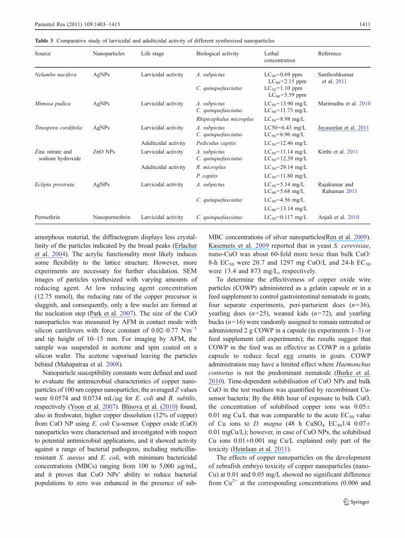
Cu NPs are considered to be potential for biologicalresistant effect; we intend to evaluate whether they couldapply for mosquito larvicidal agent. The nanoparticles
synthesis is important as the instability or aggregation ofnanoparticles would lead to a decrease in their biologicalactivities (Kvitek et al. 2008). The comparative study oflarvicidal and adulticidal activity of different nanoparticlesis shown in Table 3. The larvicidal effect of aqueous crudeleaf extracts, silver nitrate solution, and synthesized silvernanoparticles (Ag-NPs) of Mimosa pudica showed that thehighest mortality was found in synthesized AgNPs againstthe larvae of A. subpictus (LC50=8.89, 11.82, and0.69 ppm) and against the larvae of C. quinquefasciatus(LC50=9.51, 13.65, and 1.10 ppm) (Marimuthu et al.2010). The highest mortality was found in methanol,aqueous, and synthesized AgNPs, which used Nelumbonucifera plant extract against the larvae of A. subpictus(LC50=8.89, 11.82, and 0.69 ppm; LC90=28.65, 36.06, and2.15 ppm) and against the larvae of C. quinquefasciatus(LC50=9.51, 13.65, and 1.10 ppm; LC90=28.13, 35.83, and3.59 ppm), respectively (Santhoshkumar et al. 2011). Thesynthesized zinc oxide nanoparticles showed the LC50 andr2 values against the Rhipicephalus microplus (13.41 mg/L;0.982), Pediculus humanus capitis (11.80; 0.966 mg/L),and the larvae of A. subpictus (3.19; 0.945 mg/L) and C.quinquefasciatus (4.87; 0.970 mg/L), respectively (Kirthi etal. 2011). Fifty percent hydroethanolic extracts of Bonning-hausenia albiflora whole plant, Calotropis procera root,Citrus maxima flower, Acorus calamus rhizome, andWeidelia chinensis whole plant showed acaricidal efficacyranging from 4% to 35% within 24 h of application on R.microplus; rhizome extract of A. calamus revealed that
Fig. 2 XRD pattern of copper nanoparticles synthesized by polyolprocess using Cu (II) acetate hydrate showing strong diffraction peaksof copper at 26.79°, 34.52°, and 70.40° of 2θ, which corresponds to(111), (200), and (220) crystal planes
Fig. 3 SEM micrographicimage of synthesized coppernanoparticles at variousresolutions. a Image magnifica-tion, ×5,000; b imagemagnification, ×10,000; c imagemagnification, ×15,000
Parasitol Res (2011) 109:1403–1415 1409
79.31% of correlation with log concentration in probitmortality could be assigned to the concentration of theextract, and the regression line of the extract showed theLC85 as 11.26% (Ghosh et al. 2011). Martinez-Velazquez etal. (2011) have reported that the repellency activity of theOcimum basilicum essential oil against R. microplus wouldbe important, since the major components were linalool(30.61%), estragole (20.04%), α-farnesene (6.96%), eugenol(6.61%), and 1, 8-cineole (6.2%).
UV-vis spectra of the resulting nanoparticle solutions(supporting Information) displayed a surface plasmon (SP)resonance band at 600 nm, characteristic of copper nano-particles. Copper nanoparticles synthesized by other methods
were reported to display an SP band at 570 nm (Kim et al.2006; Salzemann et al. 2004). In UV-vis, a strong charac-teristic absorption peak around 567 nm is noted for thecopper nanoparticles in nanocopper-loaded chitosan-attachedcellulose fibers because of the surface plasmon resonanceeffect (Mary et al. 2009). The XRD indicated the presence ofCu2O when polymer is used as matrix, but the formation ofoxide can be avoided if the carboxylic acids are used assurfactants during the preparation(Khanna et al. 2007).Thesepeaks can be indexed to face-centered cubic unit cells, andsuch a superlattice formation can be attributed to the interdigitation of the ligand chains chemisorbed on the copperparticles (Ang et al. 2004). As may be observed with an
Fig. 4 a AFM 3D image ofcopper nanoparticles synthe-sized by polyol process. b AFM2D image of the copper nano-particle by polyol process
1410 Parasitol Res (2011) 109:1403–1415
amorphous material, the diffractogram displays less crystal-linity of the particles indicated by the broad peaks (Erlacheret al. 2004). The acrylic functionality most likely inducessome flexibility to the lattice structure. However, moreexperiments are necessary for further elucidation. SEMimages of particles synthesized with varying amounts ofreducing agent. At low reducing agent concentration(12.75 mmol), the reducing rate of the copper precursor issluggish, and consequently, only a few nuclei are formed atthe nucleation step (Park et al. 2007). The size of the CuOnanoparticles was measured by AFM in contact mode withsilicon cantilevers with force constant of 0.02–0.77 Nm−1
and tip height of 10–15 mm. For imaging by AFM, thesample was suspended in acetone and spin coated on asilicon wafer. The acetone vaporised leaving the particlesbehind (Mahapatraa et al. 2008).
Nanoparticle susceptibility constants were defined and usedto evaluate the antimicrobial characteristics of copper nano-particles of 100 nm copper nanoparticles; the averaged Z valueswere 0.0574 and 0.0734 mL/μg for E. coli and B. subtilis,respectively (Yoon et al. 2007). Blinova et al. (2010) found,also in freshwater, higher copper dissolution (12% of copper)from CuO NP using E. coli Cu-sensor. Copper oxide (CuO)nanoparticles were characterised and investigated with respectto potential antimicrobial applications, and it showed activityagainst a range of bacterial pathogens, including meticillin-resistant S. aureus and E. coli, with minimum bactericidalconcentrations (MBCs) ranging from 100 to 5,000 μg/mL,and it proves that CuO NPs’ ability to reduce bacterialpopulations to zero was enhanced in the presence of sub-
MBC concentrations of silver nanoparticles(Ren et al. 2009).Kasemets et al. 2009 reported that in yeast S. cerevisiae,nano-CuO was about 60-fold more toxic than bulk CuO:8-h EC50 were 20.7 and 1297 mg CuO/L and 24-h EC50
were 13.4 and 873 mg/L, respectively.To determine the effectiveness of copper oxide wire
particles (COWP) administered as a gelatin capsule or in afeed supplement to control gastrointestinal nematode in goats,four separate experiments, peri-parturient does (n=36),yearling does (n=25), weaned kids (n=72), and yearlingbucks (n=16) were randomly assigned to remain untreated oradministered 2 g COWP in a capsule (in experiments 1–3) orfeed supplement (all experiments); the results suggest thatCOWP in the feed was as effective as COWP in a gelatincapsule to reduce fecal egg counts in goats. COWPadministration may have a limited effect where Haemonchuscontortus is not the predominant nematode (Burke et al.2010). Time-dependent solubilisation of CuO NPs and bulkCuO in the test medium was quantified by recombinant Cu-sensor bacteria: By the 48th hour of exposure to bulk CuO,the concentration of solubilised copper ions was 0.05±0.01 mg Cu/L that was comparable to the acute EC50 valueof Cu ions to D. magna (48 h CuSO4 EC501/4 0.07±0.01 mgCu/L); however, in case of CuO NPs, the solubilisedCu ions 0.01±0.001 mg Cu/L explained only part of thetoxicity (Heinlaan et al. 2011).
The effects of copper nanoparticles on the developmentof zebrafish embryo toxicity of copper nanoparticles (nano-Cu) at 0.01 and 0.05 mg/L showed no significant differencefrom Cu2+ at the corresponding concentrations (0.006 and
Table 3 Comparative study of larvicidal and adulticidal activity of different synthesized nanoparticles
Source Nanoparticles Life stage Biological activity Lethalconcentration
Reference
Nelumbo nucifera AgNPs Larvicidal activity A. subpictus LC50=0.69 ppmLC90=2.15 ppm
Santhoshkumaret al. 2011
C. quinquefasciatus LC50=1.10 ppmLC90=3.59 ppm
Mimosa pudica AgNPs Larvicidal activity A. subpictus LC50=13.90 mg/L Marimuthu et al. 2010C. quinquefasciatus LC50=11.73 mg/L
Rhipicephalus microplus LC50=8.98 mg/L
Tinospora cordifolia AgNPs Larvicidal activity A. subpictus LC50=6.43 mg/L Jayaseelan et al. 2011C. quinquefasciatus LC50=6.96 mg/L
Adulticidal activity Pediculus capitis LC50=12.46 mg/L
Zinc nitrate andsodium hydroxide
ZnO NPs Larvicidal activity A. subpictus LC50=11.14 mg/L Kirthi et al. 2011C. quinquefasciatus LC50=12.39 mg/L
Adulticidal activity R. microplus LC50=29.14 mg/L
P. capitis LC50=11.80 mg/L
Eclipta prostrata AgNPs Larvicidal activity A. subpictus LC50=5.14 mg/L Rajakumar andRahuman 2011LC90=5.68 mg/L
C. quinquefasciatus LC50=4.56 mg/L
LC90=13.14 mg/L
Permethrin Nanopermethrin Larvicidal activity C. quinquefasciatus LC50=0.117 mg/L Anjali et al. 2010
Parasitol Res (2011) 109:1403–1415 1411
0.03 mg/L), but 0.1 mg/L nano-Cu had a greater toxicitythan 0.06 mg/L Cu2+(Bai et al. 2010). Increasing use ofmetallic nanomaterials is likely to result in the release ofthese particles into aqueous environments; however, toaddress this issue, acute toxicity of soluble copper, and80 nm nano-Cu suspensions were examined in zebrafish.The results demonstrate that nano-Cu is acutely toxic tozebrafish, with a 48 h LC50 concentration of 1.5 mg/L(Griffitt et al. 2007). Branchial uptake of ionic silver andcopper has been well documented in freshwater fish andappears to occur primarily through apical membranesodium channels and the copper transporter protein (Buryand Wood 1999; Bury et al. 1999). Toxicity of CuOnanoparticles (nano-CuO) to P. subcapitata were determinedusing the Organisation for Economic Co-operation andDevelopment (OECD) 201 algal growth inhibition testtaking in account potential shading of light, and the resultsfor no observable effect level (NOEL) for nano-CuO were0.42 mg Cu/L and for bulk CuO 8.03 mg Cu/L (Aruoja et al.2009). Marine invertebrates such as the ragworm Hedistediversicolor and the bivalve mollusc Scrobicularia plana(Byrne and Halloran 2001; Solé et al. 2009) represent goodcandidates. Soluble forms of copper are highly toxic toaquatic organisms (Eisler 2007; Luoma and Rainbow 2008).In H. diversicolor originating from highly Cu contaminatedsite, Cu granules were observed as a major sink forbioaccumulated Cu (Mouneyrac et al. 2003). Waterbornecopper exposure (0.1 mg/L) induced 77% reduction of thesodium uptake in the crustacean amphipod Gammarus pulexdue to a reduction in sodium influx by the inhibition ofNa+/K+ ATPase, primarily arising from interference with -SHgroups on Na+K+ ATPase (Brooks and Mills 2003).
Nano-copper induced in rats overt hepatotoxicity andnephrotoxicity at 200 mg kg−1 day−1 for 5 days, which mainlyinvolved scattered dot hepatocytic necrosis and widespreadrenal proximal tubule necrosis (Lei et al. 2008). Once in thegastrointestinal tract, metallic nanoparticles of copper candissolve rapidly resulting in local high concentrations ofcopper ions sufficient to disrupt copper homeostasis as seen instudies with mice orally exposed to nanocopper (Meng et al.2007). An increase of metallothionein-like protein wasobserved in bivalves exposed to Cu either as CuO NPs orsoluble Cu, catalase, glutathione-S-transferase, and superoxidedismutase have been classified as antioxidant systems ofdefense in various aquatic species such as bivalves (Almeidaet al. 2007). Brooks and Mills (2003) demonstrated thatwaterborne copper exposure (0.1 mg/L) induced 77%reduction of the sodium uptake in the crustacean amphipodGammarus pulex due to a reduction in sodium influx by theinhibition of Na+/K+ ATPase, primarily arising from inter-ference with –SH groups on Na+/K+ ATPase.
Nanoparticles of CuOwere identified as being important inecotoxicological assays due to their relatively low dissolution
rate but their potentially high toxicity towards organisms(Stone et al. 2010). Shi et al. (2011) reported that thesolubility of CuO NP varied with both pH and presence ofother ions. CuO NP and comparable doses of soluble Cuwere applied to duckweeds, Landoltia punctata. Wang et al.(2010) showed that Cu2
+ stress caused the rapid release ofH2O2 and chlorotic leaves, and it stunted root growth anddevelopment from the possible roles of reactive oxygenspecies, nitrous oxide, and metallothionein in tomato plantresponses to copper toxicity. Copper at concentrations of 30and 65 mg/L has been found to stimulate grazing activityand digestive function of Tetrahymena (Nicolau et al. 2004).Fan et al. (2011) showed that the nano-TiO2 remarkablyenhanced the toxicity of copper to D. magna by increasingthe copper bioaccumulation, and the level of metallothioneindecreased from 135 mg/g−1 wet weight to 99 mg/g−1 wetweight at a Cu2+ level of 100 mg/L−1.
References
Almeida EA, Bainy ACD, Loureiro APM, Martinez GR, Miyamoto S,Onuki J, Barbosa LF, Garcia CCM, Prado FM, Ronsein GE,Sigolo CA, Brochini CB, Martins AMG, Medeiros MHG,DiMascio P (2007) Oxidative stress in Perna perna and otherbivalves as indicators of environmental stress in the Brazilianmarine environment: antioxidants, lipid peroxidation and DNAdamage. Comp Biochem Physiol A 146:588–600
Amerasinghe PH, Amerasinghe FP (1999) Multiple host feeding infield populations of Anopheles culicifacies and Anophelessubpictus in Sri Lanka. Med Vet Entomol 13(2):124–131
Ang TP, Wee TSA, Chin WSJ (2004) Three-dimensional self-assembled monolayer (3D SAM) of n-alkanethiols on coppernanoclusters. Phys Chem B 108:11001–11010
Anjali CH, Sudheer Khan S, Margulis-Goshen K, Magdassi S,Mukherjee A, Chandrasekaran N (2010) Formulation of waterdispersible nanopermethrin for larvicidal applications. EcotoxicolEnviron Saf 73:1932–1936
Anyaogu KC, Fedorov AV, Neckers DC (2008) Synthesis, character-ization, and antifouling potential of functionalized copper nano-particles. Langmuir 24(8):4340–4346
Arnold WR, Santore RC, Cotsifas JS (2005) Predicting coppertoxicity in estuarine and marine waters using the biotic ligandmodel. Mar Pollut Bull 50:1634–1640
Aruoja V, Dubourguier HC, Kasemets K, Kahru A (2009) Toxicity ofnanoparticles of CuO, ZnO and TiO2 to microalgae Pseudo-kirchneriella subcapitata. Sci Total Environ 407(4):1461–1468
Bai W, Tian W, Zhang Z, He X, Ma Y, Liu N, Chai Z (2010) Effectsof copper nanoparticles on the development of zebrafishembryos. J Nanosci Nanotechnol 10(12):8670–8676
Bjorn PZ, Hermann HD, Max L, Heide S, Barabara KG, Hartmut D(2003) Epidemiological investigation on chronic copper toxicityto children exposed via the public drinking water supply. SciTotal Environ 302:127–144
Blinova I, Ivask A, Heinlaan M, Mortimer M, Kahru A (2010)Ecotoxicity of nanoparticles of CuO and ZnO in natural water.Environ Pollut 158:41–47
Breman JG, Martin AS, Mills A (2004) Conquering the intolerableburden of malaria: what’s new, what’s needed: a summary. Am JTrop Med Hyg 71(Suppl 2):1–15
1412 Parasitol Res (2011) 109:1403–1415
Brooks SJ, Mills CL (2003) The effect of copper on osmoregulation inthe freshwater amphipod Gammarus pulex. Comp BiochemPhysiol A Mol Integr Physiol 135(4):527–537
Burke JM, Soli F, Miller JE, Terrill TH, Wildeus S, Shaik SA, GetzWR, Vanguru M (2010) Administration of copper oxide wireparticles in a capsule or feed for gastrointestinal nematode controlin goats. Vet Parasitol 168(3–4):346–350
Bury NR, Wood CM (1999) Mechanism of branchial apical silveruptake by rainbow trout is via the proton-coupled Na(þ) channel.Am J Physiol 277:1385–1391
Bury NR, Grosell M, Grover AK, Wood CM (1999) ATP dependentsilver transport across the basolateral membrane of rainbow troutgills. Toxicol Appl Pharmacol 159:1–8
Byrne PA, Halloran JO (2001) The role of bivalve molluscs as tools inestuarine sediment toxicity testing: a review. Hydriobiologia465:209–217
Cavalcanti ESBC, de Morais SM, Lima MAA, Santana EWPS (2004)Larvicidal activity of essential oils from Brazilian against Aedesaegypti L. Mem Inst Oswaldo Cruz 99(5):541–544
Cetin H, Yanikoglu A, Cilek JE (2010) Larvicidal activity of selectedplant hydrodistillate extracts against the house mosquito, Culexpipiens, a West Nile virus vector. Parasitol Res. doi:10.1007/s00436-010-2136-z
Chandra G, Bhattacharjee I, Chatterjee S (2010) A review onAnopheles subpictus Grassi—a biological vector. Acta Trop 15(2):142–154
Chen C,MengH, Xing G, Chen C, Zhao Y, Jia G,Wang T, Yuan H, Ye C,Zhao F, Chai Z, Zhu C, Fang X, Ma B, Wan L (2006) Acutetoxicological effects of copper nanoparticles in vivo. Toxicol Lett163:109–120
Cioffi N, Ditaranto N, Torsi L, Picca RA, Sabbatini L, Valentini A,Novello L, Tantillo G, Bleve-Zacheo T, Zambonin PG (2005)Analytical characterization of bioactive fluoropolymer ultra-thincoatings modified by copper nanoparticles. Anal Bioanal Chem381:607–616
Dhanda V, Kaul HN (1980) Mosquito vectors of Japanese encephalitisvirus and their bionomics in India. Proc Indian Natl Sci Acad46B:759
Dhas NA, Raj CP, Gedanken A (1998) Synthesis, characterization,and properties of metallic copper nanoparticles. Chem Mater10:1446–1452
Dhingra N, Jha P, Sharma VP, Cohen AA, Jotkar RM, Rodriguez PS,Bassani DG, Suraweera W, Laxminarayan R, Peto R (2010)Adult and child malaria mortality in India: a nationallyrepresentative mortality survey. Lancet 376(9754):1768–1774
Ducornez S, BarréN MRJ, deGarine-Wichatisky M (2005) Diag-nosis of amitraz resistance in Boophilus microplus in NewCaledonia with modified larval packet test. Vet Parasitol130:285–292
Eisler R (2007) Eisler’s encyclopedia of environmentally hazardouspriority chemicals. Elsevier Science, Oxford
Erlacher A, Ambrico M, Capozzi V, Augelli V, Jaeger H, Ullrich B(2004) X-ray, absorption and photocurrent properties of thin-filmGaAs on glass formed by pulsed-laser deposition. Semicond SciTechnol 19:1322–1324
Esteban-Tejeda L, Malpartida F, Esteban-Cubillo A, Pecharromán C,Moya JS (2009) Antibacterial and antifungal activity of a soda-lime glass containing copper nanoparticles. Nanotechnology 20(50):505701
Fan W, Cui M, Liu H, Wang C, Shi Z, Tan C, Yang X (2011) Nano-TiO2 enhances the toxicity of copper in natural water to Daphniamagna. Environ Pollut 159:729–734
FAO (2004) Ticks: acaricide resistance: diagnosis management andprevention in: guidelines resistance management and integratedparasite control in ruminants. FAO Animal Production and HealthDivision, Rome
Fernandes FF (2001) Toxicological effects and resistance to pyretroidsin Boophilus microplus from Goiás Brasil. Arq Bras Med VetZootec 53:548–552
Fernandes FF, Freitas EPS (2007) Acaricidal activity of an oleores-inous extract from Copaifera reticulata (Leguminosae: Caesalpi-nioideae) against larvae of the southern cattle tick, Rhipicephalus(Boophilus) microplus (Acari: Ixodidae). Vet Parasitol 147:150–154
Fernandes FF, Freitas EPS, Costa AC, Silva IG (2005) Larvicidalpotential of Sapindus saponaria to control the cattle tickBoophilus microplus. Pesqui Agropecu Bras 40:1243–1245
Furlong J (1993) Controle do carrapato dos bovinos na RegiãoSudeste do Brasil. Bolm Téc.8 Escola de Veterinária daUniversidade Federal de Minas Gerais, Belo Horizonte
Galhardi CM, Diniz YS, Faine LA, Rodrigues HG, Burneiko RC,Ribas BO, Novelli EL (2004) Toxicity of copper intake: lipidprofile, oxidative stress and susceptibility to renal dysfunction.Food Chem Toxicol 42:2053–2060
Gericke A, Govere JM, Durrheim DN (2002) Insecticide susceptibilityin the South African malaria mosquito Anopheles arabiensis(Diptera: Culicidae). S Afr J Sci 98:205–208
Ghosh S, Azhahianambi P, de la Fluente J (2006) Control of ticks ofruminants with special emphasis on livestock farming system inIndia-present and future possibilities for integrated control: areview. Exp Appl Acarol 40:49–66
Ghosh S, Sharma AK, Kumar S, Tiwari SS, Rastogi S, Srivastava S,Singh M, Kumar R, Paul S, Ray DD, Rawat AK (2011) In vitroand in vivo efficacy of Acorus calamus extract againstRhipicephalus (Boophilus) microplus. Parasitol Res 108:361–370. doi:10.1007/s00436-010-2070-0
Griffitt RJ, Weil R, Hyndman KA, Denslow ND, Powers K, Taylor D,Barber DS (2007) Exposure to copper nanoparticles causes gillinjury and acute lethality in zebrafish (Danio rerio). Environ SciTechnol 41(23):8178–8186
Hargreaves K, Koekemoer LL, Brooke BD, Hunt RH, Mthembu J,Coetzee M (2000) Anopheles funestus resistant to pyrethroidinsecticides in South Africa. Med Vet Entomol 14:181–189
Hay SI, Gething PW, Snow RW (2010) India’s invisible malariaburden. Lancet 376(9754):1716–1717
Heinlaan M, Ivask A, Blinova I, Dubourguier H-C, Kahru A (2008)Toxicity of nanosized and bulk ZnO, CuO and TiO2 to bacteriaVibrio fischeri and crustaceans Daphnia magna and Thamnoce-phalus platyurus. Chemosphere 71(7):1308–1316
Heinlaan M, Kahru A, Kasemets K, Arbeille B, Prensier G,Dubourguier HC (2011) Changes in the Daphnia magna midgutupon ingestion of copper oxide nanoparticles: a transmissionelectron microscopy study. Water Res 45(1):179–190
Hoedojo PF, Atmosoedjono S, Purnomo TT (1980) A study on vectorsof Bancroftian filariasis in West Flores, Indonesia. SoutheastAsian J Trop Med Public Health 11(3):399–404
Horcas I, Fernández R, Gómez-Rodríguez JM, Colchero J, Gómez-Herrero J, Baro AM (2007) WSXM: a software for scanningprobe microscopy and a tool for nanotechnology. Re Sci Instrum78:013705
Huang HH, Yan FQ, Kek YM, Chew CH, Xu GQ, Ji W, Oh PS, TangSH (1997) Synthesis, characterization, and nonlinear opticalproperties of copper nanoparticles. Langmuir 13:172–175
Jayaseelan C, Rahuman AA, Rajakumar G, Vishnu Kirthi A,Santhoshkumar T, Marimuthu S, Bagavan A, Kamaraj C, ZahirAA, Elango G (2011) Synthesis of pediculocidal and larvicidalsilver nanoparticles by leaf extract from heartleaf moonseedplant, Tinospora cordifolia Miers. Parasitol Res. doi:10.1007/s00436-010-2242-y
Jesse B, Mary RL (2004) Maintaining copper homeostasis: regulationof copper-trafficking proteins in response to copper deficiency oroverload. J Nutr Biochem 15:316–322
Parasitol Res (2011) 109:1403–1415 1413
Kamaraj C, Bagavan A, Rahuman AA, Zahir AA, Elango G, PandiyanG (2009) Larvicidal potential of medicinal plant extracts againstAnopheles subpictus Grassi and Culex tritaeniorhynchus Giles(Diptera: Culicidae). Parasitol Res 104(5):1163–1171
Karlsson HK, Cronholm P, Gustafsson J, Moller L (2008) Copperoxide nanoparticles are highly toxic: a comparison between metaloxide nanoparticles and carbon nanotubes. Chem Res Toxicol 21(9):1726–1732
Kasemets K, Ivask A, Dubourguier H-C, Kahru A (2009) Toxicity ofnanoparticles of ZnO, CuO and TiO2 to yeast Saccharomycescerevisiae. Toxicol In Vitro 23(6):1116–1122
Khanna PK, Gaikwad S, Adhyapak PV, Singh N, Marimuthu R (2007)Synthesis and characterization of copper nanoparticles. Mat Let61:4711–4714
Kim YH, Kang YS, Lee WJ, Jo BG, Jeong JH (2006) Synthesis of Cunanoparticles prepared by using thermal decomposition of Cu-oleatecomplex. Mol Cryst Liq Cryst 445:231–238
Kiran SR, Bhavani K, Devi PS, Rao BRR, Reddy KJ (2006)Composition and larvicidal activity of leaves and stem essentialoils of Chloroxylon swietenia DC against Aedes aegypti andAnopheles stephensi. Bioresour Technol 97:2481–2484
Kirthi AV, Rahuman AA, Rajakumar G, Marimuthu S, SanthoshkumarT, Jayaseelan C, Velayutham K (2011) Acaricidal, pediculocidaland larvicidal activity of synthesized ZnO nanoparticles usingwetchemical route against blood feeding parasites. Parasitol Res.doi:10.1007/s00436-011-2277-8
Kumar RV, Mastai Y, Diamant Y, Gedanken A (2001) Sonochemicalsynthesis of amorphous Cu and nanocrystalline Cu2O embeddedin a polyaniline matrix. J Mater Chem 11:1209–1213
Kuppusamy C, Murugan K (2009) Mosquitocidal effect of Andographispaniculatenees against the malaria vector, Anopheles stephensiListon (Diptera: culicidae). Int j integr Bio 5(2):75–81
Kvitek L, Panacek A, Soukupova J, Kolar M, Vecerova R (2008) Effectof surfactant and polymers on stability and antibacterial activity ofsilver nanoparticles(NPs). J Phys Chem 112:5825–5834
Lei R, Wu C, Yang B, Ma H, Shi C, Wang Q, Wang Q, Yuan Y, LiaoM (2008) Integrated metabolomic analysis of the nano-sizedcopper particle-induced hepatotoxicity and nephrotoxicity in rats:a rapid in vivo screening method for nanotoxicity. Toxicol ApplPharmacol 232(2):292–301
Liu Z, Bando Y (2003) A novel method for preparing coppernanorods and nanowires. Adv Mater 15:303–305
Luoma S, Rainbow PS (2008) Metal contamination in aquaticenvironments: science and lateral management. CambridgeUniversity Press, Cambridge
Mahapatraa O, Bhagatb M, Gopalakrishnana C, Arunachalamb KD(2008) Ultrafine dispersed CuO nanoparticles and their antibacterialactivity. J Experi Nanosci 3(3):185–193
Mann EL, Nathan A, James WM, Sallie WC (2002) Copper toxicityand cyanobacteria ecology in the Sargasso Sea. Limnol Oceanogr47(4):976–988
Marimuthu S, Rahuman AA, Rajakumar G, Santhoshkumar T, KirthiAV, Jayaseelan C, Bagavan A, Zahir AA, Elango G, Kamaraj C(2010) Evaluation of green synthesized silver nanoparticlesagainst parasites. Parasitol Res. doi:10.1007/s00436-010-2212-4
Martinez-Velazquez M, Castillo-Herrera GA, Rosario-Cruz R, Flores-Fernandez JM, Lopez-Ramirez J, Hernandez-Gutierrez R, DelCarmen Lugo-Cervantes E (2010) Acaricidal effect and chemicalcomposition of essential oils extracted from Cuminum cyminum,Pimenta dioica and Ocimum basilicum against the cattle tickRhipicephalus (Boophilus) microplus (Acari: Ixodidae). ParasitolRes 108:481–487. doi:10.1007/s00436-010-2069-6
Mary G, Bajpai SK, Chand N (2009) Copper (II) ions and coppernanoparticles-loaded chemically modified cotton cellulosefibers with fair antibacterial properties. J Appl Polym Sci113:757–766
Meng H, Chen Z, Xing G, Yuan H, Chen C, Zhao F, Zhang C,Zhao Y (2007) Ultrahigh reactivity provokes nanotoxicity:Explanation of oral toxicity of nano-copper particles. ToxicolLett 175:102–110
Mohan R, Shanmugharaj AM, Sung Hun R (2011) An efficientgrowth of silver and copper nanoparticles on multiwalled carbonnanotube with enhanced antimicrobial activity. J Biomed MaterRes B Appl Biomater 96(1):119–126
Mortimer M, Kasemets K, Kahru A (2010) Toxicity of ZnO and CuOnanoparticles to ciliated protozoa Tetrahymena thermophila.Toxicol 269(2–3):182–189
Mouneyrac C, Mastain O, Amiard JC, Amiard-Triquet C, Beaunier P,Jeantet AY, Smith BD, Rainbow PS (2003) Trace-metal detoxifica-tion and tolerance of the estuarine worm Hediste diversicolorchronically exposed in their environment. Mar Biol 143:73–744
Muturi EJ, Burgess P, Novak RJ (2008) Malaria vector management:where have we come from and where are we headed? Am J TropMed Hyg 78:536–537
National Vector Borne Disease Control Programme (NVBDCP)(2008) Malaria, magnitude of the problem. Available at http://www.nvbdcp.gov.in/malaria3.html
Nicolau A, Mota M, Lima N (2004) Effect of different toxiccompounds on ATP content and acid phosphatase activity inaxenic cultures of Tetrahymena pyriformis. Ecotoxicol EnvironSaf 57:129–135
Panicker KN, Bai MG, Rao USB, Viswam K, Suryanarayanamurthy U(1981) An. subpictus vector of malaria in coastal villages ofSouth-East India. Curr Sci 50:694–695
Park BK, Jeong S, Kim D, Moon J, Lim S, Kim JS (2007) Synthesisand size control of monodisperse copper nanoparticles by polyolmethod. J Colloid Interface Sci 311(2):417–424
RajakumarG, RahumanAA (2011) Larvicidal activity of synthesized silvernanoparticles usingEclipta prostrata leaf extract against filariasis andmalaria vectors. Acta Trop. doi:10.1016/j.actatropica.2011.03.003
Ranson H, Rossiter L, Ortelli F, Jensen B, Wang X, Roth CW, CollinsFH, Hemingway J (2001) Identification of a novel class of insectglutathione S-transferases involved in resistance to DDT in themalaria vector, Anopheles gambie. Biochem J 359:295–304
Rao TR (1984) The anophelines of India. Malaria Research Unit(ICMR), Delhi, p 518
Rawani A, Ghosh A, Chandra G (2010) Mosquito larvicidal activitiesof Solanum nigrum L. leaf extract against Culex quinquefasciatusSay. Parasitol Res 107(5):1235–1240
Ren G, Hu D, Cheng EW, Vargas-Reus MA, Reip P, Allaker RP(2009) Characterisation of copper oxide nanoparticles for antimi-crobial applications. Int J Antimicrob Agents 33(6):587–590
Ruparelia JP, Chatterjee AK, Duttagupta SP, Mukherji S (2008) Strainspecificity in antimicrobial activity of silver and copper nano-particles. Acta Biomater 4(3):707–716
Rushton EK, Jiang J, Leonard SS, Eberly S, Castranova V, Biswas P,Elder A, Han X, Gelein R, Finkelstein J, Oberdörster G (2010)Concept of assessing nanoparticle hazards considering nano-particle dosemetric and chemical/biological response metrics. JToxicol Environ Health A 73(5):445–461
Sabatini GA, Kemp DH, Hughes S, Nari A, Hansen J (2001) Tests todetermine LC50 and discriminating doses formacrocyclic lactonesagainst the cattle tick, Boophilus microplus. Vet Parasitol 95:53–62
Salzemann C, Lisiecki I, Urban J, Pileni MP (2004) Anisotropiccopper nanocrystals synthesized in a supersaturated medium:nanocrystal growth. Langmuir 20:11772
Santhoshkumar T, Rahuman AA, Rajakumar G, Marimuthu S,Bagavan A, Jayaseelan C, Zahir AA, Elango G, Kamaraj C(2011) Synthesis of silver nanoparticles using Nelumbo nuciferaleaf extract and its larvicidal activity against malaria and filariasisvectors. Parasitol Res 108:693–702
Sharma VP (2003) Malaria and poverty in India. Curr Sci 84(4):513–515
1414 Parasitol Res (2011) 109:1403–1415
Shelton AM, Wang P, Zhao J-Z, Roush RT (2007) Resistance to insectpathogens and strategies to manage resistance: An update. In:Laceyand LA, Kaya HK (eds) Field manual of techniques ininvertebrate pathology. Springer, New York
Shi J, Abid AD, Kennedy IM, Hristovaa KR, Silk WK (2011) Toduckweeds (Landoltia punctata), nanoparticulate copper oxide ismore inhibitory than the soluble copper in the bulk solution.Environ Pollut. doi:10.1016/j.envpol.2011.01.028
Snow RW, Guerra CA, Noor AM, Myint HY, Hay SI (2005) Theglobal distribution of clinical episodes of Plasmodium falciparummalaria. Nature 434(7030):214–217
Solé M, Kopecka-Pilarczyk J, Blasco J (2009) Pollution biomarkers intwo estuarine invertebrates, Nereis diversicolor and Scrobiculariaplana, from a Marsh ecosystem in SW Spain. Environ Int35:523–531
Stone V, Nowack B, Baun A, van den Brink N, Kammer F, DusinskaM, Handy R, Hankin S, Hassellöv M, Joner E, Fernandes TF(2010) Nanomaterials for environmental studies: classification,reference material issues, and strategies for physico-chemicalcharacterisation. Sci Total Environ 408:1745–1754
Templeton AC, Wuelfing WP, Murray RW (2000) Monolayer-protected cluster molecules. Acc Chem Res 33:27
Thenmozhi V, Rajendran R, Ayanar K, Manavalan R, Tyagi BK(2006) Long-term study of Japanese encephalitis virus infectionin Anopheles subpictus in Cuddalore district, Tamilnadu, SouthIndia. Trop Med Int Health 11(3):288–293
Thomas KV, Brooks S (2010) The environmental fate and effects ofantifouling paint biocides. Biofouling 26(1):73–88
Vitulli G, Bernini M, Bertozzi S, Pitzalis E, Salvadori P, Coluccia S,Martra G (2002) Nanoscale copper particles derived fromsolvated Cu atoms in the activation of molecular oxygen. ChemMater 14:1183–1186
Wang L, Yang L, Yang F, Li X, Song Y, Wang X, Hu X (2010)Involvements of H2O2 and metallothionein in NO-mediated tomatotolerance to copper toxicity. J Plant Physiol 167(15):1298–1306
WHO (2005) World malaria report. WHO/HTM/MAL/2005. WHO,Geneva, p 1102
WHO (2008) World malaria report 2008. WHO, GenevaYang B, Wang Q, Lei R, Wu C, Shi C, Wang Q, Yuan Y, Wang Y, Luo
Y, Hu Z, Ma H, Liao M (2010) Systems toxicology used innanotoxicology: mechanistic insights into the hepatotoxicity ofnano-copper particles from toxicogenomics. J Nanosci Nano-technol 10(12):8527–8537
Yeh MS, Yang YS, Lee YP, Lee HF, Yeh YH, Yeh CS (1999) Formationand characteristics of Cu colloids from CuO powder by laserirradiation in 2-propanol. J Phys Chem B 103:6851–6857
Yoon KY, Hoon Byeon J, Park JH, Hwang J (2007) Susceptibilityconstants of Escherichia coli and Bacillus subtilis to silver andcopper nanoparticles. Sci Total Environ 373(2–3):572–575
Zhang W, Cao Q, Xie J, Ren X, Lu C, Zhou Y, Yao Y, Meng Q (2003)Structural, morphological, and magnetic study of nanocrystallinecobalt–nickel–copper particles. J Coll Inter Sci 257(2):237–243
Parasitol Res (2011) 109:1403–1415 1415