computer design of vaccines: approaches, software tools and informational resources
TRANSCRIPT
Current Computer-Aided Drug Design, 2005, 1, 207-222 207
1573-4099/05 $50.00+.00 © 2005 Bentham Science Publishers Ltd.
Computer Design of Vaccines: Approaches, Software Tools andInformational Resources
Boris N. Sobolev1,*, Ludmila V. Olenina2, Ekaterina F. Kolesanova1, Vladimir V. Poroikov1 andAlexander I. Archakov1
1Institute of Biomedical Chemistry of Rus. Acad. Med. Sci., Pogodinskaya Street, 10; Moscow, 119121, Russia2Institute of Gene Biology of Rus. Acad. Sci., Vavilov Street, Moscow, 117334, Russia
Abstract: Development of computer methods in molecular biology and fast growth of microbialgenomics data enabled new approach based on selecting in silico antigenic components to design vaccineconstructs. It is expected that application of this technology will eliminate side effects of new vaccinesand reduce the time consumption and financial expenses. The bioinformatics methods of sequenceanalysis are used to reveal the most prospective proteins or protein fragments of infectious agents ascandidates for vaccine design. In these studies the specialized molecular immunology databases arewidely used. The new approach ("Reverse vaccinology") could help in designing vaccines against diseaseswhere traditional methods are not successful, e.g. when the viral genome reveals the extreme variabilityand permanent changes of antigenic properties that make difficulties for selection of molecular targets formedicines and candidate vaccines. A number of informational resources are already designed to collectand provide genomic data on certain microbes or viruses. The peculiarity of such resources is presentationof data, characterizing the different genomic variants of the same infectious agents. These structural datacoupled with information on functional/immune features and software tools have to compose basis forconstructing a new generation of vaccines against "common" and new infections such as AIDS, HepatitisC, and SARS. The approaches published in literature, as well as the authors’ original results arediscussed.
Keywords: Vaccine design, immune response, genomics, bioinformatics, immunoinformatics, sequence analysis, epitopeprediction, informational resources.
1. INTRODUCTION: FROM TRADITIONAL VACCI-NES TO ARTIFICIAL VACCINE CONSTRUCTS
Vaccination was pioneered over 200 years ago andrapidly accepted throughout the Europe. However, itsprotective mechanisms remained unclear. New vaccinesappeared in about a century, after profound studies ofinfection processes and microorganisms started. Thedevelopment and application of effective vaccines enabled tocontrol or even eliminate several dangerous diseases. Sixvaccines against human and animal infections have beendeveloped from 1980 to 1987 [1]. Since then, the number ofvaccine-controlled infections increased with a middle rate.Currently, about 25 human diseases are controlled byvaccination. Since the beginning of the vaccination history,vaccine design technologies were significantly improved.Progress in immunology provides understanding for moreand more subtle mechanisms of the immune protection.Wide-ranging studies of biopolymers via exploitinginformational technologies enable to determine structuralpatterns of immunogenic components and design artificialconstructs, expected to display the desired protective effects[2].
Traditionally, vaccines are subdivided into live(attenuated microbe or virus cultures), killed (inactivated
*Address correspondence to this author at the Institute of BiomedicalChemistry of Rus. Acad. Med. Sci., Pogodinskaya Street, 10; Moscow,119121, Russia; Tel: +7 095 247 30 29; E-mail: [email protected]
infectious agents), and subunit ones. Vaccines of the lattertype consist of individual chemically purified components,which can invoke immune responses sufficient to protectfrom an infection and avoid undesired effects following theinoculation of the whole pathogenic agents – even if they areattenuated or killed. Meanwhile, the subunit vaccinepreparation and usage is associated with certain problems[3]:
• Long-term cultivation of pathogenic bacteria, virusesor protozoa for the industrial production ofimmunogenic components is very expensive.
• Purification and detoxification of vaccine products isalso cost consuming.
• The risk of an infectious agent leakage alwaysremains.
• Side effects cannot be excluded completely uponsubunit vaccine inoculations.
• If a virus displays a high genetic variability, it is verydifficult to isolate a chemical component, able toinvoke an effective immune response against allstrains of the virus.
Modern molecular biological techniques enable to clone,display and isolate biological macromolecules or theirfragments, which can be used as immunogenic components.Molecular constructs composed of such components form anew generation of subunit vaccines. These preparationspossess the following advantages:
208 Current Computer-Aided Drug Design, 2005, Vol. 1, No. 2 Sobolev et al.
Table 1. Some Artificial Vaccines at Different Stages of Research and Development
Infectious agent Vaccine References
Hepatitis B virus Several licensed vaccines [13, 14]
Borrelia burgdorferi(Lyme disease)
Earlier licensed LYMERix vaccine was withdrawnfrom the market. Now a modified candidate antigen is
proposed for new vaccine design[15]
Bordetella pertussis Licensed vaccine [16]
Hepatitis E virus III phase of clinical trials [17]
HIV I-III phases of clinical trials [18, 19]
Malaria plasmodium III phases of clinical trials [20]
Influenza virus Trials on animals [21-23]
Mycobacterium tuberculosis Trials on animals [24, 25]
Streptococcus pneumoniae Trials on animals, volunteers immunization with testingprotective antibodies on mice
[26, 27]
• Relatively cheap and safe production technologies. Ifa vaccine is based on short peptides – up to 30 aminoacid residues – chemical synthesis is economicallyreasonable [4]. In other cases, recombinant productscan be produced in reasonably priced expressionsystems – from bacterial and viral vectors totransgenic plants [3].
• Edible vaccines based on transgenic plants. Suchpreparations could be used as an input into themucosa (the natural gate of many infections) that hasthe perfect immune control system [1, 5].Furthermore, this way eliminates complicationsowing to injections, which became very unpopulardue to the HIV/AIDS threat.
• At last, a researcher can define candidate molecularregions before starting the vaccine development.Rapid increase of information on microbial and viralgenomes presented as symbol strings, provides theinput data for computer analysis. By using softwaretools hypothetical proteins encoded by the genomenucleotide sequences can be defined and potentialimmunogenic components in decoded amino acidsequences can be detected.
Preliminary in silico studies enable to facilitate theexperimental work and accelerate the vaccine development.The process of successive application of differenttechnologies - computing (genome data analysis),experimental analysis, pre-clinical and clinical trials forvaccine development is defined now by a new term "ReverseVaccinology" [6]. Hence, an artificial vaccine design is basedon the genome sequence analysis. Computationaltechnologies are used as a tool to design new vaccineproducts, based on a more rigorous scientific basis. Now anyresearcher can select immunogenic molecules or fragments,keeping in mind the immune mechanisms, which thedeveloping vaccine should provoke. Obviously, effectiveartificial vaccines should be developed when traditionalvaccine technologies fail. Moreover, a new approach can beapplied to develop more effective and safe vaccines thanthose already available. Today, the detection of a new
microbe or virus includes a partial or whole genomesequencing with an obligatory computational analysis. Thisfact provides a high motivation to apply the "Reversevaccinology" approach.
Genome studies can significantly accelerate vaccinedevelopment. The recent story of Severe Acute RespiratorySyndrome (SARS) is just one example of such acceleration.First cases of SARS were reported in February 2003, thougha single case of a similar disease was registered in November2002 [7]. The genome sequence of SARS virus waspublished already in May 2003 [8]. The sequence analysisenabled to reveal the most conservative regions in the aminoacid sequence and select candidate immunogenic componentsfor the vaccine design. The results of studies on theexperimental vaccine that invoked virus-neutralizingantibodies in animals were published in December 2003 [9-12]. Such rapid progress has become possible in the post-genomic era, when researchers can use computationalmethods to analyze quickly growing sequence data.
Now several vaccines obtained with genomics andbioinformatics methods are in pre-clinical or clinical trials.
It should be noted that development of vaccine facesseveral obstacles that could be overcome with the help ofanalytical methods. LYMERix vaccine designed against theLyme disease pathogen (Borrelia burgdorferi) is such anexample. The recombinant microbial outer surface protein(rOspA) of B. burgdorferi was used as the vaccine antigeniccomponent. It caused an effective protection in vaccinatedpersons. However, a part of them complained of arthritic andmuscle pain. These symptoms were more severe than thosecaused by the Borrelia natural infection, despite of thecommon opinion that artificial vaccines are speciallydesigned to avoid side effects. Most patients suffering fromside effects had HLA-DR4+ marker, which is found in one-third of the population. The firm withdrew LYMERix fromthe market. An immunogenic region responsible forautoimmune reactions was later predicted in the OspAsequence [28, 29]. Recently, Willett and co-authors reportedthat directed amino acid substitutions could eliminate therisk of autoimmune reactions. Since the mutant OspA
Computer Design of Vaccines Current Computer-Aided Drug Design, 2005, Vol. 1, No. 2 209
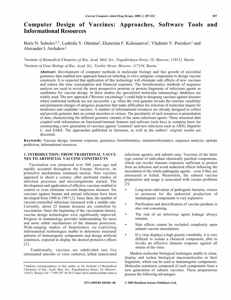
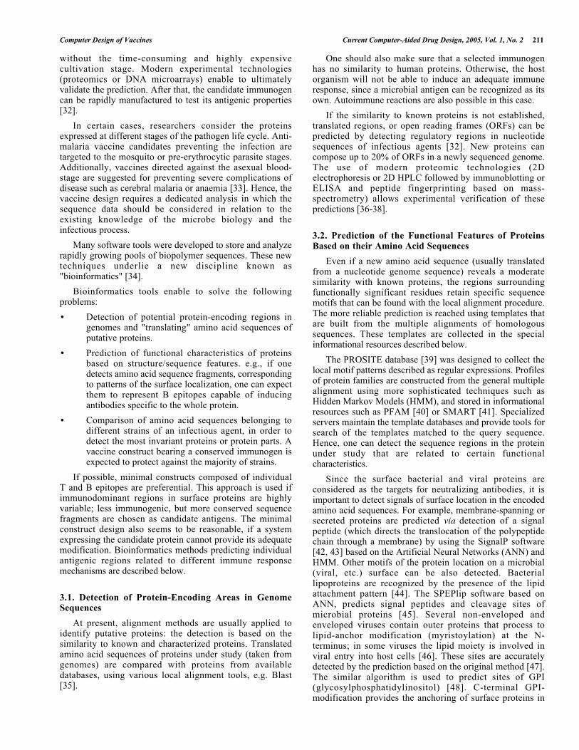
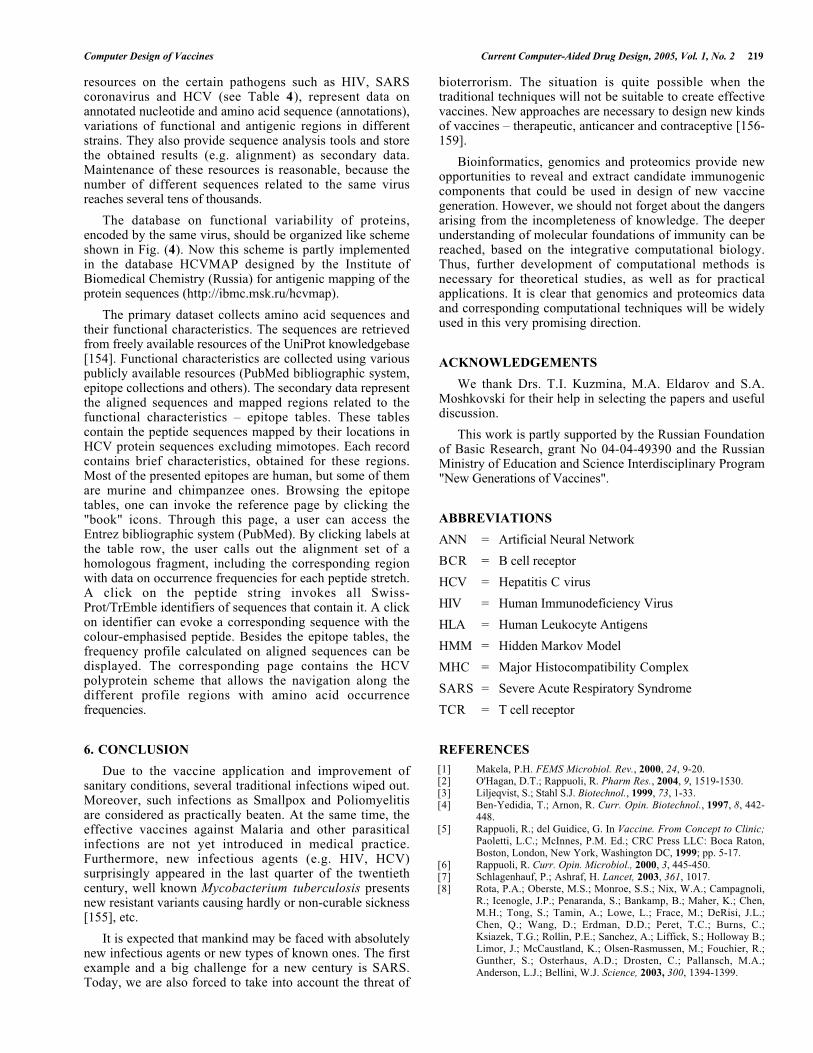
Fig. (1). Interactions of different types of lymphocytes.
a) Interaction between the cytotoxic lymphocyte (CTL) and somatic host cell, expressing foreign (e.g. viral) protein. A peptidefragment (CTL epitope) of processed protein (see text) coupled to MHC-I is recognized by lymphocyte receptor (TCR). Co-stimulatory molecule pairs involved into interaction of two cell are shown by arrows.
b) Interactions between the T-helper lymphocyte and B lymphocyte. The antigen bound by B-cell receptor (BCR) and captured byendocytosis is processed into peptide fragments (T helper epitopes) coupled to MHC-II molecules are recognized by TCRs. As a resultof this interaction, the B-lymphocyte gives rise to plasma cell clone that produce soluble immunoglobulins (antibodies) revealingthe BCR antigen-recognizing specificity.
protein retains the ability to elicit the anti-microbial immuneresponse, authors consider it as an antigenic component ofthe second generation of Lyme disease vaccine [29].
2. EFFECTOR MECHANISMS OF IMMUNERESPONSE AND VACCINE CONSTRUCTDEVELOPMENT
Prior to the discussion of computational methods used invaccine design, let us consider briefly the immunologicalconcepts that should be taken into account when a newvaccine is developed (Fig. 1).
Immune response includes the interaction of B and Tlymphocytes with an antigen or its part, resulting in aspecific recognition of definite parts of a foreign moleculecalled antigenic determinants, or epitopes. ImmunoglobulinB cell receptors (BCR) recognize surface antigen regions orB-epitopes. T cell receptors (TCR) interact with peptidefragments of a protein antigen (T epitopes) formed in thehost cell by limited proteolysis. These peptides are presentedat the host cell surface bound to the MajorHistocompatibility Complex (MHC). Human MHC proteinsare designated as Human Leukocyte Antigens (HLA). MHCclass I molecules, expressed in all nuclear cells, bindfragments of proteins synthesized in the host cell, includingintracellular parasites (viruses, rickettsiae, some bacteria)
proteins, and cleaved in proteasomes. MHC class IImolecules, expressed by several types of cells (Blymphocytes and plasma cells, macrophages, dendritic cellsand some others), bind fragments of antigens that arecaptured by these cells via endocytosis and processed inendolysosomes. Each MHC class is encoded by a group ofmultiallele loci (genes). MHC polymorphism should betaken into account during the T epitope identification, aswell as vaccine design.
TCRs bind epitopes associated with MHC proteins;hence the recognition of a protein fragment as a T epitope isdefined both by TCR and MHC specificities. When a Tlymphocyte interacts with a host cell that presents anantigen, the lymphocyte stimulation results in two possibleevents depending on the T cell type. Cytotoxic Tlymphocytes (CTL) interact with peptides bound to MHC-I,initiating the complex reaction cascade, resulting in thedeath of cells presenting the respective T epitopes. T-helperlymphocytes (Th lymphocytes) start to proliferate after theinteraction with MHC-II-bound peptides and give rise tonew cell clones. Th cells are divided into subgroupsdiscriminated with produced cytokines. It is considered thatTh1 subtype cells participate in the stimulation of cytotoxicresponses, and Th2 cells stimulate the antibody productionvia activating B and plasma cells. Th stimulation of theantibody production is achieved by the interaction of Th
210 Current Computer-Aided Drug Design, 2005, Vol. 1, No. 2 Sobolev et al.
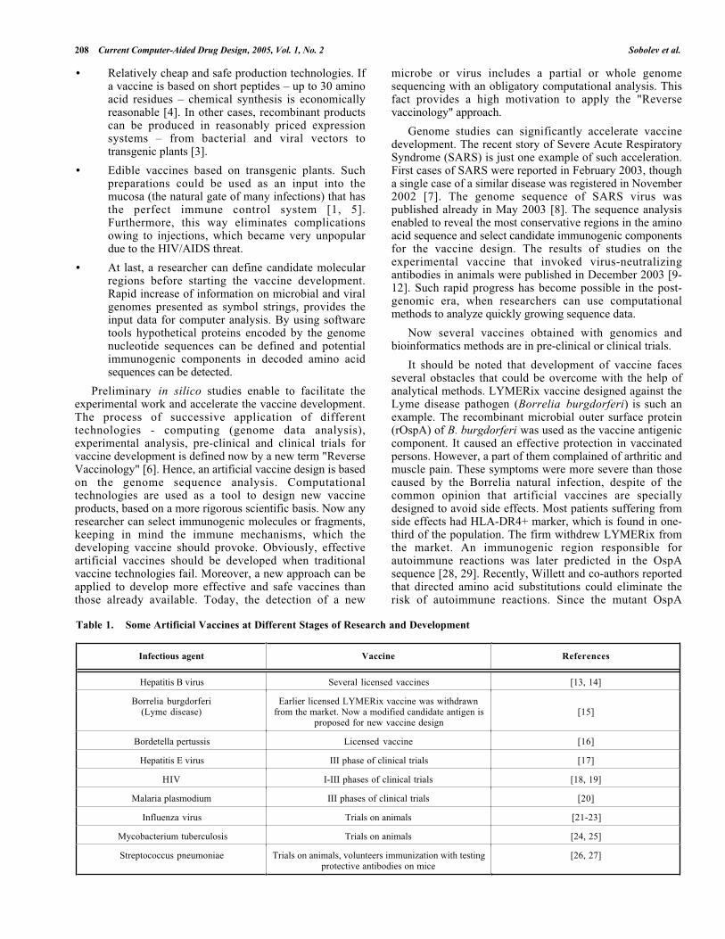
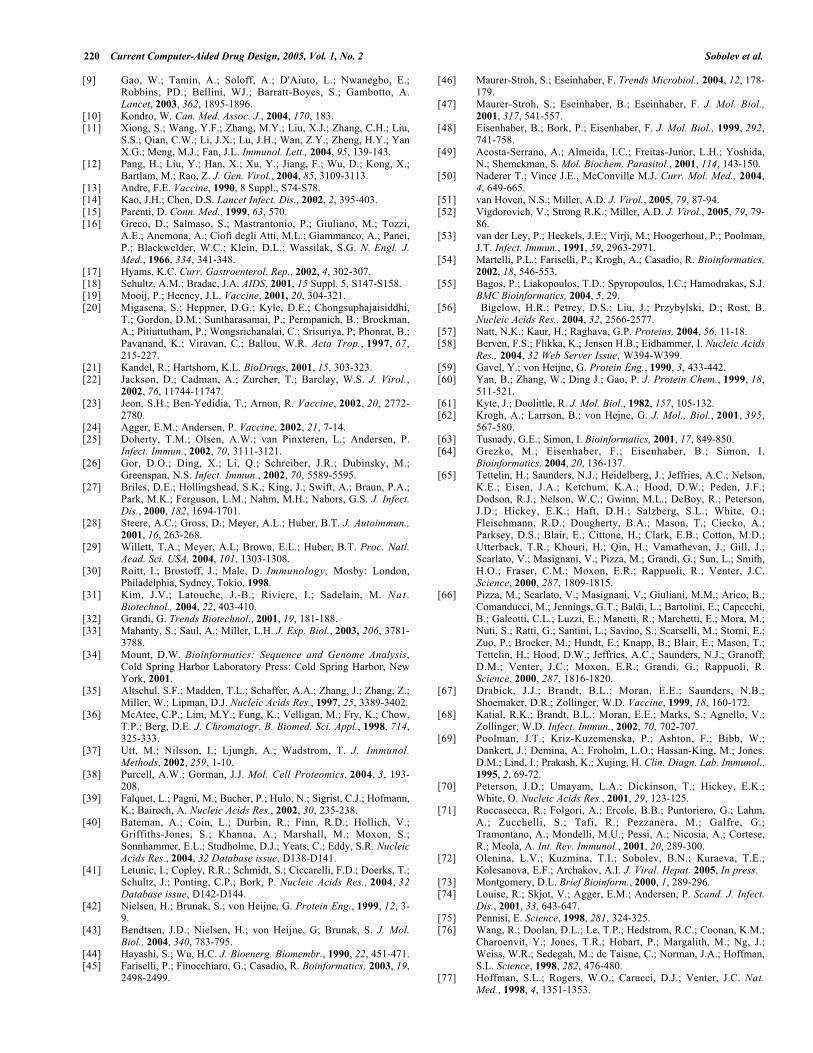
Fig. (2). From genome to vaccine. Units related to computational studies enclosed by double line.
lymphocytes with antigen-presenting B cells. TCR of Thcell recognizes the Th epitope bound by MHC-II at B cellsurface. Some other ligand–receptor pairs also participate inthe interaction between the B and T cells. This processresults in mutual stimulation to proliferation of theinteracting cells. A portion of the proliferating B cells givesrise to plasma cells producing antibodies; these freeimmunoglobulin (Ig) molecules have the same recognitionspecificity as the ancestor B cell receptor.
The portion of B and T lymphocytes stimulated duringthe primary immune response proliferate into memory cells,which provide a faster secondary immune response to theknown antigen. Properly speaking, the vaccination procedureis aimed at producing sufficient pools of memory cells ableto raise secondary T- and B-cell-based immune responses tothe infection. For more details, see the perfect monograph byRoitt and co-authors [30].
Before starting development of the vaccine, one shoulddefine the effector immune mechanisms, which are the mostefficient against the respective infection. Various vaccineconstructs differ in the stimulation of individual immuneprocesses. Free or coupled to biopolymer carriers syntheticpeptides and isolated recombinant proteins usually stimulatethe antibody production. In order to induce cytotoxic
reactions, other vehicles such as viral vectors, plasmids, orpeptide-loaded dendritic cells should be used. Newtechnologies using artificial antigen-presenting cells andtheir cell-free substitutes are expected to have a major impacton investigation of T-cell immunity as well asimmunotherapy [31].
It is necessary to keep in mind that a way of a vaccineadministration defines the type of antibodies produced inresponse to the vaccination. If an infection gate is mucosa,IgA antibodies dominate in protective immune reactions.Hence, vaccines against such infections should be aimed atraising IgA-producing cells [5].
3. GENOMICS STUDIES AND BIOINFORMATICSAPPLICATION TO THE DEVELOPMENT OFVACCINE CONSTRUCTS
Fast development of sequencing techniques enables todecode genomes of human, animals, plants as well asbacteria, viruses and protozoa parasites. By analyzing agenome sequence of an infectious agent, one can detectencoded proteins and use them in a vaccine design (Fig. 2).Thus, protein immunogenic components can be predicted
Computer Design of Vaccines Current Computer-Aided Drug Design, 2005, Vol. 1, No. 2 211
without the time-consuming and highly expensivecultivation stage. Modern experimental technologies(proteomics or DNA microarrays) enable to ultimatelyvalidate the prediction. After that, the candidate immunogencan be rapidly manufactured to test its antigenic properties[32].
In certain cases, researchers consider the proteinsexpressed at different stages of the pathogen life cycle. Anti-malaria vaccine candidates preventing the infection aretargeted to the mosquito or pre-erythrocytic parasite stages.Additionally, vaccines directed against the asexual blood-stage are suggested for preventing severe complications ofdisease such as cerebral malaria or anaemia [33]. Hence, thevaccine design requires a dedicated analysis in which thesequence data should be considered in relation to theexisting knowledge of the microbe biology and theinfectious process.
Many software tools were developed to store and analyzerapidly growing pools of biopolymer sequences. These newtechniques underlie a new discipline known as"bioinformatics" [34].
Bioinformatics tools enable to solve the followingproblems:
• Detection of potential protein-encoding regions ingenomes and "translating" amino acid sequences ofputative proteins.
• Prediction of functional characteristics of proteinsbased on structure/sequence features. e.g., if onedetects amino acid sequence fragments, correspondingto patterns of the surface localization, one can expectthem to represent B epitopes capable of inducingantibodies specific to the whole protein.
• Comparison of amino acid sequences belonging todifferent strains of an infectious agent, in order todetect the most invariant proteins or protein parts. Avaccine construct bearing a conserved immunogen isexpected to protect against the majority of strains.
If possible, minimal constructs composed of individualT and B epitopes are preferential. This approach is used ifimmunodominant regions in surface proteins are highlyvariable; less immunogenic, but more conserved sequencefragments are chosen as candidate antigens. The minimalconstruct design also seems to be reasonable, if a systemexpressing the candidate protein cannot provide its adequatemodification. Bioinformatics methods predicting individualantigenic regions related to different immune responsemechanisms are described below.
3.1. Detection of Protein-Encoding Areas in GenomeSequences
At present, alignment methods are usually applied toidentify putative proteins: the detection is based on thesimilarity to known and characterized proteins. Translatedamino acid sequences of proteins under study (taken fromgenomes) are compared with proteins from availabledatabases, using various local alignment tools, e.g. Blast[35].
One should also make sure that a selected immunogenhas no similarity to human proteins. Otherwise, the hostorganism will not be able to induce an adequate immuneresponse, since a microbial antigen can be recognized as itsown. Autoimmune reactions are also possible in this case.
If the similarity to known proteins is not established,translated regions, or open reading frames (ORFs) can bepredicted by detecting regulatory regions in nucleotidesequences of infectious agents [32]. New proteins cancompose up to 20% of ORFs in a newly sequenced genome.The use of modern proteomic technologies (2Delectrophoresis or 2D HPLC followed by immunoblotting orELISA and peptide fingerprinting based on mass-spectrometry) allows experimental verification of thesepredictions [36-38].
3.2. Prediction of the Functional Features of ProteinsBased on their Amino Acid Sequences
Even if a new amino acid sequence (usually translatedfrom a nucleotide genome sequence) reveals a moderatesimilarity with known proteins, the regions surroundingfunctionally significant residues retain specific sequencemotifs that can be found with the local alignment procedure.The more reliable prediction is reached using templates thatare built from the multiple alignments of homologoussequences. These templates are collected in the specialinformational resources described below.
The PROSITE database [39] was designed to collect thelocal motif patterns described as regular expressions. Profilesof protein families are constructed from the general multiplealignment using more sophisticated techniques such asHidden Markov Models (HMM), and stored in informationalresources such as PFAM [40] or SMART [41]. Specializedservers maintain the template databases and provide tools forsearch of the templates matched to the query sequence.Hence, one can detect the sequence regions in the proteinunder study that are related to certain functionalcharacteristics.
Since the surface bacterial and viral proteins areconsidered as the targets for neutralizing antibodies, it isimportant to detect signals of surface location in the encodedamino acid sequences. For example, membrane-spanning orsecreted proteins are predicted via detection of a signalpeptide (which directs the translocation of the polypeptidechain through a membrane) by using the SignalP software[42, 43] based on the Artificial Neural Networks (ANN) andHMM. Other motifs of the protein location on a microbial(viral, etc.) surface can be also detected. Bacteriallipoproteins are recognized by the presence of the lipidattachment pattern [44]. The SPEPlip software based onANN, predicts signal peptides and cleavage sites ofmicrobial proteins [45]. Several non-enveloped andenveloped viruses contain outer proteins that process tolipid-anchor modification (myristoylation) at the N-terminus; in some viruses the lipid moiety is involved inviral entry into host cells [46]. These sites are accuratelydetected by the prediction based on the original method [47].The similar algorithm is used to predict sites of GPI(glycosylphosphatidylinositol) [48]. C-terminal GPI-modification provides the anchoring of surface proteins in
212 Current Computer-Aided Drug Design, 2005, Vol. 1, No. 2 Sobolev et al.
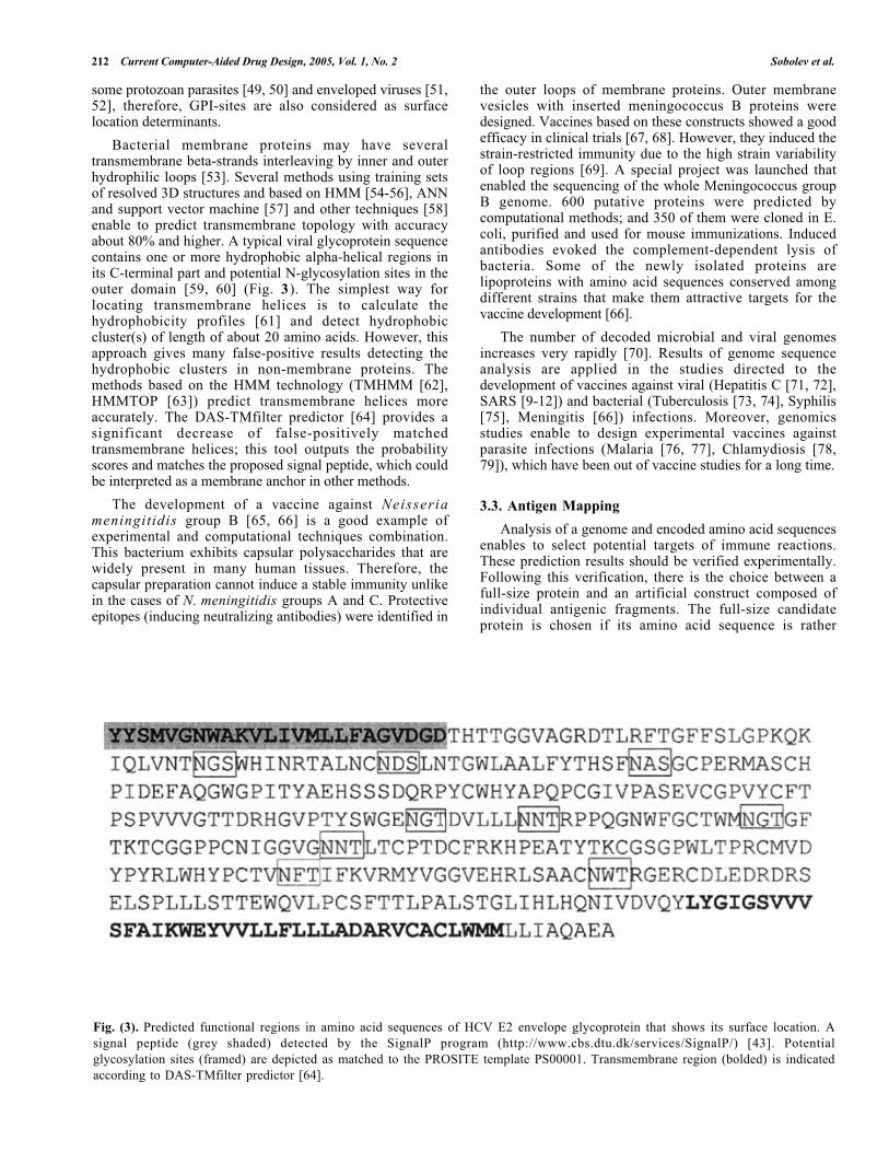
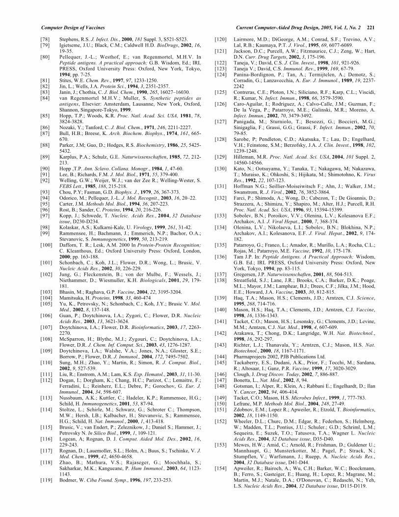
Fig. (3). Predicted functional regions in amino acid sequences of HCV E2 envelope glycoprotein that shows its surface location. Asignal peptide (grey shaded) detected by the SignalP program (http://www.cbs.dtu.dk/services/SignalP/) [43]. Potentialglycosylation sites (framed) are depicted as matched to the PROSITE template PS00001. Transmembrane region (bolded) is indicatedaccording to DAS-TMfilter predictor [64].
some protozoan parasites [49, 50] and enveloped viruses [51,52], therefore, GPI-sites are also considered as surfacelocation determinants.
Bacterial membrane proteins may have severaltransmembrane beta-strands interleaving by inner and outerhydrophilic loops [53]. Several methods using training setsof resolved 3D structures and based on HMM [54-56], ANNand support vector machine [57] and other techniques [58]enable to predict transmembrane topology with accuracyabout 80% and higher. A typical viral glycoprotein sequencecontains one or more hydrophobic alpha-helical regions inits C-terminal part and potential N-glycosylation sites in theouter domain [59, 60] (Fig. 3). The simplest way forlocating transmembrane helices is to calculate thehydrophobicity profiles [61] and detect hydrophobiccluster(s) of length of about 20 amino acids. However, thisapproach gives many false-positive results detecting thehydrophobic clusters in non-membrane proteins. Themethods based on the HMM technology (TMHMM [62],HMMTOP [63]) predict transmembrane helices moreaccurately. The DAS-TMfilter predictor [64] provides asignificant decrease of false-positively matchedtransmembrane helices; this tool outputs the probabilityscores and matches the proposed signal peptide, which couldbe interpreted as a membrane anchor in other methods.
The development of a vaccine against Neisseriameningitidis group B [65, 66] is a good example ofexperimental and computational techniques combination.This bacterium exhibits capsular polysaccharides that arewidely present in many human tissues. Therefore, thecapsular preparation cannot induce a stable immunity unlikein the cases of N. meningitidis groups A and C. Protectiveepitopes (inducing neutralizing antibodies) were identified in
the outer loops of membrane proteins. Outer membranevesicles with inserted meningococcus B proteins weredesigned. Vaccines based on these constructs showed a goodefficacy in clinical trials [67, 68]. However, they induced thestrain-restricted immunity due to the high strain variabilityof loop regions [69]. A special project was launched thatenabled the sequencing of the whole Meningococcus groupB genome. 600 putative proteins were predicted bycomputational methods; and 350 of them were cloned in E.coli, purified and used for mouse immunizations. Inducedantibodies evoked the complement-dependent lysis ofbacteria. Some of the newly isolated proteins arelipoproteins with amino acid sequences conserved amongdifferent strains that make them attractive targets for thevaccine development [66].
The number of decoded microbial and viral genomesincreases very rapidly [70]. Results of genome sequenceanalysis are applied in the studies directed to thedevelopment of vaccines against viral (Hepatitis C [71, 72],SARS [9-12]) and bacterial (Tuberculosis [73, 74], Syphilis[75], Meningitis [66]) infections. Moreover, genomicsstudies enable to design experimental vaccines againstparasite infections (Malaria [76, 77], Chlamydiosis [78,79]), which have been out of vaccine studies for a long time.
3.3. Antigen Mapping
Analysis of a genome and encoded amino acid sequencesenables to select potential targets of immune reactions.These prediction results should be verified experimentally.Following this verification, there is the choice between afull-size protein and an artificial construct composed ofindividual antigenic fragments. The full-size candidateprotein is chosen if its amino acid sequence is rather
Computer Design of Vaccines Current Computer-Aided Drug Design, 2005, Vol. 1, No. 2 213
conserved among various strains of an infectious agent andthe expression system for this protein provides its correctmodification similar to that in the natural host cells [3]. Thecosts for the cell culture maintenance and protein purificationare also taken into account. In the other case, small peptidesynthesis can be preferable. Sometimes, production of arecombinant chimeric protein with insertions of conservedimmunogenic regions, represents a successful and cost-saving compromise between the above-mentionedapproaches.
Even if the first approach seems to be successful, thedetailed antigen mapping is necessary for the exactlocalization of antigenic determinants, estimation of theirvariability/conservativity, studies of individual immuneresponse variations, and testing the possibility ofautoimmune reactions. Antigenic mapping is an obligatorystep if a synthetic peptide or a chimeric recombinant vaccineis to be designed. In order to induce antibody response to apathogen, a vaccine construct should contain B epitopes ableto provoke the synthesis of pathogen-neutralizing antibodies,and Th epitopes recognizable by Th2 lymphocytes that willstimulate the B cell conversion into antibody-producingplasma and memory cells. If the cytotoxic immune responseagainst a pathogen is a target, the vaccine construct shouldinclude cytotoxic as well as Th1 cell-stimulating T epitopes.The approaches and software for B and T epitope predictionsare described below.
3.3.1. B Epitope Prediction
In general, B epitopes represent chemical structures ableto interact with antigenic-binding regions of antibody orBCR. Any chemical structure of an infectious agent that isrecognized as a foreign (polypeptide, glycan, lipid,glycolipid etc.) by the host B cells, in principle, is able tocause an antibody-dependent immune response against itself.However, in this paper we will consider only polypeptide Bepitopes, for three principal reasons. First, these epitopesrepresent more diverse pool than others. Second, theirrecognition as non-selves is sometimes not as obvious asthat of non-peptidic B-epitopes (see the above mentionedexample of N. meningitidis group B antigen search). Third,a huge set of bioinformatics methods exists that shouldhelp, and really does in certain cases, reveal B epitopes inproteins.
B epitopes are formally classified into two groups:linear, or continuous, and conformational, or discontinuous.The former term corresponds to B epitopes that arecomposed of continuous stretches of amino acid residuesrepresenting fragments of protein primary structures. Thelatter term corresponds to B epitope structures that areformed with amino acid residues or their groups not adjacentto one another in the protein primary structure, but put inthe vicinity after the polypeptide chain folding [80]. Thisclassification is somewhat ambiguous since the recognitionand presentation of the so-called linear, or continuous Bepitopes may also depend on the protein conformation.Experimental results show that the majority of protein Bepitopes is conformational. Linear B epitopes compose aportion of about 10% of all protein B epitopes usuallydetected.
Taking into account sizes of contact sites betweenimmunoglobulin molecules and antigens, B epitope surfaceshould be coincided to stretch to 4-8 amino acid residues[81]. This so-called contact B epitope makes the mainthermodynamic contribution into the antigen-antibodyinteraction [82]. Another point of view takes into accountthe distance between the paratope and epitope atoms withoutregard for thermodynamic calculations. The B epitopestructure defined by this way extends up to 22 amino acidresidues [83]. Both short and long B epitopes can be presentwith either linear amino acid residue stretches or groups ofresidues assembled together upon protein folding.Nevertheless, it is obvious that only a part of atomic groupsof a 22-residue stretch forms the contact B epitope.
While long B epitopes as well as conformational ones (ofany length) can be assumed only from 3D structure data,short linear B epitopes can be predicted from amino acidsequences. These predictions are based on the fact thatbecause of their physicochemical properties, different aminoacid residues are differently distributed between the surfaceand the interior of the protein globule, as well as betweenvarious secondary structure elements. A certain index can beassigned to each amino acid residue, depending on itsphysicochemical properties or occurrence frequencies incertain structures. Indices are retrieved from special scalescontaining index values for all 20 natural amino acids,averaged for small overlapping stretches of the amino acidsequence of the protein under study, and plotted along thesequence [84]. Some scales used for B epitope predictionsare listed below:
• Hydrophobicity / hydrophilicity scales are the mostcommon. Scale values are obtained from experimentaldata and represent: free energy of transfer from waterto ethanol [85] or from an organic solvent to water[86]; surface tension of amino acid solutions [87];retention time of HPLC obtained for peptides ofdifferent composition [88].
• Flexibility scale is calculated from temperature factorvalues obtained for various regions in proteinstructures resolved by X-ray crystallography. The useof this scale is based on the suggestion thatflexibility of protein molecule portions correlateswith their antigenicity [89].
• Acrophilicity [90] scale is calculated from thedistribution of residues standing away from protein3D-structure core and the accessibility scale [91] alsorepresents the degree of surface exposures of aminoacid residues in resolved X-ray protein structures.
• Antigenicity scale [92] is based on statisticallycalculated occurrence frequencies of amino acidresidues in linear epitopes experimentally detected in20 proteins.
Secondary structure prediction is also applied for Bepitope predictions. These methods are usually based onstatistics of amino acid residue occurrences in certainsecondary structure elements in solved 3D protein structures[93]. Sequence regions predicted as turns or loops of apolypeptide chain are considered as possible antigenicdeterminants.
214 Current Computer-Aided Drug Design, 2005, Vol. 1, No. 2 Sobolev et al.
Though these techniques were developed more thantwenty years ago, the main principles of the antigenicityprediction by using the scales still remain unchanged. Ingeneral, all above-mentioned methods have been designed topredict surface-located regions of the protein molecule. Eventhe most accurate ones reveal about 65% residues that arereally found in antigenic regions. However, all modernsoftware for the B epitope predictions uses differentcombinations of the above mentioned scales that enable toincrease the predictive power [94]. Nevertheless, theseprogram tools still remain popular up to now since theyallow a researcher to exclude regions with low prognosticscores from the following pre-experimental B epitope search[95].
More sophisticated method, which processes sequencealignment data with the help of ANN technology, enables topredict B epitopes stretches with average accuracy of 68 %.The latter value comparable with those obtained with thescale methods seems to provide an effective practical upperlimit for the accuracy of predicting accessibility from asequence only [96].
The most precise way is to locate loop regions in amodel of 3D structure of a protein. Unfortunately, currentlythe number of solved 3D structures is much less than thenumber of determined amino acid sequences. However, onecan build a 3D model if a sequence under study reveals highhomology with one or several proteins with known 3Dstructures [97]. The sequence of a protein under study isfolded into a 3D model by using the known 3D structures astemplates. This approach was applied to the detection ofantigenic determinants in the envelope protein of JapaneseEncephalitis Virus; the homologous protein of Tick-bornEncephalitis Virus was used as the 3D template. Minimalenergy calculations for the revealed fragments showed that avaccine construct should maintain the native conformation ofan inserted epitope and induce antibodies against the wholeprotein from the native source [98].
3.3.2. T Epitope Prediction
T epitopes are fragments of proteins formed in host cellsby limited proteolysis in proteasomes or endosomes,exposed at the cell surface bound to MHC proteins, leadingto T lymphocyte activation via the interaction with TCR.
According to the crystallographic data, peptide ligandsadopt extended conformations inside the MHC cavity andare associated with MHC by hydrogen bonds and van derWaals interactions. Docking of a peptide and MHC ispossible, if the peptide sequence satisfies to specificpositional motifs. T epitope motif is composed of sequentialpositions, each of them being specified in terms of aminoacid type preferences. So-called anchor positions are themost restricted with regard to their amino acid compositionand thus, are the most significant for MHC binding [99]. Tepitope prediction methods are generally aimed at searchingthe motifs specific to MHC binding. In order to predict Tepitopes, one should detect the cleavage sites in a processedprotein and locate anchor amino acid positions in these cutstretches. The positional pattern methods were developed atfirst.
Structural features of MHC-I- and MHC-II–bindingcavities differ significantly. The MHC-I binding pocket
holds 8-11-mer peptides and is closed at both ends; the moststable hydrogen bonds are formed with amino acid residuesclose to the ligand peptide termini. A common MHC-Iligand (that is recognized by cytotoxic CD8+ lymphocytes)is a 9-mer peptide with two anchor residues in the secondand ninth positions. The MHC-II cavity is opened at bothends and accommodates longer peptides, which can extendthe cavity; anchor positions are located along the pocket[100]. Peptide ligands for MHC-II may vary in length from12 to 25 residues, however, a typical MHC-II binding motifcorresponds to a 12-15-mer peptide with two or four anchorresidues depending on MHC–II allele pocket.
Each MHC locus or even locus allele has its specificpeptide ligand motif, which represents the local differencesin structure of the binding cavity including anchor residueallocation. These motifs can be calculated based on analysisof a vast amount of both cytotoxic and helper T epitopemapping data. Experimentally found sequences of peptidesbound by MHC (MHC ligands) are stored in specializedfreely available databases such as SYFPEITHI [99] andFIMM [101]. The corresponding servers provide tools forsearching T epitope motifs in a query amino acid sequencebased on the position-dependent matrices, which containscores for 20 amino acid residues for each peptide ligandposition. EPIPREDICT software uses matrices calculatedfrom data obtained with the help of synthetic combinatorialpeptide libraries; it enables to describe the MHC-peptideinteractions in quantitative manner and predict peptidesspecific to the certain HLA-II alleles [102].
The other methods that used the ANN [103] and HMMtechnology [104], are also applied for MHC-ligandprediction.
The accuracy of these methods depends on the underlineddata set quality. Yu and co-authors [105] carried out trials ofprediction tools using positional matrices, ANN and HMM.They used datasets composed from experimentallyestablished peptides that bind or do not bind to allele-specific MHCs. Integrative accuracy scores were calculated,accounting the sensitivity and specificity values accessed bythe leave-one-out cross-validation. All used tools revealedthe good predictive performance for HLA-A*2001, and poorperformance (close to random choice) for HLA-B*3501. TheANN and HMM methods revealed the significantimprovement of accuracy with increase of the data sets.
Another method implements the partial least squares-based, multivariate, statistical approach to the quantitativeprediction of peptide binding to MHC-I [106] and MHC–II[107]. The authors used data collected in JenPep database[108] that contains experimental quantitative data on theMHC-peptide binding. The suggested approach showed thebest results compared to four other methods revealing 24from 25 known T-cell epitopes specified to MHC-II at thebenchmark. The same authors applied their approach todesign successfully series of high-affinity HLA-A2 peptides[109].
So, methods based on the sequence and functional dataare limited to a small number of MHC alleles. However,this number still increases, and earlier defined motifs arecorrected due to the continuous work on T epitope mappingusing new high-throughput experimental techniques:
Computer Design of Vaccines Current Computer-Aided Drug Design, 2005, Vol. 1, No. 2 215
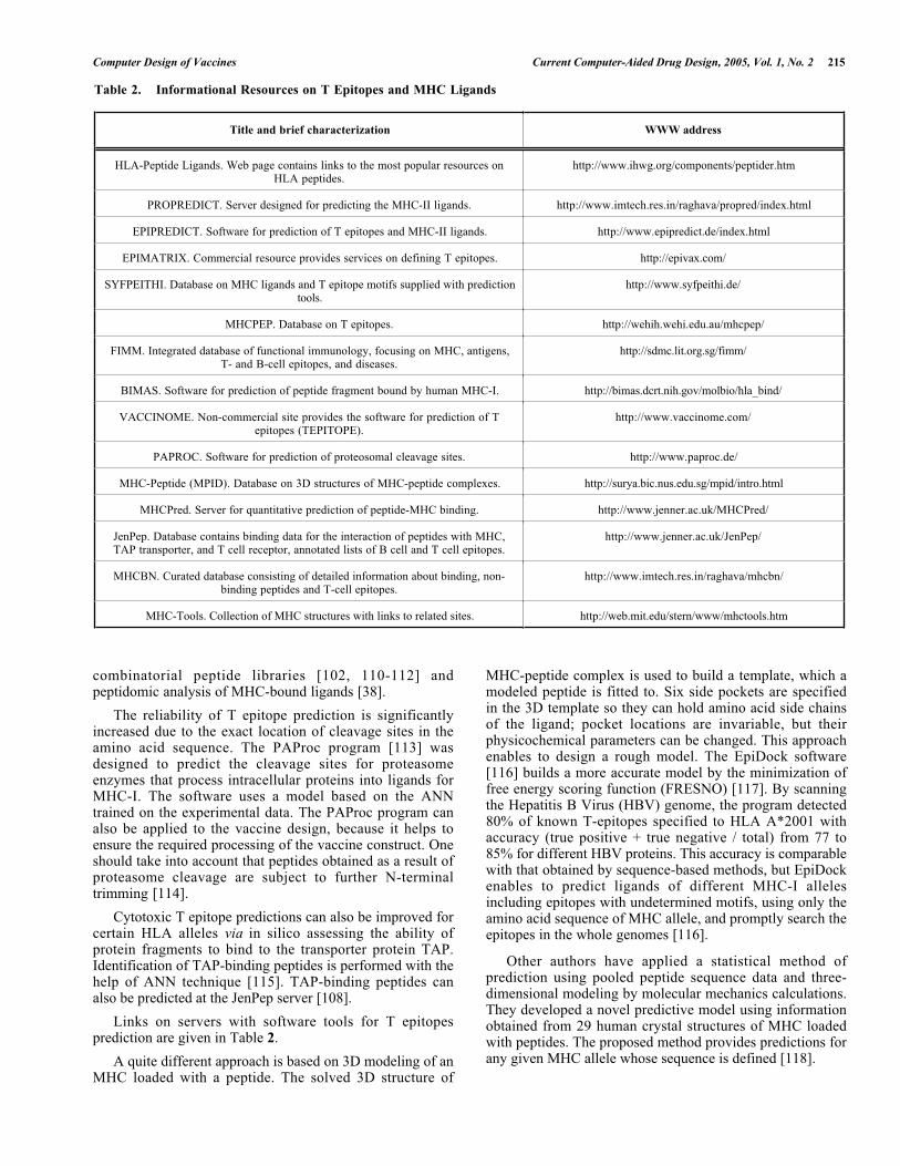
Table 2. Informational Resources on T Epitopes and MHC Ligands
Title and brief characterization WWW address
HLA-Peptide Ligands. Web page contains links to the most popular resources onHLA peptides.
http://www.ihwg.org/components/peptider.htm
PROPREDICT. Server designed for predicting the MHC-II ligands. http://www.imtech.res.in/raghava/propred/index.html
EPIPREDICT. Software for prediction of T epitopes and MHC-II ligands. http://www.epipredict.de/index.html
EPIMATRIX. Commercial resource provides services on defining T epitopes. http://epivax.com/
SYFPEITHI. Database on MHC ligands and T epitope motifs supplied with predictiontools.
http://www.syfpeithi.de/
MHCPEP. Database on T epitopes. http://wehih.wehi.edu.au/mhcpep/
FIMM. Integrated database of functional immunology, focusing on MHC, antigens,T- and B-cell epitopes, and diseases.
http://sdmc.lit.org.sg/fimm/
BIMAS. Software for prediction of peptide fragment bound by human MHC-I. http://bimas.dcrt.nih.gov/molbio/hla_bind/
VACCINOME. Non-commercial site provides the software for prediction of Tepitopes (TEPITOPE).
http://www.vaccinome.com/
PAPROC. Software for prediction of proteosomal cleavage sites. http://www.paproc.de/
MHC-Peptide (MPID). Database on 3D structures of MHC-peptide complexes. http://surya.bic.nus.edu.sg/mpid/intro.html
MHCPred. Server for quantitative prediction of peptide-MHC binding. http://www.jenner.ac.uk/MHCPred/
JenPep. Database contains binding data for the interaction of peptides with MHC,TAP transporter, and T cell receptor, annotated lists of B cell and T cell epitopes.
http://www.jenner.ac.uk/JenPep/
MHCBN. Curated database consisting of detailed information about binding, non-binding peptides and T-cell epitopes.
http://www.imtech.res.in/raghava/mhcbn/
MHC-Tools. Collection of MHC structures with links to related sites. http://web.mit.edu/stern/www/mhctools.htm
combinatorial peptide libraries [102, 110-112] andpeptidomic analysis of MHC-bound ligands [38].
The reliability of T epitope prediction is significantlyincreased due to the exact location of cleavage sites in theamino acid sequence. The PAProc program [113] wasdesigned to predict the cleavage sites for proteasomeenzymes that process intracellular proteins into ligands forMHC-I. The software uses a model based on the ANNtrained on the experimental data. The PAProc program canalso be applied to the vaccine design, because it helps toensure the required processing of the vaccine construct. Oneshould take into account that peptides obtained as a result ofproteasome cleavage are subject to further N-terminaltrimming [114].
Cytotoxic T epitope predictions can also be improved forcertain HLA alleles via in silico assessing the ability ofprotein fragments to bind to the transporter protein TAP.Identification of TAP-binding peptides is performed with thehelp of ANN technique [115]. TAP-binding peptides canalso be predicted at the JenPep server [108].
Links on servers with software tools for T epitopesprediction are given in Table 2.
A quite different approach is based on 3D modeling of anMHC loaded with a peptide. The solved 3D structure of
MHC-peptide complex is used to build a template, which amodeled peptide is fitted to. Six side pockets are specifiedin the 3D template so they can hold amino acid side chainsof the ligand; pocket locations are invariable, but theirphysicochemical parameters can be changed. This approachenables to design a rough model. The EpiDock software[116] builds a more accurate model by the minimization offree energy scoring function (FRESNO) [117]. By scanningthe Hepatitis B Virus (HBV) genome, the program detected80% of known T-epitopes specified to HLA A*2001 withaccuracy (true positive + true negative / total) from 77 to85% for different HBV proteins. This accuracy is comparablewith that obtained by sequence-based methods, but EpiDockenables to predict ligands of different MHC-I allelesincluding epitopes with undetermined motifs, using only theamino acid sequence of MHC allele, and promptly search theepitopes in the whole genomes [116].
Other authors have applied a statistical method ofprediction using pooled peptide sequence data and three-dimensional modeling by molecular mechanics calculations.They developed a novel predictive model using informationobtained from 29 human crystal structures of MHC loadedwith peptides. The proposed method provides predictions forany given MHC allele whose sequence is defined [118].
216 Current Computer-Aided Drug Design, 2005, Vol. 1, No. 2 Sobolev et al.
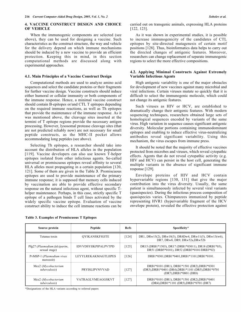
Table 3. Examples of Promiscuous T Epitopes
Source protein Peptide Refs Specificity*
Tetanus toxin QYIKANSKFIGITE [124] DR1, DRw15(2), DRw18(3), DR4Dw4, DRw11(5), DRw13(w6),DR7, DRw8, DR9, DRw52a,DRw52b
Pfg27 (Plasmodium falciparim,sexual stage)
IDVVDSYIIKPIPALPVTPD [125] DR15 (DRB1*1503), DR17 (DRB1*03011), DR18 (DRB1*03),DR51 (DRB5*0101), DR52 (DRB3*0101/DRB3*02)
PvMSP-1 (Plasmodium vivaxmerozoit)
LEYYLREKAKMAGTLIIPES [126] DRB1*0301,DRB1*0401,DRB1*1101,DRB1*0101.
Mce2 (Mycobacteriumtuberculosis) PRYISLIPVNVVAD [127]
DRB1*0101 (DR1), DRB1*1501 (DR2),DRB1*0301(DR3),DRB1*0401 (DR4),DRB1*1101 (DR5),DRB1*0701
(DR7),DRB1*0801 (DR8)
Mce2 (Mycobacteriumtuberculosis)
VATRAGLVMEAGGSKVT [127] DRB1*0101 (DR1), DRB1*1501 (DR2),DRB1*0401(DR4),DRB1*1101 (DR5),DRB1*0701 (DR7)
*Designations of the HLA variants according to referred papers
4. VACCINE CONSTRUCT DESIGN AND CHOICEOF VEHICLE
When the immunogenic components are selected (seeabove), they can be used for designing a vaccine. Suchcharacteristics as the construct structure, the way and vehiclefor the delivery depend on which immune mechanismsshould be induced by a new vaccine to provide an efficientprotection. Keeping this in mind, in this sectioncomputational methods are discussed along withexperimental approaches.
4.1. Main Principles of a Vaccine Construct Design
Computational methods are used to analyze amino acidsequences and select the candidate proteins or their fragmentsfor further vaccine design. Vaccine constructs should induceeither humoral or cellular immunity or both components ofthe immune response. Hence, a minimal vaccine constructshould contain B-epitopes or/and CTL T epitopes dependingon the required immune reactions, as well as Th epitopesthat provide the maintenance of the immune response. As itwas mentioned above, the cleavage sites inserted at thetermini of T epitope regions provide the necessary antigenprocessing. However, lysosomal protease cleavage sites (thatare not predicted reliably now) are not necessary for smallpeptide constructs, as the MHC-II pocket allowsaccommodating long peptides (see above).
Selecting Th epitopes, a researcher should take intoaccount the distribution of HLA alleles in the population[119]. Vaccine developers can also use known T-helperepitopes isolated from other infectious agents. So-calleduniversal or promiscuous epitopes reveal affinity to severalHLA alleles most propagating in a certain population [120,121]. Some of them are given in the Table 3. Promiscuousepitopes are used to provide maintenance of the primaryimmune response; it is supposed that memory cells inducedby vaccination are able to provide effective secondaryresponse on the natural infectious agent, without specific T-helper maintenance. Perhaps, in this case, strictly specific Tepitope of a pathogen binds T cell lines activated by thewidely specific vaccine epitope. Evaluation of vaccineconstruct ability to induce the cell immune reactions can be
carried out on transgenic animals, expressing HLA proteins[122, 123].
As it was shown in experimental studies, it is possibleto increase immunogenicity of the candidates of CTLepitopes by site-directed mutagenesis of certain motifpositions [128]. Thus, bioinformatics data helps to carry outthe directed changes of antigenic features. Moreover,researchers can change replacement of separate immunogenicregions to select the more effective compositions.
4.2. Applying Minimal Constructs Against ExtremelyVariable Infectious Agents
High antigenic variability is one of the major obstaclesfor development of new vaccines against many microbial andviral infections. Certain viruses mutate so quickly that it isdifficult to select the immunogenic molecule, which couldnot change its antigenic features.
Such viruses as HIV or HCV, are established todramatically change their antigenic features. With modernsequencing techniques, researchers obtained large sets ofhomological sequences encoded by variants of the samevirus. High variation in sequence causes significant antigenicdiversity. Molecular portions containing immunodominantepitopes and enabling to induce effective virus-neutralizingantibodies reveal significant variability. Using thismechanism, the virus escapes from immune press.
It should be noted that the majority of effective vaccinesprotected from microbes and viruses, which reveal cytopathiceffects. Agents that do not reveal cytopathic activity (e.g.HIV and HCV) can persist in the host cell, generating themultiple variants in the absence of significant immuneresponse [129].
Envelope proteins of HIV and HCV containhypervariable regions [130, 131] that give the majorcontribution into the virus diversity. Usually, the samepatient is simultaneously infected by several viral variants(quasispecies). During the infectious process composition ofquasispecies varies. Chimpanzees immunized by peptiderepresenting HVR1 (hypervariable fragment of the HCVenvelope protein), revealed the effective protection against
Computer Design of Vaccines Current Computer-Aided Drug Design, 2005, Vol. 1, No. 2 217
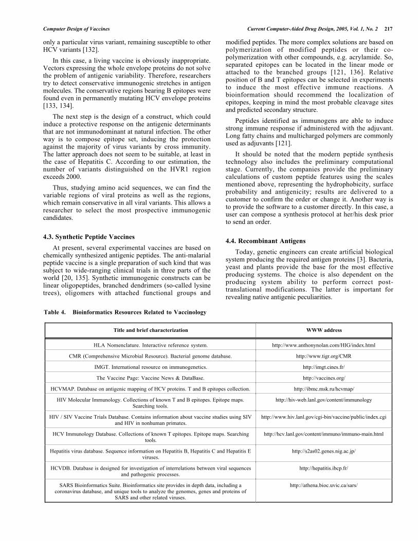
Table 4. Bioinformatics Resources Related to Vaccinology
Title and brief characterization WWW address
HLA Nomenclature. Interactive reference system. http://www.anthonynolan.com/HIG/index.html
CMR (Comprehensive Microbial Resource). Bacterial genome database. http://www.tigr.org/CMR
IMGT. International resource on immunogenetics. http://imgt.cines.fr/
The Vaccine Page: Vaccine News & DataBase. http://vaccines.org/
HCVMAP. Database on antigenic mapping of HCV proteins. T and B epitopes collection. http://ibmc.msk.ru/hcvmap/
HIV Molecular Immunology. Collections of known T and B epitopes. Epitope maps.Searching tools.
http://hiv-web.lanl.gov/content/immunology
HIV / SIV Vaccine Trials Database. Contains information about vaccine studies using SIVand HIV in nonhuman primates.
http://www.hiv.lanl.gov/cgi-bin/vaccine/public/index.cgi
HCV Immunology Database. Collections of known T epitopes. Epitope maps. Searchingtools.
http://hcv.lanl.gov/content/immuno/immuno-main.html
Hepatitis virus database. Sequence information on Hepatitis B, Hepatitis C and Hepatitis Eviruses.
http://s2as02.genes.nig.ac.jp/
HCVDB. Database is designed for investigation of interrelations between viral sequencesand pathogenic processes.
http://hepatitis.ibcp.fr/
SARS Bioinformatics Suite. Bioinformatics site provides in depth data, including acoronavirus database, and unique tools to analyze the genomes, genes and proteins of
SARS and other related viruses.
http://athena.bioc.uvic.ca/sars/
only a particular virus variant, remaining susceptible to otherHCV variants [132].
In this case, a living vaccine is obviously inappropriate.Vectors expressing the whole envelope proteins do not solvethe problem of antigenic variability. Therefore, researcherstry to detect conservative immunogenic stretches in antigenmolecules. The conservative regions bearing B epitopes werefound even in permanently mutating HCV envelope proteins[133, 134].
The next step is the design of a construct, which couldinduce a protective response on the antigenic determinantsthat are not immunodominant at natural infection. The otherway is to compose epitope set, inducing the protectionagainst the majority of virus variants by cross immunity.The latter approach does not seem to be suitable, at least inthe case of Hepatitis C. According to our estimation, thenumber of variants distinguished on the HVR1 regionexceeds 2000.
Thus, studying amino acid sequences, we can find thevariable regions of viral proteins as well as the regions,which remain conservative in all viral variants. This allows aresearcher to select the most prospective immunogeniccandidates.
4.3. Synthetic Peptide Vaccines
At present, several experimental vaccines are based onchemically synthesized antigenic peptides. The anti-malarialpeptide vaccine is a single preparation of such kind that wassubject to wide-ranging clinical trials in three parts of theworld [20, 135]. Synthetic immunogenic constructs can belinear oligopeptides, branched dendrimers (so-called lysinetrees), oligomers with attached functional groups and
modified peptides. The more complex solutions are based onpolymerization of modified peptides or their co-polymerization with other compounds, e.g. acrylamide. So,separated epitopes can be located in the linear mode orattached to the branched groups [121, 136]. Relativeposition of B and T epitopes can be selected in experimentsto induce the most effective immune reactions. Abioinformation should recommend the localization ofepitopes, keeping in mind the most probable cleavage sitesand predicted secondary structure.
Peptides identified as immunogens are able to inducestrong immune response if administered with the adjuvant.Long fatty chains and multicharged polymers are commonlyused as adjuvants [121].
It should be noted that the modern peptide synthesistechnology also includes the preliminary computationalstage. Currently, the companies provide the preliminarycalculations of custom peptide features using the scalesmentioned above, representing the hydrophobicity, surfaceprobability and antigenicity; results are delivered to acustomer to confirm the order or change it. Another way isto provide the software to a customer directly. In this case, auser can compose a synthesis protocol at her/his desk priorto send an order.
4.4. Recombinant Antigens
Today, genetic engineers can create artificial biologicalsystem producing the required antigen proteins [3]. Bacteria,yeast and plants provide the base for the most effectiveproducing systems. The choice is also dependent on theproducing system ability to perform correct post-translational modifications. The latter is important forrevealing native antigenic peculiarities.
218 Current Computer-Aided Drug Design, 2005, Vol. 1, No. 2 Sobolev et al.
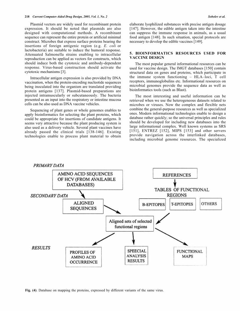
Fig. (4). Database on mapping the proteins, expressed by different variants of the same virus.
Plasmid vectors are widely used for recombinant proteinexpression. It should be noted that plasmids are alsodesigned with computational methods. A recombinantsequence can represent the entire protein or artificial minimalconstruct. Microbes that express surface proteins bearing theinsertions of foreign antigenic region (e.g. E. coli orlactobacteria) are suitable to induce the humoral response.Attenuated Salmonella strains enabling to intracellularreproduction can be applied as vectors for constructs, whichshould induce both the cytotoxic and antibody-dependentresponse. Virus-based construction should activate thecytotoxic mechanisms [3].
Intracellular antigen expression is also provided by DNAvaccination, when the protein-encoding nucleotide sequencesbeing inoculated into the organism are translated providingprotein antigens [137]. Plasmid-based preparations areinjected intramuscularly or subcutaneously. The bacteriapresented as an input into the respiratory or intestine mucosacells can be also used as DNA vaccine vehicles.
Sequencing of plant genes or whole genomes enables toapply bioinformatics for selecting the plant proteins, whichcould be appropriate for insertions of candidate antigens. Itseems very attractive because the plant producing system isalso used as a delivery vehicle. Several plant vaccines havealready passed the clinical trials [138-146]. Existingtechnologies enable to process plant material to obtain
elaborate lyophilized substances with precise antigen dosage[147]. However, the edible antigen taken into the intestinecan suppress the immune response in animals, as a usualfood antigen [148]. In such situation, special protocols arenecessary to develop the edible vaccines [149].
5. BIOINFORMATICS RESOURCES USED FORVACCINE DESIGN
The most popular general informational resources can beused for vaccine design. The IMGT databases [150] containstructural data on genes and proteins, which participate inthe immune system functioning – HLA–loci, T cellreceptors, immunoglobulins etc. Informational resources onmicrobial genomes provide the sequence data as well asbioinformatics tools (such as Blast).
The most interesting and useful information can beretrieved when we use the heterogeneous datasets related tomicrobes or viruses. Now the complex and flexible netscombine the general-purpose resources as well as specializedones. Modern informational technologies enable to design adatabase rather quickly; so the universal principles and rulesshould be developed for including new databases into thelarge informational complex. Well known systems as SRS[151], ENTREZ [152], MIPS [153] and other serversprovide navigation across the interlinked databases,including microbial genome resources. The specialized
Computer Design of Vaccines Current Computer-Aided Drug Design, 2005, Vol. 1, No. 2 219
resources on the certain pathogens such as HIV, SARScoronavirus and HCV (see Table 4), represent data onannotated nucleotide and amino acid sequence (annotations),variations of functional and antigenic regions in differentstrains. They also provide sequence analysis tools and storethe obtained results (e.g. alignment) as secondary data.Maintenance of these resources is reasonable, because thenumber of different sequences related to the same virusreaches several tens of thousands.
The database on functional variability of proteins,encoded by the same virus, should be organized like schemeshown in Fig. (4). Now this scheme is partly implementedin the database HCVMAP designed by the Institute ofBiomedical Chemistry (Russia) for antigenic mapping of theprotein sequences (http://ibmc.msk.ru/hcvmap).
The primary dataset collects amino acid sequences andtheir functional characteristics. The sequences are retrievedfrom freely available resources of the UniProt knowledgebase[154]. Functional characteristics are collected using variouspublicly available resources (PubMed bibliographic system,epitope collections and others). The secondary data representthe aligned sequences and mapped regions related to thefunctional characteristics – epitope tables. These tablescontain the peptide sequences mapped by their locations inHCV protein sequences excluding mimotopes. Each recordcontains brief characteristics, obtained for these regions.Most of the presented epitopes are human, but some of themare murine and chimpanzee ones. Browsing the epitopetables, one can invoke the reference page by clicking the"book" icons. Through this page, a user can access theEntrez bibliographic system (PubMed). By clicking labels atthe table row, the user calls out the alignment set of ahomologous fragment, including the corresponding regionwith data on occurrence frequencies for each peptide stretch.A click on the peptide string invokes all Swiss-Prot/TrEmble identifiers of sequences that contain it. A clickon identifier can evoke a corresponding sequence with thecolour-emphasised peptide. Besides the epitope tables, thefrequency profile calculated on aligned sequences can bedisplayed. The corresponding page contains the HCVpolyprotein scheme that allows the navigation along thedifferent profile regions with amino acid occurrencefrequencies.
6. CONCLUSION
Due to the vaccine application and improvement ofsanitary conditions, several traditional infections wiped out.Moreover, such infections as Smallpox and Poliomyelitisare considered as practically beaten. At the same time, theeffective vaccines against Malaria and other parasiticalinfections are not yet introduced in medical practice.Furthermore, new infectious agents (e.g. HIV, HCV)surprisingly appeared in the last quarter of the twentiethcentury, well known Mycobacterium tuberculosis presentsnew resistant variants causing hardly or non-curable sickness[155], etc.
It is expected that mankind may be faced with absolutelynew infectious agents or new types of known ones. The firstexample and a big challenge for a new century is SARS.Today, we are also forced to take into account the threat of
bioterrorism. The situation is quite possible when thetraditional techniques will not be suitable to create effectivevaccines. New approaches are necessary to design new kindsof vaccines – therapeutic, anticancer and contraceptive [156-159].
Bioinformatics, genomics and proteomics provide newopportunities to reveal and extract candidate immunogeniccomponents that could be used in design of new vaccinegeneration. However, we should not forget about the dangersarising from the incompleteness of knowledge. The deeperunderstanding of molecular foundations of immunity can bereached, based on the integrative computational biology.Thus, further development of computational methods isnecessary for theoretical studies, as well as for practicalapplications. It is clear that genomics and proteomics dataand corresponding computational techniques will be widelyused in this very promising direction.
ACKNOWLEDGEMENTS
We thank Drs. T.I. Kuzmina, M.A. Eldarov and S.A.Moshkovski for their help in selecting the papers and usefuldiscussion.
This work is partly supported by the Russian Foundationof Basic Research, grant No 04-04-49390 and the RussianMinistry of Education and Science Interdisciplinary Program"New Generations of Vaccines".
ABBREVIATIONS
ANN = Artificial Neural Network
BCR = B cell receptor
HCV = Hepatitis C virus
HIV = Human Immunodeficiency Virus
HLA = Human Leukocyte Antigens
HMM = Hidden Markov Model
MHC = Major Histocompatibility Complex
SARS = Severe Acute Respiratory Syndrome
TCR = T cell receptor
REFERENCES[1] Makela, P.H. FEMS Microbiol. Rev., 2000, 24, 9-20.[2] O'Hagan, D.T.; Rappuoli, R. Pharm Res., 2004, 9, 1519-1530.[3] Liljeqvist, S.; Stahl S.J. Biotechnol., 1999, 73, 1-33.[4] Ben-Yedidia, T.; Arnon, R. Curr. Opin. Biotechnol., 1997, 8, 442-
448.[5] Rappuoli, R.; del Guidice, G. In Vaccine. From Concept to Clinic;
Paoletti, L.C.; McInnes, P.M. Ed.; CRC Press LLC: Boca Raton,Boston, London, New York, Washington DC, 1999; pp. 5-17.
[6] Rappuoli, R. Curr. Opin. Microbiol., 2000, 3, 445-450.[7] Schlagenhauf, P.; Ashraf, H. Lancet, 2003, 361, 1017.[8] Rota, P.A.; Oberste, M.S.; Monroe, S.S.; Nix, W.A.; Campagnoli,
R.; Icenogle, J.P.; Penaranda, S.; Bankamp, B.; Maher, K.; Chen,M.H.; Tong, S.; Tamin, A.; Lowe, L.; Frace, M.; DeRisi, J.L.;Chen, Q.; Wang, D.; Erdman, D.D.; Peret, T.C.; Burns, C.;Ksiazek, T.G.; Rollin, P.E.; Sanchez, A.; Liffick, S.; Holloway B.;Limor, J.; McCaustland, K.; Olsen-Rasmussen, M.; Fouchier, R.;Gunther, S.; Osterhaus, A.D.; Drosten, C.; Pallansch, M.A.;Anderson, L.J.; Bellini, W.J. Science, 2003, 300, 1394-1399.
220 Current Computer-Aided Drug Design, 2005, Vol. 1, No. 2 Sobolev et al.
[9] Gao, W.; Tamin, A.; Soloff, A.; D'Aiuto, L.; Nwanegbo, E.;Robbins, PD.; Bellini, WJ.; Barratt-Boyes, S.; Gambotto, A.Lancet, 2003, 362, 1895-1896.
[10] Kondro, W. Can. Med. Assoc. J., 2004, 170, 183.[11] Xiong, S.; Wang, Y.F.; Zhang, M.Y.; Liu, X.J.; Zhang, C.H.; Liu,
S.S.; Qian, C.W.; Li, J.X.; Lu, J.H.; Wan, Z.Y.; Zheng, H.Y.; YanX.G.; Meng, M.J.; Fan, J.L. Immunol. Lett., 2004, 95, 139-143.
[12] Pang, H.; Liu, Y.; Han, X.; Xu, Y.; Jiang, F.; Wu, D.; Kong, X.;Bartlam, M.; Rao, Z. J. Gen. Virol., 2004, 85, 3109-3113.
[13] Andre, F.E. Vaccine, 1990, 8 Suppl., S74-S78.[14] Kao, J.H.; Chen, D.S. Lancet Infect. Dis., 2002, 2, 395-403.[15] Parenti, D. Conn. Med., 1999, 63, 570.[16] Greco, D.; Salmaso, S.; Mastrantonio, P.; Giuliano, M.; Tozzi,
A.E.; Anemona, A.; Ciofi degli Atti, M.L.; Giammanco, A.; Panei,P.; Blackwelder, W.C.; Klein, D.L.; Wassilak, S.G. N. Engl. J.Med., 1966, 334, 341-348.
[17] Hyams, K.C. Curr. Gastroenterol. Rep., 2002, 4, 302-307.[18] Schultz, A.M.; Bradac, J.A. AIDS, 2001, 15 Suppl. 5, S147-S158.[19] Mooij, P.; Heeney, J.L. Vaccine, 2001, 20, 304-321.[20] Migasena, S.; Heppner, D.G.; Kyle, D.E.; Chongsuphajaisiddhi,
T.; Gordon, D.M.; Suntharasamai, P.; Permpanich, B.; Brockman,A.; Pitiuttutham, P.; Wongsrichanalai, C.; Srisuriya, P; Phonrat, B.;Pavanand, K.; Viravan, C.; Ballou, W.R. Acta Trop., 1997, 67,215-227.
[21] Kandel, R.; Hartshorn, K.L. BioDrugs, 2001, 15, 303-323.[22] Jackson, D.; Cadman, A.; Zurcher, T.; Barclay, W.S. J. Virol.,
2002, 76, 11744-11747.[23] Jeon, S.H.; Ben-Yedidia, T.; Arnon, R. Vaccine, 2002, 20, 2772-
2780.[24] Agger, E.M.; Andersen, P. Vaccine, 2002, 21, 7-14.[25] Doherty, T.M.; Olsen, A.W.; van Pinxteren, L.; Andersen, P.
Infect. Immun., 2002, 70, 3111-3121.[26] Gor, D.O.; Ding, X.; Li, Q.; Schreiber, J.R.; Dubinsky, M.;
Greenspan, N.S. Infect. Immun., 2002, 70, 5589-5595.[27] Briles, D.E.; Hollingshead, S.K.; King, J.; Swift, A.; Braun, P.A.;
Park, M.K.; Ferguson, L.M.; Nahm, M.H.; Nabors, G.S. J. Infect.Dis., 2000, 182, 1694-1701.
[28] Steere, A.C.; Gross, D.; Meyer, A.L.; Huber, B.T. J. Autoimmun.,2001, 16, 263-268.
[29] Willett, T.A.; Meyer, A.L; Brown, E.L.; Huber, B.T. Proc. Natl.Acad. Sci. USA, 2004, 101, 1303-1308.
[30] Roitt, I.; Brostoff, J.; Male, D. Immunology, Mosby: London,Philadelphia, Sydney, Tokio, 1998.
[31] Kim, J.V.; Latouche, J.-B.; Riviere, I.; Sadelain, M. Nat .Biotechnol., 2004, 22, 403-410.
[32] Grandi, G. Trends Biotechnol., 2001, 19, 181-188.[33] Mahanty, S.; Saul, A.; Miller, L.H. J. Exp. Biol., 2003, 206, 3781-
3788.[34] Mount, D.W. Bioinformatics: Sequence and Genome Analysis,
Cold Spring Harbor Laboratory Press: Cold Spring Harbor, NewYork, 2001.
[35] Altschul, S.F.; Madden, T.L.; Schaffer, A.A.; Zhang, J.; Zhang, Z.;Miller, W.; Lipman, D.J. Nucleic Acids Res., 1997, 25, 3389-3402.
[36] McAtee, C.P.; Lim, M.Y.; Fung, K.; Velligan, M.; Fry, K.; Chow,T.P.; Berg, D.E. J. Chromatogr. B. Biomed. Sci. Appl., 1998, 714,325-333.
[37] Utt, M.; Nilsson, I.; Ljungh, A.; Wadstrom, T. J. Immunol.Methods, 2002, 259, 1-10.
[38] Purcell, A.W.; Gorman, J.J. Mol. Cell Proteomics, 2004, 3, 193-208.
[39] Falquet, L.; Pagni, M.; Bucher, P.; Hulo, N.; Sigrist, C.J.; Hofmann,K.; Bairoch, A. Nucleic Acids Res., 2002, 30, 235-238.
[40] Bateman, A.; Coin, L.; Durbin, R.; Finn, R.D.; Hollich, V.;Griffiths-Jones, S.; Khanna, A.; Marshall, M.; Moxon, S.;Sonnhammer, E.L.; Studholme, D.J.; Yeats, C.; Eddy, S.R. NucleicAcids Res., 2004, 32 Database issue, D138-D141.
[41] Letunic, I.; Copley, R.R.; Schmidt, S.; Ciccarelli, F.D.; Doerks, T.;Schultz, J.; Ponting, C.P.; Bork, P. Nucleic Acids Res., 2004, 32Database issue, D142-D144.
[42] Nielsen, H.; Brunak, S.; von Heijne, G. Protein Eng., 1999, 12, 3-9.
[43] Bendtsen, J.D.; Nielsen, H.; von Heijne, G; Brunak, S. J. Mol.Biol., 2004, 340, 783-795.
[44] Hayashi, S.; Wu, H.C. J. Bioenerg. Biomembr., 1990, 22, 451-471.[45] Fariselli, P.; Finocchiaro, G.; Casadio, R. Boinformatics, 2003, 19,
2498-2499.
[46] Maurer-Stroh, S.; Eseinhaber, F. Trends Microbiol., 2004, 12, 178-179.
[47] Maurer-Stroh, S.; Eseinhaber, B.; Eseinhaber, F. J. Mol. Biol.,2001, 317, 541-557.
[48] Eisenhaber, B.; Bork, P.; Eisenhaber, F. J. Mol. Biol., 1999, 292,741-758.
[49] Acosta-Serrano, A.; Almeida, I.C.; Freitas-Junor, L.H.; Yoshida,N.; Shemckman, S. Mol. Biochem. Parasitol., 2001, 114, 143-150.
[50] Naderer T.; Vince J.E., McConville M.J. Curr. Mol. Med., 2004,4, 649-665.
[51] van Hoven, N.S.; Miller, A.D. J. Virol., 2005, 79, 87-94.[52] Vigdorovich, V.; Strong R.K.; Miller, A.D. J. Virol., 2005, 79, 79-
86.[53] van der Ley, P.; Heckels, J.E.; Virji, M.; Hoogerhout, P.; Poolman,
J.T. Infect. Immun., 1991, 59, 2963-2971.[54] Martelli, P.L.; Fariselli, P.; Krogh, A.; Casadio, R. Bioinformatics,
2002, 18, 546-553.[55] Bagos, P.; Liakopoulos, T.D.; Spyropoulos, I.C.; Hamodrakas, S.J.
BMC Bioinformatics, 2004, 5, 29.[56] Bigelow, H.R.; Petrey, D.S.; Liu, J.; Przybylski, D.; Rost, B.
Nucleic Acids Res., 2004, 32, 2566-2577.[57] Natt, N.K.; Kaur, H.; Raghava, G.P. Proteins, 2004, 56, 11-18.[58] Berven, F.S.; Flikka, K.; Jensen H.B.; Eidhammer, I. Nucleic Acids
Res., 2004, 32 Web Server Issue, W394-W399.[59] Gavel, Y.; von Heijne, G. Protein Eng., 1990, 3, 433-442.[60] Yan, B.; Zhang, W.; Ding J.; Gao, P. J. Protein Chem., 1999, 18,
511-521.[61] Kyte, J.; Doolittle, R. J. Mol. Biol., 1982, 157, 105-132.[62] Krogh, A.; Larrson, B.; von Hejne, G. J. Mol., Biol., 2001, 395,
567-580.[63] Tusnady, G.E.; Simon, I. Bioinformatics, 2001, 17, 849-850.[64] Grezko, M.; Eisenhaber, F.; Eisenhaber, B.; Simon, I.
Bioinformatics, 2004, 20, 136-137.[65] Tettelin, H.; Saunders, N.J.; Heidelberg, J.; Jeffries, A.C.; Nelson,
K.E.; Eisen, J.A.; Ketchum, K.A.; Hood, D.W.; Peden, J.F.;Dodson, R.J.; Nelson, W.C.; Gwinn, M.L.; DeBoy, R.; Peterson,J.D.; Hickey, E.K.; Haft, D.H.; Salzberg, S.L.; White, O.;Fleischmann, R.D.; Dougherty, B.A.; Mason, T.; Ciecko, A.;Parksey, D.S.; Blair, E.; Cittone, H.; Clark, E.B.; Cotton, M.D.;Utterback, T.R.; Khouri, H.; Qin, H.; Vamathevan, J.; Gill, J.;Scarlato, V.; Masignani, V.; Pizza, M.; Grandi, G.; Sun, L.; Smith,H.O.; Fraser, C.M.; Moxon, E.R.; Rappuoli, R.; Venter, J.C.Science, 2000, 287, 1809-1815.
[66] Pizza, M.; Scarlato, V.; Masignani, V.; Giuliani, M.M.; Arico, B.;Comanducci, M.; Jennings, G.T.; Baldi, L.; Bartolini, E.; Capecchi,B.; Galeotti, C.L.; Luzzi, E.; Manetti, R.; Marchetti, E.; Mora, M.;Nuti, S.; Ratti, G.; Santini, L.; Savino, S.; Scarselli, M.; Storni, E.;Zuo, P.; Broeker, M.; Hundt, E.; Knapp, B.; Blair, E.; Mason, T.;Tettelin, H.; Hood, D.W.; Jeffries, A.C.; Saunders, N.J.; Granoff,D.M.; Venter, J.C.; Moxon, E.R.; Grandi, G.; Rappuoli, R.Science, 2000, 287, 1816-1820.
[67] Drabick, J.J.; Brandt, B.L.; Moran, E.E.; Saunders, N.B.;Shoemaker, D.R.; Zollinger, W.D. Vaccine, 1999, 18, 160-172.
[68] Katial, R.K.; Brandt, B.L.; Moran, E.E.; Marks, S.; Agnello, V.;Zollinger, W.D. Infect. Immun., 2002, 70, 702-707.
[69] Poolman, J.T.; Kriz-Kuzemenska, P.; Ashton, F.; Bibb, W.;Dankert, J.; Demina, A.; Froholm, L.O.; Hassan-King, M.; Jones,D.M.; Lind, I.; Prakash, K.; Xujing, H. Clin. Diagn. Lab. Immunol.,1995, 2, 69-72.
[70] Peterson, J.D.; Umayam, L.A.; Dickinson, T.; Hickey, E.K.;White, O. Nucleic Acids Res., 2001, 29, 123-125.
[71] Roccasecca, R.; Folgori, A.; Ercole, B.B.; Puntoriero, G.; Lahm,A.; Zucchelli, S.; Tafi, R.; Pezzanera, M.; Galfre, G.;Tramontano, A.; Mondelli, M.U.; Pessi, A.; Nicosia, A.; Cortese,R.; Meola, A. Int. Rev. Immunol., 2001, 20, 289-300.
[72] Olenina, L.V.; Kuzmina, T.I.; Sobolev, B.N.; Kuraeva, T.E.;Kolesanova, E.F.; Archakov, A.I. J. Viral. Hepat. 2005, In press.
[73] Montgomery, D.L. Brief Bioinform., 2000, 1, 289-296.[74] Louise, R.; Skjot, V.; Agger, E.M.; Andersen, P. Scand. J. Infect.
Dis., 2001, 33, 643-647.[75] Pennisi, E. Science, 1998, 281, 324-325.[76] Wang, R.; Doolan, D.L.; Le, T.P.; Hedstrom, R.C.; Coonan, K.M.;
Charoenvit, Y.; Jones, T.R.; Hobart, P.; Margalith, M.; Ng, J.;Weiss, W.R.; Sedegah, M.; de Taisne, C.; Norman, J.A.; Hoffman,S.L. Science, 1998, 282, 476-480.
[77] Hoffman, S.L.; Rogers, W.O.; Carucci, D.J.; Venter, J.C. Nat.Med., 1998, 4, 1351-1353.
Computer Design of Vaccines Current Computer-Aided Drug Design, 2005, Vol. 1, No. 2 221
[78] Stephens, R.S. J. Infect. Dis., 2000, 181 Suppl. 3, S521-S523.[79] Igietseme, J.U.; Black, C.M.; Caldwell H.D. BioDrugs, 2002, 16,
19-35.[80] Pellequer, J.-L.; Westhof, E.; van Regenmortel, M.H.V. In
Peptide antigens. A practical approach; G.B. Wisdom, Ed.; IRLPRESS, Oxford University Press: Oxford, New York, Tokyo,1994; pp. 7-25.
[81] Stites, W.E. Chem. Rev., 1997, 97, 1233-1250.[82] Jin, L.; Wells, J.A. Protein Sci., 1994, 3, 2351-2357.[83] Janin, J.; Chothia, C. J. Biol. Chem., 1990, 265, 16027–16030.[84] van Regenmortel M.H.V.; Muller, S. Synthetic peptides as
antigens, Elsevier: Amsterdam, Lausanne, New York, Oxford,Shannon, Singapore-Tokyo, 1999.
[85] Hopp, T.P.; Woods, K.R. Proc. Natl. Acad. Sci. USA, 1981, 78,3824-3828.
[86] Nozaki, Y.; Tanford, C. J. Biol. Chem., 1971, 246, 2211-2227.[87] Bull, H.B.; Breese, K. Arch. Biochem. Biophys., 1974, 161, 665-
670.[88] Parker, J.M; Guo, D.; Hodges, R.S. Biochemistry, 1986, 25, 5425-
5432.[89] Karplus, P.A.; Schulz, G.E. Naturwissenschaften, 1985, 72, 212-
213.[90] Hopp ,T.P. Ann. Sclavo. Collana. Monogr., 1984, 1, 47-60.[91] Lee, B.; Richards, F.M. J. Mol. Biol., 1971, 55, 379-400.[92] Welling, G.W.; Weijer, W.J.; van der Zee R.; Welling-Wester, S.
FEBS Lett., 1985, 188, 215-218.[93] Chou, P.Y; Fasman, G.D. Biophys. J., 1979, 26, 367-373.[94] Odorico, M.; Pellequer, J.-L. J. Mol. Recognit., 2003, 16, 20–22.[95] Carter, J.M. Methods Mol. Biol., 1994, 36, 207-223.[96] Rost, B.; Sander, C. Proteins, 1994, 20, 216-226.[97] Kopp, J.; Schwede, T. Nucleic. Acids Res., 2004, 32 Database
issue, D230-D234.[98] Kolaskar, A.S.; Kulkarni-Kale, U. Virology, 1999, 261, 31-42.[99] Rammensee, H.; Bachmann, J.; Emmerich, N.P.; Bachor, O.A.;
Stevanovic, S. Immunogenetics, 1999, 50, 213-219.[100] Dafforn, T. R.; Lesk, A.M. 2000 In Protein-Protein Recognition;
C. Kleanthous, Ed.; Oxford University Press: Oxford, London,2000; pp. 163-188.
[101] Schonbach, C.; Koh, J.L.; Flower, D.R.; Wong, L.; Brusic, V.Nucleic Acids Res., 2002, 30, 226-229.
[102] Jung, G.; Fleckenstein, B.; von der Mulbe, F.; Wessels, J.;Niethammer, D.; Wiesmuller, K.H. Biologicals, 2001, 29, 179-181.
[103] Bhasin, M.; Raghava, G.P. Vaccine, 2004, 22, 3195-3204.[104] Mamitsuka, H. Proteins, 1998, 33, 460-474[105] Yu, K., Petrovsky, N.; Schonbach, C.; Koh, J.Y.; Brusic V. Mol.
Med., 2002, 8, 137-148.[106] Guan, P.; Doytchinova, I.A.; Zygori, C.; Flower, D.R. Nucleic
Acids Res., 2003, 13, 3621-3624.[107] Doytchinova, I.A.; Flower, D.R. Bioinformatics, 2003, 17, 2263-
2270.[108] McSparron, H.; Blythe, M.J.; Zygouri, C.; Doytchinova, I.A.;
Flower, D.R. J. Chem. Inf. Comput. Sci., 2003, 43, 1276-1287.[109] Doytchinova, I.A.; Walshe, V.A.; Jones, N.A.; Gloster, S.E.;
Borrow, P.; Flower, D.R. J. Immunol., 2004, 172, 7495-7502.[110] Sung, M.H.; Zhao, Y.; Martin, R.; Simon, R. J. Comput. Biol.,
2002, 9, 527-539.[111] Liu, R.; Enstrom, A.M.; Lam, K.S. Exp. Hematol., 2003, 31, 11-30.[112] Dogan, I.; Dorgham, K.; Chang, H.C.; Parizot, C.; Lemaitre, F.;
Ferradini, L.; Reinherz, E.L.; Debre, P.; Gorochov, G. Eur. J.Immunol., 2004, 34, 598-607.
[113] Nussbaum, A.K.; Kuttler, C.; Hadeler, K.P.; Rammensee, H.G.;Schild, H. Immunogenetics, 2001, 53, 87-94.
[114] Stoltze, L.; Schirle, M.; Schwarz, G.; Schroter C.; Thompson,M.W.; Hersh, LB.; Kalbacher, H.; Stevanovic, S.; Rammensee,H.G.; Schild, H. Nat. Immunol., 2000, 1, 413-418.
[115] Brusic, V.; van Endert, P.; Zeleznikow, J.; Daniel S.; Hammer, J.;Petrovsky N. In Silico Biol., 1999, 1, 109-121.
[116] Logean, A; Rognan, D. J. Comput. Aided Mol. Des., 2002, 16,229-243.
[117] Rognan, D.; Lauemoller, S.L.; Holm, A.; Buus, S.; Tschinke, V. J.Med. Chem., 1999, 42, 4650-4658.
[118] Zhao, B.; Mathura, V.S.; Rajaseger, G.; Moochhala, S.;Sakharkar, M.K.; Kangueane, P. Hum Immunol., 2003, 64, 1123-1143.
[119] Bodmer, W. Ciba Found. Symp., 1996, 197, 233-253.
[120] Lairmore, M.D.; DiGeorge, A.M.; Conrad, S.F.; Trevino, A.V.;Lal, R.B.; Kaumaya, P.T. J. Virol., 1995, 69, 6077-6089.
[121] Jackson, D.C.; Purcell, A.W.; Fitzmaurice, C.J.; Zeng, W.; Hart,D.N. Curr. Drug Targets, 2002, 3, 175-196.
[122] Taneja, V.; David, C.S. J. Clin. Invest., 1998, 101, 921-926.[123] Taneja V.; David, C.S. Immunol. Rev., 1999, 169, 67-79.[124] Panina-Bordignon, P.; Tan, A.; Termijtelen, A.; Demotz, S.;
Corradin, G.; Lanzavecchia, A. Eur. J. Immunol., 1989, 19, 2237-2242
[125] Contreras, C.E.; Ploton, I.N.; Siliciano, R.F.; Karp, C.L.; Viscidi,R.; Kumar, N. Infect. Immun., 1998, 66, 3579-3590.
[126] Caro-Aguilar, I.; Rodriguez, A.; Calvo-Calle, J.M.; Guzman, F.;De la Vega, P.; Patarroyo, M.E.; Galinski, M.R.; Moreno, A.Infect. Immun., 2002, 70, 3479-3492.
[127] Panigada, M.; Sturniolo, T.; Besozzi, G.; Boccieri, M.G.;Sinigaglia, F.; Grassi, G.G.; Grassi, F. Infect. Immun., 2002, 70,79-85.
[128] Sarobe, P; Pendleton, C.D.; Akatsuka, T.; Lau, D.; Engelhard,V.H.; Feinstone, S.M.; Berzofsky, J.A. J. Clin. Invest., 1998, 102,1239-1248.
[129] Hilleman, M.R. Proc. Natl. Acad. Sci. USA, 2004, 101 Suppl. 2,14560-14566.
[130] Kato, N.; Ootsuyama, Y.; Tanaka, T.; Nakagawa, M; Nakazawa,T.; Muraiso, K.; Ohkoshi, S.; Hijikata, M.; Shimotohno, K. VirusRes., 1992, 22, 107-123.
[131] Hoffman N.G.; Seillier-Moiseiwitsch F.; Ahn, J.; Walker, J.M.;Swanstrom, R. J. Virol., 2002, 76, 3852-3864.
[132] Farci, P.; Shimoda, A.; Wong, D.; Cabezon, T.; De Gioannis, D.;Strazzera, A.; Shimizu, Y.; Shapiro, M.; Alter, H.J.; Purcell, R.H.Proc. Natl. Acad. Sci. USA, 1996, 93, 15394-15399.
[133] Sobolev, B.N.; Poroikov, V.V.; Olenina, L.V.; Kolesanova E.F.;Archakov, A.I. J. Viral Hepat., 2000, 7, 368-374.
[134] Olenina, L.V.; Nikolaeva, L.I.; Sobolev, B.N.; Blokhina, N.P.;Archakov, A.I.; Kolesanova, E.F. J. Viral. Hepat., 2002, 9, 174-182.
[135] Patarroyo, G.; Franco, L.; Amador, R.; Murillo, L.A.; Rocha, C.L.;Rojas, M.; Patarroyo, M.E. Vaccine, 1992, 10, 175-178.
[136] Tam J.P. In: Peptide Antigens. A Practical Approach; Wisdom,G.B. Ed.; IRL PRESS, Oxford University Press: Oxford, NewYork, Tokyo, 1994; pp. 83-115.
[137] Gregersen, J.P. Naturwissenschaften, 2001, 88, 504-513.[138] Streatfield, S.J.; Lane, J.R.; Brooks, C.A.; Barker, D.K.; Poage,
M.L.; Mayor, J.M.; Lamphear, B.J.; Drees, C.F.; Jilka, J.M.; Hood,E.E.; Howard, J.A. Vaccine, 2003, 30, 812-815.
[139] Haq, T.A.; Mason, H.S.; Clements, J.D.; Arntzen, C.J. Science,1995, 268, 714-716.
[140] Mason, H.S.; Haq, T.A.; Clements, J.D.; Arntzen, C.J. Vaccine,1998, 16, 1336-1343.
[141] Tacket, C.O.; Mason, H.S.; Losonsky, G.; Clements, J.D.; Levine,M.M.; Arntzen, C.J. Nat. Med., 1998, 4, 607-609.
[142] Arakawa, T.; Chong, D.K.; Langridge, W.H. Nat. Biotechnol.,1998, 16, 292-297.
[143] Richter, L.J.; Thanavala, Y.; Arntzen, C.J.; Mason, H.S. Nat.Biotechnol., 2000, 18, 1167-1171.
[144] Pharmaprojects 2002, PJB Publications Ltd.[145] Tackaberry, E.S., Dudani, A.K., Prior, F.; Tocchi, M.; Sardana,
R.; Altosaar, I.; Ganz, P.R. Vaccine, 1999, 17, 3020-3029.[146] Clough, J. Drug Discov. Today, 2002, 7, 886-887.[147] Bonetta, L., Nat. Med., 2002, 8, 94.[148] Gotsman, I.; Alper, R.; Klein, A.; Rabbani E.; Engelhardt, D.; Ilan
Y. Cancer, 2002, 94, 406-414.[149] Tacket, C.O.; Mason, H.S. Microbes Infect., 1999, 1, 777-783.[150] Lefranc, M.P. Methods Mol. Biol., 2004, 248, 27-49.[151] Zdobnov, E.M.; Lopez R.; Apweiler, R.; Etzold, T. Bioinformatics,
2002, 18, 1149-1150.[152] Wheeler, D.L.; Churc, D.M.; Edgar, R.; Federhen, S.; Helmberg,
W.; Madden, T.L.; Pontius, J.U.; Schuler.; G.D.; Schriml, L.M.;Sequeira, E.; Suzek, T.O.; Tatusova, T.A.; Wagner L. NucleicAcids Res., 2004, 32 Database issue, D35-D40.
[153] Mewes, H.W.; Amid, C.; Arnold, R.; Frishman, D.; Guldener U.;Mannhaupt, G.; Munsterkotter, M.; Pagel, P.; Strack, N.;Stumpflen, V.; Warfsmann, J.; Ruepp, A. Nucleic Acids Res.,2004, 32 Database issue, D41-D44.
[154] Apweiler, R.; Bairoch, A.; Wu, C.H.; Barker, W.C.; Boeckmann,B.; Ferro, S.; Gasteiger, E.; Huang, H.; Lopez, R.; Magrane, M.;Martin, M.J.; Natale, D.A.; O'Donovan, C.; Redaschi, N.; Yeh,L.S. Nucleic Acids Res., 2004, 32 Database issue, D115-D119.

222 Current Computer-Aided Drug Design, 2005, Vol. 1, No. 2 Sobolev et al.
[155] Kato-Maeda, M.; Bifani, P.J.; Kreiswirth, B.N.; Small, P.M. J.Clin. Invest., 2001, 107, 533-537.
[156] Lollini, P.L.; Forni, G. Trends Immunol., 2003, 24, 62-66.
[157] Davis, I.D.; Jefford, M.; Parente, P.; Cebon, J. J. Leukoc. Biol.,2003, 73, 3-29.
[158] Meng, W.S.; Butterfield, L.H. Pharm. Res., 2002, 19, 926-932.[159] Espinoza-Delgado, I. Oncologist, 2002, 7 Suppl. 3, 20-33.
Received: November 3, 2004 Accepted: January 4, 2005