comparison of the effects of l-carnitine and acetyl-l-carnitine on carnitine levels, ambulatory...
TRANSCRIPT

117
Ann. N.Y. Acad. Sci. 1033: 117–131 (2004). © 2004 New York Academy of Sciences.doi: 10.1196/annals.1320.011
Comparison of the Effects of L-Carnitine and Acetyl-L-Carnitine on Carnitine Levels, Ambulatory Activity, and Oxidative Stress Biomarkers in the Brain of Old Rats
JIANKANG LIU,a,b ELIZABETH HEAD,c HIROHIKO KURATSUNE,d CARL W. COTMAN,c AND BRUCE N. AMESa,b
aDepartment of Molecular and Cell Biology, University of California, Berkeley, California 94720, USAbNutritional Genomics Center, Children’s Hospital Oakland Research Institute, Oakland, California 94609, USAcInstitute for Brain Aging and Dementia, University of California, Irvine, California 92697, USAdDepartment of Hematology and Oncology, Osaka University, Osaka 565-0871, Japan
ABSTRACT: L-Carnitine and acetyl-L-carnitine (ALC) are both used to improvemitochondrial function. Although it has been argued that ALC is better thanL-carnitine in absorption and activity, there has been no experiment to comparethe two compounds at the same dose. In the present experiment, the effects ofALC and L-carnitine on the levels of free, acyl, and total L-carnitine in plasmaand brain, rat ambulatory activity, and biomarkers of oxidative stress areinvestigated. Aged rats (23 months old) were given ALC or L-carnitine at 0.15%in drinking water for 4 weeks. L-Carnitine and ALC were similar in elevatingcarnitine levels in plasma and brain. Both increased ambulatory activitysimilarly. However, ALC decreased the lipid peroxidation (malondialdehyde,MDA) in the old rat brain, while L-carnitine did not. ALC decreased the extentof oxidized nucleotides (oxo8dG/oxo8G) immunostaining in the hippocampalCA1 and cortex, while L-carnitine did not. ALC decreased nitrotyrosineimmunostaining in the hippocampal CA1 and white matter, while L-carnitinedid not. In conclusion, ALC and L-carnitine were similar in increasing ambula-tory activity in old rats and elevating carnitine levels in blood and brain. How-ever, ALC was effective, unlike L-carnitine, in decreasing oxidative damage,including MDA, oxo8dG/oxo8G, and nitrotyrosine, in old rat brain. These datasuggest that ALC may be a better dietary supplement than L-carnitine.
KEYWORDS: acetyl-L-carnitine (ALC); L-carnitine; ambulatory activity;malondialdehyde (MDA); nitrotyrosine; oxo8dG/oxo8G
Address for correspondence: Bruce N. Ames, Ph.D., Nutritional Genomics Center, Children’sHospital Oakland Research Institute, 5700 Martin Luther King Jr. Way, Oakland, CA 94609.Voice: 510-450-7625; fax: 510-597-7128.

118 ANNALS NEW YORK ACADEMY OF SCIENCES
INTRODUCTION
L-Carnitine has been described as a conditionally essential nutrient for humans.L-Carnitine facilitates entry of long-chain fatty acids into mitochondria for utiliza-tion as fuel and facilitates removal from mitochondria of short-chain and medium-chain fatty acids that accumulate as a result of normal and abnormal metabolism.1
Experimental data demonstrate an age-associated decrease of tissue levels of L-carnitine in animals, including humans, and an associated decrease in the integrityof the mitochondrial membrane.2–4 Because carnitine is essential for translocationof fatty acids into mitochondrial matrix, it is plausible that carnitine deficiencywould cause disorders in fatty acid utilization.5 L-Carnitine deficiency is associatedwith cardiomyopathy,6,7 and L-carnitine administration protects the myocardiumagainst diphtheria toxin, ischemia, myocardial infarction, and adriamycin-induceddamage.8 ALC, like L-carnitine, is present in high concentration in the brain as wellas muscle, and provides acetyl equivalents for the production of the neurotransmitteracetylcholine.7,9,10 ALC has been shown to delay or reverse age-related deficits inmitochondrial function, such as in the heart and liver.11–16 In addition, ALC im-proves age-associated cognitive dysfunction and neurodegeneration in animals17–22
and in Alzheimer’s patients,23,24 as well as decreases oxidative stress.20,25,26
L-Carnitine and its acyl esters may act as an oxidant either having a primary anti-oxidant activity (inhibiting free radical generation, scavenging the initiating freeradicals, and terminating the radical propagation reactions) or, more likely, func-tioning as a secondary antioxidant (repairing oxidized polyunsaturated fatty acidsesterified in membrane phospholipids).27,28 ALC inhibits lipid peroxidation andxanthine oxidase activity in rat skeletal muscle.29 ALC reduces lipid peroxidationand lipofuscin concentration in aged rat brain.30 ALC also inhibits oxidant-inducedDNA single-strand breaks.31 ALC may possess a direct antioxidant activity asdemonstrated in vivo.32,33 Related compounds, such as L-propionyl L-carnitine andL-carnitine, have been shown to have antioxidant activity by chelating metals,34 andinhibiting the age-associated increase in lipid peroxidation.25,26 An antioxidant role ofL-propionyl L-carnitine has also been implicated in ischemia-reperfusion injury.35 L-Carnitine in rats prevented doxorubicin cardiotoxicity as monitored by echocardio-graphy, release of myosin light chain-1, and aldehydic lipid peroxidation products.36
It was suggested that the protective effects of L-carnitine were due to its beneficialimpact on energy metabolism and/or its antioxidant effects because of the attenua-tion of the doxorubicin-induced lipid peroxidation. ALC prevented oxygen radical–induced cell death in human diploid fibroblast cell lines, which was explained as dueto increasing the activity of antioxidant enzymes and sustaining the activity of mito-chondrial complex I–NADH ubiquinone reductase and complex IV–cytochromeoxidase of the electron transport chain.37 In addition, ALC protected mitochondrialcomplex III ubiquinol cytochrome c reductase, perhaps as an iron chelator.38
Very little data are available concerning brain L-carnitine status of old rats. Onereport by Maccari et al.4 showed that aged Sprague-Dawley rats (30 months)exhibited a statistically significant decrement of total L-carnitine levels in the brain,serum, heart, and tibial muscle, accompanied by a marked increment in the liver.Another report39 demonstrated that L-carnitine levels in muscles of old male Wistarrats were significantly lower than that in young animals. In our previous dose-responsestudy,3 we demonstrated that ALC at a 0.15% level (in drinking water) can increase

119LIU et al.: EFFECTS OF L-CARNITINE AND ACETYL-L-CARNITINE
plasma and brain carnitine levels as efficiently as 0.5% and 1.5%, but the highestdose (1.5%) causes oxidative damage.16 ALC is more widely used than L-carnitinein animal research and clinical trials to gain metabolic benefits to the brain, heart,liver, and other organs. ALC, compared with L-carnitine, is more efficient to crossthe blood-brain barrier.40 There are limited data to compare the two compounds atthe same dose in aging animals. The aim of the present study was to compare theeffects of the relatively lower optimal dose (0.15%) of L-carnitine and ALC on thelevels of free, acyl, and total L-carnitine in plasma and brain, ambulatory activity,and biomarkers of oxidative stress, including lipid peroxidation, protein oxidation,and oxidative nucleic acid damage in the brain of old rats.
MATERIALS AND METHODS
Materials
ALC (hydrochloride salt) was a gift of Sigma-Tau (Pomezia, Italy). All otherreagents were reagent grade or the highest quality available from Sigma (St. Louis,MO), unless otherwise indicated.
Animals
Fischer 344 male rats were obtained from the National Institute on Aging.Control animals were fed an AIN93M diet from Dyets (Bethlehem, PA) and MilliQ(Millipore) water (pH 5.2). The rats in the experimental groups were fed drinkingwater with either 0.15% ALC or L-carnitine for 4 weeks. The young rats were 4.5months and the old ones were 24.5 months at the start of the experiment; they were5.5 months (young) and 25.5 months (old) when they were terminated. Prior to tissuecollection, animals were fasted overnight, and sacrificed between 9:00 and 12:00 inthe morning. The animals were fully anesthetized with ether prior to exsanguinationsby perfusion with PBS. The brains were removed, immediately put into liquidnitrogen, and stored in a −80°C freezer until analysis.
Assay for Total L-Carnitine, Free L-Carnitine, and Acyl L-Carnitine
Levels of total L-carnitine, free L-carnitine, and acyl L-carnitine were determinedwith an enzymatic cycling method with L-carnitine dehydrogenase.41,42 Briefly,brain tissues were homogenized with 5 volumes of ice-cold Tris-HCl buffer (pH 7.5)with 100 mM sodium chloride and 40 w/v% sucrose, and then deproteinized withice-cold 15% trichloroacetate (480 µL/mL homogenate) and centrifuged. The pH ofthe resulting supernatants was neutralized with 1 M potassium hydroxide. After neu-tralization, specimens were again centrifuged and analyzed immediately with theL-carnitine assay kits (Asahi Chemical Industry, Tagatagun, Japan) as instructed.
Ambulatory Activity Tests
Rats were moved each night from group housing to individual cages (48 × 25 ×20 cm) at 4 h prior to the quantification of ambulatory parameters. The testing room

120 ANNALS NEW YORK ACADEMY OF SCIENCES
was on a 12-h light/dark cycle (lights on 6 A.M. to 6 P.M.). At 8 P.M., a very low lightilluminated the test subjects for video tracking. Quantification began at 9 P.M. andcontinued for 4 h. One hour later, the low light was turned off and the room remainedin total darkness until 6 A.M., when the standard light/dark cycle began. A video sig-nal from a camera suspended directly above the individual cages was fed directlyinto a Videomex-V (Columbus Instruments, Columbus, OH) computer systemrunning the Multiple Objects Multiple Zones software. The system quantifiedambulatory activity parameters and was calibrated to report distance traveled in cen-timeters. In addition to total distance traveled, the time each subject spent in ambu-latory (locomotor), stereotypic (grooming), and resting (nonmovement) activity wasrecorded by an IBM computer. At 9 A.M., animals were removed from individualhousing and returned to group housing. Results are shown as the mean centimeterstraveled per hour ± SEM.16,43
Malondialdehyde Assay
Lipid peroxidation was assayed using a sensitive and specific gas chromatography–mass spectrometry method for detecting malondialdehyde (MDA).44–46 The GC-MSassay was carried out as reported previously with a Hewlett-Packard 5890 Series IIgas chromatograph interfaced to a 5989 mass spectrometer equipped with a J&WScientific (Folsom, CA) DBWAX capillary column (15 mm × 0.25 mm i.d., 0.25-µmfilm thickness). Briefly, the protein-bound MDA is hydrolyzed with H2SO4, MDAis converted into a stable derivative using pentafluorophenyl hydrazine at room tem-perature, and the derivative is detected using the GC-MS in the negative chemicalionization mode. 2,6-tert-Butyl-4-methylphenol (BHT, 67 µmol) was added at thebeginning of the assay to prevent artifactual in vitro oxidation.
Immunohistochemical Studies on Nitrotyrosine and Oxo8dG/Oxo8G
Subsets of rats from each treatment condition (n = 4 young rats; n = 5 old rats;n = 3 old rats treated with 0.15% ALC; n = 3 old rats treated with 0.15% L-carnitine)were anesthetized with ether and perfused with 4% paraformaldehyde for 1.5–2 h.The brain was removed and postfixed for preparing paraffin sections. Sections ofhippocampus were incubated with polyclonal antinitrotyrosine (1:100, Upstate Bio-technology, NY) monoclonal anti-8-hydroxy-2′-deoxyguanosine/8-hydroxyguanosine(oxo8dG/oxo8G; 1:2000; QED Bioscience, San Diego, CA) and visualized by usingpreviously published immunocytochemical methods.21 Two independent analyseswere done on each rat. To quantify the extent of oxo8dG/oxo8G immunolabeling, a525 × 410 µm area of staining was captured by using a 2.5 photo eyepiece, a Sony(Tokyo) high-resolution charge-coupled device (CCD) video camera (XC-77), andthe built-in video capture capabilities of a Macintosh 8100/80AV. All sections froma given region were captured sequentially during one session and were analyzedblind with respect to treatment condition. Subsequently, public domain image anal-ysis software (IMAGE 1.55, NIH) and gray-scale thresholding were used to separatepositive staining from background and to calculate the percentage of area occupiedby oxo8dG/oxo8G immunoreactivity.

121LIU et al.: EFFECTS OF L-CARNITINE AND ACETYL-L-CARNITINE
RESULTS
Plasma L-Carnitine Levels
The results from rat plasma are shown in FIGURE 1 (A: free L-carnitine; B: acylcarnitine; C: total L-carnitine). There is an age-related decrease of acyl L-carnitineand total L-carnitine in plasma, although not free L-carnitine. L-Carnitine and ALCelevated carnitine levels in plasma in old rats similarly (FIG. 1).
Brain L-Carnitine Levels
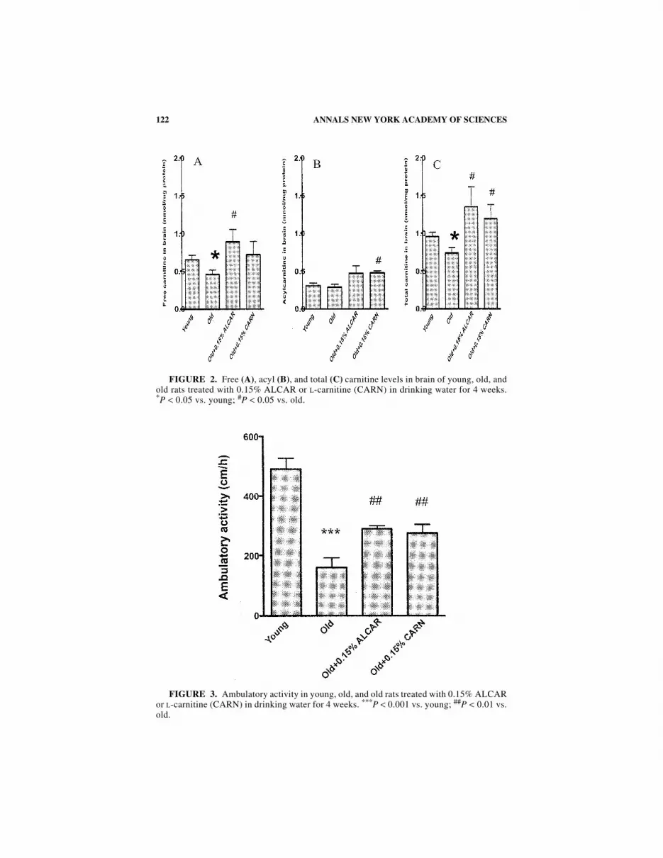
The results from rat brain are shown in FIGURE 2 (A: free L-carnitine; B: acylcarnitine; C: total L-carnitine). There is an age-related decline in free and total L-carnitine levels in the brain. L-Carnitine and ALC elevated carnitine levels in thebrain in old rats similarly (FIG. 2).
Ambulatory Activity
Ambulatory activity declined markedly with age. This decline was partiallyreversed by both ALC and L-carnitine treatment (FIG. 3). L-Carnitine and ALC hada similar effect on increasing ambulatory activity in old rats.
FIGURE 1. Free (A), acyl (B), and total (C) carnitine levels in plasma of young, old,and old rats treated with 0.15% ALCAR or L-carnitine (CARN) in drinking water for4 weeks. *P < 0.05 vs. young; #P < 0.05 and ##P < 0.01 vs. old.
122 ANNALS NEW YORK ACADEMY OF SCIENCES
FIGURE 3. Ambulatory activity in young, old, and old rats treated with 0.15% ALCARor L-carnitine (CARN) in drinking water for 4 weeks. ***P < 0.001 vs. young; ##P < 0.01 vs.old.
FIGURE 2. Free (A), acyl (B), and total (C) carnitine levels in brain of young, old, andold rats treated with 0.15% ALCAR or L-carnitine (CARN) in drinking water for 4 weeks.*P < 0.05 vs. young; #P < 0.05 vs. old.

123LIU et al.: EFFECTS OF L-CARNITINE AND ACETYL-L-CARNITINE
Lipid Peroxidation
As shown in FIGURE 4, old rats had a significant increase in MDA in their brainwhen compared to young rats. ALC decreased the MDA level in the old rat brain, butL-carnitine did not (FIG. 4).
Oxo8dG/Oxo8G Immunostaining of Young and Old Brain:Effects of ALC and L-Carnitine Feeding
We showed previously that the oxidative damage to nucleic acids stained byoxo8dG/oxo8G immunoreactivity occurred predominantly in RNA (>90%).21 Hippo-campal area CA1, underlying white matter, and frontoparietal cortex showed an effectof age [F(1,6) = 8.82, P < 0.025] on levels of oxo8dG/oxo8G immunoreactivity (byrepeated ANOVA analysis). Significant increases in oxo8dG in the deep layers andwhite matter of the frontoparietal cortex along with increases in CA1 and CA4account for the majority of the age effects (FIG. 5). To compare differences in 0.15%ALC with 0.15% L-carnitine for each brain region, repeated-measure ANOVA wasused. Overall, ALC resulted in significantly reduced oxo8dG, which post hoc com-parisons (Scheffe) showed to be significant in the frontotemporal cortex (P < 0.019)and in area CA1 (P < 0.001). The level of oxo8dG/oxo8G immunoreactivity was notreduced in rats treated with L-carnitine as compared to untreated controls.
FIGURE 4. Malondialdehyde (MDA) in young, old, and old rats treated with 0.15%ALCAR or L-carnitine (CARN) in drinking water for 4 weeks. *P < 0.05 vs. young;#P < 0.05 vs. old.

124 ANNALS NEW YORK ACADEMY OF SCIENCES
FIGURE 5. (Top) Representative photographs for oxo8dG/oxo8G immunostaining inbrain area CA1, cortical neurons of the frontoparietal cortex, and underlying white matter.(Bar = 50 µm.) (Bottom) Average nitrotyrosine immunostaining rankings in area of hippo-campal CA1 and cortical neurons of the frontoparietal cortex. Old rats, compared to youngrats, showed significantly higher oxo8dG/oxo8G immunoreactivity in hippocampal CA1and cortical neurons of the frontoparietal cortex. ALCAR reduced the extent of oxo8dG/oxo8G immunoreactivity in area CA1 and in neurons of the frontoparietal cortex in old rats,although only significant in the cortex. Old rats treated with 0.15% L-carnitine showed noreduction in age-associated oxo8dG/oxo8G immunoreactivity.

125LIU et al.: EFFECTS OF L-CARNITINE AND ACETYL-L-CARNITINE
FIGURE 6. (Top) Representative photographs for nitrotyrosine immunostaining inbrain area CA1 and white matter of the frontoparietal cortex. (Bar = 50 µm.) (Bottom)Average nitrotyrosine immunostaining rankings in area CA1 and in the white matter of thefrontoparietal cortex. Old rats, compared to young rats, showed higher nitrotyrosineimmunoreactivity in the hippocampal CA1 area and the white matter of the frontoparietalcortex. ALCAR significantly reduced age-associated increases in nitrotyrosine in the whitematter, but L-carnitine did not.

126 ANNALS NEW YORK ACADEMY OF SCIENCES
Nitrotyrosine Immunostaining of Young and Old Brain:Effects of ALC and L-Carnitine Feeding
Old rats showed increased nitrotyrosine immunoreactivity relative to young ratsin 3/4 regions of the hippocampus that included area CA4 (P < 0.01), CA3(P < 0.01), and CA1 (P < 0.03) (Mann-Whitney U tests). Nitrotyrosine immuno-reactivity in the dentate gyrus was not significantly different between young and oldrats. In addition, there was a trend towards increasing nitrotyrosine levels in thewhite matter of cortex of old rats (P < 0.03), but levels in deep-layer cortical neuronsof the frontoparietal cortex were similar in young and old animals. As shown in FIG-URE 6, ALC and L-carnitine administration resulted in similar levels of nitrotyrosineimmunoreactivity, except in the white matter where ALC significantly reducedstaining, but L-carnitine did not (P < 0.034) (Kruskal-Wallis nonparametric test).
DISCUSSION
The current study directly compared the similar doses of ALC and L-carnitine inaged rats on measures of ambulatory activity; free, acyl, and total carnitine levels;and biomarkers of brain oxidative damage. Both compounds increased peripheraland central carnitine levels, and increased ambulatory activity in old animals. How-ever, only ALC treatment resulted in a decrease in biomarkers of oxidative damage,including lipid peroxidation (MDA), protein oxidation (nitrotyrosine), and oxidativenucleic acid damage (oxo8dG/oxo8G) in the brain of old rats. The concentrationsused here for L-carnitine and ALC are weight/volume. The corresponding moleconcentrations for the 0.15% solution are 7.78 mM for L-carnitine and 6.25 mM forALC, respectively. Therefore, the effect of ALC should be even greater if comparedto the same mole concentration of L-carnitine.
There are difficulties with several enzymatic assays for L-carnitine, including aradiochemical method with L-carnitine O-acetyltransferase, and spectrophotometricmethods involving L-carnitine acetyltransferase or L-carnitine dehydrogenase.42
These methods measure NADH production, which has low molar absorptivity at340 nm, making it difficult to assay carnitine in samples from carnitine-deficientdiseases. In addition, the NADH produced is consumed by another dehydrogenase,which thus causes erroneous measurements. We have used a carnitine kit (AsahiIndustrial, Tokyo), which is highly sensitive and specific for determining L-carnitinelevels by use of carnitine dehydrogenase (EC 1.1.1.108, purified from Alcaligenessp.). The method involves a new enzymatic cycling technique with NADH, thio-NAD+, and L-carnitine dehydrogenase, and measures the increase of absorbance at405 nm of thio-NADH produced during the reaction.42 Due to the high sensitivity,this method allowed us to determine carnitine concentrations at a few micromolesper liter, or in less than 5 µL of plasma. Our results for plasma and brain tissues inyoung and aged Fischer 344 rats are consistent with those reported by Maccari et al.4
in Sprague-Dawley rats, suggesting the method is quite reliable in measuring plasmaand tissue carnitine levels. At the dose of 0.15%, ALC and L-carnitine were botheffective in restoring the age-associated decrease of levels of plasma and braincarnitine to those of young rats.

127LIU et al.: EFFECTS OF L-CARNITINE AND ACETYL-L-CARNITINE
Administration of 0.1% and 1.0% L-carnitine can significantly increase carnitinelevels in young rat serum, liver, kidney, heart, and skeletal muscle.47 A recent studyshowed that a daily dose of 100 mg/kg body weight ALC in drinking water elevatedtotal carnitine levels in plasma, heart, skeletal muscle, and cerebral cortex in oldrats.48 Kuratsune et al. showed that patients with chronic fatigue syndrome have adeficiency of serum acyl carnitine, and ALC supplementation improves the dailyactivity and reduces the symptoms.42,49 They investigated the brain uptake in rhesusmonkeys of acetyl carnitine labeled in different positions by positron emissiontomography and found a high uptake of [2–11C]-acetyl-L-carnitine into the brain,suggesting that endogenous serum acetyl-L-carnitine has some role in conveying anacetyl moiety into the brain, especially under an energy crisis.50 Oral and intra-venous administration of multiple doses of ALC can increase its plasma and CSFconcentration in patients suffering from Alzheimer’s disease.51 Consistent with ourresults, all of these results suggest that dietary supplementation of L-carnitine andALC can elevate peripheral and central carnitine levels similarly.
Our previous report16 showed that old rats had a remarkable decline in ambula-tory activity and that this decline reflected a true loss in activity because the calcu-lated speeds of animals, when they moved, were not significantly different fromthose of young rats. The results here show that ALC and L-carnitine administrationincreased the age-associated decline in ambulatory activity similarly.
Our results on MDA, nitrotyrosine, and oxo8dG/oxo8G (mainly RNA oxidation)in young and old rats are consistent with other reports and support the propositionthat accumulated oxidative damage to macromolecules such as lipid, protein, andnucleic acid may be a major contributor to cellular aging and the degenerativediseases that accompany aging.21,43,52–60 In a previous report,16 we showed that,although 1.5% ALC significantly restored age-associated mitochondrial decay andambulatory decline in old rats, the ALC administration caused an increase in oxidantproduction per oxygen consumed, as measured by 2′,7′-dichlorofluorescin fluores-cence level. This suggests that ALC, by increasing the cellular oxygen consumption,may increase oxidative stress. In the present study, we have demonstrated that, incontrast, 0.15% ALC inhibited oxidative damage to lipids, protein, and nucleicacids. Thus, the oxidative stress increase is a side effect of the high dose of ALCbecause a low dose of ALC can restore most of the age-associated mitochondrialdecay to the young rat level, and endogenous antioxidant defense systems can copewith the increased oxygen consumption. These results also suggest that ALC at lowdoses, unlike L-carnitine, is effective as an antioxidant.
The more effective action of ALC compared to L-carnitine on cognitive functionand oxidative stress may be attributed to the acetyl group or its greater efficiency incrossing the blood-brain barrier.40 ALC is suggested to donate an acetyl group tocholine to form acetylcholine and this hypothesis is the basis of using ALC in thetreatment of age-related cholinergic deficits.61 ALC enhances acetylcholine releasein rat striatum and hippocampus62 and enhances cholinergic synaptic function in oldrats.22 Because transfer of the acetyl moiety from ALC to choline is dependent onthe concentration of the ALC and requires the presence of coenzyme A,63 it mayprovide an explanation for our observed greater effect of ALC than L-carnitine oncognitive function. The hypothesis that the acetyl group of the ALC molecule is theactive group is further supported by the observation that ALC did (but L-carnitinedid not) affect the regeneration of the transected sciatic nerve in rats.64

128 ANNALS NEW YORK ACADEMY OF SCIENCES
In conclusion, ALC and L-carnitine increased ambulatory activity similarly in oldrats and elevated carnitine levels in old rat blood and brain. However, L-carnitine didnot decrease or enhance oxidative damage, while ALC did decrease MDA, nitro-tyrosine, and oxo8dG/oxo8G in old rat brain. These data suggest that ALC is a moreeffective dietary supplement than L-carnitine.
ACKNOWLEDGMENTS
This work was supported by grants from the Ellison Medical Foundation (GrantSS-0422-99), the National Institute on Aging (Grant AG17140), the Wheeler Foun-dation Fund of the University of California, the National Institute of EnvironmentalHealth Sciences Center (Grant ES01896), and the National Center for Complemen-tary and Alternative Medicine Research Scientist Award (K05 AT001323-4) toB. N. Ames; as well as from the National Institute on Aging (Grant AG12694) toC. W. Cotman. We thank Sigma-Tau for the gift of acetyl-L-carnitine and Carol Wehrfor assistance in animal care.
REFERENCES
1. REBOUCHE, C.J. 1992. Carnitine function and requirements during the life cycle.FASEB J. 6: 3379–3386.
2. COSTELL, M., J.E. O’CONNOR & S. GRISOLIA. 1989. Age-dependent decrease ofcarnitine content in muscle of mice and humans. Biochem. Biophys. Res. Commun.161: 1135–1143.
3. LIU, J., H. ATAMNA, H. KURATSUNE & B.N. AMES. 2002. Delaying brain mitochondrialdecay and aging with mitochondrial antioxidants and metabolites. Ann. N.Y. Acad.Sci. 959: 133–166.
4. MACCARI, F., A. ARSENI, P. CHIODI et al. 1990. Levels of carnitines in brain and othertissues of rats of different ages: effect of acetyl-L-carnitine administration. Exp.Gerontol. 25: 127–134.
5. IOSSA, S., M.P. MOLLICA, L. LIONETTI et al. 2002. Acetyl-L-carnitine supplementationdifferently influences nutrient partitioning, serum leptin concentration, and skeletalmuscle mitochondrial respiration in young and old rats. J. Nutr. 132: 636–642.
6. TRIPP, M.E., M.L. KATCHER, H.A. PETERS et al. 1981. Systemic carnitine deficiencypresenting as familial endocardial fibroelastosis: a treatable cardiomyopathy. N.Engl. J. Med. 303: 385–390.
7. WABER, L.J., D. VALLE, C. NEILL et al. 1982. Carnitine deficiency presenting as familialcardiomyopathy: a treatable defect in carnitine transport. J. Pediatr. 101: 700–705.
8. FERLINI, C., C. DE ANGELIS, R. BISELLI et al. 1999. Sequence of metabolic changesduring X-ray-induced apoptosis. Exp. Cell Res. 247: 160–167.
9. BRESOLIN, N., L. FREDDO, L. VERGANI & C. ANGELINI. 1982. Carnitine, carnitine acyl-transferases, and rat brain function. Exp. Neurol. 78: 285–292.
10. DOLEZAL, V. & S. TUCEK. 1981. Utilization of citrate, acetylcarnitine, acetate, pyru-vate, and glucose for the synthesis of acetylcholine in rat brain slices. J. Neurochem.36: 1323–1330.
11. PARADIES, G., F.M. RUGGIERO & P. DINOI. 1992. Decreased activity of the phosphatecarrier and modification of lipids in cardiac mitochondria from senescent rats. Int. J.Biochem. 24: 783–787.
12. PARADIES, G., F.M. RUGGIERO, G. PETROSILLO et al. 1994. Effect of aging and acetyl-L-carnitine on the activity of cytochrome oxidase and adenine nucleotide translocase inrat heart mitochondria. FEBS Lett. 350: 213–215.

129LIU et al.: EFFECTS OF L-CARNITINE AND ACETYL-L-CARNITINE
13. PARADIES, G., G. PETROSILLO, M.N. GADALETA & F.M. RUGGIERO. 1999. The effect ofaging and acetyl-L-carnitine on the pyruvate transport and oxidation in rat heartmitochondria. FEBS Lett. 454: 207–209.
14. GADALETA, M.N., V. PETRUZZELLA, M. RENIS et al. 1990. Reduced transcription ofmitochondrial DNA in the senescent rat: tissue dependence and effect of L-carnitine.Eur. J. Biochem. 187: 501–506.
15. VILLA, R.F., L. TURPEENOJA, G. BENZI & A.M. GIUFFRIDA STELLA. 1998. Action of L-acetylcarnitine on age-dependent modifications of mitochondrial membrane proteinsfrom rat cerebellum. Neurochem. Res. 10: 909–916.
16. HAGEN, T.M., R.T. INGERSOLL, C.M. WEHR et al. 1998. Acetyl-L-carnitine fed to oldrats partially restores mitochondrial function and ambulatory activity. Proc. Natl.Acad. Sci. USA 95: 9562–9566.
17. TAGLIALATELA, G., A. CAPRIOLI, A. GIULIANI & O. GHIRARDI. 1996. Spatial memoryand NGF levels in aged rats: natural variability and effects of acetyl-L-carnitine treat-ment. Exp. Gerontol. 31: 577–587.
18. SHARMAN, E.H., N.D. VAZIRI, Z. NI et al. 2002. Reversal of biochemical andbehavioral parameters of brain aging by melatonin and acetyl L-carnitine. Brain Res.957: 223–230.
19. MCKAY HART, A., M. WIBERG & G. TERENGHI. 2002. Pharmacological enhancement ofperipheral nerve regeneration in the rat by systemic acetyl-L-carnitine treatment.Neurosci. Lett. 334: 181–185.
20. YASUI, F., S. MATSUGO, M. ISHIBASHI et al. 2002. Effects of chronic acetyl-L-carnitinetreatment on brain lipid hydroperoxide level and passive avoidance learning insenescence-accelerated mice. Neurosci. Lett. 334: 177–180.
21. LIU, J., E. HEAD, A. GHARIB et al. 2002. Memory loss in old rats is associated with brainmitochondrial decay and RNA/DNA oxidation: partial reversal by feeding acetyl-L-carnitine and/or R-alpha-lipoic acid. Proc. Natl. Acad. Sci. USA 99: 2356–2361.
22. ANDO, S., T. TADENUMA, Y. TANAKA et al. 2001. Enhancement of learning capacity andcholinergic synaptic function by carnitine in aging rats. J. Neurosci. Res. 66: 266–271.
23. MONTGOMERY, S.A., L.J. THAL & R. AMREIN. 2003. Meta-analysis of double blindrandomized controlled clinical trials of acetyl-L-carnitine versus placebo in thetreatment of mild cognitive impairment and mild Alzheimer’s disease. Int. Clin.Psychopharmacol. 18: 61–71.
24. KIDD, P.M. 1999. A review of nutrients and botanicals in the integrative managementof cognitive dysfunction. Altern. Med. Rev. 4: 144–161.
25. KALAISELVI, T. & C. PANNEERSELVAM. 1998. Effect of L-carnitine on the status ofliperoxidation and antioxidants in aging rats. J. Nutr. Biochem. 9: 575–581.
26. RANI, P.J. & C. PANNEERSELVAM. 2002. Effect of L-carnitine on brain lipid peroxidationand antioxidant enzymes in old rats. J. Gerontol. Biol. Sci. Med. Sci. 57: B134–B137.
27. ARDUINI, A. 1992. Carnitine and its acyl esters as secondary antioxidants? Am. HeartJ. 123: 1726–1727.
28. ARDUINI, A., S. DOTTORI, F. MOLAJONI et al. 1995. Is the carnitine system part of theheart antioxidant network? In The Carnitine System: A New Therapeutical Approachto Cardiovascular Medicine, pp. 169–181. Kluwer. Dordrecht.
29. DI GIACOMO, C., F. LATTERI, C. FICHERA et al. 1993. Effect of acetyl-L-carnitine onlipid peroxidation and xanthine oxidase activity in rat skeletal muscle. Neurochem.Res. 18: 1157–1162.
30. KAUR, J., D. SHARMA & R. SINGH. 2001. Acetyl-L-carnitine enhances Na(+),K(+)-ATPaseglutathione-S-transferase and multiple unit activity and reduces lipid peroxidationand lipofuscin concentration in aged rat brain regions. Neurosci. Lett. 301: 1–4.
31. BOERRIGTER, M.E., C. FRANCESCHI, E. ARRIGONI-MARTELLI et al. 1993. The effect of L-carnitine and acetyl-L-carnitine on the disappearance of DNA single-strand breaks inhuman peripheral blood lymphocytes. Carcinogenesis 14: 2131–2136.
32. SCHINETTI, M.L., D. ROSSINI, R. GRECO & A. BERTELLI. 1987. Protective action ofacetylcarnitine on NADPH-induced lipid peroxidation of cardiac microsomes. DrugsExp. Clin. Res. 13: 509–515.
33. GEREMIA, E., C. SANTORO, D. BARATTA et al. 1988. Antioxidant action of acetyl-L-carnitine: in vitro study. Med. Sci. Res. 16: 699–700.

130 ANNALS NEW YORK ACADEMY OF SCIENCES
34. REZNICK, A.Z., V.E. KAGAN, R. RAMSEY et al. 1992. Antiradical effects in L-propionylcarnitine protection of the heart against ischemia-reperfusion injury: the possiblerole of iron chelation. Arch. Biochem. Biophys. 296: 394–401.
35. PAULSON, D.J., A.L. SHUG & J. ZHAO. 1992. Protection of the ischemic diabetic heartby L-propionylcarnitine therapy. Mol. Cell. Biochem. 116: 131–137.
36. LUO, X., B. REICHETZER, J. TRINES et al. 1999. L-Carnitine attenuates doxorubicin-induced lipid peroxidation in rats. J. Free Radicals Biol. Med. 26: 1158–1165.
37. TESCO, G., S. LATORRACA, P. PIERSANTI et al. 1992. Protection from oxygen radicaldamage in human diploid fibroblasts by acetyl-L-carnitine. Dementia 3: 58–60.
38. BACON, B.R., R. NEILL & C.H. PARK. 1986. Iron-induced peroxidative injury toisolated rat hepatic mitochondria. J. Free Radicals Biol. Med. 2: 339–342.
39. HANSFORD, R.G. & F. CASTRO. 1982. Age-linked changes in the activity of enzymes ofthe tricarboxylate cycle and lipid oxidation, and of carnitine content, in muscles ofthe rat. Mech. Ageing Dev. 19: 191–200.
40. KIDO, Y., I. TAMAI, A. OHNARI et al. 2001. Functional relevance of carnitine transporterOCTN2 to brain distribution of L-carnitine and acetyl-L-carnitine across the blood-brain barrier. J. Neurochem. 79: 959–969.
41. YAMAGUTI, K., H. KURATSUNE, Y. WATANABE et al. 1996. Acylcarnitine metabolismduring fasting and after refeeding. Biochem. Biophys. Res. Commun. 225: 740–746.
42. KURATSUNE, H., K. YAMAGUTI, M. TAKAHASHI et al. 1994. Acylcarnitine deficiency inchronic fatigue syndrome. Clin. Infect. Dis. 18: S62–S67.
43. HAGEN, T.M., R.T. INGERSOLL, J. LYKKESFELDT et al. 1999. (R)-Alpha-lipoic acid–supplemented old rats have improved mitochondrial function, decreased oxidativedamage, and increased metabolic rate. FASEB J. 13: 411–418.
44. YEO, H.C., H.J. HELBOCK, D.W. CHYU & B.N. AMES. 1994. Assay of malondialdehydein biological fluids by gas chromatography–mass spectrometry. Anal. Biochem. 220:391–396.
45. LIU, J., H.C. YEO, S.J. DONIGER & B.N. AMES. 1997. Assay of aldehydes from lipidperoxidation: gas chromatography–mass spectrometry compared to thiobarbituricacid. Anal. Biochem. 245: 161–166.
46. YEO, H.C., J. LIU, H.J. HELBOCK & B.N. AMES. 1999. Assay of malondialdehyde andother alkanals in biological fluids by gas chromatography–mass spectrometry.Methods Enzymol. 300: 70–78.
47. REBOUCHE, C.J. 1983. Effect of dietary carnitine isomers and gamma-butyrobetaine onL-carnitine biosynthesis and metabolism in the rat. J. Nutr. 113: 1906–1913.
48. TANAKA, Y., R. SASAKI, F. FUKUI et al. 2004. Acetyl-L-carnitine supplementationrestores decreased tissue carnitine levels and impaired lipid metabolism in aged rats.J. Lipid Res. 45: 729–735.
49. KURATSUNE, H., K. YAMAGUTI, G. LINDH et al. 1998. Low levels of serum acylcarnitinein chronic fatigue syndrome and chronic hepatitis type C, but not seen in otherdiseases. Int. J. Mol. Med. 2: 51–56.
50. KURATSUNE, H., Y. WATANABE, K. YAMAGUTI et al. 1997. High uptake of [2–11C]-acetyl-L-carnitine into the brain: a PET study. Biochem. Biophys. Res. Commun.231: 488–493.
51. PARNETTI, L., A. GAITI, P. MECOCCI et al. 1992. Pharmacokinetics of IV and oralacetyl-L-carnitine in a multiple dose regimen in patients with senile dementia ofAlzheimer type. Eur. J. Clin. Pharmacol. 42: 89–93.
52. AMES, B.N., M.K. SHIGENAGA & T.M. HAGEN. 1993. Oxidants, antioxidants, and thedegenerative diseases of aging. Proc. Natl. Acad. Sci. USA 90: 7915–7922.
53. HARMAN, D. 1981. The aging process. Proc. Natl. Acad. Sci. USA 78: 7124–7128.54. HARMAN, D. 1999. Aging: minimizing free radical damage. J. Anti-Aging Med. 2: 15–36.55. STADTMAN, E.R. 1992. Protein oxidation and aging. Science 257: 1220–1224.56. AGARWAL, S. & R.S. SOHAL. 1994. Aging and protein oxidative damage. Mech. Ageing
Dev. 75: 11–19.57. AGARWAL, S. & R.S. SOHAL. 1995. Differential oxidative damage to mitochondrial
proteins during aging. Mech. Ageing Dev. 85: 55–63.58. LIU, J., D.W. KILLILEA & B.N. AMES. 2002. Age-associated mitochondrial oxidative
decay: improvement of carnitine acetyltransferase substrate-binding affinity, and

131LIU et al.: EFFECTS OF L-CARNITINE AND ACETYL-L-CARNITINE
activity in brain by feeding old rats acetyl-L-carnitine and/or R-alpha-lipoic acid.Proc. Natl. Acad. Sci. USA 99: 1876–1881.
59. HAGEN, T.M., V. VINARSKY, C.M. WEHR & B.N. AMES. 2000. (R)-Alpha-lipoic acidreverses the age-associated increase in susceptibility of hepatocytes to tert-butyl-hydroperoxide both in vitro and in vivo. Antioxid. Redox Signal. 2: 473–483.
60. HAGEN, T.M., J. LIU, J. LYKKESFELDT et al. 2002. Feeding acetyl-L-carnitine and lipoicacid to old rats significantly improves metabolic function while decreasing oxidativestress. Proc. Natl. Acad. Sci. USA 99: 1870–1875.
61. SODERBERG, M., C. EDLUND, K. KRISTENSSON & G. DALLNER. 1990. Lipid compositionsof different regions of the human brain during aging. J. Neurochem. 54: 415–423.
62. IMPERATO, A., M.T. RAMACCI & L. ANGELUCCI. 1989. Acetyl-L-carnitine enhancesacetylcholine release in the striatum and hippocampus of awake freely moving rats.Neurosci. Lett. 107: 251–255.
63. WHITE, H.L. & P.W. SCATES. 1990. Acetyl-L-carnitine as a precursor of acetylcholine.Neurochem. Res. 15: 597–601.
64. FERNANDEZ, E., R. PALLINI, C. GANGITANO et al. 1989. Effects of L-carnitine, L-acetyl-carnitine, and gangliosides on the regeneration of the transected sciatic nerve in rats.Neurol. Res. 11: 57–62.