comparative sequence, structure and redox analyses of klebsiella pneumoniae dsba show that...
TRANSCRIPT

Comparative Sequence, Structure and Redox Analyses ofKlebsiella pneumoniae DsbA Show That Anti-VirulenceTarget DsbA Enzymes Fall into Distinct ClassesFabian Kurth1, Kieran Rimmer2, Lakshmanane Premkumar1, Biswaranjan Mohanty2, Wilko Duprez1, MariaA. Halili1, Stephen R. Shouldice1¤a, Begoña Heras1¤b, David P. Fairlie1, Martin J. Scanlon2,3*, Jennifer L.Martin1*
1 Division of Chemistry and Structural Biology, Institute for Molecular Bioscience, The University of Queensland, Brisbane, Queensland, Australia, 2 Faculty ofPharmacy and Pharmaceutical Sciences, Medicinal Chemistry, Monash Institute of Pharmaceutical Sciences, Monash University, Parkville, Victoria, Australia,3 ARC Centre of Excellence for Coherent X-ray Science, Monash University, Parkville, Victoria, Australia
Abstract
Bacterial DsbA enzymes catalyze oxidative folding of virulence factors, and have been identified as targets forantivirulence drugs. However, DsbA enzymes characterized to date exhibit a wide spectrum of redox properties anddivergent structural features compared to the prototypical DsbA enzyme of Escherichia coli DsbA (EcDsbA).Nonetheless, sequence analysis shows that DsbAs are more highly conserved than their known substrate virulencefactors, highlighting the potential to inhibit virulence across a range of organisms by targeting DsbA. For example,Salmonella enterica typhimurium (SeDsbA, 86 % sequence identity to EcDsbA) shares almost identical structural,surface and redox properties. Using comparative sequence and structure analysis we predicted that five otherbacterial DsbAs would share these properties. To confirm this, we characterized Klebsiella pneumoniae DsbA(KpDsbA, 81 % identity to EcDsbA). As expected, the redox properties, structure and surface features (from crystaland NMR data) of KpDsbA were almost identical to those of EcDsbA and SeDsbA. Moreover, KpDsbA and EcDsbAbind peptides derived from their respective DsbBs with almost equal affinity, supporting the notion that compoundsdesigned to inhibit EcDsbA will also inhibit KpDsbA. Taken together, our data show that DsbAs fall into differentclasses; that DsbAs within a class may be predicted by sequence analysis of binding loops; that DsbAs within a classare able to complement one another in vivo and that compounds designed to inhibit EcDsbA are likely to inhibitDsbAs within the same class.
Citation: Kurth F, Rimmer K, Premkumar L, Mohanty B, Duprez W, et al. (2013) Comparative Sequence, Structure and Redox Analyses of Klebsiellapneumoniae DsbA Show That Anti-Virulence Target DsbA Enzymes Fall into Distinct Classes. PLoS ONE 8(11): e80210. doi:10.1371/journal.pone.0080210
Editor: Vladimir N. Uversky, University of South Florida College of Medicine, United States of America
Received August 25, 2013; Accepted September 30, 2013; Published November 14, 2013
Copyright: © 2013 Kurth et al. This is an open-access article distributed under the terms of the Creative Commons Attribution License, which permitsunrestricted use, distribution, and reproduction in any medium, provided the original author and source are credited.
Funding: This work was supported by an ARC (www.arc.gov.au) Australian Laureate Fellowship (FL0992138) to JLM—which also supported PhDscholarships to FK and WD—and an NHMRC (www.nhmrc.gov.au) Project Grant (APP1009785) to MJS and BH. JLM is also an Honorary NHMRCResearch Fellow (455829). The funders had no role in study design, data collection and analysis, decision to publish, or preparation of the manuscript.
Competing interests: The authors have declared that no competing interests exist.
* E-mail: [email protected] (JLM); [email protected] (MJS)
¤a Current address: Janssen-Cilag Pty Ltd., Macquarie Park, New South Wales, Australia¤b Current address: Institute for Molecular Science, Latrobe University, Melbourne, Victoria, Australia
Introduction
Antibiotic resistance has increased dramatically over the lastdecade and the consequent lack of treatment options poses amajor threat for public health [1]. One approach to develop newchemical classes of antibacterials is to target virulence factorsthat cause disease in antibiotic resistant organisms [2]. Mostpathogenic Enterobacteriaceae encode an oxidative foldingpathway essential for virulence factor production [2-5].Typically, the oxidative folding machinery includes a solublethioredoxin-fold protein, DsbA, and an integral membrane
protein partner, DsbB [6-8]. The disulfide form of DsbA is highlyoxidizing and donates its disulfide bond to unfolded substrateproteins [9], leaving DsbA in the inactive reduced form. Theinner membrane protein DsbB, in concert with its cofactorubiquinone, interacts with reduced DsbA to oxidize the activesite cysteines and convert DsbA to its functionally competentdisulfide form [10]. Inhibition of the interaction between DsbAand substrate proteins or between DsbA and its partner DsbBcould constitute a means of blocking virulence factor formationand thereby of inhibiting virulence of bacterial pathogens.Supporting this notion, deletion of DsbA homologues in
PLOS ONE | www.plosone.org 1 November 2013 | Volume 8 | Issue 11 | e80210

pathogenic organisms results in diminished virulence ininfection models [2,11] and deletion of dsbA or dsbB inuropathogenic E. coli (UPEC) severely attenuated its ability tocolonize the bladder [11,12].
The characteristic properties of EcDsbA include: an activesite CPHC motif that forms a destabilizing disulfide (Tm reducedEcDsbA 350 K; Tm oxidized EcDsbA 342 K) [13]; the more N-terminal of the two cysteines is nucleophilic and highly acidic,pKa 3.3 (usual value for a cysteine is 8-9) [9]; and EcDsbA ishighly oxidizing (redox potential -122 mV) [9]. The past 5 yearshas seen the characterization of DsbA enzymes from manyother bacteria including DsbAs with varying degrees ofsequence identity to EcDsbA such as Neisseria meningitidisDsbA1 (NmDsbA1, 23% identity), Pseudomonas aeruginosaDsbA (PaDsbA, 30%) and Vibrio cholerae DsbA (VcDsbA, orTcpG, 40%). These DsbAs share a similar structural fold withEcDsbA though their surface properties vary [14] and theyexhibit a wide range of redox properties (Table 1). Importantly,the EcDsbA hydrophobic groove that interacts with its essentialpartner EcDsbB is considerably truncated in NmDsbA1,PaDsbA and VcDsbA [15-17]. This modification and othersurface changes in these DsbAs indicate that they fall into aseparate class, distinct from EcDsbA, and that inhibitorsdesigned against EcDsbA may not inhibit members of thisclass of DsbA. Conversely, DsbAs closely related to EcDsbAshould be susceptible to the same mode of chemical inhibition.
Here we tested how close the sequence relationship must beto produce similar redox properties and binding interactions.We investigated two well-characterised DsbAs sharing 86%sequence identity, from E. coli K-12 strain (EcDsbA) and S.enterica Typhimurium DsbA strain SL1344 (SeDsbA), byapplying comparative structural, sequence and redox analysesto identify properties conserved across these two enzymes.The results allow us to place DsbAs of five other Gram-negative bacteria Enterobacteriaceae, namely Shigella flexneri
Table 1. Comparison of structures and redox properties ofDsbAs.
Seq id toEcDsbA
RMSD(Å)
RMSD#Cα E°’ (mV)
pKa“Cys30”
Tm (K)(red/ox)
Other DsbAsa 10 - 40 %
1.3 -2.9
122 -167
-80/-163 3.0 - 5.1337-357 /331-341
EcDsbAb 100 % 0.6 176 -122 3.3 350 / 341SeDsbAc 86 % 0.9 176 -126 3.3 351 / 343KpDsbA 81 % 0.8 176 -116 3.2 347 / 335VcDsbAd 40 % 1.8 168 - 116 5.1 357 / 346NmDsbA1e 23 % 2.6 163 - 80 3.0 348 / 333
a. [14] , redox potential range for NmDsbA1 (-80 , WpDsbA (- 163); pKa range,NmDsbA1 (3.0), VcDsbA (5.1); Tm oxidised (min) NmDsbA3, (max) VcDsbA andreduced (min) NmDsbA3, (max) VcDsbA.b. [6] [14],, RMSD of EcDsbA derived from the overlay of molecules A and B fromthe asymmetric unit in 1FVK.c. [43] and [14]d. [54]e. [51]doi: 10.1371/journal.pone.0080210.t001
8401 (SfDsbA, 100% sequence identity to EcDsbA),Enterobacter cloacae SCF-1 (EnDsbA, 84%), Citrobacterkoseri ATCC BAA-895 (CkDsbA, 84%), Cronobacter sakazakiiSP291 (CsDsbA, 82%) and K. pneumonia 342 (KpDsbA, 81%)into the same DsbA cluster as SeDsbA and EcDsbA. Toassess whether the redox and structural properties aremaintained in this DsbA group we focused on KpDsbA, whichshares the lowest sequence identity with EcDsbA. Wedetermined the high resolution crystal structure of reducedKpDsbA and the NMR solution structure of oxidized KpDsbA,and we measured the redox properties of this enzyme. Asexpected, the redox properties, surface characteristics andbinding properties of KpDsbA are similar to those of EcDsbAsuggesting that inhibitors developed against EcDsbA are likelyto also be effective against other members of this DsbAsubclass.
Materials and Methods
Protein productionCodon-optimized K. pneumoniae dsbA (GenBank®
accession number ACI08793), lacking the sequence coding forthe predicted signal sequence (19 aa), was cloned into amodified pMCSG7 (Midwest Center for Structural Genomics)vector compatible with ligation-independent cloning. Thismodified vector encoded a leader sequence consisting of an N-terminal His6-tag followed by a linker containing the tobacco-etch virus protease (TEV) recognition sequence. KpDsbA wasexpressed in BL21(DE3)pLys cells using autoinduction medium[18] and purified with Talon® resin (Clontech, Australia). TheHis6-tag was removed by TEV protease, leaving theengineered KpDsbA with two additional amino acids (S–1 andN0) at the N-terminus. A final size-exclusion chromatographystep using a Superdex75 column (GE Healthcare, USA)yielded highly purified KpDsbA, as judged by SDS-PAGE.Oxidized or reduced KpDsbA was prepared using a 25-foldmolar excess of copper-(II)-1,10-phenanthroline or DTT,respectively. Oxidizing/reducing agent was then removed andthe protein buffer-exchanged into 10 mM HEPES, pH 7.4 inone step using GE-25 Sephadex desalting resin forcrystallization and biochemical experiments.
Preparation of E. coli DsbA (CAA56736), S. entericaTyphimurium DsbA (AAB81592) and E. coli DsbC (AAA83074),lacking the periplasmic leader signal were purified as describedfor KpDsbA. For peptide oxidation experiments, E. coli DsbB(AAC74269) membrane extracts were prepared as describedpreviously [19] and re-suspended in phosphate buffered saline(PBS, 137 mM NaCl, 2.7 mM KCl, Na2HPO4 10 mM andKH2PO4, pH 7.4) containing 10 % glycerol.
KpDsbA Complementation of EcDsbAThe ability of KpDsbA to rescue non-motile E. coli dsbA- null
(JCB817) and dsbA-/dsbB- double-null (JCB818) strains wasassessed in a cell-swarming assay as described previously[16]. The mature KpDsbA coding sequence was cloned intopBAD33 under an arabinose inducible promotor with theEcDsbA periplasmic signal sequence. A wild-type EcDsbAcloned into pBAD33 vector was used as a positive control.
Comparative Analysis of Klebsiella pneumoniae DsbA
PLOS ONE | www.plosone.org 2 November 2013 | Volume 8 | Issue 11 | e80210

Non-motile E. coli dsbA- deficient (JCB817) or dsbA- / dsbB-
double-mutant (JCB818) [3] cells (2x10⁶) transformed with aKpDsbA or EcDsbA pBAD33 inducible vector were spottedonto the center of a soft M63 minimal agar plate containing 40mg/mL of each amino acid (except L-cysteine). Plates wereincubated at 37 °C and motility of cells monitored using aMolecular Imager® Gel Doc™ system from BIO-RAD (CA94547, USA) after 3-7 h. Complementation experiments wererepeated as biological triplicates.
KpDsbA Disulfide Reductase ActivityUnder mild reducing conditions, DsbA proteins can reduce
the intermolecular disulfide bonds formed between insulinchains A and B [3]. The rate of disulfide bond reduction can bespectroscopically followed at OD650nm by an increase in turbidityresulting from production of the insoluble insulin chain B [20].Samples were prepared in 1 cm cuvettes containing 10 μM ofprotein (KpDsbA, EcDsbA or EcDsbC), 0.33 mM DTT and 2mM EDTA in 100 mM NaH2PO4 / Na2HPO4 titrated to pH 7.0.Catalysis was initiated by the addition of 0.131 mM insulin(I0516, Sigma-Aldrich, Australia) to the sample mixture. Theassay was repeated three times and data were plotted showingstandard deviations.
Measurement of KpDsbA Redox PotentialThe standard redox potential of KpDsbA was measured
using its intrinsic tryptophan fluorescence, as describedpreviously for EcDsbA [6]. Oxidized KpDsbA was incubated for12 h at 25 °C in degassed 100 mM NaH2PO4 / Na2HPO4 buffer(pH 7.0, 1 mM EDTA, 298K), containing 1 mM oxidizedglutathione (GSSG) and varying concentrations of reducedglutathione (GSH) (0–2 mM). KpDsbA (200 µL) from eachredox condition was dispensed into a 96-well plate (TPP AG,Switzerland #92096) and tryptophan fluorescence wasmeasured (excitation at 280 nm, emission set to 332 nm) usinga microplate reader (Synergy H1 and Gen5 2.0 software,Biotek, USA). Data were normalized and the redox potentialwas calculated as described for EcDsbA [6]. In brief, theequilibrium constant Keq was calculated using the equation: Y =([GSH]2 / [GSSH])/(Keq + ([GSH]2 / [GSSH])), where Y is thefraction of reduced protein at equilibrium. The redox potentialfor KpDsbA was calculated from the Nernst equation: E0’
KpDsbA =E0’
GSH/GSSH - (RT/nF)lnKeq where E0’GSH/GSSH = - 240 mV, R is the
ideal gas constant 8.314 JK-1mol-1, T is the absolutetemperature in K, n is the number of electrons transferred (n =2), F is the Faraday constant 9.648x10⁴ Cmol-1 and Keq is theequilibrium constant derived from the binding equation. Allmeasurements were performed as biological triplicates. Thegraph shows a plot of the average values including error barsrepresenting the standard deviation for the replicates.
KpDsbA Thiolate Anion pKa DeterminationThe pH-dependent absorbance of the catalytic thiolate anion
of KpDsbA was followed at 240 nm [21] using a CARY 50UV/VIS spectrophotometer (Agilent Technologies, USA). ThepH titration measurements of oxidized or reduced KpDsbA (40μM) in 2 mL composite buffer (10 mM Tris, 10 mM sodiumcitrate, 10 mM K2HPO4, 10 mM KH2PO4, 200 mM KCl, and 1
mM EDTA) were conducted at 22 °C. Absorbance (λ = 240 and280 nm) was measured between pH 6.5 and 2.0 in 0.25increments. The pKa value was calculated from the fittedcurves of three replicates using the Henderson-Hasselbalchequation (pH = pKa - log ([A240 ⁄A280]red ⁄ [A240 ⁄A280]oxid)).Experiments were repeated at least three times. Plotted datarepresent average values and error bars represent thestandard deviations across the replicates.
Relative Stability of Oxidized and Reduced Forms ofDsbA Enzymes
Temperature-induced unfolding of native SeDsbA andKpDsbA was determined as described previously [13] using aJasco J-810 circular dichroism (CD) spectropolarimeter (Jasco,USA). The redox state of the protein was confirmed usingEllman’s reagent [22]. The largest difference in molar ellipticityfor oxidized or reduced enzymes was calculated from initial far-UV CD spectra (from 250 nm to 190 nm) recorded at 25 °C and95 °C, respectively. The unfolding of oxidized and reducedprotein (SeDsbAox = 220 nm, SeDsbAred = 220.5 nm andKpDsbAox = 211 nm, KpDsbAred = 209.5 nm) was monitored ata heat rate of 1 K / min from 298 K to 368 K in a 1 mm quartzcuvette. All measurements were carried out with 10 µM proteinin 100 mM NaH2PO4 / Na2HPO4, 1 mM EDTA at pH 7.0.Samples for measurement of reduced enzyme contained 0.75mM DTT. Raw data were analyzed in Prism and fitted to a two-state unfolding model as described previously [23]. Thestandard deviation was measured from three replicates.
KpDsbA Dithiol Oxidation ActivityA peptide (CQQGFDGTQNSCK) with a 1,4,7,10-
tetraazacyclododecane-1,4,7,10-tetraacetic acid (DOTA) groupamide-coupled to the N-terminus, and a methylcoumarinamide-coupled to the ε-amino group of the C-terminal lysine,was purchased from AnaSpec (Fremont, CA). Lyophilizedpeptide was re-suspended in 100 mM imidazole, pH 6, at aconcentration of 2 mM. Europium trifluoromethanesulfonate(Sigma Aldrich, Australia) solution (100 mM) was added to thepeptide at a molar ratio of 2:1 and incubated for 5 min at roomtemperature, to allow europium chelation. The peptide solutionwas then immediately aliquoted, flash frozen in liquid nitrogenand stored at -80°C. An increase in fluorescence occurs uponoxidation of the peptide cysteines to form a disulfide. Thus,fluorescence can be used to monitor the capacity of DsbAenzymes to catalyse dithiol oxidation.
Assays were conducted using a Synergy H1 multimode platereader (BioTek, USA) with the excitation wavelength set to 340nm and emission to 615 nm. A 150 μs delay before readingand 100 μs reading time were used for time-resolvedfluorescence. The assay was performed in a white 384-wellplate (Perkin Elmer OptiPlate-384, Part #: 6007290). The bufferconsisted of 50 mM MES, 50 mM NaCl and 2 mM EDTA at pH5.5. The reaction consisted of a 50 μL solution in each well,containing 160 nM EcDsbA, KpDsbA or SeDsbA, 1.6 μMEcDsbB (crude membrane extracts, containing ubiquinone)and 8 μM peptide substrate added last to initiate the reaction.Samples containing buffer and DsbA or buffer and peptidewere used as controls. Data were measured for three
Comparative Analysis of Klebsiella pneumoniae DsbA
PLOS ONE | www.plosone.org 3 November 2013 | Volume 8 | Issue 11 | e80210

replicates and are presented as mean values, with thestandard error of the mean indicated by error bars.
KpDsbA Crystallization and Crystal StructureDetermination
After initial screening using the UQ ROCX facilities, crystalsof reduced KpDsbA were grown at 20 °C in VDXm 24-wellplates (Hampton Research) using the hanging-drop vapordiffusion method. Screening plates were imaged and incubatedin a RockImager 1000 (Formulatrix, MA, USA). Dropscontained 0.5 μL of 180 mg/mL reduced KpDsbA and 0.5 μL ofcrystallization solution (0.1 M succinic acid pH 5.3, 25 % (w/v)polyethylene glycol 1500 and 15 % (v/v) 2-methyl-2,4-pentanediol). For diffraction data measurement, crystals werefrozen in liquid nitrogen without additional cryo-protectant.Diffraction data were measured at the Australian Synchrotronmicro-focus MX2 beamline using BlueIce software [24].Reflections were processed in Mosflm [25] and XDS [26],analyzed and converted to MTZ in Pointless [27] and scaled inSCALA [27]. Phases were obtained by molecular replacement(MR) using PHASER [28] with EcDsbA as template (PDB code:1DSB) . The initial model was improved by iterative modelbuilding in COOT [29] and refinement in PHENIX [30].However, the progress of refinement was stalled with a high R-factor/Rfree of 25.7 % / 29.3 %. Diffraction data analysis inPhenix.xtriage indicated that the crystal was merohedrallytwinned with a twinning fraction of 0.42. Further refinementcycles were performed using the twin target function asimplemented in PHENIX with the twinning operator h,-h-k,-l.Two fold non-crystallographic symmetry (NCS) is present(which does not align with space group axes), though NCS wasnot used at any stage of refinement. The refinement finallyconverged after several TLS refinement cycles. No atoms weremodeled into additionally spherical density located betweenchain D (L133) and chain B (T57) because it was not obviouswhat was bound. The stereochemical quality of the final modelwas assessed using MolProbity [31]. A summary of the dataprocessing and refinement statistics are provided in Table 2.
Molecular figures were generated in PyMOL (The PyMOLMolecular Graphics System, Version 1.5.0.4 Schrödinger, LLC)and figures of the electrostatic potential were generated usingAPBS [32]. The surface, including the proportion of carbonatoms lining the hydrophobic groove in KpDsbA, wascalculated using the CastP server [33], by averaging over allsix molecules within the asymmetric unit. RMSD calculationsand structural alignments were conducted using PyMOL aswell as FATCAT [34].
NMR Structure Determination of Oxidized KpDsbAA sample of uniformly 13C,15N labeled oxidized KpDsbA (1.3
mM) was prepared in 50 mM MES (pH 6.5, 10% 2H2O and 90% 1H2O). NMR experiments were conducted at 303 K on either600 MHz or 800 MHz spectrometers equipped withcryogenically cooled probes. All spectra were acquired withstandard pulse sequences and processed using TOPSPIN3.1(Bruker BioSpin). HN, N, Cα, Cα-1, Cβ, Cβ-1 peak lists weregenerated manually in CARA using 2D [15N,1H]-HSQC, 3DHNCA, 3D CBCA(CO)NH and 3D HNCACB spectra and used
as the input for automated backbone assignments using UNIO-MATCH. These assignments were refined manually andextended using 3D 15N-resolved [1H,1H]-NOESY. Hβ, Hα
assignments were obtained using a 3D HBHA(CBCACO)NHspectrum. HN, N, Cα and Cβ assignments together with Hβ, Hα
were provided as input for UNIO-ATNOS/ASCAN forautomated side-chain assignments using 3D 15N-, 13Cali - and13Caro - resolved [1H,1H] NOESY datasets [35,36]. Upper limitsfor distance restraints used in structure calculations wereautomatically generated from NOESY datasets using UNIO-ATNOS/CANDID and the structure of oxidized KpDsbA wasdetermined using the torsion angle dynamics programCYANA3.0 [37]. Conformers with lowest CYANA target functionvalues were energy minimized using OPALp and validatedusing structure validation tools (http:/www.pdb.org/ and http:/www.nihserver.mbi.ucla.edu/). Structures were inspected and
Table 2. X-ray data measurement and refinement statisticsfor KpDsbA.
Data collection ValueSpace group P 32Unit cell dimensions a (Å) 91.5b (Å) 91.5c (Å) 147.2α, β, γ (°) 90, 90, 120Wavelength (Å) 0.95369Resolution (Å) 53.94 - 1.99 (2.10 - 1.99)Number measured reflections 527,166Number of unique reflections 94,694Rmergea 0.091 (0.566)Rp.i.m. 0.043 (0.264)<I>/<σI> 11.1 (2.9)Redundancy 5.6 (5.5)Completeness (%) 99.9 (99.9)
Refinement statistics Number of Reflections 94,693Resolution (Å) 53.9-1.99 (2.02 -1.99)Rfree (%) 19.6 (31.9)Rwork (%) 16.1 (27.8)Number of monomers in a.u. 6Number of protein atoms 16622Number of waters 371B factors (Å2) Wilson 29.6Protein atoms 39.4Waters 41.4RMSD Bond length (Å) 0.004RMSD Bond angles (°) 0.740Ramachandran favored / outlier (%) 97.4 / 0Molprobity clashscore / scoreb 2.23 [99th(712)] / 1.12 [100th(12290)]
a. The values in parentheses refer to the highest resolution shell.b. 100th Molprobity [31] percentile is the best among structures of comparableresolution; 0th percentile is the worst. The number of structures included in thecomparison is given in parentheses within square brackets.doi: 10.1371/journal.pone.0080210.t002
Comparative Analysis of Klebsiella pneumoniae DsbA
PLOS ONE | www.plosone.org 4 November 2013 | Volume 8 | Issue 11 | e80210

analyzed with MOLMOL [38]. Table 3 summarizes the NMRstatistics.
Binding Affinity of DsbA-Interacting PeptidesCrystal structures of the EcDsbA:EcDsbB complex revealed
that the P2 loop region of EcDsbB interacts with EcDsbA[39,40]. Two peptides derived from the P2 loop sequences ofEcDsbB and KpDsbB (Ec – PSPFATCD and Kp – PSPFQTCD)were synthesized by solid-phase methods using Fmocdeprotection on rink-amide MBHA resin (leading to C-terminalamidation) and capped by N-terminal acetylation. Amidationand acetylation ensure that there are no charges on thepeptide termini, as these are not present in the native DsbB
Table 3. Parameters for structure calculation andcharacterization of 20 lowest energy minimized NMRconformers of oxidised KpDsbA (1─188).
Quantitya ValueNOE upper distance limits 3859intraresidual 813short-range 1052medium-range 969long-range 1025Residual target function value [Å2] 3.3 ± 0.2Residual NOE violations number ≥ 0.1 Å 36.8 ± 7.5maximum [Å] 0.16 ± 0.11Residual dihedral angle violations number ≥ 2.5° 1.1 ± 0.6maximum [°] 4.2 ± 3.2AMBER energies [kcal/mol] total -7513 ± 381van der Waals -562 ± 213electrostatic -8402 ± 159
RMSD from mean coordinatesb [Å] For well-defined regions (1-15,24-187) backbone 0.67 ± 0.17heavy atoms 1.03 ± 0.13For TRX domain (1-15, 24-62,146-187) backbone 0.55 ± 0.12heavy atoms 0.99 ± 0.11For helical domain (67-142) backbone 0.44 ± 0.08heavy atoms 0.81 ± 0.09
Ramachandran plot statisticsc most favoured regions [%] 77.7additional allowed regions [%] 19.6generously allowed regions [%] 1.2disallowed regions [%] 1.5a Except for the top five entries (those relating to NOEs), average values andstandard deviations for the 20 energy-minimized conformers are given. The top sixentries represent the output generated in the final cycle of the UNIO-ATNOS/CANDID-CYANA3.0 calculation. b The numbers in parentheses indicate theresidues for which the RMSD was calculated. c As determined by PROCHECK.doi: 10.1371/journal.pone.0080210.t003
loop sequence. Binding affinity was measured using aMicroCal™ Auto-iTC200 from (GE Healthcare, USA) at 25 °C.The sample cell was loaded with 200 μL of 100 μM KpDsbA orEcDsbA in 25 mM HEPES, 50 mM NaCl, pH 7.4, and DMSO0.8 %. The peptide (3 mM) diluted in the same buffer wastitrated with an initial injection of 0.5 μL into DsbA, followed by19 consecutive injections (2.0 μL) offset by 180 s, while thesolution was constantly stirred (1000 rpm). Data were fitted to asingle-site binding model using MicroCal™ Origin 7.0 software(Origin 7 SR4 v7.0552). Experiments were conducted intriplicate and affinity and thermodynamic parameters arereported as means and standard deviations (Table 4).
Comparative Sequence and Structural AnalysesThe sequence conservation of ten virulence factors
previously identified [2] as substrates of DsbA were analyzedhere. Sequences from published and validated DsbA substratevirulence factors were taken from the original literature andused to search the publicly available UniProt database [41] forpotential homologues in E. coli, S. enterica Typhimurium andK. pneumoniae. Most of the 10 factors were originally identifiedin those three organisms except YscC and Caf1M, which wereinitially reported in Yersinia pestis. A protein-protein BLASTsearch was performed using the UniProt bacterial genomedatabase with a threshold of P < 0.0001. Unless statedotherwise, homologues were identified in pathogenic strains,i.e. E. coli UPEC O6:K15:H31 and EPEC O127:H6 / O55:H7,S. enterica Typhimurium SL1344 and non-motile K.pneumonieae (hvKP1 / MGH 78578 / NTUH-K2044). Sequenceidentity between homologues was extracted from the UniProtprotein BLAST results. All other sequence alignments reportedherein (e.g. for Table 1) were conducted using ClustalW2 [42].
Results
Binding Residues of EcDsbA are conserved in SeDsbAand DsbAs of Five Other Enterobacteriaciae
EcDsbA and SeDsbA share 86 % sequence identity and bothhave been characterized previously [14,43]. SeDsbA cancomplement EcDsbA [44] in a null mutant motility assay,indicating that SeDsbA is able to interact with the EcDsbAbinding partner EcDsbB and with the EcDsbA substrate E. coliFlgI [45]. Both are weak disulfide reductants in the standard
Table 4. Affinity and enthalpy for DsbB-derived peptidesbinding to DsbA proteins1.
DsbA DsbB-peptide Stoichiometry Kd (μM) ΔH (kcal/mol)EcDsbA PSPFATCD 1.0 16.1 ± 1.8 -8.4 ± 0.1 PSPFQTCD 0.99 10.9 ± 0.6 -9.1 ± 0.2KpDsbA PSPFATCD 0.93 17.9 ± 1.5 -9.5 ± 0.7 PSPFQTCD 0.97 16.7 ± 0.6 -11.1 ± 0.2
1. Apparent dissociation constant (Kd) and enthalpy of binding (ΔH) at 20 °Cobtained from three independent ITC experiments. See Figure S3 forrepresentative ITC traces.doi: 10.1371/journal.pone.0080210.t004
Comparative Analysis of Klebsiella pneumoniae DsbA
PLOS ONE | www.plosone.org 5 November 2013 | Volume 8 | Issue 11 | e80210

insulin reduction assay for redox enzymes [43]. Both aresimilarly oxidizing enzymes: the redox potentials of EcDsbAand SeDsbA are -122 and -126 mV, respectively [9,43],whereas the range for all DsbAs is -80 to -163 mV (Table 1). Inboth EcDsbA and SeDsbA the measured pKa of thenucleophilic cysteine is 3.3 [7,43], though values vary across allDsbAs from 3.0 to 5.1 (Table 1). Although disulfide bondsgenerally stabilize folded proteins, the disulfide form of DsbAenzymes is destabilizing [6,7]. The melting temperatures of theoxidized and reduced forms of EcDsbA and SeDsbA arealmost identical (reduced 350 K and 351 K; oxidized 341 K and342 K, respectively) [13] (Figure S1), whereas the range ofmelting temperatures across all DsbAs varies considerably(Table 1). Importantly, the crystal structures of EcDsbA andSeDsbA can be superimposed with an RMSD of 0.8 Å for 176Cα atoms, whereas across all structurally characterized DsbAsthe RMSD with EcDsbA varies from 1.3 Å to 2.9 Å (for 122-167Cα atoms) (Table 1) [14].
Two catalytically relevant EcDsbA complex structures havebeen described, a complex between EcDsbA and EcDsbB[39,40,46] and one between EcDsbA and a peptide segment ofSigA, an autotransporter protein from Shigella flexneri [47].Analysis of these structures revealed that the binding interfacecomprises the N-terminal regions of the active site helix H1, aswell as loops L1 (the first of two loops connecting thethioredoxin and helical domains), L2 (the second of two loopsconnecting the thioredoxin and helical domains, also referred toas the cisPro loop) and L3-H7 (residues in the loop precedingand at the N-terminal region of helix H7) (Figure 1A). Ahypothesis is that DsbAs sharing overall high sequence identitywith EcDsbA and with highly conserved loop lengths andresidues in these regions will share similar binding activities. Asshown in Figure 1B, SeDsbA falls into this cluster as doesShigella flexneri (SfDsbA, P52235), Enterobacter cloacae(EnDsbA. E3G5L9), Citrobacter koseri (CkDsbA, A8AL80) andCronobacter sakazakii (CsDsbA, I2ED40) and K. pneumoniae(KpDsbA) (Figure 1B). Of these, the DsbA with lowestsequence identity to EcDsbA is KpDsbA (81 %) encoded by animportant human pathogen responsible for many antibiotic-resistant nosocomial infections [1,48,49]. To determine whetherKpDsbA falls within the same class as EcDsbA, weinvestigated its structure, surface, redox and binding propertiesand compared them with EcDsbA.
KpDsbA Complements EcDsbA in vivoThe E. coli protein FlgI is required for E. coli motility and, in
turn, FlgI requires the DSB machinery of E. coli to function. FlgIfunction is impaired in E. coli dsbA- deficient (JCB817) anddsbA-/dsbB- double-mutant (JCB818) strains due to theabsence of EcDsbA mediated dithiol oxidase activity [50]. As aconsequence, these E. coli strains are non-motile. Intriguingly,K. pneumoniae is non-motile and does not encode a FlgIhomologue. We tested whether KpDsbA was able to catalysedisulfide bond formation of E. coli FlgI using an in vivocomplementation strategy [3]. We demonstrated that KpDsbA –like SeDsbA [44] – can fully restore the motility of dsbA-
deficient strains, but not in the double dsbA-/dsbB- mutant cells(Figure S2). This experiment shows that KpDsbA is able to
oxidize FlgI cysteines and this requires the presence ofEcDsbB.
Some distantly related DsbAs do not complement EcDsbA inthis assay, including Gram-negative Wolbachia pipientis α-DsbA1 [23] and Gram-positive Staphylococcus aureus DsbA[13]. However, rescue or partial rescue of motility has beenobserved for a wide range of DsbA homologues, some sharingquite low sequence identity with EcDsbA, such as VcDsbA (40%), PaDsbA (30 %) and NmDsbA1 (23 %) [15-17,51].Consequently, EcDsbA complementation may not be a suitableguide for categorizing DsbA enzymes into distinct classes.
KpDsbA has redox properties almost identical to thoseof EcDsbA and SeDsbA
EcDsbA exhibits weak insulin reductase activity in thepresence of dithiothreitol [52] whereas the E. coli disulfideisomerase EcDsbC is highly active in this assay. Reduction ofthe intermolecular disulfide bonds between the A and B chainsof insulin results in precipitation of the B chain and this can bemonitored by measuring the OD650nm. We found that purifiedrecombinant KpDsbA has the same weak insulin reductaseactivity as EcDsbA (Figure 2A) and SeDsbA [43]. The activityof other characterized DsbA enzymes varies. NmDsbA1, forexample, has a much weaker activity than that of EcDsbA [15],and DsbA from Mycobacterium tuberculosis (MtbDsbA) isinactive in this assay [53]. In contrast, TcpG (VcDsbA) fromVibrio cholerae catalyses insulin reduction much faster thanEcDsbA [54].
We next determined the standard redox potential of KpDsbArelative to glutathione ([GSH]2/GSSG, E0’ = -240 V). Theequilibrium constant for KpDsbA was estimated from the[GSH]2/GSSG titration experiment to be 61.4 ± 0.1 µM (Figure2B), which corresponds to a standard redox potential of -116mV. This value falls very close to the values reported forEcDsbA (-122 mV [9]) and SeDsbA (-126 mV [43]) consideringthe wide range of values reported across all DsbA enzymes(-80 to -163 mV) [14].
The pKa value of the nucleophilic cysteine in the active siteCXXC motif is a key determinant of DsbA reactivity towardssubstrate proteins. We measured the pKa value for thenucleophilic cysteine of KpDsbA using pH-dependent thiolateabsorbance at λ = 240 nm (Figure 2C). The pKa
Cys30 forKpDsbA was found to be 3.2, nearly identical to that of EcDsbAand SeDsbA (3.3) compared with the observed range for otherDsbAs (3.0-5.1).
We also confirmed that reduced KpDsbA (Tmred 347.1 ± 0.2
K) is more stable than oxidized KpDsbA (Tmox 335.8 ± 0.3 K)
(Figure 2D). The melting temperatures fall between valuesreported previously for EcDsbA (Tm
red 350.9 ± 0.2 K, Tmox 341.7
± 0.2 K [7]) and those for SeDsbA (Tmred 351.2 ± 0.2 K, Tm
ox
342.8 ± 0.4 K) reported here (Figure S1). Again, the rangereported for all DsbAs is much wider (Tm
red 337-357 / Tmox
331-341 K) [51,54].We then tested the dithiol oxidase activity of KpDsbA using a
fluorescently labeled peptide substrate. The activity wasmonitored by the increase in europium fluorescence resultingfrom cyclization of the substrate peptide through formation ofan intramolecular disulfide bond. In the presence of EcDsbB,
Comparative Analysis of Klebsiella pneumoniae DsbA
PLOS ONE | www.plosone.org 6 November 2013 | Volume 8 | Issue 11 | e80210

we found that the rate for KpDsbA and SeDsbA catalyzeddisulfide bond formation was almost indistinguishable from thatof EcDsbA measured at the same concentration of enzyme(Figure 3). This result suggests that KpDsbA (and SeDsbA) isable to interact in the same way as EcDsbA with the peptidesubstrate and with EcDsbB. TcpG has a similar activity toEcDsbA in this assay [54], whereas MtbDsbA is inactive in thepresence of EcDsbB [53].
Crystal structure of reduced KpDsbAWe determined the crystal structure of reduced KpDsbA
(PDB: 4MCU) at 1.99 Å resolution by molecular replacement,using EcDsbA as the template. As expected, the structure isvery similar to that of EcDsbA (Figure 4A). The asymmetric unitcontains six KpDsbA molecules each adopting the typical DsbAfold. Structural superposition of these six independent copiesyielded a root mean square deviation (RMSD) < 0.45 Å for 176Cα atoms between residues Gly6 - Val181. Likewise, structural
Figure 1. Comparison of EcDsbA and SeDsbA. A. Structural superposition of EcDsbA (magenta, PDB id: 1FVK) and SeDsbA(blue, PDB Id: 3L9S). N- and C-termini, helices (H1 - 7) and strands (β1-5) are indicated. In addition, surface loops (L1 – L3)predicted to be involved in binding EcDsbB periplasmic loop P2 or substrate are labeled in red. Active site cysteines are shown asorange spheres and the cisPro motif in the L2 loop is indicated by a yellow star. B. Sequences of EcDsbA loops that bind DsbB(blue/red) or SigA substrate (blue). Homologues with highly conserved loop sequences are shown: S. flexneri (SfDsbA, P52235), S.enterica Typhimurium (SeDsbA E1WE53), C. koseri (CkDsbA, A8AL80), E. cloacae (EnDsbA, E3G5L9), C. sakazakii (CsDsbA,I2ED40) and K. pneumoniae (KpDsbA B5XZJ6). Conserved residues are shown in grey, and variable residues in black.doi: 10.1371/journal.pone.0080210.g001
Comparative Analysis of Klebsiella pneumoniae DsbA
PLOS ONE | www.plosone.org 7 November 2013 | Volume 8 | Issue 11 | e80210

alignment of KpDsbA with EcDsbA (1FVK, 1.7 Å, molecule B)and SeDsbA (3L9S, 1.6 Å) gave RMSD values < 0.9 Å for theidentical range of 176 Cα atoms. By comparison, highresolution crystal structures of distantly related DsbAs havemuch higher RMSDs covering a smaller range of equivalent Cαatoms (e.g. PaDsbA (PDB code 3H93) and EcDsbA (1FVK,molecule B), 161 Cα atoms RMSD of 2.4 Å) [16]. These highervalues are a consequence of structural deviations including atruncated helix H7 and a shortened hydrophobic groove.
The structure of the catalytic site of KpDsbA is strictlyconserved with that of EcDsbA, comprising the active site motif30Cys-Pro-His-Cys33 located at the N-terminal end of helix H1and the adjacent cisPro (Val-Pro151) L2 loop (Figure 4B). Thecysteine residues (Cys30 and Cys33) are present in thereduced state in the crystal structure. A hydrophobic patch anda large groove surrounds the nucleophilic Cys30, as alsooccurs in EcDsbA and SeDsbA (Figure 4C). As expected,these surface features are lined with residues contributed fromthe L1, L2 and L3 loops.
The six independent copies of KpDsbA in the crystalstructure allow an analysis of conformational variability of theloop residues forming the binding surface. This revealed thatthe side chains of His32, Phe63, Leu64, Gln147, Thr167 andMet170 adopt various rotamer conformations, whereas there isno evidence of conformational variability in Tyr29, Cys30,Pro31, Val149, Pro150, and Phe173 (Figure 5A). The sidechain variations do not influence the surface accessibility of thehydrophobic groove, which was calculated to be 371 ± 32 Å2 byCastP [33] across the 6 molecules. Moreover, the hydrophobicnature of the groove is unaffected by the side chainconformational variability as indicated by the proportion ofcarbon atoms lining this groove (69 ± 3 %) [33].
NMR Solution Structure of KpDsbA is Similar to theCrystal Structure
Previous studies have demonstrated that there are minimaldifferences between reported structures (crystal and NMR) ofoxidized and reduced EcDsbA. To determine if this was also
Figure 2. KpDsbA redox properties. A. Disulfide bond reduction activity of KpDsbA (▲), EcDsbA (■) EcDsbC (●) and a controlwithout enzyme (△) was monitored spectrophotometrically. SeDsbA activity has been published elsewhere [43]. B. Redox equilibriaof KpDsbA with glutathione (GSH/GSSG). C. Determination of the nucleophilic Cys33 (CXXC) pKa. The pH-dependent absorbanceof the thiolate anion at 240 nm was fitted to the Henderson-Hasselbach equation D. Temperature induced unfolding of oxidized (ox,■) and reduced (red, □) KpDsbA was determined by far-UV CD spectroscopy, showing that the reduced form is more stable than theoxidized form.doi: 10.1371/journal.pone.0080210.g002
Comparative Analysis of Klebsiella pneumoniae DsbA
PLOS ONE | www.plosone.org 8 November 2013 | Volume 8 | Issue 11 | e80210

the case for KpDsbA, a semi-automated NMR approach wasused to determine the structure of oxidized KpDsbA (PDB ID:2MBS, BMRB ID: 19413). Following UNIO-ATNOS/ASCAN,manual verification and refinement enabled assignment of 89.2% of the non-labile proton resonances in KpDsbA. These wereused to generate the NOE-based distance constraints for finalstructure calculation. Twenty conformers with lowest targetfunction and least violations of input restraints were chosen torepresent the structure of oxidized KpDsbA (Figure S4 A/B). Itwas not possible to assign several backbone amideresonances corresponding to residues in the β1- β2 loop (Ile16,Gly18, Glu19, Gln21, Val22, Leu23), so that this regionappears to be largely disordered in the NMR ensemblecompared with the rest of the structure. The backbone (N, Cα,C’) and all-heavy atom RMSD for the 179 well-defined residues(1–15, 24-187) of the 20 KpDsbA conformers were 0.67 ± 0.17Å and 1.03 ± 0.13 Å, respectively. Structural statistics aresummarized in Table 3. As observed for other DsbA structures,the individual thioredoxin and helical domains can besuperimposed with higher precision than the entire structure.This is most likely due to inter-domain motion, which has alsobeen reported in the structures of EcDsbA [55] and VcDsbA[56]. Residues which fall into disallowed Ramachandranregions include the unassigned residues Glu19, Gln21, Val22,and His32, and residues in loop regions, i.e. Lys55, Phe63,Leu64, Asn155 and Met170.
The overall conformation of the NMR structure of oxidizedKpDsbA is similar to that of the crystal structure of reducedKpDsbA (Figure 5C). For example, superposition of molecule Ain the crystal structure of reduced KpDsbA with the first
Figure 3. In vitro peptide dithiol oxidation. A. Dithioloxidase activities of EcDsbA (■), SeDsbA (●) and KpDsbA (➉)were monitored using a fluorescently labeled peptide substrate.Samples lacking the partner protein EcDsbB (KpDsbA/peptide△), EcDsbA/peptide □, SeDsbA/peptide ○, or buffer alone ▼)showed no increase in signal over the same time period.doi: 10.1371/journal.pone.0080210.g003
structure in the NMR ensemble of oxidized KpDsbA, yields anRMSD of 1.09 Å over 169 Cα atoms. To make a similarcomparison, the crystal structures of oxidized (1FVK, moleculeB) and reduced (1A2L, molecule B) EcDsbA have an RMSD of0.45 Å (over 186 Cα atoms) and the crystal structure ofoxidized EcDsbA (1FVK, molecule B) and the first structure inthe NMR ensemble of reduced EcDsbA (1A24) have an RMSDof 1.95 Å over 181 Cα atoms [57,58].
The structures of the catalytic sites and hydrophobic surfacefeatures are similar, considering that the cysteines of the CXXCmotif are oxidized in the NMR structure and reduced in thecrystal structure (Figure S4C). As has been noted previouslyfor other DsbA solution and crystal structures [56,59], L3 ofKpDsbA is a relatively flexible part of the protein in both NMRand crystal structures (Figure 5B and C). Thus, overall thestructures of oxidized and reduced KpDsbA are similar,notwithstanding the different conditions and approaches usedfor structure determination.
Binding Affinity of DsbB peptides is similar for KpDsbAand EcDsbA
The similar surface features and similar predicted bindingresidues of KpDsbA and EcDsbA suggested that theseenzymes would interact with binding partners with similaraffinity. The crystal structures of the EcDsbA:EcDsbB complexshowed that the second periplasmic loop P2 of EcDsbB bindsdirectly to EcDsbA [39,40]. The binding residues are 98-PSPFATCD-104 and these are highly conserved in KpDsbB(98-PSPFQTCD-104). These two P2 peptides weresynthesized and isothermal titration calorimetry (ITC) was usedto assess their binding affinity for both enzymes. KpDsbA andEcDsbA were found to bind to PSPFATCD and PSPFQTCDwith similar affinities (Kd 11-18 µM, Table 4, Figure S3A). Weinvestigated the interaction of KpDsbA with PSPFQTCD bystructural superposition of KpDsbA onto the structure ofEcDsbA in the EcDsbA:EcDsbB complex structure. ResidueAla of EcDsbB PSPFATCD was mutated in silico toPSPFQTCD, using the most commonly observed rotamer forglutamine. The superimposed model showed that the P2 loopmatched the surface of KpDsbA very well, with no clashesapparent between the P2 residues and KpDsbA (Figure S3B).
Discussion
We have shown that the structural, surface, redox andbinding properties of EcDsbA, SeDsbA and KpDsbA enzymesare highly conserved, and that these three DsbAs and fourother DsbAs (from Enterobacter cloacae, Citrobacter koseri,Shigella flexneri and Cronobacter sakazakii) might beconsidered an Enterobacteriaceae subclass of DsbA.Carbapenem-resistant Enterobacteriaceae are responsible fora large proportion of difficult to treat community- and hospital-acquired infections [60] and there is an urgent need to developnovel therapeutic strategies to tackle these so-called ‘superbugs’ [61].
One approach to generate new classes of antibacterials is totarget virulence rather than viability of bacteria. Anantivirulence approach is predicted to lead to less selective
Comparative Analysis of Klebsiella pneumoniae DsbA
PLOS ONE | www.plosone.org 9 November 2013 | Volume 8 | Issue 11 | e80210

Figure 4. Crystal structure of KpDsbA. A. Superposition of crystal structures of KpDsbA (cyan, PDB Id: 4MCU) and EcDsbA(magenta, PDB id: 1FVK). The N- and C-termini, helices (H1 - 7) and strands (β1-5) are indicated. Surface loops L1 – L3 arelabeled in red, and active site cysteines are shown as orange spheres. B. Electron density in the active site region of KpDsbAindicates that the cysteines are reduced. The 2Fo - Fc map was created using Phenix (model-map correlations) [30] and iscontoured at 1.0 σ C. Electrostatic surface representation of EcDsbA, SeDsbA and KpDsbA (left, middle, right). Positive andnegative electrostatic potentials are contoured from blue (+7.5 kT/e) to red (-7.5 kT/e). The hydrophobic grooves of all threeenzymes are indicated by a dashed oval [8,43].doi: 10.1371/journal.pone.0080210.g004
Comparative Analysis of Klebsiella pneumoniae DsbA
PLOS ONE | www.plosone.org 10 November 2013 | Volume 8 | Issue 11 | e80210

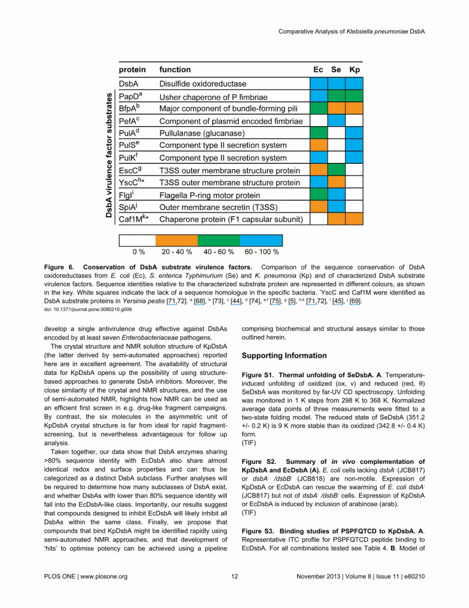
pressure for resistance development, since most virulencetraits are not essential for survival [62]. Targeting virulence mayalso expand the repertoire of antimicrobial targets, preserve theendogenous host microbiome and extend the lifespan ofconventional antibiotics [61]. Most antivirulence strategiesdeveloped to date target individual virulence factors [61-65]and this has yielded some successes [66,67]. However, thearmory of DsbA substrate virulence factors expressed indifferent Enterobacteriaceae varies (Figure 6), so that drugs
targeting specific virulence factors may not be effective againstall Enterobacteriaceae. On the other hand, DsbA itselfcatalyzes assembly of many virulence factors [68-70] andDsbA knockouts severely attenuate virulence in infectionmodels [12]. Targeting DsbA is therefore a compellingapproach for the development of anti-virulence agents,because DsbA inhibitors should inhibit a range of virulencetraits. Significantly, our findings point to the opportunity to
Figure 5. Conformational variability in X-ray and NMR structures of KpDsbA. A Superimposition of the six KpDsbA molecules(blue) in the asymmetric unit shows the limited conformational variability in the side chains of active site and L1, L2, and L3 loopresidues (stick representation). B. Cartoon representation of the KpDsbA crystal structure (Molecule D), with Cα atoms colored bytemperature factor (B-factor). Molecule D was selected as its temperature factor distribution is the most pronounced due to minimalcrystallographic contacts. In particular, the high B-factor of loop L3 indicates mobility in that region, consistent with the NMR data C.Stereo diagram of representative states of reduced (X-ray, cyan) and oxidized (NMR, yellow) structures of KpDsbA. Red arrowshighlight differences in the structures at N-terminal and L3 loop regions.doi: 10.1371/journal.pone.0080210.g005
Comparative Analysis of Klebsiella pneumoniae DsbA
PLOS ONE | www.plosone.org 11 November 2013 | Volume 8 | Issue 11 | e80210
develop a single antivirulence drug effective against DsbAsencoded by at least seven Enterobacteriaceae pathogens.
The crystal structure and NMR solution structure of KpDsbA(the latter derived by semi-automated approaches) reportedhere are in excellent agreement. The availability of structuraldata for KpDsbA opens up the possibility of using structure-based approaches to generate DsbA inhibitors. Moreover, theclose similarity of the crystal and NMR structures, and the useof semi-automated NMR, highlights how NMR can be used asan efficient first screen in e.g. drug-like fragment campaigns.By contrast, the six molecules in the asymmetric unit ofKpDsbA crystal structure is far from ideal for rapid fragment-screening, but is nevertheless advantageous for follow upanalysis.
Taken together, our data show that DsbA enzymes sharing>80% sequence identity with EcDsbA also share almostidentical redox and surface properties and can thus becategorized as a distinct DsbA subclass. Further analyses willbe required to determine how many subclasses of DsbA exist,and whether DsbAs with lower than 80% sequence identity willfall into the EcDsbA-like class. Importantly, our results suggestthat compounds designed to inhibit EcDsbA will likely inhibit allDsbAs within the same class. Finally, we propose thatcompounds that bind KpDsbA might be identified rapidly usingsemi-automated NMR approaches, and that development of‘hits’ to optimise potency can be achieved using a pipeline
comprising biochemical and structural assays similar to thoseoutlined herein.
Supporting Information
Figure S1. Thermal unfolding of SeDsbA. A. Temperature-induced unfolding of oxidized (ox, ν) and reduced (red, θ)SeDsbA was monitored by far-UV CD spectroscopy. Unfoldingwas monitored in 1 K steps from 298 K to 368 K. Normalizedaverage data points of three measurements were fitted to atwo-state folding model. The reduced state of SeDsbA (351.2+/- 0.2 K) is 9 K more stable than its oxidized (342.8 +/- 0.4 K)form.(TIF)
Figure S2. Summary of in vivo complementation ofKpDsbA and EcDsbA (A). E. coli cells lacking dsbA- (JCB817)or dsbA- /dsbB- (JCB818) are non-motile. Expression ofKpDsbA or EcDsbA can rescue the swarming of E. coli dsbA-
(JCB817) but not of dsbA- /dsbB- cells. Expression of KpDsbAor EcDsbA is induced by inclusion of arabinose (arab).(TIF)
Figure S3. Binding studies of PSPFQTCD to KpDsbA. A.Representative ITC profile for PSPFQTCD peptide binding toEcDsbA. For all combinations tested see Table 4. B. Model of
Figure 6. Conservation of DsbA substrate virulence factors. Comparison of the sequence conservation of DsbAoxidoreductases from E. coli (Ec), S. enterica Typhimurium (Se) and K. pneumonia (Kp) and of characterized DsbA substratevirulence factors. Sequence identities relative to the characterized substrate protein are represented in different colours, as shownin the key. White squares indicate the lack of a sequence homologue in the specific bacteria. *YscC and Caf1M were identified asDsbA substrate proteins in Yersinia pestis [71,72]. a [68], b [73], c [44], d [74], e,f [75], g [5], h,k [71,72], i [45], j [69].doi: 10.1371/journal.pone.0080210.g006
Comparative Analysis of Klebsiella pneumoniae DsbA
PLOS ONE | www.plosone.org 12 November 2013 | Volume 8 | Issue 11 | e80210

the interaction of the KpDsbA (molecule A) with PSPFQTCDgenerated by structural superposition on the EcDsbA:EcDsbBcomplex [76].(TIF)
Figure S4. NMR structure of oxidized KpDsbA. A. Overlayof the 20 NMR models; disordered region highlighted in blue.B. lowest energy NMR conformer. c. magnification of the activesite region showing the disulfide bond formed between thecysteines in the averaged NMR solution structure of oxidizedKpDsbA.(TIF)
Acknowledgements
We thank the Australasian Crystallography School, especiallyProf. Eleanor Dodson FRS for her advice and support to FK in
solving the molecular replacement problem for KpDsbA. Wethank the beam-line support team at the Australian Synchrotronfor data collection advice and acknowledge use of theUniversity of Queensland Remote Operation Crystallization andX-ray (UQ ROCX) Diffraction Facility and thank Mr Karl Byrieland Mr Gordon King for their expert assistance.
Author Contributions
Conceived and designed the experiments: FK KR MJS JLM.Performed the experiments: FK KR LP BM WD MH SRS BH.Analyzed the data: FK KR LP BM WD MH BH MJS JLM.Contributed reagents/materials/analysis tools: FK KR SRS DPFMJS JLM. Wrote the manuscript: FK KR LP MJS JLM.Provided critical comment on the manuscript: JLM FK KR LPBM WD MH SRS BH DPF MJS.
References
1. Souli M, Galani I, Giamarellou H (2008) Emergence of extensivelydrug-resistant and pandrug-resistant Gram-negative bacilli in Europe.Euro Surveill 13(47): 19045. PubMed: 19021957.
2. Heras B, Shouldice SR, Totsika M, Scanlon MJ, Schembri MA et al.(2009) DSB proteins and bacterial pathogenicity. Nat Rev Microbiol 7:215-225. doi:10.1038/nrmicro2087. PubMed: 19198617.
3. Bardwell JC, McGovern K, Beckwith J (1991) Identification of a proteinrequired for disulfide bond formation in vivo. Cell 67: 581-589. doi:10.1016/0092-8674(91)90532-4. PubMed: 1934062.
4. Dutton RJ, Boyd D, Berkmen M, Beckwith J (2008) Bacterial speciesexhibit diversity in their mechanisms and capacity for protein disulfidebond formation. Proc Natl Acad Sci U S A 105: 11933-11938. doi:10.1073/pnas.0804621105. PubMed: 18695247.
5. Miki T, Okada N, Kim Y, Abe A, Danbara H (2008) DsbA directsefficient expression of outer membrane secretin EscC of theenteropathogenic Escherichia coli type III secretion apparatus. MicrobPathog 44: 151-158. doi:10.1016/j.micpath.2007.09.001. PubMed:17933489.
6. Wunderlich M, Glockshuber R (1993) Redox properties of proteindisulfide isomerase (DsbA) from Escherichia coli. Protein Sci 2:717-726. doi:10.1002/pro.5560020503. PubMed: 8495194.
7. Zapun A, Bardwell JC, Creighton TE (1993) The reactive anddestabilizing disulfide bond of DsbA, a protein required for proteindisulfide bond formation in vivo. Biochemistry 32: 5083-5092. doi:10.1021/bi00070a016. PubMed: 8494885.
8. Martin JL, Bardwell JC, Kuriyan J (1993) Crystal structure of the DsbAprotein required for disulphide bond formation in vivo. Nature 365:464-468. doi:10.1038/365464a0. PubMed: 8413591.
9. Huber-Wunderlich M, Glockshuber R (1998) A single dipeptidesequence modulates the redox properties of a whole enzyme family.Fold Des 3: 161-171. doi:10.1016/S1359-0278(98)00024-8. PubMed:9562546.
10. Kobayashi T, Kishigami S, Sone M, Inokuchi H, Mogi T et al. (1997)Respiratory chain is required to maintain oxidized states of the DsbA-DsbB disulfide bond formation system in aerobically growingEscherichia coli cells. Proc Natl Acad Sci U S A 94: 11857-11862. doi:10.1073/pnas.94.22.11857. PubMed: 9342327.
11. Ireland PM, McMahon RM, Marshall LE, Halili M, Furlong et al. (: Sep202013) Disarming Burkholderia pseudomallei: Structural andfunctional characterisation of a disulfide oxidoreductase (DsbA)required for virulence in vivo. Antioxid Redox Signal: Sep 20 Epub.
12. Totsika M, Heras B, Wurpel DJ, Schembri MA (2009) Characterizationof two homologous disulfide bond systems involved in virulence factorbiogenesis in uropathogenic Escherichia coli CFT073. J Bacteriol 191:3901-3908. doi:10.1128/JB.00143-09. PubMed: 19376849.
13. Heras B, Kurz M, Jarrott R, Shouldice SR, Frei P et al. (2008)Staphylococcus aureus DsbA does not have a destabilizing disulfide. Anew paradigm for bacterial oxidative folding. J Biol Chem 283:4261-4271. PubMed: 18077463.
14. Shouldice SR, Heras B, Walden PM, Totsika M, Schembri MA et al.(2011) Structure and function of DsbA, a key bacterial oxidative folding
catalyst. Antioxid Redox Signal 14: 1729-1760. doi:10.1089/ars.2010.3344. PubMed: 21241169.
15. Vivian JP, Scoullar J, Rimmer K, Bushell SR, Beddoe T et al. (2009)Structure and function of the oxidoreductase DsbA1 from Neisseriameningitidis. J Mol Biol 394: 931-943. doi:10.1016/j.jmb.2009.09.065.PubMed: 19815019.
16. Shouldice SR, Heras B, Jarrott R, Sharma P, Scanlon MJ et al. (2010)Characterization of the DsbA oxidative folding catalyst fromPseudomonas aeruginosa reveals a highly oxidizing protein that bindssmall molecules. Antioxid Redox Signal 12: 921-931. doi:10.1089/ars.2009.2736. PubMed: 19788398.
17. Hu SH, Peek JA, Rattigan E, Taylor RK, Martin JL (1997) Structure ofTcpG, the DsbA protein folding catalyst from Vibrio cholerae. J Mol Biol268: 137-146. doi:10.1006/jmbi.1997.0940. PubMed: 9149147.
18. Studier FW (2005) Protein production by auto-induction in high densityshaking cultures. Protein Expr Purif 41: 207-234. doi:10.1016/j.pep.2005.01.016. PubMed: 15915565.
19. Bader M, Muse W, Zander T, Bardwell J (1998) Reconstitution of aprotein disulfide catalytic system. J Biol Chem 273: 10302-10307. doi:10.1074/jbc.273.17.10302. PubMed: 9553083.
20. Holmgren A (1979) Thioredoxin catalyzes the reduction of insulindisulfides by dithiothreitol and dihydrolipoamide. J Biol Chem 254:9627-9632. PubMed: 385588.
21. Nelson JW, Creighton TE (1994) Reactivity and ionization of the activesite cysteine residues of DsbA, a protein required for disulfide bondformation in vivo. Biochemistry 33: 5974-5983. doi:10.1021/bi00185a039. PubMed: 8180227.
22. Ellman GL (1959) Tissue sulfhydryl groups. Arch Biochem Biophys 82:70-77. doi:10.1016/0003-9861(59)90090-6. PubMed: 13650640.
23. Kurz M, Iturbe-Ormaetxe I, Jarrott R, Shouldice SR, Wouters MA et al.(2009) Structural and functional characterization of the oxidoreductasealpha-DsbA1 from Wolbachia pipientis. Antioxid Redox Signal 11:1485-1500. doi:10.1089/ars.2008.2420. PubMed: 19265485.
24. McPhillips TM, McPhillips SE, Chiu HJ, Cohen AE, Deacon AM et al.(2002) Blu-Ice and the Distributed Control System: software for dataacquisition and instrument control at macromolecular crystallographybeamlines. J Synchrotron Radiat 9: 401-406. doi:10.1107/S0909049502015170. PubMed: 12409628.
25. Battye TG, Kontogiannis L, Johnson O, Powell HR, Leslie AG (2011)iMOSFLM: a new graphical interface for diffraction-image processingwith MOSFLM. Acta Crystallogr D Biol Crystallogr 67: 271-281. doi:10.1107/S0907444910048675. PubMed: 21460445.
26. Kabsch W (2010) Xds. Acta Crystallogr D Biol Crystallogr 66: 125-132.doi:10.1107/S0907444909047337. PubMed: 20124692.
27. Evans P (2006) Scaling and assessment of data quality. ActaCrystallogr D Biol Crystallogr 62: 72-82. doi:10.1107/S0108767306098564. PubMed: 16369096.
28. McCoy AJ, Grosse-Kunstleve RW, Adams PD, Winn MD, Storoni LC etal. (2007) Phaser crystallographic software. J Appl Crystallogr 40:658-674. doi:10.1107/S0021889807021206. PubMed: 19461840.
Comparative Analysis of Klebsiella pneumoniae DsbA
PLOS ONE | www.plosone.org 13 November 2013 | Volume 8 | Issue 11 | e80210

29. Emsley P, Lohkamp B, Scott WG, Cowtan K (2010) Features anddevelopment of Coot. Acta Crystallogr D Biol Crystallogr 66: 486-501.doi:10.1107/S0907444910007493. PubMed: 20383002.
30. Adams PD, Afonine PV, Bunkóczi G, Chen VB, Davis IW et al. (2010)PHENIX: a comprehensive Python-based system for macromolecularstructure solution. Acta Crystallogr D Biol Crystallogr 66: 213-221. doi:10.1107/S0907444909052925. PubMed: 20124702.
31. Chen VB, Arendall WB 3rd, Headd JJ, Keedy DA, Immormino RM et al.(2010) MolProbity: all-atom structure validation for macromolecularcrystallography. Acta Crystallogr D Biol Crystallogr 66: 12-21. doi:10.1107/S1744309109042018. PubMed: 20057044.
32. Baker NA, Sept D, Joseph S, Holst MJ, McCammon JA (2001)Electrostatics of nanosystems: application to microtubules and theribosome. Proc Natl Acad Sci U S A 98: 10037-10041. doi:10.1073/pnas.181342398. PubMed: 11517324.
33. Dundas J, Ouyang Z, Tseng J, Binkowski A, Turpaz Y et al. (2006)CASTp: computed atlas of surface topography of proteins withstructural and topographical mapping of functionally annotatedresidues. Nucleic Acids Res 34: W116-W118. doi:10.1093/nar/gkl601.PubMed: 16844972.
34. Li Z, Ye Y, Godzik A (2006) Flexible Structural Neighborhood - adatabase of protein structural similarities and alignments. Nucleic AcidsRes 34: D277-D280. doi:10.1093/nar/gkj124. PubMed: 16381864.
35. Fiorito F, Herrmann T, Damberger FF, Wuthrich K (2008) AutomAutomated amino acid side-chain NMR assignment of proteins using(13)C- and (15)N-resolved 3D [ (1)H, (1)H]-NOESY. J Biomol NMR 42:23-33
36. Mohanty B, Serrano P, Pedrini B, Jaudzems K, Geralt M et al. (2010)Comparison of NMR and crystal structures for the proteins TM1112 andTM1367. Acta Crystallogr Sect F Struct Biol Cryst Commun 66:1381-1392. doi:10.1107/S1744309110020956. PubMed: 20944235.
37. Güntert P (2004) Automated NMR structure calculation with CYANA.Methods Mol Biol 278: 353-378. PubMed: 15318003.
38. Koradi R, Billeter M, Wüthrich K (1996) MOLMOL: a program fordisplay and analysis of macromolecular structures. J Mol Graph 14:29-55. 8744573.
39. Inaba K, Murakami S, Suzuki M, Nakagawa A, Yamashita E et al.(2006) Crystal structure of the DsbB-DsbA complex reveals amechanism of disulfide bond generation. Cell 127: 789-801. doi:10.1016/j.cell.2006.10.034. PubMed: 17110337.
40. Malojcić G, Owen RL, Grimshaw JP, Glockshuber R (2008) Preparationand structure of the charge-transfer intermediate of the transmembraneredox catalyst DsbB. FEBS Lett 582: 3301-3307. doi:10.1016/j.febslet.2008.07.063. PubMed: 18775700.
41. UniProt Consortium (2012) Reorganizing the protein space at theUniversal Protein Resource (UniProt). Nucleic Acids Res 40: D71-D75.doi:10.1093/nar/gkr981. PubMed: 22102590.
42. Larkin MA, Blackshields G, Brown NP, Chenna R, McGettigan PA et al.(2007) Clustal W and Clustal X version 2.0. Bioinformatics 23:2947-2948. doi:10.1093/bioinformatics/btm404. PubMed: 17846036.
43. Heras B, Totsika M, Jarrott R, Shouldice SR, Guncar G et al. (2010)Structural and functional characterization of three DsbA paraloguesfrom Salmonella enterica serovar typhimurium. J Biol Chem 285:18423-18432. doi:10.1074/jbc.M110.101360. PubMed: 20233716.
44. Bouwman CW, Kohli M, Killoran A, Touchie GA, Kadner RJ et al.(2003) Characterization of SrgA, a Salmonella enterica serovarTyphimurium virulence plasmid-encoded paralogue of the disulfideoxidoreductase DsbA, essential for biogenesis of plasmid-encodedfimbriae. J Bacteriol 185: 991-1000. doi:10.1128/JB.185.3.991-1000.2003. PubMed: 12533475.
45. Hiniker A, Bardwell JC (2004) In vivo substrate specificity ofperiplasmic disulfide oxidoreductases. J Biol Chem 279: 12967-12973.PubMed: 14726535.
46. Inaba K, Ito K (2008) Structure and mechanisms of the DsbB-DsbAdisulfide bond generation machine. Biochim Biophys Acta 1783:520-529. doi:10.1016/j.bbamcr.2007.11.006. PubMed: 18082634.
47. Paxman JJ, Borg NA, Horne J, Thompson PE, Chin Y et al. (2009) Thestructure of the bacterial oxidoreductase enzyme DsbA in complex witha peptide reveals a basis for substrate specificity in the catalytic cycleof DsbA enzymes. J Biol Chem 284: 17835-17845. doi:10.1074/jbc.M109.011502. PubMed: 19389711.
48. Boucher HW, Talbot GH, Bradley JS, Edwards JE, Gilbert D et al.(2009) Bad bugs, no drugs: no ESKAPE! An update from the InfectiousDiseases Society of America. Clin Infect Dis 48: 1-12. doi:10.1086/591855. PubMed: 19035777.
49. Keynan Y, Rubinstein E (2007) The changing face of Klebsiellapneumoniae infections in the community. Int J Antimicrob Agents 30:385-389. doi:10.1016/j.ijantimicag.2007.06.019. PubMed: 17716872.
50. Dailey FE, Berg HC (1993) Mutants in disulfide bond formation thatdisrupt flagellar assembly in Escherichia coli. Proc Natl Acad Sci U S A90: 1043-1047. doi:10.1073/pnas.90.3.1043. PubMed: 8503954.
51. Lafaye C, Iwema T, Carpentier P, Jullian-Binard C, Kroll JS et al.(2009) Biochemical and structural study of the homologues of the thiol-disulfide oxidoreductase DsbA in Neisseria meningitidis. J Mol Biol 392:952-966. doi:10.1016/j.jmb.2009.07.056. PubMed: 19631659.
52. Hillson DA, Lambert N, Freedman RB (1984) Formation andisomerization of disulfide bonds in proteins: protein disulfide-isomerase.Methods Enzymol 107: 281-294. doi:10.1016/0076-6879(84)07018-X.PubMed: 6503714.
53. Premkumar L, Heras B, Duprez W, Walden P, Halili M et al. (2013)Rv2969c, essential for optimal growth in Mycobacterium tuberculosis, isa DsbA-like enzyme that interacts with VKOR-derived peptides and hasatypical features of DsbA-like disulfide oxidases. Acta Crystallogr D BiolCrystallogr 69: 1981-1994. doi:10.1107/S0907444913017800. PubMed:24100317.
54. Walden PM, Heras B, Chen KE, Halili MA, Rimmer K et al. (2012) The1.2 Å resolution crystal structure of TcpG, the Vibrio cholerae DsbAdisulfide-forming protein required for pilus and cholera-toxin production.Acta Crystallogr D Biol Crystallogr 68: 1290-1302. doi:10.1107/S0907444912026388. PubMed: 22993083.
55. Guddat LW, Bardwell JC, Zander T, Martin JL (1997) The unchargedsurface features surrounding the active site of Escherichia coli DsbAare conserved and are implicated in peptide binding. Protein Sci 6:1148-1156. doi:10.1002/pro.5560060603. PubMed: 9194175.
56. Horne J, d'Auvergne EJ, Coles M, Velkov T, Chin Y et al. (2007)Probing the flexibility of the DsbA oxidoreductase from Vibrio cholerae -a 15N - 1H heteronuclear NMR relaxation analysis of oxidized andreduced forms of DsbA. J Mol Biol 371: 703-716. doi:10.1016/j.jmb.2007.05.067. PubMed: 17585933.
57. Guddat LW, Bardwell JC, Glockshuber R, Huber-Wunderlich M, ZanderT et al. (1997) Structural analysis of three His32 mutants of DsbA:support for an electrostatic role of His32 in DsbA stability. Protein Sci 6:1893-1900. doi:10.1002/pro.5560060910. PubMed: 9300489.
58. Schirra HJ, Renner C, Czisch M, Huber-Wunderlich M, Holak TA et al.(1998) Structure of reduced DsbA from Escherichia coli in solution.Biochemistry 37: 6263-6276. doi:10.1021/bi980136y. PubMed:9572841.
59. Guddat LW, Bardwell JC, Martin JL (1998) Crystal structures ofreduced and oxidized DsbA: investigation of domain motion andthiolate stabilization. Structure 6: 757-767. doi:10.1016/S0969-2126(98)00077-X. PubMed: 9655827.
60. Ho J, Tambyah PA, Paterson DL (2010) Multiresistant Gram-negativeinfections: a global perspective. Curr Opin Infect Dis 23: 546-553. doi:10.1097/QCO.0b013e32833f0d3e. PubMed: 20802331.
61. Clatworthy AE, Pierson E, Hung DT (2007) Targeting virulence: a newparadigm for antimicrobial therapy. Nat Chem Biol 3: 541-548. doi:10.1038/nchembio.2007.24. PubMed: 17710100.
62. Rasko DA, Sperandio V (2010) Anti-virulence strategies to combatbacteria-mediated disease. Nat Rev Drug Discov 9: 117-128. doi:10.1038/nrd3013. PubMed: 20081869.
63. Alksne LE, Projan SJ (2000) Bacterial virulence as a target forantimicrobial chemotherapy. Curr Opin Biotechnol 11: 625-636. doi:10.1016/S0958-1669(00)00155-5. PubMed: 11102800.
64. Lee YM, Almqvist F, Hultgren SJ (2003) Targeting virulence forantimicrobial chemotherapy. Curr Opin Pharmacol 3: 513-519. doi:10.1016/j.coph.2003.04.001. PubMed: 14559097.
65. Barczak AK, Hung DT (2009) Productive steps toward an antimicrobialtargeting virulence. Curr Opin Microbiol 12: 490-496. doi:10.1016/j.mib.2009.06.012. PubMed: 19631578.
66. Pinkner JS, Remaut H, Buelens F, Miller E, Aberg V et al. (2006)Rationally designed small compounds inhibit pilus biogenesis inuropathogenic bacteria. Proc Natl Acad Sci U S A 103: 17897-17902.doi:10.1073/pnas.0606795103. PubMed: 17098869.
67. Felise HB, Nguyen HV, Pfuetzner RA, Barry KC, Jackson SR et al.(2008) An inhibitor of gram-negative bacterial virulence proteinsecretion. Cell Host Microbe 4: 325-336. doi:10.1016/j.chom.2008.08.001. PubMed: 18854237.
68. Jacob-Dubuisson F, Pinkner J, Xu Z, Striker R, Padmanhaban A et al.(1994) PapD chaperone function in pilus biogenesis depends onoxidant and chaperone-like activities of DsbA. Proc Natl Acad Sci U SA 91: 11552-11556. doi:10.1073/pnas.91.24.11552. PubMed: 7972100.
69. Miki T, Okada N, Danbara H (2004) Two periplasmic disulfideoxidoreductases, DsbA and SrgA, target outer membrane protein SpiA,a component of the Salmonella pathogenicity island 2 type III secretionsystem. J Biol Chem 279: 34631-34642. doi:10.1074/jbc.M402760200.PubMed: 15169785.
Comparative Analysis of Klebsiella pneumoniae DsbA
PLOS ONE | www.plosone.org 14 November 2013 | Volume 8 | Issue 11 | e80210

70. Lin D, Rao CV, Slauch JM (2008) The Salmonella SPI1 type threesecretion system responds to periplasmic disulfide bond status via theflagellar apparatus and the RcsCDB system. J Bacteriol 190: 87-97.doi:10.1128/JB.01323-07. PubMed: 17951383.
71. Jackson MW, Plano GV (1999) DsbA is required for stable expressionof outer membrane protein YscC and for efficient Yop secretion inYersinia pestis. J Bacteriol 181: 5126-5130. PubMed: 10438793.
72. Zav'yalov VP, Chernovskaya TV, Chapman DA, Karlyshev AV,MacIntyre S et al. (1997) Influence of the conserved disulphide bond,exposed to the putative binding pocket, on the structure and function ofthe immunoglobulin-like molecular chaperone Caf1M of Yersinia pestis.Biochem J 324 ( 2): 571-578. PubMed: 9182720.
73. Zhang HZ, Donnenberg MS (1996) DsbA is required for stability of thetype IV pilin of enteropathogenic escherichia coli. Mol Microbiol 21:787-797. doi:10.1046/j.1365-2958.1996.431403.x. PubMed: 8878041.
74. Sauvonnet N, Pugsley AP (1998) The requirement for DsbA inpullulanase secretion is independent of disulphide bond formation inthe enzyme. Mol Microbiol 27: 661-667. doi:10.1046/j.1365-2958.1998.00722.x. PubMed: 9489677.
75. Pugsley AP, Bayan N, Sauvonnet N (2001) Disulfide bond formation insecreton component PulK provides a possible explanation for the roleof DsbA in pullulanase secretion. J Bacteriol 183: 1312-1319. doi:10.1128/JB.183.4.1312-1319.2001. PubMed: 11157944.
76. Inaba K, Murakami S, Nakagawa A, Iida H, Kinjo M et al. (2009)Dynamic nature of disulphide bond formation catalysts revealed bycrystal structures of DsbB. EMBO J 28: 779-791. doi:10.1038/emboj.2009.21. PubMed: 19214188.
Comparative Analysis of Klebsiella pneumoniae DsbA
PLOS ONE | www.plosone.org 15 November 2013 | Volume 8 | Issue 11 | e80210